Chapter 14 The Swollen Limb
The development of diffuse or more localized swelling in one or more limbs can present a diagnostic challenge, requiring a systematic approach to identify the cause. Although the metacarpal, metatarsal, and pastern regions are most commonly affected, an entire limb may be swollen, or swelling may initially be restricted to the antebrachium or crus or the carpus or tarsus, with swelling subsequently spreading distally. This chapter discusses an approach to diagnosis and management but does not provide exhaustive differential diagnoses and treatments. Some conditions are discussed in more detail in other chapters.
Diagnosis
Many stabled horses develop some degree of enlargement of the distal limbs, especially the hindlimbs, that dissipates with work. Termed filled legs or cold edema (stocked-up, stoved-up), this swelling may be controlled by applying stable bandages and is of no consequence.
History
Accurate diagnosis of the cause of limb swelling requires knowledge of the history.
It may be pertinent to establish when the owner last thought that the horse was normal, especially in horses that are not inspected regularly while kept at pasture. The actual duration of swelling may be longer than the owner recognized. The veterinarian also should bear in mind that some owners are remarkably unobservant, despite maintaining that they look at and groom the horse thoroughly daily.
Swelling in a single limb usually reflects a local problem, whereas swelling in several limbs may be caused by systemic disease or a primary skin problem. The differential diagnosis should include the following: subsolar abscess (see Chapter 28); mud fever or scratches; scabby skin lesions on the palmar aspect of the fetlock; other bacterial pyodermas; hemorrhage or thrombosis (see Chapter 37); desmitis or tendonitis (see Chapters 69 to 72); cellulitis associated with superficial digital flexor tendonitis (see Chapter 69); skin necrosis and cellulitis after topical application of proprietary products; infected tendon or tendon sheath; cold edema; cellulitis caused by trauma or infection; fracture; hypertrophic osteopathy (see Chapter 37); muscle rupture (see Chapter 13); muscle trauma resulting in compartment syndrome (see Chapter 83); lymphangitis; photosensitization; equine viral arteritis; heart failure; and hypoproteinemia.
Clinical Examination
A systematic clinical examination should be performed by careful observation and palpation. The veterinarian should assess the horse’s posture, demeanor, and attitude. Depression may reflect pain or infection.
The digital pulse amplitudes should be assessed: any increase highly suggests a primary foot problem. The response to pressure and percussion applied with hoof testers should be evaluated. Pulse rate, the quality of the peripheral pulses, capillary refill time, and careful auscultation of the heart and lungs should reveal whether a primary cardiac problem exists.
The mucous membranes should be examined for evidence of petechial hemorrhages, which can be seen with purpura hemorrhagica. The veterinarian should relate the swelling to the color of the limbs; swelling confined to white limbs may result from photosensitization.
The clinician should establish the degree of lameness and bear in mind that mild stiffness may result from extensive limb swelling and mechanical restriction. Extremely severe lameness often reflects infection—periarticular (e.g., peritarsal cellulitis; see Chapters 44 and 107), intraarticular, intrathecal, intratendonous, or subsolar. A horse with a fracture may be less lame. Severe lameness may also be associated with an acute muscle tear or hemorrhage. Although there may be no obvious swelling in the acute stage, diffuse filling of the affected limb may develop over the following 24 to 48 hours distal to the site of injury.
The results of this clinical examination should suggest the likely causes of the swelling, but definitive diagnosis might not be possible without further investigation. This may include radiography, ultrasonography, routine hematological testing, measurement of total protein and fibrinogen, and liver enzyme levels if indicated. Paired serum samples may be required to confirm equine viral arteritis. Treating the horse symptomatically to reduce the soft tissue swelling may be helpful to facilitate more accurate palpation. This may include the use of nonsteroidal antiinflammatory drugs (NSAIDs), hydrotherapy, poulticing or leg sweats, bandaging, and walking, with or without antimicrobial medication. Without evidence of a primary infectious process the response to corticosteroids may be helpful diagnostically, because limb filling may be an immune-mediated response. The clinician should be prepared to make repeated examinations if a primary diagnosis is not readily apparent.
Early periosteal new bone associated with hypertrophic osteopathy is readily overexposed, and greatly reduced exposure factors are required for its radiological detection. On the first day of examination the results of radiographic and ultrasonographic examinations may be misleading, and repeated examinations may be necessary. After trauma, laceration, or both to the antebrachium, crus, and metacarpal or metatarsal regions, delayed-onset lameness caused by an occult spiral fracture of the radius, tibia, or third metacarpal or metatarsal bones is possible. Many oblique radiographic images or follow-up examination may be necessary to identify the fracture. If extensive cellulitis occurs around a joint or tendon sheath, but intraarticular or intrathecal infection is suspected, the examiner should be cautious about performing synoviocentesis through infected tissues, because iatrogenic intraarticular or intrathecal infection may ensue. If skin lesions are identified as a possible primary cause of limb swelling, but these fail to respond to topical or systemic treatment, obtaining skin biopsies for culture and histological examination or seeking specialist advice from a dermatologist may be necessary.
Management
Mud Fever
Mud fever (scratches or pastern dermatitis) is associated with bacterial or fungal skin infection and usually is restricted to the palmar or plantar aspect of the pastern but sometimes extends farther proximally if severe. Mud fever is associated with many excoriated skin lesions, which may develop severe crusting. Deep fissures may develop in the skin, especially if the condition goes unrecognized or in horses with many skin folds in the pastern region. Extensive edematous swelling often extends up the metacarpal and metatarsal regions. If the condition is mild, no associated lameness may occur, but severe lesions are associated with marked stiffness. The condition can occur in horses kept out in wet, muddy conditions or in horses that are stabled but work in a muddy environment. Certain soil types seem to be associated with a higher occurrence. Some horses seem prone to recurrent episodes, although this may in part reflect management practices. The condition is difficult to manage if the horse is left in wet, muddy pasture, and it must be stabled. The affected areas should be clipped and thoroughly cleaned with chlorhexidine solution. The scabs should be softened to facilitate removal. If the condition is mild, no further treatment may be required, but if the condition is more severe, daily topical application of lanolin-based emollient cream with trimethoprim and sulfadiazine and dexamethasone is indicated, sometimes combined with systemic antimicrobial treatment. Alternatively, a proprietary topical preparation can be used. The limbs should be carefully cleaned and dried after exercise.
Scabs on the Palmar Aspect of the Fetlock
Some horses seem prone to develop many small skin scabs on the palmar or plantar aspects of the fetlock. The scabs appear to be related to work on specific surfaces, which presumably cause skin irritation and subsequent bacterial infection. These skin lesions are often associated with diffuse swelling and can be exquisitely painful. The lesions rarely resolve spontaneously but usually respond to penicillin therapy.
Cellulitis Caused by Trauma
Direct trauma to a limb may result in extensive edematous soft tissue swelling unassociated with infection. If skin abrasion is concurrent, the site of the wound relative to synovial structures susceptible to infection must be evaluated carefully. Lameness may vary in degree, but if severe, the possibility of fracture must be considered. If little soft tissue covers the underlying bones, radiographic examination is prudent to eliminate the possibility of a fracture. With primary cellulitis, treatment with NSAIDs, rest, and controlled exercise is usually all that is required. A variety of commercial boots and wraps are available that provide hot and cold therapy combined sometimes with pulsed pressure, which may be beneficial in reducing soft tissue swelling. The limb should be bandaged between treatments.
Cellulitis Caused by Infection
Cellulitis associated with infection may result from blunt trauma, a penetrating wound, previous injection, or recent surgery, but in many horses no underlying cause is identified.1,2 Thoroughbred racehorses may be most susceptible.2,3 It usually results in fairly extensive soft tissue swelling, which tends to be warmer and more painful than swelling from noninfectious cellulitis (see Figure 5-8). Associated lameness may also be severe, depending in part on the location of infection. If the infection is untreated, abscessation may develop in muscular areas and may require surgical drainage. Cellulitis may also be concurrent with infectious osteitis or osteomyelitis (see Chapter 37). Horses with acute infectious cellulitis usually respond well to systemic broad-spectrum antimicrobial treatment (e.g., crystalline penicillin and gentamicin), unless clostridial organisms are involved (see Chapter 83). The most common bacterial isolates are Staphylococcus aureus and Streptococcus species. Analgesia may be necessary and should be used to try to prevent secondary laminitis in the contralateral limb.
Lymphangitis
So-called lymphangitis occurs more commonly in hindlimbs than forelimbs and is often unilateral but may be bilateral. Diffuse soft tissue swelling occurs throughout the limb, often extending to the distal aspect of the limb from immediately below the stifle. The superficial lymphatic vessels may appear more prominent than usual. Serum may ooze through taut skin. The degree of swelling usually results in mechanical stiffness that improves with progressive walking. Careful inspection may reveal some small skin lacerations, often in the more distal part of the limb. Once a horse has had a severe attack of lymphangitis, it seems prone to recurrence, often after seemingly innocuous skin abrasions. Although the condition appears to be triggered by infection, antimicrobial treatment alone is inadequate and usually must be combined with long-term corticosteroid treatment (dexamethasone 0.05 to 0.2 mg/kg once daily intravenously or intramuscularly, using the lowest dose necessary to control edema, and replacing with prednisolone 0.5 to 1 mg/kg intramuscularly or by mouth twice daily, when the dexamethasone dose is <0.04 mg/kg), together with aggressive hydrotherapy and walking exercise, with or without bandaging and topical application of leg sweats. Bandaging the metatarsal region in the face of more proximal limb swelling tends to result in persistent proximal limb swelling that cannot move distally. Although prompt aggressive treatment may resolve clinical signs, persistence of marked filling for more than a week may result in chronic enlargement of the limb. Ulcerative lymphangitis occurs much less frequently and is caused by bacterial or fungal infection.
Purpura Hemorrhagica
Purpura hemorrhagica usually occurs as a sequela to previous streptococcal respiratory infection but occasionally follows other antigenic stimuli. Purpura hemorrhagica results in extensive submucosal petechial hemorrhages, evident clinically in the mucous membranes but actually more widespread, including muscle and viscera. Facial swellings and limb edema may be extensive (Figure 14-1). The horse is usually depressed, inappetent, and pyrexic. Purpura hemorrhagica is an acute, probably immune-mediated, necrotizing vasculitis. Aggressive treatment with penicillin and corticosteroids (see the previous discussion) is required.
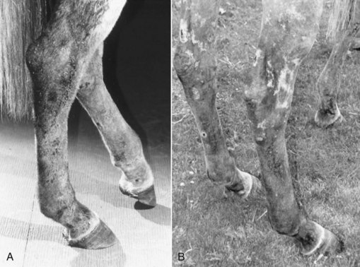
Fig. 14-1 Photographs before (A) and after (B) antibiotic and corticosteroid therapy of an aged Thoroughbred broodmare with suspected purpura hemorrhagica. Before therapy all four limbs were edematous and erythematous, and there was diffuse crusting and with serum discharge. Signs were not limited to the distal limbs; the mare had intermittent gastrointestinal pain and hyperemia of mucous membranes.
(Courtesy Mike W. Ross.)