DISEASES OF THE PARANASAL SINUSES

SINUSITIS

JOHN R. PASCOE

Definition and Etiology

Sinusitis refers to inflammation of the paranasal sinuses, from primary microbial infection or secondary bacterial infection associated with dental disease or other sinus disease. Lined by respiratory mucosa, the paranasal sinuses are at risk for developing diseases affecting the respiratory tract.1173 Sinus empyema, accumulation of pus within a sinus cavity, may result from bacterial or viral infection.

Incidence of sinusitis is relatively low, likely less than 0.5% of disease in equine practice.1174 In 256 horses with sinonasal disease, primary sinusitis (24%) and dental disease—associated sinusitis (22%) occurred with similar frequency and were the most common cause of sinusitis, whereas sinus cysts (13%), sinonasal neoplasia (8%), ethmoid hematoma (8%), sinonasal trauma (6%), mycosis (5%), and rostral maxillary tooth infection (4%) were the other more common causes.1175 The frontal and maxillary sinuses are more commonly involved, with the corresponding conchal sinuses and the sphenopalatine sinus affected less often. Median age of horses affected with sinonasal disease is 7 to 11 years.1175

Acute or chronic upper respiratory tract infections of viral or bacterial origin can result in primary sinusitis; Streptococcus species infection is the most common. Maxillary sinusitis caused by disease of the third (caudal roots) through sixth cheek teeth (modified Triadian numbers 109 to 111, 209 to 211) often results from alveolar periostitis, patent infundibula, or fractured or split teeth.1173 Dental defects permit access of food material or bacteria to the tooth root and sinus cavity, with extension to the frontal sinus likely through the frontomaxillary opening. Reported neoplasms include osteoma, osteosarcoma, adenocarcinoma, lymphosarcoma, squamous cell carcinoma, and fibroma.1176-1179

Clinical Signs and Differential Diagnosis

Signs vary depending on cause, location, and extent of sinus involvement, with unilateral nasal discharge (serous, mucoid, mucopurulent, purulent) being the most frequent sign.1175,1180 Physical examination of a horse with sinusitis should include observation for epiphora, facial asymmetry, altered nasal airflow, abnormal breath odor, mandibular lymphadenopathy, and sinocutaneous fistula.

Nasal discharge is typically unilateral because the nasomaxillary opening is located rostral to the caudal edge of the nasal septum. Intermittent or continuous, nasal discharge need not be related to a previous upper respiratory infection. Malodorous discharge is commonly associated with dental sinusitis and sinonasal mycosis, whereas mucopurulent discharge is more commonly associated with sinus cysts.1175 Sanguineous discharge is common with ethmoid hematoma and sinonasal trauma; however, guttural pouch mycosis, pulmonary hemorrhage, nasal turbinate necrosis, and neoplastic or granulomatous lesions should also be considered. Other differential diagnoses of nasal discharge should include guttural pouch empyema or mycosis; acute pharyngitis (strangles, rhinopneumonitis, and influenza); neoplasia or necrosis of the turbinates; ethmoid hematoma; and pulmonary disease.

  Page 588 

After nasal discharge, submandibular lymphadenopathy is the most common sign, particularly when microbial infection is a component of sinusitis. Facial swelling occurs with frequency similar to that of lymphadenopathy, typically when sinus drainage is obstructed or there is an expansile mass within the sinus, or as an acute sign after facial trauma. Occasionally exophthalmos can occur with marked sinus distortion.

Patency of the nasomaxillary opening generally precludes facial distortion. Loss of patency occurs when inspissated exudate accumulates or mucosal lining tissue reaction obstructs the opening. Expansion of the sinus may result in reduced nasal airflow, particularly ipsilaterally, caused by distortion of the architecture of the nasal passages, and in such instances abnormal respiratory noise rather than nasal discharge may be the primary presenting sign.1178

Epiphora occurs if there is nasolacrimal duct involvement from trauma, or compression or destruction of the duct from underlying sinus disease. Approximately one third to one half of horses with dental sinusitis, sinus cyst, and sinonasal trauma have epiphora as a presenting sign.1175

Percussion of the affected sinus may reveal dullness or pain, although normal resonance does not preclude the possibility of sinusitis. If there is bone thinning over gas above a fluid line (as can occur with some maxillary sinus cysts), percussion may elicit increased resonance.

Careful examination of the oral cavity for signs of dental or periodontal disease should be performed when any of these signs are present. Particular attention should be paid to examination of the occlusal surface of the teeth with a very fine dental pick (e.g., 22-gauge needle); however, it should be recognized that periapical abscess formation can occur without defects in the occlusal surface. Accuracy of diagnosis of dental disease is substantially enhanced by use of intraoral endoscopy and CT.

Clinical Pathology

The hemogram in animals with sinusitis generally remains within the normal range, although acute sinusitis of infectious origin may be associated with neutrophilia. With chronic sinusitis there may be concurrent hyperfibrinogenemia. Sinus fluid obtained by percutaneous centesis should be examined cytologically (including a Gram stain) and submitted for microbial culture and susceptibility testing to differentiate among bacterial, fungal, and neoplastic disease. Flecks of feed material indicate sinusitis secondary to dental abnormalities.

Laboratory Aids and Definitive Diagnostic Tests

A presumptive diagnosis of sinusitis can be made from the physical examination and associated clinical signs.1175,1181 Procedures most helpful in establishing a diagnosis of sinus disease in 85 horses were radiography (92%), endoscopy (38%), percutaneous centesis (21%), and examination of the oral cavity (20%).1177

Endoscopic examination of the nasal cavity may permit identification of exudate draining from the nasomaxillary opening or in advanced cases may reveal distortion of the nasal cavity secondary to sinus enlargement. Middle meatus examination is an important component of endoscopic examination. Diagnosis beyond recognition of the potential source of the nasal discharge is limited unless there is an obvious mass or abnormal tissue is observed. Sinoscopy can be accomplished by using an arthroscope1182 or flexible endoscope1183 inserted through small trephine holes in either the maxillary or the frontal sinus. Examination of the rostral compartment of the maxillary sinus requires a separate portal unless the bony septum between the rostral and caudal compartments has been destroyed. Observation may be limited by fluid or tissue especially in the rostral compartment of the maxillary sinus but can potentially be enhanced after sinus lavage and aspiration.

Standard radiographic projections include the standing lateral, dorsoventral, and right and left oblique views.1184,1185 Radiographic findings include fluid lines within the sinus, space-occupying soft-tissue densities, areas of decreased bone density, fractures, or dental abnormalities. Dental root disease is identified radiographically by a loss of continuity of the lamina dura and lysis of the tooth root or surrounding bone, combined with new bone formation and cement deposition.1184 Anatomy of normal equine skulls as demonstrated by CT and MRI has been described.1186-1188 Diagnostic accuracy, especially determination of the extent of involvement of structures within the skull, can be enhanced by CT.1189,1190 CT imaging enhances diagnostic accuracy of tooth involvement.1190 Findings associated with dental caries include hypoattenuation of cementum, destruction of enamel, and filling of the infundibular cavity with gas, whereas with dental decay there is gas accumulation in the root area or fragmentation of the root, and sinus mucosal thickening. Additional changes with sinusitis typically involve the maxilla with endosteal sclerosis, thickening, periosteal reaction, and deformation, especially involving the facial crest.1190 Scintigraphic examination may improve specificity in identification of dental involvement in sinusitis.1191,1192

Percutaneous sinus centesis may provide a definitive diagnosis and allow an avenue for subsequent therapy. Cytologic evaluation, with concurrent microbial culture and antibiotic susceptibility testing, may elucidate the cause of the sinusitis.1177 Isolation of a single organism such as Streptococcus species generally indicates a primary sinusitis, whereas polymicrobial infection is more compatible with sinusitis of dental origin. Visual examination of the oral cavity, especially intraoral endoscopy, and careful probing of the occlusal surfaces with a dental pick may identify dental abnormalities.

Necropsy Findings

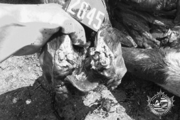
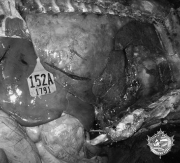
Affected sinuses contain fluid or tissue of variable color and consistency. Fluid character ranges from clear and odorless with cystic sinus disease to white, yellow, or green purulent fluid with a variable, but often putrid, odor in sinusitis resulting from other causes. Sinusitis of dental origin has a characteristically pungent and unpleasant odor. Granulomatous lesions have been reported to appear as large lobular gelatinous masses filling the sinus cavity. The gross appearance of neoplastic lesions within the sinus cavity depends on the type of neoplasm. Neoplasia may cause surrounding soft-tissue and bony destruction, whereas large, benign space-occupying lesions may result in distortion of the nasal turbinates and nasal septum, as well as external facial bone distortion.

Treatment and Prognosis

Not infrequently, horses with a chronic mucopurulent nasal discharge from sinusitis have a history of response to antimicrobial therapy, followed by recurrence of the discharge after antibiotic therapy ceases. Definitive diagnosis of sinusitis can be accomplished using the techniques described earlier. Sinoscopy permits examination of the paranasal sinuses and in some instances facilitates treatment.1184,1193

  Page 589 

Suggested treatment for primary sinusitis or empyema involves daily lavage of the sinus through a percutaneous centesis site with 1 L of saline, to which a broad-spectrum antibiotic or antiseptic has been added. Once the results of culture and susceptibility testing are available, the appropriate antibiotic should be administered locally in the flush solution, as well as systemically, for 14 days. Resolution or reduction in the volume of nasal discharge is an indication of successful therapy. If little progress is made after 10 to 14 days or if drainage recurs, sinusotomy (trephination or bone flap technique, standing or recumbent anesthetized) may be required to resolve the condition.1194 The prognosis is generally favorable if primary sinusitis is not chronic and if the mucous membrane is not markedly thickened.1195 Chronic sinus disease (longer than 6 months) carries a poor prognosis, and, for resolution to occur, surgical removal of the thickened, infected mucous membrane is required; creation of sinonasal drainage by sinus fenestration is also recommended. Isolation of Pseudomonas species from a sinus aspirate generally indicates an unfavorable prognosis.1180

Sinusitis that results from secondary factors is generally not responsive to medical management. Such conditions include diseased teeth, granulomas, or neoplasia; surgical removal of the inciting cause is required, and adjunctive treatment may be required. The prognosis for sinusitis associated with dental abnormalities is usually favorable once the diseased tooth has been removed.1195,1196 If the periodontal ligament is intact, endodontic therapy can be used to save the tooth. This is accomplished by surgical apicoectomy and retrograde occlusion of the root canal after debridement of the pulp. In geriatric horses with dental-associated sinusitis, when economic constraints limit surgical options, sinusotomy and periodic sinus lavage and antibiotic therapy have been used successfully to manage nasal discharge.

The prognosis for resolution of granulomatous lesions is generally guarded and depends on surgical access and extent of the lesion. Neoplastic lesions are often well established and have metastasized, either locally or regionally, by the time they become clinically apparent; the prognosis for resolution is generally guarded to poor.1176 In a series of 16 horses with sinus neoplasia, 11 were euthanized because of the extent of the lesion, four lesions recurred after surgical removal, and one horse with squamous cell carcinoma was successfully treated and had no recurrence at a 2-year follow-up evaluation.1177

Prevention and Control

Prevention of sinusitis in horses is difficult because of the variety of causes. Isolation of horses from those with upper respiratory bacterial or viral diseases may be of benefit in preventing primary sinusitis. Regular dental care and a proper diet may help circumvent sinusitis caused by dental abnormalities, although many cases most likely result from a variety of causes not yet defined or over which the owner or veterinarian has no control.

ETHMOID HEMATOMA

JOHN R. PASCOE

Definition and Etiology

Also termed progressive ethmoidal hematoma1197 and hemorrhagic nasal polyps,1198 ethmoid hematomas are slowly expanding angiomatous masses that appear to originate principally from the mucosal lining of the ethmoid conchae. Smaller hemangiomas arising from the mucosal lining of the frontal, maxillary, and sphenopalatine sinuses have been recognized, but the relationship between these benign endothelial tumors and ethmoid hematoma is uncertain. A relationship between paranasal sinus cysts and ethmoid hematoma has been suggested,1199 but distinctly different histologic features characterize each disorder,1200 seemingly arguing against a common cause and the likelihood that these lesions are variants of each other. The cause of ethmoid hematoma is unknown, and it remains a relatively uncommon condition.1201-1205 Although reported in a 4-week-old foal and in 3-year-old horses, most affected horses are older than 8 years—generally thoroughbred, Arabian, or warmblood horses.1201-1207

Clinical Signs

A blood-tinged nasal discharge with intermittent epistaxis from one or both nostrils is the most common clinical sign.1208 Unilateral or bilateral, epistaxis varies from blood-tinged mucoid or mucopurulent discharge to blood spots or a trickle of blood. Fulminant or fatal epistaxis as can occur with guttural pouch mycosis is uncommon. If the hematoma occupies the choana(e) or nasal cavity, a mucopurulent, occasionally malodorous nasal discharge with some blood discoloration is more commonly seen. Typically these horses have a history of abnormal respiratory noise, both inspiratory and expiratory, especially during exercise. With nasal cavity involvement, airflow is usually reduced or may be absent on the affected side. Facial distortion or asymmetry is uncommon and is more likely to occur when the hematoma occupies the frontal and maxillary sinuses. Less commonly there may be an associated history of coughing, choking, ptyalism, increased respiratory effort during resting breathing, and either head shyness or head shaking.1197 If the hematoma has expanded into the paranasal sinuses, percussion yields a dull sound.

Laboratory Aids and Definitive Diagnostic Tests

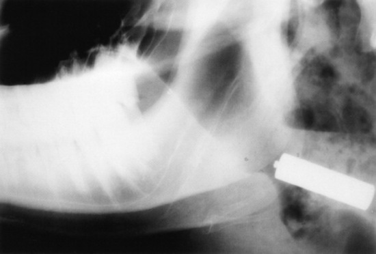
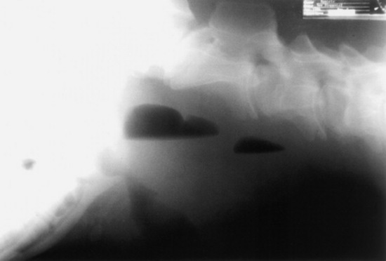
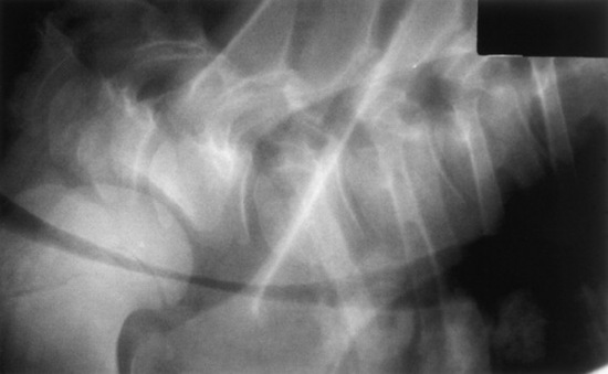
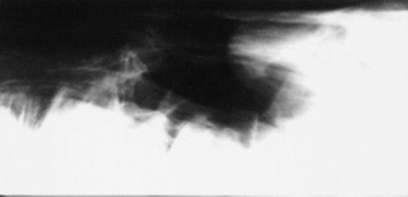
Confirmation requires endoscopy of the ethmoid conchae and skull radiography (Figs. 31-45 and 31-46); however, the origin and extent of the mass can be delineated more accurately by computed tomographic examination of the skull. Sinoscopy may be of diagnostic value in horses with ethmoid hematoma involving the paranasal sinuses without protrusion into the nasal cavity. Rarely, ethmoid hematoma infiltrates the nasal conchae; these lesions, identifiable by CT, may be missed by sinoscopy. Because ethmoid hematoma is bilateral in approximately 30% of affected horses, it is prudent to examine both the left and right ethmoidal conchae. The ethmoidal labyrinth is visible approximately 25 cm from the nares, with the endoscope positioned in the ventral nasal meatus and the viewing tip deflected dorsally. The rostral surface of the ethmoidal concha does not protrude beyond the caudal nasal cavity and has a bulbous shape and a moist pink to pale red mucosal covering.

image

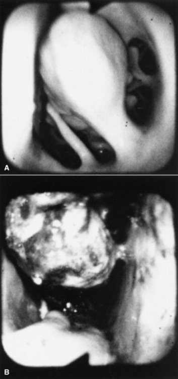
Fig. 31-45 Endoscopic view of left ethmoturbinate. A, Normal appearance. B, Ethmoturbinate obscured by an ethmoid hematoma.

image

Fig. 31-46 Lateral radiograph demonstrating an ethmoid hematoma.

Beyond the rostral surface the numerous pillars that form the ethmoidal conchae and separate the ethmoidal spaces (cellulae ethmoidales) are visible. Ethmoidal hematomas that project into the ventral meatus or through the choana into the nasopharynx often obscure the ethmoidal concha. Occasionally, unilateral ethmoid hematomas that have expanded into the nasopharynx may protrude into the contralateral ventral meatus, obscuring the view of the ethmoidal labyrinth on that side. The origin of hematomas that expand dorsally into the frontal sinus may not be visible on endoscopy, but hemorrhage that originates deep to the visible portion of the ethmoidal conchae may be evident or may be noticed from the region of the nasomaxillary opening in the middle meatus. Visible ethmoid hematomas can vary in color from deep red to red-purple or may have a yellow-brown or yellow-green-brown to bronze color. The surface is irregularly rounded, with small punctate hemorrhages or erosions, and may be partially covered in yellow-white mucopurulent material that may be admixed with blood. Often the floor of the ventral meatus and the regions of contact with the nasal cavity have pooled exudate of blood and mucopurulent matter. Manipulation of the visible surface of the hematoma with the tip of the endoscope may elicit bleeding or oozing.

  Page 590 

Recognition of a discrete, often smooth-surfaced homogeneous radiodensity originating from the ethmoidal conchae and extending into the frontal, maxillary, or sphenopalatine sinuses or into the pharynx or nasal cavity is suggestive of an ethmoid hematoma. Radiography is beneficial in determining the extent of the hematoma and in identifying suspected ethmoid hematomas that are not visible by endoscopy; however, precise definition of the origin of any hematoma is difficult from radiographic projections. Small hematomas contained within the ethmoid labyrinth may not be visible on radiographs. Computed tomographic examination of the skull allows more accurate assessment of the origin of the ethmoid hematoma,1207,1209 allows determination of the extent of involvement of the paranasal sinuses and conchae, and facilitates surgical planning.
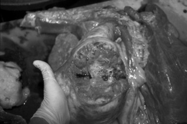
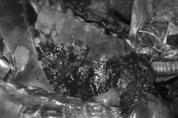
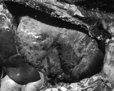
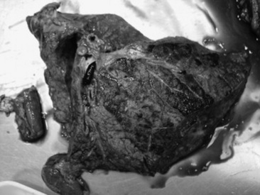
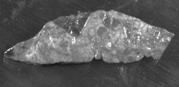
Necropsy Findings

Most of the morphologic features have been described from surgical specimens; few skulls with intact ethmoid hematomas have been examined.1198 Except in regions with necrosis or secondary infection associated with contact with the sinus or nasal cavity walls, the hematoma is a smooth-surfaced saclike structure containing blood in various stages of organization. The sac lining is generally healthy respiratory mucosa originating from a pedunculated region of the mucosal covering of the ethmoturbinal or sinus wall. On section the contents are amorphous red-black to chocolate brown, and in larger masses some evidence of irregular compartmentalization by fibrous tissue exists, especially on the inner surface of the sac.

Morphologic features include an outer covering of respiratory epithelium (flattened columnar or cuboidal ciliated epithelium containing glands, and occasionally stratified squamous epithelium1200) overlying an irregular zone of submucosal fibrous tissue, containing hemosiderophages, occasional plasma cells, and lymphocytes, less commonly neutrophils, that forms a pseudocapsule around hemorrhage in varying states of organization. There is typically variable organization of the fibrous tissue components. Endothelial cells do not show evidence of neoplasia. Thin endothelium-lined sinuses are often present within the myxomatous stroma. The respiratory epithelium is sometimes focally ulcerated and infiltrated with neutrophils, and occasionally there are squamous metaplastic changes. Ethmoid adenocarcinoma with a similar gross appearance to ethmoid hematoma has been reported in one horse.1210

Treatment and Prognosis

Because these masses slowly and progressively increase in size and can cause distortion of skull architecture if the paranasal sinuses are involved, removal is recommended. Treatment method depends on the location and size. Surgical ablation has been the preferred method; however, destruction of the hematoma by intralesional injection of formaldehyde solution is associated with less morbidity, although recurrence rates are similar to those with other methods.1204-1212 Surgical access is usually achieved by sinusotomy and then hematoma ablation by curettage, cryosurgery, or use of an Nd:YAG laser1213-1215; photoablation can also be accomplished through the biopsy channel of an endoscope. After sinusotomy, the pedunculated origin of the hematoma is identified by digital palpation and then dissected, frozen, or photoablated, and the hematoma removed. If the hematoma is friable, intact removal may not be possible, and hemorrhage may make observation of the origin of the mass difficult. After removal the paranasal sinuses and nasal cavity are packed with gauze to control postoperative hemorrhage.

  Page 591 

Surgical curettage can have the disadvantage of being associated with marked intraoperative blood loss typically from the turbinates or sinus mucosa rather than the hematoma. Temporary occlusion of both carotid arteries1214 can substantially decrease blood loss until the sinus cavity is packed with gauze. Blood loss is minimized by cryosurgical extirpation and photoablation techniques, but these approaches are not always practical when initially dealing with large hematomas. There is also minimum blood loss with transnasal photoablation; however, this technique requires multiple procedures to destroy large masses but can be performed in the standing sedated horse. Photoablation (Nd:YAG laser, 100 W in noncontact technique) is effective in controlling remnants after surgical extirpation or subsequent regrowth.1215,1216

Destruction of ethmoid hematomas by endoscopically guided intralesional injection of formalin in standing sedated horses may reduce the need for surgical ablation in many horses.1211,1217 A catheter passed through the endoscope biopsy channel is advanced through the rostral surface of the mass toward its origin, then 10% formalin (4% formaldehyde solution) is injected intralesionally. Commercially available catheters with a beveled needle tip have been used, but relatively stiff plastic tubing that will slide through the biopsy channel works will penetrate the capsule of the ethmoid hematoma. Sufficient volume (from 10 to 100 mL) is injected until fluid leaks back alongside the catheter or leaks from the mass. For visibly pedunculated masses, it is best to inject the formalin at the origin or neck of the mass. Tissue necrosis and slough occur in 5 to 10 days and may be associated with nasal discharge. Repeat injections, typically at no longer than 10- to 14-day intervals may be necessary to destroy the mass. Removal of necrotic tissue can be facilitated by use of long grasping forceps and hydropulsion. Mycotic plaques may cover the treated site during healing but typically resolve without treatment; endoscopically delivered topical natamycin has been used if mycotic plaque is extensive and associated with malodorous, purulent discharge.1218

Occasionally progression of the ethmoid hematoma may result in weakening or loss of the cribriform plate or roof of the sphenopalatine sinus.1218 During mass removal, loss of this protective bony covering may result in intraoperative or postoperative neurologic complications. Recognition of loss of the integrity of these bony plates may not occur until sinus lavage. In a rare complication death occurred after intralesional formalin injection in a horse where the cribriform plate had been eroded by the ethmoid hematoma.1219 Erosion of calvarial bone is rare, but careful computed tomographic evaluation of these regions in horses with extensive hematoma formation involving the frontal or sphenopalatine sinuses is warranted.

Irrespective of treatment method, recurrence of the hematoma occurs in 30% to 50% of cases from several months to years after the initial surgery.1198,1203-1206,1211,1212

RUMINANT RESPIRATORY SYSTEM

AMELIA R. WOOLUMS, Consulting Editor

UPPER RESPIRATORY TRACT DISEASES

AMELIA R. WOOLUMS

JOHN C. BAKER

JOHN A. SMITH

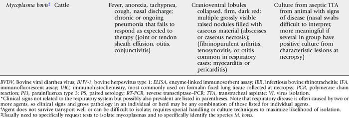
DISEASES OF THE NASAL CAVITY

AMELIA R. WOOLUMS

JOHN C. BAKER

JOHN A. SMITH

Mycotic or Bacterial Nasal Granuloma

Infectious granulomas in the nasal cavity of ruminants are not common. Documented causes include the fungal organisms Rhinosporidium seeberi and other Rhinosporidium species (which cause rhinosporidiosis), Helminthosporium species (which cause maduromycosis), Drechslera rostrata, Aspergillus species, Phycomycetes species, Stachybotrys species, and Bipolaris species. Phycomycosis is discussed in the dermatology section in Chapter 40. Nasal granuloma caused by Nocardia species bacteria has also been reported.1 There is no apparent age, breed, or seasonal predilection, and cases are sporadic. The major clinical signs are upper respiratory noise (stridor), dyspnea, and mucopurulent nasal discharge, sometimes with epistaxis. Affected animals may rub the nose, suggesting pruritus or irritation.2-3 Nasal airflow may be reduced, and open-mouth breathing may occur in advanced cases. Hot or dusty weather may accentuate the signs, giving the appearance of seasonal exacerbation, but the lesions are progressive. The granulomas may be single or multiple, unilateral or bilateral, and located anywhere in the nasal cavity. They consist of 0.5- to 5-cm yellow to yellow-green or red nodules or polyps, which may be sessile or pedunculated. Rhinosporidiosis tends to be a single unilateral polyp in the posterior nasal cavity, and maduromycosis tends to occur in the anterior cavity, but these distinctions are not consistent. Red and black spots (spores) may occur on the masses, and some may become secondarily infected with bacteria and ulcerate. Differential diagnoses include allergic rhinitis, foreign bodies, tumors, nasal actinobacillosis, nasal actinomycosis, and Oestrus ovis infection in small ruminants.

Endoscopy, biopsy, and culture of the lesions aid in the diagnosis. Histopathologic analysis reveals granulation tissue containing eosinophils, mononuclear cells, round sporangia, and sometimes hyphae or filamentous bacteria.1,2 The pathogenesis of the disease involves inoculation of eroded nasal mucosa with fungal spores or filamentous bacteria from the environment. The infectious agent causes a chronic delayed (type IV) hypersensitivity reaction, which eventually leads to the formation of a granuloma. Fungal granulomas can be more common in warm, wet climates. The granulomas can be difficult to treat, and although rarely fatal the disease is chronically debilitating, with salvage often being the most practical solution.

  Page 592 

Recommended treatments include surgical removal of the granulomas when possible and long-term sodium iodide (NaI) therapy. NaI can be administered at a dose of 66 mg/kg IV as a 20% solution, repeated at 10- to 14-day intervals until remission or iodism occurs. Iodism is characterized by lacrimation, cough, and scaling of the skin. The use of antifungal drugs to treat this condition in ruminants has not been reported.

Allergic Rhinitis and Enzootic Nasal Granuloma

Allergic rhinitis occurs in cattle and in its chronic stages may lead to the formation of granulomas. A similar condition may occur in sheep.4 The inciting antigen is frequently a plant pollen or more likely a fungal spore.5 Once homocytotropic antibody (immunoglobulin E or possibly other classes in cattle) to the allergen has developed, subsequent exposure results in a localized, ongoing, immediate (type I) hypersensitivity reaction.5-7 If recurrent exposure to the allergen occurs, repeated tissue damage by mast cell factors results in chronic epithelial, duct, and goblet cell hyperplasia and metaplasia, as well as mucous hypersecretion and granulomatous inflammation, suggesting that a type IV hypersensitivity reaction contributes to the chronic lesion.5,6,8

Any breed may be affected, but Channel Island breeds and Friesians seem most susceptible.5 The disease occurs sporadically in the United States. A familial predisposition has been reported.9 Most affected animals are between 6 months and 2 years of age. The signs are initially seasonal, usually occurring in warm, moist conditions; they include rhinorrhea, sneezing, nasal pruritus, a sudden onset of dyspnea, and stertorous inspiration.9,10 There is a profuse bilateral nasal discharge. Intense pruritus is characteristic and associated with sneezing, head shaking, and nose rubbing.10 In severe cases facial swelling, tachypnea, hyperpnea, and ulceration of the nasal mucosa may occur.6,10 Nasal foreign objects may result from the animal’s attempts to scratch the nasal mucosa. Lacrimation, chemosis, and blepharitis may also be present. In the chronic stages (the “enzootic nasal granuloma”), the signs are more constant, with seasonal exacerbations.6,8,10 The granulomas tend to be multiple, firm, white, raised nodules 1 to 2 mm in diameter with an intact mucosa, or pale pink flat plaques scattered throughout the nasal cavity. Differential diagnoses include fungal granulomas, foreign bodies, respiratory viruses, nasal actinomycosis or actinobacillosis, tumors, O. ovis infection (small ruminants), and irritation caused by inhalation of hot or irritant gases.

Endoscopy, biopsies, cultures, antigen detection tests for viruses, bacteria, or fungi, and serologic analysis can be used to rule out these differential diagnoses. Eosinophil counts in nasal secretions correlate with the susceptibility of the animal and activity of the disease, but no absolute level is diagnostic.5 Intradermal allergen testing has been suggested to aid in diagnosis, but interpretation of results needs to be done in conjunction with historical and clinical findings.7 This condition should be differentiated from fungal or bacterial granuloma because the therapy is different.

Treatment and control entail removal of the allergen, or removal of the animal from the allergen, and therapy to block the hypersensitivity reaction. Recommended drugs include various antihistamines, meclofenamic acid, and corticosteroids at standard antiinflammatory doses (0.05 to 0.2 mg of dexamethasone per kilogram IM or IV or 1 to 2.2 mg of prednisolone per kilogram IM or IV daily). Topical corticosteroid therapy can be considered in severe, acute occurrences of the disease. The adverse effects of corticosteroids on milk production and their potential to induce abortion or parturition should be considered before their use. Antihistamine therapy has had equivocal results.7

Nasal Foreign Bodies

Cattle are more prone than small ruminants to the acquisition of nasal foreign bodies. Foreign objects may be acquired as a result of attempts to scratch the nose in cases of allergic rhinitis, or because of the cow’s aggressive eating habits. Depending on the size and duration of residence of the object, signs may include head shaking, stridor, sneezing, snorting, frequent nose licking, unilateral decreases in airflow, foul odors, and serous, mucopurulent, or hemorrhagic discharges. Differential diagnoses include fungal granulomas, allergic rhinitis, tumors, nasal actinomycosis or actinobacillosis, and O. ovis (small ruminants). Many objects can be visualized on careful examination of the nasal cavity with an adequate light source, whereas some may require endoscopy for diagnosis and removal.

Nasal Trauma and Fractures

Trauma to the facial bones, sinuses, and turbinates may result from fighting, accidents caused by improper restraint, farm machinery accidents, human maliciousness, and passage of excessively large NGTs. Severe fractures can lead to facial swelling, SC emphysema, obstruction of airflow, stertor, and epistaxis. Secondary infection causes foul odors and mucopurulent nasal discharge. Differential diagnoses for the acute external swelling of the head with stertor include snakebite, actinobacillosis, actinomycosis, and phlegmon (Fusobacterium, Clostridium species). Unless the development of severe depression fractures, formation of sequestra, or severe obstruction of airflow occurs, surgery is usually not indicated. Radiographs confirm the diagnosis and help determine the need for surgical removal of potential sequestra or elevation and fixation of large displaced segments. Prophylactic antibiotics (typically penicillin, 22,000 U/kg IM or SC q12-24h) are recommended to prevent fracture infection and sinusitis, and NSAIDs (aspirin, 100 mg/kg PO twice; flunixin meglumine, 1.1 to 2.2 mg/kg IV daily or divided twice daily) may help relieve pain, swelling, and stridor. The prognosis is usually good.

Nasal Tumors and Polyps

Tumors and polyps of the nasal cavity and sinuses are rare in ruminants. Nasal tumors reported in cattle include osteomas and osteosarcomas of the sinuses, squamous cell carcinomas,11 neuroblastomas, and adenocarcinomas of the ethmoid mucosa. Ethmoid adenocarcinomas are speculated to be caused by viruses on the basis of an endemic pattern in some cases.12 They tend to occur in cattle 6 to 9 years of age and are frequently unilateral. Metastasis occurs to the lymph nodes and lungs. There is a report of a hemangiosarcoma involving the external naris of a cow.13 Signs common to all nasal tumors include mixed or inspiratory dyspnea, stridor, nasal discharge, epistaxis, foul breath odors, unilateral decreases in airflow, open-mouth breathing, and distortion of the facial bones. Differential diagnoses include fungal granulomas, atopic granulomas, foreign bodies, sinusitis, fractures, and nasal actinobacillosis and actinomycosis. Treatment has not been investigated.

  Page 593 

The majority of nasal neoplasms in sheep and goats are adenopapillomas, adenomas, or adenocarcinomas.14 Squamous cell carcinoma15 and osteoma16 have also been reported. Nasal adenocarcinomas have also been described in goats.17

An enzootic form of nasal adenocarcinoma occurs in both sheep and goats and is associated with ovine nasal adenocarcinoma virus (ONAV)18,19 or caprine nasal adenocarcinoma virus (CNAV),20 respectively. These agents are β retroviruses that are closely related to, but distinct from, jaagsiekte sheep retrovirus (JSRV), the cause of ovine pulmonary adenocarcinoma (OPA).20,21 Although nasal adenocarcinoma has been difficult to consistently reproduce experimentally, inoculation of kids with concentrated cell-free and bacteria-free filtrate containing virus from naturally infected goats has resulted in disease.22 Neoplastic transformation is limited to secretory epithelial cells of the nasal turbinates, but CNAV appears to have a wider tissue tropism than ONAV; in one study viral provirus incorporated into host DNA was found in many tissues of infected goats but was largely confined to tumor tissue of infected sheep.20 There is no breed or sex predisposition for enzootic nasal adenocarcinoma; affected animals are most commonly young adults, but the tumor has been identified as early as 4 months of age.18 Signs include progressive inspiratory dyspnea; stridor; exercise intolerance; mouth breathing; serous, mucoid, or mucopurulent nasal discharge, which is typically profuse; tachypnea; decreased airflow; head shaking; sneezing; exophthalmos; and facial asymmetry.14,17,18,21 The lesions may be unilateral or bilateral, originating in the olfactory region of the ethmoid turbinates. The neoplasia arises either from Bowman’s glands18 or serous glands of the nasal mucosa.17 The tumor begins as a small nodule that can grossly resemble the mucoid polyps that occur in animals with chronic rhinitis. Over time the adenocarcinoma grows into a soft, gray to grayish pink, mucoid, nodular, cystic mass. The tumor is benign but locally expansive, often entering the sinuses and eroding overlying bone.17 Histologically the tumor is typically classified as a low-grade adenocarcinoma, but it may also be identified as an adenopapilloma or adenoma.17 Initially affected animals eat and drink normally and maintain body condition, but as the tumor expands the animal begins to lose condition, and death eventually occurs as a result of inanition, asphyxia, or aspiration pneumonia. Necrosis or secondary bacterial infection of the tumor can occur and may lead to the production of foul-smelling, purulent discharge and systemic signs related to bacterial infection, such as fever, depression, and hyperemic mucous membranes.

Differential diagnoses for nasal neoplasia in small ruminants include nasal fungal or bacterial granuloma, actinobacillosis, actinomycosis, O. ovis infection, and sinusitis. Endoscopy and radiology are helpful in establishing an initial diagnosis of a nasal mass. Preoperative or antemortem pinch biopsies and exfoliative cytologic analysis frequently are nondiagnostic, and findings may be misleading.14,15 Identification of tumors in multiple animals in a herd or flock supports a diagnosis of enzootic nasal adenocarcinoma. Definitive identification of infection with ONAV or CNAV can be difficult. Serologic tests are not reliable because animals do not consistently produce antibody to the viruses,23 possibly because of the presence of endogenous retroviruses24 that share epitopes with ONAV and CNAV and induce the development of immunologic tolerance. Recently PCR has been used to specifically identify and differentiate ONAV, CNAV, and JSRV in tissues of affected animals.25 Surgical management of nasal adenocarcinoma in sheep has been described.26

OESTRUS OVIS INFESTATION

AMELIA R. WOOLUMS

Definition and Etiology

O. ovis is parasite of the nasal passages and sinuses of sheep and, less commonly, goats. Goats are relatively resistant to infection and, even when housed with sheep, tend to have a lower prevalence of infection.27 Occasionally humans and other animals are accidentally infected, with conjunctival infection the most common form of disease in humans.28,29 The pathogenic stage of the parasite is the larval stage. The first instar larvae are deposited near the nostrils of sheep by the adult female fly and migrate into the nasal and ethmoid turbinates; in the ethmoids the larvae molt to second instars, which migrate to the sinuses before molting again and becoming third instar larvae. Third instar larvae, which are yellow-white in color and have a dark dorsal stripe with rows of spines on the ventrum of each segment, return to the nasal passages and are sneezed out onto the ground, where they pupate and eventually develop into adult flies.30,31 The adult is active during warm months, and the parasite may overwinter either as a first instar larva in the host or as a pupa in the ground. Larvae can persist in the upper airways for weeks to months, and appear to be able to arrest development for a period of time if necessary to avoid climate extremes.32 Adult flies have a rudimentary mouthpiece and are not able to feed; thus the larvae must ingest adequate nutrients while in the host to support the life of the adult fly.33

Clinical Signs

The larvae cause irritation of the nasal passages and sinuses, leading to mucoid to mucopurulent and sometimes blood-tinged nasal discharge, sneezing, nose rubbing, and inspiratory stridor. Adults flies cause annoyance by flying around the heads of animals; thus both stages can lead to decreased productivity by decreasing the time spent grazing by affected animals. Occasionally the larvae may cause sinusitis or pneumonia owing to secondary bacterial infection associated with irritation caused by the larval infestation. Differential diagnoses to consider include nasal foreign bodies, allergic rhinitis, nasal adenocarcinoma, fungal rhinitis, trauma, sinusitis, actinobacillosis, or actinomycosis.

Pathogenesis

The larvae cause direct irritation to the nasal passages and sinuses, and this irritation may predispose animals to the development of secondary bacterial rhinitis, sinusitis, and/or pneumonia. Interstitial pneumonia has also been seen on occasion in affected sheep, presumably induced by inhalation of parasite antigens and inflammatory mediators from the upper respiratory tract.34

Epidemiology

Infection is more common in warm climates. Recent European surveys have identified O. ovis in 35% to 91% of sheep surveyed,35,36 with seroprevalences of 46% to 69%.27,36,37 A majority of flocks surveyed had at least one infected animal.27,35 No North American reports describing the prevalence of O. ovis infestation have been published in recent decades.

Diagnosis

Diagnosis is usually presumptive, based on typical clinical signs. The larvae can also be identified via radiographs of the head or by endoscopy, but these tests may not be practical for field use. Serologic diagnosis has been used for research studies of the epidemiology of O. ovis infection, but these tests are unlikely to be available at most diagnostic laboratories.

  Page 594 

Treatment and Prevention

The larvae are susceptible to ivermectin at 200 μg/kg PO.38 Moxidectin at 0.2 mg/kg as a 0.1% oral drench was not effective against O. ovis39; these investigators found that the same dose given as a 1% injectable solution was effective, but this route is not approved in the United States. Pour-on eprinomectin at 0.5 mg/kg (applied immediately after shearing)40 and injectable doramectin at 200 μg/kg IM41 have also been shown to be effective in sheep, but these treatments are also not approved for use in sheep or goats in the United States. Because the adult fly should be killed by freezing weather, in areas with a season of freezing weather it is logical to treat after the first hard freeze, when sheep will no longer be susceptible to infection until warm weather returns. Prevention is aimed at regular strategic treatment with effective anthelmintics to prevent long-term infection with the larvae.

CONGENITAL CYSTIC NASAL TURBINATES IN CATTLE

An apparently developmental anomaly that results in signs of nasal obstruction has been reported in cattle.42 The nasal conchae lack the normal communication with the nasal cavity and become filled with a thick white fluid, which may account for the enlargement. Signs are evident at or near birth and include progressive stridor, tachypnea, decreased airflow, exercise intolerance, mouth breathing, and short, convex nasal bones. Digital, radiographic, and endoscopic examinations of the nasal cavity reveal large, smooth, bilateral cystic ventral nasal conchae, often bilobate. Differential diagnoses include foreign bodies, trauma, and tumors. Surgical removal of the conchae with bilateral dorsolateral nasal bone flaps relieves the obstruction. Transnasal removal using obstetric wire has been described.43

DISEASES OF THE SINUSES

JOHN R. PASCOE

SINUSITIS

Definition and Etiology

Inflammation of the paranasal sinuses is most common in cattle and occurs infrequently in sheep and goats. Typically the frontal or maxillary sinuses are involved, and a variety of bacteria may be isolated. The proximate cause for infection is usually dehorning (frontal sinusitis) or infected teeth (maxillary sinusitis). Other causes include extension of actinomycosis or nasal neoplasia into the sinus, injuries to the horn, facial fractures, respiratory viruses (including malignant catarrhal fever [MCF], IBR, and parainfluenza viruses), sinus cysts,44,45 lymphosarcoma,46 and O. ovis (in sheep).47-50

Clinical Signs and Differential Diagnosis

Sinusitis associated with dehorning may be acute or may occur weeks to months later; typically only one sinus is affected. Nonspecific clinical signs include anorexia, lethargy, reluctance to move, and fever. When sinusitis occurs acutely after dehorning, the portal of entry is frequently open and discharging pus, and the animal is often febrile (39.5° C to 40.5° C). In chronic sinusitis, signs may include unilateral or bilateral nasal discharge, mild stridor, changes in airflow, and foul breath odors that are frequently unilateral; fever is not common. The animal may hold its head at an odd angle (extended up or down, tilted) and may squint the eyelids as if in pain. 49,51 With chronicity, frontal bone distortion, exophthalmos, and neurologic signs may occur.51,52 In one report of 12 cattle with frontal sinusitis, four cattle had abnormal posture, with an extended head and neck, partially closed eyes, and a tendency to head-press or to rest the head on a stationary object; the other 8 cattle were apprehensive and intolerant of head manipulation.51 Extension of infection may involve the CNS.

Occasionally sinusitis irritates the animal sufficiently that it may rub its head on the ground, driving more debris into the sinus.50 Maxillary sinus cysts have been observed in cattle.44 Typical signs included unilateral facial swelling over the affected sinus; mucopurulent, nonfetid nasal discharge; and radiographic evidence of septal deviation. One cow had stertorous respiration with diminished airflow. Differential diagnoses for sinusitis include facial fractures, nasal tumors, actinomycosis, actinobacillosis, retrobulbar abscess, and lymphosarcoma.

Diagnosis

Diagnosis can usually be made based on clinical signs. Diagnostic aids may be useful in selected cases, particularly when there is no recent history of dehorning, or in cases of maxillary sinusitis. The hemogram is quite variable and is of little assistance in diagnosis. Percussion of the sinus may reveal a dull, full sound and may elicit pain. If the bone has been greatly thinned and has gas underlying it, percussion may produce a hyperresonant sound. Fractures, soft-tissue masses, dental disease, fluid in the frontal sinus, or lysis of bony septa may be evident on radiographs.51 Sinus centesis may yield purulent material, which should be cultured and examined cytologically. A small area over the affected sinus is clipped and surgically prepared, and local anesthetic is infiltrated subcutaneously. Then a small stab incision is made through the skin and periosteum, and a Steinmann pin is used to drill a small hole. Polyethylene tubing is inserted, and attempts are made to aspirate material. A small amount of sterile isotonic fluid can be injected and aspirated to obtain a washing. The stab incision is closed unless sinusitis is confirmed.

Treatment and Prognosis

Trephine sites for sinusotomy (Fig. 31-47) are as follows:

1 Dorsal frontal sinus
2 Postorbital diverticulum
3 Rostral frontal sinus
4 Turbinate portion of the frontal sinus
5 Maxillary sinus
image

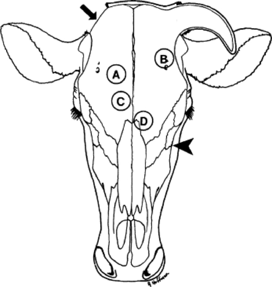
Fig. 31-47 Trephine sites for sinusotomy. A, Dorsal frontal sinus. B, Postorbital diverticulum. C, Rostral frontal sinus. D, Turbinate portion of frontal sinus. The maxillary sinus is trephined ventral to a line from the infraorbital foramen to the medial canthus (arrowhead). If draining tracts are present at the poll, an additional sinusotomy can be made in the cornual portion of the frontal sinus (arrowhead).

Cattle that have frontal sinusitis after dehorning should be treated by sinusotomy and drainage of the sinus.53 Sinusotomy sites should be based on anatomic landmarks51 and modified as needed to accommodate any frontal bone distortion or wounds related to dehorning.51 Sinusotomy should be performed 3 to 4 cm from midline, intersecting a line drawn between the caudal aspect of the orbits. If draining tracts are present at the poll, an additional sinusotomy can be made in the cornual portion of the frontal sinus.51 Sinusotomy is performed after sedation and local anesthetic infiltration of the centesis site(s). A 2-cm—diameter circular piece of skin is excised, and a 19-mm (¾-inch) trephine used to create an opening into the sinus, through which purulent fluid should be evacuated and the sinus lavaged.

Additional trephine sites that permit access to other regions of the frontal sinus include the postorbital diverticulum, which is trephined approximately 4 cm caudal to the dorsal rim of the orbit, just above the temporal crest of the frontal bone; the rostral frontal sinus, which is trephined just caudal to a line between the centers of the orbits and to either side of the midline; and the turbinate portion of the frontal sinus, which is trephined just rostral to the line described and to either side of the midline.

  Page 595 

Access to the maxillary sinus is achieved by trephining ventral to a line from the infraorbital foramen to the medial canthus. If an infected tooth is the cause of maxillary sinusitis, a sinusotomy is created with a trephine over the affected tooth to repel it; otherwise, the hole is usually made just dorsal and caudal to the facial tuberosity. The trephine site should be higher in the sinus of younger animals because the tooth roots are longer.

If the frontomaxillary and nasomaxillary openings are still patent, one trephine site may be sufficient, with the natural opening providing ventral drainage. In more chronic cases, two trephine sites (for ingress and egress) are needed. Another alternative in chronic sinusitis is the use of a curved steel sinus probe (1 cm diameter × 55 cm long), which is forcefully driven through the septal plates of the frontal sinus into the nasal meatus for ventral drainage.54 The frontal sinus is very compartmentalized in mature sheep and goats, and effective drainage is difficult. Therapy should therefore be aggressive in these species, even in early cases, and double trephination or bone flaps for exposure and curettage should be considered.

If a tooth has been repelled, a roll of gauze or dental impression material should be used to occlude the alveolar socket to prevent feed material from entering the sinus. A strip of umbilical tape tied around the gauze roll or a wire in the dental material is passed through socket, sinus, and trephine hole and secured to the face by tying around another roll of gauze as a stent. These gauze packs are replaced each time the sinus is flushed. The sinus is lavaged daily with dilute antiseptic solutions such as 0.1% povidone iodine or chlorhexidine in saline, or 1:1000 potassium permanganate. Lavage is continued until infection is resolved. Enzymes (papain or 200,000 U of streptokinase and 50,000 U of streptodornase in at least 10 mL of normal saline solution) may help remove thick exudate.

Parenteral antibiotics and NSAIDs (aspirin, 100 mg/kg PO twice daily or flunixin meglumine, 1.1 to 2.2 mg/kg IV daily or divided twice daily) are indicated if systemic signs are present. In the absence of microbial culture and susceptibility results, penicillin (22,000 U/kg IM or SC q12-24h) is recommended as the antibiotic of choice because Arcanobacterium (Actinomyces) pyogenes is the most common organism isolated from cattle with chronic frontal sinusitis resulting from dehorning. Pasteurella multocida is the most common organism isolated from infections of the frontal sinus not associated with dehorning.51 Penicillin may be effective against some isolates of P. multocida; alternatively, oxytetracycline can be administered if P. multocida is suspected (11 mg/kg IV or SC q24h, or 20 mg of long-acting oxytetracycline per kilogram SC q72h). If bacterial culture and susceptibility results are available, antimicrobial therapy should be modified accordingly. Early cases often resolve in 10 to 14 days, with a good prognosis. Long-term therapy (weeks) is frequently needed in chronic cases; the prognosis is more guarded, and salvage is often the best option.

Prevention and Control

Dehorning ruminants as neonates, particularly if a “closed” method such as a dehorning iron is used, is the most effective way to prevent frontal sinusitis. In larger cattle surgical dehorning with primary skin closure achieved under aseptic conditions minimizes the likelihood of sinusitis.55 When this is not practical, dehorning should be avoided in rainy, windy, or dusty conditions, and fly control must be used. The dehorning of mature sheep or goats leaves massive wounds that typically take 4 to 6 weeks to close by second intention and so are susceptible to infection; special care, such as bandaging for the initial 7 to 10 days, must be taken.56 Sinusitis did not occur in goats aged 2 to 24 months dehorned with a technique in which primary skin closure was achieved.57

DISEASES OF THE PHARYNX, LARYNX, AND TRACHEA

AMELIA R. WOOLUMS

JOHN C. BAKER

JOHN A. SMITH

PHARYNGEAL TRAUMA, ABSCESSES, CELLULITIS, AND GRANULOMAS

Definition and Etiology

Pharyngeal trauma may result in hematomas, foreign body granulomas, cellulitis, or abscesses. Trauma usually results from careless use of balling guns, dose syringes, paste-type anthelmintics or calcium preparations, specula, and stomach tubes. Rough, stemmy feeds (especially when chopped), grass awns, briars, and foreign objects (e.g., nails, baling wire) may also cause punctures. Migrating foreign objects or medications (e.g., mineral oil, anthelmintics) may cause pharyngeal granulomas. Hematomas and puncture wounds often result in abscess formation. Diffuse cellulitis may also result. Common bacteria involved include Arcanobacterium (Actinomyces) pyogenes, Actinobacillus species, Pasteurella species, Bordetella species, Fusobacterium necrophorum, and Streptococcus species. In cases of particularly virulent bacterial invasion, the condition can become rapidly fatal. C. pseudotuberculosis (CLA) may localize in the pharyngeal nodes of sheep and especially goats. CLA is discussed further on p. 658 and also in relation to the hemolymphatic system in Chapter 37.

  Page 596 

Clinical Signs and Differential Diagnosis

Signs of pharyngeal trauma vary with the severity of the resulting reaction (e.g., peracute cellulitis vs. chronic abscess or granuloma). Prominent signs include inspiratory dyspnea with stertorous inspiratory sounds and a prolonged inspiratory phase; extended head and neck; ptyalism, which is often profuse; quidding; evident pain on swallowing or reluctance to swallow solid feed but willingness to drink liquids; prolonged chewing of boluses; regurgitation of food or saliva through the nostrils caused by pharyngeal paresis; mucopurulent to bloody nasal discharge and fetid odors, usually bilateral; cough; bloat; and visible or palpable swelling in the pharyngeal area.58,59 Megaesophagus has been reported subsequent to pharyngeal trauma.60 Palpation of the pharynx may increase the stertor and cause pain. In severe cases, systemic signs of fever, anorexia, depression, dehydration, and forestomach stasis may be present. Aspiration pneumonia may be a secondary complication.

Differential diagnoses include pharyngeal tumors; lymphosarcoma; sialoliths; rabies; botulism; actinobacillosis; necrotic laryngitis; laryngeal abscesses, trauma, edema, or paralysis; and laryngeal tumors.

Diagnosis

A thorough manual examination of the oropharynx or a visual examination with an adequate speculum and light source usually confirms the diagnosis of a pharyngeal swelling and often reveals a puncture that is discharging pus. Cases in which the infection is diffuse can be more difficult to recognize, and endoscopy or radiography can be particularly helpful.58 Restraining the jaws with a McAllum speculum allows a guarded needle attached to a length of tubing and a syringe to be inserted into the swelling for aspiration of any swelling identified. This helps to differentiate localized abscesses from granulomas, hematomas, cellulitis, and tumors; allows culture and sensitivity determinations on abscesses; and may aid in cytologic diagnosis of granulomas or tumors. Radiographs may reveal foreign bodies (Fig. 31-48) or air densities (Fig. 31-49) in the pharyngeal tissues. The CBC usually reflects a chronic inflammatory process, with a neutrophilic leukocytosis and a left shift, or a neutrophil-lymphocyte reversal. Dehydration is frequently evident. If the animal is unable to swallow, large amounts of bicarbonate may be lost through the saliva, which may lead to evidence of metabolic acidosis on blood gas analysis or serum biochemical profile.

image

Fig. 31-48 Radiograph of the pharyngeal area of a cow. Note magnet located in the retropharyngeal tissue.

image

Fig. 31-49 Radiograph of the pharyngeal area of a cow. Note air densities in the tissue, suggestive of abscess formation.

Treatment and Prognosis

Discrete pharyngeal abscesses are usually best drained into the pharynx. Whenever possible the procedure should be done on the standing animal without sedation to preserve the cough reflex and prevent aspiration. A good oral speculum and excellent restraint are needed. The head should be kept lowered. A guarded blade such as a hook blade from a fetotomy set is introduced into the pharynx, and the abscess is lanced. The cavity is flushed with a mild antiseptic such as 0.2% povidone iodine in saline solution, again taking care to prevent aspiration. Other options include drainage to the exterior, drainage and flushing with a large-gauge needle and tubing, and extirpation.59 Removal of a bacterial granuloma from the pharynx of a cow via electrocautery has been described.61 Systemic antibiotics are administered in accordance with culture and sensitivity results, or, in their absence, procaine penicillin G (22,000 U/kg IM or SC q12-24h), tetracyclines (11 mg/kg IV or SC daily, or 20 mg/kg of long-acting oxytetracycline SC q72h), or sulfadimethoxine (55 mg/kg IV loading dose followed by 27.5 mg/kg IV q24h) are used. NSAIDs (aspirin, 100 mg/kg PO twice daily, or flunixin, 1.1 to 2.2 mg/kg IV daily or divided twice daily) help relieve pain, swelling, and stertor. In animals with severe persistent dyspnea, tracheostomy may be necessary. Granulomas and diffuse cellulitis are likewise treated medically with appropriate antimicrobials and antiinflammatory drugs. Supportive therapy such as IV fluids or feeding through a rumenostomy site may be necessary if the animal refuses to eat or drink. The prognosis for most pharyngeal abscesses, hematomas, cellulitis, and granulomas is usually good with appropriate therapy.

  Page 597 

DORSAL DISPLACEMENT OF THE SOFT PALATE

Although rare, dorsal displacement of the soft palate has been reported in cattle.62 Respiratory noise is apparent on inspiration and expiration but is loudest on inspiration. Diagnosis can be made by endoscopic examination. Treatment is similar to that used in horses and can include conservative therapy, which consists of antiinflammatory drug therapy and rest, or the condition can be surgically corrected.

SUBEPIGLOTTIC CYST

Although rare, a subepiglottic pharyngeal cyst causing upper airway obstruction has been reported in cattle.63 Surgical removal by a peroral approach has been described.63

NECROTIC LARYNGITIS (CALF DIPHTHERIA, LARYNGEAL NECROBACILLOSIS)

Definition, Etiology and Epidemiology

Acute to chronic infection of the laryngeal mucosa and cartilage of young cattle is very common, particularly in feedlots. Laryngeal contact ulcers64 probably provide a damaged mucosal surface, which then allows invasion of the cartilage by F. necrophorum, which is the proximate cause of the lesions. It has also been suggested that Histophilus somni is the primary agent inducing a perilaryngeal vasculitis and that F. necrophorum represents a secondary bacterial invader.

The disease is most common where cattle are housed in dirty or crowded conditions and in feedlots. Most feedlot cases occur in animals on feed for longer than 30 days. The incidence is sporadic. Cases occur year round, but there appears to be a higher incidence in fall and winter. The disease has a worldwide distribution.

Clinical Signs and Differential Diagnosis

The problem occurs most commonly in calves from 3 to 18 months of age, up to about 24 months. It is characterized by an acute onset of a moist, painful cough, which the animal may attempt to suppress because of pain. Frequently a severe inspiratory dyspnea with a loud guttural stertor and open-mouth breathing with the head and neck extended are observed. The animal may salivate, make frequent painful swallowing movements, and stand and sip water continually. Signs of systemic disease including anorexia, depression, fever (as high as 106° F [41.1° C]), and hyperemic mucous membranes are present. There is often a bilateral nasal discharge and a fetid odor to the breath. The larynx may be visibly or palpably swollen, and palpation may elicit a cough, cause pain, and markedly increase the dyspnea and stertor. If untreated, many calves will die in 2 to 7 days as a result of systemic effects of bacterial toxins and upper airway obstruction. Recovered cases may have a chronic roaring respiration and a harsh, dry cough because of the misshapen larynx. Aspiration pneumonia and chronic “poor doers” are common sequelae. Differential diagnoses include pharyngeal trauma (abscess, cellulitis), severe viral laryngitis (e.g., IBR), actinobacillosis, and laryngeal edema, abscesses, trauma, paralysis, and tumors.

Diagnosis

The diagnosis is usually made presumptively based on clinical signs alone. A laryngoscopic or endoscopic examination can help confirm the diagnosis, but care must be taken to prevent further stress and respiratory embarrassment. Acute cases show changes in the CBC consistent with any acute septic condition: leukopenia caused by neutropenia with a left shift. In chronic or ongoing cases, leukocytosis with neutrophilia, monocytosis, and hyperfibrinogenemia may be present.

Pathophysiology

F. necrophorum normally does not penetrate intact mucous membranes. Laryngeal contact ulcers are thought to provide the portal of entry for F. necrophorum, which is ubiquitous. Laryngeal contact ulcers are also very common in slaughter cattle and are speculated to be caused by the following combination of factors: (1) an acute mucositis from mixed upper respiratory infections (such as IBR virus, bovine respiratory syncytial virus [BRSV], parainfluenza virus 3 [PI3], mycoplasma, and bacteria, including Mannheimia, Pasteurella, and Histophilus species); (2) reflex coughing and swallowing, which accelerate the rate of laryngeal closure; and (3) resulting erosion of the swollen membranes over the vocal processes and medial angles of the arytenoid cartilages.65 It has also been proposed that necrotic laryngitis results from a perilaryngeal vasculitis initiated by H. somni with secondary invasion by F. necrophorum.66 Necrotic laryngitis can alter pulmonary function such that the growth rate is impeded, and also predisposes to secondary bacterial pneumonia.67

  Page 598 

Necropsy Lesions

The lesions are typically located over the vocal processes and medial angles of the arytenoid cartilages. Acute lesions consist of marked edema, hyperemia, and swelling of the mucous membrane around a necrotic ulcer, with accumulated exudate. The lesions spread along the vocal processes and vocal folds and may extend into the cricoarytenoideus dorsalis muscles. In chronic cases the lesions consist of a focus of necrotic cartilage surrounded by purulent exudate, with a tract extending to the mucosal surface (Fig. 31-50). The tract opening is surrounded by granulation tissue and may drain pus. The arytenoid cartilage may be rotated into the lumen or may contain mucosal cavities lined with thin, hyperemic epithelium.65

image

Fig. 31-50 Postmortem photograph of necrotic laryngitis. Note the purulent material medial to the arytenoid cartilages.

Photograph contributed by Feedlot Health Management Services, Okotoks, AB, Canada.

Treatment, Prognosis, Prevention, and Control

Oxytetracycline (11 mg/kg IV or SC q24h, or 20 mg/kg of long-acting oxytetracycline SC q72h) or procaine penicillin G (22,000 U/kg IM or SC q12-24h) is appropriate; a sulfonamide, streptomycin, or tylosin is also usually effective. NSAIDs (aspirin, 100 mg/kg PO twice daily, or flunixin, 1.1 to 2.2 mg/kg IV once daily or divided twice daily) reduce swelling, inflammation, and fever. Cases with severe respiratory distress may benefit from one or two doses of steroids (dexamethasone 0.5 to 0.2 mg/kg IV or IM once or twice), but repeated doses of steroids are not recommended. A tracheostomy may be necessary in severe cases to relieve dyspnea and rest the larynx. Good nursing and supportive care are also important, including shelter, adequate ventilation, easy access to feed and water, and oral or IV fluids if needed. The prognosis is good when the condition is detected very early and treated vigorously; when extensive cartilage necrosis occurs, a fatal outcome or chronic ill-thrift with stertorous breathing is expected. There are no specific control measures. The proposed pathogenesis would suggest that measures to control other respiratory diseases may reduce the incidence of necrotic laryngitis.

LARYNGEAL GRANULOMAS

Laryngeal granulomas have been described in cattle.68 They may originate from laryngeal contact ulcers that have been described in feedlot cattle at slaughter (see the previous section on necrotic laryngitis for more information).64

LARYNGEAL PAPILLOMATOSIS

Papillomas of the larynx are common in feedlot cattle. They are caused by a papovavirus, which is thought to enter laryngeal contact ulcers (see the previous section on necrotic laryngitis).64 Characteristic signs of laryngeal papillomatosis are stertorous respiration and cough. Differential diagnoses include necrotic laryngitis, pharyngeal trauma, abscess, or granuloma; actinobacillosis; and laryngeal abscesses, trauma, edema, paralysis, and tumors. The lesions are sessile to pedunculated, yellow, frondlike, 1- to 10-mm growths over the vocal processes of the arytenoid cartilages.69 Treatment usually is not indicated but involves surgical removal. Measures to decrease other respiratory infections and thereby decrease contact ulcers may lower the incidence of papillomas.

LARYNGEAL ABSCESSES

Abscessation of the arytenoid cartilages caused by Arcanobacterium (Actinomyces) pyogenes has been reported in calves70 and sheep.71 Clinical signs include tachypnea, extension of the head and neck, cyanosis, and a severe progressive dyspnea with marked stertor that can be localized to the larynx. Many affected animals remain alert and afebrile and continue to eat until the terminal stages of severe dyspnea. Endoscopy reveals generalized edema and hyperemia of the laryngeal mucosa and obstruction of the rima glottidis by swelling of one or both arytenoids. Radiographs may demonstrate soft-tissue swelling of the larynx. The condition has been speculated to be initiated by grass awns, trauma, hereditary predisposition, or congenital cavitations in the cartilages. In sheep, rams appear to be more commonly affected than ewes, and two reports found a breed predisposition in Texels and Southdowns, although other reports have indicated that various breeds are affected.71 The necropsy lesions consist of encapsulated abscesses containing pus and necrotic debris in the arytenoid cartilage, usually in the vicinity of the vocal cord. A tract from the abscess typically opens in an area of granulation tissue in the laryngeal mucosa. Treatment consists of tracheostomy, antibiotics (usually penicillin at 22,000 U/kg IM or SC q12-24h), and antiinflammatory drugs (aspirin, 100 mg/kg PO twice daily, or flunixin, 1.1 to 2.2 mg/kg IV daily or divided twice daily). The prognosis for recovery is considered guarded unless the condition is detected in the earliest stages and treated vigorously; however, one affected heifer lived for at least 1 year and was bred successfully after permanent surgical tracheostomy for chronic arytenoid abscessation.72

OTHER LARYNGEAL OBSTRUCTIONS (LARYNGEAL TRAUMA, EDEMA, PARALYSIS, AND FOREIGN OBJECTS)

Other laryngeal obstructions are all sporadic and may manifest with similar signs. Trauma to the larynx may result from roping or injury in restraint devices. Inappropriate placement of an endotracheal tube can potentially damage the larynx. The respiratory system is the main target organ for anaphylaxis in ruminants, and laryngeal edema can be a prominent component of this syndrome, which is discussed fully in this chapter on p. 652. Inhalation of smoke or other noxious gases also may cause laryngeal edema. Paralysis of the larynx was reported in a sheep with a false carotid aneurysm73; presumably other lesions of the neck or anterior mediastinum could cause laryngeal paralysis through involvement of the recurrent laryngeal nerves. Laryngeal hemiplegia has been reported in association with Sarcocystis species infection of the muscles of the larynx and pharynx in a ram.74 Foreign objects more commonly lodge in the pharynx, but sharp objects and food materials may be aspirated and lodge in the larynx. Signs common to these laryngeal obstructions include inspiratory dyspnea, prolongation of the inspiratory phase, mouth breathing, stertor, cyanosis, salivation, and extension of the head and neck. Palpation of the larynx may reveal swelling and may exaggerate the dyspnea and stertor. Differential diagnoses include necrotic laryngitis, laryngeal abscesses, severe viral laryngitis, actinobacillosis, and tumors. Endoscopy and radiology are required in most cases to differentiate these conditions. Hematologic analysis may give some indication of the presence of bacterial infection; a stress leukogram may also be seen. Tracheostomy is indicated in all severe cases. Surgical correction of laryngeal obstruction by tracheolaryngostomy has been described in cattle.75

  Page 599 

Laryngeal trauma and paralysis may resolve spontaneously or may require reconstructive surgery. The therapy of anaphylaxis is discussed on p. 652. Foreign objects should be removed surgically or endoscopically. NSAIDs (aspirin, 100 mg/kg PO twice daily, or flunixin, 1.1 to 2.2 mg/kg IV daily or divided twice daily) may help reduce swelling, edema, and respiratory embarrassment in all forms of obstruction.

TRACHEAL COLLAPSE AND STENOSIS

Definition and Etiology

Tracheal collapse or stenosis is infrequently reported in cattle and goats.76-81 The cause is usually unknown, but the problem may result from cranial thoracic trauma, roping, tracheostomies, or possibly congenital defects. In cattle the majority of reports have described tracheal collapse in calves in which signs were usually first evident at several weeks of age.76-79 The majority of these cases also involved the thoracic trachea (Fig. 31-51), suggesting a congenital lesion. However, in most calves with tracheal collapse the condition has been a result of dystocia at birth, especially breech presentations, which suggests a traumatic cause.82 In contrast to reports in cattle, tracheal collapse has been reported in a mature goat that showed no previous signs of respiratory abnormality.81

image

Fig. 31-51 Radiograph demonstrating collapse of the thoracic trachea in a calf.

Tracheal collapse has also been reported in Texel-cross lambs with inherited chondrodysplasia83; affected lambs were normal at birth but developed exercise intolerance as early as 1 week of age. Severely affected lambs developed fatal respiratory compromise when exercised. Tracheal stenosis can also occur in lambs born to ewes that ingest Veratrum californicum root and rhizome material at days 31 through 33 of gestation.84

Clinical Signs and Differential Diagnosis

Clinical signs of tracheal collapse may include fever, tachycardia, tachypnea, cyanosis, and mucosal hyperemia with vessel engorgement, but affected animals may have normal vital signs and are otherwise alert and in good condition. Dyspnea is usually induced or exacerbated by excitement or exercise or may be severe at rest. Stertorous respiration is usually evident, is frequently worse on inspiration, and can often be localized to the trachea on auscultation. The inspiratory phase is prolonged, and a “honking” cough is characteristic, especially with intrathoracic collapse. Palpation may reveal or induce the collapse when the cervical trachea is involved. Tracheal palpation or elevation of the head may increase the stertor or induce the honking cough. In some cases there may be other evidence of trauma (fractured ribs, sternebrae), and in some animals pneumonia may be present. There is no response to antibiotics, steroids, or tracheostomy.

Texel-cross lambs with tracheal collapse resulting from inherited chondrodysplasia also exhibited retarded growth, forelimb varus, and reluctance to walk. Severity of signs varied among affected individuals; some died or were euthanized within weeks because of respiratory compromise, whereas others survived to breeding age.83 Animals that survived for several months often developed arthritis characterized by severe erosive lesions of the articular cartilage of major joints including the shoulder, hip, and stifle. Lambs with congenital tracheal stenosis resulting from maternal ingestion of V. californicum died within 5 minutes of birth after signs of severe respiratory distress.

  Page 600 

Other possible disorders with signs similar to those seen in animals with tracheal collapse or stenosis are tracheal foreign bodies, tracheal actinobacillosis, neoplasms, bronchopneumonia, necrotic laryngitis, and extratracheal compressive lesions (e.g., abscesses, tuberculosis, hematomas).

Diagnosis

Any deviation of the hemogram from normal is probably a reflection of associated pneumonia or stress. Endoscopy and radiography are the most helpful ancillary aids. Care must be taken when restraining, sedating, and passing the endoscope in these animals; oxygen should be available. In cattle with idiopathic tracheal collapse, radiography (see Fig. 31-51) and endoscopy usually reveal a dorsoventral flattening, most typically in the caudal cervical and cranial thoracic trachea, although lateral collapse and collapse in other locations (cranial cervical, intrathoracic) are occasionally encountered.

Pathophysiology

Proposed causes of tracheal collapse in other species include congenital malformations, genetic or nutritionally induced weakness of cartilage, deficient innervation of the trachealis muscles, trauma, ischemic lesions from endotracheal tube cuffs, and primary pulmonary disease. No histologic differences were noted in tracheal rings from collapsed and normal segments in calves in one report.76

As previously mentioned, most calves with tracheal collapse have a history of dystocia at birth. During delivery, compression of the chest wall with fracture of the first pair of ribs may cause injury at the thoracic inlet.85 However, signs of tracheal collapse are not immediately evident at birth, but develop over time.

Lambs with tracheal collapse associated with inherited chondrodysplasia had clinical signs most similar to a condition in humans that results from a mutation in the diastrophic dysplasia sulphate transporter gene,86 but the causative mutation in sheep has not yet been reported. Preliminary data indicated that the chondrodysplasia was inherited via a recessive mode of inheritance.83 Tracheal stenosis in lambs born to ewes fed V. californicum was presumed to be caused by cyclopamine, a steroidal alkaloid which is the toxic principle in highest concentration in V. californicum. It was notable that lambs developed tracheal stenosis when ewes were fed V. californicum at 31 to 33 days of gestation, whereas craniofacial defects typically recognized in lambs born to exposed dams are seen when the ewe ingests the toxic plant by day 14 of gestation.84

Necropsy Lesions

At necropsy, affected animals may have either a laterally78 or dorsally77,81 compressed trachea; typically several centimeters of the trachea are affected. Necropsy of sheep with inherited chondrodysplasia reveals a trachea that is flaccid, flattened, and sometimes irregularly kinked. Tracheal rings are markedly thickened, and the lumen is narrow, possibly extremely so. Diffuse pulmonary congestion and edema, with epidcardial ecchymoses, may occur as a result of terminal anoxia and respiratory distress. Other lesions include exaggerated convex curvature of the ribs, anglular limb deformities, and erosive arthritis with exposure of the subchondral bone in one or more joints. Histologically, hyaline cartilage in affected organs is hypercellular and disorganized, with foci of apparent chondrolysis. Chondrocytes are larger than normal, although shrunken, apparently necrotic chondrocytes are also seen.83 Necropsy of lambs with congenital tracheal stenosis resulting from maternal ingestion of V. californicum reveals pronounced lateral flattening of the trachea. Tracheal rings are reduced in number, with abnormal size and shape and irregular spacing. Histologically the cartilage rings in affected lambs were flattened and thin and had a thinner zone of proliferating chondrocytes on their outer surface compared with normal age-matched controls.84

Treatment and Prognosis

Mild cases may respond to confinement sufficiently to be fed out for slaughter. A number of surgical treatments have been proposed in other species, including anastomosis, bisection of tracheal rings, internal and external prostheses, and plication of the dorsal membrane.76 External prostheses have been used successfully in calves,76,82,85 adult cattle,80 and a goat.87 A favorable prognosis for surgical correction is estimated at approximately 30%.82 A detailed description of surgical repair in calves by external prostheses has been published.82 Successful management of lambs with congenital tracheal collapse or stenosis has not been reported, although mildly affected lambs with congenital chondrodysplasia survived into adulthood. Because of the apparent genetic basis for the disease, breeding of affected animals should not be recommended.

TRACHEAL FOREIGN BODIES AND MASSES

Ruminants may occasionally inhale foreign objects that lodge in the trachea. There are also two reports of tracheal actinomycosis that resulted in signs of tracheal obstruction.88 Signs include a chronic cough, inspiratory dyspnea, audible stridor that can be localized to the trachea, extension of the head and neck, open-mouth breathing, and salivation. Differential diagnoses should include pharyngeal trauma, necrotic laryngitis, laryngeal abscesses, trauma, edema, or paralysis, tracheal collapse, and extratracheal compressive lesions. Endoscopy and radiology are important aids to diagnosis. Care must be exercised in restraint and in passage of the endoscope. Some small objects may be retrieved by a snare passed through an endoscope; others may require tracheostomy. When possible, the tracheostomy should be performed below the object. The actinomycotic masses are soft, pedunculated lesions with a granular surface containing small yellow foci. Of the two reported cases of tracheal actinomycosis, one died of asphyxiation and the other responded initially to partial surgical removal of the masses, tracheostomy, and therapy with sodium iodide, penicillin, and streptomycin but relapsed some months later.88a

TRACHEAL EDEMA SYNDROME OF FEEDLOT CATTLE

Tracheal edema syndrome has also been referred to as tracheal stenosis in feedlot cattle. In this condition extensive edema and hemorrhage in the dorsal wall of the trachea result in coughing, dyspnea, and stertor, which has given rise to the term “honker cattle.”89 Tracheal edema syndrome occurs in two forms, characterized by either acute dyspnea or a chronic cough. It is not known whether the two forms are related. Although the syndrome has been recognized for years, no controlled research has been undertaken to determine the cause or risk factors of this syndrome, and the cause is unknown. Theories regarding possible causes include infections with upper respiratory viruses or bacteria such as P. multocida or H. somni, trauma to the trachea from feedbunks, passive congestion and edema from excessive fat accumulation in the thoracic inlet, hypersensitivity reactions, and mycotoxins.90

  Page 601 

The acute dyspnea syndrome occurs mainly in heavy feedlot cattle in the latter two thirds of the feeding period and is most common in southern plains feedlots. It is sporadic and more common in summer, possibly because of exacerbation by hot weather. At one extreme, sudden deaths without the onset of noticeable clinical signs have been reported, and at the other extreme subclinical disease was evidenced by lesions in animals at slaughter that did not have clinical signs. Other factors that increase respirations also may cause signs to appear. Signs include an acute onset of dyspnea and loud guttural inspiratory sounds that can be localized to the lower trachea. Open-mouth breathing, extension of the head and neck, and cyanosis, leading to recumbency and death by asphyxiation, are present. Differential diagnoses for this form include pharyngeal trauma or abscess; necrotic laryngitis; IBR; laryngeal abscess, tumor, foreign object, edema, or paralysis; tracheal foreign object, mass, or collapse; and AIP.

The chronic form occurs in lighter cattle (135 to 400 kg [300 to 900 lb]) and is more common in western plains feedlots. It is also sporadic but less seasonal. Affected animals may have a history of IBR or pneumonia. The main sign is a continuous, frequent, deep, hacking, nonproductive cough. The animal may be unthrifty but is otherwise normal in appearance. The main differential diagnoses are necrotic laryngitis or mild, chronic suppurative pneumonia. Endoscopy and visualization of the lesions subsequently described aid in the diagnosis. Necropsy of the acute form reveals an edematous thickening of the submucosa and mucosa of the dorsal trachea (Fig. 31-52), as much as 5 cm thick and extending 20 to 30 cm from the midcervical area to the thoracic inlet or tracheal bifurcation. There is also extensive mucosal, submucosal, and peritracheal edema and/or hemorrhage, possibly related at least in part to agonal breathing. There may be no other lesions of the airway or lungs,90 or abnormalities (e.g., pulmonary edema, bronchitis, interlobular edema and emphysema, alveolar hemorrhage) may be seen.89 Lesions in the chronic form consist of hyperemia of the mucosa of the caudal third of the trachea, with a thin layer of mucopurulent exudate. The mucosa may have a cobblestone appearance or even large, fiber-like projections and polyps. No effective treatment exists for the chronic form. Corticosteroids (dexamethasone, 0.05 to 0.2 mg/kg IM or IV; prednisolone, 1 to 2.2 mg/kg IM or IV daily) are recommended for the acute form, as well as such practices as preventing stress, providing shade, and cooling with water sprays and fans. Broad-spectrum antibiotics have been recommended by some,89,90 whereas others have not found them to be necessary.91 However, because animals with tracheal edema syndrome may be difficult to distinguish clinically from animals with conditions that could respond to antimicrobials (such as necrotic laryngitis), antimicrobials are often administered. Because withdrawal times associated with antimicrobial administration could delay shipment of affected animals to slaughter, drugs with a short withdrawal time should be used if salvage is an option. Tracheostomy may not be helpful if placed proximal to the obstruction, and relief of dyspnea via tracheostomy may require insertion of an endotracheal tube to the tracheal bifurcation. Oxygen administration could be beneficial if available. Recovered patients tend to relapse and should be salvaged.

image

Fig. 31-52 Postmortem photograph of tracheal edema syndrome. Note severe mucosal and submucosal thickening, which has obstructed the trachea by approximately 50%.

Photograph contributed by Feedlot Health Management Services, Okotoks, AB, Canada.

LOWER RESPIRATORY TRACT DISEASES

AMELIA R. WOOLUMS

TREVOR R. AMES

JOHN C. BAKER

CLINICAL CLASSIFICATION OF PNEUMONIA

AMELIA R. WOOLUMS

TREVOR R. AMES

JOHN C. BAKER

In an effort to simplify the differential diagnosis of the bewildering array of lower respiratory diseases of cattle, a classification system based on pathophysiology and clinical signs has been suggested.92 Three classifications were proposed:

1 Bronchial pneumonia is characterized pathophysiologically by invasion of pathogenic organisms that gain access to the lung through the pulmonary tree. It is characterized clinically by depression, fever, and other signs of sepsis such as hyperemic mucous membranes or scleral injection, and an anterior-ventral distribution of abnormal lung sounds and lesions (see the following section). Bronchial pneumonia is the final outcome of the respiratory disease complex of ruminants, and because viruses play an important role in this disease complex, viral causes of respiratory tract disease in ruminants are placed in this category.
2 The interstitial pneumonias are a very diverse group of (usually) noninfectious diseases. Although it is difficult to make generalizations, these diseases are characterized pathophysiologically by an interstitial reaction that usually results from ingestion or inhalation of toxins or allergens. Clinically affected animals tend not to be as depressed and septic, the abnormal lung sounds and lesions are diffusely distributed, and there is little or no response to antibiotic therapy.
3 Metastatic pneumonia is characterized pathophysiologically by septic embolization of the lungs from other foci in the body, classically liver abscesses and postcaval thrombi. Clinically, cases of metastatic pneumonia exhibit signs of sepsis as with bronchopneumonia, but with widespread pulmonary lesions and abnormal lung sounds, and the eventual development of hemoptysis (see descriptions of vena caval thrombosis and metastatic pneumonia in this chapter).
  Page 602 

THE BRONCHOPNEUMONIAS (RESPIRATORY DISEASE COMPLEX OF CATTLE, SHEEP, AND GOATS)

AMELIA R. WOOLUMS

TREVOR R. AMES

JOHN C. BAKER

The respiratory disease complex of ruminants consists of the single clinical entity of bronchopneumonia but is caused by numerous combinations of infectious agents, compromised host defenses, and environmental conditions. Bronchopneumonia causes greater economic losses than any other disease of feedlot cattle or lambs and is one of the most common causes of dairy calf mortality.

In dairy calves, bronchopneumonia, often called enzootic pneumonia, is most common in housed calves. It is called shipping fever in beef calves because the greatest incidence of bronchopneumonia occurs after shipment to stocker operations or feedlots; the term bovine respiratory disease (BRD) is also used to describe feedlot bronchopneumonia. Bronchopneumonia also occurs in nursing beef calves and in mature beef or dairy cows, sometimes in outbreaks that can have significant morbidity or mortality. Little is known about which factors are specifically the most important for putting these populations at risk for bronchopneumonia, but they are likely similar to risk factors described for housed dairy calves or feedlot cattle. The infectious agents and risk factors of bronchopneumonia of sheep and goats are very similar to those of calves.

Bronchopneumonia of ruminants is a disease of multifactorial causation that occurs when a certain combination of host, environment, and infectious agent characteristics (risk factors) is active. The numerous infectious agents (Boxes 31-3 and 31-4) that are associated with bronchopneumonia are ubiquitous in ruminant populations, and the bacteria most often associated with pneumonic lesions are part of the normal resident flora of the ruminant nasopharynx. In addition to recognition of the microbial agents that contribute to the development of bronchopneumonia in ruminants, understanding of the management practices that also play a role is necessary for developing successful programs of prevention.

Box 31-3 Viral Agents Associated with Respiratory Tract Diseases in Ruminants

BOVINE

Viruses of Major Importance (Commonly Isolated or Generally Accepted to be Important Contributors to Ruminant Respiratory Disease)

Bovine herpesvirus type 1 (IBR)
Bovine respiratory syncytial virus (BRSV)
Bovine viral diarrhea virus (BVDV)
Bovine parainfluenza virus type 3 (PI3)
Bovine respiratory coronavirus
Alcelaphine herpesvirus 1 and 2/ovine herpesvirus 2 (malignant catarrhal fever)

Viruses of Minor Importance (Uncommonly Isolated or of Uncertain Importance in Ruminant Respiratory Disease)

Bovine herpesvirus type 4 (DN-599, Movar 33/63, FTC-2)
Bovine adenovirus
Bovine rhinovirus
Bovine reovirus
Bovine enterovirus
Calicivirus
Influenza virus

OVINE AND CAPRINE

Ovine or bovine respiratory syncytial virus (ORSV, BRSV)
Parainfluenza virus type 3 (PI3)
Adenovirus
Bluetongue virus
Ovine progressive pneumonia (maedi-visna) virus of sheep*
Pulmonary carcinoma of sheep*

* Progressive pneumonias, capable of causing severe chronic respiratory disease.

Box 31-4 Bacteria, Mycoplasma, Ureaplasma, and Chlamydia Species Associated with Bronchopneumonia of Cattle, Sheep, and Goats

Bacteria of Major Importance (Commonly Isolated or Generally Accepted to be Important Contributors to Ruminant Respiratory Disease)

Mannheimia haemolytica
Pasteurella multocida
Histophilus somni
Mycoplasma bovis
Mycoplasma dispar
Ureaplasma species
Mycoplasma ovipneumonia (sheep and goats only)
Mycoplasma mycoides subsp. mycoides, large colony variant (goats)
Arcanobacterium (Actinomyces) pyogenes (secondary opportunistic pathogen)

Bacteria of Minor Importance (Uncommonly Isolated or of Uncertain Importance in Ruminant Respiratory Disease)

Pseudomonas aeruginosa
Escherichia coli
Streptococcus species
Staphylococcus species
Moraxella species
Salmonella species
Bacteroides species (anaerobe)
Peptococcus indolicus (anaerobe)
Fusobacterium species (anaerobe)
Chlamydia species

INFECTIOUS AGENTS ASSOCIATED WITH THE RESPIRATORY COMPLEX OF CATTLE, SHEEP, AND GOATS

Etiology

Numerous infectious agents have been isolated from cases of bronchopneumonia in ruminants (see Boxes 31-3 and 31-4). Although infectious bronchopneumonia of ruminants is usually caused by two or more infectious agents acting together, some agents can also cause significant disease alone. Therefore the clinical and epidemiologic characteristics, means of diagnosis, and treatment and prevention of the infectious agents will be first considered individually. A description of epidemiology, diagnosis, and treatment of “undifferentiated” bronchopneumonia (bronchopneumonia with no specific causative diagnosis attempted) will then follow. The clinical signs, gross pathologic lesions, and recommended methods of diagnosis for the major infectious causes of ruminant respiratory disease are listed in Table 31-9.

Table 31-9 Clinical and Gross Pathologic Characteristics of Common Infectious Agents That Cause Respiratory Disease in Ruminants

image image image image

VIRAL AGENTS

Bovine Herpesvirus Type 1 (Infectious Bovine Rhinotracheitis Virus)

Definition and Etiology

Bovine herpesvirus 1 (BHV-1) is an enveloped DNA virus that is classified as an alphaherpesvirus in the family Herpesviridae. It is associated with multiple, distinct disease syndromes of cattle that include IBR, conjunctivitis (see Chapter 39), infectious pustular vulvovaginitis (IPV), balanoposthitis, abortion (see Chapter 43, encephalomyelitis (see Chapter 35), and mastitis.93,94 Although only a single serotype of BHV-1 is currently recognized, three subtypes have been identified on the basis of restriction endonuclease cleavage patterns. These subtypes are referred to as BHV-1.1 (respiratory infections), BHV-1.2 (respiratory and genital infections), and BHV-1.3 (neurologic infections). BHV-1.3 has been reclassified as a distinct herpesvirus and is designated as BHV type 5.93 Only the respiratory manifestations of infection with BHV-1 are discussed here. The respiratory form is characterized by rhinitis, tracheitis, and pyrexia and is referred to as IBR. BHV-1 can also cause pneumonia as part of a severe generalized infection in newborn calves.95 The virus is also of great importance in the bovine respiratory disease complex because of its role in enhancing secondary bacterial bronchopneumonia by causing respiratory injury and immunosuppression,96 as discussed later.

  Page 603 
  Page 604 

Clinical Signs

Clinical signs vary and range from mild to severe. Genetic factors also appear to be an important determinant of the severity of BHV-1 infection, specifically the type-I interferon genotype.97 Clinical signs include pyrexia, anorexia, dramatic drop in milk production in dairy cattle, increased respiratory rate, a slight degree of hyperexcitability, ptyalism, coughing, and nasal discharge that progresses from serous to mucopurulent. Dyspnea characterized by open-mouth breathing may appear if the larynx or trachea becomes partially blocked with mucopurulent material. Auscultation of the lungs reveals harsh bronchovesicular sounds and referred tracheal sounds. Clinical signs of lower respiratory tract infection may be exacerbated by secondary bacterial pneumonia, which is a common sequela to BHV-1 infection. Severe hyperemia and reddening of the muzzle can occur, which is the reason for the common name of “red nose” to describe BHV-1 infection. Pustules may develop on the nasal mucosa and later form diphtheritic plaques. Conjunctivitis with excessive ocular discharge may be present. Conjunctivitis with corneal opacity can also occur as the principal manifestation of BHV-1 infection and may be misdiagnosed as infectious bovine keratoconjunctivitis (pinkeye). Abortions may occur concurrently with respiratory disease, but they can also occur as late as 100 days after infection.93,94 Abortions may also occur in cattle that escape serious respiratory disease.

On rare occasions, neonatal calves may suffer from both an acute respiratory and a systemic form of BHV-1 infection. The infection is characterized by rhinitis, marked lacrimation, inflammation and necrosis of the soft palate, laryngotracheitis, and ulceration of the GI tract.95 Vaccination of calves within 3 days of age with modified live viral vaccines has also been associated with severe fatal systemic BHV-1 infection.98 The severity of disease in vaccinated calves was suspected to be caused by lack of maternal antibody and resultant widespread multiplication of the vaccine virus.

Pathogenesis

The route of infection is by direct contact with infected cattle or aerosol; apparent transmission by aerosol has been recognized among calves separated by as little as 4 m.99 At least three of the surface glycoproteins of BHV-1, gC, gD, and gB, mediate host cell attachment and entry through interaction with heparan sulfate proteoglycans and other host cell proteins.100,101 Epithelial cells of the respiratory tract are the initial target of infection after respiratory exposure, and after initial infection the virus can spread intracellularly to neighboring epithelial cells via intracellular bridges. Lymphocytes and monocytes are also susceptible to infection; although infection with these cells produces little virus, they appear to be a means by which the virus reaches extrarespiratory sites after respiratory infection. In severe field cases of BHV-1 infection the virus can be found in multiple organs including the esophagus, spleen, and liver.102,103

Respiratory disease caused by BHV-1 is mediated by two major mechanisms: (1) direct injury and destruction of infected epithelial cells in the upper respiratory tract and bronchi, with resultant inflammation and increased susceptibility to infection with secondary opportunistic pathogens; and (2) immunosuppression resulting from dysfunction of neutrophils, lymphocytes, and macrophages. Immunosuppressive actions of BHV-1 include decreased neutrophil chemotaxis and mitogen-induced blastogenesis of lymphocytes,104 decreased expression of MHC class I molecules,105 and induction of apoptotic death of lymphocytes and monocytes.106 BHV-1 infection modifies expression and activity of cell surface receptors in ways that can be harmful; binding of the adhesion molecule ICAM-1 (CD18/CD11a) on bovine leukocytes to Mannheimia haemolytica leukotoxin was increased after BHV-1 infection of cattle, leading to enhanced leukocyte death.107

  Page 605 

Another important mechanism of pathogenesis of BHV-1 is the ability to establish latent infection in neural tissue.101 After acute infection, BHV-1 can be found in a latent state in the trigeminal ganglia; recent research indicates that the virus may also persist latently in the tonsil.108 During latent BHV-1 infection, virus is not actively produced in infected cells; thus latently infected cattle do not shed virus. However, an RNA molecule termed the latency related transcript (LRT) is abundantly produced in latently infected cells. Research indicates that the LRT prevents apoptosis in cells receiving signals that should trigger apoptosis, suggesting that the LRT functions at least in part to promote survival of infected cells.101 One or more proteins encoded by the LRT appear to be required for the virus to reactivate from latency.109 Stress or administration of glucocorticoids causes reactivation of the latent virus, leading to shedding of virus and the possibility of infection of in-contact susceptible animals. Latency appears to occur in effectively all cattle that are infected with BHV-1, thus ensuring that the virus can be spread during times of stress by animals that have been free of clinical disease from BHV-1 infection for months to years. Calves that are exposed to BHV-1 when they have moderate levels of circulating maternal antibody can develop latent infection while never showing signs of clinical disease.110 Latency of BHV-1 is thus an important mechanism by which the virus can persist and spread in a group of cattle.

Epidemiology

Studies of antibody prevalence to BHV-1 indicate that infection is widely distributed in the cattle population.93,94 Adult cattle are thought to be the principal reservoirs of infection.93,94 Infections of the respiratory tract by BHV-1 are prevalent when large concentrations of beef or dairy cattle are assembled, although BHV-1 does not appear to have an important role in enzootic pneumonia of calves.111-113 Feedlot cattle appear to have higher attack rates, more severe disease, and higher case fatality rates than do range or dairy cattle. This is likely because of the stressful conditions experienced by feeder calves at the time of entry into feedlots. Entry into feedlots may also coincide with decline of passive immunity. The case fatality rate is generally low unless complicated with secondary bacterial pneumonia. Historically, disease caused by BHV-1 infection was most commonly recognized within the first few weeks after feedlot entry. However, since the 1990s, disease resulting from BHV-1 has sporadically been recognized in cattle that have been in feedlots for several months; these “late breaks” of disease are noteworthy in that they often occur in cattle that have been previously vaccinated against BHV-1.114,115 Although BHV-1 is a relatively genetically stable virus, there is evidence that mutations are occurring in BHV-1 isolates currently circulating in cattle populations, which may help the virus escape host immunity.115 Studies have shown that, although commercially available vaccines can still prevent disease resulting from experimental challenge with modern BHV-1 isolates, viral shedding is increased relative to cattle challenged with older BHV-1 isolates that are more similar to the strains of virus included in vaccines.115,116 Repeat vaccination later in the feeding period has been recommended to decrease risk of late BHV-1 breaks,114 although the efficacy of this practice has not been confirmed in clinical trials.

The fact that BHV-1 is capable of establishing latent infections in neural tissue is undoubtedly important in the epidemiology of disease caused by the virus. Under periods of stress, latent infections can become reactivated, resulting in viral shedding. Modified live BHV-1 vaccines are also capable of causing latent infections.93,117

Necropsy Findings

IBR is rarely fatal in mature cattle unless it occurs during periods of severe stress or is complicated by secondary bacterial infection of the lung. Lesions caused by BHV-1 infection include rhinitis, laryngitis, and tracheobronchitis. The mucosa of the nasal passages and trachea can be congested or hemorrhagic. Pustular lesions (sometimes referred to as plaques) may be observed, and these lesions may coalesce to form adherent necrotic material on respiratory (Fig. 31-53), ocular, and reproductive mucosa.103 Usually the inflammatory lesions induced by BHV-1 do not extend into airways contained within the lung, but secondary bacterial bronchopneumonia with the expected pathology is common. Conjunctivitis and, less commonly, keratitis may be present. Esophageal erosions have been identified in severe natural outbreaks.102 Perinatal infection of calves can lead to fatal systemic disease, with necrotic foci found in the liver, adrenal glands, kidneys, and other organs.98 Although intranuclear inclusion bodies are a feature of herpesvirus infections, they are not a common histologic feature of BHV-1.

image

Fig. 31-53 Postmortem photograph of opened trachea from animal infected with BHV-1. Note multiple white fibrinonecrotic plaques and, distal to plaques, yellow fibrinopurulent material on tracheal mucosa.

Photograph contributed by Feedlot Health Management Services, Okotoks, AB, Canada.

Diagnosis

BHV-1 can be diagnosed by virus isolation or immunofluorescent antibody (IFA) testing to identify virus in nasal swabs or conjunctival scrapings antemortem, or from samples collected postmortem. Infection can also be diagnosed by paired serology, with tests for serum neutralizing antibody (SN) most commonly done.

Treatment and Prevention

Cattle infected with BHV-1 should receive supportive care. Administration of antimicrobials appropriate to prevent infection with bacteria likely to cause secondary bronchopneumonia is appropriate (Table 31-10). Good-quality feed and water should be made easily available, and additional stressors such as movement or mixing should be avoided or postponed. NSAIDs such as aspirin or flunixin meglumine (1.1 to 2.2 mg/kg IV daily or divided twice daily) may help severely affected individuals maintain water and feed consumption. Steroids should not be administered. Because protective immunity develops rapidly after either intranasal or IM vaccination (see later), BHV-1 vaccination in the face of an outbreak may help limit disease, although the efficacy of this practice has not been tested in a controlled study.

Table 31-10 Antimicrobials Approved by the FDA for Treatment of Bronchopneumonia of Beef Cattle

image image
  Page 606 

Efforts to prevent BHV-1 infection should include practices that optimize host immunity and avoiding management practices that put cattle at risk, such as mixing newly introduced cattle with established populations. Many brands of inactivated and attenuated BHV-1 vaccines for SC or IM administration are commercially available; products are available that contain BHV-1 alone or in combination with other pathogens. Temperature-sensitive, modified live vaccines are also available for intranasal administration; intranasal vaccines also include parainfluenza type 3 (PI3). Numerous reports confirm that either parenteral or intranasal BHV-1 vaccines can protect cattle from disease after experimental challenge with virulent BHV-1118-122 or BHV-1 followed by M. haemolytica123; only a few are referenced here. Unfortunately there are few studies of BHV-1 vaccine efficacy in field trials, which better reflect the impact of vaccination in the “real-life” setting. In one older trial, vaccinated feedlot cattle were actually more likely to develop respiratory disease than unvaccinated cattle124; in a more recent trial there was no difference in respiratory disease between vaccinated and unvaccinated cattle, but vaccinated cattle had improved feed efficiency over the first 28 days after entering the feedlot.125 The lack of field trials confirming vaccine efficacy may result in part from reluctance on the part of veterinarians or producers who perceive the vaccines to be useful to allow the inclusion of control groups of animals that are not vaccinated; researchers have admitted to being thwarted by this obstacle when organizing a field trial of BHV-1 vaccine efficacy.126 In spite of the lack of published field trials supporting efficacy of BHV-1 vaccines to prevent respiratory disease, they are widely used; over 90% of feedlot cattle are reported to receive BHV-1 vaccines.127 Historically it was thought that vaccination with modified live BHV-1 leads to protective immunity for years, or perhaps even the life of the animal, but recent evidence of infection and disease in feedlot animals within months of vaccination114,115 indicates that this is not the case.

  Page 607 

While both modified live and inactivated BHV-1 vaccines are available, modified live vaccines are preferable when it is safe to use them because they are more likely to stimulate a cell-mediated immune response similar to that induced by infection.128 Few studies to date have compared modified live to inactivated vaccines in the same trial. When this comparison has been made it has been to compare protection against experimental BHV-1 infection, with modified live vaccines providing superior protection in some129 but not other130 studies. Improved efficacy of inactivated vaccines in more recent studies130 may be a result of inclusion of more advanced adjuvants, which can greatly improve the immune response to inactivated vaccines. Modified live BHV-1 vaccines are capable of causing latent infections, which could theoretically lead to shedding and spread of the vaccine strain of virus to in-contact naïve animals.94,117 Because modified live BHV-1 vaccines can induce abortion, the manufacturer’s recommendations regarding administration should be closely followed. Modified live vaccines may also lead to severe disease if administered to calves within a few days of birth98; intranasal modified live BHV-1 vaccines may be safer than parenterally administered modified live virus vaccines in very young calves.131

If a modified live vaccine is to be used, the veterinarian must then decide whether to use a product for intranasal or for parenteral administration. Both have been shown to provide protection rapidly; in one study, intranasal vaccination protected calves challenged within 48 hours of vaccination,118 whereas other studies have shown that intramuscularly or subcutaneously administered modified live vaccine can protect cattle challenged 72 hours after vaccination.119,122 Both types of vaccines can induce protection in cattle before serum neutralizing antibodies are evident,121,122 indicating that cell-mediated rather then humoral immunity is responsible for protection soon after vaccination. In one trial that compared the protection afforded by intranasal versus parenteral BHV-1 vaccine, feedlot cattle that received intranasal vaccine had improved feed efficiency over cattle that received parenteral vaccine, but there was no difference in morbidity or mortality attributable to respiratory disease between the two groups.125 Intranasally administered BHV-1/PI3 vaccines are theoretically preferable to parenteral vaccines because they induce a better mucosal immune response; they may also have an advantage when calves are vaccinated in the presence of passive immunity.120,132 It appears that intranasal modified live vaccines are more likely to be spread to in-contact animals than are parenteral modified live vaccines; two studies confirmed BHV-1 shedding by nonvaccinated cattle in contact with cattle vaccinated intranasally with modified live BHV-1,133,134 although two different studies could not identify evidence that modified live virus vaccine given intramuscularly spread to nonvaccinated cattle.135,136

Vaccines that have gene deletions have been developed and used in Europe.137 These vaccines allow serologic differentiation of vaccinated from naturally infected animals and have application for BHV-1 control or eradication programs.

Herpesviruses in Sheep and Goats

Herpesviruses have been isolated from sheep and goats. It remains undetermined whether herpesviruses have a role, and if so, to what extent, in respiratory disease of small ruminants.

Respiratory Syncytial Viruses of Cattle, Sheep, and Goats

Definition and Etiology

BRSV is an enveloped RNA virus that is classified as a nonhemagglutinating pneumovirus of the family Paramyxoviridae. This virus was named for the characteristic cytopathic effect it produces in vitro and in vivo, which is the formation of syncytial cells. BRSV shares many similarities in its biology and epidemiology with the human respiratory syncytial virus (HRSV). HRSV is considered to be the most important respiratory tract pathogen of infancy and early childhood. Although BRSV and HRSV are closely related, they are distinct viruses.138 RSV has also been isolated from sheep and goats with respiratory disease. Studies using RNAse mismatch cleavage analysis indicate that ovine RSV may be distinct from HRSV and BRSV, whereas caprine RSV may be more closely related to BRSV.139,140 The possibility of interspecies transmission has not been well defined. However, a European serologic survey undertaken to determine whether nonbovine species were likely to harbor BRSV infection found that only goats, in addition to cattle, were commonly seropositive.141 These results indicate either that goats are commonly infected with BRSV or that antigenic similarities between caprine RSV and BRSV led to production of cross-reactive antibodies in many goats. Although two major antigenic subtypes, A and B, of human RSV have been clearly defined, research has shown that BRSV is less variable than human RSV at the genetic level, as indicated by evaluation of nucleotide sequences in genes for viral proteins or by digestion of viral RNA with restriction enzymes.142 However, in spite of minimal to moderate differences among BRSV isolates at the genetic level, isolates can differ notably in their reactivity with monoclonal antibodies directed against individual BRSV proteins143,144; this indicates that apparently minor genetic differences could lead to changes that allow the virus to evade host immunity. Differences in molecular weights of viral proteins also divide isolates into two major groups.145 A study of genetic variability in BRSV isolates collected over several decades showed that genetic change occurred over time146,147 and that BRSV isolates commonly included in commercial vaccines differed a relatively large amount from some isolates collected from recent natural outbreaks.146 Studies have also shown that genetic differences in BRSV isolates can be grouped by geographical origin, with some European isolates differing to a notable degree from isolates from the United States or Japan.146,147 Most important, European research identified genetic changes in a region of the BRSV G glycoprotein (the viral attachment protein) that was previously thought to be highly conserved and difficult or impossible for the virus to change.146 These changes appear to be driven by vaccination, indicating that antigenic pressure induced in cattle by vaccination can lead BRSV to change in unexpected ways. These data indicate that commercial vaccines may need to be reformulated periodically to include isolates of the virus that are similar to isolates currently circulating in natural populations.

  Page 608 

Clinical Signs

Clinical signs of BRSV infection can vary from inapparent to severe in an infected group of cattle.148-151 Signs of disease are limited to the respiratory system. Infected animals may display elevated rectal temperature of 40° C to 42.2° C (104° F to 108° F), depression, decreased feed intake, elevated respiratory rates, ptyalism, cough, and nasal and lacrimal discharges. Signs of disease may progress rapidly, and early signs may be missed. Thoracic auscultation may reveal increased bronchial and bronchovesicular sounds; fine crackles or wheezes may be heard, particularly in the middle or dorsocaudal lung fields. Absence of bronchovesicular sounds may be noticed in the dorsal or dorsocaudal lung fields if an emphysematous bulla is present, or if a bulla has ruptured, leading to pneumothorax. In later stages of the disease, dyspnea can become pronounced and is characterized by increased expiratory effort and mouth breathing. SC emphysema and intermandibular edema are sometimes noted. Dramatic reduction in milk production has been reported in dairy cattle. Duration of disease is variable (1 to 2 weeks). A biphasic clinical course has been described but does not appear to be a consistent finding. Experimental infections of lambs with ovine RSV caused a mild primary pneumonia and was also capable of causing lower respiratory tract lesions in calves and deer.152

Pathogenesis

The means of transmission appears to be contact with infected cattle and aerosols. The incubation period is 3 to 5 days. Infection with BRSV can cause bronchitis, bronchiolitis, alveolitis, and interstitial pneumonia.148-151 After experimental infection, BRSV is found in cells of the nasal, tracheal, and bronchial epithelium by 2 days postinfection and in bronchiolar and alveolar epithelial cells by day 4 postinfection.153 The virus causes epithelial cells to fuse, resulting in the characteristic multinucleated cells, or syncytia, which are seen in airways and alveoli. Epithelial cells, which may undergo apoptosis after BRSV infection,153 slough into the lumen of airways and are phagocytized by neutrophils or alveolar macrophages.153 Infection of alveolar macrophages and circulating peripheral blood mononuclear cells occasionally occurs,154,155 and BRSV infection of macrophages decreases important functions of these cells including Fc receptor expression, phagocytosis, phagosome-lysosome fusion, and production of factors that induce neutrophil chemotaxis.156,157 Infection leads to bronchitis, bronchiolitis, alveolitis, and sometimes AIP; these changes lead to clinical signs referable to small airway and alveolar disease (expiratory dyspnea, auscultable wheezing) and hypoxemia as evidenced by arterial blood gas analysis.158

The severity of disease after BRSV infection is related to host immunity. Both humoral and cell-mediated immunity contribute to protection. Clinical signs of infection are less severe in animals with moderate to high levels of serum antibody against BRSV,159,160 and more extensive pathology is seen in calves depleted of CD8+ T cells before infection.161 Protection after intranasal vaccination is related to the rapidity in onset of nasal BRSV-specific IgA production,162 indicating that mucosal immunity is likely also important in protection against naturally occurring disease. Host immunity after exposure to BRSV can minimize disease, and clinical signs are usually the most noticeable in calves under 6 months of age.149,163 However, ruminants appear to be susceptible to reinfection with BRSV throughout their lives, and adult cattle can develop severe disease after BRSV infection.149,150 Because genetic variability among BRSV isolates is not great, reinfection does not seem to be solely a result of viral strain variation. Although data investigating the duration of BRSV immunity after natural infection are limited, it appears that natural infection does not confer lifelong immunity, and some cattle may be reinfected annually,164 although clinical signs are not always apparent.

While host immunity can protect ruminants from severe disease after BRSV infection, the host immune response can also contribute to disease. Evidence for BRSV immunopathogenesis comes from both natural outbreaks and experimental challenge studies. Cattle with severe disease after natural infection have a higher proportion of degranulated mast cells in their lungs than less severely affected cattle,165,166 indicating that mast cell mediators contribute to physiologic and pathologic changes seen in severe cases. Serum tryptase, a preformed enzyme present in mast cell granules, was significantly increased in the serum of cattle experiencing severe naturally occurring BRSV infection,166 also supporting a contribution of mast cells in severe BRSV-induced disease. In experimentally challenged cattle, BRSV-specific IgE has sometimes been associated with disease severity.167,168 Exposure to the fungus Saccharopolyspora rectivirgula (formerly M. faeni), the spores of which induce pulmonary hypersensitivity in cattle and other species, enhanced BRSV-specific IgE production in calves, indicating that environmental allergens can also affect the nature of the immune response to BRSV infection.168 Recent research showed that calves infected with BRSV before infection with Histophilus somni developed higher levels of H. somni–specific serum IgE than did calves infected with H. somni alone.169 Thus BRSV infection can lead to production of virus-specific IgE, which may be enhanced by coexposure to allergens, and BRSV infection may also increase IgE production in response to infection with other agents. In these cases, cross-linking of IgE bound to mast cells by binding of BRSV or other specific antigen is expected to lead to bronchoconstriction, pulmonary edema, and other signs of mast cell mediator release and immediate hypersensitivity.

Perhaps the most convincing evidence that BRSV can trigger immunopathogenesis is the fact that certain BRSV vaccines have on rare occasion been shown to cause enhanced disease when vaccinated animals are subsequently infected with BRSV. Vaccine-enhanced disease has been seen after both natural170,171 and experimental172,173 infection. Vaccine-enhanced disease has also occurred in human infants vaccinated with a formalin-inactivated RSV vaccine,174 and formalin-inactivated BRSV vaccines have caused enhanced disease in some172,174 but not all158 studies. Inactivated BRSV vaccines have also been shown to prevent disease after infection175,176; therefore not all inactivated BRSV vaccines enhance disease. It appears that the dose of BRSV protein, the adjuvant included in the vaccine, and probably also other factors related to the formulation of the vaccine affect whether enhanced disease is likely after infection of cattle given a particular BRSV vaccine.177 Apparent vaccine-enhanced disease has also been reported in cattle that received a modified live vaccine in the early stage of a natural BRSV outbreak.170 It is important to note that BRSV vaccines are used very commonly, and vaccine-enhanced disease is very rare; thus vaccination is still recommended as part of any plan to control disease caused by BRSV. Many gaps exist in our understanding of what aspects of the immune response contribute to disease severity after BRSV infection or vaccination. Most research completed to date has focused on enhanced disease in calves that received formalin-inactivated BRSV. The data suggest that when enhanced disease occurs in calves vaccinated with formalin-inactivated BRSV, it is related to a relatively strong response by T helper type 2 (Th2) cells, as evidenced by decreased production of the Th1 prototype cytokine interferon gamma,178 increased production of BRSV-specific IgE,179 and pulmonary eosinophil infiltration.173 The association of BRSV-specific IgE production with disease severity in some experimental studies indicates that at least some individual ruminants will produce IgE after BRSV vaccination or infection, which may contribute to severe disease in these individuals.

  Page 609 

The presence of co-infection with other pathogens such as M. haemolytica180 or bovine virus diarrhea (BVD) virus (BVDV)181 can also increase the severity of disease after BRSV infection. Although genetic variation among BRSV isolates is not extensive, experimental challenge with certain isolates of BRSV can lead to serious disease,158,182 whereas challenge with other isolates leads to only minimal disease.183,184 This indicates that, in spite of limited differences among viral isolates at the genetic level, variation among BRSV isolates may still contribute to variations in disease severity.

Epidemiology

Prevalence of antibodies to BRSV in the cattle population of the United States ranges from approximately 60% to 80%.151 The virus has been recognized in association with respiratory tract disease in nursing beef calves, in dairy calves, and in cattle entering into feedlots. BRSV was demonstrated to be involved in 14% to 71% of respiratory disease outbreaks in North American and European studies of calf pneumonia outbreaks involving several farms.185 Seroconversion to BRSV has been significantly associated with treatment for respiratory disease in feedlot cattle,186,187 and cattle with low antibody titers to BRSV at feedlot entry have increased risk of developing disease.186 Although feedlot cattle are commonly infected with BRSV after arrival, a recent survey of necropsy findings at 72 Canadian feedlots found BRSV at postmortem of only 11% of the cattle that died or were euthanized because of pneumonia.188 The low rate of isolation at necropsy suggests that if the virus contributes to the development of pneumonia in feedlot cattle, it is often no longer present by the time the animal dies or is euthanized.

BRSV therefore represents an important virus in the bovine respiratory disease complex on the basis of its frequency of occurrence and predilection for causing infection of the lower respiratory tract. In general, morbidity rate tends to be high in outbreaks of BRSV, whereas case fatality rate is variable, ranging from none to as high as 20%.185

Cattle are most likely the principal reservoirs of infection, although a European serologic survey of different species found that goats were also often seropositive,141 indicating that goats may be a source of BRSV infection for cattle, and vice versa. The mechanism by which BRSV persists in the cattle population is not known. Possibly a similar epidemiologic pattern to the one that has been described for HRSV also exists for BRSV. HRSV is capable of reinfecting the host throughout his or her life; however, severe lower respiratory tract disease occurs only in association with the initial exposure. Subsequent exposure results in mild upper respiratory tract disease. Similarly, adult cattle may periodically undergo subclinical to mild infections and serve as a source of infection for susceptible young stock. However, a recent study concluded that transmission among seropositive cattle was not a plausible mechanism of BRSV persistence in a dairy herd.189 These authors suggested that persistent BRSV infection in individuals is a more plausible explanation of population persistence of BRSV. BRSV has been identified in B lymphocytes in tracheobronchial and mediastinal lymph nodes of calves 71 days after experimental infection,190 so it may be that individual cattle harbor the virus long term and periodically begin shedding it, allowing it to periodically reappear in herds even if they are closed to new introductions. However, more research is needed before persistent infection is clearly proven to be a means by which BRSV remains established in herds.

Necropsy Findings

Grossly, BRSV infection can cause signs of AIP; therefore differential diagnoses for the typical gross lesions include other causes of AIP (see later discussion). Signs of AIP are most evident in the dorsocaudal lung; affected lung is heavy, with a rubbery texture, and fails to collapse when the thorax is opened. Individual lobules may appear dark, and interstitial or bullous emphysema is often present in the dorsocaudal lung in severe cases (Fig. 31-54). Pathologic emphysema must be differentiated from that sometimes caused by agonal breathing of cattle. Cranioventral lobes or lobules are often dark and collapsed because of atelectasis or consolidation (Fig. 31-55). Histologic lesions depend on the stage of infection. Neutrophilic and later mononuclear bronchitis, bronchiolitis, and alveolitis are present in infected animals. Syncytial cells may be seen in the airways or alveoli; intracytoplasmic inclusion bodies are rarely present.149,160,182 Signs of AIP including alveolar epithelial hyperplasia, hyaline membrane formation, and interstitial inflammatory cell infiltrate, hemorrhage, and edema are seen in severe cases. Later, evidence of chronic bronchitis and bronchiolitis obliterans can be found.149

image

Fig. 31-54 Postmortem photograph of bovine lung with interstitial emphysema often seen in severe BRSV infection, as well as other causes of acute interstitial pneumonia.

Photograph contributed by Dr. Amelia Woolums, University of Georgia, Athens, Ga.

image

Fig. 31-55 Postmortem photograph of lungs from calf with severe BRSV infection.

Photograph contributed by Dr. Laurel Gershwin, University of California, Davis, Calif.

Diagnosis

Infection is diagnosed by identification of the virus in nasal secretions, tracheal aspirates, or lung lavage fluid from live calves or in lung tissue collected postmortem. BRSV is difficult to isolate as it does not survive transport well; thus methods of identification that do not require the virus to be alive (such as immunofluorescence, IHC, and RT-PCR) are preferable to virus isolation for the identification of BRSV. IHC of formalin-fixed tissue is convenient because the virus can be identified in tissue processed for histopathologic evaluation. If virus isolation is to be attempted from samples collected in the field, it is recommended that that veterinarian contact his or her diagnostic laboratory before collecting samples so that samples are collected and transported in ways that optimize likelihood of success. Although virus is readily identifiable in lung tissue of cattle early in the course of disease (within approximately 8 days of infection), virus is less likely to be found in lung tissue by 10 to 15 days postinfection, even when IHC is used.153,172,182

  Page 610 

Seroconversion as evidenced by paired serology also supports a diagnosis; virus neutralizing or ELISA assays are most commonly used to identify BRSV-specific antibody. Identification of BRSV-specific IgM in a single acute sample is also diagnostic,191 but testing for BRSV-specific IgM may not be available at most diagnostic laboratories.

Treatment and Prevention

Treatment of BRSV is supportive and aimed at preventing secondary bacterial infection and limiting the inflammatory response in bronchioles and alveoli. Antimicrobial therapy appropriate for common secondary bacterial pathogens (see Table 31-10) is appropriate. Antiinflammatory therapy with NSAIDs (flunixin meglumine at 1.1 to 2.2 mg/kg IV daily or divided twice daily) is considered appropriate, but controlled studies of these drugs in animals with disease caused by BRSV are limited. Administration of one or two doses of steroid therapy (dexamethasone 0.05 to 0.2 mg/kg IV or IM once or twice) is appropriate in animals with severe respiratory distress or evidence of AIP. Intranasal oxygen insufflation is appropriate if available. Diuretic therapy with furosemide (0.5 to 1 mg/kg IM or IV once or twice daily) is warranted in animals suspected of having AIP. Although the prognosis for animals with uncomplicated BRSV infection is good, the prognosis for animals with AIP is guarded. Animals with severe respiratory distress should be handled with care, as even careful manipulation to administer treatment can lead to rapid respiratory decompensation and death.

Both modified live and inactivated BRSV vaccines for IM or SC administration are commercially available. Certain commercially available vaccines have been shown to protect calves against virulent experimental challenge.175,176,192 Results of some well-designed trials of BRSV vaccine efficacy have also been published.193-196 In some studies the effect of BRSV vaccination on all clinical respiratory disease (i.e., not only disease caused specifically by BRSV) was evaluated,193,195,196 whereas in other studies protection against disease caused by BRSV was specifically evaluated by identification of clinical respiratory disease and simultaneous seroconversion to BRSV in vaccinated cattle.194 Vaccination decreased all respiratory morbidity in some195 but not other193,195,196 groups of cattle evaluated. Vaccination decreased disease specifically caused by BRSV in large groups of calves if all calves in the group were vaccinated, but not if half of the calves in the group were vaccinated.194 A recent large clinical trial evaluating the effect of vaccination of feedlot cattle with a modified live vaccine containing BHV-1, PI3, and BVDV, with or without BRSV, found that pens of cattle that received the vaccine including BRSV had lower overall morbidity and mortality and lower numbers of respiratory deaths than pens of cattle that received the vaccine that did not include BRSV.196 There is no RSV vaccine licensed for use in small ruminants, and no controlled studies of the effects of extralabel administration of BRSV vaccines licensed for cattle on respiratory disease in sheep or goats have been published.

Bovine Virus Diarrhea Virus

BVDV is an enveloped RNA virus of the genus Pestivirus in the family Flaviviridae. A wide spectrum of disease has been associated with BVDV infection, including subclinical infection, bovine virus diarrhea and mucosal disease (see Chapter 32), immunosuppression, repeat breeding problems, abortion, fetal mummification (see Chapter 43), congenital defects, immunotolerance, and persistent infections. Only the contribution of this virus to respiratory disease is discussed here.

Historically the role of BVDV in the bovine respiratory disease complex has been controversial,197-199 but more recent research provides good evidence that the virus contributes to bovine respiratory disease and related decreased productivity of cattle in the field in at least some cases.200,201 Experimental challenge of cattle with BVDV alone leads to mild pneumonia,202,203 and occasionally herd outbreaks of BVDV are first identified by the presence solely of signs that are likely to be interpreted as evidence of respiratory disease, such as fever, tachypnea, and loud bronchovesicular sounds.204 Experimental infection studies have also shown that respiratory disease caused by M. haemolytica,202 BHV-1,205 or BRSV181 is significantly more serious when cattle are co-infected with BVDV. This synergistic effect of BVDV with other respiratory pathogens is believed to be a result of immunosuppression caused by BVDV, which is discussed in detail in Chapter 32. The ability of BVDV to cause respiratory disease appears to depend in part on the strain of infecting virus.203,206

Although the results of experimental challenge studies show that BVDV can cause mild respiratory disease alone and can contribute to the development of more serious disease in combination with other pathogens, data from field cases of respiratory disease are generally considered of greatest relevance. Studies of naturally occurring bovine respiratory disease have shown that in some situations BVDV can be isolated from a majority of cattle affected with shipping fever pneumonia, particularly in association with M. haemolytica,188,207 or in association with Mycoplasma bovis in animals with chronic pneumonia.208,209 Seroepidemiologic studies have indicated an association of BVDV with respiratory disease in some but not all cases.198,210 The most convincing data linking BVDV to respiratory disease has come from studies of calves purchased and assembled at sale barns for shipment to feedyards across several states.201 In 2 consecutive years BVDV was significantly more likely to be isolated from calves that were treated for respiratory disease than calves in the same group that were not treated; calves treated for respiratory disease were also significantly more likely to seroconvert to BVDV than penmates who were not treated for respiratory disease. The majority of BVDV isolates collected from calves in this study were BVDV type 1b. If this finding is representative of the BVDV isolates circulating in cattle throughout the United States, it could have important implications for the ability of vaccines to adequately protect cattle, as commercially available vaccines currently contain BVDV type 1a almost exclusively.211 More research will be necessary to determine how frequently viral strains in BVDV vaccines will need to be updated so that they provide adequate protection from currently circulating strains.

  Page 611 

In addition to contributing directly to bovine respiratory disease by infecting the respiratory tract and enhancing the pathogenicity of co-infecting bacteria or viruses, BVDV may also contribute to respiratory disease by impairing the ability of cattle to respond properly to vaccination against other respiratory pathogens. Calves infected with BVDV before BHV-1 vaccination shed BHV-1 longer after subsequent BHV-1 infection than calves that were not co-infected with BVDV.212 In another study, calves persistently infected with BVDV failed to develop a serologic response to a M. haemolytica vaccine, in contrast to control animals not infected with BVDV.213

In summary, although the importance of BVDV in the bovine respiratory disease complex has previously been debated, recent research indicates that BVDV is significantly associated with the development of bovine respiratory disease in at least some cases. Whereas experimental challenge studies indicate that BVDV can cause at least mild respiratory disease when acting alone, the virus appears to contribute most importantly by impairing the host’s ability to resist infection and limit disease caused by other pathogens that infect the animal at the same time or soon after BVDV infection occurs.

Parainfluenza Virus Type 3 of Cattle, Sheep, and Goats

Definition and Etiology

Parainfluenza virus type 3 (PI3) is an enveloped RNA virus classified in the family Paramyxoviridae. The virus has been associated with respiratory tract disease in cattle, sheep, and goats. It hemagglutinates and hemadsorbs red blood cells of certain species, which means that serum antibodies can be identified by hemagglutination inhibition assays. Variation in virulence among strains of PI3 has been reported.214 Significant similarities between bovine PI3 and human PI3 have led to efforts to use bovine PI3 as a modified live intranasal vaccine for humans; bovine PI3 was found to be safe and immunogenic in a clinical trial in human infants.215

Clinical Signs

Uncomplicated PI3 infections result in subclinical to mild signs. Clinical signs, if present, may include fever, cough, nasal and ocular discharge, increased respiratory rate, increased bronchovesicular sounds, and wheezes.216-218 The most important role of PI3 is in predisposing the respiratory tract to subsequent infection by other viruses and bacteria such as Mannheimia (Pasteurella) haemolytica.219 Severity of signs increases with the development of secondary bacterial pneumonia, and if death occurs it is usually the result of secondary bacterial infection. Infection with PI3 is widespread in sheep and goats.220-221 Only one serotype of ovine PI3 has been identified, and it is related to but distinct from the bovine strain. Most infections are inapparent to mild.

Pathogenesis

Infection with PI3 can lead to signs referable to both upper and lower respiratory tract infection. After experimental infection of calves, clinical signs are evident by 2 days postinfection, and signs peak 4 days postinfection. Virus is found in the nasal passages, trachea, and bronchiolar and alveolar epithelial cells. The virus damages the pulmonary mucociliary apparatus222 and depresses several important functions of alveolar macrophages such as Fc receptor expression, phagocytosis, and microbicidal activity,223 which are important in predisposing infected animals to secondary bacterial pneumonia.

Epidemiology

The widespread prevalence of antibodies to this virus indicates that it commonly circulates in ruminant populations.191,221,224 This finding suggests the possibility of repeat infections or at least the persistence of antibodies after infection. Inapparent or subclinical infections with PI3 are common; in one report, 28 groups of calves seroconverted to PI3 over 8 months, but respiratory disease was seen in association with PI3 infection in only four of these groups.225 Although infection can often be inapparent, if environmental and managerial practices are suboptimal, PI3 may become an important initiator of respiratory tract disease. Infection appears to spread rapidly in susceptible cattle housed at high population densities and in close contact. In seroepidemiologic surveys evaluating groups of calves in herds experiencing outbreaks of respiratory disease, seroconversion to PI3 has been associated with 14% to 38% of outbreaks evaluated.111,112,226 In some surveys PI3 is the virus most commonly isolated from the lungs of calves that die or are euthanized because of pneumonia.227 Feedlot cattle commonly seroconvert to PI3 soon after feedlot arrival, and seroconversion has been associated with treatment for respiratory disease in some186 but not all187 cases. In spite of the fact that feedlot cattle frequently become infected with PI3 after feedlot entry, a recent survey of necropsy findings at 72 Canadian feedlots found PI3 at postmortem examination of only 4% of the cases with pneumonia.188 The low rate of isolation at necropsy suggests that if the virus contributes to the development of pneumonia, it may no longer be present by the time the animal dies or is euthanized.

Necropsy Findings

Lesions of PI3 infection alone are rarely seen during postmortem examination. Experimental PI3 infection results in congestion of the respiratory mucosa, swelling of lymph nodes associated with the respiratory tract, and lobular consolidation concentrated in the cranioventral lung.218,219 Bronchiolitis and alveolitis are seen histologically with both proliferative and degenerative changes in the epithelial cells of the bronchioles and alveoli. Syncytia and intranuclear and intracytoplasmic inclusion bodies may be seen.218,219 In many respects, pathologic features of PI3 infection are similar to those caused by BRSV, although the lesions produced by the latter are generally more extensive.

Diagnosis

PI3 can be isolated from nasal swabs of infected animals. Unlike BRSV, which is also in the paramyxovirus family, PI3 is not particularly difficult to isolate. Diagnosis can also be confirmed with paired serology, with hemagglutination inhibition or virus neutralizing assays most commonly used.

  Page 612 

Treatment and Prevention

There is no specific treatment for infection with PI3; as for other respiratory viruses of ruminants, administration of appropriate antibiotics to prevent secondary infection with likely bacteria is recommended. As for any viral respiratory tract infection, supportive care is indicated, such as providing readily available good-quality feed and water and avoiding or postponing additional stressors such as movement or mixing of animals. Both inactivated and modified live PI3 vaccines are available for parenteral administration, and modified live PI3 vaccines are available in combination with BHV-1 for intranasal administration; these vaccines are labeled for use in cattle but not in sheep or goats. A univalent modified live intranasal PI3 vaccine is available in Europe.218 Experimental challenge studies have shown that both parenteral and intranasal vaccines can decrease viral shedding and clinical signs after challenge.216-218228 No field trials have specifically evaluated the effect of PI3 vaccines to decrease respiratory disease. One study reported that a modified live BHV-1/PI3 vaccine licensed for use in cattle decreased clinical signs and viral shedding when administered before experimental challenge of sheep, but the vaccinated sheep also appeared to become latently infected with BHV-1.228 The investigators noted that induction of latent BHV-1 infection could be an important negative side effect of vaccination, as sheep could theoretically spread BHV-1 to other in-contact ruminants, such as cattle. Although the practice appears to occur commonly, administration of BHV-1/PI3 vaccines to sheep or goats constitutes an extralabel use of these products.

Bovine Coronavirus

Coronaviruses are enveloped, single-stranded positive sense RNA viruses of the family Coronaviridae. Bovine coronavirus is a major cause of calf diarrhea and has also been implicated as a cause of winter dysentery in cattle. Although it has been known for some time that bovine coronavirus can infect the respiratory tract of calves,229 the practical relevance of this agent as a respiratory pathogen has been debated. However, evidence is accumulating that bovine coronavirus may be an important contributor to outbreaks of respiratory disease in some cases.

One reason for the lack of awareness regarding a role for coronavirus in respiratory disease may be the fact that standard cell lines used for isolation of other respiratory viruses are often not permissive to bovine respiratory coronavirus infection. Thus, coronavirus may not be isolated if only standard cell lines are used to attempt to isolate viruses from cattle experiencing respiratory disease. Bovine respiratory coronavirus can readily be recovered by using human rectal tumor-18G cell lines.230 An antigen-capture ELISA and a sensitive RT-PCR assay have also been developed for identification of the virus.231 It is not yet clear whether the coronaviruses that have been associated with respiratory disease in cattle are different in important ways from the coronaviruses that cause enteric disease; studies comparing biologic and antigenic properties of enteric and respiratory isolates have yielded mixed results.232,233

An association of coronavirus with two naturally occurring outbreaks of shipping fever was described in a well-detailed report.234 In these outbreaks, which were characterized by high morbidity and mortality, coronavirus was isolated from the nasal passages of over 80% of the cattle in the early stages of the outbreaks. No other respiratory viruses were identified in most of the cattle, but M. haemolytica was also isolated from a majority of cattle as the two epizootics progressed. A few cattle with high serum antibody titers against coronavirus at feedlot arrival did not shed the virus. However, a clear causative role for coronavirus was difficult to identify because so few cattle remained healthy that it was apparently not possible to find a difference in viral shedding or seroconversion in animals that had respiratory disease versus those that did not. In a separate report the researchers used Evans’s criteria of causation, which have been suggested as more appropriate than Koch’s postulates for evaluation of causative factors in complex diseases such as the bovine respiratory disease complex, to support a causative role for coronavirus in the outbreaks they studied.235 Other researchers attempting to confirm or refute an important causative role for coronavirus in bovine respiratory disease have nearly always found the virus when calves are sampled soon after feedlot entry.231,236,237 Moreover, it is very common for a majority of cattle to have antibody titers to the virus at feedlot arrival or to seroconvert soon after feedlot entry; therefore it is clear that cattle are often infected with the virus at times when respiratory disease is likely to occur.231,236,238 However, the importance of the virus in causing respiratory disease has still been debated238 because seroconversion has not been significantly associated with treatment for respiratory disease or with decreased weight gain during the feeding period.238-240 One group did find that cattle shedding coronavirus from the respiratory tract soon after feedlot entry were significantly more likely to have pneumonia at slaughter as compared with cattle not shedding the virus.240 Another group found that vaccinating calves intranasally with a modified live coronavirus and rotavirus vaccine commercially marketed to decrease diarrhea caused by these viruses (i.e., an extralabel use of the vaccine) significantly decreased the subsequent rate of treatment for respiratory disease in cattle that had relatively low serum antibody titers against coronavirus at arrival.237 These studies provided direct or indirect evidence that coronavirus caused respiratory disease in the cattle under study.

In summary, some of the currently available data support an important role for coronavirus in contributing to the bovine respiratory disease complex, but other data do not. In this regard coronavirus is similar to other infectious agents, which can be identified as important players in some outbreaks studied but not identified at all in others. More research would be useful to better characterize the relevance of bovine respiratory coronavirus as a respiratory pathogen. Currently no vaccines are commercially marketed for the prevention or control of respiratory disease caused by bovine respiratory coronavirus infection.

Malignant Catarrhal Fever Virus

The African (wildebeest-associated) form of malignant catarrhal fever (MCF) is caused by alcelaphine herpesvirus types 1 and 2. A causative agent for the American (sheep-associated) form is believed to be ovine herpesvirus type 2. The occurrence of MCF in the cattle population is sporadic. There is multisystemic involvement, including involvement of the respiratory tract. MCF is discussed in detail in Chapter 32.

Bovine Herpesvirus Type 4

The bovine type 4 herpesviruses are serologically distinct from other herpes viruses such as BHV-1 and BHV-2 (bovine mammillitis).94 Although the level of antibody prevalence to BHV type 4 (BHV-4) is high in the cattle population of the United States, the pathogenic role of this virus remains unclear. It has been implicated in several disease conditions of cattle, including respiratory tract disease, reproductive disorders (abortions and metritis), mammillitis, and enteric disease.241 It has also been isolated from apparently healthy cattle. Several of these viruses (DN-599, Movar 33/36, FTC-2) have been isolated from cattle with respiratory tract disease. Intranasal inoculation of DN-599 into calves produced respiratory disease, but the importance of this group of viruses in the bovine respiratory disease complex is poorly defined. Currently they are not thought to be important enough to warrant vaccine development.

  Page 613 

Adenoviruses of Cattle, Sheep, and Goats

Adenoviruses are nonenveloped double-stranded DNA viruses of the family Adenoviridae. Ten serotypes of bovine adenovirus (BAV) are currently recognized.242 BAV infection is widespread and is frequently subclinical243,244; adenoviruses are also often isolated in association with other viruses and bacteria,94,243 making it difficult to assign causation in naturally occurring disease outbreaks. Adenoviruses have been associated with pneumonia, enteritis, conjunctivitis, keratoconjunctivitis, and “weak calf syndrome.”245 When calves develop pneumonia and enteritis at the same time (“pneumoenteritis”), adenoviral infection should particularly be considered as a possible cause.94

Six antigenic types of ovine adenovirus and two types of caprine adenovirus have been identified.246 Little information concerning the incidence and distribution of adenovirus infection in the sheep and goat population is available; however, it appears likely that this virus causes widespread infection.247 A study in Iowa reported that adenovirus infections were widespread in the sheep population and that the prevalence of active infection based on seroconversion rates was approximately 45%.247 The majority of isolations of this virus have been from young lambs, and it has been isolated in association with respiratory and enteric disease. Experimental infections result in mild disease with anorexia, pyrexia, increased respiratory rates, coughing, and diarrhea. Gross lesions observed include atelectasis, edema, and consolidation of the lungs.248 Ovine adenovirus serotype 6 has been shown under experimental conditions to act synergistically with Mannheimia (Pasteurella) haemolytica in the production of pneumonia in lambs.249

Bovine Rhinovirus

Bovine rhinovirus is a nonenveloped single-stranded RNA virus in the family Picornaviridae. Two serotypes of bovine rhinovirus are officially recognized.94 Infection with this virus appears to be widespread in the cattle population. By 10 to 12 months of age virtually 100% of beef and dairy cattle in Missouri are seropositive to rhinovirus.250 Clinical signs of rhinovirus infections range from inapparent signs to fever, depression, decreased appetite, increased respiratory rate, lacrimation, conjunctivitis, salivation, coughing, and nasal discharge. Little research is available that characterizes the relative importance of rhinovirus in the bovine respiratory disease complex.

Bovine Reovirus

Bovine reovirus is a nonenveloped, double-stranded segmented RNA virus in the family Reoviridae. Three mammalian serotypes are recognized. Reoviruses have been isolated from the respiratory and digestive tracts of apparently healthy cattle. Infections appear to be common in cattle, and bovine isolates are antigenically identical to human serotypes. The importance of reovirus infections in bovine respiratory tract disease is unclear. Subclinical infections appear to predominate under field conditions.

Bovine Enterovirus

Enteroviruses are nonenveloped single-stranded positive sense RNA viruses in the family Picornaviridae. Over 60 strains of bovine enterovirus have been isolated from the respiratory, reproductive, and digestive tracts of cattle. The majority of these isolates have been obtained from healthy animals, although isolations have been made from cattle in association with abortions, enteritis, and respiratory disease. In general, infections with enteroviruses are common and transient and usually not considered to be pathogenic.

Calicivirus

A calicivirus has been isolated from dairy calves from a herd with a persistent respiratory disease problem.251 This virus caused only minimal disease in experimentally infected calves, but a persistent infection was produced.

Influenza

Influenza virus has not historically been considered to cause naturally occurring respiratory disease in ruminants in North America. However, it is possible to experimentally infect cattle with influenza,252 and a report from England describes several cattle that seroconverted to human influenza A virus during outbreaks of clinical respiratory disease and decreased milk production in dairy cattle.253 Antibodies from the cattle reacted most strongly with a human H1N1 virus (A/England/33/80) and a human H3N2 virus (A/England/427/88). Retrospective analysis of banked serum samples also identified measurable antibody titers to human influenza in 49% to 59% of samples tested, with several sets of paired sera taken from animals during respiratory disease outbreaks showing evidence of seroconversion, with relatively high virus neutralizing antibody titers.253,254 Attempts were made to isolate influenza virus from cattle with signs of respiratory disease but were not successful.254 It was not possible to rule out other more recognized causes of bovine respiratory disease in the outbreaks in which cattle seroconverted to influenza, so it has not been proven conclusively that influenza is a significant contributor to naturally occurring bovine respiratory disease. However, the relatively high rate of seropositivity seen in cattle in England suggests that the virus may commonly circulate among cattle in at least a subclinical form, and thus cattle may be a significant reservoir of virus that could infect other species. More research is necessary to determine the significance of influenza infection in cattle and other ruminants. More information about influenza virus is presented in the section on equine influenza on p. 543.

BACTERIAL AND CHLAMYDIAL AGENTS

Mannheimia Haemolytica

Definition and Etiology

M. haemolytica (formerly Pasteurella haemolytica) is a gram-negative aerobic bacteria of the family Pasteurellaceae. There are at least 12 serotypes of M. haemolytica, and some ruminant isolates are untypable with currently available laboratory tools. Five serovars previously classified as M. haemolytica have been determined to be separate species and have been reclassified as Pasteurella trehalosi or Mannheimia glucosida.255,256P. trehalosi causes pneumonia or systemic disease with multiorgan infection in sheep,257 whereas M. glucosida is a low virulence opportunistic pathogen of sheep.258 Some serotypes of M. haemolytica are pathogenic, whereas others are nonpathogenic commensals of the ruminant nasopharynx, and the serotypes that are pathogenic for cattle are not the same as those that are pathogenic for sheep or goats.259-261 Serotype A1 is the most common isolate from pneumonic lungs of cattle, and serotype A6 is the next most common262,263; serotype A2 is the most common isolate from the nasopharynx of normal cattle.264 Serotype is also relevant to pathogenicity of isolates from sheep and goats; both M. haemolytica and P. trehalosi can be isolated from the nasopharynx of normal small ruminants,265 but M. haemolytica serotype A2 is the most common isolate from sheep and goats with pneumonia, and serotypes A7, A9, and several others have also been associated with disease.259,261,266

  Page 614 

Clinical Signs

Cattle, sheep, or goats infected with M. haemolytica display a dull or depressed attitude and lose interest in eating. Fever, tachypnea, and depression are often the only abnormal signs early in the course of infection267-269; coughing is not a prominent sign in the acute stage of disease268,269 unless co-infection with another agent such as BHV-1 or BRSV is present. Animals may display evidence of thoracic pain, such as standing with elbows abducted or catching the breath before expiration; these signs are a result of the painful fibrinous pleuritis caused by M. haemolytica. Because M. haemolytica is a gram-negative organism, endotoxin (lipopolysaccharide) is produced by the agent, and thus cattle with mannheimiosis may show signs of endotoxemia, including fever, tachycardia, tachypnea, salivation, respiratory distress, pale or dark mucous membranes with prolonged capillary refill time, and cool extremities. Thoracic auscultation may reveal harsh or loud bronchovesicular sounds consistent with pulmonary consolidation,268 particularly over the cranioventral lung. Disease after M. haemolytica infection can be fatal, particularly if it is not treated; it can also lead to chronic pneumonia associated with secondary invasion with agents such as P. multocida, Mycoplasma bovis, or Arcanobacterium (Actinomyces) pyogenes.

It is important to remember that naturally occurring disease caused by M. haemolytica is commonly preceded by infection with a viral agent such as BHV-1, BRSV, or BVD; therefore the clinical signs described previously for these and other respiratory viruses may also be present in animals infected with M. haemolytica.

Pathogenesis

M. haemolytica is a normal inhabitant of the nasal pharyngeal mucosa,96,265 but not the lung, and is considered an opportunistic pathogens. Calves and lambs become infected at an early age and carry Pasteurella as a minor part of the upper respiratory tract flora. Only a small percentage of nasal swabs yields positive results for M. haemolytica serotype A1 in healthy, unstressed calves,270 but if several areas of the nasal mucosa are cultured at necropsy, it is often possible to isolate M. haemolytica from animals that previously had negative findings on nasal swabs.271 The stress of transportation or viral infection causes a breakdown of the defense mechanisms that hold the nasal mucosa infections in check, resulting in a rapid proliferation of virulent M. haemolytica serotype A1. A greater number of calves will yield positive nasal mucosa swab M. haemolytica results during and after transport, and there is a large increase in the numbers of M. haemolytica in positive samples.270,272 BHV-1 and PI3 viruses have been shown to have the same effect as transportation on Pasteurella populations of the nasal mucosa. M. haemolytica has been demonstrated in the tracheal air of stressed, healthy calves harboring the organism on their nasal mucosa. Some of these inhaled organisms are deposited deep within the lung and normally are cleared within hours. But under conditions of impaired pulmonary defenses caused by stress, nutritional deficiencies, or preexisting viral infection, M. haemolytica is able to proliferate rapidly within the lung and with the aid of its virulence factors and toxins to produce a severe lobar necrotizing fibrinous pleuropneumonia. Calves infected with respiratory viruses, including BHV-1, PI3, BVDV, and BRSV, or Mycoplasma species have increased susceptibility to severe bronchopneumonia when exposed to M. haemolytica.96 Similarly, infection of lambs with PI3 or adenovirus followed in several days by M. haemolytica causes severe pneumonia.220,273

Once M. haemolytica becomes established in the lungs, interactions between the bacteria and the host defenses result in tissue damage and elimination of the invader. A major virulence factor of M. haemolytica is an exotoxin that is lethal to ruminant leukocytes, the leukotoxin. Leukotoxin, which is produced by M. haemolytica during the logarithmic phase of growth, causes cytolysis of ruminant platelets, lymphocytes, macrophages, and neutrophils.274 There is diversity in the gene encoding the leukotoxin molecule among M. haemolytica serotypes, and genetic diversity is related to differences in toxicity. For example, the leukotoxin encoded by some pathogenic bovine serotype A1 strains differs from the leukotoxin encoded by ovine A1 strains by only one amino acid but is substantially more toxic for bovine leukocytes than for ovine leukocytes.260 Leukotoxin binds to cells via CD18, the beta subunit of the β2 integrins CD11a/CD18 (LFA-1), CD11b/CD18 (Mac-1), and CD11c/CD18 (CR-4)275,276; although leukotoxin can bind to cells from nonruminant species, only ruminant cells are killed by the toxin.274 Contact with leukotoxin increases expression of LFA-1 on ruminant leukocytes, making the cells even more sensitive to injury by leukotoxin.277 At low concentrations, leukotoxin induces leukocyte death by apoptosis, whereas at higher concentrations the toxin causes cell lysis.278M. haemolytica expressing a mutant nontoxic leukotoxin induced less lung pathology in calves as compared with M. haemolytica producing functional toxin, but clinical signs were not different between the two groups of calves, indicating that leukotoxin is not the only virulence factor of importance in causing disease.279

Another virulence factor that certainly contributes to disease resulting from M. haemolytica is lipopolysaccharide, or endotoxin. As in all species, exposure of ruminants to endotoxin from gram-negative bacteria induces a multitude of responses leading to inflammation, including initiation of the complement and coagulation cascades, activation of endothelial cells and recruitment of neutrophils, and activation of neutrophils and alveolar macrophages, leading to their production of proinflammatory cytokines, which further amplify the inflammatory response. The proinflammatory cytokines TNF-α, IL-1β, and IL-8 are expressed in the lungs of cattle within 48 hours of M. haemolytica infection.280 Calves exposed to a preparation containing M. haemolytica endotoxin develop leukopenia, fever, tachypnea, diarrhea, and dyspnea as early as 2 hours after exposure.281 Endotoxin potentiates the effects of leukotoxin by inducing increased expression of β2-integrins on leukocytes, which contain CD18, the receptor for leukotoxin.277,282 Bovine alveolar macrophages first exposed to endotoxin were susceptible to death induced by concentrations of leukotoxin too low to cause cell death alone; and macrophages exposed to both endotoxin and leukotoxin produced more of the proinflammatory cytokines IL-8 and TNF-α than did macrophages exposed only to leukotoxin.282 These data indicate that leukotoxin and endotoxin work in synergy to cause disease in ruminants. The production of IL-8 is of particular importance, because IL-8 is a major inducer of neutrophil chemotaxis.283 The massive influx of neutrophils induced by IL-8 and other inflammatory mediators is a key factor in lung tissue destruction caused by M. haemolytica. Calves experimentally depleted of neutrophils are completely protected from gross and microscopic lesions of the severe fibrinonecrotic bronchopneumonia that is induced by intratracheal inoculation of M. haemolytica in calves with normal neutrophil levels.284 Lysis of neutrophils results in the release of lysosomal products, including elastase, collagenase, and reactive oxygen intermediates. These chemicals are bactericidal but also capable of destroying the neutrophils themselves and surrounding tissues. Neutrophil-mediated damage to the endothelial cells results in exudation and thrombosis, which produce the classic lesions of necrosis and fibrinous exudation.285

  Page 615 

In addition to leukotoxin and endotoxin, M. haemolytica possesses other factors that contribute to its virulence. The bacteria have a polysaccharide capsule that aids in attachment and prevents phagocytosis by neutrophils,286 and iron-regulated outer membrane proteins (IROMPs) that bind transferrin and alter the function of neutrophils.287,288 The bacteria produce adhesions that mediate attachment to host cells,289 and neuraminidase produced by the bacteria may aid in host colonization by decreasing the viscosity of respiratory mucus290 and decreasing the repellant negative charge on host cells by cleaving sialic acid residues.291

Epidemiology

In many studies over several decades, M. haemolytica has been found to be the most common bacterial isolate from feedlot cattle with fatal fibrinous bronchopneumonia.188,292-296M. haemolytica was isolated postmortem from 25% to 30% of cattle that died or were euthanized because of pneumonia in two studies evaluating causes of mortality in feedlot cattle.188,296 Seroconversion to M. haemolytica or M. haemolytica leukotoxin has been significantly associated with treatment for respiratory disease or “undifferentiated fever” in feedlot cattle in some186,297 although not all298 studies. Booker and colleagues found that the odds of developing undifferentiated fever (case definition similar to that usually used for undifferentiated respiratory disease) was 2.8 times greater for cattle that developed a fourfold rise in antibody titer to M. haemolytica leukotoxin, as compared with cattle that did not seroconvert to leukotoxin.297 And whereas O’Connor and co-workers found no association between seroconversion to M. haemolytica and treatment for undifferentiated respiratory disease, they did find that vaccination against M. haemolytica was significantly associated with protection against the development of undifferentiated respiratory disease in feedlot cattle in their study, which indirectly indicated a role for M. haemolytica in the development of respiratory disease.298 Although M. haemolytica is recognized as the bacteria most commonly contributing to fibrinous pneumonia in feedlot cattle, one study that used BAL to identify bacteria present in the lungs of feedlot cattle before antimicrobial treatment for acute pneumonia found that M. haemolytica was not identified more frequently in the lungs of cattle with pneumonia than normal controls, whereas P. multocida was significantly associated with bronchopneumonia.299 In the study by Allen and colleagues, none of the calves died or were euthanized because of the naturally occurring disease, indicating that disease was not severe. Because M. haemolytica is relatively more likely to cause acute fatal pneumonia than other bacterial pathogens of the ruminant lung, it may be overrepresented in necropsy surveys in which only fatal cases of disease are sampled.

In contrast to the situation in feedlot cattle, M. haemolytica is not commonly associated with outbreaks of pneumonia in calves. At postmortem examination of 43 calves from 34 outbreaks of respiratory disease, M. haemolytica was isolated from only two calves111; in another study, M. haemolytica was isolated from calves in 4 of 14 outbreaks of respiratory disease.112 Although M. haemolytica is not as commonly isolated from calves as are other bacterial pathogens, it can be a cause of fatal disease; in one study, only M. haemolytica was significantly more often isolated from calves with fatal pneumonia as compared with calves with subclinical pneumonia.300

Necropsy Findings

Infection with M. haemolytica causes fibrinopurulent bronchopneumonia (Fig. 31-56). The infection is aerogenous, so disease occurs primarily in the cranioventral lung, but in severe cases, a majority of the lung may be affected. Affected areas of lung are dark red to purple or gray-brown (Fig. 31-57), firm, and heavy; discolored areas may be wedge-shaped owing to thrombosis of a vessel supplying the affected region during the severe inflammatory response (Fig. 31-58). The interlobular septa are expanded by clear to yellow gelatinous material that represents proteinaceous fluid that has leaked from blood vessels in the lung. The inflamed areas of lung are covered with yellow fibrin that may adhere to the pleura of the thoracic wall, and the pleural cavity usually contains straw-colored fluid, which may be voluminous.267-269 In cases that have been ongoing for a few days, firm, gritty lumps that are dry and crumbly on sectioning may be identified; these represent areas of necrotic tissue.269 Bronchial lymph nodes may be swollen, wet, and dark red. Histologically, alveoli are filled with edema and fibrin, and there is massive infiltration of neutrophils and macrophages. “Oat cells” can be seen, which are necrotic leukocytes with streaming of the chromatin.188 Foci of coagulative necrosis are often found rimmed by a basophilic border of leukocytes; coagulative necrosis may expand to fill entire lobules. Hemorrhage is often present both within and between alveoli, and an occasional vessel may be found to contain a thrombus. Interlobular septa and lymphatics can be found dilated with edema and fibrin. Bronchioles are filled with leukocytes and may have foci of epithelial necrosis.188,268,269M. haemolytica may also produce pneumonia characterized by firm, dark red pulmonary consolidation without fibrinous pleuritis; this is especially common in dairy calves or sheep and goats. Animals that survive the acute stage of pneumonia caused by M. haemolytica may have multiple abscesses and pleural adhesions; in these cases other bacteria such as P. multocida, Mycoplasma bovis, or Arcanobacterium (Actinomyces) pyogenes may be contributing to pathology.

image

Fig. 31-56 Postmortem photograph of fibrinopurulent bronchopneumonia caused by Mannheimia haemolytica. Note extensive dark red consolidated cranioventral lung and fibrin on pleural surface.

Photograph contributed by Dr. Amelia Woolums, University of Georgia, Athens, Ga.

image

Fig. 31-57 Postmortem photograph of necrotizing bronchopneumonia caused by Mannheimia haemolytica. Abnormal lung is dark red to gray-brown in color.

Photograph contributed by Dr. Amelia Woolums, University of Georgia, Athens, Ga.

image

Fig. 31-58 Postmortem photograph of infarct (dark red triangular lesion at uppermost edge of tissue) in lung of calf with necrotizing bronchopneumonia caused by Mannheimia haemolytica.

Photograph contributed by Dr. Amelia Woolums, University of Georgia, Athens, Ga.

  Page 616 

Diagnosis

Diagnosis of pneumonia due to M. haemolytica is most reliably made by culture of lungs with typical gross pathology at postmortem evaluation of animals that die or are euthanized as representative cases in outbreaks. Antemortem diagnosis in individual animals can be made by collecting fluid by transtracheal aspiration (TTA) or BAL and submitting the fluid for aerobic bacterial culture. These methods are not practical for widespread use in the field, but they may be warranted for valuable individual animals or to identify the cause of an outbreak of respiratory disease in several animals. Cytologic evaluation of TTA or BAL samples would be expected to reveal septic purulent inflammation characterized by large numbers of neutrophils that may be degenerate, exhibiting toxic change, and possibly containing intracellular bacteria. Thoracocentesis can be a useful diagnostic procedure to identify fluid accumulation in the thorax; fluid collected shows a high percentage of neutrophils with a high (>3 g/dL) total protein content if bacterial pleuropneumonia is the cause of fluid accumulation. Pleural fluid collected by thoracocentesis can also be submitted for aerobic bacterial culture to confirm infection with M. haemolytica. In a hospital with the necessary equipment, transthoracic ultrasound evaluation can be used to confirm the presence of consolidated lung tissue and pleural effusion with fibrin.301 Thoracic radiography, if possible, is expected to show evidence of consolidation of the cranial lung and possibly pleural fluid accumulation.302 Evidence of pneumonia identified by either radiography or transthoracic ultrasound evaluation has been shown to correlate strongly with findings on postmortem examination of calves with bacterial bronchopneumonia.301,302

Collecting bacteria by nasal swabs could be a reliable means of diagnosis if M. haemolytica isolated from swabs can be serotyped,303 but because nonpathogenic M. haemolytica can be present in nasal passages of normal animals and can be distinguished only by serotyping, simple identification of the bacteria in nasal swabs as identified by culture is not diagnostic.

Serologic tests can be used to identify serum antibodies to M. haemolytica, and seroconversion identified by paired serologic testing could be used to confirm infection with M. haemolytica in one or more ruminants. However, these tests are most commonly used for research applications, and they may not be available at all diagnostic laboratories.

Treatment and Prevention

Treatment of M. haemolytica requires administration of an antimicrobial effective against the organism. Many antimicrobials are currently labeled for the treatment of M. haemolytica (see Table 31-10). The decision to choose any one of the many approved products is based on multiple factors including regional susceptibility of M. haemolytica isolates, the number of times it is possible to treat animals, withdrawal times, and cost. See the discussion on pp. 630–633 for further information on treatment of bacterial bronchopneumonia.

In addition to appropriate antimicrobial therapy, treatment to prevent the adverse effects of endotoxin should be considered for ruminants suspected or confirmed to have pneumonia caused by M. haemolytica. The NSAID flunixin meglumine can ameliorate the inflammatory response to endotoxin, and treatment with flunixin meglumine has been shown to improve outcome in individual animals infected with M. haemolytica. However, administration of NSAID drugs such as flunixin meglumine has not been shown to be reliably cost-effective in the treatment of large numbers of feedlot cattle with respiratory disease. Thus a consideration of the value of the animals treated may be necessary before NSAID therapy can be justified in an entire group of calves.

Prevention of infection and disease caused by M. haemolytica is approached by three avenues: administering antimicrobials prophylactically to animals at high risk of disease; increasing host immunity by ensuring adequate passive transfer and administering vaccines against M. haemolytica; and minimizing factors such as viral respiratory tract infection, mixing of animals from various sources, and long-distance shipment, which increase the susceptibility of animals to infection and disease caused by pathogenic serotypes of M. haemolytica.

Prophylactic or metaphylactic administration of antimicrobials effective against M. haemolytica is a reliable means of decreasing morbidity and mortality associated with respiratory disease in calves at high risk for disease because of recent weaning, uncertain immune status, mixing with cattle from a variety of sources, and long-distance shipment. Administration of tilmicosin to groups of such high-risk calves either before shipment or on arrival at feedlots decreased the proliferation of M. haemolytica serotype A1 in the nasopharynx of calves and was associated with decreased treatment for respiratory disease as compared with calves not treated with tilmicoson.304 In a later study, administration of florfenicol at arrival decreased colonization of the nasopharynx with M. haemolytica A1 and delayed treatment for respiratory disease.270 Further discussion of the role of metaphylactic administration of antimicrobials to control bovine respiratory disease can be found on p. 639.

  Page 617 

Mixing recently weaned calves from multiple sources and shipping them long distances is a well-known precursor to outbreaks of fibrinous pneumonia305; and because M. haemolytica is the species of bacteria most commonly associated with fatal fibrinous pneumonia, it follows that efforts to minimize stresses and improve immunity for calves moving through the marketing chain should lessen disease caused by M. haemolytica. Recent studies have indicated that preconditioning, wherein vaccination and stressful procedures such as weaning and castration are carried out well in advance of mixing and shipment, can decrease costs associated with fibrinous pneumonia in feedlot cattle.306 However, preconditioning should not be considered a guarantee against all respiratory disease; disease that is sometimes severe can occur in preconditioned calves. Further discussion of the role of preconditioning to prevent bovine respiratory disease is continued on p. 638.

Vaccination can be used as part of a plan to decrease disease caused by M. haemolytica. Many experimental and commercially marketed vaccines have been tested for efficacy in preventing either disease resulting from experimental infection with M. haemolytica or naturally occurring respiratory disease, and the published literature is too extensive to describe in detail here. In summary, some research indicates that vaccination can lessen disease, other research shows that vaccination has no effect, and a few older reports show that M. haemolytica vaccination can enhance disease after infection.307 Although commercially marketed vaccines are tested for safety, and no published reports exist that describe enhanced disease caused by currently available products, the fact that M. haemolytica bacterins historically caused enhanced disease is a reason to avoid the use of autogenous products that are not thoroughly tested for safety before administration. To induce protective immunity, a vaccine against M. haemolytica must contain leukotoxin; additional benefit may be gained by including other antigens associated with the bacterial cell wall.

Experimental challenge studies have shown that currently available commercial vaccines can lessen disease caused by infection with M. haemolytica.308-310 However, data from well-designed field trials, which are a more relevant measure of the value of vaccination in the field, have reported mixed results. Most of the published field trials that have evaluated currently available (as of 2007) vaccines have evaluated a leukotoxin-rich bacterial culture supernatant vaccine (Presponse, Fort Dodge Animal Health, Fort Dodge, Ia). Of four trials carried out by different investigators in different regions, vaccinated calves had decreased respiratory morbidity (treatment for respiratory disease) in two trials,311,312 but not in the other two trials.313,314 Mortality resulting from respiratory disease was lower in vaccinated calves in two trials312,313 but not in the other two trials.311,314 Fewer vaccinated calves required a second treatment for respiratory disease (repulls) in three trials312-314; this variable was not examined in the fourth trial. Two trials found an economic advantage to vaccinating calves with this vaccine312,313; the other two trials did not measure costs related to vaccination. In all of these trials calves were vaccinated at feedlot arrival; in some cases subsets of calves also received a vaccination before arrival.312,314

Another recent trial evaluated a commercially available vaccine containing leukotoxin and cell-associated antigens (Pulmo-Guard, Boehringer Ingelheim, St. Joseph, Mo.), and found that vaccination of calves at feedlot arrival had no effect on respiratory morbidity or mortality or on the number of calves that required a second treatment for respiratory disease.315 A trial testing an M. haemolytica vaccine currently available in Canada (Pneumo-Star, Biostar Inc., Saskatoon, Saskatchewan) found that vaccination at arrival was associated with decreased respiratory morbidity.298

This short summary of clinical trials testing the efficacy of M. haemolytica vaccines yields a clue to the reason for the ongoing debate regarding whether these vaccines are useful: about half the time field trials indicate that the vaccines decrease illness or death related to respiratory disease, and about half the time they do not. Therefore there is support for using M. haemolytica vaccines, and there is support for not using them. Note also that no large-scale clinical trial has compared two different M. haemolytica vaccines, so it is not possible to give an evidence-based recommendation in favor of any vaccine over another. It must be remembered that in none of these clinical trials was the actual cause of respiratory disease determined, which is typical of large-scale field trials. Therefore vaccination was expected to decrease all respiratory disease, not just respiratory disease caused by M. haemolytica. This may be an unreasonable expectation for a vaccine from an immunologic perspective, but from a practical perspective, this is what producers expect. In general, if vaccination against M. haemolytica is used, evidence best supports its use in “lightweight” (300 to 600 lb) calves at high risk for disease. In one trial, vaccination decreased disease in calves with relatively high morbidity, but no effect was seen in a group of calves with low respiratory morbidity.311M. haemolytica vaccination is also recommended as part of many preconditioning programs, particularly if calves are to be sent directly to the feedlot without any backgrounding period.316

Vaccination of sheep or goats using commercially available vaccines labeled for use in cattle is not likely to provide reliable protection against disease, as serotypes of M. haemolytica that most commonly cause disease in sheep and goats are not included in vaccines for cattle. Moreover, although vaccination with sheep-associated serotypes can provide protection against infection, cross protection among different serotypes is not reliable,261 indicating that an effective vaccine will likely need to contain antigens from multiple serotypes. There is a vaccine labeled for use in sheep and goats in the United States (M. haemolyticaP. multocida Bacterin, Colorado Serum Co., Denver, Colo.), but published clinical trials testing the efficacy of this product are lacking.

Pasteurella Multocida

Definition and Etiology

P. multocida is a gram-negative aerobic bacteria of the family Pasteurellaceae. Like M. haemolytica, P. multocida can be found in the nasopharynx of healthy ruminants. Although P. multocida is regularly isolated from the lungs of ruminants with bronchopneumonia, there has been debate over the years as to whether this species is a primary pathogen—that is, capable of causing disease alone—or whether some other primary stressor or insult is required to occur before this agent can participate in disease. As with M. haemolytica, experimental challenge of calves with P. multocida alone does cause clinical signs and pathologic change in the lung similar to that seen in natural outbreaks of disease (described later),268,317,318 but large numbers of the bacteria must be administered in a way that bypasses the upper respiratory tract (most commonly by intratracheal instillation). Alternatively, prior administration of a viral respiratory pathogen makes animals more susceptible to disease caused by experimental P. multocida infection. These findings indicate that some insult that weakens the ability of the respiratory tract to resist advancement of these bacteria into the lower airways is necessary for most if not all cases of naturally occurring disease to occur. However, because P. multocida can cause lung lesions that can be severe,317 and because the bacteria can exacerbate disease caused by primary viral infection, it is logical to consider this agent when planning strategies to treat and control ruminant respiratory disease.

  Page 618 

P. multocida is a diverse species of bacteria that is classified into five serogroups (A, B, D, E, and F) based on antigenic differences in the bacterial capsule. Serogroups B and E cause hemorrhagic septicemia, a disease predominantly seen in Asia and Africa319; these serogroups are rarely isolated in the United States. Serogroup A is by far the predominant serogroup associated with ruminant respiratory disease,263,320 although serogroups D and F may be common in some regions, particularly in sheep.321 In addition to the alphabetic serogroup designation, isolates may also receive a numeric designation based on cell wall antigen types (for example, P. multocida A3 has been commonly isolated from cases of bovine pneumonia).318 A recent study characterized 153 P. multocida isolates from cases of pneumonia and mastitis in cattle in England and Wales based on the outer membrane proteins expressed by the bacteria. This research indicated that relatively few strains of P. multocida cause the majority of disease in cattle. Moreover, although a few strains isolated from cattle had also been associated with disease in swine and poultry, the majority of strains were uniquely associated with their host species of origin.320 Other research indicated that certain strains of P. multocida had a predilection for the respiratory tract of sheep, whereas other strains were associated with the reproductive tract.321 Taken together, these data suggest that differences among strains of P. multocida are related to the type of disease caused and the host likely to be affected. Therefore merely isolating P. multocida without further characterizing the isolate could make it difficult to know with confidence whether the type of bacteria isolated was actually contributing to disease. It has been suggested that strain variation is related to differences in severity of respiratory disease occurring in ruminants infected with P. multocida.317

Clinical Signs

Calves infected with P. multocida alone display clinical signs of fever, increased respiratory rate, and sometimes depression, coughing, and mucoid to mucopurulent nasal discharge.268,317,318 Loud or harsh bronchovesicular sounds may be heard over the cranioventral lung fields owing to pulmonary consolidation, and coarse crackles may be heard as a result of air moving through exudate in large airways. Because P. multocida is a gram-negative organism that produces endotoxin, signs of endotoxemia (tachypnea, tachycardia, fever) may also contribute to signs that accompany infection with the organism. Compared with calves infected with M. haemolytica, calves infected with P. multocida tend to have less severe signs, and signs of disease last for a shorter time.268,317P. multocida is more commonly isolated from young calves with pneumonia111,112 than with other bacterial respiratory pathogens such as M. haemolytica, but it is less commonly isolated from feedlot cattle with acute bronchopneumonia.188 Thus P. multocida is considered to be of relatively greatest importance in contributing to calf pneumonia. P. multocida has been found to overgrow M. haemolytica in the lungs of calves experimentally challenged only with M. haemolytica268; therefore P. multocida may also be an important contributor to cases of subacute to chronic pneumonia in feedlot cattle initiated by other organisms.

Pathogenesis

Little is known regarding the pathogenesis of P. multocida in ruminant respiratory disease. In addition to lipopolysaccharide (LPS), the organism also has a capsule that allows it to resist phagocytosis. Outer membrane proteins, particularly iron-regulated outer membrane proteins, are likely to contribute to the ability of the bacteria to establish and proliferate within the host.322,323 The prominent role of P. multocida in enzootic calf pneumonia (ECP) and rather minor role in acute bronchopneumonia of feedlot cattle suggests that prolonged impairment of the respiratory defense mechanisms is necessary for this organism to establish in the lungs in sufficient numbers to create bronchopneumonia. It is likely that the organism is chronically inhaled in small numbers into the lungs of calves with persistent damage to the respiratory defenses from infectious agents such as viruses or mycoplasmas and environmental damage from inadequate housing and ventilation, allowing it to colonize and produce an expanding lesion. This proposed pathogenesis agrees with the insidious onset that is commonly observed with ECP, and would also fit the association of P. multocida with chronic or ongoing pneumonia in feedlot cattle. It is important to remember that culture of P. multocida from pneumonic lung does not exclude the possibility of other bacteria such as M. haemolytica being the primary pathogen, because P. multocida has been shown to overgrow M. haemolytica in challenge studies using large doses of pure cultures of M. haemolytica.268

Epidemiology

P. multocida is commonly isolated from the lungs of calves that die or are euthanized because of bronchopneumonia, with mycoplasmas being the only bacteria isolated more often in surveys involving multiple farms experiencing outbreaks of calf pneumonia.111,112 In contrast, M. haemolytica is more frequently isolated postmortem from feedlot cattle with fibrinous pneumonia than is P. multocida.188,296 However, one study of bacterial isolates from BAL of feedlot cattle found that P. multocida was the only species isolated more frequently from cattle with clinical signs attributed to acute pneumonia as compared with normal cattle.299 A notable aspect of this study was that animals were sampled before treatment with antimicrobials; in surveys of bacteria identified postmortem, animals have usually received antimicrobial treatment before death, which may bias the microbiologic findings.

Necropsy Findings

P. multocida produces a purulent bronchopneumonia with plum-colored cranioventral consolidation and purulent exudate on cut section within the airways; when calves are experimentally infected with P. multocida alone, the lesion is usually not extensive and is typically confined to the cranioventral lung lobes.268,317,318 Histologically, bronchopneumonia characterized by infiltration of neutrophils and macrophages into alveoli and airways is seen. Microscopic evidence of abscesses may be present. Fibrin deposition with expansion of lymphatics and interlobular septa with edema, and focal areas of coagulative necrosis, has been reported in calves infected with P. multocida,317,318 but this lesion is more typical of infection with M. haemolytica.268

  Page 619 

Diagnosis

Bronchopneumonia caused by P. multocida is diagnosed as described for M. haemolytica. The species is most commonly identified by culture of lung lesions identified at postmortem examination of affected animals. Because of the association of P. multocida with chronic or ongoing pneumonia, identification of this agent may indicate that the animal has been affected with bronchopneumonia for several days to weeks.

Treatment and Prevention

Treatment of P. multocida is as described for M. haemolytica. Several antimicrobials are labeled specifically for the treatment of P. multocida, and products labeled for the treatment of M. haemolytica are also likely to be effective against this organism (see Table 31-10). Because P. multocida seems to be associated with chronic or ongoing pneumonia, effective treatment of infection with this species may require longer therapy than the 3 to 5 days historically recommended for ruminants with bronchopneumonia, but this recommendation has not been tested in controlled studies and would be an extralabel use of some antimicrobials.

Because prior insult to respiratory defense mechanisms appears necessary for infection with P. multocida to cause disease, prevention of disease is likely to be aided by undertaking efforts to prevent other primary respiratory injury. This would include efforts to prevent infection with viral respiratory pathogens and to establish management practices that help minimize respiratory tract disease.

As compared with M. haemolytica, relatively little is known about protective immunity to P. multocida. Modified live vaccines can protect calves from disease caused by experimental challenge.324,325 Antibody responses to several outer membrane proteins were associated with protection in one study, and these were induced by live but not killed vaccine.325 Several commercial vaccines are marketed that contain P. multocida in combination with M. haemolytica; one such vaccine is approved for use in sheep and goats as well as cattle (M. haemolyticaP. multocida Bacterin, Colorado Serum, Denver, Colo.). There are currently no vaccines marketed that contain only P. multocida. No published large-scale trials have specifically evaluated the effect of vaccination against P. multocida on respiratory disease in the field.

Histophilus somni (Formerly Haemophilus somnus)

Definition and Etiology

H. somni is a gram-negative aerobic bacteria of the family Pasteurellaceae. The name of this organism was recently changed form the previous name, Haemophilus somnus.326 H. somni can be found on the genital and upper respiratory mucosa of normal ruminants,327-330 but it can also cause a variety of diseases, including septicemia, thrombotic meningoencephalitis (TEME), endometritis, abortion and infertility, pneumonia, pleuritis, laryngitis, otitis, conjunctivitis, myocarditis, mastitis, and polyarthritis.331 Only pneumonia and pleuritis resulting from this organism are considered here. Two bacteria very similar to H. somni, Haemophilus agni and Haemophilus ovis, have been isolated from sheep with septicemia, meningitis, mastitis, and reproductive abnormalities; DNA hybridization studies have indicated that these three organisms should all be classified as H. somni.332 Although specific serotypes of H. somni have not been associated with disease as is the case for M. haemolytica and P. multocida, differences in virulence among different strains have been demonstrated in experimental challenge studies.333 Moreover, differences in expression of molecules considered to be virulence factors have been demonstrated when strains from healthy animals are compared with strains from diseased animals.334 Thus it appears that some isolates of H. somni are more likely to cause disease than others, but more research is needed to confirm the characteristics that define a pathogenic isolate.

Clinical Signs

As mentioned earlier, H. somni can cause disease in a variety of organ systems, and the clinical signs of infection will depend on the organ system affected. The possibility of concurrent infection of multiple systems should be considered in patients suspected to have disease caused by H. somnus. In a Canadian retrospective study from the early 1990s, the majority of animals presented to a regional diagnostic laboratory for necropsy as a result of haemophilosis had disease in more than one organ system.335 Signs of respiratory tract infection can range from mild to severe and can include fever, tachypnea, cough, nasal discharge, and depression. Severe cases of disease can be fatal.333,336 Harsh or loud bronchovesicular sounds may be heard on thoracic auscultation because of pulmonary consolidation, particularly over the cranioventral lung fields. Affected animals may have signs of pain owing to fibrinous pleuritis,336 including reluctance to move, standing with elbows abducted, and catching the breath before expiration. The cell wall of H. somni contains lipooligosaccharide (LOS), which induces inflammatory responses identical to those induced by LPS from E. coli337; therefore animals with pleuritis or pneumonia caused by H. somni could also have clinical signs referable to endotoxemia, including tachypnea, tachycardia, dark or pale mucous membranes with prolonged capillary refill time, salivation, or dyspnea.

Pathogenesis

H. somni can live on respiratory or genital mucous membranes without causing disease, and it is not entirely clear what factors related to the pathogen or host must change for disease to occur. The physical and immunologic barriers presented by the upper respiratory tract are apparently of major significance; calves exposed to H. somni by aerosol did not develop disease,338 whereas instillation of the bacteria directly into the trachea or bronchi led to disease that could be severe.333,336 As has been shown for M. haemolytica and P. multocida, primary infection by a viral respiratory pathogen is likely a predisposing factor that allows H. somni to advance and establish in the lower respiratory tract in many cases. Calves infected with BRSV before infection with H. somni had disease of greater severity than that seen in calves infected with either BRSV or H. somni alone.169,336

H. somni has many features that help the bacteria escape the immune response. The bacteria have outer membrane proteins that bind to the Fc region of antibody, allowing them to escape opsonization.334 When the bacteria are ingested by neutrophils or macrophages, they are able to resist being killed.339 The bacterial LOS induces inflammatory responses similar to the LPS of other gram-negative bacteria,337 and H. somni is able to periodically change the structure and antigenicity of its LOS, which is likely to be important in evading the host immune response.340

An important aspect of the pathology caused by H. somni is the formation of vasculitis and vascular thrombi; this was first recognized in the context of the neurologic lesion caused by the bacteria, which is known as thrombotic meningoencephalomyelitis (TME), but vascular thrombi are also identified histologically in the lungs of animals with pneumonia caused by H. somni.336 For some time the exact cause of the vascular thrombosis typical of H. somni infection was not known, but recent research indicates that the bacteria cause death of vascular endothelial cells by inducing apoptosis (programmed cell death).341 Apoptotic death of endothelial cells is mediated in part through the bacterial LOS, which interacts with the P2X7 purinergic receptor on endothelial cells, inducing activation of certain caspases, enzymes that activate the apoptotic pathway and initiate a cell suicide program.328 Although the entire pathway leading to vasculitis and thrombosis has not been completely elucidated, thrombosis is no doubt mediated in part by exposure of the vascular basement membrane on endothelial cell death, which leads to exposure of the basement membrane, activation of platelets and the coagulation cascade, and thrombosis.

  Page 620 

Another important aspect of H. somni is the propensity of infection to induce IgE production by the host.169,342 Infection with BRSV before infection with H. somni led to production of high levels of H. somni–specific IgE, which was associated with disease of increased severity as compared with control calves.169 Cattle vaccinated with four different H. somni bacterins were all found to develop serum levels of H. somni—specific IgE that were significantly higher than levels seen in control unvaccinated cattle.342 Because IgE mediates type I (immediate) hypersensitivity, animals that produce IgE against H. somni after vaccination or primary infection could have signs of an allergic or anaphylactic response on subsequent revaccination or reinfection. Such IgE-mediated hypersensitivity reactions have been proposed to contribute to the adverse reactions sometimes seen in cattle after vaccination for H. somni.342 In addition to inducing IgE production, H. somni directly produces histamine,343 which could further contribute to the development of hypersensitivity-like signs during H. somni infection. Among other actions, histamine increases permeability of bronchial epithelium; this may aid the bacteria in moving out of the airway and into the lung parenchyma.

Epidemiology

Exposure to H. somni is common, with 25% to 100% of cattle in various populations having serum antibodies.344 A retrospective study of bovine carcasses submitted to a regional diagnostic laboratory in western Canada from 1970 to 1990 indicated that cases affected with the neurologic form of haemophilosis were decreasing, whereas cases with respiratory and/or myocardial disease caused by the organism were increasing.335 The reason for the apparent shift in type of disease caused in cattle could not be determined from the study, and a similar study has not been repeated to see if the trend is continuing.

It is common for feedlot cattle to have measurable antibody titers to H. somni at feedlot entry,187,297 indicating that cattle are commonly exposed on the farm of origin or in transit to the feedlot. Seroconversion is also common in the first few weeks after feedlot entry,297,298 indicating that cattle also continue to be exposed to H. somni in the feedlot. Several Canadian reports have indicated that H. somni can be a significant contributor to the development of respiratory disease or undifferentiated fever in feedlot cattle,297,345,346 although studies of feedlot respiratory disease do not always find the bacteria involved. A causative association is typically inferred either by the association of seroconversion with respiratory morbidity and mortality during the period of study or by the association of H. somni vaccination with decreased respiratory morbidity and mortality. By these measures it appears that most disease resulting from H. somni occurs within the first 2 months of the feeding period,346 and perhaps even within the first 2 weeks.298 In some cases the association of seroconversion or vaccination with all morbidity and mortality is also evaluated, but one study found that when causes of mortality were separated into those in which it was biologically plausible for H. somni to be involved and those in which it was not (e.g., cattle with musculoskeletal injuries), vaccination was associated with decreased mortality attributable to H. somni but not with a decrease in all causes of mortality.346 A causative association for H. somni (or any respiratory pathogen) is sometimes assumed when animals with high antibody titers to the organism at feedlot entry ultimately have lower respiratory morbidity and mortality,297 although the case has been made that this is not a reliable marker of causation.298 A decreasing antibody titer to H. somni was associated with decreased likelihood of disease in one study297 and increased likelihood of disease in another187; this demonstrates that it is not always easy to develop a unified theory regarding the role of infectious agents and the immune response from the available data. It is also noteworthy that high-quality field research on the role of H. somni in feedlot respiratory disease in recent years has come almost entirely from Canada; it is not clear whether this indicates that H. somni is a less significant contributor to disease in the United States or whether the research has just not been done.

H. somni is uncommonly isolated in surveys of the causes of pneumonia in young calves,112,300 but it can cause bronchopneumonia that is significant; one necropsy survey found pneumonia in 12 of 15 calves under 8 weeks of age submitted because of disease resulting from H. somni, and in 8 of the 15 calves, pneumonia was the only lesion found.335

Necropsy Findings

H. somni produces lesions similar to that of P. multocida, and it may rarely produce lesions that resemble M. haemolytica, creating a fibrinous pleuropneumonia.331,333,336 Grossly, plum or red to brown consolidated lobules are seen in the cranioventral lung (Fig. 31-59), sometimes with abscesses containing brown-red fluid material. Interlobular septa can be distended with edema and fibrin, and hemorrhage may be grossly visible.333 Purulent material is seen within airways on the cut surface of lung. The surface of the pleura may be flecked with fibrin, or in some cases fibrin deposition may be extensive, with varying amounts of straw-colored fluid in the pleural cavity. Bronchial lymph nodes may be enlarged, with ecchymotic hemorrhages on the cut surface.

image

Fig. 31-59 Postmortem photograph of lungs from feedlot steer with bronchopneumonia caused by Haemophilus somni. Note extensive dark red, consolidated cranioventral lung. Laceration of caudoventral lung is iatrogenic and not relevant to the lesion.

Photograph contributed by Feedlot Health Management Services, Okotoks, AB, Canada.

  Page 621 

Histologically, inflammatory cells predominately made up of neutrophils infiltrate the alveoli and airways. Edema, hemorrhage, and fibrin can be seen in alveoli and interstitial spaces, and areas of coagulation necrosis surrounded by inflammatory cells may be found. Interlobular septa are expanded with fibrin, and thrombi are seen in blood vessels.333,336

A massive fibrinous pleuritis with pleural effusion sometimes results from septicemic spread of H. somni. This condition can be differentiated from the fibrinous necrotizing lobar pleuropneumonia of shipping fever caused by M. haemolytica because the H. somni lesion involves only the pleural surface, not the lung.

Diagnosis

Bronchopneumonia or pleuropneumonia caused by H. somni is diagnosed as described for M. haemolytica. The bacteria can be difficult to isolate, so samples collected for culture should be transported to the diagnostic laboratory without delay, and the diagnostic bacteriology laboratory should be specifically requested to attempt to isolate H. somni if involvement of the agent is suspected. Samples are ideally taken from animals before antimicrobial treatment, as H. somni is susceptible to a wide variety of antimicrobials, and treatment makes it difficult to isolate the bacteria.331 Serologic tests have been used to identify antibodies to H. somni for studies of the epidemiology of H. somni infection,297,298 and seroconversion as measured via paired serology could be used to confirm infection in a group of cattle, but these assays may not be available at all diagnostic laboratories. IHC has been used to identify H. somni in association with lesions in formalin-fixed tissues,347 and this may be an additional test available at some diagnostic laboratories.

Treatment and Prevention

H. somni is susceptible to a variety of antimicrobials, including tetracycline, penicillin, sulfonamides, and erythromycin, as well as more recently developed antimicrobials, many of which are labeled for use in the treatment of H. somni (see Table 31-10). In a feedlot with unusually high morbidity and mortality because of haemophilosis, prophylactic treatment with oxytetracycline was administered at different time points within the first 2 weeks of the feeding period in an effort to decrease haemophilosis morbidity or mortality, to no avail (although morbidity from all causes of respiratory disease was decreased).348

Several H. somni vaccines are commercially available; all are killed whole bacteria preparations (bacterins). Vaccination can prevent disease caused by experimental challenge; in one study, vaccination of 10-week-old calves with a commercial bacterin significantly decreased clinical signs and pulmonary pathology resulting from H. somni challenge.333 Clinical trials of H. somni bacterins in the 1980s gave mixed results, with evidence that vaccination decreased respiratory morbidity and/or mortality in some trials but not in others346,349,350; and very little research has provided more information since then. In a cow-calf herd with a high incidence of calf pneumonia, vaccination of calves at 3 and 5 weeks of age with an M. haemolytica and H. somni bacterin in combination with a modified live BRSV vaccine led to decreased treatment rates as compared with calves not vaccinated or calves vaccinated with either the bacterin or the BRSV vaccine alone.351 The decrease in treatment rates only tended toward statistical significance; this may have been because small numbers of calves were enrolled in the study. The specific influence of H. somni vaccination in this trial could not be determined. The same authors found that vaccination of cows with M. haemolytica and H. somni bacterin 4 weeks and/or 7 weeks prepartum increased antibody titers to both pathogens in the serum of their calves at 3 days and 1 month of age. Furthermore, calves vaccinated at 1 and 2 months in the face of maternal antibody had significantly higher titers at 4 and 6 months of age than unvaccinated calves.352 The effect of antibody titers on disease in the calves was not examined. A recent trial of the effect of vaccination on respiratory morbidity in feedlot cattle showed no effect of H. somni vaccination.298

The fact that H. somni vaccines induce cattle to produce IgE against the bacteria342 indicates that vaccinated cattle may be at increased risk for a hypersensitivity reaction after a booster vaccine or infection. Although some older clinical trials showed a benefit associated with vaccination in terms of decreased respiratory morbidity and/or mortality, H. somni vaccination may also put cattle at risk for adverse reactions. Therefore it may be most prudent to recommend vaccination only for groups of cattle on operations in which significant morbidity or mortality caused by infection with H. somni has been confirmed.

As described for M. haemolytica and P. multocida, preventing primary injury to the respiratory tract by viral infection and limiting other causes of stress that suppress the host immune response are expected to help animals resist disease caused by H. somni.