Herpesviruses:

Equine herpesvirus 1 myeloencephalopathy: Equine herpesvirus 1 (EHV-1) (an alpha-herpesvirus) is an important cause of equine abortion and perinatal foal infection and death, in addition to myeloencephalitis. EHV-1 can also cause rhinopneumonitis. EHV-1 does not appear to be neuronotropic, which is in contrast to some herpesvirus encephalitides of other species in which the virus replicates in neurons (herpes simplex viral infection in the human, infectious bovine rhinotracheitis viral infection in calves, and pseudorabies viral infection in pigs). In addition to vasculitis being the principal lesion, the infection in the horse also differs somewhat from most other herpetic infections of the CNS in being primarily a disease of the adult, although young animals can be affected.

Equine herpesvirus myeloencephalopathy begins with inhalation of the EHV-1. The virus infects epithelial cells of the nasopharynx and spreads to local lymphoreticular tissue, where it infects lymphocytes and macrophages (monocytes). Through leukocytic trafficking by macrophages (monocytes), EHV-1 is transferred to endothelial cells of the CNS.

The neurologic disease has been experimentally reproduced by intranasal inoculation of the virus (EHV-1, subtype 1), which can replicate in the epithelium of the respiratory or intestinal tracts after infection. Intranuclear inclusions occur in the nasal mucosa. Infection of mononuclear leukocytes (predominantly, but not exclusively, T lymphocytes and macrophages) then occurs and is followed by a cell-associated viremia. The virus, which is endotheliotropic, even though infection of neurons and astrocytes can occur, localizes in small arteries and capillaries of the CNS and some other tissues after direct spread from the circulating infected cells. Inflammation of endothelial cells then results in vasculitis leading to thrombosis and infarction of the neural tissue supplied by the thrombosed vessel. Latent infection of the trigeminal ganglion and lymphoid tissues can also occur.

The characteristic lesion in the CNS caused by EHV-1 infection is a vasculitis affecting endothelial cells of small blood vessels with thrombosis and resulting in focal CNS necrosis (infarction). Lesions occur in both the gray and white matter of the spinal cord, medulla oblongata, mesencephalon, diencephalon, and cerebral cortex (Fig. 14-80, A). The endothelium appears to be the initial site of involvement (Fig. 14-80, B), with the subsequent intimal and medial degeneration resulting in hemorrhage, thrombosis, extravasation of plasma proteins into the perivascular space, axonal swelling with ballooning of the myelin sheath and degeneration of the cell body, and variable mononuclear cellular cuffing. Other lesions include cerebrospinal ganglioneuritis and vasculitis in nonneural tissues, including the endometrium, nasal cavity, lungs, uvea of the eye, hypophysis, and skeletal muscle. Inclusion bodies are apparently not observed in CNS lesions.

image

Fig. 14-80 Equine herpesvirus 1 myeloencephalopathy, brain, midsagittal section.
A, Hemorrhage, brainstem, horse. Focal or multifocal areas of hemorrhage and/or necrosis (arrow) are characteristic of equine herpesvirus encephalitis but also occur in equine arteritis virus encephalitis, cerebrospinal nematodiasis, and equine protozoal encephalomyelitis (Sarcocystis neurona). B, Chronic vasculitis and hemorrhage, brainstem, horse. Vasculitis is the primary lesion. The virus localizes in small arteries, venules, and capillaries of the CNS, resulting in vasculitis and fibrinoid necrosis, which at times leads to thrombosis and focal infarction of the brain and spinal cord. (A courtesy College of Veterinary Medicine, University of Illinois. B courtesy Dr. J. Simon, College of Veterinary Medicine, University of Illinois.)

The neurologic form of EHV-1 infection has a worldwide distribution and affects other Equidae, including zebras in addition to the horse, but appears to be relatively uncommon when compared with the incidence of abortion and upper respiratory tract disease caused by EHV-1. The neurologic disease may accompany or follow outbreaks of respiratory disease or abortion. An outbreak of epizootic acute encephalitis in Thomson’s gazelle (Gazella thomsoni) was reported in 1997 from a zoologic garden in Japan. That disease resembled equine herpesvirus encephalitis, and the virus, named gazelle herpesvirus 1 (GHV-1), was serologically related to EHV-1 and had a strong tropism for endothelium.

Protozoa:

Equine Protozoal Encephalomyelitis (Sarcocystosis): Equine protozoal encephalomyelitis is a disease in horses caused by Sarcocystis neurona. The organism enters the body through ingestion of sporocysts, but how the parasite enters the CNS is unclear. Experimental studies suggest that ingested sporocysts multiply in visceral tissue, perhaps the intestine, and then are transported to the CNS probably via leukocytic trafficking. In the CNS, the typical sequence of events in the pathogenesis of the characteristic lesion is thought to be (1) leukocytic trafficking with focal parasitic activation and replication, (2) marked inflammation, (3) edema, and (4) pronounced tissue destruction affecting both white and gray matter.

Gross lesions can occur throughout the neuraxis but are more common in the spinal cord, particularly the cervical and lumbar intumescences, than in the brain. In the brain, lesions are most commonly seen in the brainstem. When gross lesions are present, they consist of discolored necrotic foci, often with hemorrhage of varied sizes (Fig. 14-81).

image

Fig. 14-81 Protozoal encephalomyelitis, brain, sagittal section, horse.
A, Note the large focus of hemorrhage and necrosis (arrow) in the caudal medulla caused by Sarcocystis neurona. B, Lumbar spinal cord, transverse section. Myelitis due to Sarcocystis neurona infection. Prominent focal hemorrhage and necrosis are present in the right lateral funiculus and in the right and left ventral funiculi. (A courtesy College of Veterinary Medicine, University of Illinois. B courtesy Dr. R. Storts, College of Veterinary Medicine, Texas A&M University.)

Microscopic lesions occur in both the white and gray matter and include necrosis, hemorrhage, and accumulations of lymphocytes, macrophages, neutrophils, eosinophils, and a few multinucleated giant cells in perivascular areas and the neuropil, or less commonly, the leptomeninges and the axonal swelling. Gemistocytic astrocytosis can be prominent. In lesions, Sarcocystis neurona is small and crescent-shaped to round, has a well-defined nucleus, and is often arranged in aggregates or rosettes (Fig. 14-82). Organisms can be difficult to detect but occur intracellularly in neurons, giant cells, neutrophils, or macrophages or occur extracellularly in cysts within the neuropil. Sarcocystis neurona is not typically seen in vascular endothelial cells, a common site for other members of this genus.

image

Fig. 14-82 Sarcocystis neurona, brain, horse.
Sarcocystis neurona is a small, crescent-shaped to round protozoal organism found in neurons, endothelial cells, and microglial cells. They can be arranged in nonencysted aggregates (arrow) or rosettes intracellularly and in CNS tissue often with a mixed leukocytic cellular inflammatory response of varied severity. Organisms can be difficult to detect in histologic sections. H&E stain. (Courtesy Dr. J. Simon, College of Veterinary Medicine, University of Illinois.)

Sarcocystis neurona can be propagated in tissue culture where it develops in the host cell cytoplasm. It divides by endopolygeny, with development of schizonts containing merozoites arranged in rosettes around a prominent residual body. The schizont stage of the organism differs from those of the genera Toxoplasma, Isospora, Eimeria, Besnoitia, Hammondia, and Neospora because the merozoites lack rhoptries but resemble the schizont stage of other genera such as Sarcocystis spp. and Frenkelia. It is proposed that an opossum coccidian, Sarcocystis falcatula, and Sarcocystis neurona are synonymous. Studies have suggested that the opossum represents the definitive host and avian species represent the intermediate host, providing a reservoir for infection of the horse.

Sarcocystis spp. infection in nonequine species involves organisms similar to Sarcocystis neurona. Such organisms have been associated with encephalomyelitis in cattle, sheep, and dogs, as well as raccoons, but such infections are sporadic. Lesions and organisms have additionally been seen in the CNS of infected bovine fetuses.

Clinical infection typically occurs in young adult horses, and signs depend on the area of the CNS parasitized. Signs can include depression, behavioral changes, seizures, gait abnormalities, ataxia, facial nerve paralysis, head tilt, paralysis of the tongue, urinary incontinence, dysphagia, atrophy of masseter and/or temporal muscles, and atrophy of the quadriceps and/or gluteal muscles.

Degenerative Diseases

Metabolic:

Equine Degenerative Myeloencephalopathy: Equine degenerative myeloencephalopathy has been reported in a variety of pure and mixed breed horses. A similar disease has been reported in zebras. The specific cause of this disorder is unknown. Although a firm relationship does not currently exist, there is some evidence to suggest that vitamin E deficiency could play a role in this disease, as well as in a disorder in Morgan and Haflinger breeds termed neuraxonal dystrophy. Investigators have found low vitamin E levels in some horses and that vitamin E supplementation has resulted in clinical improvement or reduced the occurrence of the disease on some premises with a high incidence of degenerative myeloencephalopathy.

Vitamin E functions as an antioxidant, protecting cells from free radical–mediated injury. Vitamin E deficiency associated with abetalipoproteinemia and various fat malabsorption syndromes in humans, as well as experimentally induced deficiency, result in neurologic disorders with similarities to the equine diseases. A hereditary predisposition for equine degenerative myeloencephalopathy has not been excluded, but a specific mechanism has so far been elusive. Although this disease might be multifactorial or even represent entirely different syndromes with overlapping clinical presentations and lesions, they are grouped here because of similarities in clinical presentation and lesions.

In horses with degenerative myeloencephalopathy, lesions are microscopic. Axonal degeneration in the spinal cord is the most notable lesion, is bilateral, and can affect all funiculi, but the dorsal funiculus is least affected. Dorsal spinocerebellar tracts in lateral funiculi and septomarginal areas of the ventral funiculus are often severely affected. Myelin loss occurs secondary to the axonal degeneration. Depending on duration of clinical illness, astrogliosis occurs. Another less dramatic lesion in affected horses is the formation of eosinophilic spheroids (Fig. 14-83). In horses, spheroids have been described in the nucleus gracilis, medial and lateral cuneate nuclei of the terminal brainstem, and thoracic nucleus of the spinal cord. Spheroids represent a focal eosinophilic swelling in the course of an axon that can be somewhat homogeneous, laminated, or granular. The swellings are filled with amorphous debris, membranous profiles, and effete organelles.

image

Fig. 14-83 Axonal spheroids and dystrophic axons, degenerative myeloencephalopathy, spinal cord, dorsal gray horn, horse.
Axons of the thoracic nucleus are swollen, rounded, and pale pink (axonal spheroids, which are accumulations of neurofilaments and effete organelles) (arrows). Although attributed to vitamin E deficiency, its role in CNS disease is not firmly established. Vitamin E functions as an antioxidant, protecting cells from free radical–mediated injury. H&E stain. (Courtesy Dr. J.F. Zachary, College of Veterinary Medicine, University of Illinois.)

Neuronal dystrophy is a clinically similar syndrome that occurs in Morgan and Haflinger horses. Lesions consist of eosinophilic spheroids in brainstem nuclei as previously noted, but the severe axonal degeneration does not occur in the spinal cord. The cause of axonal spheroids and dystrophic axons in equine degenerative myeloencephalopathy and neuraxonal dystrophy is unclear; however, alteration of axoplasmic flow is suspected. Immunohistochemical analyses have shown that spheroids and dystrophic axons contain elevated quantities of proteins involved in movement, docking, and fusion of synaptic vesicles to plasma membranes. These findings suggest that disruption of axoplasmic flow plays a role in the pathogenesis of dystrophic axons in equine degenerative myeloencephalopathy and neuraxonal dystrophy.

Clinically, equine degenerative myeloencephalopathy and neuroaxonal dystrophies typically occur in young horses. The onset is insidious and clinical signs are symmetric with spasticity, ataxia, and paresis of the limbs.

Primary Neuronal Degeneration:

Primary cerebellar neuronal degeneration: Equine cerebellar abiotrophy, a primary cerebellar neuronal degeneration, occurs in Arabian or part-Arabian foals and Swedish Gotland ponies. An autosomal recessive mode of inheritance is suspected, but the mechanism of injury is unclear. Although grossly the cerebellum may be slightly reduced in size, microscopically Purkinje cells and their proximal axons are swollen (Fig. 14-84). With time, there may be loss of Purkinje cells and neurons of the granular layer. Clinical signs appear between the time of birth and 9 months of age and include head tremors, ataxia, and spasticity.

image

Fig. 14-84 Equine cerebellar degeneration, cerebellum, horse.
Purkinje cells in the cerebellum show changes consistent with necrosis, such as shrunken cell bodies and nuclear pyknosis (arrows). H&E stain. (Courtesy Dr. J.F. Zachary, College of Veterinary Medicine, University of Illinois.)

Nutritional:

Vitamin E Deficiency: See the previous section on Equine Degenerative Myeloencephalopathy.

Toxicoses:

Microbial Toxins:

Leukoencephalomalacia: Ingestion of moldy feed, mainly corn or corn by-products, contaminated with the fungus Fusarium moniliforme causes an acute fatal neurologic disease in horses called leukoencephalomalacia. The primary toxin isolated from Fusarium moniliforme has been named fumonisin B1, although other fumonisins have been extracted. The exact mechanism of injury has not been fully defined. Based on the character and progression of lesions, vascular damage has been inferred as the primary injury. Although unproved, the gross lesion is thought to be an infarct by some pathologists. Fumonisins disrupt cellular membranes, are associated with lipid peroxidation of cells and cellular membranes, inhibit synthesis of macromolecules and DNA, and may enhance production of TNF-α by macrophages. Also, fumonisins inhibit the enzyme ceramide synthase, interfering with the synthesis of sphingolipids. Sphingolipids are bioactive compounds that participate in the regulation of cell growth, cell differentiation, cell metabolic functions, and apoptotic cell death.

Gross lesions involve the white matter of the frontal and parietal lobes of the cerebral hemispheres most commonly, but cases with involvement of major white matter tracts in the brainstem and deep cerebellar white matter have occurred (Fig. 14-85). As a result of the white matter damage, including edema, brain swelling is marked with flattening of cerebrocortical gyri. The lesions are often bilateral but not symmetric, are unequal in severity, and can be quite extensive. The characteristic gross lesion at the time of death is yellow gelatinous malacia and liquefaction of the affected white matter due predominately to the breakdown of lipids accompanied by hemorrhage. The reason white matter, including subcortical white matter, is principally involved—whereas the cerebral cortical gray matter is spared—is thought to be related to a unique vulnerability of the blood vessels in the white matter. The mechanism for this selective vulnerability is unknown.

image

Fig. 14-85 Leukoencephalomalacia, brain, horse.
A, Sagittal section. The white matter of the frontal and parietal lobes is necrotic (malacia). The gray matter is not affected. This disease is caused by the toxin fumonisin B1 produced by the fungus Fusarium moniliforme that grows in damaged feed grains. Note that this case demonstrates the extent and distribution of liquefactive necrosis in the white matter in this disease. A more typical presentation is shown in B. B, Transverse section. The white matter of the three cerebral gyri located at the top of the illustration has areas of yellow gelatinous softening (arrows) and hemorrhage. Because of the absence of cavitation (liquefactive necrosis), the age of this lesion is likely less than the lesion depicted in C. C, Note the severe injury of the white matter. Myelinated axons are fragmented, and myelin debris is abundant. Numerous macrophages are present in the space previously occupied by myelinated axons, and they are phagocytosing the cellular debris. H&E stain. (A courtesy Dr. J. Simon, College of Veterinary Medicine, University of Illinois. B courtesy Dr. W. Crowell, College of Veterinary Medicine, University of Georgia; and Noah’s Arkive, College of Veterinary Medicine, The University of Georgia. C courtesy Dr. W. Haschek-Hock, College of Veterinary Medicine, University of Illinois.)

Microscopically, the affected white matter is coagulated or liquefied, and the neuropil is disrupted by accumulation of pink-staining proteinaceous fluid with scattered neutrophils and macrophages (Fig. 14-85, C). The border of the lesion is surrounded by diffuse or perivascular edema, perivascular hemorrhage, and blood vessels with small leukocytic cuffs. Blood vessel walls are degenerate or necrotic, and some are infiltrated with neutrophils, plasma cells, and eosinophils. Although not often detected, thrombosis occurs. Less characteristic changes include edema and perivascular cuffing in the leptomeninges and neuronal necrosis in deeper layers or the entire width of the overlying gray matter. Similar lesions have been reported in the spinal cord where gray matter is preferentially affected.

Leukoencephalomalacia can also be associated with hepatotoxicity, or hepatotoxicity can be the sole manifestation. Additionally, other animals, including pigs, ducks, and chickens, are susceptible, but clinical disease and lesions generally reflect pulmonary, hepatic, or renal injury. Clinical signs can include depression, somnolence, head pressing, aimless wandering, blindness, or seizures. Rapid progression of these clinical signs followed by death is typical ranging from 1 to 10 days after onset.

Plant Toxins:

Centaurea spp. poisoning: Horses grazing Centaurea solstitialis (yellow star thistle) or Centaurea repens (Russian knapweed) develop a disorder known as nigropallidal encephalomalacia. This disease is similar to Parkinson’s disease in humans and has been proposed as a model for experimental studies. The specific cause of the syndrome is not proved. A sesquiterpene lactone isolated from Centaurea repens termed repin could provide a basis for the neurotoxicity. Cytotoxicity in cell culture was associated with depletion of glutathione (a major antioxidant), an increase in reactive oxygen species, and evidence of membrane damage in PC12 cells (a pheochromocytoma cell line) and mouse astrocytes. High concentrations of monoamine oxidase involved in dopamine metabolism normally found in the dopaminergic striatonigral tract (i.e., striatum and substantia nigra [regulate balance and movement]) could render these areas of the brain more susceptible to oxidative damage caused by repin. The mechanism by which repin might cause an increased oxidative state with glutathione depletion, free radical production, and damage to cellular membranes is unknown. Repin is also reported to inhibit dopamine release in the rat striatum, potentially contributing to the clinical manifestations of the disorder. Other potential toxins isolated from Centaurea solstitialis include the excitotoxic amino acids aspartate and glutamate. It is unlikely that these amino acids are involved in the naturally occurring disease.

Gross brain lesions are sharply demarcated foci of yellow discoloration and malacia in the globus pallidus (pallidal) and substantia nigra (nigro) (Fig. 14-86). Lesions are usually bilateral and vary in severity; however, unilateral lesions also occur. Microscopically, necrosis with loss of neurons is the primary lesion; however, axons, glia, and blood vessels also are necrotic. The debris is phagocytosed by macrophages (monocytes) recruited into the lesion from the blood stream.

image

Fig. 14-86 Equine nigropallidal encephalomalacia, brain, transverse section through the midbrain at the level of the rostral colliculi, horse.
This lesion is caused by yellow star thistle poisoning. Note the symmetrically cavitated (malacia) lesions in the substantia nigra (arrows), resulting from necrosis and phagocytosis by gitter cells. (Courtesy Dr. L. Lowenstine, School of Veterinary Medicine, University of California-Davis; and Noah’s Arkive, College of Veterinary Medicine, The University of Georgia.)

Grazing the plants for 1 month or longer during the hot summer months when other forage is dried and unpalatable can cause clinical disease. Affected horses are somnolent, have persistent chewing movements, and have difficulty in prehension of feed and drinking water. Death generally is due to emaciation and starvation.

Miscellaneous Conditions: For discussion of miscellaneous conditions affecting individual animal species, see sections covering diseases specific for the species.

Cholesteatomas: Cholesteatomas, also called cholesterol granulomas, form in choroid plexuses of the ventricles in horses as an aging change. These masses are usually incidental, but reports suggest that if they grow large enough and occlude the flow of CSF, acquired hydrocephalus can result. Cholesteatomas are tan to yellow-brown firm masses with a smooth to in some cases papilliform surface (Fig. 14-87). Occasionally the masses are mineralized. This lesion is thought to result from edema and minor but repeated hemorrhages within the choroid plexuses, which result in cholesterol deposits. These deposits elicit a foreign body inflammatory reaction (foreign body granuloma) in the choroid plexus.

image

Fig. 14-87 Cholesterol granuloma (cholesteatoma), brain, sagittal section, horse.
The choroid plexus of the lateral ventricle contains an expansile mass consisting chiefly of cholesterol and a granulomatous inflammatory response (arrow). (Courtesy College of Veterinary Medicine, University of Illinois.)

Circulatory Disturbances

Peripartum Asphyxia Syndrome: Peripartum asphyxia syndrome (dummy foal, neonatal maladjustment syndrome, or barker foal) is attributed to impairment of normal umbilical blood flow between the mare and foal during parturition, resulting in decreased vascular flow to the brain. Causes usually are those related to interruption of umbilical blood flow, such as a twisted or pinched umbilical cord as occurs in a dystocia or premature separation of the placenta, possibly caused by endophyte fescue toxicity.

Gross lesions are laminar cortical necrosis and look similar to those described for bovine polioencephalomalacia. In the early stages of the disease, the cerebral gyri are edematous and swollen. The cortex may have a yellow-gray discoloration distributed in a laminar pattern several days after onset of the disease. If the animal survives for a longer period of time, there is cerebrocortical atrophy. Microscopically, initial lesions consist of laminar cortical edema and neuronal necrosis, followed by gitter cell accumulation and phagocytosis of cellular debris.

Clinical signs in foals with peripartum asphyxia syndrome include barking like dogs, seizures, aimless wandering, absence of a suckling reflex, and loss of affinity for the mare. Affected foals are usually normal for the first 12 to 24 hours and then decline rapidly as neuronal necrosis ensues.

Disorders of Ruminants (Cattle, Sheep, and Goats)

Disorders that occur in many or all animal species are discussed in the section on Disorders of Domestic Animals.

Diseases Caused by Microbes

Bacteria:

Listeriosis: Listeriosis, a bacterial disease with particular affinity for the CNS, is seen mainly in domestic ruminants. There is compelling evidence that Listeria monocytogenes, a facultative intracellular Gram-positive bacterium, invades through the mucosa of the oral cavity and into sensory and motor branches of the trigeminal nerve. Other cranial nerve branches that innervate the oral cavity and pharynx may also be involved. The bacteria migrate via sensory axons using retrograde axonal transport to the trigeminal ganglion and then into the brain (medulla) or via motor axons directly to the midbrain and medulla (motor neurons—nucleus of cranial nerve V). The infection can then spread rostrally and caudally to other areas of the brainstem. Lesions are occasionally found in the cerebellum and cranial cervical spinal cord. These sites are likely the result of direct extension of infection because Listeria monocytogenes is a motile bacterium that spreads from cell to cell in its replicative phase.

The mechanism of tissue injury is not completely defined; however, injury to neurons and axons is likely a secondary bystander effect related to inflammation. A correlation between the degree of cell-mediated immunity and severity of brain damage suggests that immunologic injury may occur. Recent studies in a murine model, however, indicate the T lymphocyte response is only slight in early stages of fatal listeriosis. Immunization before experimental infection enhances CD4+ and CD8+ T lymphocyte responses and provides greater survivability. Other studies have shown increased production of TNF-α and IL-10 in fatal murine infections. The former is capable of causing brain damage (apoptotic cell death) and augmenting other CNS immune responses; the latter has a suppressive effect on neuroimmunologic reactions. The organism also produces a hemolysin (listeriolysin) that is a virulence factor required for intracellular multiplication.

Once bacteria enter the CNS, recent experimental studies suggest that Listeria monocytogenes can directly infect neurons, microglia, choroid plexus epithelial cells, and macrophages recruited in the inflammatory exudate. The bacteria spread from cell to cell using a secreted phospholipase that cleaves a variety of phospholipids, including sphingomyelin (a component of myelin) and phospholipids in cell membranes (see Fig. 4-29). Axonal injury and neuronal death are likely attributable to inflammatory processes, especially to the action of listeriolysin and lipases.

Gross lesions are usually absent, but leptomeningeal opacity, foci of yellow-brown discoloration (0.1 to 0.2 mm in diameter in the area of the nuclei of cranial nerves V and VIII), hemorrhage, necrosis in the terminal brainstem, and cloudy CSF have been noted. Microscopically, a meningoencephalitis centered about the pons and medulla and involving both gray and white matter is characteristic (Fig. 14-88). The lesions, however, can extend from the diencephalon to the caudal medulla or cranial cervical spinal cord. Small, early lesions consist of loose clusters of microglial cells. With time, these lesions enlarge and contain variable numbers of neutrophils (Fig. 14-88, B) and later neutrophils dominate (microabscesses), but in some foci, macrophages can be the principal cell type. Necrosis and accumulation of gitter cells can be prominent in some cases. Numerous Gram-positive bacilli can be detected in some lesions (Fig. 14-88, C). Leptomeningitis is regularly present and is often severe, and the exudate is composed of macrophages, lymphocytes, plasma cells, and fewer neutrophils. Cranial ganglioneuritis involving the trigeminal nerve and ganglion is often present.

image

Fig. 14-88 Listeriosis, medulla, cow.
A, Microabscesses. Note the areas of faint blue discoloration in this subgross magnification of the medulla (arrows). The less well-defined blue areas are aggregates of neutrophils (microabscesses), and the blue linear lesions are perivascular cuffs. Listeria monocytogenes, the causative agent, uses retrograde axonal transport via the cranial nerves to enter the CNS and localize in the medulla (brainstem) and proximal cervical spinal cord. The lesion is rarely visible on gross observation. H&E stain. B, Early microabscesses (arrows) and inflammation are the result of inflammatory mediators that have injured axons (arrowheads) and will lead to Wallerian degeneration, seen here at the stage of swollen eosinophilic axons. H&E stain. C, Listeria monocytogenes, which is Gram-positive (blue coccobacilli), can sometimes be detected in microabscesses in a histologic section stained with a Gram stain. Gram stain. (Courtesy Dr. M.D. McGavin, College of Veterinary Medicine, University of Tennessee.)

Listeriosis presents in three disease forms: meningoencephalitis, abortion and stillbirth, and septicemia. The last commonly develops in young animals, possibly from an in utero infection. The encephalitic and genital forms of the disease rarely occur together in an individual animal or in the same flock or herd. Infections in humans also occur. Clinical signs in meningoencephalitic listeriosis are related to lesions in the brainstem and include dullness, torticollis, circling, unilateral facial paralysis, and drooling caused by pharyngeal paralysis. Signs of cranial nerve dysfunction occur because of inflammation in the medulla and pons. Death usually occurs within a few days after the initial signs and is preceded by recumbency and paddling of the limbs. Silage is the most common source of infection. If silage is contaminated with soil containing Listeria monocytogenes and is improperly prepared and stored (pH > 5.4), the organism can multiply.

Thrombotic Meningoencephalitis: Histophilus somni (formerly Haemophilus somnus), a small Gram-negative bacillus, causes a septicemic infection in cattle with variable clinical presentations, including pneumonia, polyarthritis, myocarditis, abortion, and meningoencephalitis. The disease is most prevalent in feedlot cattle but can occur in other situations. All manifestations, particularly meningoencephalitis, tend to be sporadic with single to multiple animals in a herd affected. The CNS form of the disease has been termed thrombotic meningoencephalitis (TME), previously referred to as thromboembolic meningoencephalitis (TEME). Mural thrombi from local vascular injury rather than thromboemboli from distal sites of vascular injury, such as the lungs, are the major type of thrombus in this disease.

The pathogenesis of Histophilus somni (formerly Haemophilus somnus) infection is not completely understood. Many cattle harbor the organism in the upper digestive tract without evidence of disease, but under some circumstances, it invades to cause severe clinical infection. The mechanism(s) of invasion into the bloodstream is not definitely known, but the respiratory tract is the initial site of bacterial replication followed by hematogenous spread to the CNS. Once the bacteria gain access to the circulation (bacterial emboli), emboli lodge at the interface of the gray and white matter in microvessels. Organisms adhere to endothelial cells, presumably via a receptor-mediated interaction, which results in endothelial cell contraction and desquamation. Subendothelial collagen is exposed, initiating a cascade of events culminating in vasculitis, thrombosis, and infarction. The bacteria within the infarct replicate and cause inflammation. Lipooligosaccharide, a toxic factor produced by Histophilus somni (formerly Haemophilus somnus), might protect the bacteria from host defenses. Bovine neutrophils, blood monocytes, and alveolar macrophages are incapable of killing the organism, allowing the infection to become established.

Gross lesions in the CNS are irregularly sized foci of hemorrhage and necrosis scattered randomly, and visible both externally and on cut surfaces (Fig. 14-89). Lesions are most frequent in the cerebrum, commonly at the cortical gray matter–white matter interface. The location of the lesion may reflect a change in the diameter and flow patterns of blood vessels, allowing bacteria to lodge, adhere, and replicate in these vessels. The spinal cord also has lesions. Other lesions include brain swelling caused by edema and leptomeningitis with cloudiness of the CSF.

image

Fig. 14-89 Thrombotic meningoencephalitis (previously referred to as thromboembolic meningoencephalitis), cerebrum, steer.
A, On the surface of the cerebral cortex (arrows) are several red-brown lesions. These lesions are areas of necrosis, hemorrhage, and inflammation secondary to vasculitis and thrombosis caused by Histophilus somni (formerly Haemophilus somnus). Such septic infarcts are distributed randomly (hematogenous portal of entry) throughout the CNS, including the spinal cord. The lesions depicted here are unusually severe. B, A thrombus (arrow) is present in the vascular lumen. Note the acute inflammatory response, edema, fibrinogenesis, and hemorrhage in the vessel wall. H&E stain. (A courtesy Dr. H. Leipold, College of Veterinary Medicine, Kansas State University. B courtesy Dr. M.D. McGavin, College of Veterinary Medicine, University of Tennessee.)

Microscopic lesions in all organs, including the CNS, are initially marked vasculitis and vascular necrosis, which are followed by thrombosis and infarction. Septic vasculitis, the initial event, is followed by edema and an influx of neutrophils and macrophages in and around vessel walls and adjacent parenchyma. Colonies of small Gram-negative bacilli are frequent in thrombi, in and around affected vessels, and in areas of necrosis.

Clinically, affected cows are initially ataxic and circle, head-press, and appear blind. As the disease progresses, they may have convulsions, become comatose, and die.

Viruses:

Herpesviruses:

Bovine malignant catarrhal fever: Malignant catarrhal fever is usually a sporadic highly fatal disease of cattle and other ruminants, including deer, buffalo, and antelope, and can involve several animals in a herd. The disease has a worldwide distribution, and the clinicopathologic features do not differ significantly from one part of the world to another. The primary target tissues are the vasculature and lymphoid organs and epithelial tissue (particularly the respiratory and gastrointestinal tracts), but the kidneys, liver, eyes, joints, and CNS are also affected in some cattle. The virus appears to be transferred between lymphoid tissue/cells and endothelial cells via leukocytic trafficking in T lymphocytes. Two general types of the disease occur, the sheep-associated and wildebeest-derived forms. The causative agents involved belong to the herpesvirus subfamily Gammaherpesvirinae. The disease occurring outside Africa, caused by ovine herpesvirus 2 (OHV-2), often involves close contact of presumed “carrier” sheep with susceptible ruminants. The disease has recently been reported in muskox (Ovibos moschatus), Nubian ibex (Capra nubiana), and gemsbok (Oryx gazella). In Africa and occasionally in wildlife facilities outside the continent, the source of the infection (designated Alcelaphine herpesvirus 1 [AHV-1]) is the wildebeest. Two other antigenically related viruses, which apparently do not cause natural disease, include Alcelaphine herpesvirus 2 (AHV-2) and Hippotragine herpesvirus 1 (HiHV-1), which have been isolated from African hartebeest and roan antelope, respectively. It is generally accepted that cattle and other susceptible ruminants contract the disease in nature after respiratory or oral infection during association with carrier sheep (presumed) and wildebeest, particularly at the time of parturition. A cell-mediated and cytotoxic lymphocytic process has been proposed as involved in the development of the necrotizing vasculitis.

Gross lesions of the CNS include active hyperemia and cloudiness of the leptomeninges caused by nonsuppurative meningoencephalomyelitis and vasculitis. Lymphocytic perivascular cuffing and varying degrees of necrotizing vasculitis occur in the leptomeninges and in all parts of the brain and occasionally in the spinal cord, with the white matter most consistently involved. Other lesions in the affected CNS include variable neuronal degeneration, microgliosis, choroiditis, necrosis of ependymal cells, and ganglioneuritis. Clinical signs referable to CNS infection may include trembling, shivering, ataxia, and nystagmus.

Infectious bovine rhinotracheitis: Although bovine herpesvirus 1 (BoHV-1) occasionally causes a nonsuppurative meningoencephalitis, primarily in young cattle, two variants of BoHV-1 isolated in Argentina and Australia (referred to as BoHV-1, subtypes 3a and 3b, respectively) and recently BoHV-5 (isolated in South America, mainly Argentina and Brazil) have a particular tropism for the CNS. Recent evidence suggests that BoHV-5 uses an intranasal route to infect and replicate in the nasal mucosa and then enters the CNS by retrograde axonal transport using the trigeminal and olfactory nerves.

Gross lesions are nonspecific and include meningeal congestion and petechiation in ventral areas of the brain. Microscopic lesions consist of lymphomonocytic meningoencephalitis (nonsuppurative) with the occasional presence of neutrophils. Other changes include neuronal degeneration, vasculitis, focal malacia, and presence of intranuclear acidophilic inclusions in neurons and astrocytes.

Clinically, outbreaks of disease occur in young cattle ranging in age from 5 to 18 months. Lesions can also involve the eyes (conjunctivitis) and tissues of the reproductive, alimentary, and integumentary systems in addition to the nervous system.

Lentiviruses:

Visna: Visna, which means wasting, is a slowly progressive, transmissible disease of sheep. The disease is caused by a strain of the ovine maedi-visna virus (MVV) complex, one of the eight basic “lenti” (slow) viruses (family Retroviridae). A different strain of the same virus causes a lymphocytic interstitial pneumonia referred to as maedi.

In recent years, the understanding of the factors associated with infection, including the mechanism of lesion development, has improved. Visna is a persistent viral disease that probably results from the ability of the virus to form provirus and integrate into the host genome. The virus can also change its antigenic characteristics (antigenic drift), which enables it to escape the effects of the host’s immune response. Although the visna virus and other lentiviruses can infect promonocytes and monocytes in the bone marrow and blood, viral replication is restricted in these cells, where it remains as provirus DNA until maturation and differentiation to macrophages. Primary viral replication occurs in cells of monocyte-macrophage-microglial lineage. Thus visna virus enters the CNS by way of leukocytic trafficking of virus-infected macrophages. Visna virus can also be present in oligodendrocytes and astrocytes located in foci of demyelination. It has been suggested that the virus gains access to these cells via close contact with infected monocytes. Recent studies suggest the virus can also infect and productively replicate in endothelial cells. This mechanism may provide an additional route for viral entry into the CNS and the potential for alterations of the blood-brain barrier.

Although gross lesions are not frequently observed, they may occur in areas of prominent inflammation where they appear as yellow-tan areas. Early microscopic lesions primarily affect the gray and white matter subjacent to the ependyma of the ventricular system of the brain and central canal of the spinal cord. These lesions are characterized by a nonsuppurative encephalomyelitis accompanied by pleocytosis, variable edema, CNS necrosis, astrocytosis, choroiditis, and nonsuppurative leptomeningitis. The inflammatory response is thought to be caused by the upregulation of expression of major histocompatibility complex class II genes that occurs in virus-infected macrophages. Degeneration of myelin sheaths also occurs at this time but is often accompanied by axonal degeneration, suggesting that it could be a secondary lesion, such as in Wallerian degeneration. Oligodendroglial and neuron injury and necrotic cell death are attributable to cytokines and other toxic factors secreted by inflammatory cells and glial cells. Recent in vitro studies suggest that caspase activation leading to apoptotic cell death may play a role in visna.

Neuronal cell bodies and oligodendroglia are normal (except in areas of necrosis) during this stage of the disease. The latter cells can become infected, however, which leads to later development of demyelination. Primary demyelination, particularly in the spinal cord, has been described in animals infected for prolonged periods (months to years). Such animals also have periventricular inflammation.

The primary demyelination that occurs during the late stages of the disease process (6 months to 8 years after infection) has been proposed to result from oligodendroglial cell infection. The main sources of excreted virus are the udder and lungs (as mainly cell-associated virus), and transmission occurs most readily between the dam and lamb via the milk and between confined individuals, probably via respiratory secretions. The MVV has also been detected in semen of infected rams. It is important to emphasize that no profound immune deficiency occurs with MVV infections, as is the case for immunodeficiency lentiviral infections that infect CD4+ T lymphocytes. Nonetheless, secondary infections can still significantly accompany MVV infection.

In addition to pneumonia, MVVs can also cause mastitis, arthritis (uncommonly of the carpus or hind limb joints), and a mesangial glomerulitis. Irrespective of the target organ, the lesions are to be regarded as chronic and lymphoproliferative, in contrast to the well-known immunodeficiency infections caused by human immunodeficiency virus 1 (HIV-1), simian immunodeficiency virus (SIV), and FIV.

Viral strain differences and breed of sheep can influence the lesions that develop. For example, the visna form of the disease does not commonly occur in ovine breeds of North America, although some degree of visna-like lesions can be seen with maedi. Visna originally was described in Iceland, but sheep with similar lesions of the CNS have been detected in The Netherlands, Kenya, the US, and Canada. CNS signs include an abnormal hindlimb gait that progresses to incoordination and rear limb paresis over a period of weeks or months.

Caprine leukoencephalomyelitis arthritis: Caprine leukoencephalomyelitis arthritis was first described in the US and has since been recognized in other parts of the world. The infection can be readily transmitted by colostrum and milk after birth, or by direct contact. A close relationship exists between the MVVs of sheep and the caprine arthritis encephalitis (CAE) virus, but genomic differences between them can be demonstrated. As with MVV infection of sheep, CAE viral infection is also a lymphoproliferative disease with a tropism for the macrophage, which acts as a carrier for the virus.

Gross lesions in the nervous system include tan- to salmon-colored foci of necrosis and inflammation that can occasionally be detected in the brain (Fig. 14-90) and most prominently in the spinal cord. Microscopic lesions of the nervous system are similar to those of visna (nonsuppurative encephalomyelitis) but can be more severe with CAE. Nonneural lesions include interstitial pneumonia of moderate severity in some affected kids. Such lungs fail to collapse completely and are mottled red or blue. As discussed under visna, the mechanism of infection and cell death appear similar.

image

Fig. 14-90 Leukoencephalitis, caprine retrovirus-induced encephalitis, brainstem, goat.
A focal area of the brainstem (arrow) is yellow-brown and was found, microscopically, to be infiltrated by lymphocytes, plasma cells, and histiocytes. A similar lesion commonly occurs in the spinal cord. (Courtesy Dr. H.E. Whiteley, College of Veterinary Medicine, University of Illinois.)

The pattern of disease in CAE is age dependent. Neurologic manifestations are usually seen in young kids 2 to 4 months of age, but unlike visna in sheep, there is a more rapid progression and signs progressing to quadriplegia develop within weeks to months. As with visna, affected goats have pleocytosis. In adult goats, the primary target tissue is the synovium of the joints, and animals that survive the initial infection have lymphoproliferative synovitis and arthritis develop. Pneumonia, lymphocytic mastitis, and encephalomyelitis also occur in adult animals.

Chlamydia:

Sporadic Bovine Encephalomyelitis: See Web Appendix 14-1.

Prions: See the section on Disorders of Domestic Animals for additional discussion of prions.

Bovine Spongiform Encephalopathy: BSE (mad cow disease) was originally identified in the United Kingdom in 1986 but likely existed there as early as April 1985. Through the end of 2003, more than 183,000 cows from more than 35,000 herds were reported with BSE. With regard to the origins of BSE, epidemiologic evidence strongly suggests that this disease was caused initially (during the early 1980s) by the feeding of rations containing meat and bone meal supplements that were contaminated with the scrapie agent. Countries that have reported cases of BSE or are considered to have a substantial risk to have animals with BSE include Albania, Austria, Belgium, Bosnia-Herzegovina, Bulgaria, Canada (confirmed May 2003), Croatia, Czech Republic, Denmark, Federal Republic of Yugoslavia, Finland, France, Germany, Greece, Hungary, Ireland, Israel, Italy, Liechtenstein, Luxembourg, former Yugoslavia Republic of Macedonia, The Netherlands, Norway, Oman, Poland, Portugal, Romania, Slovak Republic, Slovenia, Spain, Sweden, Switzerland, Japan, and United Kingdom (Great Britain, including Northern Ireland and the Falkland Islands).

In December 2003, BSE was confirmed in a single dairy cow from a herd in Washington state. This cow was apparently imported into the US 2 to 3 years earlier from a farm in northern Alberta, Canada, which was the source of the positive BSE case in Canada confirmed in May 2003. In 2004, additional cases of BSE likely linked to a common source were reported in Washington state, Oregon, and Canada, and in June 2005 a cow tested positive for BSE in Texas.

Signs that accompany BSE include changes in behavior such as nervousness or aggressiveness, abnormal posture, abnormal gait, incoordination, difficulty in rising, decreased milk production, and loss of body weight despite continued appetite. Clinically affected cattle deteriorate until they either die or require euthanasia. This clinical period usually ranges from 2 weeks to 6 months. All cases of the disease in cattle have occurred in adult animals, with an age range of 3 to 11 years, but most animals have clinical signs develop between 3 and 5 years of age.

The National Veterinary Services Laboratory (Animal and Plant Health Inspection Service [APHIS]) of the US Department of Agriculture has developed and implemented through local and regional veterinary diagnostic laboratories procedures for sampling, preparing, and submitting brains for BSE analysis. Space limitations prevent describing these procedures. They can be obtained through APHIS or a diagnostic laboratory. Dead or sick animals that display neurologic signs and are suspected of being potential BSE candidates are tested using immunohistochemical analysis, Western blot analysis, or enzyme-linked immunosorbent assay (ELISA) to identify PrPSc in brain tissue. Genetically valuable animals that may be exported or have their embryos or DNA saved for future use and are from TSE risk groups can be tested for PrPSc. Immunohistochemical analysis for PrPSc of samples obtained (i.e., under anesthesia) from biopsies of tonsil and lymph follicles of the third eyelid from at-risk groups show promise as diagnostic tools.

Ovine Spongiform Encephalopathy (Scrapie): Scrapie is best known as a degenerative disease that affects the CNS of sheep and was first recognized in Great Britain and other countries of Western Europe more than 250 years ago. The disease, which is currently reported throughout the world except for Australia and New Zealand, also occurs naturally in the domestic goat. The name is derived from the characteristic clinical signs of pruritus, which often results in loss of wool in sheep. The disease progresses inexorably with early signs of subtle change in behavior or temperament followed by scratching and rubbing against fixed objects because of the pruritus. Additional signs include incoordination, weight loss (despite retention of appetite), biting of the feet and limbs, lip smacking, gait abnormalities, trembling (when suddenly stressed), recumbency, and eventually death after 1 to 6 months or longer.

Much of our current understanding of the pathogenesis of the natural infection of scrapie in Suffolk sheep has been advanced by Dr. William Hadlow and co-workers. It should be noted that Dr. Hadlow is a veterinary pathologist and diplomate of the American College of Veterinary Pathologists, who first recognized and reported the similarity between scrapie and kuru of humans. This important contribution led to the current understanding of the TSEs and a Nobel Prize for the medical scientist (Dr. D. Gajdusek) who originally investigated kuru in the South Pacific.

Degenerative Diseases

Metabolic:

Primary Neuronal Degeneration:

Primary cerebellar neuronal degeneration: Primary cerebellar neuronal degeneration has also been reported to occur in Yorkshire piglets, Merino and Charolais lambs, Holstein Friesian calves, Angus calves, and a moose. An autosomal recessive mode of inheritance is suspected or documented in several of the diseases, but the mechanism of injury is unclear. Grossly, the cerebellum can be normal or reduced in size and atrophic. Microscopically, lesions may include loss of Purkinje cells, variable neuronal depletion in the granular layer, fusiform swellings of proximal Purkinje cell axons, and astrogliosis in the molecular layer.

Animals with postnatal primary cerebellar neuronal degeneration are normal at birth or at the time of ambulation. Onset of ataxia with various other clinical signs referable to cerebellar disease begins weeks or months after a period of apparently normal development. Initial clinical signs are often subtle. Progression of the signs can be slow or rapid, relentless, or with static periods. Some individuals reach a stage without further progression of signs, but this is not typical of the syndrome in most animals.

Nutritional:

Vitamin B1 (Thiamine) Deficiency:

Thiamine deficiency in ruminants: Thiamine deficiency in cattle, sheep, and less commonly, goats, has been termed polioencephalomalacia or cerebrocortical necrosis. Rumen microbes are able to synthesize thiamine. Conclusive evidence of an absolute thiamine deficiency as the sole cause of polioencephalomalacia in ruminants has been elusive. Evidence or theories linking thiamine with the ruminant disorder include the following:

1. Clinical response to thiamine injection in some individuals

2. Decreases in ruminal thiamine, or overgrowth of thiaminase-producing microbes such as Bacillus thiaminolyticus

3. Ingestion of thiaminase-containing plants such as bracken fern

4. Production of inactive thiamine analogs

5. Decreased absorption or increased fecal excretion of thiamine

Gross lesions, if present, are limited primarily to the cerebral cortex. Initially, 2 days after onset, the surface of the brain can be swollen (cerebral edema) as indicated by flattening of cerebrocortical gyri and narrow sulci. In rare cases with more severe brain swelling, brain displacement with herniation of the parahippocampal gyri beneath the tentorium cerebelli and the vermis of the cerebellum into the foramen magnum can occur. By 4 days after onset, yellow discoloration of the cerebrocortical gray matter occurs (Fig. 14-91), and it is at this time that autofluorescence (see later for more detail) is seen when the brain is examined under 375 nm ultraviolet light (Fig. 14-92). Eight to 10 days after onset, edematous separation and necrosis involving the middle to deeper lamina or gray-white matter interface may be appreciable (Fig. 14-93). In advanced cases with prolonged survival, areas of marked atrophy of cerebral gyri with an attenuated or absent gray matter zone are covered by meninges (Fig. 14-94).

image

Fig. 14-91 Acute polioencephalomalacia, cerebral cortex, cross-sectional view, cow.
Cerebral cortical gyri are yellow and swollen (arrows). The cause of this yellow color is unknown but has been shown experimentally not to be caused by ceroid-lipofuscin pigments. Changes involving the sulci and gyri in acute polioencephalomalacia are shown in Fig. 14-93. (Courtesy Dr. L. Roth, College of Veterinary Medicine, Cornell University.)

image

Fig. 14-92 Acute polioencephalomalacia, brain, cerebral hemispheres and midbrain, steer.
A, Compare the relative lack of gross lesions with those revealed by ultraviolet (UV) light in B. B, Bilaterally symmetric laminar pattern of apple-green autofluorescence (from mitochondrial derivatives) involving the full-thickness of the cortex indicates areas of necrosis in the gray matter. Although not shown here, the colliculi were also autofluorescent. The cerebrum is exposed to 365 nm ultraviolet (UV) light from a Wood’s lamp. Similar results can be obtained with fixed (preserved) brain. (Courtesy Dr. P.N. Bochsler, School of Veterinary Medicine, University of Wisconsin-Madison.)

image

Fig. 14-93 Acute cerebrocortical polioencephalomalacia, thiamine deficiency, brain, parietal lobe, level of thalamus, goat.
Note the liquefactive necrosis with varying degrees of tissue separation (arrows) in the deep cortex. Scale bar = 2 cm. (Courtesy Dr. R. Storts, College of Veterinary Medicine, Texas A&M University.)

image

Fig. 14-94 Chronic cerebral cortical atrophy, brain, cow.
Cerebral cortical gyri are atrophic. Gyri are narrow, and as a result, the sulci are widened. In this case, the loss of cerebral cortex was caused by thiamine deficiency several years previously. (Courtesy College of Veterinary Medicine, University of Illinois.)

Microscopically, the earliest lesions are laminar cortical necrosis and astrocytic swelling. Laminar cortical necrosis is characterized by neuronal necrosis (ischemic change), with a laminar pattern of edema in the cerebral cortex. Neurons in the middle to deep lamina of the parietooccipital lobes of the cerebral cortex are preferentially affected (Fig. 14-95, A). In the early stages or in mild cases, lesions can be limited to the depths of cerebrocortical sulci, but generally there is involvement of entire gyri that may be confluent over extensive areas of the cortex. After 4 to 5 days, neuronal necrosis and edema are more severe, and there is an early influx of blood monocytes that mature into tissue macrophages and become gitter cells as they phagocytose necrotic debris. Macrophages and gitter cells are observed most commonly in perivascular and perineuronal spaces and in the pia arachnoid (see Fig. 14-23, A). After 8 to 10 days, necrosis and edema have resulted in laminar separation (at the gray matter–white matter interface) in which there are prominent accumulations of macrophages (Fig. 14-95, B). Lesions that accompany the necrosis include vascular prominence caused by endothelial cell and perithelial cell hypertrophy and hyperplasia, congestion, and a minimal, if any, influx of neutrophils. Bilaterally symmetric focal lesions, similar to those seen in carnivores, occur in the thalamus and midbrain or colliculi and rarely in other brainstem structures. Animals that survive can have cerebral atrophy and develop hydrocephalus ex vacuo and have been known to live 1 to 2 years. It should be noted that laminar cortical necrosis can be caused by a variety of metabolic abnormalities. In ruminants, in addition to thiamine deficiency, water deprivation–sodium ion toxicosis, lead poisoning, and high sulfur intake can cause polioencephalomalacia and laminar cortical necrosis. Although autofluorescence at 365 nm ultraviolet light has been historically attributed to the accumulation of ceroid-lipofuscin in lipophages resulting from lipid degeneration in injured neuronal cell membranes, this supposition has recently been questioned. A recent study of cerebrocortical necrosis reported that autofluorescent substances in degenerating neurons occurred in structures resembling mitochondria and that ceroid-lipofuscin pigments were not demonstrated microscopically in damaged CNS. An association between mitochondria and autofluorescence has also been shown in the neuronal ceroid lipofuscinoses (Batten disease, ceroid-lipofuscin storage diseases). These diseases are characterized by the accumulation of an autofluorescent (365 nm ultraviolet light), intracytoplasmic storage material in CNS neurons composed mostly of subunit c of mitochondrial ATP synthase.

image

Fig. 14-95 Polioencephalomalacia, cerebral cortex, cross-sectional view, cow.
A, Acute stage. Note the zone of edema and acute neuronal necrosis affecting lamina 4-6 (area between arrows) of the cerebral cortex. Monocytes can be seen in the piarachnoid layer and subarachnoid space (upper right) in response to neuronal injury and the need to phagocytose cellular debris. Monocytes will also rapidly appear in perivascular spaces of blood vessels in the area of laminar edema and neuronal necrosis. H&E stain. B, Chronic stage. Areas of microcavitation in the deep cortical laminae next to the subcortical white matter are poorly stained (area between arrows) when compared with those of the normal superficial cortex (left). W, White matter. H&E stain. (A courtesy Dr. W. Haschek-Hock, College of Veterinary Medicine, University of Illinois. B courtesy Dr. J.F. Zachary, College of Veterinary Medicine, University of Illinois.)

Clinically, the disease is seen most commonly in cattle 6 to 18 months of age fed concentrated rations. In sheep, most cases occur in younger age groups (2 to 7 months). Clinical signs in cattle and small ruminants may include depression, stupor, ataxia, head pressing, apparent cortical blindness, opisthotonos, convulsions, and recumbency with paddling and then death. If animals survive or respond to therapy, clinical signs can persist.

Polioencephalomalacia commonly occurs in cattle fed rations rich in carbohydrates with little roughage and is also associated with clinical or subclinical acidosis that may precipitate changes in rumen microflora. The disorder also occurs in association with other dietary factors, including cobalt deficiency, molasses- and urea-based diets, and diets high in elemental sulfur and sulfates and sulfides, some of which are not specifically associated with thiamine deficiency.

Recently, there has been considerable interest in the relationship of high dietary sulfur to polioencephalomalacia. All sources of sulfur, including formulated rations, plants rich in sulfur (Kochia scoparia), and high concentrations of sulfur in the drinking water, are additive. Total dietary intake should not exceed 0.3% to 0.4%. The exact mechanism to explain sulfur-induced polioencephalomalacia in ruminants has not been proved. Although deficiency or disordered metabolism of thiamine can be involved, it would seem not to be the sole cause or, in some instances, even a major factor in the disorder. Alternatively, polioencephalomalacia could represent a multifactorial metabolic disorder with multiple causes, all culminating in cerebrocortical necrosis. Any unifying hypothesis concerning the mechanism(s) of brain damage would have to reconcile all these variables.

Copper Deficiency: Swayback and enzootic ataxia are diseases caused by copper deficiency in lambs and kid goats. Swayback refers to the congenital form of the disease, whereas in enzootic ataxia, onset is delayed for up to 6 months after birth. Although copper deficiency is involved, the pathogenesis is poorly understood. Lesions occur in the cerebrum, brainstem, and spinal cord in the congenital form, but only in the brainstem and spinal cord in cases with a postnatal onset.

Additionally, deficiency of copper can affect wool, hair growth, and pigmentation; musculoskeletal development; and integrity of connective tissue. Copper deficiency can be primarily to the result of copper-deficient soils and inadequate intake from forage or secondary from defective absorption because of interactions between copper, molybdenum, zinc, cadmium, or inorganic sulfates. Copper is a component of several enzyme systems, including cytochrome and lysyl oxidases, such as mitochondrial cytochrome-c oxidase; dopamine β-monooxygenase; peptidyl α-amidating monooxygenase; tyrosinase; and superoxide dismutase and the protein ceruloplasmin. These enzyme systems are essential for energy generation by mitochondria in the brain, regulating oxidative stress, catecholamine synthesis, and the modification of peptide neurotransmitters.

The pathogenesis of the lesions that occur in swayback and enzootic ataxia is poorly understood. It has been suggested that cerebral lesions result from loss of embryonic cells at the same stage of brain development as occurs in porencephaly and hydranencephaly after in utero viral infection or dysgenesis caused by a biochemical disturbance. Biochemical disturbances could also account for the axonal/neuronal degeneration in the brainstem and spinal cord. Altered function of the mitochondrial enzyme cytochrome oxidase leading to energy failure could play a role in cerebral dysgenesis and axonal and ultimately neuronal degeneration.

More intriguing is the possible involvement of the enzyme copper-zinc superoxide dismutase. A mutation in this enzyme is present in approximately 20% of humans with familial amyotrophic lateral sclerosis (ALS) and in some individuals with the sporadic form of the disease. This human disorder, which is classified as a motor neuron disease, has a much later onset, and the mutation results in a “gain of function” of the enzyme rather than lack of function as might be present in copper deficiency. Neurofilament accumulation in brainstem and spinal ventral horn neurons and fiber tract degeneration (corticospinal in humans versus spinocerebellar in lambs and kids with copper deficiency) are similar. Drawing a relevant association between these diseases of humans and abnormal function of superoxide dismutase in swayback and enzootic ataxia in animals is premature at this time, however. It is also possible that the cortical white matter degeneration and brainstem or spinal lesions arise from different mechanisms.

Grossly, approximately 50% of congenitally affected lambs and rarely kids have bilateral cerebrocortical lesions. Externally the cerebral cortex can be focally soft and fluctuant or collapsed. These foci correspond to areas of rarefaction, which have either a gelatinous consistency or cystic cavities filled with clear serous fluid, in the white matter of the corona radiate and centrum semiovale. Microscopically, a variable astrogliosis is associated with the degeneration of white matter, but the cavities lack a capsule of glial fibers. Myelin degradation and an influx of macrophages are minimal. Delicate neuronal and astroglial processes traverse the cavities. Neuronal necrosis in cortical gray matter overlying these white matter lesions is sometimes observed.

Microscopic lesions in the brainstem and spinal cord in both the congenital (swayback) and delayed-onset (enzootic ataxia) forms of copper deficiency are similar in lambs and kids, and they affect both gray and white matter. Large multipolar neurons of the brainstem reticular formation, certain brainstem nuclei—such as the red and vestibular nuclei—and the ventral, lateral, and less commonly, dorsal horns of the spinal cord are affected. Neuronal cell bodies lack stainable Nissl’s substance (chromatolysis). The cytoplasm is variably dense, pink, and homogeneous to fibrillar as a result of the accumulation of neurofilaments, and nuclei are often displaced to an eccentric position against the cell membrane. The extent of neuronal necrosis varies. Lesions in the white matter of the spinal cord consist of bilateral areas of pallor in the dorsolateral aspects of the lateral funiculi (corresponding roughly to the spinocerebellar tracts) and also in the ventral funiculi adjacent to the ventral median fissure. The pallor of the white matter is due to degeneration of myelinated axons. Involvement of the medial (septomarginal) aspect of the dorsal funiculi is infrequent. In the terminal brainstem, lesions are similar but have a somewhat scattered distribution. The spinocerebellar tracts extending into the middle cerebellar peduncles are affected. Astrogliosis is usually mild. Definitive microscopic lesions in the cerebellum and ventral spinal nerve rootlets and peripheral nerves are usually lacking in lambs but can be frequent in kids. Changes in Purkinje cells are analogous to those already noted in neurons in other areas. Additionally, ectopic Purkinje cells and thinning of the granular cell layer occur. Bergmann’s glial cell processes in the molecular layer hypertrophy. Lesions in ventral spinal nerve rootlets and peripheral nerve are caused by axonal degeneration secondary to injury of motor neurons in the ventral gray horns.

Clinically, CNS disease as a result of copper deficiency in animals is mainly a disorder of sheep and goats and can be present at birth (swayback in lambs, rarely kids), or onset can be delayed up to 6 months (enzootic ataxia in lambs and kids). Swayback occurs in newborn lambs from ewes with inadequate dietary copper intake. Affected lambs can be born dead, weak, or unable to stand. If mobile, they are ataxic. Enzootic ataxia is characterized by ataxia.

Toxicoses:

Microbial Toxins:

Clostridium perfringens type D encephalopathy (pulpy kidney disease, overeating disease): Clostridium perfringens type D enterotoxemia, associated with epsilon toxin production, is a disease of sheep, goats, and cattle, but only sheep commonly exhibit the neurologic manifestations of the disease. Brain damage is due to vascular injury and breakdown of the blood-brain barrier. Binding of epsilon toxin to endothelial cell surface receptors results in opening of tight junctions, disturbed transport processes, increased vascular permeability that results in vasogenic edema, swelling of astrocytic foot processes, and ultimately necrosis caused by hypoxic-ischemic mechanisms. Some of the effects of epsilon toxin can be mediated by an adenyl cyclase–cAMP system.

Gross lesions are absent in some peracute cases but when present, consist initially of bilaterally symmetric foci of malacia, leading to yellow-gray to red foci with malacia and cavitation (Fig. 14-96, A). Lesions can be found in the internal capsule, basal nuclei, thalamus, hippocampus, rostral colliculus and substantia nigra, pons, corona radiata of frontal cortex, and cerebellar peduncles, especially the middle peduncle. Lesions in other tissue consist of pulmonary congestion and edema, serous pericardial effusion, petechiation, and soft (pulpy) kidneys (see Fig. 11-49).

image

Fig. 14-96 Focal symmetric encephalomalacia, brain, transverse section at the level of the basal nuclei and rostral thalamus, sheep.
A, There are bilateral discoloration and malacia (arrows) in a portion of the basal nuclei. These lesions are caused by an enterotoxin produced by Clostridium perfringens type D. B, Early stage. Note the acute neuronal necrosis (red neurons) and perivascular and perineuronal edema. Inflammatory cells (arrows), including neutrophils and monocytes that will mature into macrophages, are beginning to appear in the perivascular space and will migrate to the necrotic neurons and phagocytose the debris. H&E stain. C, Later stage. Microscopically, the walls of arterioles can be hyalinized and endothelial cell nuclei swollen and vesicular (not shown here) with perivascular laking of proteinaceous fluid. Pericapillary hemorrhage and acute necrosis of neurons and macroglia can occur. With longer survival, as seen here, lesions include destruction of neuropil, accumulation of neutrophils and foamy macrophages (gitter cells [left half of the figure]), and lymphocytic perivascular cuffing. In this case, the inflammatory response is pronounced and would not be considered typical of this disease. H&E stain. (A courtesy Dr. D. Cho, College of Veterinary Medicine, Louisiana State University; and Noah’s Arkive, College of Veterinary Medicine, The University of Georgia. B, Courtesy Dr. B.E. Walling, College of Veterinary Medicine, University of Illinois. C, Courtesy Dr. J. Simon, College of Veterinary Medicine, University of Illinois.)

Microscopically, the CNS lesion in acute cases is vasogenic edema secondary to vascular injury. The fluid in perivascular spaces is frequently protein rich and eosinophilic. Walls of arterioles can be hyalinized, and endothelial cell nuclei swollen and vesicular (Fig. 14-96, B). Vasogenic edema, which is interstitial in location, gives a light or pink-staining, spongy appearance to the CNS parenchyma. Both gray matter and white matter are affected. Pericapillary hemorrhage and acute necrosis of neurons and macroglia occur. Other changes occur with longer survival and include axonal swelling, accumulation of neutrophils and foamy macrophages (Fig. 14-96, B and C), vascular prominence caused by perithelial-endothelial nuclear enlargement or swelling, lymphocytic perivascular cuffing, and liquefactive necrosis.

Sheep of all ages, except newborns, are susceptible; incidence peaks between 3 and 10 weeks of age and in feeder lambs shortly after arrival at a feedlot. Resistance of newborns can be related to lack in the gut of pancreatic proteolytic enzymes necessary for activation of the epsilon toxin, and to trypsin inhibitors in colostrum. Lambs are typically in good condition and are found dead.

Disorders of Pigs

Disorders that occur in many or all animal species are discussed in the section on Disorders of Domestic Animals.

Diseases Caused by Microbes

Viruses:

Enteroviruses:

Enterovirus-induced porcine polioencephalomyelitis: See Web Appendix 14-1.

Coronaviruses:

Hemagglutinating encephalomyelitis: See Web Appendix 14-1.

Herpesviruses:

Pseudorabies: Pseudorabies virus (porcine herpesvirus 1), an alphaherpesvirus, causes encephalitis primarily in pigs; several species of domestic and wild animals are also susceptible. The disease is also known as Aujeszky’s disease and “mad itch.” Pseudorabies is not related to rabies but was named because its clinical signs sometimes resemble those seen with rabies.

The route of natural infection in pigs is intranasal, pharyngeal, tonsillar, or pulmonary by direct contact or aerosolization, followed by reproduction of virus in epithelial cells of the upper respiratory tract. The virus then travels to the tonsil and local lymph nodes by way of the lymph vessels. After replication in the nasopharynx, virus invades sensory nerve endings and is then transported in axoplasm via the trigeminal ganglion and olfactory bulb to the brain. The virus has also been reported to be capable of spreading transsynaptically. Recent studies have additionally shown that some strains produce lesions in the gastrointestinal tract and myenteric plexuses, suggesting that infection might spread from the intestinal mucosa to the CNS via autonomic nerves. In latently infected pigs, the oronasal epithelium can be recurrently infected by virus spreading from the nervous system, followed by its excretion in oronasal fluid. The mechanisms that allow for latency and recrudescence are unclear, but neuronal apoptosis may play a role. The virus can also spread hematogenously, although in low titer, to other tissues of the body. Cellular attachment, entry, and cell-to-cell spread of the virus are mediated by glycoprotein projections that extend from the surface of the viral particle.

Gross lesions in pigs occur in several nonneural tissues, including organs of the respiratory system, lymphoid system, digestive tract, and reproductive tract. Focal tissue necrosis also occurs in the liver, spleen, and adrenal glands, particularly in young suckling pigs, and mortality can be high. The CNS is free of gross lesions except for leptomeningeal congestion. Microscopic lesions in pigs are characterized by a nonsuppurative meningoencephalomyelitis with trigeminal ganglioneuritis. Injury to CNS tissue can be marked, with neuronal degeneration and necrosis. Intranuclear eosinophilic inclusion bodies are not commonly detected in pigs but can be present in neurons, astrocytes, oligodendroglia, and endothelial cells. In cattle, sheep, dogs, and cats, the pathogenesis involving axonal spread to the CNS is comparable to that of pigs, with lesions that include nonsuppurative encephalomyelitis accompanied by ganglioneuritis. Intranuclear inclusion bodies, either eosinophilic or basophilic in their staining characteristics, have been described in neurons of the brain.

The disease in susceptible species other than pigs is generally fatal. Although pigs—particularly young, suckling piglets—can die from infection, most mature pigs remain persistently infected and act as latent carriers. Infection of secondary hosts, such as cattle, sheep, dogs, and cats, regularly involves direct or indirect contact with pigs. Secondary hosts acquire the virus through ingestion, inhalation, and wound infection. Dogs and cats often acquire the virus by ingesting organs from pigs that contain the pseudorabies virus.

Pestiviruses:

Classic swine fever (hog cholera): See Web Appendix 14-1 and Chapters 4 and 10.

Degenerative Diseases

Toxicoses:

Microbial Toxins:

Edema disease (enterotoxemic colibacillosis): Edema disease is a disorder of rapidly growing, healthy feeder pigs being fed a high-energy ration. The disease is caused by strains of Escherichia coli producing a Shiga-like toxin, which is similar to toxins produced by Shigella dysenteriae and is designated Shiga-like toxin type IIe (SLT-IIe). This toxin causes necrosis of smooth muscle cells in small arteries and arterioles and a reduction focally in the degree of circulation to the CNS parenchyma, leading to infarction manifested grossly as malacia in the CNS. Glycolipid cell surface receptors on endothelial cells, globotriaosylceramide or globotetraosylceramide, are binding sites for the toxin, and their presence confers susceptibility to the disease. Binding of the toxin to these receptors can initiate a chain of inflammatory and immunologic reactions that lead to vascular damage.

The basic lesion is an angiopathy that leads to edematous and hypoxic-ischemic injury in a variety of tissue, including the brain. Grossly, edema is present in the subcutis often prominent in the palpebrae, cardiac region of the gastric submucosa, gallbladder, colonic mesentery, mesenteric lymph nodes, larynx, and lungs, and serous effusions occur in the thoracic cavity and pericardial sac (see Figs. 7-126 and 7-127). Congestion and sometimes hemorrhage also occur. The characteristic gross lesions in the brain are usually bilaterally symmetric foci of necrosis in the caudal medulla, but they can extend rostrally as far as the basal nuclei. The lesions are yellow-gray, soft, and slightly depressed.

The primary microscopic lesion, a degenerative angiopathy/vasculitis, is noted most frequently and is most severe in the caudal medulla to the diencephalon and in cerebral and cerebellar meninges (see Fig. 10-42). Cerebral, cerebellar, and spinal blood vessels are also affected. Initially, perivascular edema results from early vascular injury. Edema is followed by necrosis of medial smooth muscle cells, deposition of fibrinoid material, and accumulation of macrophages and lymphocytes in the adventitia. Although endothelial cells and their nuclei become swollen and vesicular, this layer generally remains intact, and consequently thrombosis is not a feature. Lesions associated with the angiopathy include pallor and spongiosis of the CNS parenchyma caused by vasogenic edema and necrosis of neurons and glia. An influx of macrophages into the necrotic lesions can be observed with longer survival times.

Pigs are usually 4 to 8 weeks of age, but younger and older pigs can be affected. Clinically, affected animals are initially ataxic and then become laterally recumbent with rhythmic paddling of the limbs. As the disease progresses, they may become comatose and die. Most pigs die within 24 hours; however, pigs that survive for several days typically develop CNS lesions (see Table 14-1).

Disorders of Dogs

Disorders that occur in many or all animal species are discussed in the section on Disorders of Domestic Animals.

Diseases Caused by Microbes

Viruses:

Morbillivirus:

Canine distemper: Canine distemper is one of the most important diseases of the canine species. It is caused by a Morbillivirus (family Paramyxoviridae) and has a worldwide distribution. Morbilliviruses other than CDV include measles virus, rinderpest virus, peste des petits ruminants virus, phocine distemper virus of seals, equine Morbillivirus, and dolphin and porpoise Morbilliviruses. The virus is pantropic and has a particular affinity for lymphoid and epithelial tissues (lung, gastrointestinal tract, urinary tract, skin) and the CNS (including the optic nerve) and eye. In the CNS, there is demyelination without any substantial amount of inflammation.

Dr. Brian Summers (Cornell University, College of Veterinary Medicine) has commented on the sequence of events in CDV infection based on his studies of its pathogenesis. CDV is spread between dogs by aerosol transmission. The virus is trapped in the mucosa of the nasal turbinates (centrifugal turbulence), infects local macrophages, and is spread by macrophages (leukocytic trafficking) to regional lymph nodes (retropharyngeal). CDV replicates in these regional lymph nodes and replication is followed by a primary viremia that infects systemic lymph nodes, spleen, and the thymus approximately 48 hours after exposure. With infection of the lymphoid system, immunosuppression can occur, resulting in secondary bacterial infections, such as conjunctivitis, rhinitis, and bronchopneumonia, which are commonly seen in CDV infections.

Four to 6 days after the primary viremia, a secondary viremia occurs largely via leukocytic trafficking. CDV spreads from cells of the lymphoid system to infect the CNS and epithelial cells of the respiratory mucosa, urinary bladder mucosa, and gastrointestinal tract. In the CNS, trafficking leukocytes form perivascular cuffs, and from these cells CDV is disseminated throughout the CNS. It should be noted that the degree of inflammation in the CNS at this stage is minimal.

Under laboratory conditions, the severity of the disease and the cell populations and areas infected in the CNS depend on the (1) age of the dog, (2) strain of CDV, and (3) kinetics of the antiviral immune response. Virtually all cells of the CNS, including the meninges, choroid plexus, neurons, and glia, are susceptible to infection, but oligodendrocytes are novel in that the infection in these cells is usually defective (incomplete). In dogs infected experimentally with the A75-17 strain of CDV, isolated from a dog with CDV in 1975, approximately one-third of the dogs died from encephalomyelitis and the effects attributable to severe immunosuppression. One-third of infected dogs developed a timely systemic immune response, and the CNS disease was quickly resolved and they recovered. Finally, one-third of the dogs infected with CDV developed a subacute to chronic inflammatory/demyelinating disease of the white matter with some gray matter involvement because of a delayed and deficient immune response.

The encephalomyelitis of canine distemper is initiated after viral entry into the CNS, perhaps 1 week after exposure to CDV. Leukocytic trafficking spreads CDV to the gray and white matter of the CNS and to epithelial cells and macrophages of the choroid plexuses. The virus is shed from choroid plexus epithelial and ependymal cells into the CSF in infected macrophages. It is disseminated throughout the ventricular system, infects ependymal cells lining the ventricular system, and then spreads locally to infect astrocytes and microglia. Periventricular white matter lesions, especially those surrounding the fourth ventricle, are the result of this sequence of events.

By 25 days after exposure, CDV-infected leukocytes in the perivascular cuffs have disappeared; however, lesions in the white matter consist of periventricular foci of myelin degeneration and swollen astrocytes. CDV infects astrocytes, microglia, and other cells. Microglia and recruited blood monocytes phagocytose myelin fragments.

Inflammatory mediators released from lymphocytes, microglial cells, and trafficking macrophages result in expansion of the initial lesions. These inflammatory mediators may cause necrosis of cells and cell processes in the focus but do not result in a “selective” demyelinating process affecting axons. The characteristic white matter vacuolation (intramyelinic edema) seen in H&E stained sections of CDV-infected CNS is apparently caused by a direct effect of the virus on oligodendrocytes, as it appears in the earliest white matter lesions before they have acquired an “inflammatory” character—a dense infiltrate of lymphocytes, monocytes, and plasma cells.

Gross lesions characteristically occur in the cerebellum (medullary area, folial white matter, and subpial white matter) and cerebellar peduncles (with both white matter and sometimes gray matter involvement of the pons). Lesions also occur in medulla oblongata (particularly in the subependymal area of the fourth ventricle), rostral medullary vellum, cerebrum (both white matter and gray matter), optic nerves, optic tracts, spinal cord, and meninges.

Microscopically, in addition to demyelination there is status spongiosus, astrocytic hypertrophy and hyperplasia with focal and variable syncytial cell formation, reduced numbers of oligodendroglia, and variable neuronal degeneration (Fig. 14-97, A). Inclusion bodies (cytoplasmic, nuclear, or both) are detectable, particularly in astrocytes, which are important target cells for the distemper virus, but also in ependymal cells and occasionally in neurons (Fig. 14-97, B). The earliest evidence of myelin injury is a ballooning change resulting from a split in the myelin sheath, or more degenerative changes including axonal swelling. This lesion is also variably associated with astroglial and microglial proliferation. This initial injury of the myelin sheath, which has been suggested to be a result of perturbed astrocytic function after viral infection, is followed by a progressive removal of compact myelin sheaths by phagocytic microglial cells that infiltrate the myelin lamellae and variable axonal necrosis.

image

Fig. 14-97 Canine distemper, dog.
A, Acute polioencephalomyelitis. Hippocampal formation. Note the necrotic (red [arrows]) neurons and edema of the dentate gyrus. Low numbers of mononuclear inflammatory cells are present. B, Inclusion bodies, brain, midbrain periventricular white matter, dog. Distinct acidophilic (red) intranuclear inclusion bodies (arrows) are present in astrocytes and some gemistocytes. Similar inclusions can be observed in the cytoplasm of epithelial cells throughout the body (bladder epithelium, respiratory epithelium, gastric epithelium). H&E stain. (A courtesy Dr. W. Haschek-Hock, College of Veterinary Medicine, University of Illinois. B courtesy Dr. M.D. McGavin, College of Veterinary Medicine, University of Tennessee.)

A late stage of demyelination, which is a reflection of an affected animal’s improved immune status, is more pronounced and is characterized by nonsuppurative inflammation (perivascular cuffing, leptomeningitis, and choroiditis) and also can be accompanied by tissue degeneration and accumulation of gitter cells.

In addition to the dog, animals of the families Ailuridae (red panda), Canidae (fox, wolf), Hyaenidae (hyena), Mustelidae (ferret, mink), Procyonidae (raccoon, panda), Ursidae (bear), Viverridae (civet, mongoose), and Felidae (exotic felids including lions, tigers, and leopards) are also reported to be susceptible to canine distemper viral infection. Additionally, canine distemper has recently been reported in javelinas (collared peccaries) of the family Tayassuidae in the US.

Neurologic signs in all affected species include convulsions, myoclonus, tremor, disturbances in voluntary movement, circling, hyperesthesia, paralysis, and blindness.

Old-dog encephalitis: Old-dog encephalitis is thought to arise from long-term persistent infection of the CNS with a defective form of CDV. This pathogenesis has been demonstrated in experimental infections with the CDV. Although the virus has the same general polypeptide composition and contains all of the major viral proteins as the one causing conventional distemper, some differences among peptides have been reported. The mechanisms involved in development of lesions are not known; however, they result in a proliferation of nonsuppurative inflammatory cells.

Lesions are primarily in the cerebral hemispheres and brainstem. Microscopic lesions are characterized primarily by demyelination with a disseminated, nonsuppurative encephalitis with variable, sometimes prominent, lymphoplasmacytic perivascular cuffing, microgliosis, astrogliosis, and variable leptomeningitis and neuronal degeneration. Nuclear and cytoplasmic inclusions, positive for distemper viral antigen, have been detected in neurons and astrocytes in the cerebral cortex, thalamus, and brainstem but in contrast to distemper, not in the cerebellum.

Old-dog encephalitis is a rare condition occurring in mature adult dogs. Clinical signs include depression, circling, head pressing, visual deficits, seizures, and muscle fasciculations.

Degenerative Diseases

Metabolic:

Aging-Related Degenerative Myelopathy (German Shepherd Myelopathy): A degenerative myelopathy is most commonly seen in the German shepherd, but a similar condition has been described in other large canine breeds (Belgium shepherd, Old English sheepdog, Rhodesian ridgeback, Weimaraner, and great Pyrenees). Based on its prevalence in German shepherds, it has been suggested that there is a genetic “aging” predisposition in this breed. Altered suppressor lymphocyte activity has been noted in affected dogs, but the relevance to the CNS disease is unknown. Some investigators have reported low vitamin E concentrations and suggested oxidative stress injury; others have found elevated concentrations of acetylcholinesterase in CSF. The cause of this disorder remains to be discovered.

Gross lesions in the CNS are not present in dogs with age-related degenerative myelopathy; however, atrophy of caudal axial and appendicular muscles occurs. Microscopic lesions are most notable in the thoracic spinal cord and can be diffuse or multifocal. Dorsal aspects of the lateral and ventromedial areas of ventral funiculi can be more severely affected, but lesions can be diffuse in all funiculi. Lesions consist predominately of ballooning and degeneration of myelin sheaths and less prominently of axonal degeneration and loss. Degeneration of dorsal nerve rootlets and peripheral nerves and loss of neuron cell bodies in spinal gray matter occur as well.

Clinically, affected dogs are usually older than 8 years, but the disease has occurred as early as 5 years of age. They have progressive ataxia referable to the thoracolumbar spinal cord and muscle weakness.

Primary Neuronal Degeneration:

Multisystem neuronal degeneration:

Progressive neuronal abiotrophy of Kerry blue terriers: Progressive neuronal abiotrophy of Kerry blue terriers (cerebellar cortical and extrapyramidal nuclear abiotrophy of Kerry blue terriers, abiotrophy of Kerry blue terriers, or hereditary striatonigral and cerebello-olivary degeneration of Kerry blue terriers) is a well-characterized example of a disease with multisystem neuronal degeneration. The disease is inherited and affects connected neural systems, including basal nuclei and the substantia nigra (i.e., striatonigral) and the cerebellar cortex and caudal olivary nucleus (cerebello-olivary).

The pathogenesis is unknown, but an excitotoxic mechanism associated with abnormalities in the glutaminergic corticostriatal and in the granule–Purkinje cell neurotransmitter systems is proposed. The caudate nucleus and cerebellar cortex are believed to be the primary sites of involvement, whereas lesions in the olivary nucleus and substantia nigra represent transsynaptic degeneration.

Gross lesions are a slight reduction in size of the cerebellum and narrowing of cerebellar folia (Fig. 14-98). In more advanced stages, small foci of softening and discoloration occur in the caudal olivary nucleus and substantia nigra (Fig. 14-99). Lesions in the caudate nucleus initially consist of a vague area of pallor in the body of the nucleus that progresses to marked malacia and ultimately cavitation. There can be similar involvement of the putamen at this advanced stage. Microscopic lesions in chronologic order are degeneration and loss of cerebellar Purkinje and granule cells, followed by neuronal loss in the caudal olivary nucleus, caudate nucleus, putamen, and substantia nigra. Astrogliosis occurs in later stages and is especially prominent in the cerebellar molecular layer. The caudate nucleus and putamen are eventually reduced to microcystic cavities bisected by a few nerve and glial fibers.

image

Fig. 14-98 Striatonigral and cerebello-olivary degeneration, brain, cerebellum, Kerry blue terrier.
Marked atrophy and thinning of folia of dorsal cerebellum (arrows) has resulted in increased width of sulci. (Courtesy Dr. D. Montgomery and Dr. R. Storts, College of Veterinary Medicine, Texas A&M University.)

image

Fig. 14-99 Striatonigral and cerebello-olivary degeneration, brain, rostral parietal lobe, at level of optic chiasma, Kerry blue terrier.
Note malacia (softening), microscopically due to microcavitation and loss of neurons, in caudate nuclei (arrows) and putamen (arrowheads). (Courtesy Dr. D. Montgomery and Dr. R. Storts, College of Veterinary Medicine, Texas A&M University; and Vet Pathol 20:143-159, 1983.)

In Kerry blue terriers, this hereditary disease, with onset from 5 weeks to image months of age, is characterized clinically by rear limb ataxia, intention tremors, hypermetria of front and rear limbs, and atrophy of appendicular and epaxial muscles, presumably from disuse.

Multisystem neuronal degeneration of the red-coated English cocker spaniel: Multisystem neuronal degeneration of the red-coated English cocker spaniel is suspected of being inherited. The pathogenesis is unknown. Bilaterally symmetric diffuse nerve cell loss, astrogliosis, and axonal swellings occur in several nuclei, including septal nuclei, globus pallidus, subthalamic nuclei, substantia nigra, tectum, medial geniculate bodies, and cerebellar and vestibular nuclei. Central cerebellar white matter, corpus callosum, thalamic striae, and subcortical (particularly subcallosal gyri) white matter involvement also are noted. White matter lesions consist of axonal spheroids, intense astrogliosis, subtle myelin loss, and perivascular accumulation of macrophages. Clinical signs occur during the first year of life and consist of progressive ataxia and mental deterioration.

Multisystemic neuronal degeneration of the Cairn terrier: Multisystemic neuronal degeneration of the Cairn terrier has features of an inherited disorder. The pathogenesis is unknown. Widespread neuronal chromatolysis affecting multiple neuronal systems is observed in the CNS and PNS. Affected areas include brainstem sensory and motor nuclei, cerebellar roof nuclei, ventral and dorsal gray columns of the spinal cord, and spinal and autonomic ganglia. Other lesions include degeneration in lateral and ventral spinal funiculi, necrosis of substantia gelatinosa and adjacent white matter most notable in caudal thoracic and cranial lumbar segments, and degeneration in dorsal and ventral spinal nerve rootlets and peripheral nerves. Clinical signs occur between image and 5 months of age, and there is onset of progressive cerebellar ataxia, spastic paresis, and collapse.

Primary cerebellar neuronal degeneration: Primary cerebellar neuronal degenerations, as described previously, have been reported to occur in many breeds of dogs and cats such as American Staffordshire terriers, Australian kelpie dogs, Italian hounds, border collies, Brittany dogs, beagle dogs, Portuguese podengo dogs, Scottish terriers, and domestic shorthair cats. This list is not inclusive and serves only to provide a few common examples. Although most common in young animals of the breeds listed, cerebellar degeneration in Brittany dogs occurs between 7 and 14 years of age. An autosomal recessive mode of inheritance is suspected or documented in several of the diseases. Grossly, the cerebellum can be normal or reduced in size and atrophic. Microscopically, the distribution and characteristics of lesions vary, depending on the breed and species of animal affected. A detailed discussion of the microscopic lesions for each breed and species is outside the scope of this chapter; however, lesions may include a combination of the following changes: loss of Purkinje cells and neurons of the granular layer, astrogliosis, fusiform swelling of proximal Purkinje cell axons, and axonal degeneration in the cerebellum, brainstem, and spinal cord.

Recently, neuronal vacuolation and spinocerebellar degeneration has been reported in young Rottweiler dogs. This breed variant of primary cerebellar neuronal degeneration raised biomedical concerns because of the similarities between the vacuolar lesions in neurons of this disease and those of the TSEs (scrapie, BSE). Analyses for protease-resistant scrapie prion protein were performed and were negative. The cause of this disease has not been determined but appears to have a hereditary basis. Apoptotic cell death is apparently not involved in neuronal degeneration. No gross lesions are observed in the brain, but atrophy of the dorsal cricoarytenoid muscles of the larynx has been reported. Microscopic lesions are characterized by spongiform change affecting neuron cell bodies and the neuropil. The cytoplasm of neurons of the cerebellar roof nuclei and nuclei of the extrapyramidal system contain one or more clear vacuoles (1 to 45 mm in diameter). Similar vacuoles are found in neurons in both dorsal nerve root ganglia, myenteric plexus, and other ganglia of the autonomic nervous system. Purkinje cells are also vacuolated, and in terminal stages of the disease there is degeneration with segmental Purkinje cell loss.

Clinical signs, seen as early as 6 weeks (commonly between 3 and 8 months of age in both sexes), include generalized weakness, ataxia, proprioceptive deficits, and paresis that progress in severity over the course of the disease.

Nutritional:

Vitamin B1 (Thiamine) Deficiency:

Thiamine deficiency in carnivores: In monogastric carnivores and humans, the relationship between neurologic disease and thiamine deficiency per se is firmly established, and lesions in these species are similar. In carnivores (dog, cat, mink, and fox), there is an absolute dietary requirement for vitamin B1. Dietary factors, such as the ingestion of fish containing thiaminase, deficient diets, or diets in which the vitamin has been destroyed by other means such as heating, can all lead to thiamine deficiency.

Gross and microscopic lesions are bilaterally symmetric and commonly involve brainstem nuclei, especially caudal colliculi and periventricular nuclei, but the cerebral cortex and cerebellum have also been affected (Fig. 14-100). Lesions consist of neuronal degeneration, neuronal necrosis, status spongiosus, myelin degeneration, and a secondary vascular endothelial and perithelial cell hypertrophy and hyperplasia. Necrotic neurons are those in the caudal colliculi (auditory nerves) that project via axons to the medial geniculate nucleus and those whose cell bodies are located in the periventricular nuclei of the hypothalamus that regulate the release of endocrine hormones from the anterior pituitary. Hemorrhage and an influx of macrophages also occurs in some cases.

image

Fig. 14-100 Thiamine deficiency encephalopathy, midbrain, caudal colliculi, dog.
In the dog, lesions of thiamine deficiency encephalopathy are generally restricted to the brainstem. Note the symmetrically cavitated (malacic) lesions in the caudal colliculi (arrows) resulting from neuronal necrosis. (Courtesy Dr. J. Edwards, College of Veterinary Medicine, Texas A&M University; and Dr. J. King, College of Veterinary Medicine, Cornell University.)

Clinical signs in carnivores may include a combination of the following: anorexia, vomiting, depression, wide-based stance, ataxia, spastic paresis, circling, seizures, muscle weakness, recumbency, opisthotonus, coma, or death.

Miscellaneous Conditions:

Dural Ossification: Dural ossification (ossifying pachymeningitis and dural metaplasia) is a metaplastic aging change in dogs, particularly the large breeds. The dura of the cervical and lumbar enlargements of the spinal cord, usually on the ventral and ventrolateral aspects, have well-differentiated bone that can form bone marrow, giving this bone a red color (Fig. 14-101). Thoracolumbar pain in affected dogs is thought to arise after flexion/extension of the spinal column as a result of compression of spinal roots as they penetrate the metaplastic dura.

image

Fig. 14-101 Osseous metaplasia, dura mater, dog.
Also called ossifying pachymeningitis and dural ossification, the dura mater contains well-differentiated bone and bone marrow (arrows). With movement of the vertebrae, the metaplastic bone can impinge on nerve roots and cause pain in large breed dogs. (Courtesy College of Veterinary Medicine, University of Illinois.)

Inherited Necrotizing Myelopathy of Afghan Hounds: Inherited necrotizing myelopathy of Afghan hounds seems to have an autosomal recessive inheritance. The lesions are similar to subacute combined degeneration of the spinal cord caused by vitamin B12 deficiency in humans and primates, but B vitamin status in affected dogs is considered normal. A similar disease has been reported in Dutch Kooiker dogs, but the relative degrees of axonal degeneration versus demyelination have not been adequately described.

Topographically, gross lesions in the cervical cord are in ventral and less commonly dorsal funiculi, all funiculi in the thoracic cord, and ventral funiculi in lumbar areas. In well-developed lesions, there is severe destruction of the spinal white matter, with an influx of macrophages and myelin degradation that progresses to microcavitation. Neuronal cell bodies in spinal gray matter and ventral nerve rootlets are unaffected. Necrosis, perivascular accumulations of macrophages, and astrogliosis in the dorsal nucleus of the trapezoid body are variable.

Clinical signs begin between 3 and 13 months of age and progress rapidly to paraplegia or tetraplegia within 1 to 3 weeks.

Leptomeningeal Fibrosis: Aging dogs have varying degrees of leptomeningeal fibrosis involving the recesses of the cerebral sulci (Fig. 14-102). This lesion is not present in the leptomeninges covering the outermost surfaces of the gyri. This latter feature can be useful in differentiating meningeal fibrosis from suppurative meningitis. Except for mild meningitis, exudates also accumulate within the leptomeninges on the outermost surfaces of the gyri.

image

Fig. 14-102 Meningeal fibrosis, leptomeninges (pia-arachnoid mater), dog.
In old dogs, the leptomeninges can have areas of fibrosis (white areas around blood vessels in sulci), particularly in the sulci. This lesion must not be confused with acute leptomeningitis and accumulation of exudate in the leptomeninges and subarachnoid space. In the latter, the exudate extends into the sulci and also covers the gyri (see Fig. 14-43, A). (Courtesy College of Veterinary Medicine, University of Illinois.)

Reticulosis/Granulomatous Meningoencephalitis: Reticulosis has been reported to occur in several species but is most common in the dog. The term reticulosis, at best a somewhat imprecise designation, is used here to give a historic perspective. The spectrum of lesions originally included three disease processes: inflammatory (granulomatous meningoencephalitis) reticulosis, neoplastic reticulosis, and microgliomatosis (proliferation of microglia). Some pathologists have considered the inflammatory and neoplastic forms of reticulosis to represent two opposing ends of a spectrum, with intermediate or transitional forms falling in between. Microgliomatosis is considered to be distinct from the other two types. Recently, an additional designation, granulomatous meningoencephalomyelitis, has been used to describe a process that is considered to be analogous, at least in some instances, to the inflammatory form of reticulosis. Ophthalmic lesions also accompany this form and involve the optic nerve, optic disk, and retina.

When present, gross lesions of the three forms are rarely discrete. They frequently are gray-white to red, expansive areas within the brain, which result in a loss of structure (Fig. 14-103, A). However, lesions can have irregular, well-defined margins and a gelatinous or rubbery consistency or appear granular. Microscopically, the inflammatory form of reticulosis is characterized by perivascular accumulation of well-differentiated lymphocytes, monocytes, plasma cells, and epithelioid cells with occasional occurrence of neutrophils and giant cells, plus reticulin fibers and collagen (Fig. 14-103, B). Not infrequently, cells will be predominantly epithelioid. The inflammatory lesion has recently been characterized as consisting primarily of CD3+ T lymphocytes and activated macrophages with strong MHC class II expression, which led to the suggestion that the underlying mechanism of the disease process was a T lymphocyte–mediated delayed-type hypersensitivity of an organ-specific autoimmune disease.

image

Fig. 14-103 Granulomatous meningoencephalitis (GME), also termed inflammatory reticulosis, transverse section of midbrain just rostral to the pons, dog.
A, The mesencephalon is swollen, discolored, markedly distorted, and soft as the result of extensive granulomatous inflammation (arrows), which has displaced the midline to the right. The mesencephalic aqueduct is also compressed and distorted. B, Note the accumulation of granulomatous inflammatory cells in the perivascular space. Such layers of cells expand over time and compress adjacent neural tissue, resulting in Wallerian-like degeneration of affected myelinated axons and atrophy of affected neuron cell bodies. H&E. (A courtesy Dr. J. Edwards, College of Veterinary Medicine, Texas A&M University; and Dr. J. King, College of Veterinary Medicine, Cornell University. B courtesy Dr. J.F. Zachary, College of Veterinary Medicine, University of Illinois.)

With the neoplastic form of reticulosis, cells are arranged around vessels and are also usually present in the tissue. The cells are less differentiated than in the inflammatory form of the disease, and mitotic figures are common. Also, prominent concentrically arranged reticulin fibers are present around vessels and around neurons and glial cells in the tissue.

Reticulosis, with clinical signs of ataxia and proprioceptive deficits, appears to occur more commonly in small breeds such as poodles and terriers. Age of affected dogs is variable and ranges from 9 months to 10 years. In three separate studies involving a total of 85 dogs with reticulosis (including all three forms), the sex distribution was 56 females and 29 males.

Traumatic Injury

Intervertebral Disk Disease: Although the anatomy of vertebrae and intervertebral disks is similar in dogs and humans, there are notable differences in the anatomy of the spinal cord and spinal nerve roots that result in differences between the two species in clinical signs caused by herniated disks. Disk herniations in humans commonly occur laterally, rather than dorsally as in dogs, contributing to the differences in clinical presentations (i.e., lateral herniations compress spinal nerve rootlets, whereas dorsal compression in dogs impinges on the spinal cord directly). In humans, the spinal cord terminates at the level of the second lumbar vertebra. Spinal nerve roots forming the cauda equina traverse in the remaining lumbar and sacral vertebrae before they exit the spinal canal to innervate structures. In dogs, the spinal cord terminates at the level of the sixth lumbar vertebra, and nerve roots forming the cauda equina traverse in the remaining lumbar, sacral, and coccygeal vertebrae before they exit the spinal canal to innervate structures. Therefore in humans, disk disease involving the caudal lumbar vertebrae (caudal to L2) results in compression of spinal nerve roots that innervate limbs and are reflected clinically in a condition known as sciatica that is defined as referred pain in the sciatic nerve. In dogs, herniated disks in the lumbar vertebra primarily compress the spinal cord and under certain circumstances spinal nerves. Dogs thus present clinically with different neurologic signs.

Differences in clinical signs between dogs and humans caused by herniated disks arise not only from anatomic differences discussed previously but also from postural differences and the application of shear and stress forces applied to the vertebrae and intervertebral disks. Humans, with bipedal locomotion and erect posture, dissipate the forces of walking (also running) by transferring these forces from the legs up the vertebral column to the lumbar vertebrae (i.e., first in line to absorb and dissipate forces). In addition, lumbar vertebrae are not stabilized by the rib cage, and thus lumbar vertebrae must also absorb rotational forces (i.e., twisting forces) of motion. Therefore lumbar vertebrae are the primary sites for disk herniation in humans; however, because of the anatomy discussed earlier, herniated disks compress nerve roots and not the spinal cord. This arrangement results in pain but rarely leg paralysis.

Dogs, having quadrupedal locomotion and horizontal posture, normally dissipate the forces of walking by transferring these forces up the limbs at right angles to the vertebral column and spinal cord. However, when a dog jumps with downward motion, as an example, from a chair to the floor, the force is directed down the vertebral column and results in greater “end on” compression of disks and increased likelihood of herniation. Additionally, thoracic vertebrae are fixed in place by the ribs, and lumbar vertebrae can rotate freely around the axial skeleton. This arrangement directs the impact of stress and shear forces to the thoracolumbar vertebrae and is a primary site of disk herniation and spinal cord compression. In dogs, because the spinal cord is compressed, paralysis of the rear limbs results.

Intervertebral disk disease occurs in the canine species, particularly dogs of the chondrodystrophic breeds typified by the dachshund and Pekingese (Fig. 14-104). Contiguous vertebrae are held together by the annulus fibrosus of intervertebral disks and dorsal and ventral longitudinal ligaments. This anatomic arrangement results in the spinal canal being properly aligned in an axial plane, so the spinal cord can traverse through the space without compression. Extradural space surrounding the cervical, cranial thoracic, and caudal lumbar spinal cord is sufficient to allow for the accumulation of herniated disk material without substantial compression of the spinal cord. In contrast, there is little extradural space surrounding the thoracolumbar spinal cord; this is the site most likely to have clinically significant disk herniation. As a result in dogs, thoracolumbar disk herniation is often more debilitating then cervical disk herniation.

image

Fig. 14-104 Intervertebral disk disease, dog.
A, Disk rupture (herniated intervertebral disk), spinal cord compression. Disk material compresses the spinal cord (arrow) resulting in Wallerian degeneration. B, Vertebral column, lumbar vertebrae. Herniated intervertebral disk (arrow) protrudes into the vertebral canal. C, Herniated intervertebral disk, spinal cord. The disk material (arrows) lies in the epidural space, touches the dura mater, and compresses the overlying spinal cord. An area of necrosis, possibly caused by infarction is present in the ventral area of the left lateral funiculus (arrowhead). The multiple small holes in all funiculi are the sites of lost nerves as the result of spinal cord compression, which caused Wallerian degeneration. (Courtesy Dr. M.D. McGavin, College of Veterinary Medicine, University of Tennessee.)

Degeneration of intervertebral disks in chondrodystrophic breeds of dogs is a genetically programmed metaplastic change of the nucleus pulposus, resulting in peripheral to central replacement of the nucleus pulposus with cartilage. It begins as early as 6 months of age, progresses rapidly, and results in the loss of elasticity of the nucleus pulposus. The loss of elasticity places additional mechanical stress forces on the annulus fibrosus, which itself is experiencing degenerative changes similar to those occurring in the nucleus pulposus. The annulus fibrosus is thinnest and thus weakest at its point of contact with the spinal canal. If the annulus fibrosus is ruptured after stress forces placed on the disk by movement, such as jumping downward from a chair, fragments of the nucleus pulposus can be released into the spinal canal (Hansen type I herniation). If the annulus fibrosus ruptures dorsally, fragments compress the ventral funiculi of the spinal cord. If the annulus fibrosus ruptures dorsolaterally to laterally, fragments compress the ventral funiculi, lateral funiculi, and/or spinal nerve roots. Degeneration of intervertebral disks in nonchondrodystrophic breeds of dogs is an aging change of the nucleus pulposus resulting from fibrous metaplasia. This change causes a gradual loss of elasticity of the nucleus pulposus, which may be noticed clinically by 8 to 10 years of age. The gradual loss of elasticity places mechanical stress forces on the annulus fibrosus and results in its protrusion into and compression of the spinal canal (Hansen type II herniation).

Disk herniation causes injury to the CNS by several mechanisms. As discussed previously, the primary injury is caused by the physical trauma of compression and the resulting Wallerian degeneration of affected axons. Additionally, disk material can compress the vascular supply to a spinal cord segment resulting in ischemia, neuronal excitotoxicity, and necrosis. Type I herniation causes the most severe spinal cord damage because there is insufficient time for the spinal cord to compensate and for collateral circulation to develop, as may occur in type II herniation.

Disorders of Cats

Disorders that occur in many or all animal species are discussed in the section on Disorders of Domestic Animals.

Diseases Caused by Microbes

Viruses:

Coronaviruses:

Feline infectious peritonitis: Feline infectious peritonitis (FIP), which is caused by a coronavirus and has a worldwide distribution, is mainly a disease of domestic cats, although wild Felidae can be affected. There are two recognized feline coronaviruses (FCoV): feline enteric coronavirus (FECV) and feline infectious peritonitis virus (FIPV), which cause FECV and FIP infections, respectively. The viruses for each infection are antigenically and morphologically indistinguishable and are currently considered to represent avirulent (FECV) and virulent (FIPV) strains of the same basic FCoV virus. After ingestion, FECV infects and replicates in epithelial cells of the intestine and usually is an insignificant infection, although severe intestinal disease can occur. It has been proposed that when FECV gains the ability (by mutation) to replicate in macrophages, then FIP can occur.

The FIP virus (FIPV) enters the susceptible cat primarily by ingestion of contaminated saliva or feces, although transmission by direct inoculation (e.g., cat bites, licking open wounds) and in utero (rarely) have been reported. After infection, the virus replicates in macrophages that spread the virus to the liver, visceral peritoneum and pleura, uvea, and the meninges and ependyma of the brain and spinal cord.

After dissemination of the virus in the body, the development of disease depends on the type and degree of immunity that develops. Virus containment with resistance to disease occurs following development of a strong cell-mediated immunity. Humoral immunity by itself is not protective and can actually enhance development of the effusive form of FIP (wet form) by two proposed mechanisms. The first involves the development of virus-antibody-complement complexes that particularly accumulate in the same areas as infected macrophages around small blood vessels, resulting in inflammation and subsequent vascular injury (type III hypersensitivity) accompanied by effusion of large amounts of fluid. The second mechanism involves a process referred to as antibody-dependent enhancement (demonstrated to occur experimentally), which involves uptake of virus-antibody-complement complexes by macrophages followed by significant viral replication. The heavily infected macrophages, frequently perivascularly oriented, release cytokines that result in alteration of endothelial junctional complexes that leads to leakage of substantial amounts of fluid.

Noneffusive FIP (i.e., dry form), in comparison, is thought to occur when partial cell-mediated immunity (type IV hypersensitivity) develops and represents an intermediate stage between nonprotective humoral immunity alone and protective cellular immunity. Support for this mechanism is the fact that cats that develop the noneffusive form of FIP after experimental infection usually have a preceding and transient bout of effusive-type disease. In addition, there is evidence to support the theory that cats recovered from FIP are immune by a process of “infection immunity” or “premunition.” Once these cats no longer retain such infections, they seem to also lose protective (cell-mediated) immunity and are, in fact, more sensitive to a subsequent challenge exposure because of the presence of humoral antibody.

The basic lesion in effusive and noneffusive FIP is a pyogranulomatous inflammation, leading to vasculitis followed by an inconsistent vascular necrosis resulting in infarction. The effusive form is typified by serositis, accumulation of fluid in the abdominal and thoracic cavities, with varying degrees of severity of inflammation. Lesions of the noneffusive form more frequently result in leptomeningitis, chorioependymitis, focal encephalomyelitis, and ophthalmitis, although involvement of the kidneys, hepatic and mesenteric lymph nodes, and less frequently, serosa and other abdominal viscera can occur. In the CNS, pyogranulomatous vasculitis tends to affect blood vessels of (1) the leptomeninges, especially in sulci and near their entrance into subjacent CNS tissue and around the circle of Willis (Fig. 14-105) and (2) the periventricular white matter, especially around the fourth ventricle (Fig. 14-106). The uvea, retina, and optic nerve sheath are also commonly involved in FIP.

image

Fig. 14-105 Pyogranulomatous vasculitis, feline infectious peritonitis, cat.
A, Ventral brain, cerebral vasculature of the circle of Willis. A white-yellow pyogranulomatous inflammation distorts and obscures the blood vessels. Lesions are attributed to deposition of immune complexes (type III hypersensitivity), and in some cases possibly with a cell-mediated component, in the vessel walls that results in inflammation (arrows). The character of the inflammatory response can vary from an exudate with accumulation of serous fluid and fibrin mixed with neutrophils and histiocytes to a reaction that is more pyogranulomatous, and in which commonly there are lymphocytes and plasma cells. The severity and magnitude of the lesion depicted here is much more dramatic than usual. B, A cross-sectional view of A. The pyogranuloma (arrows) is principally in the subarachnoid space and has compressed the adjacent cerebral cortex. (A and B, Courtesy Dr. J. Sundberg, College of Veterinary Medicine, University of Illinois.)

image

Fig. 14-106 Pyogranulomatous vasculitis, feline infectious peritonitis (FIP), cat.
A, Periventricular white matter (arrows) beneath the fourth ventricle (between the medullary velum and medulla). The type III hypersensitivity and pyogranulomatous inflammation that occur with FIP cause vascular and perivascular injury, vasogenic edema, and parenchymal disruption. H&E stain. B, A higher magnification of A. Ventriculitis and ependymitis are evident. Note the prominent perivascular cuffs of small mononuclear cells and macrophages. Inset: Higher magnification of B. H&E stain. (Courtesy Dr. J.F. Zachary, College of Veterinary Medicine, University of Illinois.)

FIP generally occurs sporadically in cats of all ages, but it is most common in younger cats between the ages of 3 months and 3 years and can be clinically significant because it can result in death. The disease manifests itself in effusive (wet) or noneffusive (dry) forms. Clinical signs caused by involvement of blood vessels in the CNS can include behavioral changes, dullness, coma, paresis, ataxia, paralysis, and seizures.

Degenerative Diseases

Primary Neuronal Degeneration:

Primary Cerebellar Neuronal Degeneration: A discussion of primary cerebellar neuronal degeneration in dogs and cats is in the previous section on Disorders of Dogs.

Circulatory Disturbances

Feline Ischemic Encephalopathy: The cause of feline ischemic encephalopathy has not been definitively established. Although specific vascular lesions (thrombosis or vasculitis) have been found in only a few cases, an ischemic mechanism is suspected and is consistent with the character of the brain damage. Recent evidence strongly supports an aberrant cerebrospinal migration of Cuterebra larva after entry into the brain via the nasal cavity. A vascular-mediated vasospasm of the middle cerebral artery, resulting from hemorrhage or a toxin elaborated by the parasite, has also been proposed. Possible excitotoxic effects of a parasitic toxin have also been suggested.

The gross lesions are unilateral or uncommonly, bilateral necrosis (but not symmetric) of the white and gray matter of the cerebral hemispheres, usually in the area supplied by the middle cerebral artery (Fig. 14-107). The necrosis can be multifocal or involve up to two-thirds of one hemisphere. Hemorrhages can occur in the CNS or leptomeninges. In chronic cases, cerebral atrophy, most severe adjacent to the middle cerebral artery of the affected hemisphere, can occur. Microscopically, lesions include vasculitis, thrombosis, ischemia, and infarction, and the cerebral cortical lesions follow the sequence of changes in infarction listed in Table 14-1.

image

Fig. 14-107 Feline ischemic encephalopathy, brain.
A, Lateral view of a collapsed area of the cerebral cortex. Note the torturous pattern of the vascular supply, likely a component of the reparative response to ischemic injury. B, Transverse section at the junction between the left parietal and occipital lobes, level of thalamus, cat. Chronic feline ischemic encephalopathy with unilateral cerebral degeneration-atrophy. The dorsolateral aspect of the left cerebral hemisphere has undergone necrosis, followed by cyst formation and collapse after phagocytic removal of the necrotic debris. Cysts (arrows) have placed the previously existing parenchyma and the left lateral ventricle (LV) has expanded into the area of lost tissue (hydrocephalus ex vacuo). (A courtesy of Dr. V. Hsiao and Dr. A. Gillen, College of Veterinary Medicine, University of Illinois. B courtesy Dr. R. Storts, College of Veterinary Medicine, Texas A&M University.)

Feline ischemic encephalopathy has a peracute to acute onset and affects cats of any age. Clinical signs usually reflect unilateral cerebral involvement. The disease most often occurs in the summer months and is accompanied by signs that can include depression, mild ataxia, seizures, behavioral changes, and blindness.

Peripheral Nervous System (PNS)

Structure and Function

During the last few years, there have been important changes in the approach and terminology used to categorize diseases that involve that portion of the nervous system outside of the CNS. The traditional approach divided these diseases into those of the PNS and the autonomic nervous system (ANS). Advances in neuroscience have resulted in the PNS being divided into three divisions: the sensorimotor division (formerly the PNS), the autonomic division (formerly the ANS), and the enteric division (a newly labeled system). The sensorimotor division is formed by sensory neurons (afferent components of cranial and spinal nerves, sensory receptors, and cranial and spinal ganglia) and motor neurons (efferent components of cranial and spinal nerves, and lower motor neurons) that innervate skeletal muscle via myoneural junctions (Fig. 14-108). The autonomic and enteric divisions consist of networks of afferent and efferent nerves and their ganglia (Meissner’s [submucosal] and Auerbach’s [myenteric] plexuses) that regulate, as examples, the contractility and relaxation of smooth muscle of the vascular and alimentary (peristalsis) systems and glandular secretions via sympathetic and parasympathetic fibers. Afferent and efferent nerve fibers of the autonomic and enteric divisions are carried in the afferent and efferent branches of the sensorimotor division (cranial and spinal nerves).

image

Fig. 14-108 Myoneural junctions.
Peripheral nerve with terminal axons ending at myoneural junctions on muscle fibers (arrow). Dissected and glycerol-mounted muscle fibers. (Courtesy Dr. M.D. McGavin, College of Veterinary Medicine, University of Tennessee.)

Peripheral nerves are composed of groups of axons, both myelinated and nonmyelinated, and of varying caliber (Fig. 14-109). As with the CNS, conspicuous components of axons are neurofilaments and microtubules. Neurofilaments provide structural support; microtubules are intimately involved in bidirectional axoplasmic flow of structural components, nutrients, and trophic factors to and from the cell body required for maintenance of the axons and neuronal integrity. Transport from the neuron cell body to the distal axon (anterograde flow) occurs at fast (400 mm per day or about 0.25 mm per minute) and slow (1 to 4 mm per day) rates. Retrograde transport from the distal axon to the cell body progresses at a rate of 200 mm per day (about 0.125 mm per minute).

image

Fig. 14-109 Organization of a peripheral nerve, and sensory and motor branches and their coverings.
In human (bipeds) neuroanatomy, spinal nerve rootlets are termed anterior and posterior rather than ventral and dorsal, respectively, in animals. (From McCance KL, Huether SE: Pathophysiology: the biologic basis for disease in adults & children, ed 4, St Louis, 2002, Mosby.)

Supporting cells in the PNS include Schwann cells and fibroblasts of the endoneurium and the satellite cells (Schwann cell–like cells) of the dorsal root ganglion. Schwann cells surround both myelinated and nonmyelinated axons and are responsible for formation of the myelin sheaths (see Fig. 14-109). In contrast to the CNS, where one oligodendroglial cell can send out numerous processes to myelinate many different axonal internodes of several different axons, one Schwann cell myelinates one internode of one axon. As a result, the entire length of an axon in the PNS is myelinated by many individual Schwann cells.

Although Schwann cells do not appear to play a role in axon guidance during formation of the PNS, these cells are necessary for maintenance of axons and secrete neurotrophic factors that play a role in regeneration. Axons are grouped into fascicles along with surrounding loosely organized tissue fibrils and specialized endoneurial fibroblastic cells with phagocytic capabilities (see Fig. 14-109). When an axon is damaged so badly as to cause Wallerian degeneration, removal of the debris is by these putative endogenous phagocytic cells, augmented by an influx of blood monocytes. Mast cells and small blood vessels also are present among the nerve fibers.

Depending on species and anatomic location, endothelial cells of endoneurial blood vessels can be joined by tight junctions preventing free passage of some macromolecules and providing an incomplete blood-nerve barrier. Collagen bundles and modified fibroblastic cells, termed perineurial cells, form the perineurium that ensheathes individual nerve fascicles. The perineurium contributes some barrier properties by preventing the free diffusion of macromolecules into the nerve fascicles. The fibrous epineurium is continuous with the dura mater as a peripheral nerve joins the CNS and encloses groups of nerve fascicles. The epineurium contains fibroblasts, mast cells, and adipocytes, the latter probably providing some protection to the nerve. Satellite cells are found in dorsal root ganglia within the matrix formed by the endoneurium that envelops cell bodies of peripheral nerves. They function in a supportive, nonmyelinating role, much like perineuronal oligodendroglia in the CNS.

The autonomic and enteric divisions of the PNS function primarily to transmit impulses from the CNS to peripheral organs (efferent nerves) that regulate (involuntary control) the function of these organ systems (heart, vascular system, visceral smooth muscle, and exocrine and endocrine glands). These effects include but are not limited to the rate and force of contraction and relaxation in smooth (visceral organs and blood vessels) and striated muscle (heart). Afferent nerves, which transmit from the periphery to the CNS, mediate visceral sensation and vasomotor and respiratory reflexes through baroreceptors and chemoreceptors in the carotid sinus and aortic arch. Autonomic and enteric functions are regulated in the medulla, pons, and hypothalamus of the CNS.

The autonomic division has two structural and functional components: the sympathetic and parasympathetic systems. These systems usually have opposing effects on innervated organ systems. The parasympathetic system acts, for example, to lessen the effects of increased vasoconstriction (smooth muscle) and contractility (heart rate) exerted by the sympathetic system.

The enteric division of the PNS system exerts effects on digestive processes, such as motility, secretion, and absorption, and blood flow. The main components of the enteric nervous system are myenteric plexuses (Auerbach’s plexuses), located between the longitudinal and circular layers of muscle, and submucosal plexuses (Meissner’s plexuses) that innervate esophageal and intestinal smooth muscle. Injury to these plexuses can lead to dysautonomias, which are discussed in a later section.

Defense Mechanisms: Barrier Systems

Blood-Nerve Barrier

The blood-nerve barrier regulates the free movement of certain substances from the blood to the endoneurium of peripheral nerves. Barrier properties are conferred by tight junctions between endothelial cells of the capillaries of the endoneurium and perineurium, and by selective transport systems in the endothelial cells.

Responses of the Axon to Injury

See a discussion on wallerian degeneration and central chromatolysis in the section on Responses of Neurons to Injury in the section on the CNS.

Disorders of Domestic Animals

Peripheral Neuronopathies and Myelinopathies

Many disorders affecting the CNS are also manifested in lesions in the PNS, either (1) because of damage to neuron cell bodies of lower motor neurons residing in the CNS or (2) because the PNS is equally vulnerable to the disease. An example in the first case is lysosomal storage diseases, in which substrate accumulates in cell bodies of lower motor neurons. Cell death and axonal degeneration of the PNS are the endpoints of a chronic and progressive process of substrate accumulation that interferes with cellular biochemical processes and transport systems. In the second case, substrate also accumulates in cell bodies of sensory neurons located in the dorsal root ganglion of the PNS, resulting in cell death and axonal degeneration. Despite this caveat, there are certain diseases that primarily affect the PNS. Depending on whether the lesion is in a sensory or motor nerve or both, diseases of the PNS can manifest clinically as motor disturbance, sensory deprivation, or a combination of motor and sensory alterations. Space constraints do not allow an exhaustive coverage of disorders of the PNS. Many of the reported disorders seem to represent isolated occurrences in a specific breed. This section covers the major types of PNS diseases with reference to specific disorders for illustrative purposes.

Congenital/Hereditary/Familial Diseases:

Primary Sensory Neuropathies: Primary sensory neuropathies included here are the hereditary, familial, and breed- or species-associated syndromes reported in a variety of domestic animals that result in degeneration of PNS sensory neurons (dorsal root ganglion) or axons innervating the limbs. Two examples of primary sensory neuropathies have been described in English pointers and long-haired dachshunds. In pointer dogs, the onset is 2 to 12 months of age with signs of self-mutilation and insensitivity to pain resulting in neuropododermatitis or acral mutilation syndrome (Fig. 14-110). Additional signs can include ataxia, loss of conscious proprioception, and patellar hyporeflexia. In dachshunds, a sensory neuropathy is manifested shortly after birth by ataxia and alterations in the function of the autonomic division of the PNS, such as urinary incontinence and digestive disturbances. Lesions in pointer dogs consist of small dorsal root ganglia with neuronal loss and replacement by satellite cells (nodules of Nageotte) and mild reduction in size of dorsal nerve rootlets because of degeneration and loss of myelinated and nonmyelinated axons with the presence of cell bands of Büngner (indicative of attempts at remyelination). In dachshunds, lesions are a distal axonopathy with loss of large myelinated and unmyelinated axons. Lesions can occur in the vagus nerve.

image

Fig. 14-110 Neuropododermatitis (acral mutilation syndrome), dog.
This disorder, a primary sensory peripheral neuronopathy with self-mutilation and insensitivity to pain, is caused by the absence of (or small) dorsal root ganglia, reduction in size of dorsal nerve rootlets, and degeneration and loss of myelinated and nonmyelinated sensory axons. This dog wore off its footpads when placed on a concrete run. Satellite cell proliferation is commonly present in other large autonomic ganglia (i.e., celiac ganglion). (Courtesy College of Veterinary Medicine, University of Illinois.)

Other degenerative distal axonopathies of the PNS have also been reported in Birman cats and in dog breeds, including Bouvier des Flandres, Siberian huskies and crossbreeds, Boxer dogs (sensory axonopathy), Rottweilers (sensory axonopathy), dachshunds (sensory axonopathy), Dobermans (dancing Doberman disease is thought to be a primary myopathy), German shepherds (giant axonal neuropathy), and Dalmatians. They may have a genetic basis and be inherited.

Gross lesions in the PNS are inapparent; however, microscopically, depending on the breed involved, spheroids can be found in cranial and spinal nerves and brainstem nuclei. Affected animals are usually young (birth to 15 months of age) and show signs of ataxia, muscle weakness (paresis and tetraparesis) followed by muscle atrophy, proprioceptive deficits, urinary incontinence, and digestive disturbances (enteric division involvement).

Dysautonomias:

Hereditary dysautonomias: Dysautonomia is a degeneration of neurons in the ganglia of the enteric division of the PNS that has been reported in dogs, cats (Key-Gaskell syndrome), a llama, sheep, and horses. The cause is unknown, and a hereditary basis is suspected in some cases. A toxic cause has been postulated in the cat. Lesions recently reported in sheep with abomasal emptying defect resemble those reported in dysautonomic diseases of humans and other animals.

Lesions are observed in peripheral and enteric (autonomic) ganglia and vary from neuronal chromatolysis and nuclear pyknosis in more acute cases to loss of neurons and proliferation of satellite cells in cases with longer duration (Fig. 14-111). Minimal-to-mild leukocytic infiltrates occur, but the lesions are not overtly inflammatory. In cats and dogs, clinical signs are varied and include gastrointestinal disturbances, urinary incontinence, mydriasis, unresponsive pupils, bradycardia, and other signs associated with autonomic dysfunction.

image

Fig. 14-111 Dysautonomia, submucosal (Auerbach’s) plexus, dog.
Neuronal central chromatolysis, nuclear pyknosis, and loss of neurons are the characteristic histologic features of enteric dysautonomia reported in dogs, cats, a llama, and horses. (Courtesy Dr. J.F. Zachary, College of Veterinary Medicine, University of Illinois.)

Acquired dysautonomias:

Equine grass sickness (equine dysautonomia): Equine grass sickness (equine dysautonomia) is discussed in the later section on Disorders of Horses.

Peritonitis-induced dysautonomias: Degeneration of autonomic neurons in the myenteric and submucosal ganglion (plexuses) can occur in animals with peritonitis. The degree of neuronal degeneration appears to be related to the severity and type of inflammatory response in the peritoneal cavity and the ability of inflammatory mediators and other potentially toxic molecules to reach the ganglia by diffusion or hematogenously. Degeneration does not appear to progress to neuronal cell death if the peritonitis is resolved. Affected neurons have vesicular nuclei that are 2 to 3 times normal size (see Fig. 14-111). Nissl substance is also displaced (central chromatolysis). Nerve fiber bundles are edematous, and supporting cells can be remarkably hyperplastic and compress adjacent supporting stroma.

This lesion is thought to arise from inflammatory cytokine-mediated injury to autonomic neurons, and this may cause alterations of intestinal motility. It appears that the morphologic changes observed in autonomic and enteric neuron cell bodies are reversible with resolution of the peritonitis. Whether the neuronal lesion results from diffusion, hematogenous, or retrograde axonal transport of cytokines to the ganglion from the site of inflammation remains to be proved.

This lesion is likely the cause of paralytic ileus seen with peritonitis.

Hypomyelination/Dysmyelination Diseases: In contrast to the CNS, congenital and postnatal disorders of myelin formation are rare in the PNS but have been described in dogs, calves, and a cat. These disorders are thought to have a genetic predisposition and to be inherited. In dogs, hypomyelination has been described in golden retrievers with onset at approximately 7 weeks of age. Clinical signs include a peculiar hopping gait, depressed spinal reflexes, and circumduction of the limbs while walking. Lesions in peripheral nerves include thin myelin sheaths, increased numbers of Schwann cells, neurolemma cells with abnormally increased cytoplasmic volume, and no evidence of active demyelination or effective remyelination. The lesions are believed to involve a defect in Schwann cells or an abnormal axon–Schwann cell interaction.

In calves, a myelinopathic peripheral neuropathy has been described in Santa Gertrudis–Brahman cross-breeds. Microscopically, lesions were present in the vagus nerves, somatic peripheral nerves of the brachial plexuses, and the sciatic nerves. Dorsal and ventral spinal nerve roots also had similar lesions. There was “sausage-shaped” thickening of the myelin sheaths as a result of excess myelin arranged about the axons or as irregularly folded myelin sheaths not surrounding axons. Onset of clinical signs was at 6 to 10 months of age. Clinical signs were dysphagia, abnormal rumination with bloat, and a weak shuffling gait. Congenital hypomyelination has also been described in a 2-month-old Dorsett lamb with tremors and incoordination.

Demyelination Diseases: A variety of injuries similar to those described in the CNS can cause primary demyelination in the PNS. Specific demyelinating diseases are covered in the next sections. In response to injury, Schwann cells can proliferate to restore the myelin sheaths, often forming longitudinal columns along the course of a degenerated axon termed cell bands of Büngner. Remyelination results in internodes that are shorter than the internodes of adjacent normal myelinated axons, and this change is used microscopically to detect areas of remyelination in peripheral nerves. Another lesion that occurs with repeated episodes of demyelination is proliferation of Schwann cell processes forming concentric whirls, called onion bulbs, that surround the axon.

Coyotillosis: Another cause of primary demyelination is the shrub coyotillo (Karwinskia humboldtiana) affecting mainly small ruminants in the semidesert areas of the southwestern US. Seeds in the fruit contain polyphenolic compounds that are toxic when ingested. Four toxic compounds have been isolated, including a substance called karwinol A that induces primary demyelination of peripheral nerves.

Endocrine Diseases: Endocrine disorders, such as hypothyroidism, hyperadrenocorticism, and diabetes mellitus, can affect the PNS. The lesions of these neuropathies are not well characterized and can include evidence of primary demyelination, remyelination, and axonal degeneration. Distal portions of the axon are commonly affected. The extent to which demyelination or axonal degeneration is the primary lesion remains to be determined. From a clinical standpoint, it may be difficult to distinguish neurologic signs from those signs attributed to hormonal-influenced injury of myofibers. Clinical signs can be caused by sensory and motor deficits.

Nutritional Diseases:

Vitamin A, Vitamin D, and Riboflavin Deficiencies: Nutritional axonopathies are relatively uncommon and are chiefly caused by vitamin A and some of the B vitamin deficiencies. Vitamin A deficiency results indirectly in peripheral neuropathy by affecting bone growth and remodeling. In neonatal calves, the neuropathy is due to narrowing of the optic foramina caused by continued bone deposition with decreased resorption resulting in compression of the optic nerves, Wallerian degeneration, and blindness. B vitamin deficiencies are primarily diseases of pigs and poultry. In pigs, deficiency of pantothenic acid (B-complex vitamin, vitamin B5) causes a sensory neuropathy with axonal degeneration, demyelination, and chromatolysis and neuron loss in the dorsal root ganglia, resulting in proprioceptive deficits, goose stepping, and dysmetria. The exact sequence of events in pantothenic acid–deficiency neuropathy is controversial because one study in pigs described initial lesions in the axon, whereas a second study described initial lesions in the cell body. Riboflavin deficiency in poultry, named curly toe paralysis, is primarily a demyelinating neuropathy. Peripheral nerves are swollen because of endoneurial edema, and there is subsequent demyelination with mild axonal degeneration.

Vitamin E Deficiency:

Equine motor neuron disease: Equine motor neuron disease is discussed in the later section on Disorders of Horses.

Toxic Diseases:

Chemicals: Toxic diseases resulting from chemicals are discussed in greater detail in the section on the CNS. Examples of chemical toxins causing distal axonal degeneration are the vinca alkaloids, vincristine and colchicine, both causing disassembly of microtubules and inhibiting axoplasmic flow. Taxol, an alkaloid from the western yew (Taxus brevifolia), promotes the assembly of and stabilizes microtubules but also causes an axonopathy. An outbreak of distal polyneuropathy has been reported in cats fed commercial diets contaminated with the ionophore salinomycin, which is used as a coccidiostatic drug in poultry and growth promoter in cows. There was acute onset of lameness and paralysis affecting the hindlimbs that progressed to the forelimbs. Demyelination of peripheral nerves followed the axonal degeneration. Some toxins seem to cause different patterns of injury in the CNS and PNS. For example, in the CNS, lead is noted to cause neuronal necrosis, whereas in the PNS, demyelination, preferentially affecting Schwann cells, is prominent in some species.

Other Toxic Neuropathies: A number of toxins can affect the PNS, with or without damage in the CNS. The initial toxic effects can be at the level of the neuron cell body, the axon, or the myelin sheaths. Examples of toxins targeting neuronal cell bodies are organomercurial compounds such as methylmercury and the cancer chemotherapeutic agent doxorubicin. Methylmercury is particularly toxic because it directly alters biochemical reactions. Although methylmercury poisoning can result from ingestion of water or forage contaminated with industrial discharge, in animals the consumption of fish containing excessive concentrations of methylmercury is the most likely source of the toxin. Fish accumulate methylmercury in their muscle as a result of a “normal” environmental process called biomethylation. Biomethylation converts elemental mercury to methylmercury, which is ingested in the diet of fish. In mercury poisoning, sensory neuron cell bodies of the dorsal root ganglion are preferentially involved and the motor neurons are spared; whereas with doxorubicin, both dorsal root ganglion and autonomic cell bodies are affected. Experimental studies suggest that neuronal cell death is caused by apoptosis of neuronal cell bodies resulting in axonal and Wallerian degeneration.

Autoimmune Diseases:

Neuromuscular Junction Diseases:

Myasthenia gravis: Myasthenia gravis is a disorder of neuromuscular impulse transmission at myoneural junctions and results in flaccid paralysis of skeletal muscle. The disease can be caused by an autoimmune mechanism (acquired) or result from inherited genetic abnormalities (congenital). In autoimmune myasthenia gravis, the antibody binds to acetylcholine receptors (type II hypersensitivity) on postsynaptic muscle membranes. This interaction results in distortion of receptors and blocks binding of the receptors with acetylcholine. Acquired myasthenia gravis often occurs concurrently with thymic abnormalities such as thymoma and thymic hyperplasia. Because the thymus is responsible for immunologic self-tolerance, thymic abnormalities leading to induced alterations in tolerance have been suggested as the mechanism for developing an antibody response against acetylcholine receptors.

Congenital myasthenia gravis is caused by a genetically determined deficiency in the number of acetylcholine receptors expressed in motor end plates.

There are no gross or microscopic lesions in the PNS or CNS caused by myasthenia gravis. Clinical signs and lesions are the result of impairment of skeletal and esophageal muscles and muscle weakness followed by muscle atrophy.

Diseases Caused by Microbes

Bacteria:

Botulism: Botulism is characterized by a flaccid paralysis caused by the neurotoxin of Clostridium botulinum type A, B, or C in North America and type D in South Africa. The bacterium is a ubiquitous Gram-positive spore-forming anaerobe commonly found in soil. This disease most commonly occurs in horses in North America and cows in South Africa. Different forms of botulism affect foals and adult horses. Toxicoinfectious botulism occurs in foals. Foals contract the disease by ingesting soil contaminated with clostridial spores. In past years, spores were thought to vegetate, replicate, and produce toxin in gastric or duodenal ulcers induced by stress or steroids and nonsteroidal antiinflammatory drugs (NSAIDs) passed in mares’ milk. However, recent retrospective studies suggest that gastric or duodenal ulcers are not involved in the pathogenesis of toxicoinfectious botulism in foals. Currently, the site of bacterial colonization and toxicoinfection in foals is unknown. In human infants, the large intestine is thought to be the site of colonization and toxicoinfection.

In adult horses, poisoning after ingestion of toxin-contaminated forage and less commonly wound botulism occur. Adult horses contract the disease principally through the ingestion of preformed toxin in contaminated feeds, usually haylage that is prepared and stored improperly. Less commonly, adult horses contract the disease through tissue injury and an anaerobic environment, such as hoof abscesses and skin wounds. Spores of Clostridium botulinum are either carried into wounds by nails or other contaminated foreign objects or into gastric ulcers by ingestion of contaminated soil. In either case, the spores germinate only in necrotic tissue that has an anaerobic environment. The bacteria replicate and produce exotoxin, which is absorbed through the capillary endothelium and enters the bloodstream. In adult horses that ingest contaminated feeds, the toxin is absorbed from the alimentary system and enters the bloodstream.

Other than the wound where the bacteria replicates, the toxin of Clostridium botulinum causes no macroscopic and microscopic tissue lesions. Once botulinum toxin is in the bloodstream, it enters myoneural junctions and binds to receptors on presynaptic terminals of peripheral cholinergic synapses (see Fig. 4-27). The toxin is then internalized into vesicles, translocated to the cytosol, and then mediates the proteolysis of components of the calcium-induced exocytosis apparatus, thus interfering with acetylcholine release. Inhibition (blockage) of the release of acetylcholine results in flaccid paralysis of muscles innervated by cholinergic cranial and spinal nerves, but there is no impairment of adrenergic or sensory nerves.

Clinically, affected horses have progressive paralysis of the muscles of the limbs, mandible, larynx/pharynx, upper eyelid, tongue, and tail. Death is usually caused by flaccid paralysis of the diaphragm resulting in respiratory failure. Blockage of acetylcholine release at presynaptic cholinergic terminals is permanent. Improvement occurs only when axons develop (sprout) new terminals to replace those damaged by botulinum toxin.

Viruses and Protozoa: Inflammation of the PNS can occur in conjunction with viruses such as herpesvirus and rabies. Neuritis of the cauda equina in horses and dogs is primarily an inflammatory disorder with secondary demyelination. This neuritis could have an immune-mediated basis, and adenovirus type 1 has been isolated from affected horses, suggesting a previous viral infection. Polyradiculoneuritis and to a lesser extent ganglionitis occur in toxoplasmosis and neosporosis. The vomiting in pigs infected with hemagglutinating encephalomyelitis virus of pigs is presumed to result from altered function of the vagal nucleus and its ganglion and gastric intramural autonomic plexuses.

Lysosomal Storage Diseases

Globoid Cell Leukodystrophy: Peripheral nerves are also affected in globoid cell leukodystrophy and lesions are typified by primary demyelination followed by axonal degeneration. Small sensory branches of peripheral nerves are useful sites for biopsies to make the diagnoses (Fig. 14-112). Gross lesions are not evident; however, microscopically, such areas have pronounced loss of myelin and abundant globoid cells (activated blood monocytes).

image

Fig. 14-112 Globoid cell leukodystrophy, small branch of a peripheral sensory nerve, dog.
Primary demyelination, secondary axonal degeneration, and globoid cells (arrows) between the nerve fibers. Also see Figs. 14-63 through 14-65. H&E stain. (Courtesy Dr. J.F. Zachary, College of Veterinary Medicine, University of Illinois.)

Traumatic Injury

Trauma to peripheral nerves (lower motor or sensory) is relatively common in animals and can result from lacerations, violent stretching and tearing, or compression or contusion. Reaction patterns after PNS injury are analogous to those in the CNS, but peripheral nerves have a greater capacity for repair. Three patterns of lesions in the PNS have been described. Mild injury that leaves the axon intact (neurapraxia) can result in temporary conduction block, but total recovery of function is possible. More severe damage that destroys the axon but leaves the connective tissue framework intact (axonotmesis) results in Wallerian degeneration distal to the point of injury, but the potential for regeneration and reinnervation is good. Finally, severance of the nerve with destruction of the supporting framework (neurotmesis) results in Wallerian degeneration distal to the injury with the potential for regeneration but little chance of normal reinnervation. Destruction of the supporting framework results in fibrosis between the proximal and distal ends of the nerve and this gap may be large depending on the severity of the injury. Fibrous tissue can obstruct the regenerating proximal axon from reaching the distal supporting framework of the axon. If the regenerative response is exuberant but unproductive, a “potentially” palpable bulbous-like growth can form at the severed stump of the proximal axon called a “neuroma.” The pattern of Wallerian degeneration and the reaction of the neuronal cell body to damage of its axon are described in an earlier section.

Recurrent Laryngeal Paralysis: Recurrent laryngeal paralysis is discussed in the later section on Disorders of Horses.

Neurogenic Cardiomyopathy (Brain-Heart Syndrome): Neurogenic cardiomyopathy, or brain-heart syndrome, is discussed in the later section on Disorders of Dogs.

Neurogenic Shock: Neurogenic shock is caused by an alteration in the function of the ANS and its regulation of muscle tone in systemic blood vascular beds (Fig. 14-113). The onset of neurogenic shock usually coincides with traumatic injury to the CNS; however, the factors that determine whether it occurs are poorly understood. It is thought to be caused by massive discharge of the ANS. After trauma, there is immediate vasoconstriction of vascular smooth muscle. Vasoconstriction is shortly followed by vasodilatation, expanded circulatory volume, and a reduction in blood pressure leading to shock. Brain-heart syndrome in veterinary medicine is likely a manifestation of neurogenic shock and vasoconstriction of arterioles leading to myocardial necrosis.

image

Fig. 14-113 Pathophysiologic mechanism of neurogenic shock. SVR, Systemic vascular resistance. (From Huether SE, McCance KL: Understanding pathophysiology, ed 2, St Louis, 2000, Mosby.)

Tumors

The objective of this section is to not be all encompassing regarding neoplasms of the PNS but to review one of the best-known examples of how neoplasia can involve this system. Terminology used for neoplasms considered to be of nerve origin in animals is quite confusing. For example, the terms schwannoma, neurofibroma, and neurilemmoma have all been used at various times by different pathologists to identify the same neoplasm. More recently, it has been accepted that the classification of such tumors in veterinary pathology is somewhat arbitrary, therefore these tumors have been simply grouped as benign or malignant peripheral nerve sheath tumors. Malignant tumors show more anaplastic cytoarchitectural features and aggressive growth into adjacent normal tissue. Peripheral nerve sheath tumors occur in both cranial (Fig. 14-114) and spinal nerves (Fig. 14-115) of the PNS. Currently, there are no reliable immunohistochemical markers for these tumors.

image

Fig. 14-114 Peripheral nerve sheath tumors.
A, Inner surface of the cranial vault, cranial nerves, dog. These tumors are usually lobulated, well-defined, tan, solitary to multiple masses that arise from the coverings of a cranial or spinal nerve (arrows). In the CNS, the trigeminal nerve is usually affected, and the masseter and temporalis muscles innervated by them may atrophy. Tumors compress the nerves causing Wallerian degeneration. B, Brain from dog in A. Peripheral nerve sheath tumors (arrows). (A and B courtesy Dr. J.F. Zachary, College of Veterinary Medicine, University of Illinois.)

image

Fig. 14-115 Peripheral nerve sheath tumor, spinal nerve, cow.
These tumors are similar to those described in Fig. 14-114 and occur most commonly in cows and dogs. The terms schwannoma, neurofibroma, and neurilemmoma have been used by different pathologists to identify the same neoplasm. Although schwannoma has been proposed as the best term to classify these tumors, the term peripheral nerve sheath tumor groups all morphologic diagnoses under a common umbrella. (Courtesy College of Veterinary Medicine, University of Illinois.)

Schwannomas of animals have been best recognized in the canine and bovine species and less commonly in the cat. In the dog, the neoplasm most commonly affects the cranial (fifth) or spinal nerve roots (posterior cervical-anterior thoracic roots of the brachial plexus and their extensions and roots at the thoracic and lumbar levels). Although schwannomas of the skin have been reported, there should always be careful consideration of other neoplasms, such as hemangiopericytoma and fibromas, that can have similar morphologic features. The bovine neoplasm occurs most commonly in mature animals, although the lesion has also been reported in young calves and involves the cranial eighth nerve, brachial plexus, and intercostal nerves. Additionally, autonomic nerves of the liver, heart, mediastinum, and thorax can be affected. The skin can be infrequently involved.

Grossly, schwannomas are nodular or varicose thickenings along nerve trunks or nerve roots. They can be firm or soft (gelatinous) and white or gray. Schwannomas of the spinal cord nerve roots can remain inside the dura mater or extend through the vertebral foramina to the exterior.

The main microscopic characteristics described for humans are also applicable for schwannomas of animals. In humans, the schwannoma is characterized by two morphologic features, known as Antoni A and B tissue, that occur in variable proportions within the neoplasm. Antoni A tissue is cellular and consists of monomorphic spindle-shaped Schwann cells. These cells have poorly defined eosinophilic cytoplasm and pointed basophilic nuclei and are present in a collagenous stroma of variable extent. The nuclei of these cells are commonly arranged in rows, between which are parallel arrays (stacks) of their cytoplasmic processes, and this arrangement is called a Verocay body. Antoni B areas are also composed of Schwann cells, but their cytoplasm is inconspicuous and their nuclei appear to be suspended in a copious myxoid, often microcystic, matrix. Also, schwannomas do not typically contain nerve fibers.

Schwannomas are differentiated from neurofibromas, which consist of Schwann cells, perineurial cells, and fibroblasts. Some microscopic features of the neurofibroma include elongated spindle cells with poorly defined pale eosinophilic, tapering wavy or buckled nuclei, and numerous small nerve fibers (that are not present in schwannomas). Presence of mast cells is also reported. These neoplastic components are situated in a variably prominent fibromyxoid to myxoid matrix (myxoid neurofibroma), although another variant of the neoplasm contains prominent collagen (collagenous neurofibroma).

Disorders of Horses

Peripheral Neuropathies

Congenital/Hereditary/Familial Diseases:

Colonic Agangliosis: Colonic agangliosis (lethal white foal syndrome) is a disorder involving development of the enteric division of the PNS and is analogous to Hirschsprung’s disease in infants. This disease occurs most commonly in foals of American Paint Horses with Overo markings. Affected foals have white or nearly white skin color. Specific information on these skin marking patterns can be obtained from the American Paint Horse Association. The gene, which results in the “colonic agangliosis” phenotype being expressed, is inherited as a homozygous dominant.

Recently, mutations in the endothelin-B receptor gene have been detected in affected horses and in some patients with Hirschsprung’s disease. Both glial-derived neurotrophic factor (GDNF) and endothelin-3 (ET-3) are required for normal development of the enteric nervous system and enteric ganglia. It is proposed that GDNF is required for proliferation and differentiation of neuronal precursor cells destined to populate the gut. ET-3 might modulate these effects by inhibiting differentiation, thus allowing sufficient time for precursor cells to migrate and populate the intestinal wall in a cranial to caudal progression before they differentiate to form enteric ganglia.

There are no gross lesions in the intestine related to the enteric division of the PNS. Microscopically, the myenteric and submucosal enteric ganglia are absent and the areas affected vary but can extend anywhere between the ileum and distal large colon. Affected foals die soon after birth from functional blockage of the ileum and/or colon because of the lack of innervation and thus normal gut motility.

Toxic Diseases:

Acquired Dysautonomias:

Equine grass sickness (equine dysautonomia): Equine dysautonomia has been suggested to be caused by ingestion of botulinum toxin–contaminated feeds principally affecting postganglionic sympathetic and parasympathetic neurons. Brainstem cranial nerve nuclei have neuronal chromatolysis followed by degeneration and loss of lower motor neurons of the general visceral efferent nuclei of cranial nerves III and X and the general somatic efferent nuclei of cranial nerves III, V, VII, and XII. It has been suggested that equine dysautonomia should be classified as a multisystem disease. Clinically, injury of neurons results in dysphagia and gut stasis (colic).

Although the cause is unknown, oxidative stress, excitotoxicity, fungal toxins, and changes in weather have been proposed. Pasture grasses stressed by rapid growth or sudden cold weather can have reduced concentrations of antioxidants and increased concentrations of glutamate and aspartate (excitotoxic amino acids) and the neurotoxin malonate. It has been proposed that ingestion of high concentrations of these compounds either directly (excitotoxicity-apoptotic cell death) or indirectly (nitric oxide toxicity) induces neuronal injury within the ANS, resulting in alimentary system dysfunction. Because mycotoxins were suspected as a cause of equine grass sickness, studies conducted to investigate this hypothesis demonstrated six species of fungi in pastures from confirmed cases of grass sickness. The significance of these fungi in the pathogenesis of grass sickness is unclear. The similarity between neural lesions induced by Clostridium botulinum group III toxins and those in equine dysautonomia has been noted, but the role of such toxins in the disease remains unproved. A recent case report suggests that equine dysautonomia is caused by ingesting grass blades contaminated with botulinum neurotoxin; thus clinically, equine grass sickness has been referred to as a form of botulism.

There are no gross lesions in the PNS, except potentially for lesions related to paralytic ileus; however, microscopically and principally in the small intestine (ileum), the cell bodies of neurons in ganglia of the autonomic and enteric divisions of the PNS are chromatolytic, have displaced and pyknotic nuclei, are swollen and vacuolated, and with time there is neuronal loss and satellite cell proliferation in affected ganglia. Equine dysautonomia affects horses, ponies, and donkeys primarily between the ages of 2 and 7 years old. The disorder occurs principally between the months of April and July. Injury to enteric neurons results clinically in acute to chronic dysphagia and gut stasis (colic). The only way to diagnose equine grass sickness antemortem is by taking a biopsy of the small intestine during surgery.

Nutritional Diseases:

Deficiencies:

Equine motor neuron disease: Equine motor neuron disease (EMND) resembles amyotrophic lateral sclerosis (ALS) in humans. Because vitamin E concentrations are very low in affected horses, this and other dietary antioxidant deficiencies have been suggested as a possible factor in the mechanism of equine motor neuron disease. Thus dietary factors, especially the long-term absence (>1 year) of green feeds with high vitamin E concentrations, have been implicated in the pathogenesis of the disease. Vitamin E supplementation may be useful in treating this disease if detected and treated early in its course.

Neural injury in EMND involves the cell bodies and axons of lower motor neurons (ventral horn cells, cranial nerves). Microscopically, cell bodies are swollen, have chromatolysis, and contain spheroids. As the disease progresses, the cell bodies become shrunken and degenerate and are removed by neuronophagia. When the cell bodies are lost, the resulting empty neuronal space can be replaced by astrogliosis. The axons of affected lower motor neurons have lesions consistent with Wallerian degeneration.

The injury in lower motor neurons has been attributed to an oxidative stress mechanism because vitamin E is an antioxidant that offsets the harmful effects of free radicals and reactive oxygen species that can cause membrane lipid peroxidation. However, it is not linked to a mutation in the equine Cu/Zn superoxide dismutase gene. This gene regulates the production of the enzyme superoxide dismutase, whose function is to convert free radicals and reactive oxygen species (highly toxic to cells) to hydrogen peroxide (much less toxic to cells). The enzyme catalase is used to convert hydrogen peroxide to water and oxygen molecules. The muscle lesion in EMND is atrophy of type I myofibers secondary to loss of type 1 lower motor neurons.

Clinically, EMND is characterized by progressive degeneration and loss of lower motor neurons resulting in muscle atrophy, weight loss, difficulty standing, and muscle fasciculation

Vitamin E deficiency:

Equine degenerative myeloencephalopathy: Equine degenerative myeloencephalopathy is discussed in the section on Disorders of Horses in the section on the CNS.

Traumatic Injury

Recurrent Laryngeal Paralysis: Laryngeal paralysis (roarer syndrome) is caused by axonal injury to the left recurrent laryngeal nerve, which results in atrophy of the left dorsal, lateral, and transverse cricoarytenoid muscles and consequently dysfunction of the larynx and laryngeal folds (see Fig. 15-18). The cricoarytenoid dorsalis muscle is the main abductor muscle of the larynx, which keeps the arytenoid cartilages in a lateral position. The cause of this axonopathy is unknown, and there may be different causes for different age groups of animals and different forms of the disease. Known causes include (1) transection of the axon by extension of inflammation from the guttural pouches because the nerve runs through the pouch within a connective tissue fold and (2) other trauma to the nerve. There is also some evidence that laryngeal paralysis may be inherited in younger horses. Currently a genetic age-onset abnormality of axoplasmic flow appears to be the most likely cause in horses in which trauma and inflammation can be excluded as causes.

Affected horses have disabilities of performance and a characteristic and diagnostic “roaring” sound with inspiration. Laryngeal hemiplegia can affect the right or left dorsal cricoarytenoid muscles; however, 95% of cases involve the left side. The cause of this specificity is unclear. Some have suggested it is related to the long course of the left recurrent laryngeal, which extends down into the chest and loops under the arch of the aorta to return to the larynx, but this hypothesis is weakened by the fact that the axonal injury is distal to where the nerve innervates the larynx.

Gross lesions can vary from being recognizable to being inapparent. Microscopically the lesion is Wallerian degeneration. Laryngeal hemiparesis is primarily a disease of large horse breeds between the ages 2 and 7 years old.

Disorders of Dogs

Peripheral Neuropathies

Congenital/Hereditary/Familial Diseases:

Dysautonomias: A discussion on hereditary dysautonomias can be found in the section on Disorders of Domestic Animals.

Miscellaneous neuropathies:

Canine inherited hypertrophic polyneuropathy: Canine inherited hypertrophic polyneuropathy is a familial disorder in Tibetan mastiffs. The primary defect is in the Schwann cells, but the pathogenesis is undetermined. There are no gross lesions in the PNS. Microscopic lesions consist of demyelination with onion bulb formation. The Schwann cell’s cytoplasm is distended by accumulations of actin filaments. Axonal degeneration occurs but is mild. Clinical signs in canine inherited hypertrophic polyneuropathy begin at 7 to 10 weeks of age. They include pelvic limb muscle weakness, depressed spinal reflexes, and muscle atrophy that later progress to involve forelimbs and eventually cause recumbency. Neuropathies with primary developmental demyelination have also been reported in Alaskan malamutes and in beagle–basset hound crosses. A hypertrophic polyneuropathy has rarely been reported in unrelated domestic cats with onset at approximately 1 year of age.

Toxic Diseases:

Dysautonomias:

Acquired dysautonomias:

Peritonitis-induced dysautonomias: Peritonitis-induced dysautonomias are discussed in the section on Disorders of Domestic Animals.

Miscellaneous Neuropathies:

Acute Idiopathic Polyneuritis: Acute idiopathic polyneuritis (coonhound paralysis) is an acute, fulminating polyradiculoneuritis with ascending paralysis that occurs in dogs after the bite or scratch of a raccoon. By definition, polyradiculitis refers to disease or injury involving multiple cranial or spinal nerve roots, whereas polyradiculoneuritis refers to disease or injury involving multiple cranial or spinal nerve roots and their corresponding peripheral nerves.

Coonhound paralysis has been compared with Guillain-Barré syndrome. This human syndrome typically follows a viral illness, vaccination, or some other antecedent disease that results in an autoimmune response resulting in primary demyelination of cranial and spinal rootlets and nerves and delayed conduction of action potentials down the axon. Humoral and cell-mediated components are suspected to be involved in the autoimmune response.

Coonhound paralysis, like Guillain-Barré syndrome, is believed to represent an autoimmune primary demyelination. Despite the lack of close association of macrophages with the degenerating myelin and axons early in the development of the lesions, secretion of TNF-α by these cells could explain both the demyelination and axonal degeneration.

Acute idiopathic polyneuritis has been reported in dogs without an association with raccoons and also occurs rarely in cats, suggesting multiple factors might be involved in this type of nerve damage. Lesions in coonhound paralysis are most severe in ventral spinal nerve rootlets and progressively diminish distally in the peripheral nerve. Involvement of dorsal spinal nerve rootlets and ganglia is not constant and relatively minor. Lesions in the ventral nerve rootlets consist of segmental demyelination with a variable influx of neutrophils, depending on the acuteness and severity of clinical signs, along with lymphocytes, plasma cells, and macrophages (Fig. 14-116). Axonal degeneration is a common sequela. Evidence of remyelination with cell bands of Büngner and axonal sprouting occur during the recovery phase, but the effectiveness of the latter to establish continuity of the nerve rootlet and thus reinnervation of muscle is limited.

image

Fig. 14-116 Polyradiculoneuritis, coonhound paralysis, peripheral nerve, dog.
This disease is thought to result from an autoimmune response leading to primary demyelination of cranial and spinal rootlets and nerves. Myelin sheaths in this peripheral nerve are distended and fragmented along their length (arrowheads) and have been infiltrated by a mixed population of inflammatory cells consisting of lymphocytes, macrophages (1), and plasma cells (2). Enlarged spaces in the myelin sheath, termed digestion chambers (arrows), which form in response to inflammatory and degradative processes, contain myelin debris and macrophages (not shown in this example). Axonal degeneration can occur secondary to primary demyelination. H&E stain. (Courtesy Drs. R.A. Doty, J.J. Andrews, and J.F. Zachary, College of Veterinary Medicine, University of Illinois.)

A chronic polyradiculoneuritis with infiltrations of lymphocytes, plasma cells, or macrophages; demyelination; and variable axonal degeneration in cranial and spinal nerve rootlets and cranial nerves is also reported in dogs and cats. With repeated episodes of demyelination, onion bulbs can be apparent. Both sensory and motor nerves can be involved with sensory disturbances and muscle atrophy.

Clinically, affected dogs have signs of coonhound paralysis that develop 1 to 2 weeks after exposure to raccoon saliva. Initial signs of hyperesthesia, weakness, and ataxia are replaced in 1 to 2 days by tetraparesis and/or tetraparalysis that may last from weeks to months. Dogs can die from respiratory paralysis. Recovery is common, but the paralysis can be prolonged in dogs with extensive muscular atrophy.

Traumatic Injury

Neurogenic Cardiomyopathy (Brain-Heart Syndrome): Neurogenic cardiomyopathy is a syndrome in dogs characterized by unexpected death 5 to 10 days after diffuse CNS injury (usually hit by car). Affected dogs die of cardiac arrhythmias caused by myocardial degeneration. Grossly the myocardium has numerous discrete and coalescing pale white streaks and/or poorly defined areas of necrosis. Neurogenic cardiomyopathy is thought to be caused by overstimulation of the heart by autonomic neurotransmitters and systemic catecholamines released at the time of trauma. It is unknown why there is a 5- to 10-day delay in the development of myocardial necrosis.

Suggested Readings

Abbott, NJ, Patabendige, AA, Dolman, DE, et al. Structure and function of the blood-brain barrier. Neurobiol Dis. 2010;37(1):13–25. Web. Aug 5, 2009

Ballabio, A, Gieselmann, V. Lysosomal disorders: from storage to cellular damage. Biochim Biophys Acta. 2009;1793(4):684–696. Epub 2008 Dec 8. Review

Dubey, JP, Lindsay, DS, Saville, WJ, et al. A review of Sarcocystis neurona and equine protozoal myeloencephalitis (EPM). Vet Parasitol. 2001;95:89–131.

Edgar, JM, Nave, KA. The role of CNS glia in preserving axon function. Curr Opin Neurobiol. 2009;19(5):498–504. Web. Sep 16, 2009

Elsheikha, HM, Mansfield, LS. Molecular typing of Sarcocystis neurona: current status and future trends. Vet Parasitol. 2007;149(1-2):43–55. Epub 2007 Aug 16. Review 21

Goering, R, Dockrell, H, Zuckerman, M, et al. Mims’ medical microbiology, ed 4. St. Louis: Mosby; 2007.

Giaume, C, Koulakoff, A, Roux, L, et al. Astroglial networks: a step further in neuroglial and gliovascular interactions. Nat Rev Neurosci. 2010;11(2):87–99.

Hadlow, WJ, Kennedy, RC, Race, RE. Natural infection of Suffolk sheep with scrapie virus. J Infect Dis. 1982;146:657–664.

Hanisch, UK. Microglia as a source and target of cytokines. Glia. 2002;40:140–155.

Harkin, KR, Andrews, GA, Nietfeld, JC. Dysautonomia in dogs: 65 cases (1993-2000). J Am Vet Med Assoc. 2002;220:633–639.

Jackson, AC. Rabies pathogenesis. J Neurovirol. 2002;8:267–269.

Jolly, RD, Walkley, SU. Lysosomal storage diseases of animals: an essay in comparative pathology. Vet Pathol. 1997;34:527–548.

Kandel ER, Schwartz JH, Jessell TM, eds. Principles of neural science, ed 4, New York: McGraw-Hill, 2000.

Kaur, G, Han, SJ, Yang, I, et al. Microglia and central nervous system immunity. Neurosurg Clin N Am. 2010;21(1):43–51.

Kierszenbaum, AL. Histology and cell biology: an introduction to pathology, ed 2. St Louis: Mosby; 2006.

Lacbawan, FL, Muenke, M. Central nervous system embryogenesis and its failures. Pediatr Dev Pathol. 2002;5:425–447.

Lynch, MA. The multifaceted profile of activated microglia. Mol Neurobiol. 2009;40(2):139–156. Web. Jul 23, 2009

Maclachlan, NJ, Dubovi, EJ. Fenner’s veterinary virology, ed 4. San Diego: Academic Press; 2010.

Maxie, MG, Youssef, S, The nervous system ed 5. Jubb, KVF, Kennedy, PC, Palmer, N, eds. Pathology of domestic animals. San Diego: vol 1; 2007:vol 1.

Meuten, DJ. Tumors in domestic animals,, ed 4. Ames: Iowa State Press; 2002.

Nelson, PT, Soma, LA, Lavi, E. Microglia in diseases of the central nervous system. Ann Med. 2002;34:491–500.

Oliver, JE, Lorenz, MD, Kornegay, JN. Handbook of veterinary neurology, ed 4. Philadelphia: Saunders; 2004.

Parent, A. Carpenter’s human neuroanatomy. Baltimore: Williams & Wilkins; 1996.

Perry, VH, Nicoll, JA, Holmes, C. Microglia in neurodegenerative disease. Nat Rev Neurol. 2010;6(4):193–201. Epub 2010 Mar 16. Review

Prusiner, SB. Prion biology and diseases, ed 2. Cold Spring Harbor, NY: Cold Spring Harbor Laboratory Press; 2004.

Spencer, PS, Schaumburg, HH, Ludolph, AC. Experimental and clinical neurotoxicology, ed 2. New York: Oxford University Press; 2000.

Squire, LR, Berg, D, Bloom, F, et al. Fundamental neuroscience, ed 3. San Diego: Academic Press; 2008.

Summers, BA, Cummings, JF, de Lahunta, A. Veterinary neuropathology. St Louis: Mosby; 1995.

van Noort, JM, Bsibsi, M. Toll-like receptors in the CNS: implications for neurodegeneration and repair. Prog Brain Res. 2009;175:139–148.

Vandevelde, M, Zurbriggen, A. Demyelination in canine distemper virus infection: a review. Acta Neuropathol. 2005;109(1):56–68. Web. Jan 12, 2005

Venneti, S. Prion diseases. Clin Lab Med. 2010;30(1):293–309. Review

Wang, W, Bu, B, Xie, M, et al. Neural cell cycle dysregulation and central nervous system diseases. Prog Neurobiol. 2009;89(1):1–17. Web. Feb 4, 2009

Young, B, Stevens, A, Lowe, JS. Wheater’s basic histopathology: a colour atlas and text, ed 5. Edinburgh: Churchill Livingstone; 2006.

Web Appendix 14-1 Necropsy

Difficulties in the Examination of the CNS

There are inherent difficulties in performing a satisfactory examination of the CNS in all animal species, including rapid autolysis and the fact that removal of the brain and spinal cord is very hard work in older, large animals whose bones are very hard. The gross examination of the CNS requires time (30 to 60 minutes) and a reasonable degree of physical strength.

Necropsy Procedures

The brain can be removed from the calvarium without great difficulty, but experience gained in veterinary school (diagnostic necropsy rotations) does make the process more efficient and helps to preserve the anatomic integrity of the brain. In the cow and other mammals with large frontal sinuses (pig), the task is more difficult. The bone of the calvarium is cut and removed using a handsaw, hatchet, or Stryker saw (electrically powered saw) with a large blade in large animals or a small handsaw, Stryker saw with a small blade, bone rongeurs, or bone cutting forceps in smaller animals. The equipment used depends on the thickness of the bone and the experience of the prosector.

The head is usually disarticulated from the body at the occipitoatlantal joint before examination. A midline incision should be cut through the skin over the calvarium from the occipitoatlantal joint to a point midway between the eyes. The skin is reflected laterally to expose the entire calvarium and the masseter, temporalis, and other muscles. These muscles should be dissected free from the calvarium. The bone of the calvarium is cut in a plane extending from the foramen magnum to a point just behind the eye on each lateral side of the calvarium. A single cut is then made in the calvarial bone behind the eyes at right angles to the initial two cuts. This approach results in connecting the planes of all these cuts through the foramen magnum. If the cuts are complete and connected, the calvarium can be pulled free from the head by pulling in a rostral to caudal direction. In animals with frontal sinuses, the same process is essentially repeated twice to expose the brain.

Once the calvarium is removed, the dura mater, if not removed with the calvarium, should be cut along the midline and reflected laterally to expose the surface of the brain. The falx cerebri and tentorium cerebelli must be cut and removed before attempting to remove the brain; otherwise the brain can be torn during removal. The head is turned upside down and at the foramen magnum, the cranial nerve roots severed in descending numeric order to release the brain from the cranial vault. The force of gravity pulls the brain out of the cavity onto the necropsy table.

The spinal cord can be removed from small and large animals, but the process is labor intensive; without experience and proper equipment, the process is prone to spinal cord damage and risk to the prosector. The vertebral column must be separated from the limbs, ribs, and visceral and thoracic organs. In large animals, the vertebral column is cut transversely into cervical, thoracic, and lumbar segments. Each segment is then cut in a lateral to medial direction on a sagittal plane to the depth of the spinal cord on a heavy-duty band saw. When the vertebral canal is reached, the dura mater is longitudinally cut the length of the cord, the spinal nerves are severed, and the spinal cord removed. A similar method can be used in large dogs, but in smaller animals, a complete dorsal laminectomy can be performed and then the above procedure is followed. The anatomic orientation of the spinal cord needs to be maintained for subsequent histopathologic evaluation.

Even light compression of the spinal cord during necropsy fractures the gray matter. Heavier pressure squeezes it out like toothpaste. The CNS is very delicate and must be handled carefully. Bone dust from saws can cause confusion in interpretation of the histological sections.

Gross Examination before Fixation

Once the CNS has been removed from the calvarium and vertebral canal and before placing the brain and spinal cord in a fixative, they should be carefully examined for any gross abnormalities. The CNS is a symmetric organ with the right and left sides mirroring each other in structural and functional features across an imaginary midline. These sides should be compared for any alterations in size, symmetry, or color. The sulci, gyri, and the meninges should be examined for inflammation and for any changes suggestive of edema. It is not advisable to section an unfixed specimen of brain or spinal cord unless absolutely necessary.

It is also at this time that a decision must be made regarding the saving of specimens for ancillary testing such as for bacteriology, virology, and toxicology. The necessity for these tests will be based on the history, findings of the postmortem examination, and probability of a specific disease occurring in the animal. Fresh CNS tissue should be provided for these ancillary tests in quantities sufficient and satisfactory for the requested analysis, but not in a manner that interferes with further macroscopic and microscopic evaluation of the CNS after fixation. Contact your regional diagnostic laboratory for specific information regarding proper sample selection for these ancillary tests and for evaluation of tissues for rabies virus.

Fixation Procedures

The CNS should be fixed by immersion in 10% neutral-buffered formalin for histopathologic evaluation. Once the CNS is fixed, it should be sliced like a “loaf of bread” from the olfactory bulbs to the cauda equina in approximately 1-cm-thick sections. The cut surfaces of each section should be carefully examined for changes in size, shape, color, and symmetry when compared with the opposite side. Specimens for microscopic evaluation should be taken from any areas that have visible changes and from areas of the CNS that likely would have lesions for diseases listed in the list of differential diagnoses. In addition, all routine microscopic evaluations of the CNS, in which gross lesions are or are not detected should include specimens from the cerebral cortices (two areas), thalamus/hypothalamus, hippocampus, cerebellum and roof nuclei, brainstem (two areas), and the cervicothoracic and thoracolumbar enlargements of the spinal cord.

The spinal cord and peripheral nerve can be fixed by immersion in formalin. Fetal and neonatal brains should be fixed in 20% neutral-buffered formalin. It is best to fix the entire brain without prior sectioning by submerging it in 10% neutral-buffered formalin and then adding 37% formalin until the brain floats in the solution. The brain should not be randomly sliced in the hope of better tissue fixation. These procedures may vary based on the academic institution.

Diseases Caused by Microbes

Viruses

Borna Disease: Borna disease is an encephalomyelitis caused by an unusual enveloped RNA virus that replicates in the nucleus of neurons, has no cytopathic effect, and until 1997, its virions had not been visualized ultrastructurally. The disease has been recognized in Central Europe for more than 250 years. Borna disease virus has been classified as the prototype of a new virus family, Bornaviridae.

Experimental evidence indicates that infection of the CNS via olfactory nerves can follow intranasal viral exposure. The virus is highly neurotropic, similar to rabies virus, and is transported by retrograde axonal transport from the periphery to the CNS. Infection of astrocytes, oligodendroglia, ependyma, choroid plexus epithelial cells, and Schwann cells occurs by direct extension when these cells are in close proximity to virus-infected neurons. The virus can also infect the retina by retrograde axonal transport via the optic nerve and cause blindness under experimental conditions. After reaching the CNS, the virus spreads intraaxonally and transsynaptically (proposed) as described for rabies virus. The virus can then also spread centrifugally via PNS, resulting in infection of various nonneural tissues, including the lacrimal and salivary glands, endocrine tissues, such as the pituitary and adrenal glands, and other tissues. It has also been determined that peripheral blood monocytes can be infected. Experimentally the specific lesions that develop in the CNS appear to depend on a viral-induced, cell-mediated immune mechanism. Antibody to Borna disease virus does not appear to play any significant role in the disease process.

There are no major gross lesions in the CNS. Microscopic lesions, which are limited to the nervous system, consist of a nonsuppurative encephalomyelitis with neuronal degeneration. Lesions are confined largely to the gray matter and are most severe in the midbrain, midbrain-diencephalon junction, hypothalamus, and hippocampus. Inflammation of the meninges and spinal cord is generally mild. Small, round-to-oval, eosinophilic intranuclear inclusions occur in neurons of the brainstem, hippocampus, and cerebrospinal ganglia. In the PNS, inflammation occurs in the cranial, spinal, and autonomic ganglia and in the peripheral nerves.

The natural infection, which tends to occur more frequently in the spring and summer, has a broad host range, having been reported in horses and sheep (the most frequently cited species), but also in cattle, goats, domestic cats (staggering disease in Sweden and Austria), dogs, rodents, rabbits, deer, alpacas, llamas, pygmy hippopotami, sloths, ostriches, nonhuman primates, and humans. Borna disease virus infection in horses was originally considered to result in a high mortality, up to 80% to 100% after 1 to 3 weeks of clinical illness. More recent evidence indicates that the majority of infected animals are either asymptomatic or have mild clinical disease that can be accompanied by behavioral changes, followed by recovery. The clinically severe disease in the horse is still largely confined to certain regions in Germany and Switzerland; the asymptomatic infection, which probably also occurs in other species, including humans, is considered to have a worldwide distribution. Proposed peripheral spread of virus by axonal transport in the autonomic and enteric divisions of the PNS has led to the suggestion that infection of these neurons might cause lesions responsible for colic and other gastroenteric dysfunctions in horses.

Borna disease might also be the first infectious agent recognized to be causally significant in human biologic psychiatry. It is known that behavioral alterations can accompany Borna disease infection in animals (e.g., horses, sheep, domestic cats, and nonhuman primates), especially during the early stages, and also in humans (schizophrenia, unipolar, and bipolar psychiatric disorders particularly). A proposed mechanism for behavioral alterations involves viral protein interference with neurotransmitter function of infected neurons, particularly those located in the limbic lobe of the brain.

Classic Swine Fever: Classic swine fever (hog cholera) of pigs is caused by a pestivirus. It has a worldwide distribution, except for several countries, including the United States (US), from which it has been successfully eradicated. Infection under natural conditions occurs by the oronasal route. The virus initially infects epithelial cells of the tonsillar crypts and surrounding lymphoid tissue and then spreads to submandibular and pharyngeal lymph nodes where it replicates. The virus disseminates via leukocytic trafficking to the spleen, bone marrow, visceral lymph nodes, and lymphoid tissue of the intestine where high titers of virus are attained. Target cells for virus replication include endothelial cells, lymphoid cells and macrophages, and epithelial cells. Hematogenous spread of the virus via leukocytic trafficking to endothelial cells throughout the infected pig is usually completed in 5 to 6 days. Infected animals die of disseminated intravascular coagulation.

Lesions of the acute disease, which primarily result from a tropism of the virus for vascular endothelium with subsequent hemorrhage, are present in many organs, including the kidneys, intestinal serosa, lymph nodes, spleen, liver, bone marrow, lungs, skin, heart, stomach, gallbladder, and CNS. Grossly, cerebral edema may be observed. Microscopic lesions of the CNS occur in both gray and white matter and tend to be most prominent in the medulla oblongata, pons, colliculi, and thalamus, but also occur in the cerebrum, cerebellum, and spinal cord. Lesions are characterized by swelling, proliferation, and necrosis of endothelium; perivascular lymphocytic cuffing; hemorrhage and thrombosis; microgliosis; and neuronal degeneration. Choroiditis and leptomeningitis also occur. Special histochemical stains may be required to satisfactorily identify elementary bodies in mononuclear inflammatory cells. Clinical signs resulting from involvement of the CNS include ataxia, paresis, and convulsions.

Enterovirus-Induced Porcine Polioencephalomyelitis: Teschen disease and Talfan disease, the enterovirus encephalomyelitides of pigs caused by porcine enteroviruses (family Picornaviridae), are characterized by polioencephalomyelitis. Teschen disease is caused by porcine enterovirus, possibly by serotypes 2 and 3. Natural infection occurs by the oral route and is followed by viral localization and replication in the tonsil, Peyer’s patches, and the intestinal tract (primarily ileum, large intestine, and cervical and mesenteric lymph nodes). These viruses then enter the bloodstream and spread hematogenously through the blood-brain barrier to the CNS, where they target motor neurons.

No gross lesions are detectable. Cerebral, cerebellar, and spinal cord involvement vary with the different viruses causing the polioencephalomyelitis. All forms of the disease are characterized microscopically by a nonsuppurative polioencephalomyelitis, which targets motor neurons of the ventral gray horns and craniospinal ganglia. These viruses cause degeneration of neurons with acute swelling, central chromatolysis, necrosis, neuronophagia, microgliosis, and axonal degeneration. Neuronal necrosis is accompanied by lymphocytic perivascular cuffs, especially in the spinal cord. Astrocytosis and particularly astrogliosis also occur. Ganglioneuritis, particularly of dorsal root ganglia of the spinal cord, and variable leptomeningitis of varying severity also occur.

Clinical signs include ataxia, excessive squealing, altered or lost vocalization, irritability, muscular tremors/rigidity, grinding of the teeth, and convulsions. In the different diseases, severity varies from notable and associated with death of affected animals (Teschen disease occurring sporadically in Europe and Africa) to less severe disease with signs that include fever, diarrhea, and paralysis, sometimes most severe in the hindlegs, in pigs in North America and some other regions of the world.

Hemagglutinating Encephalomyelitis Viral Infection of Pigs: In 1958 a disease of nursing pigs characterized by high morbidity, vomiting, anorexia, constipation, and severe progressive emaciation was reported in Ontario, Canada. The causal agent was found to be a coronavirus. Infection by the oronasal route, which has been demonstrated experimentally, is followed by viral replication in epithelial cells of the nasal mucosa, tonsils, lungs, and small intestine. After local replication, the virus spreads to the CNS by retrograde axonal transport in the peripheral nerves, which include the trigeminal and olfactory nerves, vagus, and extensions from intestinal plexuses to the spinal cord. Neurons of craniospinal ganglia also are infected. The vomiting associated with the disease is presumed to result from altered function of neurons (in the vagal nucleus and its ganglion, and gastric intramural plexuses in the enteric division of the PNS) secondary to viral infection.

Gross lesions of the CNS are not present. Microscopic lesions occur in the respiratory tract, stomach, and CNS and PNS. Similar to enterovirus encephalomyelitis, lesions in the CNS are most pronounced in the gray matter and are characterized by a nonsuppurative meningoencephalomyelitis with neuronal degeneration, lymphocytic perivascular cuffs, and microglial nodules. The caudal brainstem, particularly the medulla and pons, and spinal cord are affected. In the peripheral ganglia, the lesions are nonsuppurative inflammation and neuronal degeneration.

Clinically affected animals may show vomiting caused by neuronal lesions in enteric plexuses, depression, hyperesthesia, trembling, ataxia, convulsions, and paddling of the limbs if laterally recumbent.

Japanese Encephalitis: Japanese encephalitis is a particularly important disease in humans, but infection also occurs in horses, pigs, cattle, and sheep. The causative virus is classified as a member of the family Flaviviridae (closely related to St. Louis encephalitis and West Nile virus) and is transmitted by mosquitoes, mainly Culex tritaeniorhynchus. In nature, infection is maintained in a cycle involving vector mosquitoes, birds, and pigs. Although young susceptible pigs can have signs, detectable illness is not a feature of viral infection in adult or pregnant pigs. However, transplacental fetal infection during pregnancy can result in mummification and stillbirth of fetuses, or the birth of weak live pigs with nervous signs accompanied by nonsuppurative encephalitis and neuronal degeneration.

Factors involved in the pathogenesis of this viral infection include the recent finding that in rats the ability of the virus to infect neurons is closely associated with neuronal immaturity. Such an age-dependent susceptibility of brain-to-viral infection has also been noted with other flaviviruses, including St. Louis encephalitis virus and yellow fever virus. The fact that fetal and neonatal pigs and young horses appear to be more susceptible than adult animals suggests that such a correlation could also exist in naturally occurring infections of animals.

Grossly, CNS lesions include mild leptomeningeal congestion and hyperemia and occasional hemorrhages within the brain and spinal cord. Microscopically, neurons in the CNS are the target cell. Lesions are characterized by an early leptomeningitis and encephalitis in which neutrophils predominate, followed by nonsuppurative encephalomyelitis. This virus also causes degeneration of neurons, especially Purkinje cells of the cerebellum, with necrosis, neuronophagia, microgliosis, and axonal degeneration. Neuronal necrosis is accompanied by lymphocytic perivascular cuffs. There are no inclusion bodies. The lesions are distributed diffusely throughout the nervous system, but affect the gray matter more than the white matter. Well-documented outbreaks of meningoencephalomyelitis caused by Japanese encephalitis virus in horses have been reported. Young or immature horses are more susceptible to infection than older animals. Its geographic distribution includes India, China, and southeastern Asia.

Louping Ill: Louping ill, a tick-transmitted sheep encephalomyelitis, has been recognized in the British Isles for at least 200 years. It is primarily a disease of sheep but also affects cattle, horses, pigs, goats, red deer, dogs, humans, and the red grouse. It is caused by a flavivirus (family Flaviviridae) and occurs in the British Isles and Norway. A similar disease of sheep occurs in Turkey, Greece, Bulgaria, and Spain. Some cases have been determined to be caused by similar flaviviruses but are distinct from each other and from louping ill virus. The disease occurs in the spring and summer when ticks are alive.

After infection caused by the tick Ixodes ricinus, the virus replicates in lymph nodes and spleen and reenters the blood stream to cause a high-titered viremia, which is the probable route of infection of neurons. Excretion of the virus in milk of infected ewes and goats has also been reported, with the suggestion that transmission by ingestion is of possible significance in suckling kids.

No major gross lesions are present. Microscopic lesions are characterized by a meningoencephalomyelitis, which is primarily nonsuppurative, although occasional neutrophils can be present. Specific changes also include neuronal degeneration, necrosis, and neuronophagia, most consistently occurring in Purkinje cells of the cerebellar cortex but also affecting neurons of the medulla oblongata, pons, and spinal cord. Lesions in Purkinje cells have been proposed to be at least partially responsible for the unique clinical signs of a peculiar leaping gait displayed by affected animals. No inflammation of spinal ganglia occurs, but inflammation has been detected in sciatic nerves.

Chlamydia-Induced Vasculopathies

Sporadic Bovine Encephalomyelitis: Sporadic bovine encephalomyelitis (chlamydial encephalomyelitis) is uncommon and is caused by Chlamydophila psittaci (formally named Chlamydia psittaci). The disease was first described in the US, but cases have since occurred in other countries. Chlamydiae are obligate intracellular organisms now classified as bacteria. Chlamydophila psittaci is inhaled or ingested via an oronasal route and infects epithelial cells of oronasal mucous membranes and lungs, endothelial cells, and monocytes and lymphocytes. It appears to spread systemically to infect endothelial cells within the CNS via leukocytic trafficking in monocytes and lymphocytes. Lesions are nonsuppurative meningoencephalomyelitis and serofibrinous polyserositis, arthritis, and tenosynovitis. Gross changes in the CNS, when present, are limited to active hyperemia and edema of the leptomeninges. Microscopic lesions extend throughout the neuraxis and consist of leptomeningeal and perivascular infiltrates of lymphocytes, plasma cells, and a few neutrophils. The basilar leptomeninges are most severely affected. Leukocytes extend into the adventitia of blood vessels accompanied by endothelial swelling and necrosis leading to vasculitis, thrombosis, and ischemia. Additional lesions include neuronal degeneration and parenchymal necrosis with microgliosis. Immune-mediated mechanisms have been suggested as the cause of the vasculitis. Calves younger than 6 to 12 months of age are most susceptible. Affected animals initially are ataxic but terminally may become recumbent and opisthotonos develops.

Web Glossary 14-1:

Astrocytosis: Increased numbers of astrocytes.

Astrogliosis: Reactive astrocytic response with increased number (variable), length, and complexity of cell processes. In the CNS, reparative processes after injury, such as inflammation and necrosis, are facilitated by astrogliosis.

Axonopathy, distal, of the PNS: A neuropathy with degeneration of the terminal and preterminal axon of peripheral nerves.

Axonopathy, distal of the CNS and PNS: Degeneration of axons involving distal portions of peripheral nerves, and distal portions of long axons in the CNS (spinal cord).

Axonotmesis: Axonal injury of a peripheral nerve in which there is degeneration of the part distal to the site of trauma, leaving the supporting framework intact and allowing for improved potential for regeneration and effective reinnervation.

Blood-brain barrier of the CNS: A barrier to free movement of certain substances from cerebral capillaries into CNS tissue. Relies on tight junctions between capillary endothelial cells and selective transport systems in these cells. Endothelial cell basement membrane and foot processes of astrocytes abutting the basement membrane may play role in barrier function.

Blood-CSF barrier of the CNS: A barrier that consists of tight junctions located between epithelial cells of the choroid plexus and the cells of the arachnoid membrane that respectively separate fenestrated blood vessels of the choroid plexus stroma and dura mater from the CSF.

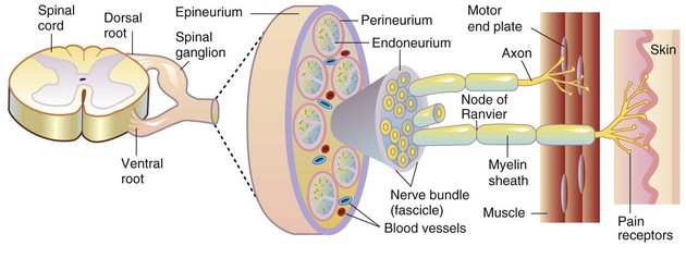
Blood-nerve barrier: A barrier to free movement of certain substances from the blood to the endoneurium of peripheral nerves. Barrier properties are conferred by tight junctions between capillary endothelial cells of the endoneurium and between perineurial cells and selective transport systems in the endothelial cells.

Brain edema: Increase in tissue water within the brain that results in an increase in brain volume. The fluid may be present in the intracellular or extracellular compartments or both. The term also is used to include the accumulation of plasma, especially in association with severe injury to the vasculature.

Brain swelling: Marked, rapidly developing, sometimes unexplained, increase in cerebral blood volume and brain volume because of relaxation (dilation) of the arterioles that occurs after brain injury.

Büngner, cell bands of: A column of proliferating Schwann cells that forms within the space previously occupied by an axon following wallerian degeneration. The proliferating column of cells is surrounded by the persisting basement membrane of the original Schwann cells.

Central chromatolysis: Dissolution of cytoplasmic Nissl substance (arrays of rough endoplasmic reticulum and polysomes) in the central part of the neuronal cell body that results from injury to the neuron (often involving the axon). The cell body is swollen, and the nucleus frequently is displaced peripherally to the cell membrane. These structural changes functionally represent a response to injury that can be found (if the cell survives) by axonal regeneration with protein synthesis to produce components of the axon required for fast and slow axonal transport.

Cranium bifidum: A dorsal midline cranial defect through which meninges alone or meninges and brain tissue may protrude into a sac (-cele), covered by skin.

Demyelination: A disease process in which demyelination (destruction of the myelin sheath) is the primary lesion, although some degree of axonal injury may occur. Primary demyelination is caused by injury to myelin sheaths and/or myelinating cells and their cell processes. Secondary demyelination occurs with axonal injury, as in wallerian degeneration.

Dysraphism: Dysraphia, which literally means an abnormal seam, refers to a defective closure of the neural tube during development. This defect, which may occur at any point along the neural tube, is exemplified by anencephaly, prosencephalic hypoplasia, cranium bifidum, spina bifida, and myeloschisis.

Encephalitis: Inflammation of the brain.

Encephalo-: A combining form that refers to the brain.

Encephalopathy: A degenerative disease process of the brain.

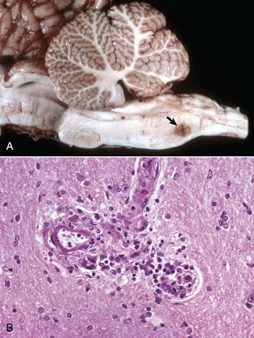
Ganglionitis: Inflammation of peripheral (sensory or autonomic or both) ganglia.

Gemistocyte: Reactive, hypertrophied astrocyte that develops in response to injury of the CNS. The cell body and processes of gemistocytes become visible with conventional staining (e.g., H&E stain). The cell bodies and processes of normal astrocytes are not visible with H&E staining.

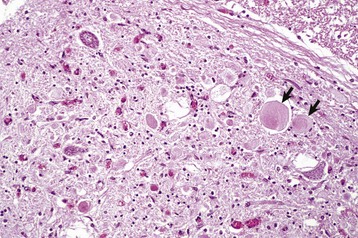
Gitter cell: Macrophage that accumulates in areas of necrosis of CNS tissue. The cytoplasm is typically distended, with abundant lipid-containing material derived from the lipid-rich nervous tissue. Gitter cell nuclei are often displaced peripherally to the cell membrane. These cells are often referred to as “foamy” macrophages.

Hydranencephaly: A large, fluid-filled cavity in the area normally occupied by CNS tissue of the cerebral hemispheres resulting from abnormal development. The nervous tissue may be so reduced in thickness that the meninges form the outer part of a thin-walled sac. The lateral ventricles are variably enlarged because of their expansion into the area normally occupied by tissue.

Hydrocephalus: Accumulation of excess CSF resulting from obstruction within the ventricular system (noncommunicating form) associated with enlargement of any or all of the following: lateral ventricles, third ventricle, mesencephalic aqueduct, and fourth ventricle. Hydrocephalus can also occur with communication of the CSF between the ventricular system and the subarachnoid space (communicating form). Hydrocephalus ex vacuo (compensatory hydrocephalus) is characterized by an expansion of the lateral ventricle (or ventricles) that follows loss of brain tissue.

Leuko-: Combining form referring to white matter of the brain or spinal cord.

Leukoencephalitis: Inflammation of the white matter of the brain.

Macroglia: A collective term referring to astrocytes and oligodendrocytes. Has also been variously used to refer solely to astrocytes or to astrocytes, oligodendrocytes, and ependymal cells of the CNS, and Müller cells of the retina.

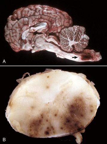
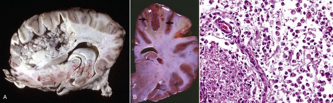
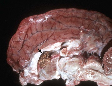
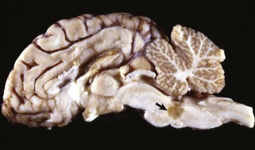
Malacia: Grossly detectable (macroscopic lesion) softening of CNS tissue, usually the result of necrosis.

Meningo-: Combining form referring to meninges.

Meningomyelocele: A form of spina bifida in which meninges and spinal cord herniate through a defect in the vertebral column into a sac (-cele) covered by skin.

Microglia: Resident cells of the CNS believed to arise from monocytes that populate the brain during embryonic development.

Motor neuron, lower: Large multipolar neurons in the brainstem and ventral horns of the spinal cord with axons extending into the PNS.

Motor neuron, upper: Motor neurons with axons residing solely in the CNS that control lower motor neurons.

Myelitis: Inflammation of the spinal cord.

Myelo-: Combining form referring to spinal cord.

Myelopathy: A degenerative disease process of the spinal cord.

Myeloschisis: Similar to spina bifida, except in its severe form is characterized by complete failure of the spinal neural tube to close, therefore a lack of development of the entire dorsal vertebral column.

Neuroglial cells: Astrocytes, oligodendroglia, ependymal cells, and microglia of the CNS.

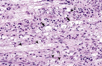
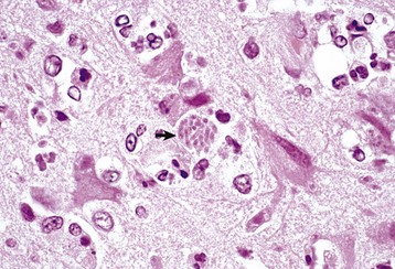
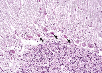
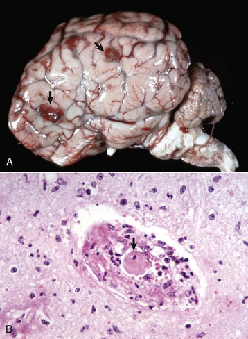
Neuronophagia: Accumulation of microglial cells around a dead neuron.

Neuropil: The gray matter feltwork that consists of intermingled and interconnected processes of neurons (axons and dendrites) and their synaptic junctions, plus processes of oligodendroglia, astrocytes, and microglia.

Neurapraxia: Traumatic injury to a peripheral nerve with temporary conduction block but with no permanent axonal damage.

Neurotmesis: Complete transection of a nerve and supporting framework with little potential for normal reinnervation.

Onion bulb: Concentric arrays of Schwann cell cytoplasm around an axon signifying multiple episodes of demyelination and remyelination.

Polio-: Combining form referring to gray matter of the CNS.

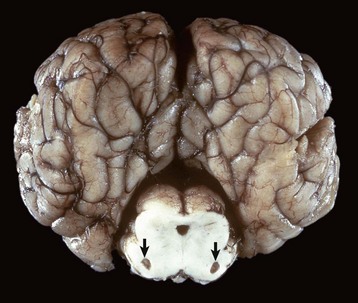
Polioencephalomalacia: Softening (usually the result of necrosis) of the gray matter of the brain.

Polioencephalomyelitis: Inflammation of the gray matter of the brain and spinal cord.

Poliomyelomalacia: Softening (usually the result of necrosis) of the gray matter of the spinal cord.

Porencephaly: A cleft or cystic defect in the cerebral hemisphere that communicates with the subarachnoid space and also may communicate with the ventricular system. The defect may contain CSF.

Radiculoneuritis (polyradiculoneuritis): Inflammation of a spinal nerve rootlet (or rootlets).

Rarefaction: Reduction in density of CNS tissue that may result from edema, necrosis, and the like. This lesion is usually observed microscopically.

Satellitosis: An accumulation of oligodendroglia around neuronal cell bodies. Although this feature can be seen in normal brains, some consider that it may also be associated with neuronal injury.

Sclerosis: Literally means induration or hardening and, when used in describing lesions of the CNS, often refers to induration or hardening of the brain or spinal cord resulting from astrogliosis (astrocytic scar formation).

Spina bifida: A dorsal midline defect involving one to several vertebrae of the spinal column caused by failure of the neural tube to close, permitting exposure of the underlying meninges and spinal cord. The lesion may be associated with herniation of meninges alone or meninges and spinal cord tissue into a sac (-cele) covered by skin, or there may be no herniation (spina bifida occulta).

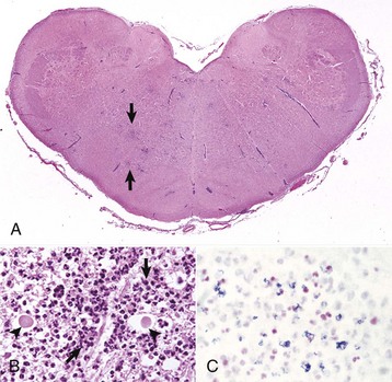
Status spongiosus: An encompassing term meaning the presence of small focal, ovoid to round “clear (unstained or poorly stained [H&E stain])” spaces in the CNS. The lesion can result from several different tissue alterations, which include splitting of the myelin sheath, accumulation of extracellular fluid, swelling of cellular (e.g., astrocytic and neuronal) processes, and axonal injury (wallerian degeneration) when swollen axons are no longer detectable within distended spaces.

Syringomyelia: A tubular cavitation (syrinx) in the spinal cord that is not lined by ependyma and may extend over several segments.

Wallerian degeneration: Degeneration of the distal component of an injured (compressed or severed) axon. Although the term originally referred to injury of axons in the PNS, current usage also includes the CNS. This process also results in functional and structural alterations in the cell body (central chromatolysis) and proximal internode segment of the axon, and in secondary demyelination.


*Dr. R.W. Storts, College of Veterinary Medicine, Texas A&M University, and Dr. D.L. Montgomery, Wyoming State Veterinary Laboratory, made contributions to this chapter in the third and fourth editions.