Hepatic Complications of Organ or Bone Marrow Transplantation

The use of transplantation for bone marrow, renal, hepatic and other organ disorders has generated a challenging group of hepatic complications. The liver may be damaged by toxic drugs or graft-versus-host disease in patients undergoing bone marrow transplantation, whereas patients receiving a liver transplant may have graft failure or rejection, and may develop sinusoidal obstruction syndrome, as already discussed. Although the clinical settings are obviously different for each patient population, the common themes of toxic or immunologically mediated liver damage, infection of immunosuppressed hosts, recurrent disease, and post-transplant lymphoproliferative disorder are readily apparent. The following focuses on post-transplant graft-versus-host disease and liver rejection.

GRAFT-VERSUS-HOST DISEASE AND LIVER REJECTION

The liver has the unenviable position of being attacked by graft-versus-host and host-versus-graft mechanisms, in the setting of bone marrow transplantation and liver transplantation, respectively. These processes are discussed in detail in Chapter 6. More than other solid organs, liver transplants are reasonably well tolerated by recipients. That being said, the hepatic morphologic features that are peculiar to immunological attack after transplantation deserve comment.

Morphology. Liver damage after bone marrow transplantation is the consequence of acute or chronic graft-versus-host disease. In acute graft-versus-host disease, which occurs 10 to 50 days after bone marrow transplantation, donor lymphocytes attack the epithelial cells of the liver. This results in hepatitis with necrosis of hepatocytes and bile duct epithelial cells, and inflammation of the parenchyma and portal tracts. In chronic hepatic graft-versus-host disease (usually more than 100 days after transplantation), there is portal tract inflammation, selective bile duct destruction, and eventual fibrosis. Portal vein and hepatic vein radicles may show endothelitis, a process in which a subendothelial lymphocytic infiltrate lifts the endothelium from its basement membrane. Cholestasis may be observed in both acute and chronic graft-versus-host disease.

In transplanted livers, acute rejection is characterized by infiltration of a mixed population of inflammatory cells that include eosinophils into portal tracts, bile duct and hepatocyte injury, and endothelitis (Fig. 18-41). The severity of the rejection is graded according to the BANFF scheme, which is important for clinical management.67 With chronic rejection a severe obliterative arteritis of small and larger arterial vessels (arteriopathy) results in ischemic changes in the liver parenchyma. Alternatively, bile ducts are progressively destroyed, because of either direct immunological attack or obliteration of their arterial supply, resulting in loss of the graft.

image

FIGURE 18-41 Transplanted liver with acute cellular rejection. Note the mixed inflammatory cell infiltration including eosinophils in portal tracts, bile duct damage, and endotheliitis.

Hepatic Disease Associated with Pregnancy

Hepatic diseases may occur in women with chronic liver disease who become pregnant, or they may develop during pregnancy in women who were not affected by liver disease. Abnormal liver tests occur in 3% to 5% of pregnancies.68 Viral hepatitis (HAV, HBV, HCV, and even HBV + HDV) is the most common cause of jaundice in pregnancy. While these women require careful clinical management, pregnancy does not specifically alter the course of the liver disease. The one exception is HEV infection, which, for unknown reasons, runs a more severe course in pregnant patients, with fatality rates of 10% to 20%.

A very small subgroup of pregnant women (0.1%) develops hepatic complications directly attributable to pregnancy: preeclampsia and eclampsia, acute fatty liver of pregnancy, and intrahepatic cholestasis of pregnancy. In extreme cases of the first two conditions, the outcome is fatal.

PREECLAMPSIA AND ECLAMPSIA

Preeclampsia affects 3% to 5% of pregnancies and is characterized by maternal hypertension, proteinuria, peripheral edema, coagulation abnormalities, and varying degrees of disseminated intravascular coagulation (Chapter 22). When hyper-reflexia and convulsions occur the condition is called eclampsia and may be life-threatening. Alternatively, subclinical hepatic disease may be the primary manifestation of preeclampsia, as part of a syndrome of hemolysis, elevated liver enzymes, and low platelets, dubbed the HELLP syndrome.69

Morphology. The affected liver in preeclampsia is normal in size, firm, and pale, with small red patches due to hemorrhage. Occasionally, yellow or white patches of ischemic infarction can be seen. Microscopically, the periportal sinusoids contain fibrin deposits with hemorrhage into the space of Disse, leading to periportal hepatocellular coagulative necrosis. Blood under pressure may coalesce and expand to form a hepatic hematoma; dissection of blood under Glisson’s capsule may lead to catastrophic hepatic rupture (Fig. 18-42). Patients with hepatic involvement in preeclampsia may show modest to severe elevation of serum aminotransferases and mild elevation of serum bilirubin. Hepatic dysfunction sufficient to cause a coagulopathy signifies far-advanced and potentially lethal disease. Definitive treatment in severe cases requires termination of the pregnancy. In mild cases patients may be managed conservatively. Women who survive mild or severe preeclampsia recover without sequelae.

image

FIGURE 18-42 Eclampsia. Subcapsular hematoma dissecting under Glisson’s capsule in a fatal case of eclampsia.

(Courtesy of Dr. Brian Blackbourne, Office of the Medical Examiner, San Diego, CA.)

ACUTE FATTY LIVER OF PREGNANCY (AFLP)

AFLP presents with a spectrum ranging from modest or even subclinical hepatic dysfunction (evidenced by elevated serum aminotransferase levels) to hepatic failure, coma, and death. It is a rare disease affecting 1 in 13,000 deliveries. Affected women present in the latter half of pregnancy, usually in the third trimester. Symptoms are directly attributable to incipient hepatic failure, including bleeding, nausea and vomiting, jaundice, and coma. In 20% to 40% of cases the presenting symptoms may be those of coexistent preeclampsia.

Morphology. The diagnosis of acute fatty liver rests on biopsy identification of the characteristic microvesicular fatty transformation of hepatocytes. In severe cases there may be lobular disarray with hepatocyte dropout, reticulin collapse, and portal tract inflammation, making distinction from viral hepatitis difficult. Diagnosis depends on (1) a high index of suspicion and (2) confirmation of microvesicular steatosis using special stains for fat (oil-red-O or Sudan black) on frozen tissue sections; electron microscopy may also be used to demonstrate the steatosis.

While this condition most commonly runs a mild course, women with AFLP can progress within days to hepatic failure and death. The primary treatment for AFLP is termination of the pregnancy. The pathogenesis of this disease is unknown, but mitochondrial dysfunction has been implicated. In a subset of patients, both mother and father carry a heterozygous deficiency in mitochondrial long-chain 3-hydroxyacyl coenzyme A (CoA) dehydrogenase. The homozygous-deficient fetuses fare well during pregnancy but cause hepatic dysfunction in the mother, because long-chain 3-hydroxylacyl metabolites produced by the fetus or placenta are washed away into the maternal circulation and cause hepatic toxicity. This is a rare instance of the fetus causing metabolic disease in the mother.70

INTRAHEPATIC CHOLESTASIS OF PREGNANCY

The onset of pruritus in the third trimester, followed by darkening of the urine and occasionally light stools and jaundice, heralds the development of this enigmatic syndrome. Serum bilirubin (mostly conjugated) rarely exceeds 5 mg/dL; alkaline phosphatase may be slightly elevated. Liver biopsy reveals mild cholestasis without necrosis. The altered hormonal state of pregnancy seems to combine with biliary defects in the secretion of bile salts or sulfated progesterone metabolites to engender cholestasis. Although this is generally a benign condition, the mother is at risk for gallstones and malabsorption, and the incidence of fetal distress, stillbirths, and prematurity is modestly increased. Perhaps most importantly, the pruritus can be extremely distressing for the pregnant mother.

Nodules and Tumors

Hepatic masses may come to attention for a variety of reasons. They may generate epigastric fullness and discomfort or be detected by routine physical examination or radiographic studies for other indications. Nodular hyperplasias are not neoplasms; the remaining lesions discussed in this section are true neoplasms.

NODULAR HYPERPLASIAS

Solitary or multiple hyperplastic hepatocellular nodules may develop in the noncirrhotic liver. Two such conditions, having confusingly overlapping names, are focal nodular hyperplasia and nodular regenerative hyperplasia. The common factor in both types of nodules seems to be either focal or diffuse alterations in hepatic blood supply, arising from obliteration of portal vein radicles and compensatory augmentation of arterial blood supply.

Morphology. Focal nodular hyperplasia appears as a well-demarcated but poorly encapsulated nodule, ranging up to many centimeters in diameter (Fig. 18-43A). It presents as a spontaneous mass lesion in an otherwise normal liver, most frequently in young to middle-aged adults. The lesion is generally lighter than the surrounding liver and is sometimes yellow. Typically, there is a central gray-white, depressed stellate scar from which fibrous septa radiate to the periphery (Fig. 18-43B). The central scar contains large vessels, usually arterial, that typically show fibromuscular hyperplasia with eccentric or concentric narrowing of the lumen. The radiating septa show foci of intense lymphocytic infiltrates and exuberant bile duct proliferation along septal margins. The parenchyma between the septa shows essentially normal hepatocytes but with a thickened plate architecture characteristic of regeneration. Long-term use of anabolic hormones or of contraceptives have been implicated in the development of focal nodular hyperplasia.

image

FIGURE 18-43 Focal nodular hyperplasia. A, Resected specimen showing lobulated contours and a central stellate scar. B, Low-power photomicrograph showing a broad fibrous scar with hepatic arterial and bile duct elements and chronic inflammation, present within hepatic parenchyma that lacks the normal sinusoidal plate architecture.

Nodular regenerative hyperplasia denotes a liver entirely transformed into roughly spherical nodules, in the absence of fibrosis. Microscopically, plump hepatocytes are surrounded by rims of atrophic hepatocytes. The variation in parenchymal architecture may be missed on an H&E stain, and reticulin staining is required to appreciate the changes in hepatocellular architecture. Nodular regenerative hyperplasia can lead to the development of portal hypertension and occurs in association with conditions affecting intrahepatic blood flow, including solid-organ (particularly renal) transplantation, bone marrow transplantation, and vasculitis. It also occurs in HIV-infected persons.71

BENIGN NEOPLASMS

Cavernous hemangiomas, blood vessel tumors identical to those occurring elsewhere (see Chapter 11), are the most common benign liver tumors. They appear as discrete red-blue, soft nodules, usually less than 2 cm in diameter, generally located directly beneath the capsule. Histologically, the tumor consists of vascular channels in a bed of fibrous connective tissue (Fig. 18-44). Their chief clinical significance is that they should not be mistaken for metastatic tumors, and that blind percutaneous biopsies not be performed on them.

image

FIGURE 18-44 Hemangioma. The photomicrograph shows the vesicular channels embedded in fibrous stroma.

Hepatic Adenoma

Benign neoplasms developing from hepatocytes are called hepatic adenomas or liver cell adenomas. Although they may occur in men, hepatic adenomas most frequently occur in young women who have used oral contraceptives; tumors generally regress if contraceptive use is terminated. The incidence of adenoma is approximately 1 in 100,000. Hepatic adenomas have clinical significance for three reasons: (1) when they present as an intrahepatic mass they may be mistaken for the more ominous hepatocellular carcinomas; (2) subcapsular adenomas have a tendency to rupture, particularly during pregnancy (under estrogen stimulation), causing life-threatening intraperitoneal hemorrhage; (3) rarely, they may transform into carcinomas, particularly, when the adenoma arises in an individual with glycogen storage disease, and adenomas in which mutations of the β-catenin gene are present.

Pathogenesis.

Although hormonal stimulation is clearly associated with the development of solitary hepatic adenoma, the causal events are unknown. Mutations in the genes encoding the transcription factor HNF1α and β-catenin have been identified in 50% and 15% of the hepatic adenomas, respectively.72 Multiple hepatic adenoma (adenomatosis) syndromes can occur in individuals with maturity-onset diabetes of young (MODY3), with HNF1 mutations.73

Morphology. Liver cell adenomas are pale, yellowtan, and frequently bile-stained nodules, found anywhere in the hepatic substance but often beneath the capsule (Fig. 18-45A). They may reach 30 cm in diameter. Although they are usually well demarcated, encapsulation might not be present. The tumor commonly presents as a solitary lesion, but multiple lesions (adenomatosis) can occur. Histologically, liver cell adenomas are composed of sheets and cords of cells that may resemble normal hepatocytes or have some variation in cell and nuclear size (Fig. 18-45B). Abundant glycogen may generate large hepatocytes with a clear cytoplasm. Steatosis is commonly present. Portal tracts are absent; instead, prominent solitary arterial vessels and draining veins are distributed through the substance of the tumor.

image

FIGURE 18-45 Liver cell adenoma. A, Resected specimen presenting as a pendulous mass arising from the liver. B, Microscopic view showing cords of hepatocytes, with an arterial vascular supply (arrow) and no portal tracts.

MALIGNANT TUMORS

Malignant tumors occurring in the liver can be primary or metastatic. Most of the discussion in this section deals with primary hepatic tumors. Primary carcinomas of the liver are relatively uncommon in North America and western Europe (0.5% to 2% of all cancers) but represent 20% to 40% of cancers in many other countries. Most primary liver cancers arise from hepatocytes and are termed hepatocellular carcinoma (HCC). Much less common are carcinomas of bile duct origin, cholangiocarcinomas. The incidence of these two cancers is increasing in the United States.

Before embarking on a discussion of the major forms of malignancy affecting the liver, two rare forms of primary liver cancer deserve brief mention: hepatoblastomas and angiosarcomas. Angiosarcoma of the liver resembles those occurring elsewhere. The primary liver form is of interest because of its association with exposure to vinyl chloride, arsenic, or Thorotrast (Chapters 9 and 11. The latency period after exposure to the putative carcinogen may be several decades. These highly aggressive neoplasms metastasize widely and generally kill within a year. The major features of hepatoblastoma are discussed next.

Hepatoblastoma

Hepatoblastoma is the most common liver tumor of young childhood. Its incidence, which is increasing, is approximately 1 to 2 in 1 million births.74 The tumor is usually fatal within a few years if not treated. This tumor has two anatomic variants:

The epithelial type, composed of small polygonal fetal cells or smaller embryonal cells forming acini, tubules, or papillary structures vaguely recapitulating liver development (Fig. 18-46)
The mixed epithelial and mesenchymal type, which contains foci of mesenchymal differentiation that may consist of primitive mesenchyme, osteoid, cartilage, or striated muscle
image

FIGURE 18-46 Hepatoblastoma. The photograph shows proliferating hepatoblasts.

A characteristic feature of hepatoblastomas is the frequent activation of the WNT/β-catenin signaling pathway.75 Chromosomal abnormalities are common in hepatoblastomas, and FOXG1, a regulator of the TGF-β pathway, is highly expressed in some subsets of the tumor.76 Hepatoblastoma may be associated with familial adenomatous polyposis syndrome and Beckwith-Wiedmann syndrome. The treatment is chemotherapy and complete surgical resection. The therapy has raised the 5-year survival to 80%.

Hepatocellular Carcinoma (HCC)

On a global basis, there are more than 626,000 new cases per year of primary liver cancer, almost all being HCC, and approximately 598,000 patients die from this cancer every year,77 the third most frequent cause of cancer deaths. About 82% of HCC cases occur in developing countries with high rates of chronic HBV infection, such as in southeast Asian and African countries; 52% of all HCC cases occur in China. In the United States the incidence of liver cancer increased by 25% between 1993 and 1998, mainly due to HCV and HBV chronic infection. There is a clear predominance of males with a ratio of 2.4 : 1.

Pathogenesis.

Several general factors relevant to the pathogenesis of HCC were discussed in Chapter 7. Some issues specifically related to HCC deserve emphasis here.

Four major etiologic factors associated with HCC have been established: chronic viral infection (HBV, HCV), chronic alcoholism, non-alcoholic steatohepatitis (NASH), and food contaminants (primarily aflatoxins). Other conditions include tyrosinemia, glycogen storage disease, hereditary hemochromatosis, non-alcoholic fatty liver disease, and α1-antitrypsin deficiency. Many factors, including genetic factors, age, gender, chemicals, hormones, and nutrition, interact in the development of HCC. The disease that is most likely to give rise to HCC is the extremely rare hereditary tyrosinemia, in which almost 40% of patients develop the tumor despite adequate dietary control.

The pathogenesis of HCC may be different in high-incidence, HBV-prevalent populations versus low-incidence Western populations, in which other chronic liver diseases such as alcoholism, non-alcoholic steatohepatitis, chronic HCV infection, and hemochromatosis are more common. In high-prevalence regions the HBV infection begins in infancy by the vertical transmission of virus from infected mothers, which confers a 200-fold increased risk for HCC development by adulthood. Cirrhosis may be absent in as many as half of these patients, and the cancer often occurs between 20 and 40 years of age. In the Western world where HBV is not prevalent, cirrhosis is present in 75% to 90% of cases of HCC, usually in the setting of other chronic liver diseases. Thus, cirrhosis seems to be a prerequisite contributor to the emergence of HCC in Western countries but may have a different role in HCC that develops in endemic areas. In China and southern Africa, where HBV is endemic, there may also be exposure to aflatoxin, a toxin produced by the fungus Aspergillus flavus, which contaminates peanuts and grains. Aflatoxin can bind covalently with cellular DNA and cause a specific mutation in codon 249 of p53 (Chapter 9).

Although the precise mechanisms of carcinogenesis are unknown, several events have been implicated. Repeated cycles of cell death and regeneration, as occurs in chronic hepatitis from any cause, are important in the pathogenesis of HCCs (Chapter 7). It is thought that the accumulation of mutations during continuous cycles of cell division may damage DNA repair mechanisms and eventually transform hepatocytes. Preneoplastic changes can be recognized morphologically by the occurrence of hepatocyte dysplasia. Progression to HCC might result from point mutations in selected cellular genes such as KRAS and p53, and constitutive expression of c-MYC, c-MET (the receptor for hepatocyte growth factor), TGF-α, and insulin-like growth factor 2. Recent global gene expression studies revealed that approximately 50% of HCC cases are associated with activation of WNT or AKT pathways. A subgroup of tumors expresses a high proportion of genes present in fetal liver and liver progenitor cells, suggesting that at least some HCCs may be generated from liver stem cells (Chapter 3).

Molecular analysis of tumor cells in HBV-infected individuals showed that most nodules are clonal with respect to the HBV DNA integration pattern, suggesting that viral integration precedes or accompanies a transforming event. In HBV-induced carcinogenesis not only the disruption of cell genome caused by virus integration but also the site of integration can be important. Depending on the integration site, HBV integration may activate proto-oncogenes that contribute to tumorigenicity. Alternatively, it has been proposed that the HBV X-protein, a transcriptional activator of multiple genes, might be the main cause of cell transformation. The situation is even more uncertain regarding the mechanisms of HCV carcinogenesis. HCV is a RNA virus that does not disrupt DNA, and it does not produce oncogenic proteins. However, there are indications that the HCV core and NS5A proteins may participate in the development of HCC.78

Universal vaccination of children against HBV in endemic areas can dramatically decrease the incidence of HBV infection, and most likely, the incidence of HCC. Such a program, started in Taiwan in 1984, has reduced HBV infection rates from 10% to less than 1% in 20 years.79

Morphology. HCC may appear grossly as (1) a unifocal (usually large) mass (Fig. 18-47A); (2) multifocal, widely distributed nodules of variable size; or (3) a diffusely infiltrative cancer, permeating widely and sometimes involving the entire liver. All three patterns may cause liver enlargement, particularly the large unifocal and multinodular patterns. The diffusely infiltrative tumor may blend imperceptibly into a cirrhotic liver background.

image

FIGURE 18-47 Hepatocellular carcinoma. A, Liver removed at autopsy showing a unifocal, massive neoplasm replacing most of the right hepatic lobe in a noncirrhotic liver; a satellite tumor nodule is directly adjacent. B, Microscopic view of a well-differentiated lesion; tumor cells are arranged in nests, sometimes with a central lumen.

HCCs are usually paler than the surrounding liver, and sometimes take on a green hue when composed of well-differentiated hepatocytes capable of secreting bile. All patterns of HCCs have a strong propensity for invasion of vascular structures. Extensive intrahepatic metastases ensue, and occasionally, long, snakelike masses of tumor invade the portal vein (with occlusion of the portal circulation) or inferior vena cava, extending even into the right side of the heart. HCC spreads extensively within the liver by obvious contiguous growth and by the development of satellite nodules, which can be shown by molecular methods to be derived from the parent tumor. Metastasis outside the liver is primarily via vascular invasion, especially into the hepatic vein system, but hematogenous metastases, especially to the lung, tend to occur late in the disease. Lymph node metastases to the perihilar, peripancreatic, and para-aortic nodes above and below the diaphragm are found in fewer than half of HCCs that spread beyond the liver. If HCC with venous invasion is identified in explanted livers at the time of liver transplantation, tumor recurrence is likely to occur in the transplanted donor liver.

HCCs range from well-differentiated to highly anaplastic undifferentiated lesions. In well-differentiated and moderately differentiated tumors, cells that are recognizable as hepatocytic in origin are disposed either in a trabecular pattern (recapitulating liver cell plates) (Fig. 18-47B) or in an acinar, pseudoglandular pattern. In poorly differentiated forms, tumor cells can take on a pleomorphic appearance with numerous anaplastic giant cells, can be small and completely undifferentiated, or may even resemble a spindle cell sarcoma.

A distinctive variant of HCC is the fibrolamellar carcinoma, which was first described in 1956. This variant constitutes 5% of HCCs. It occurs in young male and female adults (20 to 40 years of age) with equal incidence. Patients usually do not have underlying chronic liver diseases, and so the prognosis is better than the conventional HCC.80 The etiology of fibrolamellar carcinoma is unknown. It usually presents as single large, hard “scirrhous” tumor with fibrous bands coursing through it. On microscopic examination it is composed of well-differentiated polygonal cells growing in nests or cords, and separated by parallel lamellae of dense collagen bundles. The tumor cells have abundant eosinophilic cytoplasm and prominent nucleoli (Fig. 18-48).

image

FIGURE 18-48 Fibrolamellar carcinoma. A, Resected specimen showing a demarcated nodule in an otherwise normal liver. B, Microscopic view showing nests and cords of malignant-appearing hepatocytes separated by dense bundles of collagen.

Clinical Features.

The clinical manifestations of HCC are seldom characteristic and, in the Western population, often are masked by those related to the underlying cirrhosis or chronic hepatitis. In areas of high incidence such as tropical Africa, patients usually have no clinical history of liver disease, although cirrhosis may be detected at autopsy. In both populations most patients have ill-defined upper abdominal pain, malaise, fatigue, weight loss, and sometimes awareness of an abdominal mass or abdominal fullness. In many cases the enlarged liver can be felt on palpation, with sufficient irregularity or nodularity to permit differentiation from cirrhosis. Jaundice, fever, and gastrointestinal or esophageal variceal bleeding are inconstant findings.

Laboratory studies may be helpful but are rarely conclusive. Elevated levels of serum α-fetoprotein are found in 50% of persons with HCC. However, false-positive results are encountered with yolk-sac tumors and many non-neoplastic conditions, including cirrhosis, massive liver necrosis (with compensatory liver cell regeneration), chronic hepatitis (especially HCV infection), normal pregnancy, fetal distress or death, and fetal neural tube defects such as anencephaly and spina bifida. Laboratory testing for α-fetoprotein and other proteins (such as serum carcinoembryonic antigen levels) often fails to detect small HCC lesions. Recently, staining for Glypican-3 has been used to distinguish early HCC from dysplastic nodules. Most valuable for detection of small tumors are imaging studies: ultrasonography, hepatic angiography, computed tomography, and magnetic resonance imaging. Molecular analysis of HCC is actively being pursued and will most likely lead to new HCC classifications that can help determine treatment options. As already mentioned, some molecular signatures of HCC have already been identified.81

The natural course of HCC involves the progressive enlargement of the primary mass until it seriously disturbs hepatic function, or metastasizes, generally first to the lungs and then to other sites. Overall, death usually occurs from (1) cachexia, (2) gastrointestinal or esophageal variceal bleeding, (3) liver failure with hepatic coma, or, rarely, (4) rupture of the tumor with fatal hemorrhage. The 5-year survival of large tumors is dismal, with the majority of patients dying within the first 2 years. With implementation of screening procedures and advances in imaging, the detection of HCCs less than 2 cm in diameter has increased in countries where such facilities are available. These small tumors can be removed surgically with good prognostic outcomes. Radiofrequency ablation is used for local control of large tumors, and chemoembolization can also be used, according to a clinical algorithm that has been widely adopted.82 Recent findings show that the kinase inhibitor sorafenib can prolong the life of individuals with advanced-stage HCC.83

Cholangiocarcinoma (CCA)

Cholangiocarcinoma, the second most common hepatic malignant tumor after HCC, is a malignancy of the biliary tree, arising from bile ducts within and outside of the liver.84 It accounts for 7.6% of cancer deaths worldwide and 3% of cancer deaths in the United States. The prevalence of the disease in the United States is variable, the highest being in Hispanics (1.22 per 100,000 population), and the lowest in African-Americans (0.17–0.5 per 100,000). The risk factors for development of CCA include primary sclerosing cholangitis (PSC), congenital fibropolycystic diseases of the biliary system (particularly Caroli disease and choledochal cysts that will be discussed later), HCV infection, and previous exposure to Thorotrast (formerly used in radiography of the biliary tract). Most cholangiocarcinomas in the Western world, however, arise without evidence of such antecedent conditions. In southeast Asia, where the incidence rates are higher, a major risk factor is chronic infection of the biliary tract by the liver fluke Opisthorchis sinensis and its close relatives. According to their localization, CCAs are classified into intrahepatic and extrahepatic forms. Eighty to 90% of the tumors are extrahepatic. However, the incidence of intrahepatic tumors has increased during the last two decades in the United States, western Europe, and Japan, while the incidence of extrahepatic CCA has remained constant. The extrahepatic forms include perihilar tumors known as Klatskin tumors, which are located at the junction of the right and left hepatic ducts forming the common hepatic duct, and distal bile duct tumors. A subgroup of distal tumors arise in the immediate vicinity of the ampulla of Vater. Tumors of this region also include adenocarcinoma of the duodenal mucosa and pancreatic carcinoma (discussed in Chapters 17 and 19, respectively) and are collectively referred to as periampullary carcinomas.

Fifty to 60% of all CCAs are perihilar (Klatskin) tumors, 20% to 30% are distal tumors, and about 10% are intrahepatic. In any case the prognosis is dismal, with survival rates of about 15% at 2 years after diagnosis. The median time from diagnosis to death for intrahepatic CCAs is 6 months, even after surgery. Intrahepatic CCAs are not usually detected until late in their course, and come to the attention because of obstruction of bile flow, or as a symptomatic liver mass. In contrast, hilar and distal tumors present with symptoms of biliary obstruction, cholangitis, and right upper quadrant pain.

Morphology. Extrahepatic CCAs are generally small lesions at the time of diagnosis. Most tumors appear as firm, gray nodules within the bile duct wall; some may be diffusely infiltrative lesions; others are papillary, polypoid lesions. Most are adenocarcinomas that may or may not secrete mucin. Uncommonly, squamous features are present. For the most part, an abundant fibrous stroma accompanies the epithelial proliferation. Klatskin tumors generally have slower growth than other CCAs, show prominent fibrosis, and infrequently involve distal metastases.

Intrahepatic CCAs occur in the noncirrhotic liver and may track along the intrahepatic portal tract system to create a treelike tumorous mass within a portion of the liver. Alternatively, a massive tumor nodule may develop. In either instance, vascular invasion and propagation along portal lymphatics may be prominent features, giving rise to extensive intrahepatic metastasis (Fig. 18-49A). By microscopy, CCAs resemble adenocarcinomas arising in other parts of the body, and they may show the full range of morphologic variation. Most are well- to moderately differentiated sclerosing adenocarcinomas with clearly defined glandular and tubular structures lined by cuboidal to low columnar epithelial cells (Fig. 18-49B). These neoplasms are usually markedly desmoplastic, with dense collagenous stroma separating the glandular elements. As a result, the tumor substance is extremely firm and gritty. Lymph node metastasis and hematogenous metastases to the lungs, bones (mainly vertebrae), adrenals, brain, or elsewhere are present at autopsy in about 50% of cases. Mixed variants occur, in which elements of both HCC and CCA are present. Three forms are recognized: (1) separate tumor masses of HCC and CCA within the same liver; (2) “collision tumors,” in which tumorous masses of HCC and CCA commingle at an identifiable interface; and (3) tumors in which elements of HCC and CCA are intimately mixed at the microscopic level. These “mixed tumors” are infrequent, but careful microscopic examination of CCAs can often reveal small foci of hepatocellular differentiation. The HCC-CCA may be generated from a common bipotential precursor cell (oval cells, Chapter 3), capable of producing both hepatocytes and bile duct epithelial cells (cholangiocytes).

image

FIGURE 18-49 Cholangiocarcinoma. A, Liver removed at autopsy showing a massive neoplasm in the right hepatic lobe and innumerable metastases permeating the entire liver. B, Microscopic view showing tubular glandular structures embedded in a dense sclerotic stroma.

Pathogenesis.

Several signaling pathways, some listed here, are involved in the pathogenesis of CCA. Among these is IL-6 overexpression that leads to activation of AKT and the anti-apoptotic protein MCL-1. Also increased in CCAs is the expression of COX-2, ERB-2, and c-MET. KRAS expression is increased in 20% to 100% of cases in different studies, and p53 expression is decreased in about 40% of cases. Other alterations involve amplification of epidermal growth factor receptors, and decreases in the expression of the cell cycle regulator and tumor suppressor p16/ink4A. In addition to cytologic diagnosis, fluorescence in situ hybridization using specific probes for epidermal growth factor receptors and digital image analysis to determine ploidy are now being used to improve diagnostic accuracy. Surgery, when possible, is the only treatment that is potentially curative.

METASTATIC TUMORS

Involvement of the liver by metastatic malignancy is far more common than primary hepatic neoplasia. The liver and lungs share the dubious distinction of being the visceral organs that are most often involved in the metastatic spread of cancers. Although the most common primary sources producing hepatic metastases are those of the colon, breast, lung, and pancreas, any cancer in any site of the body may spread to the liver, including leukemias, melanomas, and lymphomas. Typically, multiple nodular metastases are found that often cause striking hepatomegaly and may replace over 80% of existent hepatic parenchyma. The liver weight can exceed several kilograms. Metastasis may also appear as a single nodule, in which case it may be resected surgically. There is a tendency for metastatic nodules to outgrow their blood supply, producing central necrosis and umbilication when viewed from the surface of the liver. Always surprising is the amount of metastatic involvement that may be present in the absence of clinical or laboratory evidence of hepatic functional insufficiency. Often the only telltale clinical sign is hepatomegaly, sometimes with nodularity of the free edge. However, with massive destruction of liver substance or direct obstruction of major bile ducts, jaundice and abnormal elevations of liver enzymes may appear.

THE BILIARY TRACT

Disorders of the biliary tract affect a significant portion of the world’s population. Over 95% of biliary tract disease is attributable to cholelithiasis (gallstones). In the United States, gallstones affect 20 million people, and more than 700,000 cholecystectomies are performed annually at a cost of approximately $6 billion.

As much as 1 L of bile is secreted by the liver per day. Between meals bile is stored in the gallbladder, where it is concentrated. The adult gallbladder has a capacity of about 50 mL. The organ is not essential for biliary function, since humans do not suffer from indigestion or malabsorption of fat after cholecystectomy.

Congenital Anomalies

The gallbladder may be congenitally absent, or there may be gallbladder duplication with conjoined or independent cystic ducts. A longitudinal or transverse septum may create a bilobed gallbladder. Aberrant locations of the gallbladder occur in 5% to 10% of the population, most commonly partial or complete embedding in the liver substance. A folded fundus is the most common anomaly, creating a phrygian cap (Fig. 18-50). Agenesis of all or any portion of the hepatic or common bile ducts and hypoplastic narrowing of biliary channels (true “biliary atresia”) may also occur.

image

FIGURE 18-50 Phrygian cap of the gallbladder; the fundus is folded inward.

Disorders of the Gallbladder

CHOLELITHIASIS (GALLSTONES)

Gallstones afflict 10% to 20% of adult populations in developed countries. It is estimated that more than 20 million persons in the United States have gallstones, totaling some 25 to 50 tons in weight! The vast majority of gallstones (>80%) are “silent,” and most individuals remain free of biliary pain or other complications for decades. There are two main types of gallstones. In the West, about 90% are cholesterol stones, containing more than 50% of crystalline cholesterol monohydrate. The rest are pigment stones composed predominantly of bilirubin calcium salts.85

Prevalence and Risk Factors.

Certain populations are far more prone than others to develop gallstones. The major risk factors are listed in Table 18-9 and discussed below.

TABLE 18-9 Risk Factors for Gallstones

CHOLESTEROL STONES
Demography: northern Europeans, North and South Americans, Native Americans, Mexican-Americans
Advancing age
Female sex hormones
Female gender
Oral contraceptives
Pregnancy
Obesity and metabolic syndrome
Rapid weight reduction
Gallbladder stasis
Inborn disorders of bile acid metabolism
Hyperlipidemia syndromes
PIGMENT STONES
Demography: Asians more than Westerners, rural more than urban
Chronic hemolytic syndromes
Biliary infection
Gastrointestinal disorders: ileal disease (e.g., Crohn disease), ileal resection or bypass, cystic fibrosis with pancreatic insufficiency

Cholesterol gallstones are more prevalent in the United States and western Europe and uncommon in developing countries. The prevalence rates of cholesterol gallstones approach 75% in Native Americans of the Pima, Hopi, and Navajo groups; pigment stones are rare in these populations. Pigment gallstones, the predominant type of gallstone in non-Western populations, arise primarily in the setting of bacterial infections of the biliary tree and parasitic infestations.

The risk factors most commonly associated with the development of cholesterol stones are:

Age and Sex. The prevalence of cholesterol gallstones increases throughout life. In the United States, fewer than 5% to 6% of people under age 40 have stones, in contrast to 25% to 30% of those older than age 80. The prevalence in Caucasian women is about twice as high as in men. With both aging and gender, hypersecretion of biliary cholesterol seems to play the major role. In aging populations there is an increase in patients with gallstone disease associated with the metabolic syndrome and obesity.
Environmental Factors. Estrogenic influence, including oral contraceptives and pregnancy, increases the expression of hepatic lipoprotein receptors and stimulates hepatic HMG-CoA reductase activity, enhancing both cholesterol uptake and biosynthesis, respectively. Clofibrate, used to lower blood cholesterol, increases hepatic HMG-CoA reductase and decreases conversion of cholesterol to bile acids by reducing cholesterol 7-α-hydroxylase activity. The net result of these influences is excess biliary secretion of cholesterol. Obesity and rapid weight loss also are strongly associated with increased biliary cholesterol secretion.
Acquired Disorders. Gallbladder stasis, either neurogenic or hormonal, fosters a local environment that is favorable for both cholesterol and pigment gallstone formation.
Hereditary Factors. Much progress has been made recently in identifying susceptibility factors for cholesterol gallstones. These investigations have focused on genes encoding hepatocyte proteins that transport biliary lipids, known as ATP-binding cassette (ABC) transporters. In particular, a common variant of the protein heterodimer encoded by the ABCG5 and ABG2 genes that participates in biliary cholesterol secretion, confers a genetic risk for the development of cholesterol gallstones. The variant is known as D19H, and it is estimated that it may contribute 8% to 11% of the risk for the formation of cholesterol gallstones. (The odds ratios are 2–3 for heterozygous carriers of D19H, and 7 for homozygous carriers). Individuals with the D19H variant absorb less, but synthesize more, cholesterol, suggesting that HMG-CoA inhibitors (statins) may decrease the risk of gallstone formation in these individuals.
Pathogenesis of Cholesterol Stones.

Cholesterol is rendered soluble in bile by aggregation with water-soluble bile salts and water-insoluble lecithins, both of which act as detergents. When cholesterol concentrations exceed the solubilizing capacity of bile (supersaturation), cholesterol can no longer remain dispersed and nucleates into solid cholesterol monohydrate crystals. Cholesterol gallstone formation involves four simultaneous conditions (Fig. 18-51): (1) The bile must be supersaturated with cholesterol; (2) hypomotility of the gallbladder promotes nucleation; (3) cholesterol nucleation in the bile is accelerated; (4) hypersecretion of mucus in the gallbladder traps the nucleated crystals, leading to their aggregation into stones.

image

FIGURE 18-51 The four contributing factors for cholelithiasis: supersaturation, gallbladder hypomotility, crystal nucleation, and accretion within the gallbladder mucous layer.

Pathogenesis of Pigment Stones.

Pigment gallstones are complex mixtures of abnormal insoluble calcium salts of unconjugated bilirubin along with inorganic calcium salts. Disorders that are associated with elevated levels of unconjugated bilirubin in bile such as hemolytic syndromes, severe ileal dysfunction (or bypass), and bacterial contamination of the biliary tree, increase the risk of developing pigment stones. Unconjugated bilirubin is normally a minor component of bile, but it increases when infection of the biliary tract leads to release of microbial β-glucuronidases, which hydrolyze bilirubin glucuronides. Thus, infection of the biliary tract with Escherichia coli, Ascaris lumbricoides, or the liver fluke O. sinensis, increases the likelihood of pigment stone formation. In hemolytic syndromes the secretion of conjugated bilirubin into the bile increases. However, because about 1% of bilirubin glucuronides are deconjugated in the biliary tree, the large amounts of unconjugated bilirubin produced may exceed its solubility.

Morphology. Cholesterol stones arise exclusively in the gallbladder and are composed of cholesterol, ranging from 100% pure (which is rare) down to around 50%. Pure cholesterol stones are pale yellow, round to ovoid, and have a finely granular, hard external surface (Fig. 18-52), which on transection reveals a glistening radiating crystalline palisade. With increasing proportions of calcium carbonate, phosphates, and bilirubin, the stones show discoloration and may be lamellated and gray-white to black. Most often, multiple stones are present that range up to several centimeters in diameter. Rarely, there is a single much larger stone that may virtually fill the fundus. Surfaces of multiple stones may be rounded or faceted, because of tight apposition. Stones composed largely of cholesterol are radiolucent; sufficient calcium carbonate is found in 10% to 20% of cholesterol stones to render them radiopaque.

image

FIGURE 18-52 Cholesterol gallstones. Mechanical manipulation during laparoscopic cholecystectomy has caused fragmentation of several cholesterol gallstones, revealing interiors that are pigmented because of entrapped bile pigments. The gallbladder mucosa is reddened and irregular as a result of coexistent chronic cholecystitis.

Pigment gallstones are trivially classified as “black” and “brown.” In general, black pigment stones are found in sterile gallbladder bile, and brown stones are found in infected intrahepatic or extrahepatic ducts. “Black” pigment stones contain oxidized polymers of the calcium salts of unconjugated bilirubin, small amounts of calcium carbonate, calcium phosphate, and mucin glycoprotein, and some cholesterol monohydrate crystals. “Brown” pigment stones contain pure calcium salts of unconjugated bilirubin, mucin glycoprotein, a substantial cholesterol fraction, and calcium salts of palmitate and stearate. The black stones are rarely greater than 1.5 cm in diameter, are almost invariably present in great number (with an inverse relationship between size and number; Fig. 18-53), and may crumble to the touch. Their contours are usually spiculated and molded. Brown stones tend to be laminated and soft and may have a soaplike or greasy consistency. Because of calcium carbonates and phosphates, approximately 50% to 75% of black stones are radiopaque. Brown stones, which contain calcium soaps, are radiolucent. Mucin glycoproteins constitute the scaffolding and interparticle cement of all stones, whether pigment or cholesterol.

image

FIGURE 18-53 Pigment gallstones. Several faceted black gallstones are present in this otherwise unremarkable gallbladder from a patient with a mechanical mitral valve prosthesis, leading to chronic intravascular hemolysis.

Clinical Features.

Gallstones may be present for decades before symptoms develop, and 70% to 80% of patients remain asymptomatic throughout their lives. It has been estimated that asymptomatic patients convert to symptomatic ones at the rate of 1% to 4% per year, and the risk diminishes with time. Prominent among symptoms is biliary pain, which tends to be excruciating and constant or “colicky” (spasmodic), as a result of the obstructive nature of gallstones in the biliary tree and perhaps in the gallbladder itself. Inflammation of the gallbladder (cholecystitis, discussed below), in association with stones, also generates pain. More severe complications include empyema, perforation, fistulas, inflammation of the biliary tree (cholangitis), and obstructive cholestasis or pancreatitis with ensuing problems. The larger the calculi, the less likely they are to enter the cystic or common ducts to produce obstruction; it is the very small stones, or “gravel,” that are the more dangerous. Occasionally a large stone may erode directly into an adjacent loop of small bowel, generating intestinal obstruction (“gallstone ileus” or Bouveret’s syndrome). Most notable is the increased risk for carcinoma of the gallbladder, discussed later.

CHOLECYSTITIS

Inflammation of the gallbladder may be acute, chronic, or acute superimposed on chronic. It almost always occurs in association with gallstones. In the United States cholecystitis is one of the most common indications for abdominal surgery. Its epidemiologic distribution closely parallels that of gallstones.

Acute Cholecystitis

Acute calculous cholecystitis is an acute inflammation of the gallbladder, precipitated 90% of the time by obstruction of the neck or cystic duct.86 It is the primary complication of gallstones and the most common reason for emergency cholecystectomy. Cholecystitis without gallstones called acalculous cholecystitis may occur in severely ill patients and accounts for about 10% of patients with cholecystitis.87

Pathogenesis.

Acute calculous cholecystitis results from chemical irritation and inflammation of the obstructed gallbladder. The action of mucosal phospholipases hydrolyzes luminal lecithins to toxic lysolecithins. The normally protective glycoprotein mucus layer is disrupted, exposing the mucosal epithelium to the direct detergent action of bile salts. Prostaglandins released within the wall of the distended gallbladder contribute to mucosal and mural inflammation. Gallbladder dysmotility develops; distention and increased intraluminal pressure compromise blood flow to the mucosa. These events occur in the absence of bacterial infection; only later in the course may bacterial contamination develop. Acute calculous cholecystitis frequently develops in diabetic patients who have symptomatic gallstones.

Acute acalculous cholecystitis is thought to result from ischemia. The cystic artery is an end artery with essentially no collateral circulation. Contributing factors may include inflammation and edema of the wall compromising blood flow, gallbladder stasis, and accumulation of microcrystals of cholesterol (biliary sludge), viscous bile, and gallbladder mucus, causing cystic duct obstruction in the absence of frank stone formation. It occurs in patients who are hospitalized for unrelated conditions. Risk factors for acute acalculous cholecytitis include: (1) sepsis with hypotension and multisystem organ failure; (2) immunosuppression; (3) major trauma and burns; (4) diabetes mellitus; and (5) infections.

Morphology. In acute cholecystitis the gallbladder is usually enlarged and tense, and it may assume a bright red or blotchy, violaceous to green-black discoloration, imparted by subserosal hemorrhages. The serosal covering is frequently layered by fibrin and, in severe cases, by a definite suppurative, coagulated exudate. There are no specific morphologic differences between acute acalculous and calculous cholecystitis, except for the absence of macroscopic stones in the acalculous form. In calculous cholecystitis, an obstructing stone is usually present in the neck of the gallbladder or the cystic duct. The gallbladder lumen may contain one or more stones and is filled with a cloudy or turbid bile that may contain large amounts of fibrin, pus, and hemorrhage. When the contained exudate is virtually pure pus, the condition is referred to as empyema of the gallbladder. In mild cases the gallbladder wall is thickened, edematous, and hyperemic. In more severe cases it is transformed into a green-black necrotic organ, termed gangrenous cholecystitis, with small-to-large perforations. The invasion of gas-forming organisms, notably clostridia and coliforms, may cause an acute “emphysematous” cholecystitis. The inflammatory reactions are not histologically distinctive and consist of the usual patterns of acute inflammation.

Clinical Features.

An attack of acute cholecystitis begins with progressive right upper quadrant or epigastric pain, frequently associated with mild fever, anorexia, tachycardia, sweating, nausea, and vomiting. Most patients are free of jaundice; the presence of hyperbilirubinemia suggests obstruction of the common bile duct. Mild to moderate leukocytosis may be accompanied by mild elevations in serum alkaline phosphatase values.

Individuals with acute calculous cholecystitis usually, but not always, have experienced previous episodes of pain. Acute calculous cholecystitis may appear with remarkable suddenness and constitute an acute surgical emergency or may present with mild symptoms that resolve without medical intervention. In the absence of medical attention, the attack usually subsides in 7 to 10 days and frequently within 24 hours. However, as many as 25% of patients progressively develop more severe symptoms, requiring immediate surgical intervention. Recurrence is common in patients who recover.

Clinical symptoms of acute acalculous cholecystitis tend to be more insidious, since symptoms are obscured by the underlying conditions precipitating the attacks. A higher proportion of patients have no symptoms referable to the gallbladder; diagnosis therefore rests on a high index of suspicion. In the severely ill patient, early recognition of the condition is crucial, since failure to do so almost ensures a fatal outcome. As a result of either delay in diagnosis or the disease itself, the incidence of gangrene and perforation is much higher in acalculous than in calculous cholecystitis. In rare instances, primary bacterial infection can give rise to acute acalculous cholecystitis, including agents such as Salmonella typhi and staphylococci. A more indolent form of acute acalculous cholecystitis may occur in the outpatient population in the setting of systemic vasculitis, severe atherosclerotic ischemic disease in the elderly, in patients with AIDS, and with biliary tract infection.

Chronic Cholecystitis

Chronic cholecystitis may be a sequel to repeated bouts of mild to severe acute cholecystitis, but in many instances it develops in the apparent absence of antecedent attacks. Since it is associated with cholelithiasis in more than 90% of cases, the patient populations are the same as those for gallstones. The evolution of chronic cholecystitis is obscure, in that it is not clear that gallstones play a direct role in the initiation of inflammation or the development of pain, particularly since chronic acalculous cholecystitis shows symptoms and histology similar to those of the calculous form. Rather, supersaturation of bile predisposes to both chronic inflammation and, in most instances, stone formation. Microorganisms, usually E. coli and enterococci, can be cultured from the bile in about one third of cases. Unlike acute calculous cholecystitis, obstruction of gallbladder outflow is not a requisite. Nevertheless, the symptoms of calculous chronic cholecystitis are similar to those of the acute form and range from biliary colic to indolent right upper quadrant pain and epigastric distress. Since most gallbladders that are removed at elective surgery for gallstones show features of chronic cholecystitis, one must conclude that biliary symptoms often emerge following long-term coexistence of gallstones and low-grade inflammation.

Morphology. The morphologic changes in chronic cholecystitis are extremely variable and sometimes minimal. The serosa is usually smooth and glistening but may be dulled by subserosal fibrosis. Dense fibrous adhesions may remain as sequelae of preexistent acute inflammation. On sectioning, the wall is variably thickened, and has an opaque gray-white appearance. In the uncomplicated case the lumen contains fairly clear, green-yellow, mucoid bile and usually stones (Fig. 18-54). The mucosa itself is generally preserved.

image

FIGURE 18-54 Chronic cholecystitis with Rokitansky-Aschoff sinus. A, The gallbladder mucosa is infiltrated by inflammatory cells. B, Outpouching of the mucosa through the wall forms Rokitansky-Aschoff sinus (contains bile).

On histologic examination the degree of inflammation is variable. In the mildest cases, only scattered lymphocytes, plasma cells, and macrophages are found in the mucosa and in the subserosal fibrous tissue. In more advanced cases there is marked subepithelial and subserosal fibrosis, accompanied by mononuclear cell infiltration. Reactive proliferation of the mucosa and fusion of the mucosal folds may give rise to buried crypts of epithelium within the gallbladder wall. Outpouchings of the mucosal epithelium through the wall (Rokitansky-Aschoff sinuses) may be quite prominent. Superimposition of acute inflammatory changes implies acute exacerbation of an already chronically injured gallbladder.

In rare instances extensive dystrophic calcification within the gallbladder wall may yield a porcelain gallbladder, notable for a markedly increased incidence of associated cancer. Xanthogranulomatous cholecystitis is also a rare condition in which the gallbladder has a massively thickened wall, is shrunken, nodular, and chronically inflamed with foci of necrosis and hemorrhage. Finally, an atrophic, chronically obstructed gallbladder may contain only clear secretions, a condition known as hydrops of the gallbladder.

Clinical Features.

Chronic cholecystitis does not have the striking manifestations of the acute forms and is usually characterized by recurrent attacks of either steady or colicky epigastric or right upper quadrant pain. Nausea, vomiting, and intolerance for fatty foods are frequent accompaniments.

Diagnosis of both acute and chronic cholecystitis is important because of the following complications:

Bacterial superinfection with cholangitis or sepsis
Gallbladder perforation and local abscess formation
Gallbladder rupture with diffuse peritonitis
Biliary enteric (cholecystenteric) fistula, with drainage of bile into adjacent organs, entry of air and bacteria into the biliary tree, and potentially, gallstone-induced intestinal obstruction (ileus)
Aggravation of preexisting medical illness, with cardiac, pulmonary, renal, or liver decompensation
Porcelain gallbladder, with increased risk of cancer, although surveys of this risk have yielded widely discrepant frequencies.

Disorders of the Extrahepatic Bile Ducts

CHOLEDOCHOLITHIASIS AND ASCENDING CHOLANGITIS

These conditions are considered together, since they frequently go hand in hand. Choledocholithiasis is defined as the presence of stones within the bile ducts of the biliary tree, as opposed to cholelithiasis (stones in the gallbladder). In Asia, there is a much higher incidence of primary stone formation within the biliary tree than in Western countries. The stones are usually pigmented and associated with biliary tract infections, as noted earlier in the discussion of gallstones. Choledocholithiasis may be asymptomatic or may cause symptoms from (1) obstruction, (2) pancreatitis, (3) cholangitis, (4) hepatic abscess, (5) secondary biliary cirrhosis, and (6) acute calculous cholecystitis.

Cholangitis is the term used for bacterial infection of the bile ducts. Cholangitis can result from any lesion that creates obstruction to bile flow, most commonly choledocholithiasis and biliary strictures. Less common causes include indwelling stents or catheters, tumors, acute pancreatitis, and rarely, fungi, viruses, or parasites. Bacteria most likely enter the biliary tract through the sphincter of Oddi; infection of intrahepatic biliary radicles is termed ascending cholangitis. The bacteria are usually enteric gram-negative aerobes such as E. coli, Klebsiella, Interococcus, or Enterobacter. Clostridium and Bacteroides are usually present as a mixed infection. Cholangitis usually presents with fever, chills, abdominal pain, and jaundice, accompanied by acute inflammation of the wall of the bile ducts with entry of neutrophils into the luminal space. The most severe form of cholangitis is suppurative cholangitis, in which purulent bile fills and distends bile ducts. Since sepsis rather than cholestasis tends to dominate the picture, prompt diagnostic evaluation and intervention are imperative in these seriously ill patients.

BILIARY ATRESIA

The infant presenting with neonatal cholestasis has been discussed already in the context of intrahepatic disorders. A major contributor to neonatal cholestasis is biliary atresia, representing one third of infants with neonatal cholestasis and occurring in approximately 1 : 12,000 live births. Biliary atresia is defined as a complete or partial obstruction of the lumen of the extrahepatic biliary tree within the first 3 months of life. It is characterized by progressive inflammation and fibrosis of intrahepatic or extrahepatic bile ducts.88 Biliary atresia is the single most frequent cause of death from liver disease in early childhood and accounts for 50% to 60% of children referred for liver transplantation, as a result of the rapidly progressing secondary biliary cirrhosis.

Pathogenesis.

Two major forms of biliary atresia are recognized; they are based on the presumed timing of luminal obliteration. The fetal form accounts for as many as 20% of cases and is commonly associated with other anomalies resulting from ineffective establishment of laterality of thoracic and abdominal organ development. These include malrotation of abdominal viscera, interrupted inferior vena cava, polysplenia, and congenital heart disease. The presumed cause is aberrant intrauterine development of the extrahepatic biliary tree. Much more common is the perinatal form of biliary atresia, in which a presumed normally developed biliary tree is destroyed following birth. Although the etiology of perinatal biliary atresia remains unknown, viral infection and autoimmunity presumably play a critical role in the pathogenesis. Reovirus, rotavirus, and cytomegalovirus have been implicated in different cases.89

Morphology. The salient features of biliary atresia include inflammation and fibrosing stricture of the hepatic or common bile ducts, periductular inflammation of intrahepatic bile ducts, and progressive destruction of the intrahepatic biliary tree. On liver biopsy, florid features of extrahepatic biliary obstruction are evident in about two thirds of cases, that is, marked bile ductular proliferation, portal tract edema and fibrosis, and parenchymal cholestasis. In the remainder, inflammatory destruction of intrahepatic ducts leads to paucity of bile ducts and absence of edema or bile ductular proliferation on liver biopsy. When biliary atresia is unrecognized or uncorrected, cirrhosis develops within 3 to 6 months of birth.

There is considerable variability in the anatomy of biliary atresia. When the disease is limited to the common duct (type I) or hepatic bile ducts (type II) with patent proximal branches, the disease is surgically correctable (Kasai procedure). Unfortunately, 90% of patients have type III biliary atresia, in which there also is obstruction of bile ducts at or above the porta hepatis. These cases are noncorrectable, since there are no patent bile ducts amenable to surgical anastomosis. Moreover, in most patients, bile ducts within the liver are initially patent but are progressively destroyed.

Clinical Features.

Infants with biliary atresia present with neonatal cholestasis, discussed earlier. These infants exhibit normal birth weight and postnatal weight gain, a slight female preponderance, and the progression of initially normal stools to acholic stools as the disease evolves. At the time of presentation, serum bilirubin values are usually in the range of 6 to 12 mg/dL, with only moderately elevated aminotransferase and alkaline phosphatase levels. The success of surgical resection and bypass of the biliary tree is limited by subsequent bacterial contamination of the intrahepatic biliary tree and intrahepatic progression of the disease. Liver transplantation with accompanying donor bile ducts remains the primary hope for salvage of these young patients. Without surgical intervention, death usually occurs within 2 years of birth.

CHOLEDOCHAL CYSTS

Choledochal cysts are congenital dilations of the common bile duct, presenting most often in children before age 10 with the nonspecific symptoms of jaundice and/or recurrent abdominal pain that are typical of biliary colic.90 Approximately 20% of cases become symptomatic only in adulthood; these sometimes occur in conjunction with cystic dilation of the intrahepatic biliary tree (Caroli disease, discussed earlier). The female-to-male ratio is 3 : 1 to 4 : 1. These uncommon cysts may take the form of segmental or cylindric dilation of the common bile duct, diverticuli of the extrahepatic ducts, or choledochoceles, which are cystic lesions that protrude into the duodenal lumen. Choledochal cysts predispose to stone formation, stenosis and stricture, pancreatitis, and obstructive biliary complications within the liver. In the older patient the risk of bile duct carcinoma is elevated.

Tumors

Although heterotopic tissues and carcinoids, fibromas, myomas, neuromas, hemangiomas, and their malignant counterparts have been described in the biliary tract, the neoplasms of primary clinical importance are those derived from the epithelium lining the biliary tree. Adenomas are benign epithelial tumors, representing localized neoplastic growth of the lining epithelium. Adenomas are classified as tubular, papillary, and tubulopapillary and are similar to adenomas found elsewhere in the alimentary tract. Inflammatory polyps are sessile mucosal projections with a surface stroma infiltrated with chronic inflammatory cells and lipid-laden macrophages. These lesions may be difficult to differentiate from neoplasms on imaging studies. Adenomyosis of the gallbladder is characterized by hyperplasia of the muscle layer, containing intramural hyperplastic glands.

Malignant tumors of the extrahepatic bile ducts have already been discussed under “Cholangiocarcinomas.”

CARCINOMA OF THE GALLBLADDER

Carcinoma of the gallbladder is the most common malignancy of the extrahepatic biliary tract. It is slightly more common in women and occurs most frequently in the seventh decade of life. The incidence in the United States is 1 in 50,000. Only rarely is it discovered at a resectable stage, and the mean 5-year survival rate has remained for many years at about 5% to 12% despite surgical intervention. The most important risk factor associated with gallbladder carcinoma is gallstones (cholelithiasis), which are present in 95% of cases.91 However, it should be noted that only 0.5% of patients with gallstones develop gallbladder cancer after twenty or more years. In Asia, where pyogenic and parasitic diseases of the biliary tree are common, the coexistence of gallstones in gallbladder cancer is much lower. Presumably, gallbladders containing stones or infectious agents develop cancer as a result of irritative trauma and chronic inflammation. Carcinogenic derivatives of bile acids are believed to play a role.

Morphology. Carcinomas of the gallbladder show two patterns of growth: infiltrating and exophytic. The infiltrating pattern is more common and usually appears as a poorly defined area of diffuse thickening and induration of the gallbladder wall that may cover several square centimeters or may involve the entire gallbladder. Deep ulceration can cause direct penetration of the gallbladder wall or fistula formation to adjacent viscera into which the neoplasm has grown. These tumors are scirrhous and have a very firm consistency. The exophytic pattern grows into the lumen as an irregular, cauliflower mass but at the same time invades the underlying wall. The luminal portion may be necrotic, hemorrhagic, and ulcerated (Fig. 18-55A). The most common sites of involvement are the fundus and the neck; about 20% involve the lateral walls.

image

FIGURE 18-55 Gallbladder adenocarcinoma. A, The opened gallbladder contains a large, exophytic tumor that virtually fills the lumen. B, Malignant glandular structures are present within a densely fibrotic gallbladder wall.

Most carcinomas of the gallbladder are adenocarcinomas. They may be derived from adenomas, which are present in 1% of cholecystectomy specimens. Some of the carcinomas are papillary in architecture and are well to moderately differentiated; others are infiltrative and poorly differentiated to undifferentiated (Fig. 18-55B). About 5% are squamous cell carcinomas or have adenosquamous differentiation. A minority may show carcinoid or a variety of mesenchymal features (carcinosarcoma). Papillary tumors generally have a better prognosis than other tumors. By the time these neoplasms are discovered, most have invaded the liver centrifugally, and many have extended to the cystic duct and adjacent bile ducts and portal-hepatic lymph nodes. The peritoneum, gastrointestinal tract, and lungs are common sites of seeding.

Clinical Features.

Preoperative diagnosis of carcinoma of the gallbladder is the exception rather than the rule, occurring in fewer than 20% of patients. Presenting symptoms are insidious and typically indistinguishable from those associated with cholelithiasis: abdominal pain, jaundice, anorexia, and nausea and vomiting. Early detection of the tumor may be possible in patients who develop a palpable gallbladder and acute cholecystitis before extension of the tumor into adjacent structures, or when the carcinoma is an incidental finding during a cholecystectomy for symptomatic gallstones. Surgical resection, often including adjacent liver, is the only effective treatment, when possible, but chemotherapy regimens are also used.

REFERENCES

1 MacSween R et al.: Developmental anatomy and normal structure. In MacSween R (ed): Pathology of the Liver, 4th ed. 2002.

2 Crawford JM. Development of the intrahepatic biliary tree. Semin Liver Dis. 2002;22:213.

3 Lim YS, Kim WR. The global impact of hepatic fibrosis and end-stage liver disease. Clin Liver Dis. 2008;12:733.

4 Lee WM. Etiologies of acute liver failure. Semin Liver Dis. 2008;28:142.

5 Fontana RJ. Acute liver failure due to drugs. Semin Liver Dis. 2008;28:175.

6 Haussinger D, Schliess F. Pathogenetic mechanisms of hepatic encephalopathy. Gut. 2008;57:1156.

7 Munoz SJ. The hepatorenal syndrome. Med Clin N Am. 2008;92:813.

8 Rodriguez-Rosin R, Krowka MJ. Hepatopulmonary syndrome—a liver induced lung vascular disease. N Engl J Med. 2008;358:2378.

9 Friedman SE. Mechanisms of hepatic fibrogenesis. Gastroenterology. 2008;134:1655.

10 Sanyal AJ, et al. Portal hypertension and its complications. Gastroenterology. 2008;134:1715.

11 Schuppan D, Afdhal NH. Liver cirrhosis. Lancet. 2008;371:838.

12 Desmet VJ, Roskams T. Cirrhosis reversal: a duel between dogma and myth. J Hepatol. 2004;40:860.

13 Iwakiri Y, Groszmann RJ. Vascular endothelial dysfunction in cirrhosis. J Hepatol. 2007;46:927.

14 Bosch J. Vascular deterioration in cirrhosis: The big picture. J Clin Gastroenterol. 2007;41:S247.

15 Servedio V, et al. Spectrum of UGT1A1 mutations in Crigler-Najjar (CN) syndrome patients: identification of twelve novel alleles and genotype-phenotype correlation. Hum Mutat. 2005;25:325.

16 Kartenbeck J, et al. Absence of the canalicular isoform of the MRP gene-encoded conjugate export pump from the hepatocytes in Dubin-Johnson syndrome. Hepatology. 1996;23:1061.

17 O’Leary JG, Pratt DS. Cholestasis and cholestatic syndromes. Curr Opin Gastroenterol. 2007;23:232.

18 Paulusma CC, et al. ATP8B1 requires an accessory protein for endoplasmic reticulum exit and plasma membrane lipid flippase activity. Hepatology. 2008;47:268.

19 Suchy FJ, Ananthanarayanan M. Bile salt excretory pump: biology and pathobiology. J Pediatr Gastroenterol Nutr. 2006;43(Suppl 1):S10.

20 Sundaram SS, Sokol RJ. The multiple facets of ABCB4 (MDR3) deficiency. Current Treat Options Gastroenterol. 2007;101:495.

21 Feigelstock D, et al. The human homolog of HAVcr-1 codes for a hepatitis A virus cellular receptor. J Virol. 1998;72:6621.

22 Martin A, Lemon SM. Hepatitis A virus: from discovery to vaccines. Hepatology. 2006;43:S164.

23 Andre FE. Universal mass vaccination against hepatitis A. Curr Top Microbiol Immunol. 2006;304:95.

24 Pungpapong S, et al. Natural history of hepatitis B infection: an update for clinicians. May Clin Proc. 2007;82:967.

25 Blumberg BS, et al. A “new” antigen in leukemia sera. JAMA. 1965;191:541.

26 Block TM, et al. Molecular virology of hepatitis B virus for clinicians. Clin Liver Dis. 2007;11:685.

27 Bertoletti A, Gehring AJ. The immune response during hepatitis B virus infection. J Gen Virol. 2006;87:1439.

28 Pawlotsky JM. Hepatitis C virus population dynamics during infection. Curr Top Microbiol Immunol. 2006;299:261.

29 Missiha SB, et al. Disease progression in chronic hepatitis C: modifiable and nonmodifiable factors. Gastroenterology. 2008;134:1699.

30 Koytak ES, Yurdaydin C, Glenn JS. Hepatitis D. Current Treat Options Gastroenterol. 2007;10:456.

31 Purcell RH, Emerson SU. Hepatitis E: an emerging awareness of an old disease. J Hepatol. 2008;48:494.

32 Williams CF, et al. Persistent GB virus C infection and survival in HIV-infected men. N Engl J Med. 2004;350:981.

33 Yim HJ, Lok AS. Natural history of chronic hepatitis B virus infection: what we knew in 1981 and what we know in 2005. Hepatology. 2006;43:S173.

34 Weber R, et al. Liver-related deaths in persons infected with the human immunodeficiency virus: the D:A:D study. Arch Intern Med. 2006;166:1632.

35 Goodman ZD. Grading and staging systems for inflammation and fibrosis in chronic liver diseases. J Hepatol. 2007;47:598.

36 Leifeld L, et al. Intrahepatic activation of caspases in human fulminant hepatic failure. Liver Int. 2006;26:872.

37 Krawitt EL. Autoimmune hepatitis. N Engl J Med. 2006;354:54.

38 Czaja AJ. Autoimmune liver disease. Curr Opin Gastroenterol. 2007;23:255.

39 Arundel C, Lewis JH. Drug-induced liver disease in 2006. Curr Opin Gastroenterol. 2007;23:244.

40 Bleibel W, et al. Drug-induced liver injury: review article. Dig Dis Sci. 2007;52:2463.

41 Aithal GP. Dangerous liaisons: drug, host and the environment. J Hepatol. 2007;46:995.

42 Gramenzi A, et al. Review article: alcoholic liver disease—pathophysiological aspects and risk factors. Aliment Pharmacol Ther. 2006;24:1151.

43 Ongi JP, Younossi ZM. Epidemiology and natural history of NAFLD and NASH. Clin Liv Dis. 2007;11:1.

44 Yeh MM, Brunt EM. Pathology of non-alcoholic fatty liver disease. Am J Clin Pathol. 2007;128:837.

45 Farrell GC, Larter CZ. Nonalcoholic fatty liver disease: from steatosis to cirrhosis. Hepatology. 2006;43:S99.

46 Greenfield V, Cheung O, Sanyal AJ. Recent advances in non-alcoholic fatty liver. Curr Opin Gastroenterol. 2008;24:320.

47 Pietrangelo A. Hereditary hemochromatosis. Annu Rev Nutr. 2006;26:251.

48 Du X, et al. The serine protease TMPRSS6 is required to sense iron deficiency. Science. 2008;320:1088.

49 Whitington PF. Neonatal hemochromatosis: a congenital alloimmune hepatitis. Semin Liver Dis. 2007;27:243.

50 Gordeuk VR, et al. Iron overload in Africans and African-Americans and a common mutation in the SCL40A1 (ferroportin 1) gene. Blood Cells Mol Dis. 2003;31:299.

51 Pfeiffer RF. Wilson’s disease. Semin Neurol. 2007;27:123.

52 Ioachimescu OC, Stoller JK. A review of alpha-1 antitrypsin deficiency. COPD. 2005;2:263.

53 Perlmutter DH. Pathogenesis of chronic liver injury and hepato-cellular carcinoma in alpha-1-antitrypsin deficiency. Pediatr Res. 2006;60:233.

54 Kaplan MM, Gershwin ME. Primary biliary cirrhosis. N Engl J Med. 2005;353:1261.

55 Jones DE. Pathogenesis of primary biliary cirrhosis. Gut. 2007;56:1615.

56 Abe M, Onji M. Natural history of primary biliary cirrhosis. Hepatology Res. 2008;38:639.

57 Weismüller TJ, et al. The challenges in primary sclerosing cholangitis—Aetiopathogenesis, autoimmunity, management and malignancy. J Hepatol. 2008;48:S38.

58 Maggs JL, Champamn RW. An update on primary sclerosing cholangitis. Curr Opin Gastroenter. 2008;24:377.

59 Nakanuma Y, Zen Y. Pathology and immunopathology of immunoglobulin G4-related sclerosing cholangitis: the latest addition to the sclerosing cholangitis family. Hepatol Res. 2007;37(Suppl 3):S478.

60 Everson GT, et al. Advances in the management of polycycstic liver disease. Expert Rev Gastroenterol Hepatol. 2008;2:563.

61 Masyuk T, LaRusso N. Polycystic liver disease: New insights into disease pathogenesis. Hepatology. 2006;43:906.

62 Gunay-Aygun M, et al. Autosomal recessive polycystic kidney disease and congenital hepatic fibrosis: summary statement of a first National Institutes of Health/Office of Rare Diseases conference. J Pediatr. 2006;149:159.

63 Warthen DM, et al. Jagged1 (JAG1) mutations in Alagille syndrome: increasing the mutation detection rate. Hum Mutat. 2006;27:436.

64 Chawla Y, Dhiman RK. Intrahepatic portal venopathy and related disorders of the liver. Semin Liv Dis. 2008;208:270.

65 Pulliainen AT, Dehio C. Bartonella henselae: Subversion of vascular endothelial cell functions by translocated bacterial effector proteins. Int J Biochem Cell Biol. 2008. epub.

66 DeLeve LD. Hepatic microvasculature in liver injury. Semin Liver Dis. 2007;27:390.

67 Demetris A, et al. Update of the International Banff Schema for Liver Allograft Rejection: working recommendations for the histopathologic staging and reporting of chronic rejection. An International Panel. Hepatology. 2000;31:792.

68 Hay JE. Liver disease in pregnancy. Hepatology. 2008;47:1067.

69 Baxter JK, Weinstein L. HELLP syndrome: the state of the art. Obstet Gynecol Surv. 2004;59:838.

70 Browning MF, et al. Fetal fatty acid oxidation defects and maternal liver disease in pregnancy. Obstet Gynecol. 2006;107:115.

71 Mallet V, et al. Nodular regenerative hyperplasia is a new cause of chronic liver disease in HIV-infected patients. AIDS. 2007;21:187.

72 Zucman-Rossi J, et al. Genotype-phenotype correlation in hepatocellular adenoma: new classification and relationship with HCC. Hepatology. 2006;43:515.

73 Reznik Y, et al. Hepatocyte nuclear factor-1 alpha gene inactivation: cosegregation between liver adenomatosis and diabetes phenotypes in two maturity-onset diabetes of the young (MODY)3 families. J Clin Endocrinol Metab. 2004;89:1476.

74 Linabery AM, Ross JA. Trends in childhood cancer incidence in the U.S. (1992–2004). Cancer. 2008;112:416.

75 Cairo S, et al. Hepatic stem-like phenotype and interplay of Wnt/beta catenin and Myc signaling in aggressive childhood liver cancer. Cancer Cell. 2008;9:14.

76 Adesina AM, et al. FOXG1 is overexpressed in hepatoblastoma. Hum Pathol. 2007;38:400.

77 Parkin DM, et al. Global cancer statistics, 2002. CA Cancer J Clin. 2005;55:74.

78 Levrero M. Viral hepatitis and liver cancer: the case of hepatitis C. Oncogene. 2006;25:3834.

79 Ni YH, et al. Two decades of universal hepatitis B vaccination in Taiwan: impact and implication for future strategies. Gastroenterology. 2007;132:1287.

80 Torbenson M. Review of the clinicopathologic features of fibrolamellar carcinoma. Adv Anat Pathol. 2007;14:217.

81 Lee JS, et al. A novel prognostic subtype of human hepatocellular carcinoma derived from hepatic progenitor cells. Nat Med. 2006;12:410.

82 Llovet JM, et al. Design and endpoints of clinical trials in hepatocellular carcinoma. J Nat Cancer Inst. 2008;100:698.

83 Llovet JM, et al. Sorafenib in advanced hepatocellular carcinoma. N Eng J Med. 2008;359:378.

84 Gores GJ, Blechacz B. Cholangiocarcinoma: Advances in pathogenesis, diagnosis and treatment. Hepatology. 2008;48:308.

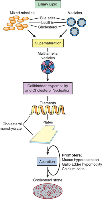
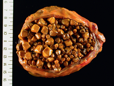
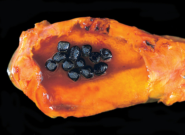
85 Lammert F, Miguel JF. Gallstone disease: from genes to evidence based therapy. J Hepatol. 2008;48:S124.

86 Strasberg SM. Acute calculous cholecystitis. New Engl J Med. 2008;358:2804.

87 Elwood DR. Cholecystitis. Surg Clin N Am. 2008;88:1241.

88 Petersen C. Pathogenesis and treatment opportunities for biliary atresia. Clin Liver Dis. 2006;10:73. vi,

89 Mack CL. The pathogenesis of biliary atresia: evidence for a virus-induced autoimmune disease. Semin Liver Dis. 2007;27:233.

90 Kerkar N, et al. The hepatic fibrocystic diseases. Clin Liver Dis. 2006;10:55. v–vi,

91 Randi G, et al. Gallbladder cancer worldwide: geographical distribution and risk factors. Int J Cancer. 2006;118:1591.