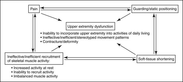
Evaluation and intervention of the upper extremity are complex tasks that require an understanding of multiple systems. Therapists need to remain open-minded about their interventions and consider the complexity of causes that interfere with upper extremity use (Fig. 10-10). Many of the various problems associated with upper extremity function overlap and build on each other. The following paragraphs review common impairments in stroke survivors that may or may not interfere with integrating the upper extremity into daily occupations.
Improving proximal stability to enhance distal mobility has long been a tenet of occupational therapy interventions. Postural adjustments stabilize supporting body parts while other parts (e.g., the upper extremities) are being moved.73 The following studies describe the effect of postural adjustments on arm function.
In the classic study by Belenkii, Gurfinkle, and Paltsev,16 unimpaired subjects who were evaluated while standing were asked to raise their arm to a horizontal position after they heard an external signal. Various EMG studies were performed to trace the pattern of muscle activation. The results demonstrated that the postural muscle synergies of the trunk and lower extremities were activated before (by 90 milliseconds) the anterior deltoid, the primary muscle used to perform this motion. The subjects then were evaluated in the supine position while performing the same task. No lower extremity activation was detected in this position (i.e., a different pattern of postural adjustments). The following conclusions can be inferred from this study:
1. Postural adjustments are task-specific.
2. Training of the upper extremity in the supine position does not automatically carry over to activities performed while sitting or standing (if postural control is a limiting factor).
3. Having different disciplines treat one particular half of the body is detrimental to patients’ progress because upper extremity function depends on postural support from the lower extremities and trunk.
Bouisset and Zattara29 replicated the previous study and demonstrated that an upward and forward trunk movement resulting from spine and/or lower limb extension precedes upper limb movement. This movement pattern is familiar to therapists who cue their patients to focus on spinal extension and the associated anterior pelvic tilt while treating arm function.
Horak and colleagues88 compared postural adjustments of subjects with and without hemiplegia during a variety of tasks with different parameters. The hemiplegic subjects demonstrated the same sequence of muscle activation as the subjects without hemiplegia, although activity on the hemiparetic side was delayed. In addition, the hemiparetic individuals were not capable of making rapid movements with the unimpaired arm. This was hypothesized to result from a delay in the anticipatory activity of the contralateral hemiplegic muscles. This study dispels the myth of “good” and “bad” sides after a stroke, especially when postural control is compromised.
In their study of postural adjustments during arm movements, Cordo and Nashner52 were able to demonstrate that when subjects’ postural stability was increased (e.g., by outside shoulder support or placing a finger lightly on a support rail), postural activity was reduced, and voluntary movement enhanced. This concept is crucial to understand when treating upper extremity dysfunction. As support is increased, the postural demands of the task are decreased and vice versa. The therapist can control the patient’s level of postural stability by manipulating the following treatment environment factors: positioning—supine to sitting to standing; type of support surface—stationary or unstable surfaces; positioning of objects used in activities—near or far, base of support, and amount of external stability.
Cordo and Nashner52 also made a critical distinction between associated postural adjustments that precede voluntary movements (e.g., reaching) and automatic postural adjustments that follow external perturbations (e.g., standing on a bus that stops at a light or being moved by the therapist). Training in one type of adjustment cannot be assumed to carry over into other types of adjustments.
Woollacott, Bonnet, and Yabe194 demonstrated that their subjects’ postural activity varied depending on the task being performed (pushing, pulling) and whether they received information in advance regarding the goal of the task.
Massion119 points out that voluntary movements are “accompanied by postural adjustments which show three main characteristics: (1) they are ‘anticipatory’ with respect to movement and minimize the perturbations of posture and equilibrium due to the movement, (2) they are adaptable to the conditions in which the movement is executed, and (3) they are influenced by the instructions given to the subject concerning the task to be performed.”
Postural control disorders in stroke patients have been well-documented. Lee109 emphasized the detrimental impact that postural dysfunction has on free arm movements and therefore ADL. Although a variety of muscles can serve as postural stabilizers, postural control of the trunk is critical for upper extremity function.19 See Chapter 7.
Occupational therapists must use their activity analysis skills to help patients develop the missing trunk control components. (See Table 7-9 for examples of the effects of object positioning on trunk control and weight shifting during reaching activities.) Functional mobility patterns requiring increased trunk control (e.g., scooting) should be incorporated into treatment plans for upper extremity function. See Chapter 14.
Postural control evaluations should be performed within the context of upper extremity tasks such as reaching or performing ADL and IADL. Evaluating postural control separately does not provide the therapist with sufficient information for intervention. (See Chapters 7 and 8 for more information related to postural control.)
Until relatively recently, the impact of weakness (a negative symptom) on stroke patients’ functional status has long been ignored. The motor control deficits in patients previously were attributed exclusively to spasticity, which resulted in treatment focused on inhibiting the spasticity. Many therapists considered upper extremity muscle tests for strength difficult to interpret because of common “synergy patterns.” Bourbonnais and colleagues31 demonstrated that the patterns of activity in the elbow flexor muscles were not consistent with established synergistic patterns. Weakness of the upper extremity musculature plays a major role in upper extremity dysfunction, most likely more than the positive symptoms after stroke. Muscle weakness is reflected by the inability of patients to generate normal levels of muscle force.30 Stroke survivors who have written about their experiences focus on the difficulty in force production. Brodal35 reflected on his own stroke: “It was a striking and repeatedly made observation that the force needed to make a severely paretic muscle contract is considerable. . . . Subjectively this is experienced as a kind of mental force, a power of will. In the case of a muscle just capable of being actively moved the mental effort needed was very great.”
Bourbonnais and Vanden Noven30 reviewed the physiological changes in the nervous system that contribute to muscle weakness in patients with hemiparesis. They summarized specific changes at the motor neuron and muscle levels that decrease a patient’s ability to produce force. Box 10-7 summarizes these changes.
Box 10-7 Physiological Changes Contributing to Weakness
Motor neuron changes: loss of agonist motor units, changes in recruitment order of motor units, and changes in the firing rates of motor units
Nerve changes: changes in peripheral nerve conduction
Muscle changes: changes in the morphological and contractile properties of motor units and in the mechanical properties of muscles
Bohannon and colleagues21 found that static strength deficits of the shoulder medial rotator and elbow flexor muscles did not correlate with antagonist muscle spasticity. They concluded that therapists might determine the capacity for force production for an agonist muscle based on its own tone rather than that of its antagonist.
Gowland and colleagues79 studied agonist and antagonist activity during upper limb movements in stroke patients and concluded that treatment should be aimed at improving motor neuron recruitment rather than reducing antagonist activity. In their study, patients who could not perform select upper extremity tasks had EMG values significantly and consistently lower than those of patients who were successful at the task.
Indeed recent empirical evidence highlights the relationship between weakness and loss of function. Findings include:
In a study of 93 community dwelling stroke survivors, Harris and Eng85 concluded that paretic upper limb strength had the strongest relationship with variables of activity and best explained upper limb performance in ADL. Grip strength was also a factor.
A longitudinal study of 27 stroke survivors found that weakness was the main and only contributor to activity limitations as opposed to spasticity or contracture.6
Chae and colleagues46 described the relationship between poststroke upper limb muscle weakness and cocontraction, and clinical measures of upper limb motor impairment and physical disability. The authors measured EMG activity of the paretic and nonparetic wrist flexors and extensors of 26 chronic stroke survivors. Upper limb motor impairment and physical disability were assessed with the Fugl-Meyer motor assessment and the arm motor ability test. They concluded that muscle weakness and degree of cocontraction correlate significantly with motor impairment and physical disability in upper limb hemiplegia.
Mercier and Bourbonnais123 compared the relative strength of different muscle groups of the paretic upper limb and assess the relationship with motor performance. The maximal active torques of five muscle groups were measured in both upper limbs. Upper limb function was assessed using the Box and Block Test, the Finger-to-Nose Test, the Fugl-Meyer Test, and the TEMPA. They concluded that “the relative forces for shoulder flexion and handgrip are the best predictors of the upper limb function.” Additionally, they concluded that the results “do not confirm classical clinical teaching regarding the distribution of weakness following stroke (e.g., proximal to distal gradient; extensors more affected than flexors) but support the hypothesis that strength is related to the function of the paretic upper limb.”
From a treatment planning perspective, integrating strengthening interventions is imperative in efforts to regain limb function. Bohannon and Smith23 analyzed strength deficits in stroke patients and verified that muscle strength improves in stroke patients with hemiplegia who are undergoing rehabilitation. Empirical evidence supports the use of strengthening interventions in this population without deleterious effects:
Flinn70 presented a case study of a young female with left-sided hemiplegia. Her treatment program focused on participating in graded functional tasks that systematically increased the motor demands on the more affected upper extremity. Her task-oriented treatment program was augmented by resistive exercises using elastic tubing. Substantial results after six months of therapy included improved level of occupational performance in ADL and IADL, improved manual muscle test scores (which increased from 2/5 to the 4/5 and 5/5 ranges), improved hand function, and improved grip strength scores. Identifying the underlying problems (in this case, weakness and an inability to control excess degrees of freedom) is of utmost importance when planning treatment strategies.
Bütefisch and colleagues39 examined the effect of a standardized training on movements of the affected hand in 27 hemiparetic patients using a multiple baseline approach. The training consisted of repetitive hand and finger flexions and extensions against various loads and was carried out twice daily during 15-minute periods. Grip strength, peak force of isometric hand extensions, peak acceleration of isotonic hand extensions, and contraction velocities as indicators of motor performance significantly improved during the training period. Additionally, 24 out of 27 patients improved on the Rivermead Motor Assessment. The authors further challenged traditional therapy (the Bobath concept) aimed at reducing enhanced muscle tone without reinforcing the activity in centrally paretic hand. In the study, patients undergoing this treatment approach alone did not experience a significant improvement in the motor capacity of the hand. The authors emphasized the importance of frequent movement repetition for the motor rehabilitation of the centrally paretic hand and challenge conventional therapeutic strategies that focus on spasticity reduction instead of early initiation of active movements.
Sterr and Freivogel166 “assessed whether intensive training increases spasticity and leads to the development of ‘pathologic movement patterns,’ a concern often raised by Bobath-trained therapists. The authors used a baseline-control repeated-measures test to study 29 patients with chronic upper limb hemiparesis who received daily shaping training. Their results suggest that training has no adverse effects on muscle tone and movement quality.”2
A systematic review of multiple studies concluded that “Strengthening interventions increase strength, improve activity, and do not increase spasticity. These findings suggest that strengthening programs should be part of rehabilitation after stroke.”
In their strength training study after stroke, Badics and colleagues10 concluded that “The extent of strength gain was positively correlated with the intensity and the number of exercising units. Muscle tone, which was abnormally high at baseline, did not further increase in any one case. The results of this study showed that targeted strength training significantly increased muscle power in patients with muscle weakness of central origin without any negative effects on spasticity.”
In their review of weakness and strengthening post stroke, Patten and colleagues141 identified nine trials of progressive resistive training after stroke. They concluded “All of these studies reported positive adaptations to strength training . . . With one exception, all studies strongly suggest positive effects of strength training on various indices of functional outcome . . .” They further concluded that “while insufficient data exist to draw firm conclusions at this time, functional effects of strengthening appear persistent. Four of the available studies evaluated effects of strength training on spasticity and found no deleterious effects.”
The debate about which type of muscle contraction (eccentric, concentric, or isometric) is the most effective in strengthening patients has been long-standing. Muscle groups need to contract in a variety of ways to complete functional tasks successfully. For example, when a person reaches for a can of soup on a high shelf, the shoulder musculature must contract (concentrically) to bring the hand to the level of the shelf, maintain the contraction (isometrically) to locate the correct item, and control the weight of the arm and item in gravity (eccentrically) as the can is placed with control on the countertop.
In a study of dynamical muscle strength training in stroke patients, Engardt and colleagues65 found that eccentric contractions were more effective than concentric contractions. Twenty patients with hemiparesis resulting from strokes participated in activities that elicited concentric or eccentric contractions. After the treatment, significant improvements resulted in the relative strength of paretic muscles during eccentric and concentric actions in the group that was trained solely with eccentric contractions (i.e., eccentric training increased the strength of both types of contractions); this was not true for the group that only received concentric contraction training. Therefore, the authors determined eccentric contraction training to be more advantageous and efficient (Box 10-8). See Table 10-1 for a review of evidence-based interventions related to strengthening.
Spasticity, which is a positive symptom according to Jackson classification system, has been a subject of debate by various authors. Although an abundance of research has been done on spasticity, disagreements still exist about its definition, physiological basis, treatment, and evaluation. Glenn and Whyte75 define spasticity as “a motor disorder with persistent increase in the involuntary reflex activity of a muscle in response to stretch. Four specific phenomena may be variably observed in the constellation of spasticity: hypertonia (frequently velocity dependent and demonstrating the clasp-knife phenomenon), hyperactive (phasic) deep tendon reflexes, clonus, and spread of reflex responses beyond the muscle stimulated.” In addition, Babinski sign is characteristic, and hyperactive tonic neck or vestibular reflexes may be present.116
Several different phenomena commonly observed in stroke rehabilitation including hyperactive stretch reflexes, increased resistance to passive movement, posturing of the extremities, excessive cocontraction, and stereotypical movement synergies are clumped together in the category of spasticity. Spasticity has become a catchall term for a variety of problems. Rather than being a specific symptom, spasticity is related to a variety of neural and nonneural factors. Therefore, spasticity cannot be treated uniformly by surgical, physical, or pharmacological procedures. Spastic paresis is a commonly used term that implies a cause-and-effect relationship (i.e., a cause-and-effect relationship between positive and negative symptoms). This belief has been challenged recently.
Preston and Hecht147 provide further information regarding the clinical presentation of spasticity to include the following:
Patients having difficulty initiating rapid alternating movements
Abnormally timed EMG activation of the agonist and antagonist
Fluctuation of spasticity as a result of a change in position
Usual patterns include upper extremity flexion and lower extremity extension
Bobath18 stated that there is “An intimate relationship between spasticity and movement . . . spasticity must be held responsible for much of the patient’s motor deficit.” Treatment techniques were based on “helping the patient gain control over the released patterns of spasticity by their inhibition.” Patients were treated under the assumption that “Weakness of muscles may not be real, but relative to the opposition by spastic antagonists.” A variety of studies have been published that refute these assumptions. See Chapter 6.
Sahrmann and Norton159 studied normal subjects and subjects with upper motor neuron symptoms. The movement pattern studied was alternating flexion and extension of the elbow. The analysis of their EMG findings showed that the primary cause of impaired movement was not antagonist stretch reflexes but was limited and prolonged agonist contraction recruitment and delayed cessation of agonist contractions after movement had stopped. Rather than focusing treatment on inhibiting spasticity, therapists should train patients to perform alternating movement patterns (e.g., hand-to-mouth patterns) efficiently.
Fellows, Kaus, and Thilmann67 studied the importance of hyperreflexia and paresis on voluntary arm movements in normal subjects and subjects with spasticity resulting from a unilateral ischemic cerebral lesion. The subjects with spasticity showed a lower maximum movement velocity; the more marked the paresis, the greater the reduction in maximum velocity. No relationship was found between the degree of voluntary movement impairment and level of passive muscle hypertonia in the antagonist. The conclusion was that agonist muscle paresis, rather than antagonist muscle hypertonia, had the most significant effect on impaired voluntary movement.
In their study on overcoming limited elbow movement in the presence of antagonist hyperactivity, Wolf and colleagues190 concluded that functional elbow improvements could be made without first training the patient specifically to inhibit hyperactivity.
Landau108 performed pharmacological interventions that effectively abolished the hyperactive stretch reflexes in his patients. This intervention did not result in a corresponding improvement in motor behavior.
Ada, O’Dwyer, and O’Neill6 examined the relationship between the motor impairments (spasticity and weakness) and their impact on physical activity. They specifically aimed to study the contribution of weakness and spasticity to contracture, and the contribution of all three impairments to limitations in physical activity during the first 12 months after stroke. The authors followed 27 stroke survivors for one year. They found that “the major independent contributors to contracture were spasticity for the first four months after stroke (p = 0.0001-0.10) and weakness thereafter (p = 0.01-0.05). However, the major and only independent contributor to limitations in physical activity throughout the year was weakness (p = 0.0001-0.05).” “For the first time, from a longitudinal study, the findings show that spasticity can cause contracture after stroke, consistent with the prevailing clinical view. However, weakness is the main contributor to activity limitations.”
In the traditional evaluation of spasticity, the therapist moves the patient’s limb quickly in a direction opposite to the pull of the muscle group being tested, and the examiner feels for a resistance to the movement. The gold standard for rating resistance is the Ashworth Scale9 or the Modified Ashworth Scale24 (Box 10-9).
2 Slight hypertonus; noticeable catch when limb is moved
3 More significant hypertonus, but affected limb still moves easily
1 Slight increase in muscle tone, manifested by a catch and release or by minimal resistance at the end of the range of motion when the affected part is moved in flexion or extension
1+ Slight increase in muscle tone, manifested by a catch, followed by minimal resistance throughout the remainder (less than half) of range of motion
2 More significant increase in muscle tone throughout most of the range of motion but affected part is moved easily.
3 Considerable increase in muscle tone; difficult passive movement
The response of a spastic muscle to stretch has been argued not to be the same during passive and active movement. In addition, spasticity is a multidimensional problem that incorporates neural and nonneural components (e.g., altered soft-tissue compliance). Therefore, some authors have questioned the usefulness of test measures such as the Ashworth Scale and are investigating a more comprehensive evaluation of spasticity.
Although the research on spasticity does not support focusing treatment on suppressing stretch reflexes, it does support treatment focusing on preventing secondary structural muscle changes in patients with spasticity.
Hufschmidt and Mauritz’s study90 suggested that spastic contracture is the result of degenerative changes (e.g., atrophy and fibrosis) and changes of the passive and contractile muscle properties.
In their study on spastic and rigid muscles, Dietz, Quintern, and Berger61 concluded that the actual muscle fibers undergo changes, which explains the increased muscle tone in spastic patients.
For treating patients with spasticity, Perry142 emphasized early mobilization and assistance with developing evolving motor control into effective function. These two interventions result in minimal contractures and prevent improper use of patients’ available control mechanisms. Hummelsheim and colleagues91 studied the results of sustained stretch in spastic patients. They found that sustained muscle stretch of approximately 10 minutes led to significant reduction in the spastic hypertonus in the elbow, hand, and finger flexors. They hypothesized that this benefit is due to stretch receptor fatigue or adaptation to the new extended position.
Little and Massagli116 also emphasized using a stretching program incorporating pain prevention and patient education focusing on the adverse effects of spasticity (contracture), use of slow movements, and importance of daily stretching.
In addition to the mentioned techniques, specific modalities and their physiological bases have been described in the literature and include local cooling, vibration therapy, and electrical stimulation.
Perry142 summarized the effective rehabilitation of a patient with spasticity by using five categories: contracture minimization, realistic planning, muscle strength preservation and restoration, enhancement of returning control, and substitution for permanent functional loss.
Carr, Shepherd, and Ada43 summarized their treatment approach based on the assumption that clinical spasticity is a manifestation of length-associated muscle changes and disordered motor control: “The development of spasticity will be less severe if soft-tissue length can be maintained and if motor training emphasizes elimination of unnecessary muscle force and training muscle synergies as part of specific actions.”
Again, the point must be emphasized that many of the observed phenomena that occur during treatment should not be attributed automatically to spasticity and require more in-depth evaluations and treatment plans (Box 10-10; Table 10-6).
Box 10-10 Treatment of Spasticity
Guide appropriate use of available motor control.
Avoid using excessive effort during movement.
Encourage slow and controlled movements.
Teach specific functional synergies during tasks.
Avoid use of repetitive compensatory movement patterns.
Keep spastic muscles on stretch via positioning or orthotics to prevent contracture.
Teach the patient or caretaker specific stretching techniques targeted at the spastic muscles.
Use activities to enhance the agonist/antagonist relationship.
Refer when appropriate for pharmacological or surgical interventions.147
Table 10-6 Suggested Interventions for Problems Commonly Thought to Be Caused by Spasticity*
OBSERVATIONS DURING TREATMENT | SUGGESTED INTERVENTIONS |
---|---|
Posturing of upper extremity—usually consisting of retraction, posterior trunk rotation, internal rotation, elbow flexion, and wrist and digit flexion—during difficult tasks (e.g., gait, transfers, and dressing) | Upper extremity posturing indicates that the task is difficult for the patient. Treatment should include increasing the efficiency of task performance by building in trunk and lower extremity control, incorporating the upper extremity into the task (e.g., by bilateral ironing or using arm as postural support), and teaching the patient to relax the upper extremity after difficult tasks. |
Stereotypical flexor patterns when attempting to move arm against gravity | Evaluate components of movement pattern and identify factors that limit efficient movement (e.g., weakness, postural dysfunction, malalignment, and inappropriate task choice). Provide activities that elicit the missing components of movement pattern. |
Flexion posture when resting | Implement a contracture prevention program. Provide adequate positioning and teach safe, self range-of-motion exercises. |
“Catch” felt during quick-stretch evaluation of upper extremity | Do not assume that this phenomenon is resulting in observed movement dysfunction. Instead, interpret it as a red flag warning that soft-tissue shortening may be present or developing. |
* This table represents a variety of functional limitations and problems traditionally considered to be the direct result of spasticity. Although sometimes interconnected, these problems stem from different sources and must be treated accordingly.
Preston and Hecht147 have comprehensively reviewed the literature related to spasticity management, including topics such as oral and intrathecal medications, nerve blocks, orthopedic surgery, and neurosurgical interventions.
Nerve blocks are being used increasingly as an adjunct therapy in the rehabilitation process. Preston and Hecht147 differentiated between short-term blocks such as procaine and bupivacaine used to diagnose and assist in the evaluation process and long-term blocks such as phenol and botulinum toxin type A (Botox).
Rousseaux, Kozlowski, and Froger154 assessed the efficacy of Botox treatment on disability, especially in manual activities, and attempted to identify predictive factors of improvement in 20 patients with stroke. They concluded that botulinum toxin A is efficient in improving hand use in patients with relatively preserved distal motricity and in increasing comfort in patients with severe global disorders. Similarly, Bakheit and colleagues11 completed a randomized controlled trial to assess the efficacy of Botox in decreasing spasticity in stroke survivors. They concluded that treatment with Botox reduced muscle tone in patients with poststroke upper limb spasticity.
While the positive effect at the impairment level (i.e., reduction of spasticity) has been well-documented, the effect on functional limitations is not as clear.164,183 As spasticity increases, the risk for soft-tissue shortening is heightened, which in fact may lead to a vicious circle of problems such as spasticity, soft-tissue shortening, overrecruitment of shortened muscles, and increased stretch reflexes. Secondary problems that may occur if the spasticity is not managed in a therapy program include the following:
Deformity of the limbs, specifically the distal upper limb (elbow to digits)
Impaired upright function caused by soft-tissue contracture (e.g., plantar flexion contractures resulting in a loss of the ankle strategies required to maintain upright stance)
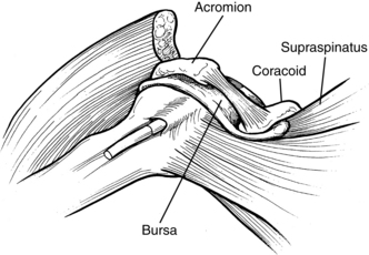
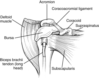
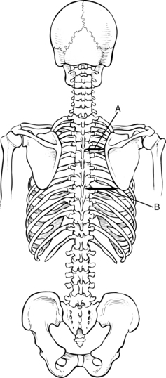
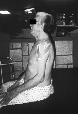
Pain syndromes resulting from loss of normal joint kinematics. These syndromes are usually related to soft-tissue contracture blocking full joint excursion. A typical example of this issue is the loss of full passive external rotation of the glenohumeral joint. Attempts at forced abduction in these cases results in a painful impingement syndrome of the tissues in the subacromial space.
Impaired ability to manage basic ADL tasks, specifically upper extremity dressing and bathing of the affected hand and axilla when flexor posturing is present
Loss of reciprocal arm swing during gait activities
Risk for falls because of postural malalignment78
In summary, although reducing spasticity does not appear to result in automatic improvements related to function, therapists must manage spasticity to prevent soft-tissue contracture, prevent deformity, and maintain a flexible and mobile arm.
Contracture in stroke patients results from immobilization and may be attributed to spasticity (particularly during the first four months after stroke) and weakness thereafter,6 improper positioning, postural malalignment, a lack of variation in limb postures (e.g., prolonged sling use), or a combination of various factors. The formation of contractures indicates a poor prognosis for limb function. Perry142 discussed the vicious circle of contracture and spasticity: “contractures stiffen tissues, immobility creates contractures. Spasticity preserves the contracture by excluding the intramuscular fibrous tissues from the stretching force.”
Botte, Nickel, and Akeson28 have reviewed the literature correlating spasticity and contracture. As the stroke patient progresses to a state of spasticity, the increased activity of the spastic muscles may result in characteristic posturing of the limb, resulting in increased stiffness of the soft-tissue surrounding the joint and the eventual formation of fixed contracture. The authors further pointed out that contracture is associated with loss of elasticity and fixed shortening of involved tissues. Contracture may occur in a variety of soft-tissues including the following: skin, subcutaneous tissue, muscle, tendon, ligament, joint capsule, vessels, and nerves.
Halar and Bell82 categorized contracture as arthrogenic (resulting from cartilage damage, joint incongruency, or capsular fibrosis), soft-tissue related (skin, tendons, ligaments, subcutaneous tissue), and myogenic (shortening of the muscle by intrinsic or extrinsic factors). The therapist must consider the difference between myogenic and joint contracture, especially if the muscle spans two or more joints (e.g., the wrist and hand). The therapist can differentiate contractures by flexing the proximal joint and noting the resulting position of the distal joints. Joint contracture is not affected by changes in proximal joint position. See Chapter 13.
Booth27 reviewed the physiological and biochemical effects of immobilization on muscle. His findings indicate that muscle strength rapidly declines during limb immobilization because of a decrease in muscle size; muscle fatigability increases rapidly after immobilization. His observations also indicate that muscle atrophy in immobilized limbs begins rapidly, and a decrease in muscle size is greatest in the early phases of immobilization.
Soft-tissue and joint mobilization are the treatments of choice for preventing contracture. The benefits of mobilization include maintenance of joint lubrication,28 prevention of secondary orthopedic problems (impingements), maintenance of soft-tissue length, and possible reduction of spasticity by acting on the nonneural components of spasticity.
Contracture is prevented by deliberate and frequent limb movement, with active movement being preferred over passive when possible. Perry142 pointed out that it is essential to move the patient through complete ROM and not just the middle ranges. Therapists must determine what a full ROM is for each patient and must consider age-related factors. Determining the full ROM on the less affected side may be helpful. A joint that moves or is moved through its full ROM several times daily develops almost no deformities. Although the therapist should maintain the patient’s ability to participate in all ranges of trunk and upper extremity activities, the therapist should pay particular attention to the following ranges:
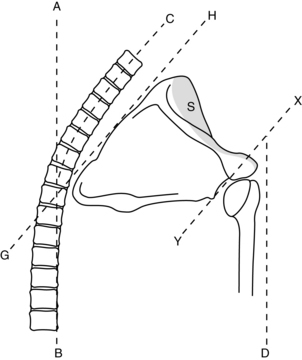
The mobility of the scapula on the thoracic wall with emphasis on protraction and upward rotation should be maintained because this range is critical in the prevention of soft-tissue impingement in the subacromial space during overhead movements of the arm and in preparation for forward reach patterns. Overhead ranges should not be attempted unless the scapula is freely gliding in upward rotation.
Maintaining external (lateral) rotation of the glenohumeral joint allows abduction of the arm as the humerus rotates laterally to permit the greater tuberosity of the humerus to clear the acromial process. Bohannon and colleagues,22 Ikai and colleagues,93 and Zorowitz and colleagues203 concluded that loss of external rotation ROM was the factor most significantly correlated to shoulder pain.
Elbow extension is important because the majority of stroke patients favor elbow flexion as a rest posture.
The therapists also should maintain wrist extension with concurrent radial deviation. During wrist ROM exercises, therapists must realize that the range of wrist deviation is at a maximum when the wrist is slightly flexed and a minimum when the wrist is fully flexed. Wrist extension is at a maximum during neutral deviation and a minimum during ulnar deviation.100
Composite flexion of the digits leads to collateral ligament elongation. Therapists must maintain this length to prevent deformity and prepare the hand for return of motor function.
Composite extension of the wrist and digits results in long flexor elongation.
Digits ranged in intrinsic plus (metacarpophalangeal flexion and interphalangeal extension) and intrinsic minus (metacarpophalangeal extension and interphalangeal flexion).
Halar and Bell82 recommended active ROM and passive ROM combined with a terminal stretch at least twice per day if contracture is beginning to develop. Therapists must use low-load prolonged stretch if a contracture has developed (see Chapter 13). During the terminal stretch, the therapist should stabilize the proximal body part well. The therapist may distract the joint slightly during the stretch to prevent soft-tissue impingement. The therapist must monitor scapula position during passive ROM activities. If necessary, the therapist should support the scapula in a position of protraction and upward rotation. In addition, the therapist must support the humerus in an external rotation position. The elbow crease should be facing up (not medially toward the trunk) to ensure proper alignment (Fig. 10-12).
Positioning is another effective means of maintaining soft-tissue length and can be used to promote low-load, prolonged stretch. Therapists must address positioning needs of patients while they are in bed or wheelchairs/armchairs (see Chapter 26) and anytime they are in a recumbent position. Effective positioning encourages proper joint alignment, variations in joint position, comfort, and the maintenance of stretch in areas at risk for contracture. Common areas of concern during patient positioning include head and neck alignment, trunk alignment, glenohumeral joint alignment, scapula alignment, maintenance of abduction, external rotation, elbow extension, and maintenance of long flexor length.
A thorough literature review comparing authors’ strategies on bed positioning has been published.41 This review found no consensus on some issues and multiple discrepancies on strategies. Many of the positioning protocols are based on the principle of inhibiting primitive reflexes, a topic of considerable debate.
Patients are engaged in therapy only a portion of the day. Studies have shown that patients in rehabilitation units spend almost half of their days engaged in passive pursuits including sitting unoccupied and lying in bed.15 Therefore, patients at risk for developing contracture because of limb immobilization are good candidates for participation in a positioning program in addition to therapy.
The positioning suggestions in Box 10-11 are based on Carr and Kenney’s review41 of the positioning literature and highlights the consensus of reviewed authors.
Box 10-11 Suggested Bed Positioning
POSITIONING OF PATIENT | |
On unaffected side | |
On affected side | |
In supine | |
Although the positioning suggestions in Box 10-11 represent the consensus of many authors, major areas of intervention are missing, which result in the controversies surrounding this area of intervention. For example, glenohumeral joint support remains controversial. Although most authors agree that the scapula should be protracted with a pillow, no consensus exists about support of the humerus. If only the scapula is protracted with a pillow, the humerus takes on a position of relative extension. Therefore, only support of both the scapula and humerus achieves the original goal of proper joint alignment (Fig. 10-13).
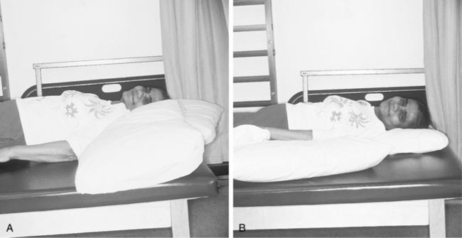
Figure 10-13 A, Bed positioning with only the scapula supported. The humerus takes on a position of relative extension, with the head of the humerus migrating anteriorly. B, Proper support of scapula and humerus ensures proper biomechanical alignment of shoulder joint.
At this point, no definitive studies support one type of positioning more than another with few exceptions. Ada and colleagues5 determined that positioning patients in supine with the affected shoulder abducted to 45-degrees and the elbow flexed to 90-degrees and placed in maximum comfortable external rotation, with towels or pillows to support the forearm 30 minutes per day prevented contracture of the internal rotators. Occupational therapists must decide what their intervention goals are and critically analyze their effectiveness. Therapists should not use general, generic strategies for bed positioning; instead, they should evaluate each patient’s positioning needs individually.
Strategies to teach patients safe ROM activities they can perform themselves need to be initiated as soon as patients are medically stable. Although the clasped-hand position followed by overhead movements of both extremities has been advocated by some authors, this position may not be the most effective, especially for trauma prevention. This movement pattern does not account for factors such as scapula-humeral rhythm (especially if weakness, malalignment, or tightness around the scapula exists), overzealous patients who do not or cannot respect their pain, or critical shoulder biomechanics. Many patients observed performing this type of ROM activity have their trunk hyperextended, scapula retracted, and humerus internally rotated. This type of alignment does not correspond with an ROM pattern that emphasizes forward flexion of the humerus; it promotes proximal patterns (e.g., retraction) that should be discouraged (Fig. 10-14). Recommended techniques for patients performing ROM activities by themselves safely include the following:
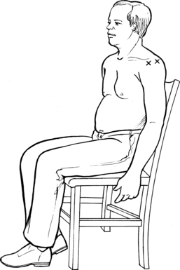
1. “Towel on table”: The patient is seated at a table with both arms on top of a towel. The less affected arm guides the towel around the table, with the majority of movement occurring in the trunk and from hip flexion. The patient’s goal is to “polish the table” while holding positions at the end of desired ROM. The farther the patient’s chair is positioned from the table, the greater the ROM. This technique not only enhances the range of the glenohumeral and elbow joint but also encourages scapula protraction and weight-shifting. Excessive effort is minimized because the towel assists the movement (Fig. 10-15).
2. “Rock the baby”: The patient’s less affected arm cradles the more affected arm, lifts it to 90 degrees, and places it into positions of horizontal abduction and adduction. Increased horizontal adduction on the more affected side encourages scapula protraction. This technique also encourages trunk rotation (Fig. 10-16).
3. While seated or standing, the patient reaches down to the floor and allows both arms to dangle. This position encourages extension of the elbow, wrist, and digits and forward flexion of the humerus with scapula protraction. The activity is an especially useful technique for patients after they have performed an excessively difficult activity (e.g., gait, transfer, or dressing) that results in stereotypical arm posturing (Fig. 10-17).
4. While seated or standing, the patient places the more affected extremity onto a table or counter so that the forearm is bearing the weight. With the extremity in this position, the patient turns the trunk away from the supported extremity. As the trunk turns farther away and is enhanced by the posterior reach of the less affected arm, the external rotation of the more affected shoulder increases (Fig. 10-18).
5. Davis57 has advocated rolling over the protracted scapula (from supine to side lying) several times to mobilize the scapula.
6. If the scapula of a patient is mobile and stays mobile, the range of abduction and external rotation may be increased by having the patient lie supine, placing the hands behind the head, and allowing the elbows to fall toward the bed (Fig. 10-19). This is a common resting position for an individual who has unimpaired upper extremity function. This technique should be used judiciously and only for patients who move slowly, respect pain, and have a mobile scapula. Therapists may use the five techniques outlined previously for almost all patients because they inherently follow biomechanical principles.
7. Avoid the use of overhead pulleys.104
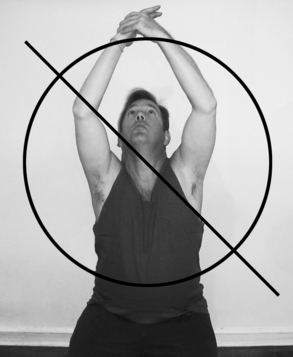
Figure 10-14 Because of multiple biomechanical concerns (e.g., impingement), self-overhead range of motion is discouraged.
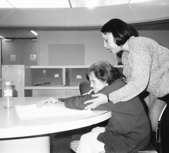
Figure 10-15 “Towel on table.” Therapist is training patient to perform safe self range-of-motion activity. As the patient pushes the towel toward bottle, range of motion is gained in humeral flexion, scapular protraction, and elbow extension (which are ranges required for functional reach). Much of the range is gained by hip and trunk flexion.
Figure 10-16 “Rock the baby.” The patient lifts upper extremity to chest level (A) and abducts (B) and adducts (C) horizontally, allowing trunk rotation.
Figure 10-17 Patient performs self range-of-motion activity by reaching to floor. This pattern is especially effective after a difficult task that results in stereotypical posturing.
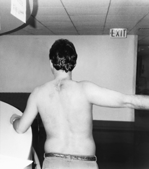
Figure 10-18 External rotation of the left glenohumeral joint is achieved by reaching to side and behind with opposite arm.
Figure 10-19 Internal rotators stretch to be used judiciously for patients who respect their own pain. This rest posture is effective at maintaining external rotation and abduction of the glenohumeral joint. If range is lacking, the humerus can be supported with a towel until patient gains increased external rotation and horizontal abduction.
The ultimate strategy used to decrease contracture and maintain ROM is encouraging functional use of the trunk and upper extremity. A person who has never had a stroke maintains ROM of an extremity by incorporating it into ADL. Activities that eliminate maladaptive positions during activities, improve balanced muscle activity on both sides of the joints, and focus on activities that encourage ROMs that are commonly decreased in stroke patients (e.g., external rotation, forward flexion, abduction, and protraction) should be incorporated into a comprehensive upper extremity program. (See Chapter 13 for other adjunct treatments to prevent or correct soft-tissue shortening.)
Shoulder-hand syndrome (SHS) is classified as a reflex sympathetic dystrophy disorder or complex regional pain syndrome type I. The painful lesion that precipitates SHS is a proximal trauma such as a shoulder, neck, or rib cage injury or a visceral source such as stroke. The syndrome begins with severe pain and progresses to stiffness in the shoulder and pain throughout the extremity. Other symptoms include moderate to considerable swelling of the wrist and hand, vasomotor changes, and atrophy.106 If untreated, SHS may result in a frozen shoulder and permanent hand deformity.34
Although the cause of SHS remains obscure, most authors associate it with a change in the autonomic nervous system (primarily sympathetic).50 A study by Braus, Krauss, and Strobel34 suggested that the SHS in hemiplegic patients is initiated by a peripheral lesion (e.g., a tissue or nerve injury). The authors hypothesized that increased neural activity after a peripheral injury or inflammation leads to a central sensitization responsible for the severe pain associated with SHS. Autopsy data collected by the authors confirmed microbleeding in the area of the suprahumeral joint of the affected side. If the underlying cause is in fact peripheral, then prevention programs theoretically would be effective.
The reported incidence of SHS varies from 27%34 to 25%174 to 12.5%58 to 1.56%.143 Males seem to be slightly more affected than females.58,174 The majority of patients with SHS symptoms have partial motor loss, moderate or severe sensory loss, and varying degrees of spasticity.58 Associated risk factors include subluxation, considerable weakness, moderate spasticity, deficits in confrontational field testing (following hemianopsia or neglect), and altered shoulder biomechanics that may compromise the suprahumeral joint structures.34
Daviet and colleagues56 examined 71 patients with hemiplegia; 34.8% had a complex regional pain syndrome type I. They identified four main clinical factors in the prognosis of complex regional pain syndrome type I as motor deficit, spasticity, sensory deficits, and initial coma. They also concluded that shoulder subluxation, unilateral neglect, and depression did not seem to be determinant predictive factors of complex regional pain syndrome type I severity.
Three stages of SHS have been described (Box 10-12).
Box 10-12 Stages of Shoulder-Hand Syndrome/Complex Regional Pain Syndrome Type I
The patient complains of shoulder and hand pain, tenderness, and vasomotor changes (with symptoms of discoloration and temperature changes). Chances of reversal are high at this stage.
Davis and colleagues58 outlined the major diagnostic criteria for SHS based on the following clinical symptoms:
Shoulder: loss of ROM and pain during abduction, flexion, and external rotation movements
Wrist: intense pain during extension movements, dorsal edema, and tenderness during deep palpation
Hand: edema over metacarpals and no tenderness
Digits: moderate fusiform edema, intense pain during flexion of the metacarpophalangeal and proximal interphalangeal joints, and loss of skin lines
The Tepperman and colleagues174 study concluded that metacarpophalangeal tenderness during compression was the most valuable clinical sign of reflex sympathetic dystrophy, with a predictive value of 100%. Vasomotor changes and interphalangeal tenderness had the next highest predictive value at 72.7%. Therapists must remember that many of the mentioned signs and symptoms can be found in stroke patients without SHS. If a patient has several characteristic signs and symptoms, one safely can make a diagnosis on clinical grounds alone.50 Although the diagnosis for SHS is primarily clinical, the most effective way to confirm its presence is to use a differential neural blockade. The physician may use a stellate ganglion block to alleviate the symptoms, which interrupts the abnormal sympathetic reflex; the diagnosis of SHS is confirmed if the block alleviates symptoms.
Therapists should prevent SHS, so that it will not have to be treated. Davis57 has developed a prevention protocol that focuses on the following:
Therapists gaining full understanding of the anatomy and physiology of normal and hemiplegic shoulders
Proper handling of the upper extremity, including avoiding arm traction during mobility, ADL, and gait activities; supporting the arm as necessary, preventing prolonged arm dangling, and using the trunk and scapula rather than the arm as support during transitional movements
Staff education focusing on the mentioned handling techniques
Mobilizing the scapula to ensure gliding when raising or performing ROM activities with the arm
Family education focusing on proper extremity handling and transfer techniques; training families not to guard at the affected upper extremity during ambulation (because a balance loss would result in an automatic reflex—grabbing the patient’s arm)
Edema control that begins as soon as signs of it are observed (see Chapter 12)
Training patients to take responsibility for protecting their affected arm
Davis and colleagues58 hypothesize that therapists can control certain factors contributing to SHS. One factor is the extravasation of intravenous fluids. The team should infuse intravenous fluids into the less affected arm if possible; if not, fluids should be infused proximal to the wrist on the affected side. This strategy prevents infiltration around the needle and a possible edema syndrome. Another contributing factor is poor positioning. Therapists should position patients so that they cannot roll over onto the affected arm, pin it down, and compromise circulation. The other factor is immobilization of a painful shoulder by the patient. Davis57 wrote, “In this sense, a painful shoulder (but not necessarily SHS) can evolve into SHS through immobility and consequent circulatory problems. Therefore, proper management of the hemiplegic patient in order to prevent trauma to the shoulder is critical.”
In a recent prospective, two-part study performed by Braus, Krauss, and Strobel,34 a prevention protocol was implemented that focused on protecting the affected upper extremity from trauma. All patients, relatives, and members of the therapy and medical teams received detailed instructions when patients initially were hospitalized to avoid peripheral injuries to the affected limb. Wheelchair and bed positioning were modified to ensure no pain resulted from improper positioning. Passive movements of the upper extremity were not made unless the scapula was fully mobilized. Any activity or position that caused pain was changed immediately, and no infusions into the veins of the hemiplegic hands were performed. These strategies alone decreased the incidence of SHS from 27% to 8%.
If symptoms of SHS begin to develop, therapists should make an early diagnosis and begin aggressive treatment. In the study by Braus, Krauss, and Strobel,34 patients who already had definite SHS symptoms were placed in an experimental group (that received a 14-day treatment with low doses of orally administered corticosteroids and daily therapy) or a placebo group (that received placebo medication and daily therapy). Of the 36 patients in the experimental group, 31 were free of symptoms after 10 days of treatment. Chu, Petrillo, and Davis50 and Davis57 also have advocated use of orally administered corticosteroids with therapy.
Kondo and colleagues103 tested and published a protocol for controlled passive movement by trained therapists and restriction of passive movement by the patients to prevent shoulder hand syndrome (Box 10-13).
Box 10-13 Protocol to Prevent Shoulder Hand Syndrome
The protocol shown below is for prevention of shoulder-hand syndrome in patients in the early stages of recovery after cerebrovascular accident (CVA). Both therapist and patient should follow the instructions and restrictions to passive movement for the first 4 months after CVA. Active movement, however, need not be restricted if the patient can move his or her affected fingers and arm, because active movement effectively diminishes hand edema and stiffness.
The shoulder joint should not be moved beyond 90 degrees during abduction and flexion. External and internal rotation should be performed in the adducted position. If the patient complains of pain in a certain position, the exercise must be stopped. During the next session, the therapist should not attempt to move the arm beyond the position that produced pain during the previous session.
There is no restriction to passive movement of these joints by the therapist, but if the patient complains of pain in a certain position, the exercise must be stopped. During the next session, the therapist should not attempt to move the joint beyond this position that produced pain during the previous session.
The proximal joint should be supported and held in a neutral position during passive movement of the distal joint. Only 1 joint should be moved at a time. During finger flexion, the wrist must be supported and be held in a neutral position. During finger extension, the wrist must be kept in flexed position.
The patient should not use the nonaffected arm to move his or her affected shoulder passively. Active movement is encouraged but the range of motion should not go beyond 90 degrees of abduction and flexion. External and internal rotation should be performed in the adducted position.
There are no restrictions for these joints. Active movement is encouraged.
The patient should not use the nonaffected arm to move his or her affected fingers and thumb passively. Active movement of the affected fingers and thumb is encouraged.
From Kondo I, Hosokawa K, Soma M et al: Protocol to prevent shoulder-hand syndrome after stroke. Arch Phys Med Rehabil 82(11): 1619–1623, 2001.
Therapy intervention should be symptom specific. Therapists must alleviate edema immediately and maintain joint mobility while preventing pain.104,187 Davies55 advocates using activities that result in increased upper extremity ROM but actually result from trunk and hip flexion (e.g., towel exercises, pushing away a therapy ball while seated, and reaching to the floor). Mobilizing the scapula, which can be accomplished by the therapist or having the patient roll onto the protracted scapula from the supine to the side-lying position, also has been described.
Research is beginning to show that peripheral lesions are the cause of SHS in stroke patients, so interventions should incorporate this knowledge. Inappropriate ROM exercises (e.g., overhead ROM activities in patients without scapula mobility or overzealous exercise) and mishandling during ADL (e.g., pulling on the affected arm during transfers, bathing, dressing, and bedtime activities) are factors to consider. In addition to evaluating and treating SHS, occupational therapists play a major role in staff education. All staff and family members who physically move patients need to be aware of appropriate techniques so as to prevent injuries.
Orthopedic problems associated with stroke have been well-documented. These complications have a negative impact on functional outcomes, prolong rehabilitation, and are one of the main causes of upper extremity pain syndromes after stroke. Indeed a recent magnetic resonance imaging (MRI) study of 89 chronic stroke survivors documented:162
Thirty-five percent of subjects exhibited a tear of at least one rotator cuff, biceps, or deltoid muscle.
Fifty-three percent of subjects exhibited tendinopathy of at least one rotator cuff, biceps, or deltoid muscle.
The prevalence of rotator cuff tears increased with age.
In approximately 20% of cases, rotator cuff and deltoid muscles exhibited evidence of atrophy. Atrophy was associated with reduced motor strength and reduced severity of shoulder pain.
The rotator cuff guides and leads the movements of the shoulder joint. The cuff supplies the strength needed to complete the ROM in the shoulder joint and seats the head of the humerus into the glenoid fossa.
Najenson, Yacubovic, and Pikielni131 studied 32 hemiplegic patients with severe upper limb paralysis; 18 patients served as controls by having their less affected side evaluated. Forty percent of the patients had a rotator cuff tear on the affected side. None of the patients had complaints about the affected shoulder before the stroke. Only 16% of the patients in the control group had ruptured rotator cuffs on the less involved side; all three seemed to be long-standing tears.
Najenson, Yacubovic, and Pikielni131 also discussed the pathophysiology of a rotator cuff tear in hemiplegic patients. Many older patients are predisposed to rotator cuff ruptures because of degenerative changes associated with aging. Cuff tears commonly result from impingement of the cuff between the greater tuberosity and acromial arch (Fig. 10-20), which occurs when the humerus is forced into abduction without external rotation (e.g., during inappropriate passive ROM activities or activities that are not sensitive to shoulder biomechanics [reciprocal pulleys]). Therapists who have a thorough understanding of joint alignment can prevent impingement during treatment.
Figure 10-20 Impingement of soft tissues located in subacromial space. Impingement occurs between the head of humerus and acromion/coracoid. Impingement occurs during forced humeral flexion/abduction without concurrent upward rotation of scapula and/or external rotation of humerus.
Nepomuceno and Miller134 found seven rotator cuff tears and one a transverse bicipital tendon tear in 24 subjects with painful hemiplegic shoulders. None of the patients had premorbid pathological conditions of the shoulder. With one exception, all patients with soft-tissue lesions had left-sided hemiplegia. (This study did not evaluate the presence of visual field loss or neglect.)
Therapists should note that a relationship between rotator cuff age and wear has been documented. After age 50-years-old, the percentage of lesions significantly increases.
Adhesive changes in the hemiplegic shoulder are considered to result from immobilization, synovitis, or metabolic changes in joint tissue. Hakuno and colleagues81 studied adhesive changes in hemiplegic shoulders and found that hemiplegia had a significant influence on the prevalence of adhesive changes in the shoulder. Adhesive changes were found in 30% of patients’ affected glenohumeral joints as opposed to 2.7% on the less involved side.
Rizk and colleagues152 examined 30 hemiplegic patients by arthrography of the shoulder and found that 23 patients had capsular constriction typical of frozen shoulder (adhesive capsulitis). Therefore, the authors advocated early passive ROM for the shoulder.
Roy, Sands, and Hill155 used the following clinical criteria for adhesive capsulitis: shoulder pain, external rotation of less than 20 degrees, and abduction of less than 60 degrees. Ikai and colleagues93 concluded that adhesive capsulitis is a main cause of shoulder pain and documented adhesive changes in 74% of subjects in their study via shoulder arthrogram. They recommended that “correct positioning and shoulder ROM exercises are advisable in hemiplegic patients with shoulder subluxation.”
Kaplan and colleagues101 identified brachial plexus injury in five of 12 patients in their study. All five had EMG evidence indicating neuropathy of the upper trunk of the brachial plexus on the side affected by the stroke. The deltoid, biceps, and infraspinatus muscles were involved. Moskowitz and Porter130 also summarized the findings in five stroke survivors with “traction neuropathies” of the upper trunk of the brachial plexus.
Merideth, Taft, and Kaplan124 reviewed the diagnostic and treatment procedures for stroke survivors with brachial plexus injuries. Physical examination findings included flaccidity and atrophy of the supraspinatus, infraspinatus, deltoid, and biceps muscles in the affected upper extremity with increased muscle tone or distal movement (an atypical pattern of recovery). EMG criteria for diagnosing brachial plexus injuries include the finding of fibrillation potentials in the muscles innervated by the upper trunk of the brachial plexus.
Treatment of these patients included positioning and passive and active ROM activities. During active ROM activities, Effects of gravity were monitored to prevent further traction. Using a positioning pillow, the affected upper extremity was positioned as follows: externally rotated 45 degrees, 90 degrees of elbow flexion, and forearm neutral. Patients used slings while ambulating and were educated not to sleep on their affected side, which could result in compression and traction injuries to the upper trunk. (Many authors encourage sleeping on the affected side if this pathological condition is not present.) A major component of the treatment program was the education of the patient, staff, and families regarding proper care and positioning of the upper extremity.
Although pain syndromes have been discussed previously in the context of orthopedic injuries and SHS, their impact on functional recovery is significant, so this section specifically reviews the literature on hemiplegic shoulder pain.
The incidence of shoulder pain in hemiplegic patients has been reported to be as high as 72%.22,155,184 Roy, Sands, and Hill155 identified strong associations between hemiplegic shoulder pain and prolonged hospital stays, arm weakness, poor recovery of arm function, ADL, and lower rates of discharge to the home. Those responsible for stroke patients have the onus to be aware of hemiplegic shoulder pain and to diagnose, relieve, and prevent this syndrome. Although shoulder pain is obviously not the only variable leading to prolonged hospital stays, it is a potentially preventable variable over which occupational therapists have much control.
Pain can limit patient’s activities, such as rolling in bed, transferring, putting on a shirt or blouse, and bending to reach the feet to put on shoes and socks. The occurrence of shoulder pain also has been linked to depression.160
The literature concerning hemiplegic shoulder pain is confusing at times and often contradictory. The following review was obtained from a selection of articles from a variety of disciplines. The focus of the review is clinical correlations associated with hemiplegic shoulder pain.
In their study of 55 patients, Roy, Sands, and Hill155 found positive correlations between hemiplegic shoulder pain and “glenohumeral malalignment without descent of the humeral head” and between hemiplegic shoulder pain and reflex sympathetic dystrophy (SHS). The study did not confirm a strong association between spasticity (measured by the Ashworth Scale) and hemiplegic shoulder pain.
Joynt99 found significant correlations between loss of motion and shoulder pain and questioned the relationship between neglect/perceptual dysfunction and pain. His left-sided hemiplegic subjects had a higher incidence of shoulder pain, which led him to question whether the incidence of trauma was increased. He found no correlation between shoulder pain and subluxation, spasticity, strength, or sensation.
Joynt99 identified the subacromial area as a pain-producing location in a significant number of cases. Of 28 patients who received a subacromial injection of 1% lidocaine, more than half obtained moderate or significant pain relief and improved ROM. The author suggested that physical agent modalities, steroid injections, and careful ROM activities focusing on impingement prevention were significant in reducing pain.
The subacromial area is prone to trauma during therapy and patient handling. The subacromial space includes the supraspinatus tendon, long head of the biceps, and subacromial bursa40 (Fig. 10-21). All of these structures are prone to impingement and inflammation. Structure impingement can develop easily in hemiplegic patients during ROM activities because the normal scapulohumeral rhythm becomes impaired. If the scapula is not rotated upward (by therapist’s manipulation or active control), the humerus becomes blocked by the acromion and causes impingement, inflammation, and pain (see Fig. 10-20). Combined motions of scapula retraction with forward flexion should be avoided to prevent impingement. Instead, the scapula should glide freely and be protracted and upwardly rotated during upper extremity activities. Objects for reaching activities should be placed in front or below waist level of the patient to encourage humeral forward flexion with scapular protraction. Indeed, Dromerick and colleagues64 designed a study to clarify the pathophysiology of hemiplegic shoulder pain by determining the frequency of abnormal shoulder physical diagnosis signs and the accuracy of self-report. They found:
Weakness of shoulder flexion, extension, or abduction was present in 94% of subjects.
Pain was present by self-report in 37%.
The most common findings on physical examination (proactive tests and palpation) was bicipital tendon tenderness (54%), followed by supraspinatus tenderness (48%).
The Neer sign was positive in 30%.
28% had the triad of bicipital tenderness, supraspinatus tenderness, and the Neer sign.
Self-reported pain was a poor predictor of abnormalities elicited on the examination maneuvers, even in those without neglect.
Some patients may develop inflammation around the biceps tendon and supraspinatus insertion because of impingements. Palpation skills are important for determining which structures are involved (Fig. 10-22). To palpate the biceps tendon, the therapist palpates the acromion and drops one finger to the anterior shoulder; the biceps tendon lies in the groove between the greater and lesser tuberosities of the humerus. If the patient feels pain on application of pressure, the biceps tendon probably has been affected. (Passively rotating the humerus while palpating assists the therapist with locating the tuberosities.)
Figure 10-22 Palpation point. The x on left anterior) is palpation point for long head of biceps. The x on right (more lateral) is palpation point for supraspinatus tendon.
To palpate the supraspinatus tendon, the therapist palpates the acromion, but this time drops one finger to the lateral shoulder right below the center of the acromion. If pressure or slight friction elicits pain, the supraspinatus most likely has been affected.
Bohannon and colleagues22 studied the relationship of five variables (age, time since onset of hemiplegia, range of external rotation of the hemiplegic shoulder, spasticity, and weakness) to shoulder pain. In their study of 50 patients, 36 had shoulder pain. Range of shoulder external rotation was considered the factor related most significantly to shoulder pain. They hypothesized that hemiplegic shoulder pain was in part a manifestation of adhesive capsulitis. In this study, only patients with full external rotation were free of pain. The suggested treatment was elimination of inflammation and maintenance of ROM.
Hecht86 treated 13 patients with limited ROM and shoulder pain with percutaneous phenol blocks to the nerves of the subscapularis (a major shoulder internal rotator). Immediate and significant improvements were observed in the flexion, abduction, and external rotation ROMs; pain relief also was noted. This study indicates that the subscapularis is a key muscle and should be addressed during treatment focusing on maintaining soft-tissue length. The subscapularis muscle may tighten in patients with the previously mentioned pain syndrome. If the humerus resists external rotation with the arm at the side during evaluation, the therapist can presume the subscapularis to be a factor contributing to the deformity. Similarly, subscapularis injection of botulinum toxin A appears to be of value in the management of shoulder pain in spastic hemiplegic patients.200 These studies adds more support to the concept of focusing on maintaining the range of humeral external rotation to prevent resulting complications.
Bohannon and Andrews20 studied 24 patients in an effort to establish a relationship between subluxation and pain. Despite the emphasis placed on reduction of subluxation, the relationship between shoulder pain and subluxation has not been established. Their study did not find an association between shoulder pain and subluxation (which was defined in this study as the separation between the acromion and the humeral head). A study by Arsenault and colleagues8 also found no significant relationship between subluxation and shoulder pain.
A more recent study by Zorowitz and colleagues203 also focused on the correlation between subluxation and pain. Results showed that shoulder pain did not correlate with age, vertical or horizontal subluxation, shoulder flexion, abduction, or Fugl-Meyer Assessment scores, but it did correlate with the degree of shoulder external rotation. Wanklyn, Forster, and Young186 also found an association between reduced external rotation and hemiplegic shoulder pain, with an incidence as high as 66%. This association was believed to be due to abnormal muscle tone or structural changes, namely adhesions. Similarly, Ikai and others93 evaluated 75 subjects and found no correlation between subluxation and pain.
Kumar and colleagues104 demonstrated a positive correlation between shoulder pain and therapy programs that did not consider biomechanical shoulder alignment during treatment. Patients were assigned to one of three exercise groups: ROM initiated by the therapist, skateboard treatment, and overhead pulley treatment. Of the patients who developed pain during the treatment programs, 8% were in the ROM group, 12% in the skateboard group, and 62% in the overhead pulley group. The probable cause of this discrepancy was soft-tissue damage resulting from forced abduction without external rotation. This study showed that poorly prescribed activities by the therapist could be the cause of pain syndromes. This study found no significant relationship between subluxation and pain.
In a three-year study of 219 hemiplegic patients, Van Ouwenaller, Laplace, and Chantraine184 found that 85% of the patients who developed pain had spasticity (an increased myotactic reflex) compared with 18% of flaccid patients. They also found that 50% of the patients who developed pain had anteroinferior subluxations (which were not defined). The authors advocated use of muscle relaxation techniques for the shoulder girdle.
Jensen98 attributed shoulder pain to traumatic tendinitis resulting from unskilled and strenuous joint treatment during ADL (e.g., bathing, dressing, and bed mobility) and bilateral ROM activities of more than 90 degrees resulting in “jamming [of] soft-tissue against the acromion resulting in lesions” (see Fig. 10-20). Jensen suggested the following precautions: educating all staff members, placing signs over patients’ beds to warn staff of the shoulder instability, supporting the arm during the acute stage, avoiding treatment that may cause soft-tissue impingement, having a thorough understanding of shoulder anatomy, and dissuading use of pulley exercises and self-ROM activities.
Lastly, Wanklyn, Forster, and Young186 found a 27% increased incidence of shoulder pain in dependent patients after discharge, which may reflect improper handling at home by caregivers. They suggested a greater emphasis on patient and caregiver education regarding proper transfer techniques and correct handling of the hemiplegic arm (Box 10-14).
Box 10-14 Hemiplegic Shoulder Pain Prevention
Maintain and/or increase passive glenohumeral joint external rotation.
Maintain scapula mobility on the thorax.
Avoid passive or active shoulder movements beyond 90 degrees (flexion and abduction) unless the scapula is gliding toward upward rotation and sufficient external rotation is available. These two movements are necessary to prevent shoulder impingement.
Educate the patient, family, and staff about potential complications related to an unstable shoulder.
Teach patients and caregivers proper management during activities of daily living to avoid shoulder traction and forced overhead movements. Specific activities that should be addressed include applying deodorant, transfers, guarding during ambulation, bathing the axilla, and upper body dressing.
Educate patients regarding different types (e.g., stretch versus sharp) of pain. Avoid sharp pain during any shoulder movements or activities.
Provide positioning to prevent a dangling upper extremity. Assess shoulder positions in bed, in wheelchair, and during upright function.
Avoid activities that may cause impingements such as use of overhead pulleys, forced overhead self range of motion, or overaggressive passive range of motion by the therapist.
Immediately after a stroke, patients lose their ability to maintain upright control and become malaligned because of the effects of gravity, weakness, and muscle imbalance.
Occupational therapists must be able to identify malalignments to treat upper extremity dysfunction effectively. The following section discusses common trunk and upper extremity alignment problems and reviews activities to counteract the adverse effects of malalignment.
After a stroke, patients commonly lose their ability to perform postural adjustments and maintain postural alignment because of weakness, a loss of equilibrium, and righting reactions; the trunk assumes an asymmetrical posture.18,19,55
The first area to observe is the patient’s pelvis and its effect on spinal alignment. Patients typically bear weight asymmetrically through their pelvis (by one ischial tuberosity accepting more weight than the other), which results in lateral spine flexion. This lateral flexion causes the trunk musculature to become shortened on the nonweight-bearing side and lengthened on the weight-bearing side55 (Fig. 10-23). At the same time, patients tend to assume a posterior pelvic tilt, which results in spinal flexion. Again the result is a muscle imbalance, with the anterior musculature (abdominals) becoming shortened and the posterior muscles (extensors) becoming elongated. Davies55 hypothesized that patients sit with posterior pelvic tilt to compensate for weak abdominals. Patients assume this “safe” posture to prevent themselves from falling backward. The spinal flexion that results from the posterior tilt leads to loss of natural lumbar spine lordosis and accentuated thoracic spine kyphosis.
Figure 10-23 Asymmetrical trunk posture in patient with left hemiplegia. Note the left trunk shortening, right trunk elongation/overstretching, rib cage shift, loss of scapula stability on rib cage, relative downward rotation of scapula, increased weight-bearing on right ischial tuberosity, and shoulder asymmetry (left hemiplegia).
Abdominal weakness (especially the obliques) results in a destabilization of the rib cage. A lack of balance between the obliques results in trunk and rib cage rotation.100 See Chapter 7.
Upper extremity malalignment commonly results from pelvic and trunk malalignments. When in a resting position, the scapula is flush on the rib cage (the scapulothoracic joint) and upwardly rotated. When one palpates the scapula, the distance between the inferior angle and the vertebral column should be greater than the distance between the medial border of the scapular spine and the vertebral column100 (Fig. 10-24). In the resting position the glenoid fossa of the scapula faces upward, forward, and outward. Therefore, the trunk and rib cage must be stable to support the scapula properly. In hemiplegic patients, the scapula loses its orientation on the thoracic wall and assumes a position of relative downward rotation.40
Figure 10-24 Normal resting posture of the scapula in upward rotation. A is the distance (in finger breadths or centimeters) from the medial border of the spine of the scapula to the vertebral column. B is the distance from the inferior angle of the scapula to the vertebral column. Distance B should be greater than distance A if the scapula is aligned appropriately. If A equals B or A is greater than B, then the scapula has assumed a position of relative downward rotation.
Cailliet40 described several events that result in a downwardly rotated scapula (Fig. 10-25), such as lateral flexion toward the hemiparetic side. The lateral flexion may be due to trunk weakness, perceptual dysfunction that results in an inability to perceive midline, or excess activity in unilateral trunk flexors (i.e., latissimus dorsi). Downward rotation also can be caused by unopposed muscle activity that depresses and downwardly rotates the scapula (i.e., rhomboids, levator scapulae, and latissimus dorsi) or by generalized weakness in the muscles that orient the scapula in a position of upward rotation (i.e., serratus anterior, upper and lower trapezius).
Figure 10-25 A, Scapular alignment with a straight spine (xy glenoid angle). B, Paresis with downward rotation of scapula (AB glenoid angle). C, Relative downward rotation of scapula with functional scoliosis (CD glenoid angle).
(From Cailliet R: The shoulder in hemiplegia, Philadelphia, 1980, FA Davis.)
Thus far the loss of pelvic/trunk, rib cage, and scapula control have been reviewed. All of the aforementioned alignment changes have an effect on the stability and alignment of the glenohumeral joint. The mechanisms of glenohumeral joint subluxation remains controversial. As reviewed by Cailliet40 and Basmajian,14 the following factors assist in maintaining glenohumeral joint stability: the angle of the glenoid fossa when facing forward, upward, and outward; the support of the scapula on the rib cage; the seating of the humeral head in the fossa by the supraspinatus; possible support from the superior capsule; and contraction of the deltoid and cuff muscles when passive support is eliminated by slight abduction of the humerus.40 Cailliet stated that any change in these factors may play a role in causing subluxation (Fig. 10-26).
Figure 10-26 Possible biomechanics of subluxation from malalignment. Line AB indicates an aligned spine (the goal of treatment). Instead the spine assumes a position of lateral flexion (curve CB). The scapula downwardly rotates (GH), resulting in a downward angulation of the glenoid fossa (XY). Because of the scapula position, the supraspinatus (S) loses its mechanical line of pull, making it ineffective and prone to overstretching. The result is a subluxation of the glenohumeral joint.
(Modified from Cailliet R: Shoulder pain, Philadelphia, 1990, FA Davis.)
Basmajian’s EMG studies14 confirmed that the supraspinatus prevents downward migration of the humeral head when a downward load is applied to the upper extremity (e.g., when a person holds a briefcase). Authors previously believed that the deltoid performed this function, but the deltoid actually shows no activity during this function. The author pointed out that the supraspinatus is a horizontally positioned muscle that runs through the supraspinous fossa and can be effective only if the scapula is oriented correctly on the thorax.
The upward orientation of the glenoid fossa creates a “cradle” for the humeral head. As the humerus is pulled downward, it is forced to move laterally by the slope of the fossa.14 The supraspinatus (and superior portion of the capsule) prevents this lateral movement and therefore downward migration. Basmajian14 also pointed out that this mechanism is not effective if the humerus is abducted. This position predisposes patients to subluxation by eliminating the described mechanism. Many patients are positioned so that their humerus is abducted slightly because of the lateral trunk flexion toward the more affected side or due to passive positioning.
The relationship between scapula rotation and inferior subluxation has been challenged. Prevost and colleagues148 evaluated both shoulders of 50 stroke survivors with inferior subluxations using tridimensional radiograph. Results included the following:
The affected and nonaffected shoulders were different in terms of the vertical position of the humerus vis-a-vis the scapula.
The orientation of the glenoid cavities was also different; the subluxed one faced less downward.
The angle of abduction of the arm of the affected side was significantly greater than on the nonaffected side, but the relative abduction of the arm was on the same order of magnitude for both sides.
No significant relationship existed between the orientation of the scapula and the severity of the subluxation.
The abduction of the humerus was weakly (r = 0.24) related to the subluxation, which partly explained the weak association found between the relative abduction of the arm and the subluxation.
Overall, the authors concluded that the position of the scapula and the relative abduction of the arm cannot be considered important factors in the occurrence of inferior subluxation in hemiplegia.
Similarly, Culham, Noce, and Bagg54 examined 17 subjects with high tone and 17 subjects with low tone based on the Ashworth Scale. Linear and angular measures of scapular and humeral orientation were calculated from tridimensional coordinates of bony landmarks collected using an electromagnetic device with subjects in a seated position with arms relaxed by their sides. Glenohumeral subluxation was measured from radiographs. They found the following:
The scapula was farther from the midline and lower on the thorax on the affected side in the low-tone group.
Glenohumeral subluxation was greater in the low-tone group.
The scapular abduction angle was significantly greater on the nonaffected side in the low-tone group compared with the affected side in this group and with the nonaffected side in the high-tone group.
In the high-tone group, no differences were found between the affected and nonaffected sides in the angular or linear measures.
No significant correlation was found between scapular or humeral orientation and glenohumeral subluxation in either group.
Chaco and Wolf45 confirmed that the supraspinatus did not respond to loading in the hemiplegic patients they studied. Although not immediate, subluxation developed later in the study in the patients who remained flaccid. They inferred that the joint capsule holds the head of the humerus in relation to the glenoid fossa, but unless the supraspinatus starts responding, it cannot prevent subluxation indefinitely. Therefore, subluxation appears to be caused by the weight of the arm and mechanical stretch to the joint capsule and traction to unresponsive shoulder musculature.
Ryerson and Levit156,157 described three patterns of subluxation in the glenohumeral joint. They emphasized that the therapist must assess trunk posture, determine the position of the scapula on the trunk, evaluate scapular mobility and rhythm, and examine the alignment and mobility of the glenohumeral joint before setting treatment goals for the shoulder. Table 10-7 reviews Ryerson and Levit’s subluxation classifications, including inferior, anterior, and superior subluxations.
Hall, Dudgeon, and Guthrie83 assessed the validity of three clinical measures (palpation, arm length discrepancy, and thermoplastic jig measurement) for evaluating shoulder subluxation in adults with hemiplegia resulting from a stroke. These measures were combined with anterior/posterior radiographic examinations of the hemiplegic shoulder; results indicated that palpation had the highest correlation with successful subluxation evaluation. In their technique for palpating subluxation, the patient is seated with the upper extremity unsupported at the side in neutral rotation; trunk stability was maintained during the evaluation. During palpation, the therapist measured subluxation by palpating the subacromial space (the distance between the acromion and the superior aspect of the humeral head) with the index and middle fingers. The authors concluded that their findings provided cautious optimism in terms of measuring and identifying subluxation. Prevost and colleagues149 also validated that palpation is a reliable measurement tool in the evaluation of subluxation. One should note that the evaluator should palpate both shoulders for comparison.
Hall, Dudgeon, and Guthrie83 used a 0 (no subluxation) to 5 (2½ finger widths of subluxation) scale during their study. Bohannon and Andrews20 used a 3-point scale to demonstrate interrater reliability for measuring subluxation: none, 0; minimal, 1; and substantial, 2.
Shoulder alignment problems directly effect the alignment and control of the distal extremity. Boehme19 states that rotational movements of the forearm “occur at the proximal end with the radius rotating on a vertical axis . . . the ulnar head is displaced, . . . the mechanics are made possible by concurrent external rotation of the humerus.” The typical alignment of the humerus after stroke is one of internal rotation, which blocks forearm rotation.
Kapandji100 states that when the elbow is flexed (a typical posture), pronation is reduced to 45 degrees. Boehme19 points out that when the wrist is bound by flexion and ulnar deviation (the typical posture of the CVA patient), control of forearm rotation also is blocked.
Wrist motion can become limited by virtue of its own alignment. The range of deviation is at its minimum when the wrist is in flexion and at its maximum when the wrist is in a neutral position or slight flexion. Flexion and extension ranges of the wrist are at a minimum when the hand has an ulnar deviation and at a maximum when the hand has a neutral deviation.100
A loss of palmar arches in the hand results in an inferior movement of the metacarpals followed by a distal hyperextension of the metacarpophalangeals and flexion of the proximal interphalangeal joints and distal interphalangeal joints, the typical claw-hand posture. See Chapter 13.
Anatomically, therapists must remember that only one bony attachment connects the entire limb to the axial skeleton, the sternoclavicular joint. (The scapulothoracic joint is not a true joint; the scapula rides on the thoracic cage and is maintained by muscular attachments only.) Therefore, the clavicle serves as an anatomical link between the shoulder complex and trunk. This point should solidify the interdependence between the trunk and upper extremity. Any malalignments in the proximal segments have deleterious effects on the upper extremity (Fig. 10-27).
Figure 10-27 Shoulder anatomy. Seven joints make up the shoulder complex. The sternoclavicular joint is the only bony attachment of the shoulder to the trunk, with the clavicle serving as a bridge between the trunk and shoulder. Skeletal alignment of the shoulder joint depends on trunk alignment and stability. For example, if the pelvis becomes malaligned (pelvic obliquity), the vertebral column, the rib cage, and other components lose their alignment (see Fig. 10-23).
The musculature acting on the shoulder has proximal points of attachment. A group of upper extremity muscles (the trapezius, rhomboids, serratus anterior, and levator scapulae) runs between the trunk and scapula, and another (the pectoralis and latissimus dorsi) runs between the trunk and humerus. Another group of muscles (the deltoid, rotator cuff, and coracobrachialis) attaches from the humerus to the scapula. These attachments emphasize the interdependence of trunk alignment and extremity control.
Mohr127 pointed out that biomechanical malalignment produces a pattern of movement that looks like stereotypical patterns used by patients with spasticity. For example, patients who gain early control of scapula elevation and humeral abduction continue to use this pattern and also flex the trunk, resulting in more elevation and abduction. As the scapula tips forward, it predisposes the humerus to internal rotation and extension because of its position in the fossa. The distal arm follows into elbow flexion, pronation, and wrist and digit flexion. The author stated that if a normal individual only activates the scapula elevators with humeral abductors, the resulting pattern looks similar to the patterns used by stroke survivors.
These alignment problems need to be addressed before and throughout the treatment session. Therapists should correct them by mobilization techniques, positioning, and appropriate activity choices. The therapist needs to ensure alignment during ROM activities and maintain appropriate alignment during functional activities. For example, the alignment of the trunk and pelvis of patients who are trying to feed themselves has a direct effect on the quality of the extremity movement pattern. Even in persons without a known neuropathological condition, the quality of the eating activity clearly is compromised if they assume a forward flexed and laterally flexed static posture rather than an aligned and active trunk posture.
Ryerson and Levit156 suggest patients perform activities that maintain enhanced trunk alignment and simultaneously coordinate movements of the scapula, trunk, and humerus.
Shoulder supports include any devices used to align, protect, or support an affected proximal limb. Shoulder supports include bed-positioning devices, adaptations to seating systems, and slings. The use of shoulder supports, especially slings, has been debated in the literature for at least 30 years. A recent review concluded that “There is insufficient evidence to conclude whether slings and wheelchair attachments prevent subluxation, decrease pain, increase function or adversely increase contracture in the shoulder after stroke.”4
Much of the debate is fueled by the variety of available slings, the controversy regarding their effectiveness, when and how they should be used, and whether they add to the already numerous complications resulting from an extremity affected by stroke.
Boyd and Gaylard32 published the results of their survey of Canadian occupational therapists who prescribe slings. The respondents most frequently indicated that the goals of using a sling were to decrease and prevent subluxation and pain. The respondents frequently measured the effectiveness of their interventions by the level of resulting pain relief, subluxation assessments, and the amount of hand swelling. Less frequent measures of effectiveness included ROM, spasticity, and body awareness.
In light of the previously proposed cause of subluxation (see Loss of Biomechanical Alignment), Cailliet40 suggested that if the goal of treatment is to provide glenohumeral joint stability, then the device must support the scapula on the rib cage with the glenoid fossa facing upward, forward, and outward and must compensate for a lack of support by the rotator cuff and possibly the superior capsule. At this point, no slings are available on the market that assist in realigning the scapula on the rib cage. Therefore, slings cannot be prescribed to “reduce a subluxation.” They may lift the head of the humerus to the level of the glenoid fossa, but the scapular and trunk alignments (the key to correcting shoulder malalignment) remain impaired. This reduction may be seen as treating a symptom of a larger problem. The therapist must realize that they may find cases in which treating this symptom is appropriate. Analysis is critical for determining which goals certain interventions are achieving. Palpating the subluxation before and after the sling is donned is not sufficient. The therapist must evaluate the effect (if any) of the sling on the more proximal segments.
In their review of the literature, Smith and Okamoto165 identified desirable and undesirable features of slings. Proper positioning of the humeral head in addition to humeral abduction, external rotation, and elbow extension are cited as desirable positions as opposed to humeral adduction, internal rotation, and elbow flexion. The latter positions typically cause problems in the maintenance of tissue length in the stroke population. The sling also should permit the impaired extremity to provide postural support when the patient is seated and should allow self-ROM. In terms of positioning, the sling should provide neutral wrist support, unobstructed hand function, finger abduction, and scapula protraction and elevation.
Smith and Okamoto165 emphasized that if a therapist expects compliance with sling use, comfort, cosmetic appeal, and easy donning and doffing are crucial. The authors published a checklist to assist therapists in analyzing the slings they provide. The percentage of therapists using slings has been reported to be as high as 94%,32 despite the fact no definitive studies support or reject the use of slings. Several studies have compared and contrasted the effectiveness of various supports. Zorowitz and colleagues204 compared the following four supports:
1. The single-strap hemisling: The strap has two cuffs that support the elbow and wrist. The arm is held in a position of adduction, internal rotation, and elbow flexion.
2. The Bobath roll: This strap includes a foam roll that is placed in the affected axilla beneath the proximal humerus. The shoulder is maintained in a position of abduction and external rotation with elbow extension.
3. The Rolyan humeral cuff sling: This figure-of-eight strap system has an arm cuff that is sized to fit distally on the humerus of the affected arm. The shoulder is positioned in slight external rotation.
4. The Cavalier shoulder support: This type of support provides bilateral axillary support and consists of bilateral straps that are positioned along the humeral head and integrated posteriorly into a brace that rests between the scapula.
In this study, 20 patients were evaluated in the listed supports with anteroposterior shoulder radiography. The authors evaluated the vertical, horizontal, and total asymmetries of glenohumeral joint subluxation compared with the opposite shoulder. In terms of vertical asymmetry, the single-strap hemisling corrected the vertical displacement, the Cavalier support did not alter vertical displacement, and the remaining supports significantly reduced but did not correct vertical displacement.
Although as a group, the subjects had no significant horizontal asymmetry when no supports were used, the Bobath roll and the Cavalier support produced a significant lateral displacement of the humeral head of the more affected shoulder. This fact is of interest because one proposed goal of a sling is to decrease or prevent subluxation; this study demonstrated that equipment not well-researched actually may cause shoulder asymmetry in patients who previously had none.
In terms of total asymmetry, the Rolyan humeral cuff sling was the only support that significantly decreased (although it did not eliminate) total subluxation asymmetry.
Moodie, Brisbin, and Morgan128 evaluated the effectiveness of five shoulder supports: the Bobath roll, an acrylic plastic lap tray on a wheelchair, a wheelchair-mounted arm trough, a conventional triangular sling (which is much like an arm cast support), and the Hook Hemi Harness (which has two adjustable shoulder cuffs with a suspension strap that are tightened while the affected arm is lifted, resulting in shoulders of equal height). Anteroposterior radiographs of 10 subjects demonstrated that the conventional sling, lap tray, and arm trough were effective in decreasing the width of the glenohumeral space to normal. The Bobath roll and the Hook Hemi Harness were not effective in reducing the subluxation. The authors pointed out that although the conventional sling decreased the subluxation, it reinforced the flexor pattern found in the upper extremity.
Brook and colleagues36 compared the effects of three supports: the Bobath sling, an arm trough/lap board, and the Harris hemisling (which has two straps and cuffs that cradle the elbow and wrist, holding the arm in a position of adduction, internal rotation, and elbow flexion). The Harris hemisling resulted in good vertical correction; in comparison the Bobath sling did not correct the subluxation as well, the arm trough/lap board was less effective and tended to overcorrect, and the Bobath sling tended to distract the joint horizontally.
An important note is that none of the mentioned studies discussed scapular or trunk alignment; they only addressed the glenohumeral joint.
Hurd, Farrell, and Waylonis92 alternately placed 14 patients into a control group (which used no sling) or treatment group (which used a sling). These patients were treated identically in all other respects. The patients were evaluated initially and again two to three weeks later and three to seven months later. No appreciable difference in shoulder ROM, shoulder pain, or subluxation was found between the treated or control groups. No evidence of increased incidence of peripheral nerve or plexus injury was noted in the control group. The authors concluded that the hemisling does not need to be used uniformly by all patients with a flaccid limb after a stroke. They suggested that a sling might be useful when used with discrimination but did not elaborate on this point.
Some authors have suggested that slings be prescribed to prevent overstretching of soft tissue. Chaco and Wolf45 proposed that permanent subluxation of the glenohumeral joint could be prevented by avoiding loading on the joint when the limb is flaccid. They concluded that the joint capsule holds the head of the humerus in relation to the fossa when the supraspinatus is not responding but cannot prevent subluxation for an unlimited time unless the cuff responds.
If the joint capsule is prevented from stretching during the stage in which the limb is flaccid, patients may have a better opportunity to develop adequate muscle function to maintain joint alignment. Kaplan and colleagues101 advised using a sling during the flaccid stage to prevent distraction of the joint resulting in a possible brachial plexus injury.
Some therapists have suggested that sling use may increase body neglect and interfere with body image, although this hypothesis has not been researched. Although they have not been specifically related to sling use, the learned nonuse studies of Taub, Uswatte, and Pidikiti173 may influence therapists’ decisions about whether to prescribe a sling, especially for a patient in the acute phase.
Zorowitz204 stated that “although supports are commonly used during the rehabilitation of stroke survivors, there is no absolute evidence that supports prevent or reduce long-term shoulder subluxation when spontaneous recovery of motor function occurs, or that a support will prevent supposed complications of shoulder subluxation. Without proper training in the use of a support, stroke survivors may face potential complications such as pain and contracture.” Although the literature does not give definitive answers about when or whether to use slings, one can infer the following guidelines:
Therapists should minimize sling use during the rehabilitation process.
Slings may be useful for supporting the more affected extremity during initial transfer and gait training.
Slings that position the extremity in a flexor pattern should never be worn unless the patient is in an upright posture; in these cases, they should be worn only for select activities (initial mobility training) and short periods. This type of sling should never be worn by patients in recumbent postures.
Therapists must evaluate each patient’s clinical picture. Therapists need to weigh the pros and cons of slings and clarify the goal of sling use (Box 10-15). Following prescription of the sling, the therapist must reevaluate the effectiveness of the sling (i.e., determine whether the sling truly is meeting the predetermined goal).
Therapists must become familiar with a variety of slings. One particular sling will not meet the needs of every patient (Fig. 10-28).
Therapists should continue to investigate the use of alternative means to support the more affected extremity during activities performed in the upright position, such as putting the hand in a pocket, receiving support from an over-the-shoulder bag, using functional electrical stimulation, and adding scapular or humeral taping/strapping protocols to present treatment plans. A recent review concluded that “There is some evidence that strapping the shoulder delays the onset of pain but does not decrease it, nor does it increase function or adversely increase contracture.”4 There are a variety of taping/strapping techniques suggested. Optimal protocols require further analysis. (Fig. 10-29).
Box 10-15 Considerations when Prescribing a Sling
Figure 10-28 A, Pouch sling. Sling is only to be used for short periods with patients in upright postures and frees therapist’s hands to control trunk and lower extremities. This sling may be appropriate for initial phases of walking, transfer, and upright function training. B, Shoulder saddle sling. Sling supports distal weight of extremity and can be worn under clothing. This style of sling can be worn all day because it does not block distal function or hold extremity in a flexor pattern. C, The GivMohr Sling (www.givmohrsling.com, 505-292-1144). (A and B courtesy of Sammons Preston Rolyan, Inc, Bolingbrook, Ill.)
Figure 10-29 Taping/strapping is being used more commonly to treat shoulder instability. Further research is required to determine its effectiveness.
One may infer from the literature that the most effective way to reduce the level of subluxation is to provide the patient with activities that enhance trunk and scapula alignment, activate the rotator cuff, and enhance functional use of the extremity during weight-bearing and reach patterns.
Therapists should consider the following treatment principles:
Maintain a client-centered approach to the treatment of upper extremity dysfunction.
Evaluate and plan treatments that focus on improving occupational performance.
Focus treatment on task-specific training.
Incorporate resistance training into treatment plans.
Maintain mobility (upward rotation and protraction) of the scapula and humeral external rotation to prevent pain syndromes and prepare for return of function.
Maintain soft-tissue length and joint mobility in the trunk, head, and neck, and more affected upper extremity.
Provide appropriate positioning strategies for times when patients are not involved in activities and are in recumbent postures.
Provide opportunities for patients to use the upper extremity outside of structured therapy time.
Train all caregivers (staff and family) in the appropriate handling of the more affected upper extremity during ADL and mobility.
Evaluate and treat any pain syndrome immediately and consistently until symptoms are alleviated.
Guide appropriate usage of available motor control by providing functional activities that correspond to the patient’s level of recovery. Discourage participation in activities that require extra effort.
Grade activities systematically and with control to increase level of control and functional use.
Prevent learned nonuse by incorporating the upper extremity into daily life immediately after the stroke.
Encourage patients to take responsibility for the protection, maintenance, and improvement of their more affected upper extremity.
Avoid the use of aggressive passive range of motion (PROM) and overhead pulleys.
CASE STUDY Upper Extremity Function after Stroke
J.C. is a 60-year-old male who suffered a right middle cerebral artery stroke one week before referral. J.C. was in his usual state of good health until he experienced a sudden onset of left-sided weakness. Before this incident, J.C. had just sold his antique store to enjoy retirement. J.C. lives alone, and his interests include reading, gardening, watching movies, wine tasting, and restoring furniture. J.C.’s evaluation and occupational therapy treatment plan (focusing on improved upper extremity function for this study) were as follows.
J.C. was alert and oriented, followed complex commands, had no evidence of cognitive-perceptual deficits with the exception of questionable difficulty with activities incorporating spatial relations components, and had intact sensation. His resting sitting posture consisted of a posteriorly tilted pelvis with minimal functional kyphosis, increased weight-bearing on the left ischial tuberosity, right trunk shortening, and a posteriorly rotated left rib cage. J.C. required minimal assistance with postural adjustments while performing reaching tasks with the right upper extremity. At rest, his left scapula was rotated downward and had minimal winging. The left glenohumeral joint had an anterior-inferior subluxation.
When asked to demonstrate any arm function, J.C. attempted to lift his arm against gravity with a resulting pattern of active lateral trunk flexion to the right, active scapula retraction and elevation, and active humeral abduction; during this attempted movement, the distal extremity fell passively into gravity with a resulting pattern of humeral internal rotation, pronation, and wrist flexion.
Passive range of motion was within normal limits after the scapula was mobilized and gliding with the exception of lacking 20 degrees of external rotation. No evidence of spasticity was found on quick stretch. J.C.’s muscle grades were grossly 2 out of 5; scapula and humerus (except external rotation), 0 out of 5; elbow, 3 out of 5; forearm, 2 out of 5; wrist, 1 out of 5; finger flexion, 3+ out of 5; finger extension, 2 out of 5; and finger abduction/adduction, 1 out of 5. J.C. did not have selective control of his extremity; instead he moved in gross patterns. He was not able to incorporate his left upper extremity into his ADL on initial evaluation. Limitations to J.C.’s ability to use his upper extremity were identified as inefficient movement patterns (“stereotypical”) due to loss of postural control, weakness, and trunk and upper extremity malalignments.
Treatment goals for the first week were as follows:
1. Roll independently while protecting the left upper extremity.
2. Stretch independently (using the towel-on-table program).
3. Independently position the left upper extremity on a table while eating and performing leisure activities.
4. Independently relieve pressure by lateral weight shifting in the wheelchair. (J.C. was instructed to perform this in front of the dining table with both forearms supported on the table.)
At this stage, J.C. also was provided with a half swing-away lap tray and bed positioning items, including a pillow for under his left scapula and left elbow.
Treatment focused on left upper extremity protection during transitional movements and reaching activities using the right upper extremity in all directions, with a focus on trunk responses and inclusion of rotational activities to recruit abdominal muscle activity. Activities such as repotting plants were used because they required a variety of reach patterns and were previously enjoyed by J.C. At this point the left upper extremity was used to stabilize objects (e.g., the bag of soil).
J.C. was given a polystyrene plastic cup and asked to support his forearm on his lap tray, place the cup upside down into his left hand, and practice releasing it. As the task became easier, he turned the cup right side up to increase the difficulty level. During therapy, treatment focused on controlling the distal arm from the mouth to the table (eccentrically) with his elbow supported on the table and the therapist supporting the humerus with J.C.’s hand empty.
Treatment goals for the second and third weeks were as follows:
1. Independently hold a toothpaste tube in the left hand while unscrewing the cap with the right hand.
2. Lift the arm from the lap to the lap tray without the right upper extremity assisting.
3. Independently stretch the left wrist and digits into extension.
At this stage, J.C. progressed to assuming standing postures in front of a work surface. Activities included buffing tables and sliding papers across the table past arm’s length with the left upper extremity to encourage scapula protraction. Wiping the table (hand-over-hand) and focusing on patterns to the far left were used to maintain soft-tissue length and encourage external rotation. As the task became easier, J.C. held the towel in his left hand and wiped the table using only his left upper extremity.
Treatment goals for the third, fourth, and fifth weeks were as follows:
1. Locking wheelchair brakes independently with the left upper extremity.
2. Use both upper extremities to pull pants up from midthigh to waist while standing with close supervision.
3. Independently support the left upper extremity in the pants pocket while walking.
Week 4 (the final week of inpatient treatment) goals and treatment activities included the following:
1. Independently opening a kitchen drawer with the left upper extremity while standing.
2. Holding an over-the-shoulder bag with the left upper extremity while walking.
3. Using both hands to don a sock.
4. Turning sink faucets on and off with the left upper extremity while standing.
The goals and treatment activities were not considered different entities. Treatment was task- and goal-specific.
When discharged from inpatient rehabilitation, J.C. was able to use his left upper extremity as a postural support during forearm and extended arm weight-bearing activities, integrate use of his left upper extremity during self-care activities (but limited to movement patterns below chest level [e.g., in lap activities and reaching below the hips]), integrate use of his left hand into fine motor activities, and carry items in his left hand while walking. Movement patterns that required further antigravity shoulder patterns, increased hand control, and strengthening with resistance were the focus of outpatient occupational therapy.
1. Which factors contribute to glenohumeral joint subluxation?
2. Which factors contribute to a painful shoulder condition after a stroke?
3. In what way does biomechanical malalignment of the trunk and upper extremity contribute to ineffective and inefficient movement patterns?
4. Describe the learned nonuse phenomenon and treatments aimed at its prevention or reversal.
5. Which factors contribute to a malaligned scapula?
6. Describe a treatment progression aimed at increasing manipulation patterns.
7. What are the components of a task-oriented approach to improving upper limb function?
1. Ada L, Canning CG, Call JH, et al. Task-specific training of reaching and manipulation. In: Bennett KMB, Castiello U, editors. Insights into the reach to grasp movements. Amsterdam: Elsevier Science, 1994.
2. Ada L, Dorsch S, Canning CG. Strengthening interventions increase strength and improve activity after stroke. a systematic review. Aust J Physiother. 2006:52;4:241-248.
3. Ada L, Foongchomcheay A. Efficacy of electrical stimulation in preventing or reducing subluxation of the shoulder after stroke. a meta-analysis. Aust J Physiother. 2002:48;4:257-267.
4. Ada L, Foongchomcheay A, Canning CG. Supportive devices for preventing and treating subluxation of the shoulder after stroke. Cochrane Database Syst Rev. 2009;1:1-25.
5. Ada L, Goddard E, McCully J, et al. Thirty minutes of positioning reduces the development of shoulder external rotation contracture after stroke: a randomized controlled trial. Arch Phys Med Rehabil. 2005;86(2):230-234.
6. Ada L, O’Dwyer N, O’Neill E. Relation between spasticity, weakness and contracture of the elbow flexors and upper limb activity after stroke. an observational study. Disabil Rehabil. 2006:28;13–14:891-897.
7. Armagan O, Tascioglu F, Oner C. Electromyographic biofeedback in the treatment of the hemiplegic hand. A placebo-controlled study. Am J Phys Med Rehabil. 2003:82;11:856-861.
8. Arsenault AB, Bilodeau M, Dutil E, et al. Clinical significance of the V-shaped space in the subluxed shoulder of hemiplegics. Stroke. 1991;22(7):867-871.
9. Ashworth B. Carisoprodol in multiple sclerosis. Practitioner. 1964;192:540.
10. Badics E, Wittmann A, Rupp M, et al. Systematic muscle building exercises in the rehabilitation of stroke patients. NeuroRehabilitation. 2002;17(3):211-214.
11. Bakheit AM, Thilman AF, Ward AB, et al. A randomized, double blind, placebo-controlled study of the efficacy and safety of botulinum toxin type A in upper limb spasticity in patients with stroke. Eur J Neurol. 2001;8(6):559.
12. Barreca S, Gowland C, Stratford P, et al. Development of the Chedoke Arm and Hand Activity Inventory. theoretical constructs, item generation, and selection. Top Stroke Rehabil. 2004:11;4:31-42.
13. Barreca SR, Stratford PW, Lambert CL, et al. Test-retest reliability, validity, and sensitivity of the Chedoke arm and hand activity inventory: a new measure of upper-limb function for survivors of stroke. Arch Phys Med Rehabil. 2005;86(8):1616-1622.
14. Basmajian JV. The surgical anatomy and function of the arm-trunk mechanism. Surg Clin North Am. 1963;43:1471.
15. Bear-Lehman J, Bassile CC, Gillen G. A comparison of time-use on an acute rehabilitation unit. subjects with and without stroke. Phys Occup Ther Geriatr. 2001:20;1:17.
16. Belenkii VY, Gurfinkle VS, Paltsev YI. Elements of control of voluntary movements. Biophysics. 1967;12:135.
17. Blennerhassett W, Dite J. Additional task-related practice improves mobility and upper limb function early after stroke:. a randomised controlled trial. Austral J Physiother. 2004:50;4:219-224.
18. Bobath B. Adult hemiplegia: evaluation and treatment, ed 3. Oxford: Butterworth-Heinemann; 1990.
19. Boehme R. Improving upper body control: an approach to assessment and treatment of tonal dysfunction. Tucson, AZ: Therapy Skill Builders; 1988.
20. Bohannon RW, Andrews AW. Shoulder subluxation and pain in stroke patients. Am J Occup Ther. 1990;44(6):507-509.
21. Bohannon RW, Larkin PA, Smith MB, et al. Relationship between static muscle strength deficits and spasticity in stroke patients with hemiparesis. Phys Ther. 1987;67(7):1068-1071.
22. Bohannon RW, Larkin PA, Smith MB, et al. Shoulder pain in hemiplegia. a statistical relationship with five variables. Arch Phys Med Rehabil. 1986:67;8:514-516.
23. Bohannon RW, Smith MB. Assessment of strength deficits in eight paretic upper extremity muscle groups of stroke patients with hemiplegia. Phys Ther. 1987;67(4):552-555.
24. Bohannon RW, Smith MB. Interrater reliability of a modified Ashworth scale of muscle spasticity. Phys Ther. 1987;67(2):206-207.
25. Bolton DA, Cauraugh JH, Hausenblas HA. Electromyogram-triggered neuromuscular stimulation and stroke motor recovery of arm/hand functions. a meta-analysis. J Neurol Sci. 2004:223;2:121-127.
26. Bonaiuti D, Rebasti L, Sioli P. The constraint induced movement therapy. a systematic review of randomised controlled trials on the adult stroke patients. Eura Medicophys. 2007:43;2:139-146.
27. Booth FW. Physiologic and biochemical effects of immobilization on muscle. Clin Orthop. 1987;219:15-20.
28. Botte MJ, Nickel VL, Akeson WH. Spasticity and contracture. physiologic aspects of formation. Clin Orthop. 1988;233:7-18.
29. Bouisset S, Zattara M. A sequence of postural movements precedes voluntary movement. Neurosci Lett. 1981;22(3):263.
30. Bourbonnais D, Vanden Noven S. Weakness in patients with hemiparesis. Am J Occup Ther. 1989;43(5):313-319.
31. Bourbonnais D, Vanden Noven S, Carey KM, et al. Abnormal spatial patterns of elbow muscle activation in hemiparetic human subjects. Brain. 1989;112(1):85-102.
32. Boyd E, Gaylard A. Shoulder supports with stroke patients. a Canadian survey. Can J Occup Ther. 1986:53;2:61.
33. Braun SM, Borm PJ, Beurskens AJ, et al. The effects of mental practice in stroke rehabilitation: a systematic review. Arch Phys Med Rehabil. 2006;87(6):842-852.
34. Braus DF, Krauss JK, Strobel JS. The shoulder-hand syndrome after stroke. a prospective clinical trial. Ann Neurol. 1994:36;5:728-733.
35. Brodal A. Self-observations and neuro-anatomical considerations after a stroke. Brain. 1973;96(4):675-694.
36. Brooke MM, de Lateur BJ, Diana-Rigby GC, et al. Shoulder subluxation in hemiplegia. effects of three different supports. Arch Phys Med Rehabil. 1991:72;8:582-586.
37. Brouwer BJ, Ambury P. Upper extremity weightbearing effect on corticospinal excitability following stroke. Arch Phys Med Rehabil. 1994;75(8):861-866.
38. Burgess MK. Motor control and the role of occupational therapy. past, present, and the future. Am J Occup Ther. 1989:43;5:345.
39. Butefisch C, Hummelsheim H, Denzler P, Mauritz KH. Repetitive training of isolated movements improves the outcome of motor rehabilitation of the centrally paretic hand. J Neurol Sci. 1995;130(1):59-68.
40. Cailliet R. The shoulder in hemiplegia. Philadelphia: FA Davis; 1980.
41. Carr EK, Kenney FD. Positioning of the stroke patients. a review of the literature. Int J Nurs Stud. 1992:29;4:355-369.
42. Carr JH, Shepherd RB. A motor relearning programme for stroke, ed 2. Rockville, MD: Aspen; 1982.
43. Carr JH, Shepherd RB, Ada L. Spasticity. research findings and implications for intervention. Physiotherapy. 1995:81;8:421.
44. Carr JH, Shepherd RB, Nordholm L, et al. Investigation of a new motor assessment scale for stroke patients. Phys Ther. 1985;65(2):175-180.
45. Chaco J, Wolf E. Subluxation of the glenohumeral joint in hemiplegia. Am J Phys Med. 1971;50(3):139-143.
46. Chae J, Yang G, Park BK, Labatia I. Muscle weakness and cocontraction in upper limb hemiparesis. relationship to motor impairment and physical disability. Neurorehabil Neural Repair. 2002:16;3:241-248.
47. Chae J, Bethoux F, Bohine T, et al. Neuromuscular stimulation for upper extremity motor and functional recovery in acute hemiplegia. Stroke. 1998;29(5):975-979.
48. Chen CC, Bode RK. MAM-36. Psychometric properties and differential item functioning in neurologic and orthopedic patients. Long Beach, CA: Paper presented at the AOTA 88th Annual Conference. April 10, 2008.
49. Chen CC, Kasven N, Karpatkin HI, Sylvester A. Hand strength and perceived manual ability among patients with multiple sclerosis. Arch Phys Med Rehabil. 2007;88(6):794-797.
50. Chu DS, Petrillo C, Davis SW, et al. Shoulder-hand syndrome. importance of early diagnosis and treatment. J Am Geriatr Soc. 1981:29;2:58-60.
51. Collin C, Wade D. Assessing motor impairment after stroke. a pilot reliability study. J Neurol Neurosurg Psychiatry. 1990:53;7:576-579.
52. Cordo PJ, Nashner LM. Properties of postural adjustments associated with rapid arm movements. J Neurophysiol. 1982;47(2):287-302.
53. Crow L, Lincoln NB, Nouri FM, et al. The effectiveness of EMG biofeedback in the treatment of arm function after stroke. Int Disabil Stud. 1989;11(4):155-160.
54. Culham EG, Noce RR, Bagg SD. Shoulder complex position and glenohumeral subluxation in hemiplegia. Arch Phys Med Rehabil. 1995;76(9):857-864.
55. Davies PM. Steps to follow: the comprehensive treatment of patients with hemiplegia. New York: Springer-Verlag; 2000.
56. Daviet JC, Preux PM, Salle JY, et al. Clinical factors in the prognosis of complex regional pain syndrome type I after stroke. Am J Phys Med Rehabil. 2002;81(1):34-39.
57. Davis J. The role of the occupational therapist in the treatment of shoulder-hand syndrome. Occup Ther Pract. 1990;1(3):30.
58. Davis SW, Petrillo CR, Eichberg RD, et al. Shoulder-hand syndrome in a hemiplegic population. a five year retrospective study. Arch Phys Med Rehabil. 1977:58;8:353-356.
59. de Kroon J, Ijzerman M, Chae J, et al. Relation between stimulation characteristics and clinical outcome in studies using electrical stimulation to improve motor control of the upper extremity in stroke. J Rehabil Med. 2005;7(2):65-74.
60. Desrosiers J, Hebert R, Bravo G, et al. Upper extremity performance Test for the Elderly (TEMPA). Normative data and correlates with sensorimotor parameters. Arch Phys Med Rehabil. 1995;76:1125-1129.
61. Dietz V, Quintern J, Berger W. Electrophysiological studies of gait in spasticity and rigidity. evidence that altered mechanical properties of muscle contribute to hypertonia. Brain. 1981:104;3:431-449.
62. Dohle C, Pullen J, Nakaten A, et al. Mirror Therapy Promotes Recovery From Severe Hemiparesis. A Randomized Controlled Trial. Neurorehabil Neural Repair. 2009:3;3:209-217.
63. Dromerick A, Edwards DF, Hahn M. Does the application of constraint-induced movement therapy during acute rehabilitation reduce arm impairment after ischemic stroke. Stroke. 2000;31(12):2984-2988.
64. Dromerick AW, Edwards DF, Kumar A. Hemiplegic shoulder pain syndrome. frequency and characteristics during inpatient stroke rehabilitation. Arch Phys Med Rehabil. 2008:89;8:1589-1593.
65. Engardt M, Knutsson E, Jonsson M, et al. Dynamic muscle strength training in stroke patients. effects on knee extension torque, electromyographic activity, and motor function. Arch Phys Med Rehabil. 1995:76;5:419-425.
66. Exner CE. In-hand manipulation skills. In: Case-Smith J, Pehoski C, editors. Development of hand skills in the child,. Rockville, MD: American Occupational Therapy Association, 1992.
67. Fellows SJ, Kaus C, Thilmann AF. Voluntary movement at the elbow in spastic hemiparesis. Ann Neurol. 1994;36(3):397-407.
68. Fisher AG. Assessment of motor and process skills, ed 4. Fort Collins, CO: Three Star Press; 2001.
69. Fisher AG. The assessment of IADL motor skills. an application of many-faceted Rasch analysis. Am J Occup Ther. 1993:47;4:319-329.
70. Flinn N. A task-oriented approach to the treatment of a client with hemiplegia. Am J Occup Ther. 1995;49(6):560-569.
71. Francisco G, Chae J, Chawla H, et al. Electromyogram-triggered neuromuscular stimulation for improving the arm function of acute stroke survivors: a randomized pilot study. Arch Phys Med Rehabil. 1998;79(5):570-575.
72. Fugl-Meyer AR, Jaasko L, Leyman I, et al. The post stroke hemiplegic patient. a method for evaluation of physical performance. Scand J Rehabil Med. 1975:7;1:13-31.
73. Ghez C. Posture. In Kandel ER, Schwartz JH, Jessel TM, editors: Principles of neural science, ed 3, New York: Elsevier, 1991.
74. Ghez C. Voluntary movements. In Kandel ER, Schwartz JH, Jessel TM, editors: Principles of neural science, ed 3, New York: Elsevier, 1991.
75. Glenn MB, Whyte J. The practical management of spasticity in children and adults. Philadelphia: Lea & Febiger; 1990.
76. Gillen G. Improving mobility and community access in an adult with ataxia. a case study. Am J Occup Ther. 2002:56;4:462-466.
77. Gillen G. Improving activities of daily living performance in an adult with ataxia. Am J Occup Ther. 2000;54(1):89-96.
78. Gillen G. Managing abnormal tone after brain injury. Occup Ther Pract. 1998;8:18-24.
79. Gowland C, de Bruin H, Basmajian JV, et al. Agonist and antagonist activity during voluntary upper-limb movement in patients with stroke. Phys Ther. 1992;72(9):624-633.
80. Greenberg S, Fowler RS. Kinesthetic biofeedback. a treatment modality for elbow range of motion in hemiplegia. Am J Occup Ther. 1980:34;11:738-743.
81. Hakuno A, Sashika H, Ohkawa T, et al. Arthrographic findings in hemiplegic shoulders. Arch Phys Med Rehabil. 1984;65(11):706-711.
82. Halar EM, Bell KR. Contracture and other deleterious effects of immobility. In: JB DeLisa, editor. Rehabilitation medicine: principles and practice. Philadelphia: JB Lippincott, 1993.
83. Hall J, Dudgeon B, Guthrie M. Validity of clinical measures of shoulder subluxation in adults with poststroke hemiplegia. Am J Occup Ther. 1995;49(6):526-533.
84. Hardy J, Salinas S, Blanchard SA, et al. Meta-analysis examining the effectiveness of electrical stimulation in improving functional use of the upper limb in stroke patients. Phys Occup Ther Geratrics. 2003;21(4):67-78.
85. Harris JE, Eng JJ. Paretic upper-limb strength best explains arm activity in people with stroke. Physical Therapy. 2007;87(1):88-97.
86. Hecht JS. Subscapular nerve block in the painful hemiplegic shoulder. Arch Phys Med Rehabil. 1992;73(11):1036-1039.
87. Heller A, Wade D, Wood V, et al. Arm functions after stroke. measurement and recovery over the first three months. J Neurol Neurosurg Psychiatry. 1987:50;6:714-719.
88. Horak FB, Esselman P, Anderson ME, et al. The effects of movement velocity, mass displaced, and task certainty on associated postural adjustments made by normal and hemiplegic individuals. J Neurol Neurosurg Psychiatry. 1984;47(9):1020-1028.
89. Hubbard IJ, Parsons MW, Neilson C, Carey LM. Task-specific training. evidence for and translation to clinical practice. Occup Ther Int. 2009:16;3–4:175-189.
90. Hufschmidt A, Mauritz KH. Chronic transformation of muscle in spasticity. a peripheral contribution to increased tone. J Neurol Neurosurg Psychiatry. 1985:48;7:676-685.
91. Hummelsheim H, Munch B, Butefisch C, et al. Influence of sustained stretch on late muscular responses to magnetic brain stimulation in patients with upper motor neuron lesions. Scand J Rehabil Med. 1994;26(1):3-9.
92. Hurd MM, Farrell KH, Waylonis GW. Shoulder sling for hemiplegia. friend or foe. Arch Phys Med Rehabil. 1974:55;11:519-522.
93. Ikai T, Tei K, Yoshida K, et al. Evaluation and treatment of shoulder subluxation in hemiplegia. relationship between subluxation and pain. Am J Phys Med Rehabil. 1998:77;5:421-426.
94. Jackson PL, Lafleur MF, Malouin F, et al. Potential role of mental practice using motor imagery in neurologic rehabilitation. Arch Phys Med Rehabil. 2001;82(8):1133-1341.
95. Jeannerod M. The formation of finger grip during prehension. a cortically mediated visuomotor pattern. Behav Brain Res. 1986:19;2:99-116.
96. Jeannerod M. The timing of natural prehension movements. J Motor Behav. 1984;16:235.
97. Jebsen RH, Taylor N, Trieschmann RB, et al. An objective and standardized test of hand function. Arch Phys Med Rehabil. 1969;50(6):311-319.
98. Jensen EM. The hemiplegic shoulder. Scand J Rehabil Med. 1980;7:113-119. (suppl)
99. Joynt RL. The source of shoulder pain in hemiplegia. Arch Phys Med Rehabil. 1992;73(5):409-413.
100. Kapandji IA. The physiology of the joints. The upper limb, vol 1. New York: Churchill Livingstone; 1982.
101. Kaplan PE, Meridith J, Taft G, et al. Stroke and brachial plexus injury. a difficult problem. Arch Phys Med Rehabil. 1977:58;9:415-418.
102. Kopp B, Kunkel A, Flor H, et al. The arm motor ability test. reliability, validity, and sensitivity to change of an instrument for assessing disabilities in activities of daily living. Arch Phys Med Rehabil. 1997:78;6:615-620.
103. Kondo I, Hosokawa K, Soma M, et al. Protocol to prevent shoulder-hand syndrome after stroke. Arch Phys Med Rehabil. 2001;82(11):1619-1623.
104. Kumar R, Metter EJ, Mehta AJ, et al. Shoulder pain in hemiplegia. the role of exercise. Am J Phys Med Rehabil. 1990:69;4:205-208.
105. Kwakkel G, Wagenaar RC, Twisk JW, et al. Intensity of leg and arm training after primary middle-cerebral-artery stroke: a randomised trial. Lancet. 1999;354(9174):191-196.
106. Lankford LL, et al. Reflex sympathetic dystrophy. In Hunter JM, Schneider L, Mackin E, editors: Rehabilitation of the hand: surgery and therapy, ed 3, St Louis: Mosby, 1990.
107. Landau WM. Spasticity. what is it? what is it not. Feldman RG, Young RR, Koella WP, editors. Spasticity: disordered motor control. Chicago: Year Book, 1980.
108. Landau WM. Spasticity. the fable of a neurological demon and the emperor’s new therapy. Arch Neurol. 1974:31;4:217.
109. Lee WA. A control systems framework for understanding normal and abnormal posture. Am J Occup Ther. 1989;43(5):291-301.
110. Liepert J, Bauder H, Wolfgang HR, et al. Treatment-induced cortical reorganization after stroke in humans. Stroke. 2000;31(6):1210-1216.
111. Lin K. Enhancing occupational performance through occupationally embedded exercise. a meta-analysis. Occup Ther J Res. 1997:17;1:25.
112. Lin KC, Wu CY, Wei TH, et al. Effects of modified constraint-induced movement therapy on reach-to-grasp movements and functional performance after chronic stroke. a randomized controlled study. Clin Rehabil. 2007:21;12:1075-1086.
113. Lin KC, Wu CY, Liu JS, et al. Constraint-induced therapy versus dose-matched control intervention to improve motor ability. basic/extended daily functions, and quality of life in stroke. Neurorehabil Neural Repair. 2009:23;2:160-165.
114. Lin KC, Chang YF, Wu CY, Chen YA. Effects of constraint-induced therapy versus bilateral arm training on motor performance, daily functions, and quality of life in stroke survivors. Neurorehabil Neural Repair. 2009;23(5):441-448.
115. Lincoln N, Leadbitter D. Assessment of motor function in stroke patients. Physiotherapy. 1979;65:48-51.
116. Little JW, Massagli TL. Spasticity and associated abnormalities of muscle tone. JA DeLisa, editor. Rehabilitation medicine: principles and practice, ed 2, Philadelphia: JB Lippincott, 1993.
117. Lyle R. A performance test for assessment of upper limb function in physical rehabilitation treatment and research. Int J Rehabil Res. 1981;4(4):483-492.
118. Marsden CD, Merton PA, Morton HB. Human postural responses. Brain. 1981;104(3):513-534.
119. Massion J. Postural changes accompanying voluntary movements. normal and pathological aspects. Hum Neurobiol. 1984:2;4:261-267.
120. Mathiowetz V, Volland G, Kashmanin N, et al. Adult norms for the Box and Block Test of manual dexterity. Am J Occup Ther. 1985;39(6):386-391.
121. Mathiowetz V, Bass-Haugen J. Motor behavior research. implications for therapeutic approaches to central nervous system dysfunction. Am J Occup Ther. 1994:48;8:733-745.
122. McIllroy WE, Maki BE. Early activation of arm muscles follows external perturbation of upright stance. Neurosci Lett. 1995;148(3):177-180.
123. Mercier C, Bourbonnais D. Relative shoulder flexor and handgrip strength is related to upper limb function after stroke. Clin Rehabil. 2004;18(2):215-221.
124. Merideth J, Taft G, Kaplan P. Diagnosis and treatment of the hemiplegic patient with brachial plexus injury. Am J Occup Ther. 1981;35(10):656-660.
125. Meyer AZ. The philosophy of occupational therapy. Am J Occup Ther. 1977;31(10):639-642.
126. Michaelsen SM, Dannenbaum R, Levin MF. Task-specific training with trunk restraint on arm recovery in stroke. randomized control trial. Stroke. 2006:37;1:186-192.
127. Mohr JD. Management of the trunk in adult hemiplegia: the Bobath concept. Top Neurol. 1990;1(1):1-12.
128. Moodie NB, Brisbin J, Grace Morgan AM. Subluxation of the glenohumeral joint in hemiplegia. evaluation of supportive devices. Physiother Can. 1986;38:151.
129. Morris JH, van Wijck F, Joice S, et al. A comparison of bilateral and unilateral upper-limb task training in early poststroke rehabilitation:. a randomized controlled trial. Arch Phys Med Rehabil. 2008:89;7:1237-1245.
130. Moskowitz E, Porter JI. Peripheral nerve lesions in the upper extremity in the hemiplegic patient. New Engl J Med. 1963;269:776.
130a. Mudie M, Matyas T. Upper extremity retraining following stroke. effects of bilateral practice. J Neurol Rehabil. 1966;10:167-184.
131. Najenson T, Yacubovic E, Pikielni SS. Rotator cuff injury in shoulder joints of hemiplegic patients. Scand J Rehabil Med. 1971;3(3):131-137.
132. Nakayama H, Jorgensen HS, Raaschou HO, et al. Recovery of upper extremity function in stroke patients. the Copenhagen stroke study. Arch Phys Med Rehabil. 1994:75;4:394-398.
133. Neistadt ME. The effect of different treatment activities on functional fine motor coordination in adults with brain injury. Am J Occup Ther. 1994;48(10):877.
134. Nepomuceno CS, Miller JM. Shoulder arthrography in hemiplegic patients. Arch Phys Med Rehabil. 1974;55(2):49-51.
135. Nilsen D, Gillen G, Gordon A. The use of mental practice to improve upper limb recovery post-stroke. a systematic review. Am J Occup Ther. 2010;64;5.
136. Oxford Grice K, Vogel KA, Le V, et al. Adult norms for a commercially available Nine Hole Peg Test for finger dexterity. Am J Occup Ther. 2003;57(5):570-573.
137. Page SJ, Levine P, Leonard A, et al. Modified constraint-induced therapy in chronic stroke: results of a single-blinded randomized controlled trial. Phys Ther. 2008;88(3):333-340.
138. Page SJ, Levine P, Khoury JC. Modified constraint-induced therapy combined with mental practice. thinking through better motor outcomes. Stroke. 2009:40;2:551-554.
139. Page SJ. Imagery improves upper extremity motor function in chronic stroke patients. a pilot study. Occup Ther J Res. 2000:20;3:201.
140. Page SJ, Sisto SA, Levine P, et al. Modified constraint induced therapy. a randomized feasibility and efficacy study. J Rehabil Res Dev. 2001:38;5:583-590.
141. Patten C, Lexell J, Brown HE. Weakness and strength training in persons with poststroke hemiplegia. rationale, method, and efficacy. J Rehabil Res Dev. 2004:41;3A:293-312.
142. Perry J. Rehabilitation of spasticity. In: Feldman RG, Young RR, Koella WP, editors. Spasticity: disordered motor control. Chicago: Year Book, 1980.
143. Petchkrua W, Weiss DJ, Patel RR. Reassessment of the incidence of complex regional pain syndrome type 1 following stroke. Neurorehabil Neural Repair. 2000;14(1):59-63.
144. Penta M, Tesio L, Arnould C, et al. The ABILHAND questionnaire as a measure of manual ability in chronic stroke patients. Rasch-based validation and relationship to upper limb impairment. Stroke. 2001:32;7:1627-1634.
145. Platz T, Pinkowski C, van Wijck F, et al. Reliability and validity of arm function assessment with standardized guidelines for the Fugl-Meyer Test, Action Research Arm Test and Box and Block Test. a multicentre study. Clin Rehabil. 2005:19;4:404-411.
146. Poole JL, Whitney SL. Assessments of motor function post stroke. A review. Phys Occup Ther Geriatr. 2001:19;2:1-22.
147. Preston LA, Hecht JS. Spasticity management: rehabilitation strategies. Bethesda, MD: American Occupational Therapy Association; 1999.
148. Prevost R, Arsenault AB, Drouin G, et al. Rotation of the scapula and shoulder subluxation in hemiplegia. Arch Phys Med Rehabil. 1987;68(11):786-790.
149. Prevost R, Arsenault AB, Drouin G, et al. Shoulder subluxation in hemiplegia. a radiologic correlational study. Arch Phys Med Rehabil. 1987:68;11:782-785.
150. Price C, Pandyan A. Electrical stimulation for preventing and treating post-stroke shoulder pain. a systematic Cochrane review. Clin Rehabil. 2001;15:1-19.
151. Rensink M, Schuurmans M, Lindeman E, Hafsteinsdottir T. Task-oriented training in rehabilitation after stroke: systematic review. J Adv Nurs. 2009;65(4):737-754.
152. Rizk TE, Christopher RP, Pinals RS, et al. Arthrographic studies in painful hemiplegic shoulders. Arch Phys Med Rehabil. 1984;65(5):254-256.
153. Rosenbaum DR, Jorgensen MJ. Planning macroscopic aspects of manual control. Hum Mov Sci. 1992;11(1-2):61.
154. Rousseaux M, Kozlowski O, Froger J. Efficacy of botulinum toxin A in upper limb function of hemiplegic patients. J Neurol. 2002;249(1):76-84.
155. Roy CW, Sands MR, Hill LD. Shoulder pain in acutely admitted hemiplegics. Clin Rehabil. 1994;8(4):334.
156. Ryerson S, Levit K. The shoulder in hemiplegia. In Donatelli RA, editor: Physical therapy of the shoulder, ed 2, New York: Churchill Livingstone, 1991.
157. Ryerson S, Levit K. Glenohumeral joint subluxations in CNS dysfunction. NDTA Newsletter. Nov 1988:115-117.
158. Sabari JS. Motor learning concepts applied to activity-based intervention with adults with hemiplegia. Am J Occup Ther. 1991;45(6):523-530.
159. Sahrmann SA, Norton BJ. The relationship of voluntary movement to spasticity in the upper motor neuron syndrome. Ann Neurol. 1977;2(6):460-465.
160. Savage R, Robertson L. Shoulder pain in hemiplegia. a literature review. Clin Rehabil. 1988:2;1:35-44.
161. Schleenbaker RE, Mainous AG. Electromyographic biofeedback for neuromuscular reeducation in the hemiplegic stroke patient. a meta-analysis. Arch Phys Med Rehabil. 1993:74;12:1301-1304.
162. Shah RR, Haghpanah S, Elovic EP, et al. MRI findings in the painful poststroke shoulder. Stroke. 2008;39(6):1808-1813.
163. Sietsema JM, Nelson DL, Mulder RM, et al. The use of a game to promote arm reach in persons with traumatic brain injury. Am J Occup Ther. 1993;47(1):19-24.
164. Simpson DM, Gracies JM, Graham HK, et al. Therapeutics and Technology Assessment Subcommittee of the American Academy of Neurology. Assessment: Botulinum neurotoxin for the treatment of spasticity (an evidence-based review): report of the Therapeutics and Technology Assessment Subcommittee of the American Academy of Neurology. Neurology. 2008:70;19:1691-1698.
165. Smith RO, Okamoto G. Checklist for the prescription of slings for the hemiplegic patient. Am J Occup Ther. 1981;35(2):91-95.
166. Sterr A, Freivogel S. Intensive training in chronic upper limb hemiparesis does not increase spasticity or synergies. Neurology. 2004;63(11):2176-2177.
167. Sterr A, Elbert T, Berthold I, et al. Longer versus shorter daily constraint-induced movement therapy of chronic hemiparesis. an exploratory study. Arch Phys Med Rehabil. 2002:83;10:1374-1377.
168. Stewart KC, Cauraugh JH, Summers JJ. Bilateral movement training and stroke rehabilitation. a systematic review and meta-analysis. J Neurol Sci. 2006:244;1–2:89-95.
169. Stoykov ME, Corcos DM. A review of bilateral training for upper extremity hemiparesis. Occup Ther Int. 2009;16(3–4):190-203.
170. Stoykov ME, Lewis GN, Corcos DM. Comparison of bilateral and unilateral training for upper extremity hemiparesis in stroke. Neurorehabil Neural Repair. 2009;23(9):945-953.
171. Taub E, Miller NE, Novack TA, et al. Technique to improve chronic motor deficit after stroke. Arch Phys Med Rehabil. 1993;74(4):347-354.
172. Taub E, Uswatte G, King DK, et al. A placebo-controlled trial of constraint-induced movement therapy for upper extremity after stroke. Stroke. 2006;37(4):1045-1049.
173. Taub E, Uswatte G, Pidikiti R. Constraint-induced movement therapy. a new family of techniques with broad application to physical rehabilitation. J Rehabil Res Dev. 1999:6;3:237-251.
174. Tepperman PS, Greyson ND, Hilbert L, et al. Reflex sympathetic dystrophy in hemiplegia. Arch Phys Med Rehabil. 1984;65(8):442-447.
175. Tries J. EMG feedback for the treatment of upper extremity dysfunction. can it be effective. Biofeedback Self Regul. 1989:14;1:21-53.
176. Trombly CA. Observations of improvements of reaching in five subjects with left hemiparesis. J Neurol Neurosurg Psychiatry. 1993;56(1):40.
177. Trombly CA. Deficits of reaching in subjects with left hemiparesis. a pilot study. Am J Occup Ther. 1992:46;10:887-897.
178. Trombly CA, Wu CY. Effect of rehabilitation tasks on organization of movement after stroke. Am J Occup Ther. 1999;53(4):333-344.
179. Uswatte G, Taub E. Constraint-induced movement therapy. new approaches to outcome measurement in rehabilitation. Stuss DT, Winocur G, Robertson IH, editors. Cognitive neurorehabilitation: a comprehensive approach. Cambridge, UK: Cambridge University Press, 1999.
180. Uswatte G, Taub E, Morris D, et al. The Motor Activity Log-28. assessing daily use of the hemiparetic arm after stroke. Neurology. 2006:67;7:1189-1194.
181. Uswatte G, Taub E, Morris D, et al. Reliability and validity of the upper-extremity Motor Activity Log-14 for measuring real-world arm use. Stroke. 2005;36(11):2493-2496.
182. van der Lee JH, Wagenaar RC, Lankhorst GJ, et al. Forced use of the upper extremity in chronic stroke patients. results from a single-blind randomized clinical trial. Stroke. 1999:30;11:2369-2375.
183. van Kuijk AA, Geurts AC, Bevaart BJ, van Limbeek J. Treatment of upper extremity spasticity in stroke patients by focal neuronal or neuromuscular blockade. a systematic review of the literature. J Rehabil Med. 2002:34;2:51-61.
184. Van Ouwenaller C, Laplace PM, Chantraine A. Painful shoulder in hemiplegia. Arch Phys Med Rehabil. 1986;67(1):23-26.
185. Van Vliet P, Sheridan MR, Fentem P, et al. The influence of functional goals on the kinematics of reaching following stroke. Neurol Rep. 1995;19(1):11.
186. Wanklyn P, Forster A, Young J. Hemiplegic shoulder pain (HSP). natural history and investigation of associated features. Disabil Rehabil. 1996:18;10:497-501.
187. Waylett-Rendall J. Therapist’s management of reflex sympathetic dystrophy. In Hunter JM, Schneider L, Mackin E, et al, editors: Rehabilitation of the hand: surgery and therapy, ed 3, St Louis: Mosby, 1990.
188. Wilson DJ, Baker LL, Craddock JA. Functional test for the hemiparetic upper extremity. Am J Occup Ther. 1984;38(3):159-164.
189. Winstein CJ, Rose DK, Tan SM, Lewthwaite, et al. A randomized controlled comparison of upper-extremity rehabilitation strategies in acute stroke. A pilot study of immediate and long-term outcomes. Arch Phys Med Rehabil. 2004:85;4:620-628.
190. Wolf SL, Catlin PA, Blanton S, et al. Overcoming limitations in elbow movement in the presence of antagonist hyperactivity. Phys Ther. 1994;74(9):826-835.
191. Wolf SL, Catlin PA, Ellis M, et al. Assessing Wolf motor function test as outcome measure for research in patients after stroke. Stroke. 2001;32(7):1635-1639.
192. Wolf SL, Lecraw DE, Barton LA, et al. Forced use of hemiplegic upper extremities to reverse the effect of learned nonuse among chronic stroke and head-injured patients. Exp Neurol. 1989;104(2):125-132.
193. Wolf SL, Winstein CJ, Miller JP, et al. Effect of constraint-induced movement therapy on upper extremity function 3 to 9 months after stroke. the EXCITE randomized clinical trial. JAMA. 2006:296;17:2095-2104.
194. Woollacott MH, Bonnet M, Yabe K. Preparatory process for anticipatory postural adjustments. modulation of leg muscles reflex pathways during preparation for arm movements in standing man. Exp Brain Res. 1984:55;2:263-271.
195. World Health Organization. International classification of function. Geneva: The Organization; 2002.
196. Wu CY, Chen CL, Tsai WC, et al. A randomized controlled trial of modified constraint-induced movement therapy for elderly stroke survivors. changes in motor impairment, daily functioning, and quality of life. Arch Phys Med Rehabil. 2007:88;3:273-278.
197. Wu CY, Trombly CA, Lin KC. The relationship between occupational form and occupational performance. a kinematic perspective. Am J Occup Ther. 1994:48;8:679-687.
198. Wu S, Trombly CA, Lin K, et al. Effects of object affordances on movement performance. a meta-analysis. Scand J Occup Ther. 1998:5;2:83-92.
199. Yavuzer G, Selles R, Sezer N, et al. Mirror therapy improves hand function in subacute stroke: a randomized controlled trial. Arch Phys Med Rehabil. 2008;89(3):393-398.
200. Yelnik AP, Colle FM, Bonan IV, Vicaut E. Treatment of shoulder pain in spastic hemiplegia by reducing spasticity of the subscapular muscle. a randomised, double blind, placebo controlled study of botulinum toxin A. J Neurol Neurosurg Psychiatry. 2007:78;8:845-848.
201. Yen JG, Wang RY, Chen HH, Hong CT. Effectiveness of modified constraint-induced movement therapy on upper limb function in stroke subjects. Acta Neurol Taiwan. 2005;14(1):16-20.
202. Yue G, Cole KJ. Strength increases from the motor program. comparison of training with maximal voluntary and imagined muscle contractions. J Neurophysiol. 1992:67;5:1114-1123.
203. Zorowitz RD, Hughes MB, Idank D, et al. Shoulder pain and subluxation after stroke. correlation or coincidence. Am J Occup Ther. 1996:50;3:194-201.
204. Zorowitz RD, Idank D, Ikai T, et al. Shoulder subluxation after stroke. a comparison of four supports. Arch Phys Med Rehabil. 1995:76;8:763-771.