The major barrier to transplantation of organs from one individual to another of the same species (called allografts) is immunologic rejection of the transplanted tissue. Rejection is a complex phenomenon involving both cell- and antibody-mediated reactions that destroy the graft. The key to successful transplantation has been the development of therapies that prevent or minimize rejection. Discussed next is how grafts are recognized as foreign and how they are rejected.
Rejection of allografts is a response mainly to MHC molecules, which are so polymorphic that most individuals in an outbred population differ in at least some of the MHC molecules they express (except, of course, for identical twins). There are two main mechanisms by which the host immune system recognizes and responds to the MHC molecules on the graft (Fig. 4–23):
• Direct recognition. Host T cells directly recognize the allogeneic (foreign) MHC molecules that are expressed on graft cells. Direct recognition of foreign MHC seems to violate the rule of MHC restriction, which states that in every individual, all of the T cells are educated to recognize foreign antigens displayed by only that individual’s MHC molecules. It is postulated that allogeneic MHC molecules (with any bound peptides) structurally mimic self MHC and foreign peptide, and so direct recognition of the allogeneic MHC is essentially an immunologic cross-reaction. Because DCs in the graft express high levels of MHC as well as costimulatory molecules, they are believed to be the major culprits contributing to direct recognition. The most important consequence of direct recognition is the activation of host CD8+ T cells that recognize class I MHC (HLA-A, -B) molecules in the graft. These T cells differentiate into CTLs, which kill the cells in the graft. Host CD4+ helper T cells may be triggered into proliferation and cytokine production by recognition of donor class II MHC (HLA-D) molecules and drive an inflammatory response.
• Indirect recognition. In this pathway, host CD4+ T cells recognize donor MHC molecules after these molecules are picked up, processed, and presented by the host’s own APCs. This sequence is similar to the physiologic processing and presentation of other foreign (e.g., microbial) antigens. The activated CD4+ T cells then recognize APCs displaying graft antigens and secrete cytokines that induce inflammation and damage the graft. The indirect pathway is also involved in the production of antibodies against graft alloantigens; if these antigens are proteins, they are picked up by host B cells, and peptides are presented to helper T cells, which then stimulate antibody responses.
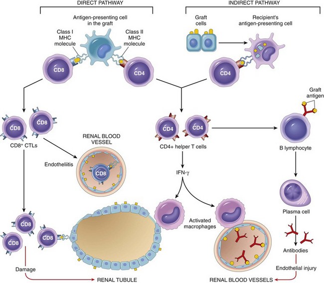
Figure 4–23 Recognition and rejection of allografts. In the direct pathway, donor class I and class II MHC antigens on antigen-presenting cells (APCs) in the graft (along with costimulators, not shown) are recognized by host CD8+ cytotoxic T cells and CD4+ helper T cells, respectively. CD4+ cells proliferate and produce cytokines (e.g., IFN-γ), which induce tissue damage by a local delayed-type hypersensitivity reaction. CD8+ T cells responding to graft antigens differentiate into CTLs that kill graft cells. In the indirect pathway, graft antigens are displayed by host APCs and activate CD4+ T cells, which damage the graft by a local delayed-type hypersensitivity reaction and stimulate B lymphocytes to produce antibodies. IFN-γ, interferon-γ; MHC, major histocompatibility complex.
Both T cells and antibodies reactive with the graft are involved in the rejection of most solid-organ allografts (Fig. 4–23).
CTLs kill cells in the grafted tissue, causing parenchymal and endothelial cell death (the latter resulting in thrombosis and graft ischemia). Cytokine-secreting CD4+ T cells trigger inflammatory reactions resembling DTH in the tissues and blood vessels, with local accumulation of mononuclear cells (lymphocytes and macrophages). Activated microphages can injure graft cells and vasculature. The microvascular injury also results in tissue ischemia, which contributes to graft destruction.
Although T cells are of paramount importance in allograft rejection, antibodies also mediate some forms of rejection. Alloantibodies directed against graft MHC molecules and other alloantigens bind to the graft endothelium and cause vascular injury through complement activation and recruitment of leukocytes. Superimposed on the resulting endothelial damage and dysfunction is thrombosis, adding further ischemic insult to the injury.
Hyperacute rejection is a special form of rejection occurring if pre-formed anti-donor antibodies are present in the circulation of the host before transplantation. This may happen in multiparous women who have anti-HLA antibodies against paternal antigens encountered during pregnancy, or in individuals exposed to foreign HLA (on platelets or leukocytes) from previous blood transfusions. Obviously, such antibodies also may be present in a patient who has previously rejected an organ transplant. Subsequent transplantation in such patients will result in immediate rejection (within minutes to hours) because the circulating antibodies rapidly bind to the endothelium of the grafted organ, with resultant complement activation and vascular thrombosis. With the current practice of screening potential recipients for pre-formed anti-HLA antibodies and cross-matching (testing recipients for the presence of antibodies directed against the donor’s lymphocytes), hyperacute rejection occurs in less than 0.4% of transplant recipients.
Morphology
On the basis of the time course and morphology of rejection reactions, they have been classified as hyperacute, acute, and chronic (Fig. 4–24). This classification is helpful for understanding the mechanism of rejection, because each pattern is caused by a different type of dominant immunologic reaction. The morphology of these patterns is described in the context of renal transplants; however, similar changes are encountered in other vascularized organ transplants.
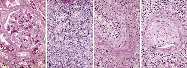
Figure 4–24 Morphologic patterns of graft rejection. A, Hyperacute rejection of a kidney allograft associated with endothelial damage and thrombi in a glomerulus. B, Acute cellular rejection of a kidney allograft with inflammatory cells in the interstitium and between epithelial cells of the tubules. C, Acute humoral rejection of a kidney allograft (rejection vasculitis) with inflammatory cells and proliferating smooth muscle cells in the intima. D, Chronic rejection in a kidney allograft with graft arteriosclerosis. The arterial lumen is replaced by an accumulation of smooth muscle cells and connective tissue in the intima.
(A–D, Courtesy of Dr. Helmut Rennke, Department of Pathology, Brigham and Women’s Hospital and Harvard Medical School, Boston, Massachusetts.)
Hyperacute rejection occurs within minutes to a few hours after transplantation in a presensitized host and typically is recognized by the surgeon just after the vascular anastomosis is completed. In contrast with a nonrejecting kidney graft, which regains a normal pink color and tissue turgor and promptly excretes urine, a hyperacutely rejecting kidney rapidly becomes cyanotic, mottled, and flaccid and may excrete only a few drops of bloody fluid. The histologic picture is characterized by widespread acute arteritis and arteriolitis, vessel thrombosis, and ischemic necrosis, all resulting from the binding of preformed antibodies to graft endothelium. Virtually all arterioles and arteries exhibit characteristic acute fibrinoid necrosis of their walls, with narrowing or complete occlusion of the lumens by precipitated fibrin and cellular debris (Fig. 4–24, A).
Acute rejection may occur within days to weeks of transplantation in a nonimmunosuppressed host or may appear months or even years later, even in the presence of adequate immunosuppression. Acute rejection is caused by both cellular and humoral immune mechanisms, and in any one patient, one or the other may predominate, or both may be present. On histologic examination, cellular rejection is marked by an interstitial mononuclear cell infiltrate with associated edema and parenchymal injury, whereas humoral rejection is associated with vasculitis.
Acute cellular rejection most commonly is seen within the first months after transplantation and typically is accompanied by clinical signs of renal failure. Histologic examination usually shows extensive interstitial CD4+ and CD8+ T cell infiltration with edema and mild interstitial hemorrhage (Fig. 4–24, B). Glomerular and peritubular capillaries contain large numbers of mononuclear cells, which also may invade the tubules, leading to focal tubular necrosis. In addition to tubular injury, CD8+ T cells also may injure the endothelium, causing an endothelitis. Cyclosporine (a widely used immunosuppressive agent) is also nephrotoxic and induces so-called arteriolar hyaline deposits. Renal biopsy is used to distinguish rejection from drug toxicity. Accurate recognition of cellular rejection is important, because patients typically respond promptly to increased immunosuppressive therapy.
Acute humoral rejection (rejection vasculitis) caused by antidonor antibodies also may participate in acute graft rejection. The histologic lesions may take the form of necrotizing vasculitis with endothelial cell necrosis; neutrophilic infiltration; deposition of antibody, complement, and fibrin; and thrombosis. Such lesions may be associated with ischemic necrosis of the renal parenchyma. Somewhat older subacute lesions are characterized by marked thickening of the intima by proliferating fibroblasts, myocytes, and foamy macrophages (Fig. 4–24, C). The resultant narrowing of the arterioles may cause infarction or renal cortical atrophy. The proliferative vascular lesions mimic arteriosclerotic thickening and are believed to be caused by cytokines that stimulate proliferation of vascular smooth muscle cells. Local deposition of complement breakdown products (specifically C4d) is used to detect antibody-mediated rejection of kidney allografts.
Patients present with chronic rejection late after transplantation (months to years) with a progressive rise in serum creatinine levels (an index of renal function) over a period of 4 to 6 months. Chronic rejection is dominated by vascular changes, interstitial fibrosis, and loss of renal parenchyma; there are typically only mild or no ongoing cellular parenchymal infiltrates. The vascular changes occur predominantly in the arteries and arterioles, which exhibit intimal smooth muscle cell proliferation and extracellular matrix synthesis (Fig. 4–24, D). These lesions ultimately compromise vascular perfusion and result in renal ischemia manifested by loss or hyalinization of glomeruli, interstitial fibrosis, and tubular atrophy. The vascular lesion may be caused by cytokines released by activated T cells that act on the cells of the vascular wall, and it may be the end stage of the proliferative arteritis described earlier.
Summary
• The graft rejection response is initiated mainly by host T cells that recognize the foreign HLA antigens of the graft, either directly (on APCs in the graft) or indirectly (after uptake and presentation by host APCs).
• Types and mechanisms of rejection comprise the following:
Because HLA molecules are the major targets in transplant rejection, better matching of the donor and the recipient improves graft survival. The benefits of HLA matching are most dramatic for living related donor kidney transplants, and survival improves with increasing number of loci matched. However, as drugs for immunosuppression have improved, HLA matching is not even attempted in some situations, such as heart, lung, liver, and islet transplantation; in such instances, the recipient often needs a transplant urgently and other considerations, such as anatomic (size) compatibility, are of greater practical importance.
Immunosuppression of the recipient is a practical necessity in all organ transplantation except in the case of identical twins. At present, drugs such as cyclosporine, the related FK506, mofetil mycophenolate (MMF), rapamycin, azathioprine, corticosteroids, antilymphocyte globulin, and monoclonal antibodies (e.g., monoclonal anti-CD3) are used. Cyclosporine and FK506 suppress T cell–mediated immunity by inhibiting transcription of cytokine genes, in particular, the gene for IL-2. Although immunosuppression has made transplantation of many organs feasible, there is still a price to be paid. Global immunosuppression results in increased susceptibility to opportunistic fungal, viral, and other infections. These patients are also at increased risk for developing Epstein-Barr virus (EBV)–induced lymphomas, human papillomavirus–induced squamous cell carcinomas, and Kaposi sarcoma. To circumvent the untoward effects of immunosuppression, much effort is devoted to trying to induce donor-specific tolerance in host T cells. One strategy being pursued is to prevent host T cells from receiving costimulatory signals from donor DCs during the initial phase of sensitization. This can be accomplished by administration of agents to interrupt the interaction between the B7 molecules on the DCs of the graft with the CD28 receptors on host T cells. This will interrupt the second signal for T cell activation and either induce apoptosis or render the T cells functionally unresponsive. The improvement in immunosuppressive therapy has led to improving survival of grafts and acute rejection is becoming less of a concern, especially for kidney and heart grafts. Chronic rejection remains a serious problem, however, especially because it responds much less effectively than does acute rejection to available immunosuppressive agents.
Hematopoietic stem cell (HSC) transplantation is used as therapy for hematopoietic and some nonhematopoietic malignancies, aplastic anemias, and certain inherited disorders, particularly immune deficiency states and severe forms of thalassemia. HSCs historically were obtained solely from donor bone marrow, but are now increasingly harvested from the peripheral blood after mobilization by administration of hematopoietic growth factors, or from the umbilical cord blood of newborns, a readily available rich source of HSCs. The recipient receives chemotherapy and/or irradiation to destroy malignant cells (e.g., in leukemia) and to create a graft bed; then, HSCs are infused into the peripheral blood, from which they home to their bone marrow niches. Rejection of allogeneic HSC transplants seems to be mediated by some combination of host T cells and NK cells that are resistant to radiation therapy and chemotherapy. Two major problems complicate this form of transplantation: graft-versus-host disease and immune deficiency.
This occurs when immunologically competent T cells (or their precursors) are transplanted into recipients who are immunologically compromised. Although GVHD happens most commonly in the setting of allogeneic HSC transplantation (usually involving minor histocompatibility mismatches between donor and recipient), it also may occur after transplantation of solid organs rich in lymphoid cells (e.g., the liver) or after transfusion of nonirradiated blood. On receiving allogeneic HSCs, an immunologically compromised host cannot reject the graft, but T cells present in the donor graft perceive the recipient’s tissue as “foreign” and react against it. This results in the activation of both CD4+ and CD8+ T cells, ultimately causing inflammation and killing host cells.
• Acute GVHD (occurring days to weeks after transplantation) causes epithelial cell necrosis in three principal target organs: liver, skin, and gut. Destruction of small bile ducts gives rise to jaundice, and mucosal ulceration of the gut results in bloody diarrhea. Cutaneous involvement is manifested by a generalized rash.
• Chronic GVHD may follow the acute syndrome or may occur insidiously. The patients develop skin lesions resembling those of SS (discussed earlier) and manifestations mimicking other autoimmune disorders.
GVHD is a potentially lethal complication that can be minimized but not eliminated by HLA matching. As another potential solution, donor T cells can be depleted before marrow transplant. This protocol has proved to be a mixed blessing: The risk of GVHD is reduced, but the incidence of graft failure and (in those with the disease) the recurrence of leukemia increase. It seems that the multifunctional T cells not only mediate GVHD but also are required for the efficient engraftment of the transplanted HSCs and elimination of leukemia cells (so-called graft-versus-leukemia effect).
These are often of prolonged duration in recipients of HSC transplants. Among the many reasons for this impairment is the slow reconstitution of the recipient’s adaptive immune system, which is destroyed or suppressed to allow the graft to take and requires many months to recover. During this vulnerable period, recipients are susceptible to a variety of infections, mostly viral, such as cytomegalovirus (CMV) and EBV infections.
Immune deficiency diseases may be caused by inherited defects affecting immune system development, or they may result from secondary effects of other diseases (e.g., infection, malnutrition, aging, immunosuppression, autoimmunity, or chemotherapy). Clinically, patients with immune deficiency present with increased susceptibility to infections as well as to certain forms of cancer. The type of infections in a given patient depends largely on the component of the immune system that is affected. Patients with defects in immunoglobulin, complement, or phagocytic cells typically suffer from recurrent infections with pyogenic bacteria, whereas those with defects in cell-mediated immunity are prone to infections caused by viruses, fungi, and intracellular bacteria. Discussed next are some of the more important primary (congenital) immune deficiencies, followed by a detailed description of the acquired immunodeficiency syndrome (AIDS), the most devastating example of secondary (acquired) immune deficiency.
Primary immune deficiency states are fortunately rare, but their study has nevertheless contributed greatly to the current understanding of the development and function of the immune system. Most primary immune deficiency diseases are genetically determined and affect either adaptive immunity (i.e., humoral or cellular) or innate host defense mechanisms, including complement proteins and cells such as phagocytes and NK cells. Defects in adaptive immunity are often subclassified on the basis of the primary component involved (i.e., B cells or T cells, or both); however, because of the interactions between T and B lymphocytes, these distinctions are not clear-cut. For instance, T cell defects frequently lead to impaired antibody synthesis, and hence isolated deficiencies of T cells may be indistinguishable from combined deficiencies of T and B cells. Most primary immune deficiencies come to attention early in life (between the ages of 6 months and 2 years), usually because the affected infants are susceptible to recurrent infections. One of the most impressive accomplishments of modern molecular biology has been the identification of the genetic basis for many primary immune deficiencies (Fig. 4–25), laying the foundation for future gene replacement therapy.
Figure 4–25 Primary immune deficiency diseases. Shown are the principal pathways of lymphocyte development and the blocks in these pathways in selected primary immune deficiency diseases. The affected genes are indicated in parentheses for some of the disorders. ADA, adenosine deaminase; CD40L, CD40 ligand (also known as CD154); CVID, common variable immunodeficiency; SCID, severe combined immunodeficiency.
X-linked agammaglobulinemia (XLA), or Bruton disease, is characterized by the failure of pre-B cells to differentiate into B cells and, as the name implies, there is a resultant absence of antibodies (gamma globulin) in the blood. It occurs at a frequency of about 1 in 100,000 male infants. During normal B cell maturation, immunoglobulin heavy chain genes are rearranged first, followed by light chain rearrangement. At each stage, signals are received from the expressed components of the antigen receptor that drive maturation to the next stage; these signals act as quality controls, to ensure that the correct receptor proteins are being produced. In XLA, B cell maturation stops after the initial heavy chain gene rearrangement because of mutations in a tyrosine kinase that is associated with the pre-B cell receptor and is involved in pre-B cell signal transduction. This kinase is called Bruton tyrosine kinase (BTK). When it is nonfunctional, the pre-B cell receptor cannot signal the cells to proceed along the maturation pathway. As a result, immunoglobulin light chains are not produced, and the complete immunoglobulin molecule containing heavy and light chains cannot be assembled and transported to the cell membrane, although free heavy chains can be found in the cytoplasm. Because the BTK gene is on the X chromosome, the disorder is seen in males.
Classically, this disease is characterized by the following:
• Absent or markedly decreased numbers of B cells in the circulation, with depressed serum levels of all classes of immunoglobulins. The numbers of pre-B cells in the bone marrow may be normal or reduced.
• Underdeveloped or rudimentary germinal centers in peripheral lymphoid tissues, including lymph nodes, Peyer patches, the appendix, and tonsils
XLA does not become apparent until the affected infant attains the age of approximately 6 months, when the transplacental supply of maternal antibodies is depleted. In most cases, recurrent bacterial infections such as acute and chronic pharyngitis, sinusitis, otitis media, bronchitis, and pneumonia suggest an underlying immune defect. The causal organisms typically are those bacterial pathogens that are cleared by antibody-mediated opsonization and phagocytosis (e.g., Haemophilus influenzae, Streptococcus pneumoniae, and Staphylococcus aureus). Because antibodies are important for neutralizing viruses, patients with XLA also are susceptible to certain viral infections, especially those caused by enteroviruses. Similarly, Giardia lamblia, an intestinal protozoan usually neutralized by secreted IgA, cannot be efficiently cleared and causes persistent infections. Fortunately, replacement therapy with intravenous immunoglobulin (IVIG) from pooled human serum allows most patients to adequately combat bacterial infections. Patients with XLA clear some viral, fungal, and protozoal infections, because their T cell–mediated immunity is intact. For unclear reasons, autoimmune diseases (such as RA and dermatomyositis) occur in as many as 20% of patients with this disease.
Common variable immunodeficiency is an umbrella term for a heterogeneous group of disorders characterized by hypogammaglobulinemia, impaired antibody responses to infection (or vaccination), and increased susceptibility to infections. The clinical manifestations are superficially similar to those of XLA, but in common variable immunodeficiency, males and females are affected equally and the onset of symptoms is much later, in the second or third decade of life. The diagnosis usually is one of exclusion (after other causes of immune deficiency are ruled out). The estimated prevalence of the disease is about 1 in 50,000. Although most patients have normal numbers of mature B cells, plasma cells are absent, suggesting a block in antigen-stimulated B cell differentiation. The defective antibody production has been variably attributed to intrinsic B cell defects, deficient T cell help, or excessive T cell suppressive activity. Paradoxically, these patients are prone to develop a variety of autoimmune disorders (hemolytic anemia, pernicious anemia), as well as lymphoid tumors. The underlying mechanism of the antibody deficiency is variable (hence the name). Some patients with this disease have mutations in B cell receptors for certain growth factors, or in molecules involved in T cell–B cell interactions. However, the genetic basis of most cases of the disease is not known.
The most common of all the primary immune deficiency diseases, IgA deficiency affects about 1 in 700 whites. As noted previously, IgA is the major immunoglobulin in mucosal secretions and is thus involved in defending the airways and the gastrointestinal tract. Although most people with this condition are asymptomatic, weakened mucosal defenses predispose patients to recurrent sinopulmonary infections and diarrhea. There is also a significant (but unexplained) association with autoimmune diseases. The pathogenesis of IgA deficiency seems to involve a block in the terminal differentiation of IgA-secreting B cells to plasma cells; IgM and IgG subclasses of antibodies are present in normal or even supranormal levels. The molecular basis for this defect is not understood.
In a normal immune response to protein antigen, IgM antibodies are produced first, followed by the sequential elaboration of IgG, IgA, and IgE antibodies. As discussed earlier in this chapter, the orderly appearance of different antibody types is called heavy-chain class (isotype) switching and is important for generating classes of antibody that can effectively activate complement and/or opsonize bacterial pathogens. The ability of IgM-producing B cells to turn on the transcription of genes that encode other immunoglobulin isotypes depends on certain cytokines, as well as on contact-mediated signals from CD4+ helper T cells. The contact-dependent signals are provided by interaction between CD40 molecules on B cells and CD40L (also known as CD154), expressed on activated helper T cells. Patients with the hyper-IgM syndrome produce normal (or even supranormal) levels of IgM antibodies to antigens but lack the ability to produce the IgG, IgA, and IgE isotypes; the underlying defect is an inability of T cells to induce B cell isotype switching. The most common genetic abnormality is mutation of the gene encoding CD40L. This gene is located on the X chromosome; consequently, in approximately 70% of the cases, hyper-IgM syndrome is X-linked. In the remaining patients, the mutations affect CD40 or other molecules involved in class switching, notably an enzyme called activation-induced deaminase. In addition to defective class switching, in those with CD40 or CD40L mutations there is also often a defect in the production of high-affinity antibodies, because the same mechanism is responsible for affinity maturation of the antibody response.
Although the disease is diagnosed and named because of the antibody abnormality, in patients with CD40 or CD40L mutations there is also a defect in cell-mediated immunity because the CD40–CD40L interaction is critical for helper T cell–mediated activation of macrophages, the central reaction of cell-mediated immunity. Male patients with the X-linked form of hyper-IgM syndrome present with recurrent pyogenic infections owing to low levels of opsonizing IgG antibodies. These patients also are susceptible to infections with a variety of intracellular pathogens that normally are combated by cell-mediated immunity, including Pneumocystis jiroveci (formerly called Pneumocystis carinii).
DiGeorge syndrome results from a congenital defect in thymic development with deficient T cell maturation. T cells are absent in the lymph nodes, spleen, and peripheral blood, and infants with this defect are extremely vulnerable to viral, fungal, and protozoal infections. Patients are also susceptible to infection with intracellular bacteria, because of defective T cell–mediated immunity. B cells and serum immunoglobulins are generally unaffected.
The disorder is a consequence of a developmental malformation affecting the third and fourth pharyngeal pouches, structures that give rise to the thymus, parathyroid glands, and portions of the face and aortic arch. Thus, in addition to the thymic and T cell defects, there may be parathyroid gland hypoplasia, resulting in hypocalcemic tetany, as well as additional midline developmental abnormalities. In 90% of cases of DiGeorge syndrome there is a deletion affecting chromosomal region 22q11, as discussed in Chapter 6. Transplantation of thymic tissue has successfully treated some affected infants. In patients with partial defects, immunity may improve spontaneously with age.
Severe combined immunodeficiency (SCID) represents a constellation of genetically distinct syndromes with the common feature of defects in both humoral and cell-mediated immune responses. Affected infants are susceptible to severe recurrent infections by a wide array of pathogens, including bacteria, viruses, fungi, and protozoans, and opportunistic infections by Candida, Pneumocystis, CMV, and Pseudomonas. These pathogens cause serious (and occasionally lethal) disease. The prevalence of the disease is approximately 1 in 65,000 to 1 in 100,000, and the frequency is 20 to 30 times higher in some Native American populations.
Despite the common clinical features, the underlying defects in individual patients are quite diverse. Some forms of SCID are caused by a single defect affecting both T and B cells, and others may result from a primary T cell deficit with secondary impairment of humoral immunity. Approximately half of the cases are X-linked; these are caused by mutations in the gene encoding the common γ chain shared by the receptors for the cytokines IL-2, IL-4, IL-7, IL-9, and IL-15. Of these cytokines, IL-7 is the most important in this disease because it is the growth factor responsible for stimulating the survival and expansion of immature B and T cell precursors in the generative lymphoid organs. Another 40% to 50% of SCID cases are inherited in an autosomal recessive fashion, with approximately half of these caused by mutations in adenosine deaminase (ADA), an enzyme involved in purine metabolism. ADA deficiency results in accumulation of adenosine and deoxyadenosine triphosphate metabolites, which inhibit DNA synthesis and are toxic to lymphocytes. The other autosomal recessive cases of SCID are attributed to defects in another purine metabolic pathway, primary failure of class II MHC expression, or mutations in genes encoding the recombinase responsible for the rearrangement of lymphocyte antigen-receptor genes.
In the two most common forms of SCID (cytokine receptor common γ chain mutation and ADA deficiency), the thymus is hypoplastic. Lymph nodes and lymphoid tissues (e.g., in the tonsils, gut, and appendix) are atrophic and lack germinal centers as well as paracortical T cells. Affected patients may have marked lymphopenia, with both T and B cell deficiency; others may have increased numbers of immature T cells and/or large numbers of B cells that are nonfunctional as a consequence of a lack of T cell help. Patients with SCID are currently treated by bone marrow transplantation. X-SCID is the first disease in which gene therapy has been used to successfully replace the mutated gene, but the approach is being reevaluated because some of the treated patients have developed T cell leukemias, presumably because the introduced gene was inserted close to a cellular oncogene.
Rare patients with mutations in genes required for T cell activation have been identified that manifest with defective cell-mediated immunity or a phenotype resembling SCID. One of the interesting ones is a mutation in a transcription factor that is required for the TH17 response. The resulting disease manifestations include fungal (and occasionally bacterial) skin infections and chronic mucocutaneous candidiasis. By contrast, mutations in genes involved in TH1 responses result in susceptibility to infections with atypical mycobacteria. These observations emphasize the importance of TH17 cells for defense against fungal infections and of TH1 cells for combating intracellular bacterial infections. Mutations in genes encoding calcium channel proteins, and other components of T cell signaling, also have been described.
Wiskott-Aldrich syndrome is an X-linked recessive disease characterized by thrombocytopenia, eczema, and a marked vulnerability to recurrent infection, ending in early death; the only treatment is bone marrow transplantation. This is a curious syndrome in that the clinical presentation and immunologic deficits are difficult to explain on the basis of the known underlying genetic defect. The thymus is initially normal, but there is progressive age-related depletion of T lymphocytes in the peripheral blood and lymph nodes, with concurrent loss of cellular immunity. Additionally, patients do not make effective antibody responses to polysaccharide antigens, and are therefore particularly susceptible to infections with encapsulated, pyogenic bacteria. Affected patients also are prone to the development of malignant lymphomas. The responsible gene encodes a protein (Wiskott-Aldrich syndrome protein) that links several membrane receptors to the cytoskeleton. Although the mechanism is not known, a defect in this protein could result in abnormal cellular morphology (including platelet shape changes) or defective cytoskeleton-dependent activation signals in lymphocytes and other leukocytes, with abnormal cell–cell adhesions and leukocyte migration.
Several genetic defects have been shown to affect molecules or cells that are important in the early innate immune response to microbes.
As discussed earlier in this chapter and in Chapter 2, complement components play important roles in inflammatory and immunologic responses. Consequently, hereditary deficiency of C3 (critical for both the classical and alternative pathways) results in an increased susceptibility to infection with pyogenic bacteria. Inherited deficiencies of C1q, C2, and C4 do not make affected persons susceptible to infections, but they do increase the risk of immune complex–mediated disease (e.g., SLE), possibly by impairing the clearance of apoptotic cells or of antigen–antibody complexes from the circulation. Deficiencies of the late components of the classical complement pathway (C5 to C9) result in recurrent infections by Neisseria (gonococci, meningococci) but not by other microbes because Neisseria have thin cell walls and are especially susceptible to lysis by the membrane attack complex, the product of the late steps of complement activation. Lack of the regulatory protein C1 inhibitor allows unfettered C1 activation, with the generation of downstream vasoactive complement mediators; the result is hereditary angioedema, characterized by recurrent episodes of localized edema affecting the skin and/or mucous membranes.
Several congenital defects in phagocytes are known. These include defects in the phagocyte oxidase (NADPH oxidase) enzyme, the cause of chronic granulomatous disease, and defects in integrins and selectin ligands, causing the leukocyte adhesion deficiencies. These disorders were described in Chapter 2.
Mutations in TLRs and their signaling pathways are quite rare, but study of the associated disorders has been informative. One of the surprises that has emerged from these diseases is that the immune deficiency typically is very restricted. For instance, patients with mutations affecting TLR3, which recognizes viral RNA, develop recurrent herpes simplex encephalitis, and those with mutations affecting MyD88, the signaling protein downstream of many TLRs, are susceptible to bacterial infections, especially severe pneumococcal lung disease, but neither suffers from multiple disseminated infections.
Summary
• Caused by mutations in genes involved in lymphocyte maturation or function, or in innate immunity
• Some of the common disorders:
• Clinical presentation: increased susceptibility to infections in early life
Immune deficiencies secondary to other diseases or therapies are much more common than the primary (inherited) disorders. Secondary immune deficiencies may be encountered in patients with malnutrition, infection, cancer, renal disease, or sarcoidosis. However, the most common cause of immune deficiency is therapy-induced suppression of the bone marrow or of lymphocyte function.
Discussed next is perhaps the most important secondary immune deficiency disease, AIDS, which has become one of the great scourges of humankind.
AIDS is a retroviral disease caused by the human immunodeficiency virus (HIV). It is characterized by infection and depletion of CD4+ T lymphocytes, and by profound immunosuppression leading to opportunistic infections, secondary neoplasms, and neurologic manifestations. Although AIDS was first described in the United States, it has now been reported in virtually every country in the world. At the end of 2009, more than 33 million people were living with HIV infection and AIDS, of which approximately 70% were in Africa and 20% in Asia; there were almost 2 million cases diagnosed and almost 2 million died of the disease that year, with a total of more than 22 million deaths since the epidemic was recognized in 1981. Although the largest number of infections is in Africa, the most rapid increases in HIV infection in the past decade have occurred in Southeast Asian countries, including Thailand, India, and Indonesia. The statistics are only slightly better in the Western world; for example, approximately 1 million U.S. citizens are infected (roughly 1 in 300). Moreover, more Americans (more than 500,000) have died of AIDS than died in both world wars combined. AIDS-related death rates continue to decline from their 1995 peak.
Because of the combined work of many scientists and clinicians, there has been an explosion of new knowledge about this modern plague. So rapid is the pace of research on the biology of HIV that any text covering the topic will probably be out of date by the time it goes to press. Nevertheless, presented next is a summary of the currently available information on HIV epidemiology, etiology, pathogenesis, and clinical features.
Epidemiologic studies in the United States have identified five groups at risk for developing AIDS, and these are similar in other countries, except as noted in the following list. Transmission of HIV occurs under conditions that facilitate the exchange of blood or body fluids that contain the virus or virus-infected cells. Thus, the major routes of HIV infection are sexual contact, parenteral inoculation, and passage of the virus from infected mothers to their newborns. In about 10% of cases, the risk factors are unknown or not reported. The case distribution data cited are for the United States.
• Men who have sex with men constitute the largest group of infected persons, accounting for 48% of reported cases in the period 2001 to 2004 and 56% of infected men (approximately 4% of whom also inject drugs). However, transmission of AIDS in this category is declining, with less than 50% of new cases attributable to male homosexual contact.
• Heterosexual contacts of members of other high-risk groups constituted about 34% of infections from 2001 to 2004. In Africa and Asia, this is by far the largest group of patients with new infections, and a majority of new cases are in women infected by male partners.
• Intravenous drug abusers with no history of homosexual behavior compose the next largest group, representing about 17% of all patients.
• Recipients of blood and blood components (but not hemophiliacs) who received transfusions of HIV-infected whole blood or components (e.g., platelets, plasma) account for about 1% of patients.
• Hemophiliacs, especially those who received large amounts of factor VIII or IX concentrates before 1985, make up less than 1% of all cases.
• The epidemiology of HIV infection and AIDS is quite different in children (diagnosed when younger than 13 years of age). About 1% of all AIDS cases occur in this population, and the vast majority (about 90%) result from vertical transmission of virus from infected mother to the fetus or newborn.
Sexual transmission is by far the major mode of infection worldwide, accounting for more than 75% of all cases of HIV transmission. Although most sexually transmitted cases in the United States are still due to male-with-male sexual contacts, the vast majority of sexually transmitted HIV infections globally are due to heterosexual activity. Even in the United States, the rate of increase of heterosexual transmission has outpaced transmission by other means; such spread accounts for the dramatic increase in HIV infection in female sex partners of male intravenous drug abusers.
The virus is present in semen, both extracellularly and within mononuclear inflammatory cells, and it enters the recipient’s body through lacerations or abrasions in mucosa. Viral transmission to newborns can occur either by direct entry of virus or infected cells into blood vessels breached through traumatic injury or by uptake into mucosal DCs. Clearly, all forms of sexual transmission are aided and abetted by the concomitant presence of other sexually transmitted diseases that cause genital ulcerations, including syphilis, chancroid, and herpes simplex virus infections. Gonorrhea and chlamydial infection also are cofactors for HIV transmission, primarily by increasing the seminal fluid content of inflammatory cells (presumably carrying HIV). In addition to male-to-male and male-to-female transmission, HIV is present in the vaginal and cervical cells of infected women and can also be spread from females to males, albeit about eight times less efficiently.
Parenteral transmission of HIV is well documented in three different groups: intravenous drug abusers (the largest group), hemophiliacs receiving factor VIII or IX concentrates, and random recipients of blood transfusion. Among intravenous drug abusers, transmission occurs through shared needles, syringes, or other paraphernalia contaminated with HIV-containing blood.
Transmission of HIV by transfusion of blood or blood products such as lyophilized factor VIII concentrates has been virtually eliminated since 1985. Four public health measures are responsible: screening of donated blood and plasma for antibody to HIV, screening for HIV-associated p24 antigen (detectable before the development of antibodies), heat treatment of clotting factor concentrates, and screening of donors on the basis of history. With all of these measures, it is estimated currently that one in 1.5 million blood donations are HIV infected, and that 20 HIV-positive blood components derived from 11 infectious donations are released each year that could potentially infect recipients. With the advent of nucleic acid testing, this already small risk is expected to show further decline.
As noted earlier, mother-to-infant vertical transmission is the major cause of pediatric AIDS. Three routes are involved: in utero, by transplacental spread; intrapartum, during delivery; and by ingestion of HIV-contaminated breast milk. Of these, the transplacental and intrapartum routes account for most cases. Vertical transmission rates worldwide vary, ranging from 25% to 35%, with a 15% to 25% rate reported in the United States; higher rates of infection occur with high maternal viral load and/or the presence of chorioamnionitis, presumably by increasing placental accumulation of inflammatory cells.
Because of the dismal outcome for AIDS, the lay public is justifiably concerned about the spread of HIV infection outside recognized high-risk groups. Many of these anxieties can be laid to rest, because extensive studies indicate that HIV infection cannot be transmitted by casual personal contact in the home, workplace, or school, and no convincing evidence for spread by insect bites has been obtained. There is an extremely small but confirmed risk of transmission of HIV infection to health care workers. Seroconversion has been documented after accidental needlestick injury or exposure of nonintact skin to infected blood in laboratory accidents, with a rate of about 0.3% per accidental exposure. By comparison, the rate of seroconversion after accidental exposure to hepatitis B–infected blood is about 6% to 30%.
AIDS is caused by HIV, a human retrovirus belonging to the lentivirus family (which also includes feline immunodeficiency virus, simian immunodeficiency virus, visna virus of sheep, and the equine infectious anemia virus). Two genetically different but antigenically related forms of HIV, called HIV-1 and HIV-2, have been isolated from patients with AIDS. HIV-1 is the more common type associated with AIDS in the United States, Europe, and Central Africa, whereas HIV-2 causes a similar disease principally in West Africa. Specific tests for HIV-2 are now available, and blood collected for transfusion also is routinely screened for HIV-2 seropositivity. The ensuing discussion relates primarily to HIV-1 and diseases caused by it, but it is generally applicable to HIV-2 as well.
Like most retroviruses, the HIV-1 virion is spherical and contains an electron-dense, cone-shaped core surrounded by a lipid envelope derived from the host cell membrane (Fig. 4–26). The virus core contains (1) major capsid protein p24, (2) nucleocapsid protein p7/p9, (3) two copies of genomic RNA, and (4) three viral enzymes—protease, reverse transcriptase, and integrase. The p24 protein is the most readily detected viral antigen and is therefore the target for the antibodies used to diagnose HIV infection in blood screening. The viral core is surrounded by a matrix protein called p17, lying beneath the virion envelope. The viral envelope itself is studded with two viral glycoproteins (gp120 and gp41), critical for HIV infection of cells. The HIV-1 proviral genome contains the gag, pol, and env genes, which code for various viral proteins. The products of the gag and pol genes are translated initially into large precursor proteins that must be cleaved by the viral protease to yield the mature proteins. The highly effective anti-HIV-1 protease inhibitor drugs prevent viral assembly by inhibiting the formation of mature viral proteins.
Figure 4–26 The structure of human immunodeficiency virus (HIV). The HIV-1 virion. The viral particle is covered by a lipid bilayer derived from the host cell and studded with viral glycoproteins gp41 and gp120.
In addition to these three standard retroviral genes, HIV contains several other genes (given three-letter names such as tat, rev, vif, nef, vpr, and vpu) that regulate the synthesis and assembly of infectious viral particles. The product of the tat (transactivator) gene, for example, is critical for virus replication, causing a 1000-fold increase in the transcription of viral genes. The nef protein activates intracellular kinase activity (affecting T cell activation, viral replication, and viral infectivity) and reduces surface expression of CD4 and MHC molecules on infected cells. The progression of HIV infection in vivo is dependent on nef; strains of simian immunodeficiency virus with mutated nef genes cause AIDS in monkeys at a markedly decreased rate, and humans infected with a nef-defective HIV-1 strain display low viral burden, with AIDS onset at a substantially slower pace than for nonmutant strains. The products of various regulatory genes are important for HIV pathogenicity, and therapeutic approaches are being developed to block their actions.
Nucleic acid sequencing of different viral isolates reveals considerable variability in many parts of the HIV genome. This high variability is due to the relatively low fidelity of the viral polymerase, with estimates of one mistake for each 105 replicated nucleotides. Most sequence variants cluster in parts of the genome encoding the envelope glycoproteins. Because the immune response against HIV-1 is targeted against its envelope, such extreme variability in antigen structure poses a formidable barrier to vaccine development.
On the basis of genomic analyses, HIV-1 can be divided into two groups, designated M (major) and O (outlier). Group M viruses, the more common form worldwide, are further divided into subtypes, or clades, designated A through J. The clades differ in their geographic distribution, with B being the most common form in Western Europe and the United States and E being the most common in Thailand. Beyond molecular homologies, the clades also show differences in modes of transmission. Thus, E clade is spread predominantly by heterosexual contact (male-to-female), presumably because of its ability to infect vaginal subepithelial DCs. By contrast, B clade virus grows poorly in DCs and may be transmitted by monocytes and lymphocytes.
The two major targets of HIV infection are the immune system and the CNS. The life cycle of the virus is best understood in terms of its interactions with the immune system.
The entry of HIV into cells requires the CD4 molecule, which acts as a high-affinity receptor for the virus (Fig. 4–27). This requirement explains the tropism of the virus for CD4+ T cells and its ability to infect other CD4+ cells, particularly macrophages and DCs. However, binding to CD4 is not sufficient for infection; the HIV envelope gp120 must also bind to other cell surface molecules (coreceptors) to facilitate cell entry. Two cell surface chemokine receptors, CCR5 and CXCR4, serve this role. HIV envelope gp120 (noncovalently attached to transmembrane gp41) binds initially to CD4 molecules (Fig. 4–27). This binding leads to a conformational change that exposes a new recognition site on gp120 for the coreceptors CXCR4 (mostly on T cells) or CCR5 (mostly on macrophages). The gp41 then undergoes a conformational change that allows it to insert into the target membrane, and this process facilitates fusion of the virus with the cell. After fusion, the virus core containing the HIV genome enters the cytoplasm of the cell.
Figure 4–27 Molecular basis of entry of human immunodeficiency virus (HIV) into host cells. Interactions with CD4 and a chemokine receptor (“coreceptor”).
(Adapted by permission from Macmillan Publishers Ltd, from Wain-Hobson S: HIV. One on one meets two. Nature 384:117, copyright 1996.)
The coreceptors are critical components of the HIV infection process, and their discovery resolved some previously unexplained observations regarding HIV tropism. It had been known that HIV strains could be classified according to their relative ability to infect macrophages and/or CD4+ T cells. Macrophage-tropic (R5 virus) strains infect both monocytes/macrophages and freshly isolated peripheral blood T cells, whereas T cell–tropic (X4 virus) strains infect only activated T cell lines. This selectivity is now explained by selective coreceptor usage. R5 strains use CCR5 as their coreceptor, and, because CCR5 is expressed on both monocytes and T cells, these cells are susceptible to infection by R5 strains. Conversely, X4 strains bind to CXCR4, which is expressed on T cell lines (and not on monocytes/macrophages), so that only activated T cells are susceptible. Of interest, approximately 90% of HIV infections initially are transmitted by R5 strains. Over the course of infection, however, X4 viruses gradually accumulate; these are especially virulent and are responsible for T cell depletion in the final, rapid phase of disease progression. It is thought that during the course of HIV infection, R5 strains evolve into X4 strains, as a result of mutations in genes that encode gp120. Persons with defective CCR5 receptors (of U.S. whites, 20% are heterozygous and 1% are homozygous for the mutant CCR5) are relatively resistant to developing AIDS, despite repeated HIV exposure in vivo. Because of the significance of HIV–coreceptor interaction in the pathogenesis of AIDS, preventing this interaction may be of significant therapeutic value.
Once internalized, the viral genome undergoes reverse transcription, leading to formation of complementary DNA (cDNA). In quiescent T cells, HIV proviral cDNA may remain in the cytoplasm in a linear episomal form. However, in dividing T cells, the cDNA enters the nucleus and becomes integrated into the host genome. After integration, the provirus may remain nontranscribed for months or years, and the infection becomes latent; alternatively, proviral DNA may be transcribed to form complete viral particles that bud from the cell membrane. Such productive infections, associated with extensive viral budding, lead to cell death. It is important to note that although HIV-1 can infect resting T cells, the initiation of proviral DNA transcription (and hence productive infection) occurs only when the infected cell is activated by exposure to antigens or cytokines. Thus, in a cruel twist, physiologic responses to infections and other stimuli promote the death of HIV-infected T cells.
HIV disease begins with acute infection, which is only partly controlled by the host immune response, and advances to chronic progressive infection of peripheral lymphoid tissues (Fig. 4–28). The first cell types to be infected may be memory CD4+ T cells (which express CCR5) in mucosal lymphoid tissues. Because the mucosal tissues are the largest reservoir of T cells in the body and a major site of residence of memory T cells, the death of these cells results in considerable depletion of lymphocytes.
Figure 4–28 Pathogenesis of human immunodeficiency virus (HIV) infection. Initially, HIV infects T cells and macrophages directly or is carried to these cells by Langerhans cells. Viral replication in the regional lymph nodes leads to viremia and widespread seeding of lymphoid tissue. The viremia is controlled by the host immune response (not shown), and the patient then enters a phase of clinical latency. During this phase, viral replication in both T cells and macrophages continues unabated, but there is some immune containment of virus (not illustrated). There continues a gradual erosion of CD4+ cells by productive infection (or other mechanisms, not shown). Ultimately, CD4+ cell numbers decline and the patient develops clinical symptoms of full-blown AIDS. Macrophages are also parasitized by the virus early; they are not lysed by HIV and they transport the virus to tissues, particularly the brain.
The transition from the acute phase to a chronic phase of infection is characterized by dissemination of the virus, viremia, and the development of host immune responses. DCs in epithelia at sites of virus entry capture the virus and then migrate into the lymph nodes. Once in lymphoid tissues, DCs may pass HIV on to CD4+ T cells through direct cell–cell contact. Within days after the first exposure to HIV, viral replication can be detected in the lymph nodes. This replication leads to viremia, during which high numbers of HIV particles are present in the patient’s blood, accompanied by an acute HIV syndrome that includes a variety of nonspecific signs and symptoms typical of many viral diseases. The virus disseminates throughout the body and infects helper T cells, macrophages, and DCs in peripheral lymphoid tissues. As the infection spreads, the immune system mounts both humoral and cell-mediated immune responses directed at viral antigens. These immune responses partially control the infection and viral production, and such control is reflected by a drop in viremia to low but detectable levels by about 12 weeks after the primary exposure.
In the next, chronic phase of the disease, lymph nodes and the spleen are sites of continuous HIV replication and cell destruction (Fig. 4–28). During this period of the disease, the immune system remains competent at handling most infections with opportunistic microbes, and few or no clinical manifestations of the HIV infection are present. Therefore, this phase of HIV disease is called the clinical latency period. Although a majority of peripheral blood T cells do not harbor the virus, destruction of CD4+ T cells within lymphoid tissues steadily progresses during the latent period, and the number of circulating blood CD4+ T cells steadily declines. More than 90% of the body’s approximately 1012 T cells are normally found in lymphoid tissues, and it is estimated that HIV destroys up to 1 to 2 × 10⁹ CD4+ T cells every day. Early in the course of the disease, the body may continue to make new CD4+ T cells, so CD4+ T cells can be replaced almost as quickly as they are destroyed. At this stage, up to 10% of CD4+ T cells in lymphoid organs may be infected, but the number of circulating CD4+ T cells that are infected at any one time may be less than 0.1% of the total CD4+ T cells in a given case. Eventually, over a period of years, the continuous cycle of virus infection and T cell death leads to a steady decline in the number of CD4+ T cells in the lymphoid tissues and the circulation.
In addition to T cell depletion, abnormalities have been described in many components of the immune system, summarized in Table 4–11. Discussed next are the major defects in immune cells during the course of HIV infection.
Table 4–11 Major Abnormalities of Immune Function in AIDS
Lymphopenia |
Decreased T Cell Function in vivo |
Altered T Cell Function in vitro |
Polyclonal B Cell Activation |
Altered Monocyte or Macrophage Functions |
HLA, human leukocyte antigen.
The major mechanism of loss of CD4+ T cells is lytic HIV infection of the cells, and cell death during viral replication and production of virions (Fig. 4–29). Like other cytopathic viruses, HIV disrupts cellular functions sufficiently to cause death of infected cells. In addition to direct cell lysis, other mechanisms may cause T cell loss:
• Loss of immature precursors of CD4+ T cells, either by direct infection of thymic progenitor cells or by infection of accessory cells that secrete cytokines essential for CD4+ T cell maturation. The result is decreased production of mature CD4+ T cells.
• Chronic activation of uninfected cells by HIV antigens or by other concurrent infectious microbes may lead to apoptosis of the T cells. Because of this “activation-induced death” of uninfected cells, the numbers of T cells that die may be much greater than the number of HIV-infected cells.
• Infection of various cells in lymphoid tissues may disrupt the normal architecture, leading to impaired immune responses.
• Fusion of infected and uninfected cells causes formation of syncytia (giant cells). In tissue culture, the gp120 expressed on productively infected cells binds to CD4 molecules on uninfected T cells, followed by cell fusion, ballooning, and death within a few hours. This property of syncytia formation is confined to the X4 strain of HIV.
• Uninfected CD4+ T cells may bind soluble gp120 to the CD4 molecule, leading to aberrant signaling and apoptosis.
• Infected CD4+ T cells may be killed by HIV-specific CD8+ CTLs.
Figure 4–29 Mechanisms of CD4+ T cell loss in human immunodeficiency virus (HIV) infection. Some of the principal known and postulated mechanisms of T cell depletion after HIV infection are shown.
The loss of CD4+ cells leads to an inversion of the CD4+/CD8+ ratio in the peripheral blood. Thus, while the normal CD4+/CD8+ ratio is 1 to 2, patients with AIDS have a ratio of 0.5 or less. Such inversion is a common finding in AIDS, but it also may occur in other viral infections and is therefore not diagnostic.
Although marked reduction in CD4+ T cells is a hallmark of AIDS and can account for much of the immune deficiency late in the course of HIV infection, there is also compelling evidence for qualitative defects in T cell function that can be detected even in asymptomatic HIV-infected persons. Such defects include reduced antigen-induced T cell proliferation, impaired TH1 cytokine production, and abnormal intracellular signaling. There is also a selective loss of memory CD4+ T cells early in the course of the disease, possibly related to the abundance of these cells in mucosal tissues and higher level of CCR5 expression in this T cell subset.
Low-level chronic or latent infection of T cells (and macrophages) is an important feature of HIV infection. Although only rare CD4+ T cells express infectious virus early in the course of infection, up to 10% of lymph node T cells can be demonstrated to actually harbor the HIV genome. It is widely believed that integrated provirus, without virus production (latent infection), can persist within cells for months or years. Even with highly active antiretroviral therapy (which can eliminate most of the virus in the blood), latent virus lurks in lymph node CD4+ cells (as many as 0.05% of resting, long-lived CD4+ T cells are infected). Completion of the viral life cycle in latently infected cells requires cell activation. Thus, if latently infected CD4+ cells are activated by environmental antigens, an unfortunate consequence is increased HIV proviral DNA transcription. This increased transcription leads to virion production and, in the case of T cells, also results in cell lysis. In addition, TNF, IL-1, and IL-6 produced by activated macrophages during normal immune responses can also lead to increased HIV gene transcription (Fig. 4–28). Thus, it seems that HIV thrives when the host macrophages and T cells are physiologically activated (e.g., through intercurrent infection by other microbial agents). The life styles of most HIV-infected patients in the United States place them at increased risk for recurrent exposure to other sexually transmitted diseases; in Africa, socioeconomic conditions probably impose a higher burden of chronic microbial infections. It is easy to understand how patients with AIDS enter a vicious circle of T cell destruction; infections to which these patients are prone because of diminished helper T cell function lead to increased production of pro-inflammatory cytokines, which in turn stimulate more HIV production, followed by infection and loss of additional CD4+ T cells.
In addition to infection of CD4+ T cells, infection of monocytes and macrophages is also important in the pathogenesis of HIV disease. Similar to T cells, most of the HIV-infected macrophages are found in the tissues and not in peripheral blood. As many as 10% to 50% of macrophages in certain tissues, such as brain and lungs, may be infected. Several additional aspects of macrophage HIV infection warrant emphasis:
• Although cell division is required for integration and subsequent replication of most retroviruses, HIV-1 can infect and multiply in terminally differentiated nondividing macrophages, a property conferred by the HIV-1 vpr gene.
• Infected macrophages bud relatively small amounts of virus from the cell surface but contain large numbers of virus particles located in intracellular vesicles.
• In contrast with CD4+ T cells, macrophages are quite resistant to the cytopathic effects of HIV and can, therefore, harbor the virus for long periods.
• In more than 90% of cases, HIV infection is transmitted by R5 strains. The more virulent X4 strains that evolve later in the course of HIV infection are inefficient in transmitting HIV to monocytes. This suggests that the initial infection of macrophages (or DCs) is critical for HIV transmission.
Thus, in all likelihood, macrophages are the gatekeepers of HIV infection. Besides providing a portal for initial transmission, monocytes and macrophages are viral reservoirs and factories, whose output remains largely protected from host defenses. Circulating monocytes also provide a vehicle for HIV transport to various parts of the body, particularly the nervous system. In late stages of HIV infection, when the CD4+ T cell numbers are massively depleted, macrophages remain a major site of continued viral replication. Although the number of HIV-infected monocytes in the circulation is low, their functional deficits (e.g., impaired microbicidal activity, decreased chemotaxis, abnormal cytokine production, diminished antigen presentation capacity) have important bearing on host defenses.
In addition to macrophages, two types of DCs also are important targets for the initiation and maintenance of HIV infection: mucosal and follicular DCs. As discussed earlier, mucosal DCs capture the virus and transport it to regional lymph nodes, where CD4+ T cells are infected. Follicular DCs in the germinal centers of lymph nodes are important reservoirs of HIV. Although some follicular DCs are infected by HIV, most virus particles are found on the surface of their dendritic processes, including those bound to Fc receptors through HIV–anti-HIV antibody complexes. The antibody-coated virions localized to follicular DCs retain the ability to infect CD4+ T cells. HIV infection of macrophages and DCs also may impair the functions of these cell populations, with secondary effects on T cell responsiveness.
Although much attention has been focused on T cells and macrophages, patients with AIDS also display profound abnormalities of B cell function. Paradoxically, these patients have hypergammaglobulinemia and circulating immune complexes as a result of polyclonal B cell activation. This may result from multiple factors, including infection with CMV or EBV, both of which are polyclonal B cell activators. The HIV gp41 itself can promote B cell growth and differentiation, and HIV-infected macrophages produce increased amounts of IL-6, which enhances B cell proliferation. Despite the presence of spontaneously activated B cells, patients with AIDS are unable to mount antibody responses to newly encountered antigens. Not only is this attributable to deficient T cell help, but antibody responses against T cell–independent antigens are also suppressed, suggesting additional B cell defects. Impaired humoral immunity renders these patients susceptible to encapsulated bacteria (e.g., S. pneumoniae, H. influenzae) that require antibodies for effective opsonization and clearance.
CD4+ T cells play a pivotal role in regulating the immune response: they produce a plethora of cytokines, chemotactic factors, and hematopoietic growth factors (e.g., granulocyte-macrophage colony-stimulating factor). Therefore, loss of this “master cell” has ripple effects on virtually every other cell of the immune system, as summarized in Table 4–11.
The pathogenesis of the neurologic manifestations in AIDS deserves special mention because, in addition to the lymphoid system, the nervous system is a major target of HIV infection. Macrophages and cells belonging to the monocyte–macrophage lineage (microglial cells) are the predominant cell types in the brain that are infected with HIV. The virus is most likely carried into the brain by infected monocytes (thus, brain HIV isolates are almost exclusively of the R5 type). The mechanism of HIV-induced damage of the brain, however, remains obscure. Because neurons are not infected by HIV, and the extent of neuropathologic changes is often less than might be expected from the severity of neurologic symptoms, most experts believe that the neurologic deficit is caused indirectly by viral products and soluble factors (e.g., cytokines such as TNF) produced by macrophages and microglial cells. In addition, injury from nitric oxide induced in neuronal cells by gp41 and direct damage of neurons by soluble HIV gp120 have been postulated.
Summary
• Virus entry into cells: requires CD4 and co-receptors, which are receptors for chemokines; involves binding of viral gp120 and fusion with the cell mediated by viral gp41 protein; main cellular targets: CD4+ helper T cells, macrophages, DCs
• Viral replication: integration of provirus genome into host cell DNA; triggering of viral gene expression by stimuli that activate infected cells (e.g., infectious microbes, cytokines produced during normal immune responses)
• Progression of infection: acute infection of mucosal T cells and DCs; viremia with dissemination of virus; latent infection of cells in lymphoid tissue; continuing viral replication and progressive loss of CD4+ T cells
The clinical course of HIV infection can best be understood in terms of an interplay between HIV and the immune system. Three phases reflecting the dynamics of virus–host interaction can be recognized: (1) an early acute phase, (2) a middle chronic phase, and (3) a final crisis phase (Fig. 4–30).
• The acute phase represents the initial response of an immunocompetent adult to HIV infection. Clinically, this phase typically manifests as a self-limited illness that develops in 50% to 70% of affected persons 3 to 6 weeks after infection; it is characterized by nonspecific symptoms including sore throat, myalgia, fever, rash, and sometimes aseptic meningitis. This phase is also characterized by high levels of virus production, viremia, and widespread seeding of the peripheral lymphoid tissues, typically with a modest reduction in CD4+ T cells. Soon, however, a virus-specific immune response develops, evidenced by seroconversion (usually within 3 to 17 weeks of exposure) and by the development of virus-specific CD8+ CTLs. As viremia abates, CD4+ T cells return to nearly normal numbers. However, the reduction in plasma virus does not signal the end of viral replication, which continues within CD4+ T cells and macrophages in the tissues (particularly lymphoid organs).
• The middle, chronic phase represents a stage of relative containment of the virus. The immune system is largely intact at this point, but there is continued HIV replication that may last for several years. Patients either are asymptomatic or develop persistent lymphadenopathy, and “minor” opportunistic infections such as thrush (Candida) or herpes zoster. During this phase, viral replication in the lymphoid tissues continues unabated; thus, there is no true microbiologic latency in HIV infection. The extensive viral turnover is associated with continued loss of CD4+ cells, but a large proportion of the CD4+ cells is replenished and the decline of CD4+ cells in the peripheral blood is modest. After an extended and variable period, the number of CD4+ cells begins to decline, the proportion of the surviving CD4+ cells infected with HIV increases, and host defenses begin to wane. Persistent lymphadenopathy with significant constitutional symptoms (fever, rash, fatigue) reflects the onset of immune system decompensation, escalation of viral replication, and the onset of the “crisis” phase.
• The final, crisis phase is characterized by a catastrophic breakdown of host defenses, a marked increase in viremia, and clinical disease. Typically, patients present with fever of more than 1 month’s duration, fatigue, weight loss, and diarrhea; the CD4+ cell count is reduced below 500 cells/µL. After a variable interval, serious opportunistic infections, secondary neoplasms, and/or neurologic manifestations (so-called AIDS-defining conditions) emerge, and the patient is said to have full-blown AIDS. Even if the usual AIDS-defining conditions are not present, Centers for Disease Control and Prevention (CDC) guidelines define any HIV-infected person with CD4+ counts of 200 cells/µL or less as having AIDS.
Figure 4–30 Clinical and immune response to human immunodeficiency virus (HIV) infection. A, Clinical course. The early period after primary infection is characterized by dissemination of virus, development of an immune response to HIV, and often an acute viral syndrome. During the period of clinical latency, viral replication continues, and the CD4+ T cell count gradually decreases until it reaches a critical level below which there is a substantial risk of AIDS-associated diseases. B, Immune response to HIV infection. A cytotoxic T lymphocyte (CTL) response to HIV is detectable by 2 to 3 weeks after the initial infection and peaks by 9 to 12 weeks. Marked expansion of virus-specific CD8+ T cell clones occurs during this time, and up to 10% of a patient’s CTLs may be HIV-specific at 12 weeks. The humoral immune response to HIV peaks at about 12 weeks.
(A, Redrawn from Fauci AS, Lane HC: Human immunodeficiency virus disease: AIDS and related conditions. In Fauci AS, Braunwald E, Isselbacher KJ, et al [eds]: Harrison’s Principles of Internal Medicine, 14th ed. New York, McGraw-Hill, 1997, p 1791.)
In the absence of treatment, most patients with HIV infection develop AIDS after a chronic phase lasting 7 to 10 years. Exceptions to this time frame are seen in so-called rapid progressors and long-term nonprogressors. In rapid progressors, the middle, chronic phase is telescoped to 2 to 3 years after primary infection. Nonprogressors (less than 5% of infected persons) are defined as HIV-infected patients who remain asymptomatic for 10 years or more, with stable CD4+ counts and low levels of plasma viremia; notably, AIDS eventually develops in a majority of these patients, albeit after a much-prolonged clinical latency. Despite much study, the reason for nonprogression is not known.
Because the loss of immune containment is associated with declining numbers of CD4+ T cells, the CDC classification of HIV infection stratifies patients into three categories on the basis of CD4+ T cell counts: more than 500 cells/µL, between 200 and 500 cells/µL, and less than 200 cells/µL. Patients in the first group are generally asymptomatic; counts below 500 cells/µL are associated with early symptoms, and a decline of CD4+ T cell levels below 200 cells/µL is associated with severe immunosuppression. For clinical management, CD4+ cell counts are an important adjunct to HIV viral load measurements. The significance of these two measurements, however, is slightly different: Whereas CD4+ cell counts indicate the status of the patient’s disease at the time of measurement, the viral load provides information about the direction in which the disease is progressing.
Although this summary of the clinical course is true for untreated or refractory cases, recently developed anti-retroviral therapy has changed the course of the disease and greatly reduced the incidence of severe opportunistic infections (such as Pneumocystis pneumonia) and tumors (such as Kaposi sarcoma). The available therapy does not eliminate all of the virus, however, and the disease can recur if treatment is stopped. Also not known is whether drug-resistant viral strains will become widespread.
The clinical manifestations of HIV infection range from a mild acute illness to severe disease. Because the salient clinical features of the acute, early and chronic, middle phases of HIV infection were described earlier, only the clinical manifestations of the terminal phase, full-blown AIDS, are summarized here.
In the United States the typical adult patient with AIDS presents with fever, weight loss, diarrhea, generalized lymphadenopathy, multiple opportunistic infections, neurologic disease, and (in many cases) secondary neoplasms. The infections and neoplasms listed in Table 4–12 are included in the surveillance definition of AIDS.
Table 4–12 AIDS-Defining Opportunistic Infections and Neoplasms Found in Patients with Human Immunodeficiency Virus (HIV) infection
Infections |
Protozoal and Helminthic Infections |
Fungal Infections |
Bacterial Infections |
Viral Infections |
Neoplasms |
CNS, central nervous system.
Opportunistic infections have accounted for approximately 80% of deaths among patients with AIDS. Their spectrum is constantly changing, and their incidence is decreasing markedly as a result of more effective antiretroviral therapy. A brief summary of selected opportunistic infections is provided here.
Pneumonia caused by the opportunistic fungus P. jiroveci (representing reactivation of a previous latent infection) is the presenting feature in many cases, although its incidence is declining as a result of effective prophylactic regimens. The risk of developing this infection is extremely high in individuals with fewer than 200 CD4+ T cells/µL. Many patients present with an opportunistic infection other than P. jiroveci pneumonia (Table 4–12). Among the most common are recurrent mucosal candidiasis, disseminated CMV infection (particularly enteritis and retinitis), severe ulcerating oral and perianal herpes simplex, and disseminated infection with M. tuberculosis and atypical mycobacteria (Mycobacterium avium-intracellulare). The AIDS epidemic has caused a resurgence of active tuberculosis in the United States. Although in most cases it represents reactivation, the frequency of new infections is also increasing. Whereas M. tuberculosis manifests itself early in the course of AIDS, infections with atypical mycobacteria are seen late in the course of HIV disease, usually occurring in patients with fewer than 100 CD4+ cells/µL. Toxoplasmosis is the most common secondary infection of the CNS. Cryptococcal meningitis also is quite frequent. Persistent diarrhea, which is common in patients with AIDS, often is caused by Cryptosporidium or Isospora belli infections, but bacterial pathogens such as Salmonella and Shigella also may be involved. Because of depressed humoral immunity, patients with AIDS are susceptible to infections with S. pneumoniae and H. influenzae.
Patients with AIDS have a high incidence of certain tumors, particularly Kaposi sarcoma, non-Hodgkin lymphomas, and cervical cancer in women. The common feature of all of these diverse neoplasms is that the tumor cells in each are typically infected by an oncogenic virus. The basis of the increased risk of virus-associated malignancy is multifactorial, but defective T cell immunity is believed to be the predominant contributor.
Kaposi sarcoma, a vascular tumor that is otherwise rare in the United States (Chapter 9), was once the most common neoplasm in AIDS patients but its incidence has decreased significantly with anti-retroviral therapy. The tumor is far more common among homosexual or bisexual males than in intravenous drug abusers or patients belonging to other risk groups. The lesions can arise early, before the immune system is compromised, or in advanced stages of HIV infection. Unlike the lesions in sporadic cases of Kaposi sarcoma, those that occur in patients with AIDS are multicentric and tend to be more aggressive; they can affect the skin, mucous membranes, gastrointestinal tract, lymph nodes, and lungs. The lesions contain spindle cells that share features with endothelial cells and smooth muscle cells and are believed to be lymphatic endothelial cells or mesenchymal cells that can form vascular channels. In different patients, the lesions are monoclonal or oligoclonal or even polyclonal, an unusual feature shared by other proliferations driven by oncogenic viruses, such as certain EBV-related B cell proliferations.
Kaposi sarcoma is caused by a herpesvirus called Kaposi sarcoma herpesvirus (KSHV), or human herpesvirus-8 (HHV-8). The mechanisms by which the virus causes the vascular proliferation are uncertain. One hypothesis is that KSHV infects lymphatic endothelial or other cells, and in concert with cytokines produced by HIV-infected immune cells, stimulates proliferation of the endothelial cells. The KSHV genome contains homologues of several human oncogenes and cytokines that may contribute to the growth and survival of the proliferating vessels.
B cell non-Hodgkin lymphomas constitute the second most common type of AIDS-associated tumors. These tumors are highly aggressive, occur most frequently in severely immunosuppressed patients, and involve many extranodal sites. The brain is the most common extranodal site in late-stage HIV infection, and hence primary lymphoma of the brain is considered an AIDS-defining condition. Close to 100% of these brain lymphomas are EBV-related. In comparison only 30% to 40% of lymphomas occuring earlier in the course of HIV infection are EBV-related, emphasizing the contribution of other factors, such as chronic B cell hyperstimulation, to lymphoma risk in HIV-infected individuals. Another, less common AIDS-related lymphoma is primary effusion lymphoma, which grows exclusively in body cavities, manifesting as pleural, peritoneal, or pericardial effusions. This rare tumor is always associated with KSHV, and in many cases the tumor cells are co-infected with both KSHV and EBV.
The incidence of cervical carcinoma also is increased in patients with AIDS. This correlation is attributable to the high prevalence of human papillomavirus infection among patients with AIDS, whose immune systems are compromised. This virus is believed to be intimately associated with squamous cell carcinoma of the cervix and its precursor lesions, cervical dysplasia and carcinoma in situ (Chapter 18). Hence, gynecologic examination should be part of the routine evaluation in HIV-infected women.
In general, the incidence of the classical “AIDS-defining cancers”—Kaposi sarcoma, EBV-associated tumors, and cervical cancer—has decreased significantly with the use of antiretroviral therapy, but the relative incidence of other tumors considered “non–AIDS-defining cancers” is actually increasing. This latter group includes liver cancer, anal cancer, and Hodgkin lymphoma, all of which are types of tumors associated with various viral infections.
Involvement of the CNS is a common and important manifestation of AIDS. At autopsy, 90% of patients are found to have some form of neurologic involvement, and 40% to 60% have clinically evident neurologic dysfunction. Significantly, in some patients neurologic manifestations may be the sole or earliest presenting feature of HIV infection. In addition to opportunistic infections and neoplasms, several virally determined neuropathologic changes occur. These include an aseptic meningitis occurring at the time of seroconversion, vacuolar myelopathy, peripheral neuropathies, and (most commonly) a progressive encephalopathy clinically designated the AIDS dementia complex (Chapter 22).
Morphology
The anatomic changes in the tissues (with the exception of lesions in the brain) are neither specific nor diagnostic. In general, the pathologic features of AIDS are those of widespread opportunistic infections, Kaposi sarcoma, and lymphoma. Most of these lesions are discussed elsewhere, because they also occur in patients who do not have HIV infection. To appreciate the distinctive nature of lesions in the CNS, they are discussed in the context of other disorders affecting the brain (Chapter 22). Here the focus is on changes in the lymphoid organs.
Biopsy specimens from enlarged lymph nodes in the early stages of HIV infection reveal a marked follicular hyperplasia (Chapter 11). The medulla contains abundant plasma cells. These changes, affecting primarily the B cell areas of the node, are the morphologic counterparts of the polyclonal B cell activation and hypergammaglobulinemia seen in AIDS patients. In addition to changes in the follicles, the sinuses show increased cellularity, due primarily to increased numbers of macrophages but also contributed to by B cell lymphoblasts and plasma cells. HIV particles can be demonstrated within the germinal centers, concentrated on the villous processes of the follicular DCs. Viral DNA also can be detected in macrophages and CD4+ T cells.
With disease progression, the frenzy of B cell proliferation gives way to a pattern of severe follicular involution and generalized lymphocyte depletion. The organized network of follicular DCs is disrupted, and the follicles may even become hyalinized. These “burnt-out” lymph nodes are atrophic and small and may harbor numerous opportunistic pathogens. Because of profound immunosuppression, the inflammatory response to infections both in the lymph nodes and at extranodal sites may be sparse or atypical. For example, with severe immunosuppression, mycobacteria do not evoke granuloma formation, because CD4+ T cells are lacking. In the empty-looking lymph nodes and in other organs, the presence of infectious agents may not be readily apparent without the application of special stains. As might be expected, lymphoid depletion is not confined to the nodes; in the later stages of AIDS, the spleen and thymus also appear to be “wastelands.”
Non-Hodgkin lymphomas, often involving extranodal sites such as the brain are primarily aggressive B cell neoplasms (Chapter 11).
Since the emergence of AIDS in 1981, the concerted efforts of epidemiologists, immunologists, and molecular biologists have resulted in spectacular advances in our understanding of this disorder. Despite all this progress, however, the prognosis of patients with AIDS remains guarded. Although the mortality rate has declined as a result of the use of potent combinations of antiretroviral drugs, all treated patients still carry viral DNA in their lymphoid tissues. Can there be a cure with persistent virus? Despite the considerable effort that has been mounted to develop a vaccine, many hurdles remain to be crossed before vaccine-based prophylaxis or treatment becomes a reality. Molecular analyses have revealed an alarming degree of variation in viral isolates from different patients, rendering vaccine development even more difficult. A further complication to this task is that the nature of the protective immune response is not yet fully understood. Consequently, at present, prevention and effective public health measures, combined with antiretroviral therapy, are the mainstays in the fight against AIDS.
Amyloidosis is a condition associated with a number of inherited and inflammatory disorders in which extracellular deposits of fibrillar proteins are responsible for tissue damage and functional compromise. These abnormal fibrils are produced by the aggregation of misfolded proteins (which are soluble in their normal folded configuration) or protein fragments. The fibrillar deposits bind a wide variety of proteoglycans and glycosaminoglycans, including heparan sulfate and dermatan sulfate, and plasma proteins, notably serum amyloid P component (SAP). The presence of abundant charged sugar groups in these adsorbed proteins gives the deposits staining characteristics that were thought to resemble starch (amylose). Therefore, the deposits were called “amyloid,” a name that is firmly entrenched despite the realization that the deposits are unrelated to starch.
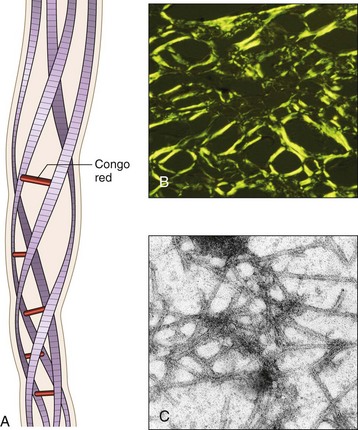
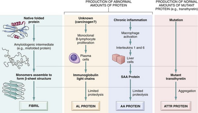
Pathogenesis of Amyloid Deposition
Amyloidosis is fundamentally a disorder of protein misfolding. Amyloid is not a structurally homogeneous protein, although it always has the same morphologic appearance. In fact, more than 20 (at last count, 23) different proteins can aggregate to form fibrils with the appearance of amyloid. Regardless of their derivation, all amyloid deposits are composed of nonbranching fibrils, 7.5 to 10 nm in diameter, each formed of β-sheet polypeptide chains that are wound together (Fig. 4–31). The dye Congo red binds to these fibrils and produces a red–green dichroism (birefringence), which is commonly used to identify amyloid deposits in tissues.
Figure 4–31 Structure of amyloid. A, Schematic diagram of an amyloid fiber showing fibrils (four are shown; as many as six may be present) wound around one another with regularly spaced binding of the Congo red dye. B, Congo red staining shows an apple-green birefringence under polarized light, a diagnostic feature of amyloid. C, Electron micrograph of 7.4- to 10-nm amyloid fibrils.
(Reproduced from Merlini G, Bellotti V: Molecular mechanisms of amyloidosis. N Engl J Med 349:583–596, 2003. Copyright 2003 Massachusetts Medical Society. All rights reserved.)
Amyloidosis results from abnormal folding of proteins, which are deposited as fibrils in extracellular tissues and disrupt normal function. Normally, misfolded proteins are degraded intracellularly in proteasomes, or extracellularly by macrophages. It appears that in amyloidosis, these quality control mechanisms fail, allowing the misfolded protein to accumulate outside cells. Misfolded proteins often are unstable and self-associate, ultimately leading to the formation of oligomers and fibrils that are deposited in tissues. The diverse conditions that are associated with amyloidosis all are likely to result in excessive production of proteins that are prone to misfolding (Fig. 4–32). The proteins that form amyloid fall into two general categories: (1) normal proteins that have an inherent tendency to fold improperly, associate to form fibrils, and do so when they are produced in increased amounts and (2) mutant proteins that are prone to misfolding and subsequent aggregation. Of the many biochemically distinct forms of amyloid proteins that have been identified, three are most common:
• The AL (amyloid light chain) protein is produced by plasma cells and is made up of complete immunoglobulin light chains, the amino-terminal fragments of light chains, or both. For unknown reasons, only a few types of immunoglobulin light chains are prone to forming aggregates. As expected, the deposition of amyloid fibril protein of the AL type is associated with some form of monoclonal B cell proliferation. Defective degradation has also been invoked as the basis for fibril formation, and perhaps particular light chains are resistant to complete proteolysis. However, there are no sequence motifs peculiar to the immunoglobulin light chains found in amyloid deposits.
• The AA (amyloid-associated) fibril is a unique nonimmunoglobulin protein derived from a larger (12-kDa) serum precursor called SAA (serum amyloid-associated) protein that is synthesized in the liver. SAA is synthesized by liver cells under the influence of cytokines such as IL-6 and IL-1 that are produced during inflammation; thus, long-standing inflammation leads to elevated SAA levels, and ultimately the AA form of amyloid deposits. However, increased production of SAA by itself is not sufficient for the deposition of amyloid. Elevation of serum SAA levels is common to inflammatory states but in most instances does not lead to amyloidosis. There are two possible explanations for this. According to one view, SAA normally is degraded to soluble end products by the action of monocyte-derived enzymes. Conceivably, people who develop amyloidosis have an enzyme defect that results in incomplete breakdown of SAA, thus generating insoluble AA molecules. Alternatively, a genetically determined structural abnormality in the SAA molecule itself renders it resistant to degradation by macrophages.
• Aβ amyloid is found in the cerebral lesions of Alzheimer disease. Aβ is a 4-kDa peptide that constitutes the core of cerebral plaques and the amyloid deposits in cerebral blood vessels in this disease. The Aβ protein is derived from a much larger transmembrane glycoprotein called amyloid precursor protein (APP) (Chapter 22).
Figure 4–32 Pathogenesis of amyloidosis. The proposed mechanisms underlying deposition of the major forms of amyloid fibrils.
Several other proteins have been found in amyloid deposits in a variety of clinical settings:
• Transthyretin (TTR) is a normal serum protein that binds and transports thyroxine and retinol, hence the name. Mutations in the gene encoding TTR may alter its structure, making the protein prone to misfolding and aggregation, and resistant to proteolysis. This leads to the formation of aggregates that deposit as amyloid. The resultant diseases are called familial amyloid polyneuropathies. TTR is also deposited in the heart of aged persons (senile systemic amyloidosis); in such cases the protein is structurally normal, but it accumulates at high concentrations. Some cases of familial amyloidosis are associated with deposits of mutant lysozyme.
• β2-Microglobulin, a component of MHC class I molecules and a normal serum protein, has been identified as the amyloid fibril subunit (Aβ2m) in amyloidosis that complicates the course of patients on long-term hemodialysis. Aβ2m fibers are structurally similar to normal β2m protein. This protein is present in high concentrations in the serum of patients with renal disease and is retained in the circulation because it is not efficiently filtered through dialysis membranes. In some series, as many as 60% to 80% of patients on long-term dialysis developed amyloid deposits in the synovium, joints, and tendon sheaths.
• Amyloid deposits derived from diverse precursors such as hormones (procalcitonin) and keratin also have been reported.
Because a given biochemical form of amyloid (e.g., AA) may be associated with amyloid deposition in diverse clinical settings, a combined biochemical and clinical classification is followed for this discussion (Table 4–13). Amyloid may be systemic (generalized), involving several organ systems, or it may be localized, when deposits are limited to a single organ, such as the heart. On clinical grounds, the systemic, or generalized, pattern is subclassified into primary amyloidosis when associated with a monoclonal plasma cell proliferation and secondary amyloidosis when it occurs as a complication of an underlying chronic inflammatory or tissue destructive process. Hereditary or familial amyloidosis constitutes a separate, albeit heterogeneous group, with several distinctive patterns of organ involvement.
Amyloid in this category usually is systemic in distribution and is of the AL type. With approximately 3000 new cases each year in the United States, this is the most common form of amyloidosis. In some of these cases, there is a readily identifiable monoclonal plasma cell proliferation; best defined is the occurrence of systemic amyloidosis in 5% to 15% of patients with multiple myeloma, a plasma cell tumor characterized by multiple osteolytic lesions throughout the skeletal system (Chapter 11). The malignant plasma cells characteristically synthesize abnormal amounts of a single specific immunoglobulin (monoclonal gammopathy), producing an M (myeloma) protein spike on serum electrophoresis. In addition to the synthesis of whole immunoglobulin molecules, plasma cells also may synthesize and secrete either the λ or κ light chain, also known as Bence Jones proteins. By virtue of their small molecular size, these proteins frequently are also excreted in the urine. Almost all patients with myeloma who develop amyloidosis have Bence Jones proteins in the serum or urine, or both. However, amyloidosis develops in only 6% to 15% of patients with myeloma who have free light chains. Clearly, the presence of Bence Jones proteins, although necessary, is by itself not sufficient to produce amyloidosis. Other variables, such as the type of light chain produced and its catabolism, contribute to the “amyloidogenic potential” and influence the deposition of Bence Jones proteins.
The great majority of patients with AL amyloid do not have classic multiple myeloma or any other overt B cell neoplasm; such cases are nevertheless classified as primary amyloidosis because their clinical features derive from the effects of amyloid deposition without any other associated disease. In virtually all such cases, patients have a modest increase in the number of plasma cells in the bone marrow, and monoclonal immunoglobulins or free light chains can be found in the serum or urine. Clearly, these patients have an underlying monoclonal plasma cell proliferation in which production of an abnormal protein, rather than production of tumor masses, is the predominant manifestation.
The amyloid deposits in this pattern are systemic in distribution and are composed of AA protein. This category was previously referred to as secondary amyloidosis, because it is secondary to an associated inflammatory condition. In fact, the feature common to most cases of reactive systemic amyloidosis is chronic inflammation. Classically, tuberculosis, bronchiectasis, and chronic osteomyelitis were the most common causes; with the advent of effective antimicrobial therapies, reactive systemic amyloidosis is seen most frequently in the setting of chronic inflammation caused by autoimmune states (e.g., RA, ankylosing spondylitis, inflammatory bowel disease). Patients with RA are particularly prone to develop amyloidosis, with amyloid deposition seen in as many as 3% of RA cases. Chronic skin infections caused by “skin-popping” of narcotics are also associated with amyloid deposition. Finally, reactive systemic amyloidosis may also occur in association with tumors not derived from immune cells, the two most common being renal cell carcinoma and Hodgkin lymphoma.
A variety of familial forms of amyloidosis have been described; most are rare and occur in limited geographic areas. The best-characterized is an autosomal recessive condition called familial Mediterranean fever. This is a febrile disorder characterized by attacks of fever accompanied by inflammation of serosal surfaces, including peritoneum, pleura, and synovial membrane. This disorder is encountered largely in persons of Armenian, Sephardic Jewish, and Arabic origins. It is associated with widespread tissue involvement indistinguishable from reactive systemic amyloidosis. The amyloid fibril proteins are made up of AA proteins, suggesting that this form of amyloidosis is related to the recurrent bouts of inflammation that characterize this disease. The gene for familial Mediterranean fever is called pyrin and encodes a protein that is a component of the inflammasome (Chapter 2). Patients have gain-of-function mutations in pyrin that result in constitutive overproduction of the pro-inflammatory cytokine IL-1 and persistent inflammation.
In contrast with familial Mediterranean fever, a group of autosomal dominant familial disorders is characterized by deposition of amyloid predominantly in the peripheral and autonomic nerves. These familial amyloidotic polyneuropathies have been described in kindreds in different parts of the world—for example, in Portugal, Japan, Sweden, and the United States. As mentioned previously, the fibrils in these familial polyneuropathies are made up of mutant forms of transthyretin (ATTRs).
Sometimes deposition of amyloid is limited to a single organ or tissue without involvement of any other site in the body. The deposits may produce grossly detectable nodular masses or be evident only on microscopic examination. Nodular (tumor-forming) deposits of amyloid are most often encountered in the lung, larynx, skin, urinary bladder, tongue, and the region about the eye. Frequently, there are infiltrates of lymphocytes and plasma cells in the periphery of these amyloid masses, raising the question of whether the mononuclear infiltrate is a response to the deposition of amyloid or instead is responsible for it. At least in some cases, the amyloid consists of AL protein and may therefore represent a localized form of plasma cell–derived amyloid.
Microscopic deposits of localized amyloid may be found in certain endocrine tumors, such as medullary carcinoma of the thyroid gland, islet tumors of the pancreas, pheochromocytomas, and undifferentiated carcinomas of the stomach, as well as in the islets of Langerhans in patients with type 2 diabetes mellitus. In these settings, the amyloidogenic proteins seem to be derived either from polypeptide hormones (medullary carcinoma) or from unique proteins (e.g., islet amyloid polypeptide).
Several well-documented forms of amyloid deposition occur with aging. Senile systemic amyloidosis refers to the systemic deposition of amyloid in elderly persons (usually in their 70s and 80s). Because of the dominant involvement and related dysfunction of the heart (typically manifesting as a restrictive cardiomyopathy and arrhythmias), this form also is called senile cardiac amyloidosis. The amyloid in this form is composed of normal transthyretin. In addition, another form typically affecting only the heart results from the deposition of a mutant form of TTR. Approximately 4% of the black population in the United States are carriers of the mutant allele, and cardiomyopathy has been identified in both homozygous and heterozygous patients.
Morphology
There are no consistent or distinctive patterns of organ or tissue distribution of amyloid deposits in any of the categories cited. Nonetheless, a few generalizations can be made. In amyloidosis secondary to chronic inflammatory disorders, kidneys, liver, spleen, lymph nodes, adrenals, and thyroid, as well as many other tissues, typically are affected. Although primary (AL) amyloidosis cannot reliably be distinguished from the secondary form by its organ distribution, it more often involves the heart, gastrointestinal tract, respiratory tract, peripheral nerves, skin, and tongue. However, the same organs affected by reactive systemic amyloidosis (secondary amyloidosis), including kidneys, liver, and spleen, also may contain deposits in the immunocyte-associated form of the disease. The localization of amyloid deposits in the hereditary syndromes is varied. In familial Mediterranean fever, the amyloidosis may be widespread, involving the kidneys, blood vessels, spleen, respiratory tract, and (rarely) liver. The localization of amyloid in the remaining hereditary syndromes can be inferred from the designation of these entities.
Whatever the clinical disorder, the amyloidosis may or may not be apparent grossly. Often small amounts are not recognized until the surface of the cut organ is painted with iodine and sulfuric acid. This yields mahogany brown staining of the amyloid deposits. When amyloid accumulates in larger amounts, the organ frequently is enlarged and the tissue typically appears gray with a waxy, firm consistency. On histologic examination, the amyloid deposition is always extracellular and begins between cells, often closely adjacent to basement membranes. As the amyloid accumulates, it encroaches on the cells, in time surrounding and destroying them. In the AL form, perivascular and vascular localizations are common.
The histologic diagnosis of amyloid is based almost entirely on its staining characteristics. The most commonly used staining technique uses the dye Congo red, which under ordinary light imparts a pink or red color to amyloid deposits. Under polarized light the Congo red–stained amyloid shows so-called apple-green birefringence (Fig. 4–33). This reaction is shared by all forms of amyloid and is caused by the crossed β-pleated configuration of amyloid fibrils. Confirmation can be obtained by electron microscopy, which reveals amorphous nonoriented thin fibrils. AA, AL, and ATTR types of amyloid also can be distinguished from one another by specific immunohistochemical staining.
Figure 4–33 Amyloidosis: hepatic involvement. A, Staining of a section of the liver with Congo red reveals pink-red deposits of amyloid in the walls of blood vessels and along sinusoids. B, Note the yellow-green birefringence of the deposits when observed under the polarizing microscope.
(Courtesy of Dr. Trace Worrell and Sandy Hinton, Department of Pathology, University of Texas Southwestern Medical School, Dallas, Texas.)
Because the pattern of organ involvement in different clinical forms of amyloidosis is variable, each of the major organ involvements is described separately.
Amyloidosis of the kidney is the most common and most serious feature of the disease. Grossly, the kidney may appear unchanged, or it may be abnormally large, pale, gray, and firm; in long-standing cases, the kidney may be reduced in size. Microscopically, the amyloid deposits are found principally in the glomeruli, but they also are present in the interstitial peritubular tissue as well as in the walls of the blood vessels. The glomerulus first develops focal deposits within the mesangial matrix and diffuse or nodular thickenings of the basement membranes of the capillary loops. With progression, the deposition encroaches on the capillary lumina and eventually leads to total obliteration of the vascular tuft (Fig. 4–34, A). The interstitial peritubular deposits frequently are associated with the appearance of amorphous pink casts within the tubular lumens, presumably of a proteinaceous nature. Amyloid deposits may develop in the walls of blood vessels of all sizes, often causing marked vascular narrowing.
Amyloidosis of the spleen often causes moderate or even marked enlargement (200 to 800 gm). For obscure reasons, either of two patterns may develop. The deposits may be virtually limited to the splenic follicles, producing tapioca-like granules on gross examination (“sago spleen”), or the amyloidosis may principally involve the splenic sinuses, eventually extending to the splenic pulp, with formation of large, sheetlike deposits (“lardaceous spleen”). In both patterns, the spleen is firm in consistency. The presence of blood in splenic sinuses usually imparts a reddish color to the waxy, friable deposits.
Amyloidosis of the liver may cause massive enlargement (as much as 9000 gm). In such advanced cases, the liver is extremely pale, grayish, and waxy on both the external surface and the cut section. Histologic analysis shows that amyloid deposits first appear in the space of Disse and then progressively enlarge to encroach on the adjacent hepatic parenchyma and sinusoids (Fig. 4–33). The trapped liver cells undergo compression atrophy and are eventually replaced by sheets of amyloid; remarkably, normal liver function may be preserved even in the setting of severe involvement.
Amyloidosis of the heart may occur either as isolated organ involvement or as part of a systemic distribution. When accompanied by systemic involvement, it is usually of the AL form. The isolated form (senile amyloidosis) usually is confined to older persons. The deposits may not be evident on gross examination, or they may cause minimal to moderate cardiac enlargement. The most characteristic gross findings are gray-pink, dewdrop-like subendocardial elevations, particularly evident in the atrial chambers. On histologic examination, deposits typically are found throughout the myocardium, beginning between myocardial fibers and eventually causing their pressure atrophy (Fig. 4–34, B).
Amyloidosis of other organs generally is encountered in systemic disease. The adrenals, thyroid, and pituitary are common sites of involvement. In such cases as well, the amyloid deposition begins in relation to stromal and endothelial cells and progressively encroaches on the parenchymal cells. Surprisingly large amounts of amyloid may be present in any of these endocrine glands without apparent disturbance of function. In the gastrointestinal tract, a relatively favored site for deposition, amyloid may be found at all levels, sometimes producing tumorous masses that must be distinguished from neoplasms. Nodular depositions in the tongue may produce macroglossia. On the basis of the frequent involvement of the gastrointestinal tract in systemic cases, gingival, intestinal, and rectal biopsies serve in the diagnosis of suspected cases. Deposition of β2-microglobulin amyloid in patients receiving long-term dialysis occurs most commonly in the carpal ligaments of the wrist, resulting in compression of the median nerve (leading to carpal tunnel syndrome).
Amyloidosis may be an unsuspected finding at autopsy in a patient who has no apparent related clinical manifestations, or it may be responsible for serious clinical dysfunction and even death. The clinical course depends on the particular sites or organs affected and the severity of the involvement. Nonspecific complaints such as weakness, fatigue, and weight loss are the most common presenting manifestations. Later in the course, amyloidosis tends to manifest in one of several ways: by renal disease, hepatomegaly, splenomegaly, or cardiac abnormalities. Renal involvement giving rise to severe proteinuria (nephrotic syndrome) (Chapter 13) often is the major cause of symptoms in reactive systemic amyloidosis. Progression of the renal disease may lead to renal failure, which is an important cause of death in amyloidosis. The hepatosplenomegaly rarely causes significant clinical dysfunction, but it may be the presenting finding. Cardiac amyloidosis may manifest as conduction disturbances or as restrictive cardiomyopathy (Chapter 10). Cardiac arrhythmias are an important cause of death in cardiac amyloidosis. In one large series, 40% of the patients with AL amyloid died of cardiac disease.
The diagnosis of amyloidosis may be suspected from the clinical signs and symptoms and from some of the findings mentioned; however, more specific tests must often be done for definitive diagnosis. Biopsy and subsequent Congo red staining is the most important tool in the diagnosis of amyloidosis. In general, biopsy is taken from the organ suspected to be involved. For example, renal biopsy is useful in the presence of urinary abnormalities. Rectal and gingival biopsy specimens contain amyloid in as many as 75% of cases with generalized amyloidosis. Examination of abdominal fat aspirates stained with Congo red is a simple, low-risk method. In suspected cases of AL amyloidosis, serum and urinary protein electrophoresis and immunoelectrophoresis should be performed. Bone marrow examination in such cases usually shows plasmacytosis, even if skeletal lesions of multiple myeloma are not present. Proteomic analysis of affected tissue is now being widely used for detection of small amounts of amyloid (from fat aspirates) and for definitive identification of the type of amyloid.
The outlook for patients with generalized amyloidosis is poor, with the mean survival time after diagnosis ranging from 1 to 3 years. In AA amyloidosis, the prognosis depends to some extent on the control of the underlying condition. Patients with myeloma-associated amyloidosis have a poorer prognosis, although they may respond to cytotoxic drugs used to treat the underlying disorder. Resorption of amyloid after treatment of the associated condition has been reported, but this is a rare occurrence.
Summary
• Amyloidosis is a disorder characterized by the extracellular deposits of misfolded proteins that aggregate to form insoluble fibrils.
• The deposition of these proteins may result from excessive production of proteins that are prone to misfolding and aggregation; mutations that produce proteins that cannot fold properly and tend to aggregate; or defective or incomplete proteolytic degradation of extracellular proteins.
• Amyloidosis may be localized or systemic. It is seen in association with a variety of primary disorders, including monoclonal plasma cell proliferations (in which the amyloid deposits consist of immunoglobulin light chains); chronic inflammatory diseases such as RA (deposits of amyloid A protein, derived from an acute-phase protein produced in inflammation); Alzheimer disease (amyloid B protein); familial conditions in which the amyloid deposits consist of mutants of normal proteins (e.g., transthyretin in familial amyloid polyneuropathies); amyloidosis associated with dialysis (deposits of β2-microglobulin, whose clearance is defective).
• Amyloid deposits cause tissue injury and impair normal function by causing pressure on cells and tissues. They do not evoke an inflammatory response.
Banchereau J, Pascual V. Type I interferon in systemic lupus erythematosus and other autoimmune diseases. Immunity. 2006;25:383. [A review of the recently discovered role of interferons in SLE and other autoimmune diseases, and the potential for targeting this family of cytokines for therapy.]
Campbell DJ, Koch MA. Phenotypic and functional specialization of FoxP3+ regulatory T cells. Nat Rev Immunol. 2011;11:119. [A current review of the properties and functions of regulatory T cells.]
Chervonsky A. Influence of microbial environment on autoimmunity. Nat Immunol. 2010;11:28. [A summary of the role of microbes and other environmental factors in the development of autoimmunity.]
Cunningham-Rundles C, Ponda PP. Molecular defects in T- and B-cell primary immunodeficiency diseases. Nat Rev Immunol. 2006;5:880. [Excellent, up-to-date review of primary immunodeficiencies.]
Davidson A, Diamond B. Autoimmune diseases. N Engl J Med. 2001;345:340. [A readable overview of the etiology, pathogenesis, and therapy for autoimmune diseases.]
Douek DC, Roederer M, Koup RA. Emerging concepts in the immunopathogenesis of AIDS. Annu Rev Med. 2009;60:471. [A balanced discussion of the pathogenesis of AIDS, and the still unresolved issues.]
Fairhurst AM, Wandstrat AE, Wakeland EK. Systemic lupus erythematosus: multiple immunological phenotypes in a complex genetic disease. Adv Immunol. 2006;92:1. [A comprehensive review of the genetics of SLE in humans and animal models, and the pathogenic mechanisms deduced from identification of susceptibility genes.]
Fischer A. Human primary immunodeficiency diseases. Immunity. 2008;28:835. [An excellent summary of primary immunodeficiencies affecting the innate and adaptive immune systems.]
Galli SJ. The development of allergic inflammation. Nature. 2008;454:445. [An excellent review of the mechanisms of inflammation in allergic diseases.]
Gonzalez-Scarano F, Martin-Garcia J. The neuropathogenesis of AIDS. Nat Rev Immunol. 2005;5:69. [A discussion of the pathogenesis of HIV-associated dementia.]
Goodnow CC. Multistep pathogenesis of autoimmune disease. Cell. 2007;130:25. [An excellent discussion of the checkpoints that prevent autoimmunity and why these might fail.]
Jancar S, Sanchez Crespo M. Immune complex–mediated tissue injury: a multistep paradigm. Trends Immunol. 2005;26:48. [A summary of the mechanisms of immune complex–mediated tissue injury.]
Katsumoto TR, Whitfield ML, Connolly MK. The pathogenesis of systemic sclerosis. Annu Rev Pathol. 2011;6:509. [An excellent review of the pathogenesis of systemic sclerosis, and the many unanswered questions.]
Kay AB. Allergy and allergic diseases. First of two parts. N Engl J Med. 2001;344:30. [Excellent two-part review of the mechanisms and manifestations of immediate hypersensitivity.]
Kyewski B, Klein L. A central role for central tolerance. Annu Rev Immunol. 2006;24:571. [A discussion of the mechanisms of central tolerance, with a focus on T cells.]
Merlini G, Bellotti V. Molecular mechanisms of amyloidosis. N Engl J Med. 2003;349:583. [Good review of the general pathogenic mechanisms in various forms of systemic amyloidosis.]
Mitchell RN. Graft vascular disease: immune response meets the vessel wall. Annu Rev Pathol. 2009;4:19. [A review of the mechanisms that lead to vascular disease in chronic graft rejection.]
Moir S, Chun TW, Fauci AS. Pathogenic mechanisms of HIV disease. Annu Rev Pathol. 2011;6:223. [A discussion of current concepts of the mechanisms by which HIV causes immunodeficiency.]
Mueller DL. Mechanisms maintaining peripheral tolerance. Nat Immunol. 2010;11:21. [A discussion of the mechanisms of peripheral tolerance, with an emphasis on T cells.]
Nankivell BJ, Alexander SI. Rejection of the kidney allograft. N Engl J Med. 2010;363:1451. [Good review of the mechanisms of recognition and rejection of allografts and the development of new strategies for treating rejection.]
Notarangelo LD. Primary immunodeficiencies. J Allergy Clin Immunol. 2010;125:S182. [An update on congenital immunodeficiencies and their molecular basis.]
O’Shea JJ, Paul WE. Mechanisms underlying lineage commitment and plasticity of helper CD4+ T cells. Science. 2010;327:1098. [An excellent review of the development and functions of helper T cell subsets, and the uncertainties in the field.]
Palmer MT, Weaver CT. Autoimmunity: increasing suspects in the CD4+ T cell lineage. Nat Immunol. 2010;11:36. [A thoughtful discussion of the central role of CD4+ T cells in the pathogenesis of autoimmune diseases.]
Pepys MB. Amyloidosis. Annu Rev Med. 2006;57:223. [An excellent review of the pathogenesis, clinical features and therapeutic approaches in amyloidosis.]
Rahman A, Isenberg DA. Systemic lupus erythematosus. New Engl J Med. 2008;358:929. [An excellent review of the pathogenesis and genetics of SLE.]
Sakaguchi S, Miyara M, Costantino CM, Hafler DA. FOXP3+ regulatory T cells in the human immune system. Nat Rev Immunol. 2010;10:490. [An excellent discussion of the properties and role of regulatory T cells in humans.]
Sakaguchi S, Yamaguchi T, Nomura T, Ono M. Regulatory T cells and immune tolerance. Cell. 2008;133:775. [An excellent review of the properties and functions of regulatory T cells.]
Stone J, Zen Y, Deshpande V. IgG4-related disease. New Engl J Med. 2011. [A comprehensive discussion of the features and likely pathogenesis of this recently recognized entity.] in press
Vercelli D. Discovering susceptibility genes for asthma and allergy. Nat Rev Immunol. 2008;8:169. [A summary of the known susceptibility genes for asthma and other allergic diseases.]
Voulgarelis M, Tzioufas AG. Pathogenetic mechanisms in the initiation and perpetuation of Sjögren’s syndrome. Nat Rev Rheumatol. 2010;6:529. [A good discussion of what is known and not known about the pathogenesis of Sjögren’s syndrome.]
Zenewicz L, Abraham C, Flavell RA, Cho J. Unraveling the genetics of autoimmunity. Cell. 2010;140:791. [An update on susceptibility genes for autoimmune diseases, how these are identified, and their significance.]