THE CELLULAR ENVIRONMENT
FLUIDS AND ELECTROLYTES, ACIDS AND BASES


The cells of the body live in a fluid environment that requires an electrolyte concentration and pH value (measure of the acidity or alkalinity of a solution) that are regulated within a very narrow range. A balance is maintained by an integration of renal, hormonal, and neural functions. Changes in the composition of electrolytes affect electrical potentials of excitatory cells and cause shifts of fluid from one compartment to another. Alterations in pH disrupt the cellular function of enzyme systems. Fluid fluctuations affect blood volume and cellular function. Disturbances in these functions are common and can be life threatening. Understanding how alterations occur and the body’s ability to compensate or correct the disturbance is important to understanding many pathophysiologic conditions.
DISTRIBUTION OF BODY FLUIDS
The fluids of the body are distributed among functional compartments, or spaces, and provide a transport medium for cellular and tissue function. Water moves freely among body compartments and is distributed by osmotic and hydrostatic forces. Two thirds of the body’s water is intracellular fluid (ICF) and one third is in the extracellular fluid (ECF) compartments. The two main ECF compartments are the interstitial fluid and the intravascular fluid, which is the blood plasma. Other ECF compartments include the lymph and the transcellular fluids, such as the synovial, intestinal, biliary, hepatic, pancreatic, and cerebrospinal fluids; sweat; urine; and pleural, synovial, peritoneal, pericardial, and intraocular fluids.
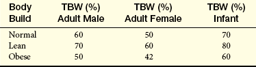
The sum of fluids within all compartments constitutes the total body water (TBW) (Table 3-1). The volume of TBW is usually expressed as a percentage of body weight in kilograms. The standard value for TBW is 60% of the weight of a 70-kg adult male, which is equivalent to 42 L of fluid (Table 3-2). The rest of the body weight is made up of fat and fat-free solids, particularly bone.
Table 3-1
| Percentage of Body Weight | Volume (L) | |
| Intracellular fluid (ICF) | 40 | 28 |
| Extracellular fluid (ECF) | 20 | 14 |
| Interstitial | (15) | (11) |
| Intravascular | (5) | (3) |
| Total body water (TBW) | 60 | 42 |
Table 3-2
Total Body Water in Relation to Body Weight
NOTE: TBW (total body water) is a percentage of body weight.
Although the amount of fluid within the various compartments is relatively constant, exchange of solutes and water occurs between compartments to maintain their unique compositions. The percentage of TBW varies with the amount of body fat and age. Because fat is water repelling (hydrophobic), very little water is contained in adipose cells. Individuals with more body fat have proportionately less TBW and tend to be more susceptible to fluid imbalances that cause dehydration.
Aging and Distribution of Body Fluids
The distribution and amount of TBW change with age (see Table 3-2). In newborn infants, TBW is about 75% to 80% of body weight because infants store less fat. The percentage of TBW decreases to about 67% of body weight during the first year of life. In the immediate postnatal period, a physiologic loss of body water occurs, which amounts to 5% of body weight, as the infant adjusts to a new environment. Infants are particularly susceptible to significant changes in TBW because of their high metabolic rate and the accelerated turnover of body fluids caused by their greater body surface area in proportion to total body size. Loss of fluids from diarrhea can represent a significant proportion of body weight. Renal mechanisms that regulate fluid and electrolyte conservation may not be mature enough to counter the losses, so dehydration may develop rapidly.
During childhood TBW slowly decreases to 60% to 65% of body weight. At adolescence the percentage of TBW approaches adult proportions, and gender differences begin to appear. Males eventually have a greater percentage of body water as a function of increasing muscle mass. Females have more body fat and less muscle as a function of estrogens and therefore have less body water.
With increasing age the percentage of TBW declines further still. The decrease is caused in part by an increased amount of fat and a decreased amount of muscle and by a reduced ability to regulate sodium and water balance. With older age the kidney becomes less efficient in producing concentrated urine, and the responses for conserving sodium become sluggish. Thirst perception may be impaired. The normal reduction of TBW in older adults becomes clinically important when the body is under stress, such as development of fever or dehydration from any cause; loss of body fluids at such times can be severe and life threatening.1
Although daily fluid intake may fluctuate widely, the body regulates water volume within a relatively narrow range. The primary sources of body water are drinking, ingestion of water in food, and water derived from oxidative metabolism. Normally, the largest amounts of water are lost through renal excretion. Lesser amounts are eliminated through the stool and through vaporization from the skin and lungs (insensible water loss) (Table 3-3).
Water Movement Between ICF and ECF
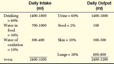
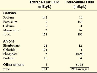
The movement of water between ICF and ECF compartments is primarily a function of osmotic forces. (Osmosis and other mechanisms of passive transport are discussed in Chapter 1.) Water moves freely by diffusion through the lipid bilayer cell membrane and through aquaporins, a family of water channel proteins that provide permeability to water.2 The osmolality of TBW is normally at equilibrium. Sodium is the most abundant ECF ion and is responsible for the osmotic balance of the ECF space. Potassium maintains the osmotic balance of the ICF space. The osmotic force of ICF proteins and other nondiffusible substances is balanced by the active transport of ions out of the cell. Normally the ICF is not subject to rapid changes in osmolality, but when there are changes in ECF osmolality, a net transfer of water from one compartment to another occurs until osmotic equilibrium is reestablished. Figure 3-1 shows a model of the maintenance of osmotic equilibrium between the ICF and ECF.
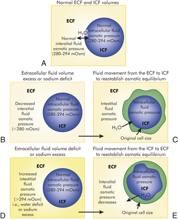
Figure 3-1 Examples of changes in osmotic equilibrium between ECF and ICF. A, Normal ECF and ICF volumes. Intracellular and extracellular fluid osmotic pressure are equal and water is equally distributed between the compartments. B, Extracellular fluid volume excess or sodium deficit. ECF volume excess or sodium deficit decreases the ECF osmotic pressure, and water is attracted to the ICF space (see C). C, Fluid movement from the ECF to ICF to reestablish osmotic equilibrium. The intracellular osmotic pressure attracts water from the ECF, causing an increase in ICF water volume with a balancing of osmotic forces between the ECF and ICF. The consequence is an increase in ICF volume and cell swelling. D, Extracellular fluid volume deficit or sodium excess. ECF volume deficit increases the ECF osmotic pressure, and intracellular water is attracted to the ECF space (see E). E, Fluid movement from the ICF to the ECF to reestablish osmotic equilibrium. Water from the intracellular space has moved to the extracellular space until the osmotic forces are equal. The consequence is a decrease in ICF water volume and cell size. ECF, Extracellular fluid; ICF, intracellular fluid.
Water Movement Between Plasma and Interstitial Fluid
The distribution of water and the movement of nutrients and waste products among the capillary, plasma, and interstitial spaces occur as a result of changes in hydrostatic pressure and osmotic forces at the arterial and venous ends of the capillary. Because water, sodium, and glucose readily move across the capillary membrane, the plasma proteins (particularly albumin) maintain the effective osmolality (concentration of solutes per kilogram of solution) by generating plasma oncotic pressure. Osmotic forces within the capillary are balanced by the hydrostatic pressure, which arises from cardiac contraction. The movement of fluid back and forth across the capillary wall is called net filtration and is best described by the Starling hypothesis:

The forces favoring filtration, or movement of water out of the capillary and into the interstitial space, include the capillary hydrostatic pressure and the interstitial oncotic pressure. The forces opposing filtration are the plasma oncotic pressure (pressure of plasma proteins) and the interstitial hydrostatic pressure. Normally the interstitial forces are negligible because only a very small percentage of plasma proteins crosses the capillary membrane and interstitial fluid moves into cells or is drawn back into the plasma. Thus the major forces for filtration are within the capillary.
As the plasma flows from the arterial to the venous end of the capillary, the force of hydrostatic pressure facilitates the movement of water across the capillary membrane. Oncotic pressure remains fairly constant because plasma proteins normally do not cross the capillary membrane. At the arterial end of the capillary, hydrostatic pressure is greater than capillary oncotic pressure and water filters into the interstitial space. Because of oncotic forces, some water moves back into the capillary, but the net effect is loss of water from the capillary. The movement of water from the plasma decreases the hydrostatic pressure within the capillary. Thus at the venous end of the capillary, oncotic pressure exceeds hydrostatic pressure. Fluids then are attracted back into the circulation, balancing the movement of fluids between the plasma and the interstitial space. The overall effect is filtration at the arterial end and reabsorption at the venous end (Figure 3-2). Interstitial hydrostatic pressure promotes the movement of about 10% of the interstitial fluid along with small amounts of protein into the lymphatics, which then returns to the circulation.
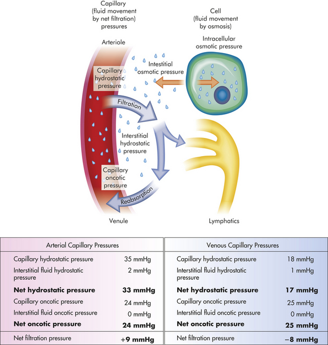
Figure 3-2 Capillary filtration forces. Water, electrolytes, and small molecules exchange freely between the vascular compartment and the interstitial space at the site of capillaries and small venules. The rate and amount of exchange are driven by the physical forces of hydrostatic and oncotic pressures and the permeability and surface area of the capillary membranes. The two opposing hydrostatic pressures are capillary hydrostatic pressure and interstitial hydrostatic pressure. The two opposing oncotic pressures are capillary oncotic pressure and interstitial oncotic pressure. The forces that favor filtration from the capillary are capillary hydrostatic pressure and interstitial oncotic pressure, and the forces that oppose filtration are capillary oncotic pressure and interstitial hydrostatic pressure. The sum of their effects is known as net filtration pressure (NFP). In the example of normal exchange above, a small amount of fluid moves to the lymph vessels, which accounts for the net filtration difference between the arterial and venous ends of the capillary.
An important factor in capillary filtration of fluid is the integrity of the capillary membrane. Changes in membrane permeability may permit the escape of plasma proteins into the interstitial space. The normal relationship defined by the Starling hypothesis is altered with the osmotic movement of water into the interstitial space, causing tissue edema.
ALTERATIONS IN WATER MOVEMENT
Edema is the excessive accumulation of fluid within the interstitial spaces. It is a problem of fluid distribution and does not necessarily indicate a fluid excess. In some conditions, sequestered fluids can cause both edema and dehydration. The pathophysiologic process is related to an increase in the forces favoring fluid filtration from the capillaries or lymphatic channels into the tissues. The four most common mechanisms are increased capillary hydrostatic pressure, decreased plasma oncotic pressure, increased capillary membrane permeability, and lymphatic obstruction (Figure 3-3).
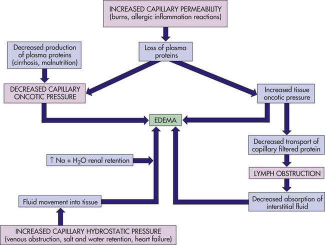
PATHOPHYSIOLOGY An increase in hydrostatic pressure can result from venous obstruction or salt and water retention. Venous obstruction can increase the hydrostatic pressure of fluid within the capillaries enough to cause fluid to escape into the interstitial spaces. Thrombophlebitis, hepatic obstruction, tight clothing around the extremities, and prolonged standing are common causes of venous obstruction. Congestive heart failure, renal failure, and cirrhosis of the liver are conditions associated with excessive salt and water retention, which in turn cause volume overload, increased venous pressure, and edema.
Losses or diminished production of plasma albumin contributes to a decrease in plasma oncotic pressure. Decreased oncotic attraction of fluid within the capillary causes fluid to move into the interstitial space. Decreased production of plasma protein and decreased oncotic pressure may occur with liver disease or protein malnutrition. Losses of plasma proteins occur with glomerular diseases of the kidney (nephrotic syndrome), serous drainage from open wounds, hemorrhage, burns, and cirrhosis of the liver.
Increases in capillary permeability are usually associated with inflammation and the immune response. (Immunity is discussed in Chapters 6, 7 and 8; inflammation is discussed in Chapters 6 and 8.) These responses are often the result of trauma such as burns or crushing injuries, neoplastic disease, and allergic reactions. Proteins escape from the plasma and produce edema through a loss of capillary oncotic pressure and a gain in interstitial fluid proteins.
The lymphatic system normally absorbs interstitial fluid and the small amount of proteins that normally pass across the capillary membrane. When the lymphatic channels are blocked (because of infection or tumor) or are surgically removed, proteins and fluid accumulate in the interstitial space, causing lymphedema. For example, lymphedema of the arm or leg will occur after surgical removal of axillary and femoral lymph nodes for treatment of carcinoma.3
CLINICAL MANIFESTATIONS Edema may be localized or generalized. Some localized edema is limited to the site of trauma, as in a sprained finger or within particular organ systems. This includes cerebral edema, pulmonary edema, pleural effusion, pericardial effusion, and ascites (accumulation of fluid in the peritoneal space). Dependent edema, in which fluid accumulates in gravity-dependent areas of the body, might be a sign of more generalized edema. Dependent edema might appear in the feet and legs when standing and in the sacral area and buttocks when supine. Dependent edema can be identified by using the fingers to press away edematous fluid in tissues overlying bony prominences. A pit will be left in the skin; hence the term pitting edema.
Edema is usually associated with weight gain, swelling and puffiness, tight-fitting clothes and shoes, limited movement of the affected area, and symptoms associated with the underlying pathologic condition. The accumulation of fluid increases the distance required for nutrients, oxygen, and wastes to move between capillaries and tissues. Increased tissue pressure may diminish capillary blood flow. Therefore, wounds heal more slowly and the risks of infection and formation of pressure sores increase. Edema of specific organs, such as the brain, lung, or larynx, can be life threatening.
Although the accumulation of fluid is excessive, it is trapped in a “third space” (i.e., the interstitial space, pleural space, pericardial space) and is not available for metabolic processes or perfusion. Therefore, a state of dehydration can develop as a result of the sequestering of the edematous fluid. An example of such sequestration occurs with severe burns, in which large amounts of vascular fluid are lost to the interstitial spaces, reducing plasma volume and causing shock (see Chapter 46).
EVALUATION AND TREATMENT Specific conditions causing edema require diagnosis. Edema may be treated symptomatically until the underlying disorder is corrected. Supportive measures include elevating edematous limbs, using compression stockings, avoiding prolonged standing, restricting salt intake, and taking diuretics.
SODIUM, CHLORIDE, AND WATER BALANCE
The kidneys and hormones have a central role in maintaining sodium and water balance. Because water follows the osmotic gradients established by changes in salt concentration, sodium balance and water balance are intimately related. Sodium is regulated by the renal effects of aldosterone from the adrenal cortex and natriuretic peptides Water balance is primarily regulated by antidiuretic hormone (ADH; also known as arginine-vasopressin) from the posterior pituitary.
Sodium and Chloride Balance
Sodium accounts for 90% of the ECF cations (positively charged ions). (The distribution of electrolytes in body compartments is summarized in Table 3-4.) As the most abundant ECF cation, along with its constituent anions (negatively charged ions) chloride and bicarbonate, sodium regulates extracellular osmotic forces and therefore regulates water balance. Sodium has many important body functions, including regulation of osmolality (interstitial and intravascular fluid volume), working with potassium and calcium to maintain neuromuscular irritability for conduction of nerve impulses, regulation of acid-base balance (through sodium bicarbonate and sodium phosphate), participation in cellular chemical reactions, and membrane transport (see Chapter 1).
The concentration of sodium is maintained within a narrow range (136 to 145 mEq/L), primarily by the kidney in conjunction with neural and hormonal mediators. The average dietary intake of sodium ranges from 5 to 6 g/day; the minimal daily requirement of sodium is 500 mg. Sweating depletes sodium and water volume and increases the body’s sodium requirement.
The kidney regulates sodium balance primarily through renal tubular reabsorption. Under normal rates of sodium intake, the tubules of the kidney function to reabsorb sodium. With an excess or deficit of sodium in relation to water, a combination of hormonal, neural, and renal mechanisms acts synergistically to control sodium balance.
The hormonal regulation of sodium balance is mediated by aldosterone, a mineralocorticoid (steroid) synthesized and secreted from the adrenal cortex (see Chapter 20). Aldosterone secretion is influenced by both plasma concentrations of sodium (Na+) and potassium (K+) and circulating blood volume (i.e., aldosterone is secreted when sodium levels are depressed, potassium levels are increased, or renal perfusion is decreased). Aldosterone increases the reabsorption of sodium and secretion of potassium by the distal tubule of the kidney. As a result, sodium concentration of the ECF is enhanced and potassium is excreted with the urine.
When circulating blood volume or blood pressure is reduced, renin, an enzyme secreted by the juxtaglomerular cells of the kidney, is released in response to sympathetic nerve stimulation and decreased perfusion of the renal vasculature. Renin stimulates the formation of angiotensin I, an inactive polypeptide, which is then converted into angiotensin II. Angiotensin II has two major functions: it stimulates the secretion of aldosterone, and it causes vasoconstriction. The aldosterone then promotes sodium and water reabsorption, conserving blood volume. The vasoconstriction elevates the systemic blood pressure and restores renal perfusion. The restoration of sodium levels, fluid volume, and renal perfusion then inhibits further release of renin. This sodium and water regulation mechanism is known as the renin-angiotensin-aldosterone system (see Chapter 35).
Natriuretic peptides are hormones that include atrial natriuretic peptide (ANP) produced by the myocardial atria, brain natriuretic peptide (BNP) produced by the myocardial ventricles, and urodilatin within the kidney. Natriuretic peptides decrease blood pressure and increase sodium and water excretion. They are natural antagonists to the renin-angiotensin-aldosterone system. ANP and BNP are released when there is an increase in transmural atrial pressure (increased volume) as may occur with congestive heart failure.4 Natriuretic peptides are sometimes called a “third factor” in sodium regulation. (Increased glomerular filtration rate is thus the first factor and aldosterone the second factor.)
Chloride is the major anion in the extracellular fluid. It provides electroneutrality, particularly in relation to sodium. The transport of chloride is generally passive and follows the active transport of sodium so that increases or decreases in chloride are proportional to changes in sodium. Because bicarbonate is the other major anion in the ECF, the concentration of chloride tends to vary inversely with changes in bicarbonate concentration.
Water Balance
Water balance is maintained by balancing the amount of water excreted with water intake by ingestion and generated by metabolism. Secretion of ADH and perception of thirst are primary factors in the regulation of water balance. Thirst is a sensation that stimulates water-drinking behavior. Thirst is experienced when water loss equals 2% of an individual’s body weight or when there is an increase in osmolality. Dry mouth, hyperosmolality, and plasma volume depletion activate osmoreceptors (neurons located in the hypothalamus that are stimulated by increased osmolality). The action of the osmoreceptors then causes thirst. Drinking water restores plasma volume and dilutes the ECF osmolality.
The secretion of ADH is initiated by an increase in plasma osmolality or a decrease in circulating blood volume and a lowered blood pressure. An increase in plasma osmolality occurs with a deficit of water or an excess of sodium in relation to water. The increased osmolality results in decreased extracellular and interstitial fluid volume and stimulates hypothalamic osmoreceptors. In addition to causing thirst, the stimulated osmoreceptors increase the release of ADH. The action of ADH is to increase the permeability of renal tubular cells to water, and water is then reabsorbed into the plasma from the distal tubules and collecting ducts of the kidney. Urine concentration increases, and the reabsorbed water decreases plasma osmolality, returning it toward normal. Like most hormones, ADH is regulated by a feedback mechanism (Figure 3-4).
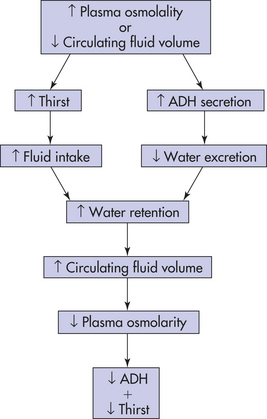
With volume depletion, such as dehydration from vomiting, diarrhea, or excessive sweating, volume-sensitive receptors and baroreceptors (stretch receptors that are sensitive to changes in volume and pressure) stimulate release of ADH. The volume receptors are located in the right and left atria and thoracic vessels; baroreceptors are in the aorta, pulmonary arteries, and carotid sinus. Secretion of ADH is caused by a decrease in atrial pressure, as occurs with decreased blood volume. The reabsorption of water mediated by ADH then promotes the restoration of plasma volume. When there is an increase in the blood volume returning to the heart from the venous system ADH secretion is inhibited and fluid volume decreases.
ALTERATIONS IN SODIUM, CHLORIDE, AND WATER BALANCE
Alterations in sodium and water balance are closely related. Water imbalances may develop because of changes in osmotic gradients caused by gain or loss of salt. Likewise, sodium imbalances occur with alterations in body water volume (see Figure 3-1). Generally the alterations can be classified as changes in tonicity, or the change in concentration of electrolytes in relation to water (see Chapter 1). Alterations can therefore be classified as isotonic, hypertonic, or hypotonic (Table 3-5).
Table 3-5
| Tonicity | Mechanism |
| Isotonic (isoosmolar) imbalance | Gain or loss of extracellular fluid (ECF) resulting in a concentration equivalent to a 0.9% sodium chloride (salt) solution (normal saline); no shrinking or swelling of cells |
| Hypertonic (hyperosmolar) imbalance | Imbalances that result in an ECF concentration >0.9% salt solution; i.e., water loss or solute gain; cells shrink in a hypertonic fluid |
| Hypotonic (hypoosmolar) imbalance | Imbalance that results in an ECF <0.9% salt solution; i.e., water gain or solute loss; cells swell in a hypotonic fluid |
Isotonic Alterations
Isotonic alterations occur when changes in TBW are accompanied by proportional changes in electrolytes and water. For example, if an individual loses pure plasma or ECF, fluid volume is depleted but the number and type of electrolytes (i.e., sodium) and the osmolality remain within a normal range. Excessive amounts of isotonic body fluids can result from excessive administration of intravenous normal saline or oversecretion of aldosterone with renal retention of both sodium and water. Losses of isotonic body fluids include hemorrhage, severe wound drainage, excessive diaphoresis (sweating), intestinal losses, and decreased fluid intake.
Isotonic volume depletion causes contraction of the ECF volume with resulting weight loss, dryness of skin and mucous membranes, decreased urine output, and symptoms of hypovolemia. Indicators of hypovolemia include a rapid heart rate, flattened neck veins, and normal or decreased blood pressure. In severe states, hypovolemic shock can occur (see Chapter 46).
Isotonic volume excesses result from excessive administration of intravenous fluids, hypersecretion of aldosterone, the effects of drugs such as cortisone, or renal failure. As the plasma volume expands, symptoms of hypervolemia develop. Weight gain and a decrease in hematocrit and plasma protein concentration caused by the diluting effect of excess plasma volume will occur. The neck veins may distend, and the blood pressure increases. Increased capillary hydrostatic pressure leads to edema formation. If the plasma volume is great enough, pulmonary edema and heart failure develop.
Hypertonic Alterations
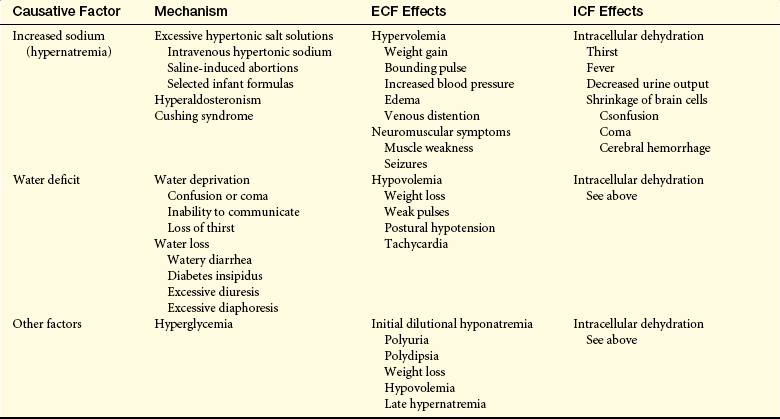
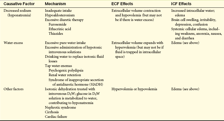
Hypertonic fluid alterations develop when the osmolality of the ECF is elevated above normal. The most common causes are an increased concentration of ECF sodium (hypernatremia) or a deficit of ECF free water. In both instances the hypertonicity of the ECF attracts water from the intracellular space, causing ICF dehydration. A primary increase in ECF sodium causes an osmotic attraction of water and symptoms of hypervolemia. In contrast, a hypertonic state caused primarily by free water loss leads to hypovolemia (Table 3-6).
Hypernatremia
PATHOPHYSIOLOGY Hypernatremia occurs when serum sodium levels exceed 147 mEq/L. Excessive serum sodium may be caused by an acute gain in sodium or a loss of water. Sodium gains cause intracellular dehydration; the movement of water to the ECF may cause hypervolemia. With an accompanying water loss, both ICF dehydration and ECF dehydration occur. Hyperosmolality is a common result of hypernatremia.
High amounts of dietary sodium rarely cause hypernatremia. More commonly, high sodium levels occur because of (1) inadequate free water intake, (2) inappropriate administration of hypertonic saline solution (e.g., as sodium bicarbonate for treatment of acidosis during cardiac arrest), (3) high sodium levels as a result of oversecretion of aldosterone (as in primary hyperaldosteronism), or (4) Cushing syndrome (caused by excess secretion of adrenocorticotropic hormone [ACTH], which also causes increased secretion of aldosterone).5
Increased sodium in relation to water deprivation or water loss is associated with fever or respiratory infections, which increase the respiratory rate and enhance water loss from the lungs. Diabetes insipidus (deficiency of ADH), diabetes mellitus, polyuria, profuse sweating, and diarrhea cause water loss in relation to sodium concentration. Infants with severe diarrhea are particularly vulnerable. Insufficient water intake also can cause hypernatremia, particularly in individuals who are comatose, confused, immobilized, or receiving gastric feedings. Those who cannot communicate because of age (infants) or disease also cannot express thirst and are at risk.
CLINICAL MANIFESTATIONS Water is redistributed to the extracellular space, and intracellular dehydration ensues. Seizures, coma, and pulmonary edema are the most serious symptoms. Thirst, fever, dry mucous membranes, hypotension, tachycardia, low jugular venous pressure, and restlessness are associated with hypernatremia as a result of water loss.
EVALUATION AND TREATMENT The serum sodium level is usually more than 147 mEq/L. If there is water loss, urine specific gravity will be greater than 1.030 and hematocrit and plasma proteins will be elevated. The treatment of hypernatremia is to give an isotonic salt-free fluid (5% dextrose in water) until the serum sodium level returns to normal. Hypervolemia and edema require treatment of the underlying clinical condition.
Hyperchloremia
Hyperchloremia occurs when serum chloride levels exceed the normal range of 97 to 105 mEq/L and is often associated with an excess of sodium (hypernatremia) or a deficit of bicarbonate (metabolic acidosis) (see p. 117) Ingestion of excessive chloride infrequently accompanies the use of an ammonium chloride diuretic. No specific symptoms are associated with chloride excess.
Alterations in chloride levels are usually secondary to their pathophysiologic processes. Treatment therefore generally is related to management of the underlying disorder.
Water Deficit
PATHOPHYSIOLOGY Dehydration describes water deficit, but dehydration is also commonly used to indicate both sodium loss and water loss (isotonic or isoosmolar dehydration). Pure water deficits (hyperosmolar or hypertonic dehydration) are rare because most people have access to water. Individuals who are comatose or paralyzed continue insensible water losses through the skin and lungs with a minimal obligatory formation of urine. Hyperventilation caused by fever also may precipitate water deficit. The most frequent cause of water loss is increased renal clearance of free water as a result of impaired tubular function or inability to concentrate the urine, as with diabetes insipidus (see Chapter 21).
CLINICAL MANIFESTATIONS Marked water deficit is manifested by symptoms of dehydration: headache, thirst, dry skin and mucous membranes, elevated temperature, weight loss, and decreased or concentrated urine (with the exception of diabetes insipidus). Skin turgor may be normal or decreased. Symptoms of hypovolemia, including tachycardia, weak pulses, and postural hypotension, may be present.
EVALUATION AND TREATMENT An elevated hematocrit and serum sodium concentration are associated with moderate water loss in addition to clinical signs and symptoms.
Treatment is to give water and stop fluid loss. Fluid replacement must be given slowly enough to prevent rapid movement of water into brain cells, which causes cerebral edema, seizures, brain injury, and death. When intravenous replacement is required, 5% dextrose in water should be used because pure water lyses red blood cells.
Hypotonic Alterations
Hypotonic fluid imbalances occur when the osmolality of the ECF is less than normal. The most common causes are sodium deficit (hyponatremia) or free water excess (water intoxication). Either of these causes leads to an intracellular overhydration (cellular edema) and cell swelling. When there is a sodium deficit, the osmotic pressure of the ECF decreases and water moves into the cell, where the osmotic pressure is greater (see Figure 3-1). The plasma volume then decreases, leading to symptoms of hypovolemia. With free water excess, both the ICF volume and the ECF volume increase, causing symptoms of hypervolemia (Table 3-7) and water intoxication with cerebral and pulmonary edema.6
Hyponatremia
PATHOPHYSIOLOGY Hyponatremia develops when the serum sodium concentration decreases to less than 135 mEq/L. Sodium deficits usually cause hypoosmolality with movement of water into cells with cell swelling. Several clinical syndromes may cause hyponatremia. These syndromes may be caused by sodium loss, inadequate sodium intake, or dilution of the body’s sodium level.
Pure sodium deficits usually are caused by diuretics7 and extrarenal losses such as vomiting, diarrhea, gastrointestinal suctioning, or burns. Inadequate intake of dietary sodium is rare but can occur in individuals on low-sodium diets, particularly among those taking diuretics. Dilutional hyponatremias occur when there is an excess of TBW in relation to total body sodium or a shift of water from the ICF to ECF space (e.g., administration of mannitol). Replacement of fluid loss with intravenous 5% dextrose in water also can cause a dilutional hyponatremia because once the glucose is metabolized, a hypotonic solution remains with a diluting effect. Use of excess hypotonic saline (e.g., 0.45 NaCl) may also result in dilution. In addition, excessive sweating may stimulate thirst and intake of large amounts of water, which dilute sodium and may be associated with endurance exercise when there is only pure water replacement.
Hyponatremia also may be hypoosmolar or hypertonic. During acute oliguric renal failure, severe congestive heart failure, or cirrhosis, renal excretion of water is impaired. Both TBW and sodium levels are increased, but TBW exceeds the increase in sodium, producing a hypotonic hyponatremia.
Hypertonic hyponatremia develops with the shift of water from the ICF to the ECF as occurs with hyperglycemia, hyperlipidemia, and hyperproteinemia. Plasma increases in glucose, lipids, or proteins displace water volume and decrease sodium concentration. Hyperglycemia increases ECF osmolality and attracts water from the ICF compartment. The osmotic fluid shift to the ECF in turn dilutes the concentration of sodium and other electrolytes.
CLINICAL MANIFESTATIONS Deficits of sodium alter the ability of cells to depolarize and repolarize normally (see Chapter 1). Behavioral and neurologic changes characteristic of hyponatremia include lethargy, headache, confusion, apprehension, seizures, and coma. Pure sodium losses may be accompanied by loss of ECF, causing an isotonic hypovolemia with symptoms of hypotension, tachycardia, and decreased urine output. Weight gain, edema, ascites, and jugular vein distention are characteristic of dilutional hyponatremias.
EVALUATION AND TREATMENT In hyponatremic states, serum sodium concentration falls to less than 135 mEq/L. With pure sodium deficits, the hematocrit and plasma protein levels may be elevated. Urine specific gravity is less than 1.010 when renal function is normal because sodium is maximally conserved.
Treatment of hyponatremia is related to the contributing disorder. Losses of sodium and water volume are calculated from the clinical evaluation, and appropriate solutions then are selected for replacement. Restriction of water intake is required in most cases of dilutional hyponatremia because body sodium levels may be normal or increased even though serum levels are low. Hypertonic saline solutions are used cautiously with severe symptoms, such as seizures.8
Hypochloremia
Loss of chloride, or hypochloremia, is usually the result of hyponatremia, or elevated bicarbonate concentration, as in metabolic alkalosis (see p. 119). Hypochloremia develops with vomiting and loss of hydrochloric acid. Sodium deficit related to restricted intake or use of diuretics is accompanied by chloride deficiency. Cystic fibrosis, for example, is also characterized by hypochloremia. As with hyperchloremia, treatment of the underlying condition is required.
Water Excess
PATHOPHYSIOLOGY When the body is functioning normally, it is almost impossible to produce an excess of TBW. However, some individuals with psychogenic disorders develop water intoxication from compulsive water drinking. Acute renal failure, severe congestive heart failure, and cirrhosis are clinical conditions that can precipitate water excess. Decreased urine formation from intrinsic renal disease or decreased renal blood flow contributes to water excess. The overall effect is dilution of the ECF with the movement of water to the intracellular space by osmosis. Water excess produces a hypotonic or hypoosmolar water imbalance and is usually accompanied by hyponatremia.
The syndrome of inappropriate secretion of ADH (SIADH), also know as vasopressin dysregulation, is another circumstance contributing to excess water.9 SIADH occurs when factors other than hyperosmolality or hypovolemia stimulate the secretion of or response to ADH. The amount of ADH is inappropriate in relation to serum sodium levels. SIADH is not caused by excess water intake but by increased renal reabsorption of water as a result of inappropriate increases in ADH. Serum sodium and osmolality are reduced by dilution. The kidney continues to excrete sodium, and urine sodium and urine osmolality are elevated; water is reabsorbed, increasing body fluid volume, and urine volume is decreased. Several clinical conditions associated with stress result in SIADH. These include fear; pain; acute infection; brain trauma; surgery; drugs, such as analgesics and anesthetics; and ADH-secreting tumor cells in the lung, pancreas, or other tissues.
CLINICAL MANIFESTATIONS The symptoms of water excess are related to the rate at which water loading has occurred. Acute excesses cause cerebral edema with confusion and convulsions. Weakness, nausea, muscle twitching, headache, and weight gain are common symptoms of chronic water accumulation.
EVALUATION AND TREATMENT Serum sodium concentration can be decreased, but this also can occur with a pure sodium deficit. Serum and urine osmolality are decreased because water will be in excess of sodium. Urine sodium will be reduced. The hematocrit is reduced from the dilutional effect of water excess.
Withholding fluid for 24 hours is effective treatment if there are no convulsions. Small amounts of intravenous hypertonic sodium chloride (i.e., 3% sodium chloride) can be given when neurologic symptoms are severe. Arginine vasopressin receptor antagonists are effective in SIADH cases.10
ALTERATIONS IN POTASSIUM, CALCIUM, PHOSPHATE, AND MAGNESIUM BALANCE
Potassium (K+) is the major intracellular electrolyte and is found in most body fluids (Table 3-8). Total body potassium content is about 4000 mEq, with most of it located in the cells. Daily dietary intake of potassium is 40 to 150 mEq/day, with an average of 1.5 mEq/kg body weight. The ICF concentration of K+ is 150 to 160 mEq/L; the ECF concentration is 3.5 to 4.5 mEq/L.
Table 3-8
Approximate Concentration of Electrolytes in Body Fluids
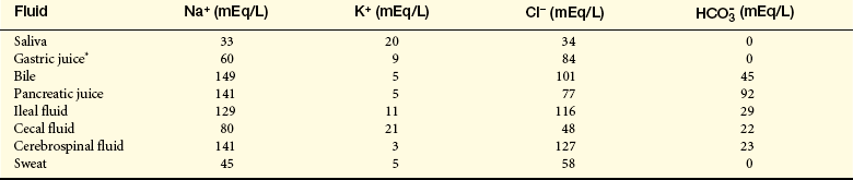
Cl−, Chloride;  , bicarbonate; H+, hydrogen; K+, potassium; Na+, sodium.
∗The Cl− concentration exceeds the Na+, K+ concentration by 15 mEq/L in gastric juice. This largely represents the secretions of HCl acid by parietal cells.
From Smith LH, Thier SO: Pathophysiology: the biological principles of disease, Philadelphia, 1981, Saunders.
The difference in the K+ intracellular to extracellular concentration is maintained by a sodium-potassium active transport system (Na+, K+-ATPase pump). The ratio of ICF K+ to ECF K+ is the major determinant of the resting membrane potential, which is necessary for the transmission and conduction of nerve impulses, maintenance of normal cardiac rhythms, and skeletal and smooth muscle contraction. (Membrane transport and membrane potentials are discussed in Chapter 1.) The diffusion of positively charged K+ out of the cell and down its concentration gradient makes the interior of cells electronegative in relation to the ECF. Changes in the ratio of ICF to ECF potassium are responsible for many of the symptoms associated with potassium imbalance.
As the predominant ICF ion, K+ exerts a major influence in the regulation of ICF osmolality and provides the balance for intracellular electrical neutrality in relation to hydrogen (H+) and Na+. Potassium is also necessary for a variety of metabolic functions and is required for glycogen deposition in liver and skeletal muscle cells.
The kidney provides the most efficient regulation of potassium balance over time. The amount of K+ excreted varies in proportion to the dietary intake (40 to 120 mEq/day). Potassium is freely filtered by the renal glomerulus, and 90% is reabsorbed by the proximal tubule and loop of Henle. The principal cells in the collecting tubule secrete potassium. The reabsorption of K+ occurs in the adjacent intercalated cell. Dietary potassium intake, aldosterone, and distal tubule urine flow determine the amount of K+ excreted from the body. Unlike sodium, the renal mechanism for conserving K+ is weak, even when total body potassium stores are depleted. However, a low K+ intake also suppresses renal K+ excretion.11
Several factors related to passive transport and aldosterone contribute to renal regulation of potassium. These factors include the concentration gradients for potassium at the distal tubule and collecting duct, changes in pH (causing acidosis or alkalosis), changes in electrical potential differences across the distal tubule, and aldosterone levels. (Renal mechanisms are described in more detail in Chapter 35.)
The concentration of potassium in the distal tubular cell is determined primarily by the plasma concentration in the peritubular capillaries. When plasma K+ concentration increases because of increased dietary intake or shifts from the ICF occur, potassium is secreted into the urine by principal cells in the distal tubules. Decreases in plasma potassium result in decreased distal tubular secretion and reabsorption by intercalated cells, although K+ losses of approximately 5 to 15 mEq/day will continue. Changes in the rate of filtrate flow through the distal tubule also influence the concentration gradient for K+ secretion. When the flow rate is high, as occurs with the administration of diuretics, the concentration of potassium in the distal tubular urine is lower, favoring the secretion of potassium.12
Changes in pH and thus in hydrogen ion concentration also affect K+ balance. Hydrogen ions move from the ECF to the ICF during states of acidosis. During acidosis, when hydrogen is moving into the cell, potassium shifts out of the cell to the ECF to maintain a balance of cations across the cell membrane. This occurs in part because of a decrease in Na+, K+-ATPase pump activity. The decreased ICF K+ results in decreased secretion of K+ into the urine by the distal tubular cells, contributing to hyperkalemia, although total body potassium may not change. In contrast, intracellular fluid levels of hydrogen are diminished during states of alkalosis. Alkalosis causes potassium to shift into the cell, so the distal tubular cells increase their secretion of K+ into the urine, contributing to hypokalemia. The management of potassium alterations associated with acid-base imbalances require that the acid-base imbalances must be treated before or concurrently with treatment of changes in potassium.
Three hormones (aldosterone, insulin, and epinephrine [β-adrenergic stimulation]) promote movement of potassium from the extracellullar to intracellular fluid. Besides acting to conserve sodium, aldosterone is a major factor in potassium regulation. When potassium concentration is increased, aldosterone is released, stimulating secretion of potassium into the urine by the distal tubules of the kidney. Aldosterone also increases the secretion of K+ from the sweat glands.
Insulin contributes to the regulation of plasma potassium levels by stimulating the Na+, K+-ATPase pump, thereby promoting the movement of potassium into liver and muscle cells simultaneously with glucose transport after eating. The intracellular movement of potassium prevents an acute hyperkalemia related to food intake. Insulin also can be used to treat hyperkalemia. However, dangerously low levels of plasma potassium can result from the administration of insulin when potassium levels are depressed. Potassium balance is especially significant in the treatment of conditions requiring insulin administration, such as insulin-dependent diabetes mellitus. Glucagon blocks entry of potassium into cells and glucocorticoids promote potassium excretion.
Catecholamines also influence K+ concentration in ECF. β1 adrenergics stimulate the movement of K+ into cells, and α-adrenergics shift K+ out of cells.13
An interesting aspect of K+ regulation is the ability of the body to adapt to increased levels of potassium intake over time. A sudden increase in potassium may be fatal, but if the intake of potassium is slowly increased by amounts no more than 120 mEq/day, the kidney is able to increase the urinary excretion of potassium and maintain potassium balance. This tolerance to increasing amounts of potassium is known as potassium adaptation.
Hypokalemia
PATHOPHYSIOLOGY Potassium deficiency, or hypokalemia, develops when the serum potassium concentration decreases to less than 3.5 mEq/L. Because intracellular and total body stores of potassium are difficult to measure, changes in potassium balance are described by the plasma concentration, although changes in total body potassium are not always reflected in the plasma potassium concentration. Generally, lowered serum potassium indicates a loss of total body potassium. Because potassium is lost from the ECF, the change in the concentration gradient favors movement of K+ from the cell to the ECF. The ICF/ECF concentration ratio is maintained, but total body K+ is depleted.
ECF hypokalemia can develop without losses of total body potassium, but only when potassium is redistributed between the ICF and ECF. For example, potassium shifts into the cell during states of respiratory or metabolic alkalosis or after administration of insulin. In the event of alkalosis, K+ shifts into the cell in exchange for H+ to maintain plasma acid-base balance. Insulin also promotes cellular uptake of K+ and can cause an ECF potassium deficit, particularly with the intake of high carbohydrate loads.14
Plasma K+ levels may be normal or elevated when total body potassium is depleted. In such instances, potassium shifts from the ICF to the ECF. One of the common causes of this problem is diabetic ketoacidosis, in which the increased hydrogen ion concentration in the ECF causes H+ to shift into the cell in exchange for potassium. A normal level of potassium is maintained in the plasma, but potassium continues to be lost in the urine, causing a deficit in total body potassium. Severe, even fatal, hypokalemia may occur if insulin is administered without also providing potassium supplements. Thus total body potassium depletion becomes evident when insulin treatment is initiated.
Potassium loss also occurs through normal body functions, but without causing hypokalemia. Average daily losses of potassium are as follows:
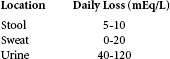
Factors contributing to the development of hypokalemia include reduced intake of potassium, increased entry of potassium into cells, and increased losses of body potassium. Dietary deficiency of potassium is a rare cause of hypokalemia. It may occur in older adults with both low protein intake and inadequate intake of fruits and vegetables and in people with alcoholism or anorexia nervosa. Generally, reduced potassium intake becomes a problem when combined with other causes of potassium depletion.
Shifts of potassium from the extracellular to intracellular space cause apparent deficits in total body potassium. Alkalosis, particularly respiratory alkalosis, is the most common clinical problem. ECF potassium will exchange with ICF hydrogen and correct the alkalosis by decreasing the pH of the ECF. Treatment of pernicious anemia with vitamin B12 or folate also may precipitate hypokalemia if the formation of new red blood cells causes enough potassium uptake to effect an extracellular decrease in potassium. Familial hypokalemic periodic paralysis is a rare genetically transmitted disease that also causes potassium to shift into the intracellular space.
Losses of potassium from body stores are most commonly caused by gastrointestinal and renal disorders. Diarrhea (from any cause), intestinal drainage tubes or fistulae, and laxative abuse also may result in hypokalemia. Normally, only 5 to 10 mEq of potassium and 100 to 150 ml of water are excreted in the stool each day. With diarrhea, fluid and electrolyte losses can be voluminous, with several liters of fluid and 100 to 200 mEq of potassium lost per day. Vomiting or continuous nasogastric suction frequently is associated with potassium depletion, partly because of the potassium lost from the gastric fluid but principally because of renal compensation for volume depletion and the metabolic alkalosis (elevated bicarbonate levels) that occurs from sodium, chloride, and hydrogen ion losses. The loss of fluid and sodium stimulates the secretion of aldosterone, which in turn causes renal losses of potassium. The elevated flow of bicarbonate at the distal tubule contributes to renal excretion of potassium because of increased tubular lumen electronegativity.
Renal losses of potassium are related to increased secretion of potassium by the distal tubule. Use of diuretics, excessive aldosterone secretion, increased distal tubular flow rate, and low plasma magnesium concentration all may contribute to urinary losses of potassium. Many diuretics, including thiazides, furosemide, ethacrynic acid, and osmotic diuretics, inhibit the reabsorption of sodium chloride, causing the diuretic effect. The distal tubular flow rate then increases, promoting potassium excretion. If sodium loss is severe, the compensating aldosterone secretion (which causes secondary hyperaldosteronism) may further deplete potassium stores. Primary hyperaldosteronism with excessive secretion of aldosterone from an adrenal adenoma also causes potassium wasting. Many kidney diseases result in a reduced ability to conserve sodium. The disordered sodium reabsorption produces a diuretic effect, and the increased distal tubule flow rate favors the secretion of potassium. Magnesium deficits stimulate renin release and hyperaldosteronism, causing hypokalemia. Several antibiotics, including amphotericin B, gentamicin, and carbenicillin, are known to cause hypokalemia.
CLINICAL MANIFESTATIONS A wide range of metabolic dysfunctions may result from potassium deficiency. Carbohydrate metabolism is affected because hypokalemia depresses insulin secretion and alters hepatic and skeletal muscle glycogen synthesis. Renal function is impaired, with a decreased ability to concentrate urine. Polyuria (increased urine) and polydipsia (increased thirst) are associated with decreased responsiveness to ADH. Chronic potassium deficits lasting more than 1 month may damage renal tissue, with resulting interstitial fibrosis and tubular atrophy.
Neuromuscular and cardiac effects of hypokalemia produce the most common symptoms.15 Neuromuscular excitability is decreased, causing skeletal muscle weakness, smooth muscle atony, and cardiac dysrhythmias. As Chapter 1 describes, the resting membrane potential (Em) is determined by the ratio of extracellular to intracellular potassium ion concentration. Because the concentration of potassium in the ECF is small, only small changes in ECF potassium are required to influence the resting membrane potential and affect neuromuscular excitability (the difference between resting membrane and threshold potentials). When extracellular potassium levels decrease rapidly, intracellular potassium diffuses more readily out of the cell and the resting membrane potential becomes more negative (i.e., from −90 to −100 mV). If the threshold potential (Et) remains stable, the difference between resting membrane potential and threshold potential increases and the cell membrane becomes hyperpolarized, requiring a stronger stimulus to initiate an action potential (decreasing excitability) (Figure 3-5).
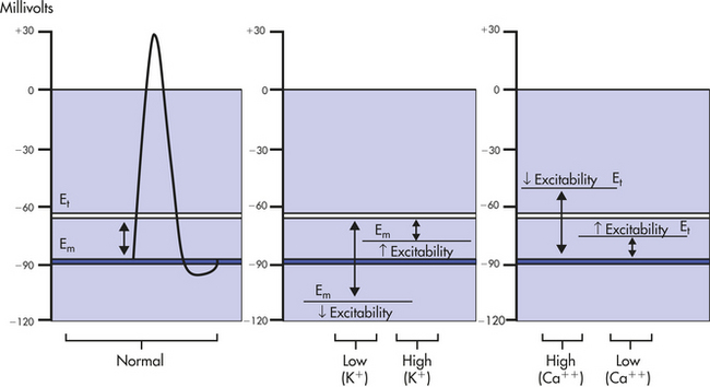
Figure 3-5 Effects of potassium (K+) and calcium (Ca++) on membrane excitability. Potassium affects resting membrane potential (Em), and calcium affects threshold potential (Et).
Factors such as calcium concentration and pH also contribute to the changes in neuromuscular excitability associated with hypokalemia. Increases in ECF calcium concentration tend to make the threshold potential less negative and decrease membrane excitability, potentiating the neuromuscular effects of hypokalemia.
The onset of symptoms is related to the rate of potassium depletion. Because the body can accommodate slow losses of potassium, the decrease in ECF concentration may be slow enough to allow potassium to shift from the intracellular space. The extracellular to intracellular potassium concentration gradient then is restored toward normal, with less severe neuromuscular changes. With acute losses of potassium, changes in neuromuscular excitability are more profound. Skeletal muscle weakness initially occurs in the larger muscles of the legs and arms and ultimately affects the diaphragm and depresses ventilation. Paralysis and respiratory arrest then can occur. Loss of smooth muscle tone is manifested by constipation, intestinal distention, anorexia, nausea, vomiting, and paralytic ileus.
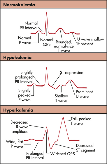
The cardiac effects of hypokalemia are related also to changes in membrane excitability (see Figure 3-5). Because potassium contributes to the repolarization phase of the action potential, hypokalemia delays ventricular repolarization and the frequency of action potentials. A variety of dysrhythmias may occur, including sinus bradycardia, atrioventricular block, and paroxysmal atrial tachycardia. The characteristic changes in the electrocardiogram reflect delayed repolarization. For instance, the amplitude of the T wave is decreased; the amplitude of the U wave is increased; and the ST segment is depressed (Figure 3-6). In severe states of hypokalemia, P waves peak and the QRS complex is prolonged. Hypokalemia also increases the risk of digitalis toxicity by slowing the sodium-potassium pump, which augments the action of digitalis in cardiac muscle by excessively increasing intracellular calcium and sodium.
EVALUATION AND TREATMENT The diagnosis of hypokalemia is significantly related to the medical history and the identification of disorders associated with potassium loss or shifts of extracellular potassium to the intracellular space. Treatment involves an estimation of total body potassium losses and correction of acid-base imbalances. Further losses of potassium should be prevented, and the individual should be encouraged to eat foods rich in potassium. The maximal rate of oral replacement is 40 to 80 mEq/day if renal function is normal. A maximal safe rate of intravenous replacement is 20 mEq/hr. Because potassium is irritating to blood vessels, a maximal concentration of 40 mEq/L should be used. Serum potassium values can be monitored until normokalemia is achieved.
Hyperkalemia
PATHOPHYSIOLOGY An elevation of ECF potassium above 5.5 mEq/L constitutes hyperkalemia. Because of efficient renal excretion, increases in total body potassium are relatively rare. Acute increases in serum potassium are handled quickly through an increase in cellular uptake and renal excretion of body potassium excesses. Excretion is partially mediated by the secretion of aldosterone because it facilitates excretion of potassium into the urine.
Excesses of serum potassium may be caused by increased intake, a shift of potassium from cells to the ECF, or decreased renal excretion. If renal function is normal, slow, long-term increases in potassium intake are usually well tolerated through potassium adaptation, although acute potassium loading can exceed renal excretion rates. Use of stored whole blood and intravenous boluses of penicillin G or replacement potassium can precipitate hyperkalemia, particularly if renal function is impaired. Dietary excesses of potassium are uncommon, but accidental ingestion of potassium salt substitutes can cause toxicity.
Movement of potassium from the ICF to the ECF occurs with cell trauma or a change in cell membrane permeability, acidosis, insulin deficiency, or cell hypoxia. Burns, massive crushing injuries, and extensive surgeries can cause loss of ICF potassium to the ECF. If renal function is sustained, potassium will be excreted. As cell repair begins, hypokalemia develops without an adequate intake of potassium.
In states of acidosis, hydrogen ions shift into the cells in exchange for ICF potassium; hyperkalemia and acidosis therefore often occur together. Because insulin promotes cellular entry of potassium, insulin deficits, which occur with conditions such as diabetic ketoacidosis, are accompanied by hyperkalemia. Hypoxia can lead to hyperkalemia by diminishing the efficiency of cell membrane active transport, resulting in the escape of potassium to the ECF. Digitalis overdose may cause hyperkalemia by inhibiting the Na+, K+-ATPase pump. This pump normally maintains intracellular potassium and moves sodium and calcium to the ECF (see Chapter 1).
Decreased renal excretion of potassium commonly is associated with hyperkalemia. Renal failure that results in oliguria (urine output <30 ml/hr) is accompanied by elevations of serum potassium. The severity of hyperkalemia is related to the amount of potassium intake, the degree of acidosis, and the rate of renal cell damage. In acute renal failure potassium levels rise more rapidly with more serious consequences than the slower rises associated with chronic renal failure. Decreases in the secretion or renal effects of aldosterone also can cause decreases in the urinary excretion of potassium. For example, Addison disease results in decreased production and secretion of aldosterone and thus contributes to hyperkalemia. Potassium-sparing diuretics (e.g., spironolactone, which inhibits sodium reabsorption and potassium and hydrogen secretion by the distal tubule) also may contribute to hyperkalemia. Frequently, however, these diuretics are used in combination with diuretics that cause potassium wasting in an attempt to balance renal potassium gains and losses.
CLINICAL MANIFESTATIONS Symptoms of hyperkalemia vary, but common characteristics are muscle weakness or paralysis and arrhythmias with changes in the electrocardiogram. During mild attacks, increased neuromuscular irritability may be manifested as tingling of lips and fingers, restlessness, intestinal cramping, and diarrhea. Severe hyperkalemia causes muscle weakness, loss of muscle tone, and paralysis. In mild states of hyperkalemia, the more rapid repolarization is reflected in the electrocardiogram as narrow and taller T waves with a shortened QT interval. Severe hyperkalemia (serum levels ≥6 mEq/L) depresses the ST segment, prolongs the PR interval, and widens the QRS complex (see Figure 3-6). Bradydysrhythmias are common in hyperkalemia, with alterations in cardiac conduction causing ventricular fibrillation or cardiac arrest.
As with hypokalemia, changes in the ratio of intracellular to extracellular potassium concentration contribute to the symptoms of hyperkalemia. If extracellular potassium concentration increases without a significant change in intracellular potassium, the resting membrane potential becomes more positive (i.e., changes from –90 to –80 mV) and the cell membrane is hypopolarized (the inside of the cell becomes less negative or partially depolarized [increase excitability]) (Electrical properties of cells are discussed in Chapter 1.) With relatively mild elevations in extracellular potassium, the cell more rapidly repolarizes and becomes more irritable (peaked T waves). An action potential then is initiated more rapidly because the distance between the resting membrane potential and the threshold potential has been shortened. With more severe hyperkalemia, the resting membrane potential approaches or exceeds the threshold potential (wide QRS merging with T wave). In this case the cell is not able to repolarize and therefore does not respond to excitation stimuli. The most serious consequence is cardiac standstill.
Like the effects of hypokalemia, the neuromuscular effects of hyperkalemia are related to the rate of increase in the ECF potassium concentration and the presence of other contributing factors, such as acidosis and calcium balance. Long-term increases in ECF potassium concentration result in shifts of potassium into the cell because the tendency is to maintain a normal ratio of intracellular/extracellular potassium concentrations. Acute elevations of extracellular potassium affect neuromuscular irritability because this ratio is disrupted.16
Because calcium influences the threshold potential, changes in extracellular fluid calcium concentration can augment or override the effects of hyperkalemia. With hypocalcemia the threshold potential becomes more negative, enhancing the neuromuscular effects of hyperkalemia. Hypercalcemia causes the threshold potential to become less negative, counteracting the effects of hyperkalemia on resting membrane potential (see Figure 3-5).
EVALUATION AND TREATMENT Hyperkalemia should be investigated when there is a history of renal disease, massive trauma, insulin deficiency, Addison disease, use of potassium salt substitutes, or metabolic acidosis. The acuity of the onset of symptoms may be related to the underlying cause.
Management of hyperkalemia is related to treating the contributing causes and correcting the potassium excess. Normalizing the extracellular potassium concentration can be achieved with a variety of methods; the treatment chosen is related to the cause and severity of the problem. Calcium gluconate can be administered to restore normal neuromuscular irritability when serum potassium levels are dangerously high. Administration of glucose, which readily stimulates insulin secretion, or administration of glucose and insulin for those with diabetes, facilitates cellular entry of potassium. Sodium bicarbonate corrects metabolic acidosis and lowers serum potassium. Oral or rectal administration of cation exchange resins, which exchange sodium for potassium in the intestine, can be effective. Dialysis effectively removes potassium when renal failure has occurred.
Calcium and Phosphate
The total body content of calcium is about 1200 g. Most calcium (99%) is located in bone as hydroxyapatite (an inorganic compound that contributes to bone rigidity), and the remainder is in the plasma and body cells. Of the calcium in the plasma, 50% is bound to plasma proteins (2.5 mEq/L), and about 40% is in the free or ionized form (2.4 mEq/L). The total fraction of calcium circulating in the blood is small (4.5 to 5.5 mEq/L, or 8.6 to 10.5 mg/dl). Ionized calcium has the most important physiologic functions.
Calcium is a necessary ion for many fundamental metabolic processes. It is the major cation for the structure of bones and teeth. It serves as an enzymatic cofactor for blood clotting and is required for hormone secretion and the function of cell receptors. Plasma membrane stability and permeability are directly related to calcium ions, as is the transmission of nerve impulses and the contraction of muscles. Intracellular calcium is located primarily in the mitochondria.
Phosphate is found primarily in bone (85%), with smaller amounts found within the intracellular and extracellular spaces. In the serum, phosphate exists in phospholipids and phosphate esters and as inorganic phosphate, which is the ionized form. The normal serum levels of inorganic phosphate range from 2.5 to 4.5 mg/dl and may be as high as 6.0 to 7.0 mg/dl in infants and young children. Intracellular phosphate has many metabolic forms, including the high-energy structures creatine phosphate and adenosine triphosphate (ATP). Phosphate acts as an intracellular and extracellular anion buffer in the regulation of acid-base balance; in the form of ATP it provides energy for muscle contraction.
Calcium and phosphate concentrations are rigidly controlled. They are related by the product of calcium (Ca++) and phosphate ( ), which is a constant (K) [Ca++ × = K]. Thus if the concentration of one ion increases, that of the other decreases.
Calcium and phosphate balance is regulated by three hormones: parathyroid hormone (PTH), vitamin D, and calcitonin. Acting together, these substances determine the amount of dietary calcium and phosphate absorbed from the intestine, the deposition and absorption of calcium and phosphate from the bone, and the renal reabsorption and excretion of calcium and phosphate by the kidney.
The parathyroid glands are sensitive to changes in serum calcium concentrations, and parathyroid hormone controls ionized calcium in the blood and extracellular fluids. The parathyroid glands secrete PTH in response to low serum calcium. (The specific actions of PTH in relation to calcium and phosphorus are described in Chapter 20.) The renal regulation of calcium and phosphate balance requires PTH. As PTH secretion is stimulated by low levels of serum calcium, reabsorption of calcium along the distal part of the nephron increases and inhibition of phosphate reabsorption by the proximal segment of the nephron increases. The net result is an increase in serum calcium and urinary excretion of phosphate. Figure 3-7 summarizes hormonal regulation of calcium.
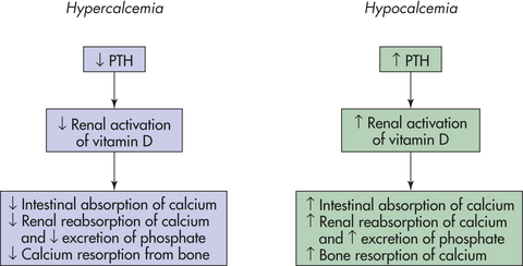
Another hormone important to calcium and phosphate regulation is vitamin D. Vitamin D (cholecalciferol) is a fat-soluble steroid ingested in food or synthesized in the skin in the presence of ultraviolet light. Several steps of activation are required before vitamin D can act on target tissues. The first step occurs in the liver; final activation is in the kidney. The renal activation of vitamin D begins when the serum calcium level decreases and stimulates secretion of PTH. PTH then acts to increase calcium reabsorption and enhance renal excretion of phosphate, producing decreased phosphate levels. The combination of low calcium, PTH secretion, and low phosphate thus causes the renal activation of vitamin D. The activated vitamin D (vitamin D3—calcitriol) then circulates in the plasma and acts to increase absorption of calcium in the small intestine, enhance bone absorption of calcium, and increase renal tubular reabsorption of calcium. When renal failure occurs, vitamin D is not activated; serum calcium levels decrease; and phosphate levels increase.
The exchange of calcium and phosphate between serum and bone is regulated also by hormones. When serum calcium levels are low, PTH increases and vitamin D3 stimulates intestinal calcium absorption and renal calcium reabsorption. Osteoclasts are stimulated to resorb bone and release calcium and phosphate into the plasma.
As calcium levels increase, an opposite adaptation occurs, leading to suppression of PTH secretion, decreased renal vitamin D activation, and decreased intestinal calcium absorption and increased renal phosphate reabsorption. Calcitonin primarily decreases calcium levels by inhibiting osteoclastic activity in bone.
The fractions of serum calcium that are freely ionized or bound to plasma proteins are influenced by pH. In states of acidosis, levels of ionized calcium increase. When alkalosis develops, with an increase in pH, protein-bound calcium increases and the physiologically active, ionized calcium decreases. The decreased concentration of ionized calcium may be great enough to cause symptoms of hypocalcemia, such as tetany.
Hypocalcemia
PATHOPHYSIOLOGY Hypocalcemia occurs when serum calcium concentrations are less than 8.5 mg/dl and ionized levels are less than 4.0 mg/dl. Deficits in calcium are related to inadequate intestinal absorption, deposition of ionized calcium into bone or soft tissue, blood administration, or decreases in PTH and vitamin D.
Nutritional deficiencies of calcium can occur in the instance of inadequate sources of dairy products or green, leafy vegetables. Excessive amounts of dietary phosphorus also bind with calcium, so neither mineral is absorbed when such an excess occurs. Blood transfusions are also a common cause of hypocalcemia because the citrate solution used in storing whole blood binds with calcium. Pancreatitis causes release of lipases into soft tissue spaces, so the free fatty acids that are formed bind calcium, causing a decrease in ionized calcium. Neoplastic bone metastases tend to inhibit bone resorption and increase calcium deposition into bone, thereby decreasing serum calcium levels.
Vitamin D deficiency, which can result from inadequate intake or avoidance of sunlight, causes decreased intestinal absorption of calcium. Malabsorption of fat, including fat-soluble vitamin D, may also contribute to calcium deficiency. Removal of the parathyroid glands with the resulting loss of PTH also causes hypocalcemia. Metabolic or respiratory alkalosis causes symptoms of hypocalcemia because the change in pH enhances protein binding of ionized calcium. Hypoalbuminemia lowers total serum calcium levels by decreasing the amount of bound calcium in the plasma.
CLINICAL MANIFESTATIONS The clinical manifestations of hypocalcemia are caused primarily by an increase in neuromuscular excitability. Calcium deficits cause partial depolarization of nerves and muscle as the threshold potential approaches the resting membrane potential (see Figure 3-5). Therefore, a smaller stimulus is required for initiating the action potential. The symptoms include confusion, paresthesias around the mouth and in the digits, carpopedal spasm (muscle spasms in the hands and feet), and hyperreflexia.
Two clinical signs are Chvostek sign and Trousseau sign. Chvostek sign is elicited by tapping on the facial nerve just below the temple. A positive sign is a twitch of the nose or lip. Trousseau sign is contraction of the hand and fingers when the arterial blood flow in the arm is occluded for 5 minutes.
Severe symptoms include convulsions and tetany, a continuous severe muscle spasm that can interfere with breathing and cause death. The characteristic electrocardiogram (ECG) change is a prolonged QT interval, indicating prolonged ventricular depolarization and decreased cardiac contractility. Intestinal cramping and hyperactive bowel sounds also may be present because hypocalcemia affects the smooth muscles of the gastrointestinal tract.
EVALUATION AND TREATMENT The health history may signify underlying pathologic conditions that require further evaluation and treatment. Severe symptoms of hypocalcemia require emergency treatment with intravenous 10% calcium gluconate. Oral calcium replacement should be initiated, and serum calcium levels should be monitored. Decreasing phosphate intake facilitates long-term management of hypocalcemia.
Hypercalcemia
PATHOPHYSIOLOGY Hypercalcemia with serum calcium concentrations exceeding 12 mg/dl can be caused by a number of diseases. The most common among these are hyperparathyroidism; bone metastases with calcium resorption from breast, prostate, cervical cancer, or hematologic malignancy; sarcoidosis; and excess vitamin D. Many tumors produce PTH and elevate the serum calcium levels. Sarcoidosis appears to increase vitamin D levels. Prolonged immobilization can also lead to hypercalcemia from bone resorption. Acidosis decreases serum binding of calcium to albumin, increasing ionized calcium.
CLINICAL MANIFESTATIONS Many symptoms of hypercalcemia are nonspecific. Because serum calcium levels are increased, a greater amount of calcium is also contained inside the cells. The threshold potential becomes more positive, and the cell membrane becomes refractory to depolarization (see Figure 3-5). Thus many of the symptoms are related to loss of cell membrane excitability. (Membrane potentials and membrane excitability are discussed in Chapter 1.) Fatigue, weakness, lethargy, anorexia, nausea, and constipation are common. Behavioral changes may occur. Impaired renal function frequently develops, and kidney stones form as precipitates of calcium salts. A shortened QT segment and depressed widened T waves also may be observed on the ECG, with bradycardia and varying degrees of heart block.
EVALUATION AND TREATMENT With elevated serum calcium levels, often a reciprocal decrease in serum phosphate values occurs. Specific diagnostic procedures to identify the contributing pathologic condition are required.
Treatment is related to severity of symptoms and the underlying disease. When renal function is normal, oral phosphate administration is effective. When acute illness and high calcium levels are present, treatment options include intravenous administration of large amounts of normal saline to enhance renal excretion of calcium, bisphosphonates in the absence of renal failure, and administration of calcitonin. Corticosteroids and the cytotoxic drug mithramycin (for use with malignant disease) also are used to treat hypercalcemia. Ultimately, the underlying pathologic condition must be treated.
Hypophosphatemia
PATHOPHYSIOLOGY Hypophosphatemia is a serum phosphate level less than 2 mg/dl and is usually an indication of phosphate deficiency. In some conditions, total body phosphate is normal but serum volumes are low. The most common causes are intestinal malabsorption and increased renal excretion of phosphate. Inadequate absorption is associated with vitamin D deficiency, use of magnesium- and aluminum-containing antacids (which bind with phosphorus), long-term alcohol abuse, and malabsorption syndromes. Respiratory alkalosis can cause severe hypophosphatemia because of cellular use of phosphorus for an accelerated glucose metabolism. Increased renal excretion of phosphorus is associated with hyperparathyroidism.
CLINICAL MANIFESTATIONS The consequences of phosphate deficiency are not clinically evident until hypophosphatemia is severe. There is reduced capacity for oxygen transport by red blood cells and disturbed energy metabolism. Transport and release of oxygen are associated with 2,3-diphosphoglycerate (2,3-DPG) and ATP. When phosphate is depleted, 2,3-DPG and ATP levels become low and diminish release of oxygen to the tissues. The oxyhemoglobin curve shifts to the left (see Chapter 32), and hypoxia can occur with bradycardia and varying degrees of heart block.
Leukocyte and platelet dysfunctions also are associated with hypophosphatemia. There is a greater risk of infection and blood-clotting impairment, with potential for hemorrhage. Nerve and muscle function can be affected because of derangement in energy metabolism. Muscle weakness may become serious enough to cause respiratory failure, and cardiomyopathies also can develop. Irritability, confusion, numbness, coma, and convulsions develop with severe phosphate losses. In response to low phosphate levels, bone resorption occurs and may lead to rickets or osteomalacia.
EVALUATION AND TREATMENT To correct the condition, the underlying cause must be identified and treated. Although serum phosphate levels are below normal, the administration of phosphate salts is dangerous, and low phosphate levels are usually not considered life threatening.17
Hyperphosphatemia
PATHOPHYSIOLOGY Hyperphosphatemia, or an elevated serum phosphate level of more than 4.5 mg/dl, develops with exogenous or endogenous addition of phosphorus to the ECF or with significant loss of glomerular filtration.18 Because most phosphate is located in cells, the cell destruction associated with treatment of metastatic tumors with chemotherapy can release large amounts of phosphate into the serum. Long-term use of phosphate-containing enemas or laxatives also may lead to hyperphosphatemia. Hypoparathyroidism can cause elevated phosphate by increasing renal tubular reabsorption of phosphate.
High levels of serum phosphate also lower serum calcium levels, and increased amounts of phosphate and calcium are deposited in bone and soft tissues. Serum calcium levels may become low enough to cause symptoms of hypocalcemia, including tetany.
CLINICAL MANIFESTATIONS Symptoms of hyperphosphatemia are related primarily to low serum calcium levels and thus are comparable to symptoms of hypocalcemia. With prolonged hyperphosphatemia, calcification of soft tissues occurs in the lungs, kidneys, and joints.
EVALUATION AND TREATMENT To correct the condition, the underlying pathologic condition must be identified and treated. Aluminum hydroxide may be administered because it binds phosphate in the gastrointestinal tract and is then eliminated but can deposit in the central nervous system, bone, and hematopoietic cells. New non-aluminum and non–calcium phosphate binders are available.19 Dialysis is required for management of renal failure.
Magnesium
Magnesium (Mg++) is a major intracellular cation. About 40% to 60% is stored in muscle and bone with 30% in the cells. A small amount (1%) is in the serum. Plasma concentration is 1.8 to 2.4 mEq/L with about one third bound to plasma proteins and the rest in ionized form. Regulation of magnesium metabolism is balanced by the small intestine and kidney. Low serum levels cause renal conservation of magnesium. Magnesium is a cofactor in intracellular enzymatic reactions, protein synthesis, nucleic acid stability, and neuromuscular excitability. Calcium and magnesium often interact in reactions at the cellular level.
Hypomagnesemia occurs when serum magnesium concentration is less than 1.5 mEq/L and increases in neuromuscular excitability and tetany are present. Malnutrition, malabsorption syndromes, alcoholism, renal tubular dysfunction, metabolic acidosis, and loop and thiazide diuretics can cause magnesium losses. Diabetes mellitus is associated with hypomagnesemia partly as a function of osmotic diuresis.20 Because magnesium inhibits potassium channels, loss of magnesium results in movement of potassium out of the cell, with renal excretion resulting in hypokalemia. Signs and symptoms of hypomagnesemia are similar to those of hypocalcemia. Depression, confusion, irritability, increased reflexes, muscle weakness, ataxia, nystagmus, tetany, convulsions, and tachyarrhythmias may be observed.21 Treatment is intramuscular or intravenous administration of magnesium sulfate.
Hypermagnesemia, in which magnesium concentration is greater than 2.5 mEq/L, is rare and usually is caused by renal failure. Magnesium-containing antacids (e.g., Gaviscon, Gelusil) can potentiate excess magnesium. Excess magnesium depresses skeletal muscle contraction and nerve function. Signs and symptoms include nausea and vomiting, muscle weakness, hypotension, bradycardia, and respiratory depression.22 Treatment is avoidance of magnesium-containing substances and removal of magnesium by dialysis.
ACID-BASE BALANCE
Acid-base balance and hydrogen ion concentration must be regulated within a narrow range for the body to function normally. Slight changes in amounts of hydrogen can significantly alter biologic processes in cells and tissues. Hydrogen ion is necessary to maintain membrane integrity and the speed of enzymatic reactions. Most pathologic conditions disturb acid-base balance, and the degree of severity may be more harmful than the disease process.
Hydrogen Ion and pH
The hydrogen ion concentration [H+] is commonly expressed as the pH, the negative logarithm of hydrogen ions in solution. The logarithmic value means that as the pH changes one unit (e.g., 7.0 to 6.0), the [H+] changes tenfold (i.e., 0.0000001 to 0.000001). The relationship is commonly expressed as follows:
As the [H+] increases, the pH decreases; likewise, as the [H+] decreases, the pH increases. The greater the [H+], the more acidic the solution and the lower the pH. The lower the [H+], the more basic the solution and the higher the pH. In biologic fluids, a pH of less than 7.4 is defined as acidic and a pH greater than 7.4 is defined as basic.
Different body fluids have different pH values as follows:
| Body Fluid | pH |
| Gastric juices | 1.0-3.0 |
| Urine | 5.0-6.0 |
| Arterial blood | 7.38-7.42 |
| Venous blood | 7.37 |
| Cerebrospinal fluid | 7.32 |
| Pancreatic fluid | 7.8-8.0 |
Body acids are formed as end products of cellular metabolism. The average person generates acid in the amount of 50 to 100 mEq/day from the metabolism of protein, carbohydrates, and fats and from loss of base in the stools. To maintain a normal pH, an equal amount of acid therefore must be neutralized or excreted. The lungs, kidneys, and bone are the major organs involved in the regulation of acid-base balance. The systems are interrelated and work together to regulate short- or long-term changes in acid-base status. Body acids exist in two forms: volatile (respiratory acids—eliminated as carbon dioxide [CO2] gas) and nonvolatile (metabolic acids—eliminated by the kidney or metabolized by the liver). The volatile acid is carbonic acid (H2CO3), which is formed from the hydration of carbon dioxide:
Carbonic acid is a weak acid, and in the presence of carbonic anhydrase, it readily dissociates into carbon dioxide. Approximately 12,000 to 15,000 millimoles of CO2 is produced in the human body per day.23 The carbon dioxide is then eliminated by pulmonary ventilation. Sulfuric, phosphoric, and other metabolic acids (lactic acid, pyruvic acid, and keto acids associated with diabetes mellitus) are nonvolatile acids produced from the metabolism of proteins, carbohydrates, and fats. (Strong acids are those that readily give up their hydrogen; weak acids do not.) Nonvolatile acids are eliminated by the renal tubules with the regulation of . Thus the lungs and kidneys, with the help of body buffer systems, are the prime regulators of acid-base balance.
Buffer Systems
Buffering occurs in response to changes in acid-base status. Buffers can absorb excessive H+ (acid) or OH− (base) without a significant change in pH. The buffer systems are located in both the ICF and ECF compartments, and they function at different rates. Buffer systems exist as buffer pairs, consisting of a weak acid and its conjugate base (Table 3-9). The most important plasma buffer systems are carbonic acid–bicarbonate and hemoglobin. Phosphate and protein are the most important intracellular buffers.
Table 3-9
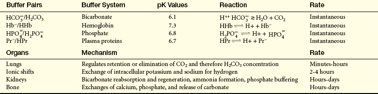
H+, Hydrogen; , bicarbonate; H2CO3, carbonic acid; Hb−, hemoglobin;  , monobasic phosphate; , dibasic phosphate; HPr, hydrogenated protein; HHb, hydrogenated hemoglobin; Pr−, protein.
An important factor for effective buffering is a function known as the pK value, which represents the pH at which a buffer pair is half dissociated. Buffer pairs can associate and dissociate (see Table 3-9).
The pK provides a rate constant for the chemical reaction. A buffer system is most effective when the pK for the buffer is close to the pH of the fluid in which the buffer is acting. For the bicarbonate–carbonic acid buffer system, the pK is 6.1. This value is not as high as the pK for other buffer systems (see Table 3-9), but this buffer system is still very effective because carbon dioxide is rapidly removed from the blood by the lungs.
The pK value is also a term in the equation used to determine pH. The relationships among pH, pK, and the ratio of bicarbonate to carbonic acid can be expressed as follows by the Henderson-Hasselbalch equation:
The pH then can be determined when specific values are included in the equation:
Carbonic Acid–Bicarbonate Buffering
The carbonic acid–bicarbonate buffer pair operates in both the lung and the kidney. The greater the carbon dioxide partial pressure (PCO2), the more carbonic acid is formed. The relationship that exists between carbonic acid (H2CO3) and carbon dioxide (PCO2) can be expressed as follows:
The 0.03 represents the solubility coefficient for carbon dioxide in water. The PCO2 of arterial blood is normally about 40 mmHg. Therefore the amount of H2CO3 is equal to about 1.2 mmol/L (0.03 × 40). As the amount of carbon dioxide increases or decreases, the amount of H2CO3 changes in the same direction.
The relationship between bicarbonate and carbonic acid is usually expressed as a ratio. When the pH is 7.40, this ratio is 20:1 (bicarbonate/carbonic acid). The ratio is defined by the amount of bicarbonate and carbon dioxide (carbonic acid) in the arterial blood. Bicarbonate concentration () is normally about 24 mEq/L. Therefore, the 20:1 ratio can be developed as follows:
The values for and Pco2 (H2CO3) can increase or decrease proportionately, but the 20:1 ratio is maintained.
The lungs can decrease the amount of carbonic acid by blowing off CO2 and leaving water. The kidneys can reabsorb bicarbonate or regenerate new bicarbonate from CO2 and water. The renal mechanism does not act as rapidly as the lungs, but the two systems are very effective together because acid concentration can be rapidly adjusted by the lungs and bicarbonate is easily reabsorbed or regenerated by the kidneys. The pH equation can be symbolically expressed as follows:

Changes in either the numerator or the denominator will change the pH. For example, if the amount of bicarbonate is decreased, the pH also decreases, causing a state of acidosis. The pH can be returned to a normal range if the value of the denominator or the amount of carbonic acid also decreases. This type of adjustment in pH is known as compensation. With compensation, a 20:1 ratio may be achieved, but the actual values for and H2CO3 are not normal. The respiratory system compensates for changes in pH by increasing or decreasing ventilation. The renal system compensates by producing more acidic or more alkaline urine. Correction occurs when the values for both components of the buffer pair return to normal (Figure 3-8).
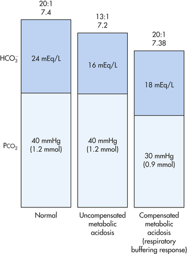
Protein Buffering
Both intracellular and extracellular proteins have negative charges and can serve as buffers for H+, but because most proteins are inside cells, they are primarily an intracellular buffer system. Hemoglobin (Hb) is an excellent intracellular buffer because of its ability to bind with H+ (forming HHb) and carbon dioxide (forming HHbCO2). Hemoglobin bound to H+ becomes a weak acid. Less saturated hemoglobin (venous blood) is a better buffer than hemoglobin saturated with oxygen (arterial blood). The hemoglobin buffer system is illustrated in Figure 3-9.
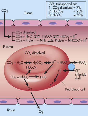
Figure 3-9 Buffering of hydrogen with hemoglobin and carbon dioxide (CO2) transport. CO2 is produced in tissue cells and diffuses to plasma, where it is transported as dissolved CO2, or it combines with water to form carbonic acid (H2CO3), or it combines with protein from which hydrogen has been released. Most of the CO2 diffuses into the red blood cells and combines with water to form H2CO3. The H2CO3 dissociates to form hydrogen (H+) and bicarbonate (). The shifts into the plasma and chloride (Cl−) shifts into the red blood cell to maintain electroneutrality. Hydrogen combines with hemoglobin that has released its oxygen to form HHb, which buffers the hydrogen and makes venous blood slightly more acidic than arterial blood.
Renal Buffering
The distal tubule of the kidney regulates acid-base balance by secreting hydrogen into the urine and reabsorbing bicarbonate with a maximum urine acidity of about 4.4 to 4.7. Buffers in the tubular fluid combine with hydrogen ions, allowing more H+ to be secreted before the limiting pH value is reached. Dibasic phosphate () and ammonia (NH3) are two important renal buffers. Dibasic phosphate is filtered at the glomerulus. About 75% is reabsorbed, and the remainder is available for buffering H+. Secreted H+combines with to form monobasic phosphate (). The remaining negative charge on the molecule makes it lipid insoluble, and it cannot diffuse back across the tubular cell and into the blood. Thus it is excreted in the urine (Figure 3-10).
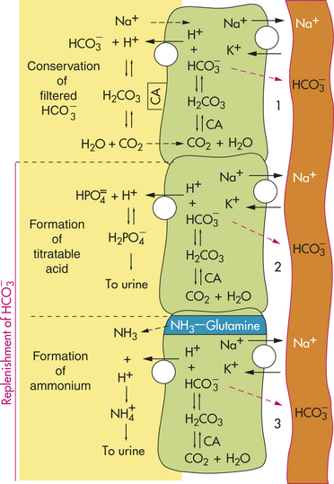
Figure 3-10 Renal excretion of acid.1, Conservation of filtered bicarbonate. Filtered bicarbonate combines with secreted hydrogen in the presence of carbon anhydrase (CA) to form carbonic acid (H2CO3), which then dissociates to water (H2O) and carbon dioxide (CO2); both diffuse into the epithelial cell. The CO2 and H2O combine to form H2CO3 in the presence of CA, and the resulting bicarbonate () is reabsorbed into the capillary. 2, Formation of titratable acid. Hydrogen ion is secreted and combines with dibasic phosphate () to form monobasic phosphate (). The secreted hydrogen is formed from the dissociation of H2CO3, and the remaining is reabsorbed into the capillary. 3, Formation of ammonium. Ammonia (NH3) is produced from glutamine in the epithelial cell and diffuses to the tubular lumen, where it combines with H+ to form ammonium ( ). Once has been formed, it cannot return to the epithelial cell (diffusional trapping), and the bicarbonate remaining in the epithelial cell is reabsorbed into the capillary.
Ammonia (NH3) is an important renal buffer; it is not ionized (does not carry a charge), and therefore it is lipid soluble and can cross the cell membrane. The presence of NH3 in the cell creates a concentration gradient, and it diffuses into the renal tubular fluid, where it combines with hydrogen to form ammonium ion (), which is eliminated in the urine (see Figure 3-10). The renal buffering of hydrogen ions requires the use of CO2 and H2O to form H2CO3. The enzyme carbonic anhydrase catalyzes the formation of H+ + . The hydrogen is secreted from the tubular cell and buffered in the lumen by phosphate and ammonia. The bicarbonate is reabsorbed. The end effect is the addition of new bicarbonate, which contributes to the alkalinity of the plasma, because the hydrogen ion is excreted from the body (see Figure 3-10).
Other Buffers
A cellular ion exchange mechanism is also an important buffering system. The best example is the shift of potassium in exchange for hydrogen during states of acidosis or alkalosis. During acidosis, potassium tends to leave the intracellular space in exchange for hydrogen. The reverse occurs during alkalosis. Although the ionic shifts facilitate buffering, the changes in intracellular or extracellular potassium concentrations may have serious consequences.
Acid-Base Imbalances
Pathophysiologic changes in the concentration of hydrogen ion or base in the blood lead to acid-base imbalances. Acidemia is a state in which the pH of arterial blood is less than 7.35. A systemic increase in hydrogen ion concentration or loss of base is termed acidosis. Alkalemia is a state in which the pH of arterial blood is greater than 7.45. A systemic decrease in hydrogen ion concentration or an excess of base is termed alkalosis. Acid-base imbalances may have a metabolic or respiratory etiology or may be of mixed etiology. Figure 3-11 summarizes the relationships among pH, PCO2, and bicarbonate during different acid-base alterations.
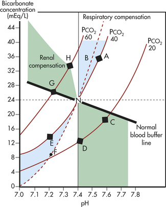
Figure 3-11 Graph of pH, PCO2, and bicarbonate relationships. Solid red lines represent different carbon dioxide partial pressure (PCO2) values. Vertical axis represents bicarbonate concentration, and horizontal axis represents acidity or alkalinity (pH) values. Thus for any indicated PCO2, there is a corresponding pH and bicarbonate concentration. Any point on the graph predicts the required PCO2, pH, and bicarbonate values. Dashed horizontal line shows behavior of bicarbonate as a pure buffer at 24 mEq/L. The normal blood buffer line represents values that would be obtained if blood were equilibrated at different CO2 values. Point N represents normal values. Point A represents uncompensated metabolic alkalosis, indicated by a normal PCO2 of 40 and pH greater than 7.4. Respiratory compensation is achieved by hypoventilation, which raises the PCO2 to point B and decreases the pH. Uncompensated respiratory alkalosis is represented by point C and reflects hypocapnia (decreased PCO2). Renal compensation for respiratory alkalosis is increased renal excretion of bicarbonate to normalize pH at point D. Uncompensated metabolic acidosis at point E represents normal PCO2 and a decrease in bicarbonate and pH. Respiratory compensation by hyperventilation is indicated by point F. Uncompensated respiratory acidosis at point G indicates high PCO2 and low pH values. Renal compensation for chronic high PCO2 values is indicated by point H.
Metabolic Acidosis
PATHOPHYSIOLOGY In metabolic acidosis, noncarbonic acids increase or bicarbonate (base) is lost from the extracellular fluid or cannot be regenerated by the kidney (Tables 3-10 and 3-11). This can occur quickly, as in lactic acidosis from poor perfusion or hypoxemia, or more slowly, as in renal failure or diabetic ketoacidosis.
Table 3-10
Primary and Compensatory Acid-Base Changes
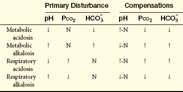
, bicarbonate; ↑-N, increase toward normal; ↓-N, decrease toward normal; PCO2, carbon dioxide partial pressure; pH, measure of the acidity or alkalinity of a solution.
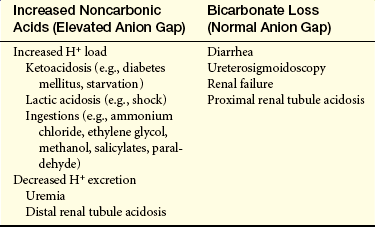
The buffer systems compensate for the excess acid and attempt to maintain the arterial pH within a normal range. Hydrogen ions will move to the intracellular space, and to maintain an ionic balance, potassium will move to the extracellular space (see p. 110). Buffering by bicarbonate lowers the serum value of hydrogen ions and increases the pH. The respiratory system compensates for a metabolic acidosis as the reduced pH stimulates hyperventilation, lowering the Paco2 and the amount of H2CO3 circulating in the blood. The kidneys excrete the excess acid as and titratable acid (). When the acidosis is severe, the buffers are unable to compensate for the increasing H+ load and the pH continues to decrease. The result is a decrease in the 20:1 ratio of bicarbonate to carbonic acid (Figure 3-12). In states of ketoacidosis, potassium is redistributed from the intracellular to the extracellular space, and is reabsorbed at the apical membrane of the renal collecting tubule (see p. 110). There is also an increase in levels of ionized calcium as acidosis decreases the amount of calcium bound to albumin (see p. 113).
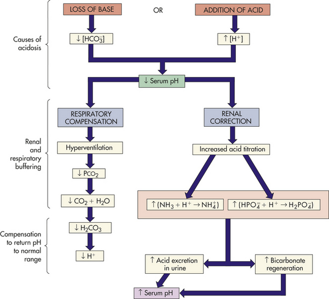
The evaluation of the anion gap can be helpful when used cautiously to distinguish different types of metabolic acidosis.24 Normally, the concentrations of cations and anions in the plasma are equivalent. Some anions, such as protein, sulfates, phosphates, and organic acids, however, are not measured in the common laboratory evaluations of the blood. Therefore the normal anion gap represents these unmeasured negative ions (sulfate, phosphate, lactate, ketoacids, albumin). A convenient measure of the anion gap is the difference between the sum of Na+ and K+ and the sum of and Cl−, or about 10 to 12 mEq:

In metabolic acidosis a normal anion gap is characteristic of conditions related to bicarbonate loss with retention of chloride to maintain an ionic balance. This is called hyperchloremic metabolic acidosis. An elevated anion gap is characteristic of acidosis associated with accumulation of anions other than chloride (see Table 3-11).
CLINICAL MANIFESTATIONS Metabolic acidosis is manifested by changes in the neurologic, respiratory, gastrointestinal, and cardiovascular systems. Headache and lethargy are early symptoms, which progress to coma with severe acidosis. Deep, rapid respirations (Kussmaul respirations) are indicative of respiratory compensation. Anorexia, nausea, vomiting, diarrhea, and abdominal discomfort are common. Severe acidosis can compromise ventricular contraction and produce life-threatening dysrhythmias and hypotension.
EVALUATION AND TREATMENT The diagnosis of metabolic acidosis is established from the health history, clinical symptoms, and laboratory findings. Arterial blood pH is below 7.35, and bicarbonate concentration is less than 24 mEq/L. The anion gap can isolate the specific cause. The underlying condition must be diagnosed to establish effective treatment. During severe acidosis (pH ≤7.1), bicarbonate administration is required to elevate the pH to a safe level, particularly if there is renal failure. Accompanying sodium and water deficits must also be corrected.25
Metabolic Alkalosis
PATHOPHYSIOLOGY Metabolic alkalosis is common and occurs when bicarbonate is increased, usually caused by excessive loss of metabolic acids. Among the conditions that can result in metabolic alkalosis are prolonged vomiting, gastrointestinal suctioning, excessive bicarbonate intake, hyperaldosteronism with hypokalemia, and diuretic therapy.26
When acid loss is caused by vomiting with depletion of ECF and chloride (hypochloremic metabolic alkalosis), renal compensation is not very effective because the volume depletion and loss of electrolytes (Na+, K+, H+, Cl−) stimulate a paradoxical response by the kidneys. The kidneys increase sodium and bicarbonate reabsorption with excretion of hydrogen. Bicarbonate is reabsorbed to maintain an anionic balance because the ECF chloride concentration is decreased. When the potassium concentration is depleted, hydrogen moves to the intracellular space and is excreted to maintain an electrochemical balance. The urine is acidic, and the reabsorbed bicarbonate prevents correction of the alkalosis (Figure 3-13). Correction is achieved when the ECF is expanded with a solution of sodium chloride and potassium. The volume replacement decreases the renal stimulus to reabsorb Na+, and chloride as an anion is replaced. Bicarbonate then can be lost in the urine, and hydrogen ion excretion decreases, correcting the pH.
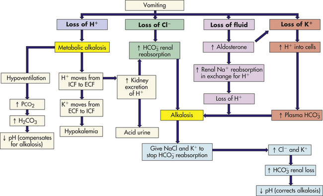
With alkalemia hydrogen ions are redistributed from intracellular to the extracellular space and potassium moves to the intracellular space to preserve electroneutrality.
With hyperaldosteronism the excess aldosterone causes sodium retention and loss of hydrogen and potassium. Mild volume expansion ensues, and bicarbonate is retained along with the sodium, thereby causing alkalosis.
Diuretics, such as thiazides, ethacrynic acid, and furosemide, produce mild alkalosis by enhancing sodium, potassium, and chloride excretion more than bicarbonate excretion.
Respiratory compensation for metabolic alkalosis occurs when the elevated pH inhibits the respiratory center. The rate and depth of ventilation are decreased, causing retention of carbon dioxide. The ratio of to H2CO3 is reduced toward normal. Respiratory compensation is not very efficient, however, and chronic or severe metabolic alkalosis requires therapeutic intervention (Figure 3-14).
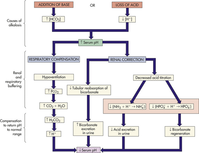
CLINICAL MANIFESTATIONS Because of the many causes of metabolic alkalosis, the symptoms vary. Some common symptoms, such as weakness, muscle cramps, and hyperactive reflexes, are related to volume depletion and electrolyte losses. Because alkalosis increases binding of Ca++ to plasma proteins (albumin), ionized calcium decreases, causing excitable cells to become hypopolarized, which initiates an action potential more easily. Paresthesias, numbness/tingling of the fingertips and perioral area, tetany, and seizures may develop (see Hypocalcemia, p. 112).
Respirations are slow and shallow to increase carbon dioxide content. Confusion and convulsions occur with severe alkalosis. Atrial tachycardia is a potential problem. The oxyhemoglobin curve is shifted to the left (see Chapter 32), decreasing the dissociation of oxyhemoglobin and increasing the risk of dysrhythmias.
EVALUATION AND TREATMENT The health history provides significant clues to the diagnosis of metabolic alkalosis. The arterial pH is greater than 7.45, and bicarbonate levels exceed 26 mEq/L. With respiratory compensation, the PCO2 rises above 40 mmHg. With hypochloremic alkalosis, serum chloride values are below normal. Serum potassium levels are usually depleted because hydrogen is released from the cells in exchange for potassium to help regulate the pH level. The K+ is then secreted from renal distal tubule cells into the urine.
With hypochloremic alkalosis or contraction alkalosis with volume depletion, a sodium chloride solution is required for correction. The renal stimulus to increase ECF volume by retaining Na+ is diminished, and can be excreted as NaHCO3 in the urine. The administration of potassium corrects alkalosis caused by hyperaldosteronism or hypokalemia. The potassium causes hydrogen to move back into the ECF and decreases loss of hydrogen from the distal tubule.
Respiratory Acidosis
PATHOPHYSIOLOGY Respiratory disorders of acid-base balance are caused by increases or decreases of alveolar ventilation in relation to the metabolic production of carbon dioxide. Respiratory acidosis occurs when there is alveolar hypoventilation. Carbon dioxide is retained, increasing [H+] (as H2CO3) and producing acidosis. Carbon dioxide excess is called hypercapnia. The common causes include depression of the respiratory center (brainstem trauma, oversedation), respiratory muscle paralysis, disorders of the chest wall (kyphoscoliosis, pickwickian syndrome, flail chest), and disorders of the lung parenchyma (pneumonitis, pulmonary edema, emphysema, asthma, bronchitis).
Respiratory acidosis may be acute or chronic. Airway obstruction is the most common cause of acute respiratory acidosis. Acute compensation for respiratory acidosis is not effective because the renal buffer mechanism takes time to function. Further, the protein buffers provide marginal compensation, and is not a good buffer for CO2. Acute uncompensated respiratory acidosis is characterized by decreased arterial pH, elevated PCO2, and normal or slightly increased bicarbonate.
Chronic respiratory acidosis is commonly associated with chronic obstructive pulmonary disease and deformities of the chest wall or neuromuscular disorders. Renal compensation is effective and is established over several days. The acidosis produced from CO2 retention stimulates the kidney to secrete hydrogen ions and regenerate bicarbonate. Serum bicarbonate and arterial PCO2 are elevated, and pH is restored toward normal (Figure 3-15).
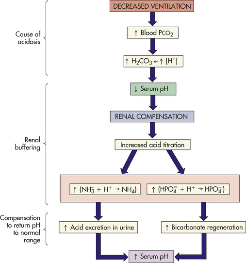
CLINICAL MANIFESTATIONS The symptoms of respiratory acidosis are related to acuity of onset and severity of PCO2 retention. Initial symptoms include headache, restlessness, blurred vision, and apprehension followed by lethargy, muscle twitching, tremors, convulsions, and coma. Neurologic symptoms are caused by a decrease in the pH of cerebrospinal fluid and vasodilation because CO2 readily crosses the blood-brain barrier. The respiratory rate is rapid at first and gradually becomes depressed because over time, the respiratory center adapts to increasing levels of CO2. Cyanosis does not occur unless there is an accompanying hypoxemia, and the skin may instead be pink from vasodilation caused by the elevated CO2.
EVALUATION AND TREATMENT The primary diagnostic indicators are an arterial pH less than 7.35 and hypercapnia. Acute respiratory acidosis must be distinguished from chronic acidosis; the health history and clinical laboratory data are therefore helpful. With renal compensation, bicarbonate levels are elevated and the pH is restored toward normal.
The restoration of adequate alveolar ventilation removes excess CO2. If alveolar ventilation cannot be maintained spontaneously because of drug overdose or neuromuscular disorders, mechanical ventilation is required. The arterial pH, PCO2, PO2, and must be carefully monitored. Rapid reduction of PCO2 can cause respiratory alkalosis with seizures and death.
Renal buffering is usually effective in compensating for uncomplicated chronic respiratory acidosis. The underlying diseases are treated to achieve maximal ventilation. In the presence of hypoxemia and hypercapnia, oxygen can function as a respiratory depressant when the respiratory center is no longer stimulated by the lower pH and elevated PCO2. Therefore, oxygen should be given cautiously.
Respiratory Alkalosis
PATHOPHYSIOLOGY Respiratory alkalosis occurs when there is alveolar hyperventilation and decreased plasma carbon dioxide (termed hypocapnia). Stimulation of ventilation is precipitated by hypoxemia, which may be caused by pulmonary disease, congestive heart failure, or high altitudes; hypermetabolic states such as fever, anemia, and thyrotoxicosis; early salicylate intoxication; hysteria; cirrhosis; and gram-negative sepsis. Improper use of mechanical ventilators can cause iatrogenic respiratory alkalosis. Secondary respiratory alkalosis may develop from hyperventilation stimulated by metabolic or respiratory acidosis.
The onset of acute respiratory alkalosis occurs within minutes of hyperventilation. Cellular buffers provide immediate compensation with shifts of H+ from ICF to ECF. The H+ shifts are not very effective, however, if PCO2 is significantly decreased. When chronic respiratory alkalosis is present, renal compensation restores pH toward normal by decreasing H+ excretion and bicarbonate absorption (Figure 3-16).
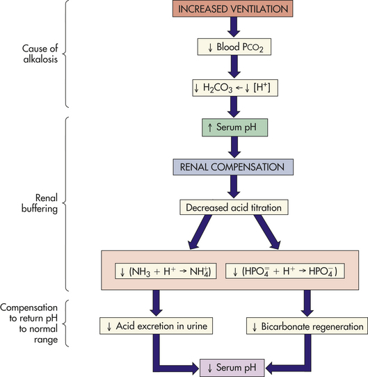
CLINICAL MANIFESTATIONS Respiratory alkalosis, like metabolic alkalosis, is irritating to the central and peripheral nervous systems. Symptoms include dizziness, confusion, tingling of extremities (paresthesias), convulsions, and coma. Carpopedal spasm and other symptoms of hypocalcemia are similar to those of metabolic alkalosis (see p. 120). Deep and rapid respirations (tachypnea) are primary symptoms that cause respiratory alkalosis.
EVALUATION AND TREATMENT The underlying disturbance must be identified. The arterial pH is greater than 7.45, and the PaCO2 is less than 38 mmHg. In acute states, bicarbonate levels are normal. With chronic respiratory alkalosis, a compensatory decrease in the bicarbonate level occurs and the pH is closer to normal.
Treating the underlying disturbance is the most effective treatment. Hypoxemia must be corrected and hypermetabolic states reversed. Symptoms from hysterical hyperventilation can be corrected by rebreathing from a paper bag, which increases the concentration of inspired carbon dioxide and reverses the respiratory alkalosis.
Acidemia 117
Acidosis 117
Aldosterone 101
Alkalemia 117
Alkalosis 117
Angiotensin I and II 101
Anion gap 118
Aquaporins 97
Baroreceptors 102
Buffering 115
Buffers 115
Calcitonin 112
Calcium 111
Chloride 102
Compensation 116
Compulsive water drinking 106
Correction 116
Decreased urine formation 106
Dehydration 104
Dilutional hyponatremia 105
Edema 98
Extracellular fluid (ECF) 96
Familial hypokalemic periodic paralysis 108
Hypercalcemia 113
Hypercapnia 121
Hyperchloremia 106
Hyperchloremic metabolic acidosis 118
Hyperkalemia 110
Hypermagnesemia 114
Hypernatremia 104
Hyperphosphatemia 113
Hyperpolarized 109
Hypertonic hyponatremia 105
Hypocalcemia 112
Hypocapnia 122
Hypochloremia 106
Hypochloremic metabolic alkalosis 119
Hypokalemia 108
Hypomagnesemia 114
Hyponatremia 104
Hypophosphatemia 113
Hypopolarized 110
Hypotonic hyponatremia 105
Hypovolemia 106
Inadequate intake 105
Interstitial fluid 98
Intracellular fluid (ICF) 96
Intravascular fluid 96
Lymphedema 100
Magnesium 114
Metabolic acidosis 117
Metabolic alkalosis 119
Natriuretic peptides 102
Net filtration 98
Nonvolatile 114
Normal anion gap 118
Osmoreceptors 102
Parathyroid hormone 111
Phosphate 111
Potassium adaptation 107
Pure sodium deficits 105
Renin 101
Renin-angiotensin-aldosterone system 102
Respiratory acidosis 121
Respiratory alkalosis 122
Sodium 101
Starling hypothesis 98
Syndrome of inappropriate secretion of ADH (SIADH) 106
Total body water (TBW) 97
Vasopressin dysregulation 106
Vitamin D 111
Volatile 114
Volume-sensitive receptors 102
Water deficits 104
Water intoxication 104
REFERENCES
1. Allison, S.P., Lobo, D.N. Fluid and electrolytes in the elderly. Curr Opin Clin Nutr Metab Care. 2004;7(1):27–33.
2. Fu, D., Lu, M. The structural basis of water permeation and proton exclusion in aquaporins. Mol Membr Biol. 2007;24(5-6):366–374.
3. Warren, A.G., et al. Lymphedema: a comprehensive review. Ann Plast Surg. 2007;59(4):464–472.
4. Lee, C.Y., Burnett, J.C., Jr. Natriuretic peptides and therapeutic applications. Heart Fail Rev. 2007;12(2):131–142.
5. Achinger, S.G., Moritz, M.L., Ayus, J.C. Dysnatremias: why are patients still dying? South Med J. 2006;99(4):353–362.
6. Offenstadt, G., Das, V. Hyponatremia, hypernatremia: a physiological approach. Minerva Anestesiol. 2006;72(6):353–356.
7. Yeates, K.E., Singer, M., Morton, A.R. Salt and water: a simple approach to hyponatremia. CMAJ. 2004;170(3):365–369.
8. Decaux, G., Soupart, A. Treatment of symptomatic hyponatremia. Am J Med Sci. 2003;326(1):25–30.
9. Multz, A.S. Vasopressin dysregulation and hyponatremia in hospitalized patients. J Intensive Care Med. 2007;22(4):216–223.
10. Lien, Y.H., Shapiro, J.I. Hyponatremia: clinical diagnosis and management. Am J Med. 2007;120(8):653–658.
11. Wang, W. Regulation of renal K transported by dietary K intake. Annu Rev Physiol. 2004;66:547–569.
12. Gennari, F.J. Disorders of potassium homeostasis, hypokalemia and hyperkalemia. Crit Care Clin. 2002;18(2):273–288. [vi].
13. Koeppen, B.M., Stanton, B.A. Renal physiology. Philadelphia: Mosby; 2007. [p 116].
14. Alazami, M., et al. Unusual causes of hypokalaemia and paralysis. QJM. 2006;99(3):181–192.
15. Alfonzo, A.V., et al. Potassium disorders—clinical spectrum and emergency management. Resuscitation. 2006;70(1):10–25.
16. Parham, W.A., et al. Hyperkalemia revisited. Tex Heart Inst J. 2006;33(1):40–47.
17. Brunelli, S.M., Goldfarb, S. Hypophosphatemia: clinical consequences and management. J Am Soc Nephrol. 2007;18(7):1999–2003.
18. Albaaj, F., Hutchison, A. Hyperphosphataemia in renal failure: causes, consequences and current management. Drugs. 2003;63(6):577–596.
19. Salusky, I.B., A new era in phosphate binder therapy: what are the options? Kidney Int Suppl. 2006(105):S10–S15.
20. Mouw, D.R., Latessa, R.A., Sullo, E.J. What are the causes of hypomagnesemia? J Fam Pract. 54(2), 2005.
21. Laires, M.H., Monteiro, C.P., Bicho, M. Role of cellular magnesium in health and human disease. Front Biosci. 2004;9:262–276.
22. Onishi, S., Yoshino, S. Cathartic-induced fatal hypermagnesemia in the elderly. Intern Med. 2006;45(4):207–210.
23. Rose, D.B., Post, T. Clinical physiology of acid-base and electrolyte disorders, ed 5. New York: McGraw-Hill; 2001.
24. Kraut, J.A., Madias, N.E. Serum anion gap: its uses and limitations in clinical medicine. Clin J Am Soc Nephrol. 2006;2(1):162–174.
25. Adrogué, H.J. Metabolic acidosis: pathophysiology, diagnosis and management. J Nephrol. 2006;19(Suppl 9):S62–S69.
26. Khanna, A., Kurtzman, N.A. Metabolic alkalosis. J Nephrol. 2006;19(Suppl 9):S86–S96.