STRUCTURE AND FUNCTION OF THE REPRODUCTIVE SYSTEMS


The male and female reproductive systems have several anatomic and physiologic features in common. Most obvious is their major function, reproduction, through which a 23-chromosome female gamete, the ovum, and a 23-chromosome male gamete, the spermatozoon (sperm cell), unite to form a 46-chromosome zygote that is capable of developing into a new individual. The male reproductive system produces sperm and delivers them to the female reproductive tract. The female reproductive system produces the ovum and, if it is fertilized, can nurture and protect it (at that point called the embryo and developing fetus) and expel it at birth. These functions are determined not only by anatomic structures but also by complex hormonal, neurologic, and psychogenic factors.1
DEVELOPMENT OF THE REPRODUCTIVE SYSTEMS
The structure and function of male and female reproductive systems depend on steroid hormones called sex hormones. Hormonal effects on the reproductive systems begin well before birth and continue for life.
Sexual Differentiation and Hormone Production in Utero
During embryonic development, the initial reproductive structures of male and female embryos are homologous (the same) and consist of one pair of primary sex organs, or gonads, and two pairs of ducts, the mesonephric ducts (wolffian ducts) and the paramesonephric ducts (müllerian ducts) (Figure 22-1). Both pairs of ducts empty into an opening called the urogenital sinus.
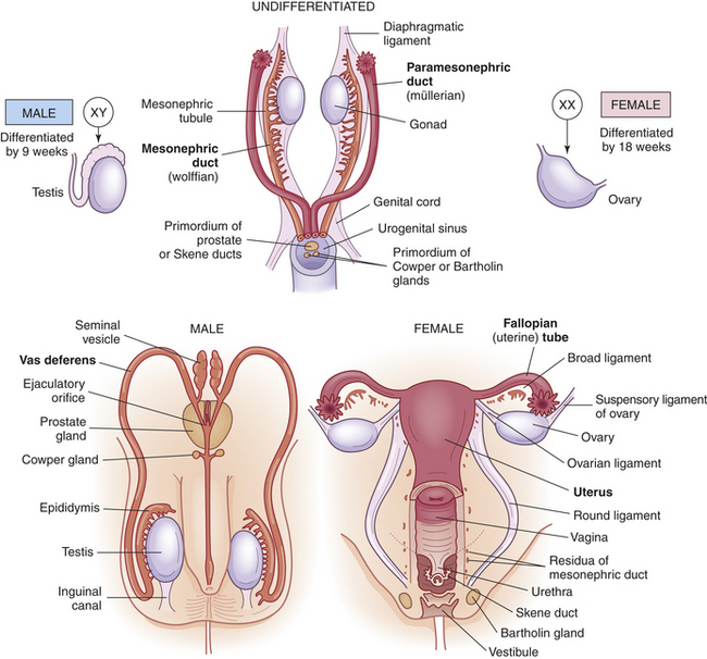
Figure 22-1 Internal genitalia development. Embryonic and fetal development of the internal genitalia.
Between 6 and 7 weeks’ gestation, the male embryo will differentiate under the influence of testes-determining factor (TDF). The TDF is from a gene in the sex-determining region on the Y chromosome (SRY). This gene stimulates the gondal development of testes, which in turn produces antimüllerian hormone (AMH) and testosterone. AMH inhibits the formation of müllerian ducts. As the testes begin to differentiate, Sertoli cells appear and aggregate to form testicular cords. Mature Sertoli cells will produce inhibin and androgen-binding protein (ABP), which is important for spermatogenesis. Leydig cells differentiate at the beginning of the eighth week and the secretion of testosterone begins.1,2 Under the influence of testosterone, the male gonads develop into two testes that produce sperm after puberty. The paramesonephric ducts degenerate and the mesonephric ducts develop (wolffian system) into the vas deferens—the two tubes that carry sperm from the testes to the urethra (see Figure 22-1). By 9 months’ gestation the male gonads (testes) have descended into the scrotum.
In female embryos the gonads produce the primary female sex hormone, estrogen. In the absence of testosterone there is a loss of the Wolffian system and the two gonads develop into ovaries at 6 to 8 weeks’ gestation. There is rapid mitotic multiplication of germ cells and by 16 to 20 weeks there is a peak of 6 to 7 million oogonia. Oogonia are transformed into oocytes throughout the remainder of the pregnancy, when they enter the first meiotic division. This will remain stable until puberty. Just before ovulation a single ovum will be formed from two meiotic divisions of the oocytes. By birth only 1 to 2 million oocytes remain.1 In females the mesonephric ducts deteriorate and the lower ends of the paramesonephric ducts join to become the uterus, fallopian tubes, cervix, and upper two thirds of the vagina. The upper portions of the paramesonephric ducts develop into the fallopian (uterine) tubes (Figure 22-2). These two ducts carry ova from the ovaries to the uterus.
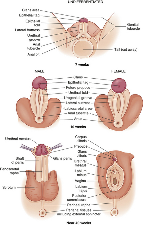
Figure 22-2 External genitalia development. Embryonic and fetal development of the external genitalia.
Like the internal reproductive structures, the external structures develop from homologous embryonic tissues. During the first 7 to 8 weeks of gestation, both male and female embryos develop an elevated structure called the genital tubercle. Figure 22-2 shows how the undifferentiated genital tubercle develops into the external reproductive organs. Under hormonal influence, male genitalia develop when testosterone is present. Female genitalia will develop if testosterone or testosterone receptors are absent even in the absence of ovaries.
Anterior pituitary development starts between the fourth and fifth weeks of fetal life and the vascular connection between the hypothalamus and the pituitary is established by the twelfth week. In the female fetus, high levels of two gonadotropins, follicle-stimulating hormone (FSH) and luteinizing hormone (LH) are excreted by the anterior pituitary. Gonadotropin-releasing hormone (GnRH) is produced in the hypothalamus by 10 weeks’ gestation; this controls the production of the gonadotropins LH and FSH.1 This cycle is referred to as the hypothalamic-pituitary axis (HPA) and stimulates the production of estrogen and progesterone by the ovary. The production of FSH and LH rises until about 28 weeks’ gestation, until the production of estrogen and progesterone by the ovaries and placenta is high enough to result in the decline of gonadotropin production.
At term, a sensitive negative-feedback system, which includes the gonadostat (also known as the GnRH pulse generator), is operative in the human fetus. The gonadostat responds to high placental estrogens by releasing low levels of GnRH. Soon after birth, sex hormones drop precipitously, probably because of withdrawal of placental steroid hormones; negative feedback action of the sex steroids on the hypothalamus and pituitary is removed; and the gonadotropins LH and FSH are released. The gonadotropins will be suppressed after the first year of life until approximately age 10. During infancy and early childhood, the gonadostat is remarkably sensitive (6 to 15 times more sensitive than in the adult) to negative feedback,1 and GnRH secretion is restrained by extraordinarily low levels of estrogen or testosterone. This feedback mechanism is probably an intrinsic neuronal inhibitory system, which suppresses endogenous GnRH secretion and gonadotropin synthesis.1,3 By age 4, low levels of gonadotropins parallel low levels of sex steroids.
Puberty
Puberty is the onset of sexual development and differs from adolescence. Adolescence is the stage of human development between childhood and adulthood and includes social, psychologic, and biologic changes. The onset of puberty normally begins between 8 and 14 years with a pulsatile release of GnRH followed by an increase in LH and FSH. This results in episodic peaks of estradiol and testosterone, which initiates the secondary sexual maturation in girls and boys, respectively. Genetics, general health, and nutrition can influence the timing of puberty. Girls who are mildly obese mature earlier and girls who have low body fat from diet and intense exercise will have delayed maturation.4 In the United States, puberty begins with accelerated growth followed by thelarche (breast development) at about 9 years of age in white girls and 8 years of age in black girls. Early puberty in girls has been linked to obesity and more recently to increased levels of leptin, a hormone secreted from adipose tissue, which acts on central nervous system (CNS) neurons that regulate appetite.3,5,6 Leptin levels increase through childhood until the onset of puberty and then decrease as puberty advances. Leptin probably contributes to increased adipose tissue, thereby allowing maturation to occur.1
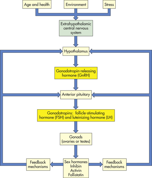
Puberty is a process that involves a complex series of interrelated physiologic changes leading to reproductive maturation.3 Reproductive maturation involves the hypothalamic-pituitary-gonadal (H-P-G) axis, the central nervous system, and the endocrine system (Figure 22-3). As puberty approaches, there is a sequential series of hormonal events that promote sexual maturation. Release of GnRH stimulates secretion of LH and then FSH, which in turn stimulates gonadal maturation (gonadarche) with estradiol secretion in girls and testosterone secretion in boys. Estradiol causes breast development, maturation of the reproductive organs (vagina, uterus, ovaries), and fat deposit in hips in girls. Estrogen and increased production of growth factors cause rapid skeletal growth in both boys and girls.7 Testosterone causes growth of the testes, scrotum, and penis. A positive feedback loop is created with gonadotropins stimulating the gonads to produce more sex hormones. (The sex hormones are discussed in the sections on the female and male systems.) Adrenarche is the increased production of adrenal androgens prior to puberty, which occurs in both sexes, and is exhibited by axillary and pubic hair growth.1,3
The most important hormonal effects occur in the gonads. In males the testes begin to produce mature sperm that are capable of fertilizing an ovum. Male puberty is complete with the first ejaculation that contains mature sperm. In females, the ovaries begin to release mature ova. Female puberty is complete at the time of the first ovulatory menstrual period; however, this can take up to 1 to 2 years after menarche. Puberty is complete when an individual is capable of reproduction.
THE FEMALE REPRODUCTIVE SYSTEM
The function of the reproductive system is to produce mature ova and, when they are fertilized, to protect and nourish them through embryonic and fetal life and expel them at birth. In females the most important reproductive organs, or genitalia, are internal. They are the ovaries, fallopian tubes, uterus, and vagina. These organs are essential to reproduction. The external genitalia have accessory functions. They protect body openings and play an important role in sexual functioning.1,8,9
External Genitalia
Figure 22-4 shows the external female genitalia, which are known collectively as the vulva, or pudendum. The major structures are as follows.
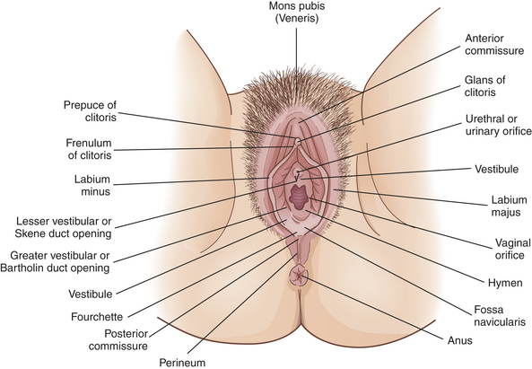
The mons pubis (also known as mons veneris) is a fatty layer of tissue over the pubic symphysis (joint of the pubic bones). During puberty the mons pubis becomes covered with pubic hair, and its sebaceous and sweat glands become more active. Estrogen causes fat to be deposited under the skin, giving the mons pubis a moundlike shape. This cushion of tissue protects the pubic symphysis during sexual intercourse.
The labia majora (singular, labium majus) are two folds of skin that arise at the mons pubis and extend back to the fourchette, forming a cleft. Like the mons pubis, the labia majora undergo changes at puberty: the amount of fatty tissue increases, pubic hair grows on the lateral surfaces, and sebaceous glands on the hairless medial surfaces begin to secrete lubricants. Because of an extensive network of nerve endings, the labia majora are highly sensitive to temperature, touch, pressure, and pain and are homologous to the male scrotum (see Figure 22-2 and Figure 22-13). The principal function of the labia majora is to protect the inner structures of the vulva.
The labia minora (singular, labium minus), two smaller, thinner folds of skin, lie within the labia majora. Anteriorly they form the clitoral hood, or prepuce, and frenulum, then split to enclose the vestibule, and converge near the anus, forming the fourchette. The labia minora are hairless, pink, and moist and are well supplied with nerves, blood vessels, and sebaceous glands. These glands secrete a bactericidal fluid that has a distinctive odor and that lubricates and waterproofs the vulvar skin. During sexual arousal the labia minora become swollen with blood.
The clitoris is a richly innervated, erectile organ that lies anterior, between the labia minora. It is a small, cylindrical structure having a glans that is visible and a shaft that lies beneath the skin (see Figure 22-4). The clitoris is homologous to the male penis. Like the penis, the clitoris is a major site of sexual stimulation and orgasm. With sexual arousal, erectile tissues in the clitoris fill with blood, causing it to enlarge somewhat. Similar to other vulvar glands, the clitoris secretes a fluid, called smegma, which has a unique odor and may be erotically stimulating to the male.
The vestibule is the area protected by the labia minora and contains the external opening of the vagina, which is called the introitus, or vaginal orifice. A thin, perforated membrane called the hymen may cover the introitus. The vestibule also contains the opening of the urethra, or urinary meatus (orifice). These structures are lubricated by two pairs of glands: Skene glands and Bartholin glands. The ducts of Skene glands (also called the lesser vestibular or paraurethral glands) open on both sides of the urinary meatus. The ducts of Bartholin glands (greater vestibular or vulvovaginal glands) open on either side of the introitus. In response to sexual stimulation, Bartholin glands secrete mucus that lubricates the inner labial surfaces, as well as enhances the viability and motility of sperm. Skene glands help lubricate the urinary meatus and the vestibule. Secretions from both sets of glands facilitate coitus. Also, in response to sexual excitement, the highly vascular tissue just beneath the vestibule fills with blood and becomes engorged.
The less hairy skin and the subcutaneous tissue that lie between the vaginal orifice and the anus are referred to as the perineum. Unlike the rest of the vulva, this area has little subcutaneous fat so that the skin is close to the underlying muscles. The perineum covers the muscular perineal body, a fibrous structure that comprises elastic fiber, connective tissue, and the common attachment of the bulbocavernosus, the external anal sphincter, and the levator ani muscles (see Figure 22-4). The perineum varies in length from 2 to 5 cm or more and stretches remarkably. The length of the perineum and the elasticity of the perineal body influence tissue resistance and injury during childbirth.
Internal Genitalia
The vagina is an elastic fibromuscular canal, 9 to 10 cm long in a reproductive-aged female, which extends up and back from the introitus to the lower portion of the uterus. As Figure 22-5 shows, it lies between the urethra (and part of the bladder) and the rectum. Mucosal secretions from the upper genital organs, menstrual fluids, and products of conception leave the body through the vagina, which also receives the penis during coitus. During sexual excitement the vagina lengthens and widens and the anterior third becomes congested with blood.
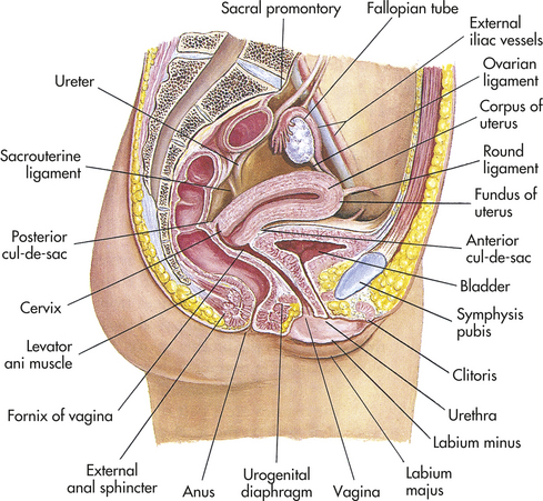
Figure 22-5 Internal female genitalia and other pelvic organs. Midsagittal view. (Modified from Seidel HM et al: Mosby’s guide to physical examination, ed 6, St Louis, 2006, Mosby.)
The vaginal wall is composed of four layers:
1. Its lining is a mucous membrane of squamous epithelial cells. (Types of epithelium are described and illustrated in Chapter 1, Table 1-7.) This layer thickens and thins in response to hormones, particularly estrogen. The squamous epithelial membrane is continuous with the membrane that covers the lower part of the uterus. In women of reproductive age, the mucosal layer is arranged in transverse wrinkles, or folds, called rugae (singular, ruga) that permit stretching during coitus and childbirth.
2. Fibrous connective tissue containing numerous blood and lymphatic vessels.
The upper part of the vagina surrounds the cervix, the lower end of the uterus (see Figure 22-5). The recessed space around the cervix is called the fornix of the vagina. The posterior fornix is “deeper” than the anterior fornix because of the angle at which the cervix meets the vaginal canal. In most women this angle is about 90 degrees. A pouch called the cul-de-sac separates the posterior fornix and the rectum.
Its elasticity and relatively sparse nerve supply enhance the vagina’s function as the birth canal. During sexual arousal the vaginal wall becomes engorged with blood, like the labia minora and clitoris. Engorgement pushes some fluid to the surface of the mucosa, enhancing lubrication. The vaginal wall does not contain mucus-secreting glands; rather, secretions drain into the vagina from the endocervical glands or enter from the vestibule, from the Bartholin and Skene glands.
Two factors help maintain the self-cleansing action of the vagina and defend it from infection, particularly during the reproductive years: (1) an acid-base balance that discourages the proliferation of most pathogenic bacteria and (2) the thickness of the vaginal epithelium. Before puberty, vaginal pH is about 7 (neutral) and the vaginal epithelium is thin. At puberty, the pH becomes more acidic (4 to 5) and the squamous epithelial lining thickens. These changes are maintained until menopause (cessation of menstruation), at which time the pH rises again to more alkaline levels and the epithelium thins out. Therefore, protection from infection is greatest during the years when a woman is most likely to be sexually active. Between puberty and menopause, vulnerability to infection varies somewhat with cyclic changes in pH and epithelial thickness. Both defenses are greatest when estrogen levels are high and the vagina contains a normal population of Lactobacillus acidophilus, a harmless resident bacterium that helps maintain pH at acidic levels. Any condition that causes vaginal pH to rise, such as douching or use of vaginal sprays or deodorants, low estrogen levels, or destruction of L. acidophilus by antibiotics, lowers vaginal defenses against infection.
Uterus
The uterus is a hollow pear-shaped organ whose lower end opens into the vagina. The functions of the uterus are to anchor and protect a fertilized ovum, provide an optimal environment while it develops, and push the fetus out at birth. In addition, the uterus plays an important role in sexual response and conception. During sexual excitement the opening of the uterus (the cervix) dilates slightly. At the same time, the uterus increases in size and moves upward and backward, creating a tenting effect in the midvagina that results in the cervix “sitting” in a pool of semen. During orgasm, rhythmic contractions facilitate movement of sperm through the cervical os while also enhancing physical pleasure.
At puberty the uterus attains its adult size and proportions and descends from the abdomen to the lower pelvis, between the bladder and the rectum (see Figure 22-5). The uterus of a mature, nonpregnant female is approximately 7 to 9 cm long and 6.5 cm wide, with muscular walls 3.5 cm thick. It is held loosely in position by ligaments, peritoneal tissue folds, and pressure of adjacent organs, especially the urinary bladder, sigmoid colon, and rectum. In most women the uterus is anteverted; that is, it is tipped forward so that it rests on the urinary bladder. However, it may be retroverted, or tipped backward. Various degrees of flexion are normal (Figure 22-6).
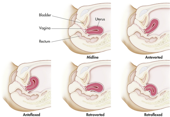
Figure 22-7 shows a cross section of the uterus. The uterus has two major parts: the body, or corpus, and the cervix. The top of the corpus, above the insertion of the fallopian tubes, is called the fundus. The diameter of the uterine cavity is widest at the fundus and narrowest at the isthmus, which is the narrowed part of the corpus just above the cervix. The cervix, or “neck of the uterus,” extends from the isthmus to the vagina. The passageway between the cervix’s upper opening (the internal os) and its lower opening (the external os) is called the endocervical canal. The entire uterus, like the upper vagina, is innervated exclusively by motor and sensory fibers of the autonomic nervous system.
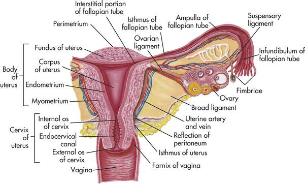
Figure 22-7 Cross section of uterus, fallopian tube, and ovary. (From Seidel HM et al: Mosby’s guide to physical examination, ed 6, St Louis, 2006, Mosby.)
The uterine wall is composed of three layers: the perimetrium, the myometrium, and the endometrium (see Figure 22-7). The perimetrium (parietal peritoneum) is the outer serous membrane that covers the uterus. The myometrium is the thick muscular middle layer. The myometrium is thickest at the fundus, apparently to facilitate birth. The endometrium, or uterine lining, is composed of a functional layer (superficial compact layer and spongy middle layer) and a basal layer. The functional layer of the endometrium is responsive to sex hormones. Between puberty and menopause this layer proliferates and sloughs off monthly. The basal layer, which is attached to the myometrium, regenerates the functional layer after it sloughs (menstruation).
The endocervical canal does not have an endometrial layer. Rather, it is lined with columnar epithelial cells (see Table 1-7). The endocervical lining is continuous with that of the outer cervix and vagina, but it is not made up of the same type of epithelial cells. The point at which the columnar epithelium of the cervix meets the squamous epithelium of the vagina is called the transformation zone, or the squamous-columnar junction. The transformation zone is especially susceptible to the oncogenic human papillomavirus (HPV), which leads to cervical dysplasia and, ultimately, cervical cancer; these are the cells sampled during a Papanicolaou test (Pap test).10
The cervix acts as a mechanical barrier to infectious microorganisms that may be present in the vagina. The external cervical os is a very small opening that contains thick, sticky mucus (the mucous plug) during the luteal phase of the menstrual cycle and all of pregnancy. During ovulation, the mucus changes under the influence of estrogen and forms watery strands, or spinnbarkeit mucus, to facilitate the transport of sperm into the uterus. In addition, the downward flow of cervical secretions moves microorganisms away from the cervix and uterus. In women of reproductive age, the pH of these secretions is inhospitable to most bacteria. Further, mucosal secretions contain enzymes and antibodies (mostly immunoglobulin A) of the secretory immune system. (The secretory immune system is discussed in Chapter 7.) These defenses do not always prevent infection, even if they are intact. Besides infection, uterine pathophysiology includes displacement of the uterus within the pelvis, benign growths (fibroids) of the uterine wall, hyperplasia of the endometrium, endometriosis, and cancer.
Fallopian Tubes
The two fallopian tubes (oviducts, uterine tubes) enter the uterus bilaterally just beneath the fundus (see Figure 22-7). Their function is to conduct the ova from the spaces around the ovaries to the uterus. From the uterus the fallopian tubes curve up and over the two ovaries. Each tube is 8 to 12 cm long and about 1 cm in diameter, except at its ovarian end, which flares out like the bell of a trumpet. This widened end, called the infundibulum, is fringed or fimbriated. The fimbriae (singular, fimbria) (fringes) move, creating a current that draws the ovum into the infundibulum. Once the ovum has entered the fallopian tube, cilia and peristalsis (muscle contractions) keep it moving toward the uterus.
The ampulla, or distal third, of the fallopian tube is the usual site of fertilization (see Figure 22-7). Sperm released into the vagina travel upward through the endocervical canal and uterine cavity and enter the fallopian tubes. If an ovum is present in either tube, fertilization can occur. Whether or not the ovum encounters sperm, it continues to travel through the fallopian tube to the uterus. If fertilized, the ovum (then called a blastocyst) implants itself in the endometrial layer of the uterine wall. If not fertilized, the ovum breaks down within 12 to 24 hours.
Disorders that affect the fallopian tubes can block the path of sperm and ovum and cause infertility or ectopic (tubal) pregnancy. Such disorders include congenital malformations, infection, and inflammation.
Ovaries
The ovaries, or the female gonads, are the primary female reproductive organs. They have two main functions: secretion of female sex hormones and development and release of female gametes, or ova.
The almond-shaped ovaries are located on both sides of the uterus and are suspended and supported by the mesovarian portions of the broad ligament, ovarian ligaments, and suspensory ligaments (see Figure 22-7). The ovaries are smaller than their male homologs, the testes. In women of reproductive age, each ovary is 3 to 5 cm long, 2.5 cm wide, and 2 cm thick and weighs 4 to 8 g. Size and weight vary somewhat from phase to phase of the menstrual cycle (see p. 792).
Figure 22-8 shows a cross section of an ovary. The central part, or medulla, is composed of connective tissue and contains many small arteries, veins, and lymphatics that enter at the hilum. Surrounding the medulla is the cortex. At birth the cortex of each ovary contains approximately 2 million ova within immature ovarian follicles. Follicles grow and undergo atresia continuously and irrevocably during a woman’s life. By puberty the number ranges between 300,000 and 500,000 ova. During puberty some of the follicles and the ova within them begin to mature. Between puberty and menopause the ovarian cortex always contains follicles and ova in various stages of development. Once every menstrual cycle (about every 28 days), usually only one of the follicles reaches maturation and discharges its ovum through the ovary’s outer covering, the germinal epithelium. During the reproductive years, 400 to 500 ovarian follicles mature completely and release an ovum, an event termed ovulation. The rest either fail to develop at all or degenerate without maturing completely.1
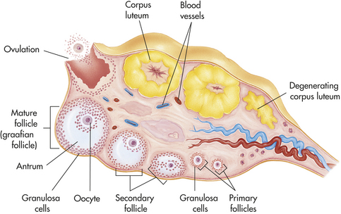
Figure 22-8 Cross section of ovary during reproductive years. (From Patton KT, Thibodeau GA: Anatomy & physiology, ed 7, St Louis, 2010, Mosby.)
Having ejected a mature ovum, the follicle develops into another structure, the corpus luteum (see Figure 22-8). The immediate fate of the corpus luteum depends on whether the ejected ovum is fertilized. If fertilization occurs, the corpus luteum enlarges and begins to secrete hormones that maintain and support pregnancy. If fertilization does not occur, the corpus luteum secretes these hormones for approximately 14 days and then degenerates, which triggers the maturation of another follicle. The ovarian cycle—the process of follicular maturation, ovulation, corpus luteum development, and corpus luteum degeneration—is continuous from puberty to menopause, except during pregnancy or hormonal contraceptive use. At menopause, this process ceases and the ovaries atrophy to the point that they cannot be felt during pelvic examination.
Four types of cells within the ovarian cortex secrete sex hormones: cells of the stroma, or tissue matrix; two types of cells in the ovarian follicle, granulosa cells and theca cells, and cells of the corpus luteum (Figure 22-9). These cells all contain receptors for the gonadotropins (LH and FSH) or for the sex hormones, which are discussed in the next section.
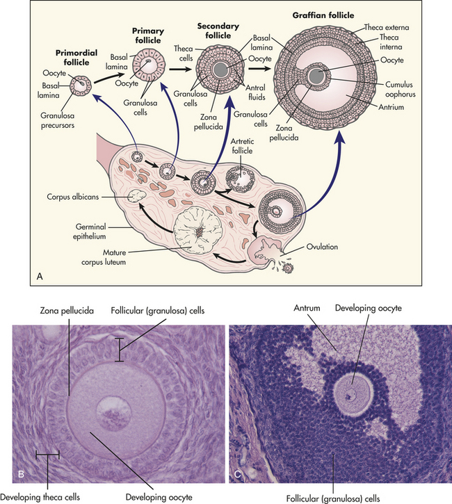
Figure 22-9 Development of an ovarian follicle. A, Schematic representation (not to scale) of the structure of the ovary, showing the various stages in the development of the follicle and its successor structure, the corpus luteum. B, A developing oocyte surrounded by hormone-secreting follicular (granulosa) cells. C, A more mature ovarian follicle has a fluid-filled cavity called the antrum. (A from Berne RM, Levy MN, editors: Physiology, ed 5, St Louis, 2003, Mosby. B and C from Patton KT, Thibodeau GA: Anatomy & physiology, ed 7, St Louis, 2010, Mosby.)
Because gonadotropins and hormones regulate ovarian function, any disorder that disrupts this process, such as abnormal pituitary or thyroid function, or reception by target cells can cause ovarian dysfunction and infertility. Benign or malignant growths, cysts, infection, or inflammation also can cause ovarian pathologic conditions.1
Female Sex Hormones
The sex hormones are all steroid hormones, that is, they are synthesized from cholesterol (see Chapter 20 and Chapter 22). Male and female sex hormones are present in all adults. However, the female body contains low levels of testosterone and other androgens, and the male body contains low levels of estrogen. Individual effects of sex hormones depend on their amount and concentration in the blood.
The dominant female sex hormones, estrogen and progesterone, are produced primarily by the ovaries. During fetal development, infancy, and childhood, sex hormone production is low. At puberty hormone production surges, triggering sexual maturation and development of secondary sex characteristics. From puberty to menopause, the sex hormones control the menstrual cycle and are produced cyclically, that is, production surges and diminishes monthly, creating the ovarian and uterine changes associated with the menstrual cycle. These hormones are also produced in higher levels during pregnancy by the placenta and inhibit ovulation. Androgens are produced in small amounts by the ovaries and the adrenals and have important functions in women.
Estrogens
Estrogen is a generic term for three similar hormones: estradiol, estrone, and estriol. Estradiol (E2) is the most potent and plentiful of the three and is principally produced by the ovaries (ovarian follicle and corpus luteum). The ovary secretes about 95% of circulating estradiol, with limited amounts secreted by the adrenal cortex. Androgens are converted to estrone in ovarian and peripheral adipose tissue, and estriol is the peripheral metabolite of estrone and estradiol.
Estrogen has numerous biologic effects, many of which involve interactions with other hormones. Estrogen is needed for maturation of reproductive organs, development of secondary sex characteristics, closure of long bones after the pubertal growth spurt, regulation of the menstrual cycle, and endometrial regeneration after menstruation. Estrogen also has metabolic effects on the bones, liver, blood vessels, brain and central nervous system, kidneys, and skin. After menopause, ovarian production of estradiol and estrone is markedly diminished. For this reason, postmenopausal women are susceptible to osteoporosis, a condition in which bone density is reduced. At this time, the majority of estrogen is derived from extraovarian and extraglandular production of estrones.1 (Hormone levels during perimenopause are discussed in the section on menopause, p. 807.)
Like other steroid hormones, estrogens are derived from cholesterol in a complex, enzyme-mediated series of reactions. (Mechanisms of hormone synthesis and action are described in Chapter 20.) The hypothalamus secretes GnRH in a pulsating manner that stimulates gonadotropin (LH and FSH) release from the anterior pituitary. Gonadotropins trigger ovarian production of estrogen. The primary function of LH is to stimulate theca cells of the ovarian follicle to produce androgens, mainly androstenedione. (Androgens are discussed further on p. 800 and in the section on male reproductive function.) Some of these androgens are converted to estrogen by the theca cells themselves, and others diffuse into the granulosa cells. Within the granulosa layer, FSH induces conversion (aromatization) of androgens to estrogens. Estrogens are then released into the bloodstream. Estrogen and FSH together increase FSH receptors in the follicle, stimulating additional granulosa cells until a dominant follicle is determined.
Disturbances of estrogen production can be caused by abnormalities that affect (1) secretion of GnRH by the hypothalamus, (2) secretion of LH or FSH by the anterior pituitary, (3) hormonal feedback mechanisms, or (4) structural integrity of the ovaries. Estrogen’s role in the menstrual cycle is described on p. 794.
Progesterone
Luteinizing hormone (LH) stimulates the ovary to release the ova and secrete progesterone, the second major female sex hormone. LH surge occurs when there is a peak level of estrogen, about 24 to 36 hours before ovulation. LH promotes luteinization of the granulosa in the dominant follicle and results in progesterone production. A rising level of progesterone can be detected from the preovulatory follicle as early as day 10 of the menstrual cycle. Small amounts of progesterone also are secreted steadily by the adrenal cortex. During the follicular phase the ovary and the adrenal glands each contribute approximately 50% of total progesterone production. Conversely, large amounts are secreted cyclically from the ovary while the corpus luteum is active for about 9 to 13 days after ovulation. Together, estrogen and progesterone control the menstrual cycle. The opposing and complementary effects of progesterone and estrogen are listed in Table 22-1. Progesterone secreted by the corpus luteum stimulates the thickened endometrium to become more complex in preparation for implantation of a blastocyte. If conception and implantation do occur, the corpus luteum persists and secretes progesterone (and estrogen) until the placenta is well established at approximately 8 to 10 weeks’ gestation.
Table 22-1
Complementary and Opposing Effects of Estrogen and Progesterone
| Structure | Effect of Estrogen | Effect of Progesterone |
| Vaginal mucosa | Proliferation of squamous epithelium; increase in glycogen content of cells; layering (cornification) of cells | Thinning of squamous epithelium; decornification |
| Cervical mucosa | Production of abundant fluid secretions that favor survival and enhance motility of sperm | Production of thick, sticky secretions that tend to “plug” the cervical os |
| Fallopian tube | Increase of motility and ciliary action | Decrease of motility and ciliary action |
| Uterine muscle | Increase of blood flow; increase of contractile proteins and uterine muscle and myometrial excitability and action potential; increase of sensitization to oxytocin | Relaxation of myometrium; decrease of sensitization to oxytocin |
| Endometrium | Stimulation of growth; increase in number of progesterone receptors | Activation of glands and blood vessels; accumulation of glycogen and enzymes; decrease in number of estrogen receptors |
| Breasts | Growth of ducts; promotion of prolactin effects | Growth of lobules and alveoli; inhibition of prolactin effects |
Progesterone is sometimes called the hormone of pregnancy. Its effects in pregnancy include (1) maintenance of the thickened endometrium; (2) relaxation of smooth muscle in the myometrium, which prevents premature contractions and helps the uterus expand; (3) thickening of the myometrium, which prepares it for the muscular work of labor; (4) prevention of lactation until the fetus is born; and (5) prevention of additional maturation of ova by way of suppressing FSH and LH, thereby stopping the menstrual cycle.
Androgens
Although androgens are primarily male sex hormones, small amounts are produced in the ovary and adrenal cortex in women. Some androgens are precursors of female sex hormones, notably androstenedione. At puberty, androgens contribute to the skeletal growth spurt and cause growth of pubic and axillary hair. The androgens also activate sebaceous glands, accounting for some cases of acne during puberty, and play a role in libido.
The Menstrual Cycle
Besides pregnancy, the obvious manifestation of female reproductive functioning is menstrual bleeding (the menses), which starts with menarche (first menstruation) and ends with menopause (cessation of menstrual flow). In the United States the average age of first menstruation is 12.1 years in black females and 12.7 years in white females, with a range from 9 to 17 years. Menarche appears to be related to body weight, especially percentage of body fat (ratio of fat to lean tissue), which theoretically may trigger a change in the metabolic rate and lead to hormonal changes associated with ovulation. The presence of leptin, a hormone secreted from adipose tissue, helps bring about the onset of pubery.3 At first, cycles are anovulatory and may vary in length from 10 to 60 days or more. As adolescence proceeds, regular patterns of menstruation and ovulation are established at intervals ranging from 25 to 35 days.11–13 During adulthood, menstruation continues to recur in a recognizable and characteristic pattern, with the length of the menstrual cycle varying considerably among women. If a woman is to experience regular and predictable menstruation, it usually happens by 20 years of age. Around age 25 in more than 40% of cycles, menstrual cycle length is between 25 and 28 days; the percentage increases to 60% between ages 25 and 35 years.1 The commonly accepted cycle average is 28 (27 to 30) days, with rhythmic intervals of 21 to 35 days considered normal. Approximately 2 to 4 years before menopause, cycles begin to lengthen again. Menstrual cyclicity and regular ovulation are dependent on (1) the activity of the gonadostat (GnRH pulse generator); (2) the pituitary secretion of gonadotropins; and (3) estrogen (estradiol) positive feedback for the preovulatory LH surge, oocyte maturation, and corpus luteum formation.3
Phases
The menstrual cycle consists of three phases of one event, ovulation. The three phases are the follicular/proliferative phase; the luteal/secretory phase; and the ischemic/menstrual phase, known as menstruation (Figure 22-10). During ovulation an ovum from a mature ovarian follicle is released.
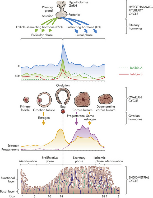
Figure 22-10 The menstrual cycle. (From Lowdermilk DL, Perry SE, Bobak IM: Maternity and women’s health care, ed 8, St Louis, 2004, Mosby.)
During menstruation (menses), the functional layer of the endometrium disintegrates and is discharged through the vagina. Menstruation is followed by the follicular/proliferative phase (FP). This phase is named for two simultaneous processes: maturation of an ovarian follicle and proliferation of the endometrium (see Figure 22-10). During the follicular/proliferative phase, a number of follicles are stimulated to mature. The anterior pituitary gland has a pulsatile secretion of FSH that recruits, or perhaps rescues, a dominant ovarian follicle from apoptosis. From the time of recruitment to achieving preovulatory status, the dominant follicle will mature over 85 days.1 While the follicle is developing, FSH induces aromatization of androgen to estrogen and stimulates granulosa cells to produce a follicular fluid rich in estrogen. Together estrogen and FSH increase FSH receptors in the granulosa cells of the primary follicle, which makes the granulosa cells more sensitive to FSH. The FSH and estrogen combine to induce production of LH receptors on the granulosa cells, thus promoting LH stimulation to combine with FSH stimulation with a more rapid secretion of follicular estrogen. As estrogen increases, FSH levels drop decreasing growth of the less-developed follicles (see Figure 22-10). Estrogen causes cells of the endometrium to proliferate. LH is required for final follicular growth and ovulation. About 2 days before ovulation, an LH surge from the anterior pituitary causes progesterone production in the granulosa layer of the ovary with a decreased rate of estrogen secretion. Final maturation of the dominant follicle then occurs. By the time the ovarian follicle is mature, the endometrial lining is restored. When the lesser follicles fail to achieve full maturity, they retain their ability to respond to LH and steroid production, even though they return to stromal tissue. Increase in stromal tissue in the late follicular phase is associated with a rise in androgen levels. Androgen production enhances the process of follicle atresia and may stimulate libido at the point of ovulation.1
Ovulation marks the beginning of the luteal/secretory phase of the menstrual cycle. The ovarian follicle begins its transformation into a corpus luteum (see Figure 22-8), hence the name luteal phase. Pulsatile secretion of LH from the anterior pituitary stimulates the corpus luteum to secrete progesterone, which in turn initiates the secretory phase of endometrial development. Glands and blood vessels in the endometrium branch and curl throughout the functional layer, and the glands begin to secrete a thin glycogen-containing fluid, hence the name secretory phase. If conception occurs, the nutrient-laden endometrium is ready for implantation. If conception and implantation do not occur, the corpus luteum degenerates and ceases its production of progesterone and estrogen. Without progesterone or estrogen to maintain it, the endometrium enters the ischemic (blood-starved) portion of the menstrual phase and disintegrates. Menstruation occurs, marking the beginning of another cycle.
Ovarian cycles appear to have a minimum length of 24 to 26.5 days: the primary ovarian follicle requires 10 to 12.5 days to develop, and the luteal phase appears relatively fixed at 14 days (±3 days). Menstrual blood flow usually lasts 3 to 7 days, but it may last as long as 8 days or stop after 1 to 2 days and still be considered within normal limits. Bleeding is consistently scant to heavy and varies from 30 to 80 ml, with most blood loss occurring during the first 3 days of menses. Menstrual discharge consists of blood, mucus, and desquamated endometrial tissue and does not clot under normal circumstances. It is usually dark and produces a characteristic musty odor on oxidation. Factors such as severe emotional stress, illness, malnutrition, and seasonal variation may affect the length of the menstrual cycle.1,12–14
Hormonal Controls
Hormonal control of the menstrual cycle depends on complex interactions among the hypothalamus, the anterior pituitary, and the ovaries (or hypothalamic-pituitary-ovarian [H-P-O] axis).14 GnRH production is stimulated as a result of feedback mechanisms originating in the dominant follicle, which is determined in the first 5 to 7 days of the cycle. GnRH is secreted by the hypothalamus into the hypophyseal portal system and travels to the anterior pituitary, where it stimulates the secretion of LH and FSH. FSH and LH are released from the anterior pituitary in pulses that correspond to the pulsatile secretion of GnRH.
During the early follicular phase, estrogen levels rise steadily and, through negative feedback, suppresses FSH and positively increase the production of LH. During the late follicular phase, the preovulatory rise in progesterone facilitates the positive feedback of estrogen; estrogen levels begin to increase, stimulating a surge of LH secretion from the anterior pituitary. The midcycle surge of LH causes ovulation. Rising estrogen and progesterone levels during the luteal phase may have some inhibitory effect on the anterior pituitary, thereby reducing LH and FSH secretion. Just before the onset of menstruation, FSH and LH levels begin to increase slightly, probably because of declining estrogen and progesterone levels (see Figure 22-10).
A variety of growth factors and autocrine/paracrine peptides influence hormonal control and follicular response.1 During the early follicular stage, FSH stimulates FSH and LH receptors, insulin-like growth factor 1, and production of inhibin and activin; after ovulation, inhibin release comes under the control of LH. Activin stimulates the secretion of FSH and increases the pituitary response to GnRH. Activin increases FSH-binding in the granulosa cells in the dominant follicle. FSH stimulates inhibin secretion from granulosa cells and it, in turn, suppresses FSH synthesis. Inhibin B is primarily secreted in the follicular phase of the cycle but sharply spikes when ovulation occurs. Inhibin A is secreted in the luteal phase and further suppresses FSH. Inhibin also restrains prolactin and growth hormone release, interferes with GnRH receptors, and promotes breakdown of intracellular gonadotropins.15,16 To a lesser degree, follistatin, a polypeptide produced by the pituitary but found primarily in the follicles, suppresses FSH activity by binding to activin. In summary, the balance between activin and inhibin regulates FSH secretion, and follistatin inhibits activin and boosts inhibin activity. Inhibin and activin also regulate LH stimulation of androgen synthesis in theca cells. Figure 22-10 summarizes fluctuating estrogen, progesterone, gonadotropin, and inhibin levels during the menstrual cycle.1
Interestingly, inhibins, activins, and follistatins are structurally similar, belonging to the same family of nonsteroidal polypeptides, and are synthesized by granulosa cells in response to FSH. Inhibin is synthesized and secreted from both granulosa and luteal ovarian cells, activin from granulosa cells only, and follistatin from pituitary cells. These peptides are secreted into the follicular fluid and ovarian venous effluent. The expression of these peptides is not limited to the ovary; they are present in many tissues throughout the body and serve as regulators.3 Understanding of the function and structural complexity of these polypeptides and their interaction with GnRH, gonadotropins, and sex hormones has increased monumentally over the past decade. New information is gained through research and published on a regular basis.
Ovarian Cycle
By stimulating follicles, gonadotropins initiate their growth and maturation. The most important hormonal event is a rise in FSH. The decline in the late luteal phase of estrogen, progesterone, and inhibin secretion allows FSH to rise; concurrently there is a slight increase in LH levels (see Figure 22-10). More specifically, FSH stimulates granulosa cell growth and initiates estrogen production in these cells in the next cycle. At this time a group of ovarian follicles is recruited and begins to mature; the exact number depends on the remaining pool of inactive follicles. As the follicles mature, granulosa cells multiply, increasing estradiol secretion. Within a few days of the cycle, one follicle becomes dominant and the others atrophy. The mechanism for follicular recruitment or dominance is unknown. Once dominance is acquired, it is not transferable but may be related to FSH receptors, blood supply, or the ability to convert androgens to estradiol. The dominant follicle begins to secrete progressively larger amounts of estradiol, which exerts a positive-feedback effect causing the LH surge. (The dynamic process of follicular growth is outlined in Box 22-1.) Ovulation generally occurs 1 to 2 hours before the final progesterone surge, or about 12 to 36 hours after the onset of the LH surge; specific timing may reflect seasonal variations. Progesterone, proteolytic enzymes, and prostaglandins (E and F series) trigger mechanisms controlling follicular rupture and release of the ovum.1 Possible mechanisms include thinning, stretching, degradation, and digestion of the follicular wall and contraction of smooth muscle cells of the follicle. The role of prostaglandins is essential to ovulation, and infertility patients should be advised to avoid the use of drugs that inhibit prostaglandin synthesis.17
The LH surge also transforms the granulosa cells of the ovulatory follicle into the corpus luteum. The corpus luteum secretes estrogen and progesterone in amounts that depend in part on adequate development of the follicle before ovulation. Progesterone acts centrally and locally within the ovary to suppress new follicular growth during the early and midluteal phases. If pregnancy does not occur, the corpus luteum persists for 11 to 14 days and then regresses and eventually disappears. An increase in pulse frequency of GnRH from a low level reactivates hormonal control of the menstrual cycle.
Uterine Phases
Uterine phases of the menstrual cycle—proliferative, secretory, and ischemic/menstrual phases—involve cyclic endometrial changes controlled by estrogen and progesterone. Hormonal effects are influenced by the presence of receptors and numerous growth factors, peptides, and enzymes that act as intermediaries between the sex steroids and the endometrium.3 During the midfollicular phase, increasing levels of estrogen contribute to endometrial repair and proliferation, thus increasing endometrial thickness. Once ovulation occurs and serum progesterone levels increase, the endometrial tissue develops secretory characteristics. If implantation of a fertilized ovum does not take place, endometrial tissue begins to break down approximately 11 days after ovulation. The period of breakdown is sometimes called the ischemic phase (see Figure 22-10). Sloughing of tissue (menstrual bleeding) begins about 14 days after ovulation.
Cervical mucus also undergoes cyclic changes. During the proliferative phase the cervical mucus is thin and watery. Peak estrogen levels occur just before ovulation and maximally stimulate the cervical glands to produce mucus. Cervical mucus becomes abundant and more elastic (spinnbarkeit). In the presence of estrogen, tiny channels develop in the mucus, which allows sperm access to the interior of the uterus. Changes in the consistency of cervical mucus can be used to identify fertile intervals.
Vaginal Response
Vaginal endothelium also responds to cyclic hormonal changes. Under the influence of estrogen, epithelial cells of the vagina grow maximally during the follicular/proliferative phase. After ovulation, layers of keratinized cells overgrow the basal epithelium, a process known as cornification. Near the end of the luteal phase, leukocytes invade vaginal epithelium, removing the outer layers in a process termed decornification.
Body Temperature
Basal body temperature (BBT) undergoes characteristic biphasic changes during menstrual cycles in which ovulation occurs. During the follicular phase the BBT fluctuates around 37° C (98° F). During the luteal phase, the average temperature increases by 0.2° to 0.5° C (0.4° to 1.0° F). At the end of the luteal phase, 1 to 3 days before the onset of menstruation, BBT declines to follicular-phase levels. The shift in temperature is related to ovulation, corpus luteum formation, and increased serum progesterone levels. Progesterone probably acts on the thermoregulatory center of the hypothalamus to increase body temperature. Changes in BBT are used to document ovulatory cycles but are not useful to predict the exact timing of ovulation.
THE MALE REPRODUCTIVE SYSTEM
In men the external genitalia perform the major functions of reproduction, which are to produce sperm and deliver them to the female reproductive tract. Sperm are produced in the male gonads, the testes, and delivered to the female vagina by the penis. The internal male genitalia have a more accessory function. They consist of conducting tubes and fluid-producing glands, all of which aid in the transport of sperm from the testes to the urethral opening of the penis. The male reproductive and urinary structures are shown in Figure 22-11.
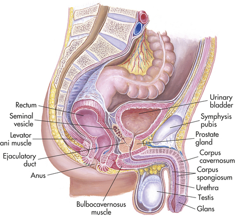
Figure 22-11 Structure of the male reproductive organs. (From Seidel HM et al: Mosby’s guide to physical examination, ed 6, St Louis, 2006, Mosby.)
External Genitalia
In men the testes (singular, testis) are the essential organs of reproduction. Like the ovaries, the testes have two functions: (1) production of gametes (in this case, sperm) and (2) production of sex hormones (in this case, androgens and testosterone). The testes are suspended outside the pelvic cavity because sperm production requires an environment that is 1° or 2° C (34° or 36° F) cooler than body temperature.
During embryonic and fetal life, the testes develop within the abdomen (see Figure 22-1). Then, about 3 months before birth, the testes start to descend toward the developing scrotum. About 1 month before birth, they enter twin passageways called inguinal canals. The inguinal canals are vaginal processes created by outpouchings of the peritoneum (lining of the abdominal cavity). The descent of a testis is shown in Figure 22-12. Each testis moves down outside the peritoneum until it is suspended in the scrotal sac by its supply lines: the ducts, blood vessels, lymphatic vessels, and nerves of the spermatic cord. When descent is complete, the abdominal end of each vaginal process closes up and the inguinal canal disappears. If peritoneal closure at the site of the inguinal canal is incomplete or weak, an inguinal hernia may occur later in life. The scrotal end of each vaginal process becomes the outer covering of the testis, the tunica vaginalis.
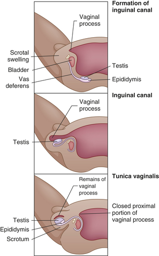
Figure 22-12 Descent of a testis. The testes descend from the abdominal cavity to the scrotum during the last 3 months of fetal development.
Figure 22-13 shows a sagittal section of a mature testis. The adult testis is ovoid and varies considerably in length (3 to 6 cm), width (2 to 3.5 cm), depth (3 to 4 cm), and weight (10 to 40 g). The testis is almost entirely surrounded by an outer covering, the tunica vaginalis, which separates the testis from the scrotal wall, and an inner covering, the tunica albuginea. Inward extensions of the tunica albuginea form septa that separate the testis into about 250 compartments, or lobules, each of which contains several tortuously coiled ducts called seminiferous tubules. The seminiferous tubules constitute the bulk (80%) of testicular volume and are the site of sperm production. (Sperm production, termed spermatogenesis, is described on p. 800.) Tissue surrounding these ducts contains blood and lymphatic vessels, fibroblastic support cells, macrophages, mast cells, and Leydig cells. Leydig cells, which occur in clusters and account for about 1% to 5% of testicular volume, produce androgens, chiefly testosterone.
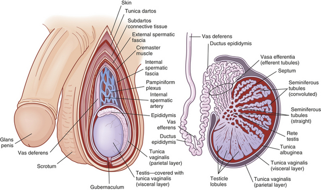
Figure 22-13 The testes. External and sagittal views showing interior anatomy. (Redrawn from Seidel HM et al: Mosby’s guide to physical examination, ed 6, St Louis, 2006, Mosby.)
The two ends of each seminiferous tubule join and leave the lobule through a short, straight section called the tubulus rectus. Sperm travel from the seminiferous tubules into these straight sections, which lead to the central portion of the testis, the rete testis. From the rete testis, sperm move through the efferent tubules, or vasa efferentia, to the epididymis, where they mature.
The testes are innervated by adrenergic fibers, whose sole function apparently is to regulate blood flow to the Leydig cells. The testes receive arterial blood from the internal spermatic and differential arteries. Arterial blood flows over the surface of the testes before entering the parenchyma (functional tissues). Surface flow cools the blood to temperatures that promote spermatogenesis, approximately 1° to 2°C (34° to 36° F) below body core temperature.18
Epididymis
The epididymis (plural, epididymides) is a comma-shaped structure that curves over the posterior portion of each testis (see Figure 22-13). It consists of a single highly packed and markedly coiled (60 to 70 cm when uncoiled) duct measuring 5 cm long, whose structural function is to conduct sperm from the efferent tubules to the vas deferens. The duct can become inflamed from infection by microorganisms that ascend the urethra or from the prostate, causing epididymitis. The epididymis has physiologic functions as well. When sperm enter the head of the epididymis, they are not fully mature or motile, nor are they capable of fertilizing an ovum. During the 12 days (or more) sperm take to travel the length of the epididymis, they receive nutrients and testosterone from the epididymal epithelium, and some biochemical or physiologic mechanism enhances their capacity for fertilization.19
The tail of the epididymis is continuous with the vas deferens (ductus deferens), a duct with muscular layers capable of powerful peristalsis that transports sperm toward the urethra. After traveling the length of the epididymis, sperm are stored in the epididymal tail and vas deferens. The vas deferens enters the pelvic cavity through the spermatic cord.
Scrotum
The testes, epididymides, and spermatic cord are enclosed and protected by the scrotum. The scrotum is a skin-covered fibromuscular sac that is homologous to the female labia majora (see Figure 22-2). The skin of the scrotum is thin and has rugae (wrinkles or folds), which enable it to enlarge or relax away from the body. At puberty the scrotal skin darkens, develops active sebaceous glands, and becomes sparsely covered with hair. Just under the skin lies a layer of connective tissue (fascia) and smooth muscle, the tunica dartos (see Figure 22-13). The tunica dartos also forms a septum that separates the two testes. Exposure to cold temperatures causes the tunica dartos to contract, pulling the testes close to the warm body. In warm temperatures the tunica dartos relaxes, suspending the testes away from body heat. These mechanisms promote optimal temperatures for spermatogenesis. In addition, scrotal sensitivity to touch, pressure, temperature, and pain protects the testes against potential harm. During sexual excitement, the scrotal skin and tunica thicken, the scrotum tightens and lifts, and the spermatic cords shorten, partially elevating the testes toward the body. As excitement plateaus, the engorged testes increase 50% in size, rotate anteriorly, and flatten against the body, signaling impending ejaculation.
Penis
The penis has two main functions: delivery of sperm to the female vagina and elimination of urine. (Urine formation and excretion are the subjects of Chapter 35.) Embryonically, the penis is homologous to the female clitoris (see Figure 22-2).
Figure 22-11 shows a sagittal section of the adult penis and its anatomic relation to other urogenital structures. Externally the penis consists of a shaft with a tip, the glans, which contains the opening of the urethra. For protection, the skin of the glans folds over the tip of the penis, forming the prepuce, or foreskin. At birth, the foreskin is adhered to the glans. Penile erections, which commonly occur, cause the adhesions to break so that by age 3 years the foreskin becomes completely retractable. The skin of the penis is continuous with that of the groin, scrotum, and inner thighs. It is hairless, movable, and darker than surrounding skin.
Internally, the penis consists of the urethra and three compartments: two corpora cavernosa and the corpus spongiosum (Figure 22-14). The three compartments are separated by Buck fascia and, like the testes, are enclosed by a tunica albuginea. The urethra passes through the corpus spongiosum and ends at a sagittal slit in the glans. If the urethra is not completely surrounded by the corpus spongiosum, the meatus may open on the ventral surface of the penile shaft (hypospadias) or on the dorsal surface (epispadias).
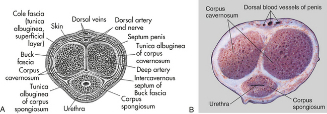
Figure 22-14 The penis. A, Cross section of the penis. B, Cross section of the shaft of the penis showing three columns of erectile, or cavernous, tissue. (A from Thompson JM et al: Mosby’s clinical nursing, ed 5, St Louis, 2002, Mosby. B from Patton KT, Thibodeau GA: Anatomy & physiology, ed 7, St Louis, 2010, Mosby.)
Penetration of the female vagina is made possible by the erectile reflex, a process in which erectile tissues within the corpora cavernosa and corpus spongiosum become engorged with blood, generally 20 to 50 ml. The erectile tissues consist of vascular spaces, or chambers, which are supplied with blood by arterioles (small arteries). Most of the time the arterioles are constricted through tonic noradrenaline release from sympathetic nerves so that not much blood flows through the erectile tissues. Sexual stimulation, however, causes the arterioles to dilate through release of nitric oxide and fill with blood.20 Their rapid expansion fills the erectile tissues, causing an erection. Erection apparently is maintained by compression or constriction of veins that drain the corpora cavernosa and corpus spongiosum. When sexual stimulation ceases or orgasm and ejaculation occur, these veins open up, blood flows out of the arterioles, and the penis becomes flaccid (soft and pendulous).
Erection is under the control of the autonomic nervous system but can be stimulated or inhibited by central nervous system input. Stimulation of mechanoreceptors of the penis, particularly of the glans, causes parasympathetic nerves of the autonomic nervous system to relax smooth muscle in the walls of penile arterioles. At the same time the effects of sympathetic nerves, which normally cause arteriolar smooth muscle to constrict, are inhibited.
Erections begin in utero and continue throughout life, but ejaculation does not occur until sperm production begins at puberty. Growth of the penis and scrotal contents continues well past puberty, however, and may not be complete until the late teens or early 20s. Penis size, when flaccid, varies considerably; with an erection, differences in penis size diminish. Sexual excitement causes the corpora cavernosa to increase in length and width and become rigid; the penis becomes erect. Stimulation of the glans, which is endowed with copious sensitive nerve endings, provides maximum erotic sensation. With sexual arousal, skin color deepens, the glans doubles in size, and the urethral meatus dilates. Ejaculation occurs with frequent, strong contractions of the vas deferens, epididymis, seminal vesicles, prostate, urethra, and penis.21
Internal Genitalia
Figure 22-13 shows the anatomy of the internal genitalia and their relation to other pelvic organs. The internal genitalia consist of ducts and glands. The ducts—the two vasa deferentia, the ejaculatory duct, and the urethra—conduct sperm and glandular secretions from the testes to the urethral opening of the penis. The glands—the prostate gland, two seminal vesicles, and two Cowper (or bulbourethral) glands—secrete fluids that serve as a vehicle for sperm transport and create an alkaline, nutritious medium that promotes sperm motility and survival. Together the sperm and the glandular fluids comprise semen.
Sperm leave the epididymides and travel rapidly through the internal ducts in a process called emission. Emission occurs just seconds before ejaculation, at the moment when sexual arousal peaks. Emission always leads to ejaculation.
Emission occurs as smooth muscle in the walls of the epididymides and vasa deferentia begins to contract rhythmically, pushing sperm and epididymal secretions through the vasa deferentia. Each vas deferens is a firm, elastic fibromuscular tube that begins at the tail of the epididymis, enters the pelvic cavity within the spermatic cord, loops up and over the bladder, and ends in the prostate gland (Figure 22-15; see also Figure 22-11). Sperm are moved along by peristaltic contractions of smooth muscle in the walls of the vas deferens.
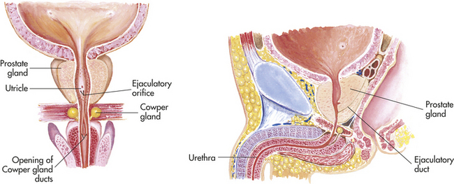
Figure 22-15 Anatomy of the prostate gland and seminal vesicles. (From Seidel HM et al: Mosby’s guide to physical examination, ed 6, St Louis, 2006, Mosby.)
As sperm leave the ampulla (wide portion) of the vas deferens, the seminal vesicles secrete a nutritive glucose-rich fluid into the ejaculate (semen). The seminal vesicles are a pair of glands, each about 4 to 6 cm long, that lie behind the urinary bladder and in front of the rectum. The seminal vesicles provide fructose as a source of energy for ejaculated sperm, and secrete prostaglandins that promote smooth muscle contraction assisting with sperm transport. The ducts of the seminal vesicles join the ampulla of the vas deferens to become the ejaculatory duct, which contracts rhythmically during emission and ejaculation. As can be seen in Figures 22-11 and 22-15, the ejaculatory duct joins the urethra, where both pass through the prostate gland. During emission and ejaculation a sphincter (muscle surrounding a duct) closes, preventing urine from entering the prostatic urethra.21
The prostate gland is composed of alveoli and ducts embedded in fibromuscular tissue. It measures 4 cm in diameter and weighs approximately 20 g. While semen moves through the prostatic portion of the urethra, the prostate gland contracts rhythmically and secretes prostatic fluid into the mixture. Prostatic fluid is a thin, milky substance with an alkaline pH that helps sperm survive in the acid environment of the female reproductive tract. In addition, clotting enzymes and fibrinolysin in prostatic fluids help mobilize sperm after ejaculation.
Cowper glands (bulbourethral glands), whose ducts secrete mucus into the urethra near the base of the penis, are the last pair of glands to add fluid to the ejaculate. Ejaculation occurs as semen reaches the base of the penis and muscles there begin the rhythmic contractions that push semen out. Normally a man ejaculates between 2 and 6 ml of semen, containing 75 million to 400 million sperm. About 98% of the ejaculate consists of glandular fluids; 60% to 70% of volume comes from the seminal vesicles and 20% from the prostate. Therefore, the ejaculate of a man who has undergone vasectomy (a surgical procedure that prevents sperm from entering the vas deferens) is not reduced by much: about 2%.
Spermatogenesis
Spermatogenesis begins at puberty and continues for life. In this respect, spermatogenesis differs markedly from oogenesis (production of primordial ova), which occurs during fetal life only.
Spermatogenesis takes place within the seminiferous tubules of the testes (see Figure 22-13). The basement membrane of each seminiferous tubule is lined with diploid (46-chromosome) germ cells called spermatogonia (singular, spermatogonium). These cells undergo continuous mitotic division. (Mitotic division, in which a cell divides into two identical cells, is described in Chapter 1.) Some of the spermatogonia move away from the basement membrane and mature, becoming primary spermatocytes (Figure 22-16). The primary spermatocytes undergo meiosis, a type of cell division that results in two haploid (23-chromosome) cells called secondary spermatocytes. (Meiosis is described and illustrated in Chapter 4.) The two secondary spermatocytes then undergo meiosis, resulting in four spermatids. It is the spermatids that differentiate into spermatozoa, or sperm, each of which contains 23 chromosomes (Figure 22-17).
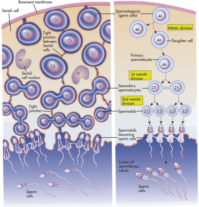
Figure 22-16 Seminiferous tubule. Section shows process of meiosis and sperm cell formation. (From Patton KT, Thibodeau GA: Anatomy & physiology, ed 7, St Louis, 2010, Mosby.)
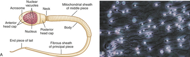
Figure 22-17 Mature sperm cell (spermatozoon). A, Anatomy of mature sperm cell. B, Human sperm with nuclear material glowing with a fluorescent dye. (B from Patton KT, Thibodeau GA: Anatomy & physiology, ed 7, St Louis, 2010, Mosby.)
The development of spermatids into sperm depends on the presence of Sertoli cells (nondividing support cells) within the seminiferous tubules. The spermatids attach themselves to Sertoli cells, from which they receive the nutrients and the hormonal signals they need to develop into sperm.22
The process of spermatogenesis, from mitotic division of a spermatogonium to maturation of the spermatids, takes about 70 to 80 days. Mature sperm migrate from the seminiferous tubules to the epididymis, where their capacity for fertilization continues to develop. Although they are completely mature by the time they are ejaculated, the sperm do not become motile (capable of movement) until they are activated by biochemicals in semen and in the female reproductive tract.
Male Sex Hormones
The male sex hormones are androgens. Testosterone, the primary male sex hormone, is an androgen. Mainly Leydig cells of the testes and, to a lesser degree, the adrenal glands produce testosterone and other androgens. In men, sex hormone production is relatively constant with some diurnal variation.
The androgens have a number of physiologic actions related to growth and development of male tissues and organs. They are responsible for fetal differentiation and development of the male urogenital system and have some effects on the fetal brain. After birth, the Leydig cells become quiescent until activated by the gonadotropins during puberty. At puberty, androgens cause the sex organs to grow and secondary sex characteristics to develop.
Testosterone affects nervous and skeletal tissues, bone marrow, skin and hair, and sex organs. It has an anabolic effect on skeletal muscle tissue, thereby contributing to the difference in body weight and composition between men and women. Testosterone also stimulates growth of the musculature and cartilage of the larynx, causing a permanent deepening of the voice. Testosterone directly stimulates the bone marrow and indirectly stimulates renal erythropoietin production to achieve increased hemoglobin and hematocrit levels. Because sebaceous gland activity is stimulated by testosterone, acne may develop. In the presence of testosterone, hair becomes coarser in texture and facial hair, axillary hair, and pubic hair grow in male patterns. Later in life, testosterone causes baldness in genetically susceptible individuals. Testosterone is required for spermatogenesis and for secretion of fluid by the prostate gland, seminal vesicles, and Cowper glands. Testosterone is also associated with an increase in libido (sex drive). Other, less-understood, effects of testosterone include alterations in fatty acid and cholesterol metabolism.
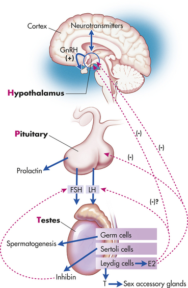
The regulation of androgen production and spermatogenesis is achieved by a complex feedback system involving the extrahypothalamic central nervous system, the hypothalamus, the anterior pituitary, the testes, and the androgen-sensitive end organs. These relationships, which are essentially the same in women, are summarized in Figure 22-3. Extrahypothalamic influences include such variables as physiologic and psychologic stress, which may inhibit or augment hypothalamic activity. In the hypothalamus, neurotransmitters regulate GnRH synthesis and pulsatile release (about every 3 hours) into the hypophyseal portal veins. Norepinephrine stimulates GnRH secretion, and serotonin and dopamine inhibit GnRH secretion. GnRH is transported by portal flow to the median eminence of the pituitary gland, where it binds to receptors and stimulates the synthesis and secretion of gonadotropins, LH, and FSH. LH and FSH, which are named for their effects in the female reproductive system, have important effects on the male system as well. LH acts on the Leydig cells to regulate testosterone secretion. FSH acts on the seminiferous tubule Sertoli cells to promote spermatogenesis. FSH secretion is inhibited by inhibin secreted by the Sertoli cells. Similar to their action in the female gonad, inhibin functions as an autocrine/paracrine regulator in the male gonad. Inhibin inhibits proliferation of spermatogonia by regulating pituitary FSH levels. In addition, inhibin facilitates LH stimulation of androgen biosynthesis in Leydig cells.
Ninety-eight percent of testosterone, the major steroid hormone produced by the testis, binds to either sex hormone–binding globulin (SHBG) (40%) or albumin (48%). The remaining 2% remains unbound in the plasma and is free to enter cells and wield its metabolic effects. Changes in the amount of available SHBG affect the amount of testosterone within tissues. The testes secrete only 25% of circulating estrogen (estradiol). The majority is produced by peripheral conversion of testosterone and androstenedione. Estrogens help regulate GnRH and LH secretion. Peripheral conversion of testosterone by 5-alpha-reductase also produces dihydrotestosterone (DHT), another potent androgen. DHT is necessary for external virilization during embryogenesis and androgen activity beginning at puberty and continuing throughout adulthood. Prolactin, a polypeptide synthesized and secreted from the pituitary, helps maintain biosynthesis of testosterone. However, elevated prolactin levels may suppress biosynthesis.23
In summary, hormones secreted at each level of the hypothalamic-pituitary-testicular (H-P-T) axis control and coordinate testicular function (Figure 22-18). This control is exerted through positive and negative feedback signals by (1) sex steroids that inhibit hypothalamic GnRH secretion and pituitary LH responsiveness to GnRH; and (2) testicular inhibin that inhibits pituitary FSH and, possibly, circulating estrogens (E2). Any disruption along the H-P-T axis may lead to hypogonadism or infertility.
STRUCTURE AND FUNCTION OF THE BREAST
The breasts are modified sebaceous glands that lie on the ventral surface of the thorax, within the superficial fascia of the chest wall. They extend vertically from the second rib to the sixth or seventh intercostal space and laterally from the side of the sternum to the midaxillary line. Breast tissue also may extend into the axilla; this tissue is known as the tail of Spence.
The Female Breast
The female breast is composed of 15 to 20 pyramid-shaped lobes that are separated and supported by Cooper ligaments (Figure 22-19). Each lobe contains 20 to 40 lobules that are subdivided into glandular alveoli. The alveoli are composed of secretory acini that synthesize milk (lactation) after pregnancy (see Figure 22-19). Milk is continuously secreted into the alveolar lumen and is stored there until the myoepithelial cells are stimulated by oxytocin, which triggers the let-down reflex.24 The alveoli empty into a network of lactiferous ducts. These ducts reach the skin through 9 or 10 openings (pores) in the nipple. The lobes and lobules are surrounded and separated by muscle strands and fatty connective tissue. The amount of fatty connective tissue varies from individual to individual, depending on weight and genetic and endocrine factors, and contributes to the diversity of breast size and shape.
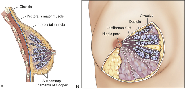
Figure 22-19 Schematic diagram of breast. (From Riordan J: Breastfeeding and human lactation, ed 3, Sudbury, MA, 2005, Jones and Bartlett.)
An extensive capillary network surrounds the alveoli and is supplied by perforating branches of the internal mammary artery, the thoracoacromial artery, the internal and lateral thoracic arteries, and the intercostal arteries. Venous return follows arterial supply, with relatively rapid emptying into the superior vena cava. The breasts receive sensory innervation from branches of the second through sixth intercostal nerves and the cervical plexus. This accounts for the fact that breast pain may be referred to the chest, back, scapula, medial arm, and neck. Lymphatic drainage of the breast occurs largely through axillary nodes, but approximately 25% occurs through transpectoral and other draining nodes (Figure 22-20).2,25
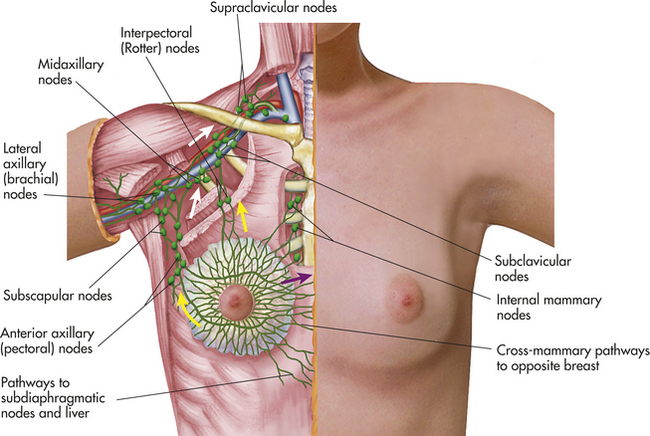
Figure 22-20 Lymphatic drainage of the female breast. (From Seidel HM et al: Mosby’s guide to physical examination, ed 6, St Louis, 2006, Mosby.)
The nipple is a pigmented, cylindrical structure that is usually located at the fourth or fifth intercostal space. It measures 0.5 to 1.3 cm in diameter and is approximately 10 to 12 mm in height when erect. On its surface lie multiple openings, one from each lobe. The areola is the pigmented circular area around the nipple. It may be 15 to 60 mm in diameter. A number of sebaceous glands, the glands of Montgomery, are located within the areola and aid in lubrication of the nipple during lactation. The nipple and areola contain smooth muscles that receive motor innervation from the sympathetic nervous system. Breast-feeding, sexual stimulation, and exposure to cold cause the nipple to become erect.
The fetal and early postnatal development of breast tissue does not depend on hormones, although fetal breast tissue does become progressively responsive to hormonal stimulation. During childhood, breast growth is latent and growth of the nipple and areola keeps pace with body surface growth. (Male breast development normally does not progress any further.) At the onset of puberty in the female, estrogen secretion stimulates mammary growth. Breast development, or thelarche, is usually the first sign of puberty in the female. Full differentiation and maturation of breast tissue occur over approximately 4 years and are mediated by a variety of hormones, including estrogen, progesterone, prolactin, growth hormone, thyroxine, insulin, and cortisol. Estrogen promotes the increase in size of the breast by the formation of a mass of tissue under the areola, increases the size and pigmentation of the areola, and development of the lobular ducts. During pregnancy increased levels of estrogen promote further development of the lobular ducts. Progesterone stimulates development of cells lining the alveoli to produce milk. Lactation (milk production) occurs after childbirth in response to increased levels of prolactin. Prolactin secretion, in turn, increases by continued breast-feeding. Oxytocin, another hormone released during and after delivery, controls milk ejection from alveolar cells. Variations in breast development are listed in Box 22-2.
During the reproductive years, the breast undergoes cyclic changes in response to changes in the levels of estrogen and progesterone associated with the menstrual cycle. During the follicular/proliferative phase of the menstrual cycle, high estradiol levels increase the vascularity of breast tissue and stimulate proliferation of ductal and alveolar tissue. This effect is sustained into the luteal/secretory phase of the cycle. During this phase, progesterone levels influence the growth of the alveoli and increase and contribute to the breast changes induced by estradiol. Specific effects of estrogen and progesterone include dilation of the ducts and conversion of the alveolar cells into secretory cells; and fluid secretion, mitotic activity, and deoxyribonucleic acid (DNA) production of nonglandular tissue and glandular epithelium.1 Most women experience some degree of premenstrual breast fullness, tenderness, and increased nodularity. Breast volume may increase as much as 10 to 30 ml. Because the length of the menstrual cycle does not allow for complete regression of new cell growth, breast growth continues at a slow rate until approximately 35 years of age. Because of the cyclic changes that occur in breast tissue, clinical breast examination is recommended at the conclusion of or a few days after menses, when hormonal effects are minimal and breasts are at their smallest and least tender.
The function of the female breast is primarily to provide a source of nourishment for the newborn. Physiologically, breast milk is the most appropriate nourishment for newborns. Not only does its composition change over time to meet the changing digestive capabilities and nutritional requirements of the infant but it also contains immune cells, specific immunoglobulins, especially immunoglobulin A (IgA), and nonspecific antimicrobial factors, such as lysozymes and lactoferrin, that protect the infant against infection and allergies and asthma. Evidence suggests breast-feeding decreases future incidence in the infant of adult obesity, lowers atherosclerotic disease, and lowers the incidence of types 1 and 2 diabetes.26 During lactation, high prolactin levels interfere with hypothalamic-pituitary hormones that stimulate ovulation. Lactation usually suppresses the menstrual cycle (lactational amenorrhea) and prevents ovulation for the first 6 months postpartum.27 In many parts of the world lactational amenorrhea is a means of contraception. Breasts are also a source of pleasurable sexual sensation and in Western cultures have become a sexual symbol.
The Male Breast
Until puberty, development of the male breast is similar to that of the female breast. In the absence of sufficiently high levels of estrogen and progesterone, the male breast does not develop any further. The normal male breast consists of a small, underdeveloped nipple; some fatty and fibrous tissue; and a few ductlike structures in the subareolar area. The male breast may appear enlarged in obese men because of accumulation of fatty tissue. During puberty some males experience gynecomastia, a condition in which the breasts enlarge temporarily as a result of hormonal fluctuations. Thirty percent to 60% of men may have palpable breast tissue related to circulating or local production of estrogen from elevated aromatase activity, particularly in adipose tissue.28
TESTS OF REPRODUCTIVE FUNCTION
Diagnostic tests of the male and female reproductive systems are performed to determine the cause of infertility, to detect the presence of cancerous lesions, or to identify the presence of sexually transmitted infections. (Alterations of the reproductive systems are discussed in Chapter 23; sexually transmitted infections are discussed in Chapter 24.) Procedures include laboratory tests, such as cultures, tests, stains, biopsies, serologic testing, and hormonal assays. Radiographic procedures are performed to identify abnormal growths or structures. Direct observation of reproductive organs is completed by laparoscopy or colposcopy.
Infection and Cancer Tests
Tests, stains, cultures, and serologic testing commonly are used to detect infectious diseases. Tests are prepared by spreading a layer of specimen material on a glass slide. The specimen may be evaluated microscopically as a wet mount or dried and stained with different dyes. Gram staining is a technique that allows the differential identification of two categories of bacteria according to the tendency of the microorganism to selectively absorb different components of the stain. Fluorescent antibody testing and DNA probe testing (nucleic acid hybridization detection method) are fairly quick, inexpensive, and accurate methods that can be used to detect some bacterial and viral infections.
A culture is the growth of microorganisms in a nutrient medium selectively prepared to support the growth of particular microorganisms that are obtained from body secretions or tissues. Bacteria as well as viruses, such as cytomegalovirus, can be cultured.
Serologic testing identifies whether an antigen-antibody reaction has occurred in response to an infectious microorganism. Several different techniques can be used to identify the formation of antigen-antibody complexes. With immunofluorescent testing, fluorescein-labeled antibodies react with specific antigen, such as the spirochetes of syphilis. The fluorescent pattern of the reaction can be microscopically observed under ultraviolet light. The flocculation (agglutination) test is the precipitation of clumped cells caused by the reaction of antigen with homologous antiserum. The Venereal Disease Research Laboratory (VDRL) test for syphilis is a type of flocculation test. Certain viral diseases can be diagnosed using specific serologic antibody markers, for example, the diagnosis of hepatitis A and hepatitis B viruses. Radioimmunoassay (RIA) and enzyme-linked immunosorbent assay (ELISA) are more specific tests used to document viral infections. Tests used for the diagnosis of sexually transmitted infections are presented in Table 22-2.
Table 22-2
Diagnosis of Sexually Transmitted Infections
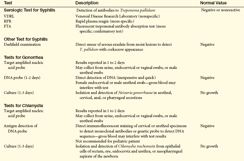
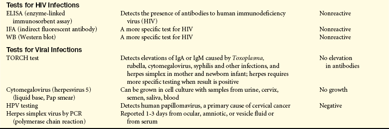
DNA, Deoxyribonucleic acid; HPV, human papillomavirus; IgA, immunoglobulin A; IgM, immunoglobulin M; TORCH, toxoplasmosis, rubella, syphilis and other infections (hepatitis B, coxsackie virus, Epstein-Barr virus, varicella-zoster virus, and human parvovirus), cytomegalovirus, herpes simplex.
Tissue biopsy is the surgical resection of a tissue specimen from a suspected site. The biopsy provides cells and tissues to be used to identify infectious processes and to differentiate benign from malignant conditions and primary from metastatic lesions. In the reproductive tract, tissue may be obtained from the vagina, cervix (by cone or brush biopsy), endometrium of the uterus (by curettage), ovary, testis, prostate, or penis.
Needle biopsy is a technique of aspirating small amounts of tissue by positioning a needle in the tissue and applying negative pressure by pulling back on the plunger of the attached syringe as the needle is moved back and forth at the biopsy site. There is relatively less discomfort with this procedure than with a tissue biopsy that requires opening the surface of
the skin to localize the tissue site. Normal findings indicate no abnormal cells or tissues on histologic examination.
The Papanicolaou (Pap) test is a procedure commonly used for the cytologic examination of the female reproductive tract. Cells from body tissues and fluids (from the vagina or endocervix) are stained and examined for the number and types of cells and abnormalities in their morphology. Specimens also may be obtained from the mouth; nipple discharge; or amniotic, pleural, or spinal fluid aspirations. The Pap test is particularly useful for diagnosis of premalignant (dysplastic), malignant, atypical, and inflammatory cells usually as a result of HPV infection. Because of the increased prevalence of Pap test screening, the rate of invasive cervical cancer has declined steadily over the past 30 years from 55% to 16%. Pap test screening is no longer necessary annually if the woman is older than 30, has had three negative Pap tests, and is in a mutually monogamous relationship. She may consider Pap tests every 2 to 3 years.2 Liquid-based methods can test for HPV. If the high-risk type is detected, further evaluation with colposcopy (magnification of rapid growing cells) with biopsy may be required.25
Evaluations of the breast are commonly performed to detect tumors or to differentiate solid masses from cysts and benign from malignant tumors. Screening or diagnostic mammography, surgical or fine-needle biopsy (aspiration), and ultrasonography are specific examination techniques. Less common tests include thermography, which is used for screening; chest x-rays, which are used to detect pulmonary metastases; and computed tomography (CT) scanning, which is used when liver or brain metastases are suspected. Mammography is a low-dose radiographic examination used to identify nonpalpable (less than 1 cm) or unrecognized lesions. Breast cancer can be detected by radiography 2 to 3 years before its clinical presentation, providing an excellent prognosis for cure. The American Cancer Society recommends clinical breast examinations yearly and annual mammograms starting at age 40. Earlier mammograms are recommended if a woman has a first-degree relative with early onset breast cancer, or is positive for the BRCA gene.25
Ultrasonography is performed chiefly to differentiate cystic from solid lesions; it is not diagnostic of malignancy. Fine-needle aspiration, an inexpensive and relatively simple procedure, is used to aspirate cysts or to obtain a specimen for cytologic examination. The main problem with any needle biopsy, or aspiration, is sampling error caused by improper positioning of the needle, which creates false-negative test results. The rate of false-positive results with fine needle aspiration is 1% to 2%; the rate of false-negative results is 10%.9
Fertility Tests
Tests of reproductive function are performed most commonly when infertility exists. Both partners are examined, and several diagnostic evaluations may be completed. The types of tests and their normal values are summarized in Tables 22-3 and 22-4. The man is evaluated for number, amount, structure, and motility of sperm and obstruction along the reproductive tract. Tests for women determine whether (1) the reproductive tract (cervix, uterus, fallopian tubes) is adequately patent to allow for passage of ovum and sperm, (2) ovulation occurs normally, (3) the endometrium is responding normally to hormones, and (4) reproductive tissues are free of tumors or infections. Hormonal assays evaluate the adequacy of pituitary function and target organ response. The position and size of organs or the presence of tumors can be detected by direct observation procedures using a laparoscope or by radiographic studies, such as plain films, computerized scans, or tomography.16
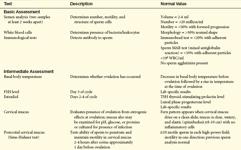
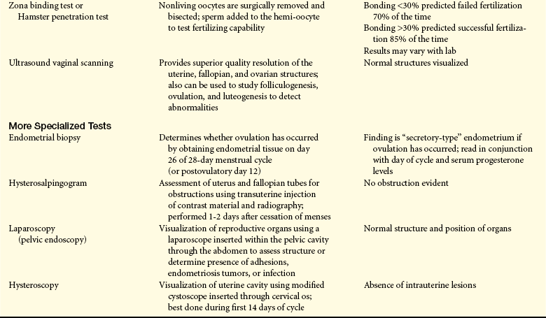
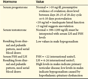
Aging and Reproductive Function
Aging and the Female Reproductive System
Menopause is a normal developmental event that is experienced universally by midlife women. In the United States, women reach menopause between ages 48 and 55 years, at a median age of 51.4 years. The mean age for smokers is 2 years sooner than nonsmokers (50 versus 52 years).16 Age of menopause tends to be genetically predetermined and does not appear to be affected by age at menarche, childbearing or lactation, use of oral contraceptives, socioeconomic class, or race. If a woman stops menstruating before 40 years of age it is called premature ovarian failure, and occurs in about 1% of the population. Besides smoking, younger age at menopause has been associated with abnormalities of the X chromosome causing gonadal dysgenesis and undernourishment. Thinner women experience menopause at a slightly younger age, probably related to body fat. Irregular menses in women in their early 40s also may be a predictor of an earlier menopause. Alcohol consumption has been associated with later menopause, perhaps because of higher blood and urinary levels of estrogen.16
Perimenopause is the transitional period between reproductive and nonreproductive years, a transition lasting 2 to 8 years.1,16 Five to 10 years before menopause, approximately 90% of women note mild to extreme variability in frequency and quality of menstrual flow. Perimenopause symptoms depend on the sensitivity of the target tissue receptors. Symptoms usually begin with a lengthening of the menstrual cycle, which correlates with anovulatory cycles. Unpredictable or irregular ovulation uniformly precedes menopause.1 The perimenopause experience varies among women and from cycle to cycle in the same woman. How individual women experience symptoms is unpredictable. Estradiol levels remain in the normal to slightly elevated range until about 1 year before menopause.1
Around 37 to 38 years of age, 10 to 15 years before menstruation ceases, women experience accelerated follicular loss that begins when the total number of follicles reaches about 25,000 and ends when the supply of follicles is depleted (see discussion under Hormonal Controls, earlier in this chapter). This loss correlates with a subtle but definite increase in FSH and a decrease in inhibin. Increased FSH stimulation seems to accelerate follicular loss, and declining inhibin production disturbs the negative feedback influence over pituitary secretion of FSH. Perimenopausal cycles are marked by elevated FSH, decreased inhibin, normal LH, and slightly elevated estradiol levels1 (Figure 22-21). A hallmark characteristic of impending menopause is the initial “monotropic” FP increase in FSH without corresponding changes in LH levels. Lower inhibin B and increased activin A (a potent stimulator of FSH) levels contribute to this phenomenon.1
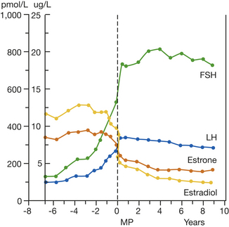
Figure 22-21 The perimenopausal hormone transition. Mean circulating hormone levels. FSH, Follicle-stimulating hormone; LH, luteinizing hormone.
Increased FSH levels and reduced ovarian secretion of inhibin reflect quality and capability of aging follicles. In a study of pregnancy rates during in vitro fertilization (IVF), women older than 40 years of age had as many oocytes recovered during laparoscopy, equal estrogen and progesterone levels, and similar pregnancy rates when compared with younger women. Yet, despite similar estradiol (E2) levels, inhibin response to exogenous hyperstimulation was significantly lower in the older women. Further, inhibin levels were significantly related to progesterone levels. In this study, 60% of women more than 40 years of age who became pregnant through IVF spontaneously aborted, compared with 30% of younger women.29
In summary, hormonal findings during the years surrounding menopause indicate that the process of folliculogenesis changes (Box 22-3). Initially, FSH levels are significantly elevated, whereas pulsatile secretion by the pituitary is maintained. Later, LH levels rise. In younger women, FSH stimulates inhibin, which in turn suppresses FSH. However, through an undetermined mechanism, perimenopausal women produce high levels of FSH in early FP and lower FP inhibin levels. The ovary, in response to high FSH, recruits increasing numbers of follicles; these follicles only partially develop, with a net effect of irregular ovulation, lower progesterone levels, and depleted follicle reserve. The increase in developing follicles leads to increased E2 levels. It is believed that lower levels of inhibin and higher levels of activin counteract the usual effect of the negative feedback loop found in younger women.1 Table 22-5 summarizes endocrine events occurring during the perimenopause.
Table 22-5
Endocrine Events Associated with Perimenopause
| Hormone Changes | Effects |
| Estradiol (E2) levels | Erratic and intermittent increase |
| Mean FP level 1 greater than mean FP level in younger women | First in FP (inverse relationship between length of FP and estradiol level) |
| FP level may be greater than midcycle peak level in fertile women | Later during premenstrual phase |
| Ovulatory cycles | Short or insufficient LP (decreased fertility) |
| Progesterone levels | Decreased in ovulatory cycle; minimal during anovulatory cycles |
| Anovulatory cycles | Increased to about 50%; perhaps more in later perimenopause |
| FSH levels | Variable, then increased |
| LH levels | Normal initially, then increased |
| Inhibin levels | Correlate with progesterone levels |
FP, Follicular phase; FSH, follicle-stimulating hormone; LH, luteinizing hormone; LP, luteal phase.
The majority of health concerns and public health issues focus on menstrual cycle changes, vasomotor symptoms, or hot flashes/flushes, potential for bone loss (osteoporosis is discussed in Chapter 42), and the emotional symptoms that may accompany the perimenopausal transition.1,30 Heavy menstrual bleeding, menorrhagia (flooding), is one of the most distressing complaints and affects approximately 50% of women. It is also the complaint that in the past put women at high risk for hysterectomy. Increased endometrial bleeding is correlated with a change from ovulatory to anovulatory cycles and is associated with unopposed high E2 levels the week before menses. Estrogen causes endometrial tissue to thicken. Because women with anovulatory cycles have longer exposure to periods of unopposed estrogen, the mean thickness of their endometrium, as measured by ultrasound, can be greater than that of ovulatory women.29,30 Thicker endometrium without corresponding stromal support from progesterone production leads to heavier periods, menorrhagia, or midcycle bleeding, metrorrhagia9 (Table 22-6).
Table 22-6
Impact of High Estrogen Levels on Menstrual Cycle and Symptomatology
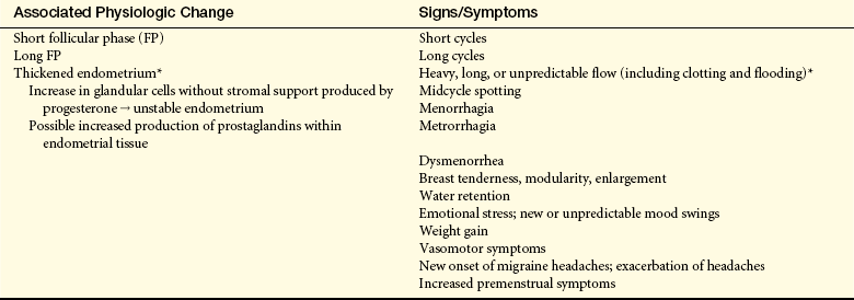
∗Symptoms aggravated by anovulatory cycles; leads to dysfunctional uterine bleeding (see Chapter 23).
In the United States, most women and clinicians link hot flashes/flushes, or vasomotor flush, with menopause and low estrogen levels. However, a rapid change in estrogen levels (withdrawal or increase), rather than low estrogen levels, induces hot flushes. Vasomotor symptoms are characterized by a rise in skin temperature, dilation of peripheral blood vessels, increased blood flow to the hands, increased skin conductance, and transient increase in heart rate (average of 9 beats per minute and up to 20), followed by a temperature drop and profuse perspiration over the area of flush distribution. Vasomotor flush usually occurs in the face and neck and may radiate into the chest and other parts of the body. Dizziness, nausea, headaches, or palpitations may accompany the flush. Vasomotor symptoms vary in frequency, intensity, and duration, lasting 1 to 5 minutes (mean 2.7 minutes). Anywhere from 11% to 85% of menopausal women experience vasomotor flushes.29,30 Most (about 60% to 65%) have symptoms for 1 to 5 years, 25% for 6 to 10 years, and 10% to 15% for 10 to 15 years or longer.
Women with higher cyclic E2 levels, that is, premenopausal women with premenstrual vasomotor symptoms and women who report breast, fluid, and premenstrual mood symptoms during the early stages of the perimenopause, are predisposed to withdrawal vasomotor symptoms later in the perimenopausal transition. Anecdotal reports from participants in various research studies suggest that vasomotor symptoms occur just before flow and may be used to predict menstruation.30 Cyclic vasomotor symptoms, which typically begin early in the perimenopausal transition, differ from noncyclic vasomotor symptoms, which are experienced in late perimenopause. Early vasomotor symptoms typically occur during sleep, often in the early hours of the morning, with few or no daytime vasomotor symptoms; are occasionally preceded by an aura (feelings of anxiety, anger, or panic); and may be associated with nausea, palpitations, dizziness, or faint feelings. Cyclic symptoms tend to occur over several days before and during menses, whereas midcycle vasomotor symptoms are less common and more likely to occur in anovulatory cycles when progesterone does not follow high estrogen midcycle peak. In summary, vasomotor symptoms commonly start when estrogen levels are erratic in the early perimenopause, and are highly correlated with a decreased quality of life, especially when sleep is disturbed. Understanding the physiology of vasomotor symptoms and developing a range of nonhormonal and hormonal strategies for control are important priorities in perimenopause research.
Based on the integration of collected data, a temporal chart outlining phases of the perimenopausal transition and associated endocrine changes and symptoms has been developed.29 Although the transition is divided into five phases, the vast variability in timing and degree of symptoms experienced by individual women may make clear predictions impossible. The information outlined in Table 22-7 provides a template to visualize the complex physiology of the perimenopause and the dynamic changes that occur during this time.
Table 22-7
Postulated Perimenopausal Transition Timeline
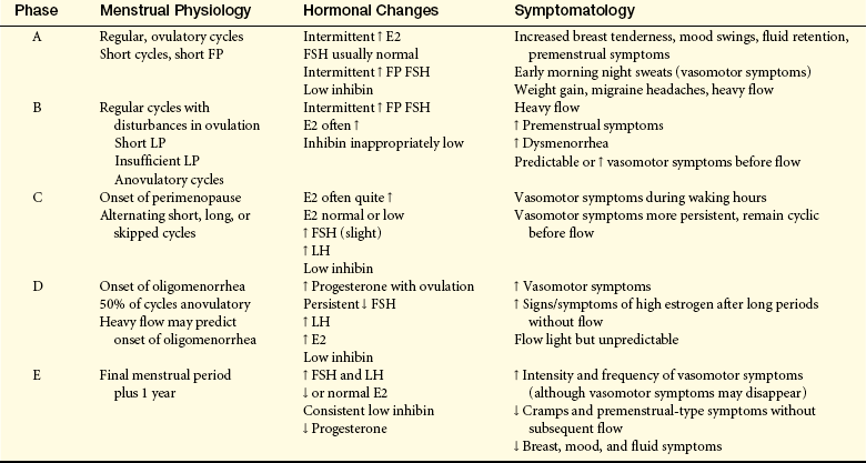
E2, Estradiol; FP, follicular phase; FSH, follicle-stimulating hormone; LP, luteal phase.
Several other physiologic changes are associated with the postmenopausal period. These changes affect breast tissue, urogenital structures, bone density or risk for osteoporosis, risk for heart disease and possible memory loss or Alzheimer disease. Only changes in the breast and urogenital structures are addressed here (other topics are addressed in appropriate chapters).
As ovarian function changes, breast tissue involutes. Two phases of involution have been described: the premenopausal phase and the postmenopausal phase. The premenopausal phase occurs between 35 and 45 years of age. During this phase there is a moderate decrease in mammary tissue. During the postmenopausal phase, glandular breast tissue is significantly reduced and there is some increase of fat deposits and connective tissue. These changes contribute to the reduction in size and firmness of breast tissue.
The urogenital tract undergoes a number of changes. The ovaries begin to decrease in size around age 30 years, and shrinkage accelerates after age 60 years. Gradually, over the years, the uterus atrophies and decreases in size. The vagina shortens, narrows, and loses some of its elasticity. Vaginal walls lose their ability to lubricate quickly. Intercourse before adequate lubrication may be painful. The vaginal pH, usually maintained at 4 to 6 before menopause, increases to between 6.5 and 8 and contributes to a higher incidence of vaginitis. The cervix atrophies and the cervical os decreases in size. The vaginal epithelium also atrophies; this atrophy can cause vaginal irritation, burning, itching (pruritus), white discharge (leukorrhea), painful intercourse (dyspareunia), and vaginal bleeding. The labia majora and minora become less prominent, and some pubic hair is lost. Urethral tone declines, as does muscle tone throughout the pelvic area. Urinary frequency, urgency, and incontinence are associated with estrogen deficiency.
Sexually active women have less vaginal atrophy; presumably, sexual activity and stimulation maintains vaginal vasculature and circulation.1 Regular intercourse (once or twice per week) and masturbation are associated with vaginal pliability, vaginal function, and continued lubrication with arousal.
Aging and the Male Reproductive System
Men maintain reproductive capacity longer than women. There is no known discrete event, comparable to menopause, that characterizes aging of the male reproductive system, although the term andropause is sometimes used to describe the hypogonadism associated with male aging.31 Gradual changes do occur, and aging in the male reproductive system is characterized by hypogonadism, testosterone deficiency, erectile dysfunction, and proliferative disorders of the prostate gland (see Chapter 23).32 Aging changes are also influenced by chronic diseases and use of medications.
Components of male sexual behavior include both sexual drive and erectile and ejaculatory capacity. Libido, or sexual drive, is a complex phenomenon that requires a baseline hormonal milieu but is influenced significantly by health status and environmental, social, and psychologic factors. Aging causes specific physical changes that influence erectile and ejaculatory capabilities. Alterations in sexual response include the need for longer stimulation to achieve full erection; slower and less forceful ejaculation, with less pelvic muscle involvement; decreased vasocongestive response; and longer refractory period (time during which erection and ejaculation are not possible), up to 24 hours in some men.
The testes undergo several age-related structural changes, including decreased weight, atrophy, and softening. Degenerative changes in the seminiferous tubules may include thickening of the basement membrane; increase in lumen size; germ cell (spermatogonium) arrest and a decrease in spermatogenic activity; and collapse of tubules, followed by complete obstruction caused by sclerosis and fibrosis. Areas of mild to severe degenerative change may be interspersed with areas having intact tubules. These morphologic changes may result from atherosclerosis (arterial clogging) in the testicular vascular bed.33 Alterations of the seminiferous tubules do not appear to diminish sperm counts, but they do reduce fertility because a greater percentage of the sperm lack motility or have structural abnormalities.
Aging probably causes changes in the production of male sex hormones, levels of SHBG, and responsiveness of target tissues. Hormone synthesis by the testes and testicular responsiveness to the gonadotropins (FSH and LH) are diminished, and pituitary secretion of these gonadotropins are elevated.32,34 The reduced levels of testosterone may be related to alterations in the Leydig cells, the testosterone producers of the testes. The number of Leydig cells decreases as age increases, perhaps because of atherosclerotic changes in arteries that supply blood to the testes.35 Even if testosterone levels are not decreased, older men may have less unbound testosterone in their blood, decreasing the amount of unbound hormone available to stimulate target tissues. Decreased testosterone levels have several effects, including functional deterioration of the accessory sex organs (the prostate gland, seminal vesicles, epididymis, and ductus deferens); loss of muscle mass, strength, and endurance; increased visceral fat, osteopenia, and cognitive decline; and, in many men, decrease in libido. This last effect also may be caused by alterations in other variables that affect libido.36,37 Modifiable risk factors for low testosterone and symptoms of androgen deficiency include health status and waist circumference.38
Activin 794
Adrenarche 784
Androgens 792
Areola 803
Bartholin gland (greater vestibular or vulvovaginal gland) 785
Breast 802
Cervix (neck of the uterus) 787
Clitoris 785
Cornification 795
Corpus cavernosum (pl., corpora cavernosa) 798
Corpus luteum 789
Corpus spongiosum 798
Corpus of uterus (body of uterus) 787
Cowper gland (bulbourethral gland) 800
Cul-de-sac 786
Culture 805
Cytologic examination 806
Decornification 795
Developing fetus 781
Dihydrotestosterone (DHT) 802
Efferent tubule 797
Ejaculatory duct 800
Embryo 781
Emission 799
Endocervical canal 788
Endometrium 788
Epididymis 797
Erectile reflex 799
Estradiol (E2) 790
Estrogen 790
Fallopian tube (oviduct, uterine tube) 789
Fimbria (pl., fimbriae) 789
Fine-needle biopsy (aspiration) 807
Follicle-stimulating hormone (FSH) 783
Follicular/proliferative phase 793
Follistatin 794
Foreskin (prepuce) 798
Fornix 786
Fundus of uterus 787
Glands of Montgomery 803
Glans 798
Gonad 781
Gonadarche 784
Gonadostat 784
Gonadotropin-releasing hormone (GnRH) 784
Granulosa cell 789
Hymen 785
Infundibulum 789
Inguinal canal 796
Inhibin 794
Introitus 785
Isthmus of the uterus 787
Labium majus (pl., labia majora) 784
Labium minus (pl., labia minora) 785
Leptin 784
Leydig cell 797
Libido 800
Luteal/secretory phase 794
Luteinizing hormone (LH) 783, 791
Mammography 807
Menarche 792
Menopause 792
Menstruation (menses) 793
Mons pubis 784
Myometrium 788
Needle biopsy 805
Nipple 803
Ovarian cycle 789
Ovarian follicle 789
Ovaries 789
Ovulation 789
Ovum 781
Papanicolaou (Pap) test 806
Penis 798
Perimenopause 807
Perimetrium (parietal peritoneum) 788
Perineal body 786
Perineum 786
Premature ovarian failure 807
Primary spermatocyte 800
Progesterone 791
Prolactin 802
Prostate gland 800
Puberty 784
Rete testis 797
Ruga (pl., rugae; pertains to vagina and testes) 786
Scrotum 797
Secondary spermatocyte 800
Semen 799
Seminal vesicle 800
Seminiferous tubule 797
Serologic testing 805
Sertoli cell (nondividing support cell) 800
Sex hormone 781
Sex hormone–binding globulin (SHBG) 801
Skene gland (lesser vestibular or paraurethral gland) 785
Spermatic cord 796
Spermatids 800
Spermatogenesis 797
Spermatogonium (pl., spermatogonia) 800
Spermatozoon (sperm cell) 781
Squamous-columnar junction 789
Testis (pl., testes) 786
Testosterone 800
Theca cell 789
Thelarche, 784 803
Tissue biopsy 805
Tubulus rectus 797
Tunica albuginea 797
Tunica dartos 797
Tunica vaginalis 796
Ultrasonography 807
Urethra 799
Urinary meatus 785
Uterus 787
Vagina 786
Vas deferens (ductus deferens) 797
Vasomotor flush 810
Vasomotor symptom 809
Vestibule 785
Vulva 784
REFERENCES
1. Speroff, L., Fritz, M.A. Clinical gynecology, endocrinology, and infertility, ed 7. Baltimore: Williams & Wilkins; 2005.
2. Coad, J., Dunstall, M. Anatomy and physiology for midwives, ed 2. London: Churchill Livingstone; 2006.
3. Gordon, K., Oehninger, S. Reproductive physiology. In Copeland L.J., Farrell J.F., eds.: Textbook of gynecology, ed 2, Philadelphia: Saunders, 2000.
4. Di Vall, S.A., Radovick, S. Pubertal development and menarche. Ann N Y Acad Sci. 2008;1135:19–28.
5. Kaplowitz, P.B. Link between body fat and the timing of puberty. Pediatrics. 2008;121(Suppl 3):S208–S217.
6. Klein, K.O., et al. Effect of obesity on estradiol level, and its relationship to leptin, bone maturation, and bone mineral density in children. J Clin Endocrinol Metab. 1998;83(10):3469.
7. Chagin, A.S., Savendahl, L. Estrogens and growth: review. Pediatr Endocrinol Rev. 2007;4(4):329–334.
8. Berne R.M., Levy M.N., eds. Physiology, ed 5, St Louis: Mosby, 2003.
9. Lowdermilk, D.L., Perry, S.E. Maternity and women’s health care, ed 8. St Louis: Mosby; 2004.
10. Schiffman, M., et al. Human papillomavirus and cervical cancer. Lancet. 2007;370(9590):890–907.
11. Rics, F.J., et al, A cross-cultural study of menstrual cycle characteristics of women practicing the sympto-thermal method of natural family planning. Komenich, P., et al, eds. The menstrual cycle, vol 2. New York: Springer, 1981.
12. Trealor, A.E., et al. Variation of the human menstrual cycle through reproductive life. Int J Fertil. 1970;12:77–126.
13. Adams Hillard, P.J. Menstruation in young girls: a clinical perspective. Obstet Gynecol. 2002;99(4):655–662.
14. Golub, S. Periods: from menarche to menopause. Newbury Park, NJ: Sage; 1992.
15. deKretser, D.M., et al. Inhibins, activans and follistatin in reproduction. Hum Reprod Update. 2002;8(6):529–541.
16. Stenchevor, M.A., et al. Comprehensive gynecology, ed 4. St Louis: Mosby; 2001.
17. Sirois, J., et al. Cyclooxygenase-2 and its role in ovulation: a 2004 account. Hum Reprod Update. 2004;19(5):373–385.
18. Jung, A., Schuppe, H.C. Influence of genital heat stress on semen quality in humans. Andrologia. 2007;39(6):203–215.
19. Cooper, T.G. Sperm maturation in the epididymis: a new look at an old problem. Asian J Androl. 2007;9(4):533–539.
20. Prieto, D. Physiological regulation of penile arteries and veins. Int J Impot Res. 2007;2(1):17–29.
21. Ohi, D.A., et al. Anejaculation and retrograde ejaculation. Urol Clin North Am. 2008;35(2):211–220. [viii].
22. Sofikitis, N., et al. Hormonal regulation of spermatogenesis and spermiogenesis. J Steroid Biochem Mol Biol. 2008;109(3-5):323–330.
23. Buvat, J. Hyperprolactinemia and sexual function in men: a short review. Int J Impot Res. 2003;15(5):373–377.
24. Riordan, J. Breastfeeding and human lactation,, ed 3. Sudbury, MA: Jones and Bartlett; 2005.
25. Shuiling, K.D., Likis, F.E. Women’s gynecologic health. Sudbury, MA: Jones and Bartlett; 2006.
26. Hosea Blewett, H.J., et al. The immunological components of human milk. Adv Food Nutr Res. 2008;54:45–80.
27. King, J. Contraception and lactation. J Midwifery Womens Health. 2007;52(6):614–620.
28. Narula, H.S., Carlson, H.E. Gynecomastia. Endocrinol Metab Clin North Am. 2007;36(2):497–519.
29. Prior, J.C. Perimenopause: the complex endocrinology of the menopause transition. Endocr Rev. 1998;19(4):397.
30. Dagwood, M.Y. Menopause. In Copeland L.J., Farrell J.E., eds.: Textbook of gynecology, ed 2, Philadelphia: Saunders, 2000.
31. Kevorkian, R. Andropause. Mo Med. 2007;104(1):68–71.
32. Sampson, N., et al. The ageing male reproductive tract. J Pathol. 2007;211(2):206–218.
33. Hafez, E.S.E., Hafez, B., Hafez, S.D. An atlas of reproductive physiology in men. London: Taylor & Francis Group; 2003.
34. Shulman, C., Lunenfeld, B. The ageing male. World J Urol. 2002;20(1):4–10.
35. Haider, S.G. Cell biology of Leydig cells in the testis. Int Rev Cytol. 2004;233:181–241.
36. Raynor, M.C., et al. Androgen deficiency in the aging male: a guide to diagnosis and testosterone replacement therapy. Can J Urol. 2007;14(Suppl 1):63–68.
37. Valenti, G., Ceresini, G., Maggio, M. Androgen deficiency in older men. Minerva Ginecol. 2007;59(1):43–49.
38. Hall, S.A., et al. Correlates of low testosterone and symptomatic androgen deficiency in a population-based sample. J Clin Endocrinol Metab. 2008;93(10):3870–3877.