TUBERCULOSIS

Tuberculosis (TB) is the second leading cause of death from an infectious disease. Ten million to 15 million persons in the United States are infected with TB. Case rates of TB for all ages are higher in urban, low-income areas and among non-Caucasian racial and ethnic groups. In recent years, foreign-born children have accounted for more than one fourth of newly diagnosed cases of TB in children 14 years of age or younger in the United States (American Academy of Pediatrics, 2006b).

TB is caused by Mycobacterium tuberculosis, an acid-fast bacillus not readily decolorized by acids after staining. Children are susceptible to the human (M. tuberculosis) and the bovine (Mycobacterium bovis) organisms. In parts of the world where TB in cattle is not controlled or milk is not pasteurized, the bovine type is a common source of infection.

Although the causative agent for TB is the tubercle bacillus, other factors influence the degree to which the organism produces an altered state in the host. These factors include heredity (resistance to the infection may be genetically transmitted), gender (higher rates in adolescent girls), age (lower resistance in infants, higher incidence during adolescence), stress (emotional or physical), nutritional state, and intercurrent infection (especially human immunodeficiency virus [HIV], measles, and pertussis). Children with HIV infection have an increased incidence of TB disease, and all children with TB should be tested for HIV.

The source of TB infection in children is usually an infected member of the household or a frequent visitor to the home such as a baby-sitter or domestic worker. The airway is the usual portal of entry for the organism. In the lungs a proliferation of epithelial cells surrounds and encapsulates the multiplying bacilli in an attempt to wall it off, thus forming the typical tubercle. Extension of the primary lesion at the original site causes progressive tissue destruction as it spreads within the lung, discharges material from foci to other areas of the lungs (e.g., bronchi, pleura), or produces pneumonia. Erosion of blood vessels by the primary lesion can cause widespread dissemination of the tubercle bacillus to near and distant sites (miliary TB). Extrapulmonary TB may be manifested as superior lymphadenitis, meningitis, and osteoarthritis and may appear in the middle ear and mastoid and on the skin (American Academy of Pediatrics, 2006b). With the exception of meningitis, treatment for extrapulmonary TB may be the same drug regimen as for pulmonary TB.

Diagnostic Evaluation

Diagnosis is based on information derived from physical examination, history, tuberculin skin testing, radiographic examinations, and cultures of the organism. The clinical manifestations of the disease are extremely variable (Box 23-11).

BOX 23-11   Clinical Manifestations of Tuberculosis

May be asymptomatic or produce a broad range of symptoms:

image Fever

image Malaise

image Anorexia

image Weight loss

image Cough (may or may not be present; progresses slowly over weeks to months)

image Aching pain and tightness in the chest

image Hemoptysis (rare)

With progression:

image Increasing respiratory rate

image Poor expansion of lung on the affected side

image Diminished breath sounds and crackles

image Dullness to percussion

image Persistent fever

image Generalized symptoms

image Pallor, anemia, weakness, and weight loss

The tuberculin skin test (TST) is the most important indicator of whether a child has been infected with the tubercle bacillus. The standard dose of purified protein derivative (PPD) is 5 tuberculin units, which is administered using a 27-gauge needle and a 1-ml syringe intradermally into the volar aspect of the forearm. Creation of a visible wheal is crucial to accurate testing. A change in TB screening procedures has been recommended by the American Academy of Pediatrics (2006b); universal testing of all children for TB is no longer recommended. Subsequently a targeted testing method is employed wherein only children and adolescents at high risk for contracting the disease, in addition to those patients at risk for progression to TB disease, are screened. A risk factor questionnaire has been developed to facilitate screening pediatric populations at high risk; factors on the questionnaire include a close association with persons having latent or active disease, foreign birth, or foreign travel (Pediatric Tuberculosis Collaborative Group, 2004). The entire questionnaire is available in the Pediatric Tuberculosis Collaborative Group (2004) reference. Recommendations for TST of children are listed in Box 23-12.

BOX 23-12   Tuberculin Skin Test (TST) Recommendations for Infants, Children, and Adolescents*

CHILDREN FOR WHOM IMMEDIATE TST IS INDICATED

Contacts of persons with confirmed or suspected contagious tuberculosis (contact investigation).

Children with radiographic or clinical findings suggesting tuberculosis disease.

Children immigrating from endemic countries (e.g., Asia, Middle East, Africa, Latin America).

Children with travel histories to endemic countries or significant contact with indigenous persons from such countries.

CHILDREN WHO SHOULD HAVE ANNUAL TST

Children infected with human immunodeficiency virus (HIV).

Incarcerated adolescents.

CHILDREN WHO SOME EXPERTS RECOMMEND SHOULD BE TESTED EVERY 2 TO 3 YEARS

Children with ongoing exposure to the following people: HIV-infected people, homeless people, residents of nursing homes, institutionalized adolescents or adults, users of illicit drugs, incarcerated adolescents or adults and migrant farm workers; foster children with exposure to adults in the preceding high-risk groups are included.

CHILDREN WHO SOME EXPERTS RECOMMEND SHOULD BE CONSIDERED FOR TST AT 4 TO 6 AND 11 TO 16 YEARS

Children whose parents immigrated (with unknown TST status) from regions of the world with high prevalence of tuberculosis; continued potential exposure by travel to the endemic areas or household contact with persons from the endemic areas (with unknown TST status) should be an indication for repeat TST.

CHILDREN AT INCREASED RISK FOR PROGRESSION OF INFECTION TO DISEASE

Children with other medical risk factors, including diabetes mellitus, chronic renal failure, malnutrition, and congenital or acquired immunodeficiencies, deserve special consideration. Without recent exposure, these people are not at increased risk of acquiring tuberculosis infection. Underlying immune deficiencies associated with these conditions theoretically would enhance the possibility for progression to severe disease. Initial histories of potential exposure to tuberculosis should be included for all of these patients. If these histories or local epidemiologic factors suggest a possibility of exposure, immediate and periodic TST should be considered.

An initial TST should be performed before initiation of immunosuppressive therapy, including prolonged steroid administration, for any child with an underlying condition that necessitates immunosuppressive therapy.


*Bacille Calmette-Guérin (BCG) immunization is not a contraindication to TST.

If child is well, TST should be delayed for up to 10 weeks after return.

Initial tuberculin skin testing is done at the time of diagnosis or circumstance, beginning as early as 3 months of age.

From American Academy of Pediatrics, Committee on Infectious Diseases, Pickering L, editor: Red book: 2006 report of the Committee on Infectious Diseases, ed 27, Elk Grove Village, Ill, 2006, The Academy.

A positive reaction indicates that the individual has been infected and has developed sensitivity to the tubercle bacillus. It does not, however, confirm the presence of active disease. Once an individual reacts positively, he or she will always react positively. A previously negative reaction that becomes positive indicates that the person has been infected since the last test. Guidelines for interpreting the TST are listed in Box 23-13. Prompt radiographic evaluation of all children with a positive TST reaction is recommended. The American Academy of Pediatrics (2006b) recommends that administration of the TST and interpretation of the results be performed and read by trained health care professionals.

BOX 23-13   Definition of Positive Tuberculin Skin Test (TST) Results in Infants, Children, and Adolescents*

INDURATION ≥5 mm

Children in close contact with known or suspected contagious cases of tuberculosis disease

Children suspected to have tuberculosis disease:

image Findings on chest x-ray film consistent with active or previously active tuberculosis

image Clinical evidence of tuberculosis disease

Children receiving immunosuppressive therapy or who have immunosuppressive conditions, including human immunodeficiency virus (HIV) infection

INDURATION ≥10 mm

Children at increased risk of disseminated disease:

image Those younger than 4 years of age

image Those with other medical risk conditions, including Hodgkin disease, lymphoma, diabetes mellitus, chronic renal failure, or malnutrition

Children at increased risk of exposure to tuberculosis disease:

image Those born, or whose parents were born, in high-prevalence (TB) regions of the world

image Those frequently exposed to adults who are HIV infected, homeless, users of illicit drugs, residents of nursing homes, incarcerated or institutionalized, or migrant farm workers

image Those who travel to high-prevalence (TB) regions of the world

INDURATION ≥15 mm

Children 4 years of age or older without any risk factors


*These definitions apply regardless of previous Bacille Calmette-Guérin (BCG) immunization; erythema at TST site does not indicate a positive test result. TSTs should be read at 48 to 72 hours after placement.

Evidence by physical examination or laboratory assessment that would include tuberculosis in the working differential diagnosis (e.g., meningitis).

Including immunosuppressive doses of corticosteroids.

From American Academy of Pediatrics, Committee on Infectious Diseases, Pickering L, editor: Red book: 2006 report of the Committee on Infectious Diseases, ed 27, Elk Grove Village, Ill, 2006, The Academy. The Academy

The term latent tuberculosis infection (LTBI) is used to indicate infection in a person who has a positive TST, no physical findings of disease, and normal chest radiograph findings. The term tuberculosis disease is used when a child has clinical symptoms or radiographic manifestations caused by the M. tuberculosis organism. A diagnosis of LTBI or TB disease in a child is a sentinel event usually representing recent transmission of the M. tuberculosis organism.

Therapeutic Management

Medical management of TB disease in children consists of adequate nutrition, pharmacotherapy, general supportive measures, prevention of unnecessary exposure to other infections that further compromise the body’s defenses, prevention of reinfection, and sometimes surgical procedures.

The recommended drug regimen for LTBI in children and adolescents includes a daily dose of isoniazid (INH) for 9 months or alternatively 2 or 3 times per week with direct observation of therapy (DOT). DOT means that a health care worker or other responsible, mutually agreed-on individual is present when medications are administered to the patient. Rifampin (daily for 6 months; alternatively DOT twice weekly for 6 months) may be used to treat the child or adolescent who is INH resistant (American Academy of Pediatrics, 2006b).

For the child with clinically active TB, the goal is to achieve sterilization of the tuberculous lesion. Recommended drug therapy for treating TB disease includes combinations of INH, rifampin, and pyrazinamide (PZA). The American Academy of Pediatrics (2006b) recommends a 6-month regimen consisting of INH, rifampin, and PZA given daily for the first 2 months, followed by INH and rifampin given 2 or 3 times a week by DOT for the remaining 4 months. DOT decreases the rates of relapse, treatment failures, and drug resistance and is recommended for treatment of children and adolescents with TB in the United States.

If the child is suspected of having multidrug-resistant TB, a fourth medication such as streptomycin (IM injection only) or ethambutol is added. Optimal therapy for TB in children with HIV infection has not been established, and consultation with a specialist is advised. Therapy should always include at least three drugs initially and be continued for at least 9 months. INH, rifampin, and PZA usually with ethambutol or an aminoglycoside should be given for at least the first 2 months. The three-drug regimen can be used after drug-resistant disease is excluded.

Surgical procedures may be required to remove the source of infection in tissues that are inaccessible to pharmacotherapy or that are destroyed by the disease. Orthopedic procedures may be performed for correction of bone deformities, and bronchoscopy may be done for removal of a tuberculous granulomatous polyp.

Prognosis.: Most children recover from primary TB infection and are often unaware of its presence. However, very young children have a higher incidence of disseminated disease. TB is a serious disease during the first 2 years of life, during adolescence, and in children who are HIV positive. Except in cases of tuberculous meningitis, death seldom occurs in treated children. Antibiotic therapy has decreased the death rate and the hematogenous spread from primary lesions.

Prevention.: The only definite means to prevent TB is to avoid contact with the tubercle bacillus. Maintaining an optimal state of health with adequate nutrition and avoiding fatigue and debilitating infections promote natural resistance but do not prevent infection. Pasteurization and routine testing of milk and elimination of diseased cattle have reduced the incidence of bovine TB.

Limited immunity can be produced by administration of BCG (bacille Calmette-Guérin), a live vaccine containing bovine bacilli with reduced virulence (attenuated). In most instances, positive tuberculin reactions develop after inoculation with BCG. The distribution of BCG is controlled by local or state health departments, and the vaccine is not used extensively, even in areas with a high prevalence of disease. BCG vaccination is not generally recommended for use in the United States. However, it may be recommended for long-term protection of infants and children with negative TST who are not infected with HIV and who (1) are at high risk for continuing exposure to persons with infectious pulmonary TB or (2) are continuously exposed to persons with TB who have bacilli resistant to both INH and rifampin when the child cannot be removed from the environment or given antituberculosis drug therapy (American Academy of Pediatrics, 2006b).

Nursing Care Management

Children with TB receive their nursing care in ambulatory settings, outpatient departments, schools, and public health settings. Most children are not contagious and require only standard precautions. Children with no cough and negative sputum smears can be hospitalized in a regular patient room. However, airborne precautions and a negative-pressure room are required for children who are contagious and hospitalized with active TB disease. Infection control for hospital personnel in contagious cases should include the use of a personally fitted air-purifying N95 or N100 respirator (PAPR) for all patient contacts.

Asymptomatic children with TB can attend school or daycare facilities if they are receiving pharmacotherapy. They can return to regular activities as soon as effective therapy has been instituted, adherence to therapy has been documented, and clinical symptoms have diminished. Children receiving pharmacotherapy for TB can receive measles and other age-appropriate live virus vaccines unless they are receiving high-dose corticosteroids, are severely ill, or have specific contraindications to immunization. Children with TB should also receive optimal nutrition and adequate rest.

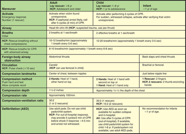
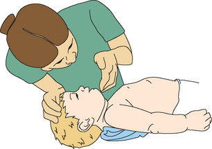
Nurses assume several roles in management of the disease, including helping the family understand the rationale for diagnostic procedures, assisting with radiographic examinations, performing and interpreting skin tests, and obtaining specimens for laboratory examination. Skin tests must be carried out correctly to obtain accurate results. The tuberculin is injected intradermally with the bevel of the needle pointing upward. A wheal 6 to 10 mm in diameter should form between the layers of the skin when the solution is injected properly. If the wheal is not formed, the procedure is repeated. The volar or dorsal surface of the forearm is the usual injection site. The reaction to the skin test is determined in 48 to 72 hours; reactions occurring after 72 hours should be measured and considered the result. The size of the transverse diameter of induration, not the erythema, is measured. The diameter transverse to the long axis of the forearm is the only one standardized for measurement purposes (American Academy of Pediatrics, 2006b).

Sputum specimens are difficult or impossible to obtain from an infant or young child because they swallow any mucus coughed from the lower respiratory tract. The best means for obtaining material for smears or culture is by gastric washing (i.e., aspiration of lavaged contents from the fasting stomach). The procedure is carried out and the specimen obtained early in the morning before the customary breakfast time. In some cases an induced sputum specimen may be obtained by administering aerosolized normal saline for 10 to 15 minutes, followed by CPT and suctioning of the nasopharynx for sputum collection.

Because the success of therapy depends on compliance with the drug regimen, parents are instructed about the importance and rationale for DOT. Case finding in the community and follow-up of known contacts—individuals from whom the affected child may have acquired the disease and persons who may have been exposed to the child with the disease—are essential control measures.

PULMONARY DYSFUNCTION CAUSED BY NONINFECTIOUS IRRITANTS

FOREIGN BODY ASPIRATION

Small children characteristically explore matter with their mouths and are prone to aspirate a foreign body (FB). Small children also place objects such as beads, paper clips, small magnets, or food items in the nose, which can easily be aspirated into the trachea. FB aspiration can occur at any age but is most common in children 1 to 3 years of age. Severity is determined by the location, type of object aspirated, and extent of obstruction. For example, dry vegetable matter, such as a seed, nut, or piece of carrot or popcorn, that does not dissolve and that may swell when wet creates a particularly difficult problem. The high fat content of potato chips and peanuts may cause the added risk of lipoid pneumonia. “Fun foods” are the worst offenders in terms of potential for choking. Offending foods in the order of frequency of choking are hot dog, round candy, peanut or other nut, grape, cookie or biscuit, other meat, carrot, peas, apple, and peanut butter. Other items include plastic or glass beads, button or disc batteries, and coins. Objects such as small lithium or cadmium batteries may cause esophageal or tracheal corrosion.

Diagnostic Evaluation

The diagnosis of FB aspiration is suspected on the basis of the history and physical signs. Initially, an FB in the air passages produces choking, gagging, wheezing, or coughing. Laryngotracheal obstruction most commonly causes dyspnea, cough, stridor, and hoarseness because of decreased air entry. Up to half of all children with FB ingestion may be asymptomatic (Uyemura, 2005). Cyanosis may occur if the obstruction becomes worse. Bronchial obstruction usually produces cough (frequently paroxysmal), wheezing, asymmetric breath sounds, decreased airway entry, and dyspnea. When an object is lodged in the larynx, the child is unable to speak or breathe. If the obstruction progresses, the child’s face may become livid, and if the obstruction is total, the child can become unconscious and die of asphyxiation. If obstruction is partial, hours, days, or even weeks may pass without symptoms after the initial period. Secondary symptoms are related to the anatomic area in which the object is lodged and are usually caused by a persistent respiratory tract infection distal to the obstruction. FB aspiration should also be suspected in the presence of acute or chronic pulmonary lesions. Often, by the time secondary symptoms appear, the parents have forgotten the initial episode of coughing and gagging.

Radiographic examination reveals opaque FBs but is of limited use in localizing nonradiographic matter. Bronchoscopy is required for a definitive diagnosis of objects in the larynx and trachea. Fluoroscopic examination is valuable in detecting FBs in the bronchi. The mainstay of diagnosis and management of FBs is endoscopy. If there is doubt about the presence of an FB, endoscopy can be diagnostic and therapeutic.

Therapeutic Management

FB aspiration may result in life-threatening airway obstruction, especially in infants because of the small diameters of their airways. Current recommendations for the emergency treatment of the choking child include the use of abdominal thrusts for children older than 1 year of age and back blows and chest thrusts for children younger than 1 year of age (see Airway Obstruction, p. 809).

An FB is rarely coughed up spontaneously. Most frequently, it must be removed instrumentally by endoscopy. Endoscopy and bronchospcopy require sedation with an agent such as IV propofol or midazolam. The procedure is carried out as quickly as possible because the progressive local inflammatory process triggered by the foreign material hampers removal. A chemical pneumonia soon develops, and vegetable matter begins to macerate within a few days, making it even more difficult to remove. After removal of the FB, the child is usually observed for any complications such as laryngeal edema, then discharged home within a matter of hours if vital signs are stable and recovery is satisfactory.

Nursing Care Management

A major role of nurses caring for a child who has aspirated an FB is to recognize the signs of FB aspiration and implement immediate measures to relieve the obstruction. All persons working with children must be prepared to deal effectively with aspiration of an FB. Choking on food or other material should not be fatal. Back blows and chest thrusts in infants and abdominal thrusts in children are simple procedures that can be used by both health professionals and laypersons to save lives. It is the responsibility of nurses to learn these techniques and to teach them to parents and other groups (see Figs. 23-16 and 23-17). To aid a child who is choking, nurses must recognize the signs of distress. Not every child who gags or coughs while eating is truly choking.

NURSINGALERT

The child in severe distress (1) cannot speak, (2) becomes cyanotic, and (3) collapses. These three signs indicate that the child is truly choking and requires immediate action. The child can die within 4 minutes.

Prevention.: Nurses are in a position to teach prevention in a variety of settings. They can educate parents singly or in groups about hazards of aspiration in relation to the developmental level of their children and encourage them to teach their children safety. Parents should be cautioned about behaviors that their children might imitate (e.g., holding foreign objects, such as pins, nails, and toothpicks, in their lips or mouth). (Prevention based on the child’s age is discussed in Chapters 10 and 12.)

ASPIRATION PNEUMONIA

Aspiration pneumonia occurs when food, secretions, inert materials, volatile compounds, or liquids enter the lung and cause inflammation and a chemical pneumonitis. Aspiration of fluid or foods is a particular hazard in the child who has difficulty with swallowing or is unable to swallow because of paralysis, weakness, debility, congenital anomalies, or absent cough reflex or in the child who is force-fed, especially while crying or breathing rapidly. Clinical signs of the aspiration of oral secretions may not be distinguishable from those of other forms of acute bacterial pneumonia. For example, if vegetable matter has been aspirated, manifestations may not appear for several weeks after the event. Classic symptoms include an increasing cough or fever with foul-smelling sputum, deteriorating chest radiographs, and other signs of lower airway involvement. These deviations may persist for weeks, however, while the child starts to feel better. Rarely, aspiration causes immediate death from asphyxia; more often the irritated mucous membrane becomes a site for secondary bacterial infection. In addition to fluids, food, vomitus, and nasopharyngeal secretions, other substances that may cause pneumonia are hydrocarbons, lipids, powder, and barium.

Nursing Care Management

Care of the child with aspiration pneumonia is the same as that described for the child with pneumonia from other causes. However, the major focus of nursing care is on prevention of aspiration. Proper feeding techniques should be carried out for weak, debilitated, and uncooperative children, and preventive measures should be used to prevent aspiration of any material that might enter the nasopharynx. Nasogastric tubes used for feedings are checked before the initiation of bolus feedings; continuous nasogastric tube feedings are also evaluated periodically for proper tube placement. Children who are at risk for swallowing difficulties as a result of illness, physical debilitation, anesthesia, or sedation are kept NPO (nothing by mouth) until they can properly swallow fluids effectively. The child who is at risk for vomiting and incapable of protecting the airway should be positioned in a side-lying recovery position (see Fig. 23-18).

ACUTE RESPIRATORY DISTRESS SYNDROME/ACUTE LUNG INJURY

ARDS is recognized in children and adults and has been associated with clinical conditions and injuries such as sepsis, trauma, viral pneumonia, fat emboli, drug overdose, reperfusion injury after lung transplantation, smoke inhalation, and near-drowning. It is characterized by respiratory distress and hypoxemia that occur within 72 hours of a serious injury or surgery in a person with previously normal lungs. Acute lung injury (ALI) is said to involve a spectrum of inflammatory disease responses to a precipitating event (Frye, 2005). Acute pulmonary inflammation with alveolar capillary membrane destruction results in significant hypoxemia, and mechanical ventilation is often required. ARDS is the most severe in the spectrum of illnesses in relation to the degree of hypoxemia. Hypoxemia is expressed as the ratio of partial pressure of oxygen (Pao2) to fraction of inspired oxygen (Fio2), or P/F ratio, with the P/F ratio for ALI being 300 or less, whereas the P/F ratio for ARDS is 200 or less. Both ALI and ARDS demonstrate radiographic evidence of bilateral alveolar infiltrates without evidence of left-sided heart failure (Rice and Bernard, 2006).

The hallmark of ARDS is increased permeability of the alveolar-capillary membrane that results in pulmonary edema. During the acute phase of ARDS, the alveolocapillary membrane is damaged, with an increasing pulmonary capillary permeability and resulting interstitial edema. Later stages are characterized by pneumocyte and fibrin infiltration of the alveoli, with the start of either the healing process or fibrosis. When fibrosis occurs, the child may demonstrate respiratory distress and the need for mechanical ventilation. In ARDS the lungs become stiff as a result of surfactant inactivation, gas diffusion is impaired, and eventually bronchiolar mucosal swelling and congestive atelectasis occur. The net effect is decreased functional residual capacity, pulmonary hypertension, and increased intrapulmonary right-to-left shunting of pulmonary blood flow. Surfactant secretion is reduced, and the atelectasis and fluid-filled alveoli provide an excellent medium for bacterial growth.

The criteria for diagnosis of ARDS in children are an acute antecedent illness or injury, acute respiratory distress or failure, no evidence of prior cardiopulmonary disease, and diffuse bilateral infiltrates evidenced on chest radiography. The child with ARDS may first demonstrate only symptoms caused by an injury or infection, but, as the condition deteriorates, hyperventilation, tachypnea, increasing respiratory effort, cyanosis, and decreasing oxygen saturation occur. At times the developing hypoxemia is not responsive to oxygen administration.

Treatment involves supportive measures, such as maintenance of adequate oxygenation and pulmonary perfusion, treatment of infection (or the precipitating cause), maintenance of adequate cardiac output and vascular volume, hydration, adequate nutritional support, comfort measures, prevention of complications such as gastrointestinal ulceration and aspiration, and psychologic support. Prone positioning may be used to improve oxygenation; this requires close communication and coordination among the health care team (Frye, 2005). Definitive therapy is directed toward improvement of oxygenation. The use of endotracheal intubation, positive end-expiratory pressure, and low tidal volume may be required to ensure maximum oxygen delivery by increasing functional residual capacity, reducing intrapulmonary shunting, and reducing pulmonary fluid. Additional supportive strategies in the treatment of ARDS in children include the use of lung-protective ventilator strategies, permissive hypercapnia, inhaled nitric oxide, exogenous surfactant administration, high-frequency ventilation, partial liquid ventilation, and extracorporeal life support (extracorporeal membrane oxygenation, or ECMO). Once the underlying cause is identified, specific treatment (e.g., antibiotics for infection) is initiated.

Prognosis.: In spite of advances in understanding and treating ARDS and ALI, mortality in children ranges from 22% (after severe trauma) to 88% (after bone marrow transplant) (Flori, Glidden, Rutherford, and others, 2005; Frankel and DiCarlo, 2004). The precipitating disorder influences the outcome; the worst prognosis is associated with uncontrolled sepsis, bone marrow transplantation, cancer, and multisystem involvement with hepatic failure. Children who recover may have persistent cough and exertional dyspnea.

Nursing Care Management

The child with ARDS is cared for in intensive care during the acute stages of illness. Nursing care involves close monitoring of cardiac output, perfusion, fluid and electrolyte balance, and renal function (urinary output), as well as assessment of oxygenation and respiratory status. Blood gas analysis and pulse oximetry are important evaluation tools. Parenteral and enteral nutritional support are often required because of the length of the acute phase of the illness. Diuretics may be administered to reduce pulmonary fluid, and vasodilators may be administered to decrease pulmonary vascular pressure. Pain management is another important consideration in the management of the child with ARDS. Nursing management also includes monitoring the effects of the numerous parenteral fluids and drugs used to stabilize the child and monitoring for changes in the child’s hemodynamic status. Most children with ARDS require invasive monitoring via a central venous line and possibly a pulmonary artery catheter to monitor oxygenation and administer medications. The nursing care of the child with ARDS also involves close observance of skin condition and prevention of breakdown, passive range of motion for prevention of muscle atrophy and contractures, and nutritional support. Respiratory distress is a frightening situation for both the child and the parents, and attention to their psychologic needs is a major element in the care of these children. The child is often sedated during the acute phase of the illness, and weaning from sedation requires close monitoring for anxiety reduction and comfort.

SMOKE INHALATION INJURY

A number of noxious substances that may be inhaled are toxic to humans. They are primarily products of incomplete combustion and cause more deaths from fires than flame injuries. The severity of the injury depends on the nature of the substances generated by the material burned, whether the victim is confined in a closed space, and the duration of contact with the smoke. Smoke inhalation results in three types of injury: heat, chemical, and systemic. Three distinct stages occur in the child suffering from inhalation injury:

image Pulmonary insufficiency, usually during the initial 12 hours

image Pulmonary edema, usually after 6 to 72 hours, with an increase in the lung fluid and interstitial edema

image Bronchopneumonia, usually after 72 hours with a resulting airway obstruction or atelectasis

Heat injury involves thermal injury to the upper airway. Air has low specific heat; therefore the injury goes no farther than the upper airway. Reflex closure of the glottis prevents injury to the lower airway.

Chemical injury involves gases that may be generated during the combustion of materials such as clothing, furniture, and floor coverings. Acids, alkalis, and their precursors in smoke can produce chemical burns. These substances can be carried deep into the respiratory tract, including the lower respiratory tract, in the form of insoluble gases. Soluble gases tend to dissolve in the upper respiratory tract.

Synthetic materials are especially toxic, producing gases such as oxides of sulfur and nitrogen, acetaldehyde, formaldehyde, hydrocyanic acid, and chlorine. Heated plastics are the source of extremely toxic vapors, including chlorine and hydrochloric acid from polyvinylchloride, and hydrocarbons, aldehydes, ketones, and acids from polyethylene. Irritant gases such as nitrous oxide or carbon dioxide combine with water in the lungs to form corrosive acids; aldehydes cause denaturation of proteins, cellular damage, and edema of pulmonary tissues. Chemical burns to the airways are similar to burns on the skin, except they are painless because the tracheobronchial tree is relatively insensitive to pain.

Inhalation of small amounts of noxious irritants produces alveolar and bronchiolar damage that can lead to obstructive bronchiolitis. Severe exposure causes further injury, including alveolocapillary damage with hemorrhage, necrotizing bronchiolitis, inhibited secretion of surfactant, and formation of hyaline membranes—manifestations of ARDS.

Systemic injury occurs from gases that are nontoxic to the airways (e.g., carbon monoxide [CO], hydrogen cyanide). However, these gases cause injury and death by interfering with or inhibiting cellular respiration. CO is responsible for more than half of all fatal inhalation poisonings in the United States. CO is a colorless, odorless gas with an affinity for hemoglobin 230 times greater than that of oxygen. When it enters the bloodstream, CO combines readily with hemoglobin to form carboxyhemoglobin (COHb). Because it is released less readily, tissue hypoxia reaches dangerous levels before oxygen is available to meet tissue needs.

NURSINGALERT

The oxygen saturation (Sao2) obtained by pulse oximetry will be normal because the device measures only oxygenated and deoxygenated hemoglobin; it does not measure dysfunctional hemoglobin, such as COHb.

Accidental CO poisoning is most often a result of exposure to fumes of heaters or smoke from structural fires, although poorly ventilated recreational vehicles with improperly operated or maintained gas lamps or stoves and cooking in underventilated areas with charcoal grills are also frequent causes. CO is produced by incomplete combustion of carbon or carbonaceous material such as wood or charcoal.

The signs and symptoms of CO poisoning are secondary to tissue hypoxia and vary with the level of COHb. Mild manifestations include headache, visual disturbances, irritability, and nausea, whereas more severe intoxication causes confusion, hallucinations, ataxia, and coma. The bright, cherry red lips and skin often described are less often observed; pallor and cyanosis are seen more frequently.

Therapeutic Management

Treatment of children with smoke inhalation injury is largely symptomatic. The most widely accepted treatment is placing the child on humidified 100% oxygen as quickly as possible and monitoring for signs of respiratory distress and impending failure. Baseline arterial blood gases (ABGs) and COHb levels are obtained. Pao2 may be within normal limits unless there is marked respiratory depression. If CO poisoning is confirmed, 100% oxygen is continued until COHb levels fall to the nontoxic range of about 10%. If CO poisoning is severe, the patient may benefit from hyperbaric oxygen therapy; however, the benefits and risks of treatment are debatable (Kao and Nañagas, 2004); at this time there are no published clinical practice guidelines for the therapy, especially in children. Hyperbaric oxygen therapy may be useful in the treatment of neurologic complications related to CO poisoning.

Respiratory distress may occur early in the course of smoke inhalation as a result of hypoxia, or patients who are breathing well on admission may suddenly develop respiratory distress. Therefore intubation equipment should be readily available. Transient edema of the airways can occur at any level in the tracheobronchial tree. Assessment and localization of the obstruction should be accomplished before severe swelling of the head, neck, or oropharynx occurs. Intubation is often necessary when (1) severe burns in the area of the nose, mouth, and face increase the likelihood of developing oropharyngeal edema and obstruction; (2) vocal cord edema causes obstruction; (3) the patient has difficulty handling secretions; and (4) progressive respiratory distress requires artificial ventilation. Controversy surrounds tracheostomy, but many prefer this procedure when the obstruction is proximal to the larynx and reserve nasotracheal intubation for lower tract involvement.

Nursing Care Management

Nursing care of the child with inhalation injury is the same as that for any child with respiratory distress. Vital signs and other respiratory assessments are performed frequently, and the pulmonary status is carefully observed and maintained. Pulmonary physical therapy is often part of the therapy, as well as mechanical ventilation if needed. Fluid requirements for children experiencing inhalation injury are greater than for those with surface burns alone; however, one concern is the development of pulmonary edema. Therefore accurate monitoring of intake and output is essential.

In addition to observation and management of the physical aspects of inhalation injury, the nurse also deals with the psychologic needs of a frightened child and distraught parents. As with any accidental injury, the parents may feel overwhelming guilt, even when the injury occurred through no fault of their own. Parents need support, reassurance, and information regarding the child’s condition, treatment, and progress.

ENVIRONMENTAL TOBACCO SMOKE EXPOSURE

Numerous investigations indicate that parental smoking is an important cause of morbidity in children. Children exposed to passive or environmental tobacco smoke have an increased number of respiratory illnesses, increased respiratory symptoms (i.e., cough, sputum, and wheezing), and reduced performance on pulmonary function tests. AOM and OME are also increased in children who have smoking parents. Indoor exposure to environmental tobacco smoke has been linked to asthma in children. Among children with asthma, there is an association between parental cigarette smoking and asthma exacerbations, trips to the emergency department, medication use, and impaired recovery after hospitalization for acute asthma. Maternal cigarette smoking is associated with increased respiratory symptoms and illnesses in children; decreased fetal growth; increased deliveries of low-birth-weight, preterm, and stillborn infants; and a greater incidence of sudden infant death syndrome (SIDS). Antenatal maternal smoking has emerged as a significant risk factor for SIDS (American Academy of Pediatrics, 2005). The risk for diagnosis of early-onset asthma in the first 3 years of life is associated with in utero exposure to maternal smoking; grandmaternal smoking was also associated with an increased risk of early-onset asthma in the grandchild even if the mother did not smoke during pregnancy (Li, Langholz, Salam, and others, 2005). Exposure to tobacco smoke during childhood may also contribute to the development of chronic lung disease in the adult.

Nursing Care Management

Nurses must provide information about the hazards of environmental smoke exposure in all their interactions with children and their family members. This information is especially important for children with respiratory and allergic illnesses. In families where smokers refuse to quit, appropriate guidance is provided for reducing smoke in the child’s environment (see Family-Centered Care box). Nurses should set an example for children and families and become advocates for “no smoking” ordinances in public places, prohibition of advertising tobacco products in the media, and inclusion of health warnings of sidestream smoke on tobacco products.* Nurses also have an important role in providing parents with affordable smoking cessation education resources, including the appropriate use of smoking cessation pharmacologic aids (Sheahan and Free, 2005).

LONG-TERM RESPIRATORY DYSFUNCTION

ASTHMA

Asthma is a chronic inflammatory disorder of the airways in which many cells (mast cells, eosinophils, and T lymphocytes) play a role. In susceptible children, inflammation causes recurrent episodes of wheezing, breathlessness, chest tightness, and cough, especially at night or in the early morning. These asthma episodes are associated with airflow limitation or obstruction that is reversible either spontaneously or with treatment. The inflammation also causes an increase in bronchial hyperresponsiveness to a variety of stimuli (National Asthma Education and Prevention Program, 2007). Recognition of the importance of inflammation has made the use of antiinflammatory agents, especially inhaled steroids, a key component in the treatment of asthma.

image FAMILY-CENTERED CARE

Decreasing Childhood Exposure to Environmental Tobacco Smoke

image Maintain a smoke-free home.

image Avoid exposing an infant to environmental smoke.

image Use an air-purifying filter in the home where smoking is unavoidable.

image Encourage exclusive breastfeeding for the first 6 months.

image If smoking cessation is in progress by breastfeeding mother, suggest she change upper clothing after smoking and before breastfeeding infant.

image Do not smoke around children.

image Change clothing after smoking and before holding an infant in close proximity.

image Restrict smoking to an isolated area of the house where the children do not play or sleep.

image Do not smoke in motor vehicles with children.

image Do not smoke in rooms children use.

image Do not allow visitors to smoke in the home.

Asthma is classified into four categories based on the symptom indicators of disease severity. These categories are intermittent, mild persistent, moderate persistent, and severe persistent. The intermittent category has the least number of symptoms; symptoms increase in frequency or intensity until the last category of severe persistent asthma (Box 23-14). These categories provide a stepwise approach to the pharmacologic management, environmental control, and educational interventions needed for each category (National Asthma Education and Prevention Program, 2007). A new component of the asthma severity classification system includes the domains of impairment and risk for each category; these categories emphasize the multifaceted aspect of the disease for consideration of effects on present quality of life and functional capacity and the future risk of adverse events (National Asthma Education and Prevention Program, 2007).

BOX 23-14   Asthma Severity Classification in Children: Ages 0 to 11*

STEP 5 OR 6: SEVERE PERSISTENT ASTHMA

Continual symptoms throughout the day

Frequent nighttime symptoms

Peak expiratory flow (PEF): <60%

Forced expiratory volume in 1 second (FEV1): <75% of predicted value

Interference with normal activity: extremely limited

Use of short-acting β-agonist for symptom control: several times a day

STEP 3 OR 4: MODERATE PERSISTENT ASTHMA

Daily symptoms

Nighttime symptoms: 3 to 4 times a month (ages 0 to 4), >1 per week but not nightly (ages 5 to 11)

PEF: 60% to 80% of predicted value

FEV1: 75% to 80%

PEF variability: >30%

Interference with normal activity: some limitation

Use of short-acting β-agonist for symptom control: daily

STEP 2: MILD PERSISTENT ASTHMA

Symptoms >2 times a week, but <1 time a day

Nighttime symptoms: 1 to 2 times a month (ages 0 to 4), 3 to 4 times a month (ages 5 to 11)

PEF or FEV1: ≥80% of predicted value

PEF variability: 20% to 30%

Interference with normal activity: minor limitation

Use of short-acting β-agonist for symptom control: <2 days a week but not daily

STEP 1: INTERMITTENT ASTHMA

Symptoms, <2 days/week

Nighttime symptoms (awakenings): none (ages 0 to 4), >2 times a month (ages 5 to 11)

PEF or FEV1: ≥80% of predicted value

PEF variability: <20%

Interference with normal activity: none

Use of short-acting β-agonist for symptom control: <2 days/week


*The presence of one clinical feature of severity is sufficient to place a patient in that category. An individual should be assigned to the most severe grade in which any feature occurs. The characteristics in this table are general and may overlap because asthma is highly variable. An individual’s classification may change over time. Risk factors for each category are not presented in this table. See the original table referenced above for additional classification data. Asthma treatment should not be based on this table.

From National Asthma Education and Prevention Program: Guidelines for the diagnosis and management of asthma: summary report 2007, retrieved March 8, 2008, from http://www.nhlbi.nih.gov/guidelines/asthma/index.htm.

Asthma prevalence, morbidity, and mortality are increasing in the United States, especially among African-Americans (Linzer, 2007). These increases may result from worsening air pollution, poor access to medical care, or underdiagnosis and undertreatment. Asthma is the most common chronic disease of childhood, the primary cause of school absences, and the third leading cause of hospitalizations in children under the age of 15. Although the onset of asthma may occur at any age, 80% to 90% of children have their first symptoms before 4 or 5 years of age. Boys are affected more frequently than girls until adolescence, when the trend reverses.

Etiology

Studies of children with asthma indicate that allergy influences both the persistence and the severity of the disease. In fact, atopy, or the genetic predisposition for the development of an immunoglobulin E (IgE)–mediated response to common aeroallergens, is the strongest identifiable predisposing factor for developing asthma (National Asthma Education and Prevention Program, 2007). Although allergens play an important role in asthma, 20% to 40% of children with asthma have no evidence of allergic disease. In addition to allergens, other substances and conditions can serve as triggers that may exacerbate asthma (Box 23-15). Asthma is a complex disorder involving biochemical, genetic, immunologic, environmental, infectious, endocrine, and psychologic factors. Evidence shows that viral respiratory infections may have a significant role in the development and expression of asthma (National Asthma Education and Prevention Program, 2007).

BOX 23-15   Triggers Tending to Precipitate or Aggravate Asthmatic Exacerbations

image Allergens

    Outdoor—Trees, shrubs, weeds, grasses, molds, pollens, air pollution, spores

    Indoor—Dust or dust mites, mold, cockroach antigen

image Irritants—Tobacco smoke, wood smoke, odors, sprays

image Exposure to occupational chemicals

image Exercise

image Cold air

image Changes in weather or temperature

image Environmental change—Moving to new home, starting new school, etc.

image Colds and infections

image Animals—Cats, dogs, rodents, horses

image Medications—Aspirin, nonsteroidal antiinflammatory drugs, antibiotics, beta blockers

image Strong emotions—Fear, anger, laughing, crying

image Conditions—Gastroesophageal reflux, tracheoesophageal fistula

image Food additives—Sulfite preservatives

image Foods—Nuts, milk or other dairy products

image Endocrine factors—Menses, pregnancy, thyroid disease

Pathophysiology

There is general agreement that inflammation contributes to heightened airway reactivity in asthma. The mechanisms contributing to airway inflammation are multiple and involve a number of different pathways. It is unlikely that asthma is caused by either a single cell or a single inflammatory mediator; rather, it appears that asthma results from complex interactions among inflammatory cells, mediators, and the cells and tissues present in the airways (National Asthma Education and Prevention Program, 2007). However, recognition of the importance of inflammation has made the use of antiinflammatory agents a key component of asthma therapy.

Another important component of asthma is bronchospasm and obstruction. The mechanisms responsible for the obstructive symptoms in asthma include (Fig. 23-4) (1) inflammatory response to stimuli; (2) airway edema and accumulation and secretion of mucus; and (3) spasm of the smooth muscle of the bronchi and bronchioles, which decreases the caliber of the bronchioles.

image

FIG. 23-4 Mechanisms of obstruction in asthma.

Bronchial constriction is a normal reaction to foreign stimuli, but in the child with asthma it is abnormally severe, producing impaired respiratory function. The smooth muscle arranged in spiral bundles around the airway causes narrowing and shortening of the airway, which significantly increases airway resistance to airflow. Airflow is determined by the size of the airway lumen, degree of bronchial wall edema, mucus production, smooth muscle contraction, and muscle hypertrophy. Because the bronchi normally dilate and elongate during inspiration and contract and shorten on expiration, the respiratory difficulty is more pronounced during the expiratory phase of respiration.

Increased resistance in the airway causes forced expiration through the narrowed lumen. The volume of air trapped in the lungs increases as airways are functionally closed at a point between the alveoli and the lobar bronchi. This trapping of gas forces the individual to breathe at higher and higher lung volumes. Consequently, the person with asthma fights to inspire sufficient air. This expenditure of effort for breathing causes fatigue, decreased respiratory effectiveness, and increased oxygen consumption. The inspiration occurring at higher lung volumes hyperinflates the alveoli and reduces the effectiveness of the cough. As the severity of obstruction increases, there is a reduced alveolar ventilation with carbon dioxide retention, hypoxemia, respiratory acidosis, and, eventually, respiratory failure.

Chronic inflammation may also cause permanent damage (airway remodeling) to airway structures, which cannot be prevented by and is not responsive to current treatments (National Asthma Education and Prevention Program, 2007).

Diagnostic Evaluation

The classic manifestations of asthma are dyspnea, wheezing, and coughing. However, children may experience symptoms that range from acute episodes of shortness of breath, wheezing, and cough followed by a quiet period to a relatively continuous pattern of chronic symptoms that fluctuate in severity (Box 23-16). An attack may develop gradually or appear abruptly and may be preceded by a URI. The age of the child is often a significant factor, since the first attack frequently occurs before the age of 5 years, with some children manifesting clinical signs and symptoms in infancy. In infancy an attack usually follows a respiratory infection. Some children may experience a prodromal itching at the front of the neck or over the upper part of the back just before an attack.

BOX 23-16   Clinical Manifestations of Asthma

COUGH

Hacking, paroxysmal, irritative, and nonproductive

Becomes rattling and productive of frothy, clear, gelatinous sputum

RESPIRATORY-RELATED SIGNS

Shortness of breath

Prolonged expiratory phase

Audible wheeze

May have a malar flush and red ears

Lips deep, dark red color

May progress to cyanosis of nail beds or circumoral cyanosis

Restlessness

Apprehension

Prominent sweating as the attack progresses

Older children sitting upright with shoulders in a hunched-over position, hands on the bed or chair, and arms braced (tripod)

Speaking with short, panting, broken phrases

CHEST

Hyperresonance on percussion

Coarse, loud breath sounds

Wheezes throughout the lung fields

Prolonged expiration

Crackles

Generalized inspiratory and expiratory wheezing; increasingly high pitched

WITH REPEATED EPISODES

Barrel chest

Elevated shoulders

Use of accessory muscles of respiration

Facial appearance—flattened malar bones, circles beneath the eyes, narrow nose, prominent upper teeth

NURSINGALERT

Shortness of breath with air movement in the chest restricted to the point of absent breath sounds accompanied by a sudden rise in respiratory rate is an ominous sign indicating ventilatory failure and imminent respiratory arrest.

The diagnosis is determined primarily on the basis of clinical manifestations, history, physical examination, and, to a lesser extent, laboratory tests. Generally, chronic cough in the absence of infection or diffuse wheezing during the expiratory phase of respiration is sufficient to establish a diagnosis.

Pulmonary function tests (PFTs) provide an objective method of evaluating the presence and degree of lung disease, as well as the response to therapy. Spirometry can generally be performed reliably on children by the age of 5 or 6 years and includes either the traditional and simple mechanical spirometer often used in clinics, offices, and the home or new computerized versions. The National Asthma Education and Prevention Program (2007) recommends that spirometry testing be done at the time of initial assessment of asthma, after treatment is initiated and symptoms have stabilized, and at least every 1 to 2 years to assess the maintenance of airway function.

Another key measurement is the peak expiratory flow rate (PEFR), which measures the maximum flow of air that can be forcefully exhaled in 1 second. PEFR is measured in liters per minute using a peak expiratory flow meter (PEFM). Three zones of measurement are typically used to interpret PEFR. The zone system is patterned after a traffic light to make the categories easy to understand and remember (see Nursing Care Guidelines box). Each child needs to establish his or her personal best value. A personal best value should be established during a 2- to 3-week period when the child’s asthma is stable. During this period, the child records the PEFR at least twice a day. After the personal best value has been established, the child’s current PEFR on any occasion can be compared with the personal best value.

Bronchoprovocation testing, direct exposure of the mucous membranes to a suspected antigen in increasing concentrations, helps to identify inhaled allergens. Exposure to methacholine, histamine, or cold or dry air may be performed to assess airway responsiveness or reactivity. Exercise challenges may be used to identify children with exercise-induced bronchospasm (Liu, Spahn, and Leung, 2004). Although these tests are highly specific and sensitive, they place the child at risk for an asthmatic episode and should be done under close observation in a qualified laboratory or clinic.

nursingcareguidelines

Interpreting Peak Expiratory Flow Rates*

image Green (80% to 100% of personal best) signals all clear. Asthma is under reasonably good control. No symptoms are present, and the routine treatment plan for maintaining control can be followed.

image Yellow (50% to 79% of personal best) signals caution. Asthma is not well controlled. An acute exacerbation may be present. Maintenance therapy may need to be increased. Call the practitioner if the child stays in this zone.

image Red (below 50% of personal best) signals a medical alert. Severe airway narrowing may be occurring. A short-acting bronchodilator should be administered. Notify the practitioner if the peak expiratory flow rate does not return immediately and stay in yellow or green zones.


*These zones are guidelines only. Specific zones and management should be individualized for each child.

Skin testing is useful in identifying specific allergens, and those obtained by the puncture technique correlate better than intracutaneous tests with symptoms and measurements of specific IgE antibody. The radioallergosorbent test (RAST) helps identify antigens against various foods and is often useful in determining appropriate therapy. It is recommended that all patients with year-round asthma symptoms be tested with skin tests or laboratory blood analysis to determine sensitization to perennial allergens (e.g., house dust mites, cats, dogs, cockroaches, molds, and fungus) (National Asthma Education and Prevention Program, 2007).

In addition to these tests, other important tests include laboratory tests (complete blood count with differential) and chest radiographs. The complete blood count may show a slight elevation in the white blood cell count during acute asthma, but elevations to more than 12,000/mm3 or an increased percentage of band cells may indicate respiratory tract infection. The presence of eosinophilia of greater than 500/mm3, on the other hand, tends to suggest an allergic or inflammatory disorder.

Frontal and lateral radiographs show infiltrates and hyperexpansion of the airways, with the anteroposterior diameter on physical examination indicating an increased diameter (suggestive of barrel chest). Additional diagnostic tests for conditions such as gastroesophageal reflux may be carried out to determine whether they may contribute to asthma symptoms. Radiography may assist in ruling out a respiratory tract infection.

Therapeutic Management

The overall goals of asthma management are to maintain normal activity levels, maintain normal pulmonary function, prevent chronic symptoms and recurrent exacerbations, provide optimum drug therapy with minimum or no adverse effects, and assist the child in living as normal and happy a life as possible. This includes facilitating the child’s social adjustments in the family, school, and community and normal participation in recreational activities and sports. To accomplish these goals, several treatment principles need to be followed (National Asthma Education and Prevention Program, 2007):

image FAMILY-CENTERED CARE

“Allergy-Proofing” the Home and Community

image Keep humidity between 30% and 50%; use dehumidifier or air conditioner if available; keep air conditioners clean and free of mold; do not use vaporizers or humidifiers.

image Encase pillows in zippered allergen-impermeable covers or wash pillows in hot water (at least 54.4° C [130° F]) every week.

image Encase mattress and box springs in zippered allergen-impermeable cover.

image Use foam rubber mattress and pillows or Dacron pillows and synthetic blankets.

image Wash bed linens every 7 to 10 days in hot water (at least 54.4° C).

image Encase polyester comforters in allergen-impermeable covers or wash in hot water (at least 54.4° C) every week; if possible, do not use comforters and use cotton blankets.

image Do not use a canopy above the bed; children should not sleep on the bottom bunk of a bunk bed.

image Store nothing under the bed; keep clothing in a closet with the door shut.

image Use washable window shades; avoid heavy curtains; if curtains are used, launder them frequently.

image Remove all carpeting if possible; if not possible, vacuum carpet once or twice a week while the child wears a mask; have child remain out of the room while vacuuming occurs and for 30 minutes after vacuuming.

image If possible, use a central vacuum cleaner with a collecting bag outside of the home or use cleaner filters (e.g., high-efficiency particulate air [HEPA] filters).

image Have air and heating ducts cleaned annually; change or clean filters monthly; cover heating vents with filter material (e.g., cheesecloth) to prevent circulation of dust, especially when heat is turned on.

image Remove unnecessary furniture, rugs, stuffed or real animals, toys, books, upholstered furniture, plants, aquariums, and wall hangings from child’s room.

image Use wipeable furniture (wood, plastic, vinyl, or leather) in place of upholstered furniture; avoid rattan or wicker furniture.

image Cover walls with washable paint or wallpaper.

image Limit child’s exposure to animals (rabbits, gerbils, hamsters) at school; teach child to stay away from zoos, petting farms, and neighbor’s pets.

image Change child’s clothes after playing outdoors; wash child’s hair nightly if child is outside and pollen count is high.

image Keep child indoors while lawn is being mowed, bushes or trees are being trimmed, or pollen count is high.

image Keep windows and doors closed during pollen season; use air conditioner if possible or go to places that are air conditioned, such as libraries and shopping malls, when the weather is hot.

image Wet-mop bare floors weekly; wet-dust and clean child’s room weekly; child should not be present during cleaning activities.

image Wash showers and shower curtains with bleach or Lysol at least once a month.

image Limit or prevent child’s exposure to tobacco and wood smoke; do not allow cigarette smoking in the house or car; select daycare centers, play areas, and shopping malls that are smoke free.

image Avoid odors or sprays (e.g., perfumes, talcum powder, room deodorizers, chalk dust at school, fresh paint, cleaning solutions).

image Avoid cellar (basement) as a play area if it is damp, and use a dehumidifier in damp basement.

image Cover all food, including pet food, and put food away in cabinets.

image Store garbage in closed containers.

image Use pesticide sprays, roach bait traps, and boric acid powder to kill cockroaches; if living in an apartment or adjacent housing, encourage neighbors to work together to get rid of cockroaches and mice.

image Repair leaking or dripping faucets; seal cracks and crevices in cabinets and pantry areas.

image A continuous care approach with regular visits (at least every 1 to 6 months) to the health care provider is necessary to control symptoms and prevent exacerbations.

image Prevention of exacerbations includes avoiding triggers, avoiding allergens, and using medications as needed.

image Therapy includes efforts to reduce underlying inflammation and to relieve or prevent symptomatic airway narrowing.

image Therapy includes patient education, environmental control, pharmacologic management, and the use of objective measures to monitor the severity of disease and guide the course of therapy.

Allergen Control.: Nonpharmacologic therapy is aimed at the prevention and reduction of exposure to airborne allergens and irritants. House dust mites and other components of house dust are frequent agents identified in children allergic to inhalants. The cockroach, another common household inhabitant, is an important allergen in many locations. Exterminating live cockroaches, carefully cleaning kitchen floors and cabinets, putting food away after eating, and taking trash out in the evening are essential measures to control cockroaches. The mouse allergen is the most recent allergen to be identified in the homes of inner-city children with asthma. The role of cat and dog dander in allergen-induced asthma has also been studied. Sensitized persons should carefully evaluate having such pets in the household; there are inconclusive data on cat dander, but there is some evidence that dog dander either has no effect or may be protective (Sharma, Hansel, Matsui, and others, 2007). Additional sources of pollutants include ozone, particulate matter produced by tobacco smoke, wood-burning stoves, pesticides, lead, mold spores, nitrogen dioxide, and sulfur dioxide; these are believed to contribute to asthma morbidity in children and should be avoided or minimized. Exposure to tobacco smoke is a significant contributing factor in the development of asthma in infants and small children (Sharma, Hansel, Matsui, and others, 2007). Recommendations for controlling allergens are found in the Family-Centered Care box.

Skin testing identifies specific allergens so steps can be taken to eliminate or avoid them. Often, simply removing the offending environmental allergens or irritants (e.g., removing carpeting from the home of a child sensitive to mold and dust particles) will decrease the frequency of asthma episodes. Dehumidifiers or air conditioners control nonspecific factors that trigger an episode, such as extremes of temperature.

Despite the proven association between the incidence of asthma and exposure to these residential hazards, little evidence-based research demonstrates an overall reduction in symptoms even with significant interventions aimed at environmental (housing) modifications such as removal of carpeting, cleaning, and extermination (Sandel, Phelan, Wright, and others, 2004; Sharma, Hansel, Matsui, and others, 2007).

Drug Therapy.: Pharmacologic therapy is used to prevent and control asthma symptoms, reduce the frequency and severity of asthma exacerbations, and reverse airflow obstruction. A stepwise approach is recommended based on the severity of the child’s asthma. Because inflammation is considered an early and persistent feature of asthma, therapy is directed toward long-term suppression of inflammation.

Asthma medications are categorized into two general classes: long-term control medications (preventive medications) to achieve and maintain control of inflammation, and quick-relief medications (rescue medications) to treat symptoms and exacerbations (National Asthma Education and Prevention Program, 2007).

Quick-relief and long-term medications are often used in combination. Inhaled corticosteroids, cromolyn sodium and nedocromil, long-acting β2-agonists, methylxanthines, and leukotriene modifiers are used as long-term control medications. Short-acting β2-agonists, anticholinergics, and systemic corticosteroids are used as quick-relief or rescue medications. Bronchodilators that relax bronchial smooth muscle and dilate the airways include β2-agonists, methylxanthines, and anticholinergics that can be used as both quick-relief and long-term medications.

Many asthma medications are given by inhalation with a nebulizer or a metered-dose inhaler (MDI). The MDI should always be attached to a spacer when an inhaled corticosteroid is administered to prevent yeast infections in the mouth. Spacers are also important for children who have difficulty coordinating or learning proper inhalation technique (Pongracic, 2003). The spacer and holder can be equipped with a mask or a mouthpiece. Pharmaceutical companies are currently mandated to produce inhalers that do not contain chlorofluorocarbons (CFCs) as the propellant because CFCs have been linked to damage and depletion of the earth’s ozone level. An alternative propellant to the CFCs is hydrofluoroalkanes (HFAs); the purported advantages include delivery of more fine particles and less oral deposition (Pongracic, 2003). The FDA mandated December 2008 as the targeted cessation date for MDIs containing CFCs to be produced and sold (US Food and Drug Administration, 2005). Several currently available CFC-free MDI devices use dry powder (and also called dry powder inhalers [DPIs]); these include the Diskus inhaler and the Turbuhaler. These devices are breath activated, and the child needs to inhale as quickly and deeply as possible to use them effectively. The Diskhaler and Aerosolizer are similar, but with the Aerosolizer the medication must be loaded into the inhaler before use. Infants and very young children who have difficulty using MDIs or other inhalers can receive their asthma medications via a nebulizer. When this device is used, the medication is mixed with saline (also available in premixed form) and nebulized with compressed air. Children are instructed to breathe normally with the mouth open to provide a direct route to the trachea.

Corticosteroids are antiinflammatory drugs used to treat reversible airflow obstruction and to control symptoms and reduce bronchial hyperresponsiveness in chronic asthma. A major change in the last two revisions of the National Asthma Education and Prevention Program guidelines is the recommendation that inhaled corticosteroids be used as first-line therapy in children over 5 years of age. Clinical studies of corticosteroids have indicated significant improvement of all asthma parameters, including decreases in symptoms, emergency visits, and medication requirements (National Asthma Education and Prevention Program, 2007).

Corticosteroids may be administered parenterally, orally, or by inhalation. Oral medications are metabolized slowly, with an onset of action up to 3 hours after administration and peak effectiveness occurring within 6 to 12 hours. Oral systemic steroids may be given for short periods of time (e.g., 3- or 10-day “bursts”) to gain prompt control of inadequately controlled persistent asthma or to manage severe persistent asthma. These drugs should be given in the lowest effective dose. These medications have few side effects (cough, dysphonia, and oral thrush), and there is strong evidence that they improve the long-term outcomes for children of all ages with mild or moderate persistent asthma. Evidence from clinical trials that monitored children for 6 years indicate that the use of inhaled corticosteroids at recommended doses does not have long-term significant effects on growth, bone mineral density, ocular toxicity, or suppression of the adrenal-pituitary axis (National Asthma Education and Prevention Program, 2007). However, primary care providers should frequently monitor (at least every 3 to 6 months) the growth of children and adolescents taking corticosteroids to assess the systemic effects of these drugs and make appropriate reductions in dosages or changes to other types of asthma therapy when necessary. The inhaled corticosteroids include budesonide and fluticasone.

Cromolyn sodium is a nonsteroidal antiinflammatory drug (NSAID) for asthma. It stabilizes mast cell membranes; inhibits activation and release of mediators from eosinophil and epithelial cells; and inhibits the acute airway narrowing after exposure to exercise, cold dry air, and sulfur dioxide. There is no way to reliably predict whether a child will respond to the drug. Cromolyn sodium has minimal side effects (occasional coughing on inhalation of the powder formulation) and may be given via nebulizer or MDI. Nedocromil sodium inhibits the bronchoconstrictor response to inhaled antigens and inhibits the activity of and release of histamine, leukotrienes, and prostaglandins from inflammatory cells associated with asthma. The drug has few side effects and is used for maintenance therapy in asthma; it is not effective for reversal of acute exacerbations and is not used in children under 5 years of age

β-Adrenergic agonists (short acting) (primarily albuterol, levalbuterol [Xopenex], and terbutaline) are used for treatment of acute exacerbations and for the prevention of exercise-induced bronchospasm. These drugs bind with the β-receptors on the smooth muscle of airways, where they activate adenylate cyclase and convert adenosine monophosphate (AMP) to cyclic AMP (cAMP). It is believed that the increased cAMP enhances binding of intracellular calcium to the cell membrane, reducing the availability of calcium and thus allowing smooth muscle to relax. Other effects of the drug help stabilize mast cells to prevent release of mediators. Most β-adrenergics used in asthma therapy affect predominantly the β2-receptors, which help eliminate bronchospasm. β1 effects, such as increased heart rate and gastrointestinal disturbances, have been minimized. These drugs can be given via inhalation or as oral or parenteral preparations. The inhaled drug has a more rapid onset of action than the oral form. Inhalation also reduces troublesome systemic side effects: irritability, tremor, nervousness, and insomnia. Levalbuterol reportedly causes fewer side effects; however, its overall effectiveness in childhood asthma is controversial (Linzer, 2007). The 2007 National Asthma Education and Prevention Program guidelines recommend the addition of a long-acting β2-agonist to a low- or medium-dosage inhaled corticosteroid to improve lung function and asthma symptoms and decrease the need for a short-acting β2-agonist. There is some evidence that this combination may actually enable the practitioner to lower the corticosteroid dose and manage asthma symptoms just as effectively (Mintz, 2004). Inhaled β-adrenergic agents should not be taken more than three or four times daily for acute symptoms.

Salmeterol (Serevent) is a long-acting β2-agonist (bronchodilator) that is used twice a day (no more frequently than every 12 hours). This drug is added to antiinflammatory therapy and used for long-term prevention of symptoms, especially nighttime symptoms, and exercise-induced bronchospasm. Salmeterol is not used in children less than 12 years of age, and it is not used to treat acute symptoms or exacerbations.

Theophylline was used for decades to relieve symptoms and prevent asthma attacks; however, it is now used primarily in the emergency department when the child is not responding to maximal therapy (Linzer, 2007). Therapeutic levels should be obtained with this drug, since it has a narrow therapeutic window.

Leukotrienes are mediators of inflammation that cause increases in airway hyperresponsiveness. Leukotriene modifiers (such as zafirlukast [Accolate] and montelukast sodium [Singulair]) block inflammatory and bronchospasm effects. These drugs are not used to treat acute episodes, but are given orally in combination with β-agonists and steroids to provide long-term control and prevent symptoms in mild persistent asthma. Montelukast is approved for children 12 months old and older, whereas zafirkulast is approved for children 7 years and older.

Anticholinergics (atropine and ipratropium [Atrovent]) may also be used for relief of acute bronchospasm. However, these drugs have adverse side effects that include drying of respiratory secretions, blurred vision, and cardiac and central nervous system stimulation. The primary anticholinergic drug used is ipratropium, which does not cross the blood-brain barrier and therefore elicits no central nervous system effects. Ipratropium, when used in combination with albuterol, has been shown to be effective during acute severe asthma in significantly improving lung function and reducing hospitalizations in children coming to the emergency department (Liu, Spahn, and Leung, 2004).

A fairly new asthma drug, omalizumab (Xolair), is a monoclonal antibody that blocks the binding of IgE to mast cells. Blocking this interaction eventually inhibits the inflammation that is associated with asthma. Because many patients with asthma are atopic and possess specific IgE antibodies to allergens responsible for airway inflammation, this drug is a promising adjunct to the treatment of asthma. It has been approved for use in children 12 years and older. The drug is administered once or twice a month by subcutaneous injection. Efficacy of omalizumab is not immediate, and clinical trials report that response to the drug was not evident before 12 weeks (Strunk and Bloomberg, 2006). Clinical trials of the drug indicate that it can be an effective therapy for patients with symptomatic moderate to severe allergic asthma that is poorly controlled with inhaled corticosteroids. The drug is expensive (Courtney, McCarter, and Pollart, 2005), however, and there have been reported cases of severe anaphylactic reactions. In early 2007 the FDA added a “black box warning” to the drug, which highlights the risk of anaphylaxis.

The use of complementary and alternative medicine (CAM) in children with asthma is reported by several sources; most common are the use of herbal products, breathing techniques, homeopathy, and acupuncture (Slader, Reddel, Jenkins, and others, 2006). The use of CAM should be carefully evaluated in conjunction with other therapies in the overall management of asthma.

Exercise.: Exercise-induced bronchospasm (EIB) is an acute, reversible, usually self-terminating airway obstruction that develops during or after vigorous activity, reaches its peak 5 to 10 minutes after stopping the activity, and usually stops in another 20 to 30 minutes. Patients with EIB have cough, shortness of breath, chest pain or tightness, wheezing, and endurance problems during exercise, but an exercise challenge test in a laboratory is necessary to make the diagnosis.

The problem is rare in activities that require short bursts of energy (e.g., baseball, sprints, gymnastics, skiing) and more common in those that involve endurance exercise (e.g., soccer, basketball, distance running). Swimming is well tolerated by children with EIB because they are breathing air fully saturated with moisture and because of the type of breathing required in swimming.

Children with asthma are often excluded from exercise by parents, teachers, and practitioners, as well as by the children themselves, since they are reluctant to provoke an attack. However, this practice can seriously hamper peer interaction and physical health. Exercise is advantageous for children with asthma, and most children can participate in activities at school and in sports with minimal difficulty, provided their asthma is under control. Participation should be evaluated on an individual basis. Appropriate prophylactic treatment with β-adrenergic agents or cromolyn sodium before exercise will usually permit full participation in strenuous exertion.

Chest Physical Therapy.: CPT includes breathing exercises and physical training. These therapies help produce physical and mental relaxation, improve posture, strengthen respiratory musculature, and develop more efficient patterns of breathing. For the motivated child, breathing exercises and controlled breathing are of value in preventing overinflation and improving efficiency of the cough. However, CPT is not recommended during acute, uncomplicated exacerbations of asthma.

Hyposensitization.: The role of hyposensitization in childhood asthma has become controversial. In the past, immunotherapy was used for seasonal allergies and when single substances were identified as the offending allergen. It is not recommended for allergens that can be eliminated, such as foods, drugs, and animal dander.

The National Asthma Education and Prevention Program guidelines (2007) recommend immunotherapy for asthma patients in the following situations:

image When there is evidence of a relationship between asthma symptoms and unavoidable exposure to an allergen to which the patient is sensitive

image When symptoms occur all year or at least during a major portion of the year

image When symptom control is difficult with drug therapy because multiple medications are required, or the patient is not responsive to available drugs, or the patient refuses to take the medications

Injection therapy is usually limited to clinically significant allergens. The initial dose of the offending allergen(s), based on the size of the skin reaction, is injected subcutaneously. The amount is increased at weekly intervals until a maximum tolerance is reached, after which a maintenance dose is given at 4-week intervals. This may be extended to 5- or 6-week intervals during the off-season for seasonal allergens. Successful treatment is continued for a minimum of 3 years and then stopped. If no symptoms appear, acquired immunity is assumed; if symptoms recur, treatment is reinstituted. Hyposensitization injections should be administered only with emergency equipment and medications readily available in the event of an anaphylactic reaction.

Prognosis.: The outlook for children with asthma varies widely. Some children’s asthma symptoms may improve at puberty, but up to two thirds of children with asthma continue to have symptoms through puberty and into adulthood. The prognosis for control or disappearance of symptoms varies in children from those who have rare and infrequent attacks to those who are constantly wheezing or are subject to status asthmaticus. In general, when symptoms are severe and numerous, when symptoms have been present for a long time, and when there is a family history of allergy, there is a greater likelihood of a poor prognosis. Risk factors that may predict persistence of symptoms into childhood (from infancy) include atopy, male gender, exposure to environmental tobacco, and maternal history of asthma (Ross, Mjaanes, and Lemanske, 2003). Many children who outgrow their exacerbations continue to have airway hyperresponsivenesss and cough as adults. Furthermore, airway hyperresponsiveness in adults appears to be associated with decreased lung function.

Although deaths from asthma have been relatively uncommon, especially in the young age-groups, since the 1980s the rate of death from asthma has increased steadily in the United States and other countries. Data indicate a significant increase in asthma symptoms, emergency department visits, and hospitalization among children ages birth to 4 years of age in 2003 to 2004. Most asthma deaths for the same period were in children ages 11 to 17 years, with significant increases seen in non-Hispanic African-Americans (Linzer, 2007). Mortality and morbidity for asthma are especially high among African-American children, whose hospitalization and death rates are three times higher than those of Caucasian and Hispanic children (Liu, Spahn, and Leung, 2004).

The adolescent age-group appears to be the most vulnerable, with the greatest increase occurring in children 10 to 14 years of age. No reliable data exist to explain this increase. Factors that have been postulated include exposure of atopic persons to more allergens (particularly in large urban centers), change in severity of the disease, abuse of drug therapy (toxicity), failure of families and practitioners to recognize the severity of asthma, and psychologic factors such as denial and refusal to accept the disease.

Risk factors for asthma deaths include early onset, frequent attacks, difficult-to-manage disease, adolescence, history of respiratory failure, psychologic problems (refusal to take medications), dependency on or misuse of asthma drugs (high use), presence of physical stigmata (barrel chest, intercostal retractions), and abnormal PFTs.

Status Asthmaticus.: Children who continue to display respiratory distress despite vigorous therapeutic measures, especially the use of sympathomimetics (e.g., albuterol, epinephrine), are considered to be in status asthmaticus. The condition may develop gradually or rapidly, often coincident with complicating conditions, such as pneumonia or a respiratory virus, that can influence the duration and treatment of the exacerbation.

NURSINGALERT

Status asthmaticus is a medical emergency that can result in respiratory failure and death if untreated.

Therapy for status asthmaticus is aimed at improving ventilation, decreasing airway resistance and relieving bronchospasm, correcting dehydration and acidosis, allaying child and parent anxiety related to the severity of the event, and treating any concurrent infection. Humidified oxygen is recommended and should be given to maintain an oxygen saturation greater than 90%. Inhaled aerosolized short-acting β2-agonists are recommended for all patients. Three treatments of β2-agonists spaced 20 to 30 minutes apart are usually given as initial therapy, and continuous administration of β2-agonists may be initiated. A systemic corticosteroid (oral, IV, or IM) may also be given to decrease the effects of inflammation. An anticholinergic such as ipratropium bromide may be added to the aerosolized solution of the β2-agonist. Anticholinergics have been shown to result in additional bronchodilation in patients with severe airflow obstruction. An IV infusion is often initiated to provide a means for hydration and to administer medications. Correction of dehydration, acidosis, hypoxia, and electrolyte disturbance is guided by frequent determination of arterial pH, blood gases, and serum electrolytes.

Additional therapies in acute asthma attacks include the use of IV magnesium sulfate, a potent muscle relaxant that acts to decrease inflammation and improves pulmonary function and peak flow rate among pediatric patients treated in the emergency department with moderate to severe asthma. Heliox may be administered to decrease airway resistance, and thereby decrease the work of breathing; heliox can be delivered via a nonrebreathing face mask from premixed tanks, which may be blended in a stand-alone unit or within a ventilator. Heliox may be used in acute exacerbations as an adjunct to β2-agonist and IV corticosteroid therapy to improve pulmonary function until the two latter medications have time to take full effect in decreasing bronchospasm; the effects of heliox are usually seen within 20 minutes of administration, whereas other drugs may take longer to exert the desired effect (Liu, Spahn, and Leung, 2004). Ketamine, a dissociative anesthetic, is believed to cause smooth muscle relaxation and decrease airway resistance caused by severe bronchospasm in acute asthma (Linzer, 2007); it may be administered as an adjunct to other therapies mentioned previously. Inhaled magnesium sulfate used in addition to a β2-agonist for acute asthma attacks has also been effective in treating acute asthma exacerbation (Blitz, Blitz, Beasley, and others, 2005).

Antibiotics should not be used to treat acute asthma attacks except when a bacterial infection resulting from another condition such as pneumonia or sinusitis is present (National Asthma Education and Prevention Program, 2007).

A child suspected of having status asthmaticus is usually seen in the emergency department and is often admitted to a pediatric intensive care unit for close observation and continuous cardiorespiratory monitoring. A key component in the prevention of morbidity is helping the child, parents, teachers, coaches, and other adults recognize features of deteriorating respiratory status, use the correct rescue drugs effectively, and immediately place the child with deteriorating respiratory status into the care of trained health care professionals instead of waiting to see if the asthma gets better on its own. The child going into early status asthmaticus is no different from the adult who is having a myocardial infarction in terms of needing trained medical assistance before the condition deteriorates to irreversible respiratory failure and possible death. Community education regarding asthma recognition and management is an important component of nursing care.

Nursing Care Management

The nursing care of the child with asthma begins with a review of the child’s health history; the home, school, and play environment; parent and child attitudes about the child’s condition; and a comprehensive physical assessment with focus on the respiratory system. Nursing care of children with asthma involves both acute and long-term care. Nurses who are involved with children in the home, hospital, school, outpatient clinic, or practitioner’s office play an important role in helping children and their families learn to live with the condition. The disease can be managed so that it does not require hospitalization or interfere with family life, physical activity, or school attendance. The nursing process in the care of the child with asthma is outlined in the Nursing Care Plan.

Physical assessment of asthma involves the same observations and techniques described in Chapter 6. In addition, the nurse notes and evaluates physical characteristics of chronic respiratory involvement, including chest configuration (e.g., barrel chest), posturing, and type of breathing. A history of the current and previous episodes and precipitating factors or events provides important information.

Nurses may perform a variety of functions in asthma care. These may include asthma education in the primary care setting and in schools and other community settings, care of the child with asthma in the acute care setting, ambulatory care, and intensive care. Nurses also obtain information on how asthma affects the child’s everyday activities and self-concept, the child’s and family’s adherence to the prescribed therapy, and their personal treatment goals. Every effort is made to build a partnership between the child and family and the health care team. Communication is an essential part of this partnership, and health care providers should routinely assess the effectiveness of patient-provider communication. In particular, the child and family’s satisfaction with asthma control and with the quality of care should be assessed. The nurse should also assess their perception of the severity of the disease and their level of social support.

One of the major emphases of nursing care is outpatient management by the family. Parents are taught how to avoid allergens, to recognize and respond to symptoms of bronchospasm, to maintain health and prevent complications, and to promote normal activities. The nurse should determine any cultural or ethnic beliefs or practices that influence self-management and that may necessitate modifications in educational approaches to meet the family’s needs.

Avoid Allergens.: One goal of asthma management is avoidance of an exacerbation. Parents need to know how to avoid allergens that precipitate asthma episodes. The nurse assists the parent in modifying the environment to reduce contact with the offending allergen(s) (see Family-Centered Care box, p. 787). Parents are cautioned to avoid exposing a sensitive child to excessive cold, wind, or other extremes of weather; smoke; sprays; or other irritants. Foods known to provoke symptoms should be eliminated from the diet.

Approximately 2% to 6% of children with asthma are sensitive to aspirin; therefore nurses should caution parents to use other analgesic-antipyretic drugs for discomfort or fever and to read package labeling. Although aspirin is rarely given to children in the United States, salicylate compounds are in other common medicines such as Pepto-Bismol. Children with aspirin-induced asthma may also be sensitive to NSAIDs and tartrazine (yellow dye number 5, a common food coloring).

NURSINGALERT

Parents are encouraged to avoid administering aspirin to any child unless specifically recommended by and under the supervision of a health practitioner. Acetaminophen is safe for children and is the analgesic of choice.

Relieve Bronchospasm.: Parents and older children are taught to recognize early signs and symptoms of an impending attack so that it can be controlled before symptoms become distressing. Most children can recognize prodromal symptoms well before an attack (about 6 hours) and implement preventive therapy. Objective signs that parents may observe include rhinorrhea, cough, low-grade fever, irritability, itching (especially in front of the neck and chest), apathy, anxiety, sleep disturbance, abdominal discomfort, and loss of appetite. A variety of easy-to-use, inexpensive PEFMs are available for use in the home and at school to assess changes in pulmonary function (see Family-Centered Care box). In general, children 5 years of age and older are able to use a PEFM successfully. However, young children need to be supervised while they are learning to use their PEFM, and their technique should be checked frequently to ensure it is correct. Children should use the same peak flow meter over time, and they should bring their peak flow meter for use at every follow-up visit. Using the same brand of meter is recommended because different brands can give significantly different values. The use of a PEFM provides objective monitoring regarding the severity of asthma and can decrease asthma episodes, health care visits, and missed school days (Burkhart, Rayens, Revelette, and others, 2007).

nursingcareplan: The Child with Asthma

image

image

image FAMILY-CENTERED CARE

Use of a Peak Expiratory Flow Meter

1. Before each use, make certain the sliding marker or arrow on the peak expiratory flow meter (PEFM) points to zero or is at the bottom of the numbered scale.

2. Stand up straight.

3. Remove gum or any food from the mouth.

4. Close your lips tightly around the mouthpiece. Be sure to keep your tongue away from the mouthpiece.

5. Blow out as hard and as quickly as you can, a “fast hard puff.” Blow from the diaphragm, not the cheeks.

6. Note the number by the marker on the numbered scale.

7. Repeat entire routine three times; wait 30 seconds between each routine.

8. Record the highest of the three readings, not the average.

9. Measure the peak expiratory flow rate (PEFR) close to the same time and same way each day (e.g., morning and evening; before or 15 minutes after taking medication).

10. Keep a chart of your PEFRs.

Children who use a nebulizer, MDI, Diskus, or Turbuhaler to deliver drugs need to learn how to use the device correctly. A study of school-age children with asthma indicated that only 7% of these children had effective MDI skills (Winkelstein, Huss, Butz, and others, 2000). The MDI device (Fig. 23-5) delivers medication directly to the airways; therefore the child needs to learn to breathe slowly and deeply for better distribution to narrowed airways (see Family-Centered Care box).

image

FIG. 23-5 Child using metered-dose inhaler with spacer and face mask.

Young children and those who are unable to manipulate the MDI or coordinate breathing should use spacers. These devices allow the parent or child to deliver the medication from the MDI into the spacer, from which the child then inhales the medication. Spacers also prevent yeast infections in the mouth when corticosteroids are inhaled via an MDI.

image FAMILY-CENTERED CARE

Use of a Metered-Dose Inhaler*

STEPS FOR CHECKING HOW MUCH MEDICINE IS IN THE CANISTER

1. If the canister is new, it is full.

2. If the canister has been used repeatedly, it might be empty. (Check product label to see how many inhalations should be in each canister.)

3. The most accurate way to determine how many doses remain in a metered-dose inhaler (MDI) is to count and record each actuation as it is used.

4. Many dry powder inhalers have a dose-counting device or dose indicator on the canister to let you know when the canister is empty.

STEPS FOR USING THE INHALER

1. Remove the cap and hold inhaler upright.

2. Shake the inhaler.

3. Tilt the head back slightly and breathe out slowly.

4. With the inhaler in an upright position, position the mouthpiece:

image About 3 to 4 cm from the mouth or

image Insert into an AeroChamber or spacer (this method is recommended for young children and for people using corticosteroids)

5. At the end of a normal expiration, depress the top of the inhaler canister firmly to release the medication (into either the AeroChamber or the mouth), and breathe in slowly (about 3 to 5 seconds). Relax the pressure on the top of the canister.

6. Hold the breath for at least 5 to 10 seconds to allow the aerosol medication to reach deeply into the lungs.

7. Remove the inhaler and breathe out slowly through the nose.

8. Wait 1 minute between puffs (if additional one is needed).


*note: Some inhaled dry-powder inhalers require a different inhalation technique. To use these dry-powder inhalers, it is important to close the mouth tightly around the mouthpiece of the inhaler and inhale rapidly and deeply.

Modified from National Asthma Education and Prevention Program: Expert Panel report II: guidelines for the diagnosis and management of asthma, NIH Pub No 97-4051, Bethesda, Md, 1997, National Heart, Lung, and Blood Institute.

The child and parents also need to be cautioned about the adverse effects of prescribed drugs and the dangers of overuse of β2-agonists. They should know that it is important to use these drugs when needed but not indiscriminately or as a substitute for avoiding the symptom-provoking allergen. Parents are cautioned against purchasing over-the-counter preparations because these medications can place the children at risk for increased dosage of a drug and toxicity.

NURSINGALERT

Long-acting β-adrenergic inhalers (salmeterol) should be used only as directed (usually every 12 hours) and not more frequently. They are not intended to relieve acute asthmatic symptoms.

The family should obtain a PEFM and learn to use this device to monitor the child’s asthma. A written asthma action plan that includes the three peak flow meter zones and the child’s asthma medications may be obtained from the child’s primary care provider. Medications used for asthma exacerbations are also included in the asthma plan. This action plan should be used to make decisions about asthma management at home and at school. The nurse may assist the child and family in preparing the written action plan, emphasizing that the child and family determine the success of the plan, not the health professionals.

Foods known to provoke symptoms should be eliminated from the diet, and parents are advised to read labels on prepared foods and snacks to determine the presence of allergens.

The child should be protected from a respiratory tract infection that can trigger an attack or aggravate the asthmatic state, especially in young children whose airways are mechanically smaller and more reactive. Annual influenza vaccinations are recommended for children with persistent asthma (American Academy of Pediatrics, 2008). Equipment used for the child, such as nebulizers, must be kept absolutely clean to decrease the chances of contamination with bacteria and fungi.

Breathing exercises and controlled breathing are taught and encouraged for motivated children, and the nurse should provide information concerning activities that promote diaphragmatic breathing, side expansion, and improved mobility of the chest wall. Play techniques that can be used for younger children to extend their expiratory time and increase expiratory pressure include blowing cotton balls or a Ping-Pong ball on a table, blowing a pinwheel, blowing bubbles, or preventing a tissue from falling by blowing it against the wall.

Self-care and asthma self-management programs are important in helping the child and family cope with asthma. Most asthma self-management programs for children convey several principles. First, asthma is a common disease that can be controlled with appropriate drug therapy, environmental control, education, and management skills. Second, it is much easier to prevent than to treat an asthma episode; adherence to a therapeutic program is necessary to prevent exacerbations. Third, children with asthma can live full and active lives.

Asthma camps provide an opportunity for children with asthma to engage in physical activity while learning about their disease in a controlled environment with their peers and health professionals. Children who attend asthma camps often demonstrate improved asthma self-management skills.

Self-contained programs and brochures for patient education are available from the Asthma and Allergy Foundation of America* and the American Lung Association. The National Heart, Lung, and Blood Institute provides educational materials for asthma education in the school setting and also copies of the Guidelines for the Diagnosis and Management of Asthma for the practitioner (National Asthma Education and Prevention Program, 2007). Another publication designed for health care practitioners, Pediatric Asthma: Promoting Best Practice, can be obtained from the American Academy of Allergy Asthma and Immunology.§

Provide Acute Asthma Care.: Children who are admitted to the hospital with acute asthma are ill, anxious, and uncomfortable. The progression or resolution of status asthmaticus is variable. The importance of continual observation and assessment cannot be overemphasized.

When β2-agonists and corticosteroids are given, the child is monitored closely and continuously for relief of respiratory distress and signs of side effects or toxicity. Oral fluid intake may be limited during the acute phase; IV fluid replacement may be required to provide adequate tissue hydration.

Older children may be more comfortable standing (Fig. 23-6), sitting upright, or leaning slightly forward (Fig. 23-7). When possible, the nurse communicates in such a way that a child can reply in a few words to avoid fatigue. Shortness of breath makes talking difficult.

image

FIG. 23-6 Child with asthma is allowed play activity as tolerated.

image

FIG. 23-7 Children with asthma may take a nebulized aerosol treatment with (A) a mask or (B) mouthpiece. (Courtesy Texas Children’s Hospital, Houston.)

Children with acute asthma are apprehensive and anxious. The calm, efficient presence of a nurse helps reassure them that they are safe and will be cared for during this stressful period. It is important to assure children that they will not be left alone and that their parents are allowed to remain with them.

Parents need reassurance and want to be informed of their child’s condition and therapies. They may believe that they have in some way contributed to the child’s condition or could have prevented the episode. Reassurance regarding their efforts expended on the child’s behalf and their parenting capabilities can help alleviate their stress. Efforts to reduce parental apprehension will also reduce the child’s distress. Anxiety is easily communicated to the child from parents and members of the staff.

Support Child or Adolescent and Family.: The nurse working with children with asthma can provide support in a number of ways. Many children voice frustration because their exacerbations interfere with their daily activities and social lives. They need education about what to do to prevent an asthma episode. These children also need reassurance from the health team that they can learn to control and cope with their asthma and live a normal life. Be aware of children, especially adolescents, who demonstrate signs of depression and may not comply with therapy as a means of passive suicide.

Children in disruptive family situations (divorce, separation, violence, custodial battles) may disregard daily asthma medication regimen or may be at higher risk as a result of neglect by adults who are in charge of their care. Adolescents struggling with a sense of identity and body image often regard asthma as a condition that will “go away,” especially if there is a time lapse between symptoms, and may abandon the therapeutic regimen. In some cases adolescents find themselves in charge of other siblings in blended family situations and may ignore their own health needs. Referral for counseling and guidance is appropriate in these situations where the child’s or adolescent’s life is potentially in harm’s way and the therapeutic regimen for asthma is abandoned due to other crises.

The short- and long-term adaptation of children with asthma often depends on the family’s acceptance of the disorder. The task of living day-to-day with affected children involves the entire family. There are periodic crises and the ever-present threat of a crisis, requiring parental vigilance; sleepless nights; frequent trips to the physician, emergency department, or hospital; and often overwhelming medical expenses. Throughout these stresses, parents are encouraged to promote as normal a life as possible for their children.

CYSTIC FIBROSIS

CF is inherited as an autosomal recessive trait; the affected child inherits the defective gene from both parents, with an overall incidence of 1:4. The mutated gene responsible for CF is located on the long arm of chromosome 7. This gene codes a protein of 1480 amino acids called the cystic fibrosis transmembrane regulator (CFTR). The CFTR protein is related to a family of membrane-bound glycoproteins. The glycoproteins constitute a cAMP-activated chloride channel and also regulate other chloride and sodium channels at the surfaces of the epithelial cells.

Pathophysiology

CF is characterized by several clinical features: increased viscosity of mucous gland secretions, a striking elevation of sweat electrolytes, an increase in several organic and enzymatic constituents of saliva, and abnormalities in autonomic nervous system function. Although both sodium and chloride are affected, the defect appears to be primarily a result of abnormal chloride movement; the CFTR appears to function as a chloride channel. Children with CF demonstrate decreased pancreatic secretion of bicarbonate and chloride and an increase in sodium and chloride in both saliva and sweat. This characteristic is the basis for the sweat chloride diagnostic test. The sweat electrolyte abnormality is present from birth, continues throughout life, and is unrelated to the severity of the disease or the extent to which other organs are involved.

The primary factor, and the one that is responsible for many of the clinical manifestations of the disease, is mechanical obstruction caused by the increased viscosity of mucous gland secretions (Fig. 23-8). Instead of forming a thin, freely flowing secretion, the mucous glands produce a thick mucoprotein that accumulates and dilates them. Small passages in organs such as the pancreas and bronchioles become obstructed as secretions precipitate or coagulate to form concretions in glands and ducts. The earliest postnatal manifestation of CF is often meconium ileus in the newborn, in which the small intestine is blocked with thick, puttylike, tenacious, mucilaginous meconium.

image

FIG. 23-8 Various effects of exocrine gland dysfunction in cystic fibrosis.

In the pancreas the thick secretions block the ducts, eventually causing pancreatic fibrosis. This blockage prevents essential pancreatic enzymes from reaching the duodenum, which causes marked impairment in the digestion and absorption of nutrients. The disturbed function is reflected in bulky stools that are frothy from undigested fat (steatorrhea) and foul smelling from putrefied protein (azotorrhea). The endocrine function of the pancreas often remains unchanged because the islets of Langerhans are normal but may decrease in number as pancreatic fibrosis progresses. The incidence of diabetes mellitus (cystic fibrosis—related diabetes [CFRD]) is greater in CF children than in the general population (Balinsky and Zhu, 2004), which may be caused by changes in pancreatic architecture and diminished blood supply over time. Consequently, with increased survival, and primarily in adolescents and adults, type 1 diabetes is becoming a more frequent finding. There is no relationship between the progression of pulmonary disease and the development of diabetes mellitus in CF. In the liver, localized biliary obstruction and fibrosis are common and become more extensive with time.

A common gastrointestinal complication associated with CF is prolapse of the rectum, which occurs in infancy and childhood and is related to large, bulky stools; malnutrition; and increased intraabdominal pressure secondary to paroxysmal cough. Affected children of all ages are subject to intestinal obstruction from inspissated or impacted feces. Gumlike masses can obstruct the bowel and produce a partial or complete obstruction, a condition that is referred to as distal intestinal obstruction syndrome.

Pulmonary complications are present in almost all children with CF, but the onset and extent of involvement are variable. Symptoms are produced by stagnation of mucus in the airways, with eventual bacterial colonization leading to destruction of lung tissue. The abnormally viscous and tenacious secretions are difficult to expectorate and gradually obstruct the bronchi and bronchioles, causing scattered areas of bronchiectasis, atelectasis, and hyperinflation. The stagnant mucus offers a favorable environment for bacterial growth. The most common pathogens are Pseudomonas aeruginosa, Burkholderia cepacia, S. aureus, H. influenzae, Escherichia coli, and Klebsiella pneumoniae.

The reproductive systems of both males and females with CF are affected. Females with CF have normal fallopian tubes and ovaries. Fertility can be inhibited by highly viscous cervical secretions, which act as a plug, blocking sperm entry. Women with CF who become pregnant have an increased incidence of premature labor and delivery and low birth weight in the infant. Favorable nutritional status and pulmonary function are positively correlated with favorable pregnancy outcomes. Most adult men (95%) with CF are sterile, which may be caused by blockage of the vas deferens with abnormal secretions or by failure of normal development of the wolffian duct structures (vas deferens, epididymis, and seminal vesicles), resulting in decreased or absent sperm production.

Growth and development are often affected in children with moderate to severe forms of CF. Physical growth may be restricted as a result of decreased absorption of nutrients, including vitamins and fat; increased oxygen demands for pulmonary function; and delayed bone growth. The usual pattern is one of growth failure (failure to thrive), with increased weight loss despite an increased appetite, and gradual deterioration of the respiratory system. Clinical manifestations of CF are listed in Box 23-17.

BOX 23-17   Clinical Manifestations of Cystic Fibrosis

MECONIUM ILEUS*

Abdominal distention

Vomiting

Failure to pass stools

Rapid development of dehydration

GASTROINTESTINAL MANIFESTATIONS

Large, bulky, loose, frothy, extremely foul-smelling stools

Voracious appetite (early in disease)

Loss of appetite (later in disease)

Weight loss

Marked tissue wasting

Failure to grow

Distended abdomen

Thin extremities

Sallow skin

Evidence of deficiency of fat-soluble vitamins A, D, E, and K

Anemia

PULMONARY MANIFESTATIONS

Initial signs:

image Wheezy respirations

image Dry, nonproductive cough

Eventually:

image Increased dyspnea

image Paroxysmal cough

image Evidence of obstructive emphysema and patchy areas of atelectasis

Progressive involvement:

image Overinflated, barrel-shaped chest

image Cyanosis

image Clubbing of fingers and toes

image Repeated episodes of bronchitis and bronchopneumonia


*In about 10% of cases.

Diagnostic Evaluation

Traditionally the diagnosis of CF was based on a positive sweat chloride test, absence of pancreatic enzymes, radiography, chronic obstructive pulmonary disease, and family history. Newer diagnostic methods make it possible to diagnose CF early in infancy so therapies can be implemented to increase the child’s overall survival and quality of life. In addition to the sweat chloride test and factors listed above, diagnosis may be confirmed by any one of the following: newborn screening, deoxyribonucleic acid (DNA) identification of mutant genes, and abnormal nasal potential difference measurement.

Universal newborn screening for CF has been proposed yet remains controversial, since many states lack the resources for such screening programs. Currently 12 states offer some type of CF newborn screening (Wilfond and Gollust, 2005); the Centers for Disease Control and Prevention (2004) emphatically recommends newborn screening for CF. The newborn screening test consists of an immunoreactive trypsinogen (IRT) analysis performed on a dried spot of blood, which may be followed by direct analysis of DNA for the presence of the ΔF508 mutation or other mutations on the same dried blood spot. Benefits of early screening and detection include earlier nutritional intervention for identified infants (Farrell, Lai, Li, and others, 2007); disadvantages include the parental anxiety false-positive results may generate. Children who were identified and treated early in infancy with aggressive nutritional support had improved height and weight well into adolescence. Although the technology is available to conduct carrier screening for the general population, this issue remains controversial, and widespread implementation of carrier screening programs is not recommended. An in utero diagnosis of CF is also possible, based on detection of two CF mutations in the fetus.

The consistent finding of abnormally high sodium and chloride concentrations in the sweat is a unique characteristic of CF. Parents may report that their infant tastes “salty” when they kiss him or her. The quantitative sweat chloride test (pilocarpine iontophoresis) involves stimulating the production of sweat with a special device (involves stimulation with 3 mA electric current), collecting the sweat on filter paper, and measuring the sweat electrolytes. The quantitative analysis requires a sufficient volume of sweat (>75 mg). Two separate samples are collected to ensure the reliability of the test for any individual. Normally sweat chloride content is less than 40 mEq/L, with a mean of 18 mEq/L. A chloride concentration greater than 60 mEq/L is diagnostic of CF; in infants less than 3 months, a sweat chloride concentration greater than 40 mEq/L is highly suggestive of CF. In some situations DNA testing may be substituted for the sweat test. The presence of a mutation known to cause CF on each CFTR gene predicts with a high degree of certainty that the individual has CF; however, multiple CFTR mutations may also be present and detected with DNA assay.

Chest radiography reveals characteristic patchy atelectasis and obstructive emphysema. PFTs are sensitive indexes of lung function, providing evidence of abnormal small airway function in CF. Other diagnostic tools that may aid in diagnosis include stool fat or enzyme analysis. Stool analysis requires a 72-hour sample with accurate recording of food intake during that time. Radiographs, including barium enema, are used for diagnosis of meconium ileus.

Therapeutic Management

Improved survival among patients with CF during the past two decades is attributable largely to antibiotic therapy and improved nutritional and respiratory management. Goals of CF therapeutic management are to (1) prevent or minimize pulmonary complications, (2) ensure adequate nutrition for growth, (3) encourage appropriate physical activity, and (4) promote a reasonable quality of life for the child and the family. A multidisciplinary approach to treatment is needed to accomplish these goals.

Management of Pulmonary Problems.: Management of pulmonary problems is directed toward prevention and treatment of pulmonary infection by improving ventilation, removing mucopurulent secretions, and administering antimicrobial agents. Many children will develop respiratory symptoms by 3 years of age. The large amounts and viscosity of respiratory secretions in children with CF contribute to the likelihood of respiratory tract infections. Recurrent pulmonary infections in the child with CF result in greater the damage to the airways; small airways are destroyed, causing bronchiectasis.

The most common pathogens responsible for pulmonary infections are P. aeruginosa, B. cepacia, S. aureus, H. influenzae, E. coli, and K. pneumoniae. P. aeruginosa and B. cepacia are particularly pathogenic for children with CF, and infections with these organisms are difficult to clear. In addition, children with CF who are chronically colonized with these organisms have poorer survival rates than children who are not colonized. Colonization and infection with methicillin-resistant S. aureus (MRSA) has recently emerged as a critical factor in lung infection and pulmonary function in patients with CF. Patients with MRSA required longer hospitalization and multiple antibiotic regimens (Ren, Morgan, Konstan, and others, 2007). Fungal colonization with Candida or Aspergillus organisms in the respiratory tract is also common in CF patients.

Prevention of infection involves a daily routine of CPT to maintain pulmonary hygiene. CPT is usually performed on average twice daily (on rising and in the evening) and more frequently if needed, especially during pulmonary infection. The Flutter mucus clearance device is a small, hand-held plastic pipe with a stainless-steel ball on the inside that facilitates removal of mucus (Fig. 23-9). It has the advantage of increasing sputum expectoration and being used without an assistant. Hand-held percussors may be used to loosen secretions. Another method to clear mucus is high-frequency chest compression, in which the child temporarily wears a mechanical vest device that provides high-frequency chest wall oscillation. Some children and adolescents with an implantable port may experience localized pain with the vest.

image

FIG. 23-9 Child using Flutter mucus clearance device. (Courtesy Scandipharm, Inc.)

Patients with CF have been found to regress when conventional CPT is discontinued. Therefore, although it is time consuming for the child and family, CPT remains the cornerstone of pulmonary therapy. Forced expiration, or “huffing,” with the glottis partially closed helps move secretions from the small airways so that subsequent coughing can move secretions forcefully from the large airways. Several studies indicate that this maneuver enhances the pulmonary function of patients with CF. Autogenic drainage involves a variety of breathing techniques, which the older child can use to force mucus in lower lobes up into the airways so it can be successfully expelled. Another mucus-clearing technique involves use of a positive expiratory pressure mask; this technique involves breathing into a mask attached to a one-way valve, which creates resistance—as the patient exhales, the airway is kept open by the pressure and mucus is forced into the upper airway for expulsion.

Bronchodilator medication delivered in an aerosol opens bronchi for easier expectoration and is administered before CPT when the patient exhibits evidence of reactive airway disease or wheezing. Another aerosolized medication is recombinant human deoxyribonuclease (DNase, known generically as dornase alfa [Pulmozyme]), which decreases the viscosity of mucus. It is well tolerated and has no major adverse effects; minor reactions are voice alterations and laryngitis. This medication, given daily via nebulization, has resulted in improvements in spirometry, PFTs, dyspnea scores, and perceptions of well-being and has reduced the viscosity of sputum.

Physical exercise is an important adjunct to daily CPT. Exercise stimulates mucus excretion and provides a sense of well-being and increased self-esteem. Any aerobic exercise that the patient enjoys should be encouraged. The ultimate aim of exercise is to increase lung vital capacity, remove secretions, increase pulmonary blood flow, and maintain healthy lung tissue for effective ventilation.

Pulmonary infections are treated as soon as they are recognized. In CF patients characteristic signs of pulmonary infection—fever, tachypnea, and chest pain—may be absent; therefore a careful history and physical examination are essential. The presence of anorexia, weight loss, and decreased activity alert the practitioner to pulmonary infection and the need for an antibiotic regimen (Boat and Acton, 2007). Aerosolized antibiotics such as tobramycin, ticarcillin, and gentamicin are beneficial for patients with frequent pulmonary exacerbations. It is not uncommon for the hospitalized child with CF to be placed on as many as two or three antibiotics and one antifungal medication to treat coexisting pulmonary infections.

IV antibiotics may be administered at home as an alternative to hospitalization. The use of peripherally inserted central catheters (PICCs) for the administration of antibiotics in children with CF is a viable option with limited complications and fewer needle punctures to obtain blood specimens and to maintain often lengthy treatment with parenteral antibiotics (Tolomeo and Mackey, 2003). Alternatively, an implanted port offers the advantage of access for blood draws and antibiotic infusion. Patients may receive antibiotic therapy at home and continue daily activities with minimum disruptions. However, when pulmonary function does not improve with outpatient management, hospitalization may be recommended for continued antibiotic therapy and vigorous CPT and postural drainage. Oxygen administration is used for children with acute episodes but must be used cautiously because many children with CF have chronic carbon dioxide retention, and the unsupervised use of oxygen can be harmful (see Oxygen Therapy, Chapter 22). With repeated infection and inflammation, bronchial cysts and emphysema may develop. These cysts may rupture, resulting in a pneumothorax.

NURSINGALERT

Signs of a pneumothorax are usually nonspecific and include tachypnea, tachycardia, dyspnea, pallor, and cyanosis. A subtle drop in oxygen saturation (measured by pulse oximetry) may be an early sign of pneumothorax.

Blood streaking of the sputum is usually associated with increased pulmonary infection and often requires no specific treatment. Hemoptysis greater than 250 ml/24 hr for the older child (less for a younger child) indicates a potentially life-threatening event and needs to be treated immediately. Sometimes bleeding can be controlled with bed rest, IV antibiotics, replacement of acute blood loss, IV conjugated estrogens (Premarin) or vasopressin (Pitressin), and correction of any coagulation defects with vitamin K or fresh frozen plasma. If hemoptysis persists, the site of bleeding should be localized via bronchoscopy and cauterized or embolized.

Treatment of nasal polyps includes intranasal corticosteroids, oral antihistamines, and decongestants. If these measures are ineffective, surgical interventions may be necessary.

Because pulmonary damage in patients with CF is believed to be caused by the inflammatory process that occurs with frequent infections, the use of corticosteroids has been studied; however, treatment with corticosteroids for prolonged periods has been associated with linear growth restriction, glucose tolerance abnormalities, and cataract formation. Antiinflammatory medications such as ibuprofen are becoming more important in the treatment of CF, but careful monitoring for adverse effects (gastrointestinal bleeding) is essential.

Management of Gastrointestinal Problems.: The principal treatment for pancreatic insufficiency is replacement of pancreatic enzymes, which are administered with meals and snacks to ensure that digestive enzymes are mixed with food in the duodenum. Enteric-coated products prevent the neutralization of enzymes by gastric acids, thus allowing activation to occur in the alkaline environment of the small bowel. The amount of enzymes depends on the severity of the insufficiency, the child’s response to enzyme replacement, and the practitioner’s philosophy. Usually one to five capsules are administered with a meal, and a smaller amount is taken with snacks. Capsules can be swallowed whole or taken apart and the contents sprinkled on a small amount of food to be taken at the beginning of the meal. The amount of enzyme is adjusted to achieve normal growth and a decrease in the number of stools to one or two per day. Pancreatic enzymes should be taken within 30 minutes of eating. The enteric-coated beads should not be chewed or crushed, since destroying the enteric coating can lead to inactivation of the enzymes and excoriation of oral mucosa. The powder form should be used cautiously because inhalation of the powder may precipitate acute bronchospasm.

Children with CF require a well-balanced, high-protein, high-caloric diet (because of the impaired intestinal absorption). In fact, they often require up to 150% of the recommended daily allowances to meet their needs for growth. Breastfeeding with enzyme supplementation should be continued whenever possible for parents who prefer this method and, when necessary, supplemented with a higher-calorie-per-ounce formula. For formula-fed infants, commercial cow’s milk—based formulas are usually adequate, although frequently a partially hydrolysated formula with medium-chain triglycerides (e.g., Pregestimil, Alimentum) may be recommended. Enzymes are mixed into cereal or fruit, such as applesauce. Because the uptake of fat-soluble vitamins is decreased, water-miscible forms of these vitamins (A, D, E, and K) are given, along with multivitamins and the enzymes. When high-fat foods are eaten, the child is encouraged to add extra enzymes. Growth failure despite adequate nutritional support may indicate deterioration of pulmonary status. Patients with CF may experience frequent anorexia as a result of the copious amounts of mucus produced and expectorated, persistent cough, effect of medications, fatigue, and sleep disruption. They may be placed on nighttime supplemental gastrostomy feedings or parenteral alimentation in an effort to build up nutritional reserves if there has been a history of inability to maintain weight.

Meconium ileus and meconium ileus equivalent, or total or partial intestinal obstruction, can occur at any age. Constipation is often the result of a combination of malabsorption (either from inadequate pancreatic enzyme dosage or a failure to take the enzymes), decreased intestinal motility, and abnormally viscous intestinal secretions. These problems usually do not require surgical interventions and may be treated with GoLYTELY or Colyte (osmotic solutions given orally or by nasogastric tubes), other laxatives, stool softeners, or rectal administration of meglumine diatrizoate (Gastrografin).

Rectal prolapse occurs in about 20% to 25% of children with CF (McMullen and Bryson, 2004). The first episode of rectal prolapse is frightening to both parents and child. Its reduction usually requires immediate guidance and intervention, which is managed by simply guiding the rectum back into place with a gloved, lubricated finger. Further management usually involves attempting to decrease the bulk of daily stools through enzyme replacement.

Children with CF often experience transient or chronic gastroesophageal reflux, which should be treated with the appropriate histamine-receptor antagonist and gastrointestinal motility drug, dietary modifications, and an upright position after feedings or meals (McMullen and Bryson, 2004).

Management of Endocrine Problems.: The management of CFRD is critical in the therapeutic treatment of the child with CF. CFRD presents a combination of insulin resistance and insulin deficiency, with unstable glucose homeostasis in the presence of acute lung infection and treatment. Children with CFRD require close monitoring of blood glucose, administration of oral glucose-lowering agents or insulin injections, and diet and exercise management; children with CF may be at increased risk for glucose management problems as a result of decreased nutrient absorption, anorexia, and severity of pulmonary illness. The prevalence of CFRD increases with age, and there is increased morbidity and mortality among children with CFRD compared to those without (Strausbaugh and Davis, 2007). Microvascular complications such as retinopathy and nephropathy may occur in children and adolescents with CFRD (Schwarzenberg, Thomas, Olsen, and others, 2007). However, ketoacidosis is reported to be rare in individuals with CFRD (Boat and Acton, 2007).

Bone health is of concern in children and adults with CF. The pancreatic insufficiency of CF and chronic steroid use present potential risks for less than optimum bone growth in such children. Assessment of bone health by history and bone mass density evaluation should be considered in assessing the child’s (8 years old and older) health status to detect and prevent osteoporosis or osteopenia (Borowitz, Baker, and Stallings, 2002).

The administration of growth hormone (somatropin [Nutropin]) is being investigated as a nutritional adjunct in children with CF to achieve optimum growth; one small study sample suggests an improvement in CF clinical status (Hardin, Rice, Ahn, and others, 2005).

Prognosis.: The median predicted survival age for the CF patient in 2005 was 36.5 years (Cystic Fibrosis Foundation, 2006). Lung, heart, pancreas, and liver transplantation have increased survival rates among some CF patients. Heart-lung and double-lung procedures have been successfully performed in children with advanced pulmonary vascular disease and hypoxia. The obstacles surrounding this technique are availability of donated organs; complications from surgery; pulmonary infections; and recurrence of obstructive bronchiolitis, which decreases transplanted lung function.

Despite considerable progress and a recent surge in new treatment modalities, CF remains a progressive and incurable disease. The pulmonary involvement ultimately determines the patient’s outcome because pancreatic enzyme deficiency is less of a problem if adequate nutrition is ensured. With advances in technology, parents and adolescents are challenged to set future goals that may include college, careers, social relationships, and marriage. Concurrently they are faced with increasing morbidity and higher rates of CF complications as they grow older.

Nursing Care Management

Assessment of the child with CF involves both pulmonary and gastrointestinal observations. Pulmonary assessment is the same as that described for asthma, with special attention to lung sounds, observation of cough, and evidence of decreased activity or fatigue. Gastrointestinal assessment primarily involves observing the frequency and nature of the stools and abdominal distention. The observer is also alert to evidence of growth failure (e.g., weight loss, muscle wasting, pallor, anorexia, decreased activity [from baseline norm]). Family members are interviewed to determine the child’s eating and eliminating habits and to confirm a history of frequent respiratory tract infections or bowel obstruction in infancy.

The nurse assesses the newborn for feeding and stooling patterns, which may indicate a potential problem such as meconium ileus. The nurse also participates in diagnostic testing such as the initial newborn screening, IRT, DNA analysis, or sweat chloride test.

Parents are often anxious and puzzled about the diagnostic tests and the possible implications of the test results. They need careful explanations of the disease, how it might affect their family, and what they can do to provide the best possible care for their child. It is crucial to involve the parents in the follow-up for early diagnostic testing; the neonate may require several follow-up visits in the first few weeks of life if initial test results are not conclusive.

The uncertainty, fear, and initial shock associated with the diagnosis are overwhelming to parents. They must face the impact of the chronic, life-threatening nature of the disease and the prospect of intensive treatment, for which they must assume a major part of the responsibility and for which they are ill prepared. They often fear that they will be unable to provide the care the child needs. One of the most difficult aspects of the diagnosis is the implications inherent in its etiology (i.e., the recognition that each parent contributed the gene responsible for the defect).

Hospital Care.: Most patients with CF require hospitalization only for treatment of pulmonary infection, uncontrolled diabetes, or a coexisting medical problem that cannot be treated on an outpatient basis. Therefore, when patients with CF are hospitalized, standard precautions with meticulous hand washing should be implemented to decrease the nosocomial spread of organisms to the CF patient and between hospitalized CF patients (especially when MRSA is prevalent). Contact precautions may be required for specific infections.

When the child with CF is hospitalized for diagnosis or treatment of pulmonary complications, aerosol therapy, chest percussion therapy, and postural drainage are instituted or continued. Respiratory therapists often initiate, supervise, and provide these treatments; however, it is the nurse’s responsibility to monitor the patient’s tolerance to the procedure and evaluate the effectiveness of the procedure in relation to treatment goals. The nurse may at times administer aerosol therapy, perform CPT, assist with mucus removal interventions such as the mechanical vest, and teach breathing exercises. CPT should not be performed before or immediately after meals. Planning CPT so that it does not coincide with meals is difficult in the hospital situation, but essential to the effectiveness of this treatment.

Supplemental oxygen therapy is administered to the child with mild or moderate respiratory distress, and the child requires frequent assessment of the tolerance to the procedure. Noninvasive pulse oximetry provides valuable data about the patient’s oxygenation status, yet nursing assessments, including observation of respiratory pattern, work of breathing, and lung auscultation, are vital assessments.

One of the nursing challenges in the care of the child with CF is encouraging compliance with the therapeutic medication regimen, which often involves a significant number of medications; pancreatic enzymes; vitamins A, D, E, and K; oral antifungals for Candida infection; antihistamines; antiinflammatory agents; and oral antibiotics. This may be overwhelming to the child. Factor in multiple inhaled bronchodilators, CPT and aerosol treatments, blood glucose monitoring and insulin administration, various other medications, and increased mucus production during the acute phase, and it is not uncommon for the child with CF to rebel and be noncompliant with this regimen. Gentle coaxing, positive reinforcement, and frank negotiation may be required to enlist cooperation for effective medication compliance.

The child’s sleep is disrupted frequently by hospital routines; therefore nursing care should be flexible enough to allow the child some quiet time without affecting vital care. In some cases a daily schedule of events, including medication administration, CPT, aerosolized therapy, and dressing changes, may need to be mutually developed with the child, nurses, and physician so the child feels he or she has some control of the care.

The diet for the child with CF represents another challenge; careful planning with a pediatric dietitian and the child’s input may help decrease the loss of appetite and weight loss that are often part of the condition. Patients with CF, and especially adolescents, enjoy foods brought from home or an occasional fast food of choice (provided these meet therapeutic requirements). Children in the early stages of CF often have a good appetite. With infection and increased lung involvement, their appetite diminishes, and eventually it becomes a challenge to tempt failing appetites. When dietary intake fails to meet the child’s needs for growth, enteral feedings or supplements may be considered (Borowitz, Baker, and Stallings, 2002). These feedings may be administered via gastrostomy tube during the night to minimize the disruption of daily activities, including school. A skin-level feeding gastrostomy affords the child few activity restrictions and minimum disruption of body image in comparison to a nasogastric tube or conventional gastrostomy tube. The child and parents are encouraged to not perceive this therapy as a last-ditch effort but as an adjunct therapy to maintain optimum growth and prevent excessive weight loss (Borowitz, Baker, and Stallings, 2002).

The child needs support during the many treatments and tests that are a part of the hospitalization. IV fluids, IV antibiotics and antifungals, and blood tests are almost always a part of the acute care treatment, and the child soon associates hospitalization with these stress-provoking procedures.

Depression, anxiety, and disturbed self-image may occur in children and adolescents with CF; young adults with severe symptoms may be especially prone to depression as a result of the realization of the poor prognosis and the reality of unmet life expectations and goals.

Providing support to both the child and the family is essential. The progressive nature of the disease makes each illness requiring hospitalization a potentially life-threatening event. Skilled nursing care and sympathetic attention to the emotional needs of the child and family help them cope with the stresses associated with repeated respiratory tract infections and hospitalizations.

The care of the child or adolescent who is immobilized as a result of CF requires the same care and attention as the child with immobility from any other chronic or acute illness, including skin care, bowel management, passive range of motion, and positioning.

Home Care.: Most children and adolescents with CF can be managed at home. The goals of care include normalization and daily activities, including school and peer involvement. The care plan should be flexible so that family activities are disrupted as little as possible. Parents may initially require assistance finding and contacting durable medical equipment companies that will provide home care equipment. They also need opportunities to learn how to use the equipment and to solve problems they may encounter while delivering therapy at home. The many aspects of home care for the child with CF are similar to those of home care for other children and are discussed in Chapter 20.

Patients and family members need education about the preferred diet of nutritious meals with tolerated fat, increased protein and carbohydrate, and the administration of pancreatic enzymes. For infants and young children, the enzymes can be mixed with pureed fruit, such as applesauce, and fed with a spoon. Capsules are usually suitable for older children. It is important to stress to parents that the enzymes, in the amount regulated to the child’s needs, should be administered at the beginning of all meals and snacks.

One of the most important aspects of educating parents for home care is teaching techniques for the removal of mucus (CPT, vest, forced expiration) and breathing exercises. The success of a therapy program depends on conscientious performance of these treatments regularly as prescribed. The number of times these therapies are performed each day is determined on an individual basis, and often parents readily learn to adjust the number and intensity of the treatments to the child’s needs. For pulmonary infection, home IV antibiotics may be prescribed. Home IV care may be preferred for willing and competent families because it reduces tension and usually brings a sense of belonging to the family members; this option, however, depends on a number of factors including availability of an agency with adequate staff to perform multiple daily home antibiotic infusions. With use of the venous access devices, such as PICC lines, and implanted ports, the parents and child can be taught the technique of direct administration into the IV line. Around-the-clock administration may be difficult for families and requires certain adjustments such as waking at least once during the night to give the drug.

Families also need information about medications and possible side effects. Children receiving multiple antibiotics may require serum drug levels to ensure therapeutic dosaging.

Children and adolescents with CF should receive routine primary care with special attention to diet, growth and development, and immunizations. Primary care providers should be alert to any weight loss or flattening in the growth curve associated with loss of appetite, which could indicate a pulmonary exacerbation in children with CF (McMullen and Bryson, 2004). In addition to all the recommended routine immunizations, CF patients should be immunized against influenza starting at age 6 months and followed by a yearly booster (American Academy of Pediatrics, 2006b). Anticipatory guidance concerning issues of discipline, how to incorporate aspects of the treatment regimen into the school environment, and delayed pubertal development are also important considerations for the primary care provider.

Home palliative care for the child or adolescent with CF who is in the terminal stages may be carried out with the assistance of hospice (see Chapter 18).

The nurse can assist the family in contacting resources that provide help to families with affected children. Various special child health services, many local clinics, private agencies, service clubs, and other community groups often offer equipment and medications either free or at reduced rates. The Cystic Fibrosis Foundation* has chapters throughout the United States to provide education and services to families and professionals.

Family Support.: The most challenging aspect of providing care for the family of a child or adolescent with CF is meeting the emotional needs of the child and family. The diagnosis, treatment, and prognosis for CF are often associated with many problems and frustrations. The diagnosis can evoke feelings of guilt and self-recrimination in parents.

The long-range problems for an infant, child, or adolescent with CF are those encountered in any chronic illness (see Chapter 18). Both the child and the family must make many adjustments, the success of which depends on their ability to cope and also on the quality and quantity of support they receive from outside sources. Combined efforts of a variety of health professionals are needed to provide the most comprehensive services to families. It is often the nurse who assesses the home situation, organizes and coordinates these services, and collects the data needed to evaluate the effectiveness of the services.

The persistent need for treatment several times a day places tremendous strain on the family. When the child is young, a family member must perform postural drainage and CPT. Children often balk at these treatments, and the parents are placed in the position of insisting on adherence. The stress and anxiety related to this routine may produce feelings of resentment in both the child and the family members. When possible, occasional trusted respite care should be available to allow parents to leave the situation for short periods without undue anxiety about the child’s welfare.

The affected child or adolescent may become resentful about the disease, its relentless routine of therapy, and the necessary curtailment it places on activities and relationships. The child’s activities are interrupted or built around treatments, medications, and diet. This imposes hardships and influences the child’s quality of life. The child should be encouraged to attend school and to join age-appropriate peer groups to foster a life that is as normal and productive as possible. Sports are often an important part of the child and adolescent’s life; interaction with peers are valuable life experiences, especially to adolescents. The child or adolescent with CF should be encouraged to participate in sports activities inasmuch as physical and pulmonary health allows. Exercise is encouraged to increase pulmonary vital capacity, promote muscle development, and enhance cardiovascular function.

ETHICAL CASE STUDY: Cystic Fibrosis and End-of-Life Care

ETHICAL DECISION MAKING MODEL

image

Michael, a 15-year-old boy, has cystic fibrosis. Since age 12 years, Michael’s respiratory status has been getting progressively worse, and he has been hospitalized for multiple respiratory exacerbations. Last evening he was admitted to the adolescent unit in acute respiratory distress. Today, he told his favorite nurse: “I am so tired, I can’t fight anymore. Can’t they just let me go home now and be comfortable?” Michael lives with his father and his paternal grandmother. His mother died 6 years ago in a motor vehicle accident.

Evaluate the Issue

Michael’s pulmonary specialist, Dr. A., believes Michael’s illness is now in the terminal stage. Michael’s paternal grandmother does not want Michael to suffer any more. She tells the nurse that she believes Michael is ready to die and she thinks a do-not-resuscitate (DNR) order and hospice care are best for Michael at this time. Michael’s father does not believe Michael’s condition has become progressively worse. He wants everything medically possible to be done for his son. He will not discuss a DNR order or hospice care.

Treat All Involved with Respect

Michael’s wishes must be heard and acknowledged. He is 15 years old and sufficiently mature to deserve respectful honesty and consideration. By approximately age 14 years, adolescents demonstrate the same kind of reasoning abilities as adults concerning health care decisions. The father’s concerns must also be addressed in an empathetic manner.

Hear All Sides

The nurse has an obligation to be an advocate for her patient. However, the nurse should also consider the father’s reasons for wanting everything possible to be done. Why is the father denying the seriousness of his son’s condition? Does the father need more information, or is he overwhelmed and terrified at the prospect of losing his son and experiencing all the grief and emotions he felt when he lost his wife?

Initiate Action

Which of the following actions is most appropriate for the nurse to take:

image Talk to Michael’s father about Michael’s comments.

image Tell the grandmother to try to get the father to agree to a DNR order and hospice.

image Discuss Michael’s comments with Dr. A.

Consider the Outcome

The ethical dilemma is how to meet the needs of Michael and his father and grandmother.

The nurse decided to share Michael’s comments with Dr. A. and determine whether he is aware of both Michael’s wishes and those of his father.

After discussion with Dr. A., the nurse and other health team members believed that Michael’s father may need more information about his son’s condition and feelings. The following plan was implemented: the family care coordinator will arrange a family conference in which all parties (including Michael) can exchange views and arrive at a decision about future care for Michael. If the family conference does not produce a consensus and Michael still maintains his position, the situation will need to be referred to the hospital ethics committee.

As the disease progresses, however, family stress should be expected, and the patient may become angry and noncompliant. It is important for the nurse to recognize the family’s changing needs and the grief they may experience as the CF worsens. Families should be made aware of sources for counseling. Patients need to be guided into activities that enable them to express anger, sorrow, and fear without guilt (see Ethical Case Study).

Transition to Adulthood.: As life expectancy continues to rise for children and adolescents with CF, issues related to marriage, sexuality, childbearing, and career choice become more pressing. Males must be informed at some point that they will often be unable to produce offspring. It is important that the distinction be made between sterility and impotence. Normal sexual relationships can be expected. Female patients may be able to bear children but should be informed of the possible deleterious effects on the respiratory system created by the burden of pregnancy. They also need to know that their children will be carriers of the CF gene. Adolescent females may need counseling concerning the use of oral contraceptives and other contraceptive options (McMullen and Bryson, 2004).

Adolescents with CF are encouraged to take personal ownership and management of the illness to maximize their life’s potential. Many adolescents and young persons with the illness enroll in college or vocational and technical training school and complete degrees either by distance learning or by attending a local school. Young people are encouraged to set life goals and live normal lives to the extent their illness allows.

Life as an independent adult should be encouraged for children with CF. From the time that children can take partial responsibility for their own care (e.g., CPT and taking enzymes), independence and accountability should be fostered. Although the prognosis for these children has improved, many will need continued support as they cope with the demands of surviving with CF.

Anticipatory grieving and other aspects related to care of a child with a terminal illness are also part of nursing care. For example, it is important to prepare the child and family members for end-of-life decisions and care.

OBSTRUCTIVE SLEEP-DISORDERED BREATHING

Pediatric obstructive sleep-disordered breathing reportedly affects between 10% and 12% of children ages 2 to 8 years; obstructive sleep apnea may occur in as many as 2% of all children (Benninger and Walner, 2007b). Obstructive sleep-disordered breathing is said to form a continuum of sleep-disordered breathing ranging from partial obstruction of the upper airway to continuous episodes of complete upper airway obstruction, with the most severe form being obstructive sleep apnea syndrome (OSAS) (Benninger and Walner, 2007b). OSAS is defined by the American Thoracic Society (1996) as a disorder of breathing during sleep with prolonged partial upper airway obstruction and/or complete obstruction that disrupts normal respiration during sleep and normal sleep patterns. Common symptoms include nightly snoring, interrupted or disturbed sleep patterns, enuresis, and daytime neurobehavioral problems (American Academy of Pediatrics, 2002; Chan, Edman, and Koltai, 2004). OSAS is to be distinguished from primary snoring, which is snoring without obstructive apnea, frequent sleep arousals, or abnormalities in gas exchange (American Academy of Pediatrics, 2002). Interestingly, children with OSAS do not exhibit daytime sleepiness as do adults; the exception may be obese children (Chan, Edman, and Koltai, 2004). If left untreated, obstructive sleep-disordered breathing may result in complications such as growth failure, cor pulmonale, pulmonary hypertension, poor learning, behavioral problems, attention deficit hyperactivity disorder, and death.

The diagnosis of obstructive sleep-disordered breathing is made by a sleep study (polysomnography), which provides evidence of sleep disturbance, respiratory pauses, and changes in oxygenation. The six-channel polysomnography can be performed in children of all ages with videotaping or audiotaping, and abbreviated (vs full night sleep study) polysomnography may be useful; however, this latter method does not predict the severity of OSAS (American Academy of Pediatrics, 2002).

A common treatment for sleep-disordered breathing in children is adenotonsillectomy, provided there is evidence of adenotonsillar hypertrophy (Benninger and Walner, 2007b). Complications of these surgical interventions are discussed previously in this chapter. Continuous positive airway pressure (CPAP) and bilevel positive airway pressure (BiPAP) may be helpful in older children with sleep-disordered breathing whose condition persists after surgical intervention. CPAP is a long-term therapy with frequent assessments to evaluate the required amount of pressure and the overall effectiveness of the intervention.

Surgical interventions such as tracheotomy may be required for children with craniofacial syndromes such as Goldenhar, Pierre Robin, Apert, and Crouzon, in which there is partial or complete upper airway obstruction (Chan, Edman, and Koltai, 2004).

Nursing care of the child with sleep-disordered breathing involves early detection by observation of the infant’s or child’s sleep patterns and active participation in the diagnostic polysomnography. Important nursing roles are inserting the pH probe into the esophagus, ensuring accurate placement by radiography, and monitoring the sleep study and the patient’s response to diagnostic therapy. Counseling families of children with sleep-disordered breathing may involve dietary counseling for exercise programs and weight management, use of the CPAP or BiPAP equipment, and direct postoperative care after the surgical intervention of tonsillectomy or adenoidectomy. The nurse can be instrumental in helping the child and family cope with the chronic illness diagnosis should intervention such as CPAP or BiPAP be required.

RESPIRATORY EMERGENCY

RESPIRATORY FAILURE

In general, the term respiratory insufficiency is applied to two situations: (1) when there is increased work of breathing but gas exchange function is near normal and (2) when normal blood gas tensions cannot be maintained and hypoxemia and acidosis develop secondary to carbon dioxide retention.

Respiratory failure is defined as the inability of the respiratory apparatus to maintain adequate oxygenation of the blood, with or without carbon dioxide retention. This process involves pulmonary dysfunction that generally results in impaired alveolar gas exchange, which can lead to hypoxemia or hypercapnia. Respiratory failure is the most common cause of cardiopulmonary arrest in children (Rotta and Wiryawan, 2003). Respiratory arrest is the cessation of respiration. Apnea is the cessation of breathing for more than 20 seconds or for a shorter period when associated with hypoxemia or bradycardia (Curley and Moloney-Harmon, 2001). Apnea can be (1) central, in which respiratory efforts are absent; (2) obstructive, in which respiratory efforts are present; and (3) mixed, in which both central and obstructive components are present.

Effective pulmonary gas exchange requires clear airways, normal lungs and chest wall, and adequate pulmonary circulation. Anything that affects these functions or their relationships can compromise respiration.

Diagnostic Evaluation

Respiratory dysfunction may have an abrupt or an insidious onset. Respiratory failure can occur as an emergency situation or may be preceded by gradual and progressive deterioration of respiratory function. Most clinical manifestations are nonspecific and are affected by variations among individual patients and differences in the severity and duration of inadequate gas exchange.

The diagnosis of respiratory failure is determined by the combined application of three sources of information:

1. Presence or history of a condition that might predispose the patient to respiratory failure

2. Observation of respiratory failure

3. Measurement of ABGs and pH

Nursing observation and judgment are vital to the recognition and early management of respiratory failure. Nurses must be able to assess a situation and initiate appropriate action within moments. Signs of respiratory failure are listed in Box 23-18.

BOX 23-18   Clinical Manifestations of Respiratory Failure

CARDINAL SIGNS

Restlessness

Tachypnea

Tachycardia

Diaphoresis

EARLY BUT LESS OBVIOUS SIGNS

Mood changes, such as euphoria or depression

Headache

Altered depth and pattern of respirations

Hypertension

Exertional dyspnea

Anorexia

Increased cardiac output and renal output

Central nervous system symptoms (decreased efficiency, impaired judgment, anxiety, confusion, restlessness, irritability, depressed level of consciousness)

Flaring nares

Chest wall retractions

Expiratory grunt

Wheezing or prolonged expiration

SIGNS OF MORE SEVERE HYPOXIA

Hypotension or hypertension

Dimness of vision

Somnolence

Stupor

Coma

Dyspnea

Depressed respirations

Bradycardia

Cyanosis, peripheral or central

Therapeutic Management

The interventions used in the management of respiratory failure are often dramatic, requiring special skills and emergency procedures. If respiratory arrest occurs, the primary objectives are to recognize the situation and immediately initiate resuscitative measures, such as airway positioning, administration of oxygen, cardiopulmonary resuscitation (CPR), suctioning, or intubation. When the situation is not an arrest, the suspicion of respiratory failure is confirmed by assessment; the severity may be defined by ABG analysis. Interventions such as administering supplemental oxygen, positioning, stimulation, suctioning, and early intubation may avert an arrest. When severity is established, an attempt is made to determine the underlying cause by thorough evaluation.

Treatment of respiratory dysfunction involves both specific and nonspecific therapy. Specific therapies are directed toward reversal of the causative factors. However, nonspecific measures are needed to maintain adequate oxygenation and enhance carbon dioxide removal until specific methods take effect. The major reasons for implementing nonspecific treatments are (1) unknown etiology, (2) lack of specific treatment for a known cause, (3) lack of time for a specific treatment to take effect, and (4) need for specialized personnel or equipment for specific treatment.

The principles of management are to (1) maintain ventilation and maximize oxygen delivery, (2) correct hypoxemia and hypercapnia, (3) treat the underlying cause, (4) minimize extrapulmonary organ failure, (5) apply specific and nonspecific therapy to control oxygen demands, and (6) anticipate complications. Monitoring the patient’s condition is critical.

Nursing Care Management

For families whose child has a respiratory arrest, support is aimed at keeping the family informed of the child’s status and helping them cope with a near-death experience or an actual death (see Chapter 18). Knowing that their child requires CPR is a frightening and often overwhelming experience for parents. Uncertainty regarding the outcome—both mortality and morbidity—is a primary concern. Traditionally, family members are not allowed to be present during resuscitation efforts in the emergency department. However, studies indicate that family presence during emergencies alleviates the family’s anger about being separated from the patient during a crisis, reduces their anxiety, eliminates doubts about what was done to help the patient, and facilitates the grieving process when the patient dies (Mangurten, Scott, Guzzetta, and others, 2006).

Regardless of whether an institution permits parental presence during CPR, nurses must consider the needs, fears, and concerns of family members during an arrest situation. If family presence is not permitted, nurses should arrange for someone to remain with the family during the code. After the child’s recovery or death, the family will continue to need support and thorough medical information regarding lifesaving measures, the prognosis if the child survives, and the cause of death if the child dies.

CARDIOPULMONARY RESUSCITATION

Cardiac arrest in children is less often of cardiac origin than from prolonged hypoxemia secondary to inadequate oxygenation, ventilation, and circulation (shock). Some causes of cardiac arrest include injuries, suffocation (e.g., FB aspiration), smoke inhalation, or infection. In small infants the small size of the airway may easily be compromised by improper positioning with the chin resting on the chest; this can easily be remedied by positioning the infant with the chin elevated (but not hyperextended) so the airway is open. This is common in infants who are not positioned properly in an infant seat or car restraint seat. Respiratory arrest is associated with a better survival rate than cardiac arrest. After cardiac arrest occurs, the outcome of resuscitative efforts is poor.

Complete apnea signals the need for rapid, vigorous action to prevent cardiac arrest. In such situations, nurses must initiate action immediately. In the hospital, emergency equipment must be available and easily accessible in all patient care areas. The status of emergency equipment must be checked at least once daily. Regardless of the cause of the arrest, basic procedures are carried out and modified somewhat according to the child’s size.

Rescuers who have infections that may be transmitted by blood or saliva or who believe they have been exposed to such an infection should not perform mouth-to-mouth resuscitation if a barrier device or mask with a one-way valve is not available. If CPR efforts are anticipated in the workplace or other out-of-hospital settings, rescuers should have access to these devices.

Outside the hospital situation the first action in an emergency is to quickly assess the extent of any injury and determine whether the child is unconscious. A child who is struggling to breathe but conscious should be transported immediately to an advanced life support (ALS) facility, with the child maintaining whatever position affords the most comfort. Attempting to transport a child by automobile wastes valuable time in obtaining help. Transportation by an emergency medical service (EMS) is recommended. Services in most large communities can institute ALS immediately or en route to a medical facility.

An unconscious child is managed with care to prevent additional trauma if a head or spinal cord injury has been sustained (see Spinal Cord Injury, Chapter 32). The circumstances in which the child is found offer clues to a possible injury. For example, a child who has been thrown from a bicycle or fallen from a tree is more likely to have sustained trauma than a child who is discovered in bed.

Resuscitation Procedure

The American Heart Association (2005) implemented several changes in CPR guidelines that incorporate the use of the automatic external defibrillator (AED) as a part of the treatment of cardiorespiratory arrest in children older than 1 year of age. The 2005 guidelines state that AEDs can be safely and effectively used in children ages 1 to 8 years; however, there are insufficient data to support or refute the use of AED in children less than 1 year old. Appropriate-sized pediatric pads must be used for small children. Health care providers are advised to give children 1 year and older a defibrillatory shock after providing approximately five cycles of CPR (approximately 2 minutes of cycles of 30 compressions and two ventilations by the lone rescuer), provided the AED is sensitive to pediatric rhythms, the device is capable of delivering a pediatric dosage of 2 joules/kg, and a shockable rhythm (usually ventricular fibrillation) is present. In a hospital situation, where weight-based defibrillation dosing is possible, manual defibrillation is the mode of choice instead of AED (Samson, Berg, Bingham, and others, 2003). When using an AED, health care providers are advised to give adults and children older than 8 years a defibrillatory shock within 5 minutes of collapse outside the hospital and within 3 minutes in the hospital.

Changes in resuscitation procedure for the lay rescuer are discussed in the text. The sequence of CPR steps for the health care provider is discussed in both the text and in Fig. 23-10.

image

FIG. 23-10 Summary of basic life support maneuvers for infants, children, and adults (newborn-neonatal information not included). CPR, Cardiopulmonary resuscitation; HCP, maneuvers used only by health care provider; AED, automated external defibrillator.(From American Heart Association: 2005 American Heart Association guidelines for cardiopulmonary resuscitation and emergency cardiovascular care, Circulation 112(24 Suppl 1):IV-166, 2005.)

If two rescuers are present, one rescuer should begin CPR while the second rescuer activates the EMS system by calling 9-1-1 and obtaining an AED. Pediatric rescuers provide five cycles of basic life support (approximately 2 minutes) before activating EMS; each cycle consists of 30 chest compressions and two ventilations. Because pediatric arrests are most commonly due to respiratory arrest, maintaining ventilation is primary.

Open the Airway.: For effective CPR the victim is placed on the back on a firm, flat surface, employing appropriate precautions. With loss of consciousness, the tongue, which is attached to the lower jaw, relaxes and falls back, obstructing the airway. To open the airway, the head is positioned with a head tilt—chin lift maneuver by the lay rescuer. Health professionals should open the airway using either a head tilt—chin lift or jaw thrust maneuver. A head tilt is accomplished by placing one hand on the victim’s forehead and applying firm, backward pressure with the palm to tilt the head back. The fingers of the free hand are placed under the bony portion of the lower jaw near the chin to lift and bring the chin forward (chin lift). This supports the jaw and helps tilt the head back (Fig. 23-11).

image

FIG. 23-11 Open airway using the head tilt—chin lift maneuver and check breathing.

The jaw thrust is accomplished by grasping the angles of the victim’s lower jaw and lifting with both hands, one on each side, displacing the mandible upward and outward. The jaw thrust is recommended only for health care workers. In suspected neck injuries the jaw thrust method should be used while the cervical spine is completely immobilized. After a patent airway has been restored by removal of foreign material and secretions (if indicated), and if the child is not breathing, maintenance of the airway is continued and rescue breathing is initiated.

Give Breaths.: To ventilate the lungs in the infant (from birth to 1 year of age), the bag-valve mask (BVM) or operator’s mouth is placed in such a way that both the mouth and the nostrils are covered (Fig. 23-12). Children (over 1 year of age) are ventilated through the mouth while the nostrils are firmly pinched for airtight contact.

image

FIG. 23-12 Mouth-to—mouth and nose breathing for infant.

The volume of air in an infant’s lungs is small, and the air passages are considerably smaller, with resistance to flow potentially higher than in adults. The rescuer should deliver small puffs of air and assess the rise of the chest to ensure that overinflation does not occur. A gentle rise of the chest is a sufficient indicator of adequate inflation. If air enters freely and the chest rises, the airway is assumed to be clear. Breaths should be given slowly with sufficient volume to make the chest rise.

Pulse Check.: After an initial two breaths, the health care provider palpates the pulse to ascertain the presence of a heartbeat. The carotid is the most central and accessible artery in children over 1 year of age. However, the infant’s short and often fat neck makes the carotid pulse difficult to palpate. Therefore in the infant it is preferable to use the brachial pulse, located on the inner side of the upper arm midway between the elbow and the shoulder (Fig. 23-13). Absence of a carotid or brachial pulse is considered sufficient indication to begin external cardiac massage. Lay rescuers are not taught to check the pulse but are taught to look for signs of circulation (e.g., normal breathing, coughing, or air movement) in response to rescue breaths.

image

FIG. 23-13 Locating brachial pulse in infant.

Chest Compression.: External chest compression consists of serial, rhythmic compressions of the chest to maintain circulation to vital organs until the child achieves spontaneous vital signs or ALS can be provided. Chest compressions are always interspersed with ventilation of the lungs.* For optimal compressions, it is essential that the child’s spine is supported on a firm surface during compressions of the sternum and that sternal pressure is forceful but not traumatic. For a small infant the hard surface can be the rescuer’s hand or forearm, with the palm supporting the infant’s back. The child’s head is positioned for optimal airway opening using the head tilt—chin lift maneuver. It is essential to prevent overextension of the head of small infants, since this tends to close the flexible trachea.

The placement of the fingers for compression in infants is at a point on the lower sternum just below the intersection of the sternum and an imaginary line drawn between the nipples (Fig. 23-14). Compressions on the child 1 to 8 years of age are applied to the lower half of the sternum (Fig. 23-15). Sternal compression to infants is applied with two fingers on the sternum, exerting a firm downward thrust; for children, pressure is applied with the heel of one hand or two hands, depending on the child’s size. Current American Heart Association (2005) guidelines include the addition of the two-thumb technique for chest compressions for infants when two health care providers are present. In the two-thumb technique, one of the two rescuers places both thumbs side by side over the lower half of the infant’s sternum; the remaining fingers encircle the infant’s chest and support the back. The two-thumb technique is not taught to lay rescuers and is not practical for the health care provider working alone.

image

FIG. 23-14 Combining chest compressions with breathing in infant.

image

FIG. 23-15 Chest compressions in child: one hand for smaller child (A) and two hands for larger child (B).

The depth of compression is adapted to the child’s size. The location, rate, and depth for children older than 8 years of age are the same as for adults.

Lone-rescuer CPR is continued at the ratio of two breaths to 30 compressions for all ages until signs of recovery appear. These signs include palpable peripheral pulses, return of pupils to normal size, the disappearance of mottling and cyanosis, and possibly return of spontaneous respiration. When two rescuers are present, they should deliver two breaths to each 15 compressions. The lay rescuer is not taught two-rescuer CPR according to the new guidelines (American Heart Association, 2005).

image

FIG. 23-16 Relief of foreign body obstruction in infant. A, Back blows. B, Chest thrusts.

image

FIG. 23-17 Abdominal thrusts in standing child for relief of foreign body obstruction.

image

FIG. 23-18 Recovery position for child after respiratory emergency.

Medications.: Medications are an important adjunct to CPR, especially cardiac arrest, and are used during and after resuscitation in children. Medications are used to (1) correct hypoxemia, (2) increase perfusion pressure during chest compression, (3) stimulate spontaneous or more forceful myocardial contraction, (4) accelerate cardiac rate, (5) correct metabolic acidosis, and (6) suppress ventricular ectopy (Table 23-3).

TABLE 23-3

Drugs for Pediatric Cardiopulmonary Resuscitation

image

image

HCI, Hydrochloride; 10, intraosseous; IV, intravenous.

*These drugs may be administered via ET tube if IV is not available; absorption rate unknown.

Dose of naloxone to reverse respiratory depression without reversing analgesia from opioids is 0.5 mcg/kg in children <40 kg (88 pounds) (American Pain Society, 1999).

Appropriate fluid therapy is initiated immediately in the hospital or by EMS personnel during transport (see Parenteral Fluid Therapy, Chapter 22, and Shock, Chapter 25). A complete supply of emergency medications is kept and maintained in all EMS vehicles and on all hospital units. The supply is checked on a regular basis (usually once on each 8- or 12-hour shift). When administering drugs during CPR (or a “code”), use a saline flush between medications to prevent drug interactions. Document all drugs, dosages, and the time and route of administration.

AIRWAY OBSTRUCTION

Attempts at clearing the airway should be considered for (1) children in whom aspiration of an FB is witnessed or strongly suspected and (2) unconscious, nonbreathing children whose airways remain obstructed despite the usual maneuvers to open them. When aspiration is strongly suspected, the child is encouraged to continue coughing as long as the cough remains forceful. In a conscious choking child, attempt to relieve the obstruction only if:

image The child is unable to make any sounds.

image The cough becomes ineffective.

image There is increasing respiratory difficulty with stridor.

NURSINGALERT

Blind finger sweeps are avoided in infants and children under 8 years old.

Infants

A combination of back blows (over the spine between the shoulder blades) and chest thrusts (on the sternum, same location as for chest compressions) is recommended to relieve the FB obstruction in infants (Fig. 23-16). A choking infant is placed face down over the rescuer’s arm with the head lower than the trunk and the head supported. For additional support the rescuer should support the arm firmly against the thigh. Up to five quick, sharp, back blows are delivered between the infant’s shoulder blades with the heel of the rescuer’s hand. Less force is required than would be applied to an adult. After delivery of the back blows, the rescuer’s free hand is placed flat on the infant’s back so that the infant is “sandwiched” between the two hands, making certain the neck and chin are well supported. While the rescuer maintains support with the infant’s head lower than the trunk, the infant is turned and placed supine on the rescuer’s thigh, where up to five quick downward chest thrusts are applied in rapid succession in the same location as external chest compressions described for CPR. Back blows and chest thrusts are continued until the object is removed or the infant becomes unconscious.

Children

A series of subdiaphragmatic abdominal thrusts (Heimlich maneuver) is recommended for children older than 1 year of age. The maneuver creates an artificial cough that forces air, and with it the FB, out of the airway. The procedure is carried out with the child in a standing, sitting, or lying position (Fig. 23-17). In the conscious choking child, upward thrusts are delivered to the upper abdomen with the fisted hand at a point just below the rib cage. To prevent damage to the internal organs, the rescuer’s hands should not touch the xiphoid process of the sternum or the lower margins of the ribs. Up to five thrusts are repeated in rapid succession until the FB is expelled.

It is neither necessary nor desirable to squeeze or compress the arms during the procedure. It is not a punch or a bear hug. The child may vomit after relief of the obstruction and should be positioned to prevent aspiration. After breathing is restored, the child should receive medical attention and be assessed for complications.

The success of the technique is primarily a result of the obstruction occurring at the end of a maximum respiration. The victim is most likely to choke on food during inspiration; therefore the tidal volume plus expiratory reserve volume is present in the lungs. When pressure is exerted on the diaphragm by the maneuver, the food bolus is ejected with considerable force by this trapped air.

If the victim is breathing or resumes effective breathing after emergency interventions, place in the recovery position: move the head, shoulders, and torso simultaneously and turn onto the side. The leg not in contact with the ground may be bent and the knee moved forward to stabilize the victim (Fig. 23-18). The victim should not be moved in any way if trauma is suspected and should not be placed in the recovery position if rescue breathing or CPR is required.

KEY POINTS

image Acute infection of the respiratory tract is the most common cause of illness in infancy and childhood.

image The incidence and severity of respiratory tract infections are influenced by the infectious agents involved, the child’s age, and the child’s natural defenses.

image Common respiratory tract infections of childhood include nasopharyngitis, pharyngitis (including tonsillitis), influenza, infectious mononucleosis, and OM.

image Croup syndromes involve acute inflammation and variable degrees of obstruction of the epiglottis, larynx, or trachea.

image The primary goals in the care of children with croup are observation for signs of respiratory distress and relief of laryngeal obstruction.

image Common infections of the lower airways are bacterial tracheitis, bronchitis, and RSV-bronchiolitis.

image Pneumonias are classified according to site (lobar, bronchial, or interstitial) or by etiologic agent (viral, bacterial, mycoplasmal), or are associated with aspiration of foreign material.

image In TB, susceptibility to the bacillus can be influenced by heredity, age, stress, poor nutrition, and intercurrent infection.

image Second-hand smoke exposure is a major environmental pollutant contributing to respiratory illness in children.

image Asthma is the leading cause of chronic illness in children.

image General therapeutic management of asthma includes assessment of asthma severity, allergen control, drug therapy, symptom management, and sometimes hyposensitization.

image Support for the family of the child with asthma includes education about the disease and its therapy and facilitation of self-management.

image CF is the most common inherited disease in children.

image The diagnosis of CF is based on newborn screening finding of elevated IRT, DNA analysis showing a CFTR mutation, and a positive sweat chloride test (increased sweat electrolyte content).

image Choking and respiratory failure are respiratory emergencies that require immediate intervention.

image Abdominal thrusts are used in children in whom FB obstruction is witnessed or strongly suspected. A combination of back blows and chest thrusts is used for infants with FB obstruction.

image In a conscious choking child, attempts to relieve the obstruction are used only if the child is unable to make any sounds, the cough becomes ineffective, or the child has increasing respiratory difficulty with stridor.

image answers to CRITICAL THINKING EXERCISE

CROUP SYNDROME

1. Yes, there are sufficient data to arrive at a possible conclusion in this situation.

2. 

a. Epiglottitis is a serious obstructive inflammatory process that occurs in children 2 to 8 years of age.

b. Symptoms of epiglottitis include throat pain, restlessness, drooling, and the child usually preferring to sit upright rather than lie down.

c. Because epiglottitis can quickly progress to severe respiratory distress, the nurse should never examine the child’s throat with a tongue depressor or take a throat culture.

d. Nursing interventions for the child with epiglottitis include monitoring the child’s respiratory status, allowing the child to remain in the position that is most comfortable, preparing to administer a racemic epinephrine (nebulized) aerosol treatment, having emergency airway equipment available, and assisting with insertion of an intravenous line and administration of antibiotics.

3. The suspicion of epiglottitis constitutes an emergency. The priority for nursing care at this time is to maintain the child’s airway, keep the child comfortable, and reassure the child and parent. An antipyretic may be administered for the fever as per standing practitioner’s orders.

4. Yes, the evidence supports the conclusion.

5. Alternative perspectives to this situation are not apparent at this time. However, after Kim’s respiratory condition stabilizes and her treatment is begun, it would be worthwhile to determine whether her immunizations are up to date. Recently, the number of cases of epiglottitis has been reduced significantly by administration of the Haemophilus influenzae type B conjugate vaccine.

References

American Academy of Otolaryngology—Head and Neck Surgery. 2000 Clinical indicators compendium. Bulletin. 2000;19:6.

American Academy of Pediatrics, Committee on Infectious Diseases. Recommended immunization schedules for children and adolescents—United States, 2008. Pediatrics. 2008;121(1):219–220.

American Academy of Pediatrics. Clinical practice guideline: diagnosis and management of bronchiolitis. Pediatrics. 2006;118(4):1774–1793.

American Academy of Pediatrics, Committee on Infectious Diseases, Pickering L, eds. Red book: 2006 report of the Committee on Infectious Diseases, ed 27, Elk Grove Village, Ill: The Academy, 2006.

American Academy of Pediatrics, Task Force on Sudden Infant Death Syndrome. The changing concept of sudden infant death syndrome: diagnostic coding shifts, controversies regarding the sleeping environment, and new variables to consider in reducing risk. Pediatrics. 2005;116(5):1245–1255.

American Academy of Pediatrics. Clinical practice guidelines: diagnosis and management of acute otitis media. Pediatrics. 2004;113(5):1451–1465.

American Academy of Pediatrics. Clinical practice guidelines: otitis media with effusion. Pediatrics. 2004;113(5):1412–1429.

American Academy of Pediatrics. Clinical practice guideline: diagnosis and management of childhood obstructive sleep apnea syndrome. Pediatrics. 2002;109(4):704–712.

American Heart Association. 2005 American Heart Association guidelines for cardiopulmonary resuscitation and emergency cardiovascular care. Circulation. 2005;112(24 Suppl 1):IV–166.

American Pain Society. Principles of analgesic use in the treatment of acute pain and chronic cancer pain, ed 4. Skokie, Ill: The Society, 1999.

American Thoracic Society. Standards and indications for cardiopulmonary sleep studies in children. Am J Respir Crit Care Med. 1996;153(2):866–878.

Balinsky, W, Zhu, CW. Pediatric cystic fibrosis: evaluating costs and genetic testing. J Pediatr Health Care. 2004;18:30–34.

Benninger, M, Walner, D. Coblation: improving outcomes for children following adenotonsillectomy. Clin Cornerstone. 2007;9(Suppl 1):S13–S23.

Benninger, M, Walner, D. Obstructive sleep-disordered breathing in children. Clin Cornerstone. 2007;9(Suppl 1):S006–S12.

Bhetwal, N, McConaghy, JR. The evaluation and treatment of children with acute otitis media. Primary Care. 2007;34(1):59–70.

Blitz, M, Blitz, S, Beasley, R, et al. Inhaled magnesium sulfate in the treatment of acute asthma. Cochrane Database Syst Rev. 2005;20(3):CD003898.

Boat, TF, Acton, JD. Cystic fibrosis. In Behrman RE, Kliegman RM, Jenson HB, eds.: Nelson textbook of pediatrics, ed 18, Philadelphia: Saunders, 2007.

Borowitz, D, Baker, RD, Stallings, V. Consensus report on nutrition for pediatric patients with cystic fibrosis. J Pediatr Gastroenterol Nutr. 2002;35(3):246–259.

Burkhart, PV, Rayens, MK, Revelette, WR, et al. Improved health outcomes with peak flow monitoring for children with asthma. J Asthma. 2007;44(2):137–142.

Carlson, L, Scudder, L. Controversies in the management of pediatric otitis media: are more definitive answers on the horizon? Adv Nurse Pract. 2004;12(2):73–77.

Centers for Disease Control and Prevention. Newborn screening for cystic fibrosis. MMWR. 2004;53(RR13):1–36.

Chan, J, Edman, JC, Koltai, PJ. Obstructive sleep apnea in children. Am Fam Physician. 2004;69(5):1147–1154. [1159-1160,].

Chávez-Bueno, S, Mejías, A, Jafri, H, et al. Respiratory syncytial virus: old challenges and new approaches. Pediatr Ann. 2005;34(1):62–68.

Chokephaibulkit, K. Q & A: avian influenza virus infection of children in Vietnam and Thailand. Pediatr Infect Dis J. 2004;23(8):793–794.

Courtney, AU, McCarter, DF, Pollart, SM. Childhood asthma: treatment update. Am Fam Physician. 2005;71(10):1959–1968.

Curley, MA, Moloney-Harmon, PA. Critical care nursing of infants and children. Philadelphia: Saunders, 2001.

Cystic Fibrosis Foundation. CFF Patient Registry: annual data report 2005. Bethesda, Md: The Foundation, 2006.

Denison, MR. Severe acute respiratory syndrome coronavirus pathogenesis, disease and vaccines: an update. Pediatr Infect Dis J. 2005;23(11 Suppl):S207–S214.

Farrell, PM, Lai, HC, Li, Z, et al. Evidence on improved outcomes with early diagnosis of cystic fibrosis through neonatal screening: enough is enough!. J Pediatr. 2007;147(3 Suppl):S30–S36.

Flori, HR, Glidden, DV, Rutherford, GW, et al. Pediatric acute lung injury. Am J Respir Crit Care Med. 2005;171(9):995–1001.

Frankel, LR, DiCarlo, JV. Acute (adult) respiratory distress syndrome (ARDS). In Behrman RE, Kliegman RM, Jenson HB, eds.: Nelson textbook of pediatrics, ed 17, Philadelphia: Saunders, 2004.

Frye, AD. Acute lung injury and acute respiratory distress syndrome in the pediatric patient. Crit Care Nurs Clin North Am. 2005;17(4):311–318.

Gerber, MA. Diagnosis and treatment of pharyngitis in children. Pediatr Clin North Am. 2005;52(3):729–747.

Goldstein, NA, Mandel, EM, Kurs-Lasky, M, et al. Water precautions and tympanostomy tubes: a randomized, controlled trial. Laryngoscope. 2005;115(2):324–330.

Hardin, DS, Rice, J, Ahn, C, et al. Growth hormone treatment enhances nutrition and growth in children with cystic fibrosis receiving enteral nutrition. J Pediatr. 2005;146(3):324–328.

Hopkins, A, Lahiri, T, Salerno, R, et al. Changing epidemiology of life-threatening upper airway infections: the reemergence of bacterial tracheitis. Pediatrics. 2006;118(4):1418–1421.

Kao, LW, Nañagas, KA. Carbon monoxide poisoning. Emerg Med Clin North Am. 2004;22(4):985–1018.

Kershner, JE. Otitis media. In Behrman RE, Kliegman RM, Jenson HB, et al, eds.: Nelson textbook of pediatrics, ed 18, Philadelphia: Saunders, 2007.

Lee, PJ, Krilov, LR. When animal viruses attack: SARS and avian influenza. Pediatr Ann. 2005;34(1):43–52.

Li, YF, Langholz, B, Salam, MT, et al. Maternal and grandmaternal smoking patterns are associated with early childhood asthma. Chest. 2005;127(4):1232–1241.

Linzer, JF. Review of asthma: pathophysiology and current treatment options. Clin Pediatr Emerg Med. 2007;8(2):87–95.

Liu, AH, Spahn, JD, Leung, DYM. Childhood asthma. In Behrman RE, Kliegman RM, Jenson HB, eds.: Nelson textbook of pediatrics, ed 17, Philadelphia: Saunders, 2004.

Mangurten, J, Scott, SH, Guzzetta, CE, et al. Effects of family presence during resuscitation and invasive procedures in a pediatric emergency department. J Emerg Nurs. 2006;32(3):225–233.

McMullen, AH, Bryson, EA. Cystic fibrosis. In Jackson PL, Vessey JA, eds.: Primary care of the child with a chronic condition, ed 4, St Louis: Mosby, 2004.

Mintz, M. Asthma update, part II, Medical management. Am Fam Physician. 2004;70(6):1061–1066.

Moore, M, Little, P. Humidified air inhalation for treating croup. Cochrane Database Syst Rev. 2006;19(3):CD002870.

National Asthma Education and Prevention Program. Guidelines for the diagnosis and management of asthma. http://www.nhlbi.nih.gov/guidelines/asthma/index.htm, 2007. [retrieved March 7, 2008, from].

O’Connor, BB. SARS—the latest menacing microbe. Nurs Spectrum. 2003;13(12DC):12–13.

Paradise, JL, Bluestone, CD, Colborn, DK, et al. Tonsillectomy and adenotonsillectomy for recurrent throat infection in moderately affected children. Pediatrics. 2002;110:7–15.

Pediatric Tuberculosis Collaborative Group. Targeted tuberculin skin testing and treatment of latent tuberculosis infection in children and adolescents. Pediatrics. 2004;114(4 Suppl):1175–1201.

Pichichero, ME, Casey, JR. Acute otitis media: making sense of recent guidelines on antimicrobial treatment. J Fam Pract. 2005;54(4):313–322.

Pongracic, JA. Asthma delivery devices: age-appropriate use. Pediatr Ann. 2003;32(1):50–54.

Powers, JH. Diagnosis and treatment of acute otitis media: evaluating the evidence. Infect Dis Clin North Am. 2007;21(2):409–426.

Rafei, K, Lichenstein, R. Airway infectious disease emergencies. Pediatr Clin North Am. 2006;53(2):215–242.

Ren, CL, Morgan, WJ, Konstan, KW, et al. Presence of methicillin resistant Staphylococcus aureus in respiratory cultures from cystic fibrosis patients is associated with lower lung function. Pediatr Pulmonol. 2007;42(6):513–518.

Rice, TW, Bernard, GR. Acute lung injury and the acute respiratory distress syndrome: challenges in clinical trial testing. Clin Chest Med. 2006;27(4):733–754.

Ross, MH, Mjaanes, CM, Lemanske, R. Asthma. In Rudolph CD, Rudolph AM, Hostetter MK, eds.: Rudolph’s pediatrics, ed 21, New York: McGraw-Hill, 2003.

Rotta, AT, Wiryawan, B. Respiratory emergencies in children. Respir Care. 2003;48(3):248–260.

Samson, RA, Berg, RA, Bingham, R, et al. Use of automated external defibrillators for children: an update: advisory statement from the pediatric advanced life support task force, International Liaison Committee on Resuscitation. Circulation. 2003;107(25):3250–3255.

Sandel, M, Phelan, K, Wright, R, et al. The effects of housing interventions on child health. Pediatr Ann. 2004;33(7):475–481.

Sayre, MR, Berg, RA, Cave, DM, et al. Hands-only (compression-only) cardiopulmonary resuscitation: a call to action for bystander response to adults who experience out-of-hospital sudden cardiac arrest. Circulation. 2008;117(16):2162–2167.

Schwarzenberg, SJ, Thomas, W, Olsen, TW, et al. Microvascular complications in cystic fibrosis—related diabetes. Diabetes Care. 2007;30(5):1056–1061.

Sharma, HP, Hansel, NH, Matsui, E, et al. Indoor environmental influences on children’s asthma. Pediatr Clin North Am. 2007;54(1):103–120.

Sheahan, SL, Free, TA. Counseling parents to quit smoking. Pediatr Nurs. 2005;31(2):98–108.

Slader, CA, Reddel, HK, Jenkins, CR, et al. Complementary and alternative medicine use in asthma: who is using what? Respirology. 2006;11(4):373–387.

Stockman, LJ, Massoudi, MS, Helfand, R, et al. Severe acute respiratory syndrome in children. Pediatr Infect Dis J. 2007;26(1):68–74.

Strausbaugh, SD, Davis, PB. Cystic fibrosis: a review of epidemiology and pathobiology. Clin Chest Med. 2007;28(2):279–288.

Strunk, RC, Bloomberg, GR. Omalizumab for asthma. N Engl J Med. 2006;354(25):2689–2695.

Tolomeo, C, Mackey, W. Peripherally inserted central catheters (PICCs) in the CF population: one center’s experience. Pediatr Nurs. 2003;29(5):355–359.

US Food and Drug Administration, Center for Drug Evaluation and Research. Questions and answers on final rule on albuterol MDI’s. http://www.fda.gov/cder/mdi/mdifaqs.htm, 2005. [retrieved June 2007 from].

Uyemura, MC. Foreign body ingestion in children. Am Fam Physician. 2005;72(2):287–291.

Van Staaij, BK, van den Akker, EH, Rover, MM, et al. Effectiveness of adenotonsillectomy in children with mild symptoms of throat infections of adenotonsillar hypertrophy: open, randomized controlled trial. BMJ. 2004;329(7467):651.

Ventre, K, Randolph, AG. Ribavirin for respiratory syncytial virus infection of the lower respiratory tract in infants and young children. Cochrane Database Syst Rev. 2007;24(1):CD000181.

Wilfond, BS, Gollust, SE. Policy issues for expanding newborn screening programs: the cystic fibrosis newborn screening experience in the United States. J Pediatr. 2005;146(5):668–674.

Winkelstein, ML, Huss, K, Butz, A, et al. Factors associated with medication self-administration in children with asthma. Clin Pediatr. 2000;39(6):337–345.

Wright, RB, Rowe, BH, Arent, RJ, et al. Current pharmacologic options in the treatment of croup. Expert Opin Pharmacother. 2005;6(2):255–261.


*http://www.cdc.gov.

*For further information on the effects of second-hand smoke on child health go to http://www.cdc.gov/tobacco/secondhand_smoke/index.htm.

*1233 20th St., NW, Suite 402, Washington, DC 20036; (800) 7-Asthma; http://www.aafa.org.

61 Broadway, 6th Floor, New York, NY 10006; (800) 548-8252; national headquarters: (212) 315-8700; http://www.lungusa.org.

NHLBI Health Information Center, PO Box 30105, Bethesda, MD 20824-0105; (301) 592-8573; fax: (240) 629-3246; http://www.nhlbi.nih.gov.

§555 E. Wells St., Suite 1100, Milwaukee, WI 53202; (414) 272-6071; http://aaaai.org.

*6931 Arlington Road, Bethesda, MD 20814-3205; (301) 951-4422 or (800) FIGHT CF; http://www.cff.org. In Canada: Canadian Cystic Fibrosis Foundation, 2221 Yonge St., Suite 601, Toronto, Ontario M4S 2B4; http://www.cysticfibrosis.ca. Two excellent publications available from the Cystic Fibrosis Foundation are What Everyone Should Know About Cystic Fibrosis, and Cystic Fibrosis: A Summary of Symptoms, Diagnosis, and Treatment. For information about specialized medications, especially dornase alfa, and equipment for CF and other pulmonary diseases, contact Cystic Fibrosis Pharmacy, HHCS Health Group, 3901 E. Colonial Drive, Orlando, FL 32803; (800) 741-4427; http://www.cfpharmacy.com.

*The American Heart Association now recommends that laypersons who witness an adult cardiac arrest perform continuous chest compressions without ventilations (Sayre, Berg, Cave, and others, 2008).