Therapeutic Management

Acute bacterial meningitis is a medical emergency that requires early recognition and immediate institution of therapy to prevent death or residual disabilities. The initial therapeutic management includes:

image Isolation precautions

image Initiation of antimicrobial therapy

image Maintenance of hydration

image Maintenance of ventilation

image Reduction of increased ICP

image Management of systemic shock

image Control of seizures

image Control of temperature

image Treatment of complications

The child is isolated from other children, usually in an intensive care unit for close observation. An IV infusion is started to facilitate the administration of antimicrobial agents, fluids, antiepileptic drugs, and blood, if needed. The child is placed on a cardiac monitor and in respiratory isolation.

Drugs.: Until the causative organism is identified, the choice of antibiotic is based on the known sensitivity of the organism most likely to be the infective agent. After identification of the organism, antimicrobial agents are adjusted accordingly.

Dexamethasone may play a role in the initial management of increased ICP and cerebral herniation, but its ability to reduce long-term complications of bacterial meningitis remains controversial. There is evidence that dexamethasone therapy decreases the risk of neurologic sequelae in children with H. influenzae type b meningitis and should be considered for use in other bacterial types of meningitis (American Academy of Pediatrics, 2006; Prober, 2007). It should not be used if aseptic or nonbacterial meningitis is suspected (Bonthius and Karacay, 2002).

Signs of gastrointestinal hemorrhage or secondary infection may complicate steroid administration. Antibiotic treatment with cephalosporins demonstrates superiority for promptly sterilizing the CSF and reducing the incidence of severe hearing impairment.

Nonspecific Measures.: Maintaining hydration is a prime concern, and IV fluids and the type and amount of fluid are determined by the patient’s condition. The optimum hydration involves correction of any fluid deficits followed by fluid restriction as ordered to prevent cerebral edema. Cerebral edema and electrolyte disturbances are associated with poor neurologic outcome following bacterial meningitis (Bonthius and Karacay, 2002). Children with bacterial meningitis must be monitored for signs of increased ICP. If needed, measures to decrease ICP are implemented (see p. 982).

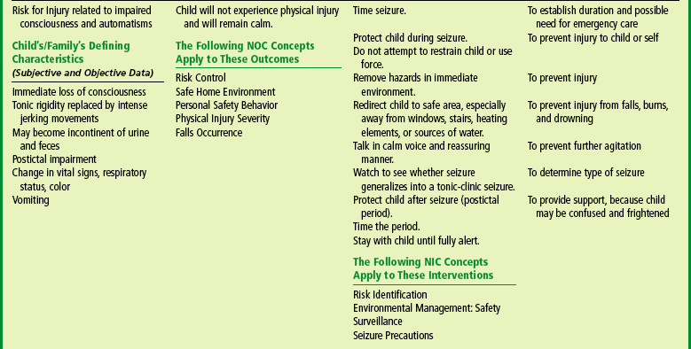
Complications are treated appropriately, such as aspiration of subdural effusion in infants and treatment for disseminated intravascular coagulation syndrome. Shock is managed by restoration of circulating blood volume and maintenance of electrolyte balance. Seizures can occur during the first few days of treatment. These are controlled with the appropriate antiepileptic drug. Hearing loss is not uncommon. The patient should undergo auditory evaluation 6 months after the illness has resolved.

Lumbar puncture is carried out as needed to determine the effectiveness of therapy. The patient is evaluated neurologically during the convalescent period.

Prognosis.: Ten percent to 15% of cases of bacterial meningitis are fatal (Centers for Disease Control and Prevention, 2000). The child’s age, duration of illness before antibiotic therapy, rapidity of diagnosis after onset, type of organism, and adequacy of therapy are important in the prognosis for bacterial meningitis. Bacterial meningitis can result in brain damage, hearing loss, or learning disability (Centers for Disease Control and Prevention, 2000; Prober, 2007).

Neonatal meningitis carries the highest mortality. However, with the development of new antibiotics and the advent of aggressive supportive care measures, the mortality rate for bacterial meningitis in children caused by H. influenzae type b, S. pneumoniae, and N. meningitidis is less than 10% in most studies (Prober, 2007).

The sequelae of bacterial meningitis are seen most often when the disease occurs in the first 2 months of life and least often in children with meningococcal meningitis. The residual deficits in infants are primarily a result of communicating hydrocephalus and the greater effects of cerebritis on the immature brain. In older children the residual effects are related to the inflammatory process itself or result from vasculitis associated with the disease. Bacterial meningitis continues to cause substantial morbidity in infants and children. The mortality rate and incidence of poor neurologic outcome are highest in patients with pneumococcal meningitis (Saez-Llorens and McCracken, 2003; Prober, 2007).

Hearing impairment is the most common sequela of this disease. Evaluation of cranial nerve VIII is needed for at least a 6-month follow-up period to assess for possible hearing loss.

Prevention.: Vaccines are available for types A, C, Y, and W-135 meningococci and H. influenzae type b. Routine meningococcal polysaccharide vaccination of children is licensed for use only in children 2 years and older (Pichichero, 2005). The new quadrivalent meningococcal conjugate vaccine (Menactra) was licensed by the U.S. Food and Drug Administration (FDA) in January 2005 for children and adults from 11 to 55 years (Pichichero, 2005). However, routine vaccinations for H. influenzae type b are recommended for all children beginning at 2 months of age (see Immunizations, Chapter 12). Pneumococcal conjugate vaccine is now recommended for all children beginning at 2 months of age (American Academy of Pediatrics, 2000a).

NURSINGALERT

Nurses should take the necessary precautions to protect themselves and others from possible infection. Parents are taught the proper procedures and supervised in their application.

NURSINGALERT

A major priority of nursing care of a child suspected of having meningitis is to administer antibiotics as soon as they are ordered. The child is placed on respiratory isolation for at least 24 hours after initiation of antimicrobial therapy.

Nursing Care Management

The room is kept as quiet as possible, and environmental stimuli are kept to a minimum because most children with meningitis are sensitive to noise, bright lights, and other external stimuli. Most children are more comfortable without a pillow and with the head of the bed slightly elevated. A side-lying position is more often assumed because of nuchal rigidity. The nurse should avoid actions that cause pain or increase discomfort, such as lifting the child’s head. Evaluating the child for pain and implementing appropriate relief measures are important during the initial 24 to 72 hours. Acetaminophen with codeine is often used. Measures are used to ensure safety because the child is often restless and subject to seizures.

The nursing care of the child with meningitis is determined by the child’s symptoms and treatment. Observation of vital signs, neurologic signs, LOC, urinary output, and other pertinent data is carried out at frequent intervals. The child who is unconscious is managed as described previously (see p. 981), and all children are observed carefully for signs of the complications just described, especially increased ICP, shock, or respiratory distress. Frequent assessment of the open fontanels is needed in the infant because subdural effusions and obstructive hydrocephalus can develop as a complication of meningitis.

Fluids and nourishment are determined by the child’s status. The child with dulled sensorium is usually given nothing by mouth. Other children are allowed clear liquids initially and, if these are tolerated, progress to a diet suitable for their age. Careful monitoring and recording of intake and output are needed to determine deviations that might indicate impending shock or increasing fluid accumulation, such as cerebral edema or subdural effusion.

One of the most difficult problems in the nursing care of children with meningitis is maintaining IV infusion for the length of time needed to provide adequate antimicrobial therapy (usually 10 days). Because continuous IV fluids are usually not necessary, an intermittent infusion device is used. In some cases children who are recovering uneventfully are sent home with the device, and the parents are taught IV drug administration.

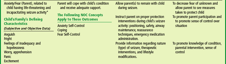
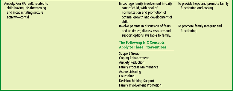
Family Support.: The sudden nature of the illness makes emotional support of the child and parents extremely important. Parents are upset and concerned about their child’s condition and often feel guilty for not having suspected the seriousness of the illness sooner. They need much reassurance that the natural onset of meningitis is sudden and that they acted responsibly in seeking medical assistance when they did. The nurse encourages the parents to openly discuss their feelings to minimize blame and guilt. They also are kept informed of the child’s progress and of all procedures, results, and treatments. In the event that the child’s condition worsens, they need the same psychologic care as parents who face the possible death of their child (see Chapter 18).

NONBACTERIAL (ASEPTIC) MENINGITIS

Aseptic meningitis is caused by many different viruses. The onset may be abrupt or gradual. The initial manifestations are headache, fever, malaise, and gastrointestinal symptoms. Signs of meningeal irritation develop 1 or 2 days after the onset of illness. Onset is more insidious in infants and toddlers. Signs and symptoms are vague and are often thought to be associated with a minor illness.

Diagnosis is based on clinical features and CSF findings. Variations in CSF values in bacterial and viral meningitis are listed in Table 28-2. It is important to differentiate this self-limiting disorder from the more serious forms of meningitis.

TABLE 28-2

Variation of Cerebrospinal Fluid Analysis in Bacterial and Viral Meningitis

MANIFESTATIONS BACTERIAL* VIRAL
White blood cell count Elevated; increased polys Slightly elevated; increased lymphs
Protein content Elevated Normal or slightly increased
Glucose content Decreased Normal
Gram stain; bacteria culture Positive Turbid or cloudy
Color Negative Clear or slightly cloudy

*Results may vary in the neonate.

Treatment is primarily symptomatic, such as acetaminophen for headache and muscle pain, maintenance of hydration, and positioning for comfort. Until a definitive diagnosis is made, antimicrobial agents may be administered and isolation enforced as a precaution against the possibility that the disease might be of bacterial origin. Nursing care is similar to the care of the child with bacterial meningitis.

ENCEPHALITIS

Encephalitis is an inflammatory process of the CNS that is caused by a variety of organisms, including bacteria, spirochetes, fungi, protozoa, helminths, and viruses. Most infections are associated with viruses, and this discussion is limited to those agents.

Etiology

Encephalitis can occur as a result of (1) direct invasion of the CNS by a virus or (2) postinfectious involvement of the CNS after a viral disease. Often the specific type of encephalitis may not be identified. The cause of more than half the cases reported in the United States is unknown. The majority of cases of known etiology are associated with the childhood diseases of measles, mumps, varicella, and rubella and, less often, with the enteroviruses, herpesviruses, and West Nile virus.

Herpes simplex encephalitis is an uncommon disease, but 30% of cases involve children. The initial clinical findings are nonspecific (fever, altered mental status), but most cases evolve to demonstrate focal neurologic signs and symptoms. Children may experience focal seizures. The CSF is abnormal in most cases. Because of a rise in the number of children with herpes simplex encephalitis, suspected cases require prompt attention, especially because the diagnosis can be difficult. The clinical diagnosis can be confirmed by the rapid appearance of immunoglobulin M antibody to herpes simplex virus type 1 in CSF and serum. The early use of IV acyclovir reduces mortality and morbidity. Empiric therapy with acyclovir is given before precise virologic diagnosis has been established. CSF should be sent for viral titers.

The multiplicity of causes of viral encephalitis makes diagnosis difficult. Most are those involved with arthropod vectors (togaviruses and bunyaviruses) and those associated with hemorrhagic fevers (arenaviruses, filoviruses, and hantaviruses). In the United States the vector reservoir for most agents pathogenic for humans is the mosquito (St. Louis or West Nile encephalitis); therefore most cases of encephalitis appear during the hot summer months and subside during the autumn.

The clinical features of encephalitis are similar regardless of the agent involved. Manifestations can range from a mild benign form that resembles aseptic meningitis, lasts a few days, and is followed by rapid and complete recovery, to a fulminating encephalitis with severe CNS involvement. The onset may be sudden or may be gradual with malaise, fever, headache, dizziness, apathy, nuchal rigidity, nausea and vomiting, ataxia, tremors, hyperactivity, and speech difficulties (Box 28-6). In severe cases the patient has high fever, stupor, seizures, disorientation, spasticity, and coma that may proceed to death. Ocular palsies and paralysis also may occur.

BOX 28-6   Clinical Manifestations of Encephalitis

ONSET: SUDDEN OR GRADUAL

Malaise

Fever

Headache

Dizziness

Apathy

Lethargy

Neck stiffness

Nausea and vomiting

Ataxia

Tremors

Hyperactivity

Speech difficulties: mutism

Altered mental status

SEVERE CASES

High fever

Stupor

Seizures

Disorientation

Spasticity

Coma (may proceed to death)

Ocular palsies

Paralysis

Diagnostic Evaluation

The diagnosis is made on the basis of clinical findings and, where possible, identification of the specific virus. Early in the course of encephalitis, CT scan results may be normal. Later, hemorrhagic areas in the frontotemporal region may be seen. Togaviruses (some of which were formerly labeled arboviruses) are rarely detected in the blood or spinal fluid, but viruses of herpes, mumps, measles, and enteroviruses may be found in the CSF. Serologic testing may be required. The first blood sample should be drawn as soon as possible after onset, with the second sample drawn 2 or 3 weeks later.

Therapeutic Management

Patients suspected of having encephalitis are hospitalized promptly for observation. Treatment is primarily supportive and includes conscientious nursing care, control of cerebral manifestations, and adequate nutrition and hydration, with observation and management as for other cerebral disorders. Viral encephalitis can cause devastating neurologic injury. Cerebral hyperemia occurs in severe viral encephalitis, and ICP monitoring to reduce the pressure may be needed (Prober, 2007). Follow-up care with periodic reevaluation and rehabilitation is important for patients who develop residual effects of the disease.

The prognosis for the child with encephalitis depends on the child’s age, the type of organism, and residual neurologic damage. Very young children (younger than 2 years of age) may exhibit increased neurologic disability, including learning difficulties and seizure disorders.

Nursing Care Management

Nursing care of the child with encephalitis is the same as for any unconscious child and for the child with meningitis. Additional nursing interventions include observation for deterioration in consciousness. Isolation of the child is not necessary; however, good hand-washing technique must be followed. A main focus of nursing management is the control of rapidly rising ICP. Neurologic monitoring, administration of medications, and support of the child and parents are the major aspects of care.

RABIES

Rabies is an acute infection of the nervous system caused by a virus that is almost invariably fatal if left untreated. It is transmitted to humans by the saliva of an infected mammal and is introduced through a bite or skin abrasion. After entry into a new host, the virus multiplies in muscle cells and is spread through neural pathways without stimulating a protective host immune response.

Approximately 88% of rabies cases come from wild animals and 12% from domestic animals. Carnivorous wild animals (skunks, raccoons, and bats) are the animals most often infected with rabies and the cause of most indigenous cases of human rabies in the United States (Centers for Disease Control and Prevention, 2001; Toltzis, 2007). The likelihood of human exposure to a rabid domestic animal has decreased greatly.

The circumstances of a biting incident are important. An unprovoked attack is more likely than a provoked attack to indicate a rabid animal. Bites inflicted on a child attempting to feed or handle an apparently healthy animal can generally be regarded as provoked. Any child bitten by a wild animal is assumed to be exposed to rabies.

NURSINGALERT

Unusual behavior in an animal is cause for suspicion; children should be warned to beware of wild animals that appear to be friendly.

Although rabies is common among wildlife species, human rabies is rarely acquired. Modern-day prophylaxis is nearly 100% successful. The highest incidence occurs in children under age 15 years. The incubation period usually ranges from 1 to 3 months but may be as short as 10 days or as long as 8 months. Only 10% to 15% of persons bitten develop the disease, but once symptoms are present, rabies progresses to a fatal outcome. In the United States, human fatalities associated with rabies occur in people who fail to seek medical attention, usually because they are unaware of their exposure.

The disease is characterized by a period of general malaise, fever, and sore throat followed by a phase of excitement that features hypersensitivity and increased reaction to external stimuli, convulsions, maniacal behavior, and choking (Box 28-7). Attempts at swallowing may cause such severe spasm of respiratory muscles that apnea, cyanosis, and anoxia are produced—the characteristics from which the term hydrophobia was derived.

BOX 28-7   Clinical Manifestations of Rabies

INITIAL SIGNS

General malaise

Fever

Sore throat

EXCITEMENT PHASE

Hypersensitivity

Increased reaction to external stimuli

Seizures

Maniacal behavior

Choking

SEVERE SPASM OF RESPIRATORY MUSCLES*

Apnea

Cyanosis

Anoxia


*From attempts at swallowing (characteristics from which the term hydrophobia was derived).

Diagnosis is made on the basis of history and clinical features. Treatment is of little avail once symptoms appear, but the long incubation period allows time for the induction of active and passive immunity before the onset of illness.

Therapeutic Management

Two types of immunizing products are available for use in humans: (1) the inactivated rabies vaccines, which induce an active immune response; and (2) the globulins, which contain preformed antibodies. The two types of products should be used concurrently for rabies postexposure treatment when prophylaxis is indicated.

The current therapy for a rabid animal bite consists of thorough cleansing of the wound and passive immunization with human rabies immunoglobulin as soon as possible after exposure to provide rapid, short-term passive immunity (Centers for Disease Control and Prevention, 1999; Toltzis, 2007).

Postexposure active immunity is conferred by administration of the human diploid cell rabies vaccine. The first intramuscular injection of the vaccine is given at the same time as the immunoglobulin (day 0) and is followed by injections at 3, 7, 14, and 28 days after the first dose (Centers for Disease Control and Prevention, 1999; Toltzis, 2007). Before antirabies prophylaxis is initiated, the local or state health department should be consulted.

Nursing Care Management

Parents and children are frightened by the urgency and seriousness of the situation. They need anticipatory guidance for the therapy and support and reassurance regarding the efficacy of the preventive measures for this dreaded disease. The vaccine is well tolerated by children, although they need preparation for the series of injections. Mass immunization is unnecessary and unlikely to be implemented. In areas in which rabies is rare, the schedule given is sufficient. However, certain circumstances may warrant preexposure vaccination, such as when a child is being taken to an area of the world where rabies in stray dogs is still a problem.

REYE SYNDROME

RS is a disorder defined as toxic encephalopathy associated with other characteristic organ involvement (Kamienski, 2003). It is characterized by fever, profoundly impaired consciousness, and disordered hepatic function.

The etiology of RS is not well understood, but most cases follow a common viral illness, most commonly influenza or varicella. RS is a condition characterized pathologically by cerebral edema and fatty changes of the liver. The onset of RS is notable for profuse vomiting and varying degrees of neurologic impairment, including personality changes and deterioration in consciousness (Carey and Balistreri, 2007). The cause of RS is a mitochondrial insult induced by different viruses, drugs, exogenous toxins, and genetic factors. Elevated serum ammonia levels tend to correlate with the clinical manifestations and prognosis.

Definitive diagnosis is established by liver biopsy. The staging criteria for RS are based on liver dysfunction and on neurologic signs that range from lethargy to coma (Box 28-8). As a result of improved diagnostic techniques, children who in the past would have been diagnosed with RS are now diagnosed with other illnesses such as viral or metabolic diseases. Cases of unrecognized, drug-induced encephalopathy by antiemetics given to children during viral illnesses have symptoms similar to those of RS.

BOX 28-8   Staging Criteria for Reye Syndrome

Stage I—Vomiting, lethargy, and drowsiness; liver dysfunction; type I electroencephalogram (EEG); follows commands; pupillary reaction brisk

Stage II—Disorientation, combativeness, delirium, hyperventilation, hyperactive reflexes, appropriate responses to painful stimuli; evidence of liver dysfunction; type I EEG; pupillary reaction sluggish

Stage III—Obtunded, coma, hyperventilation, flexion rigidity, preservation of pupillary light reaction and oculovestibular reflexes (although sluggish); type II EEG

Stage IV—Deepening coma, extension rigidity, loss of oculocephalic reflexes, large and fixed pupils, loss of doll’s eye reflex, loss of corneal reflexes; minimal liver dysfunction; type III or IV EEG; evidence of brainstem dysfunction

Stage V—Seizures, loss of deep tendon reflexes, respiratory arrest, flaccidity; type IV EEG; usually no evidence of liver dysfunction

The potential association between aspirin therapy for the treatment of fever in children with varicella or influenza and the development of RS precludes its use in these patients. However, by the time the FDA required aspirin product labeling in 1986, most of the decline in RS incidence had already occurred.

Therapeutic Management

The most important aspect of successful management of the child with RS is early diagnosis and aggressive therapy. Rapid progression through coma stages and high peak ammonia concentrations are associated with a more serious prognosis. Cerebral edema with increased ICP represents the most immediate threat to life. Recovery from RS is rapid and usually without sequelae if diagnosis is determined early and therapy is initiated promptly.

Prognosis.: Although the incidence of RS has markedly decreased, health professionals must remind parents and caregivers to avoid using both aspirin and non-aspirin–containing salicylates during febrile illnesses in children (Bhutta, Van Savell, and Schexnayder, 2003; Kamienski, 2003). Survivors may have subtle neuropsychologic deficits. Generally, recovery is good given the gravity of the disease (Bhutta, Van Savell, and Schexnayder, 2003; Kamienski, 2003).

Nursing Care Management

The most important aspect of successful management of the child with RS is early diagnosis and aggressive therapy (Bhutta, Van Savell, and Schexnayder, 2003). Cerebral edema with increased ICP represents the most immediate threat to life. Recovery from RS is rapid and usually without sequelae if there has been early diagnosis and implementation of therapy. In about one third of patients, RS causes death or long-term neurologic sequelae.

Care and observations are implemented as for any child with an altered state of consciousness (see p. 976) and increasing ICP. Accurate and frequent monitoring of intake and output is essential for adjusting fluid volumes to prevent both dehydration and cerebral edema. Because of related liver dysfunction, laboratory studies to determine impaired coagulation, such as prolonged bleeding time, should be monitored.

Parents of children with RS need to be kept informed of the child’s progress, to have diagnostic procedures and therapeutic management explained, and to be given concerned and sympathetic support.* Families need to be aware that salicylate, the alleged offending ingredient in aspirin, is contained in other products (e.g., Pepto-Bismol). They should refrain from administering any product for influenza-like symptoms without first checking the label for “hidden” salicylates.

SEIZURE DISORDERS

Seizures are caused by excessive and disorderly neuronal discharges in the brain. The manifestation of seizures depends on the region of the brain in which they originate and may include unconsciousness or altered consciousness; involuntary movements; and changes in perception, behaviors, sensations, and posture. Seizures are the most common treatable neurologic disorder in children and can occur with a wide variety of conditions involving the CNS.

EPILEPSY

Epilepsy is a condition characterized by two or more unprovoked seizures and can be caused by a variety of pathologic processes in the brain. Seizures are a symptom of an underlying disease process. A single seizure event should not be classified as epilepsy and is generally not treated with long-term antiepileptic drugs. Some seizures may result from an acute medical or neurologic illness and cease once the illness is treated. In other cases, children may have a single seizure without the cause ever being known. Once it is determined that the child has had a seizure, it is important to classify the seizure, according to the International Classification of Epileptic Seizures, and assign it to the appropriate epilepsy syndrome, according to the International Classification of Epilepsies and Epileptic Syndromes. Optimum treatment and prognosis require an accurate diagnosis and a determination of the cause whenever possible.

Etiology

Seizures in children have many different causes. Seizures are classified not only according to type, but also according to etiology. Acute symptomatic seizures are associated with an acute insult such as head trauma or meningitis. Remote symptomatic seizures are those without an immediate cause but with an identifiable prior brain injury such as major head trauma, meningitis or encephalitis, hypoxia, stroke, or a static encephalopathy such as intellectual disability or cerebral palsy. Cryptogenic seizures are those occurring with no clear cause. Idiopathic seizures are genetic in origin. A partial list of causative factors is presented in Box 28-9.

BOX 28-9   Etiology of Seizures in Children

NONRECURRENT (ACUTE)

Febrile episodes

Intracranial infection

Intracranial hemorrhage

Space-occupying lesions (cyst, tumor)

Acute cerebral edema

Anoxia

Toxins

Drugs

Tetanus

Lead encephalopathy

Shigella, Salmonella organisms

Metabolic alterations

image Hypocalcemia

image Hypoglycemia

image Hyponatremia or hypernatremia

image Hypomagnesemia

image Alkalosis

image Disorders of amino acid metabolism

image Deficiency states

image Hyperbilirubinemia

RECURRENT (CHRONIC)

Idiopathic epilepsy

Epilepsy secondary to:

image Trauma

image Hemorrhage

image Anoxia

image Infections

image Toxins

image Degenerative phenomena

image Congenital defects

image Parasitic brain disease

image Hypoglycemia injury

Epilepsy—sensory stimulus

Epilepsy-stimulating states

image Narcolepsy and catalepsy

image Psychogenic

image Tetany from hypocalcemia, alkalosis

Hypoglycemic states

image Hyperinsulinism

image Hypopituitarism

image Adrenocortical insufficiency

image Hepatic disorders

Uremia

Allergy

Cardiovascular dysfunction or syncopal episodes

Migraine

Pathophysiology

Regardless of the etiologic factor or type of seizure, the basic mechanism is the same. Abnormal electrical discharges (1) may arise from central areas in the brain that affect consciousness; (2) may be restricted to one area of the cerebral cortex, producing manifestations characteristic of that particular anatomic focus; or (3) may begin in a localized area of the cortex and spread to other portions of the brain and, if sufficiently extensive, produce generalized seizure activity.

In response to physiologic stimuli, such as cellular dehydration, severe hypoglycemia, electrolyte imbalance, sleep deprivation, emotional stress, and endocrine changes, these hyperexcitable cells activate normal cells in surrounding areas and in distant, synaptically related cells. A generalized seizure develops when the neuronal excitation from the epileptogenic focus spreads to the brainstem, particularly the midbrain and reticular formation. These centers within the brainstem, known as the centrencephalic system, are responsible for the spread of the epileptic potentials. The discharges can originate spontaneously in the centrencephalic system or be triggered by a focal area in the cortex. On the basis of these characteristic neuronal discharges (as recorded by the EEG), seizures are designated as partial, generalized, and unclassified epileptic seizures (Menkes and Sankar, 2000). In a large proportion of children focal seizures spread to other areas, ultimately becoming generalized with loss of consciousness.

Seizure Classification and Clinical Manifestations

There are many different types of seizures, and each has unique clinical manifestations. Seizures are classified into three major categories:

Partial seizures, which have a local onset and involve a relatively small location in the brain

Generalized seizures, which involve both hemispheres of the brain and are without local onset

Unclassified epileptic seizures

Descriptions of the different types of seizures are found in Box 28-10 and Table 28-3.

BOX 28-10   Classification and Clinical Manifestations of Seizures

PARTIAL SEIZURES

Simple Partial Seizures with Motor Signs

Characterized by:

image Localized motor symptoms

image Somatosensory, psychic, autonomic symptoms

image Combination of these

image Abnormal discharges remaining unilateral

Manifestations:

image Aversive seizure (most common motor seizure in children)–Eye or eyes and head turn away from the side of the focus; awareness of movement or loss of consciousness

image Rolandic (Sylvan) seizure—Tonic-clonic movements involving the face, salivation, arrested speech; most common during sleep

image Jacksonian march (rare in children)–Orderly, sequential progression of clonic movements beginning in a foot, hand, or face and moving, or “marching,” to adjacent body parts

Simple Partial Seizures with Sensory Signs

Characterized by various sensations, including:

image Numbness, tingling, prickling, paresthesia, or pain originating in one area (e.g., face or extremities) and spreading to other parts of the body

image Visual sensations or formed images

image Motor phenomena such as posturing or hypertonia

Uncommon in children younger than 8 years of age

Complex Partial Seizures (Psychomotor Seizures)

Observed more often in children from 3 years through adolescence

Characterized by:

image Period of altered behavior

image Amnesia for event (no recollection of behavior)

image Inability to respond to environment

image Impaired consciousness during event

image Drowsiness or sleep usually following seizure

image Confusion and amnesia possibly prolonged

image Complex sensory phenomena (aura)–Most frequent sensation is a strange feeling in the pit of the stomach that rises toward the throat; often accompanied by:

– Odd or unpleasant odors or tastes

– Complex auditory or visual hallucinations

– Ill-defined feelings of elation or strangeness (e.g., déjà vu, a feeling of familiarity in a strange environment)

– Strong feelings of fear and anxiety; distorted sense of time and self

– In small children, emission of a cry or attempt to run for help

Patterns of motor behavior:

image Stereotypic

image Similar with each subsequent seizure

image May suddenly cease activity, appear dazed, stare into space, become confused and apathetic, and become limp or stiff or display some form of posturing

image May be confused

image May perform purposeless, complicated activities in a repetitive manner (automatisms), such as walking, running, kicking, laughing, or speaking incoherently, most often followed by postictal confusion or sleep

image May exhibit oropharyngeal activities, such as smacking, chewing, drooling, swallowing, and nausea or abdominal pain followed by stiffness, a fall, and postictal sleep

image Rarely manifests actions such as rage or temper tantrums; aggressive acts uncommon during seizure

GENERALIZED SEIZURES

Tonic-Clonic Seizures (Formerly Known as Grand Mal)

Most common and most dramatic of all seizure manifestations

Occur without warning

Tonic Phase

Lasts approximately 10 to 20 seconds

Manifestations:

image Eyes roll upward

image Immediate loss of consciousness

image If standing, falls to floor or ground

image Stiffens in generalized, symmetric tonic contraction of entire body musculature

image Arms usually flexed

image Legs, head, and neck extended

image May utter a peculiar piercing cry

image Apneic; may become cyanotic

image Increased salivation and loss of swallowing reflex

Clonic Phase

Lasts about 30 seconds but can vary from only a few seconds to a half hour or longer

Manifestations:

image Violent jerking movements as the trunk and extremities undergo rhythmic contraction and relaxation

image May foam at the mouth

image May be incontinent of urine and feces

As event ends, movements less intense, occurring at longer intervals, then ceasing entirely

Status Epilepticus

Series of seizures at intervals too brief to allow the child to regain consciousness between the time one event ends and the next begins

image Requires emergency intervention

image Can lead to exhaustion, respiratory failure, and death

Postictal State

Manifestations:

image Appears to relax

image May remain semiconscious and difficult to arouse

image May awaken in a few minutes

image Remains confused for several hours

image Poor coordination

image Mild impairment of fine motor movements

image May have visual and speech difficulties

image May vomit or complain of severe headache

image When left alone, usually sleeps for several hours

image On awakening is fully conscious

image Usually feels tired and complains of sore muscles and headache

image No recollection of entire event

Absence Seizures (Formerly Called Petit Mal or Lapses)

Characterized by:

image Onset usually between 4 and 12 years of age

image More common in girls than in boys

image Usually cease at puberty

image Brief loss of consciousness

image Minimum or no alteration in muscle tone

image May go unrecognized because of little change in child’s behavior

image Abrupt onset; suddenly develops 20 or more attacks daily

image Event often mistaken for inattentiveness or daydreaming

image Events possibly precipitated by hyperventilation, hypoglycemia, stresses (emotional and physiologic), fatigue, or sleeplessness

Manifestations:

image Brief loss of consciousness

image Appear without warning or aura

image Usually last about 5 to 10 seconds

image Slight loss of muscle tone may cause child to drop objects

image Ability to maintain postural control; seldom falls

image Minor movements such as lip smacking, twitching of eyelids or face, or slight hand movements

image Not accompanied by incontinence

image Amnesia for episode

image May need to reorient self to previous activity

Atonic and Akinetic Seizures (Also Known as Drop Attacks)

Characterized by:

image Onset usually between 2 and 5 years of age

image Sudden, momentary loss of muscle tone and postural control

image Events recurring frequently during the day, particularly in the morning hours and shortly after awakening

Manifestations:

image Loss of tone causing child to fall to the floor violently; unable to break fall by putting out hand; may incur a serious injury to the face, head, or shoulder

image Loss of consciousness only momentary

Myoclonic Seizures

A variety of seizure episodes

May be isolated as benign essential myoclonus

May occur in association with other seizure forms

Characterized by:

image Sudden, brief contractures of a muscle or group of muscles

image Occur singly or repetitively

image No postictal state

image May or may not be symmetric

image May or may not include loss of consciousness

Infantile Spasms

Also called infantile myoclonus, massive spasms, hypsarrhythmia, salaam episodes, or infantile myoclonic spasms

Most commonly occur during the first 6 to 8 months of life

Twice as common in boys as in girls

Numerous seizures during the day without postictal drowsiness or sleep

Poor outlook for normal intelligence

Manifestations:

image Possible series of sudden, brief, symmetric, muscular contractions

image Head flexed, arms extended, and legs drawn up

image Eyes sometimes rolling upward or inward

image May be preceded or followed by a cry or giggling

image May or may not include loss of consciousness

image Sometimes flushing, pallor, or cyanosis

Infants who are able to sit but not stand:

image Sudden dropping forward of the head and neck with trunk flexed forward and knees drawn up—the salaam or jackknife seizure

Less often: alternate clinical forms

image Extensor spasms rather than flexion of arms, legs, and trunk, and head nodding

image Lightning events involving a single, momentary, shocklike contraction of the entire body

TABLE 28-3

Comparison of Simple Partial, Complex Partial, and Absence Seizures

image

Diagnostic Evaluation

Establishing a diagnosis is critical for establishing a prognosis and planning the proper treatment. The process of diagnosis in a child suspected of having epilepsy includes (1) determining whether epilepsy or seizures exist and not an alternative diagnosis, and (2) defining the underlying cause, if possible. The assessment and diagnosis rely heavily on a thorough history, skilled observation, and several diagnostic tests.

It is especially important to differentiate epilepsy from other brief alterations in consciousness or behavior. Clinical entities that mimic seizures include migraine headaches, toxic effects of drugs, syncope (fainting), breath-holding spells in infants and young children, movement disorders (tics, tremor, chorea), prolonged QT syndrome, sleep disturbances (sleepwalking, night terrors), psychogenic seizures, rage attacks, and transient ischemic attacks (rare in children) (Browne and Holmes, 2004). Cocaine intoxication should be considered in the differential diagnosis of new-onset seizure activity in newborn infants.

The history of the seizure should be equally detailed, including the type of seizure or description of the child’s behavior during the event, the age at onset, and the time at which the seizure occurs (e.g., early morning, before meals, while awake, or during sleep). Any factors that may have precipitated the seizure are important, including fever, infection, head trauma, anxiety, fatigue, sleep deprivation, menstrual cycle, alcohol, and activity (e.g., hyperventilation or exposure to strong stimuli such as bright flashing light or loud noises). If the child can describe any sensory phenomena, these are recorded. The duration and progression of the seizure (if any) and the postictal feelings and behavior (e.g., confusion, inability to speak, amnesia, headache, and sleep) are recorded. It is important to determine whether more than one seizure type exists. It is often more informative to ask parents to mime the seizure rather than relying on their oral description. Miming often reveals features, such as head turning, that would otherwise go unrecognized. Some seizures are overlooked by parents. For example, some parents may not identify brief head nods or brief single jerks as seizures unless specifically asked whether their child has these symptoms. The family history should include whether other family members have had a seizure, intellectual disability, cerebral palsy, or other neurologic disorders. A family history can offer clues to paroxysmal disorders such as migraine headaches, breath-holding spells, febrile seizures, or neurologic diseases.

A complete physical and neurologic examination, including developmental assessment of language, learning, behavior, and motor abilities, may provide clues to the cause of the seizures. A number of laboratory and neuroimaging tests may be ordered depending on the child’s age, whether this is a new onset seizure, characteristics of the seizure, and the history. Laboratory studies that may prove to be of value include a venous lead level if the history warrants or white blood cell count (for signs of infection). Blood glucose may give evidence of hypoglycemic episodes, and serum electrolytes, blood urea nitrogen, calcium, serum amino acids, lactate, ammonia, and urine organic acids may indicate metabolic disturbances. Blood for chromosomal analysis may also be tested if a genetic etiology is suspected. A toxic screen may be done if alcohol or drug abuse or withdrawal is suspected. Lumbar puncture can confirm a suspected diagnosis of meningitis. CT may be done to detect a cerebral hemorrhage, infarctions, and gross malformations. MRI provides greater anatomic detail and is used to detect developmental malformations, tumors, and cortical dysplasias (Kuzniecky, 2001).

The EEG is obtained for all children with seizures and is the most useful tool for evaluating a seizure disorder. The EEG confirms the presence of abnormal electrical discharges and provides information on the seizure type and the focus. The EEG is carried out under varying conditions—with the child asleep, awake, awake with provocative stimulation (flashing lights, noise), and hyperventilating. Stimulation may elicit abnormal electrical activity, which is recorded on the EEG. Various seizure types produce characteristic EEG patterns: high-voltage spike discharges are seen in tonic-clonic seizures, with abnormal patterns in the intervals between seizures; a three-per-second spike and wave pattern is observed in an absence seizure; and absence of electrical activity in an area suggests a large lesion, such as an abscess or subdural collection of fluid.

A normal EEG does not rule out seizures because the EEG is only a surface recording and only represents approximately 1 hour of time and therefore may show normal interictal activity. If there is concern about whether a child has seizures or the seizure type cannot be determined, then a long-term video EEG may be done to record the child during wakefulness and sleep. The full body image is recorded on video, with selected EEG channels displayed on the same screen for simultaneous recording and viewing. EEG monitoring is also available in digital EEG and digital video imaging, which allows for greater selection of EEG channels and is available in both routine and long-term EEGs. Polygraph equipment may also be used to monitor physiologic data such as respiratory effort, eye movements, heart rate, and systemic blood pressure. These techniques can be used concurrently and are especially valuable in differentiating epileptic activity from paroxysmal behavior or nonepileptic motor events.

Therapeutic Management

The goal of treatment of seizure disorders is to control the seizures or to reduce their frequency and severity, discover and correct the cause when possible, and help the child live as normal a life as possible. If the seizure activity is a manifestation of an infectious, traumatic, or metabolic process, the seizure therapy is instituted as part of the general therapeutic regimen. Management of epilepsy has four treatment options: drug therapy, the ketogenic diet, vagus nerve stimulation, and epilepsy surgery.

Drug Therapy.: It is known that persons predisposed to epilepsy have seizures when their basal level of neuronal excitability exceeds a critical point; no event occurs if the excitability is maintained below this threshold. The administration of antiepileptic drugs serves to raise this threshold and prevent seizures. Consequently, the primary therapy for seizure disorders is the administration of the appropriate antiepileptic drug or combination of drugs in a dosage that provides the desired effect without causing undesirable side effects or toxic reactions. Antiepileptic drugs are believed to exert their effect primarily by reducing the responsiveness of normal neurons to the sudden, high-frequency nerve impulses that arise in the epileptogenic focus. Thus the seizure is effectively suppressed; however, the abnormal brain waves may or may not be altered. Complete control can be achieved in 75% of children with epilepsy; good control can be achieved in another 15% (Shafer, 1999).

Therapy is begun with a single drug known to be effective and have the lowest toxicity, that is, the safest side effect profile for the child’s particular type of seizure. The dosage is gradually increased until the seizures are controlled or the child develops side effects. If the drug is effective but does not sufficiently control the seizures, a second drug is added in gradually increasing doses. Once seizures are controlled, the first drug may be tapered to reduce the potential adverse effects of polytherapy. However, this decision is individualized for each child (Browne and Holmes, 2004). Monotherapy remains the treatment method of choice for epilepsy, but polypharmacy may be a viable alternative for children who cannot attain seizure control with only one agent (Leppik, 2000).

Measurement of blood levels of the drug is important if the seizures continue once the child is on a therapeutic dose of medication, to adjust the dosage, and to assist in determining which medication may be causing the side effects if the child is on multiple antiepileptic medications. Some possible causes of low serum blood concentrations are noncompliance, poor absorption, and drug interactions. The dosage needs to be increased as the child grows. Blood cell counts, urinalysis, and liver function tests are obtained at frequent intervals in children receiving particular antiepileptic medications that can affect organ function.

If complete seizure control is maintained on an anticonvulsant drug for 2 years, it is safe to discontinue the drug for patients with no risk factors. Risk factors include children over 12 years of age at onset, history of neonatal seizures, numerous seizures before control is achieved, and the presence of a neurologic dysfunction (e.g., motor handicap or intellectual disability). Up to 25% of children whose medications are discontinued will experience seizure recurrence. Recurrence occurs most frequently within 6 months of discontinuation (Johnston, 2007).

When seizure medications are discontinued, the dosage is decreased gradually over several weeks. Sudden withdrawal of a drug is not recommended because it can cause an increase in the number and severity of seizures.

NURSINGALERT

Fosphenytoin is often used to treat seizures instead of IV phenytoin because of possible complications and drug interactions associated with IV phenytoin. If IV phenytoin is used, it should be administered via slow IV push at a rate that does not exceed 50 mg/min. Because phenytoin precipitates when mixed with glucose, only normal saline is used to flush the tubing or catheter. Fosphenytoin may be given in saline or glucose solutions at a rate of up to 150 mg PE (phenytoin equivalent)/min, and it may be given intramuscularly if necessary.

Ketogenic Diet.: The ketogenic diet is a high-fat, low-carbohydrate, and adequate protein diet (Lefevre and Aronson, 2000). Consumption of such a diet forces the body to shift from using glucose as the primary energy source to using fat, and the individual develops a state of ketosis. The diet is rigorous. All foods and liquids the child consumes must be carefully weighed and measured. The diet is deficient in vitamins and minerals; therefore vitamin supplements are necessary. Potential side effects of the diet are constipation, weight loss, lethargy, and kidney stones. It is unknown whether long-term effects such as increased blood lipids will occur (Levy and Cooper, 2003).

The ketogenic diet has been shown to be an efficacious and tolerable treatment for difficult-to-control seizures (Freeman, Kossoff, and Hartman, 2007). Outcomes of 150 children with uncontrollable seizures revealed that 7% of the children were seizure free and another 20% had a 90% decrease in seizures 1 year after instituting the ketogenic diet (Freeman, Vining, Pillas, and others, 1998). Three to six years later, 27% of these children had few or no seizures while on the ketogenic diet (Hemingway, Freeman, Pillas, and others, 2001).

Vagus Nerve Stimulation.: Vagus nerve stimulation uses an implantable device that reduces seizures in individuals who have not had effective control with drug therapy. It is currently indicated as adjunct therapy in patients 12 years and older with partial onset seizures (with or without secondary generalization). A programmable signal generator is implanted subcutaneously in the chest. Electrodes tunneled underneath the skin deliver electrical impulses to the left vagus nerve (cranial nerve X). The device is programmed noninvasively to deliver a precise pattern of stimulation to the left vagus nerve. The patient or caregiver can activate the device using a magnet at the onset of a seizure. Studies show that about one third of patients have a 50% or greater reduction in seizures after 1 year of therapy (Morris, Mueller, and Vagus Nerve Stimulation Study Group, 1999).

Surgical Therapy.: When seizures are determined to be caused by a hematoma, tumor, or other cerebral lesion, surgical removal is the treatment. In children with epilepsy, surgery is reserved for those who suffer from incapacitating, refractory seizures. Refractory seizures are usually defined as the persistence of seizures despite adequate trials of three antiepileptic medications, alone or in combination (Browne and Holmes, 2004). The epileptogenic area should be in a surgically removable and functionally silent region of the brain. Early removal of the symptomatic area is associated with seizure control and decreased use of antiepileptic drugs (Mathern, Giza, Yudovin, and others, 1999). An extensive medical (e.g., invasive EEG monitoring), psychosocial, and psychoneurologic evaluation is required.

Status Epilepticus.: Status epilepticus is a continuous seizure that lasts more than 30 minutes or a series of seizures from which the child does not regain a premorbid LOC (Shorvon and Walker, 2005; Treiman and Walker, 2006). The duration required for seizures to be considered status epilepticus continues to be debated (Chen and Wasterlain, 2006). The initial treatment is directed toward support and maintenance of vital functions, that is, attending to the ABCs of life support, administering oxygen, and gaining IV access, immediately followed by IV administration of antiepileptic agents.

Rectal diazepam is a simple, effective, and safe treatment for home or prehospital management (Pellock and Shinnar, 2005). It is available in a prefilled rectal gel syringe (Diastat) for easy administration. Rectal diazepam is not associated with respiratory depression when used as recommended (Pellock and Shinnar, 2005). Midazolam has been given successfully by intranasal route for treatment of acute epileptic seizures (Fisgin, Gurer, Senbil, and others, 2000; Kutlu, Yakinci, Dogrul, and others, 2000). Intranasal midazolam not only is safe and effective for stopping seizures, but is easier to administer than rectal diazepam (Harbord, Kyrkou, Kyrkou, and others, 2004).

For in-hospital management of status epilepticus, IV diazepam or lorazepam (Ativan) is the first-line drug of choice (Browne and Holmes, 2004). Lorazepam may be replacing IV diazepam as the drug of choice. It has a longer duration of action and causes less respiratory depression in children over 2 years of age. Concurrent IV loading with fosphenytoin is usually necessary for sustained control of seizures. Valproic acid has also been reported to be effective in status epilepticus when given rectally or intravenously (Yamamoto and Yim, 2000). The child must be closely monitored during administration to detect early alterations in vital signs that may indicate impending respiratory depression. When diazepam is ineffective, fosphenytoin or phenobarbital is given intravenously as the next line of treatment. This combination of therapy places the child at high risk for apnea, and therefore respiratory support is generally necessary. Children who continue to have seizures despite the above drug treatment may be given anesthetizing doses of midazolam, propofol, or pentobarbital. In this situation, continuous EEG monitoring is typically done to monitor for and treat electrographic seizures.

NURSINGALERT

Diazepam is incompatible with many drugs. To give intravenously, inject slowly and directly into the vein or through tubing as close as possible to the vein insertion site.

Nursing care of a child with status epilepticus includes, in addition to the ABCs of life support, monitoring blood pressure and body temperature. During the first 30 to 45 minutes of the seizure the blood pressure may be elevated. Thereafter the blood pressure typically returns to normal but may be decreased depending on the medications being administered for seizure control. Hyperthermia requiring treatment may occur as a result of increased motor activity.

Prognosis

Most children who experience a second seizure will experience additional seizures. Therefore a history of two seizures is sufficient to diagnose epilepsy (Shinnar, Berg, O’Dell, and others, 2000). Epidemiologic studies using population- or community-based cohorts show that the etiology and specific epilepsy syndromes are the most important factors affecting prognosis. Children who have intellectual disability or cerebral palsy are at the highest risk for developing epilepsy. Seizures will remit in more than two thirds of children with childhood onset of seizure. Mortality is increased in children with epilepsy; those with neurologic abnormalities or seizures that are refractory to treatment are at the highest risk (Browne and Holmes, 2004).

Nursing Care Management

An important nursing responsibility is to observe the seizure episode and accurately document the events. Any alterations in behavior preceding the seizure and the characteristics of the episode, such as sensory-hallucinatory phenomena (e.g., an aura), motor effects (e.g., eye movements, muscular contractions), alterations in consciousness, and postictal state, are noted and recorded (Box 28-11). The nurse should describe only what is observed, rather than trying to label a seizure type. He or she notes the time that the seizure began and the duration of the seizure.

BOX 28-11   General Observations: The Child During a Seizure

OBSERVATIONS DURING SEIZURE

General Description

Order of events (before, during, and after)

Duration of seizure

image Tonic-clonic—from first signs of event until jerking stops

image Absence—from loss of consciousness until consciousness is regained

image Complex partial—from first sign of unresponsiveness, motor activity, automatisms until there are signs of responsiveness to environment

Onset

Time of onset

Significant precipitating events—missed medication dosage, illness, stress, sleep deprivation, menses

Behavior

Change in facial expression

Cry or other sound

Stereotypic or automatous movements

Random activity (wandering)

Position of eyes, head, body, extremities

Unilateral or bilateral posturing of one or more extremities

Movement

Change of position, if any

Site of commencement—hand, thumb, mouth, generalized

Tonic phase—length, parts of body involved

Clonic phase—twitching or jerking movements, parts of body involved, sequence of parts involved, generalized, change in character of movements

Lack of movement or muscle tone of body part or entire body

Face

Color change—pallor, cyanosis, flushing

Perspiration

Mouth—position, deviation to one side, teeth clenched, tongue bitten, frothing at mouth, flecks of blood or bleeding

Lack of expression

Asymmetric expression

Eyes

Position—straight ahead, deviation upward or outward, conjugate or divergent gaze

Pupils—change in size, equality, reaction to light

Respiratory Effort

Presence and length of apnea

Other

Incontinence

POSTICTAL OBSERVATIONS

Duration of postictal period

State of consciousness

Orientation

Arousability

Motor ability

image Any change in motor function

image Ability to move all extremities

image Paresis or weakness

Speech

Sensations

image Complaint of discomfort or pain

image Any sensory impairment

Recollection of preseizure sensations (aura)

Based on a thorough assessment, several nursing diagnoses are identified. The more common diagnoses for the child with a seizure disorder are included in the Nursing Process box and Nursing Care Plan.

nursingprocess

The Child with Seizures

ASSESSMENT

Assess for signs and symptoms of seizures in children; observe any alterations in behavior preceding the seizure and the characteristics of the episode, motor effects, alterations in consciousness, and postictal state. A complete description of the general observations of a child experiencing a seizure is found in the Nursing Care Plan.

DIAGNOSIS (PROBLEM IDENTIFICATION)

After a thorough assessment, several nursing diagnoses are evident (see Nursing Care Plan below). Other nursing diagnoses include:

image Anxiety

image Ineffective Tissue Perfusion

image Interrupted Family Processes

image Self-Esteem Disturbance

PLANNING

Expected patient outcomes include:

image Child will be protected during a seizure.

image Influencing or triggering factors will be determined and adjustments made to lessen seizure events.

image Child will experience as few seizures as possible.

image Child and family will cope with the challenges associated with the disorder.

image Child will develop a positive self-image.

INTERVENTIONS

Numerous intervention strategies are discussed on pp. 1010–1016.

EVALUATION

The effectiveness of nursing interventions for the family and child with epilepsy is determined by continual assessment and evaluation of care based on the following guidelines:

image Observe child’s behavior for evidence of seizure activity, and assess the environment for situations that could cause injury to child in the event of a seizure; interview family regarding management of child during a seizure.

image Interview child and family regarding adherence to the medication regimen and identification of triggering factors.

image Observe and interview family regarding their feelings and concerns and their understanding of child’s condition.

image Observe child’s interactions with others, and interview child about any feelings or concerns regarding health.

nursingcareplan: The Child with Seizure Disorder

image

image

image

image

image

image

image

image

*Nursing diagnosis may also apply to child in the postictal phase, depending on type of seizure and child—sognitive level.

emergencytreatment

Seizures

TONIC-CLONIC SEIZURE

During the Seizure

Remain calm.

Time seizure episode.

If child is standing or seated, ease child down to the floor.

Place pillow or folded blanket under child’s head.

Loosen restrictive clothing.

Remove eyeglasses.

Clear area of any hazards or hard objects.

Allow seizure to end without interference.

If vomiting occurs, turn child to one side.

Do not:

image Attempt to restrain child or use force

image Put anything in child’s mouth

image Give any food or liquids

After the Seizure

Time postictal period.

Check for breathing. Check position of head and tongue.

Reposition if head is hyperextended. If child is not breathing, give rescue breathing and call emergency medical services (EMS).

Keep child on side.

Remain with child.

Do not give food or liquids until child is fully alert and swallowing reflex has returned.

Call EMS when necessary.

Look for medical identification, and determine what factors occurred before onset of seizure that may have been triggering factors.

Check head and body for possible injuries.

Check inside of mouth to see if tongue or lips have been bitten.

COMPLEX PARTIAL SEIZURE

During the Seizure

Do not restrain.

Remove harmful objects from area.

Redirect to safe area.

Do not agitate; instead, talk in calm, reassuring manner.

Do not expect child to follow instructions.

Watch to see if seizure generalizes.

After the Seizure

Stay with child and reassure until fully conscious.

CALL EMERGENCY MEDICAL SERVICES IF

Child stops breathing.

There is evidence of injury or child is diabetic or pregnant.

Seizure lasts for more than 5 minutes (unless duration of seizure is typically longer than 5 minutes) and written medical order is present.

Status epilepticus occurs.

Pupils are not equal after seizure.

Child vomits continuously 30 minutes after seizure has ended (sign of possible acute problem).

Child cannot be awakened and is unresponsive to pain after seizure has ended.

Seizure occurs in water.

This is child’s first seizure.

Modified from Seizure recognition and first aid, 2001, Epilepsy Foundation, retrieved March 5, 2007, from http://www.epilepsyfoundation.org.

The child must be protected from injury during the seizure; nursing observations made during the event provide valuable information for diagnosis and management of the disorder (see Emergency Treatment box).

It is impossible to halt a seizure once it has begun, and no attempt should be made to do so. The nurse must remain calm, stay with the child, and prevent the child from sustaining any harm during the seizure. If possible, the child should be isolated from the view of others by closing a door or pulling screens. A seizure can be upsetting to the child, other visitors, and their families. If other persons are present, they should be assured that everything is being done for the child. After the seizure, they can be given a simple explanation about the event as needed.

If the nurse is able to reach the child in time, a child who is standing or seated in a chair (including a wheelchair) is eased to the floor immediately. During (and sometimes after) the tonic-clonic seizure, the swallowing reflex is lost, salivation increases, and the tongue is hypotonic. Therefore the child is at risk for aspiration and airway occlusion. Placing the child on the side facilitates drainage and helps maintain a patent airway. Suctioning the oral cavity and posterior oropharynx may be necessary. Vital signs should be taken. The child is allowed to rest if at school or away from home. When feasible, the child is integrated into the environment as soon as possible. Sending a child with a chronic seizure disorder home from school is not necessary unless requested by the parents.

Seizure precautions are required for children who are known to have seizures or who are under observation for seizures. The extent of these measures depends on the type and frequency of the seizure (Box 28-12).

BOX 28-12   Seizure Precautions

The extent of precautions depends on type, severity, and frequency of seizures. They may include:

image Side rails raised when child is sleeping or resting

image Side rails and other hard objects padded

image Waterproof mattress or pad on bed or crib

Appropriate precautions during potentially hazardous activities may include:

image Swimming with a companion

image Showers preferred; bathing only with close supervision

image Use of protective helmet and padding during bicycle riding, skateboarding, in-line skating

image Supervision during use of hazardous machinery or equipment

Have child carry or wear medical identification.

Alert other caregivers to need for any special precautions.

Child may not drive or operate hazardous machinery or equipment unless seizure free for designated period (varies by state).

NURSINGALERT

Do not move or forcefully restrain the child during a tonic-clonic seizure, and do not place a solid object between the teeth.

Long-Term Care: Care of the child with a recurrent seizure disorder involves physical care and instruction regarding the importance of the drug therapy and, probably more significant, the problems related to the emotional aspects of the disorder. Few diseases generate as much anxiety among relatives as epilepsy. Fears and misconceptions about the disease and its treatment abound in the layperson’s mind. For many, it represents the archetype of severe hereditary affliction. Nursing care is directed toward educating the child and family about epilepsy and helping them develop strategies to cope with the psychologic and sociologic problems related to epilepsy.

Children with epilepsy are prescribed antiepileptic medications. These medications are administered at regular intervals to maintain adequate levels in the blood. The most convenient times for administration seem to be with meals or at bedtime. It is important to impress on the family the necessity of giving the antiepileptic medication regularly and for as long as required. In general, antiepileptic medications are continued until the child has been seizure free for 2 years (Johnston, 2007). The medication is then slowly tapered over a period of weeks to avoid the possibility of precipitating a seizure. It is sometimes easy to skip doses or omit them for a variety of reasons, especially when the child is free of seizures most of the time. This is particularly so when the child is older and assumes responsibility for his or her medication. The seizure threshold may be lowered during any illness, but particularly with fever. Therefore parents should be aware that if their child has an illness, he or she is at increased risk for seizures. Parents should contact their health professional if their child misses medications during an illness because of vomiting.

Rectal preparations of some antiepileptic medications are highly effective when a child is unable to take oral medications because of repeated vomiting, gastrointestinal surgery, or status epilepticus. Parents can learn to administer rectal antiepileptic medication for home treatment. Rectal diazepam is a useful adjunctive home treatment for children at risk for prolonged seizures or clusters of seizures. Hospitalization is minimized, and parental confidence is enhanced.

NURSINGALERT

Children taking phenobarbital or phenytoin should receive adequate vitamin D and folic acid, since deficiencies of both have been associated with these drugs. Phenytoin should not be taken with milk.

Nurses should educate the child and parents about the possible adverse reactions to the medications used to treat seizures. Parents should understand the common side effects and be encouraged to report their observations to their health care provider. Parents should understand that the child needs periodic physical assessment and laboratory studies. Possible adverse effects on the hematopoietic system, liver, and kidneys may be reflected in symptoms such as fever, sore throat, enlarged lymph nodes, jaundice, and bleeding (e.g., easy bruising, petechiae, ecchymoses, epistaxis). A common factor in status epilepticus is inadequate blood levels of antiepileptic drugs.

Although children with epilepsy are at increased risk for injury, limitations on activities should be relatively few. The degree to which activities are restricted is individualized for each child and depends on the type, frequency, and severity of the seizures; the child’s response to therapy; and the length of time the seizures have been controlled. To prevent head injuries, children should always wear appropriate safety devices, such as helmets, and should avoid activities involving heights. Although bike riding is safe for most children, children with frequent seizures and impairment of consciousness should avoid it. Skating, in-line skating, and skateboarding should be restricted only in children with frequent seizures. Helmets must be worn while participating in these activities.

Children with epilepsy are at higher risk for drowning than children without epilepsy. Young children should never be left alone in the bathtub, even for a few seconds. Older children and adolescents should be encouraged to use a shower and reminded not to lock the bathroom door when showering. They should never swim unsupervised.

Because the child is encouraged to attend school, camp, and other normal activities, the school nurse and teachers should be made aware of the child’s condition and therapy. They can help ensure regularity of medication administration and provision of any special care the child might need. Teachers, child care providers, camp counselors, youth organization leaders, coaches, and other adults who assume responsibility for children should be instructed regarding care of the child during a seizure so that they can act calmly for the child’s welfare and influence the attitude of the child’s peers.

Triggering Factors.: Careful and detailed documentation of seizures over time may indicate a pattern. When this occurs, the nurse or responsible adult may intervene to identify the triggering factors and make changes in the environment that may prevent seizures or decrease their frequency. Often the necessary changes are simple but can make an enormous difference in the lives of the child and family.

The most common factors that may trigger seizures in children include emotional stress, sleep deprivation, fatigue, fever, and illness (Frucht, Quigg, Schwaner, and others, 2000; Nakken, Solaas, Kjeldsen, and others, 2005). Other precipitating factors include sleep, flickering lights, menstrual cycle, alcohol, heat, hyperventilation, and fasting (Frucht, Quigg, Schwaner, and others, 2000). Some individuals have pattern-sensitive epilepsy, that is, seizures precipitated by changes in dark-light patterns, such as those that occur with a flash on a camera, automobile headlights, reflections of light on snow or water, or rotating blades on a fan. A study by Radhakrishnan, Johnson, McClelland, and others (2005) showed that most of these individuals had absence, myoclonic, or generalized tonic-clonic seizures. Some children have seizures while playing video games. These children are sensitive to intermittent photic stimulation that can trigger an epileptic episode (Fylan, Harding, Edson, and others, 1999; Ricci and Vigevano, 1999). However, the overwhelming majority of children with epilepsy can play video or computer games and watch television without the risk of seizures.

FEBRILE SEIZURES

The International League Against Epilepsy defines a febrile seizure as “a seizure in association with a febrile illness in the absence of a central nervous system infection or acute electrolyte imbalance in children older than 1 month of age without prior afebrile seizures” (Baram and Shinnar, 2002). Febrile seizures are one of the most common neurologic conditions of childhood, affecting approximately 3% to 8% of children (Sadleir and Scheffer, 2007). Most febrile seizures occur between 6 months and 3 years of age, with the average age of onset between 12 and 30 months. They are unusual after 5 years of age. Boys are affected about twice as often as girls, and there appears to be an increased familial susceptibility.

The cause of febrile seizures is still uncertain. Both animal and human studies demonstrate an age-specific susceptibility to seizures induced by fever and that it is the peak temperature that is important, not the rapidity of the temperature elevation (Baram and Shinnar, 2002). The temperature usually exceeds 38.8° C (101.8° F), and the seizure occurs during the temperature rise rather than after a prolonged elevation. Sometimes it constitutes the dramatic beginning of an illness, often an upper respiratory tract or gastrointestinal infection.

Most febrile seizures have stopped by the time the child is taken to a medical facility. However, if the seizure continues, treatment consists of controlling the seizure with IV or rectal diazepam and reducing the temperature with acetaminophen. Antiepileptic prophylaxis is not indicated. Parental education and emotional support are important interventions. Parents need reassurance regarding the benign nature of febrile seizures. Several large studies show no difference in intelligence, behavior, or academic performance in children with febrile seizures compared with either population or sibling controls (Verity, Greenword, and Golding, 1998). Parents also need education on how to protect the child from harm and observe exactly what happens to the child during the event. Attempts to lower the temperature will not prevent a seizure. Tepid sponge baths are not recommended for several reasons: they are ineffective in significantly lowering the temperature, the shivering effect further increases metabolic output, and cooling causes discomfort to the child.

Long-term antiepileptic therapy is usually not required for children with simple febrile seizures. Antipyretic therapy during febrile illness offers symptomatic relief for fever-associated symptoms but appears to be ineffective in preventing a seizure (Sadleir and Scheffer, 2007).

NURSINGALERT

If a febrile seizure lasts more than 5 minutes, parents should seek medical attention right away. Instruct them to call for emergency assistance (911) and not to place the child who is actively having a seizure in the car.

CEREBRAL MALFORMATIONS

CRANIAL DEFORMITIES

In the normal newborn the cranial sutures are separated by membranous seams several millimeters wide. For the first few hours to 1 to 2 days after birth, the cranial bones are highly mobile, which allows them to mold and slide over one another, adjusting the circumference of the head to accommodate to the changing shape and character of the birth canal. The principal sutures in the infant’s skull are the sagittal, coronal, and lambdoidal sutures, and the major soft areas at the juncture of these sutures are the anterior and posterior fontanels (see Fig. 8-6).

After birth, growth of the skull bones occurs in a direction perpendicular to the line of the suture, and normal closure occurs in a regular and predictable order. Although there are wide variations in the age at which closure takes place in individual children, normally all sutures and fontanels are ossified by the following ages:

Eight weeks—Posterior fontanel closed

Six months—Fibrous union of suture lines and interlocking of serrated edges

Eighteen months—Anterior fontanel closed

After 12 years—Sutures unable to be separated by increased ICP

Solid union of all sutures is not completed until late childhood. Closure of a suture before the expected time inhibits the perpendicular growth. Since normal increase in brain volume requires expansion, the skull is forced to grow in a direction parallel to the fused suture. This alteration in skull growth always produces a distortion of the head shape when the underlying brain growth is normal. The small head with closed and normal shape is a result of deficient brain growth; the suture closure is secondary to this brain growth failure. Failure of brain growth is not secondary to suture closure.

Various types of cranial deformities are encountered in early infancy. These include the enlarged head with frontal protrusion (bossing; characteristic of hydrocephalus), the parietal bossing that is seen in chronic subdural hematoma, the small head, and a variety of skull deformities. Some occur during prenatal development; in others, head circumference is usually within normal limits at birth, and the deviation from normal development becomes apparent with advancing age.

Prognosis.: The majority of infants with craniosynostosis have normal brain development. The exceptions are those with genetic disorders that involve brain pathologic conditions.

Nursing Care Management

Nursing care of families in which there is a child with a cranial defect involves identifying children with deformities and referring them for evaluation. Since no therapy is available for children with microcephaly, nursing care is directed toward helping parents adjust to rearing a child with brain damage (see Chapter 19).

Caring for infants who benefit from surgery requires special emphasis on observation for signs of decreased hematocrit and hemoglobin because of the large blood loss during surgery (see Family Focus box). A cardiac monitor may demonstrate a resting heart rate of 200 beats/min. Nursing care includes observation for signs of hemorrhage, infection, pain, and swelling, as well as parental education for suture care and safety. Surgical sutures should remain dry and intact. Parents need to observe for any signs of redness, drainage, or swelling and report any temperature greater than 38.4° C (101° F).

image FAMILY FOCUS

Blood Donation

Parents may wish to provide a compatible blood donor for their infant undergoing a planned surgical correction for craniosynostosis. Nurses need to inform and guide parents through this blood bank procedure.

Early surgical management of craniosynostosis allows proper expansion of the brain and the creation of an acceptable appearance. Parents require special support and education during this time, especially from the health care team (Stal, Chebret, and McElroy, 1998).

HYDROCEPHALUS

Hydrocephalus is a condition caused by an imbalance in the production and absorption of CSF in the ventricular system. When production is greater than absorption, CSF accumulates within the ventricular system, usually under increased pressure, producing passive dilation of the ventricles.

Pathophysiology

The causes of hydrocephalus are varied, but the result is either (1) impaired absorption of CSF fluid within the subarachnoid space, obliteration of the subarachnoid cisterns, or malfunction of the arachnoid villi (nonobstructive or communicating hydrocephalus); or (2) obstruction to the flow of CSF through the ventricular system (obstructive or noncommunicating hydrocephalus) (Kinsman and Johnston, 2007). The terms communicating and noncommunicating hydrocephalus traditionally referred to obstructive and nonobstructive types of hydrocephalus when pneumoencephalography was used to establish the diagnosis; because other diagnostic methods are now used, the terms may be used only as a reference point in the diagnosis. Other authorities suggest that hydrocephalus be classified according to the cause and therefore refer to either congenital or acquired hydrocephalus (Rudy, 2005). Rarely, a tumor of the choroid plexus causes increased CSF secretion. Any imbalance of secretion and absorption causes an increased accumulation of CSF in the ventricles, which become dilated (ventriculomegaly) and compress the brain substance against the surrounding rigid bony cranium. When this occurs before fusion of the cranial sutures, it causes enlargement of the skull and dilation of the ventricles (Fig. 28-6). In children younger than 10 to 12 years of age, partially closed suture lines, especially the sagittal suture, may become diastatic or opened. After 12 years of age the sutures are fused and will not open.

image

FIG. 28-6 Hydrocephalus: a block in flow of cerebrospinal fluid. A, Patent cerebrospinal fluid circulation. B, Enlarged lateral and third ventricles caused by obstruction of circulation—stenosis of aqueduct of Sylvius.

Most cases of noncommunicating hydrocephalus are a result of developmental malformations. Although the defect usually is apparent in early infancy, it may become evident at any time from the prenatal period to late childhood or early adulthood. Other causes include neoplasms, infections, and trauma. An obstruction to the normal flow can occur at any point in the CSF pathway to produce increased pressure and dilation of the pathways proximal to the site of obstruction.

Developmental defects (e.g., Arnold-Chiari malformations, aqueduct stenosis, aqueduct gliosis, and atresia of the foramina of Luschka and Magendie [Dandy-Walker syndrome]) account for most cases of hydrocephalus from birth to 2 years of age. Hydrocephalus is so often associated with myelomeningocele that all infants with this condition should be observed for its development. In the remainder of cases there is a history of intrauterine infection, perinatal hemorrhage, and neonatal meningoencephalitis. In older children hydrocephalus is most often a result of space-occupying lesions, intracranial infections, hemorrhage, or preexisting developmental defects, such as aqueduct stenosis or the Arnold-Chiari malformation (a congenital anomaly in which the cerebellum and medulla oblongata extend down through the foramen magnum).

Diagnostic Evaluation

The two factors that influence the clinical picture in hydrocephalus are the time of onset and preexisting structural lesions. In infancy, before closure of the cranial sutures, head enlargement is the predominant sign, whereas in older infants and children the lesions responsible for hydrocephalus produce other neurologic signs through pressure on adjacent structures before causing CSF obstruction (Box 28-13).

BOX 28-13   Clinical Manifestations of Hydrocephalus

INFANCY (EARLY)

Abnormally rapid head growth

Bulging fontanels (especially anterior) sometimes without head enlargement:

image Tense

image Nonpulsatile

Dilated scalp veins

Separated sutures

Macewen sign (cracked-pot sound on percussion)

Thinning of skull bones

INFANCY (LATER)

Frontal enlargement, or bossing

Depressed eyes

Setting-sun sign (sclera visible above the iris)

Pupils sluggish, with unequal response to light

INFANCY (GENERAL)

Irritability

Lethargy

Infant cries when picked up or rocked and quiets when allowed to lie still

Early infantile reflex acts may persist

Normally expected responses fail to appear

May display:

image Change in level of consciousness

image Opisthotonos (often extreme)

image Lower extremity spasticity

image Vomiting

Advanced cases:

image Difficulty in sucking and feeding

image Shrill, brief, high-pitched cry

image Cardiopulmonary embarrassment

CHILDHOOD

Headache on awakening; improvement following emesis or upright posture

Papilledema

Strabismus

Extrapyramidal tract signs (e.g., ataxia)

Irritability

Lethargy

Apathy

Confusion

Incoherence

Vomiting

In infancy the diagnosis of hydrocephalus is based on head circumference that crosses one or more grid lines on the measurement chart within a period of 2 to 4 weeks and on associated neurologic signs that are present and progressive. However, other diagnostic studies are needed to localize the site of CSF obstruction. Routine daily head circumference measurements are carried out in infants with myelomeningocele and intracranial infections. In evaluation of a preterm infant, specially adapted head circumference charts are consulted to distinguish abnormal head growth from rapid head growth that takes place normally.

The signs and symptoms in early to late childhood are caused by increased ICP, and specific manifestations are related to the focal lesion. Most commonly resulting from posterior fossa neoplasms and aqueduct stenosis, the clinical manifestations are primarily those associated with space-occupying lesions.

The primary diagnostic tools for detecting hydrocephalus are CT and MRI. Sedation is required, since the child must remain absolutely still for an accurate picture to be produced. Diagnostic evaluation of children who have symptoms of hydrocephalus after infancy is similar to that used in those with suspected intracranial tumor. In the neonate, echoencephalography is useful in comparing the ratio of lateral ventricle to cortex.

Therapeutic Management

The treatment of hydrocephalus is directed toward relief of the hydrocephalus, treatment of complications, and management of problems related to the effect of the disorder on psychomotor development. The treatment is, with few exceptions, surgical. This is accomplished by direct removal of an obstruction (such as a tumor) or placement of a shunt that provides primary drainage of the CSF from the ventricles to an extracranial compartment, usually the peritoneum (ventriculoperitoneal [VP] shunt) (Fig. 28-7).

image

FIG. 28-7 Ventriculoperitoneal shunt. Catheter is threaded beneath the skin.

Most shunt systems consist of a ventricular catheter, a flush pump, a unidirectional flow valve, and a distal catheter. In all models the valves are designed to open at a predetermined intraventricular pressure and close when the pressure falls below that level, thus preventing backflow of secretions.

The initial shunt is placed when necessary to relieve CSF obstruction, and revisions are needed when signs of malfunction appear. In all mechanisms the initial success rate is relatively high; however, shunts are associated with complications that interfere with continued shunt function or threaten the child’s life.

The major complications of VP shunts are infection and malfunction. All shunts are subject to mechanical difficulties, such as kinking, plugging, or separation or migration of the tubing. Malfunction is most often caused by mechanical obstruction either within the ventricles from particulate matter (tissue or exudate) or at the distal end from thrombosis or displacement as a result of growth. The child with a shunt obstruction is often first seen in an emergency department with clinical manifestations of increased ICP, frequently accompanied by worsening neurologic status.

The most serious complication, shunt infection, can occur at any time, but the period of greatest risk is 1 to 2 months after placement. The infection is generally a result of intercurrent infections at the time of shunt placement. Infections include septicemia, bacterial endocarditis, wound infection, shunt nephritis, meningitis, and ventriculitis. Meningitis and ventriculitis are of greatest concern, since any complicating CNS infection is a significant predictor of poor intellectual outcome. Infection is treated with massive doses of antibiotics administered by the IV route. A persistent infection requires removal of the shunt until the infection is controlled. External ventricular drainage (EVD) is used until CSF is sterile. The EVD allows for removal of CSF through a tube that is placed in the child’s ventricle and flows by gravity into a collection device.

An alternative to shunt placement is the endoscopic third ventriculostomy in children with noncommunicating hydrocephalus. In this procedure, an endoscope is used to make a small opening in the floor of the third ventricle that allows the CSF to flow freely through the previously blocked ventricle. Aldana, Kestle, Brockmeyer, and others (2003) have shown that endoscopic septal fenestration has an overall patency rate of 81%, which may eliminate the need for a CSF shunt. The complication rate of the endoscopic septal fenestration procedure was 9.3% and included intraventricular hemorrhage, sterile meningitis, and septostomy failure (Aldana, Kestle, Brockmeyer, and others, 2003).

Prognosis.: The prognosis for children with treated hydrocephalus depends largely on the rate at which hydrocephalus develops, the duration of increased ICP, the frequency of complications, and the cause of the hydrocephalus. For example, malignant tumors may have a high mortality rate regardless of other complicating factors.

Surgically treated hydrocephalus with continued neurosurgical and medical management has a survival rate of about 80%, with the highest incidence of mortality occurring within the first year of treatment. Of the surviving children, approximately one third are both intellectually and neurologically normal, and one half have neurologic disabilities.

Nursing Care Management

Preoperatively the infant with diagnosed or suspected hydrocephalus is observed carefully for signs of increasing ICP. In infants the head is measured daily at the largest point, the occipitofrontal circumference (see Head Circumference, Chapter 6, for technique). Fontanels and suture lines are gently palpated for size, signs of bulging, tenseness, and separation. An infant with normal ICP will display bulging under certain circumstances such as straining or crying; therefore such accompanying behavior should be noted. Irritability, lethargy, or seizure activity, as well as altered vital signs and feeding behavior, may indicate an advancing pathologic condition.

In older children, who are usually admitted to the hospital for elective or emergency shunt revision, the most valuable indicator of increasing ICP is an alteration in the child’s LOC and the way in which the child interacts with the environment. Changes are identified by observation and by comparison of present behavior with customary behavior, sleep patterns, developmental capabilities, and habits, all obtained through a detailed history and a baseline assessment. This baseline information serves as a guide for postoperative assessment and evaluation of shunt function.

General nursing care of the infant with hydrocephalus may present special problems. Maintaining adequate nutrition often requires flexible feeding schedules to accommodate diagnostic procedures, since feeding before or after handling can precipitate an episode of vomiting. Small feedings at more frequent intervals are often better tolerated than larger ones spaced farther apart. These infants are often difficult to feed and require extra time and innovation.

The nurse is responsible for preparing the child for diagnostic tests such as tomography and for assisting the practitioner with procedures such as a ventricular tap, which is often performed to relieve excessive pressure during the preoperative period and for CSF examination. Sedation is required, since the child must remain absolutely still during diagnostic testing. IV pentobarbital or oral chloral hydrate is commonly used for these procedures (see Preparation for Diagnostic and Therapeutic Procedures, Chapter 22).

NURSINGALERT

If surgery is anticipated, IV lines should not be placed in a scalp vein on a child with hydrocephalus.

Postoperative Care.: Routine postoperative care and observation are instituted. In addition, the infant or child is positioned carefully on the unoperated side to prevent pressure on the shunt valve and pressure areas. The child is kept flat to avoid complications resulting from too-rapid reduction of intracranial fluid. When the ventricular size is reduced too rapidly, the cerebral cortex may pull away from the dura and tear the small interlacing veins, producing a subdural hematoma. This is not a problem in children with elective shunt revision, since their intraventricular size and pressure have been normal. The surgeon indicates the position to be maintained and the extent of activity allowed. If there is increased ICP, the surgeon will prescribe elevation of the head of the bed and allow the child to sit up to enhance gravity flow through the shunt. Pain management can usually be achieved with acetaminophen with or without codeine for mild to moderate pain and opioids for severe pain (see Pain Management, Chapter 7).

Observation is continued for signs of increased ICP, which indicates obstruction of the shunt. Neurologic assessment includes evaluation of pupillary dilation (pressure causes compression or stretching of the oculomotor nerve, producing dilation on the same side as the pressure) and blood pressure (hypoxia to the brainstem causes variability in these vital signs).

NURSINGALERT

Arbitrary pumping of the shunt may cause obstruction or other problems and should not be performed unless indicated by the neurosurgeon.

The child is also observed for abdominal distention, since CSF may cause peritonitis or a postoperative ileus as a complication of distal catheter placement. In addition, intake and output are carefully monitored. Children may be placed on fluid restriction with nothing by mouth for 24 hours. The IV infusion is closely monitored to prevent fluid overload. Routine feeding is resumed after the prescribed NPO period, but the presence of bowel sounds is determined before feeding a child with a VP shunt.

Since infection is the greatest hazard of the postoperative period, nurses are continually on the alert for the usual manifestations of CSF infection, such as elevated vital signs, poor feeding, vomiting, decreased responsiveness, and seizure activity. There may be signs of local inflammation at the operative sites and along the shunt tract. The child’s diaper should be kept off the peritoneal dressing site or suture line. Antibiotics are administered by the IV route as ordered, and the nurse may also need to assist the practitioner with intraventricular instillation. The incision site is inspected for leakage, and any suspected drainage is tested for glucose, an indication of CSF.

Meticulous skin care is continued postoperatively, with extra care taken to prevent tissue damage from pressure. A pressure-reducing mattress or overlay pad underneath the child helps prevent pressure on prominent areas. Skin is inspected regularly for any signs of pressure, irritation, or infection.

Family Support.: Specific needs and concerns of parents during periods of hospitalization are related to the reason for the child’s hospitalization (shunt revision, infection, diagnosis) and the diagnostic and surgical procedures to which the child is subjected. Often parents have little understanding of anatomy; therefore they need further exploration and reinforcement of information that was given to them by the physician and neurosurgeon, as well as information about what they can expect. They are especially frightened of any procedure that involves the brain, and the fear of intellectual disability or brain damage is real and pervasive. Nurses can do much to allay their anxiety by explaining the rationale underlying the various nursing and medical activities, such as positioning or testing, and by simply being available and willing to listen to their concerns.

To prepare for the child’s discharge and home care, the parents are instructed on how to recognize signs that indicate shunt malfunction or infection and how to pump the shunt, if necessary. Active children may have accidents, such as a fall, that can damage the shunt, and the tubing may pull out of the distal insertion site or become disconnected during normal growth.

Safe transportation is an essential issue to discuss with parents. The tendency for the enlarged head to fall forward and to turn to the side, combined with poor head control, influences the type of child restraint system needed. Small infants can be restrained reclining in an approved car-restraint bed.

The management of hydrocephalus in a child is a demanding task for both family and health professionals, and helping a family cope with the child is an important nursing responsibility. It is important to emphasize that hydrocephalus is a lifelong problem and that the child will require evaluation on a regular basis. The overall aim is to establish realistic goals and an appropriate educational program that will help the child to achieve his or her optimal potential.

Families can be referred to community agencies for support and guidance. The National Hydrocephalus Foundation* and the Hydrocephalus Association provide information on the condition for families and assist interested groups in establishing local organizations. Helpful booklets are available from these and other sources.

KEY POINTS

image LOC is the most important indicator of neurologic health; altered levels include full consciousness, confusion, disorientation, lethargy, obtundation, stupor, coma, and persistent vegetative state.

image Complete neurologic examination includes LOC; posture; motor, sensory, cranial nerve, and reflex testing; and vital signs.

image Nursing care of the unconscious child focuses on ensuring respiratory management; performing neurologic assessment; monitoring ICP; supplying adequate nutrition and hydration; providing drug therapy; promoting elimination, hygienic care, proper positioning, exercise, and stimulation; and providing family support.

image Fractures resulting from head injuries may be classified as depressed, compound, basilar, and diastatic.

image Primary head injury involves features that occur at the time of trauma, including fractured skull, contusions, intracranial hematoma, and diffuse injury. Secondary complications include hypoxic brain damage, increased ICP, infection, cerebral edema, and posttraumatic syndromes.

image The young child’s response to head injury is different because of the following features: larger head size; expandable skull; larger blood volume to the brain; small subdural spaces; and thinner, softer brain tissue.

image Problems resulting from near-drowning include hypoxia and asphyxiation, aspiration, and hypothermia.

image Nursing care of the child with a brain tumor includes observing for signs and symptoms related to the tumor, preparing the child and family for diagnostic tests and operative procedures, preventing postoperative complications, planning for discharge, and promoting a return to optimal health.

image Nursing care of the child with meningitis includes administering antibiotics, taking isolation precautions, removing environmental stimuli, ensuring correct positioning, monitoring vital signs, administering IV therapy, promoting adequate fluid and nutritional status, and providing supportive care to the family.

image Routine immunization of infants with H. influenzae type b and pneumococcal conjugate vaccines has reduced the incidence of bacterial meningitis.

image Encephalitis may result from direct invasion of the CNS by a virus or from involvement of the CNS after viral disease.

image A seizure is a symptom of an underlying pathologic condition and may be manifested by sensory-hallucinatory phenomena, motor effects, sensorimotor effects, or loss of consciousness.

image Partial seizures are categorized as simple (without associated impairment of consciousness) or complex (with impaired consciousness); both types may become generalized.

image Generalized seizures are categorized as tonic-clonic convulsive absence, atonic and akinetic, myoclonic, and infantile spasms.

image Long-term care of the child with recurrent seizure disorders includes physical care and education regarding the importance of drug therapy and problems related to emotional aspects of the disorder.

image Febrile seizures are the most common type of childhood seizure.

image Many cranial deformities are amenable to surgical correction.

image Hydrocephalus is a symptom of underlying brain pathologic condition demonstrated by impaired absorption of CSF or obstruction to the flow of CSF within the ventricles.

image Therapy for hydrocephalus involves relief of the hydrocephalus, treatment of the underlying brain disorder if possible, prevention or treatment of complications, and management of problems related to psychomotor development.

image answers to CRITICAL THINKING EXERCISE

HYDROCEPHALUS

1. Yes. Emma’s fussiness, holding the back of her head, intermittent periods of lethargy, and repetitive, rapid eye blinking are signs of increased ICP.

2. 

a. Emma’s posterior fossa tumor removal places her at risk for cerebral edema with associated increased ICP.

b. Emma’s EVD may be occluded and should be assessed. Positioning of the EVD is important to evaluate, since the cerebrospinal fluid (CSF) drains by gravity; repositioning may be necessary to promote adequate drainage and decrease ICP.

c. The physical signs and behavior are indicative of increased ICP, which may occur if Emma’s EVD is obstructed or is draining improperly. There is evidence that CSF is draining on the mother’s clothing, which is an abnormal finding with an EVD; the EVD is a closed system, and breakage or malfunction may cause the child further harm if bacteria colonize the reservoir.

3. The nurse should inspect the EVD site, assess Emma’s neurologic status, and notify the medical provider of the findings. A transparent dressing should be placed over the EVD site to observe for CSF drainage, an abnormal finding. The EVD should remain positioned so that gravity drainage of CSF is enhanced (at the level of the external auditory meatus with the head at a 20- to 30-degree elevation); rapid CSF drainage is undesirable, since it may result in subdural complications. A computed tomography scan may be useful in determining the status of the drainage device.

4. Yes. Emma’s signs of increased ICP and CSF drainage on her mother’s clothes support the nurse’s actions.

5. Another consideration for some of Emma’s fussiness in the postoperative period is discomfort and pain. However, pain management should be monitored closely to avoid masking the signs of increased ICP. Once it is evident that the ICP is elevated, pain management measures should be performed, including an assessment of what Emma has taken previously to relieve pain. In addition, the nurse should explain the signs of elevated ICP to the mother and convey that obstruction of CSF flow from the EVD can cause these clinical signs and behaviors. Reassurance that the increased ICP will subside with removal of the EVD obstruction is important.

REFERENCES

Aldana, PR, Kestle, JRW, Brockmeyer, DL, et al. Results of endoscopic septal fenestration in the treatment of isolated ventricular hydrocephalus. Pediatr Neurosurg. 2003;38(6):286–294.

Alston, RD, Newton, R, Kelsey, A, et al. Childhood medulloblastoma in northwest England, 1954 to 1977: incidence and survival. Dev Med Child Neurol. 2003;45(5):308–314.

American Academy of Pediatrics, Committee on Infectious Diseases, Pickering L, eds. Red book: 2006 report of the Committee on Infectious Diseases, ed 27, Elk Grove Village, Ill: The Academy, 2006.

American Academy of Pediatrics. Recommendations for the prevention of pneumococcal infections, including the use of pneumococcal conjugant vaccine, pneumococcal polysaccharide vaccine and antibiotic prophylaxis. Pediatrics. 2000;106(2):362–366.

American Academy of Pediatrics, Committee on Sports Medicine and Fitness, Committee on Injury and Poison Prevention. Swimming programs for infants and toddlers. Pediatrics. 2000;105:868–870.

Baram, TZ, Shinnar, S. Febrile seizures. San Diego: Academic Press, 2002.

Bayir, H, Kochanek, PM, Clark, RS. Traumatic brain injury in infants and children: mechanisms of secondary damage and treatment in the intensive care unit. Crit Care Clin. 2003;19(3):529–549.

Bhutta, AT, Van Savell, H, Schexnayder, SM. Reye’s syndrome: down but not out. South Med J. 2003;96(1):43–45.

Blaney, SM, Kun, LE, Hunter, J, et al. Tumors of the central nervous system. In Pizzo PA, Pollack DG, eds.: Principles and practice of pediatric oncology, ed 5, Philadelphia: Lippincott-Raven, 2006.

Bonthius, DJ, Karacay, B. Meningitis and encephalitis in children: an update. Neurol Clin. 2002;20(4):1013–1038.

Brodeur, GM, Maris, JM. Neuroblastoma. In Pizzo PA, Pollack DG, eds.: Principles and practice of pediatric oncology, ed 5, Philadelphia: Lippincott-Raven, 2006.

Browne, TR, Holmes, GL. Handbook of epilepsy, ed 3. Philadelphia: Lippincott Williams & Wilkins, 2004.

Carey, RG, Balistreri, W. Mitochondrial hepatopathies. In Behrman RE, Kliegman RM, Jenson HTS, et al, eds.: Nelson textbook of pediatrics, ed 18, Philadelphia: Saunders, 2007.

Centers for Disease Control and Prevention. Progress toward elimination of Haemophilus influenzae type b invasive disease among infants and children—United States, 1998-2000. MMWR. 2002;51:234–239.

Centers for Disease Control and Prevention. Compendium of animal rabies prevention and control, 2001, National Association of State Public Health Veterinarians, Inc. MMWR. 2001;50(RR-8):1–9.

Centers for Disease Control and Prevention. Meningococcal disease. http://www.cdc.gov/ncidod/dbmd/diseaseinfo/meningococcal_t.htm, 2000. [retrieved March 5, 2007, from].

Centers for Disease Control and Prevention. Human rabies prevention—United States, 1999. MMWR. 1999;48(RR-1):1–21.

Chen, JWY, Wasterlain, CG. Status epilepticus: pathophysiology and management in adults. Lancet Neurol. 2006;5:246–256.

Chiaretti, A, De Benedictis, R, Polidori, G, et al. Early post-traumatic seizures in children with head injury. Childs Nerv Syst. 2000;16(12):862–866.

Curley, MAQ, Moloney-Harmon, PA. Critical care nursing of infants and children, ed 2. Philadelphia: Saunders, 2001.

Durkin, MS, Olsen, S, Barlow, B, et al. The epidemiology of urban pediatric neurological trauma: evaluation of, and implications for, injury prevention programs. Neurosurgery. 1998;42(2):300–310.

El Bashir, H, Laundy, M, Booy, R. Diagnosis and treatment of bacterial meningitis. Arch Dis Child. 2003;88(7):814–819.

Faillace, WJ. Management of childhood neurotrauma. Surg Clin North Am. 2002;82(2):349–363.

Fisgin, T, Gurer, Y, Senbil, N, et al. Nasal midazolam effects on childhood acute seizures. J Child Neurol. 2000;15(12):833–835.

Freeman, JM, Kossoff, ER, Hartman, AL. The ketogenic diet: one decade later. Pediatrics. 2007;119(3):535–543.

Freeman, JM, Vining, EP, Pillas, DJ, et al. The efficacy of the ketogenic diet: 1998—a prospective evaluation of intervention in 150 children. Pediatrics. 1998;102(6):1358–1363.

Frucht, MM, Quigg, M, Schwaner, C, et al. Distribution of seizure precipitants among epilepsy syndromes. Epilepsia. 2000;41(12):1543–1549.

Fylan, F, Harding, GF, Edson, AS, et al. Mechanisms of video game epilepsy. Epilepsia. 1999;40(Suppl 4):28–30.

Gennarelli, TA. Trauma to the head: general considerations. In Schwartz GR, ed.: Principles and practices of emergency medicine, ed 4, Philadelphia: Lippincott Williams & Wilkins, 1999.

Gupta, N, Berger, MS. Brain mapping for hemispheric tumors in children. Pediatr Neurosurg. 2003;38(6):302–306.

Harbord, JG, Kyrkou, NE, Kyrkou, MR, et al. Use of intranasal midazolam to treat acute seizures in paediatric community settings. J Pediatr Child Health. 2004;40(9-10):556–558.

Hemingway, C, Freeman, JM, Pillas, DJ, et al. The ketogenic diet: a 3-to-6-years follow up of 150 children enrolled prospectively. Pediatrics. 2001;108(4):898–905.

Johnston, MV. Seizures in childhood. In Behrman RE, Kliegman RM, Jenson HTS, et al, eds.: Nelson textbook of pediatrics, ed 18, Philadelphia: Saunders, 2007.

Kallas, HJ. Drowning and near-drowning. In Behrman RE, Kliegman RM, Jenson HTS, eds.: Nelson textbook of pediatrics, ed 17, Philadelphia: Saunders, 2004.

Kamienski, MC. Reye syndrome. Am J Nurs. 2003;103(7):54–57.

Kao, SC, Adamson, SD, Tatman, LH, et al. A survey of post-discharge side-effects of conscious sedation using chloral hydrate in pediatric CT or MR imaging. Pediatr Radiol. 1999;29(4):287–290.

Kline, NE, Sevier, N. Solid tumors in children. J Pediatr Nurs. 2003;18(2):96–102.

Kinsman, SL, Johnston, MV. Congenital anomalies of the central nervous system. In Behrman RE, Kliegman RM, Jenson HTS, et al, eds.: Nelson textbook of pediatrics, ed 18, Philadelphia: Saunders, 2007.

Kutlu, NO, Yakinci, C, Dogrul, M, et al. Intranasal midazolam for prolonged convulsive seizures. Brain Dev. 2000;22(6):359–361.

Kuzniecky, RI. Neuroimaging in pediatric epilepsy. In: Pellock JM, Dodson WE, Bourgeois BFD, eds. Pediatric epilepsy: diagnosis and therapy. New York: Demos Medical Publishing, 2001.

Lefevre, F, Aronson, N, Ketogenic diet for the treatment of refractory epilepsy in children: a systematic review of efficacy. Pediatrics 2000;105(4):e46. retrieved March 5, 2007, from http://www.pediatrics.org/cgi/content/full/105/4/e46

Leppik, IE. Monotherapy and polypharmacy. Neurology. 2000;55(Suppl 3):525–529.

Levy, R, Cooper, P, Ketogenic diet for epilepsy. Cochrane Database Syst Rev 2003;3:CD001903, 10.1002/14651858.CD001903

Ljungman, G, Kreuger, A, Andreasson, S, et al. Midazolam nasal spray reduces procedural anxiety in children. Pediatrics. 2000;105(1 Pt 1):73–78.

Lloyd, CJ, Alredy, T, Lloyd, JC. Intranasal midazolam as an alternative to general anaesthesia in the management of children with oral and maxillofacial trauma. Br J Oral Maxillofac Surg. 2000;38(6):593–595.

Masson, F, Thicoipe, M, Mokni, T, et al. Epidemiology of traumatic comas: a prospective population-based study. Brain Inj. 2003;17(4):279–293.

Mathern, GW, Giza, CC, Yudovin, S, et al. Postoperative seizure control and antiepileptic drug use in pediatric epilepsy surgery patients: the UCLA experience, 1986-1997. Epilepsia. 1999;40(12):1740–1749.

Menkes, JH, Sankar, R. Paroxysmal disorders. In: Menkes JH, Sarnat HB, eds. Child neurology. Philadelphia: Lippincott Williams & Wilkins, 2000.

Morris, GL, Mueller, WM, Vagus Nerve Stimulation Study Group E01-E05. Long term treatment with vagus nerve stimulation in patients with refractory epilepsy. Neurology. 1999;53:1731–1735.

Murray-Ryan, J, Petriccione, MM. Central nervous system tumors. In Baggott CR, Kelly KP, Fochtman D, et al, eds.: Nursing care of children and adolescents with cancer, ed 3, Philadelphia: Saunders, 2002.

Nakken, KO, Solaas, MH, Kjeldsen, MJ, et al. Which seizure-precipitating factors do patients with epilepsy most frequently report? Epilepsy Behav. 2005;6(1):85–89.

Nguyen, NP, Sallah, S, Ludin, A, et al. Neuroblastoma producing spinal cord compression: rapid relief with low dose of radiation. Anticancer Res. 2000;20(6c):4687–4690.

Palmer, J. Management of raised intracranial pressure in children. Intens Crit Care Nurs. 2000;16:319–327.

Pellock, JM, Shinnar, S. Respiratory adverse events associated with diazepam rectal gel. Neurology. 2005;64(10):1768–1770.

Pichichero, M. Meningococcal conjugate vaccine in adolescents and children. Clin Pediatr. 2005;44(6):479–489.

Prober, CG. Central nervous system infections. In Behrman RE, Kliegman RM, Jenson HTS, et al, eds.: Nelson textbook of pediatrics, ed 18, Philadelphia: Saunders, 2007.

Radhakrishnan, K, Johnson, JA, McClelland, RL, et al. Pattern-sensitive epilepsy: electroclinical characteristics, natural history, and delineation of the epileptic syndrome. Epilepsia. 2005;46(1):48–58.

Ricci, S, Vigevano, F. The effect of video-game software in video-game epilepsy. Epilepsia. 1999;40(Suppl 4):31–37.

Rivara, FP, Grossman, D. Injury control. In Behrman RE, Kliegman RM, Jenson HTS, et al, eds.: Nelson textbook of pediatrics, ed 18, Philadelphia: Saunders, 2007.

Rudy, C. Hydrocephalus. J Pediatr Health Care. 2005;19(2):111. [127-128,].

Sadleir, LG, Scheffer, IE. Febrile seizures. BMJ. 2007;334:307–311.

Saez-Llorens, X, McCracken, GH, Jr. Bacterial meningitis in children. Lancet. 2003;361(9375):2139–2148.

Scheifele, D, Halperin, S, Law, B, et al. Invasive Haemophilus influenzae type b infections in vaccinated and unvaccinated children in Canada, 2001-2003. CMAJ. 2005;172(1):53–56.

Schutzman, SA, Greenes, DS. Pediatric minor head trauma. Ann Emerg Med. 2001;37(1):65–74.

Shafer, PO. Epilepsy and seizures: advances in seizure assessment, treatment, and self-management. Nurs Clin North Am. 1999;34(3):743–759.

Shinnar, S, Berg, AT, O’Dell, C, et al. Predictors of multiple seizures in a cohort of children prospectively followed from the time of their first unprovoked seizure. Ann Neurol. 2000;48(2):140–147.

Shorvon, S, Walker, M. Status epilepticus in idiopathic generalized epilepsy. Epilepsia. 2005;46(Suppl 9):73–79.

Sotir, MJ, Ahrabi-Fard, S, Croft, DR, et al. Meningococcal disease incidence and mortality in Wisconsin, 1993-2002. Wisc Med J. 2005;104(3):38–44.

Stal, S, Chebret, L, McElroy, C. The team approach in the management of congenital and acquired deformities. Clin Plast Surg. 1998;25(4):485–491.

Swaine, BR, Tremblay, C, Platt, RW, et al. Previous head injury is a risk factor for subsequent head injury in children: a longitudinal cohort study. Pediatrics. 2007;119(4):749–758.

Task Force for the Determination of Brain Death in Children. Guidelines for the determination of brain death in children. Arch Neurol. 1987;44(6):587–588.

Toltzis, P. Rabies. In Behrman RE, Kliegman RM, Jenson HTS, et al, eds.: Nelson textbook of pediatrics, ed 18, Philadelphia: Saunders, 2007.

Treiman, DM, Walker, MC. Treatment of seizure emergencies: convulsive and non-convulsive status epilepticus. Epilepsy Res. 2006;68S:S77–S82.

Verity, CM, Greenword, R, Golding, J. Longterm intellectual and behavioral outcomes of children with febrile convulsions. N Engl J Med. 1998;338(24):1723–1728.

Vernon-Levett, P. Neurologic system. In: Slota MC, ed. Core curriculum for pediatric critical care nursing. Philadelphia: Saunders, 1998.

Weiner, WJ, Goetz, CG. Neurology for non-neurologists, ed 5. Philadelphia: Lippincott Williams & Wilkins, 2004.

Wetzell, RC. Anesthesia and perioperative care. In Behrman RE, Kliegman RM, Jenson HTS, et al, eds.: Nelson textbook of pediatrics, ed 18, Philadelphia: Saunders, 2007.

Yamamoto, LG, Yim, GK. The role of intravenous valproic acid in status epilepticus. Pediatr Emerg Care. 2000;16(4):296–298.

Yogev, R, Guzman-Cottrill, J. Bacterial meningitis in children: critical review of current concepts. Drugs. 2005;65(8):1097–1112.

Zuckerman, GB, Gregory, PM, Santos-Damiani, SM. Predictors of death and neurologic impairment in pediatric submersion injuries: the pediatric risk of mortality score. Arch Pediatr Adolesc Med. 1998;152:134–140.


*1301 Pennsylvania Ave., NW, Suite 1000, Washington, DC 20004-1707; (202) 662-0600; http://www.safekids.org.

*1608 Spring Hill Road, Suite 110, Vienna, VA 22182; (703) 761-0750; fax: (703) 761-0755; http://www.biausa.org.

*Excellent publications are available from the National Brain Tumor Foundation, 22 Battery St., Suite 612, San Francisco, CA 94111-5520; (800) 934-CURE (patient line); (415) 834-2873; http://www.braintumor.org. The pamphlet When Your Child Is Ready to Return to School is available from the American Brain Tumor Association, 2720 River Road, Des Plaines, IL 60018; (847) 827-9910; fax: (847) 827-9918; http://www.abta.org.

*National Reye’s Syndrome Foundation, (800) 233-7393 (United States only) or (419) 924-9000; fax: (419) 924-9999; e-mail: nrsf@reyessyndrome.org; http://www.reyessyndrome.org.

Rebecca J. Schultz, MSN, RN, CPNP, contributed this section.

*12413 Centralia Road, Lakewood, CA 90715-1653; (562) 924-6666, (888) 857-3434; http://www.nhfonline.org.

870 Market St., Suite 705, San Francisco, CA 94102; (415) 732-7040, (800) 598-3789; http://www.hydroassoc.org. A booklet titled About Hydrocephalus: A Book for Parents is available in English or Spanish.