Health Promotion of the Newborn and Family
ADJUSTMENT TO EXTRAUTERINE LIFE
NURSING CARE OF THE NEWBORN AND FAMILY
Initial Assessment: Apgar Scoring
Clinical Assessment of Gestational Age
Transitional Assessment: Periods of Reactivity
Maintain a Stable Body Temperature
Protect from Infection and Injury
Promote Parent-Infant Bonding (Attachment)
On completion of this chapter the reader will be able to:
 Identify the principal cardiorespiratory changes that occur during transition to extrauterine life.
Identify the immature physiologic functioning of each body system and its significance to nursing care of the newborn.
Perform an initial and transitional assessment of the newborn, including the Apgar score and gestational age assessment.
Perform a newborn physical assessment based on recognition of expected normal findings.
RELATED TOPICS and ADDITIONAL RESOURCES
 IN TEXT
Administration of Medication, Ch. 22
Birth Injuries, Ch. 9
Common Problems in the Newborn, Ch. 9
Congenital Heart Disease, Ch. 25
Diaper Dermatitis, Ch. 30
Fluoride, Ch. 12
Health Problems of Newborns, Ch. 9
Motor Vehicle Injuries, Ch. 10
Multiple Births, Ch. 3
Nutrition, Ch. 10
Physical Examination, Ch. 6
Sibling Rivalry, Ch. 12
ADJUSTMENT TO EXTRAUTERINE LIFE
The most profound physiologic change required of the neonate is transition from fetal or placental circulation to independent respiration. The loss of the placental connection means the loss of complete metabolic support, especially the supply of oxygen and the removal of carbon dioxide. The normal stresses of labor and delivery produce alterations of placental gas exchange patterns, acid-base balance in the blood, and cardiovascular activity in the infant. Factors that interfere with this normal transition or that increase fetal asphyxia (a condition of hypoxemia, hypercapnia, and acidosis) will affect the fetus’s adjustment to extrauterine life.
IMMEDIATE ADJUSTMENTS
The most critical and immediate physiologic change required of the newborn is the onset of breathing. The stimuli that help initiate the first breath are primarily chemical and thermal. Chemical factors in the blood (low oxygen, high carbon dioxide, and low pH) initiate impulses that excite the respiratory center in the medulla. The primary thermal stimulus is the sudden chilling of the infant who leaves a warm environment and enters a relatively cooler atmosphere. This abrupt change in temperature excites sensory impulses in the skin that are transmitted to the respiratory center.
Tactile stimulation may assist in initiating respiration. Descent through the birth canal and normal handling during delivery help stimulate respiration in uncompromised infants. Acceptable methods of tactile stimulation include slapping or flicking the soles of the feet or gently rubbing the newborn’s back, trunk, or extremities (American Academy of Pediatrics, 2006a). Slapping the newborn’s buttocks or back is a harmful technique and should not be done. Prolonged tactile stimulation, beyond one or two slaps or flicks to the soles of the feet or rubbing the back once or twice, can waste precious time in the event of respiratory difficulty and can cause additional damage in infants who have become hypoxemic before or during the birth process.
The initial entry of air into the lungs is opposed by the surface tension of the fluid that filled the fetal lungs and the alveoli. Some lung fluid may also be removed during the normal forces of labor and delivery. As the chest emerges from the birth canal, fluid is squeezed from the lungs through the nose and mouth. After complete delivery of the chest, brisk recoil of the thorax occurs, and air enters the upper airway to replace the lost fluid. Remaining lung fluid is absorbed by the pulmonary capillaries and lymphatic vessels.
In the alveoli the surface tension of the fluid is reduced by surfactant, a substance produced by the alveolar epithelium that coats the alveolar surface. The effect of surfactant in facilitating breathing is discussed in relation to respiratory distress syndrome (see Chapter 9).
Circulatory System
As important as the initiation of respiration are the circulatory changes that allow blood to flow through the lungs. These changes, which occur more gradually, are the result of pressure changes in the lungs, heart, and major vessels. The transition from fetal to postnatal circulation involves the functional closure of the fetal shunts: the foramen ovale, the ductus arteriosus, and eventually the ductus venosus. (For a review of fetal circulation, see Chapter 25.) Increased blood flow dilates the pulmonary vessels, pulmonary vascular resistance decreases, and systemic resistance increases, thus maintaining blood pressure. As the pulmonary vessels receive blood, the pressure in the right atrium, right ventricle, and pulmonary arteries decreases. Left atrial pressure increases above right atrial pressure, with subsequent foramen ovale closure. With the increase in pulmonary blood flow and dramatic reduction of pulmonary vascular resistance, the ductus arteriosus begins to close.
The most important factors controlling ductal closure are the increased oxygen concentration of the blood and the fall in endogenous prostaglandins. The foramen ovale closes functionally at or soon after birth. The ductus arteriosus is closed functionally by the fourth day. Anatomic closure takes considerably longer. Failure of the ductus arteriosus or foramen ovale to close results in persistence of fetal shunting of blood away from the lungs (see Chapter 25).
Because of the reversible flow of blood through the ductus during the early neonatal period, a functional murmur occasionally may be heard. In conditions such as crying or straining, the increased pressure shunts unoxygenated blood from the right side of the heart across the ductal opening, causing transient cyanosis.
PHYSIOLOGIC STATUS OF OTHER SYSTEMS
Next to establishing respiration, heat regulation is most critical to the newborn’s survival. Although the newborn’s capacity for heat production is adequate, three factors predispose the newborn to excessive heat loss:
The newborn’s large surface area facilitates heat loss to the environment, although this is partially compensated for by the newborn’s usual position of flexion, which decreases the amount of surface area exposed to the environment.
The newborn’s thin layer of subcutaneous fat provides poor insulation for conservation of heat.
The newborn’s mechanism for producing heat is different from that of the adult, who can increase heat production through shivering. The chilled neonate cannot shiver but produces heat through nonshivering thermogenesis (NST), which involves increased metabolism and oxygen consumption.
The principal thermogenic sources are the heart, liver, and brain. An additional source unique to the newborn is known as brown adipose tissue, or brown fat. Brown fat, which owes its name to its larger content of mitochondrial cytochromes, has a greater capacity for heat production through intensified metabolic activity than does ordinary adipose tissue. Heat generated in brown fat is distributed to other parts of the body by the blood, which is warmed as it flows through the layers of this tissue. Superficial deposits of brown fat are located between the scapulae, around the neck, in the axillae, and behind the sternum. Deeper layers surround the kidneys, trachea, esophagus, some major arteries, and adrenals. The location of brown fat may explain why the nape of the neck often feels warmer than the rest of the infant’s body.
Because of these factors predisposing infants to loss of body heat, it is essential that newly born infants are quickly dried and either provided with warm, dry blankets, or placed skin-to-skin with their mothers after delivery.
Although newborns’ ability to conserve heat is usually a matter of concern, they also can have difficulty dissipating heat in an overheated environment, which increases the risk of hyperthermia.
Hematopoietic System
The blood volume of the newborn depends on the amount of placental transfer of blood. The blood volume of the full-term infant is about 80 to 85 ml/kg of body weight. Immediately after birth the total blood volume averages 300 ml, but depending on how long cord clamping is delayed, as much as 100 ml can be added to the blood volume. Blood values for the newborn are listed in Appendix C.
Fluid and Electrolyte Balance
Changes occur in the total body water volume, extracellular fluid volume, and intracellular fluid volume during the transition from fetal to postnatal life. At birth the total weight of the infant is 73% fluid, compared with 58% in the adult. The infant has a proportionately higher ratio of extracellular fluid than the adult and consequently has a higher level of total body sodium and chloride and a lower level of potassium, magnesium, and phosphate.
An important aspect of fluid balance is its relationship to other systems. In addition to the rate of fluid exchange being seven times greater in the infant than in the adult, the infant’s rate of metabolism is twice as great in relation to body weight. As a result, twice as much acid is formed, leading to more rapid development of acidosis. In addition, the immature kidneys cannot sufficiently concentrate urine to conserve body water. These three factors make the infant more prone to dehydration, acidosis, and possible overhydration or water intoxication.
Gastrointestinal System
The ability of the newborn to digest, absorb, and metabolize foodstuff is adequate but limited in certain functions. Enzymes are adequate to handle the proteins and simple carbohydrates (monosaccharides and disaccharides), but deficient production of pancreatic amylase impairs use of complex carbohydrates (polysaccharides). Deficiency of pancreatic lipase limits absorption of fats, especially with ingestion of foods with high saturated fatty acid content such as cow’s milk. Human milk, despite its high fat content, is easily digested because it contains enzymes such as lipase, which assist in digestion.
The liver is the most immature of the gastrointestinal organs. The activity of the enzyme glucuronyl transferase is reduced, which affects the conjugation of bilirubin with glucuronic acid and contributes to the physiologic jaundice of the newborn. The liver is also deficient in forming plasma proteins. The decreased plasma protein concentration probably plays a role in the edema usually seen at birth. Prothrombin and other coagulation factors are also low. The liver stores less glycogen at birth than later in life. Consequently, the newborn is prone to hypoglycemia, which may be prevented by early and effective feeding, especially breastfeeding.
Some salivary glands are functioning at birth, but the majority do not begin to secrete saliva until about age 2 to 3 months, when drooling is frequent. Stomach capacity is limited to about 90 ml; thus the infant requires frequent small feedings. The colon also has a small volume; the newborn may have a bowel movement after each feeding. Newborns who breastfeed usually have more frequent feedings and more frequent stools than infants who receive formula.
The infant’s intestine is longer in relation to body size than that of the adult. Therefore there are a larger number of secretory glands and a larger surface area for absorption as compared with the adult’s intestine. Infants have rapid peristaltic waves and simultaneous nonperistaltic waves along the entire esophagus, which propel nutrients forward. The relative immaturity of the peristaltic waves, combined with decreased lower esophageal sphincter (LES) pressure, inappropriate relaxation of the LES, and delayed gastric emptying, make regurgitation a common occurrence. Progressive changes in the stooling pattern indicate a properly functioning gastrointestinal tract (Box 8-1).
The neonatal gastrointestinal mucosa may perform an important function as a barrier to foreign antigens. Both immune and nonimmune factors may play a vital role in decreasing the absorption of antigens capable of causing serious neonatal illness; however, the functional capacity of this system may be immature or altered. Feeding the infant human milk may increase the effectiveness of this defense mechanism.
Renal System
All structural components are present in the renal system, but there is a functional deficiency in the kidney’s ability to concentrate urine and to cope with conditions of fluid and electrolyte stress such as dehydration or a concentrated solute load.
Total volume of urine per 24 hours is about 200 to 300 ml by the end of the first week. However, the bladder voluntarily empties when stretched by a volume of 15 ml, resulting in as many as 20 voidings per day. The first voiding should occur within 24 hours. The urine is colorless and odorless and has a specific gravity of about 1.020.
Integumentary System
At birth all the structures within the skin are present, but many of the functions of the integument are immature. The outer two layers of the skin, the epidermis and dermis, are loosely bound to each other and very thin. Rete pegs, which later in life anchor the epidermis to the dermis, are not developed. Slight friction across the epidermis, such as from rapid removal of adhesive tape, can cause separation of these layers and blister formation. The transitional zone between the cornified and living layers of the epidermis is effective in preventing fluid from reaching the skin surface.
The sebaceous glands are very active late in fetal life and in early infancy because of the high levels of maternal androgens. They are most densely located on the scalp, face, and genitalia and produce the greasy vernix caseosa that covers the infant at birth. Plugging of the sebaceous glands causes milia.
The eccrine glands, which produce sweat in response to heat or emotional stimuli, are functional at birth, and palmar sweating on crying reaches levels equivalent to those of anxious adults by 3 weeks of age. The eccrine glands produce sweat in response to higher temperatures than those required in adults, and the retention of sweat may result in miliaria. The apocrine glands remain small and nonfunctional until puberty.
The growth phases of hair follicles usually occur simultaneously at birth. During the first few months the synchrony between hair loss and regrowth is disrupted, and there may be overgrowth of hair or temporary alopecia. Boys’ hair grows faster than girls’ hair, and in both sexes scalp hair growth is slower at the crown.
Because the amount of melanin is low at birth, newborns are lighter skinned than they will be as children. Consequently, they are more susceptible to the harmful effects of the sun.
Musculoskeletal System
At birth the skeletal system contains larger amounts of cartilage than of ossified bone, although the process of ossification is fairly rapid during the first year. The nose, for example, is predominantly cartilage at birth and may be temporarily flattened or asymmetric because of the force of delivery. The six skull bones are relatively soft and are separated only by membranous seams. The sinuses are incompletely formed in the newborn.
Unlike the skeletal system, the muscular system is almost completely formed at birth. Growth in size of muscular tissue is caused by hypertrophy, rather than hyperplasia, of cells.
Defenses Against Infection
The infant is born with several defenses against infection. The first line of defense is the skin and mucous membranes, which protect the body from invading organisms. The mature neonatal intestinal mucosal (gut) barrier may also play a vital role as an important defense mechanism against antigens. The second line of defense is the macrophage system, which produces several types of cells capable of attacking a pathogen. The neutrophils and monocytes are phagocytes, which means they can engulf, ingest, and destroy foreign agents. Eosinophils also probably have a phagocytic property, since they increase in number in the presence of foreign protein. The lymphocytes (T cells and B cells) are capable of being converted to other cell types, such as monocytes and antibodies. Although the phagocytic properties of the blood are present in the infant, the inflammatory response of the tissues to localize an infection is immature.
The third line of defense is the formation of specific antibodies to an antigen. Exposure to various foreign agents is necessary for antibody production to occur. Infants are generally not capable of producing their own immunoglobulin (Ig) until the beginning of the second month of life, but they receive considerable passive immunity in the form of IgG from the maternal circulation and from human milk (see p. 228). They are protected against most major childhood diseases, including diphtheria, measles, poliomyelitis, and rubella, for about 3 months, provided the mother has developed antibodies to these illnesses.
Endocrine System
Ordinarily the endocrine system of the newborn is adequately developed, but its functions are immature. For example, the posterior lobe of the pituitary gland produces limited quantities of antidiuretic hormone, or vasopressin, which inhibits diuresis. This renders the young infant highly susceptible to dehydration.
The effect of maternal sex hormones is particularly evident in the newborn. The labia are hypertrophied, and the breasts of both genders may be engorged and secrete milk (witch’s milk) from the first few days of life to as long as 2 months of age. Female newborns may have pseudomenstruation (more often seen as a milky secretion than actual blood) from a sudden drop in progesterone and estrogen levels.
Neurologic System
At birth the nervous system is incompletely integrated but sufficiently developed to sustain extrauterine life. Most neurologic functions are primitive reflexes. The autonomic nervous system is crucial during transition because it stimulates initial respirations, helps maintain acid-base balance, and partially regulates temperature control.
Myelination of the nervous system follows cephalocaudal-proximodistal (head-to-toe–center-to-periphery) laws of development and is closely related to observed mastery of fine and gross motor skills. Myelin is necessary for rapid and efficient transmission of some, but not all, nerve impulses along the neural pathway. The tracts that develop myelin earliest are the sensory, cerebellar, and extrapyramidal tracts. This accounts for the acute senses of taste, smell, and hearing in the newborn, as well as the perception of pain. All cranial nerves are present and myelinated except for the optic and olfactory nerves.
Sensory Functions
The newborn’s sensory functions are remarkably well developed and have a significant effect on growth and development, including the attachment process.
Vision.: At birth the eye is structurally incomplete. The fovea centralis is not yet completely differentiated from the macula. The ciliary muscles are also immature, limiting the eyes’ ability to accommodate and focus on an object for any length of time. The infant can track and follow objects. The pupils react to light, the blink reflex is responsive to minimal stimulus, and the corneal reflex is activated by a light touch. Tear glands usually do not begin to function until 2 to 4 weeks of age.
The newborn has the ability to focus momentarily on a bright or moving object that is within 20 cm (8 inches) and in the midline of the visual field. In fact, the infant’s ability to fixate on coordinated movement is greater during the first hour of life than during the succeeding several days. Visual acuity is reported to be between 20/100 and 20/400, depending on the vision measurement techniques.
The infant also demonstrates visual preferences: medium colors (yellow, green, pink) over bright (red, orange, blue) or dim colors; black-and-white contrasting patterns, especially geometric shapes and checkerboards; large objects with medium complexity rather than small, complex objects; and reflecting objects over dull ones.
Hearing.: After the amniotic fluid has drained from the ears, the infant probably has auditory acuity similar to that of an adult. The neonate reacts to a loud sound of about 90 decibels with a startle reflex. The newborn’s response to sounds of low frequency vs those of high frequency differs; the former, such as the sound of a heartbeat, metronome, or lullaby, tends to decrease an infant’s motor activity and crying, whereas the latter elicits an alerting reaction. There is also an early sensitivity to the sound of human voices, although not specifically speech sounds. For example, infants younger than 3 days of age can discriminate the mother’s voice from that of other women. As early as age 5 days, newborns can differentiate between stories repeated to them during the last trimester of pregnancy by their mother and the same stories recited after birth by a different woman.
The internal and middle ear is large at birth, but the external canal is small. The mastoid process and the bony part of the external canal have not yet developed. Consequently the tympanic membrane and facial nerve are very close to the surface and can be easily damaged.
Smell.: Newborns react to strong odors such as alcohol or vinegar by turning their heads away. Breast-fed infants are able to smell breast milk and will cry for their mothers when they smell leaking milk. Infants are also able to differentiate the breast milk of their mother from the breast milk of other women by smell. Maternal odors are believed to influence the attachment process and successful breastfeeding. Unnecessary routine washing of the breast may interfere with establishment of early breastfeeding.
Taste.: The newborn has the ability to distinguish between tastes. Various types of solutions elicit differing gustofacial reflexes. A tasteless solution elicits no facial expression; a sweet solution elicits an eager suck and a look of satisfaction; a sour solution causes the usual puckering of the lips; and a bitter liquid produces an angry, upset expression. Newborns demonstrate preferential taste for glucose water to sterile water. During early childhood the taste buds are distributed mostly on the tip of the tongue.
Touch.: At birth the infant is able to perceive tactile sensation in any part of the body, although the face (especially the mouth), hands, and soles of the feet seem to be most sensitive. There is increasing documentation that touch and motion are essential to normal growth and development. Gentle patting of the back or rubbing of the abdomen usually elicits a calming response from the infant. In turn, painful stimuli, such as a pinprick, will elicit an upset response.
NURSING CARE OF THE NEWBORN AND FAMILY
The newborn requires thorough, skilled observation to ensure a satisfactory adjustment to extrauterine life. Physical assessment following delivery can be divided into four phases:
1. The initial assessment, which includes the Apgar scoring system
2. Transitional assessment during the periods of reactivity
In addition, the nurse must be aware of behaviors that signal successful reciprocal attachment between the infant and parents. Awareness of the expected normal findings during each assessment process helps the nurse recognize any deviation that may prevent the infant from progressing uneventfully through the early postnatal period. With shorter hospitalizations, the accomplishment of thorough newborn assessment and parent teaching has become a challenge.
Initial Assessment: Apgar Scoring
The most frequently used method to assess the newborn’s immediate adjustment to extrauterine life is the Apgar scoring system (Papile, 2001). The score is based on observation of heart rate, respiratory effort, muscle tone, reflex irritability, and color (Table 8-1). Each item is given a score of 0, 1, or 2. Evaluations of all five categories are made at 1 and 5 minutes after birth and repeated until the infant’s condition stabilizes. Total scores of 0 to 3 represent severe distress, scores of 4 to 6 signify moderate difficulty, and scores of 7 to 10 indicate absence of difficulty in adjusting to extrauterine life. The Apgar score is affected by the degree of physiologic immaturity, infection, congenital malformations, maternal sedation or analgesia, and neuromuscular disorders.
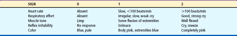
The Apgar score reflects the general condition of the infant at 1 and 5 minutes based on the five parameters described previously. The Apgar score is not a tool, however, that stands on its own to either interpret past events or predict future events linked to the infant’s eventual neurologic or physical status. Considerable discussion and controversy have centered on Apgar scoring because of its misuse as an indicator for the presence or absence of perinatal asphyxia in the medicolegal field (American Academy of Pediatrics and American College of Obstetricians and Gynecologists, 2006). In addition, the Apgar score is not used to determine the newborn’s need for resuscitation at birth.
Clinical Assessment of Gestational Age
Assessment of gestational age is an important criterion because perinatal morbidity and mortality are related to gestational age and birth weight. A frequently used method of determining gestational age is the New Ballard Scale (NBS) by Ballard, Khoury, Wedig, and others (1991) (Fig. 8-1, A). This scale, an abbreviated version of the Dubowitz scale, assesses six external physical and six neuromuscular signs. Each sign has a number score, and the cumulative score correlates with a maturity rating of 20 to 44 weeks of gestation.

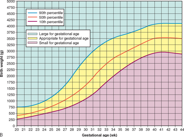
FIG. 8-1 A, New Ballard Scale for newborn maturity rating. Expanded scale includes extremely preterm infants and has been refined to improve accuracy in more mature infants. B, Intrauterine growth: birth weight percentiles based on live single births at gestational ages 20 to 44 weeks. (A, From Ballard JL, Khoury JC, Wedig K, and others: New Ballard Score, expanded to include extremely premature infants, J Pediatr 119(3): 418, 1991.) (B, Data from Alexander GR, Himes JH, Kaufman RB, and others: A United States national reference for fetal growth, Obstet Gynecol 87(2):163-168, 1996.)
The NBS includes −1 and −2 scores that reflect signs of extremely preterm infants, such as fused eyelids; imperceptible breast tissue; sticky, friable, transparent skin; no lanugo; and square-window (flexion of wrist) angle of greater than 90 degrees (see Fig. 8-1, A, and the description of the tests in Box 8-2). The examination of infants with a gestational age of 26 weeks or less should be performed at a postnatal age of less than 12 hours. For infants with a gestational age of at least 26 weeks, the examination can be performed up to 96 hours after birth. To ensure accuracy, it is recommended that the initial examination be performed within the first 48 hours of life. Neuromuscular adjustments after birth in extremely immature neonates require that a follow-up examination be performed to further validate neuromuscular criteria. The NBS scale overestimates gestational age by 2 to 4 days in infants younger than 37 weeks of gestation, especially at gestational ages of 32 to 37 weeks (Ballard, Khoury, Wedig, and others, 1991). In a blinded Spanish study, Marín, Martín, Lliteras, and others (2006) compared estimations of gestational age using NBS vs ultrasonography or mother’s last menstrual period. Researchers found general agreement between NBS and ultrasonography or last menstrual period; however, they noted that NBS tends to overestimate gestational age in very preterm newborns and in infants whose mothers had received prenatal corticosteroid therapy.
Weight Related to Gestational Age.: The weight of the infant at birth also correlates with the incidence of perinatal morbidity and mortality. However, birth weight alone is a poor indicator of gestational age and fetal maturity. Maturity implies functional capacity—;the degree to which the neonate’s organ systems are able to adapt to the requirements of extrauterine life. Therefore gestational age is more closely related to fetal maturity than is birth weight. Because heredity influences a newborn’s size, noting the size of other family members is part of the assessment process.
Intrauterine growth curves are used to classify infants according to birth weight and gestational age. The primary intrauterine growth charts that provide national reference data include the work of Alexander, Himes, Kaufman, and others (1996), which is representative of more than 3.1 million live births in the United States, and Thomas, Peabody, Turnier, and others (2000). Arbuckle, Wilkins, and Sherman (1993), and Kramer, Platt, Wen, and others (2001), have produced intrauterine growth charts using large numbers of infants representative of the Canadian population. Thomas and others (2000) concluded that intrauterine growth measured by head circumference, birth weight, and length varies according to race and gender. These researchers also found that altitude did not seem to significantly affect birth weight, as has been suggested by other authors. It is recommended that the reader access and use the most current intrauterine growth chart specific to the referent population being evaluated.
Classification of infants at birth by both birth weight and gestational age provides a more satisfactory method for predicting mortality risks and providing guidelines for management of the neonate than estimating gestational age or birth weight alone. The infant’s birth weight, length, and head circumference are plotted on standardized graphs that identify normal values for gestational age (for birth weight see Fig. 8-1, B). The infant whose weight is appropriate for gestational age (AGA) (between 10th and 90th percentiles) can be presumed to have grown at a normal rate regardless of the time of birth—preterm, term, or postterm. The infant who is large for gestational age (LGA) (above 90th percentile) can be presumed to have grown at an accelerated rate during fetal life; the small-for-gestational-age (SGA) infant (below 10th percentile) can be assumed to have intrauterine growth restriction or delay.
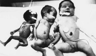
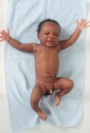
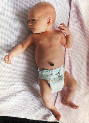
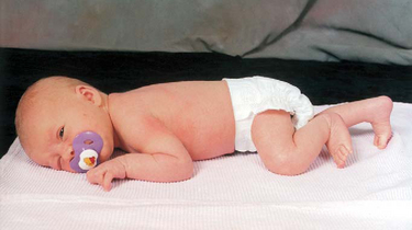
When gestational age is determined according to a standardized gestational age scale such as the NBS, the newborn will fall into one of the following nine possible categories for birth weight and gestational age: AGA—term, preterm, postterm; SGA—term, preterm, postterm; LGA—term, preterm, postterm. Fig. 8-2 illustrates the disparity between birth weights of three preterm infants of the same gestational age, 32 weeks. Birth weight and gestational age both influence morbidity and mortality; the lower the birth weight and gestational age, the higher the morbidity and mortality.
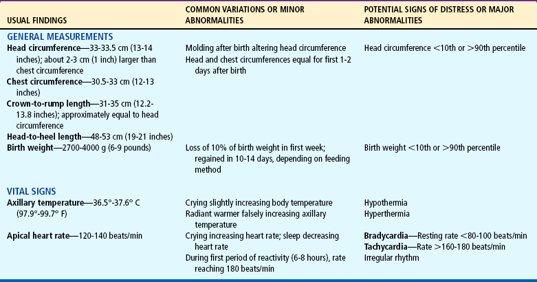
General Measurements.: Several important measurements of the newborn have significance when compared with each other and when recorded over time on a graph. For the full-term infant, average head circumference is between 33 and 35.5 cm (13 and 14 inches). Head circumference may be somewhat less immediately after birth because of the molding process that occurs during a vaginal delivery. Usually by the second or third day the skull is normal in size and contour.
Chest circumference is 30.5 to 33 cm (12 to 13 inches). Head circumference is usually about 2 to 3 cm (about 1 inch) greater than chest circumference. Because of the molding of the head during delivery, these measurements may initially appear equal.
Head circumference may also be compared with crown-to-rump length, or sitting height. Crown-to-rump measurements are usually 31 to 35 cm (12.2 to 13.8 inches) and are approximately equal to head circumference. The relationship of the head and crown-to-rump measurements is more reliable than that of the head and chest. Neonatal head circumference and crown-to-rump length may provide a more accurate means for identifying infants at risk; head circumference has been shown to be equal to or up to 1 cm more than crown-to-rump length in 62% of the infants examined and determined to be normocephalic.
Abdominal circumference need not be routinely measured in the newborn, but should be done in the event of abdominal distention to determine changes in girth over time (Conner, 2003). Abdominal circumference is measured just above the level of the umbilicus, since the umbilical cord is still attached, making measurements across the umbilicus too variable in newborns. Measuring the abdominal circumference below the umbilical region is unsuitable because bladder status may affect the reading.
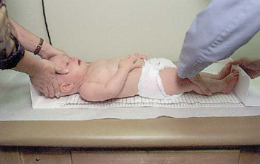
Head-to-heel length is also measured. Because of the usual flexed position of the infant, it is important to extend the leg completely when measuring total body length. The average length of the newborn is 48 to 53 cm (19 to 21 inches) (Fig. 8-3).
Measure body weight soon after birth because weight loss occurs fairly rapidly. Normally the neonate loses about 10% of the birth weight by 3 to 4 days of age because of loss of extracellular fluid and meconium, as well as limited food intake, especially in breast-fed infants. The birth weight is usually regained by the tenth to fourteenth day of life. Most newborns weigh 2700 to 4000 g (6 to 9 pounds), the average weight being about 3400 g (7.5 pounds). Accurate birth weights and lengths are important because they provide a baseline for assessment of future growth.
Another category of measurements is vital signs. Axillary temperatures are taken because insertion of a thermometer into the rectum can potentially cause perforation of the mucosa if performed incorrectly (see Table 6-3 and Fig. 8-4). Core body temperature varies according to the periods of reactivity but is usually 36.5° to 37.6° C (97.7° to 99.7° F). Skin temperature is slightly lower than core body temperature. Therefore axillary temperature may be less than rectal temperature, although the difference is small (as little as 0.2° F) between axillary and rectal sites. Because brown adipose tissue is located in the axillary pocket, axillary readings may be elevated whenever NST occurs. However, axillary readings may be normal in cold-stressed infants when NST is not triggered or is overwhelmed.
The single best method for determining the newborn infant’s temperature in any given situation remains elusive when considering the available studies. Controversy exists regarding the accuracy of tympanic membrane sensors. Bliss-Holtz (1993) compared axillary and tympanic membrane temperatures in neonates and found that tympanic membrane temperature measurements were helpful in determining the infant’s thermal state. However, other studies have found tympanic membrane temperatures to have high variability according to neonatal environment (bassinet or open crib, radiant warmer, and incubator) (Hicks, 1996; Leick-Rude and Bloom, 1998) and limited usefulness in critically ill neonates (Weiss, Poeltler, and Gocka, 1993; Wilshaw, Beckstrand, Waid, and others, 1999). One study concluded that tympanic membrane temperatures were unacceptable for detecting fever in children under 6 years of age (Lanham, Walker, Klocke, and others, 1999). A meta-analysis of 101 studies comparing tympanic membrane temperatures with rectal temperatures in children concluded that the tympanic method demonstrated a wide range of variability, limiting its application in a pediatric setting (Craig, Lancaster, Taylor, and others, 2002). The Canadian Paediatric Society (CPS) (2007) outlines concerns regarding safety and accuracy of tympanic temperature measurement in newborns because of the size of a newborn’s external ear canal relative to the size of the thermometer probe. To ensure accuracy, the probe, which may be up to 8 mm (0.3 inch) in diameter, must be deeply inserted into the ear canal to allow orientation of the sensor near or against the tympanic membrane. At birth, the average diameter of the canal is just 4 mm (0.16 inch); at 2 years of age it is just 5 mm (0.2 inch). The CPS concurs with earlier writers Bliss-Holtz (1993) and Yetman, Coody, West, and others (1993) in their conclusion that infrared tympanic membrane technology has yet to meet clinical needs for use in newborn infants.
Infrared axillary and digital thermometers are used in many neonatal units because they give rapid readings and are easy to clean; studies demonstrate their usefulness in well, full-term newborns (Sganga, Wallace, Kiehl, and others, 2000), whereas accuracy with critically ill neonates is less predictable (Seguin and Terry, 1999; Wilshaw, Beckstrand, Waid, and others, 1999). Bailey and Rose (2001) concluded that tympanic membrane temperatures (vs axillary) in healthy preterm infants were reliable, were cost effective, and decreased the amount of handling required for monitoring. Skin temperature readings have also been found to vary with probe site placement; bed type; environmental temperature; and the use of blankets, clothing, and nesting devices (Leick-Rude and Bloom, 1998). Jones, Kleber, Eckert, and others (2003) compared rectal temperatures of infants under 2 months of age with calibrated digital thermometers and mercury glass thermometers; the study of 120 infants found that the digital thermometers measured a higher temperature (mean average of 0.7° F; range 0° to 1.6° F) than the mercury glass thermometers. The researchers concluded that the error in measurement was attributable to the digital thermometer used. Advantages of digital thermometers in neonatal care include relatively easy readability by parents and caretakers in the home, improvement of discharge planning effectiveness, and decreased risk of breakage and associated complications compared with glass thermometers.
Temporal artery thermometers (TATs) are available for use in the general pediatric population, and parents often report ease of use and less discomfort with such methods. Greenes and Fleisher (2001) concluded that the TAT had limited sensitivity in infants for detecting rectal fever, yet the TAT was more accurate than the tympanic thermometer. Siberry, Diener-West, Schappell, and others (2002) compared the infrared TAT (using an infrared device) with the rectal temperature measurement (digital thermometer) and found poor predictability for fever in children 0 to 3 months old. The authors concluded that the TAT could be used as a rapid assessment screening tool to identify rectal fever in children 3 to 24 months old, but TAT was unreliable as a screening tool for infants under 3 months. Schuh, Komar, Stephens, and others (2004) compared rectal temperatures with TAT measurements in an emergency department population of children under 24 months and concluded that the TAT was not reliable for detecting rectal fever in children under 3 months but could be used as a screening tool for detecting fever of at least 38.3° C (100.9° F) in children 3 to 24 months.
In most studies regarding newborn temperature, the glass mercury thermometer is the gold standard against which other methods are compared. There is no universal agreement on placement times for glass thermometers, although 3 minutes for rectal temperature and 5 minutes for axillary temperature are considered to be adequate. The American Academy of Pediatrics (2001) has, however, issued a statement recommending that mercury thermometers no longer be used in clinics and homes to decrease mercury exposure hazard.
Nurses must be cognizant of the many variables involved (site—axillary, rectal, tympanic, skin; skin blood flow at site of measurement; environment—radiant warmer, open crib, incubator, clothing, or nesting; purpose—fever, possible sepsis, in which case the temperature may be lower than normal in newborns, and thermoregulation in the transition phase; instrument—electronic, digital, infrared) and able to make clear clinical decisions based on accurate and objective data. Further research is needed to perfect thermometers that accurately reflect the infant’s core temperature in order to effectively plan nursing care and maintain a stable temperature.
Pulse and respirations also vary according to the periods of reactivity and the infant’s behaviors but are usually in the range of 120 to 140 beats/min and 30 to 60 breaths/min, respectively. Both are counted for a full 60 seconds to detect irregularities in rate or rhythm. The heart rate is taken apically with a stethoscope, and the femoral arteries are palpated for equality of strength or fullness.
Measurement of blood pressure (BP) provides baseline data and may indicate cardiovascular problems. BP is most easily and accurately assessed using oscillometry (Dinamap), when the newborn is in a quiet or sleep state, using an appropriate cuff width–to-arm ratio of 0.45 to 0.70 (approximately half to three quarters) (Nuntnarumit, Yand, and Bada-Ellzey, 1999). Oscillometry is less reliable when mean arterial pressure is below 40 mm Hg (Chia, Ang, Wong, and others, 1990) (Fig. 8-5). The average oscillometric systolic/diastolic pressure is 65/41 mm Hg at 1 to 3 days of age. Compare BP in the upper and lower extremities, which should be equal.
A suggested schedule for monitoring heart rate, respiratory rate, and temperature is on admission to the nursery, once every 30 minutes until the newborn has been stable for 2 hours (American Academy of Pediatrics and American College of Obstetricians and Gynecologists, 2007), then once every 8 hours until discharge. However, this schedule may vary according to institutional policy. Any change in the infant, such as color, breathing, muscle tone, or behavior, necessitates more frequent monitoring.
General Appearance.: Before each body system is assessed, it is important to describe the general posture and behavior of the newborn. The overall appearance yields valuable clues to the infant’s physical status.
In the full-term neonate the posture is one of complete flexion as a result of in utero position. Most infants are born in a vertex presentation with the head flexed and the chin resting on the upper chest, the arms flexed with the hands clenched, the legs flexed at the knees and hips, and the feet dorsiflexed. The vertebral column is also flexed. It is important to recognize any deviation from this characteristic fetal position.
The infant’s behavior is carefully noted, especially the degree of alertness, drowsiness, and irritability; the latter two factors may reflect common signs of neurologic problems. Some questions to mentally ask when assessing behavior include:
Skin.: The texture of the newborn’s skin is velvety smooth and puffy, especially about the eyes, the legs, the dorsal aspect of the hands and the feet, and the scrotum or labia. Skin color depends on racial and familial background and varies greatly among newborns. In general, the Caucasian infant is usually pink to red. The African-American newborn may appear a pinkish or yellowish brown. Infants of Hispanic descent may have an olive tint or a slight yellow cast to the skin. Infants of Asian descent may be a rosy or yellowish tan. The color of Native American newborns depends on the tribe and can vary from a light pink to a dark, reddish brown. By the second or third day the skin turns to its more natural tone and is drier and flakier. Several other color changes that may be noted on the skin are described later in this chapter (see Table 8-4).
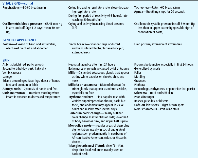

At birth the skin may be partially covered with a grayish white, cheeselike substance called vernix caseosa, a mixture of sebum and desquamating cells. It will be absorbed by 24 to 28 hours. A fine, downy hair called lanugo may be present on the skin, especially on the forehead, cheeks, shoulders, and back.
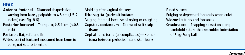
Head.: General observation of the contour of the head is important, since molding occurs in almost all vaginal deliveries. In a vertex delivery the head is usually flattened at the forehead, with the apex rising and forming a point at the end of the parietal bones and the posterior skull or occiput dropping abruptly. The usual, more oval contour of the head is apparent by 1 to 2 days after birth. The change in shape occurs because the bones of the cranium are not fused, allowing for overlapping of the edges of these bones to accommodate to the size of the birth canal during delivery. Such molding usually does not occur in infants born by elective cesarean section.
Six bones—the frontal, occipital, two parietals, and two temporals—make up the cranium. Between the junction of these bones are bands of connective tissue called sutures. At the junction of the sutures are wider spaces of unossified membranous tissue called fontanels. The two most prominent fontanels in infants are the anterior fontanel, formed by the junction of the sagittal, coronal, and frontal sutures, and the posterior fontanel, formed by the junction of the sagittal and lambdoid sutures (Fig. 8-6, A).
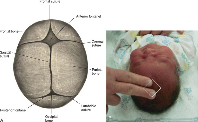
The skull is palpated for all patent sutures and fontanels, noting size, shape, molding, or abnormal closure. The sutures feel like cracks between the skull bones, and the fontanels feel like wider soft spots at the junction of the sutures. These are palpated by using the tip of the index finger and running it along the ends of the bones (see Fig. 8-6, B).
The anterior fontanel is diamond shaped and measures anywhere from barely palpable to 4 to 5 cm (about 2 inches) at its widest point (from bone to bone, rather than from suture to suture). The posterior fontanel is easily located by following the sagittal suture toward the occiput. The posterior fontanel is triangular, usually measuring between 0.5 and 1 cm (less than ½ inch) at its widest part. The fontanels should feel flat, firm, and well demarcated against the bony edges of the skull. Frequently pulsations are visible at the anterior fontanel. Coughing, crying, or lying down may temporarily cause the fontanels to bulge and become more taut.
Palpate the skull for any unusual masses or prominences, particularly those resulting from birth trauma, such as caput succedaneum or cephalhematoma (see Chapter 9). Because of the pliability of the skull, exerting pressure at the margin of the parietal and occipital bones along the lambdoid suture may produce a snapping sensation similar to the indentation of a Ping-Pong ball. This phenomenon, known as physiologic craniotabes, may be found normally, especially in newborns of breech birth, but also may indicate hydrocephalus, congenital syphilis, or rickets.
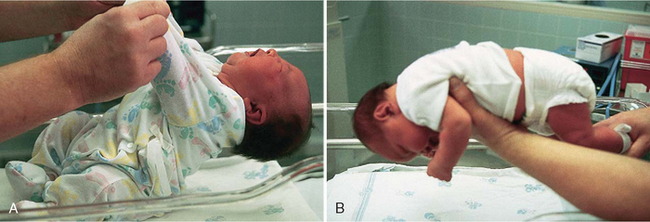
Assess the degree of head control. Although head lag is normal in the newborn, the degree of ability to control the head in certain positions should be recognized. If the supine infant is pulled from the arms into a semi-Fowler position, marked head lag and hyperextension are noted (Fig. 8-7, A). However, as the infant is brought forward into a sitting position, the infant will attempt to control the head in an upright position. As the head falls forward onto the chest, many infants will attempt to right it into the erect position. Also, if the infant is held in ventral suspension (i.e., held prone above and parallel to the examining surface), the infant will hold the head in a straight line with the spinal column (see Fig. 8-7, B). When lying on the abdomen, the newborn has the ability to lift the head slightly, turning it from side to side. Marked head lag is seen in neonates with Down syndrome, prematurity, hypoxia, and neuromuscular compromise.
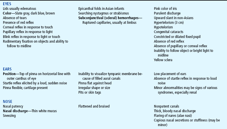
Eyes.: Because newborns tend to have their eyes tightly closed, it is best to begin the examination of the eyes by observing the lids for edema, which is normally present for the first 2 days after delivery. The eyes are observed for symmetry. Tears may be present at birth, but purulent discharge from the eyes shortly after birth is abnormal. To visualize the surface structures of the eye, the infant is held supine, and the head is gently lowered. The eyes will usually open, similar to the mechanism of a doll’s eyes. The sclera should be white and clear.
The cornea is examined for the presence of any opacities or haziness. The corneal reflex is normally present at birth but may not be elicited unless neurologic or eye damage is suspected. The pupil will usually respond to light by constricting. The pupils are normally malaligned. A searching nystagmus is common. Strabismus is a normal finding because of the lack of binocularity. The color of the iris is noted. Most light-skinned newborns have slate gray or dark blue eyes, whereas dark-skinned infants have brown eyes.
A funduscopic examination is difficult to perform because of the infant’s tendency to keep the eyes tightly closed. However, a red reflex should be elicited (see Chapter 6).
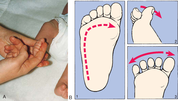
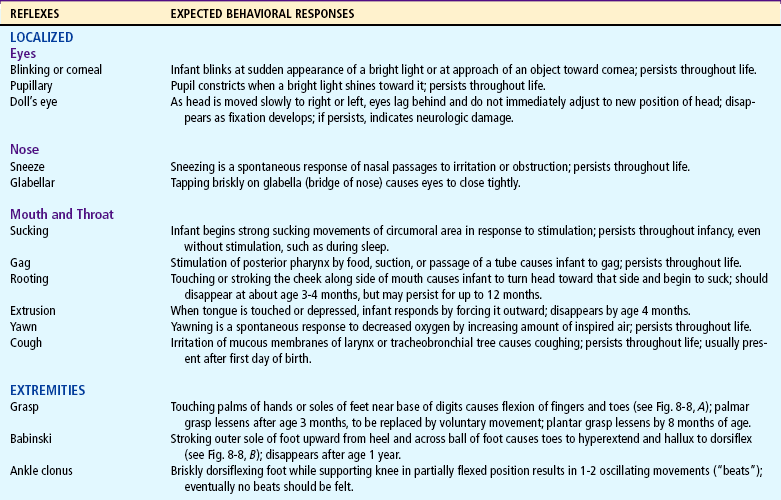
FIG. 8-8. A, Plantar or grasp reflex. B, Babinski reflex. 1, Direction of stroke. 2, Dorsiflexion of big toe. 3, Fanning of toes. (A, From Zitelli BJ, Davis HW: Atlas of pediatric physical diagnosis, ed 4, St Louis 2002, Mosby.)
Ears.: The ears are examined for position, structure, and auditory function. The top of the pinna should lie in a horizontal plane to the outer canthus of the eye (see Fig. 6-19). The pinna is often flattened against the side of the head from pressure in utero. An otoscopic examination ordinarily may not be performed because the canals are filled with vernix caseosa and amniotic fluid, making visualization of the tympanic membrane difficult.
Auditory ability is tested by a number of objective hearing tests (see p. 140). Making a loud noise close to the infant’s head may or may not elicit a response; the lack of a response, however, is not a definite indication of hearing loss. The startle reflex (see Table 8-2) may be observed when there is a sudden loud noise near the infant or the bassinet is accidentally bumped, but this often depends on the infant’s state at the time.
Nose.: The nose is usually flattened after birth, and bruises are common. Patency of the nasal canals can be assessed by holding a hand over the infant’s mouth and one canal and noting the passage of air through the unobstructed opening. If nasal patency is questionable, it is reported, because most newborns are obligatory nose breathers. Sneezing and thin white mucus are common up to several hours after birth.
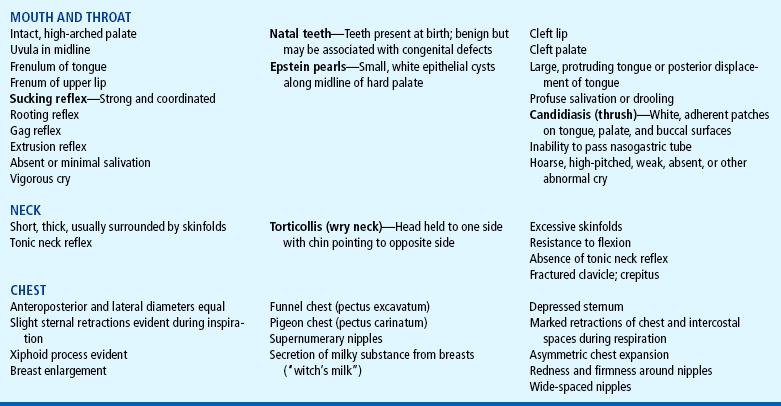
Mouth and Throat.: An external defect of the mouth such as cleft lip is readily apparent; however, the internal structures require careful inspection. The palate is normally highly arched and somewhat narrow. Rarely teeth may be present. A common finding is Epstein pearls, small, white, epithelial cysts along both sides of the midline of the hard palate. They are insignificant and disappear in several weeks.
The frenulum of the upper lip is a band of thick, pink tissue that lies under the inner surface of the upper lip and extends to the maxillary alveolar ridge. It is particularly evident when the infant yawns or smiles. It disappears as the maxilla grows.
The lingual frenulum attaches the underside of the tongue to the lower palate midway between the ventral surface of the tongue and the tip. In some cases a tight lingual frenulum, formerly referred to as tongue-tie, may restrict adequate sucking. Further evaluation may be required to ascertain adequate sucking, particularly in breast-fed infants. The treatment for a tight lingual frenulum advocated by the American Academy of Pediatrics is frenotomy, a safe and effective surgical procedure that improves comfort, effectiveness, and ease of breastfeeding for mother and infant (Barclay, 2002; Ballard, Auer, and Khoury, 2002; Coryllos, Genna, and Salloum, 2004).
Elicit the sucking reflex by placing a nipple or nonlatex gloved finger in the infant’s mouth. The infant should exhibit a strong, vigorous suck. The rooting reflex is elicited by stroking the cheek and noting the infant’s response of turning toward the stimulated side and sucking.
The uvula can be inspected while the infant is crying and the chin is depressed. However, it may be retracted upward and backward during crying. Tonsillar tissue is generally not seen in the newborn. Natal teeth, teeth present at birth, as opposed to neonatal teeth, which erupt during the first month of life, are seen infrequently and erupt chiefly at the position of the lower incisors. Teeth are reported because they are frequently found with developmental abnormalities and syndromes, including cleft lip and palate. Most natal teeth are loosely attached. However, current thinking suggests preserving them until they exfoliate naturally (McDonald and Avery, 2004), unless the tooth is attached loosely or breastfeeding is impaired by the neonate biting the breast.
Neck.: Because the newborn’s neck is short and covered with folds of tissue, adequate assessment of the neck requires allowing the head to fall gently backward in hyperextension while the back is supported in a slightly raised position. Observe for range of motion, shape, and any abnormal masses, and palpate each clavicle for possible fractures.
Chest.: The shape of the newborn’s chest is almost circular because the anteroposterior and lateral diameters are equal. The ribs are flexible, and slight intercostal retractions are normally seen on inspiration. The xiphoid process is commonly visible as a small protrusion at the end of the sternum. The sternum is generally raised and slightly curved.
Inspect the breasts for size; shape; and nipple formation, location, and number. Breast enlargement appears in many newborns of either gender by the second or third day and is caused by maternal hormones. Occasionally a milky substance, sometimes called witch’s milk, is secreted by the infant’s breasts by the end of the first week. Supernumerary nipples may be found on the chest, on the abdomen, or in the axilla.
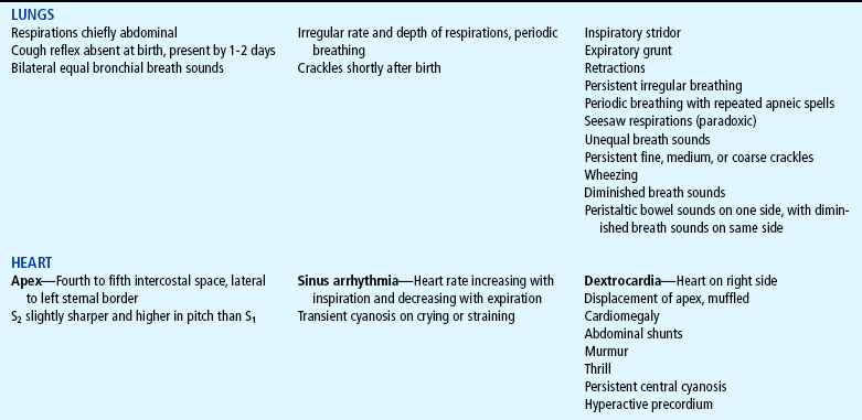
Lungs.: The normal respirations of the newborn are irregular and abdominal, and the rate is between 30 and 60 breaths/min. Pauses in respiration of less than 20 seconds’ duration are considered normal. After the initial forceful breaths required to initiate respiration, subsequent breaths should be nonlabored and fairly regular in rhythm. Periodic breathing is commonly seen in full-term newborns and consists of rapid nonlabored respirations followed by pauses of less than 20 seconds; periodic breathing may be more prominent during sleep and is not accompanied by status changes such as cyanosis or bradycardia. Occasional irregularities occur in relation to crying, sleeping, stooling, and feeding.
Perform auscultation when the infant is quiet. Bronchial breath sounds should be equal bilaterally. Any differences in auscultatory findings between symmetric sites is reported. Crackles soon after birth indicate the presence of fluid, which represents the normal transition of the lungs to extrauterine life. However, wheezes, persistence of medium or coarse crackles after the first few hours of life, and stridor should be reported for further investigation.
Heart.: Heart rate is auscultated and may range from 100 to 180 beats/min shortly after birth and, when the infant’s condition has stabilized, from 120 to 140 beats/min. The point of maximum impulse (PMI) may be palpated and is usually found at the fourth to fifth intercostal space, medial to the left midclavicular line. The PMI gives some indication of the location of the heart, which may be displaced in conditions such as congenital diaphragmatic hernia or pneumothorax. Dextrocardia, an anomaly wherein the heart is on the right side of the body, is reported, because the abdominal organs may also be reversed, with associated circulatory abnormalities.
Auscultation of the specific components of the heart sounds is difficult because of the rapid rate and effective transmission of respiratory sounds. However, the first (S 1) and second (S 2) sounds should be clear and well defined; the second sound is somewhat higher in pitch and sharper than the first. A murmur is frequently heard in the newborn, especially over the base of the heart or at the left sternal border at the third or fourth interspace. Ordinarily a murmur is not associated with specific cardiac defects, but frequently represents the incomplete functional closure of fetal shunts. (See Chapter 6 for other characteristics of murmurs.) However, always record and report any murmur or other unusual sounds.
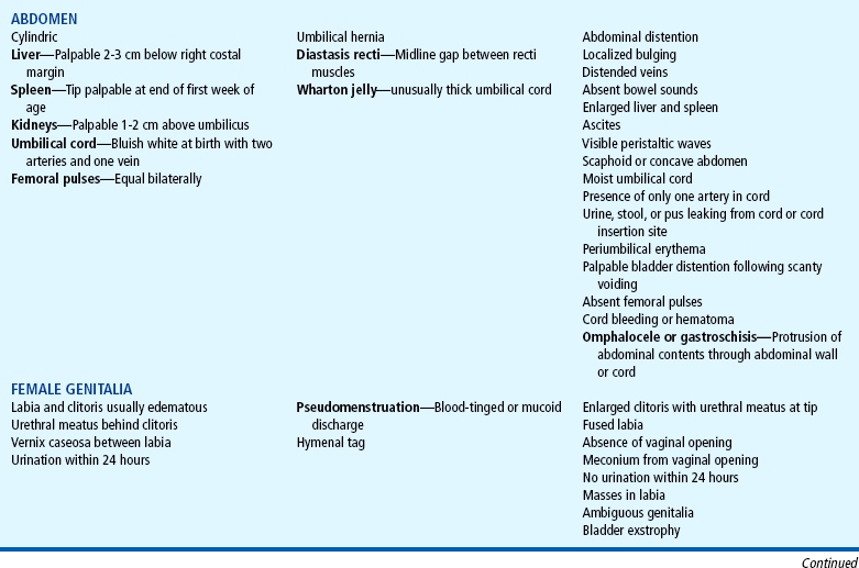
Abdomen.: The normal contour of the abdomen is cylindric and usually prominent with few visible veins. Bowel sounds are heard within the first 15 to 20 minutes after birth. Visible peristaltic waves may be observed in some newborns. Diastasis recti is a gap in the rectus muscles; this usually benign condition is visible as a raised and palpable bulge at midline, especially in the crying infant.
Inspect the umbilical cord to determine the presence of two arteries, which look like papular structures, and one vein, which has a larger lumen than the arteries and a thinner vessel wall. At birth the cord appears bluish white and moist. After clamping, it begins to dry and appears a dull, yellowish brown. It progressively shrivels in size and turns greenish black.
If the umbilical cord appears unusually large in diameter at the base, inspect for the presence of a hematoma or small omphalocele. If the cord is clamped over an existing omphalocele, part of the intestine will be clamped, causing tissue necrosis. One practical rule of thumb is to cut the cord distally 4 to 5 inches from a questionable enlargement until further examination is carried out by a practitioner. The extra length can later be cut once no pathologic condition has been identified.
Palpate after inspecting the abdomen. The liver is normally palpable 1 to 3 cm (about 0.5 to 1 inch) below the right costal margin. The tip of the spleen can sometimes be felt, but a palpable spleen more than 1 cm below the left costal margin suggests enlargement and warrants further investigation. Although both kidneys should be palpated, this maneuver requires considerable practice. When felt, the lower half of the right kidney and the tip of the left kidney are 1 to 2 cm above the umbilicus.
During examination of the lower abdomen, it is particularly important to palpate for femoral pulses, which should be strong and equal bilaterally.
Female Genitalia.: Normally, the labia minora, labia majora, and clitoris are edematous, especially after a breech delivery. However, the labia and clitoris must be carefully inspected to identify any evidence of ambiguous genitalia or other abnormalities. Normally, in a female, the urethral opening is located behind and below the clitoris.
A hymenal tag is occasionally visible from the posterior opening of the vagina. It is composed of tissue from the hymen and the labia minora. It usually disappears in several weeks. Generally, the vaginal vault is not inspected.
Vaginal discharge may be noted during the first week of life. This pseudomenstruation is a manifestation of the abrupt decrease of maternal hormones and usually disappears by 2 to 4 weeks. Fecal discharge from the vaginal opening indicates a rectovaginal fistula and is always reported. Vernix caseosa may be present in large amounts between the labia.
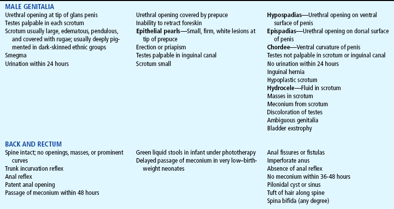
Male Genitalia.: The penis is inspected for the urethral opening, which is located at the tip. However, the opening may be totally covered by the prepuce, or foreskin, which covers the glans penis. A tight prepuce is a common finding in the newborn. It should not be forcefully retracted; locating the urinary meatus is usually possible without retracting the foreskin. Smegma, a white cheesy substance, is commonly found around the glans penis, under the foreskin. Small, white, firm lesions called epithelial pearls may be seen at the tip of the prepuce. An erection is common in the newborn.
The scrotum may be large, edematous, and pendulous in the full-term neonate, especially in the infant born in breech position. It is more deeply pigmented in dark-skinned races. A noncommunicating hydrocele commonly occurs unilaterally and disappears within a few months. Always palpate the scrotum for the presence of testes (see Chapter 6). In small newborns, particularly preterm infants, the undescended testes may be palpable within the inguinal canal. Absence of the testes may also be a sign of ambiguous genitalia, especially when accompanied by a small scrotum and penis. Inguinal hernias may or may not be manifested immediately after birth. A hernia is more easily detected when the infant is crying. Palpable lymph nodes are most commonly found in the inguinal area.
Back and Rectum.: Inspect the spine with the infant prone. The shape of the spine is gently rounded, with none of the characteristic S-shaped curves seen later in life. Any abnormal openings, masses, dimples, or soft areas are noted. A protruding sac anywhere along the spine, but most commonly in the sacral area, indicates some type of spina bifida. A small sinus, which may or may not be communicating with the spine, is a pilonidal sinus. It is frequently covered with a tuft of hair. Although it may have no pathologic significance, a pilonidal cyst may indicate the existence of spina bifida occulta or be a portal of entry into the spinal column. With the infant still prone, note symmetry of the gluteal folds. Report any evidence of asymmetry; tests for developmental dysplasia of the hip are performed by trained (or skilled) examiners (see Chapter 31).
The presence of an anal orifice and passage of meconium from the anal orifice during the first 24 to 48 hours of life indicates anal patency. If an imperforate anus is suspected, report this to the primary practitioner for further evaluation.
Extremities.: Examine the extremities for symmetry, range of motion, and reflexes. Count the fingers and toes, and note supernumerary digits (polydactyly) or fusion of digits (syndactyly). A partial syndactyly between the second and third toes is a common variation seen in otherwise normal infants. The nail beds should be pink, although slight blueness is evident in acrocyanosis.
The palms of the hands should have the usual creases (see Fig. 6-13). The full-term newborn usually has creases covering the entire sole of the foot. The soles of the feet are flat with prominent fat pads.
Observe range of motion of the extremities throughout the entire examination. The absence of arm movement signals a potential birth injury paralysis such as Klumpke or Erb-Duchenne palsy. An asymmetric or partial Moro reflex should alert the practitioner to further evaluate upper extremity mobility. Examine the lower extremities for limb length, symmetry, and hip abduction and flexion. The newborn will demonstrate full range of motion in the elbow, hip, shoulder, and knee joints. Movements should be symmetric, smooth, and unrestricted.
Also assess muscle tone. By attempting to extend a flexed extremity, determine if tone is equal bilaterally. Extension of any extremity is usually met with resistance, and when released, the extremity returns to its previous flexed position. Hypotonia suggests some degree of hypoxia or neurologic disorder and is common in an infant with Down syndrome. Asymmetry of muscle tone may indicate a degree of paralysis from brain damage or nerve damage. Failure to move the lower limbs suggests a spinal cord lesion or injury. Sustained rhythmic tremors, twitches, and myoclonic jerks characterize neonatal seizures or may indicate neonatal abstinence syndrome. (See Neonatal Seizures and Drug-Exposed Infants, Chapter 9.) Sudden asynchronous jerking movements, quivering, or momentary tremors are usually normal.
Two reflexes are elicited. The first is the grasp reflex. Touching the palms of the hands or soles of the feet near the base of the digits causes flexion or grasping (Fig. 8-8, A). The other is the Babinski reflex. Stroking the outer sole of the foot upward from the heel across the ball of the foot causes the big toe to dorsiflex and the other toes to hyperextend (see Fig. 8-8, B).
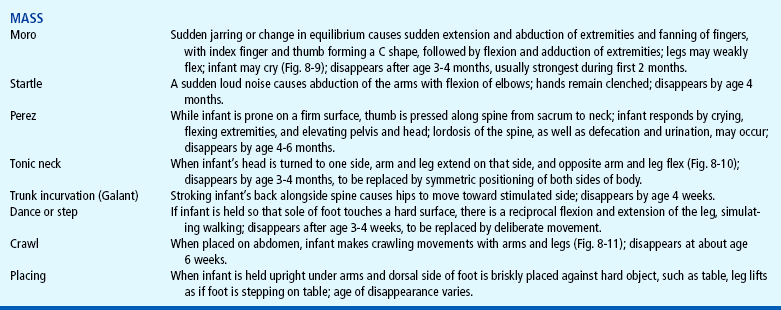
Neurologic System.: Assessing neurologic status is a critical part of the physical examination of the newborn. Much of the neurologic testing takes place during evaluation of body systems, such as eliciting localized reflexes and observing posture, muscle tone, head control, and movement. However, several important mass (total body) reflexes also need to be elicited. Test these at the end of the examination, since they may disturb the infant and interfere with auscultation. These reflexes, as well as several local reflexes, are described in Table 8-2. Record and report the absence, asymmetry, persistence, or weakness of a reflex.
Transitional Assessment: Periods of Reactivity
The newborn exhibits behavioral and physiologic characteristics that may at first appear to be signs of stress. However, during the initial 24 hours, changes in heart rate, respiration, motor activity, color, mucus production, and bowel activity occur in an orderly, predictable sequence that is normal and indicates lack of stress.
For 6 to 8 hours after birth, the newborn is in the first period of reactivity. During the first 30 minutes the infant is very alert, cries vigorously, may suck his or her fingers or fist, and appears very interested in the environment. At this time the newborn’s eyes are usually open, making this an excellent opportunity for mother, father, and child to see each other. Because the newborn has a vigorous suck, this is also an opportune time to begin breastfeeding. The infant will usually grasp the nipple quickly, satisfying both mother and infant. This is particularly important to point out to parents, because after this initially highly active state the infant may be sleepy and uninterested in sucking. Physiologically, the respiratory rate during this period is as high as 80 breaths/min, crackles may be heard, heart rate reaches 180 beats/min, bowel sounds are active, mucus secretions are increased, and temperature may decrease. Maintaining appropriate temperature for the newborn is best accomplished by practicing skin-to-skin care whereby only a diaper is worn, to allow majority of skin surface to be in contact with mother’s skin. A light blanket may be used to cover mother and newborn. Research has shown mother’s skin temperature will increase to ensure the newborn does not become hypothermic (Kimura and Matsuoka, 2007).
After this initial stage of alertness and activity, the infant enters the second stage of the first reactive period, which generally lasts 2 to 4 hours. Heart and respiratory rates decrease, temperature continues to fall, mucus production decreases, and urine or stool is usually not passed. The infant is in a state of sleep and relative calm. Any attempt at stimulation usually elicits minimal response. Because of the continued decline in body temperature, undressing or bathing is avoided during this time.
The second period of reactivity begins when the infant awakes from this deep sleep; it lasts about 2 to 5 hours and provides another excellent opportunity for child and parents to interact. The infant is again alert and responsive, heart and respiratory rates increase, the gag reflex is active, gastric and respiratory secretions are increased, and passage of meconium frequently occurs. This period is usually over when the amount of respiratory mucus has decreased. After this stage is a period of stabilization of physiologic systems and a vacillating pattern of sleep and activity.
Behavioral Assessment
Another important area of assessment is observation of behavior. Infants’ behavior helps shape their environment, and their ability to react to various stimuli affects how others relate to them. The principal areas of behavior for newborns are sleep, wakefulness, and activity, such as crying.
One method of systematically assessing the infant’s behavior is the use of the Brazelton Neonatal Behavioral Assessment Scale (BNBAS) (Brazelton and Nugent, 1996). The BNBAS is an interactive examination that assesses the infant’s response to 28 items organized according to the clusters in Box 8-3. It is generally used as a research or diagnostic tool and requires special training.
In addition to its use as an initial and ongoing tool to assess neurologic and behavioral responses, the scale can be used in assessment of initial parent-child relationships, as a preventive instrument that identifies a caregiver who may benefit from a role model, and as a guide to help parents focus on their infant’s individuality and develop a deeper attachment to their child. Studies have demonstrated that showing parents the unique characteristics of their infant causes a more positive perception of the infant to develop, with increased interaction between infant and parent.
Patterns of Sleep and Activity.: Newborns begin life with a systematic schedule of sleep and wakefulness that is initially evident during the periods of reactivity. After this initial period, it is not unusual for the infant to sleep almost constantly for the next 2 to 3 days to recover from the exhausting birth process.
Infants have six distinct sleep-wake states, which represent a particular form of neural control (Table 8-3). As maturity increases, each state becomes more precisely defined according to the behaviors observed. State is defined as a “group of characteristics that regularly occur together” (Blackburn, 2003) and includes body activity, eye and facial movements, respiratory pattern, and response to internal and external stimuli. The six sleep-wake states are quiet (deep) sleep, active (light) sleep, drowsy, quiet alert, active alert, and crying. Infants respond to internal and external environmental factors by controlling sensory input and regulating the sleep-wake states; the ability to make smooth transitions between states is called state modulation. The ability to regulate sleep-wake states is essential in the neurobehavioral development of the infant. The more immature the infant, the less able he or she is able to cope with external and internal factors that affect the sleep-wake patterns.
TABLE 8-3
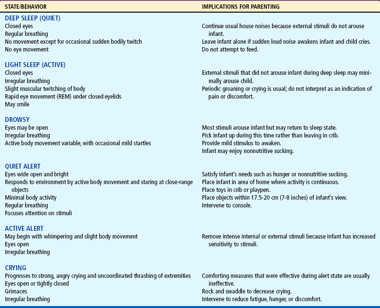
Portions adapted from Blackburn S, Loper DL: Maternal, fetal, and neonatal physiology: a clinical perspective, Philadelphia, 1992, Saunders.
Recognition and knowledge of sleep-wake states is important in the planning of nursing care. It is also important for nurses to help parents and caregivers understand the significance of the infant’s behavioral responses to daily caregiving and how these states can be altered. A classic example is the newborn who feeds vigorously in the active alert state but poorly once he or she progresses to the crying state. The neurologic assessment of a newborn in the active alert state will differ significantly from the deep sleep state.
Newborns typically spend as much as 16 to 18 hours sleeping and do not necessarily follow a pattern of light-dark diurnal rhythm. With increasing age, sleep-wake states will change, with increasing amounts of time spent in awake alert states and decreasing amounts of sleep time. Approximately 50% of total sleep time is spent in irregular or rapid eye movement sleep.
Cry.: The newborn should begin extrauterine life with a strong, lusty cry. The sounds produced by crying can be described as hunger, anger, pain, and “bid for attention” cries. Discomfort (pain) sounds initially consist of gasps and cries in which the consonant H is clearly distinguishable. The duration of crying is as variable in each infant as the duration of sleep patterns. Newborns may cry as little as 5 minutes or as much as 2 hours or more per day. Feeding usually terminates the state of crying when hunger is the cause. Swaddling or wrapping an infant snugly in a blanket promotes sleep and maintains body temperature. Rocking the infant reduces crying and induces quiet alertness or sleep. Variations in the initial cry can indicate abnormalities. A weak, groaning cry or grunting during expiration usually indicates respiratory disturbance. Absent, weak, or constant crying requires further investigation for possible drug withdrawal or a neurologic problem.
Assessment of Attachment Behaviors
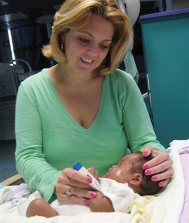
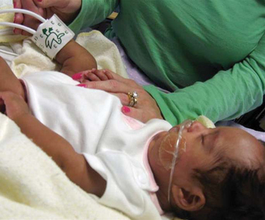
One of the most important areas of assessment is careful observation of behaviors that are thought to indicate the formation of emotional bonds between the newborn and family, especially the mother. Such behaviors include the en face position; undressing and touching the infant; smiling, kissing, and talking to the infant; and holding, rocking, and cradling the child close to the body (see Nursing Care Guidelines box). Because assessment is closely related to interventions that promote attachment (e.g., encouraging these behaviors in parents), assessing attachment behaviors is further discussed on pp. 234–235.
When the infant is brought to the parents, do they reach out for the child and call the child by name?
Do the parents speak about the child in terms of identification–whom the infant looks like; what appears special about their child compared with other infants?
When parents are holding the infant, what kind of body contact is there? Do they feel at ease in changing the infant’s position; are fingertips or whole hands used; are there parts of the body they avoid touching or parts of the body they investigate and scrutinize?
When the infant is awake, what kinds of stimulation do the parents provide? Do they talk to the infant, to each other, or to no one? How do they look at the infant–direct visual contact, avoidance of eye contact, or looking at other people or objects?
How comfortable do the parents appear in terms of caring for the infant? Do they express any concern regarding their ability or disgust for certain activities, such as changing diapers?
What type of affection do they demonstrate to the newborn, such as smiling, stroking, kissing, or rocking?
If the infant is fussy, what kinds of comforting techniques do the parents use, such as rocking, swaddling, talking, or stroking?
Physical Assessment
An essential aspect of the care of the newborn is a thorough physical assessment that includes estimation of gestational age and physical examination to identify normal characteristics and existing abnormalities. These initial and ongoing assessments are critical to establishing baseline data for planning, implementing, and evaluating care and are a nursing priority in caring for the newborn. The discussion of physical examination focuses on normal findings and variations from the norm that require little or no intervention. The reader is encouraged to review Chapter 6 for further discussion of examination techniques. General guidelines for conducting a physical examination are presented in the Nursing Care Guidelines box. Table 8-4 summarizes physical examination of the newborn.
Physical Examination of the Newborn
1. Provide a normothermic and nonstimulating examination area.
2. Check that equipment and supplies are working properly and are accessible.
3. Undress only body area examined to prevent heat loss.
4. Proceed in an orderly sequence (usually head to toe) with the following exceptions:
Observe infant’s attitude and position of flexion first to avoid disturbing him or her.
Perform all procedures that require quiet next, such as auscultating the lungs, heart, and abdomen.
Perform disturbing procedures, such as testing reflexes, last.
Measure head, chest, and length at same time to compare results.
The nursing care of the newborn is discussed on the following pages. The nursing process in the care of the newborn is outlined in the Nursing Process box.
MAINTAIN A PATENT AIRWAY
Establishing a patent airway is a primary objective in the delivery room and is the responsibility of the attending nurses and practitioners. However, maintaining a patent airway continues to be a priority goal in the nursery, with attention to proper positioning of the infant to facilitate drainage of secretions, especially after feeding. The American Academy of Pediatrics (2000a) recommends the supine position during sleep for healthy newborns. This recommendation is based on the possible association between sleeping prone and sudden infant death syndrome (see Chapter 11). Since the initial recommendation in 1992 that all infants be placed in the supine position to sleep, there has been no evidence of an increased number of complications, such as choking or vomiting, when infants are placed in this position (Malloy, 2002). There has, however, been an increase in the number of infants with cranial asymmetry, particularly unilateral flattening of the occiput (American Academy of Pediatrics, 2003c). Health care professionals must educate parents on prevention of deformational plagiocephaly by encouraging alternate positions when the infant is awake.
The Healthy Newborn and Family
DIAGNOSIS (PROBLEM IDENTIFICATION)
After a thorough assessment, several nursing diagnoses for the healthy newborn include:
Numerous goals for the healthy newborn are discussed on pp. 215–239. Expected patient outcomes include:
Newborn airway will remain patent.
Effective breathing pattern will be established.
Thermoregulation will be maintained.
Parent-infant attachment behaviors will be exhibited.
Breastfeeding or bottle feeding will be established.
Infant will exhibit no evidence of infection; immune status will be maintained.
Newborn will remain free of injury.
Family will demonstrate ability to care for infant’s basic needs.
Intervention strategies for the healthy newborn and family are discussed on pp. 215–239.
The effectiveness of nursing interventions for the newborn and family is determined by continual assessment and evaluation of care based on the following guidelines:
Observe infant’s color and respiratory pattern.
Monitor axillary temperature regularly; observe for signs of temperature instability such as respiratory distress.
Observe for any evidence of infection, especially at the umbilicus or site of circumcision; check identification bands; verify administration of prophylactic eye treatment, vitamin K injection, hepatitis B vaccine, and hearing and newborn screening tests.
Monitor infant’s feeding ability and oral intake.
Observe interactions between infant and family members; interview family regarding their feelings about the newborn.
Observe parents’ ability to provide care for infant; interview parents regarding any concerns about infant’s care at home.
Observe parents’ correct use of car safety seat restraint on discharge.
A bulb syringe is kept near the infant and is used if suctioning is required. If more forceful removal of secretions is required, mechanical suction is used. The use of the proper-size catheter and correct suctioning technique is essential to prevent mucosal damage and edema. Gentle suctioning is necessary to prevent reflex bradycardia, laryngospasm, and cardiac arrhythmias from vagal stimulation. Oropharyngeal suctioning is performed for 5 seconds, with sufficient time between suctioning to allow the infant to recuperate and reoxygenate.
MAINTAIN A STABLE BODY TEMPERATURE
Conserving the newborn’s body heat is an essential nursing goal. At birth a major cause of heat loss is evaporation, the loss of heat through moisture. The amniotic fluid that bathes the infant’s skin favors evaporation, especially when combined with the cool atmosphere of the delivery room. Heat loss through evaporation is minimized by rapidly drying the skin and hair with a warmed towel and placing the infant in a heated environment or skin-to-skin contact with the mother.
Another major cause of heat loss is radiation, the loss of heat to cooler solid objects in the environment that are not in direct contact with the infant. Loss of heat through radiation increases as these solid objects become colder and closer to the infant. The temperature of ambient or surrounding air in the incubator essentially has no effect on loss of heat through radiation. This is a critical point to remember when attempting to maintain a constant temperature for the infant, because even though the temperature of the ambient air is optimal, the infant can become hypothermic.
An example of radiant heat loss is the placement of the incubator close to a cold window, drafty doorway, or air-conditioning unit. The cold from any of these sources will cool the walls of the incubator and, subsequently, the body of the neonate. To prevent this, the infant is placed as far away as possible from exterior walls, windows, and ventilating units. If heat loss continues to be a problem, a radiant warmer may be placed over the infant or infant and mother.
The use of radiant heating devices such as heat lamps or phototherapy lights with an incubator may cause overheating of the infant because infants cannot effectively dissipate radiant heat through the Plexiglas wall of the incubator. For this same reason, an incubator should not be exposed to direct sunlight.
Heat loss can also occur through conduction and convection. Conduction involves loss of heat from the body because of direct contact of skin with a cooler solid object. This can be minimized by placing the infant on a padded, covered surface and providing insulation through clothes and blankets rather than by placing the infant directly on a hard table. Placing the newborn close to the mother, such as in her arms or on her abdomen immediately after delivery, is physically beneficial in terms of conserving heat, as well as fostering maternal attachment.
Convection is similar to conduction, except that heat loss is aided by surrounding air currents. For example, placing the infant in the direct flow of air from a fan or air-conditioner vent will cause rapid heat loss through convection. Transporting the neonate in a crib with solid sides reduces airflow around the infant.