Colour Plate
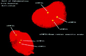
Plate 1 FISH showing Trisomy 21 (Down syndrome).
(Reproduced with kind permission from the Department of Cytogenetics, Nottingham City Hospital NHS Trust.) (See text p 305.)
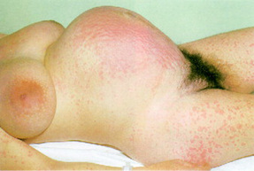
Plate 2 Pemphigoid gestationis (herpes gestationis). The generalised rash appeared at 34 weeks and responded to corticosteroids. This skin disease is rare (1 in 5-10,000 pregnancies), unique to pregnancy and of unknown aetiology. It resolves soon after delivery and may recur in successive pregnancies. Because of the increased risk of intrauterine death, fetal condition must be carefully assessed, and labour induced when the fetus is mature.
(Reproduced with kind permission from Beischer, Mackay & Colditz 1997). (See text p 351.)
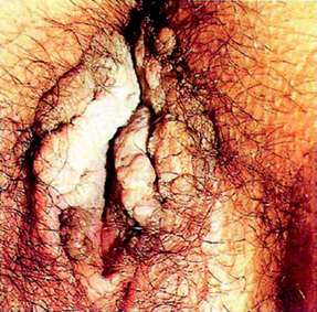
Plate 3 Massive warts in pregnancy.
(Reproduced with kind permission from Blackwell Publishing, from Adler et al 2004.) (See text p 424.)
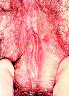
Plate 4 Primary chancre of vulva.
(Reproduced with kind permission from Blackwell Publishing, from Adler et al 2004.) (See text p 423.)
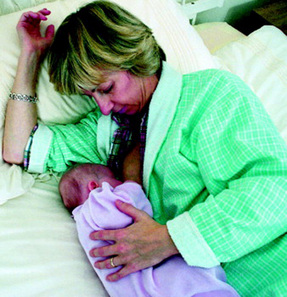
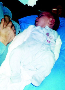
Plate 5 Mother feeding lying down.
(Reproduced with kind permission from the Health Education Board for Scotland.) (See text p 794.)
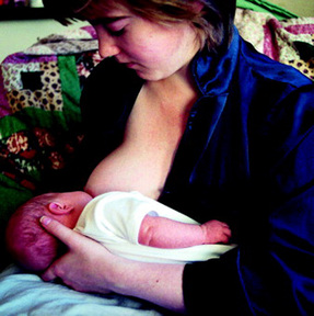
Plate 6 Mother feeding sitting up.
(Reproduced with kind permission from the Health Education Board for Scotland.) (See text p 794.)
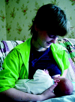
Plate 7 Mother supporting the baby’s head with her fingers.
(Reproduced with kind permission from the Health Education Board for Scotland.) (See text p 795.)
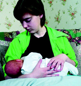
Plate 8 The baby’s head is supported by the mother’s forearm.
(Reproduced with kind permission from the Health Education Board for Scotland.) (See text p 795.)
(Photo courtesy of the Health Education Board for Scotland and Mark-it TV www.markittelevision.com.) (See text p 795.)
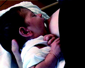
Plate 11 The baby forms a ‘teat’ from the breast and nipple.
(Photo courtesy of the Health Education Board for Scotland and Mark-it TV www.markittelevision.com.) (See text p 795.)
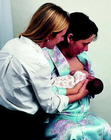
Plate 12 The midwife is kneeling by the mother to help her attach to the baby.
(Reproduced with kind permission of Nancy Durrel-McKenna.) (See text p 796.)

Plate 14 The colour of vaginal loss reported by women for the first 28 days postpartum.
(Reproduced with kind permission from Midwifery 1995 15:80.) (See text p 657.)

Plate 15 Stool colour comparator.
(Reproduced with kind permission from the Midwifery Department, Ninewells Hospital, Dundee, UK.) (See text p 798.)
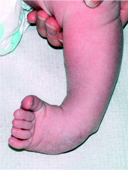
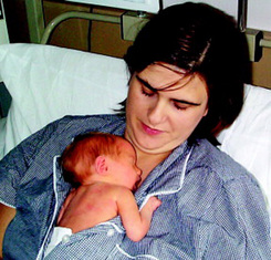
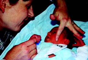
Plate 21 A jaundiced baby with its mother. Note the contrast in colour between the jaundiced skin of the baby and the mother’s unjaundiced skin.
(See text p 902.)
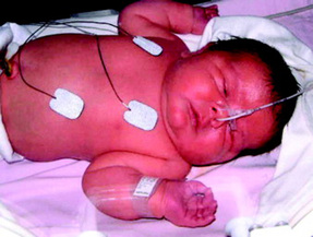
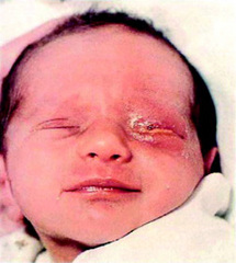
Plate 23 Ophthalmia neonatorum.
(Reproduced with kind permission from Blackwell Publishing, from Adler et al 2004.) (See text p 421.)
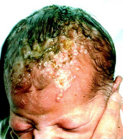

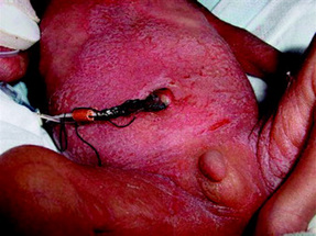
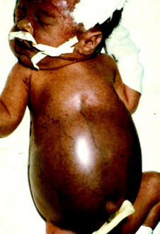

Plate 28 Forceps abrasion on cheek.
(Reproduced by permission from Thomas and Harvey 1997.) (See text p 860.)
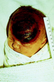
Plate 29 Scalp abrasion during vacuum-assisted birth; note the chignon.
(Reproduced by permission from Thomas and Harvey 1997.) (See text p 860.)
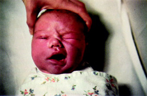
Plate 30 Right-sided facial palsy. Note that the eye is open on the paralysed side and the mouth is drawn over to the non-paralysed side.
(Reproduced by permission from Thomas and Harvey 1997.) (See text p 861.)
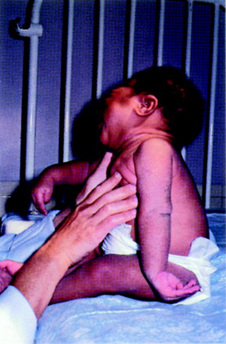
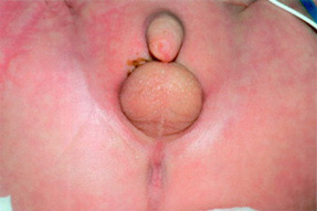
Plate 32 Imperforate anus with recto-vesical fistula (1).
(Reproduced with permission of Donna Bain.) (See text p 885.)
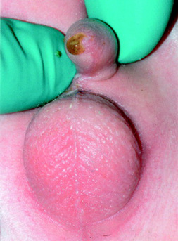
Plate 33 Imperforate anus with recto-vesical fistula (2).
(Reproduced with permission of Donna Bain.) (See text p 885.)
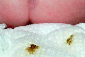
Plate 34 Imperforate anus with recto-vesical fistula and napkin containing meconium stained urine.
(Reproduced with permission of Donna Bain.) (See text p 885.)
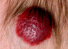
Plate 35 Evolving capillary haemangioma.
(Reproduced with permission of Sharon Murphy.) (See text p 896.)
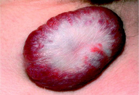
Plate 36 Regressing capillary haemangioma with typical pallor.
(Reproduced with permission of Sharon Murphy.) (See text p 896.)