Diagnostic Evaluation

The diagnosis of MCNS in children is based on the history and clinical manifestations (edema, proteinuria, hypoalbuminemia, and hypercholesterolemia in the absence of significant hematuria and hypertension) (Ruth, Kemper, Leumann, et al, 2005). Massive proteinuria is reflected in urinary excretion of protein that often reaches levels in excess of 2 g/m2 of body surface/day, with relatively greater clearance of low-molecular-weight proteins. Hyaline casts from high protein levels and sluggish flow and oval fat bodies, as well as a few red blood cells, can be found in the urine of most affected children, although there is seldom gross hematuria. Specific gravity is high and proportionate to the amount of protein concentration. If hypovolemia is not significant and the child is well hydrated, the glomerular filtration rate is usually normal.

Total serum protein concentrations are reduced, with the albumin fractions significantly reduced (<2 g/dl) and plasma lipids elevated. Serum cholesterol may be as high as 450 to 1500 mg/dl. Hemoglobin and hematocrit are usually normal or elevated, and the platelet count is high (500,000 to 1,000,000/mm3) as a result of hemoconcentration. Serum sodium concentration is usually low, approximately 130 to 135 mEq/L.

If renal biopsy is performed, it provides information regarding the glomerular status and type of nephrotic syndrome, the likely response to drugs, and the probable course of the disease. Under the microscope the foot processes of the basement membrane appear fused. The major focuses in differential diagnosis are to establish the edema as renal in origin and to distinguish MCNS from other glomerulopathies with nephrotic syndrome as a manifestation.

Therapeutic Management

Medical management consists of both general and specific measures. The primary objective is to reduce the excretion of urinary protein and maintain protein-free urine. Additional objectives include prevention or treatment of acute infection, control of edema, establishment of good nutrition, and readjustment of any disturbed metabolic processes. Children with severe symptoms may be hospitalized for assessment and observation for evidence of infection, response to therapy, and parental education.

General Measures: General treatment is principally supportive. During the edema phase the child is often limited to quiet activities, but activity is not restricted during remission. Children can be remarkably active; there is no evidence that restriction affects the ultimate outcome. Acute and intercurrent infections are treated with appropriate antibiotics, and providers make efforts to minimize the risk of infection.

Diet: The child in remission maintains a regular diet. However, salt is restricted during periods of massive edema and while on corticosteroid therapy; no salt is added at the table, and foods with very high salt content are excluded. The child tolerates this diet for a time, but it should be adjusted to the child’s appetite and must not interfere with nutrient intake. Although a low-sodium diet will not remove edema, its rate of increase may be reduced. Water is seldom restricted. A diet generous in protein is logical, but there is no evidence that it is beneficial or alters the outcome of the disease. The presence of azotemia and renal failure is a contraindication for high-protein intake.

Corticosteroid Therapy: The response of most affected children to corticosteroids has established these drugs as prime therapeutic agents in the management of nephrotic syndrome (Kim, Bellew, Silverstein, et al, 2005). Corticosteroid therapy begins as soon as the diagnosis has been determined and is administered orally in a dosage of 60 mg/m2/day in evenly divided doses. Prednisone, the safest and least expensive drug, is the steroid of choice. The drug is continued daily for 6 weeks, then reduced to 40 mg/m2 on alternate days for 6 more weeks. Studies suggest that the duration of steroid treatment for the initial episode should be at least 3 months (Hodson, Knight, Willis, et al, 2004).

The course of the disease is fairly predictable. There is little change during the first few days of therapy. In most patients diuresis occurs as the urinary protein excretion diminishes within 7 to 21 days after the initiation of steroid therapy. Other clinical manifestations stabilize or return to normal shortly thereafter. Almost 95% of patients between 1 and 10 years of age who have satisfactory laboratory measurements of C3 complement and a renal clearance of immunoglobulin G, as well as no hypertension, hematuria, or renal insufficiency, will have complete resolution of proteinuria with therapy. If the child has not responded to therapy within 28 days of daily steroid administration, the likelihood of subsequent response diminishes rapidly.

Children with MCNS are often described according to their response to corticosteroid therapy (Box 30-4). Children with MCNS typically relapse one to three times per year. Steroid-dependent children tend to have frequent relapses over many years and receive large amounts of steroids, which results in cushingoid features and may cause growth retardation. They also require supportive treatment (diuretics, diet). Steroid-resistant children are thought to have a high risk of developing chronic renal failure (CRF) (Kim, Bellew, Silverstein, et al, 2005).

BOX 30-4

CLASSIFICATION OF NEPHROTIC SYNDROME ACCORDING TO STEROID RESPONSE

Steroid sensitive—Responds to steroids; relapses may occur following illness

Frequent relapse—Two or more relapses within 6 months of initial response; four or more relapses within a 12-month period

Steroid dependent—Two consecutive relapses while on steroid therapy or within 2 weeks of steroid cessation

Steroid unresponsive or steroid resistant—Does not enter remission after 4 weeks of prednisone therapy

Modified from Bagga A, Mantan M: Nephrotic syndrome in children, Indian J Med Res 122:13-28, 2005.

image DRUG ALERT

Steroids

Children who require frequent courses of steroid therapy are highly susceptible to complications of steroids, such as growth retardation, behavior changes, cataracts, increased appetite, obesity, hypertension, gastrointestinal bleeding, bone demineralization, infections, and hyperglycemia. Children who do not respond to steroid therapy, those who have frequent relapses, and those in whom the side effects threaten their growth and general health may be considered for a course of therapy using other immunosuppressant medications.

Immunosuppressant Therapy: It is possible to reduce the relapse rate and induce long-term remission in children with frequent relapsing or steroid-resistant nephrotic syndrome with administration of an oral alkylating agent, usually cyclophosphamide (Cytoxan) or chlorambucil. Prolonged courses of cyclosporin and levamisole reduce the risk of relapse in children compared with corticosteroids alone (Durkan, Hodson, Willis, et al, 2005). The drugs share many characteristics, and the response to oral alkylating agents appears to depend on dose, duration of therapy, age, and duration of the disease.

The nurse must consider significant side effects of cyclophosphamide and discuss them with parents of children for whom this drug is contemplated. Anticipate leukopenia, and remember that cyclophosphamide may cause azoospermia with potential sterility in males treated for more than 2 to 3 months and may cause variable effects on gonadal function in females.

Diuretics: One characteristic of the edema of nephrotic syndrome is its usual lack of responsiveness to diuretic agents. However, loop diuretics, usually furosemide in combination with metolazone, are sometimes useful in cases in which edema interferes with respiration or there is hypotension, hyponatremia, or skin breakdown. In addition, plasma expanders such as salt-poor human albumin may be administered to severely edematous children requiring prompt control; however, they must be administered frequently because the glomeruli are readily permeable to albumin in the acute stage.

Prognosis: The prognosis for ultimate recovery in most cases is good. MCNS is a self-limiting disease, and in children who respond to steroid therapy the tendency to relapse decreases with time. With early detection and prompt implementation of therapy to eradicate proteinuria, progressive basement membrane damage is minimized so that renal function is usually normal or near normal when the tendency for relapses is past. It is estimated that approximately 80% of affected children have this favorable prognosis, although half the children have relapses even after 5 years, and 20% after 10 years (Bagga and Mantan, 2005).

QUALITY PATIENT OUTCOMES

Nephrotic Syndrome

• Protein-free urine

• Acute infections prevented

• Edema absent or minimal

• Nutrition maintained

• Metabolic abnormalities controlled

Nursing Care Management

Daily monitoring of intake and output is an important nursing function. Strict and accurate measurement is essential but may be difficult in very young children. In these cases the nurse can measure for output by methods such as weighing diapers. Other methods of monitoring progress include examination of the urine for albumin, daily weight, and measurement of abdominal girth. Assessment of edema such as increased or decreased swelling around the eyes and dependent areas, the degree of pitting (if noted), and the color and texture of the skin are part of nursing care. The nurse monitors vital signs to detect any early signs of complications such as shock or an infectious process.

In children hospitalized with MCNS, elevating edematous parts may be helpful to shift fluid to more comfortable distributions, but diuresis with medications and salt and water restriction to remove edema fluid are the best therapy. Areas that are particularly edematous, such as the scrotum, abdomen, and legs, may require support. Clean skin surfaces and separate them with clothing, cotton, or antiseptic powder to prevent intertrigo.

Because these children are particularly vulnerable to upper respiratory tract infection, protect them from contact with infected roommates, family, or visitors. Spontaneous peritonitis can occur secondary to migration of intestinal bacteria across the bowel wall and into the peritoneum. Monitor vital signs to detect any early signs of an infectious process.

Loss of appetite that accompanies active nephrosis creates a perplexing problem for nurses. During this time the combined efforts of the nurse, dietitian, parents, and child are necessary to formulate a nutritionally adequate and attractive diet. Salt and fluids are restricted during the edema phase. Make every effort to serve attractive meals with a minimum of fuss, but it usually requires a little creativity to get the child to eat. Games, rewards, and special treats often help, but each child is unique; trial and error may be necessary to arrive at a successful strategy. Also, the same strategy may not work consistently. (See Feeding the Sick Child, Chapter 27.)

As the edema subsides, children are allowed increased fluids. Suitable recreational and diversional activities are also an important part of their care. Once the edema fluid has been lost, children resume their usual activities without problems. Irritability and mood swings accompanying the disease process and steroid therapy are not unusual manifestations in these children and create an additional challenge to the nurse and the family.

Family Support and Home Care: Most children are treated at home during relapses. Teach parents to detect signs of relapse and to notify the health care provider if they occur. Home care is preferred unless the edema and proteinuria are severe. Instruct parents in urine testing for albumin, administration of medications, and general care. Urine is usually tested daily while the child is receiving medicine for nephrotic syndrome or if the child has an illness, and twice a week during remission. Salt is restricted to no additional salt during relapse and steroid therapy, but a regular diet is suitable for the child in remission. Instruct parents regarding avoiding contact with infected playmates, but the child is permitted to attend school. It is important for parents of children on corticosteroid therapy to be aware of the common side effects of steroid therapy (e.g., rounding of the face, increased appetite, behavior changes, abdominal distention, and hirsutism) and to distinguish some of these from the edema formation of the disease. Reassure parents that the symptoms will disappear gradually after discontinuation of the drug. The child should receive close medical or nursing observation to detect unusual but more serious side effects (see Critical Thinking Exercise).

image CRITICAL THINKING EXERCISE

Nephrotic Syndrome

Reese is an 8-year-old boy with relapsing nephrotic syndrome who has become steroid dependent. During your initial assessment in the outpatient clinic you identify the following: (1) weight has increased 2 kg (4.4 lb) in the past 2 weeks; (2) blood pressure is 100/70 mm Hg; (3) mother reports that Reese is not urinating very much, and she does not know how much he has been drinking; (4) while you are measuring Reese’s abdominal girth, he guards his abdomen and complains of stomachache; and (5) his temperature is 38° C (100.4° F) orally. You should first do which of the following correct actions?

1. Examine Reese’s abdomen while eliciting a thorough history of illness symptoms for the past 24 hours from his mother.

2. Elicit a 24-hour recall of food and fluid intake from Reese and his mother together.

3. Obtain a clean-catch urine specimen. Divide the specimen so that you can perform a dipstick analysis immediately and retain the rest of the specimen for possible urinalysis and culture after consultation with the primary health practitioner.

4. Explore the mother’s understanding of Reese’s illness and its relationship to his current condition to begin outlining your teaching plan for this family.

Questions

1. Evidence—Are there sufficient data to support your answer?

2. Assumptions—Describe some underlying assumptions about the following:

a. Peritonitis in children with nephrotic syndrome

b. Infection in a child on corticosteroid therapy

3. What are the priorities for discharge planning at this time?

4. Does the evidence support your conclusion?

The prolonged course of the relapsing form of nephrotic syndrome is taxing to both the child and the family. In the worst cases of frequent remissions and exacerbations with periodic disruption of family life by hospitalization, a severe strain is placed on the child and the family, both psychologically and financially. Parents of children with frequent relapses poorly responsive to medications need reassurance regarding this characteristic of the disease so they do not become discouraged. At the same time, impress on them the importance of long-term care to gain their cooperation. A satisfactory response is more likely when relapses are detected and therapy is instituted early, and remissions are prolonged when instructions are carried out faithfully. For example, one child had an exacerbation when his mother reduced the dosage of his drug because it was so expensive.

Social isolation is a problem for these children. Isolation is related to frequent hospitalization or confinement during relapse, the risk of infection that may precipitate an exacerbation, lack of energy, and the child’s reluctance to face friends at home or school because of the changes in appearance resulting from the disease or the medication. Both parents and child need someone to listen to their complaints, to assist them in coping with both short- and long-term problems associated with the disease, and to find solutions to their problems. Continuous support of the child and family is one of the major nursing considerations.

Renal Tubular Disorders

Disorders of renal tubular function include a variety of conditions involving one or more abnormalities in specific mechanisms of tubular transport or reabsorption, although initially glomerular function is normal or comparatively less impaired (Roth and Chan, 2001). Eventually more widespread kidney destruction with renal failure may occur. In some cases the dysfunction has little, if any, effect on renal function. These disorders may be permanent or transient and may originate as primary defects or arise as a secondary effect of metabolic disease or exogenous toxins. Renal tubular disorders may be congenital (usually displaying characteristic patterns of genetic transmission), appear without evidence of hereditary transmission, or be acquired as a result of known or unknown causes.

Unlike the classic manifestations of glomerular diseases, edema and hypertension are absent and the BUN level and routine urinalysis are usually normal. Tubular proteinuria may be demonstrated. Manifestations of tubular disorders are primarily metabolic disturbances or deficiencies, such as failure to thrive, metabolic bone disease, or persistent acidosis. The variety of these disorders is extensive, and the incidence is rare.

Tubular Function

The function of the proximal tubules is the reabsorption of substances from the glomerular filtrate, including sodium, potassium, chloride, bicarbonate, glucose, phosphate, and amino acids (Roth and Chan, 2001). A number of disorders feature impairment of reabsorption of one or more filtrate constituents, and most involve defects in the transport mechanisms for these substances. Impaired tubular reabsorption of any specific substance causes that substance to appear in the urine, sometimes with reduced levels in the blood. Examples include bicarbonate and phosphate.

The primary functions of the distal renal tubules are acidification of urine; potassium secretion; and selective and differential reabsorption of sodium, chloride, and water, which determines the final urinary concentration. Because the contribution of the distal tubule to urine composition depends in part on the volume and composition of the filtrate from the proximal tubule, the net contribution of the distal tubule is related to proximal tubular function and glomerular filtration.

Renal Tubular Acidosis

Renal tubular acidosis is a syndrome of sustained metabolic acidosis in which there is impaired reabsorption of bicarbonate or excretion of net hydrogen ion but in which glomerular function is normal or comparatively less impaired. On the basis of underlying pathophysiology, renal tubular acidosis is divided into proximal renal tubular acidosis and distal renal tubular acidosis. Proximal renal tubular acidosis results from a defect in bicarbonate reabsorption, whereas distal renal tubular acidosis results from inability to establish an adequate gradient of pH between blood and tubular fluid.

Proximal Tubular Acidosis (Type II)

Impaired bicarbonate reabsorption in the proximal tubule causes proximal tubular acidosis. It may occur as an isolated defect (primary); however, more often it appears in association with other proximal tubular disorders (secondary). As a result of a depressed renal threshold, bicarbonate reabsorption in the proximal tubule is incomplete, causing the plasma concentration of bicarbonate to stabilize at a lower level than normal. This results in a hyperchloremic metabolic acidosis. There is no impairment of distal tubular integrity or, in most cases, of the distal acidifying mechanism.

A more complex abnormality in the proximal tubules is Fanconi syndrome, in which transport mechanisms are damaged by the accumulation of toxic metabolites or the tubular epithelium is damaged by heavy metals such as lead, cadmium, or platinum. Fanconi syndrome can be part of a number of hereditary diseases, be acquired, or be idiopathic (with a cause that is not identifiable). The major clinical manifestation and presenting symptom of Fanconi syndrome is growth failure. Tachypnea from hyperchloremic metabolic acidosis is also evident. Dehydration, vomiting, episodic fever, nephrolithiasis secondary to hypercalciuria, muscle weakness or paralysis as a result of hypokalemia, and episodes of severe life-threatening acidemia (sometimes triggered by a concurrent infection) may also be seen. Complications are rare. The disorder appears to be transient and resolves spontaneously in time.

Distal Tubular Acidosis (Type I)

Distal tubular acidosis is caused by the kidney’s inability to establish a normal pH gradient between tubular cells and tubular contents. Its most characteristic feature is the inability to produce a urinary pH below 6.0 despite the presence of severe metabolic acidosis (Watanabe, 2005).

Distal renal tubular acidosis may occur as a primary, isolated defect or in association with other diseases or disorders. Most secondary causes are rare. The primary disorder is usually considered to be a hereditary defect with a variable degree of expression and a greater penetrance in females. After the age of 2 years the child usually has growth failure, often with a history of vomiting, polyuria, dehydration, anorexia, and failure to thrive. Evidence of bone demineralization may be present, along with the occasional formation of urinary calculi (urolithiasis) in older children.

The inability to secrete hydrogen ions causes an accumulation of the ions in the body, which soon depletes the available hydrogen buffer and produces a sustained acidosis. Acidosis slows normal somatic growth, and demineralization of bone occurs as bone salts are mobilized to buffer the excessive hydrogen ions. Increased serum levels of both calcium and phosphorus contribute to the development of stones within the renal system. Both sodium and potassium are secreted in larger amounts. Serum potassium levels are depleted as the distal tubules excrete large amounts of potassium ions in an attempt to conserve sodium because hydrogen ions are unable to participate in the exchange. Hyponatremia stimulates increased aldosterone secretion, which further aggravates the hypokalemia. With the depletion of bicarbonate ions, more chloride is reabsorbed in the proximal tubule to create a hyperchloremia.

Prognosis: The primary disorder is usually permanent. However, secondary effects on growth and stone formation can be avoided with early diagnosis and therapy. When the disorder occurs as a secondary complication and renal damage is prevented, the prognosis is good.

Therapeutic Management

Treatment of both proximal and distal disorders consists of the administration of sufficient bicarbonate or citrate to balance metabolically produced hydrogen ions; to maintain the plasma bicarbonate level within normal range; and to correct associated electrolyte disorders, especially hypokalemia. Proximal disorders require large volumes of bicarbonate to compensate for urinary losses; in distal disorders the alkali required to maintain a normal plasma concentration is low. Most authorities favor a mixture of sodium and potassium bicarbonate (or citrate) to prevent deficiencies of either cation. The citrate solutions (Bicitra, Polycitra, or Shohl solution) are usually more easily tolerated than bicarbonate solutions. Shohl solution is effective but has the disadvantage of requiring preparation by a pharmacist.

Nursing Care Management

Nursing goals include recognizing the possibility of renal tubular acidosis in children who fail to thrive or who display other symptoms suggestive of the disorders and referring these children for medical evaluation. Helping parents understand the importance of adhering to the medication plan as a long-term goal is essential. (See Compliance and Administration of Medication, Chapter 27.) Children who must continue the medication indefinitely need to learn the importance of taking the medications as soon as they are old enough to assume responsibility for their own care.

Nephrogenic Diabetes Insipidus

Nephrogenic diabetes insipidus (NDI) is the major disorder associated with a defect in the ability to concentrate urine. In this disorder the distal tubules and collecting ducts are insensitive to the action of antidiuretic hormone or its exogenous counterpart, vasopressin. Although several inheritance patterns have been identified, more than 90% of patients have an X-linked defect of the vasopressin receptor (Knoers and Monnens, 2004). The disease is more variable in female carriers of the defective gene, who may exhibit only a mild defect in urine-concentrating ability. The differential diagnosis for NDI should include chronic obstructive renal disorders, sickle cell disease, renal tuberculosis, and other renal disorders that may cause high urinary output with failure of the kidney to respond to vasopressin.

Clinical Manifestations and Diagnostic Evaluation

NDI is manifested in the newborn period by vomiting, unexplained fever, failure to thrive, and severe recurrent dehydration with hypernatremia. The passage of copious amounts of dilute urine, which produces severe dehydration and hypoelectrolytemia, is a serious threat to life during this period and may be responsible for the high incidence of cognitive impairment and motor retardation found in affected persons. Growth retardation is probably related to diminished food intake and poor general health because of uncontrolled polydipsia. Diagnosis is suspected on the basis of the patient and family history and confirmed by a urine osmolality value consistently below that of plasma. Lack of response to vasopressin administration rules out other causes.

Therapeutic Management

Therapy involves provision of adequate volumes of water to compensate for urinary losses and minimize urine output through diet and medication (Dell and Avner, 2007). As a result of an insatiable thirst, most of the child’s time is spent drinking and voiding, with little time for activity and stimulation. These children may go to great lengths to satisfy their thirst. A low-sodium, low-solute diet and the use of chlorothiazide or ethacrynic acid diuretics to increase the reabsorption of sodium and water in the proximal tubule help to reduce the amount of tubular fluid delivered to the distal tubules and to diminish the volume of water excreted. Urinary output may be reduced when nonsteroidal antiinflammatory drugs (NSAIDs) are administered in conjunction with chlorothiazide. Supplemental potassium may be required to prevent hypokalemia as a result of thiazide therapy. Normal growth and a normal life span are possible if the disease is recognized early and treatment is instituted and maintained.

Nursing Care Management

Nursing goals for children with NDI and their families are to recognize signs of the disorder early and assist them in coping with the long-term inconvenience of the continual thirst and elimination problems. Families need to learn to administer medications and help with diet planning for those on sodium restriction and needing supplemental potassium. The problem of ensuring adequate hydration is lifelong, and families need to adapt to away-from-home fluid needs and avoid activities that contribute to dehydration when fluids may not be available. Genetic counseling is recommended.

Miscellaneous Renal Disorders

Hemolytic Uremic Syndrome

Hemolytic uremic syndrome (HUS) is an acute renal disease characterized by a triad of manifestations: ARF, hemolytic anemia, and thrombocytopenia (Caprioli, Peng, and Remuzzi, 2005). HUS occurs primarily in infants and small children between the ages of 6 months and 3 years. It has been recognized predominantly in Caucasians and, although it occurs worldwide, is more prevalent in South Africa, Argentina, and the west coasts of North and South America. HUS represents one of the main causes of ARF in early childhood (Kliegman, Behrman, Jenson, et al, 2007).

Etiology

In the majority of cases of HUS no causative agents have been identified, although recent theories implicate genetic factors, prostacyclin deficiency, neuraminidase and agglutination, endotoxins (especially Shigella endotoxin), antithrombin III deficiency, deficiency of antioxidants, and reduced platelet aggregation. The appearance of the disease has been associated with Rickettsia organisms; viruses such as coxsackievirus, echovirus, and adenovirus; E. coli; pneumococci; Shigella organisms; and Salmonella organisms and may represent an unusual response to these infections. HUS caused by enteric infection of the E. coli O157:H7 serotype is the most prevalent pathogen in the United States and Europe, with about 70,000 cases and 60 deaths occurring annually (Bell, Griffin, Lozano, et al, 1997; Caprioli, Peng, and Remuzzi, 2005). Occurrences have been traced to undercooked meat, especially ground beef; unpasteurized apple juice; alfalfa sprouts; and public pools.

The disease usually follows an acute gastrointestinal or upper respiratory tract infection and tends to occur in scattered outbreaks in small geographic areas. HUS is clinically and pathologically similar to thrombocytopenic purpura, except for the hypertension associated with HUS.

Pathophysiology

The primary site of injury appears to be the endothelial lining of the small glomerular arterioles, but other organs and tissues may be involved (e.g., the liver, brain, heart, pancreatic islet cells, and muscles). The endothelium becomes swollen and occluded with the deposition of platelets and fibrin clots (intravascular coagulation). Red blood cells are damaged as they move through the partially occluded blood vessels. The spleen removes these fragmented red blood cells, causing acute hemolytic anemia. Fibrinolytic action on the precipitated fibrin causes these fibrin-split products to appear in the serum and urine. The characteristic thrombocytopenia is produced by the platelet aggregation within damaged blood vessels or the damage and removal of platelets.

Clinical Manifestations

The disease occurs after a prodromal period during which there is an episode of diarrhea and vomiting. Less often the preceding illness is an upper respiratory tract infection or, occasionally, varicella, measles, or a UTI.

The hemolytic process persists for several days to 2 weeks. During this time the child is anorexic, irritable, and lethargic. There is marked and rapid onset of pallor accompanied by hemorrhagic manifestations such as bruising, purpura, or rectal bleeding. Severely affected patients are anuric and often hypertensive. Seizures and stupor suggest central nervous system involvement, and there may be signs of acute heart failure. Mild cases demonstrate anemia, thrombocytopenia, and azotemia; urinary output may be reduced or increased.

Diagnostic Evaluation

The triad of anemia, thrombocytopenia, and renal failure is sufficient for diagnosis. Proteinuria, hematuria, and urinary casts are evidence of renal involvement; BUN and serum creatinine levels are elevated. A low hemoglobin and hematocrit and a high reticulocyte count confirm the hemolytic nature of the anemia.

Therapeutic Management

In general, providers direct treatment toward control of the complications and hematologic manifestations of renal failure (Siegler and Oakes, 2005). The initial supportive measures for most children are those used in managing renal failure: fluid replacement (calculated with great care), treatment of hypertension, and correction of acidosis and electrolyte disorders (Siegler and Oakes, 2005). The most consistently effective treatment is early hemodialysis, PD, or continuous hemofiltration, which is instituted in any child who has been anuric for 24 hours or who demonstrates oliguria with uremia or hypertension and seizures. Blood transfusions with fresh, washed packed cells are administered for severe anemia but are used with caution to prevent circulatory overload from added volume.

Once vomiting and diarrhea have resolved, the child is restarted on enteral nutrition. Sometimes parenteral nutrition is required for children with severe, persistent colitis and for those in whom tissue catabolism is marked. There is no substantial evidence that heparin, corticosteroids, or fibrinolytic agents are beneficial, and in some instances they may aggravate the condition. The usefulness of plasma infusion for treatment of HUS is currently being studied; it may be useful in selected cases.

Prognosis: With prompt treatment the recovery rate is approximately 95%, but residual renal impairment ranges from 10% to 50%. Death is usually caused by residual renal impairment or central nervous system injury.

Nursing Care Management

Nursing care is the same as that provided in ARF and, for children with continued impairment, includes management of chronic disease. Because of the sudden and life-threatening nature of the disorder in a previously well child, parents are often ill prepared for the impact of hospitalization and treatment. Therefore support and understanding are especially important aspects of care.

image NURSING ALERT

To prevent infection from contaminated meat, the internal temperature of the food, such as hamburger, should be at least 74° C (165° F). Cooking the ground beef until no pink color is seen may not be sufficient to kill the bacteria. Therefore a meat thermometer is needed to ensure a safe product. Discourage parents from giving children unpasteurized apple juice and unwashed raw vegetables. Also discourage the use of antimotility drugs for diarrhea.

Familial Nephritis (Alport Syndrome)

The syndrome of chronic hereditary glomerulopathy consists of hematuria, high-frequency sensorineural deafness, ocular disorders, and CRF (Anker, Amemann, and Neumann, 2003). The disease appears to be inherited as an autosomal dominant trait, which suggests a possible X-linked dominant trait, although rare male-to-male transmission does occur. Alport syndrome is uncommon but not rare and accounts for a significant percentage of persistent glomerular disease in childhood.

The clinical manifestations are indistinguishable from those of mild acute nephritis. Initial symptoms include hematuria, proteinuria, malaise, and mild edema. Onset of gross hematuria may be associated with an acute respiratory tract infection. The average age of onset is 6 years, but the condition may be noted in infancy. It begins slowly and progresses until uncontrollable renal failure develops in adolescence or early adulthood. There is usually a positive family history. Most untreated boys develop severe symptoms, whereas affected girls generally have a milder disease and a normal life expectancy.

Treatment is symptomatic and supportive. Dialysis and kidney transplantation are ultimate therapeutic measures for ESRD. Hearing loss and ocular disorders should receive appropriate attention, and families should be counseled regarding the genetic implications of the disease.

Unexplained Proteinuria

Often apparently healthy children with no suggestion of renal disease demonstrate proteinuria on routine urinalysis. The percentage of children with unexplained proteinuria ranges from 1% at 6 years of age to 11% at puberty, reaching a maximum prevalence at age 13 in girls and age 16 in boys.

Unexplained proteinuria can be categorized as (1) transient (inconstant); (2) persistent; or (3) orthostatic, or postural. Transient proteinuria is a common finding with no known cause but sometimes increases with febrile illness, exercise, cold, or emotions. Persistent proteinuria usually signifies renal disease. Orthostatic proteinuria is seen in 3% to 5% of adolescents and young adults; although proteinuria is evident in both the recumbent and the erect position, it is quantitatively and qualitatively greater in the erect position. The cause is unknown, but minor glomerular changes occur in many instances. The condition is benign and generally resolves over time.

In cases of unexplained proteinuria, it is important to confirm or exclude renal disease with appropriate diagnostic tests. Repeated examination for proteinuria, an orthostatic test, urine culture, and (if proteinuria is persistent) more definitive tests—including 24-hour protein excretion, renal ultrasound, and renal scan—are indicated.

Renal Trauma

Serious injuries of the genitourinary tract are not uncommon in the pediatric age-group, with a peak incidence between ages 10 and 20 years. Despite their relatively protected location, the kidneys are among the organs most often injured in children. However, the kidneys in children are more mobile than they are in adults, and the outer borders are less well protected. They are separated from the skin surface by only 2 to 3 cm (0.75 to 1.2 inches) in young children.

Most injuries are of the nonpenetrating or blunt type and usually involve falls, athletic injuries, and motor vehicle accidents. Cycling is the most common cause of sports-related kidney injury, causing more than three times more kidney injuries than football (Grinsell, Showalter, Gordon, et al, 2006). Penetrating trauma (e.g., gunshot or stab wound) is much less common in children. Many children have preexisting renal abnormalities, particularly congenital anomalies associated with mild to moderate hydronephrosis, that were unrecognized before the injury.

Suspect renal injury in children who complain of flank pain, and often have abrasions or contusions on the overlying skin. Hematuria is consistently present, but the amount of blood in the urine is not a reliable indicator of the seriousness of the injury. Many relatively insignificant injuries are associated with grossly bloody urine, whereas some of the most severe injuries are found in children with only microscopic hematuria (Box 30-5).

BOX 30-5

CLASSIFICATION OF RENAL INJURY

Grade I—Renal contusion or subcapsular hematoma; gross or microscopic hematuria

Grade II—Parenchymal laceration <1.0 cm deep; nonexpanding perirenal hematoma

Grade III—Parenchymal laceration >1.0 cm deep; no urinary extravasation

Grade IV—Laceration extending from parenchyma into collecting system and/or injury to main renal artery or vein

Grade V—Shattered kidney; thrombosis of main renal artery

Modified from Moore EE, Shackford SR, Pachter HL, et al: Organ injury scaling: spleen, liver, and kidney, J Trauma 29(12):1664-1666, 1989.

Renal rupture involves the actual splitting open of the kidney capsule, causing extravasation of blood or a mixture of blood and urine into the surrounding retroperitoneal space. Renal vascular injury, although unusual, requires immediate recognition and surgical intervention. Because the volume-per-minute blood flow through the kidney is greater (25% of cardiac output) than to any other abdominal organ, injury to the kidney may result in a rapid loss of blood.

Active children may or may not have a history of unusual trauma. Abdominal or flank pain and tenderness are caused by bleeding around the kidney and may or may not be associated with fever. Clots passing down the ureter may cause pain similar to that of renal colic, and dysuria is common. Patients with more severe injuries may complain of nausea or abdominal pain. There may be a palpable abdominal mass caused by loss of blood or urine into the retroperitoneum. The fibrous capsule enclosing the kidney prevents expansion of a hematoma; therefore exsanguination and shock are seldom observed, even in severe renal trauma.

Diagnosis is made on the basis of intravenous pyelography, angiography, or retrograde pyelography. Unsuspected hydronephrosis often is first detected as a result of traumatic injury.

Therapeutic Management

Severe injury requires close observation in the hospital intensive care unit and blood replacement if there is severe internal or external bleeding. In most cases bleeding subsides spontaneously. Surgical exploration is indicated if there are multiple injuries, extravasation of blood around the kidneys, or disruption of the major vessels or the collecting system. Children with less severe injuries, such as contusions only, are placed on bed rest. They should remain on bed rest for 3 days after cessation of gross bleeding, since the substance released from injured renal tissue (urinary urokinase) has strongly fibrinolytic properties that may precipitate serious bleeding. The prognosis depends on the nature and extent of the injury.

Nursing Care Management

Nursing management is directed toward recognizing and assisting in the diagnosis of renal injury. Care of both the child and the family is primarily supportive. The nurse implements all the concepts related to emergency hospitalization and care. (See Chapter 26.) Postsurgical care, if indicated, is the same as for any other surgical patient. Recommendations for children with a solitary kidney participating in sports are varied. Each child should be individually assessed and advised on the need for protective equipment before engaging in contact, collision, or limited contact activities (Rice and Council on Sports Medicine and Fitness, 2008).

Renal Failure

Renal failure is the inability of the kidneys to excrete waste material, concentrate urine, and conserve electrolytes. The disorder can be acute or chronic and affects most of the systems in the body. Two terms that are often used in relation to renal failure need some clarification: azotemia is the accumulation of nitrogenous waste within the blood, whereas uremia is a more advanced condition in which retention of nitrogenous products produces toxic symptoms. Azotemia is not life threatening, whereas uremia is a serious condition that often involves other body systems.

Acute Renal Failure

image ARF is said to exist when the kidneys suddenly are unable to appropriately regulate the volume and composition of urine in response to food and fluid intake and the needs of the organism. The principal feature is oligoanuria* associated with azotemia, acidosis, and diverse electrolyte disturbances. ARF is not common in childhood, but the outcome depends on the cause, associated findings, and prompt recognition and treatment.

imageCritical Thinking Case Study—Acute Renal Failure
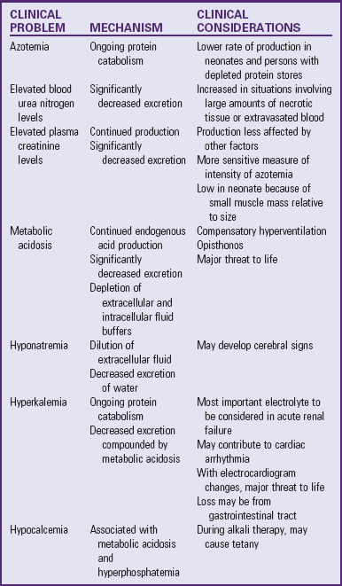
Etiology

ARF can develop as a result of a large number of related or unrelated clinical conditions: poor renal perfusion; acute renal injury; or the final expression of chronic, irreversible renal disease. The most common cause in children is transient renal failure resulting from dehydration or other causes of poor perfusion that respond to restoration of fluid volume. Causes of ARF are usually classified as prerenal, intrinsic renal, and postrenal. Severe or longstanding prerenal or postrenal causes can produce severe secondary renal damage.

Prerenal Causes: Prerenal causes of ARF are most common in children and are always related to the reduction of renal perfusion in an anatomically and physiologically normal kidney and collecting system. Dehydration secondary to diarrheal disease or persistent vomiting is the most common cause of prerenal failure in infants and children. Surgical shock and trauma (including burns) are also common causes. Hypovolemia and decreased renal perfusion cause a decreased glomerular filtration rate and stimulate the secretion of renin, aldosterone, and antidiuretic hormone, which further diminish urine flow. Extended and severe hypoperfusion (secondary to procedures such as cardiac surgery) can produce cortical or tubular necrosis; however, when medical care is available, this is seldom allowed to occur. In general, the azotemia that accompanies this type of renal failure is rapidly reversible with prompt attention to expansion of the extracellular fluid volume. Prerenal failure is often difficult to distinguish from tubular or cortical necrosis. Renal artery stenosis, altered peripheral vascular resistance related to sepsis, and hepatorenal syndrome are less common causes.

Intrinsic Renal Causes: Intrinsic renal causes of ARF constitute the largest group that requires extended management. These include diseases and nephrotoxic agents that damage the glomeruli, tubules, or renal vasculature. Glomerular disease is the most common cause of glomerular damage, whereas tubular destruction is more often caused by ischemia or nephrotoxins. Vascular damage is an uncommon cause of renal failure in childhood. The type and extent of damage determine the degree and duration of renal insufficiency, and it is difficult to predict in any given case whether acute necrosis will develop.

Postrenal Causes: ARF resulting from obstructive uropathy is uncommon in children except during the first year of life. Relief of the obstruction can restore renal function. The degree of recovery depends on the duration of the renal failure.

Pathophysiology

ARF is usually reversible, but the deviations of physiologic function can be extreme, and mortality in the pediatric age-group is still high. There is severe reduction in the glomerular filtration rate, an elevated BUN level, and decreased tubular reabsorption of sodium from the proximal tubule. Consequently, there is increased concentration of sodium in the distal tubule, which causes stimulation of the renin mechanism. The local action of angiotensin causes vasoconstriction of the afferent arteriole, which further reduces glomerular filtration and prevents urinary losses of sodium. There is a significant reduction in renal blood flow.

The pathologic conditions that produce ARF caused by glomerulonephritis, HUS, and other renal disorders are discussed in relation to those disease processes. The necrotic processes within the nephron can be cortical, tubular, or both.

Cortical Necrosis: Complete cortical necrosis usually results from severe ischemia, infection, or intravascular coagulation and represents a severe, irreversible cause of ARF. In the pediatric age-group this occurs as a fatal event, most commonly during the neonatal period as a result of hypoxia and shock. When cortical destruction is incomplete, some recovery of renal function may occur. Intravascular coagulation is believed to play a significant role as an intermediate factor in the development of ARF, especially in cases related to sepsis.

Tubular Necrosis: Damage to the renal tubules can be broadly classified as (1) secondary to renal ischemia and (2) associated with the ingestion or inhalation of substances toxic to the kidneys. Renal tubules are particularly vulnerable to a wide variety of toxic agents that produce vasoconstriction and to focal patches of ischemia that cause a uniform necrosis of the tubular epithelium down to, but not including, the basement membrane. A lesion produced by sustained reduction in renal blood flow also involves the basement membrane, which may become fragmented and ruptured to the extent that the continuity of tubular structure is disrupted. The lesions may affect any segment of the tubules, appearing at irregular intervals along with normal segments throughout the kidney.

Reepithelialization in the areas with intact basement membrane heals tubular lesions. Such healing is unable to take place in areas in which the basement membrane has been disrupted; connective tissue grows through the ruptured membrane, thus preventing reestablishment of tubular integrity. Individual cells within the nephron, but not the entire nephron, are capable of regeneration.

Clinical Course: The clinical course of the child with ARF is variable and depends on the cause. In reversible ARF there is a period of severe oliguria, or a low-output phase, followed by an abrupt onset of diuresis, or a high-output phase; this phase is followed by a gradual return to, or toward, normal urine volumes. The length of the oliguric phase in older children and adolescents is 10 to 14 days but is highly variable at all ages. It tends to be shorter (3 to 5 days) in infants, children, and milder cases. The onset of the diuretic phase appears unexpectedly, and over several days it proceeds in stepwise fashion from very low to above-normal urine volumes. During the oliguric phase, manifestations of uremia are present but may also be accompanied by other clinical disorders that make assessment difficult, such as infection, anoxia, and shock.

Clinical Manifestations

In many instances of ARF the infant or child is already critically ill with the precipitating disorder, and the explanation for development of oliguria may or may not be readily apparent. The underlying illness often overshadows the renal failure and often assumes the priority of care (e.g., the patient who is in shock from endotoxemia, the infant who is severely dehydrated from gastroenteritis, or a child who is subject to seizures as a result of hypertensive encephalopathy associated with AGN).

The prime manifestation of ARF is oliguria, generally a urinary output of less than 1 ml/kg/hr. Anuria (no urinary output in 24 hours) is uncommon except in obstructive disorders. Other symptoms related to ARF include edema, drowsiness, circulatory congestion, and cardiac arrhythmia from hyperkalemia. Seizures may be caused by hyponatremia or hypocalcemia and tachypnea from metabolic acidosis. With continued oliguria, biochemical abnormalities can develop rapidly, and circulatory and central nervous system manifestations appear.

Diagnostic Evaluation

When a previously well child develops ARF without obvious cause, a careful history is obtained to reveal symptoms that may be related to glomerulonephritis; obstructive uropathy; or exposure to nephrotoxic chemicals, such as ingestion of heavy metals or inhalation of carbon tetrachloride or other organic solvents or drugs (e.g., methicillin, sulfonamides, NSAIDs, neomycin, polymyxin, and kanamycin). Laboratory data reflect the kidney dysfunction—hyperkalemia, hyponatremia, metabolic acidosis, hypocalcemia, anemia, or azotemia (Table 30-7).

TABLE 30-7

LABORATORY FINDINGS ASSOCIATED WITH ACUTE RENAL FAILURE

image

Therapeutic Management

The most effective management of ARF is prevention. The development of ARF is a known risk in certain situations. This should be anticipated and recognized, and adequate therapy should be implemented (e.g., fluid therapy for children with hypovolemia in conditions such as dehydration, burns, and hemorrhage). Nephrotoxic drugs should be used with caution or avoided in children with renal disease, and all personnel should be knowledgeable about precautions related to their administration. For example, a generous fluid intake is needed for children receiving antimetabolite drugs and after radiotherapy.

The treatment of ARF is directed toward (1) treatment of the underlying cause, (2) management of the complications of renal failure, and (3) provision of supportive therapy within the constraints imposed by the renal failure. Treatment of poor perfusion resulting from dehydration consists of volume restoration as described in the treatment of dehydration. (See Chapter 28.) If oliguria persists after restoration of fluid volume or if the renal failure is caused by intrinsic renal damage, the physiologic and biochemical abnormalities that have resulted from kidney dysfunction must be corrected or controlled. Central venous pressure monitoring is usually implemented.

Initially a Foley catheter is inserted to rule out urine retention, to collect available urine for electrolytes and analysis, and to monitor the results of diuretic administration. The catheter may or may not be removed. Some clinicians believe that it serves little purpose during the oliguric phase and predisposes the patient to bladder infection prefer collection bags for measuring urinary output. Others maintain a catheter for hourly urine measurements.

Oliguria: When a child has persistent oliguria in the presence of adequate hydration and no lower tract obstruction, mannitol, furosemide, or both may be administered rapidly as a test to provoke a flow of urine. When glomerular function is intact, the administration of these substances will behave as nonreabsorbable solute in the tubular fluid to evoke an osmotic diuresis. The presence of mannitol in tubular fluid and the obligatory water that follows it also serve to dilute the concentration of any nephrotoxin that may be present in the tubules to below toxic levels. The furosemide blocks reabsorption of tubular filtrate. If urine flow is generated to the extent of 6 to 10 ml/kg of body weight in 1 to 3 hours, the initial dosage is reduced and continued, if needed, to sustain the flow. If no urine is produced within 2 hours after the single dose, the drugs are not repeated, and an oliguric regimen is instituted to control water balance and other abnormalities.

Fluid and Calories: The amount of exogenous water provided should not exceed the amount needed to maintain zero water balance. It is calculated on the basis of estimated endogenous water formation and losses from sensible (primarily gastrointestinal) and insensible sources. No allotment is calculated for urine as long as oliguria persists.

The child with ARF has a tendency to develop water intoxication and hyponatremia, both of which make it difficult to provide calories in sufficient amounts to meet the child’s needs and reduce tissue catabolism, metabolic acidosis, hyperkalemia, and uremia. If the child is able to tolerate oral foods, concentrated food sources high in carbohydrates and fat but low in protein, potassium, and sodium may be provided. However, many children have functional disturbances of the gastrointestinal tract, such as nausea and vomiting. Therefore the IV route is generally preferred, and nourishment usually consists of essential amino acids or a combination of essential and nonessential amino acids administered by the central venous route.

Control of water balance in these patients requires careful monitoring of feedback information, such as accurate intake and output, body weight, and electrolyte measurements. In general, during the oliguric phase, no sodium, chloride, or potassium is given unless there are other large, ongoing losses. Regular measurement of plasma electrolytes, pH, BUN, and creatinine levels is required to assess the adequacy of fluid therapy and to anticipate complications that require specific treatment.

Hyperkalemia: An elevated serum potassium level is the most immediate threat to the life of the child with ARF. Potassium ions are not being excreted, while at the same time the release of potassium from cells is accelerated by acidosis, stress, and tissue breakdown in cases associated with internal bleeding or trauma. Because cardiac arrhythmia and cardiac arrest may result, electrocardiograms (ECGs) and serum potassium ion levels are monitored regularly. Hyperkalemia can be minimized and sometimes avoided by eliminating potassium from all food and fluids, by reducing tissue catabolism, and by correcting acidosis.

image NURSING ALERT

Any of the following signs of hyperkalemia constitute an emergency and should be reported immediately:

• Serum potassium concentrations in excess of 7 mEq/L

• Presence of ECG abnormalities, such as loss of P wave, prolonged RS complex, depressed ST segment, tall and tented T waves, bradycardia, or heart block

Several measures are available to reduce the serum potassium concentration, and the priority of implementation is usually based on the rapidity with which the measures are effective. Temporary measures that produce a rapid but transient effect as follows:

• Calcium gluconate, 0.5 ml/kg, administered intravenously over 2 to 4 minutes with continuous ECG monitoring, exerts a protective effect on cardiac conduction.

• Sodium bicarbonate, 2 to 3 mEq/kg, administered intravenously over 30 to 60 minutes, elevates the serum pH to cause a transient shift of extracellular fluid potassium into the intracellular fluid. However, there is risk of hypocalcemia, tetany, and fluid overload.

• Glucose, 50%, and insulin, 1 unit/kg, administered intravenously, accelerate glycogen synthesis, causing glucose and potassium to move into the cells. Insulin facilitates the entry of glucose into cells.

These effects produce only transient protection by redistributing existing potassium stores; they do not remove potassium from the body. However, they provide relief while more definitive but slower-acting measures are being implemented. Potassium can be removed by either of two methods:

1. Administration of a cation exchange resin such as sodium polystyrene sulfonate (Kayexalate), 1 g/kg, administered orally or rectally, to bind potassium and remove it from the body. This requires time to be effective, and a sodium ion is exchanged for each potassium ion. This increased sodium concentration adds to the body fluids, which may contribute to fluid overload, hypertension, and cardiac failure.

2. Dialysis or continuous hemofiltration (see p. 1176). Hemodialysis is efficient but requires specialized facilities. PD is simpler and can be carried out in almost any hospital setting. Indications for dialysis in ARF are continued oliguria associated with any of the following:

• Severe, persistent acidosis

• Inability to reduce serum potassium levels to a safe range with other methods

• Clinical uremic syndrome consisting of nausea and vomiting, drowsiness, and progression to coma

• Circulatory overload, hypertension, and evidence of cardiac failure

A popular philosophy is to institute renal replacement therapy (see p. 1172) after 24 to 48 hours of oliguria, regardless of other symptoms. Supporters of this approach believe that early intervention is associated with reduced morbidity and mortality and permits improved nutrition with relaxed diet restrictions. The combination of renal replacement therapy and nutrition tends to reduce the complications of ARF.

Hypertension: Hypertension is a common and serious complication of ARF, and blood pressure determinations are taken every 4 to 6 hours to detect it early. The most common cause of hypertension in ARF is overexpansion of the extracellular fluid and plasma volume together with activation of the renin-angiotensin system. The goal of therapy is to prevent hypertensive encephalopathy and avoid overtaxing the cardiovascular system.

When there is a threat of encephalopathy, labetalol (a beta and alpha blocker) may be administered intravenously as bolus infusions or a continuous drip. Sodium nitroprusside may be given but requires close monitoring. For less urgent situations, hydralazine, clonidine, or verapamil may be given intravenously. Oral drugs used for acute hypertension include nifedipine, captopril, minoxidil, hydralazine, propranolol, or furosemide.

Other Complications: Other complications that may occur with ARF are anemia, seizures and coma, cardiac failure, and pulmonary edema. Anemia is commonly associated with ARF, but transfusion is not recommended unless the hemoglobin level drops below 6 g/dl. Transfusions consist of fresh, packed red blood cells given slowly to reduce the likelihood of increasing blood volume, hypertension, and hyperkalemia.

Seizures occur often when renal failure progresses to uremia and are also related to hypertension, hyponatremia, and hypocalcemia. Treatment is directed toward the specific cause when known. More obscure causes are managed with antiepileptic drugs.

Cardiac failure with pulmonary edema is almost always associated with hypervolemia. Treatment is directed toward reduction of fluid volume, with water and sodium restriction and administration of diuretics. Digitalis is ineffective and can be hazardous.

Diuretic, or High-Output, Phase: When the output begins to increase, either spontaneously or in response to diuretic therapy, monitor the intake of fluid, potassium, and sodium, and provide adequate replacement to prevent depletion and its consequences. In some cases the high-output phase is mild and lasts only a few days; in others enormous amounts of electrolyte-rich urine are passed.

Prognosis: The prognosis of ARF depends largely on the nature and severity of the causative factor or precipitating event and the promptness and competence of management. The mortality rate is less than 20%. The outcome is least favorable in children with rapidly progressive nephritis and cortical necrosis. Children in whom ARF is a result of HUS or AGN may recover completely, but residual renal impairment or hypertension is more often the rule. Complete recovery is usually expected in children whose renal failure is a result of dehydration, nephrotoxins, or ischemia. ARF following cardiac surgery has a less favorable prognosis. It is often impossible to assess the extent of recovery for several months.

QUALITY PATIENT OUTCOMES

Acute Renal Failure

• Underlying cause of ARF identified and treated

• Water balance maintained

• Hypertension controlled

• Electrolyte balance maintained

• Diet maintains calories while minimizing tissue catabolism, metabolic acidosis, hyperkalemia, and uremia

Nursing Care Management

image Nursing care of the infant or child with ARF involves addressing the underlying cause plus carefully observing and managing the renal status. The major goal is reestablishment of renal function (with emphasis on providing an adequate caloric intake to minimize reduction of protein stores); prevention of complications; and monitoring of fluid balance, laboratory data, and physical manifestations. The probability of dialysis or continuous hemofiltration is high, and the nurse must anticipate the availability of the necessary equipment. Because the child requires intensive observation and often specialized equipment, the usual disposition is admission to an intensive care unit where equipment and trained personnel are available.

imageNursing Care Plan—The Child with Acute Renal Dysfunction

The major nursing tasks in the care of the infant or child with ARF are monitoring and assessing fluid and electrolyte balance. Limiting fluid intake requires ingenuity on the part of caregivers to cope with the child who is thirsty. One strategy involves rationing the daily intake with small amounts of fluid served in containers that give the impression of larger volumes. Older children who understand the rationale of fluid limits can help determine how their daily ration should be distributed.

Meeting nutritional needs is sometimes a problem because the child may be nauseated and because getting the child to eat concentrated foods without fluids may be difficult. When nourishment is provided by the IV route, careful monitoring is essential to prevent fluid overload. This can become a major challenge in the face of nutritional requirements and administration of IV medications. The IV drugs being used may be nephrotoxic, which can require a specified volume of solution for delivery. In some instances blood products must also be delivered. Preventing fluid overload while delivering medications and calories requires concerted collaboration. In addition, nursing measures such as maintaining an optimum thermal environment, reducing any elevation of body temperature, and reducing restlessness and anxiety are used to decrease the rate of tissue catabolism.

The nurse must be continually alert for behavior changes that indicate the onset of complications. Infection from reduced resistance, anemia, and general morbidity is a constant threat. Fluid overload and electrolyte disturbances can precipitate cardiovascular complications such as hypertension and cardiac failure. Fluid and electrolyte imbalances, acidosis, and accumulation of nitrogenous waste products can produce neurologic involvement manifested by coma, seizures, or alterations in sensorium.

Although children with ARF are usually quite ill and voluntarily diminish their activity, infants may become restless and irritable, and children are often anxious and frightened. Frequent, painful, and stress-producing treatments and tests must be performed. A supportive, empathetic nurse can provide comfort and stability in a threatening and unnatural environment.

Family Support: Providing support and reassurance to parents is among the major nursing responsibilities. The seriousness and emergency nature of ARF are stressful to parents, and most feel some degree of guilt regarding the child’s condition, especially when the illness is the result of ingestion of a toxic substance, dehydration, or a genetic disease. They need reassurance and an empathetic listener. They also need to be kept informed of the child’s progress and provided explanations regarding the therapeutic regimen. The equipment and the child’s behavior are sometimes frightening and anxiety provoking. Nurses can do much to help parents comprehend and deal with the stresses of the situation.

Chronic Renal Failure

The kidneys are able to maintain the chemical composition of fluids within normal limits until more than 50% of functional renal capacity is destroyed by disease or injury. Chronic renal failure or insufficiency begins when the diseased kidneys can no longer maintain the normal chemical structure of body fluids under normal conditions (Groothoff, 2005). Progressive deterioration over months or years produces a variety of clinical and biochemical disturbances that conclude in the clinical syndrome known as uremia. The final stage of CRF, ESRD, is irreversible. Treatment with dialysis or transplantation is required when the glomerular filtration rate decreases below 10% to 15% of normal. The pattern of renal dysfunction is remarkably uniform no matter what disease process initiates the advanced disease.

Etiology

A variety of diseases and disorders can result in CRF. The most common causes of CRF before age 5 years are congenital renal and urinary tract malformations (particularly renal hypoplasia and dysplasia and obstructive uropathy) and VUR. Glomerular and hereditary renal disease predominate in children 5 to 15 years of age. The glomerular diseases that most commonly lead to CRF are chronic pyelonephritis, CGN, and glomerulonephropathy associated with systemic diseases such as anaphylactoid purpura and lupus erythematosus. Hereditary nephritis, congenital nephrotic syndrome, Alport syndrome, polycystic kidney, and several other hereditary disorders result in renal failure in childhood. Renal vascular disorders such as HUS, vascular thrombosis, or cortical necrosis are less common causes.

Pathophysiology

Early in the course of progressive nephron destruction, the child remains asymptomatic with only minimum biochemical abnormalities. Unless its presence is detected in the process of routine assessment, signs and symptoms that indicate advanced renal damage often emerge only late in the course of the disease. Midway in the disease process, as increasing numbers of nephrons are totally destroyed and most others are damaged to varying degrees, the few that remain intact are hypertrophied but functional. These few normal nephrons are able to make sufficient adjustments to stresses to maintain reasonable degrees of fluid and electrolyte balance. Definitive biochemical examination at this time reveals restricted tolerance to excesses or restrictions. As the disease progresses to the end stage because of severe reduction in the number of functioning nephrons, the kidneys are no longer able to maintain fluid and electrolyte balance, and the features of the uremic syndrome appear.

The following sections briefly summarize the pathophysiology of specific biochemical abnormalities.

Retention of Waste Products: A moderate decrease in renal function is not associated with a rise in fasting BUN concentration. With progressive nephron destruction and diminished function, the serum level of these end products of protein metabolism increases. However, the BUN level is affected by protein intake, whereas the creatinine concentration depends on muscle mass; therefore creatinine is a more reliable index of renal failure.

Water and Sodium Retention: The damaged kidneys are able to maintain sodium and water balance under normal circumstances, although the few remaining functional nephrons are required to increase their rate of filtration and reabsorption in proportion to their numbers. The limitations of this capacity become apparent under stress. The nature of abnormalities in adjustment depends on the underlying renal disease. Infants and small children with kidney dysplasia or urinary obstructive disease tend to excrete large volumes of dilute urine low in sodium content. Children with glomerular disease tend to retain both sodium and water as a result of a greater reduction of glomerular filtration than of tubular reabsorption. Children with defective sodium reabsorption from tubular disease tend to lose sodium with a corresponding osmotic water loss. Consequently, sodium excesses may cause edema and hypertension, whereas sodium deprivation can result in hypovolemia and circulatory failure. Only in ESRD is markedly reduced glomerular filtration inadequate to handle normal amounts of sodium and water. Retention of these substances leads to edema and vascular congestion.

Hyperkalemia: Dangerous hyperkalemia is uncommon in CRF until the end stage. However, the kidneys are unable to adjust readily to increased ingestion of potassium, and they require a longer period to rid the body of this excess.

Acidosis: A sustained metabolic acidosis is characteristic of CRF; it results from the damaged kidney’s inability to excrete a normal load of metabolic acids generated by normal metabolic processes. There is reduced capacity of the distal tubules to produce ammonia and impaired reabsorption of bicarbonate. Despite continuous hydrogen ion retention and bicarbonate loss, the plasma pH is maintained at a level compatible with life by other buffering mechanisms, particularly the bone salt (see the following sections).

Calcium and Phosphorus Disturbances: One of the distressing features of CRF is its effect on calcium and phosphorus homeostasis. Profound and complex disturbances in the metabolism of these substances result in significant bone demineralization and impaired growth. This appears to be related to several factors (Box 30-6). These complex disturbances in calcium, phosphorus, and bone metabolism produce growth arrest or delay; bone pain; and deformities known as renal osteodystrophy, sometimes called renal rickets, because the disorganization of bone growth and demineralization are similar to that caused by vitamin D–resistant rickets.

BOX 30-6   FACTORS RELATED TO BONE DEMINERALIZATION IN CHRONIC RENAL FAILURE

• In a state of acidosis there is dissolution of the alkaline salts of bone, which serve as buffers, and the release of phosphorus and calcium into the bloodstream.

• Reduced glomerular filtration and excretion of inorganic phosphate lead to an elevation of plasma phosphate with a concomitant decrease in serum calcium.

• Decreased serum calcium concentration stimulates the secretion of parathyroid hormone, which results in reabsorption of calcium from bones. Under normal circumstances parathyroid hormone inhibits the tubular reabsorption of phosphates.

• Diseased kidneys are unable to complete the synthesis of vitamin D to its most active form, 1,25-dihydroxycholecalciferol, which is necessary for the absorption of calcium from the gastrointestinal tract and deposition of calcium in bone. This acquired resistance to vitamin D decreases calcium absorption, permits retention of phosphorus, and contributes to secondary hyperparathyroidism.

Anemia: A consistent feature of chronic renal insufficiency is anemia, which appears to result from several factors (Box 30-7).

BOX 30-7   CAUSES OF ANEMIA IN CHRONIC RENAL FAILURE

• Shortened life span of red blood cells caused by some extracorpuscular factor associated with the uremic state

• Impaired red blood cell production resulting from decreased production of erythropoietin

• Blood loss related to increased tendency to bleed, associated with a prolonged bleeding time, probably related to impaired platelet function and laboratory blood samples

• Hyperparathyroidism

• Hypersplenism, which may be related to silicone deposition (from dialysis blood lines) and granuloma formation in the spleen

• Diseases related to hemolytic anemia, such as systemic lupus erythematosus and sickle cell disease

Growth Disturbance: One of the most striking effects of CRF in childhood, and one that can have profound psychologic and social consequences for the developing child, is delayed growth. The cause is poorly understood but may be related to nutritional and biochemical factors (Box 30-8).

BOX 30-8   PROBABLE CAUSES OF GROWTH FAILURE IN CHRONIC RENAL FAILURE

• Renal osteodystrophy

• Poor nutrition associated with dietary restrictions (especially protein) and loss of appetite

• Biochemical abnormalities associated with renal failure, such as sustained acidosis or renal sodium wasting

• Hypertension

• Corticosteroid treatment

• Tissue resistance to growth hormone

• Trace mineral and vitamin deficiencies

Sexual maturation may be delayed or may not occur in children with CRF, and secondary amenorrhea commonly develops in girls past puberty. CRF can also cause sexual dysfunction by creating imbalances in gonadal hormone levels. Decreased testosterone levels impair spermatogenesis in males; decreased estrogen, luteinizing hormone, and progesterone cause anovulation and menstrual irregularities (usually amenorrhea) in females. Autonomic neuropathy and anemia are also factors that can alter sexual function.

Other Disturbances: Children with CRF are more susceptible to infection, especially pneumonia, UTI, and septicemia, although the reason for this is not entirely clear. Hyperventilation, a manifestation of the respiratory compensatory mechanism for metabolic acidosis, and pulmonary edema may contribute to upper respiratory tract infection. These children become extraordinarily sensitive to changes in vascular volume that may cause, in addition to pulmonary overload, cerebral symptoms and circulatory manifestations such as hypertension and cardiac failure.

Numerous neurologic manifestations appear with advanced renal failure, although no specific toxin or biochemical defect has been identified. However, disturbances in enzyme function, disturbances in water and electrolyte balance, altered calcium ion concentration, hypertension, and accumulation of various “uremic toxins” have been implicated.

Clinical Manifestations

The first evidence of difficulty is usually loss of normal energy and increased fatigue on exertion. For example, the child may prefer quiet, passive activities rather than participation in more active games and outdoor play. The child is usually somewhat pale, but the change is often so subtle that it may not be evident to parents or others. Blood pressure is sometimes elevated. Growth is affected early in the development of CRF, and falling behind on the growth chart is often the first measurable sign.

Other manifestations may appear as the disease progresses. The child does not eat as well (especially breakfast), shows less interest in normal activities such as schoolwork or play, and has a decreased or increased urinary output and a compensatory intake of fluid. For example, a child who has achieved bladder control may wet the bed at night. Pallor becomes more evident as the skin develops a characteristic sallow, muddy appearance as a result of anemia and deposition of urochrome pigment in the skin. The child may complain of headache, muscle cramps, and nausea. Other signs and symptoms include weight loss, facial puffiness, malaise, bone or joint pain, growth retardation, dryness or itching of the skin, bruised skin, and sometimes sensory or motor loss. Amenorrhea is common in adolescent girls.

Therapy is generally initiated before the appearance of the uremic syndrome, although on some occasions the symptoms may be observed. Manifestations of untreated uremia reflect the progressive nature of the homeostatic disturbances and general toxicity. Gastrointestinal symptoms include loss of appetite, nausea, and vomiting. Bleeding tendencies are apparent in bruises, bloody diarrheal stools, stomatitis, and bleeding from the lips and mouth. There is intractable itching, probably related to hyperparathyroidism. Deposits of urea crystals may appear on the skin as uremic frost but are seldom seen because of the availability of dialysis and transplantation (Springhouse, 2008). There may be an unpleasant uremic odor to the breath. Respirations become deeper as a result of metabolic acidosis, and circulatory overload is manifested by hypertension, congestive heart failure, and pulmonary edema. Progressive confusion, dulling of the sensorium, and ultimately coma are signs of neurologic involvement. Other signs may include tremors, muscular twitching, and seizures.

Diagnostic Evaluation

The diagnosis of CRF is usually suspected on the basis of any of a number of clinical manifestations, a history of prior renal disease, or biochemical findings. The onset is usually gradual, and the initial signs and symptoms are vague and nonspecific. Laboratory and other diagnostic tools and tests are of value in assessing the extent of renal damage, biochemical disturbances, and related physical dysfunction. Often they can help establish the nature of the underlying disease and differentiate between other disease processes and the pathologic consequences of renal dysfunction.

Therapeutic Management

The multiple complications of CRF are managed according to medical protocols such as the National Kidney Foundation Kidney Disease Outcomes Quality Initiative evidence-based clinical practice guidelines (www.kidney.org/professionals/KDOQI). The goals of management are to promote effective renal function, maintain body fluid and electrolyte balance within acceptable limits, treat systemic complications, and promote as active and normal a life as possible for the child for as long as possible. This becomes increasingly difficult as the disease progresses toward its inevitable end. Therapeutic measures designed to relieve one manifestation may negatively affect another. For example, antihypertensive agents may further impair renal function.

Activity: Allow children unrestricted activity and to set their own limits regarding rest and extent of exertion. Encourage them to attend school. When the effort is too great, home tutoring is arranged.

Diet: Regulation of diet has been seen as the most effective means, short of dialysis, for reducing the quantity of materials that require renal excretion. The goal of the diet in renal failure is to provide sufficient calories and protein for growth while minimizing the excretory demands made on the kidney, to limit metabolic bone disease (osteodystrophy), and to minimize fluid and electrolyte disturbances. Dietary protein intake is limited to the recommended dietary allowance (RDA) for the child’s age. Restriction of protein intake below the RDA is believed to negatively affect growth and neurodevelopment. Dietary phosphorus may need to be restricted. Remember that any attempt to restrict dietary intake in children potentially restricts caloric intake and can limit growth.

Protein in the diet should include foods of high biologic value. When given with meals, substances that bind phosphorus in the intestines prevent its absorption and allow a more liberal intake of phosphorus-containing protein. Sodium and water are not usually limited unless there is evidence of edema or hypertension.

Potassium is not restricted as long as creatinine clearance remains at acceptable limits (30 to 35 ml/min). However, restrictions are instituted for patients with oliguria or anuria. Restrictions of any or all of these minerals may be imposed in later stages or at any time in which factors cause abnormal serum concentrations.

Because of modified dietary intake, altered metabolism, and poor appetite, some dietary supplementation is usually needed. Because fat-soluble vitamins can accumulate in patients with CRF, vitamins A, E, and K are not supplemented beyond normal dietary intake. An active form of vitamin D is prescribed, and water-soluble vitamin supplementation may be required if the diet is inadequate. Other dietary needs are discussed in relation to osteodystrophy and anemia. Dietary management of the child with renal failure is a difficult and complex problem that necessitates collaboration with a registered dietitian who is knowledgeable about pediatric nutrition and the impact of renal failure.

Osteodystrophy: Measures directed at prevention or correction of the calcium/phosphorus imbalance are reduction of dietary phosphorus, administration of a phosphorus-binding agent, provision of supplemental calcium, control of acidosis, and administration of an active form of vitamin D.

The reduction of protein and milk intake can control dietary phosphorus. Oral administration of phosphorus-binding agents, which combine with the phosphorus to decrease gastrointestinal absorption and thus the serum levels of phosphate, can further reduce phosphorus levels. Calcium carbonate preparations can be used as phosphorus binders. These medications act as (1) phosphate binders, (2) calcium supplements, and (3) alkalizing agents. Calcium carbonate preparations can be given with meals to bind phosphorus if the child is hyperphosphatemic or mildly hypocalcemic. If given 1 to 2 hours after meals, they act as calcium supplements for children with stable phosphorus but low calcium levels. Calcium acetate can also be used. Aluminum hydroxide gels are effective phosphorus binders but have been shown to cause aluminum loading when used in children with renal failure. Aluminum intoxication leads to altered sensorium, an inability to talk, ataxia, seizures, and severe bone disease.

When serum phosphate levels are within a normal range, appropriate therapy with an active form of vitamin D is instituted. These drugs are administered to increase the absorption of calcium through the gastrointestinal tract and include dihydrotachysterol (Hytakerol) or calcitriol (Rocaltrol). The serum calcium level is monitored weekly during periods when the drugs are being changed or regulated. Parathyroid hormone levels are measured every 2 to 3 months.

Osseous deformities that result from renal osteodystrophy, especially those related to ambulation, are troublesome and require correction if they occur. Careful attention to the management of osteodystrophy and bone growth can prevent deformities in some children.

Acidosis: Pharmacologic treatment of acidosis is initiated early in children who have chronic renal insufficiency. In addition to reducing the formation of metabolic acids by decreasing the dietary intake of protein, alkalizing agents such as sodium bicarbonate or a combination of sodium and potassium citrate (Bicitra, Polycitra, or Shohl solution) alleviate acidosis. Correction of acidosis is best attempted after calcium levels are elevated, since rapid correction may precipitate tetany in a hypocalcemic child.

Anemia: Because the anemia associated with renal failure is related to decreased production of erythropoietin, it usually cannot be successfully managed with hematinic agents. Provide sufficient sources of folic acid and iron in the diet, although this is difficult when protein sources are restricted. Inadequate intake and iron losses that may occur are managed by supplemental iron, usually ferrous sulfate. Providing adequate sources of ascorbic acid at the same time that iron-rich foods or supplements are given enhances the absorption.

The medication recombinant human erythropoietin (r-HuEPO) corrects anemia (improving energy level and general well-being) and eliminates the need for frequent blood transfusions in patients with CRF. To support the formation of new red blood cells before r-HuEPO therapy, iron stores must be adequate. Iron supplements are required in conjunction with r-HuEPO.

Hypertension: Hypertension of advanced renal disease may be managed initially by cautious use of a low-sodium diet, fluid restriction, and perhaps diuretics such as thiazides or furosemide. Strict restriction of sodium intake may be necessary in patients with oliguria. Severe hypertension may require the combination of a beta blocker and a vasodilator (propranolol and hydralazine). Other drugs that may be used include nifedipine, atenolol, minoxidil, prazosin, captopril, or labetalol, either singly or in combinations.

Growth Retardation: One major consequence of CRF is growth retardation. Children with onset of renal failure earlier in life have more severe growth impairment than those diagnosed later (Gorman, Fivush, and Frankenfield, 2005). These children grow poorly both before and after initiation of dialysis. In addition to a number of metabolic abnormalities, depletion of body protein is characteristic of children with CRF. The use of recombinant human growth hormone has shown marked acceleration in growth velocity in children with growth retardation secondary to CRF (Gorman, Fivush, and Frankenfield, 2005).

Miscellaneous Complications: Intercurrent infections are treated with appropriate antimicrobials at the first sign of infection. Most of these drugs are excreted through the kidneys; therefore the dosage is usually reduced in proportion to the decrease in renal function, and the interval between doses is extended in these children to avoid possible toxic effects from accumulation. Any drug eliminated through the kidneys is administered with caution. Serum levels of ototoxic or nephrotoxic drugs (e.g., aminoglycosides or vancomycin) are assessed regularly to ensure a safe, nontoxic level.

Dental defects are common in children with chronic kidney disease; the earlier the onset of the disease, the more severe the dental manifestations. These defects include hypoplasia, hypomineralization, tooth discoloration, alteration in the size and shape of teeth, malocclusion (secondary to deficient skeletal growth), ulcerative stomatitis, occasional oral hematomas, and an increase in calcific deposits around the teeth. Regular dental care is especially important in these children. Other nondental complications are treated symptomatically (e.g., chlorpromazine [Thorazine] or prochlorperazine [Compazine] is given for nausea, antiepileptics are given for seizures, and diphenhydramine [Benadryl] is given for pruritus). Once evidence of ESRD appears, the disease runs its relentless course and terminates in death in a few weeks unless waste products and toxins are removed from body fluids by dialysis or kidney transplantation. Since the adaptation of these techniques for infants and small children, the outlook for these patients has improved remarkably. In cases in which the patient has other serious illnesses or organ system failures and aggressive care is considered futile, the appropriate end-of-life recommendation may be for palliative care and comfort measures only.

QUALITY PATIENT OUTCOMES

Chronic Renal Failure

• Sufficient calories and protein for growth maintained

• Excretory demands made on the kidney are limited

• Metabolic bone disease (osteodystrophy) minimal

• Fluid and electrolyte disturbances managed

• Hypertension managed

• Growth retardation treated

Nursing Care Management

The child with CRF has a life maintained by drugs and artificial means, and the multiple stresses placed on these children and their families are often overwhelming. The unrelenting course of the disease process is one of progressive deterioration. There is no means by which to prevent the irreversible progress of renal insufficiency, nor is there any known cure. As the affected child progresses from renal insufficiency to uremia and then to dialysis and transplantation with intense therapy, the need for supportive nursing care is also intensified. Team effort is more important than ever and involves coordination of personnel from medicine, nursing, social services, child life, physical and occupational therapy, dietetics, and psychologic or psychiatric specialties (see Nursing Care Plan).

image NURSING CARE PLAN

The Child with Chronic Renal Failure

image

Progressive disease places a number of stresses on the child and family. There is a continuing need for repeated examinations that often entail painful procedures, side effects, and frequent hospitalizations. Diet therapy can become progressively more restrictive and intense, and parents may need help in learning to select appropriate foods, read labels carefully for sodium and potassium content, and modify meals to accommodate the child’s special needs. The child is required to take a variety of medications. Compliance is difficult when long-term therapies are involved. Ever present in all aspects of the treatment regimen is the agonizing realization that, without treatment, death is inevitable.

ESRD presents the same nonspecific stresses on child and family as any other chronic or life-threatening illness. (See Chapters 22 and 23.) The reactions and adaptation of the child and family depend on the child’s age and developmental stage, the family’s cultural and socioeconomic background, the quality of the interpersonal relationships of family members, and the communication patterns within the family. In general, the problems observed and the emotional responses to the stress of the illness are influenced less by the nature of the illness than by the family relationships and personalities (see Family-Centered Care box).

image FAMILY-CENTERED CARE

Family Priorities

Families who have children with long-term chronic illnesses, such as end-stage renal disease, spend much time in hospitals, outpatient clinics, and primary health care facilities. When they miss appointments or respond less quickly than anticipated, sometimes they are quickly labeled noncompliant. It is important to remember that families have to develop priorities for the unit as a whole. Sometimes the family may decide that it is more important for the parent to go to work or to attend a sibling’s school performance than to attend an appointment scheduled for them by health care personnel. The chronically ill child cannot and should not always be the number-one priority for the family. The professional staff who works with the family can help the parents prioritize the needs of the ill child within the needs of the family constellation.

Teresa Hall, MS, RN

Hathaway Children’s Services

Sylmar, California

One of the first and most noticeable changes is the alteration in physical appearance—fluctuations in weight, anemia, and failure to grow. Children must adjust to the fact that they will always be different from their peers in some ways. They will be shorter, often more tired, and unable to participate in all the activities that are attractive to young people. Children who have had diversion procedures, dialysis, and other surgeries or who urinate into a bag need to learn positive coping strategies for the alterations in their body image and for the questions and potential teasing of peers. It is not unusual for children with chronic conditions to exhibit behavioral regression. This is particularly so for children with renal failure because their appearance is often of a child much younger than their chronologic age.

School is often difficult for these children. Frequent absences for illnesses, evaluations, or treatments disrupt the educational process and socialization. Teachers and school systems are not always sympathetic to the rights and needs of a child with a chronic illness (e.g., the right to equal education and the need for flexibility and special help at times), which places an additional burden on the parents.

In some families illness and stressful experiences act as a unifying force; in other families stress aggravates preexisting problems and contributes to family disharmony. The relentless nature of the disease and its therapies not only places physical and emotional stresses on the family but is also a chronic drain on the family finances. Insurance rarely covers the full cost of the multiple hospitalizations and outpatient expenses. The federal Medicare program, for which most children qualify, funds ESRD care. However, there are many hidden costs, such as transportation to special treatment centers, meals, and sometimes lodging away from home. Some private foundations, churches, and community groups provide temporary assistance, and nurses should become familiar with those in the area of their practice that can offer financial and educational services to these families. For example, the National Kidney Foundation* and numerous other agencies provide services and information for families, including pamphlets and descriptive literature. Particularly useful are booklets written for children with renal disease.

Certain specific stresses related to ESRD and its treatment are predictable. When it first becomes apparent that kidney failure is inevitable, both the child and the parents experience depression and anxiety. Acceptance is particularly difficult if renal failure progresses rapidly after the diagnosis. Children, and especially parents, usually express denial and disbelief. Denial can also develop when progression to ESRD has been prolonged and both the child and the parents come to believe it will never occur.

Once the renal failure is established and the symptoms become progressively more distressing, parents usually perceive the initiation of hemodialysis or kidney transplantation as a positive experience. After the initial concerns of implementing the treatment, the child begins to feel better, and parental anxiety is relieved for a time.

Renal Replacement Therapy

Technologic advances in the care of children with ARF and CRF provide several renal replacement therapies for maintaining excretory function in acute disease and for prolonging life in those with ESRD. The primary modalities are hemodialysis, PD, hemofiltration, and transplantation.

Dialysis is the process of separating colloids and crystalline substances in solution by the difference in their rate of diffusion through a semipermeable membrane. This movement across the membrane is accomplished by three processes: osmosis, diffusion, and ultrafiltration (Box 30-9). Methods of dialysis currently available for clinical management of renal failure are:

BOX 30-9   PROCESSES OF FLUID AND ELECTROLYTE MOVEMENT

Osmosis—Passive movement of water from a solution of lower concentration to a solution of higher concentration of particles

Diffusion—Random movement of particles from an area of greater concentration to an area of lower concentration

Ultrafiltration—Process by which plasma water is removed because of a pressure gradient between the blood and dialysate compartments

• Hemodialysis, in which blood is circulated outside the body through artificial cellophane membranes that permit a similar passage of water and solutes

• PD, wherein the abdominal cavity acts as a semipermeable membrane through which water and solutes of small molecular size move by osmosis and diffusion according to their respective concentrations on either side of the membrane

• Hemofiltration, in which blood filtrate is circulated outside the body by hydrostatic pressure exerted across a semipermeable membrane and replaced (simultaneously) by electrolyte solution

The choice of whether to use hemodialysis, PD, or hemofiltration depends on the nature of the renal failure (acute versus chronic) and the cause of the renal failure. For chronic dialysis, family lifestyles and preferences are considered in choice of treatment. Hemodialysis is more efficient than PD but is technically more difficult in infants and very young children. In these children hemofiltration may be a viable substitute for dialysis. As a rule, dialysis is reserved for children who are in end-stage renal failure, since it requires creation of an access and special equipment. It may be used acutely for conditions such as severe metabolic acidosis, accidental poisoning, chronic heart failure with fluid overload, hyperkalemia, severe hypernatremia, severe hyperphosphatemia, and tumor lysis syndrome.

The absolute indications for dialysis are life-threatening electrolyte abnormalities; severe volume overload; and bilateral neoplastic disease or bilateral nephrectomies performed for various reasons, including intractable hypertension. Although each child is assessed on an individual basis, indications for instituting dialysis in CRF are biochemical abnormalities, including elevated BUN, acidosis, severe hyperphosphatemia, and elevated potassium. Other indications include deteriorating central nervous system function or congestive heart failure that is unresponsive to other therapy. Growth failure, severe osteodystrophy, insufficient caloric intake, and an inability to carry out normal activities are sometimes criteria for dialysis.

Most children show rapid clinical improvement with the implementation of dialysis, although it is directly related to the duration of uremia before dialysis and the extent to which dietary regulations are followed. Growth rate and skeletal maturation improve, but recovery of normal growth is uncommon. In many cases sexual development, although delayed, has progressed to completion.

Hemodialysis

Hemodialysis is the preferred dialytic method for children with acute conditions such as life-threatening hyperkalemia or poisoning with dialyzable compounds. Protein loss is less extensive than with PD. However, hemodialysis is technically difficult in small children less than 20 kg (44 lb) because their delicately balanced cardiovascular dynamics may be upset by the rapid changes in blood volume and systemic blood pressure that may occur with this method. In addition, it may be difficult to place vascular access for hemodialysis in small children.

Hemodialysis is the preferred form of dialysis for certain family situations in which any one person is unable to take the time and responsibility to perform the procedures at home. It is best suited to children who live close to the dialysis center, since they must come to the center as often as three or more times a week for treatments. Children who are not good candidates for PD because of family noncompliance, recurrent peritoneal infections, or unstable living conditions are treated with hemodialysis.

Procedure

Hemodialysis requires special dialysis equipment—the hemodialyzer, or so-called artificial kidney (Figs. 30-4 and 30-5). Hemodialyzers are available in two forms: parallel flow (plate) and hollow fiber. Hollow fiber dialyzers are preferable for children because their blood compartment is rigid and available in relatively small volumes. Pediatric dialysis can be safely carried out when the total dialysis circuit volume does not exceed 10% of the child’s estimated blood volume.

image

Fig. 30-4 Diversional activities help lessen the boredom children can experience during hemodialysis.

image

Fig. 30-5 Young child receiving hemodialysis.

Hemodialysis also requires one of three means of blood access: grafts, fistulas, or external access devices. An arteriovenous fistula is an access in which a vein and artery are connected surgically. The preferred site is the radial artery and a forearm vein. The creation of a subcutaneous (internal) arteriovenous fistula by anastomosing a segment of the radial artery and brachiocephalic vein produces dilation and thickening of the superficial vessels of the forearm to provide easy access for repeated venipuncture. Fewer complications and less restriction of activity are observed with the use of a fistula. If vessels are inadequate for an autogenous fistula, a synthetic graft may be placed in the arm or thigh with either a loop or straight configuration. Both the graft and the fistula require needle insertion at each dialysis. For short-term external vascular access, percutaneous catheters are inserted in the femoral or internal jugular veins, even in very small children. Avoid subclavian access because of the potential complication of stenosis. For long-term external vascular access, cuffed, dual-lumen (single-lumen for infants) catheters can be surgically placed similarly to other central venous access devices. They are ready to be used immediately, and do not require needles.

Various hemodialysis schedules are used, but most centers recommend dialysis three times a week for 3 to 5 hours, depending on the child’s size. The length of a hemodialysis treatment, the blood flow rate, and dialyzer characteristics contribute to adequacy of treatment. Current target levels for adequacy are a urea reduction ratio of 65% or higher or a Kt/V (Clearance × Time/Volume) of 1.4 or higher. For a complete description of the highly specialized process of hemodialysis, see the numerous references available on this topic.

Dietary limitations are necessary in chronic dialysis to avoid biochemical complications. Fluid and sodium are restricted to prevent fluid overload and its associated symptoms of hypertension, cerebral manifestations, and congestive heart failure. Potassium is restricted to prevent complications related to hyperkalemia; phosphorus restriction helps prevent parathyroid hyperactivity and its attendant risk of abnormal calcification in soft tissues. Adequate protein intake is necessary to maximize growth potential. Fluid limitations are determined by residual urinary output and the need to limit intradialytic weight gain.

Seizures during or after hemodialysis are now uncommon. With the current practice of hemodialysis, cerebral edema caused by alterations in osmolality in the brain when the BUN level is lowered rapidly (associated with dialysis disequilibrium syndrome) is rare.

Home Hemodialysis

With appropriate cannulization and proper training and education of both the child and the parents, hemodialysis can be performed at home. Time spent in transportation is eliminated, the environment is more pleasant and secure, and the child is able to assume a more active role in the treatment program. Home hemodialysis is especially advantageous for children waiting for a transplant who live a great distance from the dialysis center or for children who have had one or more kidney transplant failures.

Home hemodialysis units are available to some children, and the preparation and management are similar to those required for hemodialysis in the hospital (Geary, Piva, Tyrrell, et al, 2005). The patient is equipped with a dialysis unit that is used with the vascular access established for outpatient dialysis. Parents of children on home hemodialysis must know how to operate the equipment, connect the unit to the vascular access, and assess the child’s status. Home hemodialysis is more prevalent in sparsely populated regions of the country.

Nursing Care Management

Initiating a hemodialysis regimen is a traumatic and anxiety-provoking experience for most children. After surgery for implantation of the graft, fistula, or long-term external access device, the initial experience with the hemodialysis machine and its implication can be frightening. They need reassurance about the preparations for dialysis and the conduct of the treatment. They are anxious about repeated venipunctures (with implanted shunts and for blood chemistries) and about the sight of their blood leaving their body and entering the machine (see Atraumatic Care box). The child’s physiologic response to the treatment (e.g., nausea and vomiting, cramps, or seizures) can also cause anxiety. These are usually individual responses related to the child’s overall well-being and degree of compliance with the total medical regimen. Once the initial fear of the machine has been resolved, the nurse can help children develop strategies for dealing with restricted activity and movement for the duration of each treatment (see Fig. 30-5).

ATRAUMATIC CARE

Minimizing Pain of Access for Hemodialysis

Use buffered lidocaine or a more rapid-onset local anesthetic (e.g., procaine) with a small-gauge needle (30 gauge) to anesthetize the area before venipuncture of the graft or fistula. Or apply an anesthetizing topical preparation such as EMLA (a eutectic mixture of local anesthetics [lidocaine and prilocaine]) 1 hour before venipuncture. (See Pain Management, Chapter 7.)

With their increased need for independence and their urge for rebellion, adolescents may not adapt as well. They resent dependency on a machine, parents, professional staff, and an unrelenting therapy program. Depression, hostility, or both are common in adolescents undergoing hemodialysis. The adverse consequences of the disease include the need for diet restrictions, limitations in physical activity (resulting from lack of energy, frequent illnesses, and specific restrictions related to access), and the sense of being different from other children. Withdrawal from peers and social isolation are the rule, and noncompliance with the therapeutic regimen is not uncommon.

Body changes related to the disease process, such as growth retardation, skin color changes, and lack of sexual maturation, are stress provoking. Dietary restrictions are particularly burdensome for both children and parents. Children feel deprived when unable to eat foods previously enjoyed and unrestricted for other family members. Consequently, failure to cooperate is not uncommon. Diet restrictions are interpreted as punishment; because they may not be able to fully understand the purpose of the restrictions, some will sneak forbidden food items at every opportunity. Allowing children, especially adolescents, maximum participation in and responsibility for their own treatment program is helpful. The extent of adherence and adjustment depends on the personalities of the involved persons, the quality of their relationships, and their coping mechanisms.

After weeks, months, or years of hemodialysis, the parents and the child feel anxiety associated with the prognosis and continued pressures of the treatment. The relentless need for treatment interferes with family plans and activities, including school. Graft and fistula problems are a common source of aggravation. Most families and children on hemodialysis look to kidney transplantation as a desirable alternative to long-term treatment.

Peritoneal Dialysis

For acute conditions, PD is quick, relatively easy to learn, and safe to perform and requires minimum equipment and specially trained nurses. PD is a slow, gentle process, which decreases the stress on body organs that can occur with the rapid chemical and volume changes of hemodialysis. The procedure is indicated for neonates, children with severe cardiovascular disease, or those who are poor risks for vascular access.

Chronic PD is the preferred form of dialysis for children and parents who are independent; families who live a long distance from the medical center; infants; school-age children; and adolescents, who prefer fewer dietary restrictions and a gentler form of dialysis. Chronic PD is most often performed at home.

Contraindications for the use of PD include recent abdominal surgery or peritoneal adhesions and scarring. A higher rate of infection (peritonitis) is observed with this modality.

Procedure

In acute situations PD catheter insertion may be accomplished at the bedside; catheters for long-term use are placed surgically in the operating room with the patient under anesthesia. A catheter is inserted through the anterior abdominal wall, and the catheter cuff is sutured into place. Chronic PD catheters are tunneled through a subcutaneous tract before exiting the skin in a manner similar to implantation of central venous access devices. At the time of dialysis a commercially prepared dialysis solution is allowed to flow by gravity through the catheter and into the peritoneal cavity, where it remains while equilibrium between plasma and dialysis fluid takes place. Approximately 30 to 50 ml/kg, or 1100 ml/m2, of dialysis solution is instilled at each cycle. The fluid is then allowed to flow by gravity drainage into a receptacle, and fresh dialysis solution is again instilled.

In PD each pass or cycle is characterized by inflow time, dwell time, and drain time. The length of each portion of the cycle is part of the dialysis prescription. The dwell time varies according to the goals of the treatment (i.e., removal of water, solute, electrolyte, or all of these) (Schroder, 2005). The procedure is usually continued until renal function is restored, waste products are reduced, or (in prolonged need) the patient is switched to a form of chronic PD—continuous ambulatory peritoneal dialysis (CAPD) or continuous cycling peritoneal dialysis (CCPD). An acute PD catheter may remain in place for several weeks provided that aseptic technique is adhered to by all who enter the system.

Home Dialysis

The development of satisfactory methods for CAPD and its alternative, CCPD, has provided additional means for managing ESRD at home. In both methods commercially available sterile dialysis solution is instilled into the peritoneal cavity through the surgically implanted indwelling catheter. The warmed solution is allowed to enter the peritoneal cavity by gravity and remains a variable length of time according to the procedure used. Dialysis solution is infused and dialysate drained through a single catheter.

In CAPD the dialysis solution is instilled and the line is clamped off and worn attached to the abdomen or thigh or even placed in a pocket. Manufacturers offer a variety of disconnect devices (e.g., Y set), all of which minimize the connectivity and amount of tubing the patient carries between exchanges. The solution remains in the peritoneum for 4 to 6 hours. The dialysate is then drained via gravity into a bag. Another warmed bag is infused, and the process is repeated so that there is fluid in the abdomen continuously. The procedure is performed a minimum of three times during the day and once at night. For an active child CAPD has proved to be a satisfactory alternative to hemodialysis.

CCPD is a modification of CAPD and intermittent PD. The dialysis exchange is usually performed at night using a PD machine that warms the dialysis fluid and automates the cycles of inflow of dialysis fluid and outflow of dialysate. As with CAPD, the CCPD system is opened during the day, but only twice as opposed to multiple times. Nighttime dialysis allows the child more freedom during the day and relieves parents of the need to perform multiple exchanges.

The care and management of the procedure are the responsibility of the parents of young children. Older children and adolescents are able to carry out the procedure themselves, thus providing them with some control and less dependency. This is especially important for adolescents.

Complications: CAPD and CCPD are currently considered the methods of choice for most children who require dialysis because they are easier to initiate and maintain than hemodialysis. Peritonitis is the major complication of home PD. The patients are treated intraperitoneally with antibiotics, and some may require catheter replacement. Although the risk of infection is continuously present, most practitioners believe it is not great enough to discourage the use of these methods.

However, other complications have been noted in patients on home PD. Tunnel infections are evidenced by swelling, warmth, and tenderness along the subcutaneous catheter tract; such infections are managed with administration of antibiotics or catheter replacement. Peritoneal leaks and ventral hernias caused by the sustained intraabdominal pressure that develops within the peritoneum have also been found in a significant number of children. Few of these patients respond to a reduction in dialysis solution volume, and many require surgical intervention.

image NURSING ALERT

Observe for changes in the color of the dialysis solution draining from the child. The solution should be straw colored and clear. If the color is pink, bright yellow, or brown, or if the solution is cloudy, notify the practitioner immediately.

Nursing Care Management

The availability of home dialysis has offered a greater degree of freedom for persons undergoing long-term dialysis. It eliminates the need for a residence convenient to a dialysis unit and for frequent trips to the unit, except for monthly evaluations. The nurse is responsible for teaching the family. Education focuses on (1) the disease, its implications, and the therapeutic plan; (2) the possible psychologic effects of the disease and the treatment; and (3) the technical aspects of the procedure.

The family must learn how to take vital signs before and after the dialysis and how to interpret the significance of blood pressure and temperature variations. They need to know how to vary the composition of the dialysis solution to compensate for variations in the vital signs and to maintain an accurate record of all aspects of the treatment.

Parents of the young child using CAPD need to learn how to exchange bags and manage the procedure at home. Even newborn infants are able to benefit from PD. Older children can learn to take responsibility for their own treatments as much as possible. Encourage the family to ask questions throughout the preparation time, including those that clarify anatomy and physiology, mechanical functioning, and side effects of the disease and the treatment. The PD schedule is outlined to meet the individual needs of the patient and family. Most schedules are arranged for uninterrupted sleep at night and coordination of the dialysis with school and other activities. The nurse discusses diet, medication, and activity and explores feelings about the entire therapeutic program with the child and family.

Infection is the greatest hazard of PD; therefore instruct the family to contact the appropriate persons at the earliest evidence of peritonitis. In most instances peritonitis can be controlled with antibiotics. Unfortunately, there is a high incidence of peritonitis, and repeated infections may necessitate replacement of the catheter or its removal and abandonment of the peritoneum as an access route.

The importance of emotional and material support cannot be overemphasized. The National Kidney Foundation, mentioned previously, provides a number of services and information for families of children with renal disease. A relatively new organization, the American Association of Kidney Patients,* has been organized to promote the interest and welfare of kidney patients. It provides education and support for patients and public education regarding all areas of kidney disease.

Continuous Venovenous Hemofiltration

A third type of “dialysis” or renal replacement therapy used primarily in acute care settings is continuous venovenous hemofiltration (CVVH). This type of therapy uses specialized equipment (hemofilter, blood pump, tubing connected to a vascular access) to ultrafiltrate blood continuously at a very slow rate. With this procedure, fluid balance may be achieved within 24 to 48 hours after initiation. Continuous venovenous hemodialysis (CVVHD) is used to remove excess fluid from patients with severe oliguric fluid overload.

CVVHD is an ideal form of renal replacement therapy for children with fluid overload from surgical procedures (e.g., cardiovascular surgery) who do not have severe biochemical abnormalities. It is commonly used for critically ill children who require volume-expanding fluids such as hyperalimentation solution, albumin, or packed red cells. It creates space for the infusion of these replacement solutions in fluid-sensitive patients. CVVHD has proved to be a highly successful alternative form of dialysis for critically ill children who might not survive the rapid volume changes that occur with hemodialysis and PD.

Transplantation

Kidney transplantation is the preferred means of renal replacement therapy in the pediatric age-group. Although PD and hemodialysis are life preserving and are able to be carried out in the home in a large number of cases, neither method is compatible with a normal lifestyle. Transplantation, on the other hand, offers the opportunity for a relatively normal life.

Kidneys for transplant are available from two sources: (1) a living related donor, usually a parent, grandparent, or sibling; or (2) a deceased donor, wherein the family of a dead or brain-dead patient consents to donation of a healthy kidney. The criteria for selection of kidney recipients are liberal, but uniform criteria have not been established among the various centers that specialize in the procedure. In general, there is no limit to age. In some cases a person’s mental status (e.g., emotional instability or nonadherence to drug therapy) may be reason to defer transplantation until the recipient’s psychoemotional status improves and it is reasonable to assume that the recipient will carry out the posttransplant regimen (see Family-Centered Care box).

image FAMILY-CENTERED CARE

Medical Care Rationing

The criteria for selection of renal transplant recipients sometimes create dilemmas for professionals. In most cases the decision is simply a matter for the transplant team and the family to resolve for the benefit of the child involved. However, in some situations the solution is less clear, especially in view of the scarcity of donor kidneys and the expense of the procedure. The matter creates more questions than answers.

For example, should a child without a severe mental or physical disability take priority over one with these disabilities? Should financial responsibility be a consideration? Some youngsters with kidney transplants have discontinued their medications, thereby either causing damage to their kidney or losing the graft. Should these youngsters receive a second transplant? Should very young children whose families have proved unreliable in adhering to a therapeutic regimen be given a transplant when the success of the graft depends on following a prescribed therapeutic plan? Are young, single, adolescent mothers likely to be less adherent in following the prescribed medical regimen? Can persons on limited incomes manage to acquire the costly medications? If not, should the government subsidize payment?

What solutions to these dilemmas are available, and how do providers justify decisions? Should hospital ethics committees make the decisions?

Children who have ESRD secondary to uncontrolled malignancy must be cancer free for a specified time before transplantation (in remission for 2 years with patient off chemotherapy). Generalized infection must be eradicated before attempted transplantation, and the recipient should have adequate bladder capacity. Some children may have bladder augmentation or other genitourinary surgery as preparation for transplantation. Children with abnormal urinary tracts may be subject to more posttransplant urologic complications and infection than they would otherwise be.

Procedure

The kidney graft is placed in the extraperitoneal space, usually the anterior iliac fossa; the renal artery is anastomosed to the internal iliac or hypogastric artery; the renal vein is anastomosed to the hypogastric vein; and the ureter is implanted into the bladder or anastomosed to the recipient’s ureter. Small children receiving a large donor kidney may require placement within the abdomen with vessel anastomoses to the aorta and inferior vena cava. Unless there is medical contraindication, the recipient’s failed kidneys are left in place. Severe hypertension, neoplasm, large and continuous protein losses, and persistent severe VUR are the usual reasons for nephrectomy.

The primary goal in transplantation is the long-term survival of the grafted tissue. The means by which this is attempted include (1) securing tissues that are antigenically similar to that of the recipient and (2) suppressing the recipient’s immune mechanism.

Selection of Donor Tissue

The source of a donor kidney is either a live person or a deceased donor. The closer the genetic relationship between the donor and recipient, the better the possibility of long-term survival. The only truly compatible tissue match is that between identical twin siblings. The next best possible match is a sibling, then a parent or grandparent. In some states the use of siblings is impossible until the possible donor is of age to give consent for removal of a kidney. Unrelated donors are least likely to be compatible. Careful immunologic studies are carried out to determine the donor whose kidney is least likely to be rejected by the recipient.

Suppression of the Immune Response

After the best possible tissue match is obtained for a transplant, the survival time can be significantly lengthened by suppressing the recipient’s immune response. The immunosuppressant therapy of choice in kidney transplantation is corticosteroids (prednisone) in conjunction with cyclosporine and azathioprine. Other therapies include antilymphocyte globulin or monoclonal antibodies, administered intravenously for 14 days either for induction or rescue from rejection.

The administration of these drugs is not without hazard. The major problem encountered with nonspecific immunosuppression is that it not only suppresses the immune response to the grafted tissue but also suppresses the body’s capacity to respond to other antigenic stimuli. Consequently, the child is vulnerable to overwhelming infections.

Prednisone is an immunosuppressant and antiinflammatory agent that acts to stabilize cell walls, reduce migration of white blood cells into the inflamed area, and inhibit deposition of fibrin and collagen. It also depresses T cells, B cells, and phagocytes. A number of complications from corticosteroid therapy are cause for concern. Interference with linear growth has led many centers to use alternate-day administration in an effort to improve growth rates and to decrease other long-term side effects such as cataracts, fluid and sodium retention, hypertension, gastric ulcer, and obesity. Researchers are studying steroid free and early steroid withdrawal treatment protocols with the goal of minimizing side effects without compromising graft survival (Miller and Brennan, 2009).

Cyclosporine is a powerful immunosuppressant that acts to decrease the production of T cells. The side effects of this drug are hypertension, which may appear within several days of transplant; hirsutism; and nephrotoxicity. Serum blood levels determine maintenance doses of cyclosporine. After the initial IV therapy immediately after the transplant, the drug is administered orally. When the liquid form is used, each center has specific instructions for administering cyclosporine. The concerns are that the complete dose will be taken and absorption of the medication will be maximized.

Azathioprine, another immunosuppressant, interferes with cellular protein synthesis. The problem related to the toxic effect of azathioprine is mainly neutropenia, which is usually managed by reduced dosage. (See Chapter 36 for a discussion of immunosuppressive therapy and related nursing care.)

Rejection

Rejection of a transplanted kidney is the most common cause of transplant failure. Rejection can be one of three types: hyperacute, acute, or chronic. Hyperacute rejection is irreversible, develops immediately or within a few hours after revascularization, and is related to circulating antibodies preformed in the recipient against the donor tissue antigens.

Acute rejection usually occurs between the first few days and months after transplantation but may occur years later, especially if the patient becomes poorly compliant with immunosuppressant medications. Both biochemical and clinical abnormalities are evidence of rejection. The most common finding is fever, which is usually accompanied by swelling and tenderness over the graft, hypertension, and diminished urinary output. A severe reaction may cause oliguria. Increases in serum BUN and creatinine levels are laboratory evidence of decreased transplant function. Most acute rejection episodes respond to IV administration of methylprednisolone sodium succinate (Solu-Medrol), antilymphocyte globulin, or monoclonal antibodies.

image NURSING ALERT

The child with a kidney transplant who exhibits any of the following should be evaluated immediately for possible rejection:

• Fever

• Swelling and tenderness over graft area

• Diminished urinary output

• Elevated blood pressure

• Elevated serum creatinine

Slow, gradual deterioration of renal function that typically begins 6 months or more after transplantation characterizes chronic rejection. Elevated serum creatinine, proteinuria, or hematuria are all signs of rejection. In addition, the rejection may have symptomatology indistinguishable from that of the original kidney disease. No present therapy can halt the process, which inevitably leads to loss of the implanted kidney.

Prognosis

The overall graft survival rate for kidneys at 1, 3, and 5 years is 96%, 91%, and 86% from living donors and 94%, 81%, and 80% from deceased donors, according to the annual report of the North American Pediatric Renal Trials and Collaborative Studies (2008). Predictors of graft survival for children include age at transplantation, pretransplantation dialysis, early rejection, and race. Malignancies, infection, and hypertension are the most life-threatening problems after transplantation (Groothoff, 2005). Long-term graft survival is not guaranteed, and many children require a second or third transplant. Successful kidney transplantation does improve rehabilitation of children with CRF, both educationally and psychologically.

Nursing Care Management

The possibility of kidney transplantation often comes as a hope for relief from the rigors of dialysis or the restriction of a conservative management regimen. Most children and families respond well to a kidney transplant. Children with successful kidney transplants are usually able to resume life activities similar to those of their unaffected peers. The rehabilitation of children with kidney transplants is influenced primarily by their pattern of functioning before becoming ill. It is important to remember that transplantation is a treatment that has a far less negative impact on the child’s normal life activities than dialysis. However, stresses remain for the child and family in relation to the uncertainty of the future, the child’s health and well-being, social isolation, and financial burdens.

A variety of serious emotional and psychologic conflicts may arise as a consequence of donor selection, including ambivalence of donors faced with surgery and relinquishing a kidney, feelings of guilt if one should prove to be unacceptable as a donor, and the emotional impact of having a live relative–donated kidney rejected by the recipient. This especially can result in guilt feelings when a parent is the donor.

The child recipient responds in various ways to a kidney transplant. The concept of having a foreign body, especially a deceased donor kidney, inside their own body is sometimes disturbing to children. They often speculate about the age, sex, personality, and physical characteristics of the donor. They may fear that the kidney will wear out if it came from an older person. Some children are distressed to find that their donor kidney came from a person of the opposite sex. Corticosteroid therapy, necessary in kidney transplants, creates undesirable side effects (e.g., growth failure, obesity, characteristics of Cushing syndrome [see Fig. 38-5], acne, and hirsutism) that are often a source of emotional and social problems for older children. Gum hyperplasia, brittle fingernails, and hair breaking can also occur.

image DRUG ALERT

Medication Noncompliance After Kidney Transplant

The most common reason for poor medication adherence in childhood kidney transplant recipients is dislike of undesirable side effects. The cosmetic implications of the side effects can be overwhelming, especially to adolescent girls. Deliberate discontinuation of the drugs is most common in teenage girls. Problems with adherence are also commonly seen in children from poorly communicating families who are not supportive. (See Compliance, Chapter 27.)

Working with children and their families during the various stages of renal failure, dialysis, and transplantation is a difficult and challenging experience. Nurses must become familiar with the family; assess family strengths, weaknesses, and coping mechanisms; and be prepared to provide intensive support and guidance during the prolonged experience. The child and family need help accepting what is happening to them. They also need help in following the nurse’s anticipatory guidance regarding predictable stresses and in dealing constructively with the physical, emotional, and financial burdens that are an ongoing part of this prolonged disability.

Key Points

• The main function of the kidney is to maintain the composition and volume of body fluids in equilibrium. Common inflammatory disorders of the genitourinary tract include UTI, nephrotic syndrome, and AGN.

• Management of UTIs is directed at eliminating infection, detecting and correcting functional or anatomic abnormalities, preventing recurrences, and preserving renal function.

• VUR is the retrograde flow of bladder urine into the ureters.

• Common features of AGN are oliguria, edema, hypertension, circulatory congestion, hematuria, and proteinuria.

• Therapeutic management of AGN involves maintenance of fluid balance and treatment of hypertension.

• Nephrotic syndrome is characterized by increased glomerular permeability to protein.

• Management of nephrotic syndrome is aimed at reducing excretion of protein, reducing or preventing fluid retention by tissues, and preventing infection and other complications. These are accomplished through dietary control, use of diuretics, corticosteroid therapy, and immunosuppressant therapy.

• Primary functions of the renal tubules are acidification of urine; potassium secretion; and selective and differential reabsorption of sodium, chloride, water, and other substances.

• The most common renal tubular disorders are renal tubular acidosis and NDI.

• Management of HUS is aimed at control of hematologic manifestations and complications of renal failure.

• In ARF management is directed at determining treatment of the underlying cause, managing the complications of renal failure, and providing supportive therapy.

• Abnormalities in CRF are waste product retention, water and sodium retention, hyperkalemia, acidosis, calcium and phosphorus disturbance, anemia, hypertension, and growth disturbances.

• When the child needs home dialysis, the nurse educates the family about the disease, its implications, the therapeutic plan, possible psychologic effects of the disease, and the treatment and technical aspects of the procedure.

• The major concerns in kidney transplantation are tissue matching, prevention of rejection, and psychologic concerns involving self-image as related to possible body changes resulting from the effects of corticosteroid therapy.

Answers to Critical Thinking Exercises

Urinary Tract Infection and Constipation

1. Yes. Ginger’s mother reports a history of constipation with large, hard-formed stools occurring only every 3 or 4 days. Ginger was diagnosed with a UTI severe enough to be admitted to the hospital.

2. a. The structure of the lower urinary tract is believed to account for the increased incidence of bacteriuria in females.

b A history of hard, large stools occurring every 3 or 4 days is not a normal elimination pattern for 4-year-old children.

c The presence of a large stool mass within the colon is likely to cause pressure on the bladder and urethra and not allow the bladder to empty completely. Stasis of the urine can lead to infection.

3. The first priority at this time is to begin treatment for the UTI. Ginger’s diet and fluid intake should be evaluated and a plan developed to prevent constipation in the future.

4. Yes. Ginger’s history reflects chronic problems with constipation that must be addressed.

Nephrotic Syndrome

1. Yes. The best response is 1. One of the complications of severe nephrotic syndrome is peritonitis. A careful abdominal examination and thorough history can provide important information.

2. a. Peritonitis can occur secondary to migration of intestinal bacteria across the bowel wall and into the protein-rich acidic fluid.

b Infection is a constant source of danger to edematous children and those receiving corticosteroid therapy.

3. The fever and abdominal pain are the first priority. Actions 3 and 4 must be addressed, along with evaluation of the current stress level in the home, after the fever and pain have been addressed. Although his weight gain and reduced urinary output are major concerns, they are secondary to peritonitis. Obtaining a urine specimen for dipstick analysis is part of the initial assessment for Reese.

4. Yes. Children who are on steroid therapy are highly susceptible to complications of steroids. Infection is a major concern, and any child with fever and the other symptoms that Reese is currently exhibiting warrants a complete evaluation for peritonitis.

References

American Academy of Pediatrics, Committee on Quality Improvement, Subcommittee on Urinary Tract Infections. Practice parameters: the diagnosis, treatment and evaluation of the initial urinary tract infection in febrile infants and young children. Pediatrics. 1999;103(4):843–853.

Anker, MC, Amemann, J, Neumann, K. Alport syndrome with diffuse leiomyomatosis. Am J Med Genet A. 2003;119(3):381–385.

Bagga, A, Mantan, M. Nephrotic syndrome in children. Indian J Med Res. 2005;122(1):13–28.

Balat, A, Karakok, M, Guler, E, et al. Local defense systems in the prepuce. Scand J Urol Nephrol. 2008;42:63–65.

Bell, BP, Griffin, PM, Lozano, P, et al, Predictors of hemolytic uremic syndrome in children during a large outbreak of Escherichia coli O157-H7 infections. Pediatrics 1997;100(1):127. available at www.pediatrics.org/cgi/content/full/100/1/e12 [(accessed January 22, 2009)].

Berry, A. Helping children with dysfunctional voiding. Urol Nurs. 2005;25(3):193–201.

Bratslavsky, G, Feustel, PJ, Aslan, AR, et al. Recurrence risk in infants with urinary tract infections and a negative radiographic evaluation. J Urol. 2004;172(4 Pt 2):1610–1613.

Caprioli, J, Peng, L, Remuzzi, G. The hemolytic uremic syndromes. Curr Opin Crit Care. 2005;11(5):487–492.

Cerwinka, WH, Scherz, HC, Kirsch, AJ. Endoscopic treatment of vesicoureteral reflux with dextranomer/hyaluronic acid in children. Adv Urol. 2008. [doi: 10.1155/2008/513854].

Colodner, R, Keness, Y. Evaluation of DipStreak containing CNA-MacConkey agar: a new bedside urine culture device. Isr Med Assoc J. 2000;2(7):563–565.

Coppo, R, Amore, A. New perspectives in treatment of glomerulonephritis. Pediatr Nephrol. 2004;19(3):256–265.

Dacher, JN, Savoye-Collet, C. Urinary tract infection and functional bladder sphincter disorders in children. Eur Radiol. 2004;14(Suppl 4):L101–L106.

Dell, KMR, Avner, ED. Nephrogenic diabetes insipidus. In Kliegman RM, Behrman RE, Jenson HB, et al, eds.: Nelson textbook of pediatrics, ed 18, Philadelphia: Saunders, 2007.

Durkan A, Hodson EM, Willis NS, et al: Non-corticosteroid treatment of nephritic syndrome in children, Cochrane Database SystRev (2):CD002290, 2005.

Froom, P, Bieganiec, B, Ehrenrich, Z, et al. Stability of common analytes in urine refrigerated for 24 hours before automated analysis by test strips. Clin Chem. 2000;46(9):1384–1386.

Geary, DF, Piva, E, Tyrrell, J, et al. Home nocturnal hemodialysis in children. J Pediatr. 2005;147(3):383–387.

Gorman, G, Fivush, B, Frankenfield, D. Short stature and growth hormone use in pediatric hemodialysis patients. J Pediatr Nephrol. 2005;20(12):794–800.

Grinsell, MM, Showalter, S, Gordon, KA, et al. Single kidney and sports participation: perception versus reality. Pediatrics. 2006;118(3):1019–1027.

Groothoff, JW. Long-term outcomes of children with end-stage renal disease. Pediatr Nephrol. 2005;20(7):849–853.

Hahn, RG, Knox, LM, Forman, TA. Evaluation of poststreptococcal illness. Am Fam Physician. 2005;71(10):1949–1954.

Hardy, RD, Austin, JC. DMSA renal scans and the top-down approach to urinary tract infection. Pediatr Infect Dis J. 2008;27(5):476–477.

Hodson M, Knight JF, Willis NS, et al: Corticosteroid therapy for nephritic syndrome in children, Cochrane Database Syst Rev (2):CD001533, 2004.

Houle, AM, Cheikhelard, A, Barrieras, D, et al. Impact of early screening for reflux in siblings on the detection of renal damage. BJU Int. 2004;94(1):123–125.

Jepson, RG, Craig, JC. A systematic review of the evidence for cranberries and blueberries in UTI prevention. Mol Nutr Food Res. 2007;51(6):738–745.

Kasahara, T, Hayakawa, H, Okubo, S, et al. Prognosis of acute poststreptococcal glomerulonephritis (APSGN) is excellent in children, when adequately diagnosed. Pediatr Int. 2001;43(4):364–367.

Kim, JS, Bellew, CA, Silverstein, DM, et al. High incidence of initial and late steroid resistance in childhood nephritic syndrome. Kidney Int. 2005;68(3):1275–1281.

Kliegman, RM, Behrman, RE, Jenson, HB, et al. Hemolytic-uremic syndrome. In Kliegman RM, Behrman RE, Jenson HB, et al, eds.: Nelson textbook of pediatrics, ed 18, Philadelphia: Saunders Elsevier, 2007.

Knoers, NV, Monnens, LA. Nephrogenic diabetes insipidus. In Avner ED, Harmon WE, Niaudet P, eds.: Pediatric nephrology, ed 5, Philadelphia: Lippincott Williams & Wilkins, 2004.

Koff, SA. Non-neuropathic vesicourethral dysfunction in children. In: O’Donnel B, Koff SA, eds. Pediatric urology. Oxford: Butterworth-Heinemann, 1997.

Lavelle, MT, Conlin, MJ, Skoog, SJ. Subureteral injection of Deflux for correction of reflux: analysis of factors predicting success. Urology. 2005;65(3):564–567.

Li, PS, Ma, LC, Wong, SN. Is bag urine culture useful in monitoring urinary tract infection in infants? J Paediatr Child Health. 2002;38(4):377–381.

Miller, BW, Brennan, DC, Withdrawal or avoidance of glucocorticoids after renal transplantation. UpToDate 2009. available at www.uptodate.com [(accessed March 30, 2010)].

Mingin, GC, Hinds, A, Nguyen, HT, et al. Children with a febrile urinary tract infection and a negative radiologic workup: factors predictive of recurrence. Urology. 2004;63(3):562–565.

North American Pediatric Renal Trials and Collaborative Studies. 2008 Annual report. available at http://web.emmes.com/study/ped/annlrept/annlrept.html, 2008. [(accessed March 30, 2010)].

Ozaltin, F, Besbas, N, Bakkaloglu, A, et al. Apoptosis and proliferation in childhood acute proliferative glomerulonephritis. Pediatr Nephrol. 2005;20(11):1572–1577.

Pirker, ME, Mohanan, N, Colhoun, E, et al. Familial vesicoureteral reflux: influence of sex on prevalence and expression. J Urol. 2006;176(4 Pt 2):1776–1780.

Plachter, NB, Schulman, SL, Canning, DA. Identification and management of urinary tract infection in the preschool child. J Pediatr Health Care. 1999;13(6 Pt 1):268–272.

Raymond, J, Sauvestre, C. Microbiological diagnosis of urinary tract infections in the child: importance of rapid tests. Arch Paediatr. 1998;5(Suppl 3):260S–265S.

Raz, R, Chazan, B, Dan, M. Cranberry juice and urinary tract infection. Clin Infect Dis. 2004;38(10):1413–1419.

Rice, SG, Council on Sports Medicine and Fitness. Medical conditions affecting sports participation. Pediatrics. 2008;121(4):841–848.

Roth, KS, Chan, JC. Renal tubular acidosis: a new look at an old problem. Clin Pediatr. 2001;40(10):533–543.

Ruth, EM, Kemper, MH, Leumann, EP, et al. Children with steroid-sensitive nephrotic syndrome come of age: long term outcome. J Pediatr. 2005;147(2):202–207.

Schlager, TA, Anderson, S, Trudell, J, et al. Effect of cranberry juice on bacteriuria in children with neurogenic bladder receiving intermittent catheterization. J Pediatr. 1999;135(6):698–702.

Schroder, CS. How to increase adequacy of peritoneal dialysis in children? Peri Dial Int. 2005;24(S3):S135–S136.

Shaikh, N, Morone, NE, Bost, JE, et al. Prevalence of urinary tract infection in childhood: a meta-analysis. Pediatr Inf Dis J. 2008;27(4):302–308.

Siegler, R, Oakes, R. Hemolytic uremic syndrome: pathogenesis, treatment, and outcome. Curr Opin Pediatr. 2005;17(2):200–204.

Sillen, U. Bladder dysfunction and vesicoureteral reflux. Adv Urol. 2008. [doi:10.1155/2008/815472].

Springhouse, Uremic frost. Professional guide to signs and symptoms. ed 5. Philadelphia: Lippincott Williams & Wilkins; 2008.

Sugarman, RA. Structure and function of the neurologic system. In Huether SE, McCance KL, eds.: Understanding pathophysiology, ed 2, St Louis: Mosby, 2000.

Tseng, MH, Lin, WJ, Lo, WT, et al. Does a normal DMSA obviate the performance of voiding cystourethrography in evaluation of young children after their first urinary tract infection? J Pediatr. 2007;150:96–99.

Wadie, GM, Tirabassi, MV, Courtney, RA, et al. The deflux procedure reduces the incidence of urinary tract infections in patients with vesicoureteral reflux. J Laparoendosc Adv Surg Tech A. 2007;17(3):353–359.

Wald, ER. To bag or not to bag. J Pediatr. 2005;174(4):418–419.

Watanabe, T. Proximal renal tubular dysfunction in primary distal renal tubular acidosis. Pediatr Nephrol. 2005;20(1):86–88.

Wein, AJ, Kavoussi, LR, Norvick, AC, et al. Campbell-Walsh urology, ed 9. Philadelphia: Saunders; 2006.

Wilde, MH, Brasch, J. A pilot study of self-monitoring urine flow in people with long-term urinary catheters. Res Nurs. 2008;31:490–500.

Zorc, JJ, Levine, DA, Platt, SL, et al. Clinical and demographic factors associated with urinary tract infection in young febrile infants. Pediatrics. 2005;116(3):644–648.


*Mikel Gray, PhD, CUNP, CCCN, FAAN, originally wrote these sections.

*The definition of oligoanuria varies extensively in the literature—from 1.8 to 4 dl/m2/24 hr.

*30 E. 33rd St., New York, NY 10016; 212-889-2210 or 800-622-9010; www.kidney.org.

*3505 E. Frontage Road, Suite 315, Tampa, FL 33607; 800-749-2257; www.aakp.org.