Inflammatory Bowel Disease

IBD should not be confused with IBS. IBD is a term used to refer to two major forms of chronic intestinal inflammation: CD and ulcerative colitis (UC). CD and UC have similar epidemiologic, immunologic, and clinical features, but they are distinct disorders.

In addition to GI symptoms, both CD and UC are characterized by extraintestinal and systemic inflammatory responses. Exacerbations and remissions without complete resolution are also characteristics of IBD. Growth failure, particularly common in CD, is an important problem unique to the pediatric population. CD is also more disabling, has more serious complications, and is often less amenable to medical and surgical treatment than is UC. Because UC is confined to the colon, theoretically it may be cured by a colectomy.

The prevalence of IBD is between 12 and 40 per 100,000 persons, with 25% of these individuals being diagnosed before 20 years of age (Wong, Clark, Garnett, et al, 2009). Over the past 30 years the incidence of CD has risen, while the incidence of UC in children has remained stable. Children 6 to 17 years of age with CD appear to have a more complicated disease course compared with that of 0- to 5-year-old children (Gupta, Bostrom, Kirschner, et al, 2008).

Etiology

Despite decades of research, the etiology of IBD is not completely understood, and there is no known cure. There is evidence to indicate a multifactorial etiology. Research is focused on theories of defective immunoregulation of the inflammatory response to bacteria or viruses in the GI tract in individuals with a genetic predisposition (Silbermintz and Markowitz, 2006). In CD the chronic immune process is characterized by a T helper 1 cytokine profile, whereas in UC the response is more humoral and mediated by T helper 2 cells (Silbermintz and Markowitz, 2006).

Development of IBD has a genetic influence. Several IBD susceptibility genes have now been identified through family and twin studies (Sauer and Kugathasan, 2010). Family-based genetic studies have linked chromosome 6 in UC and the NOD2 gene in CD (Sauer and Kugathasan, 2010).

Pathophysiology

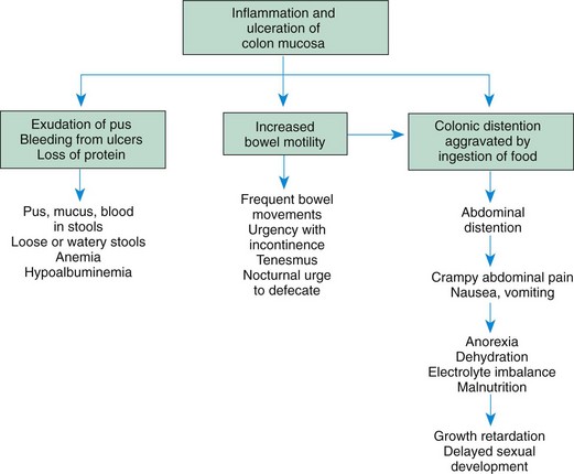
The inflammation found with UC is limited to the colon and rectum, with the distal colon and rectum the most severely affected. Inflammation affects the mucosa and submucosa and involves continuous segments along the length of the bowel with varying degrees of ulceration, bleeding, and edema. Thickening of the bowel wall and fibrosis are unusual, but longstanding disease can result in shortening of the colon and strictures. Extraintestinal manifestations are less common in UC than in CD. Toxic megacolon is the most dangerous form of severe colitis.

The chronic inflammatory process of CD involves any part of the GI tract from the mouth to the anus but most often affects the terminal ileum. The disease involves all layers of the bowel wall (transmural) in a discontinuous fashion, meaning that between areas of intact mucosa, there are areas of affected mucosa (skip lesions). The inflammation may result in ulcerations; fibrosis; adhesions; stiffening of the bowel wall; stricture formation; and fistulas to other loops of bowel, bladder, vagina, or skin.

Clinical Signs and Symptoms

Children with UC may experience mild, moderate, or severe symptoms, depending on the extent of mucosal inflammation and systemic symptoms. Most include bloody diarrhea or occult fecal blood, abdominal pain, and varying degrees of systemic manifestations and growth abnormalities (Beattie, Croft, Fell, et al, 2006; Leichtner and Higuchi, 2004). One of the earliest signs of UC may be growth failure with decreased linear growth velocity (Beattie, Croft, Fell, et al, 2006). Growth failure is most likely a result of chronic poor dietary intake caused by anorexia related to GI symptoms. UC often manifests with the insidious onset of diarrhea, possibly with hematochezia, and usually without fever or weight loss. The course of the disease may remain mild with intermittent exacerbations. Some children and adolescents are seen with grossly bloody diarrhea, cramps, urgency with defecation, mild anemia, fever, anorexia, weight loss, and moderate signs of systemic illness. Severe UC is characterized by frequent bloody stools, abdominal pain, significant anemia, fever, and weight loss. Extraintestinal manifestations are less common in UC than in CD and may precede colitis. The erythrocyte sedimentation rate (ESR) may be elevated, indicating a systemic response to an inflammatory process. Enlarged lymph nodes (lymphadenopathy), arthritis, and the skin lesions of erythema nodosum may be present.

Common presenting manifestations of CD include diarrhea, abdominal pain with cramps, fever, and weight loss. Mild GI symptoms, poor growth, and extraintestinal manifestations may be present for several years before overt GI symptoms are present. Both malabsorption and anorexia are factors that contribute to the growth problems that are prevalent in CD. The effects of UC and CD are listed in Fig. 33-4.

image

Fig. 33-4 Effects of ulcerative colitis or Crohn disease.

Children with CD have multiple risk factors for impaired bone accrual, including poor growth, delayed maturation, malnutrition, decreased activity, chronic inflammation, and steroid therapy (Dubner, Shults, Baldassano, et al, 2009). Growth delay persists in many children with CD following diagnosis, despite improved disease activity (Pfefferkorn, Burke, Griffiths, et al, 2009).

The disease process can also involve the colon, causing diarrhea, cramps, and urgency with defecation. Signs of colitis, such as gross rectal bleeding or stool with occult blood, are similar to those seen in UC. Perianal disease, including skin tags, abscesses, fissures, and fistulas, is a feature of CD. Extraintestinal manifestations include erythema nodosum, pyoderma gangrenosum, arthralgia and arthritis, uveitis and episcleritis, sclerosing cholangitis, autoimmune hepatitis, nephrolithiasis, and pneumonitis (Silbermintz and Markowitz, 2006). Table 33-2 provides a comparison of UC and CD.

TABLE 33-2

CLINICAL MANIFESTATIONS OF INFLAMMATORY BOWEL DISEASES

CHARACTERISTICS ULCERATIVE COLITIS CROHN DISEASE
Rectal bleeding Common Uncommon
Diarrhea Often severe Moderate to severe
Pain Less frequent Common
Anorexia Mild or moderate May be severe
Weight loss Moderate May be severe
Growth retardation Usually mild May be severe
Anal and perianal lesions Rare Common
Fistulas and strictures Rare Common
Rashes Mild Mild
Joint pain Mild to moderate Mild to moderate

Diagnostic Evaluation

The diagnosis of UC and CD comes from the history, physical examination, laboratory evaluation, and other diagnostic procedures. Laboratory tests include a CBC to evaluate anemia and an ESR or CRP to assess the systemic reaction to the inflammatory process. Levels of total protein, albumin, iron, zinc, magnesium, vitamin B12, and fat-soluble vitamins may be low in children with CD. Stools are examined for blood, leukocytes, and infectious organisms. A serologic panel is often used in combination with clinical findings to diagnose IBD and to differentiate between CD and UC. Observational studies on the utility of blood tests to detect perinuclear antineutrophilic cytoplasmic antibodies (pANCA) and anti-Saccharomyces cerevisiae antibodies (ASCA) showed that the combination is specific, but not sensitive for diagnosing ulcerative colitis (Reese, Constantinides, Simillis, et al, 2006).

In patients with CD, an upper GI series with small bowel follow-through assists in assessing the existence, location, and extent of disease. Upper endoscopy and colonoscopy with biopsies are an integral part of diagnosing IBD (Langan, Gotsch, Krafczyk, et al, 2007). Endoscopy allows direct visualization of the surface of the GI tract so that the extent of inflammation and narrowing can be evaluated. CT and ultrasound also may be used to identify bowel wall inflammation, intraabdominal abscesses, and fistulas. CD lesions may pierce the walls of the small intestine and colon, creating tracts called fistulas between the intestine and adjacent structures such as the bladder, anus, vagina, or skin.

Therapeutic Management

The natural history of the disease continues to be unpredictable and characterized by recurrent flare-ups that can severely impair patients’ physical and social functioning (Vernier-Massouille, Balde, Salleron, et al, 2008). The goals of therapy are to (1) control the inflammatory process to reduce or eliminate the symptoms, (2) obtain long-term remission, (3) promote normal growth and development, and (4) allow as normal a lifestyle as possible. Treatment is individualized and managed according to the type and the severity of the disease, its location, and the response to therapy.

Medical Treatment: The goal of any treatment regimen is first to induce remission of acute symptoms and then to maintain remission over time. 5-Aminosalicylates (5-ASAs) are effective in the induction and maintenance of remission in mild to moderate UC. Mesalamine, olsalazine, and balsalazide are now preferred over sulfasalazine because of reduced side effects (headache, nausea, vomiting, neutropenia, and oligospermia). Suppository and enema preparations of mesalamine are used to treat left-sided colitis. These drugs decrease inflammation by inhibiting prostaglandin synthesis. 5-ASAs can be used to induce remission in mild CD. Corticosteroids, such as prednisone and prednisolone, are indicated in induction therapy in children with moderate to severe UC and CD. These drugs inhibit the production of adhesion molecules, cytokines, and leukotrienes. Although these drugs reduce the acute symptoms of IBD, they have side effects that relate to long-term use, including growth suppression (adrenal suppression), weight gain, and decreased bone density (Baron, 2002). High doses of IV corticosteroids may be administered in acute episodes and tapered according to clinical response. Budesonide, a synthetic corticosteroid, is designed for controlled release in the ileum and is indicated for ileal and right-sided colitis; budesonide has fewer side effects than prednisone and prednisolone (Silbermintz and Markowitz, 2006). Rectal steroid therapy (enemas and foam-based preparations) are available for both induction and maintenance therapy in left-sided colitis.

Immunomodulators, such as azathioprine and its metabolite 6-mercaptopurine (6-MP), are used to induce and maintain remission in children with IBD who are steroid resistant or steroid dependent and in treating chronic draining fistulas. They block the synthesis of purine, thus inhibiting the ability of deoxyribonucleic acid (DNA) and ribonucleic acid (RNA) to hinder lymphocyte function, especially that of T cells. Side effects include infection, pancreatitis, hepatitis, bone marrow toxicity, arthralgia, and malignancy. Methotrexate is also useful in inducing and maintaining remission in CD patients unresponsive to standard therapies. Cyclosporine and tacrolimus have both been effective in inducing remission in severe steroid-dependent UC. 6-MP or azathioprine is then used to maintain remission. Patients on immunomodulating medications require regular monitoring of their CBC and differential to assess for changes that reflect suppression of the immune system, since many of the side effects can be prevented or managed by dose reduction or discontinuation of medication.

Antibiotics, such as metronidazole and ciprofloxacin, may be used as an adjunctive therapy to treat complications such as perianal disease or small bowel bacterial overgrowth in CD. Side effects of these drugs are peripheral neuropathy, nausea, and a metallic taste.

Biologic therapies act to regulate inflammatory and antiinflammatory cytokines. With the emergence of the biologic agents, specifically the use of antitumor necrosis factor-α (TNF-α) agents, progress has been made in targeting specific pathogenetic mechanisms and achieving a more prolonged clinical response (Ricart, García-Bosch, Ordás, et al, 2008; Hyams and Markowitz, 2005). TNF-α is believed to influence active inflammation.

Nutritional Support: Nutritional support is important in the treatment of IBD. Growth failure is a common serious complication, especially in CD. Growth failure is characterized by weight loss, alteration in body composition, retarded height, and delayed sexual maturation. Malnutrition causes the growth failure, and its etiology is multifactorial. Malnutrition occurs as a result of inadequate dietary intake, excessive GI losses, malabsorption, drug-nutrient interaction, and increased nutritional requirements. Inadequate dietary intake occurs with anorexia and episodes of increased disease activity. Excessive loss of nutrients (protein, blood, electrolytes, and minerals) occurs secondary to intestinal inflammation and diarrhea. Carbohydrate, lactose, fat, vitamin, and mineral malabsorption, as well as vitamin B12 and folic acid deficiencies, occur with disease episodes and with drug administration and when the terminal ileum is resected. Finally, nutritional requirements are increased with inflammation, fever, fistulas, and periods of rapid growth (e.g., adolescence).

The goals of nutritional support include (1) correction of nutrient deficits and replacement of ongoing losses, (2) provision of adequate energy and protein for healing, and (3) provision of adequate nutrients to promote normal growth. Nutritional support includes both enteral and parenteral nutrition. A well-balanced, high-protein, high-calorie diet is recommended for children whose symptoms do not prohibit an adequate oral intake. There is little evidence that avoiding specific foods influences the severity of the disease. Supplementation with multivitamins, iron, and folic acid is recommended.

Special enteral formulas, given either by mouth or continuous NG infusion (often at night), may be required. Elemental formulas are completely absorbed in the small intestine with almost no residue. A diet consisting only of elemental formula not only improves nutritional status but also induces disease remission, either without steroids or with a diminished dosage of steroids required. An elemental diet is a safe and potentially effective primary therapy for patients with CD. Unfortunately, remission is not sustained when NG feedings are discontinued unless maintenance medications are added to the treatment regimen.

TPN has also improved nutritional status in patients with IBD. Short-term remissions have been achieved after TPN, although complete bowel rest has not reduced inflammation or added to the benefits of improved nutrition by TPN. Nutritional support is less likely to induce a remission in UC than in CD. Improvement of nutritional status is important, however, in preventing deterioration of the patient’s health status and in preparing the patient for surgery.

Surgical Treatment: Surgery is indicated for UC when medical and nutritional therapies fail to prevent complications. Surgical options include a subtotal colectomy and ileostomy that leaves a rectal stump as a blind pouch. A reservoir pouch is created in the configuration of a J or S to help improve continence postoperatively. An ileoanal pull-through preserves the normal pathway for defecation. Pouchitis, an inflammation of the surgically created pouch, is the most common late complication of this procedure and had been reported to occur in up to 50% of cases. In many cases UC can be cured with a total colectomy.

Surgery may be required in children with CD when complications cannot be controlled by medical and nutritional therapy. Segmental intestinal resections are performed for small bowel obstructions, strictures, or fistulas. Partial colonic resection is not curative, and the disease often recurs.

Prognosis: IBD is a chronic disease. Relatively long periods of quiescent disease may follow exacerbations. The outcome is influenced by the regions and severity of involvement, as well as by appropriate therapeutic management. Malnutrition, growth failure, and bleeding are serious complications. The overall prognosis for UC is good.

The development of colorectal cancer (CRC) is a long-term complication of IBD. In UC, the cumulative incidence of CRC is 2.5% after 20 years, increasing to 10.8% after 30 years (Rutter, Saunders, Wilkinson, et al, 2006). Surveillance colonoscopy with multiple biopsies should begin approximately 10 years after diagnosis of UC or Crohn colitis and continue every 1 to 2 years (Rubin and Kavitt, 2006). Removal of the diseased colon prevents development of CRC. In CD, however, surgical removal of the affected colon does not prevent cancer from developing elsewhere in the GI tract.

Nursing Care Management

The nursing considerations in the management of IBD extend beyond the immediate period of hospitalization. These interventions involve continued guidance of families in terms of (1) managing diet; (2) coping with factors that increase stress and emotional lability; (3) adjusting to a disease of remissions and exacerbations; and (4) when indicated, preparing the child and parents for the possibility of diversionary bowel surgery.

QUALITY PATIENT OUTCOMES

Inflammatory Bowel Disease

• Remission without symptoms of abdominal pain, bloating, diarrhea, and rectal bleeding

• Optimum quality of life maintained by minimizing impairment of daily activities

Because nutritional support is an essential part of therapy, encouraging the anorexic child to consume sufficient quantities of food is often a challenge. Successful interventions include involving the child in meal planning; encouraging small, frequent meals or snacks rather than three large meals a day; serving meals around medication schedules when diarrhea, mouth pain, and intestinal spasm are controlled; and preparing high-protein, high-calorie foods such as eggnog, milkshakes, cream soups, puddings, or custard (if lactose is tolerated). (See Feeding the Sick Child, Chapter 27.) Using bran or a high-fiber diet for active IBD is questionable. Bran, even in small amounts, has been shown to worsen the patient’s condition. Occasionally the occurrence of aphthous stomatitis further complicates adherence to dietary management. Mouth care before eating and the selection of bland foods help relieve the discomfort of mouth sores.

When NG feedings or TPN is indicated, nurses play an important role in explaining the purpose and the expected outcomes of this therapy. The nurse should acknowledge the anxieties of the child and family members and give them adequate time to demonstrate the skills necessary to continue the therapy at home if needed (see Critical Thinking Exercise).

image CRITICAL THINKING EXERCISE

Inflammatory Bowel Disease

Susan, a 13-year-old girl, was admitted to the hospital because of bloody diarrhea, abdominal pain, and weight loss. After a thorough evaluation, including laboratory tests, radiographic studies, and gastrointestinal endoscopy procedures, the diagnosis of Crohn disease (CD) was made. Medical treatment, including corticosteroid drugs and nutritional support, was implemented during this hospitalization.

Susan has improved considerably and is to be discharged home this week. Enteral formula administered by continuous nighttime nasogastric (NG) tube infusion will be continued at home, and both Susan and her family are eager to learn how to perform these feedings. You are the nurse responsible for Susan’s discharge planning. Which interventions relating to these feedings should you include in Susan’s preparations for discharge?

1. Evidence—Are there sufficient data to formulate any specific interventions for discharge?

2. Assumptions—Describe some underlying assumptions about:

a. The goals of nutritional support for children with CD

b. Teaching required by an adolescent or family member who is administering NG tube feedings at home

c. Psychosocial issues related to CD

3. What are the priorities for discharge planning at this time?

4. Does the evidence support your conclusion?

The importance of continued drug therapy despite remission of symptoms must be stressed to the child and family members. Failure to adhere to the pharmacologic regimen can result in exacerbation of the disease. (See Compliance, Chapter 27.) Unfortunately, exacerbation of IBD can occur even if the child and family are compliant with the treatment regimen; this is difficult for the child and family to cope with.

Family Support: The nurse should attend to the emotional components of the disease and assess any sources of stress. Frequently, the nurse can help children adjust to problems of growth retardation, delayed sexual maturation, dietary restrictions, feelings of being “different” or “sickly,” inability to compete with peers, and necessary absence from school during exacerbations of the illness.

If a permanent colectomy-ileostomy is required, the nurse can teach the child and family how to care for the ileostomy. The nurse can also emphasize the positive aspects of the surgery, particularly accelerated growth and sexual development, permanent recovery, the eliminated risk of colonic cancer in UC, and the normality of life despite bowel diversion. Introducing the child and parents to other ostomy patients, especially those who are the same age, is effective in fostering eventual acceptance. Whenever possible, offer continent ostomies as options to the child, although they are not performed in all centers in the United States.

Because of the chronic and often life-long nature of the disease, families benefit from the educational services provided by organizations such as the Crohn’s and Colitis Foundation of America (CCFA).* If diversionary bowel surgery is indicated, United Ostomy Associations of America and the Wound, Ostomy and Continence Nurses Society are available to assist with ileostomy care and provide important psychologic support through their self-help groups. Adolescents often benefit by participating in peer-support groups, which are sponsored by the CCFA.

Peptic Ulcer Disease

Peptic ulcers may be classified as acute or chronic, and peptic ulcer disease (PUD) is a chronic condition that affects the stomach or duodenum. Ulcers are described as gastric or duodenal and as primary or secondary. A gastric ulcer involves the mucosa of the stomach; a duodenal ulcer involves the pylorus or duodenum. Most primary ulcers occur in the absence of a predisposing factor and tend to be chronic, occurring more frequently in the duodenum. Stress ulcers result from the stress of a severe underlying disease or injury (e.g., severe burns, sepsis, increased intracranial pressure, severe trauma, multisystem organ failure) and are more frequently acute and gastric.

About 1.7% of children in general pediatric practices have PUD, and the disease represents about 3.4% per 10,000 pediatric hospital admissions. Primary ulcers are more common in children older than 6 years, and stress ulcers are more common in infants younger than 6 months. Except for very young children, the incidence is two to three times greater in boys than in girls.

Etiology

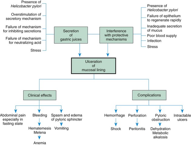
The exact cause of PUD is unknown, although infectious, genetic, and environmental factors are important. There is an increased familial incidence, and the disease is increased in persons with blood group O.

There is a significant relationship between the bacterium Helicobacter pylori and ulcers. H. pylori is a microaerophilic, gram-negative, slow-growing, spiral-shaped, and flagellated bacterium known to colonize the gastric mucosa in about half of the population of the world (Sung, Kuipers, and El-Serag, 2009). H. pylori synthesizes the enzyme urease, which hydrolyses urea to form ammonia and carbon dioxide. Ammonia then absorbs acid to form ammonium, thus raising the gastric pH. H. pylori may cause ulcers by weakening the gastric mucosal barrier and allowing acid to damage the mucosa. It is believed that it is acquired via the fecal-oral route, and this hypothesis is supported by finding viable H. pylori in feces.

In addition to ulcerogenic drugs, both alcohol and smoking contribute to ulcer formation. There is no conclusive evidence to implicate particular foods, such as caffeine-containing beverages or spicy foods, but polyunsaturated fats and fiber may play a role in ulcer formation. Psychologic factors may play a role in the development of PUD, and stressful life events, dependency, passiveness, and hostility have all been implicated as contributing factors.

Pathophysiology

Most likely, the pathologic condition is due to an imbalance between the destructive (cytotoxic) factors and defensive (cytoprotective) factors in the GI tract. The toxic mechanisms include acid, pepsin, medications such as aspirin and nonsteroidal antiinflammatory drugs (NSAIDs), bile acids, and infection with H. pylori. The defensive factors include the mucus layer, local bicarbonate secretion, epithelial cell renewal, and mucosal blood flow. Prostaglandins play a role in mucosal defense because they stimulate both mucus and alkali secretion. The primary mechanism that prevents the development of peptic ulcer is the secretion of mucus by the epithelial and mucus glands throughout the stomach. The thick mucus layer acts to diffuse acid from the lumen to the gastric mucosal surface, thus protecting the gastric epithelium. The stomach and the duodenum produce bicarbonate, decreasing acidity on the epithelial cells and thereby minimizing the effects of the low pH (Chelimsky and Czinn, 2001). When abnormalities in the protective barrier exist, the mucosa is vulnerable to damage by acid and pepsin. Exogenous factors, such as aspirin and NSAIDs, cause gastric ulcers by inhibition of prostaglandin synthesis.

Zollinger-Ellison syndrome may occur in children who have multiple, large, or recurrent ulcers. This syndrome is characterized by hypersecretion of gastric acid, intractable ulcer disease, and intestinal malabsorption caused by a gastrin-secreting tumor of the pancreas. The pathogenesis, manifestations, and complications of PUD are outlined in Fig. 33-5.

image

Fig. 33-5 Possible causes and effects of peptic ulcer.

Clinical Manifestations

The clinical manifestations of PUD vary according to the child’s age and the ulcer’s location. Common clinical manifestations include chronic abdominal pain, especially when the stomach is empty, such as during the night or early morning; recurrent vomiting; hematemesis; melena; chronic anemia; and abdominal tenderness (Box 33-9).

BOX 33-9   CHARACTERISTICS OF PEPTIC ULCERS

Neonates

Usually gastric and secondary ulcers

Commonly a history of prematurity, respiratory distress, sepsis, hypoglycemia, or an intraventricular hemorrhage

Perforation possibly leading to massive bleeding

Infants to <2-Year-Old Children

Most likely to have a secondary ulcer located equally in the stomach or duodenum

Primary ulcers less common and usually located in stomach

Likely to be noticed in relation to illness, surgery, or trauma

Hematemesis, melena, or perforation

2- to 6-Year-Old Children

Primary or secondary ulcers

Located equally in stomach and duodenum

Perforation more likely in secondary ulcers

Periumbilical pain, poor eating, vomiting, irritability, nighttime wakening, hematemesis, melena

Children 6 Years and Older

Usually primary and most often duodenal ulcers

More typical of adult type

Chance of recurrence greater

Often associated with Helicobacter pylori

Epigastric pain or vague abdominal pain

Nighttime wakening, hematemesis, melena, and anemia possible

Diagnostic Evaluation

Diagnosis is based on the history of symptoms, physical examination, and diagnostic testing. The focus is on symptoms such as epigastric abdominal pain, nocturnal pain, oral regurgitation, heartburn, weight loss, hematemesis, and melena. History should include questions relating to the use of potentially causative substances such as NSAIDS, corticosteroids, alcohol, and tobacco. Laboratory studies may include a CBC to detect anemia, stool analysis for occult blood, liver function tests (LFTs), sedimentation rate, or CRP to evaluate IBD; amylase and lipase to evaluate pancreatitis; and gastric acid measurements to identify hypersecretion. A lactose breath test may be performed to detect lactose intolerance.

Radiographic studies such as an upper GI series may be performed to evaluate obstruction or malrotation. An upper endoscopy is the most reliable procedure to diagnose PUD. A biopsy can determine the presence of H. pylori. A blood test can also identify the presence of the antigen to this organism. The 13C urea breath test measures bacterial colonization in the gastric mucosa. This test is used to screen for H. pylori in adults and children. Polyclonal and monoclonal stool antigen tests are an accurate, noninvasive method both for the initial diagnosis of H. pylori and for the confirmation of its eradication after treatment (Gisbert, de la Morena, and Abraira, 2006).

Diagnosis is based on the history (pattern of pain) and physical examination. Frequently a history of epigastric and periumbilical pain accompanies PUD. However, children often find it difficult to describe the location of their pain and frequently indicate the location by moving their hand in a circular movement all around the stomach area. Asking the child to take one finger and point to the area where it hurts the most often helps to identify the location of the pain. Pain may also be elicited during the examination with palpation. Routine laboratory studies to diagnose PUD include a CBC with differential, ESR, blood chemistry studies, urinalysis, and stool analysis to identify anemia or inflammation and to rule out infection. A 13C urea breath test is often performed to determine the presence of antibodies to H. pylori. An upper GI series is rarely helpful in identifying ulcers in children; fiberoptic endoscopy is the most reliable way to detect PUD in children. Direct visualization of the gastric and duodenal mucosa with biopsy to determine the presence of H. pylori is the most commonly used and effective way to arrive at the diagnosis.

Therapeutic Management

The major goals of therapy for children with PUD are to relieve discomfort, promote healing, prevent complications, and prevent recurrence. Management is primarily medical and consists of administration of medications to treat the infection and to reduce or neutralize gastric acid secretion. Antacids are beneficial medications to neutralize gastric acid. Histamine (H2) receptor antagonists (antisecretory drugs) act to suppress gastric acid production. Cimetidine, ranitidine, and famotidine are examples of these medications. These medications have few side effects.

PPIs, such as omeprazole and lansoprazole, act to inhibit the hydrogen ion pump in the parietal cells, thus blocking the production of acid. Although these drugs have not been well studied in children, thy are utilized in clinical practice to treat ulcers, GER, esophagitis, and gastritis. They appear to be well tolerated and to have infrequent side effects (e.g., headache, diarrhea, nausea, vomiting).

Mucosal protective agents, such as sucralfate and bismuth-containing preparations, may be prescribed for PUD. Sucralfate is an aluminum-containing agent that forms a barrier over ulcerated mucosa to protect against acid and pepsin. Sucralfate is available in both pill and liquid forms. Because sucralfate blocks the absorption of other medications, it should be given separately from other medications.

Bismuth compounds are sometimes prescribed for the relief of ulcers, but they are used less frequently than PPIs. Although these compounds inhibit the growth of microorganisms, the mechanism of their activity is poorly understood. In combination with antibiotics, bismuth is effective against H. pylori. Although concern has been expressed about the use of bismuth salts in children because of potential side effects, none of these side effects has been reported when these compounds have been used in the treatment of H. pylori infection.

Triple-drug therapy is the standard first-line treatment regimen for H. pylori (O’Connor, Gisbert, and O’Morain, 2009). Combination therapy has demonstrated 90% effectiveness in eradication of H. pylori when compared with antibiotic monotherapy. Examples of drug combinations used in triple therapy are (1) bismuth, clarithromycin, and metronidazole; (2) lansoprazole, amoxicillin, and clarithromycin; and (3) metronidazole, clarithromycin, and omeprazole. The benefits on the use of probiotics as an adjunct to treatment remain unclear, with conflicting literature on their effect on eradication and minimizing side effects (O’Connor, Gisbert, and O’Morain, 2009).

Common side effects of medications include diarrhea, nausea, and vomiting. In addition to medications, the child with PUD should have a nutritious diet and avoid caffeine. Warn adolescents about gastric irritation associated with alcohol use and smoking.

Children with an acute ulcer who have developed complications, such as massive hemorrhage, require emergency care. The administration of IV fluids, blood, or plasma depends on the amount of blood loss. Replacement with whole blood or packed cells may be necessary for significant loss.

Surgical intervention may be required for complications such as hemorrhage, perforation, or gastric outlet obstruction. Ligation of the source of bleeding or closure of a perforation is performed. A vagotomy and pyloroplasty may be indicated in children with recurring ulcers despite aggressive medical treatment.

Prognosis: The long-term prognosis for PUD is variable. Many ulcers are successfully treated with medical therapy; however, primary duodenal peptic ulcers often recur. Complications such as GI bleeding can occur and extend into adult life. The effect of maintenance drug therapy on long-term morbidity remains to be established with further studies.

Nursing Care Management

The primary nursing goal is to promote healing of the ulcer through compliance with the medication regimen. If an analgesic-antipyretic is needed, acetaminophen, not aspirin or NSAIDs, is used. Critically ill neonates, infants, and children in intensive care units should receive H2 blockers to prevent stress ulcers.

image DRUG ALERT

H2 Blockers

Critically ill children receiving IV H2 blockers should have their gastric pH values checked at frequent intervals.

For nonhospitalized children with chronic illnesses, consider the role stress plays. In children, many ulcers occur secondary to other conditions, and the nurse should be aware of family and environmental conditions that may aggravate or precipitate ulcers. Children may benefit from psychologic counseling and from learning how to cope constructively with stress.

Obstructive Disorders

Obstruction in the GI tract occurs when the passage of nutrients and secretions is impeded by a constricted or occluded lumen, or when there is impaired motility (paralytic ileus). Obstructions may be congenital or acquired. Congenital obstructions, such as esophageal or intestinal atresias and malrotation, usually appear in the neonatal period. (See Chapter 11.) Obstruction in the GI tract from many causes is characterized by similar signs and symptoms, although the progression may vary greatly.

Usually, acute intestinal obstruction is characterized by abdominal pain, nausea, vomiting, abdominal distention, and a change in stooling patterns (Box 33-10). Pain is caused by intermittent muscular contractions proximal to the obstruction as the bowel attempts to move luminal contents along the normal path. It may also be due to severe abdominal distention, which results from accumulation of gas and fluid above the level of the obstruction. As abdominal distention progresses, the abdomen may become extremely tender, rigid, and firm.

BOX 33-10   CLINICAL MANIFESTATIONS OF MECHANICAL (PARALYTIC) INTESTINAL OBSTRUCTION

Colicky abdominal pain—From peristalsis attempting to overcome the obstruction

Abdominal distention—As a result of accumulation of gas and fluid above the level of the obstruction

Vomiting—Often the earliest sign of a high obstruction; a later sign of lower obstruction (may be bilious or feculent)

Constipation and obstipation—Early signs of low obstructions; later signs of higher obstructions

Dehydration—From losses of large quantities of fluid and electrolytes into the intestine

Rigid and boardlike abdomen—From increased distention

Bowel sounds—Gradually diminish and cease

Respiratory distress—Occurs as the diaphragm is pushed up into the pleural cavity

Shock—Caused by plasma volume diminishing as fluids and electrolytes are lost from the bloodstream into the intestinal lumen

Sepsis—Caused by bacterial proliferation with invasion into the circulation

When abdominal contents continue to accumulate, nausea and vomiting occur. Vomiting of gastric contents is often the first sign of a high obstruction, such as obstruction of the pylorus, and vomiting of bile-stained material is a sign of obstruction of the small intestine. Persistent vomiting can lead to dehydration and electrolyte disturbances. Constipation and obstipation (prolonged absence of defecation) are early signs of low obstructions and later signs of higher obstructions. In acute conditions such as intussusception, the clinical manifestations are apparent within a few hours of the onset of the disorder. In other conditions such as pyloric stenosis the signs and symptoms may have a more gradual onset. Bowel sounds may initially be hyperactive, then diminish or cease. Respiratory distress may occur when the diaphragm is pushed up into the pleural cavity as a result of severe abdominal distention.

Hypertrophic Pyloric Stenosis

Hypertrophic pyloric stenosis (HPS) occurs when the circumferential muscle of the pyloric sphincter becomes thickened, resulting in elongation and narrowing of the pyloric canal. This produces an outlet obstruction and compensatory dilation, hypertrophy, and hyperperistalsis of the stomach. This condition usually develops in the first few weeks of life, causing projectile vomiting, dehydration, metabolic alkalosis, and failure to thrive. The precise etiology of HPS is not known. It is more common in first-born children, and boys are affected five times more frequently than girls. HPS is seen less frequently in African-American infants than in Caucasian infants. It is more likely to affect full-term infants than premature ones. Inheritance is polygenic, with an increased risk in the siblings and offspring of affected persons. The greatest risk of recurrence (20%) is in the first-born boy of a mother who was affected (Milla, 2004).

Pathophysiology

The circular muscle of the pylorus thickens as a result of hypertrophy. This produces severe narrowing of the pyloric canal between the stomach and the duodenum. Consequently, the lumen at this point is partially obstructed. Over time the size of the opening is reduced, and the partial obstruction may progress to complete obstruction. The hypertrophied pylorus may be palpable as an olivelike mass in the upper abdomen (Fig. 33-6).

image

Fig. 33-6 Hypertrophic pyloric stenosis. A, Enlarged muscular tumor nearly obliterates pyloric canal. B, Longitudinal surgical division of muscle down to submucosa establishes adequate passageway.

Pyloric stenosis is not a congenital disorder. Evidence suggests that local innervation may be involved in the pathogenesis. In most cases this is an isolated lesion; however, it may be associated with intestinal malrotation, esophageal and duodenal atresia, and anorectal anomalies.

Clinical Manifestations

Infants with HPS have nonbilious vomiting in the early stages (Box 33-11). The vomiting may be projectile and progressive, becoming brown in later stages if gastritis develops. Vomiting usually begins at 3 weeks of age but can start as early as 1 week and as late as 5 months. Initially the infant is hungry and irritable, but prolonged vomiting may lead to dehydration, weight loss, and failure to thrive. Gastric peristalsis may be visible on examination, and the olive-shaped mass in the epigastrium just to the right of the umbilicus may be palpated (see Fig. 33-6, A).

BOX 33-11   CLINICAL MANIFESTATIONS OF HYPERTROPHIC PYLORIC STENOSIS

Projectile vomiting

• May be ejected 3 to 4 feet from the child when in a side-lying position, 1 foot or more when in a back-lying position

• Usually occurs shortly after a feeding, but may not occur for several hours

• May follow each feeding or appear intermittently

• Nonbilious vomitus that may be blood tinged

Infant hungry, avid nurser; eagerly accepts a second feeding after vomiting episode

No evidence of pain or discomfort except that of chronic hunger

Weight loss

Signs of dehydration

Distended upper abdomen

Readily palpable olive-shaped tumor in the epigastrium just to the right of the umbilicus

Visible gastric peristaltic waves that move from left to right across the epigastrium

Diagnostic Evaluation

The diagnosis of HPS is often made after the history and physical examination. The olivelike mass is most easily palpated when the stomach is empty, the infant is quiet, and the abdominal muscles are relaxed. If the diagnosis is inconclusive from the history and physical examination, ultrasonography will demonstrate an elongated mass surrounding a long pyloric canal. If ultrasonography does not demonstrate a hypertrophied pylorus, upper GI radiography should be done to rule out other causes of vomiting.

Laboratory findings reflect the metabolic alterations created by severe depletion of both water and electrolytes from extensive and prolonged vomiting. There are decreased serum levels of both sodium and potassium, although these may be masked by the hemoconcentration from extracellular fluid depletion. Of greater diagnostic value are a decrease in serum chloride levels and increases in pH and bicarbonate (carbon dioxide content), indicative of metabolic alkalosis. The blood urea nitrogen will be elevated as evidence of dehydration.

Therapeutic Management

Surgical relief of the pyloric obstruction by pyloromyotomy is the standard therapy for this disorder. Preoperatively the infant must be rehydrated and metabolic alkalosis corrected with parenteral fluid and electrolyte administration. Replacement fluid therapy usually delays surgery for 24 to 48 hours. The stomach is decompressed with an NG tube. In infants with no evidence of fluid and electrolyte imbalance, surgery is performed without delay.

The surgical procedure is often performed by laparoscope and consists of a longitudinal incision through the circular muscle fibers of the pylorus down to, but not including, the submucosa (pyloromyotomy, sometimes called Fredet-Ramstedt procedure) (see Fig. 33-6, B). The procedure has a high success rate. The use of a small incision for the laparoscope may result in a shorter surgical time, more rapid postoperative feeding, and shorter hospital stay (van der Bilt, Kramer, van der Zee, et al, 2004).

Feedings are usually begun 4 to 6 hours postoperatively, beginning with small, frequent feedings of glucose, water, or electrolyte solution. If clear fluids are retained, about 24 hours after surgery formula is started in the same small increments. The amount and the interval between feedings are gradually increased until a full feeding schedule is reinstated, which usually takes about 48 hours.

Prognosis: The prognosis is excellent, and the mortality rate is low. Approximately 15% of infants with HPS also have GER (Milla, 2004).

Nursing Care Management

Nursing care involves primarily observation for clinical features that help establish the diagnosis, careful regulation of fluid therapy, and reestablishment of normal feeding patterns. Nurses must be alert to signs of HPS in infants and refer them for medical evaluation. Consider the possibility of HPS in the very young infant who appears alert but fails to gain weight and has a history of vomiting after meals. Base assessment on observation of eating behaviors, evidence of characteristic clinical manifestations, hydration, and nutritional status.

Preoperatively the emphasis is on restoring hydration and electrolyte balance. The infant is kept NPO and given IV fluids of glucose and electrolytes based on serum electrolyte values—usually sodium chloride solution with added potassium (when there is adequate urinary output). Careful monitoring of the IV fluids and strict monitoring of intake, output, and urine specific gravity are important. Record accurate description of any vomiting and the number and character of stools.

Observations include assessment of vital signs, particularly those that indicate fluid or electrolyte imbalances. These infants are especially prone to metabolic alkalosis from loss of hydrogen ions and depletion of potassium, sodium, and chloride, all of which are contained in gastric secretions. Assess the skin and mucous membranes for alterations in hydration status; daily weights provide added clues to water gain or loss. (See Chapter 28 for manifestations of fluid and electrolyte disturbances.)

When stomach decompression and gastric lavage are part of preoperative management, the nurse is responsible for ensuring that the NG tube is patent and functioning properly and that the type and amount of NG drainage is recorded. General hygienic care, with particular attention to the skin and mouth in dehydrated infants, is important. Protection from infection is essential because infants with impaired nutritional status are more susceptible to infection than normal newborns.

Encourage parents to visit and become involved in the child’s care. Most parents need support and reassurance that the condition is caused by a structural problem and is not a reflection of their parenting skills and capacities.

Postoperative Care: Postoperative vomiting is not uncommon, and most infants, even with successful surgery, exhibit some vomiting during the first 24 to 48 hours. IV fluids are administered until the infant is taking and retaining adequate amounts by mouth. Much of the same care that was instituted before surgery is continued postoperatively, including observation of vital signs, monitoring of IV fluids, and careful monitoring of intake and output. In addition, the infant is observed for responses to the stress of surgery and for evidence of pain. Appropriate analgesics should be given around the clock, since pain is continuous.

The NG tube may be maintained after surgery for a short time. Feedings are usually instituted within 24 hours postoperatively, beginning with clear liquids containing glucose and electrolytes. They are offered in small quantities at frequent intervals. If the infant has been breast-fed, breast milk, expressed by the mother, may be given by bottle when the infant is able to tolerate feedings, or the mother is instructed to limit nursing time and gradually increase the time to previous patterns. Supervision of feedings is an important part of postoperative care. Observe the operative site for any drainage or signs of inflammation. Poorly nourished infants may have problems with wound healing.

Intussusception

image Intussusception is the most common cause of intestinal obstruction in children between the ages of 3 months and 3 years (Waseem and Rosenberg, 2008). Intussusception is more common in males than in females and is more common in children with cystic fibrosis. Although specific intestinal lesions occur in a small percentage of the children, generally the cause is not known. More than 90% of intussusceptions do not have a pathologic lead point, such as a polyp, lymphoma, or Meckel diverticulum. The idiopathic cases may be caused by hypertrophy of intestinal lymphoid tissue secondary to viral infection.

imageAnimation—Intussusception
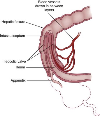
Pathophysiology

Intussusception occurs when a proximal segment of the bowel telescopes into a more distal segment, pulling the mesentery with it. The mesentery is compressed and angled, resulting in lymphatic and venous obstruction. As the edema from the obstruction increases, pressure within the area of intussusception increases. When the pressure equals the arterial pressure, arterial blood flow stops, resulting in ischemia and the pouring of mucus into the intestine. Venous engorgement also leads to leaking of blood and mucus into the intestinal lumen, forming the classic currant jelly–like stools (Wyllie, 2007). The most common site is the ileocecal valve (ileocolic), where the ileum invaginates into the cecum and then further into the colon (Fig. 33-7). Other forms include ileoileal (one part of the ileum invaginates into another section of the ileum) and colocolic (one part of the colon invaginates into another area of the colon) intussusceptions, usually in the area of the hepatic or splenic flexure or at some point along the transverse colon.

Pathophysiology Review

image

Fig. 33-7 Ileocecal valve (ileocolic) intussusception.

Clinical Manifestations

Intussusception usually manifests with the sudden onset of crampy abdominal pain, inconsolable crying, and a drawing up of the knees to the chest in an otherwise healthy child (Box 33-12). Between episodes the child appears normal. As the obstruction progresses, bilious vomiting may occur and lethargy increases. The classic triad of intussusception symptoms (abdominal pain, abdominal mass, bloody stools) is present in only 29% to 33% of children (Huppertz, Soriano-Gaabarro, Grimprel, et al, 2006). With atypical cases, lethargy may be the primary symptom. If the distal bowel remains distended, necrosis and perforation are possible.

image NURSING ALERT

The classic signs and symptoms of intussusception may not be present; a more chronic picture may occur, characterized by diarrhea, anorexia, weight loss, occasional vomiting, and periodic pain. The older child may have pain without other signs or symptoms. Because this condition is potentially life threatening, be aware of such signs and closely observe and refer these children for further medical investigation.

BOX 33-12   CLINICAL MANIFESTATIONS OF INTUSSUSCEPTION

• Sudden acute abdominal pain

• Child screaming and drawing the knees onto the chest

• Child appearing normal and comfortable between episodes of pain

• Vomiting

• Lethargy

• Passage of red, currant jelly–like stools (stool mixed with blood and mucus)

• Tender, distended abdomen

• Palpable sausage-shaped mass in upper right quadrant

• Empty lower right quadrant (Dance sign)

• Eventual fever, prostration, and other signs of peritonitis

Diagnostic Evaluation

Frequently the diagnosis can be made on subjective findings alone. However, definitive diagnosis is based on a barium enema, which clearly demonstrates the obstruction to the flow of barium. Initially an abdominal radiograph is obtained to detect intraperitoneal air from a bowel perforation, which would contraindicate a barium enema. A rectal examination reveals mucus, blood, and occasionally a low intussusception itself.

Therapeutic Management

Conservative treatment consists of radiologist-guided pneumoenema (air enema) with or without water-soluble contrast or ultrasound-guided hydrostatic (saline) enema, the advantage of the latter being that no ionizing radiation is needed (Huppertz, Soriano-Gaabarro, Grimprel, et al, 2006). Recurrence of intussusception after conservative treatment is rare. Herwig, Brenkert, and Losek (2009) found hospitalized children needed minimal interventions after undergoing enema-reduced intussusception.

IV fluids, NG decompression, and antibiotic therapy may be used before hydrostatic reduction is attempted. If these procedures are not successful, the child may require surgical intervention. Surgery involves manually reducing the invagination and, when indicated, resecting any nonviable intestine.

Prognosis: Nonoperative reduction is successful in approximately 80% of cases (Huppertz, Soriano-Gaabarro, Grimprel, et al, 2006). Surgery is required for patients in whom the contrast enema is unsuccessful. With early diagnosis and treatment, serious complications and death are uncommon.

Nursing Care Management

The nurse can help establish a diagnosis by listening to the parent’s description of the child’s physical and behavioral symptoms. It is not unusual for parents to state that they thought something was seriously wrong before others shared their concerns. The description of the child’s severe colicky abdominal pain combined with vomiting is a significant sign of intussusception.

As soon as a possible diagnosis of intussusception is made, the nurse prepares the parents for the immediate need for hospitalization, the nonsurgical technique of hydrostatic reduction, and the possibility of surgery. It is important to explain the basic defect of intussusception. The nurse can easily demonstrate this by creating a model of the defect. Use the example of a telescoping rod, or push the end of a finger on a rubber glove back into itself. Then demonstrate the principle of reduction by hydrostatic pressure by filling the glove with water, which pushes the “finger” into a fully extended position.

Physical care of the child does not differ from that for any child undergoing abdominal surgery. Even though nonsurgical intervention may be successful, the usual preoperative procedures, such as maintenance of NPO status, routine laboratory testing (CBC and urinalysis), signed parental consent, and preanesthetic sedation, are performed. For the child with signs of electrolyte imbalance, hemorrhage, or peritonitis, additional preparation, such as replacement fluids, whole blood or plasma, and NG suctioning, may be needed. Before surgery the nurse monitors all stools.

image NURSING ALERT

Passage of a normal brown stool usually indicates that the intussusception has reduced itself. This is immediately reported to the practitioner, who may choose to alter the diagnostic and therapeutic care plan.

Postprocedural care includes observations of vital signs, blood pressure, intact sutures and dressing, and the return of bowel sounds. After spontaneous or hydrostatic reduction, the nurse observes for passage of water-soluble contrast material (if used) and the stool patterns, since the intussusception may recur. Children may be admitted to the hospital or monitored on an outpatient basis. A recurrence of intussusception is treated with the conservative reduction techniques described above, but a laparotomy is considered for multiple recurrences.

Because hospitalization may be the child’s first separation from the parents, it is important to preserve the parent-child relationship by encouraging rooming-in or extended visiting. It may be the parents’ first experience with hospitalization, necessitating their preparation for procedures such as IV therapy, frequent vital sign and blood pressure monitoring, dressings, and NPO. Because of the rapidity of the onset, diagnosis, and treatment, parents may feel stunned or numb. They may ask few questions, or they may constantly make inquiries, sometimes the same ones several times. Because of the circumstances surrounding this condition, be accepting and understanding of the parents’ reactions.

Malrotation and Volvulus

image Malrotation of the intestine occurs as a result of the abnormal rotation of the intestine around the superior mesenteric artery during embryologic development. Malrotation may manifest in utero or may be asymptomatic throughout life. Infants may have intermittent bilious vomiting, recurrent abdominal pain, distention, or lower GI bleeding. Malrotation is the most serious type of intestinal obstruction because, if the intestine undergoes complete volvulus (the intestine twisting around itself), compromise of the blood supply will result in intestinal necrosis, peritonitis, perforation, and death.

imageAnimation—Volvulus, Pediatric

Diagnostic Evaluation

It is imperative that malrotation and volvulus be diagnosed promptly and surgical treatment instituted quickly. An upper GI series is the definitive procedure to diagnose this condition.

Therapeutic Management

Surgery is indicated to remove the affected area. Because of the extensive nature of some lesions, short-bowel syndrome (SBS) is a postoperative complication.

Nursing Care Management

Preoperatively the nursing care is the same as that provided to an infant or child with intestinal obstruction. Postoperatively the nursing care is similar to that provided to the infant or child who has undergone abdominal surgery.

Malabsorption Syndromes

Chronic diarrhea and malabsorption of nutrients characterize malabsorption syndromes. An important complication of malabsorption syndromes in children is failure to thrive. Most cases are classified according to the location of the supposed anatomic or biochemical defect. The term celiac disease is often used to describe a symptom complex with four characteristics: (1) steatorrhea (fatty, foul, frothy, bulky stools), (2) general malnutrition, (3) abdominal distention, and (4) secondary vitamin deficiencies.

Digestive defects are conditions in which the enzymes necessary for digestion are diminished or absent, such as (1) cystic fibrosis, in which pancreatic enzymes are absent; (2) biliary or liver disease, in which bile flow is affected; or (3) lactase deficiency, in which there is congenital or secondary lactose intolerance.

Absorptive defects are conditions in which the intestinal mucosal transport system is impaired. This may occur because of a primary defect (e.g., celiac disease) or secondary to inflammatory disease of the bowel that results in impaired absorption because bowel motility is accelerated (e.g., UC). Obstructive disorders (e.g., HD) also cause secondary malabsorption from enterocolitis.

Anatomic defects, such as extensive resection of the bowel or SBS, affect digestion by decreasing the transit time of substances and affect absorption by severely compromising the absorptive surface.

Celiac Disease (Gluten-Sensitive Enteropathy)

Celiac disease, also known as gluten-induced enteropathy, gluten-sensitive enteropathy, and celiac sprue, is a permanent intestinal intolerance to dietary wheat gliadin and related proteins that produces mucosal lesions in genetically susceptible individuals (Dieterich, Esslinger, and Schuppan, 2003). It is second only to cystic fibrosis as a cause of malabsorption in children.

The incidence is variable and has been reported in 1 in 3000 to 1 in 4000 people. The disease is seen more frequently in Europe than in the United States. It is more prevalent in women than men and is rarely reported in Asians or African-Americans. Although the exact cause is unknown, it is now generally accepted that celiac disease is an immunologically mediated small intestine enteropathy. The mucosal lesions contain features that suggest both humoral and cell-mediated immunologic overstimulation.

Pathophysiology

Celiac disease is characterized by villous atrophy in the small bowel in response to the protein gluten (Maki and Lohi, 2004). Gluten is found in wheat, barley, rye, and oat grains. When individuals are unable to digest the gliadin component of gluten, an accumulation of a toxic substance that is damaging to the mucosal cells occurs. Damage to the mucosa of the small intestine leads to villous atrophy, hyperplasia of the crypts, and infiltration of the epithelial cells with lymphocytes. Villous atrophy leads to malabsorption due to the reduced absorptive surface area (see Fig. 33-1 and the discussion of absorption on p. 1296).

Clinical Manifestations

Symptoms of celiac disease appear when solid foods such as beans and pasta are introduced in the child’s diet between the ages of 1 and 5 years (Box 33-13). There is usually an interval of several months between the introduction of gluten into the diet and the onset of symptoms. Intestinal symptoms are common in children diagnosed within the first 2 years of life. Other symptoms include failure to thrive, chronic diarrhea, abdominal distention, muscle wasting, anorexia, and irritability. (See Critical Thinking Exercise on Inflammatory Bowel Disease.)

BOX 33-13   CLINICAL MANIFESTATIONS OF CELIAC DISEASE

Impaired Fat Absorption

Steatorrhea (excessively large, pale, oily, frothy stools)

Exceedingly foul-smelling stools

Impaired Nutrient Absorption

Malnutrition

Muscle wasting (especially prominent in legs and buttocks)

Anemia

Anorexia

Abdominal distention

Behavioral Changes

Irritability

Uncooperativeness

Apathy

Celiac Crisis*

Acute, severe episodes of profuse watery diarrhea and vomiting

May be precipitated by:

• Infections (especially gastrointestinal)

• Prolonged fluid and electrolyte depletion

• Emotional disturbance


*In very young children.

Diagnostic Evaluation

The diagnosis of celiac disease is based on a biopsy of the small intestine demonstrating the characteristic changes of mucosal inflammation, crypt hyperplasia, and villous atrophy (Dieterich, Esslinger, and Schuppan, 2003).

Therapeutic Management

Treatment of celiac disease consists primarily of dietary management. Although a gluten-free diet is prescribed, it is difficult to remove every source of this protein. Some patients are able to tolerate restricted amounts of gluten. Because gluten occurs mainly in the grains of wheat and rye, but also in smaller quantities in barley and oats, these four foods are eliminated. Corn, rice, and millet are substitute grain foods.

Children with untreated celiac disease may have lactose intolerance, especially if their mucosal lesions are extensive. Lactose intolerance usually improves as the mucosa heals with gluten withdrawal. Specific nutritional deficiencies, such as iron, folic acid, and fat-soluble vitamin deficiencies, are treated with appropriate supplements.

Prognosis: Celiac disease is a chronic disease; its severity varies greatly among children. The most severe symptoms usually occur in early childhood and again in adult life. Most children who comply with dietary management are healthy and remain free of symptoms and complications. Strict dietary avoidance of gluten may minimize the risk of developing lymphoma, especially of the small intestine, one of the most serious complications of the disease.

Nursing Care Management

The main nursing consideration is helping the child adhere to the dietary regimen. Considerable time is involved in explaining the disease process to the child and parents, the specific role of gluten in aggravating the disorder, and those foods that must be restricted. It is difficult to maintain a diet indefinitely when the child has no symptoms and temporary transgressions result in no difficulties. However, the majority of individuals who relax their diet will experience a relapse of their disease and possibly exhibit growth retardation, anemia, or osteomalacia. There is also the risk of developing malignant lymphoma of the small intestine or other GI malignancies.

Although the chief source of gluten is cereal and baked goods, grains are frequently added to processed foods as thickeners or fillers. To compound the difficulty, gluten is added to many foods as hydrolyzed vegetable protein, which is derived from cereal grains. The nurse must advise parents of the necessity of reading all label ingredients carefully to avoid hidden sources of gluten.

Many of children’s favorite foods contain gluten, including bread, cake, cookies, crackers, donuts, pies, spaghetti, pizza, prepared soups, some processed ice cream, many types of chocolate candy, milk preparations such as malts, hot dogs, luncheon meats, meat gravy, and some prepared hamburgers. Many of these products can be eliminated from the infant’s or young child’s diet fairly easily, but monitoring the diet of the school-age child or adolescent is more difficult. Luncheon preparation away from home is particularly difficult, since bread, luncheon meats, and instant soups are not allowed. For families on restricted food budgets, the diet adds an additional financial burden because many inexpensive or convenient foods cannot be used.

In addition to restricting gluten, other dietary alterations may be necessary. For example, in some children who have more severe mucosal damage, the digestion of disaccharides is impaired, especially in relation to lactose. Therefore these children often need a temporarily lactose-free diet, which necessitates eliminating all milk products. In general, dietary management includes a diet high in calories and proteins, with simple carbohydrates such as fruits and vegetables, but low in fats. Because the bowel is inflamed as a result of the pathologic processes in absorption, the child must avoid high-fiber foods, such as nuts, raisins, raw vegetables, and raw fruits with skin, until inflammation has subsided.

It is important to stress long-range complications and to remind parents of the child’s physical status before dietary treatment and the dramatic improvement after treatment. The nurse can be instrumental in allowing the child to express concerns and frustration while focusing on ways in which the child can still feel normal. Encourage the child and parents to find new recipes using suitable ingredients, such as Mexican or Chinese dishes that use corn or rice. Consult a nutritionist to provide children and their families with detailed dietary instructions and education.*

Several resources are available to assist children and parents in all aspects of coping with celiac disease. The Celiac Sprue Association/United States of America provides support and guidance to families and supplies educational materials concerning a gluten-free diet, food sources, recipes, and travel information.

Short-Bowel Syndrome

SBS is a malabsorptive disorder that occurs as a result of decreased mucosal surface area, usually because of extensive resection of the small intestine. Malabsorption may be exacerbated by other factors, such as bacterial overgrowth and dysmotility. The most common causes of SBS in children are necrotizing enterocolitis, volvulus, jejunal atresias, and gastroschisis. Other causes include midgut volvulus and diffuse small bowel CD in older children. Less frequent causes include trauma to the GI tract and total colonic aganglionosis with extension into the small bowel.

The definition of SBS includes two important findings: (1) decreased intestinal surface area for absorption of fluid, electrolytes, and nutrients; and (2) a need for parenteral nutrition (PN) (Goday, 2009). The prognosis for infants with SBS has improved dramatically in the past 20 to 30 years as a result of advances in PN and enteral feeding.

Therapeutic Management

The goals of therapy for infants and children with SBS include (1) preserve as much length of bowel as possible during surgery; (2) maintain optimum nutritional status, growth, and development while intestinal adaptation occurs; (3) stimulate intestinal adaptation with enteral feeding; and (4) minimize complications related to the disease process and therapy (Goday, 2009).

Nutritional Support: Nutritional support is the long-term focus of care for children with SBS (Sadlier, 2008). The initial phase of therapy includes PN as the primary source of nutrition. The second phase is the introduction of enteral feeding, which usually begins as soon as possible after surgery. Elemental formulas containing glucose, sucrose and glucose polymers, hydrolyzed proteins, and medium-chain triglycerides facilitate absorption. Usually these formulas are given by continuous infusion through an NG or gastrostomy tube. As the enteral feedings are advanced, the PN solution is decreased in terms of calories, amount of fluid, and total hours of infusion per day.

The final phase of nutritional support occurs when growth and development are sustained exclusively by enteral feedings. When PN is discontinued, there is a risk of nutritional deficiency secondary to malabsorption of fat-soluble vitamins (A, D, E, K) and trace minerals (iron, selenium, zinc). Obtain serum vitamin and mineral levels, and require enteral supplementation of vitamins and minerals. Pharmacologic agents have been used to reduce secretory losses. H2 blockers, PPIs, and octreotide inhibit gastric or pancreatic secretion. Cholestyramine is often prescribed to improve diarrhea that is associated with bile salt malabsorption. Growth factors have also been used to hasten adaptation and to enhance mucosal growth, but these uses are still experimental.

Numerous complications are associated with SBS and long-term PN. Infectious, metabolic, and technical complications can occur. Catheter sepsis can occur after improper care of the catheter. The GI tract can also be a source of microbial seeding of the catheter. Bowel atrophy may foster increased intestinal permeability of bacteria. A lack of adequate sites for central lines may become a significant problem for the child in need of long-term PN. Hepatic dysfunction, hepatomegaly with abnormal LFTs, and cholestasis may also occur (Diamond, Sterescu, Pencharz, et al, 2009).

Bacterial overgrowth is likely to occur when the ileocecal valve is absent or when stasis exists as a result of a partial obstruction or a dilated segment of bowel with poor motility. Alternating cycles of broad-spectrum antibiotics are used to reduce bacterial overgrowth. This treatment may also decrease the risk of bacterial translocation and subsequent central venous catheter infections. Other complications of bacterial overgrowth and malabsorption include metabolic acidosis and gastric hypersecretion.

Many surgical interventions, including intestinal valves, tapering enteroplasty or stricturoplasty, intestinal lengthening, and interposed segments, have been used to slow intestinal transit, reduce bacterial overgrowth, or increase mucosal surface area. Intestinal transplantation has been performed successfully in children. Only children with a permanent dependence on PN or severe complications of long-term PN are candidates for transplantation.

Prognosis: The prognosis for infants with SBS has improved with advances in PN and with the understanding of the importance of intraluminal nutrition. Improved surgical techniques for the management of therapy-related problems and the development of more specific immunosuppressive medications for transplantation have all contributed to improved management. The prognosis depends in part on the length of the residual small intestine. An intact ileocecal valve also improves the prognosis. Infants and children with SBS die from PN-related problems, such as fulminant sepsis or severe PN cholestasis.

Nursing Care Management

The most important components of nursing care are administration and monitoring of nutritional therapy. During PN therapy, take care to minimize the risk of complications related to the central venous access device (i.e., catheter infections, occlusions, dislodgment, or accidental removal). Care of the enteral feeding tubes and monitoring of enteral feeding tolerance are also important nursing responsibilities (see Critical Thinking Exercise).

image CRITICAL THINKING EXERCISE

Short-Bowel Syndrome

The parents of a 2-year-old boy with short-bowel syndrome (SBS) call their health care professional to report that their child has passed many more stools than usual with an increased watery consistency in the past 24 hours. He also has a fever of 39° C (102.2° F) and has vomited several times. The boy is admitted to the hospital. During the initial period of nursing assessment, which of the following would you monitor?

1. Stool pH, urine pH, vital signs

2. Vital signs, weight, urine specific gravity, intake and output

3. Vital signs, stool-reducing substances, stool culture

4. Urine specific gravity, stool for blood, electrolytes

Questions

1. Evidence—Are there sufficient data to support your decision?

2. Assumptions—Describe some underlying assumptions about the following:

a. Description of SBS

b. Risk factors associated with chronic malabsorption disorders

c. Other assessment parameters

3. What are the priorities for this child at this time?

4. Does the evidence support your conclusion?

When hospitalization is prolonged, the child’s developmental and emotional needs must be met. This often requires special planning to promote normal family adjustment and adaptation of the hospital routines. Chapter 26 discusses care of the hospitalized child.

Many infants with SBS have an intestinal ostomy performed at the time of the initial bowel resection. Routine ostomy care is another important nursing responsibility. Since infants and children with SBS have chronic diarrhea, perineal skin irritation is often a problem after ostomy closure. Frequent diaper changes, gentle perineal cleansing, and protective skin ointments help prevent skin breakdown. (See Diaper Dermatitis, Chapter 13.)

When hospitalization is prolonged, the child’s developmental and emotional needs must be part of the care plan. This often requires special efforts to promote normal family adaptation to hospital routines. It may be months to years before the child no longer requires specialized nutritional support. Family members require psychosocial support and education to cope successfully with SBS.

Home Care: When long-term PN is required, preparation of the family for home care of the child is a major nursing responsibility. Preparation for home nutritional support begins as early as possible to prevent lengthy hospitalizations with subsequent problems such as developmental delays and family stresses. Many infants and children can be successfully cared for at home with enteral and parenteral nutrition if the family is thoroughly prepared and provided with adequate support services. Most families benefit from home nursing care to assist with and supervise therapy. Nurses can advocate, on behalf of patients and families, for necessary services and supplies for home care. Careful follow-up care by a multidisciplinary nutritional support service is essential. Most home health agencies now provide portable enteral and parenteral equipment, which enables the child and family to maintain a more normal and active lifestyle.

Gastrointestinal Bleeding

GI bleeding in infants and children is an uncommon but potentially serious problem (Gilger, 2004). Most actual or apparent instances of GI bleeding cause great anxiety for the parents or caregivers. Blood may be vomited or passed per rectum, but the origin of the blood may not be the GI tract. In the newborn, swallowed maternal blood at the time of delivery may account for some episodes of apparent GI bleeding. A bleeding site on the nipple of a nursing mother may lead to heme-positive stools in the breast-fed infant. Finally, blood can be swallowed during epistaxis and then passed as hematemesis or melena.

Once it has been established that the cause of bleeding is from a source in the GI tract, further investigation for the source and cause is undertaken. Upper GI bleeding comes from above the ligament of Treitz, which is attached to the duodenum at its junction with the jejunum. Lower GI bleeding comes from a source distal to the ligament of Treitz. Diagnostic studies such as endoscopy, scintigraphy, and angiography have improved the ability to localize the site of bleeding.

Etiology

The esophagus is a common site of upper GI bleeding. Esophagitis caused by GER may lead to chronic and often occult blood loss. Esophageal varices secondary to portal hypertension may cause massive bleeding. Peptic inflammation (gastritis and duodenitis) or ulceration is the most common cause of upper GI bleeding in children. Hemorrhagic gastritis may occur in the newborn infant after a difficult delivery or asphyxia. In this circumstance gastric perforation is a serious complication that requires emergent treatment. Less common causes of upper GI bleeding include bleeding disorders, vascular malformations, GI duplications, Mallory-Weiss syndrome (an esophageal tear caused by protracted vomiting), and hematobilia (bleeding into biliary passages).

In lower GI bleeding, small amounts of bright red blood in the stool of a healthy child may be due to an anal fissure. Colonic polyps are another cause of passage of bright red blood per rectum in toddlers and older children. Bleeding associated with diarrhea may indicate a serious problem. Enteric infections remain the leading cause, but the nurse should consider necrotizing enterocolitis, hemolytic uremic syndrome, IBD, and food allergy. Other causes are intussusception with the passage of blood per rectum (see p. 1323) or Meckel diverticulum with the painless passage of currant jelly–like stools (see p. 1312).

Pathophysiology

The GI tract has an extensive surface area and a rich vascular supply. Bleeding can occur anywhere along the GI tract from a vein, artery, or vascular malformation. In an otherwise healthy newborn infant, hemophilia or inherited coagulation-factor deficits are rarely accompanied by bleeding unless other conditions are superimposed (Gilger, 2004). Children with liver disease may also have deficient coagulation factors because of poor synthesis and malabsorption of vitamin K, which is a risk factor for GI bleeding.

Portal hypertension may lead to GI bleeding because the formation of portosystemic shunts can result in dilated venous channels in vulnerable locations such as the esophagus and stomach. These dilated venous channels (varices) may bleed, causing severe GI hemorrhage.

Diagnostic Evaluation

image The diagnosis of GI bleeding is often made on the basis of the history and physical examination. Hematemesis is the vomiting of bright red blood or denatured blood that looks like coffee grounds, usually representing an upper GI source of bleeding. Hematochezia is the passage of bright red blood per rectum, indicating lower GI bleeding. This blood may precede or follow a bowel movement or be mixed with or coat the stool. Bright red blood that coats the stool may be due to a hard bowel movement, hemorrhoids, or anal fissures. Blood mixed with stool indicates a bleeding source proximal to the rectum. Blood passed alone after a bowel movement is most likely due to bleeding in the perianal or rectal area, possibly caused by a polyp. Blood with mucus in the stool indicates an inflammatory or infectious condition, and currant jelly–like stools indicate vascular compromise, such as intussusception. Melena is the passage of black, tarry stools that contain denatured (digested) blood and suggests an upper GI source of bleeding. Occasionally, bright red blood may be passed per rectum from an upper GI source of bleeding when the bleeding is massive. It is important to test emesis or stool for occult blood to differentiate true bleeding from the ingestion of food containing food coloring. In older children, false-positive stool tests for occult blood can also occur with the ingestion of red meats and iron preparations.

imageCritical Thinking Exercise—Hematemesis

Laboratory studies are determined on the basis of the history and physical examination. In many instances, a CBC with platelet quantification, prothrombin, partial thromboplastin, and coagulation studies will be done. Children who have acute illness, fever, and joint pain in addition to GI bleeding need an ESR and stool studies with culture to evaluate for enteric pathogens, ova and parasites, and Clostridium difficile. When IBD is suspected, a metabolic panel to determine total protein and albumin may be added to a CBC, ESR, and LFTs. The child with massive painless rectal bleeding may require a nuclear medicine scan to rule out Meckel diverticulum. If there is evidence of portal hypertension or chronic liver disease, LFTs, liver imaging studies, and a liver biopsy may be necessary. A barium enema is performed if intussusception is suspected.

Imaging studies help differentiate among several suspected diagnoses. CT of the sinuses helps localize bleeding that is coming from the nasopharynx or sinuses. A chest radiograph may distinguish hemoptysis related to cystic fibrosis, bronchiectasis, or other chronic lung conditions from hematemesis. Angiography can be used to identify the source of bleeding and to allow embolization or vasopressin infusion for treatment. Endoscopy is the diagnostic method chosen when the source of bleeding is thought to be secondary to gastritis, esophagitis, PUD, colitis, or polyps. Endoscopic examinations also permit visualization of the intestinal mucosa and collection of biopsy specimens and cultures.

Therapeutic Management

Treatment of GI bleeding in children depends on its severity and cause. The first step in management of acute GI bleeding is to assess the magnitude of blood loss and restore the child’s hemodynamic stability. Severe bleeding necessitates hospitalization. IV fluids (normal saline or lactated Ringer solution) are administered rapidly. Oxygen therapy is indicated if the bleeding is severe. Transfusion of blood products may be required if the blood loss is significant, and any existing coagulopathy should be corrected.

Upper GI mucosal lesions are usually treated with H2 receptor antagonists (cimetidine, ranitidine, or famotidine) or PPIs (omeprazole or lansoprazole) and antacids to reduce acidity and promote mucosal healing. Variceal hemorrhage can be treated with peripheral vasopressin infusion and endoscopic sclerotherapy to hasten tissue fibrosis. Balloon tamponade to place pressure on the bleeding area may be performed as a temporary measure until endoscopic sclerotherapy can be done.

Therapy for lower GI bleeding is directed toward the primary underlying condition. The treatment may include medical or surgical management. Surgery may be required if the bleeding is severe despite aggressive medical intervention.

Nursing Care Management

The infant or child with acute and severe GI bleeding requires emergency care. Initial management includes assessment of the magnitude of bleeding and hemodynamic status and assistance with resuscitation efforts (see Critical Thinking Exercise).

image CRITICAL THINKING EXERCISE

Hematemesis

A 6-month-old infant is seen in the emergency department. The parents brought the infant to the hospital because he spit up formula with blood streaks. In the emergency department the infant has tachypnea, tachycardia, and a fever of 39° C (102.2° F). A chest x-ray film shows pneumonia. The infant is admitted to the hospital to receive antibiotics and for observation. Several hours after admission to the inpatient unit, the mother calls the nurse when the infant vomits a large amount of bright red blood. The infant is pale and lethargic. Which of the following should not be included in the initial nursing actions?

1. Call for assistance and estimate the amount of blood loss.

2. Obtain vital signs and monitor capillary refill, skin color, and behavior.

3. Prepare to pass a nasogastric tube, obtain blood for laboratory analyses, and start an intravenous line.

4. Test stool for blood (Hematest or Hemoccult).

Questions

1. Evidence—Are there sufficient data to support your decision?

2. Assumptions—Describe some underlying assumptions about the following:

a. Hematemesis in an infant

b. The diagnosis of hematemesis

3. What are the priorities for this child at this time?

4. What are the nursing actions that need to be implemented?

image NURSING ALERT

Monitor closely for signs of shock: restlessness; increased respiratory and heart rate; poor capillary refill; pallor; cool, clammy extremities; and decreased blood pressure (a late sign). Call for assistance immediately if these signs are observed.

Administer oxygen, and make certain suction equipment is available. An IV catheter should be inserted and preparation made for the administration of IV fluids, usually normal saline or lactated Ringer solution. Draw blood for laboratory analysis, including hemoglobin, hematocrit, blood urea nitrogen, creatinine, coagulation studies, and type and crossmatch. The nurse should be prepared to insert an NG tube to help locate the site of bleeding and to lavage the stomach with normal saline at room temperature if upper GI bleeding is suspected. Avoid taking rectal temperatures to prevent further irritation or damage to the rectal mucosa of a child suspected of having rectal bleeding or fissures. After the child is stabilized, ongoing monitoring in an intensive care setting may be indicated.

In cases of mild or chronic bleeding, there is more time for a thorough history and diagnostic evaluation, often in an outpatient setting. Important nursing responsibilities include assisting with the history and physical examination, diagnostic procedures, and education regarding the therapeutic plan.

The parents or caregivers of a child with GI bleeding may be extremely anxious and panic stricken. They need reassurance that most instances of bleeding are self-limiting and can be treated successfully. In life-threatening situations, special emotional support is required. Keep the family informed about the source, cause, and treatment of the bleeding.

Hepatic Disorders

The liver is an active, vital organ whose functions can be divided into several groups: (1) vascular functions of storing and filtering blood; (2) secretory function of producing bile; (3) metabolism of carbohydrate, protein, and fat; (4) synthesis of blood-clotting components and storage of iron and vitamins (A, D, B12, and K); and (5) detoxification and excretion of certain drugs and metabolic substances. Many disorders, including biliary atresia, hepatitis, and cirrhosis, can cause liver dysfunction in children. (See Chapter 11.)

Acute Hepatitis

image Hepatitis is an acute or chronic inflammation of the liver that can result from several different causes. One cause is infection. Many types of hepatitis are caused by viruses such as the hepatitis viruses, Epstein-Barr virus (EBV), cytomegalovirus (CMV), and the human immunodeficiency virus (HIV). Other causes of hepatitis are nonviral (abscess, amebiasis), autoimmune, metabolic, chemical, neoplastic, anatomic (choledochal duct cyst and biliary atresia), hemodynamic (shock, congestive heart failure), and idiopathic (sclerosing cholangitis and Reye syndrome).

imageCritical Thinking Case Study—Hepatitis

Etiology

The majority (90%) of cases of viral hepatitis are caused by six viruses:

1. Hepatitis A virus (HAV)

2. Hepatitis B virus (HBV)

3. Hepatitis C virus (HCV)

4. Hepatitis D virus (HDV)

5. Hepatitis E virus (HEV)

6. Hepatitis G virus (HGV)

In earlier resources the HCV, HDV, HEV, and HGV infections were characterized as non-A, non-B; most of these non-A, non-B infections were caused by HCV (Castiglia, 1996, 2001). In addition, CMV, EBV, and herpes simplex virus may occasionally cause hepatitis. The clinical symptoms of these viruses are similar. Epidemiologic features and serologic testing are used to differentiate the causes. Table 33-3 compares the features of HAV, HBV, and HCV.

TABLE 33-3

COMPARISON OF HEPATITIS TYPES A, B, AND C

image

HAV, Hepatitis A virus; HBV, hepatitis B virus.

Hepatitis A incidence in the United States has declined 92%, from 12.0 cases per 100,000 population in 1995 to 1.0 case per 100,000 population in 2007, the lowest rate ever recorded. Declines were greatest among children and in those states where routine vaccination of children was recommended beginning in 1999 (Daniels, Grytdal, Wasley, et al, 2009). The virus is spread directly or indirectly by the fecal-oral route by either ingestion of contaminated foods, direct exposure to infected fecal material, or close contact with an infected person. The virus is particularly prevalent in developing countries with poor living conditions, inadequate sanitation, crowding, and poor personal hygiene practices. The spread of HAV has been associated with improper food handling and high-risk areas such as households with infected persons, residential centers for the disabled, and daycare centers. The average incubation period is about 4 weeks, with a range of 15 to 50 days. Fecal shedding of the virus can occur for 2 to 3 weeks before and for a week after the onset of jaundice. During this time, although the individual is asymptomatic, the virus is most likely to be transmitted. Infants with HAV infection are likely to be asymptomatic (anicteric hepatitis). Children often have diarrhea, and their symptoms are frequently attributed to gastroenteritis. Only 1 in 12 young children develops jaundice. Most adults develop clinical signs with icteric hepatitis. The prognosis of HAV infection is usually good, and complications are rare.

Hepatitis B can be an acute or chronic infection, ranging from an asymptomatic, limited infection to fatal, fulminant (rapid and severe) hepatitis (Yuen and Lai, 2001). There are no environmental or animal reservoirs for HBV. Humans are the main source of infections. HBV may be transmitted parenterally, percutaneously, or transmucosally. Hepatitis B surface antigen (HBsAg) has been found in all body fluids, including feces, bile, breast milk, sweat, tears, vaginal secretions, and urine, but only blood, semen, and saliva have been found to contain infectious HBV particles. HBV infection from human bites has been documented, but transmission from feces has not. Hepatitis B has been acquired after blood transfusion, but the likelihood of this has been reduced through blood product–screening procedures. Adults whose occupations are associated with considerable exposure to blood or blood products, such as health care workers, are at an increased risk of contracting HBV.

Most HBV infection in children is acquired perinatally. Transmission from mother to infant during the perinatal period (i.e., blood exposure during delivery) results in chronic infection in 70% to 90% of infants if the mother is positive for HBsAg and HBeAg (Tran, 2009; American Academy of Pediatrics, 2009). Perinatal infection occurs during the birthing process when the infant comes in contact with maternal body fluids, most likely blood. It is still not known if the virus enters the infant via mucosal membranes, intestinal tract, or skin abrasions. HBsAg has been detected in breast milk, but it is not clear whether HBV infection is transmitted through ingested breast milk or from swallowed maternal blood from injured nipples (Tran, 2009). Infants and children who are not infected during the perinatal period remain at high risk for acquiring person-to-person transmission from their mother during the first 5 years of life.

HBV infection occurs in children and adolescents in specific high-risk groups: (1) individuals with hemophilia or other disorders who have received multiple transfusions, (2) children and adolescents involved in IV drug abuse, (3) institutionalized children, (4) preschool children in endemic areas, and (5) individuals engaged in heterosexual activity or sexual activity with homosexual males. The incubation period for HBV infection ranges from 45 to 160 days with an average of 120 days (American Academy of Pediatrics, 2009). HBV infection can cause a carrier state and lead to chronic hepatitis with eventual cirrhosis or hepatocellular carcinoma in adulthood.

Hepatitis C (HCV) is transmitted parenterally through exposure to blood and blood products from HCV-infected persons (Bonkovsky and Mehata, 2001). Recent improvements in donor screening and inactivation procedures for blood products such as the factor concentrates used for hemophilia patients have significantly reduced the risk of transmission through blood products. The mechanism of nonparenteral or nonpercutaneous transmission of HCV is uncertain. Sexual transmission among monogamous couples and among family contacts is uncommon. Maternal coinfection with HIV has been associated with increased risk of perinatal transmission of HCV and may depend on the HCV genotype and the serum titer of maternal HCV-RNA. All persons with HCV antibody or HCV-RNA in their blood are considered to be infectious (American Academy of Pediatrics, 2009).

The clinical course is variable. The incubation period for HCV ranges from 14 to 180 days, with an average of 45 days. The natural history of the disease in children is not well defined. Some children may be asymptomatic, but hepatitis C can become a chronic condition and can cause cirrhosis and hepatocellular carcinoma. About 60% to 70% of individuals infected with HCV develop chronic disease. Infection with HCV is the leading reason for liver transplantation in the United States (America Academy of Pediatrics, 2009).

Hepatitis D occurs in children already infected with HBV. HDV is a defective RNA virus that requires the helper function of HBV. The incubation period is from 2 to 8 weeks. Both acute and chronic forms of hepatitis D tend to be more severe than hepatitis B and can lead to cirrhosis. HDV infection occurs mostly in drug abusers, individuals with hemophilia, and persons immigrating from endemic areas.

Hepatitis E is enterically transmitted non-A, non-B hepatitis. Transmission may occur through the fecal-oral route or from contaminated water. The incubation period is 2 to 9 weeks. This illness is uncommon in children, does not cause chronic liver disease, is not a chronic condition, and has no carrier state. However, it can be a devastating disease among pregnant women, with an unusually high case-fatality rate.

HGV is blood borne but can also be transmitted by organ transplantation. High-risk groups include transfusion recipients, IV drug users, and individuals infected with HCV. Individuals with the virus are often asymptomatic, and most infections are chronic. The incubation period is unknown.

Pathophysiology

Pathologic changes occur primarily in the parenchymal cells of the liver and result in variable degrees of swelling; infiltration of liver cells by mononuclear cells; and subsequent degeneration, necrosis, and fibrosis. Structural changes within the hepatocyte account for altered liver functions, such as impaired bile excretion, elevated transaminase levels, and decreased albumin synthesis. The disorder may be self-limiting, with regeneration of liver cells without scarring, leading to a complete recovery. However, some forms of hepatitis do not result in complete return of liver function. These include fulminant hepatitis, which is characterized by a severe, acute course with massive destruction of the liver tissue causing liver failure and high mortality within 1 to 2 weeks, and subacute or chronic active hepatitis, which is characterized by progressive liver destruction, uncertain regeneration, scarring, and potential cirrhosis.

The progression of liver disease is characterized pathologically by four stages: (1) stage one is characterized by mononuclear inflammatory cells surrounding small bile ducts, (2) in stage two there is proliferation of small bile ductules, (3) stage three is characterized by fibrosis or scarring, and (4) stage four is cirrhosis.

Clinical Manifestations

The clinical manifestations and course of uncomplicated acute viral hepatitis are similar for most of the hepatitis viruses. Usually the prodromal, or anicteric, phase (absence of jaundice) lasts 5 to 7 days. Anorexia, malaise, lethargy, and easy fatigability are the most common symptoms. Fever may be present, especially in adolescents. Nausea, vomiting, and epigastric or right upper quadrant abdominal pain or tenderness may occur. Arthralgia and skin rashes may occur and are more likely in children with hepatitis B than those with hepatitis A. The transaminases, rather than the bilirubin, will often be elevated in acute hepatitis, and hepatomegaly may be present. Some mild cases of acute viral hepatitis do not cause symptoms or can be mistaken for influenza.

In young children most of the prodromal symptoms disappear with the onset of jaundice, or the icteric phase. Many children with acute viral hepatitis, however, never develop jaundice. If jaundice occurs, it is often accompanied by dark urine and pale stools. Pruritus may accompany jaundice and can be bothersome for children.

Children with chronic active hepatitis may be asymptomatic but more commonly have nonspecific symptoms of malaise, fatigue, lethargy, weight loss, or vague abdominal pain. Hepatomegaly may be present, and the transaminases are often very high, with mild to severe hyperbilirubinemia.

Fulminant hepatitis is due primarily to HBV or HCV. Many children with fulminant hepatitis develop characteristic clinical symptoms and rapidly develop manifestations of liver failure, including encephalopathy, coagulation defects, ascites, deepening jaundice, and increasing white blood cell count. Changes in mental status or personality indicate impending liver failure. Although children with acute hepatitis may have hepatomegaly, a rapid decrease in the size of the liver (indicating loss of tissue due to necrosis) is a serious sign of fulminant hepatitis. Complications of fulminant hepatitis include GI bleeding, sepsis, renal failure, and disseminated coagulopathy.

Diagnostic Evaluation

Diagnosis is based on the history; physical examination; and serologic markers for hepatitis A, B, and C. No LFT is specific for hepatitis, but serum aspartate and serum aminotransferase levels are markedly elevated. Serum bilirubin levels peak 5 to 10 days after clinical jaundice appears. Histologic evidence from liver biopsy may be required to establish the diagnosis and to assess the severity of the liver disease. Serologic markers indicate the antibodies or antigens formed in response to the specific virus and confirm the diagnosis. Serum immunologic tests are not available to detect HAV antigen, but there are two HAV antibody tests: anti-HAV immunoglobulin G (IgG) and immunoglobulin M (IgM). Anti-HAV antibodies are present at the onset of the disease and persist for life. A positive anti-HAV antibody test indicates the following: acute infection, immunity from past infection, passive antibody acquisition (e.g., from transfusion, serum immunoglobulin infusion), or immunization. To diagnose an acute or recent HAV infection, a positive anti-HAV IgM test that is present with the onset of the disease and that persists for only 2 or 3 days is required.

Diagnosis of hepatitis B is confirmed by the detection of various hepatitis virus antigens and the antibodies that are produced in response to the infection. These antibodies and antigens and their significance include:

HBsAg—Hepatitis B surface antigen (found on the surface of the virus), indicating ongoing infection or carrier state

Anti-HBs—Antibody to surface antigen HbsAg, indicating resolving or past infection

HBcAg—Hepatitis B core antigen (found on the inner core of the virus), detected only in the liver

Anti-HBc—Antibody to core antigen HbcAg, indicating ongoing or past infection

HBeAg—Hepatitis Be antigen (another component of the HBV core), indicating active infection

Anti-HBe—Antibody to HbeAg, indicating resolving or past infection

IgM anti-HBc—IgM antibody to core antigen

Tests are available for detection of all the HBV antigens and antibodies except HBcAg. HBsAg is detectable during acute infection. Presence of HBsAg indicates that the individual has been infected with the hepatitis virus. If the infection is self-limiting, HBsAg disappears in most patients before serum anti-HBs can be detected (termed the window phase of infection). IgM anti-HBc is highly specific in establishing the diagnosis of acute infection, as well as during the window phase in older children and adults. However, IgM anti-HBc usually is not present in perinatal HBV infection (American Academy of Pediatrics, 2009). Neonatal infection is most likely to occur in infants born to mothers who are HbeAg positive. In contrast, hepatitis B is much less likely to occur in infants whose mothers are HbsAg positive but HbeAg negative and who have antibodies to HBeAg (Chang, 2004).

Clinical improvement is usually associated with a decrease in or disappearance of these antigens, followed by the appearance of their antibodies. For example, anti-HBc of the IgM class often occurs early in the disease, followed by a rise in anti-HBc of the IgG class. Because the antibodies persist indefinitely, they are used to identify the carrier state (individuals with HBV who have no clinical disease but are able to transmit the organism). Persons with chronic HBV infection have circulating HBsAg and anti-HBc, and on rare occasions anti-HBsAg is present. Both anti-HBs and anti-HBc are detected in persons with resolved infection, but anti-HBs alone is present in individuals who have been immunized with the HBV vaccine.

HCV-RNA is the earliest serologic marker for HCV. HCV-RNA can be detected during the incubation period before symptoms of HCV disease are expressed. A positive HCV-RNA indicates active infection, and persistence of HCV-RNA indicates chronic infection. A negative test correlates with resolution of the disease. HCV-RNA is also used to determine patient response to antiviral therapy for HCV.

The history of all patients should include questions to seek evidence of (1) contact with a person known to have hepatitis, especially a family member; (2) unsafe sanitation practices, such as contaminated drinking water; (3) ingestion of certain foods, such as clams or oysters (especially from polluted water); (4) multiple blood transfusions; (5) ingestion of hepatotoxic drugs, such as salicylates, sulfonamides, antineoplastic agents, acetaminophen, and anticonvulsants; and (6) parenteral administration of illicit drugs or sexual contact with a person who uses these drugs.

Therapeutic Management

Treatment options for viral hepatitis are limited. The goals of management include early detection, support and monitoring of the disease, recognition of chronic liver disease, and prevention of spread of the disease. No specific effective therapy for either acute or chronic hepatitis B or hepatitis C exists. Special high-protein, high-carbohydrate, low-fat diets are generally not of value. The use of corticosteroids alone or with immunosuppressive drugs is not advocated in the treatment of chronic viral hepatitis. However, steroids have been used to treat chronic autoimmune hepatitis. Hospitalization is required in the event of coagulopathy or fulminant hepatitis. Human interferon-α has been used in the treatment of chronic hepatitis B and C in adults and is being used to treat these infections in children. Therapy for hepatitis depends on the severity of inflammation and the cause of the disorder.

A number of antiviral medications are being used currently to treat HBV and HCV (Degertekin and Lok, 2009). Telbivudine is more potent than lamivudine but is associated with a high rate of antiviral resistance compared with entecavir or tenofovir. Combined therapy with lamivudine and adefovir reduces the rate of antiviral resistance compared with lamivudine monotherapy. Individualizing dose and duration of pegylated interferon and ribavirin according to on-treatment virologic response may improve sustained virologic response rates. Several specifically targeted antiviral therapies, notably protease and polymerase inhibitors, are promising but must be used in combination with pegylated interferon and ribavirin. These agents have multiple side effects, and patients require regular monitoring and support. Many products are under current investigation in clinical trials, largely with adult patients.

Prevention: Proper hand washing and Standard Precautions prevent the spread of viral hepatitis. Prophylactic use of standard immune globulin is effective in preventing hepatitis A in situations of preexposure (such as anticipated travel to areas where HAV is prevalent) or within 2 weeks of exposure.

Hepatitis B immune globulin (HBIG) is effective in preventing HBV infection after one-time exposures such as accidental needle punctures or other contact of contaminated material with mucous membranes and should be given to newborns whose mothers are HbsAg positive. HBIG is prepared from plasma that contains high titers of antibodies against HBV. HBIG should be given within 72 hours of exposure.

Vaccines have been developed to prevent HAV and HBV infection (see Table 33-3). HBV vaccination is recommended for all newborns and for high-risk groups. HAV is recommended for infants starting at 12 months. (See Immunizations, Chapter 12.) In addition, the American Academy of Pediatrics (2009) recommends universal immunization of all adolescents with the HBV vaccine. Because HDV cannot be transmitted in the absence of HBV infection, it is possible to prevent HDV infection by preventing HBV infection. The U.S. Public Health Service recommends that individuals who received an IV immune globulin preparation called Gammagard between April 1, 1993, and February 23, 1994, be screened for HCV infection and tested for aminotransferase concentrations and for the anti-HCV globulin. Routine serologic testing for anti-HCV of children born to women previously identified as being infected with HCV is also recommended (American Academy of Pediatrics, 2009).

Prognosis: The prognosis for children with hepatitis is variable and depends on the type of virus and the child’s age and immunocompetence. Hepatitis A and E are usually mild, brief illnesses with no carrier state. Hepatitis B can cause a wide spectrum of acute and chronic illness. Infants are more likely than older children to develop chronic hepatitis. Hepatocellular carcinoma during adulthood is a potentially fatal complication of chronic HBV infection. Hepatitis C frequently becomes chronic, and cirrhosis may develop in these children. Limited data concerning hepatitis G suggest that the rate of progression to cirrhosis with this virus may be very low. The highest mortality occurs in hepatitis D. Viral hepatitis causes approximately 50% of the cases of fulminant hepatic failure. The mechanism by which fulminant hepatic failure occurs is not well understood, and survival varies.

Nursing Care Management

Nursing objectives depend largely on the severity of the hepatitis, the medical treatment, and factors influencing the control and transmission of the disease. Because children with mild viral hepatitis are frequently cared for at home, it is often the nurse’s responsibility to explain any medical therapies and infection control measures. When further assistance is needed for parents to comply with instructions, a public health nursing referral is necessary.

QUALITY PATIENT OUTCOMES

Hepatitis

• Increased awareness of hepatitis A and B vaccination

• Reduced spread of infection

• Minimal complications

• Inproved quality of life in cases of chronic hepatitis

Encourage a well-balanced diet and a schedule of rest and activity adjusted to the child’s condition. Because the child with HAV is not infectious within a week after the onset of jaundice, the child may feel well enough to resume school shortly thereafter. Caution parents about administering any medication to the child, since normal doses of many drugs may become dangerous because of the liver’s inability to detoxify and excrete them.

Standard Precautions are followed when children are hospitalized. However, these children are not usually isolated in a separate room unless they are fecally incontinent or their toys and other personal items are likely to become contaminated with feces. Discourage children from sharing their toys. (See Infection Control, Chapter 27.)

Hand washing is the single most effective measure in prevention and control of hepatitis in any setting. (For a discussion of preventive measures in the daycare center, see Chapter 15; see also Infection Control, Chapter 27.) Parents and children need an explanation of the usual ways in which HAV (fecal-oral route) and HBV (parenteral route) are spread. Parents should also be aware of the recommendation for universal vaccination against HBV for newborns and adolescents. (See Chapter 12.)

In young people with HBV infection who have a known or suspected history of illicit drug use, the nurse has the responsibility of helping them realize the associated dangers of drug abuse, stressing the parenteral mode of transmission of hepatitis, and encouraging them to seek counseling through a drug program.

Cirrhosis

Cirrhosis occurs as an end stage of many chronic liver diseases, including biliary atresia and chronic hepatitis. Infectious, autoimmune, or toxic factors and chronic diseases such as hemophilia and cystic fibrosis can cause severe liver damage. A cirrhotic liver is irreversibly damaged.

Pathophysiology

Cirrhosis occurs as a result of hepatocyte injury with necrosis, fibrosis, regeneration, and eventual degeneration. The diminished parenchymal cell mass causes regeneration of tissue with nodular areas of proliferating hepatocytes that stretch the surrounding connective tissue. Hepatocytes respond to injury with deposition of collagen that forms fibrous connective tissue. This scar tissue and nodular areas of regeneration impair the intrahepatic blood flow. Ongoing necrosis and self-perpetuation of this pathologic process are the result of cirrhosis.

Failure of hepatocellular function and portal hypertension occur and often lead to complications, including ascites, severe cholestasis, encephalopathy (hepatic coma), and GI bleeding.

Clinical Manifestations

Clinical manifestations of cirrhosis include jaundice, poor growth, anorexia, muscle weakness, and lethargy. Ascites, edema, GI bleeding, anemia, and abdominal pain may be present in children with impaired intrahepatic blood flow. Pulmonary function may be impaired because of pressure against the diaphragm due to hepatosplenomegaly and ascites. Dyspnea and cyanosis may occur, especially on exertion. Intrapulmonary arteriovenous shunts may develop, which can also cause hypoxemia. Spider angiomas and prominent blood vessels on the upper torso are often present.

Diagnostic Evaluation

The diagnosis of cirrhosis is based on (1) the history, especially in regard to prior liver disease, such as hepatitis; (2) physical examination, particularly hepatosplenomegaly or a sudden decrease in liver size; (3) laboratory evaluation, especially LFTs, such as bilirubin and aminotransferases, ammonia, albumin, cholesterol, and prothrombin time; and (4) liver biopsy for characteristic changes. Doppler ultrasonography of the liver and spleen is useful to confirm ascites, to evaluate the blood flow through the liver and spleen, and to determine the patency and size of the portal vein if liver transplantation is considered.

image NURSING ALERT

The most common complication from percutaneous liver biopsy is internal bleeding. Monitor vital signs and laboratory values, especially hematocrit, for evidence of hemorrhage and shock.

Therapeutic Management

Unfortunately, there is no successful treatment to arrest the progression of cirrhosis. The goals of management include monitoring liver function and managing specific complications such as esophageal varices and malnutrition. Assessment of the child’s degree of liver dysfunction is important so that the child can be evaluated for transplantation at the appropriate time.

Liver transplantation has improved the prognosis substantially for many children with cirrhosis. The combination of new immunosuppressive medications and new surgical techniques has resulted in 90% 1-year survival rates in many large hospital centers. The policy governing the allocation of livers for transplantation by the United Network for Organ Sharing allows patients with acute fulminant liver failure, plus those with failed liver grafts and the sickest pediatric patients, to be placed at the top of the network’s transplantation lists (Ott, 1997). Although this change has benefited many pediatric patients, the shortage of available donors for children continues to dictate transplantation decisions, and many children continue to die while waiting for a suitable donor. (See Biliary Atresia, Chapter 11.)

Nutritional support is an important therapy for children with cirrhosis and malnutrition. Supplements of fat-soluble vitamins are often required, and mineral supplements may be indicated. In some instances aggressive nutritional support in the form of continuous tube feedings or PN may be necessary.

Esophageal and gastric varices are a life-threatening complication of portal hypertension. Acute hemorrhage is managed with IV fluids, blood products, vasopressin, and gastric lavage. Balloon tamponade with a Sengstaken-Blakemore tube may be indicated. Endoscopic sclerotherapy and endoscopic banding ligation are also effective therapies for esophageal and gastric varices.

Ascites can be managed by sodium restriction and diuretics. Severe ascites with respiratory compromise can be managed with administration of albumin or by paracentesis.

Although the full mechanism of hepatic encephalopathy is unknown, failure of the damaged liver to remove endogenous toxins, such as ammonia, plays a role. Treatment is directed at limiting the ammonia formation and absorption that occur in the bowel, especially with the drugs neomycin and lactulose. Because ammonia is formed in the bowel by the action of bacteria on ingested protein, neomycin reduces the number of intestinal bacteria so less ammonia is produced. The fermentation of lactulose by colonic bacteria produces short-chain fatty acids, which lower the colonic pH, thereby inhibiting bacterial metabolism. This decreases the formation of ammonia from bacterial metabolism of protein.

Prognosis: The success of liver transplantation has revolutionized the approach to liver cirrhosis. Liver failure and cirrhosis are indications for transplantation. Careful monitoring of the child’s condition and quality of life are necessary to evaluate the need for and timing of transplantation.

Nursing Care Management

Several factors influence nursing care of the child with cirrhosis, including the cause of the cirrhosis, the severity of complications, and the prognosis. The prognosis is often poor unless successful liver transplantation occurs. Therefore nursing care of this child is similar to that for any child with a life-threatening illness. (See Chapter 23.) Hospitalization is required when complications such as hemorrhage, severe malnutrition, or hepatic failure occur. Nursing assessments are directed at monitoring the child’s condition, and interventions are aimed at treatment of specific complications. If liver transplantation is an option, the family needs support and assistance to cope (see Family-Centered Care box).

image FAMILY-CENTERED CARE

End-Stage Liver Disease

In many cases the child with liver disease and the family must cope with an uncertain progression of the disease. The only hope for long-term survival may be liver transplantation. Transplantation can be successful, but the waiting period may be long, since there are many more children in need of organs than there are donors. The procedure is expensive and is only performed at designated medical centers, which are often far from the family’s home. The nurse should recognize the unique stresses of coping with end-stage liver disease and waiting for transplantation, and should offer support and assistance to the family in coping with these stressors. The assistance of social workers and support from other parents can also be beneficial.

Key Points

• The essential functions of the GI system are to process and absorb nutrients necessary to maintain metabolic processes and support growth and development, to perform excretory functions, to provide detoxification, to maintain fluid and electrolyte balance, and to serve a lymphoid function.

• Digestion is the catabolism of foodstuffs (water, vitamins, minerals, carbohydrates, proteins, and fats) from their original complex form to simple, assimilable nutrients.

• The small intestine is the principal absorptive site in the GI system.

• Most ingested foreign bodies pass through the alimentary tract without difficulty. Those lodged in the esophagus or objects with sharp edges require further evaluation.

• Constipation is managed with diet changes and laxative therapy in an organized program to promote regular bowel habits.

• HD requires surgical removal of aganglionic segments of bowel.

• Nursing care of GER is aimed primarily at instructing caregivers regarding home care feeding and positioning, and caring for the child undergoing surgical intervention.

• Although the cause of appendicitis is poorly understood, it is typically a result of obstruction of the lumen, usually by a fecalith. Common signs and symptoms are colicky abdominal pain, guarding of the abdomen, and fever.

• Meckel diverticulum is a congenital malformation of the GI tract characterized by bloody stools.

• IBD refers to UC and CD. Chronic diarrhea and growth abnormalities are common features.

• Management of IBD includes nutritional support, sulfasalazine, corticosteroids or other immunosuppressive drugs, antibiotics, and general supportive therapy. Current research is focused on drugs that block the inflammatory response. Surgical removal of inflamed bowel may be necessary.

• Peptic ulcers are poorly understood, but contributing factors include interference with the normal protective mechanisms of the mucosal lining and the presence of H. pylori.

• General signs of GI obstruction include abdominal pain, nausea and vomiting, abdominal distention, and a decline in the amount of stool excreted.

• HPS is characterized by projectile vomiting without loss of appetite, dehydration, and metabolic alkalosis. Therapy is surgical pyloromyotomy.

• Intussusception is a common cause of intestinal obstruction during infancy. Treatment is either nonsurgical hydrostatic reduction or surgical reduction.

• Malabsorption syndromes are disorders associated with some degree of impaired digestion or absorption. They include digestive defects, absorptive defects, and anatomic defects.

• The prognosis for children with SBS improved dramatically as a result of advances in parenteral and enteral nutritional support, which is the primary therapy for this condition. Home care is important in improving these children’s quality of life.

• Celiac disease is characterized by intolerance for gluten. The nurse’s major role is to help the parents and child adhere to diet therapy.

• GI bleeding may be from the upper or lower GI tract. Initial management should include assessment of the magnitude of bleeding and restoration of hemodynamic stability.

• Viral hepatitis is caused by six types of virus: HAV, HBV, HCV, HDV, HEV, and HGV.

• HAV is spread by a fecal-oral route, whereas HBV and HCV viruses are transmitted primarily by the parenteral route. The single most effective measure in prevention and control of hepatitis in any setting is hand washing.

• Universal immunization against HBV is recommended for all newborns.

• Liver transplantation offers hope to children with end-stage liver disease.

Answers to Critical Thinking Exercises

Constipation

1. Yes, there is sufficient evidence to arrive at some conclusions for an initial plan of management.

2. a. Constipation in infancy can be caused by medical conditions such as Hirschsprung disease, hypothyroidism, or strictures, or it can be simple functional constipation.

b In infancy, changes in dietary practices such as a change from human milk to cow’s milk may precipitate functional constipation.

c Functional constipation is usually treated by dietary modifications such as increasing the amount of carbohydrate, fruit, or vegetables in the infant’s diet.

3. Initially, the nurse practitioner can tell Harry’s mother that functional constipation may occur with changes in the diet (e.g., the change from breast-feeding 6 weeks ago to bottle-feeding of cow’s milk–based formula). The nurse practitioner can recommend that Harry’s mother slowly introduce cereal and prune juice into Harry’s diet. Cereal and one or two offerings of fruit juice each day may help to prevent further constipation. Often, simple measures such as the introduction of solid foods or other dietary modifications help to remedy functional constipation.

4. The initial data seem to point to the conclusion that Harry has functional constipation. However, the one episode of diarrhea and the two episodes of passage of ribbonlike stools do not usually occur with functional constipation.

Inflammatory Bowel Disease

1. Yes, there are sufficient data to arrive at some conclusions about what to include in Susan’s discharge planning.

2. a. The goals of nutritional support for a patient with CD include (1) correction of nutrient deficits and replacement of ongoing losses, (2) provision of adequate energy and protein for healing, and (3) provision of adequate nutrients to support normal growth.

b See discussion on gavage feeding, Chapter 27, p. 1041.

c Adolescents who are diagnosed with CD must adjust to the fact that they have a chronic illness that is characterized by remissions and exacerbations. CD may affect their activities of daily living, their social interactions with peers, and their ability to attend school. An important goal of therapy for adolescents with CD is to allow them to have as normal a lifestyle as possible.

3. The most immediate priority for discharge is to teach Susan and her family how to insert the NG tube, how to administer the feedings, how to obtain the supplies needed for the tube feedings at home, and how to observe for any untoward effects of the NG feedings. As Susan’s discharge nurse, you should have Susan and another family member insert the NG tube, demonstrate how to check the placement of the NG tube, and show how to start and stop the feedings while Susan is in the hospital. You also need to arrange for the appropriate vendors to deliver the feeding tube supplies and feeding pump to Susan’s home before discharge. While doing all this teaching, you should also be alert to any questions or anxieties that Susan or her family members may express.

4. Yes. Susan is to receive nighttime NG tube infusions at home, and her family has expressed a desire to perform this procedure at home. Therefore this discharge teaching is needed and required.

Short-Bowel Syndrome

1. Yes. The best response is answer 2. Dehydration and electrolyte disturbances are common with diarrhea. Important initial nursing interventions include assessment of the child’s hydration status, including vital signs, weight, urine specific gravity, and intake and output.

2. a. SBS is a malabsorptive disorder that occurs as a result of decreased mucosal surface area, usually because of extensive resection of the small intestine.

b Children with SBS are at risk for dehydration and electrolyte imbalance with acute episodes of diarrhea contributing to dehydration.

c Laboratory analyses, including serum electrolytes, blood urea nitrogen, and creatinine, will also be necessary to guide the fluid and electrolyte therapy. Stool samples may need to be obtained to detect bacterial or viral pathogens.

3. Prevention of dehydration and electrolyte disturbances are priorities for this child. Temperature management also is important.

4. Children with chronic malabsorption may have severe diarrhea and are particularly susceptible to dehydration and electrolyte imbalance with an acute episode of illness such as infectious gastroenteritis.

Hematemesis

1. Yes. The best response is 4. Because this infant has acute severe gastrointestinal (GI) bleeding, immediate nursing actions include an assessment of hemodynamic status for possible shock (option 2). The infant should not be left unattended, and the nurse should immediately call for assistance because further vomiting and potential aspiration may occur.

2. a. Hematemesis in an infant can indicate GI bleeding and can quickly lead to shock. Immediate assessment of the child’s hemodynamic state should be completed.

b The diagnosis of GI bleeding is often made on the basis of the history and physical examination. Hematemesis is the vomiting of bright red blood, usually representing an upper GI source of bleeding.

3. Treatment of GI bleeding in children depends on its severity and cause. The first step in management of acute GI bleeding is to restore the child’s hemodynamic stability and assess the magnitude of blood loss.

4. Severe bleeding necessitates immediate attention. Intravenous fluids (normal saline or lactated Ringer solution) are administered rapidly. Oxygen therapy is indicated if the bleeding is severe. Transfusion of blood products may be required if the blood loss is significant, and any existing coagulopathy should be corrected. The nurse should also anticipate that an NG tube will be inserted to lavage the stomach and monitor for further bleeding. A correct conclusion is that blood will need to be drawn for laboratory studies, including hemoglobin, hematocrit, platelet count, white blood cell count, and a type and crossmatch for potential transfusion.

References

American Academy of Pediatrics, Committee on Infectious Diseases. Pickering L, ed. Red book: report of the Committee on Infectious Diseases, ed 28, Elk Grove Village, Ill: The Academy, 2009.

American Academy of Pediatrics, Task Force on Sudden Infant Death Syndrome. The changing concept of sudden infant death syndrome: diagnostic coding shifts, controversies regarding the sleep environment and new variables to consider in reducing risk. Pediatrics. 2005;116(5):1245–1255.

Baron, ML. Crohn disease in children: this chronic illness can be painful and isolating, but new treatments may help. Am J Nurs. 2002;102(10):26–34.

Beattie, RM, Croft, NM, Fell, JM, et al. Inflammatory bowel disease. Arch Dis Child. 2006;91(5):426–432.

Bensard, DD, Hendrickson, RJ, Fyffe, CJ, et al. Early discharge following laparoscopic appendectomy in children utilizing an evidence-based clinical pathway. J Laparoendosc Adv Surg Tech A. 2009;19(Suppl 1):S81–S86.

Bonkovsky, HL, Mehata, S. Hepatitis C: a review and update. J Am Acad Dermatol. 2001;44(2):159–182.

Byrne, WJ. Foreign bodies. In Wyllie R, Hyams JS, eds.: Pediatric gastrointestinal disease: physiology, diagnosis, management, ed 2, Philadelphia: Saunders, 1999.

Castiglia, PT. Constipation in children. J Pediatr Health Care. 2001;14(4):200–202.

Castiglia, PT. Hepatitis in children. J Pediatr Health Care. 1996;10(6):286–288.

Cavataio, F, Guandalini, S. Gastroesophageal reflux. In: Guandalini S, ed. Essential pediatric gastroenterology and nutrition. New York: McGraw-Hill, 2005.

Ceydeli, A, Lavotshkin, S, Yu, J, et al. When should we order a CT scan and when should we rely on the results to diagnose an acute appendicitis? Curr Surg. 2006;63(6):464–468.

Chang, M. Postnatal infections. In Walker WA, Goulet O, Kleinman RE, et al, eds.: Pediatric gastrointestinal disease: pathophysiology, diagnosis, management, ed 4, Hamilton, Ontario, Canada: Decker, 2004.

Chelimsky, G, Czinn, S. Peptic ulcer disease in children. Pediatr Rev. 2001;22(10):349–355.

Christian, DJ, Buyske, J. Current status of antireflux surgery. Surg Clin North Am. 2005;85(5):931–947.

Colvin, JM, Bachur, R, Kharbanda, A. The presentation of appendicitis in preadolescent children. Pediatr Emerg Care. 2007;23(12):849–855.

Craig WR, Hanlon-Dearman A, Sinclair C, et al: Metoclopramide, thickened feedings, and positioning for gastroesophageal reflux in children under two years, Cochrane Database Syst Rev (3):CD003502. DOI:10.1002/14651858.CD003502.pub2, 2004.

Daniels, D, Grytdal, S, Wasley, A, et al. Surveillance for acute viral hepatitis—United States, 2007. MMWR Surveill Summ. 2009;58(3):1–27.

Dasgupta, R, Langer, JC. Hirschsprung disease. Curr Probl Surg. 2004;41(12):942–988.

Degertekin, B, Lok, AS. Update on viral hepatitis: 2008. Curr Opin Gastroenterol. 2009;25(3):180–185.

Diamond, IR, Sterescu, A, Pencharz, PB, et al. Changing the paradigm: omegaven for the treatment of liver failure in pediatric short bowel syndrome. J Pediatr Gastroenterol Nutr. 2009;48(2):209–215.

Dieterich, W, Esslinger, B, Schuppan, D. Pathomechanisms in celiac disease. Int Arch Allergy Immunol. 2003;132(2):98–108.

Dubner, SE, Shults, J, Baldassano, RN, et al. Longitudinal assessment of bone density and structure in an incident cohort of children with Crohn’s disease. Gastroenterology. 2009;136(1):123–130.

Dupont, C, Leluyer, B, Maamri, N, et al. Double-blind randomized evaluation of clinical and biological tolerance of polyethylene glycol 4000 versus lactulose in constipated children. J Pediatr Gastroenterol Nutr. 2005;41(5):625–633.

Gilger, MA. Gastrointestinal bleeding. In Walker WA, Goulet O, Kleinman RE, et al, eds.: Pediatric gastrointestinal disease: pathophysiology, diagnosis, management, ed 4, Hamilton, Ontario, Canada: Decker, 2004.

Gilger, MA, Foreign bodies of the esophagus in children. Up To Date Online, 2003, available at www.utdol.com/utd/content/topic.do?topicKey=pedigast/2334&type=P&selectedTitle=2~5 [accessed June 3, 2009].

Gisbert, JP, de la Morena, F, Abraira, V. Accuracy of monoclonal stool antigen test for the diagnosis of H. pylori infection: a systematic review and meta-analysis. Am J Gastroenterol. 2006;101(8):1921–1930.

Goday, PS. Short bowel syndrome: how short is too short? Clin Perinatol. 2009;36(1):101–110.

Gupta, N, Bostrom, AG, Kirschner, BS, et al. Presentation and disease course in early- compared to later-onset pediatric Crohn’s disease. Am J Gastroenterol. 2008;103(8):2092–2098.

Herwig, K, Brenkert, T, Losek, JD. Enema-reduced intussusception management: is hospitalization necessary? Pediatr Emerg Care. 2009;25(2):74–77.

Hollinger, LD. Foreign bodies of the airway. In Kliegman R, Behrman R, Jenson H, et al, eds.: Nelson textbook of pediatrics, ed 18, Philadelphia: Saunders, 2007.

Huang, Y, Zheng, S, Xiao, X. A follow-up study on postoperative function after a transanal Soave 1-stage endorectal pull-through procedure for Hirschsprung’s disease. J Pediatr Surg. 2008;43(9):1691–1695.

Huertas-Ceballos A, Logan S, Bennett S, et al: Dietary interventions for recurrent abdominal pain (RAP) and irritable bowel syndrome (IBS) in childhood. Cochrane Database Syst Rev (1):CD003019. DOI: 10.1002/14651858.CD003019.pub3. 2009.

Huppertz, H-I, Soriano-Gaabarro, M, Grimprel, E, et al. Intussusception among young children in Europe. Pediatr Infect Dis J. 2006;25(1):S22–S29.

Hyams, JS, Markowitz, JR. Can we alter the natural history of Crohn disease in children? J Pediatr Gastroenterol Nutr. 2005;40(3):262–272.

Kadmon, G, Stern, Y, Bron-Harlev, E, et al. Computerized scoring system for the diagnosis of foreign body aspiration in children. Ann Otol Rhinol Laryngol. 2008;117(11):839–843.

Kaiser, S, Frenchner, B, Jorulf, HK. Suspected appendicitis in children: US and CT—a prospective randomized study. Radiology. 2002;223(3):633–638.

Kiger, JR, Brenkert, TE, Losek, JD. Nasal foreign body removal in children. Pediatr Emerg Care. 2008;24(11):785–789.

Kokke, FT, Scholtens, PA, Alles, MS, et al. A dietary fiber mixture versus lactulose in the treatment of childhood constipation: A double-blind randomized controlled trial. J Pediatr Gastroenterol Nutr. 2008;47(5):592–597.

Kwok, MY, Kim, MK, Gorelick, MH. Evidence-based approach to the diagnosis of appendicitis in children. Pediatr Emerg Care. 2004;20(10):690–698.

Langan, RC, Gotsch, PB, Krafczyk, MA, et al. Ulcerative Colitis: Diagnosis and treatment. Am Fam Physician. 2007;76:1323–1330.

Lea, E, Nawaf, H, Yoav, T, et al. Diagnostic evaluation of foreign body aspiration in children: A prospective study. J Pediatr Surg. 2005;40:1122–1127.

Leichtner, AM, Higuchi, L. Ulcerative colitis. In Walker WA, Goulet O, Kleinman RE, et al, eds.: Pediatric gastrointestinal disease: pathophysiology, diagnosis, management, ed 4, Hamilton, Ontario, Canada: Decker, 2004.

Levitt, MA, Martin, CA, Olesevich, M, et al. Hirschsprung disease and fecal incontinence: Diagnostic and management strategies. J Pediatr Surg. 2009;44(1):271–277.

Loening-Baucke, V, Pashankar, DS. A randomized, prospective, comparison study of polyethylene glycol 3350 without electrolytes and milk of magnesia for children with constipation and fecal incontinence. Pediatrics. 2006;118(2):528–535.

Maki, M, Lohi, O. Celiac disease. In Walker WA, Goulet O, Kleinman RE, et al, eds.: Pediatric gastrointestinal disease: pathophysiology, diagnosis, management, ed 4, Hamilton, Ontario, Canada: Decker, 2004.

Menezes, M, Tareen, F, Saeed, A, et al. Symptomatic Meckel’s diverticulum in children: a 16-year review. Pediatr Surg Int. 2008;24(5):575–577.

Milla, P. Motor disorders including pyloric stenosis. In Walker WA, Goulet O, Kleinman RE, et al, eds.: Pediatric gastrointestinal disease: pathophysiology, diagnosis, management, ed 4, Hamilton, Ontario, Canada: Decker, 2004.

Mills, JLA, Konkin, DE, Milner, R, et al. Long-term bowel function and quality of life in children with Hirschsprung’s disease. J Pediatr Surg. 2008;43(5):899–905.

O’Connor, A, Gisbert, J, O’Morain, C. Treatment of Helicobacter pylori infection. Helicobacter. 2009;14(Suppl 1):46–51.

Olson, DE, Kim, YW, Donnelly, LF. CT findings in children with Meckel diverticulum. Pediatr Radiol. 2009;39(7):659–663.

Ott, BB. Changes in liver transplantation policy. Pediatr Nurs. 1997;23(2):167–168.

Pfefferkorn, M, Burke, G, Griffiths, A, et al. Growth abnormalities persist in newly diagnosed children with Crohn disease despite current treatment paradigms. J Pediatr Gastroenterol Nutr. 2009;48(2):168–174.

Philichi, L. When the going gets tough: pediatric constipation and encopresis. Gastroenterol Nurs. 2008;31(2):121–130.

Reese, GE, Constantinides, VA, Simillis, C, et al. Diagnostic precision of anti–Saccharomyces cerevisiae antibodies and perinuclear antineutrophil cytoplasmic antibodies in inflammatory bowel disease. Am J Gastroenterol. 2006;101:2410–2422.

Ricart, E, García-Bosch, O, Ordás, I, et al. Are we giving biologics too late? The case for early versus late use. World J Gastroenterol. 2008;14(36):5523–5527.

Rubin, DT, Kavitt, RT. Surveillance for cancer and dysplasia in inflammatory bowel disease. Gastroenterol Clin North Am. 2006;35(3):581–604.

Rudolph, CD, Mazur, LJ, Liptak, BS, et al. Guidelines for evaluation and treatment of gastroesophageal reflux in infants and children: recommendations of the North American Society for Pediatric Gastroenterology and Nutrition. J Pediatr Gastroenterol Nutr. 2001;32(Suppl 2):S1–S31.

Rutter, MD, Saunders, BP, Wilkinson, KH, et al. Thirty-year analysis of colonoscopic surveillance program for neoplasia in ulcerative colitis. Gastroenterology. 2006;130(4):1030–1038.

Sadlier, C. Intestinal failure and long-term parenteral nutrition in children. Paediatr Nurs. 2008;20(10):37–43.

Sauer, CG, Kugathasan, S. Pediatric inflammatory bowel disease: highlighting pediatric differences in IBD. Med Clin North Am. 2010;94(1):35–52.

Silbermintz, A, Markowitz, J. Inflammatory bowel diseases. Pediatr Ann. 2006;35(4):268–274.

Strahlman, RS. Appendicitis. In Hoekelman RA, Adam H, Nelson N, et al, eds.: Primary pediatric care, ed 4, St Louis: Mosby, 2001.

Sung, JJ, Kuipers, EJ, El-Serag, HB. Systematic review: The global incidence and prevalence of peptic ulcer disease. Aliment Pharmacol Ther. 2009;29(9):938–946.

Suwandhi, E, Ton, MN, Schwarz, SM. Gastroesophageal reflux in infancy and childhood. Pediatr Ann. 2006;35(4):259–266.

Theocharatos, S, Kenny, SE. Hirschsprung’s disease: Current management and prospects for transplantation of enteric nervous system progenitor cells. Early Hum Dev. 2008;84(12):801–804.

Thurley, PD, Halliday, KE, Somers, JM, et al. Radiological features of Meckel’s diverticulum and its complications. Clin Radiol. 2009;64(2):109–118.

Tobias, N, Mason, D, Lutkenhoff, M, et al. Management principles of organic causes of childhood constipation. J Pediatr Health Care. 2008;22(1):12–23.

Tran, TT. Management of hepatitis B in pregnancy: weighing the options. Cleve Clin J Med. 2009;76(Suppl 3):S25–S29.

Uyemura, MC. Foreign body ingestion in children. Am Fam Physician. 2005;72(2):287–291.

van der Bilt, JD, Kramer, WL, van der Zee, DC, et al. Laparoscopic pyloromyotomy for hypertrophic pyloric stenosis: Impact of experience on the results of 182 cases. Surg Endosc. 2004;18(6):907–909.

Vernier-Massouille, G, Balde, M, Salleron, J, et al. Natural history of pediatric Crohn’s disease: a population-based cohort study. Gastroenterology. 2008;135(4):1106–1113.

Waseem, M, Rosenberg, HK. Intussusception. Pediatr Emerg Care. 2008;24(11):793–800.

Wenzi, TG, Schneider, S, Scheele, F, et al. Effects of thickened feeding on gastroesophageal reflux in infants: A placebo-controlled crossover study using intraluminal impedance. Pediatr. 2003;111(4 Pt 1):2355–2359.

Wong, AP, Clark, AL, Garnett, EA, et al. Use of complementary medicine in pediatric patients with inflammatory bowel disease: results from a multicenter survey. J Pediatr Gastroenterol Nutr. 2009;48(1):55–60.

Wyllie, R. Ileus, adhesions, intussusception, and closed-loop obstruction. In Kliegman R, Behrman R, Jenson H, et al, eds.: Nelson textbook of pediatrics, ed 18, Philadelphia: Saunders, 2007.

Yuen, MF, Lai, CL. Treatment of chronic hepatitis B. Lancet Infect Dis. 2001;1(4):383–393.


*386 Park Ave. S., 17th Floor, New York, NY 10016; 800-932-2423; www.ccfa.org. In Canada: Crohn’s and Colitis Foundation of Canada, www.ccfc.ca.

PO Box 66, Fairview, TN 37062-0066; 800-826-0826; www.uoaa.org. In Canada: United Ostomy Association of Canada, 344 Bloor St. W., Suite 501, Toronto, Ontario M5S 3A7; 416-595-5452; fax: 416-595-9924; www.ostomycanada.ca.

1500 Commerce Pkwy., Suite C, Mt. Laurel, NJ 08054; 888-224-9626; www.wocn.org.

*A booklet, Pointers for Parents: Coping with Celiac Sprue, provides information on shopping, cooking, and living with an affected child and is available from the Clinical Dietetics Department, Children’s Memorial Hospital, 2300 Children’s Plaza, Chicago, IL 60614; 773-880-4793.

PO Box 31700, Omaha, NE 68131-0700; 877-CSA-4CSA or 402-558-0600; www.csaceliacs.org. In Canada: Canadian Celiac Association, 5170 Dixie Road, Suite 204, Mississauga, Ontario, Canada, L4W 1E3; 905-507-6208; www.celiac.ca.