β-Thalassemia

Worldwide, thalassemia is a common genetic disorder affecting as many as 15 million people (Yalsh, 2009). The term thalassemia comes from the Greek word thalassa, meaning “sea,” and is applied to a variety of inherited blood disorders characterized by deficiencies in the rate of production of specific globin chains in hemoglobin. The name appropriately refers to people living near the Mediterranean Sea, namely, Italians, Greeks, and Syrians, or to their descendants. Evidence suggests that the high incidence of the disorder among these groups is a result of selective advantage of the trait in protecting against malaria, as is postulated for SCD. The disorder has a wide geographic distribution, however, probably as a result of genetic migration through intermarriages or possibly as a result of spontaneous mutation.

The thalassemias are classified according to the hemoglobin chain affected and the amount of the globin chain that is synthesized. The two major categories are α-thalassemia and β-thalassemia. Thalassemia is seen in various population groups, such as Asians, Africans, and inhabitants of the Mediterranean and Middle Eastern regions, and the majority of births of affected individuals occur in these groups (Cohen, Galanello, Rennell, et al, 2004; Cunningham, 2008).

β-Thalassemia is the most common of the thalassemias and occurs in four forms: two heterozygous forms, thalassemia minor (generally an asymptomatic silent carrier state) and thalassemia trait (which produces a mild microcytic anemia); thalassemia intermedia, which may involve either homozygous or heterozygous abnormalities and is manifested as splenomegaly and moderate to severe anemia; and a homozygous form, thalassemia major (also known as Cooley anemia), which results in a severe anemia that is not compatible with life without transfusion support.

Mode of Transmission

Thalassemia is an autosomal recessive disorder with varying expressivity. Both parents must be carriers to produce a child with β-thalassemia major. The typical mode of transmission is between parents who are heterozygous for thalassemia.

Pathophysiology and Clinical Manifestations

Normal postnatal HgbA is composed of two α and two β polypeptide chains. In β-thalassemia there is a partial or complete deficiency in the synthesis of the β chain of the hemoglobin molecule. Consequently there is a compensatory increase in the synthesis of α chain, and γ-chain production remains activated, which results in formation of defective hemoglobin. This unbalanced polypeptide unit is unstable; when it disintegrates, it damages the RBCs, which causes severe anemia. To compensate for the hemolytic process, an overabundance of erythrocytes is formed unless transfusion therapy suppresses the bone marrow. Excess iron from packed RBC transfusions and from the rapid destruction of defective cells is stored in various organs (hemosiderosis).

The onset of clinical manifestations in thalassemia major may be insidious and not recognized until late infancy or early toddlerhood (Box 35-5). The clinical effects of thalassemia major are primarily attributable to (1) defective synthesis of HgbA, (2) structurally impaired RBCs, and (3) the shortened life span of the erythrocyte. The major consequences of thalassemia are caused by the pathologic condition, resultant chronic hypoxia, and iron overload from the supportive treatment of multiple blood supplements (Fig. 35-5 and Box 35-5).

BOX 35-5   CLINICAL MANIFESTATIONS OF β-THALASSEMIA

Anemia (Before Diagnosis)

Pallor

Unexplained fever

Poor feeding

Enlarged spleen or liver

Progressive Anemia

Signs of chronic hypoxia

Headache

Precordial and bone pain

Decreased exercise tolerance

Listlessness

Anorexia

Other Features

Small stature

Delayed sexual maturation

Bronzed, freckled complexion (if not receiving chelation therapy)

Bone Changes (Older Children If Untreated)

Enlarged head

Prominent frontal and parietal bosses

Prominent malar eminences

Flat or depressed bridge of the nose

Enlarged maxilla

Protrusion of the lip and upper central incisors and eventual malocclusion

Generalized osteoporosis

image

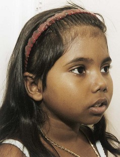
Fig. 35-5 A young girl with β-thalassemia demonstrating mild frontal bossing of the right forehead and mild maxillary prominence. (Courtesy James DeLeon, Texas Children’s Hospital, Houston.)

Anemia results from the body’s inability to maintain a level of erythropoiesis commensurate with hemolysis. The bone marrow compensates by producing large numbers of immature cells, such as normoblasts and erythroblasts; large cells that are extremely thin and form bizarre shapes; and target cells, which have abnormal staining properties. As a result of the excessive production of abnormal RBCs, their life span is severely shortened.

Aplastic crises after infection, folic acid deficiencies from the demands of bone marrow hyperplasia, and progressive hemolysis from repeated blood transfusions all worsen anemia. The spleen becomes greatly enlarged as a result of extramedullary hematopoiesis, rapid destruction of the defective erythrocytes, and, rarely, progressive fibrosis from hemochromatosis. Splenomegaly may progress until the organ’s very size interferes with the function of other abdominal organs and respiratory expansion.

With progressive anemia, signs of chronic hypoxia—namely, headache, irritability, precordial and bone pain, decreased exercise tolerance, listlessness, and anorexia—may develop. Another common symptom in these children is frequent epistaxis, although the exact reason is unknown. Hyperuricemia and gout from rapid cellular catabolism also occur.

Hemosiderosis refers to excess iron storage in various tissues of the body, especially the spleen, liver, lymph glands, heart, and pancreas, but without associated tissue injury. Hemochromatosis refers to excess iron storage that results in cellular damage. It is not known how iron storage causes tissue destruction. Chronic hypoxia is believed to be an important contributing factor.

In thalassemia, excess of hemosiderin, the iron-containing pigment from the breakdown of hemoglobin, results from decreased hemoglobin synthesis and increased hemolysis of transfused erythrocytes. Decreased production of hemoglobin results in an excess supply of available iron. In addition, the body probably responds to the anemia by increasing the rate of gastrointestinal absorption of dietary iron, since ineffective erythropoiesis is a potent controlling factor in exogenous iron use. However, the primary source of additional iron is from the hemolysis of supplemental erythrocytes and the rapid destruction of defective RBCs. With the prophylactic use of deferoxamine and/or oral chelators (deferiprone and deferasirox) to minimize excess iron storage, the characteristic changes in body structures from hemochromatosis have been greatly reduced.

Retarded growth and, especially, delayed sexual maturation are common findings. There is evidence that both may also be caused by pituitary failure, although the exact reasons for this are unclear, but the impaired growth is probably also related to hemochromatosis. It is possible that the endocrine glands are extremely sensitive to iron toxicity and that even small amounts of deposited iron can produce organ dysfunction. Children with severe disease usually exhibit significant growth retardation. The development of secondary sexual characteristics is delayed or absent in many adolescents (Cunningham, Sankaran, Nathan, et al, 2009).

Diagnostic Evaluation

Hematologic studies reveal characteristic changes in the RBCs (e.g., microcytosis, hypochromia, anisocytosis, poikilocytosis, target cells, and basophilic stippling of various stages). Low hemoglobin and hematocrit levels often occur in severe anemia, although they are typically less pronounced than the reduction in the RBC count because of the proliferation of immature erythrocytes.

Hemoglobin electrophoresis confirms the diagnosis and is helpful in distinguishing the type and severity of the thalassemia because it analyzes the quantity and kind of hemoglobin variants found in the blood. In β-thalassemia, levels of HgbF and HgbA2 (a type of normal adult hemoglobin) are elevated because neither depends on β chain polypeptides for synthesis.

Therapeutic Management

The objective of supportive therapy is to maintain sufficient hemoglobin levels to prevent bone marrow expansion and bony deformities and to provide sufficient RBCs to support growth and normal physical activity. Transfusions are the foundation of medical management, with a goal of maintaining the hemoglobin level above 9.5 g/dl, an aim that may require transfusions as often as every 3 weeks. The advantages of this therapy include (1) improved physical and psychologic well-being because of the ability to participate in normal activities, (2) decreased cardiomegaly and hepatosplenomegaly, (3) fewer bone changes, (4) normal or near-normal growth and development until puberty, and (5) fewer infections.

One of the potential complications of frequent blood transfusions is iron overload (hemosiderosis). Because the body has no effective means of eliminating the excess iron, the mineral is deposited in body tissues. To minimize the development of hemosiderosis and hemochromatosis, deferoxamine, an iron-chelating agent, is given with oral supplements of vitamin C. Vitamin C should be given only to patients who are ascorbate depleted and only while deferoxamine is being administered. Administration of vitamin C significantly augments iron excretion in response to deferoxamine, particularly in patients with vitamin C deficiency (Cunningham, Sankaran, Nathan, et al, 2009). As postulated, vitamin C may delay the conversion of ferritin to hemosiderin, which allows more iron to remain in chelatable form.

Deferoxamine is given intravenously or subcutaneously at home via a portable infusion pump over a period of 8 to 10 hours (usually during sleep) for 5 to 7 days a week. Significant liver fibrosis, cardiac dysfunction and growth impairment may be prevented if chelation therapy is adequate during childhood (Cunningham, Sankaran, Nathan, et al, 2009). Therefore adherence to an intensive schedule is required for substantial chelation therapy. The availability of oral chelators (deferiprone and deferasirox) is a major advance in the care of patient undergoing long-term transfusion therapy. Deferasirox was approved by the FDA in 2004 for the treatment of patients 2 years and older with chronic iron overload secondary to recurrent blood transfusions (Cunningham, Sankaran, Nathan, et al, 2009; Raphael, Bernhardt, Mahoney, et al, 2009). A daily dose of 20 to 30 mg/kg of deferasirox is generally well tolerated, with mild gastrointestinal events and rash as common toxicities (Cohen, Glimm, and Porter, 2008). Deferiprone is currently available only on a compassionate-use basis in the United States (Neufeld, 2008).

image DRUG ALERT

Chelation

New strategies for chelation, such as combination parenteral and oral chelation therapy and organ-targeted chelation, may soon have a considerable impact on the quality of life of patients with thalassemia (Cohen, Galanello, Rennell, et al, 2004; Naithani, Chandra, and Sharma, 2005; Cunningham, Sankaran, Nathan, et al, 2009).

New methods to assess cardiac and liver iron overload using magnetic resonance imaging have recently been developed (Cohen, Galanello, Rennell, et al, 2004; Hankins and Aygun, 2009).

In some children with severe splenomegaly who require repeated transfusions, a splenectomy may be necessary to decrease the disabling effects of abdominal pressure and to increase the life span of supplemental RBCs. Over time the spleen may accelerate the rate of RBC destruction and therefore increase transfusion requirements. After a splenectomy children generally require fewer transfusions, although the basic defect in hemoglobin synthesis remains unaffected. A major postsplenectomy complication is severe and overwhelming infection. Therefore these children are often on prophylactic antibiotics with close medical supervision for many years and should receive the pneumococcal and meningococcal vaccines in addition to regularly scheduled immunizations. (See Immunizations, Chapter 12.)

Prognosis: Most children treated with blood transfusions and early chelation therapy survive well into adulthood (Cunningham, Sankaran, Nathan, et al, 2009). The most common causes of death are heart disease, postsplenectomy sepsis, and multiorgan failure secondary to hemochromatosis (Cunningham, Sankaran, Nathan, et al, 2009). A curative treatment for some children is HSCT. (See Chapter 36.) Children younger than 16 years of age who undergo allogeneic HSCT have a high rate of complication-free survival; approximately 80% of these children are cured (Lucarelli and Gaziev, 2008). An experimental approach for correction of thalassemia through the introduction of new genetic material into pluripotent stem cells is ongoing but continues to have shortcomings (Cunningham, Sankaran, Nathan, et al, 2009; Ye, Chang, Lin, et al, 2009).

Nursing Care Management

The objectives of nursing care are to (1) promote compliance with transfusion and chelation therapy, (2) assist the child in coping with the anxiety-provoking treatments and the effects of the illness, (3) foster the child’s and family’s adjustment to a chronic illness, and (4) observe for complications of multiple blood transfusions. Basic to each of these goals is explaining to parents and older children the defect responsible for the disorder, its effect on RBCs, and the potential effects of untreated hemosiderosis (such as delayed growth and maturation and heart disease). Because this condition is prevalent among families of Mediterranean descent, the nurse also inquires about the family’s previous knowledge about thalassemia. All families with a child with thalassemia should be tested for the trait and referred for genetic counseling.

Support the Family: As with any chronic illness, the family’s needs must be met for optimum adjustment to the stresses imposed by the disorder. (See Chapter 22.) Sources of information for the family are the Cooley’s Anemia Foundation* and the Thalassemia Action Group.* Genetic counseling for the parents and fertile offspring is mandatory, and both prenatal diagnosis using amniocentesis or fetal blood sampling and screening for thalassemia trait are available. There has been a marked decline in the number of new cases of thalassemia worldwide. This may be a result of education and testing of parents.

Assist in Coping with the Effects of the Disorder: Body image alterations, decreased growth, and sexual immaturity are frequently difficult adjustment problems for older children. These children feel different from their peers, and the delayed sexual development is a major issue for the maturing adolescent with an improved life expectancy. Adolescents need an opportunity to express their thoughts and feelings about these complex issues. They can learn grooming measures that make them appear more sexually mature, such as wearing up-to-date clothing, adopting new hairstyles, and wearing well-applied makeup. Children with the characteristic bone changes may benefit from surgery or use of orthodontic appliances to improve facial structure.

With frequent transfusion therapy there is less restriction on physical activity because of severe anemia, and the nurse should encourage these children to pursue activities that they are able to tolerate. The frequency of treatment, however, can interfere with a normal lifestyle. To minimize disruptions and improve cooperation, the nurse can help arrange for blood transfusions and medical supervision at times that interfere least with the child’s regular activities, especially school.

Anemias Caused by Impaired or Decreased Production of Red Blood Cells

Impaired or decreased production of RBCs can occur as a result of either bone marrow failure or deficiency of essential nutrients. Bone marrow failure may be caused by (1) replacement of bone marrow by fibrous tissue or by neoplastic cells, as in leukemia; (2) depression of marrow activity by irradiation, chemicals, or drugs; and (3) interference with bone marrow activity caused by systemic disorders such as severe infection, chronic renal disease, widespread malignancy (without marrow infiltration), collagen diseases, or hypothyroidism. When depression of the hematologic system is extensive, aplastic anemia develops.

The reason systemic disorders affect erythrocyte production varies according to the condition. For example, in severe chronic infection there is evidence that depression of erythropoiesis is caused by a defect in the conversion of protoporphyrin into hemoglobin. In addition, there is some degree of hemolysis, although the exact mechanism is not known.

Aplastic Anemia

Aplastic anemia refers to a condition in which production of all formed elements of the blood is simultaneously depressed. The peripheral blood smear demonstrates pancytopenia or the triad of profound anemia, leukopenia, and thrombocytopenia. Hypoplastic anemia is characterized by a profound depression of RBC formation but normal or slightly decreased production of WBCs and platelets. One type of hypoplastic anemia is pure RBC aplasia, which can be congenital or acquired. The acquired defect in erythropoiesis is an autoimmune condition that occurs mostly in adults (Brodsky and Jones, 2005; Young, Calado, and Scheinberg, 2006). The congenital condition (Diamond-Blackfan syndrome) is marked by complete or almost complete absence of all cells of the erythroid series with normal production of the other myeloid cells. Its treatment, which consists of transfusions, splenectomy, and administration of corticosteroids, is similar to that for other diseases that result in profound anemia, such as the thalassemias. The prognosis varies, although long-term survival is possible. The principal causes of death are cardiac failure, hepatitis from transfusion therapy, and sepsis. Hemosiderosis and hemochromatosis (see p. 1437) also affect vital tissues necessary for survival.

Aplastic anemia can be primary (congenital, or present at birth) or secondary (acquired). The best-known congenital disorder of which aplastic anemia is an outstanding feature is Fanconi syndrome, a rare hereditary disorder that is characterized by pancytopenia, hypoplasia of the bone marrow, and patchy brown discoloration of the skin due to the deposition of melanin. It is associated with multiple congenital anomalies of the musculoskeletal and genitourinary systems. The syndrome appears to be inherited as an autosomal recessive trait with varying penetrance; therefore affected siblings may demonstrate different combinations of defects.

Several factors contribute to the development of acquired hypoplastic anemia, including suppressed erythropoiesis from multiple-transfusion therapy; hemolytic syndromes (such as SCA); and autoimmune or allergic states. Box 35-6 lists the most common causes of acquired aplastic anemia. The following discussion focuses on acquired severe aplastic anemia, which carries a poorer prognosis and follows a more rapidly fatal course than the primary types.

BOX 35-6   COMMON CAUSES OF ACQUIRED APLASTIC ANEMIA

• Human parvovirus infection, hepatitis, or overwhelming infection

• Irradiation

• Immune disorders such as hypoimmunoglobulinemia

• Drugs such as certain chemotherapeutic agents, anticonvulsants, and several antibiotics

• Industrial and household chemicals, including benzene and its derivatives, which are found in petroleum products, dyes, paint remover, shellac, and lacquers

• Infiltration and replacement of myeloid elements, such as in leukemia or the lymphomas

• Idiopathic—In most cases no identifiable precipitating cause found

Diagnostic Evaluation

The onset of clinical manifestations, which include anemia, leukopenia, and decreased platelet count, is usually insidious, not unlike that seen in leukemia. Definitive diagnosis is made by examination of bone marrow aspirates, which demonstrate the conversion of red bone marrow to yellow, fatty bone marrow.

Therapeutic Management

The objectives of treatment are based on the recognition that the underlying disease process is failure of the bone marrow to carry out its hematopoietic functions. Therefore therapy is directed at restoring function to the marrow and involves two main approaches: (1) immunosuppressive therapy to counter the presumed immunologic responses that prolong aplasia, and (2) replacement of the bone marrow through transplantation. Bone marrow transplantation is the treatment of choice for severe aplastic anemia when a suitable donor exists.

Immunosuppressive therapy is an alternative first-line treatment for children with acquired aplastic anemia who do not have a matched sibling bone marrow donor (Pongtanakul, Das, Charpentier, et al, 2008). These strategies are based on the use of antilymphocyte globulin and antithymocyte globulin (ATG). The two products are similar; therefore the terms are used interchangeably here.

The use of immunosuppressive therapy, including cyclosporin A (CSA) and ATG, with the addition of human recombinant granulocyte or granulocyte-macrophage colony-stimulating factor (G-CSF or GM-CSF) and methylprednisolone (to prevent ATG serum sickness), has greatly improved the prognosis for patients with aplastic anemia. The rationale for using ATG is the possibility that aplastic anemia may be a result of autoimmunity.

image DRUG ALERT

ATG, CSA, and G-CSF

ATG and CSA suppress T-cell–dependent autoimmune responses but do not cause bone marrow suppression. The medications are usually given intravenously over 12 to 16 hours daily for 4 days, after a test dose to check for hypersensitivity. A repeat course may be given, depending on the reduction in circulating lymphocytes and the patient’s response (see Nursing Care Management). CSA is administered orally for several weeks to months. G-CSF or GM-CSF, given parenterally, enhances bone marrow production.

In children who fail to respond to therapy with ATG, CSA, and growth factors, success has been achieved using high-dose cyclophosphamide as an effective immunosuppressive agent. Androgens may be used with ATG to stimulate erythropoiesis if the aplastic anemia is nonresponsive to initial therapies.

HSCT should be considered early in the course of the disease if a compatible donor can be found. Transplantation is more successful if performed before multiple transfusions have sensitized the child to leukocyte and HLA antigens. Children who are eligible for transplantation should be transferred to one of the medical centers that specialize in this procedure. Many different preparative regimens are available, and all aim to decrease the rate of graft-versus-host disease. All regimens include immunosuppressive therapy, and some also include irradiation (either total body or thoracoabdominal). Patients who have received a large number of transfusions before bone marrow transplantation have a higher rejection rate and lower survival rate (Marsh, 2005; Young, Calado, and Scheinberg, 2006). With the use of immunosuppressive therapy and HLA-identical sibling donor, the allogeneic bone marrow transplantation offers 90% chance of long-term survival (Hord, 2007; Trigg, 2004; Marsh, 2005).

Nursing Care Management

The care of the child with aplastic anemia is similar to the care of the child with leukemia (i.e., preparing the family for the diagnostic and therapeutic procedures, preventing complications from the severe pancytopenia, and emotionally supporting the family regarding the potentially fatal outcome). (See Chapters 23 and 36.) Because each of these nursing care interventions has been covered elsewhere, only the interventions specific to aplastic anemia are presented here. (Chapter 36 discusses bone marrow transplantation.)

image DRUG ALERT

ATG

During administration of ATG, whether into a central venous catheter or peripheral vein, the nurse must pay special attention to the infusion to prevent extravasation. Meticulous care of the venous access catheter is essential because of the child’s susceptibility to infection. Although anaphylactic reactions to ATG are rare, make emergency preparations in advance, and have epinephrine and oxygen readily available. The nurse should observe for immediate reactions to ATG, which include fever and skin rash. Delayed reactions (serum sickness) may also occur within 7 to 14 days of a course of ATG, and the manifestations are similar to those in immediate reactions. The symptoms are reversed, and in the case of serum sickness may be prevented, with corticosteroids.

Because growth factors are usually given subcutaneously over several days, an anesthetic cream, EMLA, may be used to minimize pain at the injection site. Because chemotherapeutic agents may be used, many of the common reactions to these drugs, such as nausea and vomiting, alopecia, and mucosal ulceration, may be encountered. In addition, the oral area mucosa may have extensive ecchymotic areas from thrombocytopenia that require meticulous mouth care to prevent breakdown, bleeding, and infection. Fortunately, these lesions, which look painful, cause little or no discomfort. Local anesthetics are not necessary, but anorexia is still a consequence because of the edematous nature of the lesions. Liquid, bland, and soft diets are usually tolerated best. (See Feeding the Sick Child, Chapter 27.)

Defects in Hemostasis

Hemostasis is the process that stops bleeding when a blood vessel is injured. Vascular and plasma clotting factors, as well as platelets, are required. A complex system of clotting, anticlotting, and clot breakdown (fibrinolysis) mechanisms exists in equilibrium to ensure clot formation only in the presence of blood vessel injury and to limit the clotting process to the site of vessel wall injury. Dysfunction in these systems leads to bleeding or abnormal clotting.

Mechanisms Involved in Normal Hemostasis

image Understanding the role that factor deficiencies play in promoting bleeding tendencies requires a review of the normal coagulation process of the blood. Although the coagulation process is complex, clotting depends on three main elements: vascular events, platelets, and clotting factors.

imageCritical Thinking Exercise—Bleeding
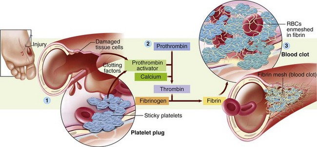
Vascular Events

At the time and site of injury, several events occur to initiate hemostasis: local vasoconstriction, compression of the blood vessels by extravasated blood, and release of von Willebrand factor by endothelial walls. Collagen present in exposed subendothelial cells acts as a site for platelet adhesion.

Platelets

Normally the platelets do not adhere to each other or to normal endothelium. However, at the time a blood vessel is injured, the following events take place: Platelet adhesion occurs at the site of the injury, providing a plug. The platelets change shape, develop pseudopods, and release a variety of chemicals to stimulate vasoconstriction and vessel repair and to activate and recruit more platelets to the injury site. Receptor sites are located on the platelets for fibrinogen and other adhesive proteins, which cause the platelets to stick together (aggregation). As the membranes of the platelets change, the phospholipids necessary for blood coagulation are exposed so that fibrin, which secures the platelet plugs to the site, can be produced. Finally, the clot compresses and is secured to the injury.

Defects in platelets and clotting factors are the most common causes of bleeding during childhood. The following discussion focuses on the major conditions that require nursing intervention.

Clotting Factors

image The clotting factors (Table 35-4 and Fig. 35-6) are activated in sequence to develop a fibrin clot. Two mechanisms exist that can generate prothrombin to produce thrombin:

TABLE 35-4

BLOOD-CLOTTING FACTORS

FACTOR NUMBER SYNONYMS
I Fibrinogen
II Prothrombin
III Platelet factor 3, thromboplastin
IV Calcium
V Labile factor, proaccelerin, Ac globulin
VII Serum prothrombin conversion accelerator (SPCA), proconvertin, stable factor
VIII Antihemophilic factor (AHF)
IX Plasma thromboplastin component (PTC), Christmas factor
X Stuart-Prower factor
XI Plasma thromboplastin antecedent (PTA)
XII Hageman factor
XIII Fibrin-stabilizing factor (FSF)
KAL Prekallikrein, Fletcher factor
HMK High-molecular-weight kininogen, Fitzgerald factor

image

Fig. 35-6 Blood clotting. The extremely complex clotting mechanism can be distilled into three basic steps: (1) release of clotting factors from both injured tissue cells and sticky platelets at the injury site (which form a temporary platelet plug); (2) series of chemical reactions that eventually result in the formation of thrombin; and (3) formation of fibrin and trapping of red blood cells (RBCs) to form a clot. (From Thibodeau GA, Patton KT: The human body in health and disease, ed 5, St Louis, Mosby, 2010.)

imageAnimation—Platelets and Blood Clotting

1. Intrinsic pathway—Factor XII, high-molecular-weight kininogen (HMK, Fitzgerald factor), and prekallikrein (KAL, Fletcher factor) react on a negatively charged surface (contact activation reaction) to activate factor XI (PTA, plasma thromboplastin antecedent). The partial thromboplastin time measures abnormalities in the intrinsic pathway (abnormalities in factors I, II, V, VIII, IX, X, XII, HMK, and KAL).

2. Extrinsic pathway—A lipoprotein tissue factor stimulates activation of factor VII. The prothrombin time measures abnormalities of the extrinsic pathway (abnormalities in factors I, II, V, VII, and X).

Table 35-5 presents laboratory tests to assess hemostasis.

TABLE 35-5

LABORATORY TESTS TO ASSESS HEMOSTASIS*

image

ASA, Acetylsalicylic acid; NSAIDs, nonsteroidal antiinflammatory drugs.

*Normal values are listed in Appendix C.

Hemophilia

The term hemophilia refers to a group of bleeding disorders resulting from congenital deficiency of specific coagulation proteins (Montgomery, Gill, and Di Paola, 2009; Sharathkumar and Pipe, 2008). Although the symptomatology is similar regardless of which clotting factor is deficient, the identification of specific factor deficiencies has allowed definitive treatment with replacement agents.

In about 80% of all cases of hemophilia, the inheritance pattern is demonstrated as X-linked recessive. (See Chapter 5.) The two most common forms of the disorder are factor VIII deficiency (hemophilia A, or classic hemophilia) and factor IX deficiency (hemophilia B, or Christmas disease), with prevalence in the general population of approximately 1 in 5000 and 1 in 50,000, respectively (Brown, 2005; Sharathkumar and Pipe, 2008). The following discussion is primarily concerned with factor VIII deficiency, which accounts for about 80% of all cases.

Modes of Transmission

Hemophilia is transmitted as an X-linked recessive disorder; however, only about 60% of affected children have a positive family history for the disease. Up to one third of all hemophilia cases may be caused by a gene mutation. The most frequent pattern of transmission is through the union of an unaffected male with a trait-carrier female. Because the treatment of persons with hemophilia has improved, the results of a union between an affected male and a normal female or a carrier female must also be considered. For example, the chances are equal (i.e., 1 in 4) that an offspring of an affected male and a carrier female will be an affected son, an affected daughter, a carrier daughter, or a normal son. Such parentage is one of the few ways in which a female inherits the disorder. Female carriers may have low levels of factor VIII and be symptomatic.

Pathophysiology and Clinical Manifestations

image The basic defect of hemophilia A is a deficiency of factor VIII (antihemophilic factor). Factor VIII is produced by the liver and is necessary for the formation of thromboplastin in phase I of blood coagulation. The less factor VIII found in the blood, the more severe the disease.

imageAnimation—Hemophilia A

A major feature of hemophilia is that its expression varies markedly with regard to the degree of bleeding severity. Hemophilia is generally classified into three groups according to the severity of the factor deficiency; 60% to 70% of children with hemophilia demonstrate the severe form of the disorder (Table 35-6).

TABLE 35-6

CLINICAL SEVERITY OF HEMOPHILIA

CLINICAL SEVERITY FACTOR VIII ACTIVITY BLEEDING TENDENCY
Severe <1% Spontaneous bleeding without trauma
Moderate 1%-5% Bleeding with trauma
Mild >5%-40% Bleeding with severe trauma or surgery

The effect of hemophilia is prolonged bleeding anywhere from or in the body. With severe factor deficiencies, hemorrhage can occur as a result of minor trauma, such as after circumcision, during loss of deciduous teeth, or as a result of a slight fall or bruise. In children with less severe deficiencies, however, the bleeding tendency may not be noted until the onset of walking.

Subcutaneous and intramuscular hemorrhages are common. Hemarthrosis, which refers to bleeding into the joint cavities, especially the knees, elbows, and ankles, is the most frequent form of internal bleeding. Bony changes and crippling deformities occur after repeated bleeding episodes over several years. Early signs of hemarthrosis are a feeling of stiffness, tingling, or ache in the affected joint, followed by a decrease in the ability to move the joint. Obvious signs and symptoms are warmth, redness, swelling, and severe pain with considerable loss of movement. Spontaneous hematuria is not uncommon. Epistaxis may occur but is not as frequent as other kinds of hemorrhage. Petechiae are uncommon in persons with hemophilia because repair of small hemorrhages depends on platelet function, not on blood-clotting mechanisms.

Bleeding into the tissue can occur anywhere but is serious if it occurs in the neck, mouth, or thorax because the airway can become obstructed. Intracranial hemorrhage can have fatal consequences and is one of the major causes of death. Hemorrhage anywhere along the gastrointestinal tract can lead to anemia, and bleeding into the retroperitoneal cavity is especially hazardous because of the large space for blood to accumulate. Hematomas in the spinal cord can cause paralysis.

Diagnostic Evaluation

The diagnosis is usually made from a history of bleeding episodes, evidence of X-linked inheritance (only one third of cases are new mutations), and laboratory findings. To understand the significance of various tests of hemostasis, it is helpful to recall the usual mechanisms to control bleeding (i.e., the function of platelets and clotting factors). The results of tests that measure platelet function, such as the bleeding time, are all normal in persons with hemophilia, whereas the results of tests that assess clotting factor function may be abnormal (see Table 35-5). The tests specific for hemophilia include factor VIII and IX assays, procedures normally done by specialized laboratories. Other tests are those that depend on specific factors for a reaction to occur, especially the partial thromboplastin time. Carrier detection is possible in classic hemophilia using DNA testing and is an important consideration in families in which female offspring may have inherited the trait.

Therapeutic Management

The primary therapy for hemophilia is replacement of the missing clotting factor. The products currently available are factor VIII concentrates, either produced through genetic engineering (recombinant form) or derived from pooled plasma, which are reconstituted with sterile water immediately before use. A synthetic form of vasopressin, 1-deamino-8-d-arginine vasopressin (DDAVP), is the treatment of choice in mild hemophilia and von Willebrand disease (vWD) (types I and IIA only) if the child shows an appropriate response. After DDAVP administration a threefold to fourfold rise in factor VIII activity should occur. Because the goal is to raise the factor VIII level at least 30%, patients with moderate factor VIII deficiency do not benefit. In addition, various therapies are employed when bleeding occurs or is anticipated (Table 35-7).

TABLE 35-7

ADJUNCT THERAPIES FOR HEMOPHILIA A

image

image DRUG ALERT

Cryoprecipitate

Cryoprecipitate is no longer recommended for use in treating factor VIII deficiency. Since the availability of highly purified factor VIII concentrate (monoclonal) in 1988 and the licensing of recombinant factor VIII concentrate in 1992 (marketed not as a blood product but as a drug), the National Hemophilia Foundation has advised practitioners to use only these products. Cryoprecipitate cannot be treated to safely eliminate hepatitis or human immunodeficiency virus (HIV).

Aggressive factor concentrate replacement therapy is initiated to prevent chronic crippling effects from joint bleeding. If replacement therapy begins immediately, local measures such as ice applications and splinting are seldom needed. Other drugs may be included in the therapy plan, depending on the source of the hemorrhage. Corticosteroids are given for hematuria, acute hemarthrosis, and chronic synovitis. It is recommended that patients with hemophilia avoid aspirin and nonsteroidal antiinflammatory drugs (NSAIDs) because they inhibit platelet function (Scott and Montgomery, 2007). However, NSAIDs such as ibuprofen are effective in relieving pain caused by synovitis and are occasionally used with caution (Curry, 2004). Oral use of ε-aminocaproic acid (EACA, Amicar) prevents clot destruction. Its use is limited to mouth trauma or surgery with a dose of factor concentrate given first. The child may rinse the mouth with this medication and then swallow it.

A regular program of exercise and physical therapy is an important aspect of management. If started early and continued throughout adulthood, planned, individualized physical activity strengthens muscles around joints and may decrease the number of spontaneous bleeding episodes.

image NURSING ALERT

Passive range-of-motion exercises should never be part of an exercise regimen after an acute episode because the joint capsule could easily be stretched and bleeding could recur. Active range-of-motion exercises are best so that the patient can gauge his or her own pain tolerance.

Treatment without delay results in more rapid recovery and a decreased likelihood of complications; therefore most children are treated at home. The family should learn how to perform venipuncture and how to administer factor VIII to children over 2 to 3 years of age. The child learns the procedure for self-administration at 8 to 12 years of age. Home treatment is highly successful, and the rewards, in addition to the immediacy, are less disruption of family life, fewer school or work days missed, and enhancement of the child’s self-esteem and independence.

Prophylactic therapy is periodic factor replacement for children with severe hemophilia to prevent bleeding complications, including arthropathy and spontaneous and life-threatening bleeding events (Scott and Montgomery, 2007; Sharathkumar and Pipe, 2008; Montgomery, Gill, and Di Paola, 2009). Primary prophylaxis in patients with severe hemophilia has been practiced for many years in developed countries and has proved to be effective in preventing naturopathy. In primary prophylaxis, factor VIII concentrate is infused on a regular basis before the onset of joint damage. Secondary prophylaxis involves the infusion of factor VIII concentrate on a regular basis after the child experiences his or her first joint bleed. The infusions are given every other day or three times a week for several weeks to promote healing. Episodic factor replacement may be a cost-effective alternative to primary prophylaxis, but prophylaxis decreases the development of joint disease compared to on-demand treatment (Manco-Johnson, Abshire, Shapiro, et al, 2007). However, prompt appropriate treatment of hemorrhage and prophylactic therapy are key to excellent care and prevention of long-term morbidity in patients with hemophilia (Montgomery, Gill, and Di Paola, 2009).

Prognosis: The progress made in hemophilia care over the years has been striking. The advent of home infusion therapy coupled with recent advances in producing safer and more effective factor concentrates has revolutionized the treatment and management of hemophilia (Montgomery, Gill, and Di Paola, 2009). Early recognition of joint and muscle bleeds is emphasized, since immediate adequate treatment with clotting factor is possible using home infusion therapy. Early treatment has significantly reduced the morbidity formerly associated with hemophilia. The availability of comprehensive hemophilia treatment centers offers the child with hemophilia and the family a coordinated multidisciplinary approach to meeting their needs and improving the child’s health and well-being.

Although there is no cure for hemophilia, its symptoms can be controlled and its potentially crippling deformities markedly reduced or even avoided. Today many children with hemophilia function with minimal or no joint damage. They have an average life expectancy and are normal in every aspect but one: they have a tendency to bleed, which is a significant inconvenience but not necessarily a life-threatening event.

Unfortunately, those individuals with hemophilia who were treated before the development of current purification techniques for factor VIII concentrate (between 1979 and 1985) may have been exposed to HIV. It is estimated that more than 50% of these patients seroconverted to HIV-positive status, and 30% developed acquired immunodeficiency syndrome (AIDS) (Butler, Schultz, Forsberg, et al, 2003). Individuals with hemophilia diagnosed since the 1990s and treated with recombinant factor products are at virtually no risk for developing HIV infection from treatment. Recombinant factor VIII and factor IX products that are devoid of human protein materials have become the treatment of choice for children and previously untreated hemophilia patients (Brown, 2005; Montgomery, Gill, and Di Paola, 2009).

Gene therapy may prove to be a treatment option in the future. Techniques are under development to introduce the factor VIII or IX genes into hepatocytes, fibroblasts, and endothelial cells using adeno-associated viral vectors (National Hemophilia Foundation and American Red Cross, 2005; Nienhuis, 2008). The scientific community remains undaunted in its attempt to make gene-addition therapy a twenty-first century reality for patients with hemophilia A and B. Gene therapy is expected to be available in the next 5 to 10 years, but many problems remain, including selection of both appropriate vectors and the appropriate cell in which to express the gene (Montgomery, Gill, and Di Paola, 2009).

Nursing Care Management

The earlier a bleeding episode is recognized, the more effectively it can be treated. Signs that indicate internal bleeding are especially important to recognize. Children are aware of internal bleeding and are reliable in telling the examiner the location of an internal bleed. In addition, the nurse maintains a high level of suspicion when a child with hemophilia shows signs such as headache; slurred speech; loss of consciousness (from cerebral bleeding); and black, tarry stools (from gastrointestinal bleeding).

QUALITY PATIENT OUTCOMES

Hemophilia

• Early recognition of signs and symptoms of hemophilia

• Bleeding episodes prevented

• Bleeding episodes treated early with factor replacement

• Adherence to prophylactic factor replacement program when indicated

• Hemarthrosis prevented when possible with limited joint damage

• Exercise program and physical therapy ongoing

Prevent Bleeding: The goal of prevention of bleeding episodes involves decreasing the risk of injury. Measures are geared mostly toward encouraging appropriate exercises to strengthen muscles and joints and to allow age-appropriate activity. During infancy and toddlerhood the normal acquisition of motor skills creates innumerable opportunities for falls, bruises, and minor wounds. Restraining the child from mastering motor development can bring more serious long-term problems than allowing the behavior. However, the environment should be made as safe as possible, with close supervision maintained during playtime to minimize incidental injuries.

For older children the family usually needs assistance in preparing the child for school. A nurse who knows the family can assist in discussing the situation with the school nurse and in joint planning of an appropriate activity schedule. Because almost all individuals with hemophilia are boys, the physical limitations with regard to active sports may require a difficult adjustment, and activity restrictions must be tempered with sensitivity to the child’s emotional and physical needs. Children should always use appropriate safety equipment. Children and adolescents with severe hemophilia can participate in noncontact sports such as swimming, golf, walking, jogging, fishing, and bowling. Football, boxing, hockey, soccer, and rugby are strongly discouraged because the risk of injury outweighs the physical and psychosocial benefits of participating in these sports (National Hemophilia Foundation and American Red Cross, 2005; Scott and Montgomery, 2007).

To prevent oral bleeding, some readjustment in dental hygiene may be needed to minimize trauma to the gums, such as using a water irrigating device, softening the toothbrush in warm water before brushing, or using a sponge-tipped disposable toothbrush. If a regular toothbrush is used, it should be soft bristled and small. Adolescents also need to be advised of the dangers of using safety razors with blades and should use an electric shaver.

Because any trauma can lead to a bleeding episode, all persons caring for these children must be aware of their disorder. These children should wear medical identification, and the nurse should encourage older children to recognize situations in which disclosing their condition is important, such as during dental extractions or injections. Health personnel need to take special precautions to prevent the use of procedures such as intramuscular injection. The subcutaneous route is substituted for intramuscular injection whenever possible. Venipunctures for blood samples are usually preferred by children. There is usually less bleeding after venipuncture than after finger or heel puncture. Neither aspirin nor any aspirin-containing compound should be used. Acetaminophen is a suitable aspirin substitute, especially for use during control of pain at home.

Recognize and Control Bleeding: The earlier a bleeding episode is recognized, the more effectively it can be treated. Factor replacement therapy should be instituted according to established medical protocol, and supportive measures may be implemented, such as RICE, which consists of (1) rest, (2) ice, (3) compression, and (4) elevation. When parents and older children learn such measures beforehand, they can be prepared to initiate immediate treatment before blood loss is excessive. Keep plastic bags of ice or cold packs in the freezer for such emergencies. However, such measures should not take the place of factor replacement.

Prevent Crippling Effects of Bleeding: As a result of repeated episodes of hemarthrosis, incompletely absorbed blood in the joints and limitation of motion, bone and muscle changes occur that may result in flexion contractures and joint fixation. Obviously prevention of bleeding is the ideal goal. However, because spontaneous bleeding is not uncommon in persons with severe hemophilia, definitive measures, including replacement therapy and physical therapy, are necessary to limit joint damage.

During bleeding episodes the joint is elevated and immobilized. Active range-of-motion exercises are usually instituted after the acute phase. This allows the child to control the degree of exercise according to the level of discomfort. Physical therapy is beneficial to promote maximum function of the joint and unaffected body parts. Success of a physical therapy plan involves control of pain by administering analgesics before therapy and adjusting the dose to provide maximum benefit.

If an exercise program is initiated in the home, a physical therapist or public health nurse may need to supervise compliance with the regimen. Rarely, orthopedic intervention, such as casting, application of traction, or aspiration of blood, may be necessary to preserve joint function. Diet is also an important consideration because excessive body weight can increase the strain on affected joints, especially the knees, and predispose the child to hemarthrosis. Consequently, children need calories that meet their energy requirements.

Support the Family and Prepare for Home Care: The development of factor concentrates has greatly changed the outlook for these children. Bleeding can be minimized, and the child can live a much more normal, unrestricted life. Children should learn to take responsibility for their disease at an early age. They learn their limitations, preventive measures, and self-administration of the factor replacement.

The needs of families who have children with hemophilia are best met through a comprehensive team approach involving physicians (pediatrician, hematologist, orthopedist), nurse practitioner, nurse, social worker, and physical therapist.

Parent-group discussions with similarly affected families are beneficial and address certain needs better than health care providers could. For example, with the improved prognosis for these children, adolescents with hemophilia and their parents face vocational and financial problems in addition to concern over future childbearing. Once children reach 21 years of age, many insurance companies will no longer insure them. This can be disastrous because of the cost of treatment, which can exceed $100,000 per year. The National Hemophilia Foundation* and the Canadian Hemophilia Society provide numerous services and publications for both health care providers and families.

Individuals who have become infected with HIV through transfusions and factor replacement products face the consequences of this dreaded disease. Consequently they need the support of health professionals, especially with regard to instruction in safe sexual practices to avoid disease transmission and public education regarding AIDS and ways to deal with public reactions to those who have AIDS. (See Nursing Care Plan: The Child with Hemophilia, in Wilson and Hockenberry, 2008.)

Identify Persons at Risk: Genetic counseling is essential as soon as possible after diagnosis. Unlike in many other disorders in which both parents carry the trait, the feeling of responsibility for this condition usually rests with the mother. Unless she has an opportunity to discuss her feelings, the couple’s relationship may suffer. Prenatal DNA testing can identify affected fetuses and identify carriers in most cases.

von Willebrand Disease

vWD is a hereditary bleeding disorder characterized by a deficiency of or defect in a protein called von Willebrand factor (vWF). The vWF protein contributes to the adherence of platelets to damaged endothelium and serves as a carrier protein for factor VIII (De Meyer, Deckmyn, and Vanhoorelbeke, 2009; Montgomery, Gill, and Di Paola, 2009). This results in prolonged bleeding time because platelets fail to adhere to the walls of the ruptured vessel to form a platelet plug. In many centers, testing using a platelet function analyzer (PFA 100) has replaced measurement of bleeding time as the most sensitive screening test for detection of platelet dysfunction and vWD (Cariappa, Wilhite, and Parvin, 2003; Brown, 2005; Scott and Montgomery, 2007) (see Table 35-5). The disease can cause mild, moderate, or severe bleeding. Most cases of vWD are mild and require intervention only for dental and surgical procedures.

The most characteristic clinical feature of vWD is an increased tendency toward bleeding from mucous membranes. The most common symptom is frequent nosebleeds, followed by gingival bleeding, easy bruising, and excessive menstrual bleeding (menorrhagia) in females. Unlike hemophilia, vWD affects both males and females because its inheritance shows an autosomal dominant pattern. However, the treatment and final outcome are similar in both disorders. Treatment of bleeding is with DDAVP and/or a specially concentrated clotting factor known as Humate-P.

Nursing Care Management

The nursing goals are similar to those for hemophilia, with special considerations related to epistaxis. Nosebleeds are often a frightening experience for the child and parents. A calm, reassuring manner can alleviate anxiety and promote the child’s cooperation. Because most of the nosebleeding originates in the anterior part of the nasal septum, bleeding can be controlled by applying pressure to the nose with the thumb and forefinger (see Emergency Treatment box). During this time the child breathes through the mouth. If local measures are not successful at stopping the bleeding, a single dose of DDAVP is usually effective. DDAVP increases vWF and factor VIII secretion from storage in the endothelial cells (Brown, 2005; Scott and Montgomery, 2007; Sharathkumar and Pipe, 2008).

image EMERGENCY TREATMENT

Epistaxis

• Have child sit up and lean forward (not lie down).

• Apply continuous pressure to nose with thumb and forefinger for at least 10 minutes.

• Insert cotton or wadded tissue into each nostril and apply ice or cold cloth to bridge of nose if bleeding persists.

• Keep child calm and quiet.

For menorrhagia, factor replacement therapy or the administration of DDAVP may be beneficial on the first day of the menstrual cycle to lessen the flow. Teaching the adolescent methods to prevent embarrassing accidents during menstruation, such as wearing plastic-lined underpants and using double sanitary pads, helps her adjust to the inconvenience. Interestingly, these females frequently do not experience excessive bleeding at the time of delivery (Pavlovich-Danis, 2001; Montgomery, Gill, and Di Paola, 2009). This is thought to be because of increased levels of factor VIII during pregnancy. Decisions regarding childbearing are difficult because of the dominant pattern of inheritance.

Idiopathic Thrombocytopenic Purpura

image Idiopathic thrombocytopenic purpura (ITP) is an acquired hemorrhagic disorder that is characterized by (1) excessive destruction of platelets (thrombocytopenia), (2) purpura (a discoloration caused by petechiae beneath the skin), and (3) normal bone marrow with a usual increase in large, young platelets. Although the cause is unknown, the disorder is believed to represent an autoimmune response to disease-related antigens. The most common thrombocytopenia of childhood accounts for 70% to 80% of cases in children younger than 10 years of age, who recover completely within 6 months (Scott and Montgomery, 2007; Wilson, 2009).

imageCritical Thinking Case Study—Idiopathic Thrombocytopenic Purpura

The disease occurs in one of two forms: an acute, self-limiting course or a chronic course (>6 months’ duration). The acute form occurs most commonly after upper respiratory tract infections; after the childhood diseases of measles, rubella, mumps, or chickenpox; or after infection with human parvovirus.

Clinical symptoms include petechiae, bruising, bleeding from mucous membranes, and prolonged bleeding from abrasions. Symptomatic bleeding does not usually occur until the platelet count is lower than 20,000/mm3. Fatal hemorrhages have been reported in less than 1% of all patients.

Diagnostic Evaluation

In ITP the platelet count is reduced to less than 20,000/mm3; therefore results of tests that depend on platelet function, such as the tourniquet test, bleeding time, and clot retraction time, are abnormal. There is no definitive test that establishes a diagnosis of ITP; several tests are usually performed to rule out other disorders of which thrombocytopenia is a manifestation, such as systemic lupus erythematosus, lymphoma, and leukemia.

Therapeutic Management

Management of ITP is primarily supportive, since the disease is self-limiting in the majority of cases. Activity is restricted at the onset while the platelet count is low and while active bleeding or progression of lesions is occurring. Treatment for acute presentation is symptomatic and has included prednisone, IV immune globulin (IVIG), and anti-D antibody. These are not curative therapies. Some experts suggest that no therapy is necessary for asymptomatic patients because there is no difference in the recovery time of platelet counts with and without treatment (Neunert, Buchanan, Imbach, et al, 2008; Scott and Montgomery, 2007; Wilson, 2009). Anti-D antibody is a plasma-derived immunoglobulin that causes a transient hemolytic anemia in Rh(D)-positive patients with ITP. With the clearance of antibody-coated RBCs, there is prolonged survival of platelets due to the anti-D antibody blockade of the Fc receptors on the reticuloendothelial cells. The platelet count usually increases approximately 48 hours after an infusion of anti-D antibody; therefore it is not appropriate therapy for patients who are actively bleeding. The benefits of choosing anti-D antibody IV therapy over prednisone or IVIG is that anti-D antibody can be given in one dose over a period of 5 to 10 minutes and is significantly less expensive than IVIG. Historically patients who were treated with prednisone first underwent a bone marrow examination to rule out leukemia, but this is now controversial because leukemia rarely manifests with a low platelet count alone (Scott and Montgomery, 2007; Wilson, 2009). Therefore the use of anti-D antibody and IVIG alleviates the need for a bone marrow examination. Before receiving the initial dose of anti-D antibody, patients must meet certain criteria. Premedication with acetaminophen 5 to 10 minutes before infusion is recommended.

image DRUG ALERT

Anti-D Antibody

After administration of anti-D antibody, observe the child for a minimum of 1 hour and maintain a patent IV line. Obtain baseline vital sign measurements before the infusion and again 5, 20, and 60 minutes after beginning the infusion. If fever, chills, or headache occurs during or shortly after the infusion, the nurse should administer acetaminophen, diphenhydramine (Benadryl), and/or hydrocortisone (Solu-Cortef). Observe the patient for an additional hour after the anti-D antibody infusion is complete.

Splenectomy is for patients who have chronic severe ITP that is not responsive to pharmacologic management and have increased risk of severe hemorrhage. It is the only treatment associated with long-term remission for majority of these children and therefore removes the risk of hemorrhage (Scott and Montgomery, 2007; Wilson, 2009). Before splenectomy is considered, it is generally recommended to wait until the child is older than 5 years of age because of the increased risk of bacterial infection. Administration of pneumococcal, meningococcal, and H. influenzae vaccines before splenectomy is recommended (if they were not previously administered). The child also receives penicillin prophylaxis after splenectomy. The appropriate length of prophylactic therapy is controversial, but in general, a minimum of 3 years of therapy is recommended.

Prognosis: The majority of children with ITP have a self-limiting course without major complications. Some children develop chronic ITP and require ongoing therapy. A splenectomy may modify the disease process, and the child may be asymptomatic.

Nursing Care Management

Nursing care is largely supportive and should include teaching regarding the possible side effects of therapy and limitation in activities while the child’s platelet count is lower than 100,000/mm3. Children with ITP should not participate in any contact sports, bike riding, skateboarding, in-line skating, gymnastics, climbing, or running. Encourage parents to engage their children in quiet activities and to prevent any injuries to the child’s head (e.g., by having the child wear protective headgear and lining the crib with protective padding). Instruct the parents to obtain prompt medical evaluation if the child sustains head or abdominal trauma. As with any condition with an uncertain outcome, the family needs emotional support.

QUALITY PATIENT OUTCOMES

ITP

• Serious bleeding episode prevented

• Activities that increase risk for serious bleeding avoided

• Treatment administered without serious side effects

image DRUG ALERT

NSAIDs

Using aspirin or other NSAIDs to control pain has critical harmful effects for these children; therefore always use substitutes (such as acetaminophen).

Disseminated Intravascular Coagulation

DIC, also known as consumption coagulopathy, is not a primary disease but a secondary disorder of coagulation that complicates a number of pathologic processes (e.g., hypoxia, acidosis, shock, and endothelial damage [burns]) and many severe systemic disease states (e.g., congenital heart disease, necrotizing enterocolitis, gram-negative bacterial sepsis, rickettsial infections, and some severe viral infections). The hallmarks of this disorder are bleeding and clotting, which occur simultaneously.

Pathophysiology

DIC occurs when the first stage of the coagulation process is abnormally stimulated (Fig. 35-7). Although there is no well-defined sequence of events, two distinct phases can be identified. First, when the clotting mechanism is triggered in the circulation, thrombin is generated in greater amounts than the body neutralize. Consequently, there is rapid conversion of fibrinogen to fibrin with aggregation and destruction of platelets. Local and widespread fibrin deposition occurs in blood vessels. The thrombi impede the blood flow with eventual necrosis of tissues. Concurrently, the fibrinolytic mechanism is activated, which causes extensive destruction of clotting factors. With a deficiency of clotting factors, the child is vulnerable to uncontrollable hemorrhage into vital organs. An additional complication is damage to and hemolysis of RBCs.

image

Fig. 35-7 Effects of disseminated intravascular coagulation. RBC, Red blood cell.

Clinical Manifestations

The signs and symptoms of DIC are the same as those of many other diseases, which often confuses the diagnosis. There is evidence of bleeding—petechiae, purpura, bleeding from openings in the skin (e.g., a venipuncture site or surgical incision), hypotension, and dysfunction of organs from infarction and ischemia.

Diagnostic Evaluation

DIC is suspected when there is an increased tendency to bleed (Box 35-7). Hematologic findings include prolonged prothrombin, partial thromboplastin, and thrombin times. There is a profoundly depressed platelet count, fragmented RBCs, and depleted fibrinogen levels.

BOX 35-7   CLINICAL MANIFESTATIONS OF DISSEMINATED INTRAVASCULAR COAGULATION

Petechiae

Purpura

Bleeding from openings in the skin

• Venipuncture site

• Surgical incision

Bleeding from umbilicus, trachea (newborn)

Evidence of gastrointestinal bleeding

Hypotension

Organ dysfunction from infarction and ischemia

Therapeutic Management

Direct treatment toward control of the underlying or initiating cause, which in most instances stops the coagulation problem spontaneously. Administration of platelets and fresh frozen plasma may be needed to replace lost plasma components, especially in the child whose underlying disease remains uncontrolled. The extremely ill newborn infant may require exchange transfusion with fresh blood. The administration of IV heparin to inhibit thrombin formation is most often restricted to cases in which there has been no response to treatment of the underlying disease or replacement of coagulation factors and platelets.

Nursing Care Management

The goals of nursing care are to be aware of the possibility of DIC in the severely ill child and to recognize signs that might indicate its presence. The skills needed to monitor IV infusion and blood transfusions and to administer heparin are the same as for any child receiving these therapies. Because the child is usually cared for in an intensive care unit, the special needs of the family must be considered. (See Chapter 26.)

Other Hematologic Disorders

Neutropenia

Neutropenia is a reduction in the number of circulating neutrophils and is usually defined as an ANC of less than 1000/mm3 in infants 2 weeks to 1 year of age or less than 1500/mm3 in children older than 1 year of age. African-Americans have ANCs that are 200 to 600/mm3 lower than those of Caucasians (Dinauer and Newburger, 2009; Segel and Halterman, 2008). The ANC is calculated by multiplying the total WBC count by the percentage of neutrophils and bands in the differential count (see Table 35-1). When the ANC is less than 500/mm3, the risk of life-threatening infection is high (Boxer, 2007; Segel and Halterman, 2008). Several different types of neutropenia occur in children (Table 35-8). This discussion focuses on the most common type: chronic benign neutropenia.

TABLE 35-8

CLINICAL AND HEMATOLOGIC FEATURES OF SOME CONGENITAL NEUTROPENIAS

image

B, Bands; MM, metamyelocytes; PMN, polymorphonuclear leukocytes; G-CSF, granulocyte colony-stimulating factor.

Modified from Lanzkowsky P: Manual of pediatric hematology and oncology, San Diego, 2005, Academic Press.

Diagnostic Evaluation

Chronic benign neutropenia generally represents disorders characterized by mild to moderate neutropenia that are not associated with an increase in infections and often have spontaneous remissions (Boxer, 2007; Segel and Halterman, 2008). Neutropenia is often detected as an incidental finding during the evaluation of a child with fever. The ANC is usually below 500/mm3, and the only physical findings (if any) are those related to infection. Oral ulcerations and skin infections are the most common manifestation of chronic benign neutropenia. However, most children have no infections despite the markedly reduced ANC. Examination of bone marrow aspirates shows normal cellularity with absence of mature neutrophils. Antineutrophil antibodies are usually present, but their absence does not exclude the diagnosis.

To determine a child’s neutrophil response during times of infection, a steroid stimulation test may be performed. The child is given a dose of IV steroid, and the neutrophil count is measured at hourly intervals for 4 to 5 hours. If the ANC increases to more than 1000/mm3 after the dose of steroid, the child will have the same response during times of infection. Failure of the ANC to rise is an indication for increased vigilance and medical attention if the child develops fever of 38.3° C (101° F) or higher. These children may require hospitalization and aggressive treatment with broad-spectrum IV antibiotics, depending on the severity of the illness.

Therapeutic Management

Therapy to increase the ANC is rarely required. Children who have recurrent or severe infections, however, may benefit from the administration of G-CSF.

CSFs are a naturally occurring group of glycoproteins. They were first discovered and characterized because of their effect on growth and differentiation of marrow cells. Recombinant DNA technology has enabled the production of large quantities of highly purified CSFs that are nearly identical to the naturally occurring substances and have successfully increased the neutrophil count in a wide variety of neutropenic conditions (Boxer, 2007; Segel and Halterman, 2008).

Children with chronic benign neutropenia have normal cellular immunity; therefore they should receive their routine childhood immunizations.

Nursing Care Management

The care of the child with neutropenia primarily focuses on educating the parents. Instruct parents to keep their child away from large indoor crowds (e.g., grocery store on Saturday morning, movie theaters, daycare centers, church nursery) and individuals who are ill. Parents also need to seek medical attention if their child has a fever of 38.3° C (101° F) or higher, or if skin lesions develop. Because G-CSF is administered parenterally only, parents need to know how to administer subcutaneous injections.

Support the Family: Neutropenia can have many effects on family life. Some parents must quit their jobs to avoid sending their child to daycare. Provide financial counseling as indicated. Parents of children with neutropenia need a listening ear for their frustrations and continued reassurance that these children usually recover by the age of image years.

Henoch-Schönlein Purpura

Henoch-Schönlein purpura (HSP), also referred to as allergic vasculitis, allergic purpura, and anaphylactoid purpura, is a relatively common acquired disorder in children characterized by a nonthrombocytopenic purpura, arthritis, nephritis, and abdominal pain.

The etiology is unknown, but the disease often follows an upper respiratory tract infection, and allergy or drug sensitivity plays a role in some instances. The disease occurs in children ages 6 months to 16 years but more frequently in children ages 2 to 11 years. It is observed more often in Caucasian children than in those of other races and almost twice as often in boys than in girls.

Pathophysiology

The disease is characterized by inflammation of small blood vessels, and the manifestations observed are influenced by the size and distribution of the affected vessels. A generalized vasculitis of dermal capillaries (and to a lesser extent small arterioles and veins), causing extravasation of RBCs, produces the petechial skin lesions. Inflammation and hemorrhage may also occur in the gastrointestinal tract, synovium, glomeruli, and central nervous system.

Clinical Manifestations

The onset of the disease may be abrupt, with the simultaneous appearance of several manifestations, or gradual, with the sequential appearance of different manifestations. The primary feature, however, is a symmetric purpura that involves the buttocks and lower extremities but may extend to include the extensor surfaces of the upper extremities and, less commonly, the upper trunk and face. The rash may be associated with maculopapular lesions, urticaria, and erythema. There is often marked edema of the scalp, eyelids, lips, ears, and dorsal surfaces of the hands and feet, especially in infants and younger children. In severe cases the skin may slough, leaving denuded areas that are similar in appearance and treatment to partial-thickness burns.

Arthritic effects are evident in two thirds of affected children and range from asymptomatic swelling around a single joint to painful, tender swelling of several joints, most often the knees and ankles. The involvement is periarticular and resolves in a few days without permanent damage or deformity.

Two thirds of affected children have gastrointestinal involvement manifested by recurrent colicky midabdominal pain, often associated with nausea and vomiting. The stools contain gross or occult blood and mucus.

Renal involvement occurs in up to 50% of affected children and is potentially the most serious long-term complication. Initially the nephritis is manifested as blood, casts, and protein in the urine. Although the majority of children with renal involvement recover completely, some develop chronic renal disease with eventual renal failure.

Diagnostic Evaluation

Diagnosis is usually established on the basis of the history and clinical manifestations. Laboratory tests are used to assess gastrointestinal and renal involvement and to determine adequacy of hemostatic function. Tests for occult blood in the stool are performed. Increased levels of immunoglobulin A are a frequent finding.

Therapeutic Management

Management is primarily supportive, with close observation for signs of renal or gastrointestinal involvement. Edema, rash, malaise, and arthralgia are usually managed with appropriate analgesics such as NSAIDs and mild sedation if necessary. Corticosteroids may be prescribed for relief of more severe edema, arthralgia, and colicky abdominal pain. The nephropathy requires careful monitoring of fluid and electrolyte balance, salt intake, and blood pressure. Antihypertensive agents may be needed.

The majority of children recover without the need for hospitalization, and in most instances a single acute episode clears spontaneously within a month. Others may have periodic recurrences for as long as 2 to 3 years before attaining permanent remission from symptoms. Rarely, death occurs from severe gastrointestinal complications, acute renal failure, or central nervous system involvement. Children with HSP nephritis should receive long-term follow-up because renal involvement is evident in 40% of the patients, many of whom exhibit severe proteinuria (Wilson, 2009).

Nursing Care Management

Nursing care of the child hospitalized with HSP is primarily supportive, with vigilant observation for signs of complications. Measure vital signs and record them at regular intervals, obtain specimens for laboratory examination, and administer medication as prescribed. Carefully observe urine and stools for fresh and occult blood.

If the child suffers from joint pain, then proper positioning, careful movement, and administration of analgesics, including opioids, helps to reduce discomfort. More severe involvement, such as gastrointestinal symptoms and nephritis, is managed as for any such disorder.

Concern about the unsightly appearance of the rash is common. Inform the child and parents that it is only a temporary phenomenon, and that the child can wear clothing that helps hide the rash, such as long-sleeved shirt, long pants, or a robe. Emphasizing good grooming and attractive apparel helps promote a more positive self-image. If the skin surface is denuded, treatment may involve débridement and dressing changes similar to that in the care of burns. (See Chapter 29.)

Immunologic Deficiency Disorders

A number of disorders can cause profound, often life-threatening alterations in the body’s immune system. The most serious are those conditions that completely depress immunity, such as severe combined immunodeficiency disease (SCID). However, the one disorder that generates the most anxiety in both the family and the community is HIV infection and the subsequent development of AIDS.

Several classifications of immune dysfunction exist. For example, AIDS, SCID, and Wiskott-Aldrich syndrome are disorders in which the body is unable to mount an immune response. The immune response can also be misdirected. In autoimmune disorders, antibodies, macrophages, and lymphocytes attack healthy cells. Some such disorders and their target organs are myasthenia gravis (muscle cells), Graves disease (thyroid cells), and type 1 diabetes (B cells in the pancreas). AIDS, SCIDS, and Wiskott-Aldrich syndrome are discussed here; the other disorders are covered elsewhere in this book.

Mechanisms Involved in Immunity

The function of the immune system is to differentiate “self” from “nonself” and to initiate a response to eliminate the “nonself” or foreign substance, known as an antigen. All cells in the body have specific cell surface markers unique to the individual. These cell surface markers are known as the major histocompatibility complex (MHC). Because the markers were first identified on human leukocytes, they are commonly referred to as human leukocyte antigens (HLAs).

The body’s protective mechanisms consist of complex, overlapping defense systems. Intact skin serves as the first line of protection for the body. Body secretions such as saliva, sweat, and tears contain chemicals that can kill many organisms. The stomach contains acids that can destroy swallowed pathogens. Organisms trapped in the mucus of the nose and mouth are expelled by sneezing or coughing. If the foreign substance has penetrated these barriers, cellular elements are mobilized.

The immune system is composed of the primary lymphoid organs (thymus, bone marrow, and probably liver) and the secondary lymphoid organs (lymph nodes, spleen, and gut-associated lymphoid tissue). The immune system has two types of function: nonspecific and specific. Nonspecific immune defenses are activated on exposure to any foreign substance but react similarly regardless of the type of antigen; they are unable to identify the antigen, except to know that it is “nonself.” The principal activity of this system is phagocytosis, the process of ingesting and digesting foreign substances. Phagocytic cells include neutrophils and monocytes (see p. 1415). Specific defenses are discussed in the following section.

Specific Immune Mechanisms

Specific (adaptive) defenses are those that have the ability to recognize the antigen and respond selectively. The components of adaptive immunity are humoral immunity and cell-mediated immunity. The cells responsible for these two forms of immunity are the lymphocytes, specifically B lymphocytes and T lymphocytes.

Humoral immunity involves antibody production and complement and is concerned with immune processes occurring outside the cells, such as on cell surfaces or in body fluids. The principal cell involved in antibody production is the B lymphocyte, which is probably produced in the bone marrow. When challenged with an antigen, B cells divide and differentiate into plasma cells. The plasma cells produce and secrete large quantities of antibodies specific to the antigen. Five classes of immunoglobulin (Ig) antibodies have been identified: IgG, IgM, IgA, IgD, and IgE, each serving a specific function.

On initial exposure to an antigen, the B-lymphocyte system begins to produce antibody, predominantly IgM, which appears in 2 to 3 days. This process is referred to as the primary antibody response. With subsequent exposure to the antigen, a secondary antibody response occurs. Specific IgG antibodies are formed within 4 to 10 days. An example of the secondary response is the response that occurs with repeat administration of an immunization agent, often called a booster. Memory B cells allow the immune system to recognize the same antigen for months or years.

When antibody reacts with antigen, they bind to form an antigen-antibody complex. This binding serves several functions. Antibody aids in the phagocytosis of antigen by sensitizing it in such a manner that it is more readily destroyed by phagocytes, a process known as opsonization.

Antibody also activates or fixes complement, the second component of humoral immunity. The complement system is a series of proteins (C1 to C9) present in serum that results in a cascade of enzymatic actions and death of a viable antigen. After being activated by antibody, complement produces a chemotactic factor that summons T lymphocytes and macrophages to the antigen site.

Cell-mediated immunity involves a variety of specific functions mediated by the T lymphocyte and occurs within the cell. T lymphocytes do not carry typical immunoglobulins on their surfaces as do the B cells. Microscopically T cells appear identical; however, they are functionally heterogeneous, and there are several subsets, including cytotoxic T cells, helper T cells, and suppressor T cells. T cells may also be classified structurally by the distinctive molecules on their surfaces, known as cluster designations (CDs). Once mature, T cells carry markers known as T2 (CD2), T3 (CD3), T5 (CD5), and T7 (CD7). Helper T cells carry a T4 (CD4) marker and a suppressor, and cytotoxic T cells carry a T8 (CD8) marker.

Specific functions of cell-mediated immunity include (1) protection against most viral, fungal, and protozoan infections and slow-growing bacterial infections, such as tuberculosis; (2) rejection of histoincompatible grafts; (3) mediation of cutaneous delayed hypersensitivity reactions, such as in tuberculin testing; and (4) probably immune surveillance for malignant cells. In addition, T lymphocytes also have regulatory functions within the immune system. For example, helper T lymphocytes help B lymphocytes and other types of T cells to mount an optimum immune response. The cellular immune response is initiated when a T lymphocyte is sensitized by antigen. In response to this contact, the T cell releases numerous humoral factors called lymphokines, which eventually bring about the death of the antigen. Interferons are a group of proteins secreted by leukocytes and infected host cells that nonspecifically inhibit viral replication, promote phagocytosis, and stimulate the killer activity of sensitized lymphocytes.

Human Immunodeficiency Virus Infection and Acquired Immunodeficiency Syndrome

HIV infection and AIDS have generated intense investigation and constitute one of the major medical, public health, and social issues of our time (Ezekowitz, 2009). In the early 1980s the first cases of AIDS were identified in adult men in urban coastal communities; eventually AIDS affected women and children and broader social and geographic groups. Advances in research and major improvements in the treatment and management of HIV infection have stabilized the incidence of new HIV infections and AIDS globally, with disproportionate distribution of HIV infections and AIDS deaths occurring among people of sub-Saharan Africa (United Nations Programme on HIV/AIDS, 2008).

Worldwide, in 1999 to 2002, it was estimated that 60% of the 42 million HIV-infected individuals were women, and 2.7 million children younger than 15 years of age were living with HIV/AIDS (Centers for Disease Control and Prevention, 2003). The estimated rate of persons living with HIV/AIDS in 2007 declined to 33 million HIV-infected individuals; over 16 million of these were women, and 2 million were children younger than 15 years of age, 90% of whom live in sub-Saharan Africa (Kline, Ferris, Jones, et al, 2009; United Nations Programme on HIV/AIDS, 2008). In 2001, it was estimated that 800,000 children younger than 15 years of age became newly infected with HIV, compared with an estimated 370,000 children younger than 15 years who became infected with HIV in 2007 (United Nations Programme on HIV/AIDS, 2008). In 2007 an estimated 270,000 HIV-infected children younger than 15 years died because of AIDS; the vast majority of them were from sub-Saharan Africa (Kline, Ferris, Jones, et al, 2009; United Nations Programme on HIV/AIDS, 2008).

The Centers for Disease Control and Prevention estimated that 100 to 200 infants with HIV infection are born in the United States annually (Paintsil and Andiman, 2009). More than ninety percent of these children acquired the disease perinatally from their mothers. The rate of mother-to-child transmission of HIV continues to decrease due to such interventions as increase HIV testing of pregnant women, elective cesarean, use of antiretroviral prophylaxis, and avoidance of breast feeding (Paintsil and Andiman, 2009; Vergidis, Falagas, and Hamer, 2009). In 2006 only about 28 HIV-infected infants were born in the United States (Noble, 2009). Transmission from mother to child can be reduced from 25% to less than 2% by the use of the preventive interventions, especially antiretroviral therapy during pregnancy, labor, and the neonatal period (Ezekowitz, 2009).

Children of minority populations in the United States are disproportionately affected by the HIV epidemic. Of the children diagnosed with AIDS, the majority were African-American, followed by Hispanic and Caucasian. Since 1996, HIV/AIDS has no longer been listed among the 10 leading causes of death for young children in the United States, regardless of nationality (Palfrey and Richmond, 2005).

Although adolescents ages 13 to 24 years of age with AIDS account for only approximately 5% of the cumulative total of AIDS cases in the United States, they are one of the fastest-growing groups of newly infected persons in the country (Yogev and Chadwick, 2007). The rising AIDS rate among adolescents may be attributed to increased participation in high-risk behaviors such as unprotected sexual contact and IV drug use.

Etiology

HIV is the primary cause of AIDS. There are different strains of HIV. HIV-2 is prevalent in Africa, whereas HIV-1 is the more common form in the United States and elsewhere. Horizontal transmission of HIV occurs through intimate sexual contact or parenteral exposure to blood or body fluids containing visible blood. Vertical (perinatal) transmission occurs when an HIV-infected pregnant woman passes the infection to her infant. There is no evidence that casual contact between infected and uninfected individuals can spread the virus.

The majority of children with HIV infection are younger than 7 years of age. Children with HIV fall into two subpopulations: infants born to HIV-infected women, and adolescents infected as a result of high-risk behaviors.

Perinatal transmission accounts for more than 90% of the AIDS cases to date in children (United Nations Programme on HIV/AIDS, 2008). This is a direct consequence of the increasingly large number of infected women. The transmission of HIV can occur in utero, intrapartum, or after delivery through breast-feeding. Maternal risk factors (e.g., viral load, stage of disease) influence the rate of perinatal transmission, which can range from 15% to 30%. Clinical trials demonstrated a significant decrease in perinatal transmission with zidovudine therapy alone or in combination with lamivudine and nevirapine monotherapy, elective cesarean delivery, and avoidance of breast-feeding for HIV-infected mothers and their newborns (Suksomboom, Poolsup, and Ket-aim, 2007; Vergidis, Falagas, and Hamer, 2009). Nevirapine administered to the mother at labor and to the infant within 48 to 72 hours of life is the most popular regimen in the developing world because of its ease of administration and low cost (Paintsil and Andiman, 2009). The World Health Organization has recommended that pregnant women be treated with an antiretroviral regimen appropriate for their own health if possible (Yogev and Chadwick, 2007). Culturally appropriate opportunities for HIV testing, diagnosis, and access to early treatment and prevention services to reduce further HIV transmission are key to reducing new infections and ultimately decreasing HIV prevalence in the United States and globally (Centers for Disease Control and Prevention, 2008; United Nations Programme on HIV/AIDS, 2008).

Transfusion of infected blood or blood products has accounted for 3% to 6% of all pediatric AIDS cases to date (Yogev and Chadwick, 2007). Before donor blood started to be routinely tested for HIV in 1985, children with hemophilia were especially at risk because factor concentrates were prepared from pooled plasma. Since the initiation of donor blood screening and development of purification techniques for factor concentrates, transfusion-associated HIV infection has become virtually nonexistent. By 1988, major advances in screening and testing of the blood supply in the United States had reduced the risk of receiving a contaminated single transfusion to 1 in 250,000 (Bove, 1987).

Sexual contact is the leading source of exposure to HIV in the United States. In the young pediatric population this is an infrequent route of transmission; a small number of children have been infected through sexual abuse. In contrast, sexual activity is a major cause of HIV infection in adolescents. Given that the average time from HIV infection to the development of AIDS in adults is 10 years, most people in their twenties with AIDS were likely infected in their teen years. Adolescents commonly take risks and experiment; participation in high-risk behaviors, including IV drug use and unsafe sexual practices, increases their risk of becoming infected with HIV.

Pathophysiology

HIV primarily infects a specific subset of T lymphocytes, the CD4 T cells, but it can also invade cells of the monocyte-macrophage lineage. The virus takes over the machinery of the CD4 lymphocyte, using it to replicate itself and rendering the CD4 cell dysfunctional. Such suppression of cell-mediated immunity places a person at risk for opportunistic infections. HIV also causes dysfunction of B cells and antigen-presenting cells, which results in suppression of humoral immunity.

Although the course of HIV infection varies among individuals, a common progression of events has been recognized. Immediately after primary infection, there is dissemination of virus and seeding of lymphoid organs, along with a transient decrease in the number of CD4 lymphocytes in peripheral blood. An immune response follows, and the resulting level of plasma virus is generally maintained for years. A period of clinical latency ensues that may be longer than 10 years in adults. The CD4 lymphocyte count gradually decreases over time; at some point, physical symptoms appear. The count eventually reaches a critical level below which there is substantial risk of opportunistic illnesses followed by death.

A more rapid progression of disease occurs in perinatally infected children. This is primarily due to the naiveté and immaturity of the developing immune system. Rapid progression of HIV infection in infants and children is also correlated with higher viral burden and faster depletion of infected CD4 lymphocytes than in adults (Yogev and Chadwick, 2007). Perinatally acquired HIV has declined with the use of preventive measures such as HIV counseling, voluntary testing practices, and highly active antiretroviral therapy (HAART). HAART, typically a combination of two nucleoside analog reverse transcriptase inhibitors and a protease inhibitor, is the current gold standard for HIV-infected pregnant women and has significantly reduced the transmission of HIV (Cibulka, 2006; Gaum and Yogen, 2002; Garcia-Tejedor, Maiques, Perales, et al, 2009).

Clinical Manifestations

The majority of infants with perinatally acquired HIV infection are clinically normal at birth. Clinical manifestations (Box 35-8) vary and include such signs as lymphadenopathy, hepatosplenomegaly, and unexplained diarrhea. Diarrhea may be a result of pathogens or HIV itself due to malabsorption of carbohydrate, protein, and fat (Anabwani, Woldetsadik, and Kline, 2005; Yogev and Chadwick, 2007). HIV-infected children often do not grow normally; they may be proportionally smaller in both length and weight for age.

BOX 35-8   COMMON CLINICAL MANIFESTATIONS OF HUMAN IMMUNODEFICIENCY VIRUS INFECTION IN CHILDREN

• Lymphadenopathy

• Hepatosplenomegaly

• Oral candidiasis

• Chronic or recurrent diarrhea

• Failure to thrive

• Developmental delay

• Parotitis

The diagnosis of AIDS in children is based on the occurrence of certain illnesses or conditions (Centers for Disease Control and Prevention, 2008). Box 35-9 lists the most common AIDS-defining conditions observed among American children. Recurrent bacterial infections, parotitis, lymphoid interstitial pneumonitis (LIP), and early onset of progressive neurologic deterioration are characteristic of children with HIV infection but are rarely seen in affected adults. Kaposi sarcoma, one of the hallmarks of adult disease, is found in fewer than 1% of affected children. Pneumocystis carinii pneumonia (PCP), a frequent cause of death, is common in both age-groups.

BOX 35-9   COMMON DEFINING CONDITIONS FOR ACQUIRED IMMUNODEFICIENCY SYNDROME IN CHILDREN

• Pneumocystis carinii pneumonia

• Lymphoid interstitial pneumonitis

• Recurrent bacterial infections

• Wasting syndrome

• Candidal esophagitis

• Human immunodeficiency virus encephalopathy

• Cytomegalovirus disease

• Mycobacterium avium-intracellulare complex infection

• Pulmonary candidiasis

• Herpes simplex disease

• Cryptosporidiosis

Central nervous system abnormalities considered to be the direct effects of HIV infection occur in most children with AIDS. Secondary infections with opportunistic and common pathogens are infrequent in this population. Either global or specific neuropsychologic deficits may occur at random intervals. Many affected children display evidence of developmental disability. Deficits in motor skills, communication, and behavioral functioning are common. Expressive language (use of language) is more frequently impaired than receptive language (understanding of language).

Diagnostic Evaluation

image For children 18 months of age and older, the HIV enzyme-linked immunosorbent assay and Western blot immunoassay are performed to detect HIV infection. In infants born to HIV-infected mothers, results of these assays are positive because of the presence of maternal antibodies derived transplacentally. Maternal antibodies may persist in the infant for up to 18 months. Therefore other diagnostic tests are employed: virus culture; polymerase chain reaction for detection of proviral DNA; and p24 antigen detection, which is HIV specific. With these techniques more than 95% of infected infants can be diagnosed by 1 to 3 months of age. Positive results on two separate tests performed on separate blood specimens are required for the diagnosis of HIV infection. Infants born to HIV-infected women are also considered HIV infected if they meet the Centers for Disease Control and Prevention surveillance case definition for AIDS. Before testing, provide counseling to the parent or guardian, including an explanation of HIV infection, the reason for the test, implications of positive test results, confidentiality issues, risk reduction behaviors, and beneficial effects of early intervention.

imageCritical Thinking Exercise—HIV Testing in Children

The Centers for Disease Control and Prevention (1994) has developed a classification system to describe the spectrum of HIV disease in children (Table 35-9). The system indicates the severity of clinical signs and symptoms and the degree of immunosuppression. Mild signs and symptoms include lymphadenopathy, parotitis, hepatosplenomegaly, and recurrent or persistent sinusitis or otitis media. Moderate signs and symptoms include LIP and a variety of organ-specific dysfunctions or infections. Severe signs and symptoms include AIDS-defining illnesses with the exception of LIP. Children with LIP have a better prognosis than those with other AIDS-defining illnesses.

TABLE 35-9

PEDIATRIC HUMAN IMMUNODEFICIENCY VIRUS INFECTION CLASSIFICATION*

image

*Children whose human immunodeficiency virus infection status is not confirmed are classified by using the above table with the letter E (for perinatally exposed) placed before the appropriate classification code (e.g., EN2).

Both category C and lymphoid interstitial pneumonitis in category B are reportable to state and local health departments as acquired immunodeficiency syndrome.

From Centers for Disease Control and Prevention: 1994 Revised classification system for human immunodeficiency virus infection in children less than 13 years of age, MMWR Recomm Rep 43(RR-12):1-10, 1994.

The clinical and immunologic classification categories are mutually exclusive. Once classified, an infant or child may not be reclassified into a less severe category, even if clinical or immunologic status improves in response to antiretroviral therapy or other factors. For children whose HIV infection is not yet confirmed, the letter E (for “vertically exposed”) is placed in front of the classification. The immune categories are based on CD4 lymphocyte counts and percentages. Age adjustment of these numbers is necessary because normal counts, which are relatively high in infants, decline steadily until 6 years of age, when they reach adult norms.

Therapeutic Management

The goals of therapy for HIV infection include slowing the growth of HIV, promoting or restoring normal growth and development, preventing complicating infections and cancers, improving quality of life, and prolonging survival. Antiretroviral drugs work at various stages of the HIV life cycle to prevent reproduction of functional new virus particles. Antiretroviral therapy regimens are continually evolving. Classes of antiretroviral agents include nucleoside reverse transcriptase inhibitors (e.g., zidovudine, didanosine, stavudine, lamivudine, abacavir); nonnucleoside reverse transcriptase inhibitors (e.g., nevirapine, delavirdine, efavirenz); and protease inhibitors (e.g., indinavir, saquinavir, ritonavir, nelfinavir, amprenavir, lopinavir, ritonavir). Combinations of antiretroviral drugs are used to stall the emergence of drug resistance, which has been observed historically in some children who received a single drug. In addition, investigational drugs are available through pediatric clinical trials. Although antiretroviral drugs are not a cure, they can delay progression of the disease (Centers for Disease Control and Prevention, 2008; Bhaskaran, Hamouda, Sannes, et al, 2008; Henry, Tebas, and Lane, 2006).

Strict scheduling requirements, side effects, and need for multiple medications, which at times are not very palatable, make it difficult for children and adolescents to take their medications at the right time and in proper coordination with their meals. Yet adhering to the medication schedule is critical to preventing the development of resistant forms of HIV (Anabwani, Woldetsadik, and Kline, 2005; Yogev and Chadwick, 2007). Clinical improvements include weight gain in children with previous growth retardation, decreased hepatosplenomegaly, improvement in symptoms of HIV-associated encephalopathy, and improvement in immune system function.

PCP is the most common opportunistic infection in children infected with HIV. It occurs most frequently between 3 and 6 months of age, when HIV status may be unclear. Therefore all infants born to HIV-infected women should receive prophylaxis during the first year of life, according to the guidelines set by the American Academy of Pediatrics (2000). For children older than 1 year of age, the need for prophylaxis depends on the presence of severe immunosuppression or a history of PCP. Trimethoprim-sulfamethoxazole is the agent of choice. If adverse effects are experienced with this medication, dapsone, atovaquone, or pentamidine may be used.

Prophylaxis is often given for other opportunistic infections, such as disseminated Mycobacterium avium-intracellulare complex infection, candidiasis, and herpes simplex. However, prophylaxis may be discontinued if the patients have experienced sustained (>6 months’ duration) immune reconstitution with HAART, regardless of a history of opportunistic infection (Yogev and Chadwick, 2007). Administration of IVIG has been helpful in preventing recurrent or serious bacterial infections in some HIV-infected children (Spector, Gelber, McGrath, et al, 1994; Yogev and Chadwick, 2007).

Immunization against common childhood illnesses, including the pneumococcal and influenza vaccines, is recommended for all children exposed to and infected with HIV. The varicella and measles-mumps-rubella (MMR) vaccine can be administered if there is no evidence of severe immunocompromise (Yogev and Chadwick, 2007; Centers for Disease Control and Prevention, 1999). Because antibody production to vaccines may be poor or decrease over time, immediate prophylaxis after exposure to several vaccine-preventable diseases (e.g., measles, varicella) is warranted. Children receiving IV gamma globulin prophylaxis may not respond to the MMR vaccine (Centers for Disease Control and Prevention, 2003).

HIV infection often leads to marked failure to thrive and multiple nutritional deficiencies. Nutritional management may be difficult because of recurrent illness, diarrhea, and other physical problems. The nurse should implement intensive nutritional interventions if the child’s growth begins to slow or weight begins to decrease.

Prognosis: Early recognition and improved medical care have changed HIV infection from a rapidly fatal disease to a chronic one. Children diagnosed with AIDS in the first year of life, particularly with opportunistic infections such as PCP, are more likely to have a shorter life expectancy. Progressive encephalopathy also carries a poor prognosis. In contrast, LIP, lymphadenopathy, hepatosplenomegaly, and parotitis are associated with a later onset of symptoms and prolonged survival.

The most accurate prognostic indicators of a poor outcome are a CD4 lymphocyte percentage of less than 15% and a high viral load of more than 100,000 copies/ml (Yogev and Chadwick, 2007). The high AIDS mortality rate should be the focus of developed countries in their unrelenting efforts to provide access to HIV testing to all women and preventive antiretroviral treatment for all HIV-infected women and children, thereby changing a fatal disease to chronic disease for children worldwide (Paintsil and Andiman, 2009).

Nursing Care Management

Education concerning the transmission and control of infectious diseases, including HIV infection, is essential for children with HIV infection and anyone involved in their care. The nurse should present basic information about Standard Precautions in an age-appropriate manner, with careful consideration of the educational level of the individual. (See Infection Control, Chapter 27.) It is important to also emphasize safety issues, including appropriate storage of special medications and equipment (e.g., needles and syringes). Unfortunately, relatives, friends, and others in the general public are fearful of contracting HIV infection. Criticism and ostracism of the child and family are common. In an effort to protect the child and deal with the community’s fear, the family may limit the child’s activities outside the home. Although certain precautions are justified in limiting exposure to sources of infections, these must be tempered with concern for the child’s normal developmental needs. Both the family and the community need ongoing education about HIV to dispel many of the myths that have been perpetuated by uninformed persons.

Prevention is a key component of HIV education. Educating adolescents about HIV is essential in preventing HIV infection in this age-group. Education should include information on the routes of transmission, the hazards of IV and other recreational drug use, and the value of sexual abstinence and safe sex practices (American Academy of Pediatrics, 2001; Yogev and Chadwick, 2007). Such education should be a part of anticipatory guidance provided to all adolescent patients. Nurses can also encourage adolescents at risk to undergo HIV counseling and testing. In addition to identifying infected teenagers and getting them into care, such counseling affords adolescents an opportunity to learn about, and possibly change, their risk behaviors.

QUALITY PATIENT OUTCOMES

HIV

• Early recognition of signs and symptoms of HIV

• HIV infection slowed or maintained

• Growth and development promoted

• No infectious complications or cancer development

• Adherence to antiretroviral therapy

• Prolonged survival

• Quality of life supported

The nurse’s role in the care of the child with HIV is multifaceted. The nurse serves as educator, direct care provider, case manager, and advocate. As with all children with chronic illnesses, these children have much involvement with the health care system. Clinic visits and hospitalizations may become frequent as the disease progresses. The physiologic care of the child is directed at minimizing exposure to infections; delaying the development of viral resistance; supplying nutritional support; providing comfort measures, including pain management; and assessing and recognizing changes in status that may indicate new complications. The scope of nursing care changes with new symptoms, changes in treatment, and disease progression. Psychologic interventions vary with the unique circumstances of each child and family.

Common psychosocial concerns include disclosing the diagnosis to the child, making custody plans when the parent is infected, and anticipating the loss of a family member. Other stressors may include financial difficulties, HIV-associated stigma, attempts to keep the diagnosis a secret, infection of other family members, and any losses associated with HIV. Most mothers of these children are single mothers who are also HIV infected. As primary caretakers, they often attend to the needs of their child first, neglecting their own health in the process. The nurse can encourage the mother to receive regular health care. Family members are often involved in the care of the child, particularly if the mother has symptomatic illness. The nurse is an integral part of the multidisciplinary team necessary for the successful management of the complex medical and social problems of these families.

The multiple complications associated with HIV disease are potentially painful (Ezekowitz, 2009). Aggressive pain management is essential for these children to have an acceptable quality of life. Their pain may be due to infections (e.g., otitis media, dental abscess), encephalopathy (e.g., spasticity), adverse effects of medications (e.g., peripheral neuropathy), or an unknown source (e.g., deep musculoskeletal pain). Pain is related not only to disease processes, but also to the various treatments these children often undergo, including venipunctures, lumbar punctures, biopsies, and endoscopies. Ongoing assessment of pain is crucial and is most easily accomplished in older children who are able to communicate. Nonverbal and developmentally delayed children are more difficult to assess. The nurse should be alert for other signs of pain: emotional detachment, lack of interactive play, irritability, and depression. Effective pain management depends on the appropriate use of pharmacologic agents, including EMLA or LMX cream, acetaminophen, NSAIDs, muscle relaxants, and opioids. Tolerance to opioids may indicate the need for increased dosing; monitoring of use ensures safety. Nonpharmacologic interventions (guided imagery, hypnosis, relaxation, and distraction techniques) are useful adjuncts.

Children with HIV infection attend daycare centers and school. It is well established that the risk of HIV transmission in school settings is minimal. These institutions are required to follow the guidelines for infection control measures as established by the Centers for Disease Control and Prevention and Occupational Safety and Health Administration. Standard Precautions describing proper management of blood and body fluids are followed. It is recommended that school personnel receive current HIV information and include it in the health education curriculum from kindergarten through twelfth grade (American Academy of Pediatrics, 2000, 1999). School nurses play a vital role in educating the school staff, students, and parents. They also are invaluable in monitoring the needs of known affected children.

Confidentiality is another major issue in daycare and school attendance. Parents and legal guardians have the right to decide whether they inform a school or daycare agency of a child’s HIV diagnosis.* Unfortunately, myths about HIV infection continue to exist, and the family often wishes to avoid any potential criticism or ostracism of the child.

Wiskott-Aldrich Syndrome

Wiskott-Aldrich syndrome (WAS) is a congenital X-linked recessive disorder characterized by a triad of abnormalities: (1) thrombocytopenia with small platelets, (2) eczema, and (3) immunodeficiency involving selective functions of B and T lymphocytes.

Pathophysiology

The abnormal gene, on the proximal arm of the X chromosome, has been identified and designated as the WAS protein (Buckley, 2007; Bonilla and Geha, 2009). The exact defect is unknown. A variety of pathologic findings are evident. The platelets are abnormally small and have a shortened life span, possibly because of a metabolic defect in their synthesis. The primary immunologic defect consists of the inability of phagocytes (macrophages) to process foreign antigens, particularly polysaccharides such as pneumococci. As a result, immunologically competent cells fail to produce normal immunoglobulin patterns. The level of IgM is diminished early in the course of the disease, whereas levels of IgG, IgA, and IgE may be elevated initially and then gradually decline (Bonilla and Geha, 2009). Typically isohemagglutinins (anti-A and anti-B agglutinins in the blood) are decreased or absent. There is a defect in antibody production that progresses to an antibody deficiency with a decrease in T suppressor lymphocytes that explains the increased susceptibility to opportunistic infections (Bonilla and Geha, 2009; Fleisher, 2006).

The thymus and lymph nodes are normal at birth but become progressively dysfunctional with age until a profound cellular immunodeficiency results. Consequently these children are highly susceptible to infection and malignancy, especially lymphoma and leukemia.

Clinical Manifestations

At birth the major effect of the disorder is increased bleeding because of the thrombocytopenia, especially bleeding at the circumcision site or bloody diarrhea, which may be the presenting feature (Bonilla and Geha, 2009). As the child grows older, recurrent infection and eczema become more severe, and the bleeding becomes less frequent.

The eczema is typical of the allergic type and readily becomes superinfected. Chronic infection with herpes simplex is a frequent problem and may lead to chronic keratitis with loss of vision. From infection, chronic pulmonary disease, sinusitis, and otitis media result. In those children who survive the bleeding episodes and overwhelming infections, malignancy presents an additional threat to survival.

Diagnostic Evaluation

The diagnosis can usually be made during the neonatal period because of the thrombocytopenia. Specific tests for immunologic function confirm the diagnosis. Carrier detection is also possible.

Therapeutic Management

Medical treatment primarily involves (1) counteracting the bleeding tendencies with platelet transfusions, (2) giving IVIG to provide passive immunity, (3) administering prophylactic antibiotics to prevent and control infection, and (4) providing aggressive local therapy for the eczema (Bonilla and Geha, 2009; Buckley, 2004). Splenectomy may improve the platelet count, although the risk of asplenic sepsis in these infants is extremely high. These children require the same prophylactic antibiotics and appropriate immunizations as does any child with asplenia. Despite their immunodeficiency, they are able to mount an adequate immunologic response to the inactivated vaccines. When an HLA-matched donor exists, HSCT is the treatment of choice. The lack of suitable therapeutic options for most WAS patients has prompted investigations into gene therapy (Notarangelo and Mori, 2005).

Nursing Care Management

Because of the poor prognosis for these children, the main nursing consideration is supporting the family in the care of a fatally ill child. (See Chapter 23.) Direct physical care at controlling the problems imposed by the disorder. The measures used to control bleeding are similar to those used in hemophilia and vWD. Another major goal is to prevent or control infection. Because eczema is a troublesome problem, nursing measures specific to this condition are especially important.

The genetic implications of this X-linked recessive disorder differ little from those for any other X-linked disease. However, the multiplicity of defects tends to affect emotional adjustment and physical care to a greater degree than in other X-linked disorders. The nurse can be especially supportive by providing short-term goals during periods of hospitalization and by focusing on long-range needs through coordinated efforts with a public health nurse.

Severe Combined Immunodeficiency Disease

SCID is a defect characterized by the absence of both humoral and cell-mediated immunity (Bonilla and Geha, 2009; Buckley, 2007). The terms Swiss-type lymphopenic agammaglobulinemia, which refers to the autosomal recessive form of the disease, and X-linked lymphopenic agammaglobulinemia have been used to describe this disorder, which, as the names imply, can follow either mode of inheritance.

Pathophysiology

The exact cause of SCID is unknown. The theories include (1) a defective stem cell that is incapable of differentiating into B or T cells; (2) defects in the organs responsible for the differentiating process, primarily the thymus and lymphoid complex; or (3) an enzymatic defect that suppresses lymphocytic cell function.

The consequence of the immunodeficiency is an overwhelming susceptibility to infection and to the graft-versus-host reaction, which can occur when any histoincompatible (unmatched) tissue from an immunocompetent donor is infused into the immunodeficient recipient. Because of its immunodeficiency, the body is unable to reject the foreign, incompatible tissue. Therefore the antigenic donor cells attack the host’s tissues. The graft-versus-host reaction is a serious complication in the treatment of SCID with HSCT.

Clinical Manifestations

The most common manifestation is susceptibility to infection early in life, most often in the first month. Specifically, the disorder in children is characterized by chronic infection, failure to completely recover from an infection, frequent reinfection, and infection with unusual agents. In addition, the history reveals no logical source of infection. Failure to thrive is a consequence of the persistent illness.

If the child should receive blood products containing viable lymphocytes, signs of graft-versus-host reaction, such as fever, skin rash, alopecia, hepatosplenomegaly, and diarrhea, are expected (Bonilla and Geha, 2009). Because tissue damage does not become evident in the reaction for 7 to 20 days, the symptoms may be mistaken for an infection. However, the presence of a graft-versus-host reaction increases the child’s susceptibility to overwhelming infection and therefore is a grave complication.

Diagnostic Evaluation

Diagnosis is usually based on a history of recurrent, severe infections from early infancy; a familial history of the disorder; and specific laboratory findings, which include lymphopenia, lack of lymphocyte response to antigens, and absence of plasma cells in the bone marrow. Documentation of immunoglobulin deficiency is difficult during infancy because of the normally delayed response of infants in producing their own immunoglobulins and maternal transfer of IgG.

Therapeutic Management

The definitive treatment is a histocompatible HSCT. If the condition is diagnosed at birth or within the first 3 months of life, more than 95% of cases can be treated successfully with HLA-identical or T-cell depleted haploidentical (half-matched) related bone marrow stem cells (Bonilla and Geha, 2009; Buckley, 2007). The most suitable donor is a sibling with HLA-matched bone marrow. Because SCID is inherited, an identical twin, who usually is a perfect donor, is not a candidate because he or she would also display the disorder.

Other approaches to the management of SCID include providing passive immunity with IVIG and maintaining the child in a sterile environment. The latter is effective only if the measure is instituted before any infectious process takes hold in the infant, and it represents an extreme effort to prevent life-threatening infections. Other transplant procedures include nonidentical-HLA bone marrow grafting and fetal liver or thymus transplantation. The results of these procedures are still uncertain, although they provide potential hope for future children born with the disorder. Recent success in treating X-linked SCID by gene therapy in Europe offers hope that gene therapy will eventually be the treatment of choice for cases of SCID in which researchers have identified the molecular bases (Bonilla and Geha, 2009; Buckley, 2007).

Nursing Care Management

Nursing care depends on the type of therapy used. If bone marrow transplantation is attempted, the care is consistent with that needed by patients undergoing bone marrow transplantation for any condition. (See Chapter 36.) To prevent infection, implement all interventions aimed at protecting the immunocompromised child. However, even with exacting environmental control, these children are prone to opportunistic infection. Chronic fungal infections of the mouth and nails with Candida albicans are frequent problems despite vigorous efforts at prevention or treatment. A hoarse voice may result from repeated esophageal and vocal cord erosions from the fungus. It is important to stress to parents that such conditions are not a result of laxity on their part in preventing them but are a result of the severe immunologic disorder. Encourage parents to immediately notify a physician regarding any evidence of a worsening infection.

Because the prognosis for a child with SCID is very poor if a compatible bone marrow donor is not available, direct nursing care at supporting the family in caring for a child with a fatal illness. (See Chapter 23.) Genetic counseling is essential because of the modes of transmission in either form of the disorder.

Key Points

• Major functions of the hematologic system include production of cells, oxygenation, distribution of nutrients to the cells, immune protection, heat regulation, and waste collection from the cells.

• The major blood-forming organs of the body are red bone marrow, the lymphatic system, and the reticuloendothelial system.

• Anemia is defined as a reduction in the number of RBCs and/or hemoglobin concentration compared with age-matched normal values. The anemias are classified by etiology, physiology, or morphology.

• The nurse’s role in treatment of anemia is to assist in establishing a diagnosis, prepare the child for laboratory tests, administer prescribed medications, decrease tissue oxygen needs, implement safety precautions, and observe for complications.

• The main nursing goal in prevention of nutritional anemia is parent education regarding well-balanced meals and correct feeding practices.

• SCA is a hereditary hemoglobinopathy in which normal adult HgbA is partly or completely replaced by sickle hemoglobin (HgbS).

• Nursing care of the child with SCA focuses on teaching the family how to prevent and recognize sickle cell complications and helping the child and parents adjust to a lifelong chronic disease.

• Nursing care of the child with thalassemia includes observing for complications of multiple blood transfusions, assisting the child in coping with the effects of illness, and fostering parent-child adjustment to long-term illness.

• Causes of acquired aplastic anemia include irradiation, drugs, industrial and household chemicals, infections, and infiltration and replacement of myeloid elements; however, the majority of cases are idiopathic.

• Clotting depends on the processes of vascular spasm, platelet aggregation, coagulation, and clot formation.

• Nursing care of the child with hemophilia involves preventing bleeding by decreasing the risk of injury, recognizing bleeding and managing it with factor replacement, preventing the crippling effects of joint degeneration, and preparing the child and family for and supporting them in home care.

• Immunodeficiency disorders render the affected individual unable to fight infectious organisms.

• HIV infection is primarily acquired in infancy from a parent with HIV infection and in adolescence from engaging in high-risk behaviors.

References

Akinyanju, OO, Otaigbe, AI, Ibidapo, MOO. Outcome of holistic care in Nigerian patients with sickle cell anaemia. Clin Lab Haematol. 2005;27:195–199.

Akman, M, Cebecci, D, Okur, V, et al. The effects of iron deficiency on infant’s developmental test performance. Acta Paediatr. 2004;93:1391–1396.

Altucher, K, Rasmussen, KM, Barden, EM, et al. Predictors of improvement in hemoglobin concentration among toddlers enrolled in the Massachusetts WIC program. J Am Diet Assoc. 2005;105(5):709–715.

American Academy of Pediatrics, Committee on Pediatric AIDS and Committee on Adolescence. Adolescents and human immunodeficiency virus infection: the role of the pediatrician in prevention and intervention. Pediatrics. 2001;107(1):188–190.

American Academy of Pediatrics, Committee on Pediatric AIDS. Identification and care of HIV-exposed and HIV-infected infants, children and adolescents in foster care. Pediatrics. 2000;106(1):149–153.

American Academy of Pediatrics, Committee on Nutrition. Iron fortification of infant formulas. Pediatrics. 1999;104(1):119–123.

Anabwani, MG, Woldetsadik, EA, Kline, MW. Treatment of human immunodeficiency virus (HIV) in children using antiretroviral drugs. Semin Pediatr Infect Dis. 2005;16:116–124.

Andrews, NC, Ullrich, CK, Fleming, MD. Disorders of iron metabolism and sideroblastic anemia. In Orkin SH, Nathan DG, Ginsburg D, et al, eds.: Nathan and Oski’s hematology of infancy and childhood, ed 7, Philadelphia: Saunders, 2009.

Armstrong-Wells, J, Grimes, S, Sidney, D, et al. Utilization of TCD screening for primary stroke prevention in children with sickle cell disease. Neurology. 2009;72:1316–1321.

Bell, MD. Red blood cell transfusions. Pediatr Rev. 2007;28(8):299–304.

Beyer, JE. Judging the effectiveness of analgesia for children and adolescents during vaso-occlusive events of sickle cell disease. J Pain Symptom Manage. 2000;19(1):63–72.

Bhaskaran, K, Hamouda, O, Sannes, M, et al. Changes in the risk of death after HIV seroconversion compared with mortality in the general population. JAMA. 2008;300(1):51–59.

Bolarman, Z, Kadikoylu, G, Yukselen, V, et al. Oral versus intramuscular cobalamin treatment in megaloblastic anemia: a single-center, prospective, randomized, open-label study. Clin Ther. 2003;25:124–134.

Bonilla, FA, Geha, RS. Primary immunodeficiency diseases. In Orkin SH, Nathan DG, Ginsburg D, et al, eds.: Nathan and Oski’s hematology of infancy and childhood, ed 7, Philadelphia: Saunders, 2009.

Bove, JR. Transfusion-associated hepatitis and AIDS: what is the risk? N Engl J Med. 1987;317:242–245.

Boxer, LA. Leukopenia. In Kliegman RM, Jenson HB, Behrman RE, et al, eds.: Nelson textbook of pediatrics, ed 18, Philadelphia: Saunders, 2007.

Brodsky, R, Jones, R. Aplastic anaemia. Lancet. 2005;365:1647–1656.

Brown, DL. Congenital bleeding disorders. Curr Probl Pediatr Adolesc Health Care. 2005;35:38–62.

Brown, K, Subramony, C, May, W, et al. Hepatic iron overload in children with sickle cell anemia on chronic transfusion therapy. J Pediatr Hematol Oncol. 2009;31(5):309–312.

Brugnara, C, Oski, FA, Nathan, DG. Diagnostic approach to the anemic patient. In Orkin SH, Nathan DG, Ginsburg D, et al, eds.: Nathan and Oski’s hematology of infancy and childhood, ed 7, Philadelphia: Saunders, 2009.

Buchanan, GR, DeBaun, MR, Quinn, CT, et al. Sickle cell disease. Hematology Am Soc Hematol Educ Program. 2004:35–47.

Buckley, RH. Evaluation of the immune system. In Kliegman RM, Jenson HB, Behrman RE, et al, eds.: Nelson textbook of pediatrics, ed 18, Philadelphia: Saunders, 2007.

Buckley, RH. A historical review of bone marrow transplantation for immunodeficiencies. J Allergy Clin Immunol. 2004;113:793–800.

Bulas, D. Screening children for sickle cell vasculopathy: guidelines for transcranial Doppler evaluation. Pediatr Radiol. 2005;35:235–241.

Butler, CC, Vidal-Alaball, J, Cannings-John, R, et al. Oral vitamin B12 versus intramuscular vitamin B12 for vitamin B12 deficiency: a systemic review of randomized control trials. Fam Pract. 2006;23(3):279–285.

Butler, RB, Schultz, JR, Forsberg, AD, et al. Promoting safer sex among HIV-positive youth with haemophilia: theory, intervention, and outcome. Hemophilia. 2003;9(2):214–222.

Cariappa, R, Wilhite, TR, Parvin, CA. Comparison of PFA-100 and bleeding time testing in pediatric patients with suspected hemorrhagic problems. J Pediatr Hematol Oncol. 2003;25(6):474–479.

Centers for Disease Control and Prevention. HIV Prevalence Estimates—United States. MMWR. 2008;57(39):1073–1076.

Centers for Disease Control and Prevention. Increases in HIV diagnoses—29 states, 1999-2002. MMWR. 2003;52(47):1145–1148.

Centers for Disease Control and Prevention. Prevention of varicella: updated recommendations of the Advisory Committee on Immunization Practices (ACIP). MMWR Recomm Rep. 1999;48(RR-6):1–5.

Centers for Disease Control and Prevention. 1994 Revised classification system for human immunodeficiency virus infection in children less than 13 years of age. MMWR Recomm Rep. 1994;43(RR-12):1–10.

Chiocca, EM. Sickle cell crisis: severe pain and potential tissue necrosis are the major concerns. AJN. 1996;96(9):49.

Cibulka, NJ. Mother-to-child transmission of HIV in the United States. AJN. 2006;106(7):56–63.

Cohen, AR, Galanello, R, Rennell, DJ, et al. Thalassemia. Hematology Am Soc Hematol Educ Program. 2004:14–34.

Cohen, AR, Glimm, E, Porter, JB. Effect of transfusional iron intake on response to chelation therapy in β-thalassemia major. Blood. 2008;111(2):583–587.

Cunningham, MJ. Update on Thalassemia: clinical care and complications. Pediatr Clin North Am. 2008;55:447–460.

Cunningham, MJ, Sankaran, VG, Nathan, DG, et al. The Thalassemias. In Orkin SH, Nathan DG, Ginsburg D, et al, eds.: Nathan and Oski’s hematology of infancy and childhood, ed 7, Philadelphia: Saunders, 2009.

Curry, H. Bleeding disorder basics. Pediatr Nurs. 2004;30(5):402–405.

DeBaun, MR, Vichinsky, E. Hemoglobinopathies. In Kliegman RM, Jenson HB, Behrman RE, et al, eds.: Nelson textbook of pediatrics, ed 18, Philadelphia: Saunders, 2007.

De Meyer, SF, Deckmyn, H, Vanhoorelbeke, K. von Willebrand factor to the rescue. Blood. 2009;113(21):5049–5057.

Diaz, M, Rosado, JL, Allen, LH, et al. The efficacy of a local ascorbic acid–rich food in improving iron absorption from Mexican diets: a field study using stable isotopes. Am J Clin Nutr. 2003;78:436–440.

Dinauer, MC, Newburger, PE. The phagocyte system and disorders of granulopoiesis and granulocyte function. In Orkin SH, Nathan DG, Ginsburg D, et al, eds.: Nathan and Oski’s hematology of infancy and childhood, ed 7, Philadelphia: Saunders, 2009.

Driscoll, MC. Sickle cell disease. Pediatr Rev. 2007;28:259–268.

Ezekowitz, RAB. Hematologic manifestations of systemic diseases. In Orkin SH, Nathan DG, Ginsburg D, et al, eds.: Nathan and Oski’s hematology of infancy and childhood, ed 7, Philadelphia: Saunders, 2009.

Fleisher, TA. Primary immune deficiencies: windows into the immune system. Pediatr Rev. 2006;27(10):363–372.

Garcia-Tejedor, A, Maiques, V, Perales, A, et al. Influence of highly active antiretroviral treatment (HAART) on risk factors for vertical HIV transmission. Acta Obstet Gynecol. 2009;88(8):882–887.

Gaum, PJ, Yogen, R. The role of protease inhibitor therapy in children with HIV. Pediatr Drugs. 2002;4:581–607.

Glader, B. The anemias. In Kliegman RM, Jenson HB, Behrman RE, et al, eds.: Nelson textbook of pediatrics, ed 18, Philadelphia: Saunders, 2007.

Glader, B. Anemias of inadequate production. In Kliegman RM, Jenson HB, Behrman RE, et al, eds.: Nelson textbook of pediatrics, ed 18, Philadelphia: Saunders, 2007.

Grace, RF, Lux, SE. Disorders of the red cell membrane. In Orkin SH, Nathan DG, Ginsburg D, et al, eds.: Nathan and Oski’s hematology of infancy and childhood, ed 7, Philadelphia: Saunders, 2009.

Gribbons, D, Zahr, LK, Opas, SR. Nursing management of children with sickle cell disease: an update. J Pediatr Nurs. 1995;10(4):232–242.

Gulbis, B, Haberman, D, DuFour, D, et al. Hydroxyurea for sickle cell disease in children and for prevention of cerebrovascular events: the Belgian experience. Blood. 2005;205(7):2685–2690.

Haining, WN, Duncan, C, Lehmann, LE. Principles of bone marrow and stem cell transplantation. In Orkin SH, Nathan DG, Ginsburg D, et al, eds.: Nathan and Oski’s hematology of infancy and childhood, ed 7, Philadelphia: Saunders, 2009.

Hankins, J, Aygun, B. Pharmacotherapy in sickle cell disease—state of the art and future prospects. Br J Haematol. 2009;145(3):296–308.

Hankins, J, Jeng, M, Harris, S, et al. Chronic transfusion therapy for children with sickle cell disease and recurrent acute chest syndrome. J Pediatr Hematol Oncol. 2005;27(3):158–161.

Heeney, M, Dover, GJ. Sickle cell disease. In Orkin SH, Nathan DG, Ginsburg D, et al, eds.: Nathan and Oski’s hematology of infancy and childhood, ed 7, Philadelphia: Saunders, 2009.

Heeney, M, Ware, RE. Hydroxyurea for children with sickle cell disease. Pediatr Clin North Am. 2008;55:483–501.

Henry, WK, Tebas, P, Lane, HC. Explaining, predicting, and treating HIV-associated CD4 cell loss. JAMA. 2006;296(12):1523–1525.

Hord, JD. The acquired pancytopenias. In Kliegman RM, Jenson HB, Behrman RE, et al, eds.: Nelson textbook of pediatrics, ed 18, Philadelphia: Saunders, 2007.

Howard, J, Davies, SC. Sickle cell disease in North Europe. Scand J Clin Lab Invest. 2007;67:27–38.

Hubbard, J. Megaloblastic and nonmegaloblastic macrocytic anemias. In: McKenzie SB, ed. Clinical laboratory hematology. Upper Saddle River, NJ: Pearson, Prentice Hall, 2004.

Kaferle, J, Strzoda, CE. Evaluation of macrocytosis. Am Fam Physician. 2009;79(3):203–208.

Khoury, H, Grimsley, E. Oxygen inhalation in nonhypoxic sickle cell patients during vasoocclusive crisis. Blood. 1995;86(10):3998.

Kline, MW, Ferris, MG, Jones, DC, et al. The pediatric AIDS corps: responding to the African HIV/AIDS health professional resource crisis. Pediatrics. 2009;123:134–136.

Lanzkowsky, P. Manual of pediatric hematology and oncology, ed 4. San Diego: Academic Press; 2005.

Lucarelli, G, Gaziev, J. Advances in the allogeneic transplantation for thalassemia. Blood Rev. 2008;22:53–63.

Mabry-Hernandez, IR. Screening for iron deficiency anemia—including iron supplementation for children and pregnant women. Am Fam Physician. 2009;79(10):897–898.

Manco-Johnson, MJ, Abshire, TC, Shapiro, AD, et al. Prophylaxis versus episodic treatment to prevent joint disease in boys with severe hemophilia. N Engl J Med. 2007;357:535–544.

Marsh, JCW. Management of acquired aplastic anaemia. Blood Rev. 2005;19:143–151.

McKenzie, SB. Introduction to anemia. In: McKenzie SB, ed. Clinical laboratory hematology. Upper Saddle River, NJ: Pearson, Prentice Hall, 2004.

Miller, ST, Sleeper, LA, Pegelow, CH, et al. Prediction of adverse outcomes in children with sickle cell disease. N Engl J Med. 2000;342:83–89.

Montgomery, RR, Gill, JC, Di Paola, J. Hemophilia and von Willebrand disease. In Orkin SH, Nathan DG, Ginsburg D, et al, eds.: Nathan and Oski’s hematology of infancy and childhood, ed 7, Philadelphia: Saunders, 2009.

Naithani, R, Chandra, J, Sharma, S. Safety of oral iron chelator deferiprone in young thalassaemics. Eur J Haematol. 2005;74:217–220.

National Hemophilia Foundation, American Red Cross. Playing it safe: bleeding disorders, sports and exercise. New York: The Foundation; 2005.

National Institutes of Health, National Heart, Lung, and Blood Institute, Division of Blood Disease and Resources, The management of sickle cell disease. NIH pub no 02-2117. Bethesda, Md: National Heart, Lung, and Blood Institute, Health Information Network; 2002.

Nead, KG, Halterman, JS, Kaczorowski, JM, et al. Overweight children and adolescents: a risk group for iron deficiency. Pediatrics. 2004;114(1):104–108.

Nelson, M, Poulter, J. Impact of tea drinking on iron status in the UK: a review. J Hum Nutr Dietet. 2004;17:43–54.

Neufeld, EJ. Oral chelators deferasirox and deferiprone for transfusional iron overload in thalassemia major: new data, new questions. Blood. 2008;107(9):3436–3441.

Neunert, CE, Buchanan, GR, Imbach, P, et al. Severe hemorrhage in children with newly diagnosed immune thrombocytopenia purpura. Blood. 2008;112(10):4003–4008.

Nienhuis, AW. Development of gene therapy for blood disorders. Blood. 2008;111(9):4431–4444.

Noble, R. United States statistics summary. available at www.avert.org/usa-statistics.htm, 2009. [(accessed July 3, 2009)].

Notarangelo, LD, Mori, L. Wiskott-Aldrich syndrome: another piece in the puzzle. Clin Exp Immunol. 2005;139:173–175.

Olivieri, NF. The β-thalassemias. N Engl J Med. 1999;341(2):99–109.

Pack-Mabien, A, Haynes, J, Jr. A primary care provider’s guide to prevention and acute care management of adults and children with sickle cell disease. J Am Acad Nurse Pract. 2009;21:250–257.

Paintsil, E, Andiman, WA. Update on successes and challenges regarding mother-to-child transmission of HIV. Curr Opin Pediatr. 2009;21:94–101.

Palfrey, JS, Richmond, JB. Health services past, present and future. In: Cosby AG, Greenberg RE, Southward LH, et al, eds. About children: authoritative resource on the state of childhood today. Elk Grove Village, Ill: American Academy of Pediatrics, 2005.

Pavlovich-Danis, S. Update on von Willebrand’s disease. Clin Advisor. 2001:28–32. [37-39 Nov-Dec].

Perkins, S. Disorders of hematopoiesis. In: Collins RD, Swerdlow SH, eds. Pediatric hematopathology. Philadelphia: Churchill Livingstone, 2001.

Pesce, MA. Reference ranges for laboratory tests and procedures. In Kliegman RM, Jenson HB, Behrman RE, et al, eds.: Nelson textbook of pediatrics, ed 18, Philadelphia: Saunders, 2007.

Platt, OS. Prevention and management of stroke in sickle cell anemia. Hematology Am Soc Hematol Educ Program. 2006:54–57.

Pongtanakul, B, Das, PK, Charpentier, K, et al. Outcome of children with aplastic anemia treated with immunosuppressive therapy. Pediatr Blood Cancer. 2008;50:52–57.

Raphael, JL, Bernhardt, MB, Mahoney, DH, et al. Oral iron chelation and the treatment of iron overload in a pediatric hematology center. Pediatr Blood Cancer. 2009;52:616–620.

Redding-Lallinger, R, Knoll, C. Sickle cell disease—pathophysiology and treatment. Curr Probl Pediatr Adolesc Health Care. 2006;36(10):346–376.

Renda, M, Fischer, P. Vegetarian diets in children and adolescents. Pediatr Rev. 2009;30:e1–e8.

Richardson, M. Microcytic anemia. Pediatr Rev. 2007;28:5–14.

Scott, JP, Montgomery, RR. Hemorrhagic and thrombotic diseases. In Kliegman RM, Jenson HB, Behrman RE, et al, eds.: Nelson textbook of pediatrics, ed 18, Philadelphia: Saunders, 2007.

Segel, GB. Hereditary spherocytosis. In Kliegman RM, Jenson HB, Behrman RE, et al, eds.: Nelson textbook of pediatrics, ed 18, Philadelphia: Saunders, 2007.

Segel, GB, Halterman, JS. Neutropenia in pediatric practice. Pediatr Rev. 2008;29:12–24.

Shah, S, Vega, R. Hereditary spherocytosis. Pediatr Rev. 2004;25(5):166–170.

Shaiova, L, Wallenstein, D. Outpatient management of sickle cell pain with chronic opioid pharmacotherapy. J Natl Med Assoc. 2004;96(7):984–986.

Sharathkumar, AA, Pipe, SW. Post-thrombotic syndrome in children: a single center experience. J Pediatr Hematol Oncol. 2008;30(4):261–266.

Spector, SA, Gelber, RD, McGrath, N, et al. A controlled trial of intravenous immune globulin for the prevention of serious bacterial infections in children receiving zidovudine for advanced human immunodeficiency virus infection. N Engl J Med. 1994;331(18):1181–1187.

Stainsby, D, Jones, H, Wells, AW, et al. on behalf of the SHOT Steering Group: Adverse outcomes of blood transfusion in children: analysis of UK reports to the serious hazards of transfusion scheme 1996-2005. Br J Haematol. 2008;141:73–79.

Steen, RG, Fineberg-Buchner, C, Hankins, G, et al. Cognitive deficits in children with sickle cell disease. J Child Neurol. 2005;20(2):102–107.

Suksomboom, N, Poolsup, N, Ket-aim, S. Systematic review of the efficacy of antiretroviral therapies for reducing the risk of mother-to-child transmission of HIV infection. J Clin Pharm Therap. 2007;32:293–311.

Telfer, PT, Lahoz, C, Ali, K, et al. Intranasal diamorphine for acute sickle cell pain. Arch Dis Child. 2009;94(12):979–980.

Thankachan, P, Walczyk, T, Muthayya, S, et al. Iron absorption in young Indian women: the interaction of iron status with the influence of tea and ascorbic acid. Am J Clin Nutr. 2008;87:881–886.

Trigg, M. Hematopoietic stem cells. Pediatrics. 2004;113(4):1051–1057.

United Nations Programme on HIV/AIDS. Status of the global HIV epidemic: 2008 report on the global AIDS epidemic. available at http://data.unaids.org/pub/globalReport/2008/jc1510_2008_global_report_pp29_62_en.pdf, 2008. [(accessed May 11, 2009)].

US Department of Health and Human Services, Agency for Healthcare Research and Quality, Guide to clinical preventive services: recommendations of the U.S. Preventive Services Task Force, 2009. (AHRQ Publication No. 09-IP006). Rockville, Md: The Agency; August 2009. available at www.ahrq.gov/clinic/pocketgd09 [(accessed April 16, 2010)].

Vergidis, PI, Falagas, ME, Hamer, DH. Meta-analytical studies on the epidemiology, prevention, and treatment of human immunodeficiency virus infection. Infect Dis Clin North Am. 2009;23:295–308.

Vichinsky, EP, Haberkern, CM, Neumayr, L, et al. A comparison of conservative and aggressive transfusion regimens in the perioperative management of sickle cell disease. N Engl J Med. 1995;332(4):206–213.

Vichinsky, E, Styles, L. Sickle cell disease: pulmonary complications. Hematol Oncol Clin North Am. 1996;10(6):1275–1286.

Watkins, D, Whitehead, VM, Rosenblatt, DS. Megaloblastic anemia. In Orkin SH, Nathan DG, Ginsburg D, et al, eds.: Nathan and Oski’s hematology of infancy and childhood, ed 7, Philadelphia: Saunders, 2009.

White, KC. Anemia is a poor predictor of iron deficiency among toddlers in the United States: for heme the bell tolls. Pediatrics. 2005;115(2):315–320.

Wilson, DB. Acquired platelet defects. In Orkin SH, Nathan DG, Ginsburg D, et al, eds.: Nathan and Oski’s hematology of infancy and childhood, ed 7, Philadelphia: Saunders, 2009.

Wilson, D, Hockenberry, MJ. Wong’s clinical manual of pediatric nursing, ed 7. St Louis: Mosby; 2008.

Yalsh, HM, Thalassemia. New York: Medscape; 2009. available at http://emedicine.medscape.com/article/958850-overview [(accessed April 16, 2010)].

Ye, L, Chang, JC, Lin, C, et al. Induced pluripotent stem cells offer approach to therapy in thalassemia and sickle cell anemia and option in prenatal diagnosis in genetic diseases. available at www.pnas.org/cg/doi/10.1073/pnas.0904689106, 2009. [(accessed June 21, 2009)].

Yogev, R, Chadwick, EG. Acquired immunodeficiency syndrome (human immunodeficiency virus. In Kliegman RM, Jenson HB, Behrman RE, et al, eds.: Nelson textbook of pediatrics, ed 18, Philadelphia: Saunders, 2007.

Young, NS, Calado, RT, Scheinberg, P. Current concepts in the pathophysiology and treatment of aplastic anemia. Blood. 2006;108(8):2509–2519.


*Sickle Cell Disease Association of America, Inc., 231 E. Baltimore St., Suite 800, Baltimore, MD 21202; 410-528-1555, 800-421-8453; fax: 410-528-1495; e-mail: scdaa@sicklecelldisease.org; www.sicklecelldisease.org; Sickle Cell Information Center, PO Box 109, Grady Memorial Hospital, 80 Jesse Hill Jr. Drive SE, Atlanta, GA 30303; 404-616-3572; fax: 404-616-5998; e-mail: aplatt@emory.edu; www.scinfo.org; National Heart, Lung, and Blood Institute Health Information Center, PO Box 30105, Bethesda, MD 20824-0105; 301-592-8573; TTY: 240-629-3255; fax: 240-629-3246; www.nhlbi.nih.gov. Guideline for the Management of Acute and Chronic Pain in Sickle-Cell Disease is available from the American Pain Society, 4700 W. Lake Ave., Glenview, IL 60025-1485; 847-375-4715; fax: 866-572-2654; e-mail: info@ampainsoc.org, www.ampainsoc.org.

A video, Sickle Cell Is More Than Pain Management—Student Manual, is available from maxiSHARE, PO Box 2041, Milwaukee, WI 53201; 800-444-7747; fax: 414-266-1540; www.maxishare.com.

*330 Seventh Ave., No. 900, New York, NY 10001; 800-522-7222; fax: 212-279-5999; e-mail: info@cooleysanemia.org; www.thalassemia.org.

*116 W. 32nd St., 11th floor, New York, NY 10001; 212-328-3700, 800-424-2634; fax: 212-328-3777; e-mail: handi@hemophilia.org; www.hemophilia.org.

400-1255 University St., Montreal, Quebec, Canada H3B 3B6; 514-848-0503, 800-668-2686; fax: 514-848-9661; e-mail: chs@hemophilia.ca; www.hemophilia.ca.

*Additional information is available from the AIDS hotline, 800-232-4636.