The onset of leukemia varies from acute to insidious. In most instances the child displays remarkably few symptoms. For example, leukemia may be diagnosed when a minor infection, such as a cold, fails to completely disappear. The child continues to be pale, listless, irritable, febrile, and anorexic. Parents often suspect some underlying problem when they observe the weight loss, petechiae, bruising without cause, and continued complaints of bone and joint pain.
At other times leukemia is diagnosed after an extended history of signs and symptoms mimicking such conditions as rheumatoid arthritis or mononucleosis. In some cases the diagnosis of leukemia accompanies some totally unrelated event, such as a routine physical examination or injury.
The history not only yields valuable medical information regarding the subsequent course of the illness, but also bears heavily on the parents’ emotional reaction to the diagnosis. In most instances the diagnosis is an unexpected revelation of catastrophic proportion.
The most important prognostic factors in determining long-term survival for children with ALL are the initial white blood cell count, the patient’s age at diagnosis, cytogenetics, the immunologic subtype, and the child’s sex (Table 36-3).
TABLE 36-3
FAVORABLE PROGNOSTIC FACTORS FOR ACUTE LYMPHOBLASTIC LEUKEMIA
FACTOR | CRITERIA |
Leukocyte count | <50,000/mm3 |
Age | 2-10 yr |
Immunologic subtype | CALLA-positive, early pre–B-cell |
FAB morphology | L1 |
Cytogenetics | Hyperdiploid (>50 chromosomes, DNA index >1.16); trisomies 4 and 10 and translocations t(12/21) (p21/q22) |
Sex | Female |
Leukemia cell burden | Minimal |
CALLA, Common acute lymphocytic leukemia antigen; DNA, deoxyribonucleic acid; FAB, French-American-British (classification system).
For children with AML, prognostic factors associated with a poorer prognosis include certain chromosome abnormalities (monosomy 7), a high white blood cell count (100,000/mm3), and AML developing after a myelodysplastic syndrome. The absence of Auer rods in the M1 subtype of AML has also been correlated with a low remission rate (Smith, Hasle, Cooper, 2011).
From the time of diagnosis, the nurse has some idea of the expected course the disease will follow. However, in some instances, because of the variety of cell types observed and the marked undifferentiation of immature cells, a definitive classification cannot be made or the diagnosis may be changed. Be aware of the importance of such events in counseling and supporting family members.
Leukemia is usually suspected from the history; physical manifestations; and a peripheral blood smear that contains immature forms of leukocytes, frequently in combination with low blood counts. Definitive diagnosis is based on bone marrow aspiration or biopsy. Typically the bone marrow shows a monotonous infiltrate of blast cells. Once the diagnosis is confirmed, an LP is performed to determine whether there is any CNS involvement, although a small number of children have CNS involvement and most are asymptomatic.
Treatment of leukemia involves the use of IV and intrathecal chemotherapeutic agents. Cranial radiation is sometimes used for resistant CNS disease or testicular relapse. Typically leukemia treatment is divided into phases: (1) induction, which achieves a complete remission or clinical disappearance of leukemic cells; (2) intensification, or consolidation, therapy, which further decreases the total tumor burden; and (3) maintenance, which consists of further chemotherapy to ensure the disease stays in remission. Although the combination of drugs and possibility of irradiation may vary according to the institution, the patient’s prognostic or risk characteristics, and the type of leukemia being treated, the following general principles for each phase are consistently employed.
Remission Induction: Almost immediately after confirmation of the diagnosis, induction therapy is begun and lasts for 4 to 6 weeks (Margolin, Rabin, Steuber, et al, 2011). The principal drugs used for induction in ALL are the corticosteroids (prednisone or dexamethasone), vincristine, and l-asparaginase, with or without doxorubicin. Oral steroids are administered daily in divided doses to maintain consistently high blood levels. Vincristine is given by IV infusion once a week for a total of four to six doses, and l-asparaginase or doxorubicin is given at various schedules. A complete remission is determined by the absence of clinical signs or symptoms of the disease and the presence of less than 5% blast cells in the bone marrow. With AML the drug therapies differ from those used for lymphoblastic leukemia. The principal drugs used for induction therapy in AML are doxorubicin or daunomycin and cytosine arabinoside; various other drugs may be added.
Because many of the drugs also cause myelosuppression of normal blood elements, the period immediately after a remission can be critical. The body is defenseless against invading organisms (especially normal bacterial flora) and highly susceptible to spontaneous hemorrhage. Consequently, supportive therapy during this time is essential.
Intensification, or Consolidation, Therapy: Intensification, or consolidation, therapy is used to further decrease the number of leukemic cells in the child’s body. Intensification therapy incorporates some of the following agents: l-asparaginase, high-dose methotrexate or intermediate-dose methotrexate with leucovorin rescue, vincristine, doxorubicin, steroids, cytarabine, intramuscular or oral methotrexate, and 6-mercaptopurine. The intensification phase consists of pulses of these agents given periodically during the first 6 months of treatment. The specific agents used for intensification therapy depend on the type of leukemia and the child’s risk factors.
Maintenance: The goal of maintenance therapy is to preserve remission and further reduce the number of leukemic cells. Combined drug regimens have been more successful in maintaining remissions and preventing drug resistance. A variety of agents are used during maintenance therapy, including a daily dose of oral 6-mercaptopurine, weekly doses of methotrexate, and intermittent pulses of steroids and vincristine, which are standard in most treatment regimens.
During maintenance therapy, weekly or monthly complete blood counts are taken to evaluate the marrow’s response to the drugs. If myelosuppression becomes severe (usually indicated by an ANC <1000/mm3), or if toxic side effects occur, therapy is temporarily stopped or the dose decreased.
Central Nervous System Prophylactic Therapy: Children with leukemia are at risk for invasion of the CNS by the leukemic cells. For this reason, all children receive CNS prophylactic therapy. Before the 1980s children with ALL received cranial-spinal irradiation. Because of the concern regarding late effects of cranial irradiation and secondary malignancies, this mode of therapy is now generally reserved for high-risk patients or those with resistant CNS disease. Depending on protocol, intrathecal methotrexate or triple intrathecal chemotherapy (consisting of methotrexate, cytarabine, and hydrocortisone) is used during induction, intensification, and maintenance therapy to prevent CNS disease.
Duration of therapy has been based on clinical experience comparing survival rates for various time intervals and is concerned with preventing deleterious effects of excessive treatment. Although the optimum time for discontinuing therapy is not known, current practice is to continue treatment for to 3 years. All children after cessation of therapy require regular medical evaluation for surveillance of relapse and long-term sequelae of treatment. Most relapses (16%) occur during the first year off therapy, about 2% to 3% of the relapses occur during each of the next 3 years, and very few relapses occur after 6 years (Margolin, Rabin, Steuber, et al, 2011).
Reinduction After Relapse: For many children, additional therapy becomes necessary when a relapse occurs, as evidenced by the presence of leukemic cells within the bone marrow. Usually reinduction for ALL includes the use of prednisone and vincristine with a combination of other drugs not previously used. Although remissions may be achieved after more than one relapse, each relapse indicates an increasingly poor prognosis. However, more long-term second and subsequent remissions are occurring, and these may have better outlooks than previously thought.
A site that is resistant to chemotherapy and is responsible for leukemic relapse is the testes. A minority of males experience relapses during maintenance therapy or have occult disease after cessation of therapy. Treatment for testicular disease includes bilateral testicular irradiation, intensive systemic chemotherapy, and CNS prophylactic therapy (Landier, 2001).
Bone Marrow Transplantation: BMT has been used successfully in treating some children with ALL and AML. In general, BMT is not recommended for children with ALL during the first remission because of the excellent results possible with chemotherapy. The group with the best results has been those with ALL who received the graft during the second remission (Bollard, Krance, and Heslop, 2011). Because of the poorer prognosis in children with AML, transplantation may be considered during the first remission when a suitable donor is available (Bollard, Krance, and Heslop, 2011).
Prognosis: The majority of children with newly diagnosed leukemia who receive effective multiagent chemotherapy will survive. More than 80% of the children achieved long-term disease-free survival, and the majority of these children developed no obvious health problems from the leukemia or its treatment (Margolin, Rabin, Steuber, et al, 2011). Prognosis after transplantation varies with the timing of the procedure and the type of leukemia; reported ranges for long-term survival are between 25% and 50% (Bollard, Krance, and Heslop, 2011). However, because many of these children faced almost certain death without transplantation, even these low figures represent a major advance. Still, the use of BMT remains controversial.
Nursing care of the child with leukemia is directly related to the regimen of therapy. Myelosuppression, drug toxicity, and leukemic infiltration cause secondary complications that necessitate supportive physical care. This discussion focuses on supportive interventions for the child with leukemia and the family. General aspects of care appropriate for the child with leukemia are discussed earlier under Nursing Care of the Child with Cancer.
Prepare the Family for Diagnostic and Therapeutic Procedures: From the time before diagnosis to cessation of therapy, children must undergo several tests, the most traumatic of which are bone marrow aspiration or biopsy and LP. Multiple finger sticks and venipunctures for blood analysis and drug infusion are common occurrences for several years after the diagnosis. Therefore the child needs an explanation of why each procedure is done and what can be expected. (See Preparation for Diagnostic and Therapeutic Procedures, Chapter 27.)
Depending on the child’s age, one way of beginning diagnostic preparation is to explain the tests, procedures, and treatment plan.* Using a drawing or letting the child look at a drop of blood under a microscope not only teaches, but also fosters trust between the nurse and the child. It also allows the nurse to assess the child’s level of understanding. An error many health professionals make is to overestimate children’s knowledge about their bodies. For example, a bone marrow aspiration makes sense only when it is clarified that the center of a bone is hollow and contains the cells that later become “working” blood cells or leukemic cells.
Provide Continued Emotional Support: Nursing care of the child with leukemia is based on typical problems the family confronts during the treatment phases. It is not unusual for a child who discontinues therapy after 2 or 3 years and maintains a permanent remission to experience many side effects. Therefore the nurse’s role is one of continual support, guidance, clarification, and judgment. Parents need to know how to recognize symptoms that demand medical attention. Although some of the reactions discussed are expected, parents should still report them to their practitioner. Warning parents of their possible occurrence beforehand also allows parents to prepare. At the same time, it reassures them that these reactions are not caused by a return of leukemic cells.
The nurse must also use judgment in recognizing which side effects are normal reactions and which indicate toxicity. Frequently it is the office or clinic nurse who screens such telephone calls and gives advice, when appropriate. Usually nausea and vomiting are not indications for drug cessation. However, severe vomiting may require immediate intervention to prevent dehydration. Signs of infection, mucosal ulceration, hemorrhagic cystitis, peripheral neuropathy, and constipation require medical evaluation.
Another aspect of continued emotional support involves prognosis. Leukemia is not invariably fatal, but present statistics must be correctly interpreted. Although more than 95% of children with ALL achieve an initial remission and almost 80% of them live 5 years or longer, remember that these are average estimates and apply to those children treated with the most successful protocols since diagnosis (Shusterman and Meadows, 2000). For the low-risk child the chances may be better, but for the high-risk child they may be significantly poorer. Of those who do survive after discontinuing therapy, a portion will relapse. At present only the passage of time is positive confirmation that the child is “cured” of the disease.
The nurse must be familiar with these statistics to interpret them correctly to parents. At the same time, the nurse must realize that a realistic understanding of the chances for survival requires an adjustment period. For example, it is not unusual for parents to interpret the “95% remission” as the probability for a cure. When a relapse occurs, parents may for the first time be able to “hear” the facts.
Statistics are numbers. Sometimes they bring hope, and at other times they bring despair. Although they are important in terms of research, better treatment, and identification of high- or low-risk populations, they present a general picture of what to expect. The nurse who is working with family members must individualize the numbers to relate to the people. An understanding of each member’s emotional needs, as well as competent care of physical ones, is essential to the positive, growth-promoting support of the family. Chapter 23 discusses comprehensive emotional support for the family through all phases of the illness.
The lymphomas, a group of neoplastic diseases that arise from the lymphoid and hematopoietic systems, are divided into Hodgkin disease and NHL. These diseases are further subdivided according to tissue type and extent of disease (staging). In children NHL is more common than Hodgkin disease. Although Hodgkin disease is extremely rare before 5 years of age, there is a striking increase in children ages 15 to 19 years, when it occurs with almost the same frequency as leukemia.
Hodgkin disease affects about 5 in 1 million children, mostly adolescents. The malignancy originates in the lymphoid system and primarily involves the lymph nodes. It predictably metastasizes to nonnodal or extralymphatic sites, especially the spleen, liver, bone marrow, lungs, and mediastinum (mass of tissues and organs separating the lungs, including the heart and its vessels, trachea, esophagus, thymus, and lymph nodes), although no tissue is exempt from involvement (Fig. 36-4). It is classified according to four histologic types: (1) lymphocytic predominance, (2) nodular sclerosis, (3) mixed cellularity, and (4) lymphocytic depletion. With present treatment protocols the histologic stage of the disease has less prognostic significance.
Clinical Staging and Prognosis: Accurate staging of the extent of disease is the basis for treatment protocols and expected prognosis. More than one staging system exists; Box 36-3 shows the Ann Arbor Staging Classification.
Each stage is further subdivided into A or B. A denotes absence of associated general symptoms. B indicates presence of symptoms, such as night sweats, fever (38° C [100.4° F]), or weight loss of 10% or more during the preceding 6 months. In stages II and III, subtype B has a significantly poorer prognosis than subtype A.
The prognosis for patients with Hodgkin disease has improved dramatically, largely as a result of the systematic staging procedure and improved treatment protocols. The prognosis is excellent in children with localized disease. The overall 10-year survival rate is as high as 90%. For relapses, complete remission may occur in 20% to 40% of patients; BMT may represent hope for a cure (Lanzkowsky, 2005). Even in those with disseminated disease, long-term remissions are possible in more than half the patients. Fortunately, now there is a trend toward response-based therapy, and often when radiation is used, it involves field radiotherapy.
Clinical Manifestations: Hodgkin disease is characterized by painless enlargement of lymph nodes. The most common finding is enlarged, firm, nontender, movable nodes in the supraclavicular or cervical area. In children the sentinel node located near the left clavicle may be the first enlarged node. Enlargement of axillary and inguinal lymph nodes is less frequent (see Fig. 36-4).
Other signs and symptoms depend on the extent and location of involvement. Mediastinal lymphadenopathy may cause a persistent, nonproductive cough. Enlarged retroperitoneal nodes may produce unexplained abdominal pain. Systemic symptoms include low-grade or intermittent fever (Pel-Ebstein disease), anorexia, nausea, weight loss, night sweats, and pruritus. Generally, such symptoms indicate advanced lymph node and extralymphatic involvement.
Diagnostic Evaluation: The history and physical examination often yield important clues to the disease, such as fevers; night sweats; weight loss; and enlarged lymph nodes, spleen, or liver. Because of the multiple organs that can become involved, diagnosis consists of several tests to confirm the presence of Hodgkin disease and to assess the extent of involvement for accurate staging. Tests include complete blood count, uric acid levels, liver function tests, erythrocyte sedimentation rate or C-reactive protein, alkaline phosphatase, and urinalysis. Radiographic tests include CT scans of the neck, chest, abdomen, and pelvis; a gallium or PET scan (to identify metastatic or recurrent disease); a chest x-ray film; and, if clinically indicated, a bone scan to detect metastasis.
A lymph node biopsy is essential to establish histologic diagnosis and staging. The presence of Sternberg-Reed cell is considered diagnostic of Hodgkin disease because it is absent in the other lymphomas; however, it may occur in infectious mononucleosis. A bone marrow aspiration or biopsy is also usually performed. With the advent of the CT and gallium scans to identify metastatic disease and multiagent chemotherapy and radiotherapy to eradicate metastatic disease, a laparotomy is avoided except in selected cases.
Therapeutic Management: The primary modalities of therapy are chemotherapy and irradiation. Each may be used alone or in combination based on the clinical staging. The goal of treatment is obviously a cure; however, aggressive therapy increases the chances of complications in the disease-free state and can seriously compromise the quality of life. Consequently, numerous research studies are currently investigating treatment options to minimize long-term complications. Because of the diversity of approaches to treatment, the following is an overview of general principles that may not apply to all children. One of the major concerns with combined radiation and antineoplastic drug therapy is the serious late effects in children with an excellent prognosis.
Radiation may entail involved field radiation, extended field radiation (involved areas plus adjacent nodes), or total nodal irradiation (the entire axial lymph node system), depending on the extent of involvement. In stage IV disease, chemotherapy is the primary form of treatment, although limited irradiation may be given to areas of bulky disease. The most effective combination of chemotherapy widely used in the past has been MOPP (mechlorethamine [Mustargen], vincristine [Oncovin], prednisone, and procarbazine), alternating with ABVD (doxorubicin [Adriamycin], bleomycin, vinblastine, and dacarbazine). However, this therapy combination of MOPP and ABVD caused severe late effects, including secondary malignancies. For this reason care providers are now using other drug combinations such as BEACOPP (bleomycin, etoposide, doxorubicin [Adriamycin], cyclophosphamide, vincristine [Oncovin], procarbazine, and prednisone) or ABVD (Hudson, Krasin, Metzger, et al, 2011).
Follow-up care of children no longer receiving therapy is essential to identify relapse and second malignancies. In children with splenectomy because of laparotomy, prophylactic antibiotics are administered for an indefinite period. Also, immunizations against pneumococci and meningococci are recommended before the splenectomy. (See Chapter 12.)
Nursing Care Management: Nursing care involves (1) preparation for diagnostic and operative procedures, (2) explanation of treatment side effects, and (3) child and family support (see Nursing Care Plan, pp. 1470-1472). Once the child is hospitalized for suspected Hodgkin disease, a battery of diagnostic tests is ordered. The family needs an explanation of why each test is performed, since many of them, such as bone marrow aspiration and lymph node biopsy, are invasive procedures. (See Chapter 27.)
Explanations of chemotherapeutic reactions vary with the specific drug regimen. The most common side effects, such as nausea and vomiting, body image changes, neuropathy, and mucosal ulceration, are discussed under Nursing Care of the Child with Cancer. Radiation results in few side effects, sometimes consisting only of a mild skin reaction. With external field radiation to the chest and abdomen, nausea and vomiting, weight loss, and mucosal ulceration (esophagitis, gastric ulcers) are common. The usual measures for providing relief are discussed on p. 1473 and outlined in Table 36-2.
The most common side effect of extensive irradiation is malaise, which may result from damage to the thyroid gland, causing hypothyroidism. Lack of energy is particularly difficult for adolescents because it prevents them from keeping up with their peers. Sometimes adolescents push themselves to the point of physical exhaustion rather than admit fatigue and succumb to the decreased activity tolerance. Parents should observe for such behavior, such as extreme fatigue at the end of the day, falling asleep at the dinner table, inability to concentrate on homework, or an increased susceptibility to infection. Regular bedtimes and periodic rest times are important for these children, especially during chemotherapy, when myelosuppression increases the risk of infection and debilitation. Before discharge, the nurse should discuss a feasible school schedule with the parents and child. If alterations are necessary, such as elimination of strenuous physical education, they are discussed with the teacher, school nurse, and principal. Follow-up care is essential to diagnose hypothyroidism early and institute thyroid replacement.
An area of concern for adolescents is the high risk of sterility from irradiation and chemotherapy. Both irradiation to the gonads and drugs, particularly procarbazine and alkylating agents, may lead to infertility. Younger patients with a greater complement of oocytes are more likely to retain ovarian function.
Although sexual function is not altered, the appearance of secondary sexual characteristics and menstruation may be delayed in the pubescent child. Adolescents should be informed of these side effects early in the course of the diagnosis and treatment. Delayed sexual maturation may be an extremely sensitive and painful area for children. (See Chapter 20.)
Approximately 60% of pediatric lymphomas are classified as NHL, with an incidence of 7 or 8 children per 1 million under age 15 years (Gross and Perkins, 2011). Histologic classification of childhood NHL is strikingly different from that of Hodgkin disease and adult NHL in several respects (Hudson, Krasin, Metzger, et al, 2011):
• The disease is usually diffuse rather than nodular.
• The cell type is either undifferentiated or poorly differentiated.
• Dissemination occurs earlier, more often, and more rapidly.
• Mediastinal involvement and invasion of meninges typically occur.
Staging and Prognosis: NHL is heterogeneous, exhibiting a variety of morphologic, cytochemical, and immunologic features, not unlike the diversity seen in leukemia. Classification is based on the pattern of histologic presentation: (1) lymphoblastic, (2) Burkitt or non-Burkitt, or (3) large cell. Immunologically these cells are also classified as T cells; B cells (an example of which is Burkitt lymphoma); or non-T, non-B cells, which lack specific immunologic properties.
The clinical staging system used in Hodgkin disease is of little value in NHL, although that system has been modified for NHL and other systems have been developed. A favorable prognosis is defined by (1) lymph node involvement only and limitation to one or two adjacent lymphatic regions (excluding the mediastinum); (2) an extranodal site in the nasopharynx, oropharynx, or other isolated extranodal site, with or without regional lymphadenopathy; or (3) gastrointestinal involvement, with or without regional lymphadenopathy, limited to the mesentery (Gross and Perkins, 2011). Box 36-4 presents the most commonly used staging system.
The use of aggressive combination chemotherapy has had a major impact on the survival rates of children with NHL. The most effective treatment regimens result in cure in almost all children with limited disease involvement; 75% to 90% of children with extensive disease are cured (Gross and Perkins, 2011; Kavan, Kabickova, Gajdos, et al, 1999).
Clinical Manifestations: Clinical manifestations depend on the anatomic site and extent of involvement. Many of the manifestations seen in Hodgkin disease may be present in NHL, although rarely does a single symptom give rise to the diagnosis. Rather, metastasis to the bone marrow or CNS may produce signs and symptoms typical of leukemia. Lymphoid tumors compressing various organs may cause intestinal or airway obstruction, cranial nerve palsies, or spinal paralysis.
The exception to the usual presentation of NHL is Burkitt lymphoma, a type of cancer that is rare in the United States but endemic in parts of Africa. It is a rapidly growing neoplasm that is most commonly seen as a mass in the jaw, abdomen, or orbit. However, no anatomic site appears exempt from involvement. Peripheral lymphadenopathy, hepatosplenomegaly, or signs of conversion to leukemia are rarely seen.
Diagnostic Evaluation: Because most children with NHL have widespread disease at diagnosis, thorough pathologic staging is unnecessary. Current recommendations for staging include a surgical biopsy for histopathologic confirmation of disease with immunophenotyping and cytogenetic evaluation; bone marrow aspiration; radiologic studies, especially CT scans of the lungs and gastrointestinal organs; and LP.
Therapeutic Management: The present treatment protocols for NHL include an aggressive approach using irradiation and chemotherapy. Similar to leukemic therapy, the protocols include induction, consolidation, and maintenance phases, some with intrathecal chemotherapy. At present the differentiation between lymphoblastic lymphoma and all other lymphomas is widely used as a way to categorize patients for specific treatment regimens (Gross and Perkins, 2011). Children with lymphoblastic lymphoma are treated with several drug protocols, most containing several chemotherapeutic agents. One of the most commonly used regimens includes cyclophosphamide or ifosfamide, vincristine, intrathecal chemotherapy, prednisone, daunomycin, 6-thioguanine, cytosine arabinoside, carmustine (BCNU), and l-asparaginase.
Children with nonlymphoblastic lymphoma are treated with cyclic drug combinations, including cyclophosphamide and intermediate- or high-dose methotrexate (Gross and Perkins, 2011). Most protocols also include an anthracycline. These children receive CNS prophylaxis with combination intrathecal chemotherapy. These multiagent regimens are administered for 6 to 24 months.
Tumors of the CNS account for about 20% of all childhood cancers and have an annual incidence of 2.4 per 100,000 children under 15 years of age. About 60% of the tumors are infratentorial (below the tentorium cerebelli), which means that they occur in the posterior part of the brain, primarily in the cerebellum or brainstem. This anatomic distribution accounts for the frequency of symptoms resulting from increased ICP. The other tumors are supratentorial or lie within the midbrain structures.
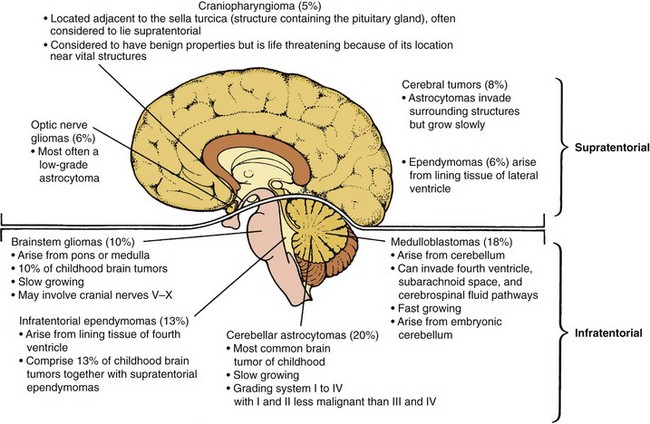
 Fig. 36-5 outlines major brain tumors of childhood.
Fig. 36-5 Location of brain tumors in children. (From McCance KL, Huether SE: Pathophysiology: the biological basis for disease in adults and children, ed 6, St Louis, 2010, Mosby.)
Because the neoplasms can arise from any cell within the cranium, it is possible to have tumors originating from the glial cells, nerve cells, neuroepithelium, cranial nerves, blood vessels, pineal gland, and hypophysis. Within each of these structures, specific cells may be involved to provide a histologic classification of the major tumors found in children. Astrocytes, cells that form most of the supportive tissue for the neurons, may form astrocytomas, the most common glial tumor (Strickler and Phillips, 2000). Brain tumors may be benign or malignant, although the designation of any tumor in the brain as “benign” should be done cautiously given the vital functions the brain controls.
The signs and symptoms of brain tumors are directly related to their anatomic location and size and to some extent the child’s age. For instance, in infants whose sutures are still open, a bulging fontanel indicates hydrocephalus. Head circumference measurements allow for detection of increased head size. Even in older children, clinical manifestations may be nonspecific. However, the most common symptoms of infratentorial brain tumors are headache, especially on awakening, and vomiting that is not related to feeding. Tumors in this area of the brain often obstruct the flow of cerebrospinal fluid, causing increased ICP and the symptoms mentioned above. In addition, patients may have symptoms related to the specific structure involved. Tumors of the cerebellum often cause nystagmus, ataxia, dysarthria, and dysmetria (Blaney, Haas-Kogan, Young-Pouissant, et al, 2011; Strickler and Phillips, 2000). Supratentorial symptoms more commonly include seizures, personality or behavioral changes, and contralateral weakness. Tumors involving the structures of the midbrain, including the hypothalamus and pituitary gland, may cause endocrinopathies such as diabetes insipidus, delayed or precocious puberty, and growth failure. Table 36-4 presents the common presenting symptoms of brain tumors.
Diagnosis of a brain tumor is based on presenting clinical signs and diagnostic imaging. Because the signs and symptoms may be vague and easily overlooked, early diagnosis necessitates a high index of suspicion during history taking. A number of tests may be employed in the neurologic evaluation (see Table 37-2), but the gold standard diagnostic procedure is MRI, which permits early diagnosis of brain tumors and assessment of tumor growth during or after treatment. Magnetic resonance (MR) angiography can be performed during the same session as MRI to determine the vascularity of the tumor (Brunelle, 2000). Another test is CT, which permits direct visualization of the brain parenchyma, ventricles, and surrounding subarachnoid space. Through the IV injection of radiographic contrast agents, intracranial blood vasculature can be demonstrated (Blaney, Haas-Kogan, Young-Pouissant, et al, 2011). MR spectroscopy is a new radiographic technique that is able to differentiate between malignant tumors and areas of necrosis (Brunelle, 2000; Lanzkowsky, 2005).
When a positive CT scan is obtained, angiography may be done to provide information about the tumor’s blood supply and degree of vascularity, which may assist the surgeon in planning the operative approach. Other tests may include an MRI of the spine, functional MRI, and electroencephalography. LP is dangerous in the presence of increased ICP because of possible brainstem herniation after sudden release of pressure.
Definitive diagnosis is based on tissue specimens obtained during surgery. Occasionally, special techniques are required for determining the cell type. This period of waiting is one of anxiety for family members, who are aware of the link between cell type and prognosis. Because of the location of some brain tumors, such as brainstem tumors, a biopsy is not possible and the diagnosis is made by imaging findings alone.
Treatment may involve the use of surgery, radiotherapy, and chemotherapy. All three may or may not be used, depending on the type of tumor. The treatment of choice is total removal of the tumor without residual neurologic damage. Patients with the most complete tumor removal have the greatest chance of survival. Several surgical advances have allowed the biopsy and removal of tumors in areas previously considered too dangerous for traditional operative techniques. Stereotactic surgery involves the use of CT and MRI in conjunction with other special computer techniques to reconstruct the tumor in three dimensions. With computer-assisted instruments, removal is sometimes possible. Stereotactic biopsy is performed with CT or MRI computer guidance for inserting the biopsy needle. This procedure has the benefit of a short hospital stay and a lower morbidity and mortality rate in comparison with an open craniotomy (Blaney, Haas-Kogan, Young-Pouissant, et al, 2011). Other procedures include the use of lasers to vaporize tumor tissue and brain mapping to determine the precise location of critical brain areas to avoid during surgery.
Radiotherapy is used to treat most tumors and to shrink the size of the tumor before attempting surgical removal. The use of chemotherapy has emerged in the past decades with an increasingly important role, either in combination with irradiation or alone. The drugs most commonly used are vincristine, cisplatin, carboplatin, cyclophosphamide, and etoposide (Strickler and Phillips, 2000). The problems of treatment are compounded by the serious late effects of all three modes of therapy. Surgery can cause injury to important areas of the brain, especially when the surgeon is attempting to remove invasive tumors. Irradiation has serious long-term consequences, which may include tissue necrosis, secondary malignancies, endocrine dysfunction, and behavioral or intellectual deficits. For these reasons, the use of irradiation is deferred for as long as possible in young children. Although there is limited information regarding a “safe age” for giving radiation, most centers consider it to be over age 3 years, but focal radiation is often given at a younger age (Mainprize, Taylor, and Rutka, 2000).
Nursing care of the child with a brain tumor is similar regardless of the type of intracranial lesion. Because a brain tumor is potentially fatal, the reader is urged to incorporate the psychologic interventions discussed in Chapter 23 with those elaborated on in this section. However, remember that many brain tumors are curable. Medulloblastoma, for instance, has a survival rate of more than 80% in those patients without metastatic disease. Despite the grave nature of some brain tumors, it is important to realize the hope that new standard therapies and emerging therapies have brought to the families of many pediatric brain tumor patients.
Assess for Signs and Symptoms: A child admitted to the hospital with neurologic dysfunction is often suspected of having a brain tumor, even though the actual diagnosis is not yet confirmed. Establishing a baseline of data for comparing preoperative and postoperative changes is an essential step toward planning physical care and preventing complications. It also allows the nurse to assess the degree of physical incapacity and the family’s emotional reaction to the diagnosis. For example, children with cerebellar astrocytoma may have displayed vague cerebellar symptoms for several years before a tumor is suspected. For these parents the revelation of a neoplasm may be more of a shock than for those who witnessed a rapid deterioration in their child’s abilities. Table 36-4 summarizes common presenting signs and assessment procedures to document significant changes in the child’s condition.
Prepare the Family for Diagnostic and Operative Procedures: The suspected diagnosis of a brain tumor is always a crisis. Despite the fact that some tumors are removed with excellent results, the physician can rarely give definitive answers regarding the prognosis until after surgery. Therefore parents and older children require much emotional support to face the diagnostic procedures and a craniotomy.
How the child is prepared for the diagnostic tests depends on the child’s age and experience. Because most of the tests involve x-ray equipment, the child may be familiar with the procedure. Chapter 27 discusses preparing children for an MRI or a CT scan. Once surgery is scheduled, the child needs an explanation of what to expect. By the time most children are late preschoolers, they know that the head and brain are important parts of their body. It may be helpful to have children draw their concept of the brain to clarify misconceptions and base the explanation on their level of understanding. Although it may be tempting to justify the surgery by stating that removing the tumor will take away various symptoms, the nurse should refrain from emphasizing this point too strenuously. Postsurgical headaches and cerebellar symptoms, such as ataxia, may be aggravated rather than improved. Surgery may not improve vision. With optic gliomas the child will be blind in one eye if the tumor is fully resected. Finally, surgical removal of the mass may be impossible, and after surgery functioning may temporarily deteriorate or result in permanent damage. Being honest before surgery most often makes honesty after the procedure easier because no false hopes were created.
It is best to deliver information in small amounts to let the child pursue additional answers. For example, some children ask about what happens when part of the tumor is left. An honest reply is that after surgery the physician will try to shrink the tumor with special x rays and medicines. Delay a further explanation of irradiation or chemotherapy until a decision regarding these treatments is made.
The hair is usually shaved in the operating room just before surgery, or sometimes in the child’s room, usually the night before surgery. When shaving is done with the child awake, the procedure is approached in a sensitive, positive way. If the child’s hair is long, braid it so that the long swatch can be saved. Showing children how they look at different stages of the process helps them prepare for the final appearance.
Once the hair is clipped short or shaved, give the child a cap or scarf to camouflage the baldness. Take every precaution to provide privacy during the procedure and to protect the child from teasing or ridicule by other children before surgery. Also emphasize that the hair will regrow shortly after surgery. Depending on the child’s immediate adjustment to the hair loss, the nurse may introduce the idea of wearing a wig until the hair grows in, particularly if additional irradiation or chemotherapy is anticipated.
Also tell children about the size of the dressing. Usually the entire scalp is covered to maintain tight wound closure, even if a small incision is made. Infratentorial head dressings may be attached to the upper back and extend forward to the neck to maintain slight extension and alignment as a precaution against wound rupture. Applying a similar dressing or “special hat” to a doll is often a less traumatic way of demonstrating the physical appearance.
Children also need a brief explanation of how they will feel after surgery and where they will be. Ordinarily they will return to a special intensive care unit, which they may visit beforehand depending on hospital policy. They should be aware that they may be sleepy for some time after surgery and that a headache is likely, although it should last only a few days.
Parents need similar explanations before surgery, especially in terms of special equipment used in the intensive care unit, dressings, and their child’s behavior. For example, they should know that it is not unusual for the child to be lethargic for a few days after surgery. The nurse may wish to encourage less frequent visiting during this period so that parents can rest and be able to support their child when the child is awake.
The nurse should participate in preoperative conferences with the physician and parents. The nurse needs to know what information the parents have been given in order to provide further explanations or emotional support when necessary.
Prevent Postoperative Complications: Usually the surgeon prescribes specific orders for taking vital signs, positioning, regulating fluids, and administering medication. These vary somewhat depending on the location of the craniotomy. The following are general principles of care for infratentorial or supratentorial surgery. Chapter 37 discusses additional aspects of care, such as care of the child with seizures and care of the unconscious child in terms of respiratory status and neurologic assessment.
Assessment: Vital signs are taken as often as every 15 to 30 minutes until the patient is stable. Temperature measurement is particularly important because of hyperthermia resulting from surgical intervention in the hypothalamus or brainstem and from some types of general anesthesia. To prepare for this reaction, a cooling blanket may be placed on the bed before the child returns to the unit, or it may be used when needed. Because the temperature control centers are affected and hypothermia can occur suddenly, the nurse monitors body temperature often when any cooling measures are employed.
The most likely types of infection are meningitis and respiratory tract infection. The probable cause of meningitis is wound contamination. The risk of respiratory tract infections is high because of the imposed immobility, danger of aspiration, and possible depression from the brainstem. The usual precautions of deep breathing and turning as allowed are instituted. Regular pulmonary assessments are performed to identify adventitious sounds or any areas of diminished or absent breath sounds. Blood pressure is also taken at frequent intervals. The deflated cuff is left on the arm between readings to allow for the least movement and disturbance of the child. Ocular signs are recorded at least every hour.
Observations for function are not instituted until the child regains consciousness. However, as soon as possible the nurse should begin testing reflexes, hand grip, and functioning of the cranial nerves. Muscle strength is usually less after surgery because of general weakness but should improve daily. Ataxia may be significantly worse with cerebellar intervention, but it slowly improves. Edema near the cranial nerves may depress important functions such as the gag, blink, or swallowing reflex.
Neurologic checks are an essential aspect of care and include pupillary reaction to light, level of consciousness, sleep patterns, and response to stimuli. Although children may be comatose for a few days, once they regain consciousness, there should be a steady increase in alertness. Regression to a lethargic, irritable state indicates increasing pressure, possibly caused by meningitis, hemorrhage, or edema.
Dressings are observed for evidence of drainage. If soiled, the dressing is not removed but reinforced with dry sterile gauze. The approximate amount of drainage is estimated and recorded.
Once the younger child is alert, the arms may need to be restrained to preserve the dressing. Even a child who has been cooperative before surgery must be closely supervised during the initial stages of regaining consciousness, when disorientation and restlessness are common. Elbow restraints are satisfactory to prevent the hands from reaching the head, although additional restraint may be necessary to preserve an infusion line and maintain a specific position.
Positioning: Correct positioning after surgery is critical to prevent pressure against the operative site, reduce ICP, and avoid the danger of aspiration. If a large tumor was removed, the child is not placed on the operative side, since the brain may suddenly shift to that cavity, causing trauma to the blood vessels, linings, and the brain itself. The nurse confers with the surgeon to be certain of the correct position, including the degree of neck flexion. The first 24 to 48 hours after brain surgery are critical. If positioning is restricted, notice of this is posted above the head of the bed. When the child is turned, every precaution is used to prevent jarring or misalignment to prevent undue strain on the sutures. Two nurses, one supporting the head and the other the body, are needed. The use of a turning sheet may facilitate turning a heavy child.
The child with an infratentorial procedure is usually positioned flat and on either side. Pillows should be placed against the child’s back, not head, to maintain the desired position. Ordinarily the head and neck are kept in midline with the body and slightly extended. In a supratentorial craniotomy the head is usually elevated above the heart to facilitate cerebrospinal fluid drainage and decrease excessive blood flow to the brain to prevent hemorrhage.
Fluid Regulation: With an infratentorial craniotomy the child is allowed nothing by mouth for at least 24 hours or longer if the gag and swallowing reflexes are depressed or the child is comatose. With a supratentorial procedure, feeding may be resumed soon after the child is alert, sometimes within 24 hours. Clear water is always started first because of the danger of aspiration. If the child vomits, stop oral liquids. Vomiting not only predisposes the child to aspiration, but also increases ICP and the risk for incisional rupture.
IV fluids are continued until fluids are well tolerated. Because of the cerebral edema postoperatively and the danger of increased ICP, fluids are carefully monitored and usually infused at one half the maintenance rate. If drugs, such as prophylactic antibiotics, are given intravenously, the medication amount is calculated as part of the IV fluid. For example, if the child is to receive 20 ml/hr and the diluted drug is 5 ml, the IV solution is reduced to 15 ml for that hour.
A hypertonic solution such as mannitol or dextrose may be necessary to remove excess fluid. These drugs cause rapid diuresis. After surgery the child may have a Foley catheter in place. Urinary output is monitored after administration of these drugs to evaluate their effectiveness.
When able to take fluids, the child should be fed to conserve strength and minimize movement. If there is any sign of facial paralysis, the child is fed slowly to prevent choking or aspiration. Scrupulous mouth care is essential to prevent oral infection. Sometimes gavage feeding is necessary when body functions are too depressed to permit safe oral feedings or the child refuses to eat or drink. In the latter instance the nurse should employ every measure to encourage acceptance of fluids or solids. (See Chapter 27 for nursing interventions.)
Comfort Measures: Headache may be severe and is largely the result of cerebral edema. Measures to relieve some of the discomfort include providing a quiet, dimly lit environment; restricting visitors; preventing any sudden jarring movement, such as banging into the bed; and preventing an increase in ICP. The last is most effectively achieved by proper positioning and prevention of straining, such as during coughing, vomiting, or defecating. Placing an ice bag on the forehead may also provide some headache relief, especially if facial edema is severe. The use of opioids, such as morphine, to relieve pain is controversial because it is thought that they may mask signs of altered consciousness or depress respirations. However, they can be given safely because naloxone can be used to reverse opioid effects, such as sedation or respiratory depression. Acetaminophen and codeine are also effective analgesics. Regardless of the drugs used, adequate dosage and regular administration are essential to provide optimum pain relief. (See Pain Assessment and Pain Management, Chapter 7.)
Monitor bowel movements to prevent constipation. Stool softeners may be given as soon as liquids are tolerated to facilitate easy passage of stool.
Brain edema may severely depress the gag reflex, necessitating suctioning of oral secretions. Facial edema may also be present, necessitating eye care if the lids remain partially open. Ice compresses applied to the eyes for short periods help relieve the edema. A depressed blink reflex also predisposes the corneas to ulceration. Irrigating the eyes with saline drops and covering them with eye dressings are important steps in preventing this complication.
Support the Family: The family’s emotional needs are great when the diagnosis is a brain tumor, and the extent of surgery, any neurologic deficits, the prognosis, and additional therapy influence these feelings. Because few definitive answers can be given before surgery, the surgeon’s report is a significant finding that can vary from a completely benign, resected neoplasm to a highly malignant, invasive, and only partially removed tumor. Although parents try to prepare themselves for a potentially fatal diagnosis, it is understandably a shock for them.
Ideally, a nurse who will be involved in the continuing care of this child should be with the family when the physician discusses the prognosis and plan of therapy. Although parents may hear only a fraction of what they are told, they can begin to put the future into perspective. Regardless of the future prospects, direct the parents’ thinking toward helping the child recover and resume a normal life to his or her fullest potential. Providing the opportunity for the family to share their concerns and questions with other families who have a child with a brain tumor may help the family cope.*
It is also a time to encourage parents to verbalize their feelings about the diagnosis. Often they express guilt for attributing the insidious onset of symptoms, such as ataxia, visual difficulty, or headache, to minor “complaints” by the child. Parents may have punished their child for clumsiness, mistaking it for carelessness, or for their declining performance in school. The nurse listens to such statements, emphasizing the normalcy of the parents’ reactions. Sometimes it may be helpful to start a discussion with a statement such as “It is difficult to know when a child’s complaints are significant because so often they are caused by minor ailments and you would never have imagined they were a result of a brain tumor.” The nurse avoids any comments that insinuate the parents should have sought medical advice sooner, since such remarks only add to the parents’ guilt feelings.
During this period the nurse should also discuss with parents what they plan to tell the child. If the child was prepared honestly, as described previously, the diagnosis can be expressed in a similar manner, such as “The surgeon removed most of the tumor, and the rest will be treated with special drugs and x-ray treatments.” During recovery the child needs additional explanation about the treatment and the reason for residual neurologic effects, such as ataxia or blindness. Because the hair was shaved before surgery, hair loss from treatment is less of a concern, although its regrowth will be delayed, depending on the length of therapy. At this point it is advisable to reintroduce the idea of a wig.
Promote Return to Optimum Functioning: The ultimate goal is a cured child who has optimum functioning. As soon as possible, the child should resume usual activities within tolerable limits, especially returning to school.* Until the skull is completely healed, the child may need to wear a helmet when engaging in any active sport. This decision is made by the child’s neurosurgeon. The school nurse and teacher should confer with the parents on activity restrictions, such as physical education, and the reactions of schoolmates to the child’s appearance.
After discharge the family needs continuing medical and emotional support from health personnel. Even with children who are long-term survivors after treatment for a brain tumor, residual disabilities, such as growth retardation, cranial nerve palsies, sensory defects, motor abnormalities (especially ataxia), intellectual deficits, dysphagia, dysgraphia, and behavioral problems, may occur (Blaney, Haas-Kogan, Young-Pouissant, et al, 2011; Loring and Meador, 2000; Riva and Giorgi, 2000). It is difficult to assess the exact cause of the nonphysical disabilities, since numerous variables influence the child’s total rehabilitation. However, the high frequency of late effects attests to the tremendous need for follow-up care despite successful treatment of the tumor.
The vast realm of possible consequences after the diagnosis of a brain tumor is not discussed here. Rather, the reader is referred to other sections of the text that deal with possible outcomes, such as the paralyzed, visually impaired, or unconscious child or the child with a ventricular shunt, seizure disorder, or meningitis. Numerous physical problems can occur with progression of the tumor that may necessitate additional procedures. For example, frequent vomiting, anorexia, and nausea may require nonoral routes of feeding, such as gastrostomy or parenteral alimentation. Trials with chemotherapy may necessitate the use of central venous access devices. Whenever these procedures are instituted, the nurse may be responsible for teaching the family appropriate home care to allow the child the highest quality of life for the longest time. (See discussion of discharge planning and home care in Chapter 25 and Nursing Care Plan: The Child with a Brain Tumor.†)
Neuroblastoma is the most common extracranial solid tumor of childhood and the most common cancer diagnosed in infancy. It occurs in about 1 in 7000 live births. The median age at diagnosis is 19 months, and it is slightly more prevalent in males. These tumors originate from embryonic neural crest cells that normally give rise to the adrenal medulla and the sympathetic nervous system. Consequently the majority of the tumors arise from the adrenal gland or from the retroperitoneal sympathetic chain. The primary site is within the abdomen; other sites include the head and neck region, chest, and pelvis.
The signs and symptoms of neuroblastoma depend on the location and stage of the disease. With abdominal tumors, the most common presenting sign is a firm, nontender, irregular mass in the abdomen that crosses the midline (in contrast to Wilms tumor, which is usually confined to one side). Compression of the kidney, ureter, or bladder may cause urinary frequency or retention.
Distant metastasis frequently causes supraorbital ecchymosis, periorbital edema, and proptosis (exophthalmos) from invasion of retrobulbar soft tissue. Lymphadenopathy, hepatomegaly and skeletal pain are also present in patients with disseminated disease. Vague symptoms of widespread metastasis include pallor, weakness, irritability, anorexia, and weight loss.
Other primary tumor sites may cause significant clinical effects such as neurologic impairment, respiratory obstruction from a thoracic mass, or varying degrees of paralysis from compression of the spinal cord. Infrequently a child may have symptoms of increased catecholamine excretion, such as flushing, hypertension, tachycardia, and diaphoresis (Weinstein, Katenstein, and Cohn, 2003).
Diagnostic evaluation is aimed at locating the primary site and areas of metastasis. A CT of the abdomen, pelvis, or chest is the preferred imaging modality to locate the primary tumor. A bone scan and MIBG (iodine-131 metaiodobenzylguanidine) scan should be performed to evaluate for the presence of skeletal metastases. Examination of the bone marrow with bilateral aspirates and biopsies should be performed in all patients. Neuroblastomas, particularly those arising on the adrenal glands or from a sympathetic chain, excrete the catecholamines epinephrine and norepinephrine. Urinary excretion of catecholamines is detected in approximately 95% of children with adrenal or sympathetic tumors. Analyzing the breakdown products that are normally excreted in the urine, namely, vanillylmandelic acid, homovanillic acid, dopamine, and norepinephrine, permits detection of a suspected tumor both before and after medical-surgical intervention. Amplification of the N-myc gene and abnormalities in chromosomes have been associated with a poorer prognosis (Grosfeld, 2000; Lau, Tai, Weitzman, et al, 2004). Increased ferritin and neuron-specific enolase are also seen in neuroblastoma.
Neuroblastoma is a “silent” tumor. In more than 70% of cases, diagnosis is made after metastasis occurs, with the first signs caused by involvement in the nonprimary site, usually the lymph nodes, bone marrow, skeletal system, skin, or liver. Because of the frequency of invasiveness, the prognosis for neuroblastoma is generally poor.
The child’s age and the stage of the disease (Box 36-5) at diagnosis are important prognostic factors. Survival is inversely correlated with age. If all stages are grouped together, the survival rates are 75% for children under 1 year of age and less than 50% for children over 1 year of age. This marked difference in survival rates by age is partly accounted for by the larger proportion of very young children with stage I, II, or IV-S disease and the absence of the N-myc gene amplification (Brodeur, Hogarty, Mosse, et al, 2011).
Infants who remain free of disease for 1 year after treatment are usually cured, but older children have experienced relapses several years after cessation of treatment. Surgical resection of the tumor in stage I infants diagnosed by ultrasonography done for other reasons appears to be almost 90% curative (Grosfeld, 2000). Neuroblastoma is one of the few tumors that demonstrate spontaneous regression (especially stage IV-S), possibly as a result of maturity of the embryonic cell or development of an active immune system.
Accurate clinical staging is important for establishing initial treatment. Therefore the purpose of surgery is both to remove as much of the tumor as possible and to obtain biopsies. In stages I and II, complete surgical removal of the tumor is the treatment of choice. If the tumors are large, partial resection is attempted, with a course of irradiation postoperatively to shrink the tumor in the hope of complete removal at a later date. Surgery is usually limited to biopsy in stages III and IV because of the extensive metastasis, although additional surgery to assess tumor regression or remove a regressed tumor is not unlikely.
The precise role of radiotherapy is unclear. It does not appear to be of any benefit in children with stage I and II disease. It is commonly used with stage III disease, although it may not improve survival expectancy. It may make a large tumor operable. Radiotherapy provides emergency management of a massive neuroblastoma causing spinal cord compression (Nguyen, Sallah, Ludin, et al, 2000). It also offers palliation for metastatic lesions in bones, lungs, liver, or brain.
Chemotherapy is the mainstay of therapy for extensive local or disseminated disease. The drugs of choice are vincristine, doxorubicin, cyclophosphamide, cisplatin, etoposide, ifosfamide, and carboplatin; they are administered in a variety of combinations according to specific protocols. In addition, the use of consolidative myeloablative therapy using autologous marrow or peripheral stem cells followed by 13-cis-retinoic acid has improved the outcome of patients with high-risk disease.
Nursing care management is similar to that discussed under Nursing Care of the Child with Cancer, including psychologic and physical preparation for diagnostic and operative procedures; prevention of postoperative complications for abdominal, thoracic, or cranial surgery; and explanation of chemotherapy and radiotherapy and their side effects (see Tables 36-2 and 36-3).
Because this tumor carries a poor prognosis for many children, evaluate and address the needs of the family in terms of coping with a life-threatening illness. (See Chapter 23.) Because of the high degree of metastasis at the time of diagnosis, many parents suffer guilt for not having recognized signs earlier. Often the guilt is expressed as anger toward professionals for not diagnosing it sooner. Parents need much support in dealing with these feelings and expressing them to the appropriate people.
Bone tumors account for about 6% of all malignant neoplasms in children. Approximately 90% of all primary malignant bone tumors in children are either osteogenic sarcoma or Ewing sarcoma; osteosarcoma, the most common, occurs in 56% of all cases. The peak age for pediatric bone tumors is 15 years, and they occur more often in males.
Most malignant bone tumors produce localized pain in the affected site, which may be severe or dull and may be attributed to trauma or the vague complaint of “growing pains.” The pain is often relieved by a flexed position, which relaxes the muscles overlying the stretched periosteum. Frequently it draws attention when the child limps, curtails physical activity, or is unable to hold heavy objects. A palpable mass is also a common manifestation of bone tumors, but systemic symptoms such as fever and other clinical symptoms such as spinal cord compression and respiratory distress are more frequent in patients with Ewing sarcoma.
Diagnosis begins with a thorough history and physical examination. A primary objective is to rule out causes such as trauma or infection. Careful questioning regarding pain is essential in attempting to determine the duration and rate of tumor growth. Physical assessment focuses on functional status of the affected area; signs of inflammation; size of the mass; and any systemic indication of generalized malignancy, such as anemia, weight loss, and frequent infection.
Definitive diagnosis is based on radiologic studies, such as plain films and CT or MRI of the primary site, CT of the chest, and radioisotope bone scans to evaluate metastasis and bone marrow examination in patients with Ewing sarcoma. A needle or surgical biopsy is necessary to establish the diagnosis. Ewing sarcoma most commonly involves the pelvis, long bones of the lower extremities, and chest wall and radiographically involves the diaphysis with detachment of the periosteum from the bone (Codman triangle). In osteosarcoma, lesions are most commonly located in the metaphyseal region of the bone, often involving the long bones. Radial ossification in the soft tissue gives the tumor a “sunburst” appearance on plain radiograph.
A better understanding of the biology of neoplastic growth has resulted in more aggressive treatment and an improved prognosis. The natural history of osteogenic sarcoma and Ewing sarcoma suggests that multiple submicroscopic foci of metastatic disease are present at the time of diagnosis despite clinical evidence of localized involvement. Before the use of aggressive multimodal therapy, pulmonary metastasis appeared in the majority of patients who were treated with surgical excision alone (Gorlick, Bielack, Teot, et al, 2011). With current therapies that include surgery and chemotherapy for osteosarcoma and surgery, radiotherapy, and chemotherapy for Ewing sarcoma, more than two thirds of patients with localized disease can be cured.
Osteosarcoma (osteogenic sarcoma) is the most common bone cancer in children and most commonly affects patients in the second decade of life during their growth spurt. It presumably arises from bone-forming mesenchyme, which gives rise to malignant osteoid tissue. Most primary tumor sites are in the diametaphyseal region (wider part of the shaft, adjacent to the epiphyseal growth plate) of long bones, especially in the lower extremities. More than half occur in the femur, particularly the distal portion, with the rest involving the humerus, tibia, pelvis, jaw, and phalanges.
Optimum treatment of osteosarcoma includes surgery and chemotherapy. The surgical approach consists of surgical biopsy followed by either limb salvage or amputation. To ensure local control, all gross and microscopic tumors must be resected. A limb salvage procedure involves en bloc resection of the primary tumor with prosthetic replacement of the involved bone. For example, with osteosarcoma of the distal femur, a total femur and joint replacement is performed. Frequently children undergoing a limb salvage procedure receive preoperative chemotherapy in an attempt to decrease the tumor size and make surgery more manageable (Lanzkowsky, 2005; Gorlick, Bielack, Teot, et al, 2011).
Critical Thinking Exercise—Osteogenic Sarcoma
Chemotherapy plays a vital role in treatment of osteosarcoma. Antineoplastic drugs, such as high-dose methotrexate with citrovorum factor rescue, doxorubicin, cisplatin, ifosfamide, and etoposide, may be administered singly or in combination and may be employed both before or after surgical resection of the tumor. The use of postoperative chemotherapy after amputation has comparable results to trials using preoperative chemotherapy followed by limb salvage surgery. Preoperative chemotherapy allows for examination of the surgical specimen at the time of definitive surgery, which predicts clinical outcome. When pulmonary metastases are found, thoracotomy and chemotherapy have resulted in prolonged survival and potential cure. These combined-modality approaches have significantly improved the prognosis in osteosarcoma to approximately 78% for nonmetastatic patients (Lanzkowsky, 2005). Newer trials have recently been completed and have incorporated muramyl tripeptide phosphatidylethanolamine to eradicate micrometastases by stimulating macrophages to kill tumor cells not eliminated by chemotherapy (Lanzkowsky, 2005).
Nursing care depends on the type of surgical approach. Obviously the family may have more difficulty adjusting to an amputation than a limb salvage procedure. In either instance, preparation of the child and family is critical. Straightforward honesty is essential in gaining the child’s cooperation and trust. The diagnosis of cancer should not be disguised with falsehoods such as “infection.” To accept the need for radical surgery, the child must be aware of the lack of alternatives for treatment. Although the responsibility of telling the child is generally left to the physician, the nurse should be present at the discussion or be aware of exactly what is said. The child should be told a few days before surgery to allow him or her time to think about the diagnosis and consequent treatment and to ask questions. (See Nursing Care Plan: The Child with a Bone Tumor.*)
Sometimes children have many questions about the prosthesis, limitations on physical ability, and prognosis in terms of cure. At other times they react with silence or with a calm manner that belies their concern and fear. Either response must be accepted, since it is part of the grieving process of a loss. For those who desire information, it may be helpful to introduce them to another amputee before surgery or to show them pictures of the prosthesis.† However, the nurse must be careful not to overwhelm children with information. A sound approach is to answer questions without offering additional information. For those who do not pursue additional information, the nurse expresses a willingness to talk.
The child is also informed of the need for chemotherapy and its side effects before surgery. Exercise caution about offering too much information at one time. When discussing hair loss, emphasize positive aspects, such as wearing a wig. Because bone tumors affect adolescents and young adults, it is not unusual for them to become angry over all the radical body alterations.
If an amputation is performed, the child is usually fitted with a temporary prosthesis immediately after surgery, which permits early functioning and fosters psychologic adjustment. If this is not done, the child requires stump care, which is the same as for any amputee. A permanent prosthesis is usually fitted within 6 to 8 weeks. During hospitalization the child begins physical therapy to become proficient in the use and care of the device.
Phantom limb pain may develop after amputation. This symptom is characterized by sensations such as tingling, itching, and, more frequently, pain felt in the amputated limb. The child and family need to know that the sensations are real, not imagined. Amitriptyline (Elavil) has been used successfully in children to decrease the pain (Olsson, 1999). In addition, an epidural is often used preoperatively as a nerve block in an effort to decrease or eliminate the occurrence of phantom limb pain. Much research is needed to further delineate the best care for these patients (Ong, Arneja, and Ong, 2006).
Discharge planning must begin early in the postoperative period. Once the child has begun physical therapy, the nurse should consult with the therapist and practitioner to evaluate the child’s physical and emotional readiness to reenter school. It is an opportune time to involve a community nurse in the child’s home care. Every effort is made to promote normalcy and gradual resumption of realistic preamputation activities.* Role-playing in anticipation of such experiences is beneficial in preparing the child for the inevitable confrontation by others. Environmental barriers, such as stairs, are assessed in terms of the accessibility in the school and home, especially because the child may need to use crutches or a wheelchair before complete healing and prosthetic competency are achieved.
The nurse encourages the child to select clothing that best camouflages the prosthesis, such as pants or long-sleeved shirts. Well-fitted prostheses are so natural looking that girls can usually wear sheer stockings without revealing the device. Emphasizing feminine or masculine apparel helps the child regain a feeling of self-identity. Even during the postoperative period, encouraging the child to wear blue jeans and a T-shirt may distract attention from the deformity and focus it on familiar aspects of appearance.
The family and child need much support in adjusting not only to a life-threatening diagnosis but also to alteration in body form and function. Because loss of a limb entails a grieving process, those caring for the child need to recognize that the reactions of anger and depression are normal and necessary. Often parents view the anger as a direct affront to them for allowing the amputation to occur, or they see the depression as rejection. These are not personal attacks but the child’s attempts to cope with a loss.
Ewing sarcomas, or the Ewing sarcoma family of tumors which includes primitive neuroectodermal tumor of the bone, are the second most common malignant bone tumor (after osteosarcoma) in childhood (Lanzkowsky, 2005). Ewing sarcoma arises in the marrow spaces of the bone rather than from osseous tissue. The tumor originates in the shaft of long and trunk bones, most often affecting the pelvis, femur, tibia, fibula, humerus, ulna, vertebra, scapula, ribs, and skull. It occurs almost exclusively in individuals under age 30 and affects Caucasians much more often than other races.
Limb salvage procedures might be feasible in extremity lesions, and amputation may be considered if the results of radiotherapy render the extremity useless or deformed (e.g., from retarded growth in young children). The treatment of choice for the majority of lesions is involved field radiotherapy and chemotherapy. A widely used drug regimen includes vincristine, doxorubicin, cyclophosphamide alternating with ifosfamide, and etoposide. The addition of ifosfamide and etoposide has increased the 3-year survival to 78% for patients with localized disease (Lanzkowsky, 2005).
The psychologic adjustment to Ewing sarcoma is typically less traumatic than it is to osteosarcoma because of the preservation of the affected limb. Many families accept the diagnosis with a sense of relief in knowing that this type of bone cancer does not necessitate amputation, and initially they may not be aware of the damaging effects on the irradiated site. Consequently they need preparation for the various diagnostic tests, including bone marrow aspiration and surgical biopsy, and adequate explanation of the treatment regimen. High-dose radiotherapy often causes a skin reaction of dry or moist desquamation followed by hyperpigmentation. The child should wear loose-fitting clothes over the irradiated area to minimize additional skin irritation. Because of increased sensitivity, protect the area from sunlight and sudden changes in temperature, such as from heating pads or ice packs. Encourage the child to use the extremity as tolerated. Occasionally the physical therapist may plan an active exercise program to preserve maximum function.
The child needs the same considerations for adjusting to the effects of chemotherapy as any other patient with cancer. The drug regimen usually results in hair loss, severe nausea and vomiting, peripheral neuropathy, and possibly cardiotoxicity. Make every effort to outline a treatment plan that allows the child maximum resumption of a normal lifestyle and activities. (See Nursing Care Plan: The Child with a Bone Tumor.†)
In addition to the cancers already discussed, several other types of solid tumors may occur in children. Wilms tumor, rhabdomyosarcoma, and retinoblastoma are unique in that they tend to be diagnosed early, typically before 5 years of age. Wilms tumor and retinoblastoma are also unusual in that they are among the few types of cancer that may occur in both hereditary and nonhereditary forms.
Wilms tumor, or nephroblastoma, is the most common kidney tumor of childhood (Skoldenberg, Christiansson, Sandstedt, et al, 2001). Its frequency is estimated to be 8 cases per 1 million children less than 15 years of age (Lanzkowsky, 2005). Eighty percent of patients with Wilms tumor are diagnosed under 5 years of age, and it has a peak incidence between 3 and 4 years of age (Lanzkowsky, 2005). Wilms tumor may be associated with several congenital malformation syndromes, including WAGR (Wilms tumor, aniridia, genitourinary anomalies, and cognitive impairment [mental retardation]) and Beckwith-Wiedemann syndrome (hemihypertrophy, macroglossia, omphalocele, and visceromegaly) (Lanzkowsky, 2005). About 2% of Wilms tumors are familial.
The most common presenting sign is painless swelling or mass within the abdomen. The mass is characteristically firm, nontender, confined to one side, and deep within the flank. If it is on the right side, it may be difficult to distinguish from the liver, although, unlike that organ, it does not move with respiration. Parents usually discover the mass during routine bathing or dressing of the child.
Other clinical manifestations are the result of compression from the tumor mass, metabolic alterations secondary to the tumor, or metastasis. Hematuria occurs in less than one fourth of children with Wilms tumor. Anemia, usually secondary to hemorrhage within the tumor, results in pallor, anorexia, and lethargy. Hypertension, probably caused by secretion of excess amounts of renin by the tumor, occurs occasionally. Other effects of malignancy include weight loss and fever. If metastasis has occurred, symptoms of lung involvement, such as dyspnea, cough, shortness of breath, and pain in the chest, may be evident.
In a child suspected of having Wilms tumor, special emphasis is placed on the history and physical examination for the presence of congenital anomalies; a family history of cancer; and signs of malignancy, such as weight loss, enlarged liver and spleen, indications of anemia, and lymphadenopathy. Specific tests include radiographic studies, such as abdominal ultrasound, CT, and MRI of the abdomen; CT of the chest to look for metastases in the lung; and Doppler ultrasound of the inferior vena cava. Laboratory studies should include a complete blood count (polycythemia is sometimes present if the tumor secretes excess erythropoietin), biochemical studies, and urinalysis. Studies to demonstrate the relationship of the tumor to the ipsilateral kidney and the presence of a normally functioning kidney on the contralateral side are essential.
Wilms tumor probably arises from a malignant, undifferentiated metanephrogenic blastoma (a cluster of primordial cells capable of initiating the regeneration of an abnormal structure). Its occurrence slightly favors the left kidney, which is advantageous because surgically this kidney is easier to manipulate and remove. Although the tumor may become large, it remains encapsulated for an extended period. During surgery the tumor is staged to maximize the effectiveness of treatment protocols (Box 36-6).
The histology of the tumor cells is also identified and classified according to two groups: favorable histology (FH) and unfavorable histology (UH). Only about 12% of Wilms tumors demonstrate UH, which is associated with a poorer prognosis and demands a more aggressive treatment protocol, regardless of the clinical stage.
Survival rates for Wilms tumor are one of the highest among all childhood cancers. Children with localized tumor (stages I and II) have a 90% chance of cure with multimodal therapy. For those children who relapse, a better expectancy of disease-free survival is associated with FH of the tumor, more than 12 months elapsing from the first complete remission, and nonabdominal recurrence (Dome, Liu, Krasin, et al, 2002; Plesko, Kramarova, Stiller, et al, 2001).
Combined treatment with surgery and chemotherapy, with or without irradiation, is based on the clinical stage and histologic pattern. In unilateral disease a large transabdominal incision is performed for optimum visualization of the abdominal cavity. The tumor, affected kidney, and adjacent adrenal gland are removed. Great care is taken to keep the encapsulated tumor intact because rupture can seed cancer cells throughout the abdomen, lymph channel, and bloodstream. The contralateral kidney is carefully inspected for evidence of disease or dysfunction. Regional lymph nodes are inspected, and a biopsy is performed when indicated. Any involved structures, such as part of the colon, diaphragm, or vena cava, are removed. Metal clips are placed around the tumor site for exact marking during radiotherapy.
If both kidneys are involved, the child may be treated with radiotherapy or chemotherapy preoperatively to shrink the tumor, allowing more conservative therapy (Graf, Tournade, and deKraker, 2000; Lanzkowsky, 2005). In some cases a partial nephrectomy is performed on the less affected kidney, with a total nephrectomy performed on the opposite side. When a transplant is feasible, such as from a twin, sibling, or parent, bilateral nephrectomy is considered as a last resort.
Postoperative radiotherapy is indicated for children with large tumors, metastasis, residual disease at the primary tumor site, UH, or recurrence. Chemotherapy is indicated for all stages. The most effective agents for treating Wilms tumor are actinomycin D and vincristine; doxorubicin and cyclophosphamide may be used for UH or advanced-stage disease (Fernandez, Geller, Ehrlich, et al, 2011; Lanzkowsky, 2005). The duration of therapy ranges from 6 to 15 months.
The nursing care of the child with Wilms tumor is similar to that of other cancers treated with surgery, irradiation, and chemotherapy. However, some significant differences are discussed for each phase of nursing intervention.
Preoperative Care: As with many of the other cancers, the diagnosis of Wilms tumor is a shock. Frequently the child has no physical indication of the seriousness of the disorder other than a palpable abdominal mass. Because the parents usually discover the mass, the nurse needs to take into account their feelings regarding the diagnosis. Whereas some parents are grateful for their detection of the tumor, others feel guilty for not finding it sooner or anger toward the practitioner for missing it on earlier examinations.
The preoperative period is one of swift diagnosis. Typically, surgery is scheduled within 24 to 48 hours of admission. The nurse is faced with the challenge of preparing the child and parents for all laboratory and operative procedures. Because of the little time available, keep explanations simple and repeat them often with attention to what the child will experience. In addition to usual preoperative observations, monitor blood pressure, since hypertension from excess renin production is a possibility.
There are several special preoperative concerns, the most important of which is to not palpate the tumor unless absolutely necessary because manipulation of the mass may cause dissemination of cancer cells to adjacent and distant sites.
Because radiotherapy and chemotherapy are usually begun immediately after surgery, parents need an explanation of what to expect, such as major benefits and side effects, although the timing of the information should be considered to avoid overwhelming the family. Ideally the nurse should be present during physician-parent conferences to answer questions as they arise.
Postoperative Care: Despite the extensive surgical intervention necessary in many children with Wilms tumor, the recovery period is usually rapid. The major nursing responsibilities are those following any abdominal surgery. (See Nursing Care Plan: The Child Undergoing Surgery, on the Evolve website.) Because these children are at risk for intestinal obstruction from vincristine-induced adynamic ileus, radiation-induced edema, and postsurgical adhesion formation, the nurse monitors gastrointestinal activity, such as bowel movements, bowel sounds, distention, and vomiting. Other considerations are frequent evaluation of blood pressure and observation for signs of infection, especially during chemotherapy. Because of the myelosuppression from the drugs, institute pulmonary hygiene measures in the immediate postoperative period to prevent complications.
Support the Family: The postoperative period is frequently difficult for parents. The shock of seeing their child immediately after surgery may be the first realization of the seriousness of the diagnosis. From surgery, the stage and pathology of the tumor is determined. The physician discusses this information with the parents. The nurse’s presence during this conversation is important to provide additional support and assess the parents’ understanding of this information.
Older children need an opportunity to deal with their feelings concerning the many procedures to which they have been subjected in rapid succession. Therapeutic play can be beneficial in helping children of any age understand what they have undergone and express their feelings.
Rhabdomyosarcoma (rhabdo, striated) is the most common soft tissue sarcoma in children. Striated (skeletal) muscle is found almost anywhere in the body, so these tumors occur in many sites, the most common of which are the head and neck, especially the orbit. The disease occurs in children in all age-groups but is most common in children younger than 5 years of age. Its incidence is approximately 8.5 per 1 million for Caucasian children but only 4.0 per 1 million for African-American children in the age-group from 2 to 19 years (Lanzkowsky, 2005).
Rhabdomyosarcoma arises from embryonic mesenchyme. Three subtypes are recognized (Box 36-7). Soft tissue sarcomas are the fourth most common type of solid tumors in children. These malignant neoplasms originate from undifferentiated mesenchymal cells in muscles, tendons, bursae, and fascia, or in fibrous, connective, lymphatic, or vascular tissue. They derive their name from the specific tissue(s) of origin, such as myosarcoma (myo, muscle).
The initial signs and symptoms are related to the site of the tumor and compression of adjacent organs (Table 36-5). Some tumor locations, such as the orbit, manifest early in the course of the illness. Other tumors, such as those of the retroperitoneal area, only produce symptoms when they are relatively big and compress adjacent organs. Unfortunately, many of the signs and symptoms attributable to rhabdomyosarcoma are vague and frequently suggest a common childhood illness, such as “earache” or “runny nose.” Rarely, the site of the primary tumor site is never identified.
Diagnosis begins with a careful history and physical examination. Radiographic studies to delineate the primary tumor site should include CT or MRI. Metastatic evaluation should include a CT of the chest, bone scan, and bilateral bone marrow aspirates and biopsies. For patients with tumors in the parameningeal area, perform an LP to examine the spinal fluid. An excisional biopsy or surgical resection of the tumor, when possible, is done to confirm the diagnosis.
Careful staging is extremely important for planning treatment and determining the prognosis. The Intergroup Rhabdomyosarcoma Study has developed a surgicopathologic staging system, shown in Box 36-8 (Lanzkowsky, 2005; Helman, 2011).
With the use of contemporary multimodal therapy, more than 80% of patients with nonmetastatic disease are expected to survive (Lanzkowsky, 2005; Helman, 2011). If relapse occurs, the prognosis for long-term survival is poor.
All rhabdomyosarcomas are high-grade tumors with the potential for metastases. Therefore multimodal therapy is recommended for all patients. Complete removal of the primary tumor is advocated whenever possible. However, because the tumor is chemosensitive, radical procedures with high morbidity should be avoided. In the majority of cases, a biopsy is followed by chemotherapy, irradiation, or both. Patients with embryonal tumors and group I disease can be treated with chemotherapy alone, whereas all others require chemotherapy and radiotherapy. Drugs that are used most often for the treatment of rhabdomyosarcoma include vincristine, actinomycin D, cyclophosphamide (VAC), ifosfamide, topotecan, irinotecan, and doxorubicin, which are administered for about 1 year (Lanzkowsky, 2005).
The nursing responsibilities are similar to those for other types of cancer, especially the solid tumors when surgery is employed. Specific objectives include (1) careful assessment for signs of the tumor, especially during well-child examinations; (2) preparation of the child and family for the multiple diagnostic tests (see p. 1478); and (3) supportive care during each stage of multimodal therapy. The reader is urged to review the Nursing Care Management section for cancer and Chapter 23 for emotional support of the family in the event of a poor prognosis.
Retinoblastoma, which arises from the retina, is the most common intraocular malignancy of childhood (Tsinopoulos, Papadopoulou, Papandroudis, et al, 2001). Approximately 3.8 cases per 1 million children occur annually, but it accounts for 11% of all cancers seen in children during the first year of life. The average age of the child at the time of diagnosis is 2 years; it is usually diagnosed earlier in hereditary cases and later in nonhereditary types. Of all cases of retinoblastoma, 60% are unilateral and nonhereditary, 25% are bilateral and hereditary, and 15% are unilateral and hereditary.
Retinoblastoma may be caused by various genetic alterations of the Rb gene, including (1) a somatic mutation in nonhereditary cases, (2) a germ-line mutation in hereditary cases, or (3) a chromosomal deletion involving chromosome 13. A “two-hit hypothesis” was developed to explain genetic and sporadic cases. Almost all bilateral retinoblastomas are considered hereditary, and 15% of individuals with unilateral disease have the hereditary form (Hurwitz, Shields, Shields, et al, 2011; Tsinopoulos, Papadopoulou, Papandroudis, et al, 2001). Hereditary retinoblastomas are transmitted as an autosomal dominant trait, with 90% penetrance (Lanzkowsky, 2005). Consequently 10% of gene carriers remain unaffected.
Children who have chromosome aberrations and retinoblastoma also often have an increased incidence of cognitive impairment and congenital malformations, although the vast majority of children with retinoblastomas apparently have normal chromosomes and intelligence.
Retinoblastoma has few grossly obvious signs. Typically the parents are the ones who first observe a whitish “glow” in the pupil, known as the cat’s eye reflex, or leukocoria (Fig. 36-6). The reflex represents visualization of the tumor as the light momentarily falls on the mass. When a tumor arises in the macular region (area directly at the back of the retina when the eye is focused straight ahead), a white reflex may be visible when the tumor is small. It is best observed when a bright light is shining toward the child as the child looks forward. Sometimes parents accidentally discover it when taking a photograph of their child using a flash attachment.
Fig. 36-6 Cat’s eye reflex. Whitish appearance of lens is produced as light falls on tumor mass in left eye.
When the tumor arises in the periphery of the retina, it must grow to a considerable size before light can strike it sufficiently to produce the cat’s eye reflex. In this situation it is visible only when the child looks in certain directions (sideways) or if the observer stands at an oblique angle to the child’s face as the child looks straight ahead. The fleeting nature of the reflex often results in a delayed diagnosis because health care professionals fail to appreciate the ominous significance of the parents’ findings.
The next most common sign is strabismus resulting from poor fixation of the visually impaired eye, particularly if the tumor develops in the macula, the area of sharpest visual acuity. Blindness is usually a late sign, but it frequently is not obvious unless the parent consciously observes for behaviors indicating loss of sight, such as bumping into objects, slowed motor development, or turning of the head to see objects lateral to the affected eye. Other signs and symptoms include heterochromia (different color of the iris), glaucoma, and pain.
A detailed family history and recording of eye symptoms is essential. Children suspected of having this disorder are referred to an ophthalmologist; the diagnosis is usually based on indirect ophthalmoscopy, ultrasound, and CT scans.
Metastatic disease at the time of retinoblastoma diagnosis is rare (Singh, Shields, and Shields, 2000); therefore staging procedures such as bone marrow aspiration, bone scan, and LP are not routinely performed.
Staging of retinoblastomas is done under indirect ophthalmoscopy before surgery to accurately determine the tumor size (measured in disc diameters [DD]) and location (according to an imaginary line called the equator drawn on the midplane of the eye) (Hurwitz, Shields, Shields, et al, 2011; Lanzkowsky, 2005).
Various classification systems have been used to stage retinoblastoma. The Reese-Ellsworth system (Box 36-9) classifies patients according to five groups and predicts survival when patients are treated with radiotherapy. A new classification system is designed based on the extent and location of the intraocular tumor and better predicts globe salvage using contemporary treatments. Cure rates for survival are much better than for retention of useful vision. The overall 10-year survival rate is nearly 90% for unilateral and bilateral tumors (Hurwitz, Shields, Shields, et al, 2011; Singh, Shields, and Shields, 2000). Retinoblastoma is one of the tumors that may spontaneously regress.
Of major concern in long-term survivors is the development of secondary tumors, especially osteosarcoma. Children with bilateral disease (hereditary form) are more likely to develop secondary cancers than are children with unilateral disease. Currently providers think these individuals are predisposed to developing cancer, and radiation increases their risk.
Treatment of retinoblastoma is complex. Enucleation may be used to treat advanced disease with optic nerve invasion in which there is no hope for salvage of vision. Irradiation can be used when there is vitreous seeding. Chemotherapy has been used to decrease the tumor size to allow treatment with local therapies such as plaque brachytherapy (surgical implantation of an iodine-125 applicator on the sclera until the maximum radiation dose has been delivered to the tumor), photocoagulation (use of a laser beam to destroy retinal blood vessels that supply nutrition to the tumor), and cryotherapy (freezing of the tumor, which destroys the microcirculation to the tumor and the cells themselves through microcrystal formation). Vincristine, carboplatin, and etoposide are the agents most commonly used.
The use of chemotherapy in advanced disease, even in group V, is controversial and has not shown improved survival. Drugs that may be used in the treatment of metastatic disease include vincristine, cyclophosphamide, doxorubicin, cisplatin, carboplatin, and etoposide. In the case of CNS disease, intrathecal chemotherapy may be administered (Hurwitz, Shields, Shields, et al, 2011; Lanzkowsky, 2005).
Prepare the Family for Diagnostic and Therapeutic Procedures and Home Care: Because the tumor is usually diagnosed in infants or very young children, most of the preparation for diagnostic tests and treatment involves parents. After indirect ophthalmoscopy the child may not see clearly, or the eyes may be sensitive to light because of pupillary dilation. Make parents aware of these normal reactions before the procedure.
Once the disease is staged, the physician confers with the parents regarding treatment. In most cases, enucleation can be avoided. In the event that an enucleation is performed, tell parents about the procedure and the benefits of a prosthesis. Parents often believe the procedure is bloody and mutilating, envisioning that the eye is “ripped out of its socket.” Actually, the surgery is similar to scooping a nut out of its shell. All the adnexal structures of the eye, such as the lids, lashes, and tear glands, are left undisturbed.
Showing parents pictures of another child with an artificial eye may help them adjust to the thought of disfigurement (Fig. 36-7). Although the loss of vision is distressing, most parents seem to realize that there is no alternative. Emphasizing that the unaffected eye retains normal vision and that the affected eye is probably already blind is particularly helpful in promoting acceptance of the imposed impairment.
After surgery the parents need to be prepared for the child’s facial appearance. An eye patch is in place, and the child’s face may be edematous or ecchymotic. Parents often fear seeing the surgical site because they imagine a cavity in the skull. On the contrary, the lids are usually closed, and the area does not appear sunken because a surgically implanted sphere maintains the shape of the eyeball. The implant is covered with conjunctiva, and when the lids are open, the exposed area resembles the mucosal lining of the mouth. Once the child is fitted for a prosthesis, usually within 3 weeks, the facial appearance returns to normal.
After an uneventful recovery from enucleation, plans can be made for discharge from the hospital, usually within 3 to 4 days postoperatively. Parents need instruction regarding care of the surgical site and preparation for any additional therapy. They should be given the opportunity to see the socket as soon after surgery as possible. A good time to do this without unduly pressuring them is during dressing changes. They should then be encouraged to participate in the dressing changes.
Care of the socket is minimal and easily accomplished. The wound itself is clean and has little or no drainage. If an antibiotic ointment is prescribed, it is applied in a thin line on the surface of the tissues of the socket. To cleanse the site, an irrigating solution may be ordered and is instilled daily or more frequently if necessary, before application of the antibiotic ointment. The dressing consists of an eye pad changed daily. Self-adhesive eye pads can also be used as dressings. Once the socket has healed completely, a dressing is no longer necessary, although there are several reasons for having the child continue to wear an eye patch. Infants and toddlers explore their environment with their hands, and without an eye patch in place, the socket is available to exploring fingers. Although there is little danger of the child injuring the socket, parents may feel more secure with the socket covered. This also helps prevent infection.
The ocularist, who fits and manufactures the prosthesis, gives initial instructions for care of the device. Once in place, the prosthesis need not be removed unless cleaning is necessary, in which case it is taken out by gently pulling down on the lower lid, which frees the lower edge of the prosthesis, and applying pressure to the upper lid. If the child resists by forcing the lids shut, a small rubber instrument resembling a plunger can be used to facilitate removal and reinsertion. The end of the plunger is moistened and placed on top of the prosthetic iris. The lower eyelid is retracted, and the prosthesis is pulled out with a downward motion.
The prosthesis is cleaned by placing it in hot water and soaking it for several minutes. Reinsertion is easier if the prosthesis remains wet. To reinsert the prosthesis, the lids are separated, and with the prosthesis held in the correct position (it should be marked to indicate the nasal side), it is pushed up under the upper lid, allowing the lower lid to cover its lower edge.
Because the prosthesis is easily removed, the child may accidentally dislodge it. Children’s reactions vary from fear that they have “lost” their eye to matter-of-fact acceptance. The first time can be disturbing to both parents and child, but it is just one part of the child’s adjusted lifestyle. If children are old enough to understand, parents can explain that they have a “special” eye that can accidentally fall out but that can also be quickly put back in place.
Safety is a major concern to prevent damage to the unaffected eye. Safety measures should be practiced at all times, and children should avoid rough contact sports or wear protective eye wear.
Support the Family: The diagnosis of retinoblastoma presents some special concerns in addition to those raised by any type of cancer. Families with a history of the disorder may feel guilt for transmitting the defect to their offspring, especially if they knowingly “played the odds” and parented an affected child. Conversely, when parents are aware of the probability and have an affected child, early treatment results in such favorable outcomes that parental adjustment may be rapid. In families with no history of retinoblastoma, the diagnosis is a shock, frequently complicated by guilt for not having discovered it sooner. Because parents frequently are the first to observe the cat’s eye reflex, they may be angry at themselves or others, especially professionals, for delaying a more thorough examination. Consider each of these variables while offering supportive care to the family.
Other concerns also relate to the hereditary aspects of the disease. Of great importance to parents is the risk of retinoblastoma in their subsequent offspring and in the offspring of the surviving affected child. With improving prognoses for these children, genetic counseling to prevent transmission of the disease is assuming greater importance. (See Chapter 5 for a discussion of the nurse’s role in genetic counseling.) Determining the risk of transmission is possible through DNA/RNA studies of the tumor cells. If a germinal mutation is found, blood samples from family members can be analyzed to see if they carry the mutation (Hurwitz, Shields, Shields, et al, 2011; Smith, Murray, Fulton, et al, 2000).
Encourage these families to seek regular follow-up care for the affected child to detect secondary tumors, and all subsequent offspring of unaffected parents and survivors should undergo regular ophthalmoscopy to detect retinoblastoma at its earliest stage (Shields and Shields, 2001) (see Nursing Care Plan, pp. 1470-1472).
Germ cell tumors account for about 1% of all childhood tumors and can arise in gonadal and extragonadal sites. Sacrococcygeal teratoma is the most common germ cell tumor and accounts for 40% of all germ cell tumors in childhood. The most common ovarian tumors are teratomas, followed by dysgerminomas and yolk sac tumors. The most common testicular tumors are yolk sac tumors, followed by teratomas. In general, most teratomas and localized gonadal tumors that are surgically resected can be observed without the need for further therapy. For patients with more advanced disease, the use of chemotherapy with cisplatin, etoposide, and bleomycin has produced excellent results.
To supplement routine health assessment, every adolescent male should know how to perform frequent testicular self-examination (TSE) to familiarize himself with his own anatomy and to ensure early detection of any abnormality. Ideally self-examination should be performed once a month beginning when physical development reaches Tanner stage 3, usually about age 13 or 14 years. (See Fig. 19-6.) Each testicle is examined individually, preferably after a warm bath or shower (when scrotal skin is more relaxed), using the thumbs and fingers of both hands and applying a small amount of firm, gentle pressure. The normal testicle is a firm organ with a smooth egg-shaped contour. The epididymis can be palpated as a raised swelling on the superior aspect of the testicle and should not be confused with an abnormality. The efficacy of teaching TSE to adolescent males has been tested, and it has been found to be successful (Han and Peschel, 2000).
Liver tumors account for 1% of all childhood cancers; the most common histologic subtype is hepatoblastoma. Surgical resection is the treatment of choice for these tumors but can only be accomplished in about 50% of cases. For this reason, chemotherapy with vincristine, 5-fluorouracil, cisplatin, and doxorubicin is commonly used in an attempt to decrease the size of the tumor so that it can be surgically resected. Liver transplantation can be used in unresectable tumors. About 70% of patients with hepatoblastoma can be cured with current therapies (Meyers, 2007).
Survival for children with cancer has greatly improved over the past 20 years. The 5-year survival rate is now about 80%. As more and more children survive, we are able to better learn about the long-term effects they experience into adulthood. Care of long-term survivors is an area of growing research, since many resources are needed to reduce the complications of treatment and enhance the survivor’s overall quality of life. Long-term effects of treatments, including surgery, chemotherapy, and radiation, can result in a multitude of effects such as neurocognitive impairment, endocrinopathy, risk for second malignancy, and major organ dysfunction (liver failure, kidney failure).
Treatment, as well as the disease, may also affect psychosocial, cognitive, emotional, and physical development. Table 36-6 describes the systemic late effects caused by cancer treatment that require careful nursing assessment.
Vigorous treatment of childhood cancers has resulted in dramatically improved survival rates. However, treatment programs combining surgery, irradiation, and chemotherapy are not without their complications. Some may occur immediately, such as loss of a limb from surgical amputation. However, current concern is with late effects—adverse changes related to treatment modalities, interactions between modes of treatment, individual characteristics of the child, and the disease process that may appear months to years after lifesaving treatment. Because more children are being cured and surviving into adulthood, increasing documentation of late effects is emerging (see Table 36-6). Almost no organ is exempt, and almost every antineoplastic agent (especially irradiation) is responsible for some adverse effect. Many factors influence the development of late effects from irradiation; some of the more important ones include the total cumulative dose given, the child’s age (the younger the child, the more radiosensitive the body organs are), and the tumor’s location.
Radiotherapy to growing bones or reproductive glands responsible for growth-related hormones can delay or stunt growth. Nurses must document growth by assessing height and weight at each visit. Any decrease in growth velocity should be further evaluated. Further assessment includes documenting parental heights, obtaining a wrist x-ray film to predict further growth potential, and assessing gonadal development and pituitary function.
Radiotherapy and the alkylating agents can cause hormonal dysfunction, decreased fertility, and sterility. The potential for gonadal dysfunction depends on the child’s age and sex, the type of treatment, and the duration and total doses of treatment. Nursing assessment must begin with careful documentation of the child’s sexual development using the Tanner staging scale. (See Pubertal Sexual Maturation, Chapter 19.)
Irradiation to developing bone and cartilage may cause numerous abnormalities. Assessment includes close observation of the irradiated bone for defects, such as spinal kyphoscoliosis, leg length discrepancy, and skull and facial disfigurement.
Irradiated bones are more fragile than other bones and may fracture easily, have functional limitations, and heal slowly in the presence of infection. Osteoporosis may develop. Children who have received irradiation to the mandibular area are at risk for dental caries, arrested tooth development, and incomplete dental calcification. A careful assessment of the oral cavity in children who have received irradiation to the mandible is performed at each clinic visit.
• Cure in childhood cancer can be defined as a disease free state 5 years from time of initial diagnosis.
• Although the cure rate for most types of childhood cancer has improved, the late effects of treatment are of increasing concern.
• The major modes of cancer therapy are surgery, chemotherapy, radiotherapy, immunotherapy, and BMT.
• Chemotherapeutic agents are classified according to their cytotoxic action: alkylating agents, antimetabolites, plant alkaloids, antitumor antibiotics, and hormones.
• Types of BMTs are allogeneic and autologous.
• Nursing goals in the care of the child with cancer are to prepare the family for diagnostic and therapeutic procedures, prevent complications of myelosuppression (e.g., infection, hemorrhage, anemia), manage problems of irradiation and drug toxicity (e.g., nausea and vomiting, anorexia, mucosal ulceration, neuropathy, hemorrhagic cystitis, alopecia, moon face, mood changes), and provide continued emotional support.
• Leukemia is the most common form of childhood cancer. Current 5-year survival rates exceed 80% in major research centers, and the majority of these children will be cured.
• The lymphomas include Hodgkin disease and NHL; Hodgkin disease affects primarily adolescents.
• Nursing care of the child with a brain tumor includes observing for signs and symptoms related to the tumor, preparing the child and family for diagnostic tests and operative procedures, preventing postoperative complications, planning for discharge, and promoting a return to optimum health.
• The treatment of osteosarcoma is limb salvage or amputation followed by chemotherapy.
• Rhabdomyosarcoma may occur almost anywhere in the body, but the most common sites are the head and neck.
• Common presenting signs in retinoblastoma are leukocoria; strabismus; and red, painful eye.
• Male adolescents should be taught to perform monthly TSEs to detect testicular tumors. Female adolescents should be taught to do monthly breast-self examination.
1. Yes, there is sufficient evidence to arrive at conclusions.
2. a. It is important to note that approximately 10 days after administration of chemotherapeutic agents, patients hit their nadir (time at which their blood counts are at the lowest). At this time in the patient’s treatment it is crucial that parents call with any fever (as defined by the treating institution), since this may be the only sign of an infection. Other areas of concern include the appearance of the central line site and dressing. Is there any drainage on the dressing, foul odors, bleeding at the site, or erythema and pain at the site?
b. Physical assessment reveals that the patient is febrile and has a potential source of infection (mucositis). Chemotherapeutic agents work on all rapidly dividing cells, including the hematopoietic cells, hair, cells that line the gastrointestinal (GI) tract from the mouth to the anus, and the rapidly dividing cancer cells. As the blood counts drop, particularly the neutrophils, patients are at risk for developing infections.
c Rapidly dividing cells are killed at a rate much quicker than they typically die on their own, which results in a delay in the repair to the mucosa. Mucositis has been defined as an inflammation or an ulceration of the mucous membranes of the GI lining. Because of the presence of bacteria in the mouth and the breaks in the mucosa, the patient is at risk for developing infections.
3. Initially medications and laboratory tests ordered should be reviewed for an acetaminophen order, for antibiotic or antifungal agents, and for parameters on how often blood should be drawn and cultures obtained. Any missing orders should be brought to the attention of the provider (physician or nurse practitioner). If a blood culture is required, it should be drawn before acetaminophen administration. Avoid use of aspirin- or ibuprofen-based medications. It is important with each assessment to pay careful attention to the signs of sepsis, which include fever or hypothermia, unexplained tachycardia, or tachypnea. A late sign of sepsis or septic shock is a drop in the patient’s blood pressure. Report any changes in the patients’ condition to the provider.
4. Yes, the documented vital signs and physical assessment support the need for careful assessment of infection and for continued monitoring for sepsis.
1. Yes, there is sufficient evidence to arrive at some possible conclusions.
2. a. Normal platelet counts are typically between 150,000 and 450,000/mm3 with some minor variations from laboratory to laboratory. Patients are at risk for spontaneous bleeding when the platelet count falls below 20,000/mm3. In some patients spontaneous bleeding from the nose, gums, or rectal area can occur at any time regardless of the platelet count. Certain medications such as ibuprofen- or aspirin-based products can interfere with platelet function regardless of the actual platelet count.
b Physical assessment reveals sites of spontaneous bleeding (buccal mucosa, sclera). Chemotherapeutic agents work on all rapidly dividing cells, which include the hematopoietic cells, hair, cells that line the gastrointestinal tract from the mouth to the anus, and the rapidly dividing cancer cells. As the platelet count drops, patients are at risk for bleeding.
3. The immediate intervention would include assessing whether the oxygen is humidified. The nose is vascular and can bleed easily if the mucosa is dried by oxygen. Inspect the length and placement of the nasal prongs and the nasal mucosa for any signs of irritation. Other interventions include transfusing platelets as ordered by a physician or nurse practitioner and having the patient use a soft toothbrush or Toothette (sponge toothbrush) for oral care.
4. Yes, the laboratory results, timing of chemotherapy, and expected nadir support these conclusions; since we know that Paul received therapy 10 days ago, we would expect to see his counts drop to their lowest point around this time.
Altman, AJ, Fu, C. Chronic leukemias of childhood. In Pizzo PA, Poplack DG, eds.: Principles and practices of pediatric oncology, ed 6, Philadelphia: Lippincott, 2011.
American Academy of Pediatrics. Long term follow-up care for pediatric cancer survivors. Pediatrics. 2009;123:906–915.
American Academy of Pediatrics. Pickering LK, ed. 2006 Red book: report of the Committee on Infectious Diseases, ed 27, Elk Grove Village, Ill: The Academy, 2006.
American Society of Health-System Pharmacists, Commission of Therapeutics. ASHP therapeutic guidelines on the pharmacologic management of nausea and vomiting in adult and pediatric patients receiving chemotherapy or radiation therapy or undergoing surgery. Am J Health Syst Pharm. 1999;56:729–764.
Anastasia, PJ. Effectiveness of oral 5-HT3 receptor antagonists for emetogenic chemotherapy. Oncol Nurs Forum. 2000;27(3):483–493.
Armenian, SH, Meadows, AT, Bhatia, S. Late effects of childhood cancer and its treatment. In Pizzo PA, Poplack DG, eds.: Principles and practices of pediatric oncology, ed 6, Philadelphia: Lippincott, 2011.
Blaney, SM, Haas-Kogan, D, Young-Pouissant, T, et al. Tumors of the central nervous system. In Pizzo PA, Poplack DG, eds.: Principles and practices of pediatric oncology, ed 6, Philadelphia: Lippincott, 2011.
Bollard, CM, Krance, RA, Heslop, HE. Hematopoietic stem cell transplantation in pediatric oncology. In Pizzo PA, Poplack DG, eds.: Principles and practices of pediatric oncology, ed 6, Philadelphia: Lippincott, 2011.
Brodeur, GM, Hogarty, MD, Mosse, YP, et al. Neuroblastoma. In Pizzo PA, Poplack DG, eds.: Principles and practices of pediatric oncology, ed 6, Philadelphia: Lippincott, 2011.
Brunelle, F. Noninvasive diagnosis of brain tumours in children. Childs Nerv Syst. 2000;16(10-11):731–734.
Cercato, MC, Nagore, E, Ramazzotti, V, et al. Self- and parent-assessed skin cancer risk factors in school-age children. Prev Med. 2008;47(1):133–135.
Cho, S, Cheng, AC, Cheng, MCK. Oral care for children with leukaemia. Hong Kong Med J. 2000;6(2):203–208.
Coiffier, B, Altman, A, Pui, CH, et al. Guidelines for the management of pediatric and adult tumor lysis syndrome: An evidence-based review. J Clin Oncol. 2008;26(16):2767–2768.
Culy, CR, Bhana, N, Plosker, GL. Ondansetron: a review of its use as an antiemetic in children. Paediatric Drugs. 2001;3(6):441–479.
Dome, JS, Liu, T, Krasin, M, et al. Improved survival for patients with recurrent Wilms tumor: the experience at St Jude Children’s Research Hospital. J Pediatr Hematol Oncol. 2002;24(3):192–198.
Fernandez, C, Geller, JI, Ehrlich, PF, et al. Renal tumors. In Pizzo PA, Poplack DG, eds.: Principles and practices of pediatric oncology, ed 6, Philadelphia: Lippincott, 2011.
Frey, MA, Guess, C, Allison, J, et al. Umbilical cord stem cell transplantation. Semin Oncol Nurs. 2009;25(2):115–119.
Gorlick, R, Bielack, S, Teot, L, et al. Osteosarcoma: biology, diagnosis, treatment and remaining challenges. In Pizzo PA, Poplack DG, eds.: Principles and practices of pediatric oncology, ed 6, Philadelphia: Lippincott, 2011.
Graf, N, Tournade, MF, deKraker, JD. The role of preoperative chemotherapy in the management of Wilms’ tumor. Urol Clin North Am. 2000;27(3):443–454.
Grosfeld, JL. Risk-based management of solid tumors in children. Am J Surg. 2000;180(5):322–327.
Gross, TG, Perkins, SL. Malignant non-Hodgkin lymphomas in children. In Pizzo PA, Poplack DG, eds.: Principles and practices of pediatric oncology, ed 6, Philadelphia: Lippincott, 2011.
Han, S, Peschel, RE. Father-son testicular tumors: evidence for genetic anticipation? Cancer. 2000;88(10):2319–2325.
Han-Markey, T. Nutritional considerations in pediatric oncology. Semin Oncol Nurs. 2000;16(2):146–151.
Hatch, E, Herbst, A, Hoover, R, et al. Incidence of squamous neoplasia of the cervix and vaginas in DES-exposed daughters. Ann Epidemiol. 2000;10(7):467–470.
Hellsten, MB. All the king’s horses and all the king’s men: pain management from hospital to home. J Pediatr Oncol Nurs. 2000;17(3):149–159.
Helman, LJ. Rhabdomyosarcoma and the undifferentiated sarcomas of childhood. In Pizzo PA, Poplack DG, eds.: Principles and practices of pediatric oncology, ed 6, Philadelphia: Lippincott, 2011.
Hockenberry, MJ, Kline, NE. Nursing support of the child with cancer. In Pizzo PA, Poplack DG, eds.: Principles and practices of pediatric oncology, ed 6, Philadelphia: Lippincott, 2011.
Hudson, MM, Krasin, M, Metzger, M, et al. Hodgkin lymphoma. In Pizzo PA, Poplack DG, eds.: Principles and practices of pediatric oncology, ed 6, Philadelphia: Lippincott, 2011.
Hurwitz, RL, Shields, CL, Shields, JA, et al. Retinoblastoma. In Pizzo PA, Poplack DG, eds.: Principles and practices of pediatric oncology, ed 6, Philadelphia: Lippincott, 2011.
Kavan, P, Kabickova, E, Gajdos, P, et al. Treatment of pediatric B-cell non Hodgkin’s lymphomas at the Motol Hospital in Prague, Czech Republic: results based on NHL BFM 90 protocols. Pediatr Hematol Oncol. 1999;16(3):201–212.
Knudson, AG, Hethcote, HW, Brown, BW. Mutation and childhood cancer: a probabilistic model for the incidence of retinoblastoma. Proc Natl Acad Sci. 1975;72(12):5116–5120.
Krane, EJ, Casillas, J, Zeltzer, LK. Pain and symptom management. In Pizzo PA, Poplack DG, eds.: Principles and practices of pediatric oncology, ed 6, Philadelphia: Lippincott, 2011.
Kumar, KS, Rajagopal, MR, Naseema, AM. Intravenous morphine for emergency treatment of cancer pain. Palliat Med. 2000;14:183–188.
Landier, W. Childhood acute lymphoblastic leukemia: current perspectives. Oncol Nurs Forum. 2001;28(5):823–833.
Lanzkowsky, P. Manual of pediatric hematology and oncology, ed 4. San Diego: Academic Press; 2005.
Lau, L, Tai, D, Weitzman, S, et al. Factors influencing survival in children with recurrent neuroblastoma. J Pediatr Hematol Oncol. 2004;26(4):227–232.
Lee, CT, Bilton, SD, Famigletti, RM, et al. Treatment planning with protons for pediatric retinoblastoma, medulloblastoma, and pelvic sarcoma: how do protons compare with conformal techniques?. Intl J Radiat Oncol Biol Phys. 2005;63(2):362–372.
Locatelli, F, Giorgiani, G, Di-Cesare-Merlone, A. The changing role of stem cell transplantation in childhood. Bone Marrow Transplant. 2008;41(Suppl 2):S3–S7.
Lohr, L. Chemotherapy-induced nausea and vomiting. Cancer. 2008;14(2):85–93.
Loring, DW, Meador, KJ. Corticosteroids and cognitive function in humans: methodological considerations. J Pediatr Hematol Oncol. 2000;22(3):193–196.
MacDonald, DJ, Lessick, M. Hereditary cancers in children and ethical and psychosocial implications. J Pediatr Nurs. 2000;15(4):217–225.
Mainprize, TG, Taylor, MD, Rutka, JT. Pediatric brain tumors: a contemporary prospectus. Clin Neurosurg. 2000;47:259–302.
Margolin, JF, Rabin, KR, Steuber, CP, et al. Acute lymphoblastic leukemia. In Pizzo PA, Poplack DG, eds.: Principles and practices of pediatric oncology, ed 6, Philadelphia: Lippincott, 2011.
Matsubara, H, Makimoto, A, Takayama, J, et al. Possible clinical benefits of the use of peripheral blood stem cells over bone marrow in the allogeneic transplantation setting for the treatment of childhood leukemia. Jpn J Clin Oncol. 2001;31(1):30–34.
Meyers, RL. Tumors of the liver in children. Surg Oncol. 2007;16:195–203.
McCloskey, DJ. Catheter related thrombosis in pediatrics. Pediatr Nurse. 2002;28(2):97–102. [105-106].
McMain, L. Principles of acute pain management. J Perioper Pract. 2008;18(11):472–478.
Nguyen, NP, Sallah, S, Ludin, A, et al. Neuroblastoma producing spinal cord compression: rapid relief with low dose of radiation. Anticancer Res. 2000;20(6c):4687–4690.
Nitenberg, G, Raynard, B. Nutritional support of the cancer patient: issues and dilemmas. Crit Rev Oncol Hematol. 2000;34(3):137–168.
Oeffinger, KC, Eshelman, DA, Tomlinson, GE, et al. Providing primary care for long-term survivors of childhood acute lymphoblastic leukemia. J Fam Pract. 2000;49(12):1133–1146.
Olsson, GL. Neuropathic pain in children. In: McGrath PJ, Finley GA, eds. Chronic and recurrent pain in children and adolescents. Seattle: IASP Press, 1999.
Oncology Nursing Society. New approaches in safe handling of hazardous drugs. ONS News. 2004;19(Suppl):31–32.
Ong, BY, Arneja, A, Ong, EW. Effects of anesthesia on pain after lower-limb amputation. J Clin Anesth. 2006;18(8):600–604.
Plesko, I, Kramarova, E, Stiller, CA, et al. Survival of children with Wilms tumor in Europe. Eur J Cancer. 2001;37(6):736–743.
Plon, SE, Malkin, D. Childhood cancer and heredity. In Pizzo PA, Poplack DG, eds.: Principles and practices of pediatric oncology, ed 6, Philadelphia: Lippincott, 2011.
Quadri, TL, Brown, AE. Infectious complications in the critically ill patient with cancer. Semin Oncol. 2000;27(3):335–346.
NIH Pub. No. 99-4649. Ries LAG, Smith MA, Gurney JG, et al, eds. Cancer incidence and survival among children and adolescents: United States SEER Program 1975-1995. Bethesda, Md: National Cancer Institute, 1999.
Riva, D, Giorgi, C. The neurodevelopmental price of survival in children with malignant brain tumors. Childs Nerv Syst. 2000;16(10-11):751–754.
Rocha, V, Wagner, JE, Sobocinski, KA, et al. Graft-versus-host disease in children who have received a cord-blood or bone marrow transplant from an HLA-identical sibling. N Engl J Med. 2000;342(25):1846–1854.
Rossetto, CL, McMahon, JE. Current and future trends in transfusion therapy. J Pediatr Oncol Nurs. 2000;17(3):160–173.
Scheurer, ME, Bondy, ML, Gurney, JG. Epidemiology of childhood cancer. In Pizzo PA, Poplack DG, eds.: Principles and practices of pediatric oncology, ed 6, Philadelphia: Lippincott, 2011.
Scully, C, Epstein, J, Sonis, S. Oral mucositis: a challenging complication of radiotherapy, chemotherapy and radiochemotherapy, part 2, Diagnosis and management of mucositis. Head Neck. 2004;26(1):77–84.
Sevilla, J, Gonzalez-Vicent, M, Madero, L, et al. Peripheral blood progenitor cell collection in low weight children. J Hematol Stem Cell Res. 2002;11(4):633–642.
Shields, JA, Shields, CL. Pediatric ocular and periocular tumors. Pediatr Ann. 2001;30(8):491–501.
Shusterman, S, Meadows, AT. Long term survivors of childhood leukemia. Curr Opin Hematol. 2000;7(4):217–222.
Singh, AD, Shields, CL, Shields, JA. Prognostic factors in retinoblastoma. J Pediatr Ophthalmol Strabismus. 2000;37(3):134–141.
Skoldenberg, EG, Christiansson, J, Sandstedt, B, et al. Angiogenesis and angiogenic growth factors in Wilms tumor. J Urol. 2001;165(6, Pt 2 of 2 Suppl):2274–2279.
Smith, FO, Hasle, H, Cooper, T. Acute myelogenous leukemia, myeloproliferative and myelodysplastic disorders. In Pizzo PA, Poplack DG, eds.: Principles and practices of pediatric oncology, ed 6, Philadelphia: Lippincott, 2011.
Smith, JH, Murray, TG, Fulton, L, et al. Siblings of retinoblastoma patients: are we underestimating their risk? Am J Ophthalmol. 2000;129(3):396–398.
Smith, MA. Molecularly targeted therapies and biotherapeutics. In Pizzo PA, Poplack DG, eds.: Principles and practices of pediatric oncology, ed 6, Philadelphia: Lippincott, 2011.
Strickler, R, Phillips, ML. Astrocytomas: the clinical picture. Clin J Oncol Nurs. 2000;4(4):153–158.
Taub, JW. Relationship of chromosome 21 and acute leukemia in children with Down syndrome. J Pediatr Hematol Oncol. 2001;23(3):175–178.
Trahan, RP, Green, M, Murray, JL. Monoclonal antibodies: applications in solid tumors and other diseases. In Rieger PT, ed.: Biotherapy: a comprehensive overview, ed 2, Boston: Jones & Bartlett, 2001.
Tsinopoulos, I, Papadopoulou, V, Papandroudis, A, et al. Retinoblastoma with an unusual presentation in a child with polydactyly: clinical associations and genetic implications. Acta Ophthalmol Scand. 2001;79(1):79–80.
Velez, I, Tamara, LA, Mintz, S. Management of oral mucositis induced by chemotherapy and radiotherapy: an update. Quintessence Int. 2004;35(2):129–136.
Weinstein, JL, Katenstein, HM, Cohn, SL. Advances in the diagnosis and treatment of neuroblastoma. Oncologist. 2003;8(3):278–292.
*Giving Cancer Drugs Intravenously: Some Guidelines is available from the American Cancer Society, 1599 Clifton Road, NE, Atlanta, GA 30329; 404-320-3333 (headquarters), 800-ACS-2345 (for general cancer information); www.cancer.org.
†Cancer Chemotherapy Guidelines can be obtained from the Oncology Nursing Society, 125 Enterprise Drive, Pittsburgh, PA 15275; 866-257-4667, 412-859-6100; www.ons.org.
*4700 W. Lake Ave., Glenview, IL 60025-1485; 847-375-4724; fax: 847-375-6478; www.aphon.org.
†PO Box 498, Kensington, MD 20895; 800-366-2223 or 301-962-3520; fax: 301-962-3521; www.candlelighters.org.
‡Home care instructions for giving medications to children and caring for a central venous catheter are available in Wilson D, Hockenberry MJ: Wong’s clinical manual of pediatric nursing, ed 7, St Louis, 2008, Mosby.
*Especially recommended is Dorfman E: The C-word: teenagers and their families living with cancer, ed 2, Portland, 1998, New Sage Press.
*Information about support groups is available from the National Brain Tumor Society, 22 Battery St., Suite 612, San Francisco, CA 94111; 800-934-CURE, 415-834-9970; e-mail: info@braintumor.org; www.braintumor.org.
*Excellent publications, including the pamphlet, When Your Child Is Ready to Return to School, are available from the American Brain Tumor Association, 2720 River Road, Des Plaines, IL 60018; 847-827-9910; fax: 847- 827-9918; e-mail: info@abta.org; www.abta.org.
†In Wilson D, Hockenberry MJ: Wong’s clinical manual of pediatric nursing, ed 7, St Louis, 2008, Mosby.
*In Wilson D, Hockenberry MJ: Wong’s clinical manual of pediatric nursing, ed 7, St Louis, 2008, Mosby.
†Information about prostheses can be obtained from the National Amputation Foundation, 40 Church St., Malverne, NY 11565; 516-887-3600; www.nationalamputation.org.
*Information about special programs for children with amputations is available from the American Childhood Cancer Organization (see footnote, p. 1480).
†In Wilson D, Hockenberry MJ: Wong’s clinical manual of pediatric nursing, ed 7, St Louis, 2008, Mosby.