The endocrine system
Endocrine examination
Anatomy
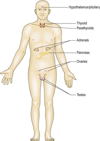
The main endocrine glands are the pituitary, thyroid, parathyroids, pancreas, adrenals and gonads: testes and ovaries (Fig. 5.1). These glands synthesise hormones which are released into the circulation and act at distant sites. Disease may result from excessive or inadequate production of hormones, or target organ hypersensitivity or resistance to the hormone. Although some endocrine glands, e.g. parathyroid glands and pancreas, respond directly to metabolic signals, most are controlled by hormones released from the pituitary gland. A wide variety of molecules act as hormones:
Symptoms and definitions
Symptoms of endocrine disturbance are frequently non-specific and affect many body systems (Box 5.1). Endocrine conditions may be detected by chance, e.g. glycosuria on screening, or if the clinician sees a patient after a significant time interval and notices diagnostic facial features, e.g. in acromegaly or hypothyroidism. Nevertheless, diagnosis often depends upon careful observation of a patient with non-specific symptoms prompting the recognition of specific features.
 5.1 Common clinical features in endocrine disease
| Symptom, sign or problem | Differential diagnoses |
| Weight gain | Hypothyroidism, polycystic ovary syndrome (PCOS), Cushing’s syndrome |
| Weight loss | Hyperthyroidism, diabetes mellitus, adrenal insufficiency |
| Short stature | Constitutional, non-endocrine systemic disease (e.g. coeliac disease), growth hormone deficiency |
| Delayed puberty | Constitutional, non-endocrine systemic disease, hypothyroidism, hypopituitarism, primary gonadal failure |
| Menstrual disturbance | PCOS, hyperprolactinaemia, thyroid dysfunction |
| Diffuse neck swelling | Simple goitre, Graves’ disease, Hashimoto’s thyroiditis |
| Excessive thirst | Diabetes mellitus or insipidus, hyperparathyroidism, Conn’s syndrome |
| Hirsutism | Idiopathic, PCOS, Cushing’s syndrome, congenital adrenal hyperplasia |
| ‘Funny turns’ | Hypoglycaemia, phaeochromocytoma, neuroendocrine tumour |
| Sweating | Hyperthyroidism, hypogonadism, acromegaly, phaeochromocytoma |
| Flushing | Hypogonadism (especially menopause), carcinoid syndrome |
| Resistant hypertension | Conn’s syndrome, Cushing’s syndrome, phaeochromocytoma, acromegaly, renal artery stenosis |
| Erectile dysfunction | Primary or secondary hypogonadism, diabetes mellitus, non-endocrine systemic disease, medication-induced (e.g. beta-blockers, opiates) |
| Muscle weakness | Cushing’s syndrome, hyperthyroidism, hyperparathyroidism, osteomalacia |
| Bone fragility and fractures | Hypogonadism, hyperthyroidism, Cushing’s syndrome |
| Altered facial appearance | Hypothyroidism, Cushing’s syndrome, acromegaly, PCOS |
Apart from diabetes mellitus, thyroid disease and some reproductive conditions, endocrine disorders are uncommon. Most patients with tiredness, excessive sweating or sexual dysfunction, for example, do not have an underlying endocrine cause (Box 5.2).
 5.2
5.2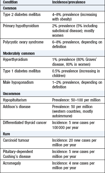
Common presenting symptoms are:
• appetite and/or weight change – in hyper/hypothyroidism, diabetes mellitus, Cushing’s syndrome
• polydipsia (excessive thirst) and/or polyuria (passing >3 litres urine/day) – in diabetes mellitus, diabetes insipidus or hyperparathyroidism
• change in facial/body hair growth and distribution – in hypogonadism, hypopituitarism, adrenal insufficiency, androgen excess. Hirsutism is the excessive growth of thick terminal hair in an androgen-dependent distribution (upper lip, chin, chest, back, lower abdomen, thighs) in women
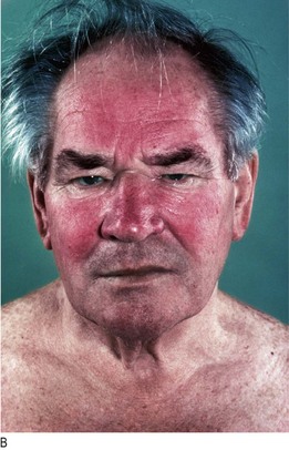
• change in skin and mucosal pigmentation and character: coarse dry skin in hypothyroidism; excessive pigmentation and/or vitiligo (areas of depigmented skin) in Addison’s disease (Fig. 5.17D); soft-tissue overgrowth and skin tags in acromegaly; acanthosis nigricans (velvety thickening and pigmentation of the major flexures, especially the axillae and groins: Fig. 5.11A) in obesity, and type 2 diabetes
• increased sweating in acromegaly and phaeochromocytoma
• temperature intolerance – in hyperthyroidism (heat) and hypothyroidism (cold)
• erectile dysfunction or loss of libido in hypogonadism
• gynaecomastia (breast tissue enlargement in men)
• galactorrhoea (breast milk production in men, or in women outwith pregnancy or breastfeeding) in pituitary adenomas producing hyperprolactinaemia
• primary or secondary amenorrhoea (p. 221) in pituitary or hypothalamic disease
• tiredness: can be a non-specific feature of diabetes mellitus, hypothyroidism or Addison’s disease.
The history
The thyroid
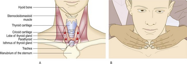
The thyroid is butterfly-shaped with two symmetrical lobes joined by a central isthmus that normally covers the second and third tracheal rings (Fig. 5.2A). The gland may extend into the superior mediastinum, or may occasionally be entirely retrosternal. Rarely, it is located higher in the neck along the line of the thyroglossal duct. If situated at the back of the tongue (lingual goitre), it may be visible through the open mouth.
Symptoms and definitions
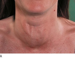
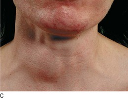
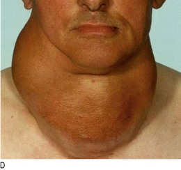
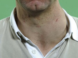
Goitre is enlargement of the thyroid gland. It is not necessarily associated with thyroid dysfunction and most patients with a goitre are euthyroid (Fig. 5.5).
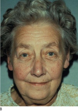
Hyperthyroidism (thyrotoxicosis) is a clinical state caused by increased levels of circulating levels of the thyroid hormones, T3 and T4. Graves’ disease is the commonest cause of hyperthyroidism. It is an autoimmune disease with a familial component, which is 5–10 times more common in women and usually presents between 20 and 50 years of age. It has specific extrathyroid features (Fig. 5.3):
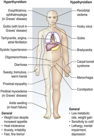
• Exophthalmos (proptosis) is increased protrusion of the eyeball from the orbit. It is caused by an inflammatory infiltration of the orbital contents (the soft tissues and extraocular muscles, not the globe). It is usually bilateral. Exophthalmos may lead to diplopia (Fig. 5.4A&B) and other features of Graves’ ophthalmopathy: conjunctival oedema, conjunctivitis, corneal ulceration, ophthalmoplegia and optic atrophy (Ch. 12).
• Pretibial myxoedema is a raised, discoloured (usually pink or brown) indurated appearance over the lower legs (Fig. 5.4D). Note that, despite the name, it is associated with Graves’ disease, not hypothyroidism.
• Thyroid acropachy is a periosteal hypertrophy of the distal phalanges which looks like finger clubbing (Fig. 5.4C).
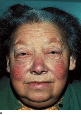
Hypothyroidism (Fig. 5.3) is caused by reduced levels of thyroid hormones and is usually due to autoimmune Hashimoto’s thyroiditis. Women are affected approximately six times more commonly than men. Many clinical features of hypothyroidism are produced by myxoedema (non-pitting oedema caused by tissue infiltration by mucopolysaccharides, chondroitin and hyaluronic acid) (Box 5.3 and Figs 5.3 and 5.7).
History
Past drug, family and social history: Ask about:
• drug therapy (amiodarone and lithium are associated with hypothyroidism), antithyroid drugs
• family history of thyroid or autoimmune disease
• living in areas of iodine deficiency, e.g. the Andes, Himalayas, Central Africa, can cause goitre and, rarely, hypothyroidism.
Examination sequence
Look for:
 arms and legs for: skin abnormalities, muscle power and the deep tendon reflexes (p. 263)
exophthalmos, diplopia, conjunctival oedema or conjunctivitis, corneal ulceration
lid retraction: this is present if the sclera is visible above the iris (Fig. 5.4A)
Fig. 5.4 Graves’ hyperthyroidism.
(A) Typical facies. (B) Severe inflammatory thyroid eye disease. (C) Thyroid acropachy. (D) Pretibial myxoedema.
lid lag: ask the patient to follow your index finger as you move it from the upper to the lower part of his visual field (Fig. 12.13). Delay between the descent of the upper eyelid in relation to that of the eyeball is lid lag.
 Examination sequence
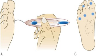
Inspect the neck from the front. Give the patient a glass of water and ask him to take a sip. Look for a swelling while he swallows.
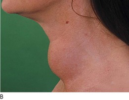
Ask the patient to sit with the neck muscles relaxed and stand behind him. Place your hands gently on the front of the neck, with your index fingers just touching (Fig. 5.2B). Ask him to swallow a sip of water and feel the gland as it moves upwards. Some patients find neck palpation uncomfortable, so be alert for any signs of distress.
Note the size, shape and consistency of any goitre and feel for any thrill.
Measure any discrete nodules with callipers.
Record the maximum neck circumference of a large goitre using a tape measure (objective measurements are useful for long-term follow-up).
Auscultate with your stethoscope for a thyroid bruit. A thyroid bruit may be confused with other sounds: bruits from the carotid artery or transmitted from the aorta are louder along the line of the artery. Transient gentle pressure over the root of the neck will interrupt a venous hum from the internal jugular vein.
Normal findings: The normal thyroid gland is palpable in ~50% of women and 25% of men. Prominent skin folds may give a false impression of goitre. The thyroid (or a thyroglossal cyst) moves upwards on swallowing since it is enveloped in the pre-tracheal fascia, which is attached to the cricoid cartilage.
Shape, surface and consistency: Simple goitres are relatively symmetrical in their earlier stages but often become nodular with time. In Graves’ disease the surface of the thyroid gland is usually smooth and diffuse; it is irregular in uninodular or multinodular goitre (Fig. 5.5). Nodules in the substance of the gland may be large or small, and single or multiple, and are usually benign. A very hard consistency suggests malignancy. Large, firm lymph nodes near a goitre suggest thyroid cancer (Fig. 5.6). Diffuse tenderness is typical of viral thyroiditis. Localised tenderness may follow bleeding into a thyroid cyst.
The parathyroids
There are usually four parathyroid glands which lie posterior to the thyroid (Fig. 5.2A). Each is about the size of a pea and produces parathyroid hormone, a peptide which increases the level of calcium in the blood.
Symptoms and definitions
Parathyroid disease is commonly asymptomatic. In hyperparathyroidism, the commonest symptoms relate to hypercalcaemia: polyuria, polydipsia, renal stones, peptic ulceration, tender areas of bone fracture deformity (‘Brown tumours’: Fig. 5.8A), and confusion or psychiatric symptoms.
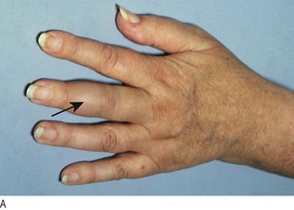
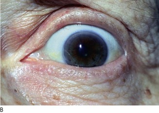
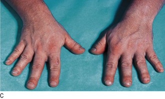
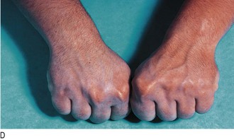
Fig. 5.8 Parathyroid disease.
(A) ‘Brown tumour’ of the phalanx (middle finger) in hyperparathyroidism. (B) Corneal calcification in hyperparathyroidism. (C) Pseudohypoparathyroidism: short metacarpals. (D) These are best seen when the patient makes a fist.
In hypoparathyroidism, hypocalcaemia may cause hyperreflexia or tetany (involuntary muscle contraction), most commonly in the hands or feet. Paraesthesiae of the hands and feet or around the mouth may occur. Hypoparathyroidism is most often caused by inadvertent damage to the glands during thyroid surgery.
Patients with the autosomal dominant condition pseudohypoparathyroidism have end-organ resistance to parathyroid hormone and typically have short stature, round face and shortening of some metacarpal bones.
History
• recent thyroid or neck surgery or irradiation
• polyuria, polydipsia or renal stones
Examination sequence
Look at the neck for scars of previous surgery.
Assess mental state (p. 21).
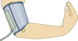
Take the BP and assess the state of hydration (p. 58).
Look at the hands. Ask the patient to make a fist and assess the length of the metacarpals.
Test for muscle weakness (p. 261) and hyperreflexia (p. 263).
Test for latent tetany: place a BP cuff on the upper arm and inflate above systolic pressure for 3 minutes. In the hand, carpal muscle contraction produces a typical picture with the thumb adducted, the proximal interphalangeal and distal interphalangeal joints extended and the metacarpophalangeal joints flexed (‘main d’accoucheur’ (hand of the obstetrician) or Trousseau’s sign: Fig 5.9).
Look for evidence of recent fractures and bone tenderness.
Abnormal findings: Parathyroid tumours are very rarely palpable.
Findings in hyperparathyroidism may include altered mental state, dehydration, proximal muscle weakness, fractures and bony tenderness. In long-standing hypercalcaemia, corneal calcification (band keratopathy) may be present (Fig. 5.8B). Renal stones may produce haematuria on stix testing.
In moderate/severe hypocalcaemia, hyperreflexia and a positive Trousseau’s sign may be present. In pseudohypoparathyroidism the metacarpals of the ring and little fingers are shortened (Fig. 5.8C and D).
The pancreas
The pancreas lies behind the stomach on the posterior abdominal wall. Its endocrine functions include the production of insulin, glucagon, gastrin, somatostatin and vasoactive intestinal peptide. Its exocrine function is to produce alkaline secretions containing digestive enzymes.
Symptoms and definitions
Diabetes mellitus: This is characterised by hyperglycaemia due to absolute or relative insulin deficiency. Insulin-dependent patients are particularly susceptible to acute metabolic decompensation due to hypoglycaemia or ketoacidosis, both of which require prompt clinical and biochemical recognition and treatment.
• Type 1: severe insulin deficiency due to autoimmune destruction of the pancreatic islets.
• Type 2: commonly affects people who are obese and insulin-resistant, although impaired β-cell function is also important.
Diabetes mellitus may present with a classical triad of symptoms:
• Polyuria (and nocturia): due to osmotic diuresis caused by glycosuria.
• Thirst: due to the resulting loss of fluid and electrolytes.
• Weight loss: due to fluid depletion and breakdown of fat and muscle secondary to insulin deficiency.
Other common symptoms are tiredness, mood changes and blurred vision (due to glucose-induced changes in lens refraction). Bacterial and fungal skin infections are common because of the combination of hyperglycaemia, impaired immune resistance and tissue ischaemia. Itching of the genitalia (pruritus vulvae in women, balanitis in men) is due to Candida yeast infection (thrush).
Examination sequence
Look for evidence of weight loss and dehydration (p. 58).
Smell the patient’s breath for the sweet smell of ketones (diabetic ketoacidosis).
Examine the skin: look for signs of infection and rashes. Look for xanthelasma and xanthomata (Fig. 6.8A and B). Examine insulin injection sites for evidence of lipohypertrophy (which may cause unpredictable insulin release), lipoatrophy (rare) or signs of infection (very rare).
Measure pulse and BP and examine the cardiovascular and peripheral vascular systems.
Examine the respiratory and gastrointestinal systems.
Examine the central nervous system.
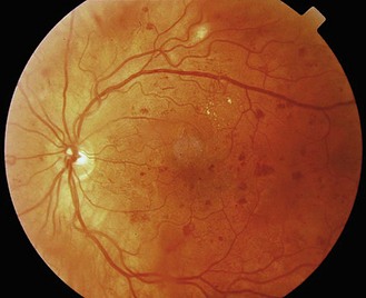
Test visual acuity and examine the eyes and optic fundi (p. 287) (Fig. 5.10).
Abnormal findings: Dehydration and Kussmaul respiration (hyperventilation with a deep, sighing respiratory pattern) are common in ketoacidosis.
Bacterial skin infections, e.g. cellulitis, boils, abscesses and fungal infections, may be seen. Acanthosis nigricans (Fig. 5.11A) occurs in hyperinsulinism and is seen frequently in patients with insulin-resistant type 2 diabetes. Necrobiosis lipoidica (a yellow indurated or ulcerated area surrounded by a red margin: Fig. 5.11B), due to collagen degeneration, may occur on the shins of some type 1 diabetic patients and often causes chronic ulceration. Xanthelasmata and xanthomata indicate significant hyperlipidaemia (Fig. 5.11C and Fig. 6.8).
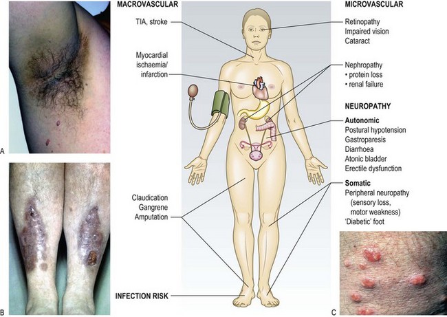
Fig. 5.11 Diabetes and the skin.
(A) Acanthosis nigricans. (B) Necrobiosis lipoidica. (C) Eruptive xanthomata.
Microvascular, neuropathic and macrovascular complications of hyperglycaemia (Fig. 5.11) can occur in patients with any type of diabetes mellitus, and may be present at diagnosis in patients with slow-onset type 2 disease. Careful examination of the eyes, cardiovascular, neurological and renal systems, and feet is essential.
Glycosuria suggests hyperglycaemia and, if accompanied by ketonuria (and Kussmaul respiration: Ch. 7) indicates ketoacidosis. Proteinuria occurs in diabetic nephropathy. Detection of nitrite ± haematuria suggests (often occult) urinary infection (Box 5.4).
 5.4
5.4
Diabetic complications
Up to 40% of patients with diabetes have peripheral neuropathy and 40% have peripheral vascular disease. 5% develop foot ulceration each year and up to 66% are associated with infection or osteomyelitis.
Cheer K, Shearman C, Jude EB. Managing complications of the diabetic foot. BMJ 2009;339:1304–1307.
Examination sequence
Diabetic patients have a 15% lifetime risk of foot ulcers, which are highly susceptible to infection. Early recognition of the ‘at-risk’ diabetic foot is essential. There are two main presentations:
Neuropathic: where neuropathy predominates but the major arterial supply is intact.
Neuro-ischaemic: where reduced arterial supply produces ischaemia and exacerbates neuropathy (Fig. 5.13).
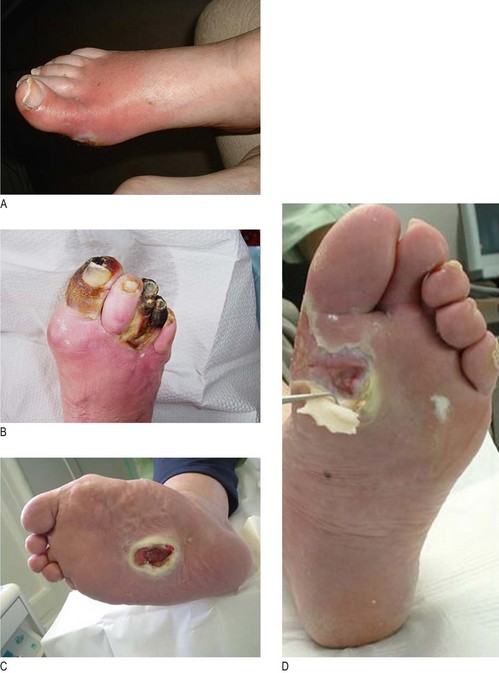
Infection complicates both presentations.
Look for hair loss and nail dystrophy.
Examine the skin (including the interdigital clefts) for excessive callus, infections and ulcers.
Feel the temperature of the feet.
Examine the dorsalis pedis and posterior tibial pulses. If absent, arrange Doppler studies to evaluate ankle : brachial pressure index (Ch. 6).
Test for peripheral neuropathy: use a nylon monofilament which buckles at a force of 10 g to apply a standard, reproducible stimulus. The technique and best sites to test are shown in Figure 5.12. Avoid areas of untreated callus.
Abnormal findings: Hair loss and nail dystrophy occur with ischaemia, causing nutritional changes. There may be skin fissures or tinea infection (‘athlete’s foot’). The foot arch may be excessive in neuropathy or collapsed (rocker-bottom sole). Both conditions cause abnormal pressures and increase risk of plantar ulceration.
Warm feet occur in neuropathy and cold feet in ischaemia.
Sensory neuropathy is present if the patient cannot feel the monofilament in any of the sites. This means loss of protective pain sensation and is a good predictor of future ulceration.
Charcot’s arthropathy is disorganised foot architecture, acute inflammation, fracture and bone thinning, usually in a patient with neuropathy but relatively good vascular supply to the lower limb. It presents acutely as a hot, red, swollen foot often impossible to distinguish clinically from infection.
• Low – no risk factors (no sensation loss, peripheral vascular disease or other risk factors).
• Moderate – one risk factor present.
• High – previous ulceration or amputation, or more than one risk factor present.
• Active – ulceration, spreading infection, critical ischaemia, unexplained red, hot, swollen foot.
The pituitary
The pituitary gland is enclosed in the sella turcica in the base of the skull beneath the hypothalamus. It is bridged over by a fold of dura mater (diaphragma sellae) with sphenoidal air spaces below and the optic chiasm above. The pituitary has two lobes:
• Anterior: which secretes several hormones (adrenocorticotrophic hormone (ACTH), prolactin, growth hormone (GH), thyroid-stimulating hormone and gonadotrophins (luteinising hormone (LH) and follicle-stimulating hormone (FSH)).
• Posterior: an extension of the hypothalamus, which secretes vasopressin (antidiuretic hormone) and oxytocin.
History
Ask about the most common symptoms – headache and excessive sweating.
Has the patient (or more often a relative or friend) noticed changes in his facial features? Photographs taken years earlier can be helpful to identify changes and the onset of the condition.
Has the patient noticed an increase in shoe, ring or glove size (Box 5.5)?
Examination sequence
5.5
Acromegaly
Clinical presentations, such as carpal tunnel syndrome, sleep apnoea, with associated coarse facial features and symptoms, such as headache, sweating, an increase in ring or shoe size, should raise the possibility of acromegaly.
Reddy R, Hope S, Wass J. Acromegaly: easily missed? BMJ 2010;341:400–401.
Look at the face for coarsening of features, thick greasy skin, enlargement of the nose, prognathism (protrusion of the mandible) and separation of the lower teeth (Fig. 5.14A, B).
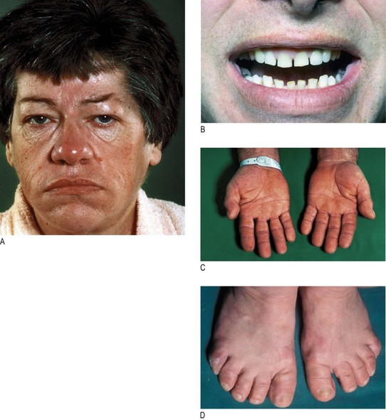
Fig. 5.14 Acromegaly.
(A) Typical facies. (B) Separation of lower teeth. (C) Large fleshy hands. (D) Widening of the feet.
Examine the hands and feet. Look for soft-tissue enlargement and complications arising from this, e.g. tight-fitting rings or shoes, carpal tunnel syndrome (Fig. 5.14C, D).
Assess the visual fields: expansion of the tumour can cause pressure on the optic chiasm, resulting in visual field defects, especially bitemporal hemianopia (Fig. 12.3).
Check the BP and perform urinalysis. Hypertension and diabetes mellitus are common associations.
Hypopituitarism: Anterior hypopituitarism is due to compression of the pituitary by a macroadenoma, infarction after childbirth (Sheehan’s syndrome), severe head trauma or cranial radiotherapy.
Apart from headache due to stretching of the diaphragma sellae and visual abnormalities, clinical presentation depends upon the deficiency of the specific anterior pituitary hormones involved. Individual or multiple hormones may be involved, so questioning in relation to the thyroid, adrenocortical and sexual function is needed.
Examination sequence
extreme skin pallor (a combination of mild anaemia and melanocyte-stimulating hormone deficiency)
reduced/absent secondary sexual hair (caused by gonadotrophin deficiency) (Fig. 5.15)
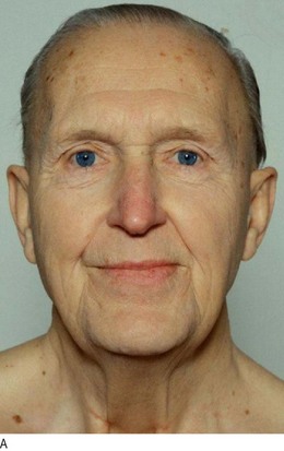
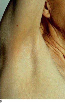
Fig. 5.15 Hypopituitarism.
(A) Hypopituitarism due to a pituitary adenoma (note the fine pale skin). (B) Absent axillary hair.
Examine the eyes for: visual field defects (most often bitemporal hemianopia); optic atrophy or cranial nerve defects (III, IV and VI) caused by a tumour compressing the optic chiasm, optic nerve or cavernous sinus.
The adrenals
The adrenals are small pyramidal organs lying immediately above the kidneys on their posteromedial surface. The adrenal medulla is part of the sympathetic nervous system and secretes catecholamines. The adrenal cortex secretes cortisol, mineralocorticoids and androgens.
Symptoms and definitions
Cushing’s syndrome: Cushing’s syndrome is caused by excess exogenous or endogenous corticosteroid exposure. Most cases are iatrogenic due to side-effects of corticosteroid therapy. ‘Endogenous’ Cushing’s usually results from an ACTH-secreting pituitary microadenoma. Other causes include a primary adrenal tumour or ‘ectopic’ ACTH secretion.
The catabolic effects of steroids cause widespread tissue breakdown (particularly in skin, muscle and bone) with central accumulation of body fat. Proximal myopathy, fragility fractures, spontaneous purpura, skin thinning and susceptibility to infection are common (Fig. 5.16). Patients may be hypertensive.
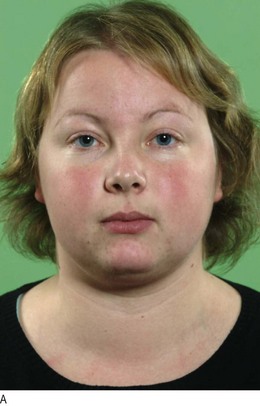
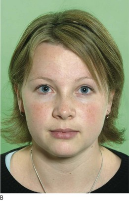
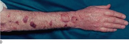
Fig. 5.16 Cushing’s syndrome.
(A) Cushingoid facies. (B) After curative pituitary surgery. (C) Typical features: facial rounding, central obesity, proximal muscle wasting and skin striae. (D) Skin thinning: purpura caused by wristwatch pressure.
Examination sequence
Look at the face and general appearance for central obesity and a round face (Fig. 5.16).
Examine the skin for thinning, hyperpigmentation, acne, hirsutism, bruising, striae (especially abdominal) and signs of infection or poor wound healing.
Examine the legs for evidence of proximal muscle weakness and oedema.
Examine the eyes for cataracts, and hypertensive changes (Fig. 6.16).
Perform urinalysis (Box 5.6).
5.6
Cushing’s syndrome
Easy bruising, facial plethora, proximal myopathy and broad purple striae best discriminate Cushing’s syndrome but these findings do not have high sensitivity. Furthermore, many features of Cushing’s syndrome are common in the general population and are less discriminatory, e.g. obesity, dorsocervical fat pad, oedema, acne and hirsutism.
Nieman LK, Biller BMK, Findling JW et al. The diagnosis of Cushing’s syndrome: an Endocrine Society clinical practice guideline. J Clin Endocrinol Metab 2008;93:1526–1540.
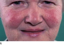
Addison’s disease: Addison’s disease is due to inadequate secretion of cortisol, usually secondary to autoimmune destruction of the adrenal cortex. Symptoms are usually non-specific, but weakness, muscle cramps, nausea, vomiting, diarrhoea or constipation may occur.
Examination sequence
Look for signs of weight loss.
Examine the entire skin surface for abnormal or excessive pigmentation: this is most prominent in sun-exposed areas or epithelia subject to trauma or pressure – skin creases, buccal mucosa and recent scars (Fig. 5.17A–C). Excess pigmentation is produced by melanocyte-stimulating hormone in primary adrenal insufficiency. It is most striking in white Europeans. Vitiligo (depigmentation of areas of skin) occurs in 10–20% of Addison’s disease cases (Fig. 5.17D).
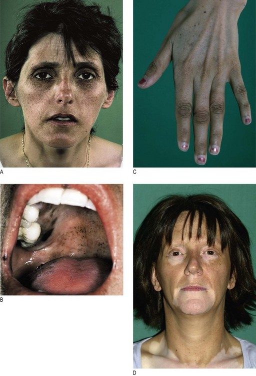
Fig. 5.17 Addison’s disease.
(A) Facial pigmentation. (B) Buccal pigmentation. (C) Skin crease pigmentation. (D) Vitiligo – particularly striking due to addisonian pigmentation of the ‘normal’ skin.
Check the BP and test for postural hypotension (p. 114). Hypotension and postural hypotension result from reduced mineralocorticoid effects.
The gonads
These glands secrete sex hormones (oestrogen and testosterone) in response to gonadotrophin (FSH and LH) release by the pituitary. They also contain the germ cells.
Symptoms and definitions
Klinefelter’s syndrome (47XYY) is the most common cause of primary hypogonadism in men (1 : 600 live male births). Diagnosis may be delayed until later life, by which time features of prolonged testosterone deficiency can be seen. Look for soft, finely wrinkled, hairless facial skin and gynaecomastia and examine the genitalia (pubic hair is often reduced/absent and the testes <3 ml in volume; Fig. 5.18).
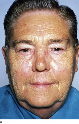
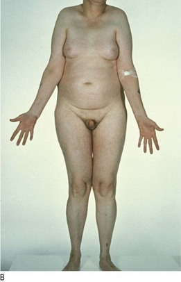
Fig. 5.18 Klinefelter’s syndrome.
(A) Hypogonadal facial skin. (B) Gynaecomastia, reduced pubic hair and small testes.
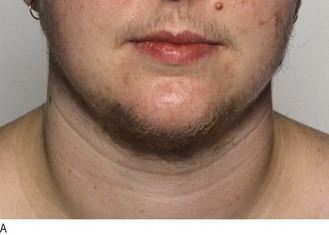
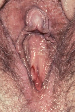
Hirsutism is common in women with polycystic ovary syndrome (Fig. 5.19). Virilisation is temporal recession of the scalp hair, deepening of the voice, breast atrophy, increased muscle bulk and clitoromegaly (Fig. 5.20). If present in women with a short history of severe hirsutism, it suggests a possible testosterone-secreting tumour.
Other endocrine disorders
Liver metastases from mid-gut carcinoid tumour release vasoactive chemicals into the systemic circulation which cause flushing, diarrhoea and bronchospasm. Bending, exercise or even palpation of the enlarged liver may induce typical skin flushing. Permanent facial telangiectasia occurs after many years of carcinoid flushing (Fig. 5.21).
Putting it all together
Because of their wide diversity of clinical features, keep alert to the possibility of endocrine disease in non-specific presentations. Perhaps more than in any other area of medicine, pattern recognition is important.
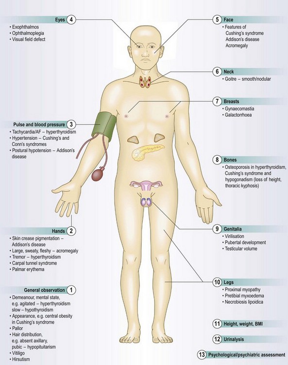
A structured approach to the general endocrine examination
Examination sequence
Carefully observe the patient’s overall appearance, manner and habitus for diagnostic clues:
Is he restless and agitated (hyperthyroidism) or slow and lethargic (hypothyroidism)?
Measure the height and weight and calculate the body mass index (p. 55). Use a stadiometer in children and adolescents (Fig. 15.20). If the patient is obese, is the adiposity centrally distributed, e.g. Cushing’s syndrome or GH deficiency.
Look for a thoracic kyphosis, which may be a sign of osteoporotic vertebral collapse.
Inspect the face and eyes for a ‘spot’ endocrine diagnosis (Figs 5.4A, 5.7A, 5.14A, 5.16A, 5.17A, 5.18A).
Look at the mouth for overgrowth of the chin and tongue (acromegaly) and for buccal pigmentation (Addison’s disease).
Examine the hands: the initial handshake may suggest a diagnosis, e.g. tremor and palmar sweating in hyperthyroidism. Look for soft-tissue overgrowth (acromegaly) or dysmorphism (abnormal metacarpal length in pseudohypoparathyroidism); skin crease pigmentation (Addison’s disease); wasting of the thenar muscles due to carpal tunnel syndrome (hypothyroidism, acromegaly) (Fig. 14.29B).
Examine the entire skin surface: look for abnormal pallor (hypopituitarism), vitiligo, skin or scar pigmentation (Addison’s disease), plethora (Cushing’s or carcinoid syndrome). Patients with Cushing’s syndrome often have thin, fragile skin with bruising after trivial trauma (Fig. 5.16D). Inspect the axillae and groins for acanthosis nigricans (obesity, diabetes mellitus) (Fig. 5.11A).
Is the body hair normal in quality and amount? Look for hirsutism in females (polycystic ovary syndrome: Fig. 5.19) and for loss of hair in the axillae and groins (hypopituitarism) (Fig. 5.15B).
Assess the pulse rate, rhythm and volume. Tachycardia and atrial fibrillation may suggest thyrotoxicosis.
Record the BP. Hypertension is a feature of several endocrine conditions, e.g. Cushing’s syndrome, phaeochromocytoma, Conn’s syndrome (primary hyperaldosteronism) (Box 5.1). Postural hypotension (p. 114) occurs in adrenal insufficiency.
Examine the eyes: look for features of thyroid disease. Assess visual acuity and perform fundoscopy in patients with diabetes mellitus. Assess visual acuities and fields (p. 288) in patients with suspected pituitary tumours (to detect bitemporal hemianopia due to compression of the optic chiasm). Look for optic atrophy in patients with longstanding optic pathway compression (Fig. 12.31A).
Examine the neck for goitre. If present, record its size, surface and consistency.
Look for gynaecomastia (common in Klinefelter’s syndrome: Fig. 5.18) and galactorrhoea: Gently massage the breast tissue towards the nipple to see if milk is expressed. Explain beforehand why you are performing this examination and watch the patient carefully since this may be uncomfortable.
Examine the abdomen: look for purple striae (Cushing’s syndrome). Carcinoid syndrome is associated with a palpable, nodular liver, which is sometimes massively enlarged. Adrenal tumours may occasionally be palpable, but be cautious if you suspect phaeochromocytoma, as palpation may precipitate a hypertensive paroxysm.
Inspect the legs for pretibial myxoedema (Graves’ disease: Fig. 5.4D), proximal muscle wasting or weakness (Cushing’s syndrome and hyperthyroidism) and delayed tendon reflexes (hypothyroidism).
Examine the feet for signs of diabetic neuropathy, ischaemia and ulcers.
Examine the external genitalia (p. 225). Inspect the amount of pubic hair and make a pubertal staging of all adolescents using Tanner gradings (Ch. 10). In men, record testicular size and consistency (p. 238). In women, look for virilising features.
Test the urine for glucose, ketones, protein and nitrite.
Formal psychological evaluation (p. 25) may be appropriate in selected patients (Cushing’s syndrome, hyperparathyroidism).
Investigations
Measure serum hormone levels to assess over- or underactivity. Suppression tests can determine whether hormonal secretion is autonomous. Stimulation tests assess hormonal reserve (or lack of it in deficiency states). Modern imaging enables visualisation of small endocrine tumours, sometimes only millimetres in diameter (Box 5.7 and Fig. 5.22).
 5.7
5.7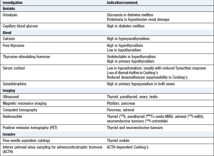
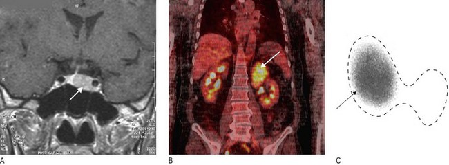
Fig. 5.22 Endocrine imaging.
(A) Magnetic resonance imaging showing pituitary macroadenoma (arrow). (B) Positron emission tomography-computed tomography scan showing an adrenal cancer (arrow). (C) 99mTechnetium radionuclide scan confirming unilateral toxic thyroid adenoma (arrowed) – dotted line shows outline of thyroid.