Chapter 4 Conformation and Lameness
The idea of a good horse with poor legs is a misnomer; the legs are the essence of the horse, and every other part of the equine machine is of only subservient and tributary importance.
The thought that the way a horse is conformed determines the way it moves is well accepted. The relationship of conformation, especially of the distal extremities, and lameness also is well recognized. “Conformation determines the shape, wear, flight of the foot, and distribution of weight.”2 Veterinarians often are asked to comment on conformation during lameness and prepurchase examinations, especially with regard to the suitability of the horse to perform the intended task. In some instances, as in the case of presale yearling evaluations, the veterinarian’s opinion is paramount, and purchase is contingent on judgment of the yearling’s potential to perform as a racehorse, given its conformation, or in some instances its conformational faults (see Chapter 99). “It is by a study of conformation that we assign to a horse the particular place and purpose to which he is best adapted as a living machine and estimate his capacity for work, and the highest success in this connection will be best attained by the judicious blending of practice with science.”3 Evaluation of conformation and its influence on lameness is based largely on observation, experience, and pattern recognition. Recognizing desirable conformational traits in horses suited for a particular sporting activity and learning when to overlook a minor fault that has little clinical relevance are important.
Relevance of Evaluation of Conformation
Conformation is one piece of the complex puzzle of a lame horse, although poor conformation does not necessarily condemn a horse to lameness: “faulty conformation is not an unsoundness … it is a warning sign.”2 All lameness diagnosticians should evaluate conformation briefly at the beginning of each examination. The association of lameness and faulty conformation will be obvious. The clinician must evaluate the horse from afar, assessing the whole horse for balance, angles and lengths, and posture and symmetry. The clinician must remember that horses come in all shapes, sizes, and types, and therefore conformation varies accordingly, but certain conformational faults produce predictable lameness conditions and are undesirable. However, good conformation is not synonymous with success, and although horses of certain body types tend to have longer strides and are more athletic than others, intelligence, aggression, “will to win,” and other intangible factors are important. It is our opinion that a well-bred horse from a successful family can endure faulty conformation much better than one with poor or mediocre breeding.
Hereditary Aspects of Conformation
Certain conformational faults appear to be highly heritable traits. Evaluation of broodmares and foals often reveals that the early conformational defects seen in a foal are present in the dam. The dam seems to contribute more to faulty conformation than does the sire, although the stallion also is important. This difference may be explained in part by the fact that fillies with faulty conformation may develop problems or be retired early and subsequently bred, whereas most stallions usually are proven performers with exceptional conformation. Conformational faults such as toed in and toed out commonly are passed down from generation to generation. Back-at-the-knee (calf-knee), offset (bench) knee, tied-in below the knee, sickle-hocked, and straight-behind conditions appear to be highly heritable. In a recent study abnormal sire phenotype (offset carpi and outward rotation) was associated with faulty yearling carpal conformation.4 Heavier foals and yearlings were more likely to have faulty carpal conformation and inward rotation of the fetlock joints.4
Certain lameness conditions are common in horses with faulty conformation, but similar lameness conditions develop inexplicably in some breeding lines year after year in offspring with apparently acceptable conformation. Lameness of the carpus or tarsus appears to be most important. For example, in Standardbreds (STBs), siblings commonly develop similar lameness conditions, such as proximal suspensory avulsion injury, carpal osteochondral fragments, distal hock joint pain, or curb.
Objective Evaluation of Conformation: Is It Possible?
An attempt to quantify conformation using a linear assessment trait evaluation system that allows the observer to assess where, given a particular trait, a single horse falls within a population of horses, was described in 1996.5 A population of 101 Irish Thoroughbred (TB) flat racehorses and 19 top stallions was used and 27 common conformational traits were evaluated, including various heights, lengths and angles, and distal extremity conformation. Of the 27 traits, six were significantly linked to age (withers’ height and conformation, back length, neck size, carpal conformation, and hind pastern conformation), and five were linked to sex (head and neck shape; neck size at the poll, the larynx the withers, and the manubrium of the sternum; and forelimb hoof pastern axis). Most traits exhibited large phenotypic variation within the population, but 21 of 27 non–age-linked traits were judged suitable for possible inclusion in a linear assessment protocol.4 Researchers judged a high percentage of horses to be toed out, suggesting this trait may even be desirable.
More recent studies have used video-image analysis, but direct physical measurements may be more accurate than those obtained by analyzing videotapes or photographs.6 Potential errors in image analysis occur because of movement of skin markers over selected bony protuberances, a phenomenon more common in the upper limb and in motion studies.6,7 Skin marker location is critical for evaluation of joint angulation and movement during locomotion or conformation analysis. Instantaneous center (or axis) of rotation (ICR) is defined as the point with zero velocity during movement of that joint; accurate measurements of joint angulation require positioning markers at the ICR.8 Conventional positions of skin markers and ICR in most joints agree well, but use of traditional marker sites on the scapulohumeral and femorotibial joints results in overestimation and underestimation, respectively, of caudal joint angles.9 Although video-image analysis may be fraught with potential or in some instances real error, objectivity is a major advantage. Other advantages include the ability to replay images, reduction of observer fatigue, elimination of observations and measurements in real time, and permanent recording of the observation. Ideally, objective measurements would withstand statistical evaluation, distinguish between desirable and undesirable traits, and account for differences among different types of horses.6
A combination of direct measurement and photography was used to evaluate conformation of Swedish Warmblood (WBL) and elite sports horses.10,11 Whereas most of the conformational defects were mild or moderate, 80% of WBL horses were toed out behind, suggesting this may be a normal finding in this breed as in the STB trotter. More than 50% of horses had bench knees and 5% were toed out in front, contrary to findings in STB trotters.10,12 Many of the elite horses were bucked kneed, whereas the riding school horses tended to have calf-knees. It was speculated that this occurred because elite horses were evaluated after competition, and muscle fatigue may have contributed to the tendency to be over at the knee. Sex had a significant influence on conformation; females were smaller and had longer bodies and smaller forearms and metacarpal regions. There were interesting findings regarding hock angle. A sickle hock is defined as a hock angle of 53 degrees or less; a large hock angle is referred to as straight behind. Sickle-hocked conformation was nearly absent in elite horses, and it was hypothesized that sickle-hocked conformation either predisposed a horse to lameness or impaired a horse’s ability to achieve upper levels of competition.10 A positive relationship between larger hock angles and soundness in STB trotters also exists.13 All results must be viewed in moderation, because it is our clinical impression that horses with excessively large hock angles (straight behind) are substantially predisposed to suspensory desmitis. In forelimbs of WBL show jumpers and the forelimbs and hindlimbs of STB trotters, smaller fetlock joint angles (less upright) were desirable.10,13
Radiology was used to assess the degree of hyperextension of the carpus to study the potential effect of back-at-the-knee (calf-knee) conformation on the subsequent development of carpal chip fractures.14 Lateromedial radiographic images of 21 horses with carpal chip fractures and of 10 normal horses were obtained, with and without the contralateral limb raised. No relationship between measured carpal angle and carpal chip fracture formation existed, suggesting that this group of TB racehorses did not develop carpal chip fractures as a result of calf-kneed conformation. The sample size was small, however, and a larger study may produce different results. Horses with severe calf-kneed conformation may develop other problems and not advance enough in training to develop carpal chip fractures. They may be judged poor surgical candidates, are not referred, or are slow.
Two recent studies evaluated TBs and Quarter Horses (QHs) using skin markers, photography (three views: front, side, back), and computer-image analysis.15,16 Of the two studies done in TBs, one evaluated longitudinal development of conformation from weaning to 3 years of age. A strong relationship between long bone lengths and withers heights for all ages supported the theory that horses are proportional. Longitudinal bone growth in the distal limb increased only 5% to 7% and was presumably completed before the yearling year. Withers height, croup height, and length of neck topline, neck bottom line, scapula, humerus, radius, and femur increased significantly from age 0 to 1 year and age 1 to 2 years. Hoof lengths (medial and lateral, right and left) grew significantly from the ages of 0 to 1 and 1 to 2 years but decreased in length from age 2 to 3 years (presumably associated with trimming).15 Changes in growth measures indicated that growth rate either slowed or reached a plateau at 2 to 3 years of age. Horses also became more offset in the right forelimb between weaning and age 3, but the offset ratios did not change with age in the left forelimb. Shoulder angle increased in all age groups (becoming more upright), and this contributed to the increase in measured height at the withers. Dorsal hoof angle (both front and hind) decreased significantly from ages 0 to 1 and 1 to 2 years but did not change in the 2- and 3-year-old groups. This study provided objective information regarding conformation and skeletal growth in the TB, which could potentially be used for selection and recognition of important conformational abnormalities.15 Measurements of length and angle were obtained from photographs in which a tape measure was used for objective criteria and an objective method was developed for measuring offset knees (Figures 4-1 to 4-4).15
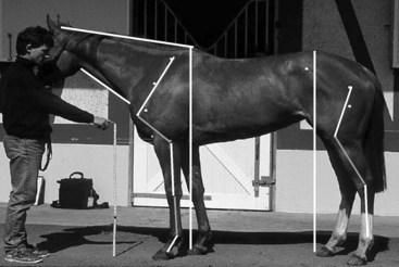
Fig. 4-1 Length measurements (centimeters) recorded from the left lateral view in a Thoroughbred conformational study.15
(Reproduced with permission from Anderson TM, McIlwraith CW: Longitudinal development of equine conformation from weanling to age 3 years in the Thoroughbred, Equine Vet J 36:563, 2004.)
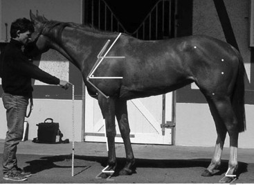
Fig. 4-2 Angle measurements (degrees) recorded from the left lateral view in a Thoroughbred study.15
(Reproduced with permission from Anderson TM, McIlwraith CW: Longitudinal development of equine conformation from weanling to age 3 years in the Thoroughbred, Equine Vet J 36:563, 2004.)
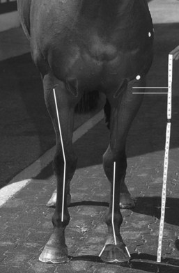
Fig. 4-3 Length (centimeters) and angle measurements (degrees) recorded from the front view in a Thoroughbred study.15
(Reproduced with permission from Anderson TM, McIlwraith CW: Longitudinal development of equine conformation from weanling to age 3 years in the Thoroughbred, Equine Vet J 36:563, 2004.)
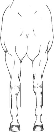
Fig. 4-4 Lines drawn to determine offset ratios. A bench-knee measurement, called an offset ratio, of the medial width/lateral width determines the amount of third metacarpal bone that is offset laterally. The medial/lateral width is the distance (centimeters) from a line drawn along the medial or lateral aspect of the third metacarpal bone to a line from the medial or lateral distal lateral radial physis parallel with the third metacarpal bone. A ratio of greater than 1.0 represents bench-kneed conformation.
(Reproduced with permission from Anderson TM, McIlwraith CW: Longitudinal development of equine conformation from weanling to age 3 years in the Thoroughbred, Equine Vet J 36:563, 2004.)
In another study the role of conformation in the development of musculoskeletal problems in the racing TB was evaluated.16 Conformation measurements were obtained from photographs of horses with markers at specific reference points and digitally analyzed as previously described.15 Clinical observations were recorded regularly for each horse, and stepwise (forward) logistic regression analysis was performed to investigate the relationship between binary response of clinical outcomes probability and conformation variables by the method of maximum likelihood. Clinical outcomes significantly (P < .05) associated with conformational variables included effusion of the front fetlock joints, effusion of the right carpal joint, effusion of the carpal joints, effusion of the hind fetlock joints, fractures of the left or right carpus, and right front fetlock and left hind fetlock lameness. Offset knees contributed to fetlock lameness (for every 10% increase in the right offset ratio, the risk of effusion in the right front fetlock increased 1.8 times and the odds of right front fetlock lameness increased by a factor of 1.26). Long pasterns increased the odds of forelimb fracture. Surprisingly, an increase in the carpal angle as viewed from the front (carpus valgus) appeared to act as a protective mechanism, because odds for the development of carpal fracture and carpal effusion decreased with increase in carpal angle (for every 1 degree increase in right carpal angle as viewed from the front, the odds of effusion in the right carpus decreased by a factor of 0.68 and the odds of a right carpal fracture decreased by a factor of 0.24).16 Horses with long shoulders had decreased odds of developing forelimb fracture (odds ratio [OR] = 0.50), but horses with long pasterns had increased odds for forelimb fracture (OR = 4.55). Long sloping pasterns were suggested as a potential cause of carpal chip fractures.14
In the second TB study described previously, ORs were created for increase in bone length of 2.54 cm (1 inch) or joint angle of 1 degree and development of lameness.16 For every 2.5 cm increase in humeral length, odds for fracture of the proximal phalanx or carpal synovitis or capsulitis increased. Increased length from elbow to ground and increased toe length increased chances for carpal fracture, and in horses with offset knees greater than 10% the potential for carpal or fetlock synovitis or capsulitis increased. The potential for fracture of the proximal phalanx increased with an increase in shoulder angle.16
A study examining conformation in 160 racing QHs in training at Los Alamitos Race Course found humeral length had a significant association with several clinical entities.17 For every 10-cm increase in humeral length the odds of an osteochondral fracture fragment of the dorsoproximal aspect of the left front proximal phalanx increased by a factor of 9.06. Similarly, ORs for carpal synovitis and capsulitis and for sustaining carpal chip fracture (8.12 in the left forelimb and 10.17 in the right forelimb) rose significantly with each 10-cm incremental increase in humeral length. The length of the left front toe was important.17 For each 1-cm increase in toe length, the odds of sustaining carpal chip fractures increased by a factor of 58.90.17 Horses with upright shoulders were at increased risk for development of osteochondral fragmentation of the proximal phalanx and those with offset carpi were at increased risk for development of synovitis and capsulitis in both forelimbs (OR = 2.26).17
The relationship of many lower limb lameness conditions with limb length is interesting and somewhat unexpected because longer limb length generally is considered desirable. In addition, a relationship between longer toes and carpal fracture is interesting. Longer toes may delay breakover of the foot, altering forelimb biomechanics, but an effect on a distant joint such as the carpus cannot be easily explained. The relationship between offset knees and lower limb lameness was expected, but unexpected were fewer carpal fractures in TBs with this conformational fault. Because development of lameness, termed clinical outcomes in these studies, is complex, confounding variables such as track conditions, training regimen, breeding, individual horse ability, and experimental error could have contributed to outcome.
Little doubt exists that acquiring objective information is useful, not only to determine what is abnormal but also to define what is normal in a population. In both WBLs and STB trotters in Europe, toed-out conformation in the hindlimbs should likely be considered normal because a majority of both breeds have this conformational trait.10,12 These populations differed, however, in forelimb conformation. Few STB trotters had bench-kneed conformation, a finding supported by one of our (MWR) clinical observations that this conformational fault is highly undesirable in this breed (see Chapter 108). Recently, variation in conformation of National Hunt racehorses established guidelines with which individual horses could be compared and highlighted significant variations in horses with different origins (Irish and French horses differed significantly in girth and intermandibular width measurements).18 Circumference and length measurements were significantly associated with withers height. No underlying pattern of combinations of conformational parameters was found, but variations were identified between left and right measurements and in hoof, stifle angle, and coxofemoral angle measurements.18
Evaluation of Conformation
Conformation determines the way a horse moves, and it is intuitive that a relationship exists between faulty conformation and the development of lameness. Therefore assessment of conformation should be an integral part of lameness examination. Conformation evaluation has four basic components: assessment of (1) balance, (2) lengths, angles, and heights, (3) muscling, and (4) conformation of the limbs. All are intertwined but should be evaluated separately, considering the whole horse not just the limbs, and then consolidated. The clinician should evaluate the horse on firm, level ground, preferably a smooth, nonslip surface that does not obscure the view of the feet. The horse should stand squarely with equal weight on all four limbs. Dynamic assessment of limb conformation while the horse is walking also is essential.
Balance
Balance is the way all parts of the horse fit together and is linked directly with assessment of lengths, angles, and heights. The horse should be proportional and thus well balanced. A horse may be visualized in thirds—the forehand, the midbody, and the hindquarters—by drawing three circles incorporating these areas (Figure 4-5). The circles should overlap but not excessively. Horses in good balance are likely to be superior athletes. Horses with a short, thick (throatlatch and shoulder regions) neck are often heavy and straight in the shoulders (Figure 4-6). A horse with a short back has naturally closer dorsal spinous processes which may be predisposed to impingement or overriding, whereas a horse with a long back (Figure 4-7) may have difficulty engaging the hindlimbs properly. The clinician must assess the relative heights of the withers and hindquarters. A horse that is taller behind than in front (rump height is greater than height at the withers) is predisposed to forelimb lameness. A horse that also is underdeveloped in the shoulders and upper forelimbs (weak up front) and heavy behind is more at risk. Limb lengths should be proportional to body size and height. In general, the body length (point of shoulder to point of rump) should be equal or slightly longer than withers height (see Figure 4-5).
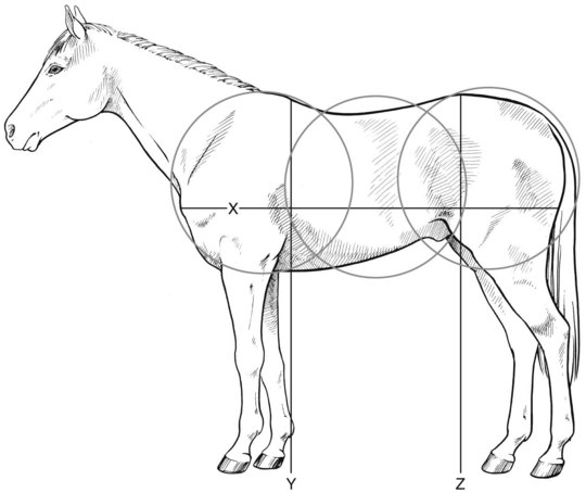
Fig. 4-5 Diagrammatic depiction of assessing balance during conformation evaluation. Three circles (from left to right: forehand, midbody, hindquarters) are visualized and should overlap by approximately one third. Excessive overlapping of circles (short coupled) or scant overlapping of circles (long, weak in the back) are common conformational abnormalities. Body length (X) should be equal to or slightly longer than withers height (Y), and withers height and rump height (Z) should be the same.
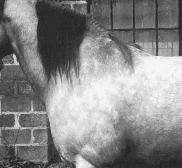
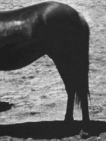
Fig. 4-6 Horse with short, heavy neck; heavy, short, and straight shoulder; and withers set forward. This horse is prone to forelimb lameness and likely to have a short stride.
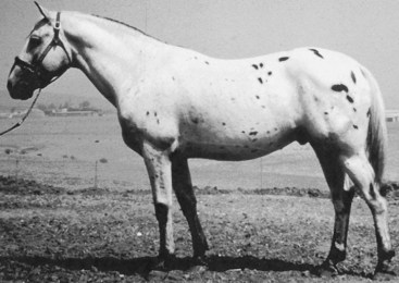
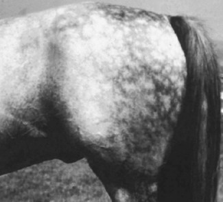
Fig. 4-7 Unbalanced Appaloosa gelding that is long and weak in the barrel (back). Rump height is slightly higher than withers height.
Head conformation is not relevant to lameness, but the size of the head relative to the body and the angulation between the head and neck influence the ease with which the horse can work “on the bit” (see Chapters 97 and 116). A horse’s ability to see is important. A horse with ocular abnormalities may exhibit bizarre behavioral abnormalities or unusual head and neck carriages, possibly misinterpreted as the result of pain. A rare cause of “being on a line” occurs in driving horses, such as a STB with unilateral blindness. The horse turns the head toward the blind side to see with the opposite eye and thus is on the contralateral line (to straighten the head, the driver must pull on the contralateral line). This mimics a contralateral (to the blind eye) lameness.
Lengths, Angles, and Heights
Body length is important in determining stride length (see Figure 4-5). Short-coupled conformation predisposes horses to short strides and problems with interference, especially racehorses. If horses are too long, they can be weak in the back. The length of the neck is important in assessing balance and should be proportional to the overall body length. Some horses have long, weak necks. The neck may be “set on low” relative to the shoulder, with a depression (ventral deviation) of the dorsal topline cranial to the withers (ewe necked), giving the appearance of prominent withers laid too far caudally. Horses should have adequate depth in the girth region (depth of girth).
Shoulder length (top of the withers to the point of the shoulder) may be related directly to stride length, and horses with longer shoulders usually have longer strides (Figure 4-8). Those with short shoulders usually have shorter strides (see Figure 4-8). Shoulder length and shoulder angle often are related; long shoulders often are more sloping (smaller shoulder angle), and short shoulders often are straight. Good shoulder length appears to be important and desirable, but recent objective data from TB and QH racehorses suggest that horses with long limbs may be at increased risk for lower limb lameness.16,17
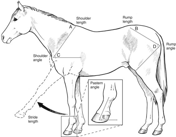
Fig. 4-8 Measurement of shoulder length (A), rump length (B), shoulder angle (C), and rump angle (D). The pastern angle (E) should be equal to the shoulder angle. Shoulder angle and length are important in determining stride length.
In TBs particularly a long radius (forearm) and short, strong third metacarpal bone (McIII) have been considered desirable for adequate strength and maximum stride length. However, more recent work16 suggests that a long forearm would lead to a long metacarpal bone, and because horses are proportional we must question this long-held impression. Chest width should be commensurate with overall body size. A wide chest with a base-narrow forelimb stance (the front feet are close when the horse is evaluated from the front) or a narrow chest with a base-wide stance are undesirable. In STB pacers a good chest width is desirable, but in trotters a narrow chest is preferred (see Chapter 108).
Rump length also is important in determining stride length, and a longer length of the rump is desirable. Many horses with long rumps have larger rump angles (flat croup), and those with short rumps have smaller rump angles (steep croup). Long, flat croup regions are desirable. The ideal horse should have a long gaskin (crus) and a short, strong metatarsal region (the hocks close to the ground) to maximize stride length.
The angles of the shoulder and rump are important factors in determining stride length and balance. Undesirable shoulder and rump angles often accompany other conformational faults and may predispose to lameness. The ideal angle of the shoulder (relative to the ground) has classically been determined to be 45 degrees (see Figure 4-8). Horses with steep shoulder angles (>50 to 55 degrees) usually have short shoulders and short, upright pasterns, which predispose to lower limb lameness. The forelimb pastern angle should be equal to the shoulder angle. A steep shoulder angle shifts the center of gravity forward, predisposing to forelimb lameness. Horses with a flat rump or croup generally have longer rump lengths and longer strides (Figure 4-9). Horses with short, steep rumps (goose rump) often have short, choppy gaits, and many have hindlimb lameness (Figure 4-10). A steep rump angle shifts the center of gravity caudally, predisposing to hindlimb lameness.
Limbs
It is critical to evaluate limb conformation with the horse standing squarely on a firm, flat surface and with an experienced handler who can make the horse cooperate. If the clinician observes a fault that may result from how the horse is standing, he or she should reevaluate the horse after repositioning. Horses often stand camped out, both behind and in front, simply as the result of improper positioning.
The plumb line concept allows evaluation of each limb from the front or back and the side (Figure 4-11). For example, a vertical line from the point of the shoulder should bisect the limb. The clinician also should evaluate the horse while it is walking because some defects are dynamic. Horses that toe in or toe out, or those with fetlock or carpus varus deformities, may stand reasonably well, particularly with corrective trimming and shoeing, but the defect may be readily apparent while the horse is walking.
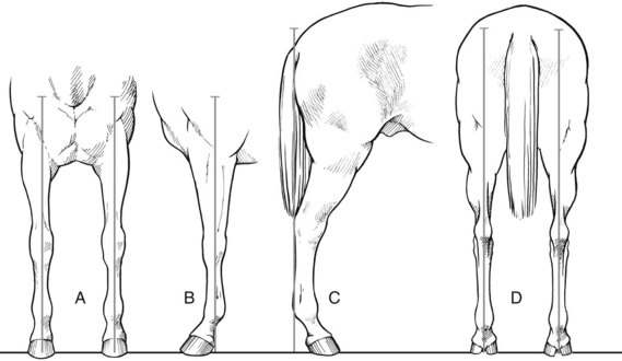
Fig. 4-11 Diagram demonstrating use of plumb lines to evaluate limb conformation from three perspectives. Vertical lines are visualized from A, the front forelimb (line runs from point of the shoulder, bisecting the limb), B, the side forelimb (line bisects elbow joint, carpus, and fetlock joint and intersects the ground approximately 5 cm behind the solar surface of the heel), and C, the side hindlimb (line runs from point of rump to the ground, touching the point of the hock and plantar aspect of the metatarsal region and intersecting the ground approximately 7.5 to 10 cm behind the heel) and from D, the back hindlimb (line drawn from point of rump, bisecting the hindlimb).
Forelimb Conformation
Front Perspective
Several forelimb conformational abnormalities are apparent when a horse is evaluated from the front. Base-wide conformation may occur alone or in combination with toed-in or toed-out conditions (Figure 4-12). Horses that are base wide stand with the forelimbs lateral to the plumb line and generally are narrow in the chest, resulting in overload of the medial aspect of the lower limb, predisposing to lameness. Horses that are base wide, toed in tend to wing out or paddle during protraction (Figures 4-12, B and 4-13, A). Winging out predisposes trotters to interference with the ipsilateral hindlimb. Base-wide, toed-out conformation appears most often in horses with uncorrected carpus valgus deformities (Figures 4-12, C and 4-13, B) and results in excessive loading on the medial aspect of the foot and misshapen feet. Interference with the contralateral forelimb may occur in severely affected horses.
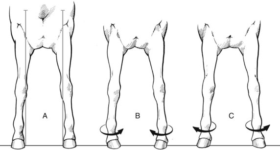
Fig. 4-12 Three variations of base-wide forelimb conformation, including (A) simple base wide, (B) base wide, toed in, and (C) base wide, toed out.
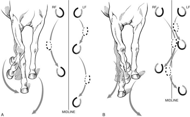
Fig. 4-13 A, Toed-in conformation often causes horses to wing out or paddle during advancement of the forelimb. B, Toed-out conformation causes horses to wing in, predisposing to interference with the contralateral forelimb.
Base-narrow conformation may occur alone or in combination with toed-in or toed-out conformation (Figure 4-14). Horses that are base narrow stand with the forelimbs inside each plumb line and overload the outside of the lower limb and foot. Horses that are base narrow, toed in tend to wing out, and those that are base narrow, toed out tend to wing in during protraction (see Figures 4-13 and 4-14).
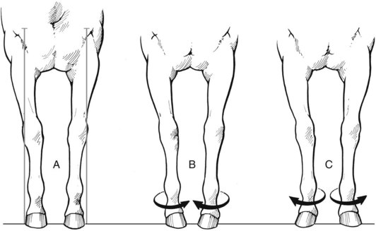
Fig. 4-14 Three variations of base-narrow forelimb conformation including (A) simple base narrow, (B) base narrow, toed in, and (C) base narrow, toed out.
With in-at-the-knee, knock-kneed, or carpus valgus conformation (Figure 4-15) the carpi are medial to the plumb line, creating an angular deformity and concentrating the weight of the horse on the medial aspect of the carpus and proximal metacarpal region. This condition, if severe, may predispose the horse to carpal lameness and splints. However, recent objective data have suggested that a certain degree of carpal valgus is protective for synovitis and capsulitis, as well as chip fractures in the carpus.16 Predisposition to carpal lameness and splints with carpal valgus conformation probably applies only to horses with severe abnormalities. In some horses, particularly foals, carpus valgus may be accompanied by external rotation of the entire limb or just the distal aspect (toed out). Severely affected horses wear the inside aspect of the hoof or shoe abnormally.
Out-at-the-knee (bowlegged, bandy-legged) conformation usually is a consequence of early carpus varus and usually is career-limiting (Figure 4-16). The carpus is bowed outward, lateral to the plumb line. Many horses are also toed in, accentuating abnormal forces on the lateral aspect of the entire distal forelimb, predisposing to osteoarthritis (OA) of the carpus or fetlock or lateral suspensory branch desmitis and sesamoiditis. In most foals with carpus varus, there is coexistent lateral deviation of the elbow, giving the appearance that the angular deformity might be arising from asymmetrical growth in the physes associated with the elbow joint. Correction is difficult but usually involves transphyseal retardation of the distal lateral radial physis.
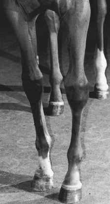
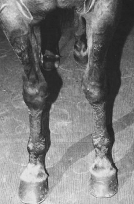
Fig. 4-16 Two-year-old Thoroughbred filly with moderate-to-severe carpus varus (out-at-the-knee) conformation in the left forelimb and mild deformity in the right forelimb. The filly also is toed in, a common finding in horses with this type of conformation. Another common finding is lateral deviation or bowing of the elbow, prompting clinical suspicion that a deformity also exists at this joint.
Offset or bench-knee conformation is classically defined as lateral positioning of the metacarpal region relative to the central axis of the radius (Figure 4-17). However, radiographs demonstrate that the actual displacement usually is at the antebrachiocarpal joint. Displacement may be unilateral or bilateral with differing degrees of severity on each side. This conformation has been often associated with carpal or metacarpal lameness (Figure 4-18). Many 2-year-old STBs with offset knees are precocious during early training but often develop carpal lameness at 3 or 4 years of age. TBs with offset knees are believed to perform better on the soft turf tracks in Europe, as opposed to the firm turf or dirt tracks in the United States. In recent studies, a relationship between offset knees and carpal lameness was not found, but horses with this conformational abnormality were at risk for fetlock lameness.16,17
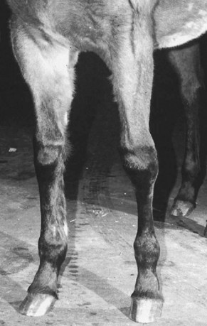
Fig. 4-17 Standardbred with a prominently offset (bench) knee in the left forelimb. An apparent lateral deviation or shifting (offset) of the cannon bone and distal extremity occurs relative to the plumb line dropped through the left radius.
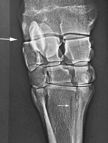
Fig. 4-18 Dorsopalmar xeroradiograph of 3-year-old Standardbred colt with longitudinal fracture of the third metacarpal bone (small arrow). Medial is to the right. The lateral displacement (“step”) at the antebrachiocarpal joint (large arrow) gives the clinical appearance of an offset (bench) knee.
Toed-in conformation, or internal rotation of the distal extremity, exists alone or in combination with abnormalities of stance (base narrow or base wide), other conformational faults such as carpus varus and offset knees, and being wide in the chest (see Figure 4-16). Horses that are toed in usually wing out (paddle) (see Figure 4-13). Toed-in conformation is particularly undesirable in a trotter because of potential interference at speed. Toed-in conformation predisposes horses to lateral splints, lameness of the lateral aspect of the fetlock joint region (e.g., lateral branch suspensory desmitis), and OA of the interphalangeal joints. Horses that are toed in wear the outside aspect of the foot.
Toed-out conformation, or external rotation of the distal extremity, is common and if mild may be considered normal or inconsequential (Figure 4-19). Mild toed-out conformation appears in 50% of STBs and is common behind in STBs and WBLs.10,12 It first develops in foals and, if pronounced, persists in the mature horse. In foals, toed-out conformation often accompanies carpus valgus deformities but may result from external rotation primarily from the fetlock joint distally or, in more severe deformities, from further proximally (Figure 4-20). Mild toed-out conformation usually resolves as a foal matures and with corrective trimming. Toed-out conformation results in abnormal wear on the inside aspect of the foot. Horses tend to wing in; if winging in is severe, particularly if accompanied by base-narrow conformation, it may interfere with the opposite forelimb (see Figure 4-13). Exostoses (splints) on the second metacarpal bone (McII) or McIII may develop, requiring protective boots to be worn during exercise. In STB pacers, interference injury occurs as high as the distal aspect of the radius.
Lateral Perspective
Horses camped out in front stand consistently with an entire forelimb ahead of the plumb line, but this conformation usually is a temporary problem with the horse’s stance and can be corrected by repositioning the horse, or it reflects pain caused by laminitis, for example. Camped under in front is unusual and usually also results from temporary malpositioning of the horse (Figure 4-21). If a horse prefers to stand camped under and is otherwise sound, however, this trait may be a sign of “extreme speed.”19
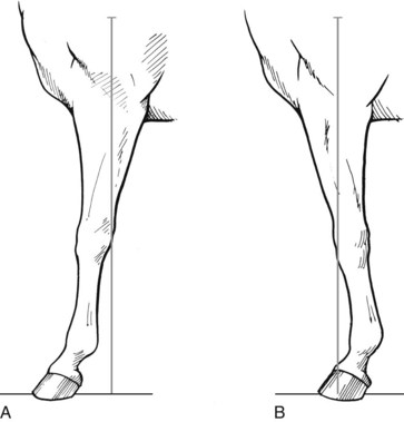
Fig. 4-21 Diagram showing (A) camped-out in front and (B) camped-under in front conformation, both in relation to plumb lines.
Back-at-the-knee or calf-knee (sheep-knee) conformation describes a concave dorsal aspect of the limb, with the carpus behind the plumb line (Figures 4-22 to 4-24). On radiographs of a normal carpus the proximal and distal rows of carpal bones are aligned in a proximal-to-distal direction, and the dorsal faces of these bones are parallel to the radius and the McIII (Figure 4-23, A). With back-at-the-knee conformation the proximal row of carpal bones is set back (Figure 4-23, B). Horses that stand back at the knee are considered predisposed to carpal injuries because of the natural tendency of the carpus to hyperextend (larger carpal angle) during fatigue. In our experience, TB racehorses are particularly at risk, despite limited contrary evidence (see page 16).14 While no associations were made between horses with back-at-the-knee conformation and lameness in recent studies in TBs and QHs, horses included in those studies were elite, and any foals with obvious back-at-the-knee conformation may have been eliminated from the case population before the studies commenced.16,17 In a different study TB foals that were back-at-the-knee improved as they matured from 1, 2, and 3 years of age.15
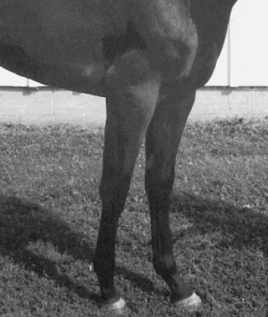
Fig. 4-22 Thoroughbred yearling with back-at-the-knee (calf-knee) conformation most noticeable in the left forelimb. This conformational fault is undesirable, particularly in racing breeds.
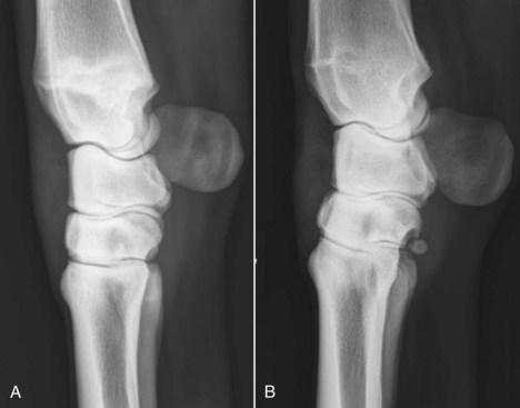
Fig. 4-23 Lateromedial radiographs of the left carpi of two 2-year-old Thoroughbred racehorses. A, There is normal carpal conformation, and the proximal and distal rows of carpal bones are aligned and parallel to the radius and third metacarpal bone. B, In a horse with carpal lameness there is palmar deviation of the proximal row of carpal bones. This radiological appearance is typical of back-at-the-knee conformation.
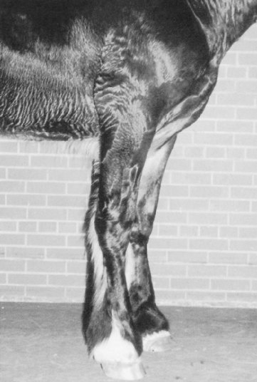
Fig. 4-24 Clydesdale yearling with calf-knee conformation and clubfoot secondary to osteochondrosis of the shoulder joint. This conformation is primarily the result of chronic lameness, decreased weight bearing, and the development of a flexural deformity.
In the STB, mild calf-knee conformation is common in pacers and acceptable, whereas in the trotter this defect is undesirable. In other breeds, mild calf-knee conformation may not directly lead to lameness. In young horses with lameness from unrelated sources such as osteochondrosis or fracture of the distal phalanx, back-at-the-knee conformation and clubfoot (a small, upright foot) may accompany flexural deformity of the limb (see Figure 4-24). This deformity is a combination of contraction (clubfoot) and laxity (calf-knee) caused by chronic lameness and partial weight bearing and warrants a guarded prognosis.
Over-at-the-knee, bucked-knee (knee-sprung), or hanging-knee conformation describes a convex dorsal surface of the carpus, with the carpus in front of the plumb line (Figure 4-25). In young, untrained horses, bucked-knee conformation may be a predictor of lameness, but in mature horses it appears to be an acquired characteristic and occurs primarily in horses that jump. Older cross-country horses, steeplechasers, jumpers, or field hunters are prone to bucked-knee conformation and often stand over at the knee with no obvious lameness. These horses may exhibit a tendency to buck forward to such an extent that they appear on the verge of collapse or prone to stumbling yet show good stability. Lame horses that stand over at the knee are often found to have pain in the proximal palmar metacarpal area or carpal sheath. Tied in below the knee (Figure 4-26) describes a distinct notch just distal to the accessory carpal bone on the palmar aspect of the limb. Normally, the McIII and the digital flexor tendons are in parallel alignment from the accessory carpal bone to the proximal sesamoid bones. With tied-in conformation the digital flexor tendons appear to enter the carpus in a dorsoproximal direction. If the horse also is bucked kneed, the tied-in appearance is accentuated. Young horses are prone to superficial digital flexor tendonitis. In STBs this defect is worse for a pacer than a trotter.
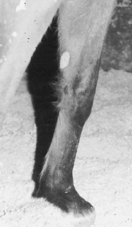
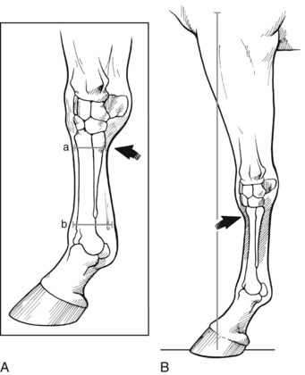
Fig. 4-26 A, Diagrammatic representation of tied in below the knee. The dorsal-palmar length of a is less than b, giving the appearance that the digital flexor tendons run obliquely, proximally to enter the distal carpal region more dorsally than expected. B, A horse that is cut-out under the knee has a concave appearance of the dorsal aspect of the distal carpus and proximal metacarpal region (arrow).
The junction of the carpus and McIII should be flat. Cut out under knee describes a notch under the dorsal surface of the carpus (see Figure 4-26). In horses with this defect, the McIII appears thin (dorsopalmar direction) and weak. Horses with this conformational abnormality also are often back at the knee, predisposing them to carpal and metacarpal problems. Some young racehorses, typically late yearlings or early 2-year-olds in training, appear to have distention of the middle carpal joint capsule or an unusually prominent distal radial epiphysis. These findings give the impression of an unusually large gap between these structures, described as “open at the knee.” The first author has not seen a correlation between this clinical observation and obvious radiological changes, although in young horses with this conformation the distal radial physis remains visible. Whether this conformation is relevant to lameness in young racehorses is debatable.
Over at the fetlock usually is seen in young horses with flexural deformity of this joint (see Chapter 59). This conformational fault may persist in a mature horse, causing upright pasterns or knuckling of the fetlock joint. In some horses this condition causes a progressive, permanent deformity and severe lameness, whereas in others a dynamic, intermittent knuckling occurs and some of these horses remain surprisingly sound. Knuckling also may be a sequel to desmitis of the accessory ligament of the deep digital flexor tendon.
Hindlimb Conformation
Lateral Perspective
Hindlimb conformational faults generally are less numerous and problematic than those in the forelimb because of differences in weight distribution and center of gravity. Plumb lines also are useful in evaluating conformation of the hindlimbs with the horse standing squarely, loading all limbs (see Figure 4-11). Camped-out conformation is unusual and generally results from faulty positioning of the horse during the examination. Horses that are truly camped out usually have short strides and poor athletic ability. Camped under behind often is associated with sickle-hocked conformation but also appears in horses that are straight behind (Figures 4-27 and 4-28). Horses with this type of conformation often have short, choppy strides (Figure 4-29).
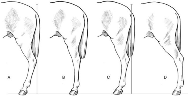
Fig. 4-27 Diagrammatic representation of conformational faults of the hindlimb from the lateral perspective. Compared with ideal conformation, horses with sickle-hocked conformation (A) have a concave dorsal surface of the limb with the distal extremity dorsal to the plumb line, but those that are straight behind (B) have large stifle and hock joint angles but smaller fetlock joint angles. Horses that are camped under (C) often are sickle hocked as well. Camped-out conformation (D) is unusual and most often results from faulty positioning of the horse.
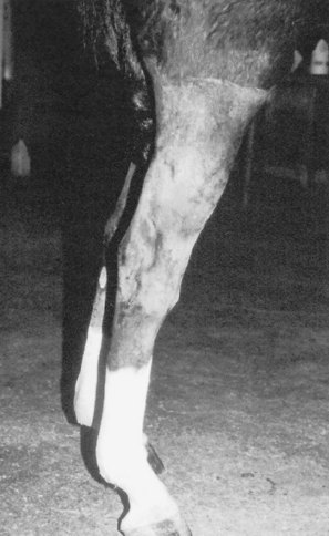
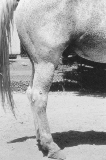
Fig. 4-29 Horse with camped-under and mild sickle-hocked conformation, a combination leading to a short, choppy gait.
A particularly severe conformational fault that leads directly to lameness is straight behind, otherwise called straight hocks or post (posty) leg. Horses that are straight behind have larger stifle and hock angles but smaller fetlock joint angles compared with ideal hindlimb conformation (see Figures 4-27 and 4-28). Straight-behind, sickle-hocked, and in-at-the-hock conformation are the three most important hindlimb conformational faults, and all may lead directly to lameness. Horses that are straight behind often develop upward fixation of the patella, a condition seen most often in WBLs. Suspensory desmitis and fetlock OA also occur frequently. Horses with normal initial hindlimb conformation may become straight behind if they develop severe suspensory desmitis and lose support of the fetlock joint (see Chapter 72).
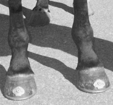
Sickle-hocked conformation is one of the most common conformational faults, and it leads directly to lameness of the tarsus and plantar soft tissues. Horses that are sickle hocked stand with the lower hindlimb well ahead of the plumb line, with an exaggerated concave dorsal surface of the hindlimb (resembling a sickle), creating a smaller than normal hock angle (see Figures 4-27, 4-29, 4-30, and 4-31). This type of conformation is often called curby conformation because horses frequently develop curb (see Chapter 78). Sickle-hocked conformation concentrates load in the distal, plantar aspect of the hock, predisposing to curb and to distal hock joint pain. In a recent study evaluating tarsal angles and joint kinematics and kinetics, horses with large hock angles (straighter behind) had less absorbed concussion during impact, smaller vertical impulse, and less extensor movement, characteristics thought to predispose these horses to OA.20 One of us (MWR) feels results of this study could be interpreted differently, since clinical experience contradicts this finding; horses with larger hock angles are at less risk of the development of OA of the distal hock joints and less absorbed concussion by the tarsus may make other structures such as the suspensory ligament at risk of injury. However, smaller net movement in horses with larger hock angles may be protective for the development of plantar tarsal conditions such as curb, a finding that correlates well with my clinical impressions.20 In foals with incomplete or delayed ossification of the tarsal bones, marked sickle-hocked conformation may occur (see Chapters 44 and 128). Sickle-hocked conformation is undesirable, particularly in racing breeds, but if mild is not detrimental. In STB pacers, some prefer a mild degree of sickle-hocked conformation because horses can extend the hindlimbs farther forward without risk of interference. In STB trotters the condition often predisposes to distal hock joint pain and curb, but these horses tend to be fast, although unsound. In Western reining horses, sickle-hocked horses may be better able to perform sliding stops.
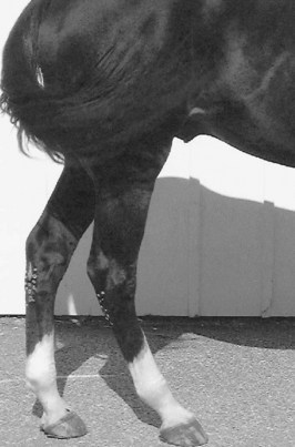
Fig. 4-30 This 4-year-old Standardbred has sickle-hocked conformation and has developed curb, which has been treated by freeze-firing, resulting in white marks on the plantar aspect of the hocks.
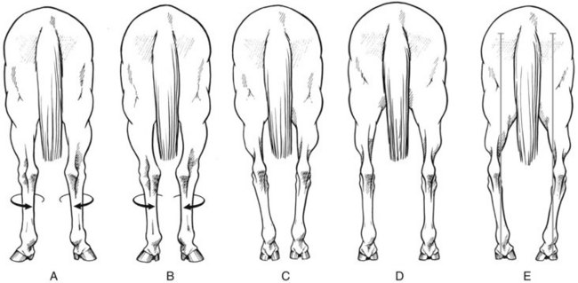
Fig. 4-31 Hindlimb conformational abnormalities viewed from the rear. A, Cow-hocked conformation is a common fault characterized by external rotation of the limb, usually without angular deformity, causing the hocks to be too close together. Mild external rotation of the hindlimbs is common and does not appear to cause lameness. B, Cow-hocked, base-narrow conformation. C, Base-narrow conformation. D, Base-wide conformation is uncommon. E, Bowlegged conformation is uncommon and undesirable.
Rear Perspective
A majority of STB and WBL horses toe out behind, which should be considered normal. Horses with mild external rotation of the distal extremity are said to be toed out and usually also have external rotation of hocks, causing the points of the hocks to be closer than normal. This fault is called cow-hocked conformation and is a rotational change of the hindlimb (Figure 4-31). Cow-hocked conformation occurs in combination with base-wide or base-narrow deformities or independently. Cow-hocked and base-narrow conformation is most common. Base-wide and base-narrow conformation may occur without cow-hocked conformation. These conformational faults seldom lead to lameness but have a substantial effect on gait in some horses. Horses that are base narrow travel closely behind, particularly at a walk. Some travel closely at a trot, pace, or gallop, whereas others seem to widen out when going faster, thus avoiding interference. Those that travel closely at speed often interfere, causing injury to the medial aspect of the contralateral hindlimb.
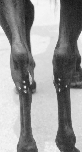
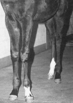
Fig. 4-32 Mature Standardbred racehorse with in-at-the-hock (tarsus valgus) conformation. This conformational abnormality is characterized by an angular deformity as opposed to the rotational deformity seen in cow-hocked conformation. The characteristic white marks were produced by cryotherapy for treatment of bilateral curb.
Bowlegged hindlimb conformation, in which the point of both hocks is truly outside the plumb line, is uncommon (see Figure 4-31). Occasionally horses that are base narrow appear to be bowlegged. Unilateral bowlegged conformation occurs in foals born with windswept deformity or in those with tarsal varus deformity. Bilateral tarsal varus deformity is unusual.
In at the hock or tarsus valgus is an angular deformity (see Figure 4-32). The deformity can be corrected in foals. If it persists in a mature horse, particularly a racehorse with other conformational abnormalities, such as sickle hocks, abnormal forces or load occur in the tarsal region, predisposing the horse to distal hock joint pain, curb, and proximal metatarsal lameness.
Horses can be toed in or toed out behind, but in general the conformational abnormality starts above the fetlock joint, causing the lower limb abnormality to be linked with the upper limb. Thus a horse that is toed in generally is bowlegged, and one that is toed out is cow hocked. In some foals, however, fetlock varus occurs independent of upper limb conformational abnormalities. This abnormality usually appears in windswept foals in which upper limb deformities have resolved, leaving a fetlock varus. This is an angular deformity, but with abnormal hoof wear, toed-in conformation can develop. Fetlock varus and the resulting toed-in conformational defect may cause OA of the fetlock and interphalangeal joints and can be career-limiting.
Conformation of the Digit
More detailed aspects of conformation of the foot and limb flight characteristics are discussed in Chapters 5, 7, and 27. Many of the changes in hoof growth or conformational changes in the hoof are the result of wear, shoeing, and exercise demands of training and performance and often are not present in a young horse.
The pastern angle has traditionally been thought to be similar to the angle of the shoulder (see Figure 4-8). However, in the recent TB study front pastern angle was only mildly correlated to the scapular spine angle, and there was variability.15 Variability was likely from shoeing and lowering of the heel. There was a good correlation between pastern and hoof angles.15 The foot-pastern axis should be straight. The pastern should be neither excessively sloped (low angle) nor upright (high angle). The angle of the pastern is important in determining the amount of load on the lower limb structures. In general, the more upright the pastern (steeper pastern angle), the shorter the stride and vice versa. Horses with upright pasterns appear to be prone to foot lameness and perhaps superficial digital flexor tendonitis. Those with long, sloping pasterns may be at risk to develop OA of the fetlock joint and proximal phalangeal fractures. Horses with short, upright pasterns but relatively normal hoof angles have a broken foot-pastern axis—the foot axis is lower than the pastern axis—and are at risk for developing foot lameness (Figure 4-33). Horses with broken back foot–pastern axis often have underrun heels and other hoof abnormalities and should be evaluated for corrective trimming if lame. If the pastern axis is lower than the foot axis (broken forward foot–pastern axis), called coon-footed, it causes undue strain on the soft tissue structures supporting the fetlock joint. This type of conformation may result from severe suspensory desmitis and loss of support of the fetlock joint.
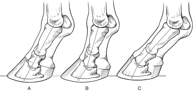
Fig. 4-33 Diagrammatic representation of ideal pastern and foot conformation and the concept of a broken pastern-foot axis. Ideally the foot and pastern angles (A) should be identical to allow full and even weight bearing on all aspects of the foot. A broken foot axis (B) occurs when pastern angle is more upright than that of the foot or vice versa (C). In both latter situations, uneven load distribution on the foot or soft tissue structures may cause lameness.
Pastern length is important and usually is related to pastern angle. Horses with long pasterns commonly have more slope or lower pastern angles. The plumb line should drop approximately 5 cm behind the heel in a well-conformed horse. In horses with long, sloping, and weak pasterns, the line drops more than 5 cm behind the heel. Those with short pasterns usually have more upright pasterns, and the plumb line drops through the foot. A variety of pastern lengths and angles occur, but the pastern length should be in proportion to the overall length of the limb.
Viewed from the front, the plumb line may divide the pastern and foot asymmetrically with more pastern and foot laterally, which often is associated with some degree of distortion of the hoof capsule, with a steeper medial wall and some flaring laterally. This results in asymmetrical loading of the distal limb joints and may predispose to lameness.
Buttress foot is an acquired firm bulge or swelling at and proximal to the dorsal aspect of the coronary band and usually reflects OA of the distal interphalangeal joint (sometimes called pyramidal disease). Bull-nose foot conformation, an abnormal convex dorsal hoof wall, is uncommon but is occasionally seen in a hind foot (Figure 4-34). It is possible to cause this abnormal hoof conformation by incorrect trimming of the dorsal hoof wall, but in most horses it is caused by faulty hoof growth. One of us (MWR) has seen this type of abnormal hind foot conformation most commonly in the TB racehorse, and it is possible there may be a relationship between bull-nose hoof conformation and lameness of the digit or other areas of the distal aspect of the hindlimb. There may be a relationship between bull-nose foot conformation and long, weak pasterns (see Figure 4-34).
Fig. 4-34 Bull-nose foot conformation in a 3-year-old Thoroughbred racehorse that developed a medial condylar fracture of the distal aspect of the third metatarsal bone requiring surgical repair. The relationship between poor hoof conformation and lameness is unknown, but in this horse long, weak pasterns are seen and the combination of conformational faults may predispose horses to injury.