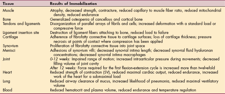
Arachidonic Acid Derivatives.: The synthesis of prostaglandins and leukotrienes begins with the cleavage (splitting) of arachidonic acid from membrane phospholipids by the action of the phospholipase (Fig. 6-13). Once this step is completed, either a cyclooxygenase (COX) enzyme or a lipoxygenase enzyme further metabolizes the arachidonic acid. The COX pathway leads to the production of several types of prostaglandins that modulate vasomotor tone and platelet aggregation (e.g., thromboxane is a strong platelet aggregator and vasoconstrictor, whereas prostacyclin [PGI2] is a strong platelet inhibitor and vaso- dilator). Clinically, prostaglandins are also important because they are mediators of the fever and pain responses associated with inflammation.24

image

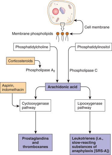
Figure 6-13 Production of prostaglandins and leukotrienes from damaged cell membranes. Note sites for pharmacologic (aspirin and prednisone) interventions. (Courtesy SH Tepper, PhD, PT, Winchester, VA, Shenandoah University.)

The lipoxygenase pathway leads to the production of leukotrienes. Leukotrienes occur naturally in leukocytes and produce allergic and inflammatory reactions similar to those of histamine. They are extremely potent mediators of immediate hypersensitivity reactions and inflammation, producing smooth muscle contraction, especially bronchoconstriction; increased vascular permeability; and migration of leukocytes to areas of inflammation. They are thought to play a role in the development of allergic and autoimmune disease such as asthma and rheumatoid arthritis. Certain leukotrienes (C4, D4, and E4) are collectively known as a slow-reacting substance of anaphylaxis (SRS-A), which is the name given when their potent bronchoconstrictor activity was discovered; they also cause leakage of fluid and proteins from the microvasculature.

The importance of the arachidonic acid metabolites in the inflammatory process is made evident by the excellent clinical response to treatment of acute and chronic inflammatory conditions with drugs that block the production of arachidonic acid (corticosteroids) or inhibit the enzyme and block the production of prostaglandins and cyclooxygenase (nonsteroidal antiinflammatory drugs [NSAIDs] such as aspirin or the newer COX-2 inhibitors). These antiinflammatory medications are commonly used for people with somatic pain or inflammatory conditions, especially rheumatoid arthritis.

Cytokines.: Leukocytes also produce polypeptide substances called cytokines (see Chapter 7) that have a wide range of inflammatory actions affecting either the cytokine-producing cells themselves (autocrine effects) or adjacent cells (paracrine effects). Cytokines also have a number of systemic “hormonal” inflammatory effects.

Two important cytokines with overlapping functions are IL-1 and TNF. As many as fifteen ILs are now identified. Most ILs direct other cells to divide and differentiate, each interleukin acting on a particular group of cells that have receptors specific for that interleukin. TNF is thought to be capable of inducing most of the actions of IL-1 with the exception of activation of lymphocytes.

IL-1 has a number of local actions that promote the inflammatory reaction and a number of systemic actions that induce metabolic, hemodynamic, and hematologic alterations (Box 6-5). These alterations are discussed in some detail because of their importance in the clinical and laboratory diagnosis of inflammation. IL-1 causes fever by raising the production of prostaglandins in the hypothalamus and thereby resetting the threshold of temperature-sensitive neurons.

Fever in turn raises the systemic metabolism and increases the systemic consumption of oxygen by approximately 10% for each degree Celsius of body temperature elevation. As a result, a decrease in systemic vascular resistance occurs, thereby producing hypotension and an increase in cardiac output to increase the flow of blood and the delivery of oxygen to various organs. These hemodynamic changes are characteristic of severe systemic infections and a febrile condition.

IL-1 also causes characteristic changes in blood chemistry. Albumin and transferrin levels are decreased, while levels of coagulation factors, complement components, C-reactive protein, and serum amyloid A increase. These changes occur because IL-1 alters the rate of synthesis of these proteins by the liver. IL-1 also increases the number of neutrophils and decreases the number of lymphocytes in the circulation.

The Blood Coagulation, Fibrinolytic, and Complement Systems.: Plasma proteins produce chemical inflammatory mediators by the enzymatic activity of proteases on plasma proteins. Plasma proteases are enzymes that act as a catalyst in the breakdown of proteins. These plasma protein systems are the blood coagulation and fibrinolytic, kinin enzymatic, and complement systems.

All of these systems can become activated by contact with by-products of cell injury or foreign materials. Examples include contact with components of denuded vascular endothelial cells revealing their underlying basement membrane, which occurs with trauma to the vessel wall and contact with bacterial endotoxins. The key plasma protein in the activation sequence of these systems is clotting factor XII, also known as Hageman factor.

The blood coagulation system (Fig. 6-14) is formed in part by plasma proteins. The design is to bandage injuries with clots (coagulation), then disassemble (lyse) the clots when the job is done. The system protects against both hemorrhage and catastrophic clotting. To maintain homeostasis, these two processes must remain in balance.

image

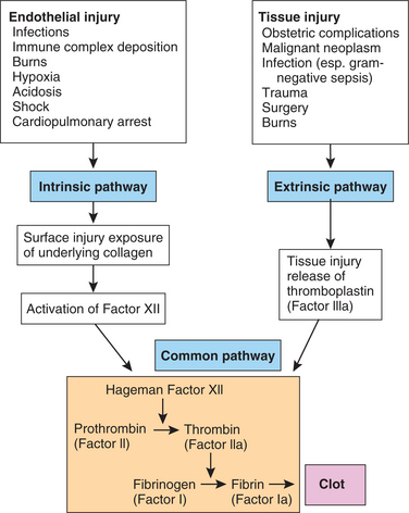
Figure 6-14 Clinical causes of the activation of a clotting cascade, intrinsic and extrinsic pathways of activation, and the mechanism by which both pathways lead to the formation of fibrin threads, or clot. In the chain reaction, inactive proenzymes (represented by Roman numerals) are converted into active enzymes (represented by Roman numerals followed by the letter “a.” The clotting cascade can follow two pathways: intrinsic and extrinsic. The intrinsic pathway is activated within the vascular compartment. The extrinsic pathway is activated outside the vascular compartment, when blood comes in contact with any tissue other than blood vessels. In the case of internal bleeding, both pathways are activated. (Courtesy SH Tepper, PhD, PT, Winchester, VA, Shenandoah University.)

Platelets circulating throughout the bloodstream are always ready to seal any damage to blood vessels with a hemostatic plug. When there is no need for the platelets, the smooth vascular walls prevent platelets from adhering and aggregating. At the same time, endothelial cells in the walls of the blood vessels make tissue plasminogen activator to prevent fibrin deposits from forming and for breaking down existing clots.

More specifically, when injury or bleeding occurs, a series of enzymes are activated sequentially to generate the enzyme thrombin, which converts the plasma protein fibrinogen to fibrin, the essential component of a blood clot. Fibrin forms a meshwork at bleeding sites to stop the bleeding and trap exudate, microorganisms, and foreign materials and keep this content contained in an area where eventually the greatest number of phagocytes will be found. This localizing effect prevents the spread of infection to other sites and begins the process of healing and tissue repair.

The fibrinolytic system (designed to dissolve these clots) is activated by the conversion of plasminogen to the enzyme plasmin (also known as fibrinolysin, which means “to loosen”). Plasmin splits or divides fibrin and lyses the blood clots. Both the coagulation and the fibrinolytic systems are activated in inflammation and function together in a system of checks and balances to preserve vascular function.

The products of fibrin degradation are chemotactic for leukocytes and increase vascular permeability. The kinin enzymatic system is also activated by Hageman factor and functions to produce bradykinin. Bradykinin is a mediator that causes dilatation and leakage of blood vessels and induces pain.

The complement system is composed of a group of plasma proteins that normally lie dormant in the blood, interstitial fluid, and mucosal surfaces. Then, through a series of enzymatic reactions, several plasma protein fragments (C3a, C3b, C5a, and C5b) are formed that are potent inflammatory mediators. These components are also active in immunologic processes. In the nomenclature used for the complement system, each complement component (C) is designated by a number (1 to 9). The individual subunits that make up each component are designated by a letter. For example, the first component of complement is designated C1. C1 is made up of three subunits that are designated C1q, C1r, and C1s. The protein fragments that are generated from the proteolytic degradation of complement components are also identified by a letter (a, b).

The complement system is activated by microorganisms or antigen-antibody complexes causing four events to occur that promote inflammation: (1) vasodilates the capillaries, which increases blood flow to the area, (2) facilitates the movement of leukocytes into the area by chemotaxis, (3) coats the surfaces of microbes to make them vulnerable to phagocytosis, and (4) formation of a membrane attack complex (MAC).

Complement activation can follow one of two pathways, the classic or the alternate pathway; each pathway produces the same active complement components. The products of the complement system bind to particles of foreign material, microorganisms, or other antigens, coating them to make them vulnerable to phagocytosis by leukocytes, a process called opsonization. Activation of the complement cascade by either pathway also results in the formation of the MAC. The MAC is inserted in cell membranes of the microorganism where it creates an opening (pore or channel) in the cell membrane, leading to influx of sodium and extracellular fluid, eventually leading to its lysis (Fig. 6-15). For example, in hemolytic anemia, MAC bores holes in the cell membrane of RBCs, causing their destruction.

image

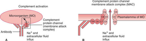
Figure 6-15 A, When an antibody attaches to an antigen (foreign protein) on a microorganism (MO), the antibody-antigen stimulates plasma-derived complement proteins to attach and form the membrane attack complex (MAC). B, This MAC forms a channel through the membrane of the invading cell and allows ions and extracellular fluid to enter, causing cytolysis (death of the microorganism). (Courtesy SH Tepper, PhD, PT, Winchester, VA, Shenandoah University.)

The plasma protease systems (blood coagulation, fibrinolytic, kinin enzymatic, and complement systems) are interconnected at several steps. This arrangement serves to amplify the stimulus for the inflammatory reaction as a balance mechanism. For example, the activation of the plasma protein Hageman factor can initiate both the coagulation (blood clotting) and the kinin systems (produces bradykinin causing dilation and vascular leakage).

The kinin system can in turn activate the fibrinolytic system by producing plasmin (splits or divides fibrin and lyses blood clots). Plasmin then can activate the complement system and further amplify these protease loops by activating Hageman factor, once again starting the cycle (Fig. 6-16).

image

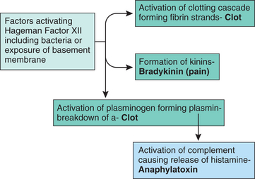
Figure 6-16 Clot formation. Revealed in this figure are the mechanisms for activating both the intrinsic and the extrinsic pathways for clot formation. Either of the above pathways leads to activation of the Hageman factor XII that results in the formation of a fibrin clot. (Courtesy SH Tepper, PhD, PT, Winchester, VA, Shenandoah University.)

PHAGOCYTOSIS.: One of the most important functions of the inflammatory reaction is to inactivate and remove the inflammatory stimulus and to begin the process of healing. The process of ingestion (phagocytosis) of microorganisms, other foreign substances, necrotic cells, and connective tissue constituents by specialized cells (phagocytes) is important in achieving this goal.

Although phagocytosis could be considered the next step in the process of acute inflammation (as a separate section after the section on Chemical Mediators of Inflammation), it is included here as part of the section on Chemical Mediators because the chemical mediators are what attract phagocytic cells to the area for removal of the dead tissue or microorganisms. After ingestion by phagocytic cells, microorganisms are killed or inactivated, and necrotic debris is removed to allow tissue healing to proceed.

The most important phagocytes involved in the inflammatory and healing reactions are neutrophils, monocytes, or when found in tissues of the body, macrophages. Macrophages have different names depending on their location (e.g., histiocytes in the skin, osteoclasts in bone, and microglial cells in the CNS).

The mechanism of phagocytosis is well understood. Phagocytosis is facilitated by the coating (opsonization) of particles to be ingested by immunoglobulin G (IgG) antibody or by the C3b component of complement. These opsonins bind to specific receptor sites located on the cell surface of neutrophils and macrophages. This receptor binding initiates a process of transmembrane signaling allowing calcium influx that activates cytoskeletal proteins within the cell. These cytoskeletal structures allow the movement of cell membranes that is necessary for phagocytosis.

The internalization of the opsonized particle begins by the enfolding of the cell surface membrane (Figs. 6-17 and 6-18). The membrane folds surround the particle to be ingested and seal it within a pouch that separates it from the cell surface and becomes an intracellular vacuole called the phagosome. The phagosomes fuse with lysosomes (containing digestive materials and bactericidal components) and acquire enzymes and other substances that allow the killing and degradation of microorganisms and other ingested materials. Many neutrophils (e.g., polymorphonuclear neutrophils [PMNs]) die in their battle with bacteria. Dead and dying leukocytes, mixed with tissue debris and lytic enzymes, form a viscous yellow fluid known as pus. Inflammations identified by their pus formations are called purulent or suppurative (see Table 6-4).

image

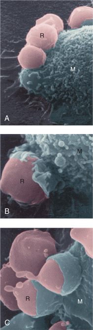
Figure 6-18 Phagocytosis. This series of scanning electron micrographs shows the progressive steps in phagocytosis of damaged red blood cells (RBCs) by a macrophage. A, RBCs (R) attach to the macrophage (M). B, Plasma membrane of the macrophage begins to enclose the RBC. C, The RBCs are almost totally ingested by the macrophage. (From Thibodeau GA, Patton KT: The human body in health and disease, ed 4, St. Louis, 2005, Mosby. Courtesy Emma Shelton.)

6-4   SPECIAL IMPLICATIONS FOR THE THERAPIST

Inflammation

Inflammation, which involves all of the processes described in this section, is a normal, healthy response to tissue injury, but it can also damage adjacent healthy tissue. Chronic activation of inflammatory cells can cause tissue injury such as occurs with rheumatoid arthritis. The role of the therapist is important in supporting the healing process and when appropriate, to limit inflammation and its consequences. The therapist must remember that finding and correcting the cause of inflammation is the key, not just addressing the inflammatory process. Poor lifestyle choices including poor nutrition, improper posture and body mechanics, and poor breathing habits can contribute to the chronicity of this condition.

Rheumatoid arthritis illustrates how the inflammatory mediators discussed are activated and how this process leads to clinical manifestations observed in a therapy practice. Inflammatory activity can be detected by the erythrocyte sedimentation rate (ESR). The therapist can review laboratory values (see Table 40-7) to assess systemic factors; in general, as the inflammation improves, the ESR decreases. Systemic effects of acute and chronic inflammation are discussed in Chapter 5.

Clinical Example

The majority of people with rheumatoid arthritis produce rheumatoid factor, an antibody that is made against the person’s own antibodies of the IgG class. In this case the IgG antibody actually functions as an antigen (Ag) capable of inducing an immune response.

This antibody-to-antibody attachment can occur in the joint space where it leads to the formation of large antibody-antigen (Ig-Ag) aggregates. Ig-Ag complexes stimulate complement activation by the classic pathway and to the formation of the strongly chemotactic cleavage products C3a and C5a. These products attract neutrophils, which then release free radicals (see Fig. 6-2) and enzymes that degrade the joint cartilage, prostaglandins, and leukotrienes that amplify the inflammatory reaction. The Ig-Ag complex is phagocytosed by synovial-lining cells that are stimulated to release collagen-degrading enzymes, prostaglandins, and IL-1.

Lymphocytes contribute to the acute reaction by the production of rheumatoid factor and are responsible for the evolution of a chronic inflammatory reaction by producing cytokines that attract and activate macrophages. The macrophages produce cytokines, such as IL-1, that further amplify the inflammatory reaction by attracting more neutrophils and lymphocytes and by stimulating the synthesis and release from fibroblasts, chondrocytes, and osteoclasts of enzymes that degrade cartilage and bone.

Clinically, the joints affected by the inflammatory process appear red and swollen and are painful; a low-grade fever may also be present. A prominent symptom is joint stiffness that is relieved by activity. With disease progression, damage to the joints occurs; with loss of cartilage, narrowing of the joint space occurs, and resorption of bone is evident on radiograph. These changes are associated with a decrease in the range of motion of the affected joints. In later stages, obvious joint deformities develop that are accompanied by muscle wasting. Antiinflammatory agents, such as aspirin and corticosteroids, are effective in providing symptomatic relief and in slowing the progression of the disease.

The inflammatory process associated with rheumatoid arthritis may also affect other organ systems (see Box 27-9). Foci of chronic inflammation can develop in muscles, tendons, blood vessels, nerves, and various organs of the body (e.g., heart and lungs). In the skin, these foci cause the deposition of connective tissue called subcutaneous nodules.

Exercise and Inflammation

Exercise and physical training definitely influence the concentration of immunocompetent cells in the circulating pool, the proportional distribution of lymphocyte subpopulations, and the function of these cells in relation to tissue injury and infection. However, the extent of these changes and their clinical significance remains a topic for intense study at this time.113 The mechanisms of exercise-associated muscle damage and the initiation of the inflammatory cytokine cascade are further discussed in Chapter 7 (see the section on Exercise and the Immune System).

Proliferation and Migration Phase

Within 2 days after a skin wound or injury, endothelial cells from viable blood vessels near the edge of the necrotic tissue begin to proliferate. The purpose of the endothelial cell proliferation is to establish a vascular network that can transport oxygen and nutrients and support the metabolism of the healing tissue. The endothelial cells bud out from the vessels and form new capillary channels that merge with each other as they develop and grow toward the tissue defect caused by the injury. This process of formation of new blood vessels is called neovascularization or angiogenesis.

The rich network of developing blood vessels with its connective tissue matrix can be seen with the naked eye in healing wounds. As described previously, the appearance of a reddish granular layer of tissue was therefore given the name “granulation tissue.” Histologically, the main cellular components of granulation tissue are the endothelial cells and the fibroblasts, although some inflammatory cells are also commonly present.

Initially, the newly formed vessels are leaky, and this leak contributes to the edematous appearance of tissue undergoing repair. As tissue healing is completed, blood flow to the newly formed vasculature shuts down, and the nonfunctional vessels are degraded, leaving few blood vessels in mature scar tissue.

Tissue gaps are replaced during the proliferation phase when the number of inflammatory cells decreases and fibroblasts, endothelial cells, and keratinocytes take over synthesis of growth factors. The result is the continued promotion of cell migration, proliferation, and formation of new capillaries and synthesis of extracellular matrix components.6 The next step is the removal of damaged matrix as new matrix builds up to fill the wound. The wound initially fills with provisional wound matrix, which consists primarily of fibrin and fibronectin. As fibroblasts are drawn into the matrix, they synthesize new collagen, elastin, and proteoglycan molecules, which cross-link the collagen of the matrix and produce the initial scar.6

Damaged proteins in the matrix have to be removed before the newly synthesized matrix components can be properly integrated. This process is facilitated by proteases secreted by neutrophils, macrophages, fibroblasts, epithelial cells, and endothelial cells. Epithelial cells are at the front of the wound edge, traveling across the highly vascularized extracellular matrix, forming granulation tissue to re-form the epidermal layer. This process can take several weeks.6

Remodeling and Maturation Phase

In the maturation phase of healing, the scar tissue is reduced and remodeled, leaving tissue smoother, stronger, less dense, and less red in color (in Caucasians) as the concentration of blood vessels in the area decreases. In all skin colors, the scar tissue becomes more like the natural skin tones of the person. The density of fibroblasts and capillaries needed in the early phase of healing but no longer needed now declines, primarily through apoptosis or programmed cell death.6 The remodeling phase can take years as the skin first produces collagen fibers, which are broken down and rearranged to withstand stress. Over time, scar tissue grows stronger, relaxes, and then lightens.

Tissue Contraction and Contracture

As the healing process proceeds, the newly formed extracellular matrix draws together, causing a shrinkage (contraction) of the healing tissue. In this manner the size of the tissue defect caused by the injury is diminished. Some fibroblasts within the healing tissue differentiate and acquire some of the morphologic and functional characteristics of smooth muscle cells (myocytes). These specialized fibroblasts are called myofibroblasts. Myofibroblasts contain abundant contractile proteins and apparently contract and contribute to the shrinkage of the healing tissue.

Tissue contraction is a normal process that contributes to tissue repair by approximating the margins of the healing tissue and speeding up the closure of wounds. In some cases, excessive shrinkage of the healing tissue occurs. This condition is called contracture. Contracture is an undesirable outcome of healing because it can be disfiguring and can impair movement or organ function. For example, people with severe burns often develop skin contractures because of the process of “hypertrophic scarring” that can result in significant movement impairments and subsequent disability.

Contracted tissue with excessive arthrofibrosis can occur in the joints (most often the shoulder and knee) after either injury or surgery. Postoperative or posttraumatic arthrofibrosis is characterized by local or global periarticular scarring that can restrict, and in some cases a thickened, fibrotic capsule inhibits motion. Arthrofibrosis can be caused by a variety of factors including prolonged immobilization, infection, or graft malposition after ligament reconstruction (e.g., ACL reconstruction).94

Studies have not been done to identify when scar tissue can be broken up by manual therapy techniques, but anecdotal evidence indicates that immature scar tissue can be successfully treated conservatively (e.g., analgesia and antiinflammatory medications, early motion, bracing, strengthening, electrical stimulation, or manual therapy techniques).

Exactly when scar tissue becomes mature is variable and remains a topic of debate. Some estimate an open window of 3 to 4 months after which time surgical (arthroscopic) manipulation is required. Forceful manipulation of the stiff joint is never advised as this can create excessive joint compression leading to articular cartilage damage and even fracture.95

Tissue Regeneration

Within a few hours after lethal injury to skin, epithelial cells, the viable cells that surround the necrotic tissue, detach from their extracellular matrix anchorage sites and separate from the other epithelial cells. The remaining epithelial cells flatten out to cover the area left bare by the necrotic cells. These epithelial cells also divide and migrate into the tissue using the extracellular matrix support provided by the proteins secreted by the fibroblasts. This process of replacement of dead parenchymal cells by new cells is called regeneration. Regeneration is a very desirable healing process because it restores normal tissue structure and function. In most cases, healing of tissue is achieved by both cell regeneration and replacement by connective tissue (scarring) called repair. In the case of skin, for example, this type of healing occurs after wounds that involve both the epidermis and dermis. In some instances, tissue healing occurs almost exclusively by the progress of regeneration (regrowth of original tissue).

Regeneration can only occur if the parenchymal cells can undergo mitosis. Cells are classified as permanent, stable, and labile based on their ability to divide. Regeneration does not occur in permanent tissues that cannot divide (e.g., cardiac myocytes or central or peripheral neurons); they are long-lived and irreplaceable. Regeneration can also only occur in labile or stable tissues and only if the inflammatory reaction that follows injury is short-lived and does not disrupt the basement membranes, other extracellular components, and vascular structures of labile or stable parenchymal cells. Labile cells, such as epithelial cells of the skin and gastrointestinal (GI) system, and bone marrow divide continuously. Hematopoietic (blood cell–forming) stem cells continuously divide, giving rise to specialized cells, such as erythrocytes and neutrophils, with finite life spans (see Fig. 21-6).

Under these conditions the regenerating parenchymal cells can use the existing connective tissue scaffolding to reconstitute the normal structure and function of the organ. This type of tissue healing can be seen after superficial mechanical injury to epithelia. An example is a superficial abrasion of the skin that causes only necrosis of the epidermis. In this case, regeneration occurs with little or no scarring.

Stable cells, such as hepatocytes, skeletal muscle fibers, and kidney cells, normally do not divide but can be induced to undergo mitosis by an appropriate stimulus. For example, if a portion of the liver is removed by surgery or if liver cells are killed by a viral infection (hepatitis), the remaining hepatocytes divide and sometimes can fully replace the missing liver tissue.

Studies have revealed some capability of neurons to regenerate (neurogenesis) but only in certain areas of the brain (e.g., hippocampus or olfactory bulb).10 The reasons for the restriction of neurogenesis to a few regions of the brain in mammals compared to a more widespread neurogenesis in other vertebrates remain unknown.104 It may be that neuronal stem cells persist in these areas throughout the lifespan but why they do not persist in all areas is still a mystery.3 What we do know is that neural stem cells residing in specific niches are able to proliferate and differentiate, giving rise to migrating neuroblasts, which in turn mature into functional neurons. These new neurons integrate into the existing circuits and contribute to the structural plasticity of certain brain areas.111 Scientific evidence suggests that the process could become more general under pathologic conditions. For example, adult neurogenesis increases under acute and chronic brain diseases. Neuronal precursors are directed to the lesions where they contribute to tissue repair. Investigations are underway to find ways to manipulate and direct the neurogenic process toward the amelioration of neurodegenerative diseases.111,137

Tissue Repair (Formation of Scar Tissue)

Skin has the remarkable ability to heal, often without scarring. Growth factors, blood components, and epithelial (skin) cells mobilize to seal off wounds and protect the body. Scarring does not occur unless the cut, incision, damage, or trauma extends beneath the surface layer (epidermis).

Tissue repair, including the formation of a connective tissue scar, requires removal of the connective tissue matrix. Without this matrix, labile cells do not regenerate or else they regenerate in an incomplete fashion. Therefore the structural integrity of the parenchymal tissue depends on the formation of this connective tissue scar (dense, irregular laying down of collagen). In many cases, however, healing of tissue is achieved by both cell regeneration and replacement by connective tissue (which is what constitutes scarring). In the case of skin, for example, both types of healing occur in wounds that involve both the epidermis and dermis.

Minimizing tissue scarring is important not only for cosmetic reasons, as is the case in skin, but also because excessive scarring can interfere with organ function. Very large tissue defects may require the use of grafts or flaps of tissue to achieve optimal healing. It is possible to minimize scarring by surgical obliteration of the tissue defect caused by injury and cell necrosis. For example, treatment of skin wounds begins with careful cleansing of the wound to remove foreign materials and bacterial contamination, which interfere with healing. This is followed by debridement to remove nonviable tissue that normally would be broken down by the inflammatory reaction.

Careful attention to hemostasis minimizes the deposition of blood into the wound. During closure, the wound margins are closely apposed under the right amount of tension by surgical sutures. A clean, closed wound is free of infectious and other foreign material, fibrin, and necrotic debris. As a result, the duration and intensity of the inflammatory reaction are minimized. Little granulation tissue forms, and the epithelial cell surface is readily reconstituted.

The healing that occurs in the type of wound described is called primary union or healing by first intention and results in a small scar (Fig. 6-19). In the presence of large tissue defects or infections, and in other conditions where surgical closure is not possible or desirable, healing occurs by secondary union. In this situation the time required for healing is longer and the amount of scarring is greater. There is a distinction between closure and healing; the wound or skin may close but healing takes much longer, as much as two years in some situations.

image

Figure 6-19 A, Healing by primary intention is the initial union of the edges of a wound, progressing to complete healing without granulation. B, Healing by secondary intention is wound closure in which the edges are separated, granulation tissue develops to fill the gap, and epithelium grows in over the granulations, producing a scar. C, Healing by tertiary intention is wound closure in which granulation tissue fills the gap between the edges of the wound, with epithelium growing over the granulation at a slower rate and producing a larger scar than results from healing from second intention. Suppuration is also usually found in tertiary wound closure. (From Lewis SL, Heitkemper MM, Dirksen SR: Medical surgical nursing: assessment and management of surgical problems, ed 7, St. Louis, 2007, Mosby.)

Even after wound closure is complete, degradation and resynthesis of collagen continue. This is a response at least in part to shifts in the stress forces to which the tissue is subjected. Cross-linking of collagen fibers continues for a period of several weeks, providing progressive strengthening of scar tissue. However, even under optimal conditions, the repaired tissue never fully regains its original stability. In the case of skin, a fully mature fibrous scar requires 12 to 18 months and is about 20% to 30% weaker than normal skin.

In some people, especially people of African or Asian descent, there is an inherited tendency to produce excessive amounts of collagen during the healing process, causing large amounts of collagen arranged in thick bundles to accumulate in the tissue. These collagenous masses are called keloids and can be seen protruding from the skin surface (Fig. 6-20). Keloids are more than just raised, hypertrophic scar tissue. Both keloid and hypertrophic scar tissue result from excess collagen formation, but hypertrophic scars generally calm down in 12 to 24 months, whereas keloids tend to grow larger and appear worse, often invading surrounding tissue.

image

Figure 6-20 Keloid (hypertrophic) scar composed predominantly of type III collagen, rather than type I collagen. Keloids result from defective remodeling of scar tissue and the persistence of type III collagen, which is typical of immature scar. Epidermis is elevated by excess scar tissue, which may continue to increase long after healing occurs. Looks smooth, rubbery, “clawlike.” Young women, black people, and people of Mediterranean descent are particularly susceptible to keloid formation. (From Rakel RE: Textbook of family medicine, ed 7, Philadelphia, 2007, Saunders.)

Several methods are used to treat keloids, although none of them are 100% successful. Surgical keloid excision followed by high-dose rate brachytherapy, form-pressure garments, and pulsed dye lasers have some reported success.14,30

Necrosis of heart tissue (myocardial infarct) results in a fibrous scar because cardiac myocytes do not replicate to any great extent. Outcomes that can result from tissue repair in various tissue and conditions are summarized in Fig. 6-9. The CNS differs in its healing process because neurons are permanent cells and do not replicate.

After tissue necrosis neither regeneration nor tissue scarring occurs. No fibroblasts are present in the brain parenchyma, and no collagen is produced. After a brain infarct (stroke), the inflammatory cells arrive from the blood circulation and clear away the necrotic tissue, leaving behind an empty cavity (cyst). Specialized CNS cells called astrocytes (glial cells) proliferate, forming dense aggregates around the necrotic area called glial scars or gliosis.

Chronic Wounds.: When a wound fails to heal normally, reepithelialization and closure do not occur. Chronic wounds can occur when the wrong biochemicals are present in the wrong amounts at the wrong times and fail to function effectively. There may be a deficiency in endogenous growth factors, which have the primary role of stimulating cell migration, proliferation, and extracellular matrix deposition. Chronic wounds remain in the inflammatory and proliferative phases.6 Understanding the normal repair process and factors that affect tissue healing can help guide the therapist in removing barriers to healing. Preparing the wound bed appropriately changes the wound’s biochemical environment back to an acute wound, thus re-initiating the healing cascade.

6-5   SPECIAL IMPLICATIONS FOR THE THERAPIST

Scar Tissue

The clinical implications of tissue repair can be seen in the example presented earlier in this chapter (see Special Implications for the Therapist: Collagen and Cell Injury in this chapter). In this example, after a transmural MI, a symptom-limited stress test will usually be given after phase II of cardiac rehabilitation, around 8 to 12 weeks after MI. With understanding of the material presented in this chapter, one can see the logical explanation.

Heart healing, which occurs primarily through the process of tissue repair, requires 8 to 12 weeks for the formation of a dense connective tissue scar. This dense scar allows for structural integrity and force transduction of the viable myocardium, leading to a complete heart contraction. Since the connective tissue scar is not contractile, this area of the heart will never return to full function. Of great importance is the fact that after MI (heart attack), a person’s aerobic fitness can improve (or exceed) to the level before his or her premorbid state with proper exercise.

SPECIFIC TISSUE OR ORGAN REPAIR

Throughout this chapter, examples of cell types and healing processes within various organs and systems of the body have been discussed. Some organs are composed of cells that cannot regenerate (e.g., heart, CNS, or peripheral nervous system cells), whereas other organs such as the liver and epithelial cells of the integumentary and GI systems can replace missing tissue through cell division (mitosis). Some cells, such as skeletal muscle cells and renal cells, do not divide but can be induced to undergo mitosis. The extent to which cells can regenerate depends on the type of cell (e.g., permanent, stable, labile), the cell’s ability to divide, the type of damage incurred (e.g., lethal, sublethal), and other factors discussed (e.g., nutrition, age, immunocompetency, vascular supply, or presence of microorganisms leading to infection). The proliferation and migration of cells, including parenchymal cells, have been discussed; regeneration can only occur if the parenchymal cells can undergo mitosis. When regeneration of parenchymal cells is not possible, the inflammatory reaction can become chronic.

Using an example of a person with TBI, who also experiences MI (see Special Implications for the Therapist: Cell Injury: Multiple Cell Injuries), healing of brain and myocardial tissue was discussed earlier in this chapter. In this final section, only those tissues not specifically included in the main body of this chapter are presented further.

Lung

After lethal injury to alveolar cells (type I and II pneumocytes), regeneration can occur only when the basement membrane remains intact. After the phagocytic removal of the necrotic cells, adjacent living epithelial cells migrate onto the remaining basement membrane and differentiate into type II pneumocytes (cells that primarily produce surfactant).

Eventually, some of these cells differentiate into type I pneumocytes (cells that permit gas exchange) and full lung function is restored. This regeneration process occurs after a bout of pneumonia. If the damage to the lung disrupts the basement membrane, incomplete and inadequate regeneration occurs and healing must be achieved by repair. Also, certain injurious agents induce lung healing by the formation of scar tissue, leading to restrictive lung disease. An example of this would include inhalation of asbestos.

Digestive Tract

The healthy gut is lined with multiple rows of villi structures. These fingerlike projections are responsible for nutrient absorption and the production of digestive enzymes. Gut cells grow single file from the base of the villi up toward the top. They slough off into the intestinal tract and pass out of the body every five days or so. Damaged or injured cells are constantly leaving, while healthy cells renew the GI environment. It takes about 3 to 4 weeks for a complete turnover of all gut cells throughout the digestive tract. A mildly to moderately impaired gut takes 3 to 6 months to heal and 12 to 18 months for a more severe intestinal injury.29

Since two-thirds of all immune system function and 90% of serotonin function take place in the gut, healing the gut can assist in bringing both of these functions back into balance. Serotonin is needed to produce melatonin, which is an essential component for good, restful sleep; the proper amount of circulating and functioning serotonin is also needed to stabilize mood.29

Peripheral Nerves

When a nerve is cut, the peripheral portion rapidly undergoes a myelin degeneration and axonal fragmentation. The lipid debris is removed by macrophages mobilized from the surrounding tissues in a process referred to as Wallerian degeneration. However, within 24 hours of section, new axonal sprouts from the central stump are observed with proliferation of Schwann cells from both the central and peripheral stumps.

Careful microsurgical approximation of the nerve may result in reinnervation. The most important factor in achieving successful nerve regeneration after repair is the maintenance of the neurotubules (basement membrane and connective tissue endoneurium), along which the new axonal sprouts can pass.17

Skeletal Muscle

Skeletal muscle is composed of contractile and connective tissue elements. Actin and myosin myofilaments make up the sarcomere units of muscle fibers. Each individual myofiber is surrounded by a delicate sheath called the endomysium (basement membrane) and then arranged in bundles. Satellite cells surround the muscle fibers and are important for tissue regeneration following injury. The greater the degree of muscle injury, the larger the amount of connective tissue that is disrupted.15

Muscle Injury

Muscle injuries, including contusion, strain, or laceration, are common injuries, occurring particularly in sports; about 90% of all muscle injuries are either contusions or strains. A muscle contusion occurs when the muscle is subject to a sudden, heavy compressive force such as a direct blow to the muscle. Muscle strains occur when excessive tensile force leads to overstraining of the myofibers.75 This is more likely to occur during eccentric contraction when the muscle is lengthening because tension is greater than the muscle’s resistance to stretch; the resultant forces are large. Muscles that cross two joints (e.g., hamstrings or gastrocnemius) are especially vulnerable to stretch injury because they are simultaneously affected by angular positions and velocities of the adjacent joints.15

The most common site of strain injury is the myotendinous junction, a region of highly folded basement membranes between the end of the muscle fiber and the tendon. These involutions maximize surface area for force transmission. The transition from compliant muscle fibers to relatively noncompliant tendon may account for the vulnerability of the myotendinous junction.

If the force of stretch on a muscle is too great to be resisted by the contractile unit, resistance shifts from the contractile unit to the connective tissues. Pathogenic stretch (passive or active) that is beyond the threshold length of the entire musculotendinous unit can result in disruption at the myotendinous junction. Complete tears do occur but less often than muscle strain.15

Muscle Regeneration or Repair

Contrary to widespread belief, muscle tissue can regenerate, but the restoration of normal structure and function is strongly dependent on the type of injury sustained. In severe infections, the muscle fibers may be extensively destroyed. However, the sarcolemmal sheaths (basement membrane and connective tissue endomysium) usually remain intact, and rapid regeneration of muscle cells within the sheaths occurs so that the function of the muscle may be completely restored.

After transection of a muscle, muscle fibers may regenerate either by growth from undamaged stumps or by growth of new, independent fibers.17 Once again, this type of regeneration after lethal cell injury to skeletal muscle fibers is possible when the basement membrane remains intact through mitotic division of “satellite cells.” Satellite cells play an integral role in normal development of skeletal muscle and are essential to the repair of injured muscle by serving as a source of myoblasts for fiber regeneration.9

Contused, or strained, muscle is capable of self-repair, but the healing process is slow and often incomplete, resulting in loss of strength and a high rate of reinjury at the site of the index injury.69,82 Recovery is largely dependent on the severity of the injury but follows the same phases of degeneration, inflammation, regeneration, and fibrosis described in this chapter.

As with all healing, muscle fiber injury is regenerated or repaired through a consistent sequence of events that go into motion as soon as an injury occurs. Hemostasis with hematoma formation and inflammation overlap in the first phase, starting in the first 24 to 48 hours after injury. This phase is followed by phagocytosis with the removal of detritus, activation of satellite cells, and subsequent myofiber regeneration. This second phase can last 6 to 8 weeks after injury. The final phase involves tissue remodeling. During the final remodeling phase, the regenerated muscle matures and contracts with reorganization of the scar tissue.69,82

With death of the muscle cell and ensuing necrosis, chemotactic agents attract macrophages within the basement membrane confines to engulf the remnants of the dead cell. Macrophages release growth factors, stimulating the division of the satellite cells. These cells migrate to the central region and begin to differentiate into expressing the usual characteristics of a skeletal muscle fiber. This healing process can occur after lethal cell injury (e.g., muscular dystrophy) when the connective tissue matrix (primarily basement membrane) is disrupted and regeneration is attempted. But disruption of basement membrane leaves the satellite cells no place to set up and multiply.

The end result is that the muscle tissue heals by forming a connective tissue scar (i.e., repair). This at least maintains the structural integrity of the tissue but not the complete functional capability. This type of healing of muscle (repair versus regeneration) could occur after the trauma of a motor vehicle accident or a knife wound.

The complete recovery of injured skeletal muscle appears to be further hindered by fibrosis, which begins during the second week after muscle injury. The resultant disorganized scar tissue that replaces damaged myofibers may be a contributing factor in the tendency for muscle injuries to recur.140

Many reports indicate that the overproduction of TGF-β in response to injury is a major cause of tissue fibrosis. Scientists have been able to use antagonists to block the profibrotic effects of TGF-β, improving both muscle structure and function, enabling nearly complete recovery of muscle strength.46,83

Muscle Stiffness

Muscle deficiency (weakness and stiffness) is a common problem as we age. Humans lose about 1% of muscle mass every year beginning in our late twenties. Without regular exercise, we can lose up to 30% in midlife. By age 40, the elasticity of muscle also decreases. Connective tissue changes involving the musculotendinous unit also occur with age as small amounts of fibrinogen (produced in the liver and normally converted to fibrin to serve as a clotting factor) leak from the vasculature into the intracellular spaces, adhering to cellular structures.

The resulting microfibrinous adhesions among the cells of muscle and fascia cause increased muscular stiffness. Activity and movement normally break these adhesions; however, with the aging process, production of fewer and less efficient macrophages combined with immobility for any reason result in reduced lysis of these adhesions.132 Other possible causes of aggravated stiffness include increased collagen fibers from reduced collagen turnover, increased cross-links of aged collagen fibers, changes in the mechanical properties of connective tissues, and structural and functional changes in the collagen protein. Tendons and ligaments also have less water content, resulting in increased stiffness.118

In the athlete, prolonged exercise can result in fatigue or damage as a result of muscle membrane leakage lasting several days after the exercise event. Research studies suggest initiation of degenerative processes in muscles after severe exercise may be the result of changes in sodium, potassium, and calcium ion content.52,106

Release of muscle enzymes, such as LDH and CK, has also been reported as an indicator of muscle damage associated with intense exercise. These enzymes are found within 6 to 24 hours of muscle injury and remain elevated up to 4 days postinjury.21,22

Motor Control and Muscle Inhibition

Neurophysiologic adaptation to chronic pain appears to result in changes in motor control and muscle recruitment strategies. Three important motor control issues seem to be part of musculoskeletal dysfunction and human movement impairment observed: feedforward mechanisms, cortical plasticity, and task-specificity.32,148 For example, studies of low back pain are reporting muscle inhibition after injury, a state in which there is no activation seen in the muscle on electromyography (EMG) even when the particular muscle under surveillance is expected to serve as the prime mover. Inhibition can be task-specific (i.e., related only to one task) or global (i.e., as if the brain has forgotten that muscle altogether).60

Task-specific inhibition shows a muscle recruitment pattern that is perfectly normal in one motion or direction but absent in another. With global inhibition, the muscle is inactive throughout most (but not all) motions and tasks involving that muscle. The presence of global inhibition signals that a different approach is required in intervention. Pain management and muscle strengthening must be done in conjunction with treatment to restore normal motor recruitment patterns.60,61

New information in the areas of motor control and muscle inhibition as these topics relate to muscle injury and repair is being reported. We may expect to see more information in the near future. Greater knowledge and understanding in these areas may help direct treatment interventions in the future.

Bone

Bone is comprised of two types of tissue: cortical and cancellous (trabecular). Cortical bone accounts for approximately 80% of skeletal tissue. It is the tough outer layer of bone, densely packed, and surrounds trabecular or cancellous bone. The remaining 20% is cancellous bone, which consists of spongy, intermeshing thin plates (trabeculae) that are in contact with the bone marrow. Bone has two surfaces referred to as periosteal (external) and endosteal (internal).

Bone must be light enough to allow locomotion but strong enough to protect internal organs and to withstand fracture while providing a readily available store of calcium and phosphorus. Skeletal shape and mass is influenced by two major factors: mechanical loading placed on it and mineral homeostasis, which is controlled by systemic and hormonal factors. Bone’s response to mechanical stress is modulated by hormones and is under genetic control, although it can also be influenced by drugs, toxins, and diseases.

Loss of bone occurs when there is an imbalance between destruction and production of bone cells or when there is a defective mineralization of bone matrix. An increase in osteoclasts or failure of osteoblasts to assemble can result in bone resorption faster than bone is being built up.

A variety of conditions can affect bone and require a reparative process, including fracture, infection, inflammation (e.g., tuberculosis or sarcoidosis), metabolic disturbances (e.g., Paget’s disease, osteoporosis, or osteogenesis imperfecta), tumors, response to implanted prostheses, bone infarction, and any other systemic diseases that have skeletal manifestations (e.g., sickle cell disease, amyloidosis, or hemochromatosis). For a discussion of these specific conditions and their impact on bone, the reader is referred to each individual chapter that includes those diseases. Only the bone response to injury and the reparative process (specifically fracture) will be discussed in this chapter.

Fracture Healing

Fracture repair is a healing process by regeneration and remodeling (i.e., without a scar) and with the potential for a return of optimal function in many cases. After an uncomplicated fracture, bone heals in similar overlapping phases previously discussed in this chapter (Fig. 6-21). At the moment of fracture, tiny blood vessels through the haversian systems are torn at the fracture site. A brief period of local internal bleeding occurs, resulting in a hematoma around the fracture site called a fracture hematoma. Bleeding from the fracture site delivers fibroblasts, platelets, and osteoprogenitor cells, which secrete numerous growth factors and cytokines. They stimulate transformation of the initial hematoma into a more organized granulation tissue, eventually promoting callus formation.

image

Figure 6-21 Fracture healing occurs in overlapping stages or phases. A, Immediate vascular response with hematoma formation and inflammatory response. B, Granulation tissue and fibrocartilage formation during early reparative phase. C, Fibrocartilaginous union (soft callus) is replaced by a fibroosseous union (bony callus). D, Remodeling phase with complete restoration of the medullary canal. (From Damjanov I: Pathology for the health-related professions, ed 3, Philadelphia, 2006, WB Saunders.)

The inflammatory phase occurs as inflammatory cells arrive at the injured site accompanied by the vascular response and cellular proliferation. Clinical evidence of this phase include pain, swelling, and heat.

Clotting factors from the blood initiate the formation of a fibrin meshwork. This meshwork is the scaffolding for the ingrowth of fibroblasts and capillary buds around and between the bony ends. By the end of the first week, phagocytic cells have removed a majority of the hematoma, and neovascularization and initial fibrosis are occurring.

The reparative phase begins during the next few weeks and includes the formation of the soft callus seen on x-rays around 2 weeks after the injury, which is eventually replaced by a hard callus. During this phase, osteoclasts (bone macrophages) clear away the necrotic bone while the periosteum and endosteum regenerate and begin to differentiate into formation of hyaline cartilage (soft callus) and primary bony spicules (hard callus). Bone growth factors, including bone morphogenetic proteins, TGF-β, PDGF, insulin-like growth factors I and II, and acid and basic fibroblast growth factors, are powerful components of the fracture healing (reparative) phase.77

Once the callus is sufficient to immobilize the fracture site, repair occurs between the fractured cortical and medullary bones when the fibrocartilaginous union (soft callus) is replaced by a fibroosseous union (hard callus). The process is called enchondral ossification. Delayed union and nonunion fractures result from errors in this phase of bone healing. The completion of the reparative phase (usually occurring between 6 and 12 weeks) is indicated by fracture stability. Radiographically, the fracture line begins to disappear.68

The remodeling phase begins with clinical and roentgenographic union (no movement occurs at the fracture site) and persists until the bone is returned to normal, including restoration of the medullary canal. During this phase, which may take months to years, the immature, disorganized woven bone is replaced with a mature organized lamellar bone that adds further stability to the fracture site. The excessive bony callus is resorbed, and the bone remodels in response to the mechanical stresses placed on it.

In the normal adult skeleton, approximately 10% to 30% of the bone is replaced or remodeled to replace microfractures from stress and maintain mineral balance. Bone remodeling is carried out by bone cells, including osteoblasts, osteoclasts, and osteocytes. Osteoblasts produce the bone matrix and initial bone mineralization while osteoclasts resorb bone. Osteocytes detect local mechanical loading and send signals to the surface osteoblasts to initiate bone remodeling.2

The time for overall bone healing varies depending on the bone involved, the fracture site and type, treatment required (e.g., immobilization versus surgical repair, the need for bone grafting or use of bone graft substitutes), degree of soft tissue injury, treatment complications, and other factors mentioned previously (e.g., age, vascular supply, nutritional status, or immunocompetency). Specific types of fractures, their treatment, and special implications for the therapist are discussed in greater detail in Chapter 27.

Tendons and Ligaments

Tendons and ligaments are dense bands of fibrous connective tissue composed of 78% water, 20% collagen, and 2% glycosaminoglycans. This composition allows them to sustain high unidirectional tensile loads, transfer forces, provide strong flexible support, and help the tissue respond to normal loads while resisting excessive mechanical or shearing forces and deformation. The viscoelastic characteristics of these tissues make them capable of undergoing deformation under tensile or compressive force, yet still capable of returning to their original state after removal of the force.

Tendons attach muscles to their osseous origins and insertions, whereas ligaments provide support to joints through bone-to-bone attachments. Both are made up of parallel fibers of type I collagen produced by fibroblasts/fibrocytes, glycosaminoglycans/proteoglycans, a small vascular supply, and sensory innervation. The mechanical properties of tendons and ligaments are dependent not only on the architecture and properties of the collagen fibers but also on the proportion of elastin that these structures contain (e.g., minimal elastin in tendons and ligaments of the extremities, substantial elastin in the ligamentum flavum).

Tendon Injury

Tendons may heal either as a result of proliferation of the tenoblasts from the cut ends of the tendon or more likely as a result of vascular ingrowth and proliferation of fibroblasts derived from the surrounding tissues that were injured at the time of the tendon injury. Because the surrounding tissues contribute so much to the healing of a tendon, adhesions are very common. With rupture of the Achilles tendon, rotator cuff tendons, or cruciate ligament(s), functional restoration requires surgical repair to appose and suture the cut ends.17 Tendon healing progresses through the same overlapping phases as other tissues: hemostasis and inflammation, cellular proliferation and matrix deposition, and long-term remodeling. Hemostasis begins immediately followed by the inflammatory process, which begins during the first 72 hours (3 to 5 days) after injury and/or surgical intervention.

Hemostasis occurs as platelets from blood plasma enter the tear to initiate clot formation. Fibrin and fibronectin form cross-links with collagen fibers to form a fragile bond, which helps reduce hemorrhage. The activity of phagocytic cells clears away the debris in the area from damaged and devitalized tissue. Chemotactic mediators attract inflammatory white blood cells to the area, including polymorphonuclear leukocytes and monocytes. The release of histamine and bradykinin increases vascular permeability.95

The inflammatory phase overlaps and transforms into the proliferative phase, which usually occurs 2 to 3 weeks after tendon injury or repair but can begin as early as 48 hours after injury.18 Granulation tissue is formed by the migration and proliferation of fibroblasts and vascular buds from the surrounding connective tissue. Capillary sprouts grow out of blood vessels around the edges of the wound-forming loops by joining with each other or with capillaries already carrying blood. The new blood vessels enhance delivery of nutrients to the healing tissue.

While this is occurring, the fibroblasts are secreting soluble type III collagen molecules, which form fibrils. A new extracellular matrix is formed. In this step, the original fibrin clot and scaffolding are replaced with more permanent repair tissue.

Approximately 2 weeks into the healing process, the collagen fibrils are oriented and rearranged into thick bundles, providing the tissue with greater strength. During this period, the affected area remains immobilized to relieve stress from the healing tissue and prevent rupture recurrence. The lack of stress causes the newly forming collagen to be deposited in random alignment without the formation of cross-links. The immature collagen is randomly oriented and has limited strength.

Now the transition from the proliferative phase to the maturation phase takes place. The maturation and remodeling phase begins around week 3 after the initial injury. The immature type III collagen is replaced by mature type I collagen; the latter aligns along tensile forces. The collagen is continually remodeled until permanent repair tissue is formed that is oriented along the lines of stress and organized to provide increasing resistance to stretch and tearing.80 Based on animal models, we know that tendon healing takes at least 12 to 16 weeks to reach a level at which the tendon can be stressed.95

Aggressive early motion that stresses the repair and exceeds the mechanical strength of the repair should be avoided. During the early weeks of the remodeling phase, the force required to rupture a lacerated and repaired tendon can be less than the force generated by a maximum muscle contraction. These findings suggest that maximum muscle contraction forces should be avoided for at least 8 weeks after tendon repair; the therapist can expect to see significant tendon weakness for a considerable period afterward.18,66

When the healing tissue has achieved adequate integrity, motion is permitted once again. The remodeling collagen then aligns to the lines of stress produced by the motion, thereby permitting the healed tendons and ligaments to provide support in line with the stress. Realignment of collagen to its usual parallel arrangement also permits the restoration of full, normal range of motion after repair. In animal studies, at 24 weeks after surgery, the tensile strength of lacerated and repaired tendons was only 50% of healthy intact tendon.66 Human tendons and ligaments regain normal strength in 40 to 50 weeks postoperatively; this means that even as long as a year after injury, the tendon or ligament may not have achieved premorbid tensile strength.

Although the process of healing is by repair (formation of a connective tissue scar), this constitutes regeneration since tendons and ligaments are originally composed of connective tissue. However, the scar tissue is weaker and larger and has compromised biomechanical integrity with an increased amount of minor collagens (types III, V, and VI), decreased collagen cross-links, and an increased amount of glycosaminoglycans.65

These changes lead to impaired function, increased risk of reinjury, and increased risk of osteoarthritis. Research on ligament healing includes studies on low-load and failure-load properties, alterations in the expression of matrix molecules, cytokine modulation of healing, and gene therapy as a method to alter matrix protein and cytokine production.84,101

Ligament Injury

Sprains and tears of the tendinous or ligamentous structures around a joint can be caused by abnormal or excessive joint motion. These injuries can be classified as first, second, or third degree, depending on the changes in structural or biomechanical integrity (ranging from injury of a few fibers without loss of integrity to a complete tear).

Common sites for this type of injury include the ankle, knee, and fingers with clinical manifestations of local pain, edema, increased local tissue temperature, ecchymosis, hypermobility or instability, and loss of motion and/or function. If, after injury, the therapist notes quick onset of joint effusion, and the joint feels hot to the touch with extremely painful and limited movement, the joint needs to be examined by a physician to rule out hemarthrosis.

In many extraarticular ligaments (e.g., medial collateral ligament), healing occurs by the same basic phases described in the previous section. However, there is variation in the manner in which ligaments heal; some intraarticular ligaments (e.g., anterior cruciate ligament) have a poor healing response. After the ligament ruptures, the thin synovial sheath is disrupted and blood dissipates, preventing clot and hematoma formation. Healing cannot take place without a foundation for repair or localization of chemotactic cytokines and growth factors.114

Recent studies have revealed that after injuries, ligament tissues such as the ACL release large amounts of matrix metalloproteinases (MMPs). These enzymes have a devastating effect on the healing process of the injured ligaments. MMPs are critically involved in the extracellular matrix turnover, which may help explain one of the reasons why the injured ACL repairs minimally. The higher levels of active MMP-2 seen in ACL injuries may disrupt the delicate balance of extracellular matrix remodeling. MMP activity is less in the medial collateral ligament (MCL), which may account for the difference in healing capacities between the MCL and the ACL.156

Cartilage

Several forms of cartilage are recognized, including articular cartilage found at the ends of the bones; fibrocartilage found in the menisci of the knee, at the annulus fibrosus, at the insertions of the ligaments and tendons into the bone, and on the inner side of tendons as they angle around pulleys (e.g., at the malleoli); and elastic cartilage found in the ligamentum flavum, external ear, and epiglottis (Table 6-6).

Table 6-6

Types of Cartilage

Types Location
Articular (hyaline) Joint surfaces, bone apophyses, epiphyseal plates, costal cartilage (ribs), fetal skeleton
Fibrocartilage Tendon and ligament insertion, meniscus, disk
Elastic Trachea (epiglottis), earlobe, ligamentum flavum
Fibroelastic Meniscus

Articular cartilage has many individual zones that make up the whole (Fig. 6-22). It is composed of hyaline cartilage made up of water (75%), chondrocytes, type II collagen (20%), and glycosaminoglycans/proteoglycans (5%). It is aneural, avascular, and alymphatic and does not appear to regenerate well after adolescence, most likely because of its avascularity and low cell-to-matrix ratio. Proteoglycan, produced by the chondrocytes and secreted into the matrix, is responsible for the compressive strength of cartilage. It binds growth factors and traps and holds water used to regulate matrix hydration.

image

Figure 6-22 Zones of cellular distribution in adult articular cartilage. A, Superficial tangential zone: type II collagen fibers are oriented tangentially to the surface providing the greatest ability to resist shear stresses. B, Transitional (middle) zone: composed primarily of proteoglycans but collagen fibers present are arranged obliquely to provide a transition between the shearing forces of the surface layer and the compression forces in the cartilage layer. C, Radial (deep) zone: collagen fibers are attached vertically (radial) into the tidemark; distributes loads and resists compression. D, Tidemark layer is located in the calcified zone; the Tidemark is the line that straddles the boundary between calcified and uncalcified cartilage; it separates hyaline cartilage from subchondral bone. E, Calcified zone: layer just above subchondral bone containing type X collagen. F, Subchondral bone. G, Cancellous bone.

Cartilage Healing

Ideal conditions for healing of articular cartilage require a source of cells, provision of matrix, removal of stress concentration, and intact subchondral bone plate with some mechanical stimulation. The exact nature of this healing process is not understood at this time. Microfracture techniques to enhance chondral resurfacing have made it possible to stimulate the formation of a durable repair cartilage cap over the lesion.123,136

In adults without intervention, the healing of articular cartilage occurs by fibrous scar tissue or fails to heal at all. This replacement tissue does not function as well as the original, and the adjacent joint surface can be affected. Fibrous scarring of the articular cartilage leads to local degenerative arthritis (Fig. 6-23).

image

Figure 6-23 Histologic sections of normal (A) and osteoarthritic (B) articular cartilage obtained from the femoral head. The osteoarthritic cartilage demonstrates surface irregularities, with clefts to the radial zone and cloning of chondrocytes. (From Harris ED: Kelley’s textbook of rheumatology, ed 7, Philadelphia, 2005, WB Saunders.)

In people with rheumatoid arthritis, stiffness and pain are common. Researchers are still investigating the underlying mechanisms contributing to mechanical stiffness. One hypothesis is that chronic pain leads to CNS plasticity. Chronic pain may elicit joint, ligament, and capsule mechanoreceptor sensitivity alterations at the spinal level, impairing proprioceptive joint responses and ultimately resulting in perceived joint stiffness.56,62

Menisci (Knee)

The menisci are fibrocartilaginous structures consisting of cartilage bundles composed mainly of collagen, although some proteoglycan is also present. The amount of proteoglycan increases dramatically in the injured, degenerate meniscus. The cells of the meniscus sometimes are called fibrochondrocytes because of their appearance and the fact that they synthesize a fibrocartilaginous matrix.53 The principal orientation of collagen fibers in the menisci is circumferential, designed to disperse compressive load, resist shear, aid in shock absorption, and withstand the circumferential tension within the meniscus during normal loading (Fig. 6-24). A few small, radially oriented fibers present on the tibial surface probably act as ties to resist lateral splitting of the menisci from undue compression.

image

Figure 6-24 Diagrammatic representation of the distribution of collagen fibers in the meniscus of a knee. Collagen is oriented throughout the connective tissues in such a way as to maximally resist the forces placed upon these tissues. The majority of the fibers in the meniscus are circumferentially arranged, with a few fibers on or near the tibial surface placed in a radial pattern. This structural arrangement enables the meniscus to resist the lateral spread that occurs during high loads generated during weight bearing. Longitudinally arranged collagen fibers facilitate shock absorption and sustain the tension generated between the anterior and posterior attachments. (From Bullough PG: Bullough and Vigorita’s orthopaedic pathology, ed 3, St. Louis, 1997, Mosby.)

At birth, the entire meniscus is vascular; by age 9 months, the inner one-third has become avascular. By adulthood, only the outer 10% to 30% of vascularity remains, with blood supplied via the perimeniscal capillary plexus off the superior and inferior medial and lateral genicular arteries.129 Blood supply to the meniscus flows from the peripheral to the central meniscus principally through diffusion or mechanical pumping (movement).4 Meniscal tears heal by migration of cells from the synovial membrane adjacent to the meniscus. The remodeling events of the healing process remain unknown. Healing of meniscal tears may be inhibited based on the location of the tear; less vascular locations have less vigorous healing capability.

Water accounts for 70% of meniscal composition, contributing to the meniscal function of joint lubrication. Water in the menisci also provides resistance to compressive loads. Collagen makes up 60% to 70% of the dry weight; 90% of it is type I collagen fibers with types II, III, V, and VI present in much smaller amounts.53 In the young individual, the menisci are usually white, translucent, and supple on palpation. In the older individual, the menisci lose their translucency, become more opaque and yellow in color, and become less supple.

Injury and degeneration leading to laceration are the two most common causes of symptoms that require surgical intervention.17 The presence of clinical symptoms of pain, swelling, locking and catching, and loss of motion often require surgical intervention. Proper management depends on the type of tear and its location (Fig. 6-25).53

image

Figure 6-25 Classification of most common meniscal tears.

Synovial Membrane

The synovial membrane lines the inner surface of the joint capsule and all other intraarticular structures (e.g., subcutaneous and subtendinous bursae sacs, tendon sheaths), with the exception of articular cartilage and the meniscus. Synovial membrane consists of two components: the intimal (cellular layer or synoviocytes) layer next to the joint space and the subintimal or supportive layer made of fibrous and adipose tissue.

The synovial membrane has three principal functions: secretion of synovial fluid hyaluronate, phagocytosis of waste material, and regulation of the movement of solutes, electrolytes, and proteins from the capillaries into the synovial fluid. This latter function provides a regulatory mechanism for maintenance of the matrix through various chemical mediators such as ILs.

Injury to any of the joint structures affects the synovium and results in hemorrhage, hypertrophy, and hyperplasia of the synovial lining cells and mild chronic inflammation.17 In the case of prolonged, chronic synovitis, such as occurs in hemophilia, abnormal synovial fluid, joint immobilization, and fibrous adhesions, a progressive destructive condition in the joint can result.

Any type of immobilization leads to contraction of the capsule. Loss of glycosaminoglycans with the associated water loss further increases capsule stiffness and results in decreased joint motion. The synovial membrane lining the inside of the capsule hypertrophies and forms adhesions between itself and the adjacent articular cartilage.97

Disk

The intervertebral disk sits between each pair of vertebrae and is made of connective tissue (collagen fibers) that help the disk withstand tension and pressure (Fig. 6-26). The disk is made of three zones: (1) the outer annulus fibrosus, a lamellated ring of alternately obliquely oriented, densely packed type I collagen fibers that insert onto the vertebral bodies; (2) the fibrocartilaginous inner annulus fibrosus, consisting of a type II collagen fibrous matrix; and (3) the viscoelastic central nucleus pulposus with type II collagen fibers along with various mucopolysaccharides and a high concentration of proteoglycans.5 This composition supports the high water content of the nucleus, which behaves biomechanically as a fluid cushion that transmits loading forces to the outer annulus fibrosus, as well as to the vertebral endplate.115

image

Figure 6-26 Zones of the adult human lumbar intervertebral disk.

The nucleus is held in place by the annulus, a series of strong ligament rings surrounding it. The annulus is primarily composed of type I collagen arranged in multiple concentric layers. This fiber arrangement allows the annulus to resist tensile, radial, and torsional forces. With acute trauma or degenerative changes and microtrauma over time, the fibers of the annulus may be disrupted.115 The normal disk’s blood supply is restricted to the peripheral outer annulus. The vertebral body’s blood vessels lie directly against the endplates but do not enter the disk itself. The nutrition of the more centrally located disk cells is derived from diffusional and convection transport of nutrients and wastes through the porous solid matrix.5 The metabolism of the avascular disk is so slow that the turnover of proteoglycans takes 500 days.145

Although the nucleus has no nerve supply, the outer third of the annulus is innervated, receiving supply from both the sinuvertebral nerve, which innervates the posterior and posterolateral regions, and the gray ramus, which is distributed primarily anteriorly and laterally. The annulus may have a role in neuromodulation of pain.115

Aging and Disk Degeneration

Our intervertebral disks change with age and demonstrate degenerative changes relatively early in life. Cell senescence in the disk has been linked with degenerative disease, with more senescence of cells in the nucleus pulposus compared to the annulus fibrosus in individuals with herniated disks.126

Disk degeneration follows a predictable pattern. First, the nucleus in the center of the disk begins to lose its ability to absorb water. This occurs as a result of a decrease in cell density in the disk that is accompanied by a reduction in synthesis of cartilage-specific extracellular matrix components such as type II collagen.47

As the proteoglycan content of the disk decreases, a loss of water-binding capacity by the disk matrix occurs and the disk becomes dehydrated. Then the nucleus becomes thick and fibrous, so that it looks much the same as the annulus. As a result, the nucleus is not able to absorb shock as well. Routine stress and strain begin to take a toll on the structures of the spine. Tears called fissures form around the annulus. Annulus injury is sufficient to cause disk degeneration. As the disk weakens, it starts to collapse, and the bones of the spine compress.145

Along with the pathology of degeneration, changes in the extracellular matrix content affecting collagen fibers can reduce the disk’s load-bearing capacity. Calcification of the vertebral endplates is another factor thought to contribute to disk degeneration. Alterations in permeability adversely affect chondrocyte metabolism. The passage of nutrients and waste products across the endplate depends on fluid flowing into the disk during the night while resting and flowing out during the day when we move about.47

Injury to the disk (herniation) is more likely in the morning soon after waking when the nucleus pulposus is maximally hydrated after a prolonged period of rest. Vigorous early morning activities increase the vertical load beyond the strength of the collagen in the annulus. Other proposed risk factors for lumbar disk herniation include lifting heavy loads, torsional stress, strenuous physical activity, and occupational driving of motor vehicles.5

Conditions, such as a major back injury or fracture, can affect how the spine works, making the changes happen even faster. Daily wear and tear and certain types of vibration can also speed up degeneration in the spine. In addition, strong evidence suggests that smoking speeds up degeneration of the spine. Scientists have also found links among family members, showing that genetics play a role in how fast these changes occur.

6-6   SPECIAL IMPLICATIONS FOR THE THERAPIST

Specific Tissue or Organ Repair

Therapists have an important role in the rehabilitation of acute injuries. Certain components of the inflammatory process must be controlled quickly for recovery to proceed. For example, if edema is a component of a joint injury, it must be controlled as quickly as possible. Studies have demonstrated that joint edema can inhibit or hinder local muscle activity, which could result in altered joint mechanics and further irritation.28,134 The anticipated goals are to facilitate wound healing and maintain the normal function of noninjured tissue and body regions. The overall goal of the rehabilitation program is to return the person to normal activity as soon as possible, yet not so fast that irritation and further inflammation of the injured area occur. A fine line exists between maximizing activity and overdoing the activity to the point of injury aggravation.

Client education is essential regarding the injured individual’s role in facilitating tissue healing. Adherence to weight-bearing guidelines, avoiding prolonged sitting or flexion of the trunk, applying ice appropriately, and performing the prescribed exercises are key to the recovery process.

Prevention

Appropriate rehabilitation is necessary for soft tissue injuries, especially severe muscle strains and frank rupture of any tendon or muscle. Return to full activity, especially competitive sports activity, can (and often does) result in recurrence of injury. Injury prevention should include addressing issues such as muscle fatigue, weakness, and inflexibility. Stronger muscles are better able to absorb energy and thereby limit the magnitude of tissue stretch. In other words, muscle strengthening is an important way to avoid muscle strain. Recent studies have questioned the need to stretch before activity. Other studies show that increased muscle flexibility is linked with fewer injuries.59 An appropriate warm-up before activity may also reduce strain injuries. Warm muscles may be more flexible and failure-resistant.105

Rehabilitation of Repaired Soft Tissues

Careful monitoring of the timeline for tissue recovery and observing presenting signs and symptoms guide the therapist in deciding when and how to progress intervention and activity level. The type of tissue involved also makes a difference since tendon tear/repair requires prolonged rest to avoid disrupting the healing process and subsequent reinjury, whereas prolonged rest and immobilization after muscle strain can result in prevent permanent stiffness. This monitoring can also be taught so the client understands the limits of his or her condition.

The process is somewhat more difficult with acute back or neck injuries than with peripheral injuries. Owing to the depth of the tissues of the spine, increased temperature and erythema are not always present or palpable if present. The therapist must rely more on muscle tone and the degree of pain with movement changes in deciding when the program can be progressed.

As a general guideline for tissue healing, during the inflammatory phase after injury or surgery (first 3 to 5 days), the soft tissue’s (e.g., tendon, ligament, or muscle) ability to hold sutures is at an all-time low. Protected rest is imperative during this stage. Gradually increasing tensile force on the healing tissue comes next. An incremental approach that is slow enough to allow and promote the stages of proliferation, maturation, and fiber realignment is required.

TENDON OR MUSCLE RUPTURE

During the proliferative phase (usually 5 to 28 days after tendon injury/repair), controlled passive movement is allowed. The repaired tissue is kept protected to avoid excessive force. Passive range of motion is continued as healing progresses and until the tissue moves into the remodeling phase of healing (around 4 to 8 weeks after injury/repair). Active range of motion is then initiated with controlled movements (e.g., gravity-eliminated positions). The idea is to prevent excessive resistance from the weight of the limb while still working on gradually increasing the force of the muscle contraction. As the repair strengthens, the therapist can allow increased muscle force through increased antigravity movements.18

Resistance with weights, rubber tubing, elastic bands, and so on is not started until at least 8 weeks after the repair. Once again, resistance is increased progressively (between 8 and 12 weeks). At the end of 12 weeks, if there have been no complications (e.g., infection, wound dehiscence) and there are no comorbidities to delay healing (e.g., tobacco use, diabetes, peripheral vascular disease), then full force muscle contraction can be tolerated.

MUSCLE STRAIN

Recovery from severe muscle strain may begin with a short period of immobilization to provide pain relief and protect the tissue during the initial phase of healing. Immobilization followed by mobilization of muscle may help muscle fiber regeneration and fiber orientation with reduced scar formation.74 As soon as pain and swelling subside, a program can be initiated to recover range of motion, strength, and endurance. Return to sports is considered safe when there is 80% return of strength compared to the noninvolved side. Surgical repair of a complete muscle tear has its own protocol. Repair is difficult as the muscle fibers do not hold sutures well.15

Modalities

Physical therapy modalities, such as transcutaneous electrical nerve stimulation (TENS), iontophoresis, and ultrasound, may be used to manage pain and limited motion, but their impact on the underlying tear and healing tissue is not known. Cryotherapy helps control postoperative pain, decreases swelling and muscle spasm, suppresses inflammation, and decreases metabolism. Transverse friction massage in the treatment of tendinitis/tendinosis is based on the soft tissue work of Cyriax.25 However, there are no scientific data to support the use of this technique.

Guidelines for nonsurgical and postoperative rehabilitation for ligament, tendon, and muscle tear/repair vary based on geographical regions and physician preferences and protocols and are reported widely in the literature. The plan of care should always be based on an understanding of the healing stages of injured tissue. The goal is to restore motion and strength without subjecting the healing tissue to excessive forces that may hinder healing or rupture the repair.

Medications

A significant percentage of those coming to outpatient therapy clinics are taking salicylates or NSAIDs.13 These medications can play a key role in recovery from an acute injury, facilitating the therapist’s role and clinical decision making. The common clinical practice to administer NSAIDs should be limited to early symptom control during the early phases of tissue healing. Prolonged NSAID use may be counterproductive for the biologic healing process since complete tissue recovery involves delicate and finely coordinated elements of cellular and metabolic inflammatory reactions, which can be interrupted by NSAIDs.92,109

Considering the widespread use of salicylates and NSAIDs, therapists must also be aware of potential side effects that would warrant communication with a physician (see Chapter 5). Irritation of the GI system is the most common potential side effect. The risk of developing peptic ulcer disease increases significantly if someone is taking more than one of these types of drugs. This pattern of drug use exists in the therapy population, in which significant numbers of subjects are taking one or more over-the-counter (OTC) antiinflammatory agents along with a prescribed NSAID.13 See Chapter 16 for a description of peptic ulcer disease.

Tissue Response to Immobilization

In addition to having an important role in the rehabilitation of acute injuries, therapists often deal with clinical problems secondary to the effects of immobilization. Although not traumatic in the classic sense, immobilization of a limb or joint can result in significant impairment and functional limitations.

Immobilization takes a variety of forms, including bed rest, casting or splinting of a body part, or non–weight-bearing status of a lower extremity. On a tissue level, significant changes can occur with immobilization (Table 6-7). Besides the inert joint structures, changes also occur in muscle, particularly a loss of strength. Such changes can occur without injury, which magnifies the importance of maintaining function in noninjured tissue and body areas. A rehabilitation program should be designed to address the needs of each of the tissues.

Table 6-7

Effects of Prolonged Immobilization

image

DEEP VENOUS THROMBOSIS

While initiating rehabilitation after immobilization, the therapist must remain vigilant for the possible presence of deep vein thrombosis (DVT). A potential complication of DVT is pulmonary embolus, which represents one of the leading causes of morbidity and mortality after orthopedic surgical procedures.42 Although a large percentage of clients with DVT are asymptomatic, severe local pain and edema, fever, chills, and malaise are all possible manifestations. The types of immobilization that carry the risk of DVT include bed rest, a limb being placed in a cast or splint, and non–weight-bearing status following a lower extremity injury, a surgical procedure, or a long car or plane ride. (See Chapter 12 for more information about DVT.)

STIFFNESS

Joint or muscular stiffness is not uncommon after immobilization and in conjunction with conditions such as arthritis that are accompanied by pain and stiffness contributing to movement dysfunction and causing mobility impairments. Affected individuals often use the term “stiffness” to generally describe various joint sensations that may be unrelated to mechanical stiffness (increased resistance to motion). It may be necessary for the therapist to concentrate on disrupting the pain cycle to relieve “stiffness” when pain sensations are misinterpreted or poorly described as stiffness.56,63

When muscular stiffness occurs as a result of aging, increased physical activity and movement can reduce associated muscular pain. As part of the diagnostic evaluation, consider a general conditioning program for the older adult reporting generalized muscle pain. Even 10 minutes a day on a stationary bike or treadmill or in an aquatics program can bring dramatic and fast relief of painful symptoms when caused by muscle deficiency.

CARTILAGE

Immobility or immobilization causes marked changes in articular cartilage. The collagen content remains unchanged, but there is a loss of glycosaminoglycans and water from the matrix, which leads to weakening and deterioration of the cartilage. Damage to the cartilage results in impairment of cartilage nutrition.97 Normally, synovial fluid accumulates in the peripheral part of the joint where the cartilage is not in contact with its opposing cartilage, and nutrition from the synovial fluid takes place there. Proliferation of the synovial membrane from immobilization causes a loss to this space and thus a reduction in cartilage nutrition. These types of alteration in the structure, quality, and nutrition of cartilage can lead to changes similar to those seen with osteoarthritis.97

Cartilage degeneration results from both loss of normal loading and loss of motion. The adverse effects of immobilization are seen in both the internal and external surfaces of cartilage. Lack of normal use also leads to loss of smoothness and the presence of fibrillation of the outer surface, potentially leading to arthritic joint changes.97

Current research is examining the effects of replacing damaged cartilage with cartilage harvested from the individual, grown in culture, and reinjected into the area of damaged cartilage to avoid this postoperative or postinjury sequela. This technology may allow the individual to regenerate a smooth, weight-bearing surface and avoid the rough, degenerative fibrocartilage formation that leads to osteoarthritis.

Advances in the fields of biotechnology and biomaterials are providing new techniques for regeneration or repair of tissue lost to injury, disease, or aging. Bioengineered tissues, including skin, bone, articular cartilage, ligaments, and tendons, are under investigation for clinical use (see the section on Tissue Engineering in Chapter 21).72,73

References

1. Akeson, WH, et al. Effects of immobilization on joints. Clin Orthop. 1987;219:28–37.

2. Aloia, JF. A colour atlas of osteoporosis. St. Louis: Mosby, 1999.

3. Arias-Carrion, O. Neurogenesis in the adult brain. Rev Neurol. 2007;44(9):541–550.

4. Arnoczky, SP. Microvasculature of the human meniscus. Am J Sports Med. 1982;10:90–95.

5. Awad, JN. Lumbar disc herniations. Clin Ortho Rel Res. 2006;443:183–197.

6. Ayello, EA. Time heals all wounds. Nursing2004. 2004;34(4):36–41.

7. Bakerman, S. Quantitative extraction of acid-soluble human skin collagen with age. Nature. 1962;196(4852):375–376.

8. Barr, AE, Barbe, MF. Inflammation reduces physiological tissue tolerance in the development of work-related musculoskeletal disorders. J Electromyogr Kinesiol. 2004;14(1):77–85.

9. Best, TM, Hunter, KD. Muscle injury and repair. Phys Med Rehabil Clin N Am. 2000;11(2):251–266.

10. Biebl, M, et al. Analysis of neurogenesis and programmed cell death reveals a self-renewing capacity in the adult rat brain. Neurosci Lett. 2000;291(1):17–20.

11. Blyth, FM. The contribution of psychosocial factors to the development of chronic pain: the key to better outcomes for patients? Pain. 2007;129(1-2):8–11.

12. Bode, MK, et al. Type I and III collagens in human colon cancer and diverticulosis. Scand J Gastroenterol. 2000;35(7):747–752.

13. Boissonnault, WG, Koopmeiners, MB. Medical history profile: Orthopaedic physical therapy outpatients. J Orthop Sports Phys Ther. 1994;20:2–10.

14. Bouzari, N. Laser treatment of keloids and hypertrophic scars. Int J Dermatol. 2007;46(1):80–88.

15. Brothers, A. Basic clinical management of muscle strains and tears. J Musculoskel Med. 2003;20(6):303–307.

16. Bucci, LR. Nutrition applied to injury rehabilitation and sports medicine. Boca Raton, FL: CRC Press, 1995.

17. Bullough, PG. Bullough and Vigorita’s orthopaedic pathology, ed 3. St Louis: Mosby, 1997.

18. Burks, R. Rehabilitation following repair of a torn latissimus dorsi tendon. Phys Ther. 2006;86(3):411–423.

19. Cameron, MH. Physical agents in rehabilitation. Philadelphia: WB Saunders, 1999.

20. Chan, AK, et al. Temperature changes in human patellar tendon in response to therapeutic ultrasound. J Athletic Training. 1998;33(2):130–135.

21. Chen, TC, Hsieh, SS. Effects of a 7-day eccentric training period on muscle damage and inflammation. Med Sci Sports Exerc. 2001;33(10):1732–1738.

22. Childs, A. Supplementation with vitamin C and N-acetyl-cysteine increases oxidative stress in humans after an acute muscle injury induced by eccentric exercise. Free Radic Biol Med. 2001;31(6):745–753.

23. Christiansen, DL, Huang, EK, Silver, FH. Assembly of type I collagen: fusion of fibril subunits and the influence of fibril diameter on mechanical properties. Matrix Biol. 2000;19(5):409–420.

24. Ciccone, CD. Free-radical toxicity and antioxidant medications in Parkinson’s disease. Phys Ther. 1998;78(3):313–319.

25. Cyriax, J. ed 8. Textbook of orthopaedic medicine: diagnosis of soft tissue lesions. London: Bailliere-Tindall; 1982;1.

26. Damjanov, I. Pathology for the health-related professions, ed 2. Philadelphia: WB Saunders, 2000.

27. Darby, IA, et al. Apoptosis is increased in a model of diabetes-impaired wound healing in genetically diabetic mice. Int J Biochem Cell Biol. 1997;29(1):191–200.

28. deAndrade, JR, Grant, C, Dixon, AS. Joint distention and reflex muscle inhibition in the knee. J Bone Joint Surg. 1965;47A:313–322.

29. DeFelice, K. How the gut heals. Developing Healthy Habits. 2007;11(4):1–2.

30. De Lorenzi, F. Is the treatment of keloid scars still a challenge in 2006? Ann Plast Surg. 2007;58(2):186–192.

31. Debus, ES, et al. The role of growth factors in wound healing. Zentralbl Chir. 2000;125(Suppl 1):49–55.

32. Descarreaux, M. Isometric force parameters and trunk muscle recruitment strategies in a population with low back pain. J Manipulative Physiol Ther. 2007;30(2):91–97.

33. Dyson, M, Suckling, J. Stimulation of tissue repair by ultrasound: a survey of mechanisms involved. Physiotherapy. 1978;64:105–108.

34. D’Vaz, AP. Pulsed low-intensity ultrasound therapy for chronic lateral epicondylitis: a randomized controlled trial. Rheumatology. 2006;45(5):566–570.

35. Edwards, SG, Calndruccio, JH. Autologous blood injections for refractory lateral epidonylitis. J Hand Surg. 2003;28A:272–278.

36. Embil, JM, et al. Recombinant human platelet-derived growth factor-BB (becaplermin) for healing chronic lower extremity diabetic ulcers: an open-label clinical evaluation of efficacy. Wound Repair Regen. 2000;8(3):162–168.

37. Engeland, CG. Mucosal wound healing: the roles of age and sex. Arch Surg. 2006;141(12):1193–1197.

38. Enwemeka, CS. Inflammation, cellularity, and fibrillogenesis in regenerating tendon: implications for tendon rehabilitation. Phys Ther. 1989;69:816–825.

39. Enwemeka, CS. The biomechanical effects of low-intensity ultrasound on healing tendons. Ultrasound Med Biol. 1990;16(8):801–807.

40. Evans, WJ. Vitamin E, vitamin C, and exercise. Am J Clin Nutr. 2000;72(Suppl 2):647S–652S.

41. Fehrenbach, E, Northoff, H. Free radicals, exercise, apoptosis, and heat shock proteins. Exerc Immunol Rev. 2001;7:66–89.

42. Ferree, BA, et al. Deep venous thrombosis after spinal surgery. Spine. 1993;18(3):315–319.

43. Finaud, J. Oxidative stress: relationship with exercise and training. Sports Med. 2006;36(4):327–358.

44. Frank, C, Akeson, WH, Woo, SL-Y. Physiology and therapeutic value of passive joint motion. Clin Orthop. 1984;185:113–125.

45. Fry, F, Kosoff, G, Eggleton, RC. Threshold ultrasound dosage for structural changes in mammalian brain. J Acoust Soc Am. 1970;48:1413–1417.

46. Fukushima, K. The use of an antifibrosis agent to improve muscle recovery after laceration. Am J Sports Med. 2001;29:394–402.

47. Ganey, T. Disc chondrocyte transplantation in a canine model: a treatment for degenerated or damaged intervertebral disc. Spine. 2003;28(23):2609–2620.

48. Garcia, A. Prothese de disque intervertebral polymerise in situ. Rachis. 1995;7:51–62.

49. Giombini, A. Short-term effectiveness of hyperthermia for supraspinatus tendinopathy in athletes: a short-term randomized controlled study. Am J Sports Med. 2006;34(8):1247–1253.

50. Giovannucci, E. Nutritional factors in human cancers. Adv Exp Med Biol. 1999;472:29–42.

51. Giovannucci, E. Response: re: tomatoes, tomato-based products, lycopene, and prostate cancer: review of the epidemiologic literature. J Natl Cancer Inst. 1999;91(15):1331. [1331A].

52. Gissel, H, Clausen, T. Excitation-induced Ca2+ influx and skeletal muscle cell damage. Acta Physiol Scand. 2001;171(3):327–334.

53. Greis, PE. Meniscal injury: I. Basic science and evaluation. JAAOS. 2002;10(3):168–176.

54. Griffin, JE, Karselis, TC. Physical agents for physical therapists, ed 2. Springfield, IL: Charles C Thomas, 1982.

55. Gum, L, et al. Combined ultrasound, electrical stimulation, and laser promote collagen synthesis with moderate changes in tendon biomechanics. Am J Phys Med Rehabil. 1997;76(4):288–296.

56. Hamstra-Wright, KL. Joint stiffness and pain in individuals with patellofemoral syndrome. JOSPT. 2005;35(8):495–501.

57. Harkness, EF. Mechanical and psychosocial factors predict new onset shoulder pain: a prospective cohort study of newly employed workers. Occup Environ Med. 2003;60(11):850–857.

58. Harkness, EF. Mechanical injury and psychosocial factors in the work place predict the onset of widespread body pain: a two-year prospective study among cohorts of newly employed workers. Arthritis Rheum. 2004;50(5):1655–1664.

59. Hartig, DE, Henderson, JM. Increasing hamstring flexibility decreases lower extremity overuse injuries in military basic trainees. Am J Sports Med. 1999;27:173–176.

60. Headley, BJ, Addressing global inhibition to treat muscles. Advance for Physical Therapists. 2001;12(5):53. www.barbaraheadley.com.

61. Headley, BJ. Scrutinizing task-specific inhibition. Adv Phys Ther. 2007;18(10):53.

62. Helliwell, PS. Central nervous system involvement in perceived joint stiffness in rheumatoid arthritis. Rheumatology (Oxford). 2004;43:255.

63. Helliwell, PS. Lack of objective evidence of stiffness in rheumatoid arthritis. Ann Rheum Dis. 1988;47:754–758.

64. Hertling, D, Kessler, RM. Management of common musculoskeletal disorders, ed 3. Philadelphia: JB Lippincott, 1996.

65. Hildebrand, KA, Frank, CB. Scar formation and ligament healing. Can J Surg. 1998;41(6):425–429.

66. Hirsch, G. Tensile properties during tendon healing. Acta Orthop Scand Suppl. 1974;153:1–145.

67. Holzheimer, RG, Steinmeitz, W. Local and systemic concentrations of pro-and anti-inflammatory cytokines in human wounds. Eur J Med Res. 2000;5(8):347–355.

68. Hoppenfeld, S, Murthy, VL. Treatment and rehabilitation of fractures. Philadelphia: Lippincott Williams and Wilkins, 2000.

69. Huard, J. Muscle injury and repair: current trends in research. J Bone Joint Surg Am. 2002;84:822–832.

70. Huston, LJ. Anterior cruciate ligament injuries in the female athlete: potential risk factors. Clin Orthop. 2000;372:50–63.

71. Ishikawa, F. Aging clock: the watchmaker’s masterpiece. Cell Mol Life Sci. 2000;57(5):698–704.

72. Isogai, N, et al. Formation of phalanges and small joints by tissue-engineering. J Bone Joint Surg Am. 1999;81(3):306–316.

73. Jackson, DW, Simon, TM. Tissue engineering principles in orthopaedic surgery. Clin Orthop. 1999;367:S31–S45. [(Suppl)].

74. Järvinen, MJ. The effects of early mobilization and immobilization on the healing process following muscle injuries. Sports Med. 1993;15:78–89.

75. Järvinen, TAH. Muscle injuries. Biology and treatment. Am J Sports Med. 2005;33(5):745–764.

76. Kahn, J. Principles and practice of electrotherapy. New York: Churchill Livingstone, 1987.

77. Khan, SN, Bostrom, MP, Lane, JM. Bone growth factors. Orthop Clin North Am. 2000;31(3):375–388.

78. Kletsas, D, et al. Fibroblast responses to exogenous and autocrine growth factors relevant to tissue repair: the effect of aging. Ann NY Acad Sci. 2000;908:155–166.

79. Kristal, AR, Cohen, JH. Invited commentary: tomatoes, lycopene, and prostate cancer, How strong is the evidence? Am J Epidemiol. 2000;151(2):109–118.

80. Leadbetter, WB. Cell-matrix response in tendon injury. Clin Sport Med. 1992;11:533–578.

81. Leduc, BE. Treatment of calcifying tendinitis of the shoulder by acetic acid iontophoresis: a double-blind randomized controlled trial. Arch Phys Med Rehabil. 2003;84(10):1523–1527.

82. Li, Y. Muscle injury and repair. Curr Opin Orthop. 2001;12:409–415.

83. Li, Y. TGF-?1 induces the differentiation of myogenic cells into fibrotic cells in injured skeletal muscle, a key event in muscle fibrogenesis. Am J Pathol. 2004;164:1007–1119.

84. Majima, T, et al. Compressive compared with tensile loading of medial collateral ligament scar in vitro uniquely influences mRNA levels for aggrecan, collagen type II, and collagenase. J Orthop Res. 2000;18(4):524–531.

85. McAllister, RM, Laughlin, MH. Vascular nitric oxide: effects on physical activity, importance for health. Essays Biochem. 2006;42:119–131.

86. McConell, GK, Kingwell, BA. Does nitric oxide regulate skeletal muscle glucose uptake during exercise? Exerc Sport Sci Rev. 2006;34(1):36–41.

87. McDiarmid, R, Ziskin, MC, Michlovitz, SL. Therapeutic ultrasound. In Michlovitz SL, ed.: Thermal agents in rehabilitation, ed 3, Philadelphia: FA Davis, 1996.

88. McDonald, DM, Thurston, G, Baluk, P. Endothelial gaps as sites for plasma leakage in inflammation. Microcirculation. 1999;6(1):7–22.

89. Mcfarlane, GJ. Role of mechanical and psychosocial factors in the onset of forearm pain. BMJ. 2000;321:676–679.

90. McGill, SM. The biomechanics of low back injury: implications on current practice in industry and the clinic. J Biomech. 1997;30:465–475.

91. McKean, JM. Epidermal growth factor differentially affects integrin-mediated adhesion and proliferation of MCL and ACL fibroblasts. Biorheology. 2004;41(2):139–152.

92. Mehallo, CJ. Practical management: nonsteroidal antiinflammatory drug (NSAID) use in athletic injuries. Clin J Sport Med. 2006;16(2):170–174.

93. Miles, CA, et al. Identification of an intermediate state in the helix-coil degradation of collagen by ultraviolet light. J Biol Chem. 2000;275(42):33014–33020.

94. Millett, PJ. Rehabilitation of the arthrofibrotic knee. Am J Ortho. 2003;32(11):531–538.

95. Millett, PJ. Rehabilitation of the rotator cuff: an evaluation-based approach. J Am Acad Orthop Surg. 2006;14:599–609.

96. Mimura, K. Study of quantification of oxidative stresses caused by lifestyle habits. Rinsho Byori. 2007;55(1):35–40.

97. Morris, J. The effects of immobilization on the musculoskeletal system. Brit J Ther Rehab. 2000;6(8):E1–6.

98. Mueller, MJ, Maluf, KS. Tissue adaptation to physical stress: a proposed “Physical Stress Theory” to guide physical therapist practice, education, and research. Phys Ther. 2002;82(4):383–403.

99. Nagai, MK, Embil, JM. Becaplermin: recombinant platelet derived growth factor, a new treatment for healing diabetic foot ulcers. Expert Opin Biol Ther. 2002;2(2):211–218.

100. Nahit, ES. Effects of psychosocial and individual psychological factors on the onset of musculoskeletal pain: common and site-specific effects. Ann Rheum Dis. 2003;62(8):755–760.

101. Nakamura, N, et al. Decorin antisense gene therapy improves functional healing of early rabbit ligament scar with enhanced collagen fibrillogenesis in vivo. J Orthop Res. 2000;18(4):517–523.

102. Narin, SO. The effects of exercise and exercise-related changes in blood nitric oxide level on migraine headache. Clin Rehabil. 2003;17(6):624–630.

103. Nielsen, HG. LeuCAM and reactive oxygen species during long-term exercise combined with sleep and energy deficiency. Med Sci Sports Exerc. 2007;39(2):275–282.

104. Ninkovic, J, Gotz, M. Signaling in adult neurogenesis: from stem cell to niche to neuronal networks. Curr Opin Neurobiol. 2007;17(3):338–340.

105. Noonan, TJ. Identification of a threshold for skeletal muscle injury. Am J Sports Med. 1994;22:257–261.

106. Overgaard, K. Membrane leakage and increased content of Na+-K+ pumps and Ca2+ in human muscle after a 100-km run. J Appl Physiol. 2002;92(5):1891–1898.

107. Paolini, JA. Topical glyceryl trinitrate treatment of chronic noninsertional Achilles tendinopathy. J Bone Joint Surg. 2004;86A(5):916–923.

108. Paolini, JA. Topical nitric oxide application in the treatment of chronic extensor tendinosis at the elbow. Am J Sports Med. 2003;31(6):915–920.

109. Paoloni, JA, Orchard, JW. The use of therapeutic medications for soft-tissue injuries in sports medicine. Med J Aust. 2005;183(7):384–388.

110. Papanas, N, Maltezos, E. Growth factors in the treatment of diabetic foot ulcers: new technologies, any promises? Int J Low Extrem Wounds. 2007;6(1):37–53.

111. Pardol, R. Understanding our own neural stem cells in situ: can we benefit from them? Front Biosci. 2007;12:3125–3132.

112. Paschalis, EP. Bone fragility and collagen cross-links. J Bone Miner Res. 2004;19(12):2000–2004.

113. Pedersen, BK, Hoffman-Goetz, L. Exercise and the immune system: regulation, integration, and adaptation. Physiol Rev. 2000;80(3):1055–1081.

114. Petrigliano, FA. Tissue engineering for anterior cruciate ligament reconstruction: a review of current strategies. Arthroscopy: J Arthr Rel Surg. 2006;22(4):441–451.

115. Pneumaticos, SG. Diskography in the evaluation of low back pain. JAAOS. 2006;14:46–55.

116. Polidori, MC, et al. Physical activity and oxidative stress during aging. Int J Sports Med. 2000;21(3):154–157.

117. Polo, M. Effect of TGF-beta2 on proliferative scar fibroblast cell kinetics. Ann Plast Surg. 1999;43(2):185–190.

118. Potter, JF. The older orthopaedic patient. General considerations. Clinical Orthopaedics and Related Research. 2004;425:44–49.

119. Poulsen, HE, Weimann, A, Loft, S. Methods to detect DNA damage by free radicals: relation to exercise. Proc Nutr Soc. 1999;58(4):1007–1014.

120. Proud, D. Nitric oxide and the common cold. Curr Opin Allergy Clin Immunol. 2005;5(1):37–42.

121. Pruimboom, L. Chronic pain: a non-use disease. Medical Hypotheses. 2007;68:506–511.

122. Ramirez, A, et al. The effect of ultrasound on collagen synthesis and fibroblast proliferation in vitro. Med Sci Sports Exerc. 1997;29(3):326–332.

123. Rand, JA. Arthroscopy and articular cartilage defects. Contemp Orthop. 1985;11:13–30.

124. Riga, D. Brain lipopigment accumulation in normal and pathological aging. Ann N Y Acad Sci. 2006;1067:158–163.

125. Roberts, M, Rutherford, JH, Harris, D. The effect of ultrasound on flexor tendon repairs in the rabbit. Hand. 1982;14:17–20.

126. Roberts, S. Senescence in human intervertebral discs. Eur Spine J. 2006;15(Suppl 3):S312–316.

127. Rodeo, SA. Analysis of collagen and elastic fibers in shoulder capsule in patients with shoulder instability. Am J Sports Med. 1998;26:634–643.

128. Rush, JW. Vascular nitric oxide and oxidative stress: determinants of endothelial adaptations to cardiovascular disease and to physical activity. Can J Appl Physiol. 2005;30(4):442–474.

129. Sekiya, JK, Ellingson, CI. Meniscal allograft transplantation. JAAOS. 2006;14(3):164–174.

130. Sheldon, H. Boyd’s introduction to the study of disease. Philadelphia: Lea and Febiger, 1992.

131. Sims, TJ, Avery, NC, Bailey, AJ. Quantitative determination of collagen cross-links. Methods Mol Biol. 2000;139:11–26.

132. Sinnott M: Assessing musculoskeletal changes in the geriatric population, American Physical Therapy Association combined sections meeting, February 3-7, 1993.

133. Spacek, P, Adam, M. Enzymatic and nonenzymatic linking elements, their development and significance in physiologic, pathologic and gerontologic changes in the body. Cas Lek Cesk. 2000;139(4):102–110.

134. Spencer, JD, Hayes, KC, Alexander, IJ. Knee joint effusion and quadriceps reflex inhibition in man. Arch Phys Med Rehabil. 1984;65:171–177.

135. Stanimirovic, D, Satoh, K. Inflammatory mediators of cerebral endothelium: a role in ischemic brain inflammation. Brain Pathol. 2000;1:113–126.

136. Steadman, JR. Outcomes of microfracture for traumatic chondral defects of the knee. J Arthroscopic Rel Surg. 2003;19(5):477–484.

137. Steiner, B. Adult neurogenesis and neurodegenerative disease. Regen Med. 2006;1(1):15–28.

138. Sung, KL, et al. Signal pathways and ligament cell adhesiveness. J Orthop Res. 1996;14(5):729–735.

139. Sussman, C, Bates-Jensen, B. Wound care: a collaborative practice manual for physical therapists and nurses. Gaithersburg, MD: Aspen, 1998.

140. Taylor, DC. Experimental muscle strain injury: early functional and structural deficits, and the increased risk for re-injury. Am J Sports Med. 1993;21:190–194.

141. Tepper, SH, Mergner, WJ. Role of the lymphatics in aiding regression of hypokalemic lesions in rat cardiac muscle. Lymphology. 1989;22:42–50.

142. Terman, A. Mitochondrial damage and intralysosomal degradation in cellular aging. Mol Aspects Med. 2006;27(5-6):471–482.

143. Thomson, PD. Immunology, microbiology, and the recalcitrant wound. Ostomy Wound Manage. 2000;46(Suppl 1A):77S–82S.

144. Tredgett, EE, et al. Transforming growth factor-beta mRNA and protein in hypertrophic scar tissues and fibroblasts: antagonism by IFN-alpha and IFN-gamma in vitro and in vivo. J Interferon Cytokine Res. 2000;20(2):143–151.

145. Urban, JP. Diffusion of small solutes into the intervertebral disc: in in vivo study. Biorheology. 1978;15:203–223.

146. Uriquidi, V, Tarin, D, Goodison, S. Role of telomerase in cell senescence and oncogenesis. Annu Rev Med. 2000;51:65–79.

147. Van der Slot-Verhoeven, AJ. The type of collagen cross-link determines the reversibility of experimental skin fibrosis. Biochem Biophys Acta. 2005;1740(1):60–67.

148. Van Vliet, PM, Heneghan, NR. Motor control and the management of musculoskeletal dysfunction. Man Ther. 2006;11(3):208–213.

149. Vogt, PM, et al. Clinical application of growth factors and cytokines in wound healing. Zentralbl Chir. 2000;125(Suppl 1):65–68.

150. Wackerhage, H, Rennie, MJ. How nutrition and exercise maintain the human musculoskeletal mass. J Anat. 2006;208(4):451–458.

151. Warden, SJ. A new direction for ultrasound therapy in sports medicine. Sports Med. 2003;33(2):95–107.

152. Woo, SL-Y, et al. Connective tissue response to immobility. Arthritis Rheum. 1975;18:257–264.

153. Yang, L, Tsai, CM, Hsieh, AH. Adhesion strength differential of human ligament fibroblasts to collagen types I and III. J Orthop Res. 1999;17(5):755–762.

154. Yu, BP, Chung, HY. Adaptive mechanisms to oxidative stress during aging. Mech Ageing Dev. 2006;127(5):436–443.

155. Zhang, S, et al. A prospective study of folate intake and the risk of breast cancer. JAMA. 1999;281(17):1632–1637.

156. Zhou, D. Differential MMP-2 activity of ligament cells under mechanical stretch injury: an in vitro study on human ACL and MCL fibroblast. J Orthop Res. 2005;23(4):949–957.