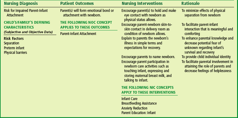
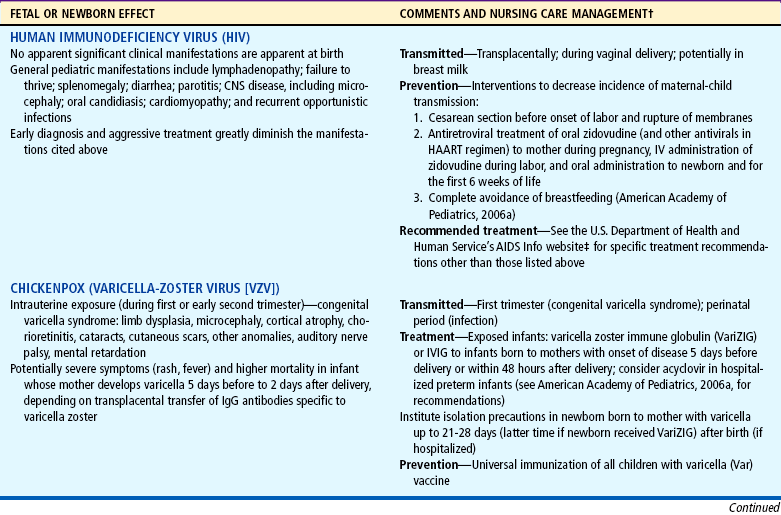
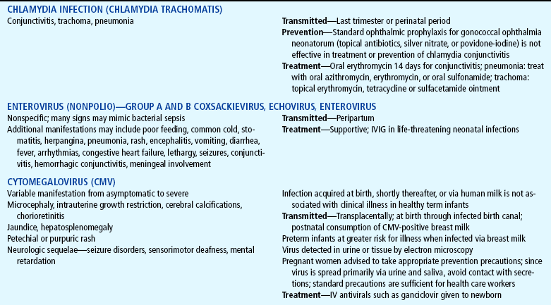
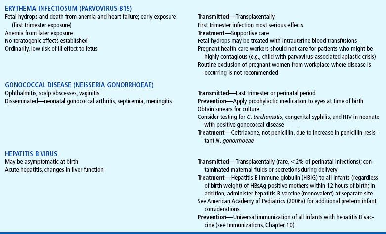
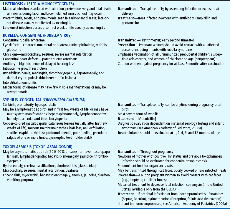
Nursing Care Management

The nursing care of the infant with jaundice is discussed in the Nursing Process box and in the following section.

Part of the routine physical assessment includes observing for evidence of jaundice at regular intervals. Jaundice is most reliably assessed by observing the infant’s skin color from head to toe and the color of the sclerae and mucous membranes. Applying direct pressure to the skin, especially over bony prominences such as the tip of the nose or the sternum, causes blanching and allows the yellow stain to be more pronounced. For dark-skinned infants, the color of the sclerae, conjunctiva, and oral mucosa is the most reliable indicator. Also, bilirubin (especially at high levels) is not uniformly distributed in skin. The nurse observes the infant in natural daylight for a true assessment of color.

The TcB is a useful screening device and is used to detect neonatal jaundice in full-term infants. Because phototherapy reduces the accuracy of the instrument, its value is limited to assessments made before the initiation of phototherapy. Institutions in which the device is used set up their own criteria based on their experience with their particular instrument. Blood samples are also taken for the measurement of bilirubin in the laboratory.

With short hospital stays, jaundice may appear after discharge. A careful history from the parents may reveal significant familial patterns of hyperbilirubinemia (older siblings of the infant). Other considerations in assessment include the ethnic origin of the family (e.g., higher incidence in Asian infants); type of delivery (e.g., induction of labor); and infant characteristics such as weight loss after birth, gestational age, sex, and the presence of any bruising. The method and frequency of feeding are assessed.

NURSINGALERT

Evidence of jaundice that appears before the infant is 24 hours of age is an indication for assessing bilirubin levels.

NURSINGTIP

While blood is drawn, phototherapy lights are turned off. Blood is transported in a covered tube to avoid a false reading as a result of bilirubin destruction in the test tube.

Prevention of jaundice may be possible with early introduction of feedings and frequent nursing without supplementation. Every effort is made to provide an optimum thermal environment to reduce metabolic needs.

Phototherapy.: The infant who receives phototherapy is placed nude under the light source and repositioned frequently to expose all body surface areas to the light. After phototherapy has been initiated, frequent serum bilirubinlevels (every 6 to 12 hours) are necessary because visual assessment of jaundice is no longer considered valid.

Several precautions are instituted to protect the infant during phototherapy. The infant’s eyes are shielded by an opaque mask to prevent exposure to the light (see Fig. 9-17). The eye shield should be properly sized and correctly positioned to cover the eyes completely but prevent any occlusion of the nares. The infant’s eyelids are closed before the mask is applied, since the corneas may become excoriated if they come in contact with the dressing. On each nursing shift the eyes are checked for evidence of discharge, excessive pressure on the lids, or corneal irritation. Eye shields are removed during feedings, which provide the opportunity for visual and sensory stimulation.

Infants who are in an open crib must have a protective Plexiglas shield between them and the fluorescent lights to minimize the amount of undesirable ultraviolet light reaching their skin and to protect them from accidental bulb breakage. Their temperature is closely monitored to prevent hyperthermia or hypothermia. Maintaining the infant in a flexed position with rolled blankets along the sides of the body helps maintain heat and provides comfort.

Accurate charting is another important nursing responsibility and includes (1) times that phototherapy is started and stopped, (2) proper shielding of the eyes, (3) type of fluorescent lamp (by manufacturer), (4) number of lamps, (5) distance between surface of lamps and infant (should be no less than 18 inches), (6) use of phototherapy in combination with an incubator or open bassinet, (7) photometer measurement of light intensity according to hospital protocol, and (8) occurrence of side effects.

Minor side effects for which the nurse should be alert include loose, greenish stools; transient skin rashes; hyperthermia; increased metabolic rate; dehydration; electrolyte disturbances, such as hypocalcemia; and priapism. To prevent or minimize these effects, the temperature is monitored to detect early signs of hypothermia or hyperthermia, and the skin is observed for evidence of dehydration and drying, which can lead to excoriation and breakdown. Oily lubricants or lotions are not used on the skin in order to prevent increased tanning or a “frying” effect. Full-term infants receiving phototherapy may require additional fluid volume to compensate for insensible and intestinal fluid loss. Because phototherapy enhances the excretion of unconjugated bilirubin through the bowel, loose stools may indicate accelerated bilirubin removal. Frequent stooling can cause perianal irritation; therefore meticulous skin care, especially keeping the skin clean and dry, is essential.

After phototherapy is permanently discontinued, there is often a subsequent increase in the serum bilirubin level, often called the rebound effect; this is usually transient and resolves without resuming therapy; however, a follow-up serum bilirubin level should be checked.

Family Support.: Parents need reassurance concerning their infant’s progress. All the procedures are explained to familiarize them with the benefits and risks. Parents need to be reassured that the naked infant under the bilirubin light is warm and comfortable. Eye shields are removed when the parents are visiting to facilitate the attachment process. The parents can be reassured that the neonate is accustomed to darkness after months of intrauterine existence and benefits a great deal from auditory and tactile stimulation (see Family Focus box).

image FAMILY FOCUS

Phototherapy and Parent-Infant Interaction

The traditional use of phototherapy has evoked concerns regarding a number of psychobehavioral issues, including parent-infant separation, potential social isolation, decreased sensorineural stimulation, altered biologic rhythms, altered feeding patterns, and activity changes. Parental anxiety is greatly increased, particularly at the sight of the newborn blindfolded and under special lights. The interruption of breastfeeding for phototherapy is a potential deterrent to successful maternal-infant attachment and interaction. Because research has demonstrated that bilirubin catabolism occurs primarily within the first few hours of the initiation of phototherapy, there is increased support for the periodic removal of the infant from treatment for feeding and holding. Intermittent phototherapy may be just as effective as continuous therapy when used correctly. The benefits of stopping phototherapy for parental feeding and holding outweigh concerns related to the clearance of bilirubin in the healthy full-term newborn with mild to moderate hyperbilirubinemia. Home phototherapy offers an additional opportunity to foster parent-infant attachment.

The initiation of any treatment requires informed consent by the parents for the therapy prescribed; however, in the case of phototherapy considerable anxiety may rightfully occur when words such as kernicterus and brain damage are used to describe possible effects of nontreatment. It is imperative that nurses remain sensitive to parents’ feelings and information needs during this process; an important nursing intervention is assessment of the parents’ understanding of the treatment involved and clarification of the nature of the therapy.

One of the most important nursing interventions is recognition of breastfeeding jaundice. Lack of familiarity among health professionals has caused many newborns prolonged hospitalization, termination of breastfeeding, and unnecessary phototherapy. Care of the new mother may include supporting successful and frequent breastfeeding. Parents also need reassurance of the benign nature of the jaundice and encouragement to resume breastfeeding if temporary cessation is prescribed. In some situations jaundice may increase the risk of parents discontinuing breastfeeding and developing the vulnerable child syndrome—a belief that their child has suffered a “close call” and is vulnerable to serious injury (see Critical Thinking Exercise).

image CRITICAL THINKING EXERCISE

Jaundice

A full-term, 5-day-old newborn is brought to the minor emergency department late in the evening for evaluation of newborn jaundice. A home health nurse visited earlier in the day and drew a serum bilirubin by heel stick; the results were total bilirubin 14.6 mg/dl, direct bilirubin 0.9 mg/dl. The father is concerned because a home health care worker mentioned that the newborn might develop brain damage if the bilirubin levels were to increase to high levels. The mother is breastfeeding every 2 to 3 hours, and the newborn has had four wet diapers and three semiliquid stools over the past 18 hours. The newborn’s birth weight was 2834 g (6.2 pounds), and her current weight (nude) is 2722 g (6 pounds). On examination the infant is active and alert, with visibly jaundiced skin and sclerae, intact reflexes, and strong suck reflex. The history reveals no prenatal or delivery complications. Apgar scores at 1 and 5 minutes were 8 and 9, respectively, and the initial assessment did not reveal any problems. The mother’s blood type is A positive, and the direct Coombs test is negative. The newborn was discharged from the birth hospital on the second day of life in apparent good health.

QUESTIONS

1. Evidence—Is there sufficient evidence to draw any conclusions about the newborn’s condition at this time?

2. Assumptions—Describe some underlying assumptions about the following:

a. Newborn jaundice in healthy full-term infant

b. Serum bilirubin levels and the newborn’s age in days; other pertinent laboratory values (may refer to Fig. 9-16)

c. Nutritional and excretory function and relation to bilirubin metabolism

d. The physical status of the infant per assessment data

3. What implications and priorities for nursing care can be drawn at this time?

4. Does the evidence objectively support your argument (conclusion)?

5. Are there alternative perspectives to your argument? What are they?

Discharge Planning and Home Care.: With short hospital stays, mothers and infants may be discharged before evidence of jaundice is present. It is important for the nurse to discuss signs of jaundice with the mother, since any clinical symptoms will probably appear at home. Home visits within 2 to 3 days after discharge to evaluate feeding and elimination patterns and jaundice are becoming routine for many health care organizations.

If home phototherapy is instituted, the hospital or home health care nurse or medical equipment company representative is usually responsible for teaching family members and assessing their abilities to implement the treatment safely. General guidelines for home care preparation and education are discussed in Chapter 20. Written instructions and supervision of care—especially the application of eye shields, if needed—are essential. The minor side effects of phototherapyare reviewed, and parents may need instruction in taking axillary temperatures and recording times and amounts of feedings and the number of wet diapers and stools. Regardless of how benign the disorder or the therapy, these parents need support and understanding. Measures should be taken to assist the mother in achieving successful breastfeeding, including consultation with a lactation specialist on an outpatient basis. Siblings also benefit from an explanation of the therapy to allay fears or misconceptions.

In jaundice associated with breastfeeding, follow-up blood studies are usually required to assess the progress of the jaundice. If temporary cessation of breastfeeding is prescribed, mothers should be taught to pump the breasts every 3 to 4 hours to maintain lactation; the expressed milk is frozen for use after breastfeeding is resumed.

HEMOLYTIC DISEASE OF THE NEWBORN

Hyperbilirubinemia in the first 24 hours of life is most often the result of HDN (erythroblastosis fetalis), an abnormally rapid rate of RBC destruction. Anemia caused by this destruction stimulates the production of RBCs, which in turn provides increasing numbers of cells for hemolysis. Major causes of increased erythrocyte destruction are isoimmunization (primarily Rh) and ABO incompatibility.

Blood Incompatibility

The membranes of human blood cells contain a variety of antigens, also known as agglutinogens, substances capable of producing an immune response if recognized by the body as foreign. The reciprocal relationship between antigens on RBCs and antibodies in the plasma causes agglutination (clumping). In other words, antibodies in the plasma of one blood group (except the AB group, which contains no antibodies) produce agglutination when mixed with antigens of a different blood group. In the ABO blood group system the antibodies occur naturally. In the Rh system the person must be exposed to the Rh antigen before significant antibody formation takes place and causes a sensitivity response known as isoimmunization.

Rh Incompatibility (Isoimmunization).: The Rh blood group consists of several antigens (with D being the most prevalent). For simplicity, only the terms Rh positive (presence of antigen) and Rh negative (absence of antigen) are used in this discussion. The presence or absence of the naturally occurring Rh factor determines the blood type.

Ordinarily, no problems are anticipated when the Rh blood types are the same in both mother and fetus or when the mother is Rh positive and the infant is Rh negative. Difficulty may arise when the mother is Rh negative and the infant is Rh positive. Although the maternal and fetal circulations are separate, fetal RBCs (with antigens foreign to the mother) sometimes gain access to the maternal circulation through minute breaks in the placental vessels. The mother’s natural defense mechanism responds to these alien cells by producing anti-Rh antibodies.

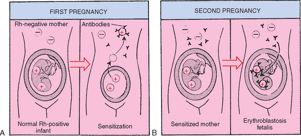
Under normal circumstances, this process of isoimmunization has no effect during the first pregnancy with an Rh-positive fetus because the initial sensitization to Rh antigens rarely occurs before the onset of labor. However, with the increased risk of fetal blood being transferred to the maternal circulation during placental separation, maternal antibody production is stimulated. During a subsequent pregnancy with an Rh-positive fetus, these previously formed maternal antibodies to Rh-positive blood cells may enter the fetal circulation, where they attack and destroy fetal erythrocytes (Fig. 9-18). Multiple gestations, abruptio placentae, placenta previa, manual removal of the placenta, and cesarean delivery increase the incidence of transplacental hemorrhage and subsequent isoimmunization (Moise, 2002).

image

FIG. 9-18 Development of maternal sensitization to Rh antigens. A, Fetal Rh-positive erythrocytes enter maternal system. Maternal anti-Rh antibodies are formed. B, Anti-Rh antibodies cross placenta and attack fetal erythrocytes.

Because the condition begins in utero, the fetus attempts to compensate for the progressive hemolysis and anemia by accelerating the rate of erythropoiesis. As a result, immature RBCs (erythroblasts) appear in the fetal circulation; hence the term erythroblastosis fetalis.

There is wide variability in the development of maternal sensitization to Rh-positive antigens. Sensitization may occur during the first pregnancy if the woman had previously received an Rh-positive blood transfusion. No sensitization may occur in situations in which a strong placental barrier prevents transfer of fetal blood into the maternal circulation. In approximately 10% to 15% of sensitized mothers, there is no hemolytic reaction in the newborn. In addition, some Rh-negative women, even though exposed to Rh-positive fetal blood, are immunologically unable to produce antibodies to the foreign antigen (Neal, 2001).

In the most severe form of erythroblastosis fetalis, hydrops fetalis, the progressive hemolysis causes fetal hypoxia; cardiac failure; generalized edema (anasarca); and effusions into the pericardial, pleural, and peritoneal spaces. The fetus may be delivered stillborn or in severe respiratory distress. The administration of maternal Rho(D) immune globulin (RhIG), early intrauterine detection of isoimmunization by ultrasonography, and subsequent treatment by fetal blood transfusions have dramatically improved the outcome of affected fetuses (Moise, 2002).

ABO Incompatibility.: Hemolytic disease can also occur when the major blood group antigens of the fetus are different from those of the mother. The major blood groups are A, B, AB, and O. In the North American Caucasian population, 46% have type O blood, 42% have type A blood, 9% have type B blood, and 3% have type AB blood.

The presence or absence of antibodies and antigens determines whether agglutination will occur. Antibodies in the plasma of one blood group (except the AB group, which contains no antibodies) will produce agglutination (clumping) when mixed with antigens of a different blood group. Naturally occurring antibodies in the recipient’s blood cause agglutination of a donor’s RBCs. The agglutinated donor cells become trapped in peripheral blood vessels, where they hemolyze, releasing large amounts of bilirubin into the circulation.

The most common blood group incompatibility in the neonate is between a mother with O blood group and an infant with A or B blood group (see Table 9-4 for possible ABO incompatibilities). Naturally occurring anti-A or anti-B antibodies already present in the maternal circulation cross the placenta and attack the fetal RBCs, causing hemolysis. Usually the hemolytic reaction is less severe than in Rh incompatibility. Unlike the Rh reaction, ABO incompatibility may occur in the first pregnancy. The risk of significant hemolysis in subsequent pregnancies is higher when the first pregnancy is complicated by ABO incompatibility (Sarici, Yurdakok, Serdar, and others, 2002).

TABLE 9-4

Potential Maternal-Fetal ABO Incompatibilities

MATERNAL BLOOD GROUP INCOMPATIBLE FETAL BLOOD GROUP
O A or B
B A or AB
A B or AB

Clinical Manifestations

Jaundice may appear shortly after birth (during the first 24 hours) in the newborn affected by HDN, and serum levels of unconjugated bilirubin rise rapidly. Anemia results from the hemolysis of large numbers of erythrocytes, and hyperbilirubinemia and jaundice result from the liver’s inability to conjugate and excrete the excess bilirubin. Most newborns with HDN are not jaundiced at birth. However, hepatosplenomegaly and varying degrees of hydrops may be evident. If the infant is severely affected, signs of anemia (notably, marked pallor) and hypovolemic shock are apparent. Hypoglycemia may occur as a result of pancreatic cell hyperplasia.

Diagnostic Evaluation

Early identification and diagnosis of Rh D sensitization are important in the management and prevention of fetal complications. A maternal antibody titer (indirect Coombs test) should be drawn at the first prenatal visit. Genetic testing allows early identification of paternal zygosity at the RHD gene locus, thus allowing earlier detection of the potential for isoimmunization and avoiding further maternal or fetal testing (Moise, 2002). Amniocentesis can be used to test the fetal blood type of a woman whose antibody screen is positive; the use of polymerase chain reaction may determine the fetal blood type and presence of maternal antibodies. The fetal hemoglobin and hematocrit can also be measured (Moise, 2002). Chorionic villus sampling has drawbacks that preclude its use, including possible spontaneous abortion of the fetus and fetomaternal hemorrhage, which would essentially makethe situation worse. With either method, if the fetus is found to be Rh negative, no further treatment is required.

Ultrasonography is considered an important adjunct in the detection of isoimmunization; alterations in the placenta, umbilical cord, and amniotic fluid volume, as well as the presence of fetal hydrops, can be detected with high-resolution ultrasonography and allow early treatment before the development of erythroblastosis. Doppler ultrasonography of fetal middle cerebral artery peak velocity has been used to detect and measure fetal hemoglobin and, subsequently, fetal anemia (Moise, 2002). Erythroblastosis fetalis caused by Rh incompatibility can also be monitored by evaluating rising anti-Rh antibody titers in the maternal circulation or by testing the optical density of amniotic fluid (delta OD450 test), since bilirubin discolors the fluid (Mari, 2000).

The disease in the newborn is suspected on the basis of the timing and appearance of jaundice (see Table 9-3) and can be confirmed postnatally by detecting antibodies attached to the circulating erythrocytes of affected infants (direct Coombs test or direct antiglobulin test). The Coombs test may be performed on cord blood samples from infants born to Rh-negative mothers if there is a history of incompatibility or further investigation is warranted.

Therapeutic Management

The primary aim of therapeutic management of isoimmunization is prevention. Postnatal therapy is usually phototherapy for mild cases and exchange transfusion for more severe forms. Although phototherapy may control bilirubin levels in mild cases, the hemolytic process may continue, causing severe anemia between 7 and 21 days of life.

Prevention of Rh Isoimmunization.: The administration of RhIG, a human gamma globulin concentrate of anti-D, to all unsensitized Rh-negative mothers after delivery or abortion of an Rh-positive infant or fetus prevents the development of maternal sensitization to the Rh factor. The injected anti-Rh antibodies are thought to destroy (by subsequent phagocytosis and agglutination) fetal RBCs passing into the maternal circulation before they can be recognized by the mother’s immune system. Because the immune response is blocked, anti-D antibodies and memory cells (which produce the primary and secondary immune responses, respectively) are not formed (Blackburn, 2007; Shaw, 2003). The inhibition of memory cell formation is especially important because memory cells provide long-term immunity by initiating a rapid immune response after the antigen is reintroduced (McCance and Huether, 2002).

To be effective, RhIG (such as RhoGAM) must be administered to unsensitized mothers within 72 hours (but possibly as long as 3 to 4 weeks) after the first delivery or abortion and repeated after subsequent pregnancies or losses. The administration of RhIG at 26 to 28 weeks of gestation further reduces the risk of Rh isoimmunization. RhIG is not effective against existing Rh-positive antibodies in the maternal circulation.

Preliminary studies have demonstrated the effectiveness of IV immunoglobulin (IVIG) at decreasing the severity of RBC destruction (hemolysis) in HDN and subsequent development of jaundice; IVIG administered to the neonate is believed to attack the maternal cells that destroy neonatal RBCs, slowing the progression of bilirubin production (Mundy, 2005). This therapy, often used in conjunction with phototherapy, may decrease the necessity for exchange transfusion.

NURSINGALERT

RhIG is administered intramuscularly, not intravenously, and only to Rh-negative women with a negative Coombs test—never to the newborn or father.

Intrauterine Transfusion.: Infants of mothers already sensitized may be treated by intrauterine transfusion, which consists of infusing blood into the umbilical vein of the fetus. The need for therapy is based on the antenatal diagnosis of isoimmunization by determining the optical density of amniotic fluid (by amniocentesis) as an index of fetal hemolysis or by serial ultrasonography, which may detect the presence of fetal hydrops as early as 16 weeks of gestation. With the advance of ultrasound technology, fetal transfusion may be accomplished directly via the umbilical vein, infusing type O Rh-negative packed RBCs to raise the fetal hematocrit to 40% to 50%; fetal movement and transfusion risks are minimized by administering vecuronium bromide for temporary fetal paralysis. The frequency of intrauterine transfusions may vary according to institution and fetal hydropic status yet may be as often as every 2 weeks until the fetus reaches pulmonary maturity at approximately 37 to 38 weeks of gestation (Moise, 2002). The use of intraperitoneal blood transfusions is employed less commonly for isoimmunization because of higher associated fetal risks; however, it may be used for cases in which intravascular access is impossible.

Exchange Transfusion.: Exchange transfusion, in which the infant’s blood is removed in small amounts (usually 5 to 10 ml at a time) and replaced with compatible blood (such as Rh-negative blood), is a standard mode of therapy for treatment of severe hyperbilirubinemia and is the treatment of choice for hyperbilirubinemia and hydrops caused by Rh incompatibility. Exchange transfusion removes the sensitized erythrocytes, lowers the serum bilirubin level to prevent bilirubin encephalopathy, corrects the anemia, and prevents cardiac failure. Indications for exchange transfusion in full-term infants may include a rapidly increasing serum bilirubin level and hemolysis despite intensive phototherapy. The criteria for exchange transfusions in preterm infants vary according to associated illness factors. The American Academy of Pediatrics (2004) practice parameter guidelines provide recommendations for initiating phototherapy and for exchange transfusion in infants at 35 weeks of gestation or more. An infant born with hydrops fetalis or signs of cardiac failure is a candidate for immediate exchange transfusion with fresh whole blood.

For exchange transfusion, fresh whole blood is typed and crossmatched to the mother’s serum. The amount of donor blood used is usually double the blood volume of the infant, which is approximately 85 ml/kg body weight, but is limited to no more than 500 ml. The two-volume exchange transfusion replaces approximately 85% of the neonate’s blood.

An exchange transfusion is a sterile surgical procedure. A catheter is inserted into the umbilical vein and threaded into the inferior vena cava. Depending on the infant’s weight, 5 to 10 ml of blood is withdrawn within 15 to 20 seconds, and the same volume of donor blood is infused over 60 to 90 seconds. If the blood has been citrated (addition of citrate phosphatedextrose adenine to prevent coagulation), calcium gluconate may be given after the infusion of each 100 ml of donor’s blood to prevent hypocalcemia.

Prognosis.: The severe anemia of isoimmunization may result in stillbirth, shock, congestive heart failure, or pulmonary or cerebral complications such as cerebral palsy. As a result of early detection and intrauterine treatment, erythroblastotic newborns are seen less often and exchange transfusions for the condition are less common. Despite the availability of effective preventive measures, Rh HDN continues to cause significant fetal morbidity and mortality in the United States.

Nursing Care Management

The initial nursing responsibility is recognizing jaundice. The possibility of hemolytic disease can be anticipated from the prenatal and perinatal history. Prenatal evidence of incompatibility and a positive Coombs test result are cause for increased vigilance for early signs of jaundice in an infant.

If an exchange transfusion is required, the nurse prepares the infant and the family and assists the practitioner with the procedure. The infant receives nothing by mouth (is NPO) during the procedure; therefore a peripheral infusion of dextrose and electrolytes is established. The nurse documents the blood volume exchanged, including the amount of blood withdrawn and infused; the time of each procedure; and the cumulative record of the total volume exchanged. Vital signs, monitored electronically, are evaluated frequently and correlated with the removal and infusion of blood. If signs of cardiac or respiratory problems occur, the procedure is stopped temporarily and resumed after the infant’s cardiorespiratory function stabilizes. The nurse also observes for signs of blood transfusion reaction and maintains the infant’s blood glucose levels and fluid balance.

Throughout the procedure attention must be given to the infant’s thermoregulation. Hypothermia increases oxygen and glucose consumption, causing metabolic acidosis. Not only do these consequences hinder the infant’s overall physical ability to withstand the long procedure, but they also inhibit the binding capacity of albumin and bilirubin and the hepatic enzymatic reactions, thus increasing the risk of kernicterus. Conversely, hyperthermia damages the donor erythrocytes, elevating the free potassium content and predisposing the infant to cardiac arrest.

The exchange transfusion is performed with the infant in a radiant warmer. However, the infant is usually covered with sterile drapes that may prevent the radiant heat from sufficiently warming the skin. The blood may also be warmed (using specially designed devices, never a microwave oven) before infusion.

After the procedure is completed, the nurse inspects the umbilical site for evidence of bleeding. The catheter may remain in place in case repeated exchanges are required.

NURSINGALERT

Signs of blood exchange transfusion reaction include tachycardia or bradycardia, respiratory distress, dramatic change in BP, temperature instability, and generalized rash.

Family Support.: Parents often feel guilty because they think they have caused the blood incompatibility. Parents should never be made to feel responsible or negligent. They are encouraged to verbalize their thoughts. Actions that were taken to prevent any problems, such as frequent antepartum examinations and blood tests, should be referred to and praised.

METABOLIC COMPLICATIONS

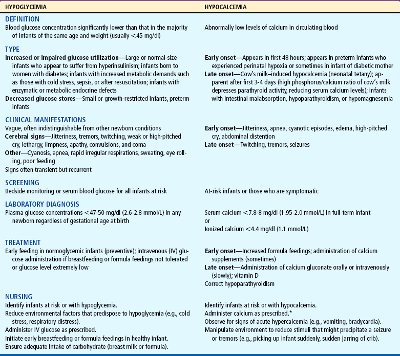
The high-risk infant is subject to a variety of complications related to physiologic function and the transition to extrauterine life. Prominent among these are fluid and electrolyte derangements, hypoglycemia, and hypocalcemia. These complications often occur concurrently with or as a secondary result of other neonatal disorders and may therefore be difficult to differentiate from other conditions. The major characteristics of hypoglycemia and hypocalcemia are outlined in Table 9-5.

TABLE 9-5

Metabolic Complications

image

*Nursing Alert: Calcium preparations should never be administered by bolus rapid infusion in infants.

RESPIRATORY DISTRESS SYNDROME

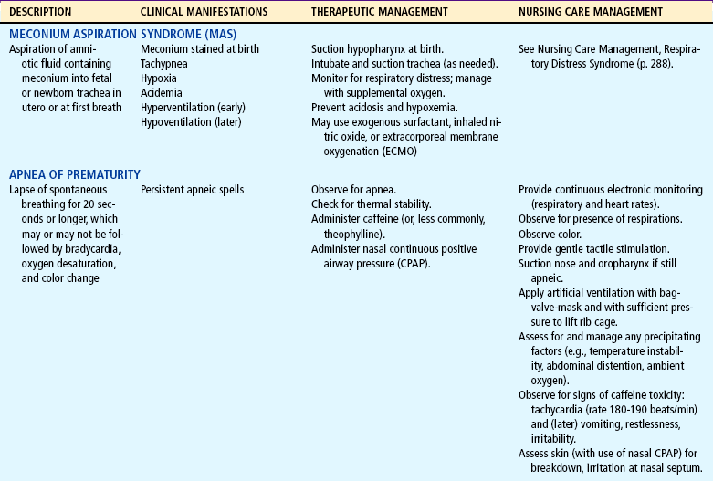
Respiratory distress is a name applied to respiratory dysfunction in neonates and is primarily a disease related to developmental delay in lung maturation. The terms respiratory distress syndrome (RDS) and hyaline membrane disease are most often applied to this severe lung disorder, which not only is responsible for more infant deaths than any other disease, but also carries the highest risk in terms of long-term respiratory and neurologic complications (see Chapter 23 for a discussion of acute RDS). It is seen almost exclusively in preterm infants. The disorder is rare in drug-exposed infants or infants who have been subjected to chronic intrauterine stress (e.g., maternal preeclampsia or hypertension). Respiratory distress of a nonpulmonary origin in neonates may also be caused by sepsis, cardiac defects (structural or functional), exposure to cold, airway obstruction (atresia), intraventricular hemorrhage, hypoglycemia, metabolic acidosis, acute blood loss, and drugs. Pneumonia in the neonatal period is respiratory distress caused by bacterial or viral agents and may occur alone or as a complication of RDS.

Pathophysiology

Preterm infants are born before the lungs are fully prepared to serve as efficient organs for gas exchange. This appears to be a critical factor in the development of RDS. The effects of lung immaturity are compounded by the presence of more cartilage in the chest wall, leading to increased compliance of the chest wall, which collapses inward in response to less compliant (stiffer) lung tissue.

There is evidence of fetal respiratory activity before birth. The lungs make feeble respiratory movements, and fluid is excreted through the alveoli. Because the final unfolding of the alveolar septa, which increases the surface area of the lungs, occurs during the last trimester of pregnancy, preterm infants are born with numerous underdeveloped and many uninflatable alveoli. Pulmonary blood flow is limited as a result of the collapsed state of the fetal lungs—poor vascular development in general and an immature capillary network in particular. Because of increased pulmonary vascular resistance, the major portion of fetal blood is shunted from the lungs by way of the ductus arteriosus and foramen ovale.

At birth, infants must initiate breathing and keep the previously fluid-filled lungs inflated with air. At the same time, the pulmonary capillary blood flow must be increased approximately tenfold to provide for adequate lung perfusion and to alter the intracardiac pressure that closes the fetal cardiac structures. Most full-term infants successfully accomplish these adjustments, but preterm infants with respiratory distress are unable to do so. Although numerous factors are involved, immaturity of the surfactant system plays a central role.

Surfactant is a surface-active phospholipid secreted by the alveolar epithelium. Acting much like a detergent, this substance reduces the surface tension of fluids that line the alveoli and respiratory passages, resulting in uniform expansion and maintenance of lung expansion at low intraalveolar pressure. Immature development of these functions produces consequences that seriously compromise respiratory efficiency. Deficient surfactant production causes unequal inflation of alveoli on inspiration and the collapse of alveoli on end expiration. Without surfactant, infants are unable to keep their lungs inflated and therefore exert a great deal of effort to reexpand the alveoli with each breath. With increasing exhaustion, infants are able to open fewer and fewer alveoli. This inability to maintain lung expansion produces widespread atelectasis.

In the absence of alveolar stability (normal functional residual capacity) and with progressive atelectasis, pulmonary vascular resistance (PVR) increases, whereas with normal lung expansion it would decrease. Consequently, there is hypoperfusion to the lung tissue, with a decrease in effective pulmonary blood flow. The increase in PVR causes partial reversion to the fetal circulation, with a right-to-left shunting of blood through the persisting fetal communications—the ductus arteriosus and foramen ovale.

Inadequate pulmonary perfusion and ventilation produce hypoxemia and hypercapnia. Pulmonary arterioles, with theirthick muscular layer, are markedly reactive to diminished oxygen concentration. Thus a decrease in oxygen tension causes vasoconstriction in the pulmonary arterioles that is further enhanced by a decrease in blood pH. This vasoconstriction contributes to a marked increase in PVR. In normal ventilation with increased oxygen concentration, the ductus arteriosus constricts and the pulmonary vessels dilate to decrease PVR.

Prolonged hypoxemia activates anaerobic glycolysis, which produces increased amounts of lactic acid. An increase in lactic acid causes metabolic acidosis; inability of the atelectatic lungs to blow off excess carbon dioxide produces respiratory acidosis. Acidosis causes further vasoconstriction. With deficient pulmonary circulation and alveolar perfusion, partial pressure of oxygen in arterial blood continues to fall, pH falls, and the materials needed for surfactant production are not circulated to the alveoli.

Diagnostic Evaluation

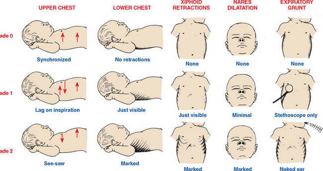
The diagnosis of RDS is made on the basis of clinical manifestations (Box 9-4) and radiographic studies. Radiographic findings characteristic of RDS include (1) a diffuse granular pattern over both lung fields that closely resembles ground glass and represents alveolar atelectasis and (2) dark streaks, or bronchograms, within the ground glass areas that represent dilated, air-filled bronchioles. It is important to distinguish between RDS and pneumonia in infants with respiratory distress. The extent of respiratory function and acid-base balance is determined by blood gas analysis. Criteria for visually evaluating the degree of respiratory distress are illustrated in Fig. 9-19. Pulse oximetry and carbon dioxide monitoring, as well as pulmonary function studies, assist in differentiating pulmonary and extrapulmonary illness and are used in the management of RDS.

BOX 9-4   Clinical Manifestations of Respiratory Distress Syndrome

Tachypnea (up to 80 to 120 breaths/min) initially*

Dyspnea

Pronounced intercostal or substernal retractions (Fig. 9-19)

Fine inspiratory crackles

Audible expiratory grunt

Flaring of the external nares

Cyanosis or pallor


*Not all infants born with respiratory distress syndrome will manifest these characteristics; the very low–birth-weight and extremely low–birth-weight infant may have respiratory failure and shock at birth because of physiologic immaturity.

image

FIG. 9-19 Criteria for evaluating respiratory distress. (Modified from Silvermann WA, Anderson DH: A controlled clinical trial of effects of water mist on obstructive respiratory signs, death rate, and necropsy findings among premature infants, Pediatrics 17:1, 1956.)

Therapeutic Management

The treatment of RDS involves immediate establishment of adequate oxygenation and ventilation and supportive care and measures required for any preterm infant, as well as those instituted to prevent further complications associated with preterm birth. The supportive measures most crucial to a favorable outcome are to:

image Maintain adequate ventilation and oxygenation

image Maintain acid-base balance

image Maintain a neutral thermal environment

image Maintain adequate tissue perfusion and oxygenation

image Prevent hypotension

image Maintain adequate hydration and electrolyte status

Nipple and gavage feedings are contraindicated in any situation that creates a marked increase in respiratory rate because of the greater hazards of aspiration. Nutrition is provided by parenteral therapy during the acute stage of the disease, while minimal enteral feeding is provided to enhance maturation of the neonate’s gastrointestinal system.

The administration of exogenous surfactant to preterm neonates with RDS has become an accepted and common therapy in most neonatal centers worldwide. Numerous clinical trials involving the administration of exogenous surfactant to infants with or at high risk for RDS demonstrate improvements in blood gas values and ventilator settings, decreased incidence of pulmonary air leaks, intraventricular hemorrhage, decreased deaths from RDS, and an overall decreased infant mortality rate (Merrill and Ballard, 2003; Stevens and Sinkin, 2007). The overall rates of some associated comorbidities (bronchopulmonary dysplasia, NEC, patent ductus arteriosus) have not decreased with surfactant replacement. Exogenous surfactant is derived from a natural source (e.g., porcine, bovine) or from the production of artificial surfactant.

Complications seen with surfactant administration include pulmonary hemorrhage and mucous plugging. Surfactant therapy is also being investigated for use in infants with meconium aspiration, infectious pneumonia, sepsis, persistent pulmonary hypertension, and lung hypoplasia concomitant with congenital diaphragmatic hernia (Finer, 2004; Stevens and Sinkin, 2007). Surfactant may be administered at birth as a preventive or prophylactic treatment of RDS or later on in the course of RDS as a rescue treatment. Surfactant is administered via an ET tube directly into the infant’s trachea. Nursing responsibilities with surfactant administration include assistance in the delivery of the product, collection and monitoring of arterial blood gases, scrupulous monitoring of oxygenation with pulse oximetry, and assessment of the infant’s tolerance of the procedure. After surfactant is absorbed, there is usually an increase in respiratory compliance that requires adjustment of ventilator settings to decrease mean airway pressure and prevent overinflation or hyperoxemia. Suctioning is usually delayed for an hour or so (depending on the type of surfactant and unit protocol) to allow maximum effects to occur. Research is in progress to investigate the possibility of delivering an aerosolized surfactant (Mazela, Merritt, and Finer, 2007). This method would decrease the problems associated with current delivery systems (contamination of the airway, interruption of mechanical ventilation, and loss of the drug in the ET tubing from reflux).

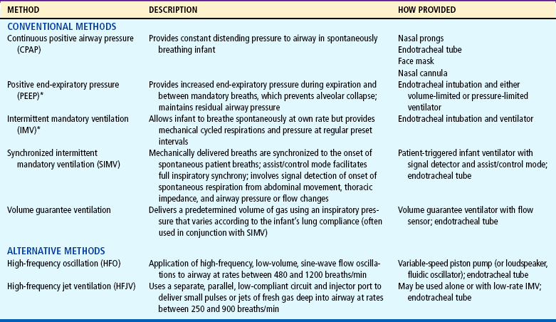
The goals of oxygen therapy are to provide adequate oxygen to the tissues, prevent lactic acid accumulation resulting from hypoxia, and at the same time avoid the potentially negative effects of oxygen and barotrauma. Numerous methods have been devised to improve oxygenation (Table 9-6). All require that the gas be warmed and humidified before entering the respiratory tract. If the infant does not require mechanical ventilation, oxygen can be supplied by nasal cannula or via nasal prongs in conjunction with continuous positive airway pressure (CPAP) (see Oxygen Therapy, Chapter 22). If oxygen saturation of the blood cannot be maintained at a satisfactory level and the carbon dioxide level (Paco 2) rises, infants will require ventilatory assistance.

TABLE 9-6

Common Methods for Assisted Ventilation in Neonatal Respiratory Distress

image

*Also referred to as conventional ventilation (vs high-frequency ventilation [HFV]).

Prevention.: The most successful approach to prevention of RDS is prevention of preterm delivery, especially in elective early delivery and cesarean section. Improved methods for assessing the maturity of the fetal lung by amniocentesis, although not a routine procedure, allow a reasonable prediction of adequate surfactant formation. Because estimation of a delivery date can be miscalculated by as much as 1 month, such tests are particularly valuable when scheduling an elective cesarean section. The combination of maternal steroid administration before delivery and surfactant administration postnatally seems to have a synergistic effect on neonatal lungs, with the net result being a decrease in infant mortality, decreased incidence of intraventricular hemorrhage, fewer pulmonary air leaks, and fewer problems with pulmonary interstitial emphysema and RDS.

Prognosis.: RDS is a self-limiting disease. Before the use of surfactant, infants typically experienced a period of deterioration (approximately 48 hours) and, in the absence of complications, improved by 72 hours. Often heralded by the onset of diuresis, this improvement was attributed primarily to increased production and greater availability of surfactant. With the administration of surfactant, lung compliance begins to improve almost immediately, resulting in lower oxygen requirements and a decreased need for ventilatory support (Stevens and Sinkin, 2007).

Infants with RDS who survive the first 96 hours have a reasonable chance of recovery. However, complications of RDS include associated respiratory conditions and problems associated with prematurity: patent ductus arteriosus and congestive heart failure, intraventricular hemorrhage, bronchopulmonary dysplasia, retinopathy of prematurity, pneumonia, air leak syndrome, sepsis, NEC, and neurologic sequelae.

Nursing Care Management

Care of infants with RDS involves all of the observations and interventions previously described for high-risk infants. In addition, the nurse is concerned with the complex problems related to respiratory therapy and the constant threat of hypoxemia and acidosis that complicates the care of patients in respiratory difficulty.

The respiratory therapist, an important member of the neonatal intensive care team, is often responsible for the maintenance of respiratory equipment. Although it may be the respiratory therapist’s responsibility to regulate the apparatus, nurses should understand the equipment and be able to recognize when it is not functioning correctly. The most essential nursing function is to observe and assess the infant’s response to therapy. Continuous monitoring and close observation are mandatory because an infant’s status can change rapidly and because oxygen concentration and ventilation parameters are prescribed according to the infant’s blood gas measurements and pulse oximetry readings.

Changes in oxygen concentration are based on these observations. The amount of oxygen administered, expressed as the fraction of inspired air (Fio2), is determined on an individual basis according to pulse oximetry or direct or indirect measurement of arterial oxygen concentration. Capillary samples collected from the heel (see Chapter 22 for procedure) are useful for pH and Paco2 determinations but not for oxygenation status. Continuous transcutaneous or pulse oximetry readings are recorded at least hourly. Blood sampling is performed after ventilator changes for the acutely ill infant and thereafter when clinically indicated.

NURSINGALERT

Suctioning is not an innocuous procedure (it may cause bronchospasm, bradycardia resulting from vagal nerve stimulation, hypoxia, or increased intracranial pressure, predisposing the infant to intraventricular hemorrhage) and should never be carried out on a routine basis. Improper suctioning technique can also cause infection, airway damage, or even pneumothoraces.

Mucus may collect in the respiratory tract as a result of the infant’s pulmonary condition. Secretions interfere with gas flow and predispose the infant to obstruction of the passages, including the ET tube. Suctioning should be performed only when necessary and should be based on individual infant assessment, which includes auscultation of the chest, evidence of decreased oxygenation, excess moisture in the ET tube, or increased infant irritability. During suctioning a variety of techniques can be used to minimize complications, including the use of a closed suctioning system (Cifuentes, Segars, and Carlo, 2003).

When nasopharyngeal passages, the trachea, or the ET tube is being suctioned, the catheter should be inserted gently but quickly; intermittent suction is applied as the catheter is withdrawn. It is imperative that the time the airway is obstructed by the catheter be limited to no more than 5 seconds, because continuous suction removes air from the lungs along with the mucus. It is recommended that the “two-person” suctioning procedure be used on infants who are acutely ill and who do not tolerate any procedure without profound decreases in oxygen saturation, BP, and heart rate. The object of suctioning an artificial airway is to maintain patency of that airway, not the bronchi. Suction applied beyond the ET tube can cause traumatic lesions of the trachea. The use of in-line suction catheters may decrease airway contamination and hypoxia.

The most advantageous positions for facilitating an infant’s open airway are on the side with the head supported in alignment by a small folded blanket or, when on the back, positioned to keep the neck slightly extended. With the head in the “sniffing” position, the trachea is opened at its maximum; hyperextension reduces the tracheal diameter in neonates.

Inspection of the skin is part of routine infant assessment. Position changes and the use of water pillows are helpful in guarding against skin breakdown.

Mouth care is especially important when infants are receiving nothing by mouth, and the problem is often aggravated by the drying effect of oxygen therapy. Drying and cracking can be prevented by good oral hygiene using sterile water. Irritation to the nares or mouth that occurs from appliances used to administer oxygen (e.g., nasal CPAP) may be reduced by the use of a water-soluble ointment.

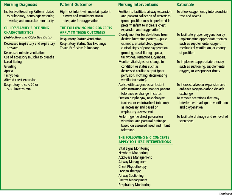
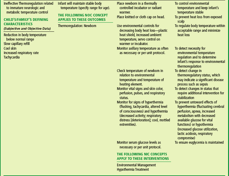
The nursing care of an infant with RDS is a demanding role; meticulous attention must be given to subtle changes in the infant’s oxygenation status. The importance of attention to detail cannot be overemphasized, particularly in regard to medication administration. (See Nursing Care Plan.)

RESPIRATORY COMPLICATIONS

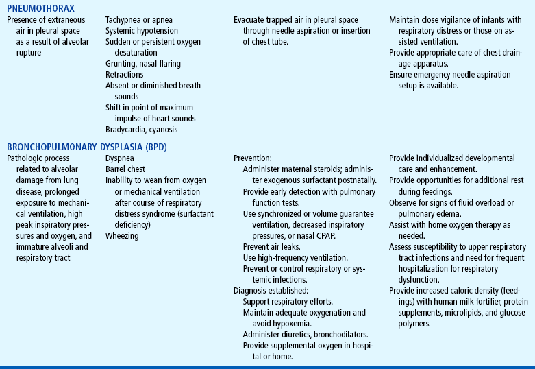
The newborn infant is vulnerable to a variety of pulmonary complications, some requiring oxygen therapy (Table 9-7). For example, the preterm infant is subject to periods of apnea,and in term, near-term, and postterm infants, intrauterine stress often causes the fetus to pass meconium, which may be aspirated before or during birth. Oxygen therapy, although lifesaving, is not without its hazards. Positive pressure introduced by mechanical apparatus has created an increase in the incidence of ruptured alveoli and subsequent pneumothorax and bronchopulmonary dysplasia (chronic lung disease). The use of nasal CPAP decreases the incidence of adverse effects associated with intubation and positive pressure ventilation in preterm infants with RDS (Davis and Henderson-Smart, 2003). Retinopathy of prematurity is observed almost exclusively in preterm infants and is related primarily to prematurity and oxygen therapy (Table 9-8 and 9-9). There is some evidence to support the resuscitation of asphyxiated newborns with 21% oxygen rather than 100% oxygen; preliminary studies demonstrate no significant neurologic morbidities at 18 to 24 months in newborns resuscitated with 21% oxygen (Saugstad, Ramji, Irani, and others, 2003; Saugstad, 2007). Proponents for room air resuscitation suggest there are fewer complications associated with oxidative stress and hyperoxemia when room air is administered (Vento and Saugstad, 2006). The 2005 American Heart Association Neonatal Resuscitation Guidelines recommend the initiation of neonatal resuscitation using either supplemental oxygen that is less than 100% oxygen or room air (no supplemental oxygen); if the neonate does not improve within 90 seconds, the use of supplemental oxygen is recommended. Pulse oximetry is recommended to monitor the infant’s oxygenation status during resuscitation and to prevent excessive use of oxygen in preterm infants (American Heart Association, American Academy of Pediatrics, 2006). Large multicenter studies are still needed to determine the optimum concentration of oxygen for resuscitation of neonates.

TABLE 9-7

Respiratory Complications

image

image

Inhaled nitric oxide (INO) and extracorporeal membrane oxygenation (ECMO) are additional therapies used in the treatment of respiratory distress and respiratory failure in neonates. INO is used in term and near-term infants with conditions such as persistent pulmonary hypertension, meconium aspiration syndrome (see Table 9-7), pneumonia, sepsis, and congenital diaphragmatic hernia to decrease or reverse pulmonary hypertension, pulmonary vasoconstriction, acidosis, and hypoxemia. Nitric oxide is a colorless, highly diffusible gas that can be administered through the ventilator circuit blended with oxygen. INO therapy may be used in conjunction with surfactant replacement therapy, high-frequency ventilation, or ECMO. INO has not proved to be significantly effective in decreasing rates of bronchopulmonary dysplasia or in improving survival rates in preterm infants (Barrington and Finer, 2007).

nursingcareplan

The High-Risk Infant with Respiratory Distress Syndrome

image

image

image

ECMO may be used in the management of term infants with acute severe respiratory failure for the same conditions as those mentioned for INO. This therapy involves a modified heart-lung machine, although in ECMO the heart is not stopped and blood does not entirely bypass the lungs. Blood is shunted from a catheter in the right atrium or right internal jugular vein by gravity to a servo-regulated roller pump, pumped through a membrane lung where it is oxygenated and through a small heat exchanger, then returned to the systemic circulation via a major artery such as the carotid artery to the aortic arch. ECMO provides oxygen to the circulation; allows the lungs to “rest”; and decreases pulmonary hypertension and hypoxemia in such conditions as persistent pulmonary hypertension of the newborn, congenital diaphragmatic hernia, sepsis, meconium aspiration, and severe pneumonia.

CARDIOVASCULAR COMPLICATIONS

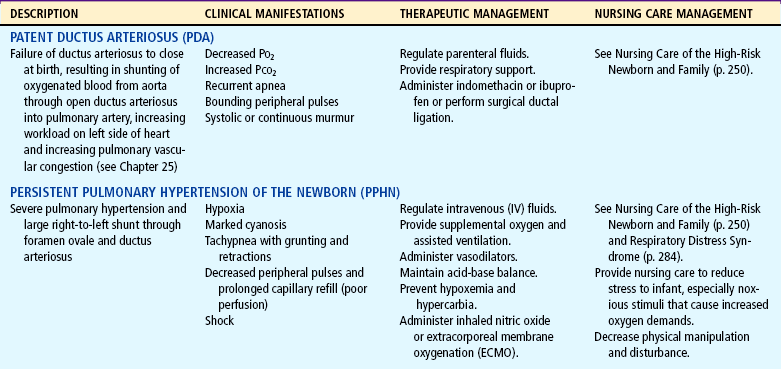
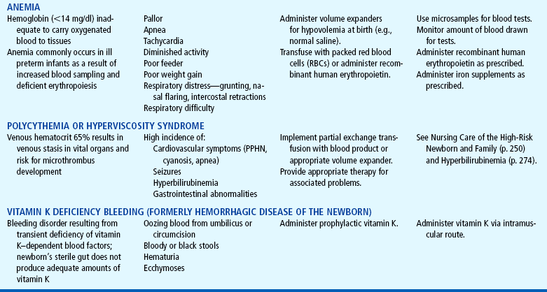
The most serious cardiovascular disorders of the newborn are the congenital heart defects. Other conditions that occur in the newborn period are usually related to prematurity (e.g., anemia, patent ductus arteriosus) or other diseases (e.g., respiratory distress). Some of these disorders are outlined in Table 9-8.

TABLE 9-8

Cardiovascular and Hematologic Complications

image

image

CEREBRAL COMPLICATIONS

Cerebral injury in newborn infants is not uncommon. Newborn infants are particularly vulnerable to ischemic injury caused by variable (both increased and decreased) cerebral blood flow subsequent to asphyxia, and preterm infants, with a fragile cerebrovascular network, are highly prone to periventricular or intraventricular hemorrhage. Fragility and increased permeability of capillaries and prolonged prothrombin time predispose the preterm infant to trauma when delicate structures are subjected to the forces of labor. The more common cerebral complications are outlined in Table 9-9.

The highest incidence of abnormal neurologic findings occurs in infants with intracranial hemorrhage and VLBW. Major neurologic problems, such as cerebral palsy, seizures, and hydrocephalus, are usually diagnosed in the first 2 years of life. Less severe deficits, such as learning disorders, ADHD, and fine and gross motor incoordination, may not be diagnosed until preschool or even school age. Cerebral palsy is one of the most common neurologic deficits in survivors of prematurity (see Chapter 32).

NEONATAL SEIZURES

Seizures in the neonatal period are usually the clinical manifestation of a serious underlying disease. The most common cause of seizures for term and preterm neonates is hypoxic ischemic encephalopathy secondary to perinatal asphyxia (Volpe, 2001). Although not life threatening as an isolated entity, seizures constitute a medical emergency because they signal a disease process that may produce irreversible brain damage. Consequently, it is imperative to recognize a seizure and its significance so that the cause, as well as the seizure, can be treated (Box 9-5).

BOX 9-5   Causes of Neonatal Seizures

METABOLIC

Hypoglycemia, hyperglycemia

Hypocalcemia

Hypernatremia, hyponatremia

Hypomagnesemia

Pyridoxine deficiency

Aminoacidurias (e.g., phenylketonuria, maple syrup urine disease)

Hyperammonemia

TOXIC

Uremia

Bilirubin encephalopathy (kernicterus)

PRENATAL INFECTIONS

Toxoplasmosis

Syphilis

Cytomegalovirus

Herpes simplex

Hepatitis

POSTNATAL INFECTIONS

Bacterial meningitis

Viral meningoencephalitis

Sepsis

Brain abscess

TRAUMA AT BIRTH

Hypoxic brain injury

Intracranial hemorrhage

Subarachnoid, subdural hemorrhage

Intraventricular hemorrhage

MALFORMATIONS

Central nervous system agenesis

Hydroencephalopathy

Tuberous sclerosis

MISCELLANEOUS

Narcotic withdrawal

Degenerative disease

Benign familial neonatal seizures

The features of neonatal seizures are different from those observed in the older infant or child. For example, the well-organized, generalized tonic-clonic seizures seen in older children are rare in infants, especially preterm infants. The newborn brain, with its immature anatomic and physiologic status and less cortical organization, is unable to allow ready development and maintenance of a generalized seizure. Instead, signs of seizures in newborns, especially preterm neonates, are subtle and include findings such as lip smacking, tongue thrusting, eye rolling, and arching (Volpe, 2001).

Jitteriness or tremulousness in the newborn is a repetitive shaking of an extremity or extremities that may be observedwith crying, occur with changes in sleeping state, or are elicited with stimulation. Jitteriness is relatively common in newborns, and in a mild degree may be considered normal during the first 4 days of life. Jitteriness can be distinguished from seizures by several characteristics:

image Jitteriness is not accompanied by ocular movement as are seizures.

image The dominant movement in jitteriness is tremor, whereas seizure movement is clonic jerking that cannot be stopped by flexion of the affected limb.

image Jitteriness is highly sensitive to stimulation, whereas seizures are not.

Jitteriness may be a sign of hypoglycemia, and infants with jitteriness should have a blood glucose level evaluated.

A tremor is defined as repetitive movements of both hands (with or without movement of legs or jaws) at a frequency of 2 to 5 per second and lasting more than 10 minutes. It is common in newborn infants and has a variety of causes, including neurologic damage, hypoglycemia, and hypocalcemia. In most instances tremors are of no pathologic significance.

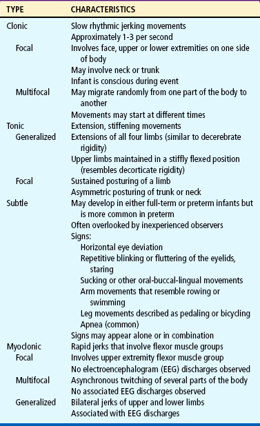
Neonatal seizures can be divided into four major types. These classifications are outlined in order of frequency in Table 9-10 and consist of clonic, tonic, subtle, and myoclonic seizures (Volpe, 2001). Clonic, multifocal clonic, and migratory clonic seizures are more common in term infants.

TABLE 9-10

Classifications of Neonatal Seizures

image

Modified from Volpe J: Neonatal seizures. In Volpe J: Neurology of the newborn, ed 4, Philadelphia, 2001, Saunders.

Diagnostic Evaluation

Early evaluation and diagnosis of seizures are urgent. In addition to a careful physical examination, the pregnancy and family histories are investigated for familial and prenatal causes. Blood is drawn for glucose and electrolyte examination, and cerebrospinal fluid may be obtained for testing of cell count and differential, protein, glucose, and culture. Electroencephalography may help identify subtle seizures but is less helpful in establishing a diagnosis. Other diagnostic procedures, such as computed tomography, magnetic resonance imaging, and cerebral ultrasound, may be indicated. A video electroencephalogram may be used to identify seizure activity in some newborns. More extensive metabolic testing may be needed when initial test results do not provide a diagnosis or the history is suggestive of an inherited metabolic disorder.

Therapeutic Management

Treatment is directed toward prevention of cerebral damage and involves correction of metabolic derangements, respiratory and cardiovascular support, and suppression of the seizure activity. The underlying cause is treated (e.g., glucose infusion for hypoglycemia, calcium for hypocalcemia, antibiotics for infection). If needed, respiratory support is provided for hypoxia, and anticonvulsants may be administered, especially when the other measures fail to control the seizures. Phenobarbital, given intravenously or orally, has been the drug of choice and is used if seizures are severe and persistent. Other drugs that may be used are phenytoin (Dilantin), lorazepam, and diazepam (Valium).

A newer drug, fosphenytoin sodium, is a water-soluble prodrug and has been designed to replace phenytoin. Fosphenytoin metabolizes to form phenytoin in the body yet can easily be diluted or mixed in dextrose and normal saline and may be given via IV or intramuscular routes. In addition, fosphenytoin does not cause pain during IV administration.

Nursing Care Management

The major nursing responsibilities in the care of infants with seizures are to recognize when the infant is having a seizure so that therapy can be instituted, to carry out the therapeutic regimen, and to observe the response to the therapy and any further evidence of seizures or other symptomatology. Assessment and other aspects of care are the same as for all high-risk infants. Parents need to be informed of their infant’s status, and the nurse should reinforce and clarify the practitioner’s explanations. The infant’s behaviors need to be interpreted for the parents, and the infant’s responses to the treatment must be anticipated and their significance explained. Parents are encouraged to visit their infant and perform the parenting activities consistent with the care plan. Seizures are a frightening phenomenon and generate a great deal of anxiety and fear, which is easily compounded by the justifiable concern of the staff. Providing support and guidance is an important nursing function.

HIGH RISK RELATED TO INFECTIOUS PROCESSES

SEPSIS

Sepsis, or septicemia, refers to a generalized bacterial infection in the bloodstream. Neonates are highly susceptible to infection as a result of diminished nonspecific (inflammatory) and specific (humoral) immunity, such as impaired phagocytosis, delayed chemotactic response, minimal or absent immunoglobulin A and immunoglobulin M (IgA and IgM), and decreased complement levels. Because of the infant’s poor response to pathogenic agents, there is usually no local inflammatory reaction at the portal of entry to signal an infection, and the resulting symptoms tend to be vague and nonspecific. Consequently, diagnosis and treatment may be delayed.

Breastfeeding has a protective benefit against infection and should be promoted for all newborns. It is of particular benefit to the high-risk neonate. Colostrum contains agglutinins that are effective against gram-negative bacteria.

Sepsis in the neonatal period can be acquired prenatally across the placenta from the maternal bloodstream or during labor from ingestion or aspiration of infected amniotic fluid. Prolonged rupture of the membranes always presents a risk for this type from maternal-fetal transfer of pathogenic organisms. In utero transplacental transfer can occur with organisms and viruses such as cytomegalovirus, toxoplasmosis, and Treponema pallidum (syphilis), which cross the placental barrier during the latter half of pregnancy. Intrapartum infection may occur via contact with an infected mother; examples of such infections include herpesvirus and HIV.

Early-onset sepsis (less than 3 days after birth) is acquired in the perinatal period; infection can occur from direct contact with organisms from the maternal gastrointestinal and genitourinary tracts. The most common infecting organism is Escherichia coli, whereas group B streptococci (GBS) rates remain low (Stoll, Hansen, Higgins, and others, 2005). E. coli, which may be present in the vagina, accounts for approximately half of all cases of sepsis caused by gram-negative organisms. GBS is an extremely virulent organism in neonates, with a high (50%) death rate in affected infants. Other bacteria noted to cause early-onset infection include Haemophilus influenzae, Citrobacter and Enterobacter organisms, coagulase-negative staphylococci, and Streptococcus viridans (Stoll, Hansen, Higgins, and others, 2005). Other pathogens that are harbored in the vagina and may infect the infant include gonococci, C. albicans, herpes simplex virus (type II), and chlamydia.

Late-onset sepsis (1 to 3 weeks after birth) is primarily nosocomial, and the offending organisms are usually staphylococci, Klebsiella organisms, enterococci, E. coli, and Pseudomonas or Candida species (Stoll, Hansen, Fanaroff, and others, 2002). Coagulase-negative staphylococci, considered to be primarily a contaminant in older children and adults, is commonly found to be the cause of septicemia in ELBW and VLBW infants. Bacterial invasion can occur through sites such as the umbilical stump; the skin; mucous membranes of the eye, nose, pharynx, and ear; and internal systems such as the respiratory, nervous, urinary, and gastrointestinal systems.

Postnatal infection is acquired by cross-contamination from other infants, personnel, or objects in the environment. Bacteria that are commonly called “water bugs” (because they are able to grow in water) are found in water supplies, humidifying apparatus, sink drains, suction machines, and most respiratory equipment. Organisms such as coagulase-negative staphylococci, which usually colonize the skin, may infect indwelling venous and arterial catheters used for infusions, blood sampling, and monitoring of vital signs. Neonatal sepsis is most common in the infant at risk, particularly the preterm infant or the infant born after a difficult or traumatic labor and delivery, who is least capable of resisting such bacterial invasion. These organisms are often transmitted by personnel from person to person or object to person by poor hand washing and inadequate housecleaning.

Diagnostic Evaluation

Diagnosis of sepsis is often based on suspicion of presenting clinical signs and symptoms. Because sepsis is so easily confused with other neonatal disorders, the definitive diagnosis is established by laboratory and radiographic examination. Isolation of the specific organism is always attempted through cultures of blood, urine, and cerebrospinal fluid. Blood studies may show signs of anemia, leukocytosis, or leukopenia. Leukopenia is usually an ominous sign because of its frequent association with high mortality. An elevated number of immature neutrophils, decreased or increased total neutrophils, and changes in neutrophil morphology also suggest an infectious process in the neonate. Other diagnostic data that are helpful in the determination of neonatal sepsis include C-reactive protein and interleukins, specifically interleukin-6 (Volante, Moretti, Pisani, and others, 2004; Laborada, Rego, Jain, and others, 2003).

Prevention

Several measures are important in the prevention of both early- and late-onset infection. Programs to screen pregnant women for GBS colonization (culture-based) and treatment of those women in labor have dramatically reduced the incidence of GBS infection in the neonate (Centers for Disease Control and Prevention, 2002b). Screening programs for other maternal infections, including hepatitis B and human immunodeficiency virus (HIV), have also been recommended. Breastfeeding by mothers infected with HIV is not recommended because the virus may be transmitted in breast milk.

Nursery procedures aimed at minimizing the risk of nosocomial infections include the practice of good hand-washing techniques, appropriate isolation precautions where indicated, and the adoption of recommended standards for spacing of infant beds.

Therapeutic Management

In addition to the institution of vigorous therapeutic measures, early recognition (Box 9-6) and diagnosis are essential to increase the infant’s chance for survival and reduce the likelihood of permanent neurologic damage. Antibiotic therapy is initiated before laboratory results are available for confirmation and identification of the exact organism. Treatment consists of circulatory support, respiratory support, aggressive administration of antibiotics, and immunotherapy.

BOX 9-6   Manifestations of Neonatal Sepsis

GENERAL SIGNS

Infant generally “not doing well”

Poor temperature control—hypothermia, hyperthermia (rare)

CIRCULATORY SYSTEM

Pallor, cyanosis, or mottling

Cold, clammy skin

Hypotension

Edema

Irregular heartbeat—bradycardia, tachycardia

RESPIRATORY SYSTEM

Irregular respirations, apnea, or tachypnea

Cyanosis

Grunting

Dyspnea

Retractions

CENTRAL NERVOUS SYSTEM

Diminished activity—lethargy, hyporeflexia, coma

Increased activity—irritability, tremors, seizures

Full fontanel

Increased or decreased tone

Abnormal eye movements

GASTROINTESTINAL SYSTEM

Poor feeding

Vomiting

Diarrhea or decreased stooling

Abdominal distention

Hepatomegaly

Hemoccult-positive stools

HEMATOPOIETIC SYSTEM

Jaundice

Pallor

Petechiae, ecchymosis

Splenomegaly

Supportive therapy usually involves administration of oxygen (if respiratory distress or hypoxia is evident), careful regulation of fluids, correction of electrolyte or acid-base imbalance, and temporary discontinuation of oral feedings. Blood transfusions may be needed to correct anemia and shock, and electronic monitoring of vital signs and regulation of the thermal environment are mandatory.

Antibiotic therapy is continued for 7 to 10 days if cultures are positive, discontinued in 48 to 72 hours if cultures are negative and the infant is asymptomatic, and most often administered via IV infusion. Antifungal and antiviral therapies are implemented as appropriate, depending on causative agents.

Prognosis.: The prognosis for neonatal sepsis is variable. Severe neurologic and respiratory sequelae may occur in ELBW and VLBW infants as a result of early-onset sepsis. Late-onset sepsis and meningitis may also result in poor outcomes for immunocompromised neonates. The trend in antenatal diagnosis of maternal GBS and subsequent maternal and neonatal treatment with antibiotic therapy has decreased the incidence of early-onset GBS disease by 70% (0.5 cases per 1000 live births in 1999), although rates of late-onset perinatal GBS disease remained constant (Centers for Disease Control and Prevention, 2002b). New rapid tests for identification of maternal GBS at delivery have been developed, but the sensitivity and specificity of these tests are still being evaluated (Aziz, Baron, D’Souza, and others, 2005); a vaccine to prevent perinatal GBS is still some years from being available.

Nursing Care Management

Nursing care of the infant with sepsis involves observation and assessment as outlined for any high-risk infant. Recognition of the existing problem is of paramount importance; it is usually the nurse who observes and assesses infants and identifies that “something is wrong” with them. Awareness of the potential modes of infection transmission also helps the nurse identify those at risk for developing sepsis. Much of the care of infants with sepsis involves the medical treatment of the illness. Knowledge of the side effects of the specific antibiotic and proper regulation and administration of the drug are vital.

Prolonged antibiotic therapy poses additional hazards for affected infants. Antibiotics predispose the infant to growth of resistant organisms and superinfection from fungal or mycotic agents, such as C. albicans. Nurses must be alert for evidence of such complications. Nystatin oral suspension is swabbed on the buccal mucosa for prophylaxis against oral candidiasis.

A number of specimens may be needed to help identify the cause and source of the infection. It is recommended that the fully flexed position be avoided for obtaining spinal fluid and that the side-lying position (modified with neck extension) or sitting position be used instead. Continual cardiorespiratory and pulse oximetry monitoring provides an ongoing assessment of the infant’s condition during the procedure.

Part of the total care of infants with sepsis is to decrease any additional physiologic or environmental stress. This includes providing an optimum thermoregulated environment and anticipating potential problems such as dehydration or hypoxia. Precautions are implemented to prevent the spread of infection to other newborns, but to be effective, activities must be carried out by all caregivers. Proper hand washing, the use of disposable equipment (e.g., linens, catheters, feeding supplies, IV equipment), disposal of excretions (e.g., vomitus, stool), and adequate housekeeping of the environment and equipment are essential. Because nurses are the most consistent caregivers involved with sick infants, it is usually their responsibility to see that standard precautions are maintained by everyone.

Another aspect of caring for infants with sepsis involves observation for signs of complications, including meningitis and septic shock, a severe complication caused by toxins in the bloodstream.

NURSINGALERT

Artificial and long natural fingernails worn by nurses have been associated with serious neonatal infection and morbidity from Pseudomonas aeruginosa (Moolenaar, Crutcher, San Joaquin, and others, 2000) and Klebsiellaorganisms in the NICU (Gupta, Della-Latta, Todd, and others, 2004).

NECROTIZING ENTEROCOLITIS

NEC is an acute inflammatory disease of the bowel with increased incidence in preterm infants. Three factors appear to play an important role in the development of NEC: intestinal ischemia, colonization by pathogenic bacteria, and substrate (formula feeding) in the intestinal lumen. The precise cause of NEC is still uncertain, but it appears to occur in infants whosegastrointestinal tract has suffered vascular compromise. Intestinal ischemia of unknown etiology, immature gastrointestinal host defenses, bacterial proliferation, and feeding substrate are now believed to have a multifactorial role in the etiology of NEC. Prematurity remains the most prominent risk factor in the development of NEC.

The damage to mucosal cells lining the bowel wall is great. Diminished blood supply to these cells causes their death in large numbers; they stop secreting protective, lubricating mucus; and the thin, unprotected bowel wall is attacked by proteolytic enzymes. Thus the bowel wall continues to swell and break down; it is unable to synthesize protective IgM, and the mucosa is permeable to macromolecules (e.g., exotoxins), which further hampers intestinal defenses. Gas-forming bacteria invade the damaged areas to produce pneumatosis intestinalis, the presence of gas in the submucosal or subserosal surfaces of the bowel.

A consistent relationship has been observed between the development of NEC and enteric feeding of hypertonic substances (e.g., formula, hyperosmolar medications). It is unclear whether this connection is a result of the formula imposing a stress on an ischemic bowel, serving as a substrate for bacterial growth, or both.

Diagnostic Evaluation

Radiographic studies show a sausage-shaped dilation of the intestine that progresses to marked distention and the characteristic pneumatosis intestinalis’“soapsuds,” or the bubbly appearance of thickened bowel wall and ultralumina. There may be air in the portal circulation or free air observed in the abdomen, indicating perforation. Laboratory findings may include anemia, leukopenia, leukocytosis, metabolic acidosis, and electrolyte imbalance. In severe cases coagulopathy (disseminated intravascular coagulation) or thrombocytopenia may be evident. Organisms are often cultured from blood, although bacteremia or septicemia may not be prominent early in the course of the disease.

Therapeutic Management

Treatment of NEC begins with prevention. Oral feedings may be withheld for at least 24 to 48 hours from infants who are believed to have suffered birth asphyxia. Breast milk is the preferred enteral nutrient because it confers some passive immunity (IgA), macrophages, and lysozymes.

Minimal enteral feedings (trophic feeding, gastrointestinal priming) have gained acceptance with no evidence of increased incidence of NEC. However, a meta-analysis of feeding studies found that, although minimal enteral feedings resulted in a reduction in days to full enteral feeds, an increased risk for NEC could not be ruled out (Tyson and Kennedy, 2005).

The role of probiotics such as Lactobacillus acidophilus and Bifidobacterium infantis administered with enteral feedings for the prevention of NEC has yet to be fully explored to advocate widespread use in all VLBW infants. In some studies probiotics decreased the incidence of NEC (Bin-Nun, Bromiker, Wilschanski, and others, 2005; Hung-Chih, Bai-Horng, An-Chyi, and others, 2005).

Medical treatment of confirmed NEC consists of discontinuation of all oral feedings; institution of abdominal decompression via nasogastric suction; administration of IV antibiotics; and correction of extravascular volume depletion, electrolyte abnormalities, acid-base imbalances, and hypoxia. Replacing oral feedings with parenteral fluids decreases the need for oxygen and circulation to the bowel. Serial abdominal radiographic films (every 6 to 8 hours in the acute phase) are taken to monitor for possible progression of the disease to intestinal perforation.

Prognosis.: With early recognition and treatment, medical management is increasingly successful. If there is progressive deterioration under medical management or evidence of perforation, surgical resection and anastomosis are performed. Extensive involvement may necessitate surgical intervention and establishment of an ileostomy, jejunostomy, or colostomy. Sequelae in surviving infants include short-bowel syndrome (see Chapter 24), colonic stricture with obstruction, fat malabsorption, and failure to thrive secondary to intestinal dysfunction. A variety of surgical interventions for NEC are available and depend on the extent of bowel necrosis, associated illness factors, and infant stability. Intestinal transplantation has been successful in some former preterm infants with NEC-associated short-bowel syndrome who had already developed life-threatening total parenteral nutrition–related complications. Transplantation may be a lifesaving option for infants who previously faced high morbidity and mortality (Kato, Mittal, Nishida, and others, 2003).

Nursing Care Management

Nursing responsibilities begin with the prompt recognition of the early warning signs of NEC. Because the signs are similar to those observed in many other disorders of the newborn, nurses must constantly be aware of the possibility of this disease in infants who are at high risk for developing NEC (Box 9-7).

BOX 9-7   Clinical Manifestations of Necrotizing Enterocolitis

NONSPECIFIC CLINICAL SIGNS

Lethargy

Poor feeding

Hypotension

Vomiting

Apnea

Decreased urinary output

Unstable temperature

Jaundice

SPECIFIC SIGNS

Distended (often shiny) abdomen

Blood in the stools or gastric contents

Gastric retention

Localized abdominal wall erythema or induration

Bilious vomitus

When the disease is suspected, the nurse assists with diagnostic procedures and implements the therapeutic regimen. Vital signs, including BP, are monitored for changes that might indicate bowel perforation, septicemia, or cardiovascular shock, and measures are instituted to prevent possible transmission to other infants. It is especially important to avoid rectal temperatures because of the increased danger of perforation. To avoid pressure on the distended abdomen and to facilitate continuous observation, infants are often left undiapered and positioned supine or on the side.

NURSINGTIP

Observe for indications of early development of NEC by checking the appearance of the abdomen for distention (measuring abdominal girth, measuring residual gastric contents before feedings, and listening for bowel sounds) and performing all routine assessments for high-risk neonates.

Conscientious attention to nutritional and hydration needs is essential, and antibiotics are administered as prescribed. The time at which oral feedings are reinstituted varies considerably but is usually at least 7 to 10 days after diagnosis and treatment. Feeding is usually re-established using human milk, if available.

Because NEC is an infectious disease, one of the most important nursing functions is control of infection. Strict hand washing is the primary barrier to spread, and confirmed multiple cases are isolated. Persons with symptoms of a gastrointestinal disorder should not care for these or any other infants.

The infant who requires surgery requires the same careful attention and observation as any infant with abdominal surgery, including ostomy care (as applicable). This disorder is one of the most common reasons for performing ileostomies on newborns. Throughout the medical and surgical management of infants with NEC, the nurse is continually alert to signs of complications, such as septicemia, disseminated intravascular coagulation, hypoglycemia, and other metabolic derangements.

HIGH RISK RELATED TO MATERNAL CONDITIONS

The health of the fetus and newborn may be affected by a number of maternal conditions; essentially, any condition affecting the mother also has the potential for negatively affecting the health of the newborn. Pregnancy-induced hypertension or HELLP (hemolysis, elevated liver enzymes, low platelets) syndrome may cause preterm delivery, intrauterine growth restriction (IUGR), asphyxia, and death if it is not detected early and appropriate interventions implemented. It is not within the scope of this text to elaborate on the pathophysiology and treatment of these conditions; however, the reader is referred to any one of the excellent maternity texts available for a detailed discussion of these conditions.

INFANTS OF DIABETIC MOTHERS

Before insulin therapy, few women with diabetes were able to conceive; for those who did, the mortality rate for both mother and infant was high. The morbidity and mortality of infants of diabetic mothers (IDMs) have been significantly reduced as a result of effective control of maternal diabetes and an increased understanding of fetal disorders. Because infants born to women with gestational diabetes mellitus are at risk for the same complications as IDMs, the following discussion of IDMs includes infants born to women with gestational diabetes mellitus.

The severity of the maternal diabetes affects infant survival. Severity of maternal diabetes is determined by the duration of the disease before pregnancy; age of onset; extent of vascular complications; and abnormalities of the current pregnancy, such as pyelonephritis, diabetic ketoacidosis, pregnancy-induced hypertension, and noncompliance. The single most important factor influencing fetal well-being is the euglycemic status of the mother. It has been found that reasonable metabolic control that begins before conception and continues during the first weeks of pregnancy can prevent malformation in an IDM. Elevated levels of hemoglobin A1c during the first trimester appear to be associated with a higher incidence of congenital malformations.

Hypoglycemia may appear a short time after birth and in IDMs is associated with increased insulin activity in the blood (see also Table 9-5, p. 285). The serum glucose level that corresponds to clinical hypoglycemia have not been well defined. Because some infants experience metabolic complications at higher levels than previously thought, some researchers recommend that serum glucose levels be maintained above 45 mg/dl (2.5 mmol/L) in infants with abnormal clinical symptoms, and as high as 50 or 60 mg/dl in other infants (Deshpande and Ward, 2005; Cornblath, Hawdon, Williams, and others, 2000).

Hypoglycemia in the IDM is related to hypertrophy and hyperplasia of the pancreatic islet cells, and thus is a transient state of hyperinsulinism. High maternal blood glucose levels during fetal life provide a continual stimulus to the fetal islet cells for insulin production (glucose easily passes the placental barrier from maternal to fetal side; insulin, however, does not cross the placental barrier). This sustained state of hyperglycemia promotes fetal insulin secretion that ultimately leads to excessive growth and deposition of fat, which probably accounts for the infants who are large for gestational age, or macrosomic. When the neonate’s glucose supply is removed abruptly at the time of birth, the continued production of insulin soon depletes the blood of circulating glucose, creating a state of hyperinsulinism and hypoglycemia within 1/2 to 4 hours, especially in infants of mothers with poorly controlled diabetes (formerly class C diabetes or beyond [class D through R]). Precipitous drops in blood glucose levels can cause serious neurologic damage or death.

IDMs have a characteristic appearance (Box 9-8). Infants of mothers with advanced diabetes may be small for gestational age, may have IUGR, or may be appropriate for gestational age because of the maternal vascular (placental) involvement. There is an increase in congenital anomalies in IDMs in addition to a high susceptibility to hypoglycemia, hypocalcemia, hypomagnesemia, polycythemia, hyperbilirubinemia, cardiomyopathy, hypomagnesemia, and RDS. Hyperinsulinemia and hyperglycemia in the diabetic mother may be factors in reducing fetal surfactant synthesis, thus contributing to the development of RDS. Although large, these infants may be delivered before term as a result of maternal complications or increased fetal size.

BOX 9-8   Clinical Manifestations of Infants of Diabetic Mothers

image Large for gestational age

image Very plump and full faced

image Abundant vernix caseosa

image Plethora

image Listlessness and lethargy

image Large placenta and umbilical cord (Wharton jelly)

image Possibly meconium stained at birth

Therapeutic Management

The most effective management of IDMs is careful monitoring of serum glucose levels and observation for accompanying complications such as RDS. The infants are examined for the presence of any anomalies or birth injuries, and blood studies for determination of glucose, calcium, hematocrit, andbilirubin are obtained on a regular basis. Studies confirm the importance of maintaining serum glucose levels above 50 mg/dl (2.8 mmol/L) in hyperinsulinemic infants with hypoglycemia to prevent serious neurologic sequelae (Cowett and Loughead, 2002).

Because the hypertrophied pancreas is so sensitive to blood glucose concentrations, the administration of oral glucose may trigger a massive insulin release, resulting in rebound hypoglycemia. Therefore feedings of breast milk or formula begin within the first hour after birth, provided that the infant’s cardiorespiratory condition is stable. Approximately half of these infants do well and adjust without complications. Infants born to mothers with uncontrolled diabetes may require IV dextrose infusions. Oral and IV intake may be titrated to maintain adequate blood glucose levels. Frequent blood glucose determinations are needed for the first 2 to 4 days of life to assess the degree of hypoglycemia present at any given time. Testing blood taken from the heel with calibrated portable reflectance meters (e.g., glucometers) is a simple and effective screening evaluation that can then be confirmed by laboratory examination.

Nursing Care Management

The nursing care of IDMs involves early examination for congenital anomalies, signs of possible respiratory or cardiac problems, maintenance of adequate thermoregulation, early introduction of carbohydrate feedings as appropriate, and monitoring of serum blood glucose levels (see Evidence-Based Practice box). The latter is of particular importance because many hypoglycemic infants may remain asymptomatic. IV glucose infusion requires careful monitoring of the site and the neonate’s reaction to therapy; high glucose concentrations (>12.5%) should be infused via a central line instead of a peripheral site.

EVIDENCE-BASED PRACTICE

Glucose Water Feedings in the Newborn Period

ASK THE QUESTION

Is 5% dextrose in water (D5W) feeding an appropriate substitute for breast milk or formula in the newborn?

SEARCH FOR EVIDENCE

Search Strategies

Articles since 1980; key terms: newborn hypoglycemia, dextrose water feedings, newborn serum glucose, newborn feeding, sterile water feeding of newborn

Databases Used

Medline, PubMed, Ovid CINAHL

CRITICALLY ANALYZE THE EVIDENCE

No systematic or randomized clinical trials evaluating the oral administration of D5W vs infant formula, water, or breast milk were found in the literature surveyed. Seven key articles and two textbooks were selected for this review.

image Noerr (2001) indicates that formula and breast milk or colostrum are ideal for the newborn who is asymptomatic (for hypoglycemia) and able to tolerate oral feedings; the free fatty acids from the breakdown of fat in formula or breast milk decrease peripheral glucose use and decrease the effect of insulin to suppress hepatic glucose production.

image Hashim and Guillet (2002) suggest that infants be fed a carbohydrate source to maintain a steady state rise in glucose. They recommend that D5W be avoided because it may lead to rapid increases and subsequent falls in serum glucose levels.

image Blackburn (2003) suggests that early feedings with a carbohydrate source initially increases blood glucose levels; this increase is accompanied by subsequent increase in plasma insulin levels and the development of cyclic changes in insulin and blood glucose levels in full-term infants; animal studies suggest that D5W, if aspirated, is just as dangerous as formula.

image The American Academy of Pediatrics and American College of Obstetricians and Gynecologists (2007) do not recommend routine supplementation with D5W for infants receiving phototherapy.

image Eidelman (2001) recommends the initiation of breastfeeding within the first 30 to 60 minutes of birth for the healthy term infant and suggests that feedings with water or dextrose water are unnecessary and counterproductive.

image Initial feedings of sterile water or glucose water in newborns were historically recommended by physicians in newborns on the premise that the infant’s propensity to spit up and possibly aspirate the feeding source into the lungs was less likely to compromise the infant’s respiratory status if the aspirate was water (or dextrose water) instead of formula, breast milk, or colostrum. Wakayama, Wilkins, and Kimura (1988) suggested that initial feedings of infant formula after surgical repair for inguinal hernia did not significantly increase the incidence of aspiration.

image Sperling and Menon (2004) indicate that the initiation of milk feedings in the full-term infant induces ketogenesis, thus sparing glucose for brain consumption and facilitating gluconeogenesis.

image Hoseth, Joergensen, Ebbesen, and others (2000) found that healthy, AGA, full-term, breast-fed infants (n = 223) were not at risk for developing hypoglycemia. Diwakar and Sasidhar (2002) had similar findings in 200 exclusively breast-fed healthy full-term, AGA infants. Blackburn (2003) suggests that healthy breast-fed infants have a higher number of ketone bodies than formula-fed infants; this alternate source of energy (ketone bodies) enables the healthy full-term infant to maintain a steady state serum glucose level while breastfeeding is being established.

image The physiologic effects of early milk intake (breast milk or formula) in the healthy newborn include the stimulation of gut maturation, promotion of intestinal mucosa integrity, and release of intestinal enzymes that enhance motility and perfusion (Blackburn, 2003). There is evidence that sterile water and dextrose water feedings do not promote intestinal maturation; these are primarily from studies in newborns who are jaundiced.

APPLY THE EVIDENCE: NURSING IMPLICATIONS

Currently no evidence exists to support the practice of oral dextrose water feedings in healthy AGA newborns as a substitute for breast milk or formula even to treat mild, asymptomatic, transient hypoglycemia. Likewise, only physiologic evidence supports the practice of early oral feedings of breast milk, formula, or colostrum in healthy newborns.

REFERENCES

American Academy of Pediatrics, American College of Obstetricians and Gynecologists. Guidelines for perinatal care, ed 6. Elk Grove Village, Ill: The Academy, 2007.

Blackburn, ST. Maternal, fetal, and neonatal physiology: a clinical perspective, ed 2. St Louis: Saunders, 2003.

Diwakar, KK, Sasidhar, MV. Plasma glucose levels in term infants who are appropriate size for gestation and exclusively breast fed. Arch Dis Child Fetal Neonatal Educ. 2002;87(1):F46–F48.

Eidelman, AI. Hypoglycemia and the breastfed neonate. Pediatr Clin North Am. 2001;48(2):377–387.

Hashim, MJ, Guillet, R. Common issues in the care of sick neonates. Am Fam Physician. 2002;66(9):1685–1692.

Hoseth, E, Joergensen, A, Ebbesen, F, et al. Blood glucose levels in a population of healthy, breast fed, term infants of appropriate size for gestational age. Arch Dis Child Fetal Neonatal Educ. 2000;83(2):F117–F119.

Noerr, B. State of the science: neonatal hypoglycemia. Adv Neonat Care. 2001;1(1):4–21.

Sperling, MA, Menon, RK. Differential diagnosis and management of neonatal hypoglycemia. Pediatr Clin North Am. 2004;51(3):703–723.

Wakayama, Y, Wilkins, S, Kimura, K. Is 5% dextrose in water a proper choice for initial postoperative feeding in infants? J Pediatr Surg. 1988;23(7):644–666.

Because macrosomic infants are at risk for problems associated with a difficult delivery, they are monitored for birth injuries such as brachial plexus injury and palsy, fractured clavicle, and phrenic nerve palsy. Additional monitoring of the infant for problems associated with this condition (polycythemia, hypocalcemia, poor feeding, and hyperbilirubinemia) is also a vital nursing function.

There is some evidence that IDMs have an increased risk of acquiring type 2 diabetes and metabolic syndrome in childhood or early adulthood (Boney, Verma, Tucker, and others, 2005); therefore nursing care should also focus on healthy lifestyle and prevention later in life with IDMs.

DRUG-EXPOSED INFANTS*

Maternal habits hazardous to the fetus and neonate include drug addiction, smoking, and alcohol abuse. Occasional withdrawal reactions have been reported in neonates of mothers who use to excess such drugs as barbiturates, alcohol, amphetamines, or antidepressants. Serious reactions are seen in neonates whose mothers abuse psychoactive drugs or are treated with methadone.

Narcotics, which have a low molecular weight, readily cross the placental membrane and enter the fetal system. Illicit substances may also be transmitted to the newborn through breast milk. When the mother is a habitual user of opiates, especially heroin or methadone, the unborn child may also become chemically dependent or passively addicted to the drug, which places such infants at risk during the perinatal and early neonatal periods. Neonatal abstinence syndrome (NAS) is the term used to describe the set of behaviors exhibited by the infant exposed to chemical substances in utero.

Clinical Manifestations

Throughout this section, unless otherwise noted, the information presented refers to drug-exposed neonates in general, regardless of which drug they have been exposed to.

The adverse effects of exposure of the fetus to drugs are varied. They include transient behavioral changes such as alterations in fetal breathing movements or irreversible effects such as fetal death, IUGR, structural malformations, or mental retardation. Determining the specific effects of individual drugs on the fetus is made difficult by polydrug use, which is common; errors or omissions in reporting drug use; and variations in the strength, purity, and types of additives found in street drugs. Maternal conditions such as poverty, malnutrition, and comorbid conditions such as sexually transmitted infections further compound the difficulty in identifying the presence and consequences of intrauterine drug exposure. Most infants who are exposed to drugs in utero may demonstrate no immediate untoward effects and appear normal at birth. Infants exposed only to heroin may begin to exhibit signs of drug withdrawal within 12 to 24 hours. If mothers have been taking methadone, the signs appear somewhat later—anywhere from 1 or 2 days to 2 to 3 weeks or more after birth. The clinical manifestations may fall into any one or all of the following categories: CNS, gastrointestinal, respiratory, and autonomic nervous system signs (Greene and Goodman, 2003). The manifestations become most pronounced between 48 and 72 hours of age and may last from 6 days to 8 weeks, depending on the severity of the withdrawal (Box 9-9). Although these infants suck avidly on fists and display an exaggerated rooting reflex, they are poor feeders with uncoordinated and ineffectual sucking and swallowing reflexes.

BOX 9-9   Signs of Withdrawal in the Neonate

NEUROLOGIC

Irritability

Seizures

Hyperactivity

High-pitched cry

Tremors

Exaggerated Moro reflex

Hypertonicity of muscles

GASTROINTESTINAL

Poor feeding

Diarrhea

Dehydration

Vomiting

Frantic, uncoordinated sucking

Gastric residuals

AUTONOMIC

Diaphoresis

Fever

Mottled skin

Nasal stuffiness

MISCELLANEOUS

Disrupted sleep patterns

Diaphoresis

Tachypnea (>60 breaths/min)

Excoriations (knees, face)

Temperature instability

Not all infants of narcotic-addicted mothers will show signs of withdrawal. Because of irregular and varying degrees of drug use, quality of drug, and mixed drug usage by the mother, some infants display mild or variable manifestations. Most manifestations are the vague, nonspecific signs characteristic of all infants in general; therefore it is important to differentiate between drug withdrawal and other disorders before specific therapy is instituted. Other conditions (e.g., hypocalcemia, hypoglycemia, sepsis) often coexist with the drug withdrawal. Additional signs seen in drug-exposed newborns include loose stools; tachycardia; fever; projectile vomiting; crying; nasal stuffiness; and generalized perspiration, which is unusual in newborns.

Diagnostic Evaluation

Newborn urine, hair, or meconium sampling may be required to identify drug exposure and implement appropriate early interventional therapies aimed at minimizing the consequences of intrauterine drug exposure. Meconium sampling for fetal drug exposure is reported to provide more screening accuracy than urine, since drug metabolites accumulate in meconium (Kandall, 1999). Urine toxicology screening has less accuracy, since it reflects only recent substance intake by the mother (Huestis and Choo, 2002). Meconium and hair testing for drug metabolites has the advantages of being noninvasive, more accurate, and easy to collect. One study examining urine, hair, and meconium samples for drug use found that, although all of these tests were reliable to a greater or lesser degree, the single most reliable method for determining prenatal drug use was a careful history collected by an experienced interviewer (Eyler, Behnke, Wobie, and others, 2005).

Therapeutic Management

The treatment of the drug-exposed infant initially consists of early identification through maternal history, presenting symptoms of NAS, or toxicology screening when substance abuse is strongly suspected. Early identification and intervention are essential to prevent further adverse effects; earlydischarge from the birth institution should be postponed until further assessment of the maternal situation and establishment of a treatment plan for the mother and infant. Drug therapies to decrease withdrawal effects include parenteral or oral administration of phenobarbital, chlorpromazine, clonidine, diazepam, methadone, and morphine. A combination of these drugs may be necessary to treat infants exposed to multiple drugs in utero, and careful attention should be given to possible adverse effects of the treatment drugs (Johnson, Gerada, and Greenough, 2003).

Prognosis.: The prognosis for drug-exposed infants depends on the type and amount of drug(s) taken by the mother and the stage(s) of fetal development in which the drug was taken. The overall mortality of infants born to narcotic-addicted mothers is increased, but with early recognition, proper treatment, and long-term follow-up, the morbidity and mortality associated with drug exposure are decreased.

Often, drug-exposed infants will exhibit poor brain and body growth at birth; however, at times, infants will not exhibit any signs that indicate exposure to harmful agents and their condition may therefore be overlooked until symptoms appear later in life. Drug-exposed infants may have chronic feeding problems; irritability; abnormal neurologic responses; abnormal parent-infant interactions; developmental and cognitive delays; learning disabilities; and behavioral problems, including ADHD.

Nursing Care Management

One of the key factors in the treatment of drug-exposed neonates is early identification of substance abuse in the pregnant woman so treatment can be initiated and side effects minimized. This is especially problematic from a social and legal standpoint because the pregnant woman is often aware of the consequences of admitting to substance abuse and may therefore be less likely to readily admit to the problem for fear of social and legal repercussions. If the mother has had good prenatal care, the practitioner is aware of the problem and may have instituted therapy before delivery. However, a number of mothers deliver their infants without the benefit of adequate care, and the condition is unknown to health care personnel at the time of delivery.

The degree of withdrawal is closely related to the amount of drug the mother has habitually taken, the length of time she has been taking the drug, and her drug level at the time of delivery. The most severe symptoms are observed in the infants of mothers who have taken large amounts of drugs over a long period. In addition, the nearer to the time of delivery that the mother takes the drug, the longer it takes the child to develop withdrawal, and the more severe the manifestations. The infant may not exhibit withdrawal symptoms until 7 to 10 days after delivery, by which time most newborns have been discharged from the birth center and caregivers are less likely to recognize signs of irritability and poor feeding as withdrawal, thus predisposing the newborn to abuse or neglect. The infant may be at further risk for subsequent abuse or neglect because of home conditions that preclude adequate newborn care and follow-up.

After the presence of NAS is identified in an infant, nursing care is directed toward treatment of the presenting signs, decreasing stimuli that may precipitate hyperactivity and irritability (e.g., dimming the lights, decreasing noise levels), providing adequate nutrition and hydration, and promoting maternal-infant relationships. Appropriate individualized developmental care is implemented to facilitate self-consoling and self-regulating behaviors. Irritable and hyperactive infants have been found to respond to physical comforting, movement, and close contact. Wrapping infants snugly and rocking and holding them tightly limits their ability to self-stimulate. Arranging nursing activities to reduce the amount of disturbance helps decrease exogenous stimulation.

Breastfeeding is encouraged in mothers who are not using illicit substances, are negative for HIV infection, and are compliant with a methadone program; breastfeeding promotes maternal-infant bonding, and small quantities of methadone passed through breast milk have not proved to be harmful.

The Neonatal Abstinence Scoring System has been developed to monitor infants in an objective manner and evaluate their response to clinical and pharmacologic interventions (Kandall, 1999; Finnegan, 1985). This system is also designed to assist nurses and other health care workers in evaluating the severity of the infant’s withdrawal symptoms. Another tool that may be used to evaluate withdrawal behavior and treatment in newborns is the Neonatal Withdrawal Inventory developed by Zahorodny, Rom, Whitney, and others (1998); it is important to note that neither of these tools is specific to preterm infants and may not be representative of withdrawal behaviors in such infants (Marcellus, 2002).

The Neonatal Intensive Care Unit Network Neurobehavioral Scale (NNNS) is a comprehensive neurologic and behavioral assessment tool that may be used to identify newborns at risk as a result of intrauterine drug exposure. The tool measures stress or abstinence, state, neurologic status, and muscle tone in the context of the newborn’s medical condition at the time of examination. The NNNS may be used for medically stable newborns who are at least 30 weeks of gestation and up to 48 weeks of corrected or conceptional age (Lester, Tronick, and Brazelton, 2004).

Loose stools, poor intake, and regurgitation after feeding predispose these infants to malnutrition, dehydration, and electrolyte imbalance. In addition, these infants burn up energy with continual activity and increased oxygen consumption at the cellular level. Frequent weighing, careful monitoring of intake and output and electrolytes, and additional caloric supplementation may be necessary. Hyperactive infants must be protected from skin abrasions on the knees, toes, and cheeks that are caused by rubbing on bed linens while in a prone position (awake). Monitoring and recording the activity level and its relationship to other activities, such as feeding and preventing complications, are important nursing functions.

A valuable aid to anticipating problems in the newborn is recognizing substance abuse in the mother. Unless the mother is enrolled in a methadone rehabilitation program, she seldom risks calling attention to her habit by seeking prenatal care. Consequently, infants and mothers are exposed to the additional hazards of obstetric and medical complications. Moreover, the nature of substance use and addiction makes the user susceptible to disorders such as infection (hepatitis B, HIV), foreign body reaction, and the hazards of inadequate nutrition and preterm birth. Methadone treatment does not prevent withdrawal reaction in neonates, but the clinical course may be modified. Also, the intensive psychologic support of mothers is a factor in the treatment and reduction of perinatal mortality. Experience has indicated that mothers are usually anxious and depressed, lack confidence, have a poor self-image, and have difficulty with interpersonal relationships. They may have a psychologic need for the pregnancy and an infant.

Initial symptoms or the recurrence of withdrawal symptoms may develop after discharge from the hospital; therefore it is important to establish rapport and maintain contact with the family so that they return for treatment if this occurs. The demands of the drug-exposed infant on the caregiver are enormous and unrewarding in terms of positive feedback. The infants are difficult to comfort, and they cry for long periods, which can be especially trying for the caregiver after the infant’s discharge from the hospital. Long-term follow-up to evaluate the status of the infant and family is very important. Sudden infant death syndrome and HIV infection are observed more commonly in infants born to users of methadone and heroin.

Many problems arise in relation to the disposition of infants of drug-dependent mothers. Those who advocate separation of mothers and children argue that the mothers are not capable of assuming responsibility for their infant’s care, that child care is frustrating to them, and that their existence is too disorganized and chaotic. Others encourage the maternal-infant bond and recommend a protected environment such as a therapeutic community, a halfway house, or continuous ongoing, supportive services in the home after discharge. Careful evaluation and the cooperative efforts of a variety of health professionals are required whether the choice is foster home placement or supportive follow-up care of mothers who keep their infants.

Alcohol Exposure

Alcohol ingestion during pregnancy is associated with both short- and long-term effects on the fetus and newborn. The quantity of alcohol required to produce fetal effects is unclear, but it is known that infants born to heavy drinkers have twice the risk of congenital abnormalities than those born to moderate drinkers (Stoll, 2007). Alcohol withdrawal can occur in neonates, particularly when maternal ingestion occurs near the time of delivery (Greene and Goodman, 2003). Signs and symptoms include jitteriness, increased tone and reflex responses, and irritability. Seizures are also common. The incidence of fetal alcohol syndrome (FAS) in the United States is about 2 per 1000 population (Stoll, 2007). FAS is based on minimal criteria of signs in each of three categories: prenatal and postnatal growth restriction; CNS malfunctions, including mental retardation; and craniofacial features such as microcephaly, small eyes or short palpebral fissures, thin upper lip, flat midface, and an indistinct philtrum. Neurologic problems in FAS children include some degree of IQ deficit, ADHD, diminished fine motor skills, and poor speech. These children have been shown to lack inhibition, have no stranger anxiety, and lack appropriate judgment skills

Infants who do not display the signs of FAS but are born to mothers who are also heavy alcohol drinkers have significantly more tremors, hypertonia, restlessness, excessive mouthing movements, crying, and inconsolability than infants of substance-abusive mothers who do not consume alcohol during pregnancy. An added concern regarding substance abuse is that many of the mothers often use several drugs, such as tranquilizers, sedatives, amphetamines, phencyclidine, marijuana, and other psychotropic agents.

Cocaine Exposure

Cocaine, a commonly used illicit drug used in the United States, has multiple modes of use. However, use of the relatively inexpensive and easily administered “crack” form is increasing, especially among women of childbearing age (Cambell, 2003). Because crack vaporizes at relatively low temperatures, it is smoked and absorbed in large quantities through pulmonary vasculature. The drug readily crosses the placenta, placing the fetus at risk (Malanga and Kosofsy, 1999).

Cocaine is a CNS stimulant and peripheral sympathomimetic. Legally it is classified as a narcotic, but it is not an opioid. The effects on the fetus are secondary to maternal effects—increased BP, decreased uterine blood flow, and increased vascular resistance. Consequently, the fetus suffers decreased blood flow and oxygenation because of placental and fetal vasoconstriction. Researchers have concluded that variables such as the mother’s lack of prenatal care; poor nutrition; and use of tobacco, alcohol, and other drugs during pregnancy compound the effects of cocaine exposure in the infant (Askin and Diehl-Jones, 2001; Tronick and Beeghly, 1999).

Infants may appear normal, or they may show neurologic problems at birth that may continue during the neonatal period. In much of the research literature, these findings were transient, and there has been variable evidence demonstrating permanent sequelae. Either of two types of behavior may emerge as a result of cocaine effects on fetal development: neurobehavioral depression or excitability. The behaviors of the depressed infant include lethargy, poor suck, hypotonia, weak cry, and difficulty in arousing. The behaviors of the excitable neonate may include a high-pitched cry, hypertonicity, rigidity, irritability, inability to be consoled, and intolerance to a change in routine (Chiriboga, Brust, Bateman, and others, 1999; Chiriboga, Kuhn, and Wasserman, 2007). Otherbehaviors may include frequent startling, poorly defined awake state, sleeping difficulties, and persistent primitive reflexes. Some infants develop late onset of symptoms (2 to 8 weeks). They may become irritable and hypertonic, experience sleep-wake disruptions, and demonstrate an inability to tolerate change; they may also be slightly febrile. However, many of the findings cited above have been refuted in other studies (Eyler and Behnke, 1999; Tronick and Beeghly, 1999).

The adverse effects on the cocaine-exposed neonate are related to dose-response. The higher the dose, the more effects are noted, such as IUGR, hypertonia, and decreased fetal head growth (Chiriboga, Brust, Bateman, and others, 1999).

Sequelae of prenatal cocaine exposure include a smaller head circumference, decreased birth length, and decreased weight. Head growth may be one of the best predictors of long-term development (Bateman and Chiriboga, 2000). Other neonatal effects of cocaine exposure include increased incidence of gastroschisis, genitourinary anomalies, and periventricular and intraventricular hemorrhage. Some studies found that long-term sequelae for newborns exposed to cocaine include lower language, motor, and cognitive scores and an increased risk for learning disabilities (Singer, Arendt, Minnes, and others, 2002; Koren, Nulman, Rovet, and others, 1998; Morrow, Culbertson, Accornero, and others, 2006); however, one study revealed no significant differences in the total or verbal IQ scores but did note an increased risk of specific cognitive impairments (Singer, Minnes, Short, and others, 2004). Arendt, Angelopoulos, Salvator, and others (1999) noted that the fine and gross motor development indexes in 2-year-old children who were exposed to cocaine prenatally were lower than in the control group. Some researchers noted that the exposed children may be affected emotionally rather than intellectually. In a study of first-grade students, Delaney-Black, Cobington, Templin, and others (1998) concluded that the children who were exposed to cocaine prenatally were rated by their teachers as having more behavior problems than the control group.

One study using the Brazelton Neonatal Assessment Scale showed that cocaine-exposed infants scored low in response to arousal, auditory, and visual stimuli (Eyler, Behnke, and Conlon, 1998). However, other studies have not found significant differences between affected infants and others (Frank, Jacobs, Beeghly, and others, 2002; Richardson, Hamel, and Goldschmidt, 1996; Messinger, Bauer, Das, and others, 2004).

Therapeutic Management.: Treatment of these infants is similar to that for other drug-exposed infants: reduction of external stimuli, supportive treatment aimed at alleviating symptoms, and, at times, mild sedation.

Nursing Care Management.: Nursing care of cocaine-exposed infants is the same as that for other drug-exposed infants. Because they have increased flexor tone, these infants respond to swaddling in a semiflexed position (Askin and Diehl-Jones, 2001). Positioning, infant massage, and limited tactile stimulation have been shown to be effective interventions. Effects of the drug from breast milk have been reported (Kandall, 1999); therefore mothers should be cautioned regarding this hazard to their infants.

Referral to early intervention programs, including child health care, parental drug treatment, individualized developmental care, and parenting education, is essential in promoting the optimum outcome for these children. Because these children often live in an impoverished environment, they are at high risk for cognitive delays, lack of child health care, and inadequate nutrition and would benefit from an early intervention program.

Methamphetamine Exposure

The fetal and neonatal effects of maternal use of methamphetamines in pregnancy are not well known but appear to be dose related (Smith, Yonekura, Wallace, and others, 2003). LBW, preterm birth, and perinatal mortality may be consequences of higher doses used throughout pregnancy. In addition, a higher incidence of cleft lip and palate and cardiac defects has been reported in infants exposed to methamphetamines in utero (Plessinger, 1998).

Methamphetamine use has increased significantly in the past 10 years in certain regions of the United States. In Smith, Yonekura, Wallace, and others’ (2003) study, 63% of pregnant women using methamphetamines reported using the drug throughout the pregnancy. A higher incidence of preterm delivery and placental abruption was associated with methamphetamine use. In addition, fetal growth restriction (small for gestational age) was slightly higher in methamphetamine-exposed offspring; however, 80% of these neonates’ mothers also had significant alcohol and tobacco use.

Study reports vary in the time of clinical manifestations of withdrawal from this drug; one study did not identify any signs of withdrawal in the first 3 days after birth, but long-term data were not collected (Smith, Yonekura, Wallace, and others, 2003). A study of infants exposed to methamphetamine in utero showed that such infants had significantly smaller head circumferences and birth weights than those not exposed; in addition, the exposed infants exhibited withdrawal signs of agitation, vomiting, and tachypnea, which were not observed in the unexposed infants (Chomchai, Na Manorom, Watanarungasan, and others, 2004). After birth, infants may experience bradycardia or tachycardia that resolves as the drug is cleared from the system. Lethargy may continue for several months, along with frequent infections and poor weight gain. Emotional disturbances and delays in gross and fine motor coordination may be seen during early childhood.

The long-term effects of methamphetamine exposure on children living in households where the product is manufactured are not known, but there are early reports of burns in exposed children and concerns regarding the effects of the toxic by-products of methamphetamine production on small children. Skin rashes and respiratory illnesses are common problems seen in methamphetamine-exposed children, but physical neglect and speech and language developmental delays are of significant concern as well (Crocker, 2005).

Marijuana Exposure

Marijuana has replaced cocaine as the most common illicit drug used by women ages 18 to 44 years (nonpregnant and pregnant) in the United States (Ebrahim and Gfoerer, 2004). Marijuana crosses the placenta. Its use during pregnancy may result in a shortened gestation and a higher incidence of IUGR (Wagner, Katikaneni, Cox, and others, 1998). A strong association has been reported between the use of marijuana and a decrease in fetal growth and infant birth weight and length (Hurd, Wang, Anderson, and others, 2005). Other investigators have found a higher incidence of meconium staining (Bandstra and Accornero, 2006). Compounding the issue of the effects of marijuana, especially among women ages 18 to 30 years (Ebrahim and Gfoerer, 2004), is multidrug use, which combines the harmful effects of marijuana, tobacco, alcohol, opiates, and cocaine. Long-term follow-up studies on exposed infants are needed.

MATERNAL INFECTIONS

The range of pathologic conditions produced by infectious agents is large, and the difference between the maternal and fetal effects caused by any one agent is also great. Some maternal infections, especially during early gestation, can result in fetal loss or malformations because the fetus’s ability to handle infectious organisms is limited and the fetal immunologic system is unable to prevent the dissemination of infectious organisms to the various tissues.

Not all prenatal infections produce teratogenic effects. Furthermore, the clinical picture of disorders caused by transplacental transfer of infectious agents is not always well defined. Some microbial agents can cause remarkably similar manifestations, and it is not uncommon to test for all when a prenatal infection is suspected. This is the so-called TORCH complex, an acronym for:

TToxoplasmosis

OOther (e.g., hepatitis B)

RRubella

CCytomegalovirus infection

HHerpes simplex

To determine the causative agent in a symptomatic infant, tests are performed to rule out each of these infections. The O category may involve testing for several viral infections (e.g., hepatitis B, varicella zoster, measles, mumps, HIV, syphilis, and human parvovirus). Bacterial infections are not included in the TORCH workup because they are usually identified by clinical manifestations and readily available laboratory tests. Gonococcal conjunctivitis (ophthalmia neonatorum) and chlamydial conjunctivitis have been significantly reduced by prophylactic measures at birth (see Chapter 8). The major maternal infections, their possible effects, and specific nursing considerations are outlined in Table 9-11.

TABLE 9-11

Infections Acquired from Mother Before, During, or After Birth*

image

image

image

image

CNS, Central nervous system; IgG, immunoglobulin G; IV, intravenous; IVIG, intravenous immunoglobulin; HAART, highly active antiretroviral therapy; HBsAg, hepatitis B surface antigen.

*This table is not an exhaustive representation of all perinatally transmitted infections. For further information regarding specific diseases or treatment not listed here, refer to American Academy of Pediatrics, Committee on Infectious Diseases, Pickering L, editor: Red book: 2006 report of the Committee on Infectious Diseases, ed 27, Elk Grove Village, Ill, 2006, The Academy.

Isolation precautions depend on institutional policy (see Infection Control, Chapter 22).

http://www.aidsinfo.nih.gov.

§See also Centers for Disease Control and Prevention: Sexually transmitted diseases treatment guidelines, 2006, MMWR Recomm Rep 55(RR-11):1-94, 2006.

Nursing Care Management

One of the major goals in care of infants suspected of having an infectious disease is identification of the causative organism. Until the diagnosis is established, standard precautions are implemented according to institutional policy. In suspected cytomegalovirus and rubella infections, pregnant personnel are cautioned to avoid contact with the infant. Herpes simplex is easily transmitted from one infant to another; therefore risk of cross-contamination is reduced or eliminated by wearing gloves for patient contact. The American Academy of Pediatrics’ Red Book: 2006 Report of the Committee on Infectious Diseases (2006a) provides guidelines for the type and duration of precautions for most bacterial and viral exposures. Careful hand washing is the most important nursing intervention in reducing the spread of any infection.

Specimens need to be obtained for laboratory examinations, and the infant and parents need to be prepared for diagnostic procedures. When possible, long-term disabilities are prevented by early evaluation and implementation of therapy. The family is taught any special handling techniques needed for the care of their infant and signs of complications or possible sequelae. If sequelae are inevitable, the family will need assistance in determining how they can best cope with the problems, such as assistance with home care, referral to appropriate agencies, or placement in an institution for care. The major goal of nursing care is prevention of these disorders with provision of adequate prenatal care for the expectant mother and precautions regarding exposure to teratogenic infections.

CONGENITAL ANOMALIES*

An appreciation of the basic mechanisms involved in morphogenesis is essential to the understanding of congenital anomalies. Morphogenesis (the study of cell differentiation and development) is a genetically controlled set of events that occurs in precisely timed sequence during embryonic and fetal life. Some of the determinants of normal morphogenesis include the proper migration of cells, a well-timed mitotic rate, and controlled cell death by apoptosis. Disturbances in any of these factors may result in abnormal morphogenesis that can be expressed as various patterns of structural defects (Jones, 2006):

Malformation—Results from deficient formation of tissues. Example: Ventricular septal defects

Deformation—Results from the action of mechanical forces on a normal tissue. These forces may be extrinsic to the developing embryo, such as uterine constraints (especially common during the second trimester of development), or intrinsic, resulting from consequences of a primary malformation. Example: Arthrogryposes, or contraction of the lower limbs

Disruption—Results from the breakdown of previously normal tissue. Example: Amnion disruption sequence (amniotic band sequence)

Dysplasia—Results from abnormal organization of cells within a tissue. Example: Hamartomas

Malformations can be classified according to the defect in morphogenesis, including incomplete morphogenesis, development of accessory tissue, or functional defects. Incomplete morphogenesis may result from lack of development (e.g., vas deferens agenesis, or the absence of vas deferens), hypoplasia (e.g., micrognathia, or poorly developed chin), incomplete separation (e.g., syndactyly, or fused digits), incomplete closure (e.g., cleft palate), or persistence of earlier location (e.g., cryptorchidism, or undescended testes). Development of accessory tissue may result in abnormalities such as polydactyly (supernumerary digits), and functional defects may be the cause of contractures, such as clubfoot.

Congenital anomalies, or birth defects, can be identified prenatally, at birth, or at any point after birth. About 2% to 3% of all births are associated with a major congenital anomaly. Accurate identification of such defects may provide a valuable clue for the presence of or potential for a genetic disorder (Box 9-10).

BOX 9-10   Assessment Clues to Genetic Disorders*

Major or minor birth defects (anomalies) and dysmorphic features—Cardiac defect, ear or eye abnormalities, micrognathia, forehead prominence, low-set hairline on forehead or nape of neck, wide-set eyes, epicanthal folds, low-set ears

Growth abnormalities—Short stature, overgrowth, asymmetric growth, intrauterine growth restriction, postnatal growth delay

Skeletal abnormalities—Limb abnormalities, asymmetry, scoliosis, pectus excavatum, hyperextensible joints, hypotonic or hypertonic muscle tone, pectus excavatum, finger or joint abnormalities

Vision or hearing problems—Coloboma of the iris, cat’s eye, hearing loss, vision loss

Metabolic disorders—Unusual odor of breath, urine, or stool; coarse facial features

Sexual development abnormalities—Ambiguous genitalia, small penis, delayed onset of puberty, primary amenorrhea, precocious sexual development, large testicles

Skin disorders—Unusual pigmentation, café-au-lait spots, dry and scaly skin, skin tumors, sparse hair, absent or unusual teeth

Recurrent infection or immunodeficiency—Ear infections, pneumonia Developmental and speech delays or loss of milestones:

image Cognitive delays—Learning disabilities, mild to severe mental retardation

image Behavioral disorders—Hyperactivity, attention deficit disorder, autistic-like behavior, aggressive behavior


*Suggests genetic etiology if two or more findings are present.

Overall, genetic disorders can be classified as chromosomal abnormalities (numeric and structural changes in the normal chromosome pattern), gene substitutions or alterations (single-gene and polygenic disorders), and complex (or multifactorial) disorders (those that result from interactions between the individual’s genetic predisposition and environmental factors). Chromosomal abnormalities account for approximately 25% of all major birth defects, and single-gene disorders account for approximately 20%. In addition, exposure to known teratogens (agents that cause congenital anomalies) accounts for about 5% of major birth defects. The etiology of the remaining 50% is currently unknown. However, many of these idiopathic birth defects occur in families in patterns similar to complex diseases such as diabetes mellitus and mental illnesses, indicating a possible multifactorial or even polygenic etiology (Nussbaum, McInnes, and Willard, 2004).

The types of malformations that can result from genetic or prenatal environmental causes can be major structural abnormalities with serious medical, surgical, or quality-of-life consequences, or they can be minor anomalies or normal variants with no serious consequences, such as a sacral dimple, an extra nipple, or a single simian crease of the hand. Malformations can occur in isolation, such as congenital heart defect, or multiple anomalies may be present. A recognized pattern of malformations resulting from a single specific cause is called a syndrome (e.g., Turner syndrome and FAS).

The identification of a genetic etiology for a birth defect has important implications for knowing:

Diagnosis—What is the disorder?

Etiology—What caused it?

Prognosis—What are possible consequences?

Therapy—What can be done?

Recurrence risk—Will it happen again?

Prenatal diagnosis—Is testing available for future pregnancies?

Establishing a diagnosis helps the family and health care team to develop an awareness of the findings that may be seen with the disorder and to initiate early intervention strategies, including genetic counseling for the parents and extended family. For example, children with DiGeorge, or 22q11 deletion, syndrome may be diagnosed through the identification of the combination of velopharyngealincompetence and cardiac defects. After the diagnosis of a chromosome microdeletion disorder is made, evaluation for immune function and renal abnormalities can be initiated. Cognitive, speech, and language delays are commonly seen in children with this disorder, and enrollment in early childhood intervention programs should be arranged as soon after diagnosis as possible.

GENETIC ETIOLOGY OF CONGENITAL ANOMALIES

Chromosomal Abnormalities

Chromosomal abnormalities are deviations in either number or structure of chromosomes, and the consequences in either situation can usually be observed in the affected individual. Numeric chromosomal abnormalities can result from the addition of one chromosome to each of the existing pair. Human somatic cells are diploid, with a chromosome complement of 2n = 46. Gametes, both ova and spermatozoa, are haploid cells, with 23 chromosomes each (n = 23). The addition of one haploid complement (23 chromosomes) to a diploid cell will mean that all pairs will now have acquired one chromosome each and are no longer pairs, but “trios.” The cell is now a triploid cell (3n = 69). This type of configuration, in which one chromosome was equally added to each pair, is termed euploidy, and is designated by the suffix -ploidy. Such an enormous increase in chromosome number (46 to 69) represents a large gene imbalance and will likely not be compatible with life. Deviations in chromosome number that are compatible with life, in spite of resulting in physical or developmental abnormalities, usually involve the gain of one (or few) chromosomes, which are added to an existing pair (only that particular pair becomes a trio). In this case, the chromosome number will increase from the typical 46 to 47 (or 48) through an unequal addition of one (or few) chromosomes to only that specific pair. This unequal addition of one (or few) chromosomes is termed aneuploidy and is designated by the suffix -somy.

The most common type of aneuploidy, from a clinical perspective, is the trisomies. A type of aneuploidy that results from the loss of one member of a chromosome pair is termed a monosomy. The only monosomy that is compatible with life is Turner syndrome, which results from the loss of one of the X chromosomes in women.

Most chromosomal abnormalities in number and structure result from abnormal cell division during germ cell formation or early cell division in the zygote. Structural abnormalities usually involve some degree of chromosome breakage. Fragments broken off a chromosome can be lost (deletion), may rearrange in abnormal configurations, or may attach to another chromosome (translocation). If the rearrangement of fragments between two chromosomes is reciprocal, that is, without loss of genetic material, the resulting configuration is termed a balanced translocation. An individual with a balanced translocation is usually normal in appearance and function but may potentially transmit to the developing offspring the translocation in an unbalanced form, resulting in spontaneous abortion or a child with congenital abnormalities. Therefore referral for genetic counseling is recommended for individuals found to have a translocation.

Both numeric and structural abnormalities of autosomes and sex chromosomes account for a variety of disorders of infancy and childhood. A few are associated with a group of characteristics that clearly indicate the precise chromosomal anomaly (Table 9-12). The most common is Down syndrome, which is caused by a trisomy of chromosome 21 (see Chapter 19 for a further discussion of Down syndrome). Other known viable autosomal trisomies involve chromosomes 18 (Edwards syndrome) and 13 (Patau syndrome). Although the prognosis for survival after birth is poor, some children have lived for several years. Abnormalities of sex chromosomes are discussed in Chapter 17.

TABLE 9-12

Common Chromosomal Abnormalities

image

*Data from Nussbaum RL, McInnes RR, Willard HF: Thompson and Thompson genetics in medicine, ed 6 (Rev Print), Philadelphia, 2004, Saunders.

Risk related to maternal age: age 30 years = 1:900; age 35 years = 1:385; age 40 years = 1:100; age 45 years and over = 1:25. From Nussbaum, McInnes, and Willard (2004).

Single-Gene Defects

Single-gene disorders are caused by a mutation or change in a single gene. A single allele of that gene on a chromosome or two alleles of the same (homologous) gene pair on both chromosomes may undergo mutations. For example, one of many genetic variants of cystic fibrosis is caused when two matched pairs of genes on chromosomes 7 carry the cystic fibrosis gene mutation. However, Marfan syndrome occurs when a mutation in the fibrillin gene occurs on one chromosome 15. Single-gene disorders are individually rare, but collectively play a significant role in human disease. It is estimated that 6% to 8% of hospitalized children have a single-gene disorder.

Multifactorial Inheritance

Some congenital anomalies are caused by multifactorial inheritance. The concept of multifactorial inheritance has acquired new and expanded importance with the identification of complex disorders. These are disease processes for which the individual has a certain genetic predisposition, but the expression of the gene (disease) depends on its interaction with environmental stimuli. Cleft lip and palate, neural tube defects, and congenital heart defects are some examples of conditions caused by multifactorial inheritance.

DEFECTS CAUSED BY CHEMICAL AGENTS

Prenatal environmental influences from chemicals such as alcohol, medications, or drugs; infectious disease; or radiation or other environmental influences may be regarded as nongenetic causes of congenital anomalies because these effects can produce congenital structural, functional, or growth defects. An agent that produces congenital malformations or increases their incidence is called a teratogen.

The relationship of the fetal and maternal circulations allows for the interchange of chemical substances across the placental membrane. Many drugs have been suspected of producing congenital malformations, and some have been definitely implicated. Some of the most recognized teratogenic drugs include alcohol, tobacco, antiepileptic medications, isotretinoin (Accutane), lithium, cocaine, and diethylstilbestrol (Table 9-13).

TABLE 9-13

Congenital Effects of Maternal Alcohol Ingestion and Tobacco Smoking

image

*http://www.marchofdimes.com.

900 17th St. NW, Suite 910, Washington, DC 20006; (202) 785-4585; http://www.nofas.org.

Fetal Alcohol Syndrome Branch, Division of Birth Defects, Child Development and Disability and Health, Centers for Disease Control and Prevention, Atlanta, http://www.cdc.gov/ncbddd/fas.

The extent to which chemical agents affect the unborn child depends on the interplay of several factors: the nature of the agent and its accessibility to the fetus, the gestational age at which exposure occurred, the level and duration of the dosage, and the genetic makeup of the fetus. For example, fetal exposure to valproic acid in the first 3 months of pregnancy may result in congenital anomalies such as neural tube defects, congenital heart defects, and distinctive facial features. The limited metabolic capabilities of the fetal liver and its immature enzyme and transport systems render the unborn child ill equipped for maintaining homeostasis when chemical disturbances are imposed by the mother or the environment. This includes both substances produced by the mother in response to a disease state (such as diabetes) and exogenous substances ingested or inhaled by the mother.

The teratogenic effect of drugs is not believed to have an effect on developing tissue until day 15 of gestation, when tissue differentiation begins to take place. Before that time, drugs usually have little effect because they are believed to have an insignificant affinity for undifferentiated tissue. Also, until implantation takes place, at approximately 7 days after conception, the embryo is not exposed to maternal blood that contains the drug. However, some drugs may affect the uterine lining, making it unsuitable for implantation. Drugs administered between days 15 and 90 may produce an effect if the tissue for which the drug has an affinity is in the process of differentiation at that time. After 90 days, when differentiation is complete, most fetal tissues are believed to be relatively resistant to teratogenic effects of drugs. However, the impact on ongoing neurologic development is not known.

Nursing Care Management

Expectant mothers are cautioned against ingesting any medication without first consulting a practitioner. To help ensure that fewer women will inadvertently take some chemical that might be harmful to the fetus, labels on medications are now required to include information regarding the possible teratogenic effects of the drug. All women of childbearing age should be educated regarding the effects of chemicals, especially alcohol, on the unborn fetus. FAS is an irreversible condition but is completely preventable. The March of Dimes* and Centers for Disease Control and Prevention have information about prevention tips, and the Genetic Alliance has information about support groups for families of children with FAS. Genetic counseling is recommended for women who have a concern about a possible teratogen during pregnancy.

FYI

One drug recognized for its carcinogenic effect is diethylstilbestrol. Large doses of this hormone, given to pregnant women in the United States between 1938 and 1971 to prevent abortion, cause adenocarcinoma of the vagina in a significant proportion of the female offspring when they reach adolescence and early adulthood.

INBORN ERRORS OF METABOLISM

Inborn errors of metabolism (IEMs) constitute a large number of inherited diseases caused by the absence or deficiency of a substance essential to cellular metabolism, usually an enzyme.When the normal metabolic process is interrupted as a result of a missing enzyme, an accumulation of substances precedes the interruption, the end product of the process is absent, or the process takes an alternate metabolic pathway. The consequence is manifested as an illness. Most IEMs are characterized by abnormal protein, carbohydrate, or fat metabolism.

Newborn screening for IEMs varies from state to state, but all states test for phenylketonuria (PKU) and congenital hypothyroidism and the majority test for galactosemia. * The purpose of screening is to identify children who may have a condition that benefits from early identification and treatment to prevent mental retardation. The screening test is most reliable if the blood sample is taken after the infant has ingested a source of protein for 24 hours. Because of early discharge of newborns, recommendations for screening include (1) collecting the initial specimen as close as possible to discharge or no later than 7 days, (2) obtaining a subsequent sample by 2 weeks of age if the initial specimen is collected before the newborn is 24 hours old, and (3) designating a primary care provider to all newborns before discharge for adequate newborn screening follow-up (Kaye, Committee on Genetics, Accurso, and others, 2006). A new screening test, tandem mass spectrometry, has the potential for identifying more than 20 IEMs in addition to the standard IEMs. With tandem mass spectrometry, earlier identification of IEMs may prevent further developmental delays and morbidities in affected children (Schultze, Lindner, Kohlmüller, and others, 2003).

A major concern is that a significantly large number of infants are not rescreened for PKU after early discharge and are at risk for a missed or delayed diagnosis of PKU. Special consideration must be given to screening infants born at home who have no hospital contact. It is always necessary to confirm the screening results with diagnostic testing.

CONGENITAL HYPOTHYROIDISM

Congenital hypothyroidism (CH) may have a number of causes and can be either permanent or transient. Transient CH is frequently associated with maternal Graves disease that was treated with antithyroid drugs. The majority of cases are sporadic (nonhereditary), but approximately 15% of all cases are transmitted as an autosomal dominant trait. The most common pathogenesis is thyroid dysgenesis, mostly with unknown causes. Worldwide, the most common cause of CH resulting in endemic cretinism is iodine deficiency. However, no matter what the cause, the manifestations (Box 9-11) and management are similar. In some conditions the thyroid deficiency is severe and manifestations develop early; in others, the symptoms may be delayed for months or years. Early detection and prompt initiation of treatment are essential, since their delay will result in various degrees of mental retardation, in which the IQ loss has a direct relationship to the time treatment is initiated. If treatment is implemented from 0 to 3 months of age, the mean IQ attained is 89 (range: 64 to 107); if treatment begins at 3 to 6 months, mean IQ will reach 71 (range: 36 to 96); treatment initiated after 6 months of age will result in a mean IQ of 54 (range: 25 to 80).

BOX 9-11   Clinical Manifestations of Congenital Hypothyroidism

BIRTH*

Poor feeding

Lethargy

Prolonged jaundice (>2 weeks)

Respiratory difficulties

Cyanosis

Constipation

Bradycardia

Hoarse cry

Large anterior and posterior fontanels

Postterm

Birth weight over 4000 g (8.8 pounds)

AGES 6 TO 9 WEEKS

Depressed nasal bridge

Short forehead

Puffy eyelids

Large tongue

Thick, dry, mottled skin

Coarse, dry, lusterless hair

Abdominal distention

Umbilical hernia

Hyporeflexia

Bradycardia

Hypothermia

Hypotension

Anemia

Widely patent cranial sutures

OLDER CHILD

Short stature

Obesity

Varying degrees of intellectual deficits

Abnormal tendon reflexes

Slow, awkward movements


*Clinical manifestations may not be obvious at birth, possibly because of maternal transfer of thyroid hormone to fetus. Manifestations may be delayed in infants with certain types of familial hypothyroidism and in breast-fed infants (may show once weaned).

If untreated, classical features.

Results of screening tests in the United States indicate that CH occurs in approximately 1 in 4000 to 1 in 3000 newborns (Kaye, Committee on Genetics, Accurso, and others, 2006). It affects all races and ethnicities, but it is more prevalent among Hispanic and American Indian or Alaskan Native people (1 in 2000 to 1 in 700 newborns) and less prevalent among African Americans (1 in 3200 to 1 in 17,000 newborns). Infants with Down syndrome have a much higher rate of either permanent or transient forms of the disorder (approximately 1 in 140 newborns) (Kaye, Committee on Genetics, Accurso, and others, 2006). Also, a higher incidence of other congenital abnormalities has been observed in infants with CH. In addition, many preterm infants have transient hypothyroidism (hypothyroxinemia) at birth as a result of hypothalamic and pituitary immaturity. Some screening programs target both primary (thyroid-based) and secondary (pituitary-based) hypothyroidism.

Diagnostic Evaluation

Because CH is one of the most common preventable causes of mental retardation, early diagnosis and treatment of this disease are essential interventions. Neonatal screening consists of an initial filter paper blood spot thyroxine (T4) measurement followed by measurement of thyroid-stimulating hormone (TSH) in specimens with low T4 values.

Tests are mandatory in all U.S. states and U.S. territories. Although a blood sample obtained by heel stick for the spot test is best obtained between 2 and 6 days of age, specimens are usually taken within the first 24 to 48 hours or before discharge as part of a concurrent screen for other metabolic defects. Early screening can result in overdiagnosis (false positives) but is preferable to missing the diagnosis.

Screening results that show a low level of T4 (<6 mcg/dl) and a high level of TSH (>60 μU/ml) indicate CH and the need for further tests to determine the cause of the disease (see Appendix C for values). Additional tests include serum measurement of T4, triiodothyronine (T3), resin uptake, free T4, and thyroid-bound globulin. Tests of thyroid gland function (thyroid scan and uptake) usually involve oral administration of a radioactive isotope of iodine (131I) and measurement of iodine uptake by the thyroid, usually within 24 hours. In CH, protein-bound iodine, T4, T3, and free T4 levels are low and thyroid uptake of 131I is decreased. Skeletal radiography is used to assess age.

In the newborn, thyroid function studies are elevated in comparison with values in older children; therefore it is important to document the timing of the tests. In preterm and sick full-term infants, thyroid function tests are usually lower than in the healthy full-term infant; a repeat T4 and TSH may be evaluated after 30 weeks (corrected age) in newborns born before that time and after resolution of the acute illness in the sick full-term infant.

Therapeutic Management

Treatment involves lifelong thyroid hormone replacement therapy as soon as possible after diagnosis to abolish all signs of hypothyroidism and reestablish normal physical and mental development. The drug of choice is synthetic levothyroxine sodium (Synthroid, Levothroid). Regular measurement of thyroxine levels is important in ensuring optimum treatment. Bone-age surveys are also performed to ensure optimum growth.

Prognosis.: If treatment is started shortly after birth, normal physical growth and intelligence are possible. The most significant factor adversely affecting eventual intellectual development appears to be inadequate treatment, which may be related to noncompliance. On the other hand, prolonged overtreatment can result in future temperament disorders, and close monitoring of adequate hormone replacement is essential, especially in the first 2 to 3 years of life (Selva, Harper, Downs, and others, 2005).

Nursing Care Management

The most important nursing objective is early identification of the disorder. Nurses caring for neonates must be certain that screening is performed, especially in infants who are preterm, discharged early, or born at home. Approximately 10% of cases are detected only by a second screening at 2 to 6 weeks of age. Nurses in community health need to be aware of theearliest signs of the disorder. Parental remarks about an unusually “quiet and good” baby and demonstrated symptoms such as prolonged jaundice, constipation, and umbilical hernia should lead to a suspicion of hypothyroidism, which requires a referral for specific tests.

After the diagnosis is confirmed, parents need an explanation of the disorder and the necessity of lifelong treatment. The child should be referred to a pediatric endocrinologist for care. The importance of compliance with the drug regimen for the child to achieve normal growth and development must be stressed (American Academy of Pediatrics, 2006b). Because the drug is tasteless, it can be crushed and added to formula, water, or food. If a dose is missed, twice the dose should be given the next day. Unless there are maternal contraindicative factors, breastfeeding is acceptable in infants with hypothyroidism. Parents also need to be aware of signs indicating overdose, such as a rapid pulse, dyspnea, irritability, insomnia, fever, sweating, and weight loss. Ideally, they should know how to count the pulse and be instructed to withhold a dose and consult their practitioner if the pulse rate is above a certain value. Signs of inadequate treatment are fatigue, sleepiness, decreased appetite, and constipation.

If the diagnosis was delayed past early infancy, the chance of permanent mental retardation is great. Parents need the same guidance in caring for their child as do others who have an offspring with cognitive impairment (see Chapter 19). They need an opportunity to discuss their feelings regarding late recognition of the disorder. Although treatment will not reverse the intellectual deficit, it may prevent further damage. Genetic counseling is important for the rare families where the etiology of CH is thyroid dyshormonogenesis, which is inherited in an autosomal recessive manner (see Genetic Evaluation and Counseling, p. 316).

PHENYLKETONURIA

PKU, an inborn error of metabolism inherited as an autosomal recessive trait (the PAH gene is located on chromosome 12q24), is caused by a deficiency or absence of the enzyme needed to metabolize the essential amino acid phenylalanine. Classic PKU is at one end of a spectrum of conditions known as hyperphenylalaninemia. Because rarer forms are a result of a deficiency in other enzymes and are diagnosed and treated differently, the following discussion of PKU is limited to the severe, classic form.

In PKU the hepatic enzyme phenylalanine hydroxylase, which normally controls the conversion of phenylalanine to tyrosine, is deficient. This results in the accumulation of phenylalanine in the bloodstream and urinary excretion of abnormal amounts of its metabolites, the phenyl acids (Fig. 9-20). One of these phenylketones, phenylacetic acid, gives urine the characteristic musty odor associated with the disease. Another is phenylpyruvic acid, which is responsible for the term phenylketonuria.

image

FIG. 9-20 Metabolic error and consequences in phenylketonuria.

Tyrosine, the amino acid produced by the metabolism of phenylalanine, is absent in PKU. Tyrosine is needed to form the pigment melanin and the hormones epinephrine and thyroxine. Decreased melanin production results in similar phenotypes of most individuals with PKU: blond hair, blue eyes,and fair skin that is particularly susceptible to eczema and other dermatologic problems. Children with a genetically darker skin color may be red haired or brunette.

The prevalence of PKU varies widely in the United States due to the fact that different states have different definition criteria for what constitutes hyperphenylalaninemia and PKU. The reported figures for PKU range from 1 per 19,000 to 1 per 13,500 live births. The disease has a wide variation of incidence by ethnic groups. The disease is most prevalent among individuals of Northern European ancestry, as well as American Indians and Alaskan Natives, whereas African-American, Hispanic, Jewish, and Asian individuals account for the lowest frequencies (Hellekson, 2001). Among African Americans, for instance, the incidence of PKU is 1 per 50,000 live births (McPhee and Ganong, 2006).

Clinical manifestations in untreated PKU include failure to thrive (growth failure); frequent vomiting; irritability; hyperactivity; and unpredictable, erratic behavior. Mental retardation is thought to be caused by the accumulation of phenylalanine and presumably by decreased levels of the neurotransmitters dopamine and tryptophan, which affect the normal development of the brain and CNS, resulting in defective myelinization, cystic degeneration of the gray and white matter, and disturbances in cortical lamination. Older children commonly display bizarre or schizoid behavior patterns such as fright reactions, screaming episodes, head banging, arm biting, disorientation, failure to respond to strong stimuli, and catatonia-like positions.

Diagnostic Evaluation*

The objective in diagnosing and treating the disorder is to prevent mental retardation. Every newborn in the United States should be screened for PKU. The most commonly used test for screening newborns is the Guthrie blood test, a bacterial inhibition assay for phenylalanine in the blood. Bacillus subtilis, present in the culture medium, grows if the blood contains an excessive amount of phenylalanine. If performed properly, this test detects serum phenylalanine levels greater than 4 mg/dl (normal value is 1.6 mg/dl), but it will not quantify the results. Other methods for testing include quantitative fluorometric assay and tandem mass spectrometry, which will give an absolute value. Only fresh heel blood, not cord blood, can be used for the test.

NURSINGALERT

Avoid “layering” the blood specimen on the special Guthrie paper. Layering is placing one drop of blood on top of the other or overlapping the specimen. This practice results in a falsely high reading, or false positive, which will lead the newborn screening department to call the family and physician to arrange for a diagnostic blood phenylalanine test to determine whether the newborn truly has PKU. Best results are obtained by collecting the specimen with a pipette from the heel stick and spreading the blood uniformly over the blot paper.

Because of the possibility of variant forms of hyperphenylalaninemia, PKU cofactor variant screen should be performed in all children diagnosed with PKU.

Therapeutic Management*

Treatment of PKU involves restricting phenylalanine in the diet. Because the genetic enzyme is intracellular, systemic administration of phenylalanine hydroxylase is of no value. Phenylalanine cannot be eliminated, since it is an essential amino acid in tissue growth. Therefore dietary management must meet two criteria: (1) meet the child’s nutritional need for optimum growth and (2) maintain phenylalanine levels within a safe range (2 to 6 mg/dl in neonates and children up to 12 years, 2 to 10 mg/dl through adolescence, and 2 to 15 mg/dl in adults) (Kaye, Committee on Genetics, Accurso, and others, 2006).

Professionals agree that infants with PKU who have blood phenylalanine levels higher than 10 mg/dl should be started on treatment to establish metabolic control as soon as possible, ideally by 7 to 10 days of age (Kaye, Committee on Genetics, Accurso, and others, 2006). The daily amounts of phenylalanine are individualized for each child and require frequent changes on the basis of appetite, growth and development, and blood phenylalanine and tyrosine levels.

Because all natural food proteins contain phenylalanine and will be limited, the diet must be supplemented with a specially prepared phenylalanine-free formula (e.g., Phenex-1 for infants or Phenex-2 for children and adults). The phenylalanine-free formula is an amino acid–modified formula essential in the low phenylalanine diet to provide the appropriate protein, vitamins, minerals, and calories for optimal growth and development. Because tyrosine becomes an essential amino acid, the phenylalanine-free formula supplies an adequate amount, but in some cases additional supplementation may be needed. The phenylalanine-free amino acid–modified formula for infants has all the nutrients necessary for adequate infant growth. Because of the low phenylalanine content of breast milk, total or partial breastfeeding may be possible with close monitoring of phenylalanine levels (Lawrence and Lawrence, 2005).

It is interesting to recall that when treatment for PKU was first instituted, it was believed that phenylalanine withdrawal during only the first 3 years of age would suffice to avoid mental retardation and other deleterious manifestations of PKU. However, most clinicians now agree that to achieve optimal metabolic control and outcome, a restricted phenylalanine diet, including medical foods and low-protein products, most likely will be medically required for virtually all individuals with classic PKU for their entire life (Kaye, Committee on Genetics, Accurso, and others, 2006). Such life-time reduction of phenylalanine intake is necessary to prevent neuropsychologic and cognitive deficits, since even a mild hyperphenylalaninemia (20 mg/dl) would produce such effects. To evaluate the effectiveness of dietary treatment, frequent monitoring of blood phenylalanine and tyrosine levels is necessary.

Phenylalanine levels greater than 6 mg/dl in mothers with PKU affect the normal embryologic development of the fetus, including mental retardation, cardiac defects, and LBW. It is recommended that phenylalanine levels below 6 mg/dl be achieved at least 3 months before conception in women with PKU (Kaye, Committee on Genetics, Accurso, and others, 2006).

Prognosis.: Although many individuals with treated PKU manifest no cognitive and behavioral deficits, many comparisons of individuals with PKU to controls show lower performance on IQ tests, with larger differences in other cognitive domains; however, their performance is still in the average range. Evidence for differences in behavioral adjustment is inconsistent despite anecdotal reports suggesting greater risk for internalizing psychopathology and attention disorders. In addition, there are insufficient data on the effects of phenylalanine restriction over many decades of life (Kaye, Committee on Genetics, Accurso, and others, 2006). Recent data suggest that treatment with tetrahydrobiopterin in addition to the phenylalanine-restricted diet may be beneficial to PKU patients (Matalon, Michaels-Matalon, Koch, and others, 2005). Total bone mineral density is considerably lower in children who are on a low-phenylalanine diet, even though calcium, phosphorus, and magnesium intakes are higher than normal.

Nursing Care Management

The principal nursing considerations involve teaching the family regarding the dietary restrictions. Although the treatment may sound simple, the task of maintaining such a strict dietary regimen is demanding, especially for older children and adolescents. In addition, mothers of children with PKU may have to spend many hours preparing special foods such as low-phenylalanine snacks. However, in one study mothers of children with PKU reported experiencing considerably less stress than those with mitochondrial disease (Read, 2003). Foods with low phenylalanine levels (e.g., vegetables; fruits; juices; some cereals, breads, and starches) must be measured to provide the prescribed amount of phenylalanine. High-protein foods, such as meat and dairy products, are eliminated from the diet. The sweetener aspartame (NutraSweet) should be avoided because it is composed of two amino acids, aspartic acid and phenylalanine, and if used will decrease the amount of natural phenylalanine that is prescribed for the day. However, medications that use aspartame as the sweetener may be used if no other nonaspartame medications are available, since the content of the artificial sweetener is minimal or can be counted in the total daily phenylalanine allowance.

Maintaining the diet during infancy presents few problems. Solid foods such as cereal, fruits, and vegetables are introduced as usual to the infant. Difficulties arise as the child gets older. Studies show a gradual decline in diet compliance with consequent increases in blood phenylalanine levels during early adolescence and young adulthood (Walter and White, 2004).

A decreased appetite and refusal to eat may reduce intake of the calculated phenylalanine requirement. The child’s increasing independence may inhibit absolute control of what he or she eats. Either factor can result in decreased or increased phenylalanine levels. During the school years, peer pressure becomes a major force in deterring the child from eating the prescribed foods or abstaining from high-protein foods such as milkshakes or ice cream. Limitations of this diet are best illustrated by an example: a quarter-pound hamburger may provide a 2-day phenylalanine allowance for a school-age child.

The assistance of a registered dietitian is essential. Parents need a basic understanding of the disorder and practical suggestions regarding food selection and preparation.* Meal planning is based on weighing the food on a gram scale; a less accurate method is the exchange list. As soon as children are old enough, usually by early preschool, they should be involved in the daily calculation, menu planning, and formula preparation. Using a computer, voice-activated calculator, cards, or colored beads can help children keep track of the daily allowance of phenylalanine foods. A system of goal setting, self-monitoring, contracts, and rewards can promote compliance in adolescents.

Preparation of the phenylalanine-free formula can present some challenges. The formula tends to be lumpy; mixing the powder with a small amount of water to make a paste, then adding the rest of the required liquid, helps alleviate this problem. A blender or mixer dissolves the powder more easily; a rechargeable hand mixer can be used when traveling. Although the taste is virtually impossible to camouflage, many new products are on the market today. Some of the complete formulas are chocolate, vanilla, strawberry, and orange flavored. Incomplete formulas are also available that do not contain the vitamins and minerals and are plain tasting; these can be added to cold foods instead of mixing as a formula. Formula bars are convenient for the active adolescent. Formula capsules are also available, but the patient would need to take 20 or more capsules per day.

Family Support.: In addition to the problem related to a child with a chronic disorder (see Chapter 18), the parents have the burden of knowing that they are carriers of the defect. Genetic counseling is especially important to inform the parents that prenatal testing is now available to detect the presence of the defective gene in heterozygotes. Counseling is also important for adults with PKU to inform them that all their offspring will be carriers for PKU (see Genetic Evaluation and Counseling, p. 316, and Family Focus box).

image FAMILY FOCUS

Supporting Families

I am a registered nurse and the mother of a child diagnosed with PKU; I also have three siblings with PKU. As a parent, I was devastated when I received a phone call informing me that my son’s blood level of phenylalanine was high and that he has PKU. The nurse attempted to comfort me by saying that she understood how I felt and what I was going through. I wanted to scream, “No, you don’t know how I feel and what I am going through!” but didn’t because I realized she thought she was helping me. It is so important for nurses to be aware of what they say when parent(s) face a crisis such as the diagnosis of chronic disease in their child. It is often best to say nothing at all. A sincere “I’m sorry” can also mean a lot.

—L.E., RN

GALACTOSEMIA

Galactosemia is a rare autosomal recessive disorder that results from various gene mutations leading to three distinct enzymatic deficiencies. The most common type of galactosemia (classic galactosemia) results from a deficiency of a hepatic enzyme, galactose 1-phosphate uridyltransferase (GALT), and affects approximately 1 of 50,000 births. The other two varieties of galactosemia involve deficiencies in the enzymes galactokinase (GALK) and galactose 4′-epimerase (GALE); these are extremely rare disorders. All three enzymes (GALT, GALK, and GALE) are involved in the conversion of galactose into glucose.

As galactose accumulates in the blood, several organs are affected. Hepatic dysfunction leads to cirrhosis, resulting in jaundice in the infant by the second week of life. The spleen subsequently becomes enlarged as a result of portal hypertension. Cataracts are usually recognizable by 1 or 2 months of age; cerebral damage, manifested by the symptoms of lethargy and hypotonia, is evident soon afterward. Infants with galactosemia appear normal at birth, but within a few days of ingesting milk (which has a high lactose content), they begin to experience vomiting and diarrhea, leading to weight loss. E. coli sepsis is also a common presenting clinical sign. Death during the first month of life is not infrequent in untreated infants. Occasionally classic galactosemia is seen with milder, chronic manifestations, such as failure to thrive, feeding difficulty, and developmental delay. This presentation is more frequent among African-American children with galactosemia (Kaye, Committee on Genetics, Accurso, and others, 2006).

Diagnostic Evaluation

Diagnosis is made on the basis of the infant’s history, physical examination, galactosuria, increased levels of galactose in the blood, and decreased levels of GALT activity in erythrocytes. The infant may display characteristics of malnutrition; signs of dehydration, decreased muscle mass, and decreased body fat may be evident (Askin and Diehl-Jones, 2003). Newborn screening for this disease is required in most states. Heterozygotes can also be identified, since heterozygotic individuals have significantly lower levels of the essential enzyme.

Therapeutic Management

During infancy, treatment consists of eliminating all milk and lactose-containing formula, including breast milk. Traditionally, lactose-free formulas are used, with soy-protein formula being the feeding of choice; however, recent research suggests that elemental formula (galactose-free) may be more beneficial than soy formulas (Zlatunich and Packman, 2005). As the infant progresses to solids, only foods low in galactose should be consumed. Certain fruits are high in galactose, and some dietitians recommend that they be avoided. Food lists should be given to the family to ensure appropriate foods are chosen.

If galactosemia is suspected, supportive treatment and care are implemented, including monitoring for hypoglycemia, liver failure, bleeding disorders, and E. coli sepsis.

Prognosis.: Follow-up studies of children treated from birth or within the first 2 months of life after symptoms appear have found long-term complications, such as ovarian dysfunction, cataracts, abnormal speech, cognitive impairment, growth retardation, and motor delay (Lashley, 2002). These findings have revealed that eliminating sources of galactose does not significantly improve the outcome. New therapeutic strategies, such as enhancing residual transferase activity, replacing depleted metabolites, or using gene replacement therapy, are needed to improve the prognosis for these children.

Nursing Care Management*

Nursing interventions are similar to those for PKU, except that dietary restrictions are easier to maintain because many more foods are allowed. However, reading food labels carefully for the presence of any form of lactose, especially dairy products, is mandatory.

GENETIC EVALUATION AND COUNSELING

Genetic counseling is a communication process concerned with the human problems associated with the occurrence, or risk of occurrence, of a genetic disorder in a family. It involves relaying information about the diagnosis, treatment options, recurrence risk, and availability of prenatal diagnosis. With the completion of the Human Genome Project, the international project to determine the total genetic information in humans, a new era of human genetics is unfolding (International Human Genome Sequencing Consortium, 2004), and it will lead to a better understanding of specifically how genetic variation contributes to health and disease. It is essential that nurses master the basic principles of heredity, understand how heredity contributes to disorders, and be aware of the types of genetic testing available (Table 9-14).

TABLE 9-14

Types of Genetic Testing

image

DNA, Deoxyribonucleic acid.

Nurses frequently encounter children with genetic diseases and families in which there is a risk that a disorder may be transmitted to or occur in an offspring. It is a responsibility of nurses to be alert to situations in which persons could benefit from a genetic evaluation and counseling (see Nursing Care Guidelines box), to be aware of the local genetic resources, to aid the family in finding services, and to offer support and care for children and families affected by genetic conditions. Local genetic clinics can be located through several sites; for example, GeneTests, a publicly funded medical genetics information resource developed for physicians and other health care providers, is available at no cost to all interested persons. Another resource is the National Society of Genetic Counselors, which lists genetic counselors by states in the United States.

nursingcareguidelines

Common Indications for Referral

image Previous child with multiple congenital anomalies; mental retardation; or an isolated birth defect, such as neural tube defect, cleft lip, or cleft palate

image Family history of a hereditary condition, such as cystic fibrosis, fragile X syndrome, or diabetes

image Prenatal diagnosis for advanced maternal age or other indication

image Consanguinity

image Teratogen exposure, such as to occupational chemicals, medications, alcohol

image Repeated pregnancy loss or infertility

image Newly diagnosed abnormality or genetic condition

image Before undertaking genetic testing and after receiving results, particularly when testing for susceptibility to late-onset disorders, such as cancer or neurologic disease

image As follow-up for a positive newborn test, as with phenylketonuria, or a heterozygote screening test, such as Tay-Sachs disease

From Nussbaum R, McInnes R, Willard H: Thompson and Thompson genetics in medicine, ed 6, Philadelphia, 2004, Saunders.

Maintaining contact with the family or referring the family to an agency that can provide a sustained relationship, usually the public health agency in their locality, is one of the most important aspects in the care of the patient and family. In a disorder that requires conscientious diet management, such as PKU or galactosemia, it is important to make certain that the family understands and follows the advice. A vital role for nurses is to advocate for the child and family as they make their way through the various specialty clinics. This is especially important for families who are more vulnerable because of cognitive, hearing, language, or financial issues and those who otherwise may have difficulty accessing health services. Nurses can reinforce the genetic information or arrange for additional genetic counseling if a family has additional questions or misunderstandings.

One of the current ethical concerns is the testing of healthy children for carrier status of a genetic condition that either will not have adverse consequences until adulthood or only has reproductive implications. The American Academy of Pediatrics’ (2001) policy statement does not support the broad use of carrier testing or screening in children or adolescents. When there is no clear medical benefit to testing in childhood, the child should be permitted to wait until adulthood to choose whether or not to be tested. Genetic counseling is recommended to help the family weigh all of the issues.

PSYCHOLOGIC ASPECTS OF GENETIC DISEASE

The diagnosis of a genetic disorder in a child can be a life-altering experience for families. They may have to reassess their perception of “self” and the loss of the dream of the perfect baby. Parents may change educational, employment, and reproductive plans after the diagnosis of a genetic disorder in their child.

Families may need to have the genetic information repeated several times. Families may also encounter ethical or moral dilemmas regarding genetic evaluation and testing options, as well as potential involvement of other family members. Nurses are pivotal caregivers in assessing the family’s understanding of the genetic disorder, psychologic responses, and coping mechanisms. Nurses may help families by providing support and attempting to alleviate possible feelings of guilt, and by helping the family make the best possible adjustment to the disorder.

It is important to stress that there is nothing shameful about an inherited or congenital defect and to emphasize any appropriate remedy. The thought of a hereditary disorder often creates intrafamily strife, hostility, and marital disharmony, sometimes to the point of family disintegration. Relatives may change reproductive plans after the diagnosis of a genetic disorder in a member, or the decision to reproduce may be postponed indefinitely on the basis of a disorder in a relative, even a remote one. Although people may understand the information on an intellectual level, they may still harbor fears on an emotional level. Nurses can help the family identify their personal strengths and offer them information about local and national support groups. (The Genetic Alliance*is a nonprofit organization that has a database of support groups for genetic conditions.) Finally, it is important to keep in mind that the infant or child has the same basic needs after the diagnosis of a genetic disorder as he or she had before the diagnosis.

KEY POINTS

image Birth injuries are usually transient and may involve soft tissue, bone, or nervous tissue.

image High-risk neonates are those newborn infants, regardless of gestational age or birth weight, who have a greater than average chance of morbidity or mortality because of conditions or circumstances associated with birth and adjustment to extrauterine life.

image Appropriate developmental care for preterm infants focuses on individualized neurobehavioral assessment, planning, diagnosis, intervention, and reevaluation to foster appropriate growth and maturation in a potentially harmful environment.

image Parents are encouraged to interact with their high-risk infant and gradually assume care of the infant as allowed by the infant’s condition.

image Because of their immature physical status, preterm infants need special attention to promote respiratory efforts, maintain body temperature, maintain fluid and electrolyte balance, prevent infection, and provide adequate nutrition for growth.

image Jaundice is a common transient problem in the newborn that results from RBC breakdown that exceeds the ability of the immature liver to metabolize and excrete.

image Preterm infants are subject to a number of complications, including apnea, sepsis, RDS, NEC, and intraventricular hemorrhage.

image Sepsis is a serious condition with generalized nonspecific manifestations that requires immediate intervention involving systemic antibiotics and observation for associated complications.

image Maternal conditions that may pose health risks in the neonatal period include maternal diabetes, perinatal infections, and substance abuse.

image Chromosomal disorders are caused by abnormalities in either chromosomal structure or number.

image Some of the most significant IEMs in the neonatal period include CH, PKU, and galactosemia. Specific population-based newborn screening for IEMs should take place in situations where the incidence of other IEMs is prevalent. Severe cognitive delays can result if these conditions are undiagnosed and untreated.

image Genetic counseling is directed toward providing individuals and families with information needed to make decisions about a course of action appropriate to them.

image Although no cure for genetic disease is presently available, various therapeutic measures are used to modify the basic defect.

image answers to CRITICAL THINKING EXERCISE

Supporting Families

JAUNDICE

1. Yes, there are sufficient data to arrive at some possible conclusions.

2. See text, pp. 274–281.

b. Levels are within acceptable limits based on available data; based on available data, ABO incompatibility–related hemolysis is not evident but may warrant further investigation.

c. Oral intake is adequate; urine and stool output is appropriate.

d. The assessment of behavior and reflexes indicates no particular concerns; the newborn seems to be healthy.

3. No immediate intervention to reduce bilirubin is warranted at this time, although the treatment is a medical decision. Nursing care should focus on alleviating parents’ concerns regarding condition of infant, who appears to be healthy, and address their concerns about the misinformation on the potential for brain damage (which is a nonexistent problem at this point). Encourage the mother to continue breastfeeding on demand and observe the infant’s activity levels, intake, and urinary and stool output. Emphasize that jaundice and hyperbilirubinemia are transient conditions of the newborn. At this point a follow-up appointment should be scheduled with the primary practitioner in 24 hours to monitor the bilirubin level, address the parents’ concerns, and monitor the infant’s weight.

4. Yes—the infant’s laboratory data and physical assessment data support these conclusions. Additionally, knowledge about physiologic hyperbilirubinemia of the newborn supports these conclusions. Phototherapy does not seem warranted at this time based on the available data.

5. One might question the need to interrupt breastfeeding; however, this does not seem necessary at this point. The available data do not point to a pathologic process; however, some may elect to obtain further laboratory data (complete blood count, reticulocyte count).

References

Alster, TS, Railan, D. Laser treatment of vascular birthmarks. J Craniofac Surg. 2006;17(4):720–723.

Altimier, L, Lutes, L. Cobedding multiples. Newborn Infant Nurs Rev. 2001;1(4):205–206.

American Academy of Pediatrics, Committee on Infectious Diseases, Pickering L, eds. Red book: 2006 report of the Committee on Infectious Diseases, ed 27, Elk Grove, Ill: The Academy, 2006.

American Academy of Pediatrics. Update on newborn screening and treatment for congenital hypothyroidism. Pediatrics. 2006;117(6):2290–2303.

American Academy of Pediatrics. Breastfeeding and the use of human milk. Pediatrics. 2005;115(2):496–506.

American Academy of Pediatrics, Task Force on Sudden Infant Death Syndrome. The changing concept of sudden infant death syndrome: diagnostic coding shifts, controversies regarding the sleeping environment, and new variables to consider in reducing risk. Pediatrics. 2005;116(5):1245–1255.

American Academy of Pediatrics, Subcommittee on Hyperbilirubinemia. Management of hyperbilirubinemia in the newborn infant 35 or more weeks of gestation (clinical practice guideline). Pediatrics. 2004;114(1):297–316.

American Academy of Pediatrics, Committee on Injury and Poison Prevention. Selecting and using the most appropriate car safety seats for growing children: guidelines for counseling parents. Pediatrics. 2002;109(3):550–553.

American Academy of Pediatrics, Committee on Bioethics. Ethical issues with genetic testing in pediatrics. Pediatrics. 2001;107(6):1451–1455.

American Academy of Pediatrics. Safe transportation of newborns at hospital discharge. Pediatrics. 1999;104(4):986–987.

American Academy of Pediatrics, American College of Obstetricians and Gynecologists. Guidelines for perinatal care, ed 6. Elk Grove Village, Ill: The Academy, 2007.

American Heart Association. Guidelines for cardiopulmonary and emergency cardiovascular care, part 13, Neonatal resuscitation guidelines. Circulation. 2005;112(24 Suppl):IV118–IV195.

American Heart Association, American Academy of Pediatrics. 2005 American Heart Association guidelines for cardiopulmonary resuscitation and emergency cardiovascular care of pediatric and neonatal patients: neonatal resuscitation guidelines. Pediatrics. 2006;117(5):e1029–e1038.

Anderson, GC. Kangaroo care of the premature infant. In: Goldson E, ed. Nurturing the premature infant: developmental interventions in the neonatal intensive care nursery. New York: Oxford University Press, 1999.

Arendt, R, Angelopoulos, J, Salvator, A, et al. Motor development of cocaine-exposed children at age two years. Pediatrics. 1999;103(1):86–92.

Askin, DF, Diehl-Jones, B. Liver, part III, Pathophysiology of liver dysfunction. Neonatal Netw. 2003;22(3):5–15.

Askin, DF, Diehl-Jones, B. Cocaine: effects of in utero exposure on the fetus and newborn. J Perinat Neonatal Nurs. 2001;14(4):83–102.

Aziz, N, Baron, EJ, D’Souza, H, et al. Comparison of rapid intrapartum screening methods for group B streptococcal vaginal colonization. J Matern Fetal Neonatal Med. 2005;18(4):225–229.

Bandstra, ES, Accornero, VH. Infants of substance abusing mothers. In Martin RJ, Fanaroff AA, Walsh MC, eds.: Fanaroff and Martin’s neonatal-perinatal medicine: diseases of the fetus and infant, ed 8, St Louis: Mosby, 2006.

Barrington, KJ, Finer, NN. Inhaled nitric oxide for preterm infants: a systematic review. Pediatrics. 2007;120(5):1088–1099.

Bateman, DA, Chiriboga, CA. Dose-response effect of cocaine on newborn head circumference. Pediatrics. 2000;106(3):e33.

Beachy, JM. Investigating jaundice in the newborn. Neonatal Netw. 2007;26(5):327–333.

Beauman, SS, Swanson, A. Neonatal infusion therapy: preventing complications and improving outcomes. Newborn Infant Nurs Rev. 2006;16(4):193–201.

Bhutani, VK, Gourley, GR, Adler, S, et al. Noninvasive measurement of total serum bilirubin in a multiracial predischarge newborn population to assess the risk of severe hyperbilirubinemia. Pediatrics. 2000;106(2):e17.

Bhutani, VK, Johnson, LH, Keren, R. Diagnosis and management of hyperbilirubinemia in the term neonate: for a safer first week. Pediatr Clin North Am. 2004;51(4):843–861.

Bhutani, VK, Johnson, L, Sivieri, EM. Predictive ability of a predischarge hour-specific serum bilirubin for subsequent significant hyperbilirubinemia in healthy term and near-term newborns. Pediatrics. 1999;103(1):6–14.

Bin-Nun, A, Bromiker, R, Wilschanski, M, et al. Oral probiotics prevent necrotizing enterocolitis in very low birth weight neonates. J Pediatr. 2005;147(2):143–146.

Blackburn, ST. Maternal, fetal, and neonatal physiology: a clinical perspective, ed 3. Philadelphia: Saunders, 2007.

Blake, WW, Murray, JA. Heat balance. In Merenstein GB, Gardner SL, eds.: Handbook of neonatal intensive care, ed 6, St Louis: Mosby, 2006.

Boney, CM, Verma, A, Tucker, R, et al. Metabolic syndrome in childhood: association with birth weight, maternal obesity, and gestational diabetes mellitus. Pediatrics. 2005;115(3):e290–e296.

Boo, NY, Foong, KW, Mahdy, ZA, et al. Risk factors associated with subaponeurotic haemorrhage in full-term infants exposed to vacuum extraction. Br J Obstet Gynaecol. 2005;112:1516–1521.

Brown, ZA, Wald, A, Morrow, RA, et al. Effect of serologic status and cesarean delivery on transmission rates of herpes simplex virus from mother to infant. JAMA. 2003;289:203–209.

Byers, JF, Waugh, WR, Lowman, LB. Sound level exposure of high-risk infants in different environmental conditions. Neonatal Netw. 2006;25(1):25–32.

Cambell, S. Prenatal cocaine exposure and neonatal/infant outcomes. Neonatal Netw. 2003;22(1):19–21.

Carver, JD, Wu, PY, Hall, RT, et al. Growth of preterm infants fed nutrient-enriched or term formula after hospital discharge. Pediatrics. 2001;107(4):683–689.

Cashore, WJ. Bilirubin and jaundice in the micropremie. Clin Perinatol. 2000;27(1):171–179.

Centers for Disease Control and Prevention. Enterobacter sakazakii infections associated with the use of powdered infant formula—Tennessee, 2001. MMWR. 2002;51(14):98–300.

Centers for Disease Control and Prevention. Prevention of perinatal group B streptococcal disease: revised guidelines. MMWR. 2002;51(RR11):1–22.

Chiriboga, CA, Brust, JC, Bateman, D, et al. Drug-response effect on fetal cocaine exposure on newborn neurologic function. Pediatrics. 1999;103(1):79–85.

Chiriboga, CA, Kuhn, L, Wasserman, GA. Prenatal cocaine exposures and dose-related cocaine effects on infant tone and behavior. Neurotoxicol Teratol. 2007;29(3):323–330.

Chomchai, C, Na Manorom, N, Watanarungasan, P, et al. Methamphetamine abuse during pregnancy and its impact on neonates born at Siriraj Hospital, Bangkok, Thailand. Southeast Asian J Trop Med Public Health. 2004;35(1):228–231.

Cifuentes, J, Segars, AH, Carlo, W. Respiratory system management and complications. In Kenner C, Lott J, eds.: Comprehensive neonatal nursing: a physiologic perspective, ed 3, Philadelphia: Saunders, 2003.

Conde-Agudelo, A, Diaz-Rossello, JL, Belizan, JM. Kangaroo mother care to reduce morbidity and mortality in low birthweight infants, Cochrane Neonatal Review. http://www.nichd.nih.gov/cochraneneonatal/Vickers/Vickers.htm, 2000. [retrieved June 20, 2007, from].

Conlon, JD, Drolet, BA. Skin lesions in the neonate. Pediatr Clin North Am. 2004;51(4):863–888. [vii-viii,].

Cornblath, M, Hawdon, JM, Williams, A, et al. Controversies regarding definition of neonatal hypoglycemia: suggested operational thresholds. Pediatrics. 2000;105(5):1141.

Cowett, RM, Loughead, JL. Neonatal glucose metabolism: differential diagnoses, evaluation, and treatment of hypoglycemia. Neonatal Netw. 2002;21(4):9–19.

Crocker, E. Meth’s burning issues. Nurse Week, Heartland Ed. 2005;6(8):22–23.

Davis, PG, Henderson-Smart, DJ, Nasal continuous positive airways pressure immediately after extubation for preventing morbidity in preterm infants. Cochrane Database Syst Rev 2003;2:CD000143.

Delaney-Black, V, Cobington, C, Templin, T, et al. Prenatal cocaine exposure and child behavior. Pediatrics. 1998;102(4):945–950.

DellaPorta, K, Aforismo, D, Butler-O’Hara, M. Co-bedding of twins in the neonatal intensive care. Pediatr Nurs. 1998;24(6):529–531.

Deshpande, S, Ward Platt, M. The investigation and management of neonatal hypoglycaemia. Semin Fetal Neonatal Med. 2005;10(4):351–361.

Dodd, VL. Implications of kangaroo care for growth and development in preterm infants. J Obstet Gynecol Neonatal Nurs. 2005;34(2):218–232.

Dohil, MA, Baugh, WP, Eichenfield, LF. Vascular and pigmented birthmarks. Pediatr Clin North Am. 2000;47(4):783–812.

Doumouchtsis, SK, Arulkumaran, S. Head injuries after instrumental vaginal deliveries. Curr Opin Obstet Gynecol. 2006;18(2):129–134.

Drummond, GS, Kappas, A. Chemoprevention of severe neonatal hyperbilirubinemia. Semin Perinatol. 2004;28(5):365–368.

Ebrahim, SH, Gfoerer, J. Pregnancy-related substance use in the United States during 1996-1998. Obstet Gynecol. 2003;101(2):374–379.

Edwards, WH, Conner, JM, Soll, RF, et al. The effect of prophylactic ointment therapy on nosocomial sepsis rates and skin integrity in infants with birth weights of 501 to 1000 g. Pediatrics. 2004;113(5):1195–1203.

Engle, WA. A recommendation for the definition of “late preterm” (near-term) and the birth weight–gestational age classification system. Semin Perinatol. 2006;30(1):2–7.

Engle, WA, Tomashek, KM, Wallman, C, et al. Late-preterm infants: a population at risk. Pediatrics. 2007;120(6):1390–1401.

Eyler, FD, Behnke, M. Early development of infants exposed to drugs prenatally. Clin Perinatol. 1999;26(1):107–150.

Eyler, FD, Behnke, M, Conlon, M. Birth outcome from a prospective, matched study of prenatal crack/cocaine use, part II, Interactive and dose effects on neurobehavioral assessment. Pediatrics. 1998;101(2):237–241.

Eyler, FD, Behnke, M, Wobie, K, et al. Relative ability of biologic specimens and interviews to detect prenatal cocaine use. Neurotoxicol Teratol. 2005;27(4):677–687.

Finer, NN. Surfactant use for neonatal lung injury: beyond respiratory distress syndrome. Paediatr Respir Rev. 2004;5(Suppl A):S289–S297.

Finnegan, LP. Neonatal abstinence. In: Nelson N, ed. Current therapy in neonatal perinatal medicine 1985-1986. Toronto: Decker, 1985.

Forsythe, SJ. Enterobacter sakazakii and other bacteria in powdered infant milk formula. Maternal Child Nutrition. 2005;1(1):44–50.

Frank, DA, Jacobs, RR, Beeghly, M, et al. Level of prenatal cocaine exposure and scores on the Bayley Scales of Infant Development: modifying effects of caregiver, early intervention, and birth weight. Pediatrics. 2002;110(6):1143–1152.

Gardner, SL, Hauser, P, Merenstein, GB. Grief and perinatal loss. In Merenstein GB, Gardner SL, eds.: Handbook of neonatal intensive care, ed 6, St Louis: Mosby, 2006.

Gardner, SL, Snell, BJ, Lawrence, RA. Breastfeeding the neonate with special needs. In Merenstein GB, Gardner SL, eds.: Handbook of neonatal intensive care, ed 6, St Louis: Mosby, 2006.

Gray, L, Watt, L, Blass, EM, Skin-to-skin contact is analgesic in healthy newborns. Pediatrics 2000;105(1):110–111. retrieved July 1, 2007, from http://www.pediatrics.org/cgi/content/full/105/1/e14

Greene, CM, Goodman, MH. Neonatal abstinence syndrome: strategies for care of the drug-exposed infant. Neonatal Netw. 2003;22(4):15–25.

Gupta, A, Della-Latta, P, Todd, B, et al. Outbreak of extended-spectrum beta-lactamase-producing Klebsiella pneumoniae in a neonatal intensive care unit linked to artificial nails. Infect Control Hosp Epidemiol. 2004;25(3):210–215.

Hellekson, KL. Practice guidelines: NIH consensus statement on phenylketonuria. Am Fam Physician. 2001;63(7):1430–1432.

Holditch-Davis, D, Blackburn, ST, VandenBerg, K. Newborn and infant neurobehavioral development. In: Kenner C, Lott J, eds. Comprehensive neonatal nursing: a physiologic perspective. Philadelphia: Saunders, 2003.

Howard-Salsman, KD. Car seat safety for high-risk infants. Neonatal Netw. 2006;25(2):117–129.

Hudic, I, Fatusic, Z, Sinanovic, O, et al. Etiological risk factors for brachial plexus palsy. J Matern Fetal Neonatal Med. 2006;19(10):655–661.

Huestis, MA, Choo, RE. Drug abuse’s smallest victims: in utero drug exposure. Forensic Sci Int. 2002;128(1-2):20–30.

Hung-Chih, L, Bai-Horng, S, An-Chyi, C, et al. Oral probiotics reduce the incidence and severity of necrotizing enterocolitis in very low birth weight infants. Pediatrics. 2005;115(1):1–4.

Hurd, YL, Wang, X, Anderson, V, et al. Marijuana impairs growth in mid-gestation fetuses. Neurotoxicol Teratol. 2005;27(2):221–229.

International Human Genome Sequencing Consortium. Finishing the euchromatic sequence of the human genome. Nature. 2004;431:931–945.

Jansen, JL. A bereavement model for the intensive care nursery. Neonatal Netw. 2003;22(3):17–23.

Johnson, CB. Head, eyes, ears, nose, mouth and neck assessment. In Tappero EP, Honeyfield ME, eds.: Physical assessment of the newborn: a comprehensive approach to the art of physical assessment, ed 3, Santa Rosa, Calif: NICU Ink, 2003.

Johnson, K, Gerada, C, Greenough, A. Treatment of neonatal abstinence syndrome. Arch Dis Child Fetal Neonatal Educ. 2003;88(1):F2–F5.

Jones, KL. Smith’s recognizable patterns of human malformation, ed 4. Philadelphia: Saunders, 2006.

Joyner, B, Soto, MA, Adam, HM. Brachial plexus injury. Pediatr Rev. 2006;27(6):238–239.

Kandall, SR. Treatment strategies for drug-exposed neonates. Clin Perinatol. 1999;26(1):231–243.

Kato, T, Mittal, N, Nishida, S, et al. The role of intestinal transplantation in the management of babies with extensive gut resections. J Pediatr Surg. 2003;38(2):145–149.

Kaye, CI, Committee on Genetics, Accurso, F, et al. Newborn screening fact sheets. Pediatrics. 2006;118(3):e934–e963.

Kenner, C. Resuscitation and stabilization of the newborn. In Kenner C, Lott JW, eds.: Comprehensive neonatal nursing, ed 3, Philadelphia: Saunders, 2003.

Kimberlin, DW. Herpes simplex virus infections of the newborn. Semin Perinatol. 2007;31(1):19–25.

Kimberlin, DW. Herpes simplex virus infections in neonates and early childhood. Semin Pediatr Infect Dis. 2005;16:271–281.

Kirkpatrick, JM, Alexander, J, Cain, RM. Recovering urine from diapers: are test results accurate? MCN. 1997;22(2):96–102.

Kliegman, RM. The relationship of neonatal feeding practices and the pathogenesis and prevention of necrotizing enterocolitis. Pediatrics. 2003;111(3):671–672.

Koren, G, Nulman, I, Rovet, J, et al. Long-term neurodevelopmental risks in children exposed in utero to cocaine: the Toronto adoption study. Ann NY Acad Sci. 1998;846:306–312.

Laborada, G, Rego, M, Jain, A, et al. Diagnostic value of cytokines and C-reactive protein in the first 24 hours of neonatal sepsis. Am J Perinatol. 2003;20(8):491–501.

LaMar, K, Dowling, DA. Incidence of infection for preterm twins cared for in cobedding in the neonatal intensive-care unit. J Obstet Gynecol Neonatal Nurs. 2006;35(2):193–198.

Lashley, FR. Newborn screening: new opportunities and new challenges. Newborn Infant Nurs Rev. 2002;2(4):228–242.

Lawrence, RA, Lawrence, RM. Breastfeeding: a guide for the medical profession, ed 6. St Louis: Mosby, 2005.

Lester, BM, Tronick, EZ, Brazelton, TB. The Neonatal Intensive Care Unit Network Neurobehavioral Scale procedures. Pediatrics. 2004;113(3 Suppl):641–667.

L’Herault, J, Petroff, L, Jeffrey, J. The effectiveness of a thermal mattress in stabilizing and maintaining body temperature during the transport of very low–birth weight newborns. Appl Nurs Res. 2001;14(4):210–219.

Lucas, A, Fewtrell, MS, Morley, R, et al. Randomized trial of nutrient-enriched formula versus standard formula for postdischarge preterm infants. Pediatrics. 2001;108(3):703–711.

Ludington-Hoe, SM, Hosseini, R, Torowicz, DL. Skin-to-skin contact (kangaroo care) analgesia for preterm infant heel stick. AACN Clin Issues. 2005;16(3):373–387.

Lund, CH, Kuller, JM. Assessment and management of the integumentary system. In Kenner C, Lott JW, eds.: Comprehensive neonatal nursing, ed 3, Philadelphia: Saunders, 2003.

Maisels, MJ. Jaundice. In: MacDonald MG, Seshia MMK, Mullett MD, eds. Avery’s neonatology. Philadelphia: Lippincott, 2005.

Maisels, MJ, Watchko, JF. Treatment of jaundice in low birthweight infants. Arch Dis Child Fetal Neonatal Educ. 2003;88(6):F459–F463.

Malanga, CJ, Kosofsy, BE. Mechanism of action of drugs of abuse on the developing fetal brain. Clin Perinatol. 1999;26(1):17–37.

Mangurten, HH. Birth injuries. In Martin RJ, Fanaroff AA, Walsh MC, eds.: Fanaroff and Martin’s neonatal-perinatal medicine: diseases of the fetus and infant, ed 8, St Louis: Mosby, 2006.

Marcellus, L. Care of substance-exposed infants: the current state of practice in Canadian hospitals. J Perinat Neonatal Nurs. 2002;16(3):51–68.

Mari, G. Noninvasive diagnosis by Doppler ultrasonography of fetal anemia due to maternal red-cell isoimmunization. N Engl J Med. 2000;342(1):9–14.

Matalon, R, Michaels-Matalon, K, Koch, R, et al. Response of patients with phenylketonuria in the US to tetrahydrobiopterin. Mol Genet Metab. 2005;86(Suppl 1):S17–S21.

Mazela, J, Merritt, TA, Finer, NN. Aerosolized surfactants. Curr Opin Pediatr. 2007;19(2):155–162.

McCain, GC. An evidence-based guideline for introducing oral feeding to healthy preterm infants. Neonatal Netw. 2003;22(5):45–50.

McCain, GC, Ludington-Hoe, SM, Swinth, JY, et al. Heart rate variability responses of a preterm infant to kangaroo care. J Obstet Gynecol Neonatal Nurs. 2005;34(6):689–694.

McCall, EM, Alderdice, FA, Halliday, HL, et al, Interventions to prevent hypothermia at birth in preterm and/or low birthweight babies. Cochrane Database Syst Rev 2005;1:CD004210.

McCance, K, Huether, S. Pathophysiology: the biological basis for disease in infants and children, ed 5. St Louis: Mosby, 2006.

McPhee, SJ, Ganong, WF. Pathophysiology of disease: an introduction to clinical medicine, ed 5. New York: Lang Medical Books, 2006.

Merenstein, GB, Gardner, SL. Handbook of neonatal intensive care, ed 6. St Louis: Mosby, 2006.

Merrill, J, Ballard, R. Pulmonary surfactant for neonatal respiratory disorders. Curr Opin Pediatr. 2003;15(2):149–154.

Messinger, DS, Bauer, CR, Das, A, et al. The maternal lifestyle study: cognitive, motor, and behavioral outcomes of cocaine-exposed and opiate-exposed infants through three years of age. Pediatrics. 2004;113(6):1677–1685.

Mills, JF, Tudehope, D, Fibreoptic phototherapy for neonatal jaundice. Cochrane Database Syst Rev 2005;1:CD002060.

Moise, KJ. Management of rhesus alloimmunization in pregnancy. Obstet Gynecol. 2002;100(3):600–611.

Moolenaar, RL, Crutcher, JM, San Joaquin, VH, et al. A prolonged outbreak of Pseudomonas aeruginosa in a neonatal intensive care unit: did staff fingernails play a role in disease transmission? Infect Control Hosp Epidemiol. 2000;21(2):80–85.

Moore, KA, Coker, K, DuBuisson, AB, et al. Implementing potentially better practices for improving family-centered care in neonatal intensive care units: successes and challenges. Pediatrics. 2003;111(4):e450–e460.

Morrow, CE, Culbertson, JL, Accornero, VH, et al. Learning disabilities and intellectual functioning in school-aged children with prenatal cocaine exposure. Dev Neuropsychol. 2006;30(3):905–931.

Mundy, CA. Intravenous immunoglobulin in the management of hemolytic disease of the newborn. Neonatal Netw. 2005;24(6):17–24.

Neal, JL. RhD isoimmunization and current management modalities. J Obstet Gynecol Neonatal Nurs. 2001;30(6):589–607.

Nussbaum, RL, McInnes, RR, Willard, HF. Thompson and Thompson genetics in medicine, ed 6 (rev reprint). Philadelphia: Saunders, 2004.

Paige, PL, Moe, PC. Neurologic disorders. In Merenstein GB, Gardner SL, eds.: Handbook of neonatal intensive care, ed 6, St Louis: Mosby, 2006.

Parker, LA, Montrowl, SJ. Neonatal herpes infection: a review. Newborn Infant Nurs Rev. 2004;4(1):62–69.

Pinelli, J, Symington, A, Non-nutritive sucking for promoting physiologic stability and nutrition in preterm infants. Cochrane Database Syst Rev 2005;4:CD001071.

Plessinger, M. Prenatal exposure to amphetamines. Obstet Gynecol Clin North Am. 1998;25(1):119–138.

Polak, JD, Ringler, N, Daugherty, B. Unit based procedures: impact on the incidence of nosocomial infections in the newborn intensive care unit. Newborn Infant Nurs Reviews. 2004;4(1):38–45.

Porter, ML, Dennis, BL. Hyperbilirubinemia in the term newborn. Am Fam Physician. 2002;65(4):599–606. [613-614,].

Premji, SS, Paes, B, Jacobson, K, et al. Evidence-based feeding guideline for very low birthweight infants. Adv Neonatal Care. 2002;2(1):5–18.

Price, AE, Ditaranto, P, Yaylali, I, et al. Botulinum toxin type A as an adjunct to the surgical treatment of the medial rotation deformity of the shoulder in birth injuries of the brachial plexus. J Bone Joint Surg Br. 2007;89(3):327–329.

Putta, LV, Spencer, JP. Assisted vaginal delivery using the vacuum extractor. Am Fam Physician. 2000;62(6):1316–1320.

Ramsey, PS, Goldenberg, PL. Obstetric management of prematurity. In Martin RJ, Fanaroff AA, Walsh MC, eds.: Fanaroff and Martin’s neonatal-perinatal medicine: diseases of the fetus and infant, ed 8, St Louis: Mosby, 2006.

Read, CY. The demands of biochemical genetic disorders: a survey of mothers of children with mitochondrial disease or phenylketonuria. J Pediatr Nurs. 2003;18(3):181–186.

Reynolds, RM, Thureen, PJ. Special circumstances: trophic feeds, necrotizing enterocolitis and bronchopulmonary dysplasia. Semin Fetal Neonatal Med. 2007;12(1):64–70.

Richardson, GA, Hamel, SC, Goldschmidt, L, et al. The effects of prenatal cocaine use on neonatal neurobehavioral status. Neurotoxicol Teratol. 1996;18(5):519–528.

Sarici, SU, Yurdakok, M, Serdar, MA, et al. An early (sixth-hour) serum bilirubin measurement is useful in predicting the development of significant hyperbilirubinemia and severe ABO hemolytic disease in a selective high-risk population of newborns with ABO incompatibility. Pediatrics. 2002;109(4):e53.

Saugstad, OD. Optimal oxygenation at birth and in the neonatal period. Neonatology. 2007;91(4):319–322.

Saugstad, OD, Ramji, S, Irani, SF, et al. Resuscitation of newborn infants with 21% or 100% oxygen: follow-up at 18 and 24 months. Pediatrics. 2003;112(2):296–300.

Schanler, RJ, Shulman, RJ, Lau, C, et al. Feeding strategies for premature infants: randomized trial of gastrointestinal priming and tube-feeding method. Pediatrics. 1999;103(2):434–439.

Schultze, A, Lindner, M, Kohlmüller, D, et al. Expanded newborn screening for inborn errors of metabolism by electrospray ionization–tandem mass spectrometer: results, outcomes, and implications. Pediatrics. 2003;111(6):1399–1406.

Selva, KA, Harper, A, Downs, A, et al. Neurodevelopmental outcomes in congenital hypothyroidism: comparison of dose and time to reach target T4 and TSH. J Pediatr. 2005;147:775–780.

Sgro, M, Shah, V, Campbell, D. Canadian Paediatric Surveillance Program: Challenge of early discharge: newborn assessment for jaundice. http://www.cps.ca/English/Surveillance/CPSP/Resources/NHS.htm, 2005. [retrieved June 28, 2007, from].

Shaw, N. Assessment and management of the hematologic system. In Kenner C, Lott J, eds.: Comprehensive neonatal nursing: a physiologic perspective, ed 3, Philadelphia: Saunders, 2003.

Singer, LT, Arendt, R, Minnes, S, et al. Cognitive and motor outcomes of cocaine-exposed infants. JAMA. 2002;287(15):1952–1960.

Singer, LT, Minnes, S, Short, E, et al. Cognitive outcomes of preschool children with prenatal cocaine exposure. JAMA. 2004;291(20):2448–2456.

Smith, L, Yonekura, ML, Wallace, T, et al. Effects of prenatal methamphetamine exposure on fetal growth and drug withdrawal symptoms in infants born at term. J Devel Behav Pediatr. 2003;24(1):17–23.

Steffensrud, S. Hyperbilirubinemia in term and near-term infants: kernicterus on the rise? Newborn Infant Nurs Rev. 2004;4(4):191–202.

Stevens, TP, Sinkin, RA. Surfactant replacement therapy. Chest. 2007;131(5):1577–1582.

Stoll, BJ, Hansen, N, Fanaroff, AA, et al. Late-onset sepsis in very low birth weight neonates: the experience of the NICHD Neonatal Research Network. Pediatrics. 2002;110(2 Pt 1):285–291.

Stoll, BJ, Hansen, NI, Higgins, RD, et al. Very low birth weight preterm infants with early onset neonatal sepsis: the predominance of gram-negative infections continues in the National Institute of Child Health and Human Development Neonatal Research Network, 2002-2003. Pediatr Infect Dis J. 2005;24(7):635–639.

Stoll, BJ. Metabolic disturbances. In Behrman R, ed.: Nelson textbook of pediatrics, ed 18, Philadelphia: Saunders, 2007.

Symington, A, Pinelli, J. Developmental care for promoting development and preventing morbidity in preterm infants (Cochrane Review). In: The Cochrane Library. Oxford: Update Software; 2003. [Issue 1].

Tappero, E. Musculoskeletal system assessment. In Tappero E, Honeyfield MA, eds.: Physical assessment of the newborn, ed 3, Santa Rosa, Calif: NICU Ink, 2003.

Thayyil, S, Marriott, L. Can transcutaneous bilirubinometry reduce the need for serum bilirubin estimations in term and near term infants? Arch Dis Child. 2005;90(12):1311–1312.

Tronick, EZ, Beeghly, M. Prenatal cocaine exposure, child development, and the compromising effects of cumulative risk. Clin Perinatol. 1999;26(1):151–171.

Tyson, JE, Kennedy, KA, Trophic feedings for parenterally fed infants. Cochrane Database Syst Rev 2005;3:CD000504.

Uchil, D, Arulkumaran, S. Neonatal subgaleal hemorrhage and its relationship to delivery by vacuum extraction. Obstet Gynecol Surv. 2003;58:687–693.

Uhing, MR. Management of birth injuries. Pediatr Clin North Am. 2004;51(4):1169–1186.

Vento, M, Saugstad, O. Oxygen therapy. In Martin RJ, Fanaroff AA, Walsh MC, eds.: Fanaroff and Martin’s neonatal-perinatal medicine: diseases of the fetus and infant, ed 8, St Louis: Mosby, 2006.

Volante, E, Moretti, S, Pisani, F, et al. Early diagnosis of bacterial infection in the neonate. J Matern Fetal Med. 2004;16(Suppl 2):13–16.

Volpe, JJ. Neurology of the newborn, ed 4. Philadelphia: Saunders, 2001.

Wagner, C, Katikaneni, LD, Cox, TH, et al. The impact of prenatal drug exposure on the neonate. Obstet Gynecol Clin North Am. 1998;25(1):169–194.

Walter, JH, White, FJ. Blood phenylalanine control in adolescents with phenylketonuria. Int J Adolesc Med Health. 2004;16(1):41–45.

Westrup, B, Sizun, J, Lagercrantz, H. Family-centered developmental supportive care: a holistic and humane approach to reduce stress and pain in neonates. J Perinatol. 2007;27(Suppl 1):S12–S18.

Wheeler, BJ. Kernicterus: ancient history or ongoing threat? Mother Baby J. 2000;5(2):21–30.

Witt, C. Skin assessment. In Tappero EP, Honeyfield ME, eds.: Physical assessment of the newborn, ed 3, Petaluma, Calif: NICU Ink, 2003.

Zahorodny, W, Rom, C, Whitney, W, et al. The neonatal withdrawal inventory: a simplified score of newborn withdrawal. Devel Behav Pediatr. 1998;19(2):89–93.

Zlatunich, CO, Packman, S. Galactosaemia: early treatment with an elemental formula. J Inherit Metab Dis. 2005;28:163–168.


*210 Spring Haven Circle, Royersford, PA 19468; http://www.brachialplexuspalsyfoundation.org.

*Information is available from Vascular Birthmarks Foundation, http://www.birthmark.org

*http://www.hmbana.org

*Information is available from the National Institute of Child Health and Human Development’s SIDS: “Back to Sleep” Campaign, http://www.nichd.nih.gov/sids.

*A resource for parents interested in developmental care products is Children’s Medical Ventures, 275 Longwater Drive, Norwell, MA 02061; (888) 766-8443; http://www.childmed.com.

*http://www.aap.org

*PO box 3696, Oakbrook, IL 60522-3696; (630) 990-0010, (877) 969-0010; http://www.compassionatefriends.org.

Contact Maureen Connelly, 4324 Berrywick Terrace, St. Louis, MO 63128; (314) 487-7582; e-mail: info@amendgroup.com; http://www.amendgroup.com.

(800) 821-6819, (636) 947-6164; http://www.nationalshareoffice.com; offers a variety of services for grieving parents and family members.

*Note that the term addiction is often associated with behaviors whereby the person seeks the drug(s) to experience a high or euphoria, escape from reality, or satisfy a personal need. Newborns who have been exposed to drugs in utero are not addicted in a behavioral sense, yet they may experience mild to strong physiologic signs as a result of the exposure. Therefore, to say that an infant born to a mother who uses substances is addicted is incorrect; drug-exposed newborn is a better term, which implies intrauterine drug exposure.

*Miguel F. da Cunha, PhD, revised this section.

*1275 Mamaroneck Ave., White Plains, NY 10605; (914) 997-4488; http://www.marchofdimes.com

http://www.cdc.gov

4301 Connecticut Ave. NW, Suite 404, Washington, DC 20008; http://www.geneticalliance.org

*Because newborn screening varies by state and policies change frequently, a good resource is the National Newborn Screening and Genetics Resource Center, http://genes-r-us.uthscsa.edu.

*Always refer patient to a genetic metabolic specialist. For a reference list, visit the American Society of Human Genetics website, http://www.ahrq.gov.

*For more information, contact American Society of Human Genetics, 9650 Rockville Pike, Bethesda, MD 20814; (301) 634-7300, (866) HUM-GENE; http://www.ahrq.gov

A resource for dietary management is Acosta PB, Yannicelli S: The Ross metabolic formula system nutrition support protocols, ed 4, Columbus, Ohio, 2001, Abbott Nutrition; (800) 227-5767; http://abbottnutrition.com.

*A helpful resource is Schuett V, editor: Low protein cookery for phenylketonuria, ed 3, Madison, Wis, 1997, University of Wisconsin Press; Madison, WI 53715.

National support groups include the National Coalition for PKU and Allied Disorders, http://www.pku-allieddisorders.org; and the Children’s PKU Network, which offers a variety of support services; contact at 3790 Via de la Valle, Suite 120, Del Mar, CA 92014; (800) 377-6677; e-mail: PKUnetwork@aol.com; http://www.pkunetwork.org.

*Information and support for parents can be found at the American Liver Foundation, http://www.liverfoundation.org; and at Parents of Galactosemic Children, Inc., PO box 2401, Mandeville, LA 74070-2401; (866) 900-PGC1; http://www.galactosemia.org.

http://www.genetests.org.

http://www.nsgc.org.

*http://www.geneticalliance.org.