When cyanotic heart disease is suspected in the neonate a chest x-ray, echocardiogram, and 12-lead ECG are performed. Careful physical examination often will reveal a characteristic murmur (refer to Tables 8-25 and 8-26 later in this chapter).
The following information focuses on supportive measures that maximize the child's arterial oxygen content and minimize the child's risk of systemic consequences of chronic hypoxemia and polycythemia. Specific management of individual defects is reviewed later in this chapter (see Specific Diseases, Cyanotic Heart Defects).
The nurse must be able to recognize changes in the child's clinical condition as soon as they occur. Signs of deterioration in the child with cyanotic heart disease include increased severity of cyanosis, increased respiratory rate and effort, irritability or lethargy, poor systemic perfusion, and the development of metabolic acidosis. These changes must be brought to the attention of the on-call provider immediately.
If the neonate has a ductal-dependent cyanotic congenital heart defect (so that most or all of the child's pulmonary blood flow is supplied through the ductus arteriosus), an acute deterioration will be observed during the first days of life when the ductus begins to close. At this point, prostaglandin E1 will be administered intravenously to maintain ductal patency. Because approximately 80% of the administered prostaglandin E1 is metabolized in one pass through the infant's lungs, the drug must be administered continuously.
Prostaglandins are endogenous lipids with a variety of systemic effects. Prostaglandin E1 has been found to produce vasodilation and smooth muscle relaxation, particularly in the wall of the ductus arteriosus. Pulmonary and systemic vasodilation also will occur. As a result, pulmonary blood flow through the ductus is enhanced and the neonate's arterial oxygen tension and oxyhemoglobin saturation usually improve significantly.
The initial IV dose of prostaglandin is 0.05 to 0.1 mcg/kg per minute. Some institutions recommend administration of an initial bolus of 0.1 mcg/kg when the infusion is begun.
Peripheral intravenous administration appears to be as effective as central venous administration of PGE1. Once the infant has demonstrated improvement in response to the PGE1 infusion (i.e., increased PaO2 and oxyhemoglobin saturation and rise in pH), the dose can be titrated to approximately 0.01 to 0.05 mcg/kg per minute.
PGE1 infusion can produce hypotension and can precipitate congestive heart failure. Additional potential side effects include vasodilation or cutaneous flush, bradycardia, pyrexia, seizure-like activity, respiratory depression, and infection. The incidence of these complications increases as the duration of infusion increases (beyond 48 h) and as long ago as the 1970s was found to be highest in neonates weighing less than 2.0 kg.370
Fluid administration may be required if hypotension develops during PGE1 therapy. If apnea occurs the infant usually resumes breathing when stimulated, although respiratory support may be indicated if apnea recurs. The seizure-like activity that occasionally is observed does not seem to indicate actual seizures, although abnormalities in EEGs have been noted; this activity disappears when the PGE1 infusion is discontinued.
Because the child with cyanotic congenital heart disease has an intracardiac (rather than an intrapulmonary) shunt, increased inspired oxygen concentrations usually will not improve systemic arterial oxygenation. However, if the child is profoundly cyanotic, dissolved oxygen (that will be increased during supplementary oxygen therapy) can become a relatively important method of increasing tissue oxygen delivery. In addition, increased inspired oxygen concentrations may reduce pulmonary vascular resistance and result in increased pulmonary blood flow.
When cyanotic heart disease is present, some systemic venous blood is entering the systemic arterial circulation and bypassing the lungs. Therefore absolutely no air can be allowed to enter any intravenous line because it may enter the cerebral circulation, producing a cerebral air embolus (stroke). The entire length of the IV tubing and system should be checked routinely, and all tubing connections must be taped securely. Any air in stopcocks or injection ports must be removed. Infusion pump “air in line” alarms will not reliably detect small amounts of air, and are not reliable to identify air in the IV administration system.
Dehydration must be prevented when cyanosis and polycythemia are present because it may result in hemoconcentration and increased blood viscosity, and a greater risk of spontaneous thromboembolic events. The child's level of hydration must be evaluated frequently. The infant's fontanelle should not be sunken, mucous membranes should be moist, and tearing should be present with cry in the infant older than approximately 6 to 8 weeks. Skin turgor should be good, and the eyes should not appear sunken. If orders for “nothing by mouth” (NPO) are required before catheterization or surgery an intravenous catheter should be inserted to enable maintenance of hydration.
When hypercyanotic spells develop, the child should be placed in the knee-chest position immediately. This position often improves pulmonary blood flow and may increase systemic oxygenation. Oxygen is administered during these episodes to promote pulmonary vasodilation, improve pulmonary blood flow, to increase dissolved oxygen, and slightly increase systemic arterial oxygen transport (refer to Box 8-12). Intravenous morphine sulfate (0.1 mg/kg per dose), propranolol (0.15-0.25 mg/kg per dose, given slowly), or continuous infusion of phenylephrine (5-20 mcg/kg bolus or infusion of 0.1-0.5 mcg/kg per minute) will be administered. Ketamine may also be used. The physician or on-call provider should be notified immediately and urgent surgical intervention is typically scheduled. Propranolol administration often is continued until surgery is performed.
Because anemia reduces the child's arterial oxygen-carrying capacity the child's hemoglobin and hematocrit levels should be monitored closely and supported. The mean corpuscular volume and mean corpuscular hemoglobin concentration also should be checked frequently, and iron supplementation ordered as needed.
When the child with cyanotic congenital heart disease and polycythemia undergoes surgical repair, postoperative bleeding should be anticipated. Fresh frozen plasma and platelets usually are ordered for postoperative administration. In some cardiovascular surgical centers, fresh, unrefrigerated whole blood is made available for use in the immediate postoperative period to provide the most active clotting factors and platelets. This blood usually is donated by family members and friends on the day of surgery (donors are typed and blood is screened for infection before surgery) and designated for use by a specific patient.
If the older child with inoperable cyanotic heart disease becomes symptomatic from profound polycythemia (hematocrit exceeding 60% to 70%), periodic phlebotomies may be performed as a palliative measure to reduce respiratory distress and improve exercise tolerance.202 The child is admitted to the hospital for the phlebotomy because the risk of cerebrovascular accident is significant.
A central venous catheter is inserted, blood is withdrawn in small increments, and the volume is replaced with saline, half-normal saline, or a glucose crystalloid solution. Although the child's red blood cell production will replace the withdrawn blood quickly, the periodic phlebotomy may provide temporary relief of symptoms such as dyspnea, poor exercise tolerance, headache, and malaise. Phlebotomy has been shown to reduce peripheral vascular resistance, improve ventricular stroke volume, increase systemic blood flow, and improve systemic oxygen transport.
• Term neonates have a very reactive pulmonary vascular bed with elevated pulmonary vascular resistance for the first weeks of life.
• The effects of cardiopulmonary bypass can lead to elevated pulmonary vascular resistance.
• Patients at risk for postoperative pulmonary hypertension may be kept sedated with neuromuscular blockade and mechanical ventilation support for at least the first 24 hours after surgery to minimize the incidence of pulmonary hypertensive episodes.
• Effective pulmonary vasodilators: Oxygen administration, alkalotic pH, sedation with pain control
• Inhaled nitric oxide is a selective pulmonary vasodilator with a half life of 3 to 6 seconds, and must be administered continuously.
• Rebound pulmonary hypertension can occur during the final weaning of nitric oxide to less than 5 parts per million.
• Endotracheal suctioning is a frequent trigger for acute pulmonary vasoconstriction.
Pulmonary hypertension is a potentially lethal condition that may be encountered anytime in patients with congenital or acquired heart disease. Many conditions can lead to the development of the elevated pulmonary vascular pressure and resistance found in pulmonary hypertension, including pulmonary, metabolic, hematologic, or immunologic problems (Table 8-16).810 Congenital heart lesions that may cause pulmonary hypertension include lesions causing increased pulmonary blood flow or elevated pulmonary venous pressure. Pulmonary hypertension often occurs in the postoperative cardiac surgical period and is common in infants who have had elevated pulmonary vascular resistance for weeks or months.935 Approximately 5% to 10% of adults with congenital heart disease develop pulmonary hypertension.240
Table 8-16 Risk Factors for Development of Pulmonary Hypertension
| Risk Factor | Condition |
| Congenital or acquired heart disease: | |
| Acyanotic systemic to pulmonary artery shunts under high pressure | AVSD, large VSD, large PDA |
| Increased pulmonary venous pressure | TAPVR, LV failure, severe MS, pulmonary vein stenosis |
| Cyanotic congenital heart disease | Truncus arteriosus, TGA with large VSD, univentricular heart with high pulmonary blood flow |
| Adult with congenital heart disease and left to right shunt | ASD |
| Eisenmenger's syndrome | Patient who has developed irreversible pulmonary vascular damage with PVR higher than SVR; cyanosis is present |
| Presence of longstanding surgical systemic to pulmonary artery shunt | Waterston shunt, Potts shunt, central shunt |
| Pretransplant recipient with longstanding cardiac dysfunction | LV failure, dilated cardiomyopathy |
| Cardiac surgery | Cardiopulmonary bypass and hypothermia may result in endothelial cell injury, release of vasoconstrictors agents, impaired nitric oxide production, formation/exposure to microemboli, and atelectasis |
| Age | Neonates have highly reactive pulmonary vascular beds |
| Chromosomal abnormality | Down syndrome: Potential development of accelerated progression and severity of pulmonary vascular disease |
ASD, Atrial septal defect; AVSD, atrioventricular septal defect; LV, left ventricle; MS, mitral stenosis; PDA, patent ductus arteriosus; TAPVR, total anomalous pulmonary venous return; TGA, transposition of the great arteries; VSD, ventricular septal defect.
All patients with pulmonary hypertension require anticipatory care interventions geared to prevent severe exacerbations of the pulmonary hypertensive condition.652
The pulmonary arteries evolve during fetal life and continue to develop after birth. At term the pulmonary arteries have muscular, thick walls and narrow lumens. Pulmonary vascular resistance (PVR) and pulmonary artery pressure (PAP) are normally higher at birth than later in life.
The thick medial muscle layer of the pulmonary arteries begins to thin immediately after birth and continues to regress during the first days of life. Within 24 hours after birth, mean pulmonary artery pressure has fallen to approximately one half of mean systemic pressure, if the ductus arteriosus has constricted normally. Normal pulmonary vascular resistance index (normalized to body surface area) is approximately 8 to 10 Wood units × m2 body surface area during the first week of life, but falls to adult levels (1-3 Wood units × m2 body surface area) within a few weeks in patients at sea level. This fall in PVR results in a parallel fall in the pulmonary artery and right ventricular systolic and end-diastolic pressures. Term neonates will have elevated pulmonary vascular resistance for the first 2 to 6 weeks of life.267 Pulmonary vascular hemodynamics are summarized in Table 8-17.
Table 8-17 Assessment of Pulmonary Hypertension
| Parameter | Sea Level at Rest | Altitude (~15,000 feet) at Rest (mm Hg) |
| Normal Pulmonary Artery Pressure (PAP) |
20/10 mm Hg | 38/14 |
| Mean PAP | 15 mm Hg | 26 |
| Hypertension | ||
| Mean PAP | ||
| Pulmonary vascular resistance, index units | ||
| Normal | 1 to 3 (infant to adult) | |
| Elevated | >3 units | |
| Newborns, term | 8 to 10 |
See Table 8-3 for Calculation of Pulmonary Vascular Resistance.
Modified from Rosensweig EB, Barst RJ: Clinical management of patients with pulmonary hypertension. In Allen HD, Driscoll DJ, Feltes TF, Shaddy RE, editors: Moss & Adams heart disease in infants, children, and adolescents including the fetus and young adult, ed 7, Philadelphia, 2008, Lippincott Williams & Wilkins.
During the neonatal period the muscular pulmonary arteries remain very reactive. In the patient with pulmonary hypertension many conditions can lead to pulmonary vasoconstriction, including alveolar hypoxia, acidosis, hyperexpansion (overdistention) of the alveoli, endotracheal suctioning, atelectasis, cardiopulmonary bypass, hypoventilation, high hematocrit, agitation, pain, and sympathetic stimulation.935,938 It should be noted that alveolar hypoxia is the most potent and consistent stimulus for pulmonary vasoconstriction, so it must be avoided in infants and children with pulmonary hypertension.745
Pulmonary hypertension is present when mean PAP is greater than 25 mm Hg at rest, or greater than 30 mm Hg with exercise,613,745 with pulmonary artery occlusion pressure less than 15 mm Hg and a pulmonary vascular resistance index greater than 3 Wood units.637 Severe elevation of PAP is present when the PAP equals or exceeds the systemic arterial pressure.
The precise mechanism for development of pulmonary hypertension in congenital heart disease is unknown.938 Congenital heart defects that markedly increase pulmonary blood flow under high pressure (such as a large ventricular septal defect) stimulate pulmonary vasoconstriction and result in persistence of the pulmonary artery medial muscle layer, so it does not thin normally immediately after birth. The normal fall in pulmonary vascular resistance occurs over weeks instead of days. As a result, symptoms attributable to congenital heart disease with increased pulmonary blood flow from an uncomplicated left-to-right shunt usually are not apparent until the full-term infant is about 4 to 12 weeks of age.
The increased pulmonary blood flow can cause progressive pulmonary arterial wall muscular thickening so the muscle layer extends into more terminal vessels. If the pulmonary hypertension continues, vessel walls hypertrophy and the lumens narrow with progressive obstruction and increased pulmonary vascular resistance.
In general, if the increased pulmonary blood flow is under high pressure, the pulmonary hypertension may develop within months or years, particularly in children with a large patent ductus arteriosus (PDA) or large septal defects such as atrioventricular septal defect (AVSD), truncus arteriosus or large VSD. If the increased pulmonary blood flow occurs under low pressure, the pulmonary hypertension may never develop or may take decades to develop, such as may be diagnosed in an adult with atrial septal defect.734
Severely increased pulmonary venous pressures also produce pulmonary hypertension, in conditions such as chronic left ventricular failure, pulmonary vein stenosis or severe mitral valve disease. Newborns with hypoplastic left heart syndrome and a restrictive atrial septal defect or intact atrial septum have severe, life threatening elevations of pulmonary artery pressures (see section, Specific Diseases, Single Functioning Ventricle).514 The risk of development of pulmonary hypertension is high among all infants with total anomalous pulmonary venous connection (TAPVC), whether pulmonary drainage is obstructed or not. Pulmonary hypertension will be severe in TAPVC with obstruction and urgent surgical intervention is required. In these conditions, pulmonary venous pressure increases. The pulmonary veins undergo similar changes and become arterialized with progressive muscular thickening.938 Children with any of these lesions should be suspected to have pulmonary hypertension and they require implementation of perioperative pulmonary hypertensive precautions during nursing care.
Children with cyanotic heart disease may develop pulmonary vascular disease within 1 year, particularly in transposition of the great arteries with ventricular septal defect. It is thought that these changes are related to the shear stresses and the development of pulmonary microemboli, which can narrow the lumina of small pulmonary arterioles. These complications are most likely when the hematocrit is 60% to 70%. Patients with a surgically created Potts (descending aorta to left pulmonary artery) or Waterston shunt (ascending aorta to right pulmonary artery) can develop pulmonary hypertension in a short period of time after shunt creation403; for this reason, these shunts are rarely performed today.
Several factors, including alveolar hypoxia, prematurity, lung disease, and congenital heart disease may affect normal postnatal pulmonary vascular development. If the infant is born prematurely the medial muscle layer of the pulmonary arteries may develop incompletely, so the muscle layer may regress in a shorter period of time. In addition, the pulmonary arteries of the extremely premature infant may demonstrate less constrictive response to hypoxia and to increased flow. For these reasons the very premature neonate may demonstrate a fall in pulmonary vascular resistance, and resultant shunting of blood from the aorta to pulmonary artery through a patent ductus arteriosus, or congenital heart lesion within a few hours after birth.
Patients living at high altitudes also characteristically demonstrate delayed postnatal fall in pulmonary vascular resistance, because their inspired oxygen tension is relatively low, creating a mild alveolar hypoxia.745 The presence of alveolar hypoxia during the first days of life may delay or prevent the normal fall in pulmonary vascular resistance, because the hypoxia stimulates pulmonary vasoconstriction. Alveolar hypoxia is present in premature neonates with severe respiratory distress syndrome; this may delay the perinatal fall in pulmonary vascular resistance. As a result, when the premature infant has lung disease and congenital heart disease (CHD), symptoms of the left to right shunt from the defect may not develop until the lung disease begins to resolve and PVR falls. In a premature infant with single ventricle, this impacts the presentation of symptoms, as well as the ratio of systemic versus pulmonary blood flows.
Patients with Down syndrome and congenital heart disease with increased pulmonary blood flow are at higher risk of developing pulmonary hypertension at an accelerated rate and greater severity than patients with the same congenital heart defect but without Down syndrome.613,926 Pulmonary hypertension can be aggravated by presence of acute or chronic respiratory illness, including respiratory syncytial virus (RSV), bronchopulmonary dysplasia (BPD), asthma, or chronic airway obstruction.96,403,717 Preoperative screening to detect RSV infection is performed prior to surgical intervention to minimize the potential for perioperative exacerbation of pulmonary hypertension by active RSV infection.
Pulmonary vascular disease results when a congenital heart defect producing increased pulmonary blood flow or pulmonary venous obstruction is left uncorrected. The pulmonary vascular bed, persistently exposed to increased blood flow and increased pressure, remodels and the changes progress into permanent obstructive vessel damage, with progressive vessel narrowing and loss of the smallest pulmonary arteries.613 Children with TGA and VSD, large PDA, or truncus arteriosus can develop pulmonary vascular disease in the first 6 months of life, if untreated.613 Children with unrepaired AVSD can develop pulmonary vascular disease by 2 years of life, particularly if Down syndrome is also present.613
If the child develops pulmonary vascular disease, left-to-right shunting of blood is reduced because the pulmonary and systemic circulations offer approximately equal resistance to flow. The child's symptoms actually may improve during this period. As pulmonary vascular resistance increases further, it exceeds systemic vascular resistance and a right-to-left shunt develops with cyanosis. The pulmonary vascular bed no longer vasodilates in response to inhaled oxygen, nitric oxide, or oral or intravenous pulmonary vasodilators. This reversal of the direction of the shunt as a result of elevated pulmonary vascular resistance is called Eisenmenger syndrome.810 Once this develops, the patient will have central cyanosis, the pulmonary vascular disease is irreversible, the disease is usually progressive, and the cardiac lesion is inoperable.613
Patients with Eisenmenger syndrome demonstrate breathlessness, central cyanosis, syncope, and right-sided heart failure.296 The patient requires ongoing medical therapy (oxygen, pulmonary vasodilators, diuretics) to maximize RV function, exercise capacity, and comfort.296,745 All patients with pulmonary vascular disease and Eisenmenger syndrome are at highest risk during anesthesia, intubation, or respiratory illnesses, because these stimuli can cause extreme elevations in pulmonary resistance and RV failure, with resultant critical drop in systemic arterial oxygen saturation, and pulmonary hypertensive crisis. Lung transplantation with correction of the congenital heart lesion or heart lung transplantation may be considered.296,395
The diagnosis of pulmonary hypertension is established primarily by echocardiography, which provides the definitive diagnosis of any underlying structural heart defects, and, in most cases estimates of the pulmonary artery pressure (PAP), the right atrial pressure, any tricuspid or pulmonary regurgitation, and evaluation of RV function. Presence of ventricular septal bowing into the LV during late systole to early diastole is an indicator of pulmonary hypertension.967 The echocardiogram guides the patient care team to identify the new development of pulmonary hypertension, and RV status, and enables close ongoing monitoring of the response to therapeutic interventions. Echocardiography quickly identifies if pulmonary hypertension develops in conditions not typically at risk for exacerbation of pulmonary hypertension, such as a post cardiac catheterization newborn following intervention for aortic valve stenosis or a child with postoperative elective atrial septal defect repair.
All of the information related to the pulmonary vascular hemodynamics is obtained through direct measurements made during cardiac catheterization to determine severity and reversibility of the pulmonary hypertension and, thus, potential for surgical intervention for the underlying defect.321 Such assessment is performed prior to cardiac transplantation to minimize the risk of RV dysfunction post transplant321 (see Chapter 17). It may also be performed for patients undergoing the Glenn or Fontan procedures for single ventricle circulation, because they require low pulmonary vascular resistance for optimal postoperative cardiac output and recovery.321 Even mild elevations in PVR may result in severe impairment of cardiac output in Fontan circulation.717
As noted above, in addition to baseline hemodynamics, the reversibility or reactivity of the pulmonary vascular resistance to vasodilator drugs can be evaluated during cardiac catheterization. Administration of 100% oxygen, inhaled nitric oxide, intravenous prostacyclin or adenosine should produce pulmonary vasodilation,638 indicating a “reactive” pulmonary vascular bed. Pulmonary vasodilation should be associated with increased pulmonary blood flow, decreased pulmonary artery pressure and increased cardiac output. A decrease in mean PAP by 10 mm Hg to reach a mean PAP < 40 mm Hg with a normal or high cardiac output is defined as a response to vasodilator therapy.637 The lowest achievable PVR can be identified. Patients may respond to pulmonary vasodilator therapies without a return to normal pulmonary vascular resistance, and therefore will be identified with elevated perioperative risk for residual pulmonary hypertension,321 so appropriate anticipatory care can be planned.
Failure to respond to administration of pulmonary vasodilators identifies irreversible pulmonary arterial damage and is termed a “fixed” pulmonary vascular bed with inoperable congenital heart disease. This is Eisenmenger syndrome, and usually corresponds to a pulmonary vascular resistance above 12 Wood units.613
The patient with pulmonary hypertension requires careful observation and support following cardiac catheterization to avoid hypoventilation and other causes of alveolar hypoxia, acidosis or any other factors that may contribute to further pulmonary vasoconstriction.638,804 Patients with suprasystemic PAP before noncardiac surgery or cardiac catheterization were found to have significant risk of major perioperative complications, including pulmonary hypertensive crisis and cardiac arrest.140 Common factors promoting pulmonary vasodilation will need to be supported, with avoidance of the clinical situations that cause further pulmonary vasoconstriction. (See section, Management of Pulmonary Hypertension).
Evaluation of actual histologic (structural) changes in pulmonary arteries requires a biopsy, which may be accomplished through a thoracotomy or thoracocscopy.717 However, these biopsies are rarely performed613 and are associated with a 30% in-hospital mortality.408
Despite optimal care, patients with pulmonary hypertension can experience exacerbations that can lead to hemodynamic instability, and, if not successfully treated, can be fatal. Anticipation and early detection is crucial to management and avoidance of progression. An acute pulmonary hypertensive event can be triggered by pulmonary vasoconstriction with resultant rise in pulmonary vascular resistance and pressures. The right ventricle is stressed but may not immediately fail, and systemic arterial blood pressure is stable initially. Signs and symptoms of a pulmonary hypertensive event include an acute rise of pulmonary artery pressure. Clinical signs can include tachycardia, and signs of poor perfusion, but a stable arterial blood pressure. Acute increases in afterload, such as reactive pulmonary vasoconstriction, are poorly tolerated during the neonatal period,935 and are poorly tolerated by a dysfunctional postoperative RV, so may rapidly progress to a hypertensive crisis. (See Table 8-18.)
A pulmonary hypertensive event can quickly deteriorate to a pulmonary hypertensive “crisis.” These crises seem to be associated with severe pulmonary vasoconstriction, an acute rise in right ventricular afterload, right ventricular failure, decreased left ventricular filling and sudden deterioration in cardiac output and systemic perfusion with a sudden fall in arterial pressure. Most of the “crises” occur with weaning of mechanical ventilation717 or suctioning, but they can occur at any time. The pulmonary artery systolic pressure rises to or exceeds systemic pressure. The signs of a pulmonary hypertensive event versus crisis are summarized in Table 8-18. Once these events develop, they often cluster and must be avoided by promoting factors that produce pulmonary vasodilation and avoiding conditions that contribute to pulmonary vasoconstriction. Rapid recognition of a pulmonary hypertensive event is critical to avoid deterioration to a crisis. A crisis left untreated quickly progresses to cardiovascular collapse, arrest, and death.
Table 8-18 Clinical Signs and Symptoms of Pulmonary Hypertension Events and Crises
| Condition | Pulmonary Hypertension Event | Pulmonary Hypertension Crisis |
| Definition | Acute rise in PAP with stable arterial blood pressure | Paroxysmal event in which PAP systolic pressures match or exceed systemic pressures, resulting in RV failure and immediate fall in left atrial preload and systemic hypotension |
| Focus on early detection of these signs: | ||
| Heart rate | Elevated | Elevated-late bradycardia |
| Arterial blood pressure | Stable | Decreased |
| O2 saturation | Stable or decreased | Decreased, cyanosis |
| Central venous/right atrial pressure | Stable or elevated | Elevated |
| Left atrial pressure | Stable | Decreased to elevated as LV end diastolic pressure rises |
| Cardiac output/SV02 | Decreased | Severely decreased |
| Serum lactate | Normal | Increased (>2.2) |
| Systemic perfusion | Decreased | Severely decreased |
From Nieves J, Kohr L: Nursing considerations in the care of patients with pulmonary hypertension. Pediatr Crit Care Med 11(2 Suppl):S74-S78, 2010.
Clinical signs of RV failure in the patient with pulmonary hypertension can include tachycardia, a loud pulmonic component of the second heart sound, palpable right ventricular heave, hepatomegaly, ascites, peripheral edema, and signs of inadequate perfusion. Patients older than 1 year of age may demonstrate elevated jugular venous pressure. A holosystolic blowing murmur of tricuspid regurgitation can be heard over the lower left sternal border.967 Signs of inadequate cardiac output will quickly produce a metabolic acidosis, drop in mixed venous oxygen saturation, and a rise in serum lactate (greater than 2.2 mmol/L). For further information, see Chapter 6 and the Postoperative Care section in this chapter (and Table 8-35 later in this chapter).
Neonates, infants, and children with many forms of heart disease are at risk for developing elevated pulmonary vascular resistance. Since it is difficult to predict if or when pulmonary hypertension will develop, surgical correction is scheduled to eliminate the possibility. Medical management of congenital heart patients at risk for developing pulmonary hypertension includes close monitoring of status to optimize the timing for interventions in each child. Almost all congenital heart lesions corrected in a timely manner have resolution of the pulmonary hypertensive structural changes with a return of normal hemodynamics.717 Lesions such as atrioventricular septal defect, large patent ductus arteriosus, and truncus arteriosus are typically repaired during the first months of life to prevent permanent pulmonary hypertension and optimize the potential for controllable pulmonary vascular resistance perioperatively. Critical pulmonary hypertensive conditions in the newborn are emergencies and are treated immediately after birth (i.e., obstructed total anomalous pulmonary venous return or hypoplastic left heart syndrome with intact atrial septum).514
Children undergoing single ventricle staged palliations require low pulmonary vascular resistance for survival.926,935 Moderate increases in PVR or PAP can lead to critical alterations or failure of Fontan or Glenn circulations. In two-ventricle circulations, elevated pulmonary vascular resistance increases right ventricular afterload and work, forcing the right ventricle to generate higher pressure to maintain normal cardiac output through the pulmonary vascular bed.
In the postoperative period if right ventricular (myocardial) dysfunction is present (particularly in the presence of a right ventriculotomy), an increase in pulmonary vascular resistance can trigger progressive right ventricular failure and low cardiac output. Children with right ventricular dysfunction, particularly postoperative newborns with a right ventriculotomy following repair of tetralogy of Fallot, truncus arteriosus, or neonatal Ross procedure, often require pulmonary hypertension precautions during care to minimize such elevations in pulmonary vascular resistance, while optimizing cardiac output.
A significant increase in PVR impedes blood flow and causes right ventricular strain that impairs right ventricular filling, producing right ventricular volume and pressure overload. Tricuspid regurgitation develops, and right atrial pressure rises as the right ventricle continues to fail.967
If the right ventricle is not able to increase pressure sufficient to maintain normal flow into the pulmonary circulation, left atrial filling decreases. As the right ventricular volume and pressure increase, the ventricular septum bows toward the left, into the LV outflow tract. The LV compliance will decrease, with an increased LV end diastolic and LA pressure. The pulmonary venous return will decrease, with further fall in cardiac output and coronary perfusion (a pulmonary hypertensive crisis).845 Treatment strategies for pulmonary hypertension management may be initiated when the systolic PAP is more than half the systemic systolic blood pressure.845
As noted, there are many neonates, infants and children at risk for the development of postoperative pulmonary hypertension. Neonates are particularly sensitive to changes in afterload and poorly tolerate sudden increases in pulmonary or systemic vascular resistance.938 In addition, some patients with apparently normal pulmonary vascular resistance may develop reactive pulmonary vasoconstriction.
Common critical care procedures that can increase pulmonary vascular resistance include hypoxia, sympathetic stimulation, acidosis, endotracheal suctioning, hypoventilation, and alveolar hyperinflation.935 Alveolar hypoxia is the most potent trigger for pulmonary vasoconstriction.745 Nurses must remain vigilant during noncardiac surgical procedures such as sedated echocardiograms, cardiac catheterization, endotracheal intubation, sedation, or exposure to anesthesia, which may lead to exacerbation of pulmonary hypertension.
In the preoperative setting caution is necessary in using additional oxygen and other pulmonary vasodilators in children with uncorrected left to right shunting lesions and identified pulmonary hypertension. Use of pulmonary vasodilators, such as oxygen, increase the volume of left-to-right shunts, increasing pulmonary blood flow, but this can also lead to worsening symptoms of congestive heart failure, pulmonary congestion and left heart overload. Excessive increases in pulmonary flow may also lead to a decrease in systemic perfusion in these children.
The nurse caring for the child postoperatively should be aware of the child's preoperative health status, intraoperative cardiovascular function, intraoperative echo findings and particular postsurgical complications, including anticipating the risk for pulmonary hypertension. Pulmonary hypertension and pulmonary vascular resistance can be elevated following cardiopulmonary bypass. Factors such as pulmonary vascular endothelial cell dysfunction, atelectasis, microemboli, hypoxic pulmonary vasoconstriction and release of potent vasoconstrictive substances can contribute to postoperative pulmonary vasoconstriction.845,935,938 The length of cardiopulmonary bypass has also been implicated in the development of postoperative pulmonary vascular reactivity.935
Treatment of any patient at risk for pulmonary hypertension requires anticipatory care with elimination of factors that cause pulmonary vasoconstriction and administration of oxygen and other therapies that promote pulmonary vasodilation. The goals of care will include avoiding severe exacerbations in pulmonary vascular resistance and providing therapies to decrease pulmonary vascular resistance, with subsequent improvement of right ventricular function and cardiac output. Nurses will need to monitor continually for risks and signs of pulmonary hypertension. Therapies to avoid triggering pulmonary vasoconstriction will focus on optimizing oxygenation, ventilation, pain control, pulmonary vasodilating medications and sedation; these are summarized in Table 8-19.
Table 8-19 Strategies to Minimize Pulmonary Vasoconstriction267,938
| Avoid | Encourage |
| Factors that RAISE PVR by pulmonary vasoconstriction | Factors that LOWER PVR by pulmonary vasodilation |
From Nieves J, Kohr L: Nursing considerations in the care of patients with pulmonary hypertension. Pediatr Crit Care Med 11(2 Suppl):S74-S78, 2010.
If a child's calculated PVR is high, or if the child is known to have increased PVR from preoperative studies or perioperative echocardiographic studies, meticulous attention to ventilation management and respiratory care are required. Precise management of ventilation in children with congenital heart disease is crucial and may have a significant effect on pulmonary vascular resistance; PVR can be controlled by manipulating aspects of ventilation.935
The child may be kept sedated with neuromuscular blockade for at least the first 24 hours after surgery to minimize the incidence of pulmonary hypertensive episodes, hemodynamic instability, and crisis.42,935 Use of oxygen and controlled ventilation promotes pulmonary vasodilation and assists with stabilizing pulmonary artery pressure.632,876 During mechanical ventilation, the tidal volume must be appropriate and barotrauma prevented. A normal functional residual capacity is optimal because it promotes lower PVR.935
Hypoventilation is avoided because it will increase PVR with the associated acidosis and hypercapnia. Hypoventilation may result from weaning ventilation, sedation, premature extubation, a significant leak around the endotracheal tube (can lead to variable tidal volume), or endotracheal tube obstruction or displacement. Pleural effusion, lobar collapse, and pulmonary infections are avoided or promptly treated; these can lead to alveolar hypoxia, hypercapnia, and acidosis with resultant increased pulmonary vasoconstriction. Low lung volumes (because of risk for atelectasis), excessive lung volumes, hyperinflation, and high PEEP are also to be avoided because they may provoke pulmonary vasoconstriction and increased pulmonary vascular resistance.935,938
Blood gases will require close monitoring to prevent and treat the patient with pulmonary hypertension. Acidosis with a blood pH below normal is avoided as it can provoke pulmonary vasoconstriction. Alveolar hypoxia must be avoided; an alveolar PaO2 below 50 to 60 mm Hg provokes pulmonary vasoconstriction. Prompt detection and treatment of acidemia and hypoxemia are required. An arterial PaO2 of greater than 100 mm Hg is expected in children with a biventricular cardiac repair who receive supplementary oxygen.
Oxygen is a potent pulmonary vasodilator.935 In infants, high levels of inspired oxygen, especially 100% oxygen (FiO2 of 1.0), decrease pulmonary vascular resistance.935
Oxygen therapy is used with caution in the patient with unrepaired single ventricle physiology (e.g., truncus arteriosus or hypoplastic left heart syndrome) because pulmonary vasodilation can cause pulmonary over-circulation with systemic hypoperfusion (see section, Specific Diseases, Single Functioning Ventricle). In single ventricle physiology the balance between systemic and pulmonary blood flow must be carefully controlled through manipulation of PVR. Cyanotic heart disease is present (no air can be allowed to enter any IV line), and a “balanced” shunt is generally present if systemic hemoglobin saturation is maintained near 80% with a PaO2 of near 40 mm Hg.935 The patient care team should develop individualized target parameters (such as pH, PaCO2, PaO2, central venous pressure) for each patient with or at risk for pulmonary hypertension that require notification of the cardiac care team and prompt intervention.
Alkalosis is a pulmonary vasodilator. Serum pH is the dominant factor causing pulmonary vascular tone changes, not the PaCO2.784 A mild alkalosis (pH approximately 7.5) can be created with mechanical hyperventilation to reliably decrease PVR in infants.935 However, hypocarbia will decrease cerebral perfusion, so should be used with caution. Alkalosis can also be achieved by adding a continuous infusion of sodium bicarbonate.784 A PaCO2 less than 25 mm Hg should be avoided because of the risk of compromise in cerebral oxygenation and perfusion.
Endotracheal suctioning has been identified as a strong stimulus provoking acute pulmonary vasoconstriction.935 Premedication with IV fentanyl can help to block the stress response.371 Hyperventilation with the delivery of 100% inspired oxygen should be provided before and after each suction pass. Hyperoxygenation has been shown to reduce suction-induced hypoxia669 and can promote pulmonary vasodilation, so additional manual ventilator breaths with higher oxygen flow should be provided before and after every suction pass. Inline, closed suctioning systems allow suctioning while maintaining a continuous gas flow and PEEP as well as lung volume.788 However, the nurse must be adept with the use of such a system. If the intubated child has an open suctioning system, two people should be available to suction the patient. One person provides hand or mechanical ventilation and ensures that oxygenation is maintained. The second provides skilled, sterile, gentle suctioning,788 while monitoring heart rate, end tidal CO2, systemic arterial oxygen saturation, and hemodynamics.
A pulmonary hypertensive event must be detected promptly to avoid the rapid deterioration to a pulmonary hypertensive crisis. If the patient deteriorates during suctioning, the suctioning is stopped and support of optimal airway, oxygenation, and ventilation provided. Hand ventilation with 100% oxygen is provided until the child's heart rate, color, and cardiovascular function are stable. During a pulmonary hypertension crisis, therapies include: administration of 100% oxygen, intravenous fentanyl bolus, additional sedation, neuromuscular blockade, alkalinization, and efforts to precisely control ventilation and improve cardiac output. The bedside nurse should also verify that infusions of vasodilator therapies (inhaled and intravenous) continue without interruption. Inotropic support, if used, may further escalate PVR. Initiation of inhaled nitric oxide may be required.
During weaning of mechanical ventilation the child's pulmonary pressure and systemic perfusion should be monitored carefully because hypoventilation will produce alveolar hypoxia, hypercapnia, and acidosis, and can result in pulmonary vasoconstriction with decreased RV function. Prevention or prompt correction of acidosis will be required. An arterial PaO2 of greater than 100 mm Hg is expected in the child with a biventricular repair receiving supplementary oxygen therapy. During the weaning of oxygen therapy, the arterial blood gas and pulse oximetry is monitored for decreasing PaO2 and arterial oxyhemoglobin desaturation. The care team should be notified if the PaO2 is less than 90 mm Hg, the arterial oxyhemoglobin saturation is less than 94%, or if a previously identified critical parameter develops. A planned, controlled extubation process is needed with continuous monitoring of systemic perfusion and staff ready to manage in the event of decompensation.845 Following extubation, supplementary oxygen and adequate analgesia (to maximize efforts for pulmonary toilet and minimize excessive oxygen consumption) are used.
Sedation and pain control are provided to minimize exacerbation of pulmonary vasoconstriction. Agitation and pain can cause stimulation of the sympathetic nervous system with subsequent pulmonary vasoconstriction.935 Continuous infusions of sedation with intermittent bolus doses are required while mechanical ventilation is provided. Narcotic bolus doses of fentanyl have been shown to blunt the stress response371 associated with endotracheal suctioning. Pain resulting from chest tube removals, suctioning, venipuncture, intravenous catheter changes or infiltration, or large dressing or tape removals can lead to agitation and pain, with additional risk for pulmonary hypertension; fentanyl premedication should be considered, particularly in labile patients. Continuous infusion of fentanyl (5 mcg/kg per hour) with doses as high as 10 to 15 mcg/kg per hour have been used to treat patients with labile pulmonary hypertension.935 Neuromuscular blockade with intravenous muscle relaxants may be required.
Normothermia is maintained to minimize additional oxygen consumption. Bathing, discomfort, and excessive stimulation are avoided until the risk for pulmonary hypertensive episodes has passed.
When a pulmonary artery (PA) catheter is present, the pulmonary artery pressure (PAP) can be monitored continuously with rapid detection of alterations. The effect of interventions on the PAP can be monitored, including the addition or weaning of therapies.935,938 PVR can be calculated and mixed venous oxygen saturation measured.132 Guidelines for PA line management and removal have been published.132
Fluid volume status requires ongoing assessment. Right ventricular dysfunction may require a high right atrial pressure and CVP to optimize RV preload, thus maintaining adequate cardiac output. Both hypovolemia and hypervolemia can alter right ventricular preload, leading to decreased cardiac output. Presence of a sinus rhythm is essential. In the patient with severe RV dysfunction and pulmonary hypertension, tachyarrhythmias with loss of atrioventricular synchrony further critically impair preload and stroke volume, decrease cardiac output, and require immediate treatment (see section, Arrhythmias).967 Development of bradycardia is ominous and may result from development of profound myocardial hypoperfusion and ischemia.804
Nurses should anticipate higher risk of pulmonary hypertensive events or crises developing with extubation, intubation, weaning oxygen, weaning ventilator support (caused by rise in PaCO2, fall in PaO2, and development of alveolar hypoxia), weaning of sedation or analgesia, discontinuing of neuromuscular blockade, weaning pulmonary vasodilators (nitric oxide), and with painful procedures, in particular suctioning. Continuous monitoring is essential.
Nurses can provide care within a set of individualized parameters identified by the care team for each patient. These may include monitoring for identified specified therapeutic limits, including the highest acceptable pressure (e.g., for right atrial and pulmonary artery pressure), the lowest acceptable invasive or noninvasive oxygenation and carbon dioxide parameters (e.g., lowest PaO2 or arterial oxyhemoglobin saturation allowed, highest end tidal CO2), blood gas limits (e.g., for target pH, PaCO2), mixed venous oxygen saturation, or serum lactate limits. These limits will be valuable during the weaning process, when pulmonary hypertensive events and hemodynamic instability may develop.
Selected patients may have a residual atrial communication (“atrial pop-off”) in place postoperatively to allow some right-to-left shunting of blood when either right ventricular end-diastolic pressure or right atrial pressure increase.938 This atrial communication may be present following newborn truncus arteriosus repair, when RV function and pulmonary hypertension are anticipated or with fenestrated Fontan procedures. This right-to-left shunt can optimize left ventricular preload, preserve cardiac output, and cause transient systemic arterial desaturation.845 Because this shunt allows systemic venous blood to enter the systemic arterial circulation, no air can be allowed to enter any IV line due to risk of systemic emboli. As the RV function improves and elevated pulmonary vascular resistance resolves, the patient will show improved arterial oxygen saturations because of less right-to-left shunting.938
The causes for pulmonary hypertension in the critical care unit are frequently multifactorial; therefore, multiple approaches to therapies are required for the best care.938 Combinations of inotropic support and vasodilator therapy may be used to treat pulmonary hypertension by the inhaled, intravenous, or oral routes of delivery. The optimal medications produce pulmonary vasodilation, decreased PVR, lower PAP, and resultant ease of RV workload without systemic arterial hypotension. Ultimately this will increase pulmonary blood flow and improve cardiac output. Inhaled nitric oxide therapy is common. Unfortunately, most parenteral and oral vasodilators dilate both arteries and veins; thus, this therapy may produce complications such as profound systemic hypotension and critical lowering of coronary perfusion pressure.938 If vasodilators are administered continuously, it is important that the child's fluid volume status be assessed, because hypotension is more likely to occur during therapy if hypovolemia is present.
Inotropic support in combination with vasodilators are used to support RV function, improve cardiac output and resolve systemic hypotension. However, pulmonary hypertension can also be exacerbated by an increase in catecholamine agents.876,938
Dobutamine maintained at less than 5 mcg/kg per minute in combination with other pulmonary vasodilators may be useful therapy. Inotropic support by use of dobutamine above 5 mcg/kg per minute, norepinephrine, dopamine, isuprel, vasopressin, epinephrine, and phenylephrine are limited by the potential for further progression of pulmonary vasoconstriction and tachycardia.967
Milrinone (Primacor) has both vasodilatory and positive inotropic effects. It has been shown to produce selective and additive pulmonary vasodilation when used in combination with inhaled nitric oxide for children after congenital heart disease surgery.471
In the immediate postoperative period, Milrinone (Primacor) is a frequently used pulmonary vasodilator, which has been shown to additionally provide positive inotropic support and increase cardiac index.151
Other agents that decrease pulmonary vascular resistance include dobutamine and isuprel, but development of tachycardia can limit use.935 Additional intravenous vasodilators include nitroglycerin or sodium nitroprusside. Prostaglandin E1 also may promote pulmonary vasodilation in the newborn. These vasodilators usually are administered in conjunction with a sympathomimetic inotropic agent (see Chapter 6).
Nitric oxide (NO) is an endothelium-derived relaxing factor. It is produced in the lung capillary cells, and it acts in vascular smooth muscle cells to promote pulmonary vasodilation. When administered by inhalation, it produces selective pulmonary vasodilation at the best ventilated alveoli, improving the match of ventilation and perfusion.267 Nitric oxide is FDA approved for treatment of pulmonary hypertension in term and near term newborns.86 Nitric oxide is rapidly inactivated by hemoglobin; thus, it does not cause systemic hypotension.938
Signs of effective therapy include improved signs of systemic perfusion status, lower pulmonary artery pressure and pulmonary vascular resistance, and improved arterial oxygen saturation and PaO2.298,619 Nitric oxide is effective at low doses (2-20 parts per million) and dilates constricted pulmonary arteries when vasoconstriction is not caused by hypoxia.617,736
The onset of effect is 1 to 3 minutes,736 with a half-life of only 3 to 6 seconds.488 The delivery of nitric oxide must be continuous—it cannot be interrupted for suctioning or transports. Most patients will have an in-line suction device (Ballard) for continual gas flow while suctioning. Toxicity is rare.
Nitric oxide administration may be a most helpful therapy for treatment of pulmonary hypertensive crisis424 and NO use may decrease time of mechanical ventilation.619 Use after a Fontan-type procedure has been described.298,935 Patients with pulmonary venous hypertensive disorders (TAPVC, congenital mitral stenosis) and low cardiac output are described as being highly responsive to NO.938 These children tend to have muscularized pulmonary veins, as well as pulmonary arteries. Both of these sites respond to the NO.938 Inhaled nitric oxide therapy can be used prophylactically for patients at risk of pulmonary hypertensive crisis.
As noted, use of nitric oxide with milrinone results in greater decreases in pulmonary artery pressure than use of milrinone alone.471 Patients with severe LV failure or left-sided obstructive lesions (as in obstructed TAPVR) treated with NO can develop increased left ventricular end-diastolic and left atrial pressures with increased pulmonary capillary wedge pressure, pulmonary edema, and poor tolerance of NO therapy.845 Cautious monitoring for effects of therapy are necessary.86
Methemoglobinemia may develop as a complication of nitric oxide inhalation; thus, the methemoglobin concentration is evaluated in the arterial blood gas several times each day and should be kept below 3%.488 Toxic methemoglobin levels are most commonly associated with nitric oxide therapy at high concentrations over prolonged time periods.929 Elevated methemoglobin levels usually respond to reduction of the inhaled concentration or discontinuation of NO.86 Nitrogen dioxide is monitored and maintained less than 3 parts per million.
Rebound pulmonary hypertension may develop during the final weaning of NO to less than 5 ppm and is associated with higher pulmonary artery pressure,37 difficult ventilation,785 systemic arterial oxygen desaturation, and cardiovascular instability.705 Thus, nitric oxide must be weaned gradually with careful attention to initiation of therapies promoting pulmonary vasodilation and avoiding stimuli that provoke pulmonary vasoconstriction. Initial therapy for the rebound pulmonary hypertension includes increased inspired oxygen, sedation, and if persistent, the reinstitution of NO. Pretreatment with Sildenafil (Viagra) or Revatio 1 hour before discontinuing NO has been shown to effectively prevent rebound pulmonary hypertension,37,643 and reduce the length of mechanical ventilation.643
If the postoperative infant does not improve with the administration of NO, an anatomic cause for elevated pulmonary vascular resistance must be considered.938 The abnormality may require surgical or cardiac catheterization intervention. Despite aggressive perioperative interventions, the severity of pulmonary hypertension and cardiovascular dysfunction may finally require initiation of mechanical support of the circulation until resolution (see Chapter 7).
Additional therapies are under investigation for the treatment of pulmonary hypertension.6,274,404 Major pathways at the cellular level have been discovered to contribute to pulmonary hypertension, including the vasodilating nitric oxide pathway and the prostaglandin pathway. The vasoconstrictor pathway effects are produced by endothelins. Pulmonary hypertension is thought to be a result of an imbalance of vasodilation or vasoconstriction. Therapeutic drug therapies are under investigation that will help, individually or used in combination, to improve the balance between pulmonary vasodilation and pulmonary vasoconstriction.967 The medications and treatment pathways are reviewed in detail by Humbert et al397 and Barst et al.60
Sildenafil (Viagra) is used for treatment of pulmonary hypertension by promoting pulmonary vasodilation. This drug is approved by the FDA for treatment of adult pulmonary hypertension. Sildenafil has a somewhat selective pulmonary vasodilating effect.935 An oral/nasogastric dose 0.5 mg/kg every 4 hours was found to be as effective as 2.0 mg/kg.720 To reduce development of rebound pulmonary hypertension with weaning of NO, the dose is given 1 to 1.5 hours before the NO is discontinued. Peak drug effect occurs in 30 to 60 minutes,967 with a half-life of 4 hours. Sildenafil is contraindicated in patients receiving oral or intravenous nitrates because of the risk of refractory hypotension.38 In children, the optimal dose for sildenafil is likely to be 0.3 to 1.0 mg/kg three times/day, but is yet to be determined.274 Pediatric congenital heart disease clinical trials in the United States are in progress.
Bosentan (Tracleer) is used as chronic oral therapy for pulmonary hypertension in adults.6 It blocks pulmonary vasoconstriction by endothelin, a potent pulmonary vasoconstrictor, endogenously secreted by the vascular endothelium. Bosentan was first found to be successful in adults in the Breathe 2006 study,295 in which patients with Eisenmenger syndrome showed significant improvement in exercise capacity and hemodynamics, fewer symptoms, but no change in pulmonary vascular disease. Bosentan is administered under monthly surveillance of hepatic function studies because it can alter hepatic function. Pregnancy is contraindicated.
Letairis (Ambrisentan and Sitaxsentan), are additional orally administered endothelin blocker medications.6 Letairis was approved by the FDA for use in adults in June 2007. Pediatric studies for endothelin blocking agents are in progress.
Epoprostenol (Flolan) is a prostacyclin producing pulmonary vasodilation, approved by the FDA for treatment of chronic severe pulmonary hypertension.404 This intravenous drug is used most often for chronic outpatient therapy.938 In the immediate postoperative unstable patient it can precipitate a systemic hypotension. Flolan is costly, has specific guidelines for initiation with dose titration, and must be administered by continuous IV infusion because it has such a short half life (2 to 3 min).404,613,967
Additional prostacyclin analogues have been shown to be effective when studied in adult patients. These include subcutaneous Treprostinil (Remodulin), a subcutaneous infusion with a longer half-life (3 to 4 hours).967 Remodulin was approved for use by the FDA in 2002. An inhaled prostaglandin drug, Iloprost (Ventavis), can be given by nebulizer treatments 6 to 9 times/day, lasting 10 to 15 minutes each.404 Iloprost was FDA approved for adults in 2004, and limited studies are available in children. Each of these medications is currently used as an “off label” therapy in children.
Patients with pulmonary hypertension may be admitted to the critical care unit for postoperative care, after cardiac catheterization, following non-cardiac surgery procedures, for respiratory illness, or when pulmonary hypertensive drugs are initiated or changed, necessitating nurses to closely monitor for responses to therapy. Communication and collaboration of current status, response to treatments, and plan of care involving all team members is imperative to avoid exacerbation of pulmonary hypertension, which can become life-threatening. Some advanced concepts in the management of these patients are listed in Box 8-13.
Box 8-13 Advanced Concepts: Pulmonary Hypertension
• Bosentan (Tracleer) blocks vasoconstriction at the pulmonary vascular endothelial level.
• Alkalotic pH can be maintained by manipulation of mechanical ventilation support and infusion of alkalotic solutions.
• Pulmonary hypertensive crises result in severe, life-threatening impairment of RV function, systemic hypoperfusion, and hypotension.
• Sildenafil administration before weaning inhaled nitric oxide has increased successful discontinuation.
• Fentanyl IV bolus blunts the stress response associated with endotracheal suctioning and painful procedures, thus decreasing incidence of acute pulmonary vasoconstriction.
Children with severe pulmonary hypertension often receive medications in the home setting, including continuous oxygen and intravenous infusions, to promote pulmonary vasodilation.745 Nurses must ensure that home medications for pulmonary hypertension are not interrupted upon hospital admission, particularly those medications with a short half-life, such as IV Flolan. Families require comprehensive nursing education focusing on their child's PHTN management.
Jo Ann Nieves and Susan M. Fernandes
• An estimated 90% of patients born with congenital heart disease today are expected to reach adulthood.
• Adult survivors with congenital heart lesions outnumber affected children.
• Few congenital heart lesions are “cured.” Most patients have ongoing risk of sequelae or complications unique to each type of congenital heart defect. These patients require lifelong follow-up by specialists in congenital heart disease.
• Transition of the adolescent into adult clinic requires a planned, progressive change resulting in an adult who can manage all aspects of care.
• Most adults with congenital heart disease are “lost to follow-up” care. Reasons include insufficient patient and family instruction regarding lifelong follow-up needs and loss of insurance coverage.
Improved surgical techniques and medical therapies for pediatric patients with congenital heart disease have made survival into adulthood an expectation, but lifelong medical surveillance is necessary to maintain optimal health. Adults with congenital heart disease (ACHD) now outnumber the population of children with congenital heart disease, with greater than 1 million adult survivors.946 An estimated 90% of patients with congenital heart disease who receive treatment are expected to reach adulthood.215 The population of ACHD patients is increasing at a rate of 5%/year.107 The patients who survived complex neonatal interventions (arterial switch, Ross, Norwood, tetralogy of Fallot repairs) from the 1980s are now adults with emerging issues. An estimated half of these survivors have moderate to complex disease, requiring lifelong, continuous care from ACHD specialists for optimal management.383,926 The most complex ACHD patients will need lifelong followup at a minimum of every 6 to 12 months at a regional ACHD program with expertise in adult CHD care.926
Unfortunately, noncompliance in follow-up may be an issue for many adolescents and adults. Many patients present after gaps in care (often >10 years) during late adolescence and early adulthood; such gaps are associated with increased morbidity and mortality.402,728,915,945 The inability to achieve continuity of care is likely multifactorial, with forces including limitations in the patient's and family's understanding of the illness and care required, limitations in health insurance, and adolescent rebellion.131,625,943 Access to adult congenital heart clinics remains limited; families report difficulty finding specialists to care for the adult patient with congenital heart disease.
Many ACHD patients “feel well” and seek follow-up only after they become ill. They can mistakenly believe they are “cured” and may not understand their lesion or potential consequences such as a ventricular dysfunction, thrombosis, arrhythmias, or pulmonary hypertension. The patient may seek medical care only when symptoms of significant sequelae have developed.
Transitioning (self-care management) programs, even when limited in scope, are thought to improve patient knowledge and compliance with medical management and follow-up.953 Given the significant risk of morbidity and mortality in patients with complex congenital heart disease without adequate follow-up, some form of transitioning (self-care management) education is imperative to improve patient outcomes.
Transitioning is defined by Blum and colleagues as, “the purposeful, planned movement of adolescents and young adults with chronic physical and medical conditions from child-centered to adult-oriented healthcare systems.”87 This notion has been publicly and academically discussed since 1984 (with the national Minnesota “Youth with Disability: The Transition Years” conference), with recommendations from several related conferences and publications.87,744
The patient with congenital heart disease shares many of the same general issues as other patients with chronic diseases (struggle for independence, personal risk-taking, noncompliance with medical therapy, participation in unprotected sexual behavior, and increased personal emotional exposure and tendency for depression).66,110,834
Patients with congenital heart disease do have many things in common with other patients with chronic disease, yet each patient with congenital heart disease is unique, given the wide spectrum of disease and variable physical, medical, and intellectual impact on any given patient. In addition, the management of the adult with congenital heart disease requires specialized training that few providers can offer. In light of these observations, Knauth and colleagues482 specified that transitioning should exclude transferring of care. They defined transitioning as a process by which adolescents and young adults with chronic childhood illnesses are prepared to take charge of their lives and their health in adulthood. It is an individualized educational process that ideally begins before children reach adolescence and continues until they are capable of assuming full responsibility for their care485 and is recommended to begin by age 12 years.926
In the ideal environment, once a patient successfully transitioned he or she would be transferred to an adult-oriented healthcare system able to care for the cardiac, other medical, and psychosocial issues. Until resources are available to supply this level of care, we must provide all patients with the necessary skills to take responsibility for their health regardless of where or by whom care is provided.
To address specific needs in congenital heart disease, the American College of Cardiology, 32nd Bethesda Conference outlined recommendations for transition and transfer of care.171,273,513,925
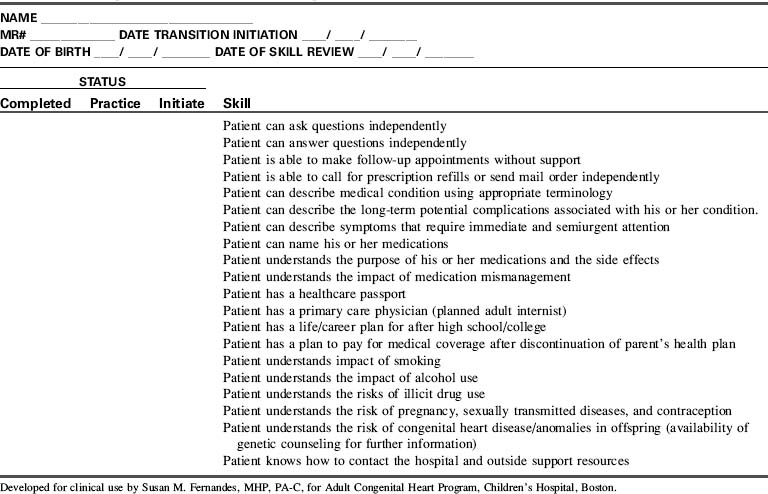
Although the optimal approach to provide transitioning (self-care management) education to adult patients with congenital heart disease has not yet been established, experts in the field proposed a life span model that focused on developmental issues faced by these patients. The areas of concentration included physical development, social and family relations, emotional health, medical issues, health behaviors, screening and prevention, and treatment issues. Using this information, many centers have developed checklists covering several of these areas to ensure that the patient has acquired the necessary skills before transfer of care. An example of such a checklist is provided in Table 8-20. In addition, either electronic or paper complete medical healthcare passports should be carried by patients with details of current and past history specifics, as well as contact information for immediate access to their ACHD healthcare providers.926
Patients who have undergone early childhood total surgical correction for patent ductus arteriosus, atrial septal defect, small ventricular defect, and mild pulmonary stenosis typically have few if any hemodynamic residua and therefore require infrequent evaluation and treatment.215 Most other patients have some form of residua or sequelae of their disease or the treatment and will require lifelong care by ACHD specialists.926
Most patients with ACHD are palliated, not “repaired” or “fixed,” and they require lifelong care.196 Complications of the defect or treatment can include arrhythmias, thrombosis, sudden death, ventricular or valvular dysfunction, pulmonary hypertension, and endocarditis.946 Additional problems can result from residual shunts, valvar disease, ventricular dysfunction, and arterial pathology. These issues may require further surgical or cardiac catheter-based interventions.215 Approximately 50% of adult patients with CHD face additional surgery, arrhythmias, complications, heart failure or, if inappropriately managed, premature death.301,304,305 Late complications increase in frequency over time, typically developing in the second, third, or fourth decade of life; they can be irreversible and potentially fatal.
Occasionally, a congenital heart defect is not diagnosed until adult years; this may be the result of widespread availability of echocardiography.510 A new diagnosis in the adult can include simple lesions, such as an atrial septal defect or a bicuspid aortic valve, to complex lesions, such as L-transposition of the great arteries diagnosed at the age 60 to 70 years,331 with the new development of symptoms, cardiac rhythm disturbances, or congestive heart failure. In some lesions, late diagnosis is complicated by the presence of pulmonary hypertension.
Cardiac catheterization based interventional procedures can be used to treat a variety of adult lesions. Interventions can include balloon dilation, stent placement, occlusion by devices, valve replacement (pulmonary or aortic), valve intervention, and therapeutic arrhythmia procedures (see Cardiac Catheterization).
ACHD survivors require appropriate testing and follow-up for likely residuae and sequelae. Echocardiograms are used but may not be optimal because of poor echocardiographic “windows” (sites to enable visualization of specific cardiac structures). Cardiac MRI (magnetic resonance imaging) is an increasingly important tool for the complete evaluation of complex congenital anatomy and surgical interventions in the adolescent and adult.197 The MRI studies can identify and follow important residual problems, and enable calculation of chamber volumes, quantify valve regurgitation, and enable evaluation of ventricular function, baffle leaks, patency of Fontan pathways, aortic arch status, residual lesions, and flow characteristics. (For additional information, see the MRI section at the end of this chapter.).197,215,314 Cardiac catheterization is used most frequently for interventions.215
Anticipatory management and ongoing care are critically important and must take place in a center with expertise in complex ACHD care. These patients require expert care for the complications associated with congenital heart interventions and aging. The goal is to monitor for and detect sequelae and provide timely treatment with regular follow-up care. Required management can include simple to complex imaging, cardiac catheter interventions, medications, cardiac pacing or defibrillator implantation, surgery, or cardiac transplantation. Development or progression of symptoms to report is summarized in Box 8-14.215
Box 8-14 Adult Congenital Heart Disease: Symptoms to Report215
If cyanotic, also report hemoptysis, joint pain, headache, epistaxis, or myalgia.
Lesion-specific guidelines and recommendations for the management of adults with congenital heart disease were published in December, 2008, by the American College of Cardiology and the American Heart Association.927 Lesion-specific pathways have also been developed for testing and follow-up of congenital heart lesions in the adult; these are covered extensively by Daniels,215,216 Gatzoulis and Webb,304 and Deanfield et al,227 It is important to note that each patient requires a unique plan for lifelong management; patients with identical original defects may have vastly different presentations in adult years. For example, patients with coarctation may have different ages at intervention, unique forms of surgery or catheterization intervention, specific associated lesions, and unique aortic arch anatomy.215 Each patient also has unique risk factors for acquired coronary artery disease, such as potential obesity, tobacco use, hypertension, diabetes mellitus, hyperlipidemia, or family history of coronary artery disease.215 Many patients do not report the symptoms that they are experiencing that might suggest an increased risk of heart failure and arrhythmias.215 Protocols for routine diagnostic testing can monitor for the development of known lesion-specific significant long-term complications and may enable prevention of serious, potentially irreversible disability.
Heart failure can be right- or left-sided, and related to systolic or diastolic dysfunction or pulmonary hypertension. Structurally significant lesions (whether unoperated or residual lesions) may be treated in selected cases by cardiac catheter interventional procedure or, when applicable, surgery.510 Ventricular dysfunction and heart failure are treated primarily with drug therapy, and treatment of pulmonary hypertension as needed (see section, Pulmonary Hypertension).
The details of a patient's surgical history is important, given the wide variation of surgical technique and the clinical implications. As an example, a Fontan procedure may have been performed with a right atrium to pulmonary artery anastomosis, a lateral tunnel, an extracardiac conduit, or a modification of any one of those. Even less complex lesions such as a coarctation repair can have numerous variations including an end-to-end anastomosis, an interposition graft or a left subclavian flap plasty, which interrupts normal arterial flow to the arm. This along with original versions of the Blalock Taussig shunt may lead to future inaccurate upper extremity blood pressures, which could have significant implications if not noted.
Arrhythmias are the most common cause of sudden cardiac death in ACHD,352 and affect up to 50% of adults with congenital heart disease.946 Patients with uncorrected and corrected congenital heart disease may present with arrhythmias. For example, patients with uncorrected atrial septal defect may develop atrial fibrillation or flutter. Ninety percent of patients with late sudden cardiac death related to arrhythmias have tetralogy of Fallot, D-transposition of the great arteries, coarctation of the aorta, or aortic stenosis.352 Antiarrhythmic therapy can be used to treat specific rhythms, but proarrhythmic effects must be monitored. Therapies are detailed by Harris.352 Treatments can include medications, cardiac pacing, implantation of cardiac defibrillators, or surgical interventions such as the Maze procedure46 in the management of patients' status post right atria to pulmonary artery Fontan procedure with subsequent development of atrial re-entry tachycardia or atrial fibrillation.864 The Maze procedure or Cox-Maze III procedure involves cryoablation (−160 °C) as well as surgical atrial incisions of the abnormal conduction pathways during open heart surgery.864
Cyanosis can affect patients with uncorrected cyanotic lesions, those with uncorrected shunt lesions and Eisenmenger syndrome, those with cyanotic heart defects and palliative procedures (e.g., systemic artery to pulmonary artery shunts, systemic vein to pulmonary artery shunts, central aorta to pulmonary artery shunts [e.g., Waterston, Potts shunt]), Fontan-type procedures with fenestrations, or complex surgical repairs where atrial fenestration remains.
Polycythemia with hematocrit rising to 60% or above may produce symptoms of headache, dizziness, dyspnea, fatigue, and neurologic changes.215 Dehydration must be avoided because of hyperviscosity and risk for stroke or emboli. All intravenous systems require meticulous removal of any air or clots because of risk of paradoxic emboli to the brain, kidney, or heart.215 Thrombocytopenia and coagulopathies are common (see Hypoxemia Caused by Congenital Heart Disease).
Phlebotomy is not routinely performed unless the patient becomes symptomatic. An equal volume of whole blood may be removed and replaced with saline or albumin. Potential complications of phlebotomy include stroke, seizures, and death.864
If the cyanosis is associated with pulmonary hypertension (e.g., Eisenmenger syndrome), the patient requires pulmonary hypertensive care precautions. In addition, medical management is required (see the following).
Cyanotic adults may also develop hyperuricemia, gouty arthritis, and altered renal function requiring treatment with allopurinol or colchicines.864 Hypoperfusion and chronic hypoxia also lead to renal disease.215
Pulmonary hypertension can develop as a consequence of congenital heart disease in 15% to 30% of patients,511 those repaired at a later age, or those with significant unrepaired left-to-right shunts.215 Patients with single ventricle with Glenn and Fontan palliations or staged correction require the lowest possible pulmonary resistance for survival.511 Evaluation of the status of the pulmonary vascular bed may include cardiac catheterization with measurement of the reactivity to pulmonary vasodilator therapy (see Common Diagnostic Tests, Cardiac Catheterization).
Patients may demonstrate hypoxemia as a result of pulmonary hypertension and progression to Eisenmenger syndrome with reversal of the intracardiac shunt. This pulmonary hypertension is typically unresponsive to oxygen.926 Pulmonary vasodilator treatments include oxygen therapy to treat hypoxemia (particularly at night),215 inhaled nitric oxide or epoprostenol,511 prostacyclines, endothelin receptor antagonists (Bosentan),912 and phosphodiesterase five inhibitors (Sildenafil).511
Pulmonary hypertensive precautions are vital during periods of illness or procedures requiring anesthesia because these situations include risk for conditions such as acidosis, alveolar hypoxia, pain, agitation, and hypoventilation that may provoke pulmonary vasoconstriction. Goals of care will include lowering PAP, decreasing PVR and RV afterload while maximizing RV function, ultimately improving cardiac output. Nursing care will focus on optimizing ventilation, improving RV function, providing adequate sedation, treating pain, avoiding metabolic and respiratory acidosis, avoiding atelectasis, avoiding anemia, and minimizing energy expenditure needs.511 In the immediate cardiac surgery postoperative period, reactivity of the pulmonary vascular bed is heightened.511 Support of systemic blood pressure may be required with use of agents such as norepinephrine to maintain coronary perfusion.511
Adequate hydration is essential, and hypotension must be avoided.215 Strategies for care attempt to decrease pulmonary artery pressure, enhance right ventricular function, and avoid development of pulmonary hypertensive events or crises with subsequent right ventricular failure (see Sectons “Pulmonary Hypertension” and “Management”).
Selected Eisenmenger syndrome patients may require phlebotomy because of symptomatic hyperviscosity, with specific guidelines to avoid acute adverse reactions and even death. For further information, see Daniels215 and Warnes et al.926
Extracardiac conduits and baffles can develop obstruction over time, requiring reintervention. The conduits connect the subpulmonary ventricle to the pulmonary arteries in tetralogy of Fallot and other complex congenital heart disease.398 Treatments include cardiac catheterization intervention (balloon angioplasty or stent implantation).
Conduit replacement is possible with early low mortality; of patients reoperated at a mean age of 9.6 years, 55% were free from conduit reoperation at 10 years, and 31.9% at 20 years.228 Developments in percutaneous pulmonary valve replacement have resulted in avoiding surgical revisions in many of the cases treated550 and are FDA approved since 2009.598 Patients with conduits require lifelong periodic reevaluation526 in adult CHD clinics.215,926
Additional impacts on cardiac health related to adult lifestyle and hereditary issues include smoking, obesity, hypertension, hypercholesterolemia, and coronary artery disease. Healthy adults (18 to 65 years old) should ideally complete at least 30 minutes of regular physical activity per day, 5 days a week,354 yet many adult patients with congenital heart disease are uncertain of their specific goals and limits, requiring additional assessment from their cardiology team and education about the benefits of exercise.246 (See http://www.heart.org/HEARTORG/Conditions/CongenitalHeartDefects/CareTreatmentfor CongenitalHeartDefects/Congenital-Heart-Defects-and-Physical-Activity_UCM_307738_Article.jsp). Aerobic functional capacity compared with healthy adults has been found to be diminished in patients with congenital heart disease, and significantly lower than normal.280 Typical low-intensity exercises such as walking, casual swimming, and dancing are encouraged. Individualized instructions are recommended (see http://www.americanheart.org/presenter.jhtml?identifier=11081).
Atherosclerotic heart disease is a major cause of morbidity and mortality in adults in the United States and many countries.369 Atherosclerotic heart disease begins to develop in early childhood, even in the patient with congenital heart disease,578 and guidelines for primary prevention in children have been identified.369
Lifestyle choices and modifiable risk factors can have a major impact on morbidity and mortality of patients with congenital heart disease. Minimizing cardiovascular risk factors, and following a healthy lifestyle can minimize the risks of complications related to coronary artery disease. Strategies include management of diet, regular exercise, and avoiding cigarette smoking.186 Obesity is a common finding in children with congenital heart disease.703 A known independent risk factor for type 2 diabetes and heart disease is obesity,683 which should be avoided. (See http://www.heart.org/HEARTORG/Conditions/CongenitalHeartDefects/CareTreatmentforCongenitalHeartDefects/Recommendations-for-Heart-Health_UCM_307739_Article.jsp).
As patients age following surgical intervention for congenital heart disease, information is accumulating regarding long-term outcomes.
Bicuspid aortic valve is the most common congenital lesion, present in about 1% to 2% of the population.215,926 The lesion can progress over time, requiring patients to be monitored for potential development of aortic valve calcification, regurgitation, and stenosis. Aortic root dilation and dissection may also develop.926
When indicated, therapeutic interventions may include interventional catheterization balloon valvuloplasty, or surgery for aortic valve intervention or replacement, or a Ross procedure (see section, Specific Diseases, Aortic Stenosis).
Coarctation of the aorta may be diagnosed late in life, and the patient may present with upper extremity hypertension with decreased lower extremity pulses and blood pressures. Late complications of repair include recoarctation, aortic aneurysm or dissection, and sudden death. Systemic hypertension has been reported in up to 70% of patients after coarctation repair,427 and must be differentiated from hypertension caused by recoarctation through use of physical exam (arm and leg blood pressure gradients) and diagnostic studies. Systemic hypertension may occur during rest or activity, and may require medical therapy.215,926 Blood pressures may be decreased in the left arm if a left subclavian artery flap was used in the original repair. The cause of the high risk of hypertension and aortic dissection is unknown.215
Recoarctation incidence is 8% to 54% and in selected cases may be treated with transcatheter therapy (stent, angioplasty).215 An aortic aneurysm may form in the left subclavian flap or patch aortoplasty repair site.197 Bicuspid aortic valve is present in about half of patients with coarctation of the aorta.527a These bicuspid valves can develop significant stenosis or regurgitation.196
The adult with an undiagnosed atrial septal defect may present with a cerebrovascular accident or transient ischemic attack following a paradoxic embolism across the atrial septal defect.215 Factors leading to arrhythmia development include chronic right heart volume overload, ventricular dysfunction,926 late date at operation, or the presence of pulmonary hypertension.352 About 50% of those patients with preoperative atrial arrhythmias have postoperative arrhythmias, particularly if they are 40 years old or older at the time of surgery. The presence of atrial arrhythmias can result in need for anticoagulation.352 Transcatheter device closure is possible for many with secundum type lesions (see Specific Defects, Atrial Septal Defect).
Adult patients presenting with sinus venosus or primum defects require surgical repair.802 Pulmonary hypertension may be present in the unrepaired adult. Atrial septal defects repaired early in life are typically symptom free, but atrial arrhythmias (fibrillation or flutter) may develop in the operated608,926 and the unoperated patient.352,926
Tetralogy of Fallot is the most common complex defect with the longest survival history.946 Most adult patients have undergone surgical repair in childhood or even adolescence,352 and some have undergone initial palliative shunt procedure. Some patients may have a right ventriculotomy with a patch or a valved conduit between the right ventricle and pulmonary artery, whereas others have surgical removal of the pulmonary valve with a transannular patch and resultant free pulmonary regurgitation.
If the pulmonary valve is present, it may become insufficient or stenotic and the distal pulmonary arteries obstructed. Postoperative patients may develop arrhythmias. Atrial arrhythmias (flutter, fibrillation or supraventricular tachycardia) develop in up to one third of patients.743 Ventricular tachycardia and sudden cardiac death are known complications.215,926 By 35 years after surgery, the estimated risk for sustained ventricular tachycardia is 11.9%, and for sudden death is 8.3%.302
RV size correlates with QRS duration. The combination of older age at correction and QRS duration of 180 ms or longer, as well as older age at initial repair352 predict risk of sustained ventricular arrhythmias and sudden cardiac death.303 Right bundle branch block (RBBB) is a common finding in patients who had surgical correction many years ago, but in itself is not predictive of a worse prognosis (although RBBB with QRS 180 ms or higher is).352
The most common problem following surgical correction of tetralogy of Fallot is pulmonary regurgitation, which causes chronic right ventricular volume overload and may progress to right ventricular enlargement and systolic dysfunction.215,926 Severe right ventricular dilation and mild global right ventricular systolic dysfunction are associated with higher risk for adverse clinical outcomes in patients with repaired tetralogy of Fallot.482 Progressive aortic root dilation and potential for aortic dissection may also develop.215 Pulmonary regurgitation is the most common reason for late reoperation.946
Tetralogy of Fallot survivors require lifelong monitoring, including regular evaluation with MRI,482,926 for each of these potential complications and to avoid progression of decreased cardiac function. Surveillance for potential arrhythmias is required. Treatment for arrhythmias includes cardiac pacing, use of implantable cardioverter/defibrillators, antiarrhythmic therapy or radiofrequency ablation, or arrhythmia intervention during reoperation352 for pulmonary valve replacement. Selected cases may undergo transcatheter pulmonary valve replacement via cardiac catheterization.598 Long-term survival at 32 years is 86%.641 Tetralogy of Fallot is the most common diagnosis for patients with implantable cardioverter defibrillators.467
Atrial switch for d-transposition of the great artery (Mustard or Senning procedure) requires extensive atrial incisions and suture lines.352 As a result, patients who undergo these procedures require lifelong regular monitoring215,926 for development of arrhythmias (atrial tachycardia, sick sinus syndrome) and are at risk for sudden cardiac death. Loss of sinus rhythm is common, with only 18%950 to 40%310 of survivors in sinus rhythm 15 to 20 years after surgery. Pacemaker implantation is anticipated in about one-fifth of adults with long-term followup after a Mustard procedure.352
Pacemakers can be used to treat bradyarrhythmias or tachyarrhythmias,352 and radiofrequency ablation or medications can be used to treat tachyarrhythmias.215 Despite close followup, about 7% of patients have sudden cardiac death after atrial switch.950
After an atrial switch procedure for d-transposition of the great arteries, the right ventricle is the systemic ventricle (pump) for life and progressive dysfunction develops by the second or third decade of life in about 15% of postoperative patients,950 with progression to ventricular arrhythmias in the failing ventricle.352 The tricuspid valve, exposed to systemic pressures, can become dysfunctional. Narrowing in the left ventricular outflow area can also develop.
Reconstruction of the atrial flow pathways can rarely result in the serious complication of baffle obstruction of pulmonic or systemic venous return.215 Baffle leaks causing atrial shunting are common but they are often small.215 If despite optimal medical management the ventricular dysfunction becomes severe, either staged surgical anatomic correction (arterial switch procedure) or cardiac transplant may be planned. Patients with d-TGA, VSD, and pulmonary stenosis repair will have undergone a Rastelli procedure with a conduit between the right ventricle and pulmonary artery, that will require continued surveillance.
Arterial switch became the treatment of choice for transposition of the great arteries in the 1980s. Follow-up shows good systemic ventricular function and sinus rhythm,946 although the patients having additional VSD repair are reported at higher risk for arrhythmias.359 Potential long-term issues include the need for serial assessment of ventricular function and surveillance for the development of arrhythmias, stenosis at the great vessel suture sites,946 neo-aortic root dilation, and development of valvar regurgitation.186
Coronary artery lesions, found in 5% of patients following the arterial switch operation, are progressive and can be treated by coronary angioplasty or surgery.719 Coronary atherosclerosis remains a concern and requires careful followup.692 The progression of coronary artery disease after the arterial switch procedure is unknown. The most common cause for reoperation is pulmonic stenosis.547 Survival at 15 years after the initial operation is 88%,547 but long-term outcome is unknown.186,215,926
Although it is a rare condition, patients with a single ventricle account for a disproportionate share of the morbidity and mortality found in adults with congenital heart disease.946 Single ventricle includes diagnoses of tricuspid atresia, mitral atresia, double inlet LV, hypoplastic right or left ventricle, or single ventricle. These patients will remain cyanotic until they undergo Fontan-type correction. The first patients with a Fontan-type staged palliation and correction are now in their fourth decade of followup.466 Young adults with a univentricular heart who are unrepaired have a poor prognosis.215
The original Fontan procedures (1970s) connected the right atrium directly to the pulmonary arteries, or connected the right atrium to the right ventricle, exposing the right atrium to high systemic venous pressure.352 Over time elevated right atrial pressure can lead to right atrial enlargement, hypertrophy, and slowed atrial conduction.352 A severely enlarged right atrium is often seen in patients with a classic Fontan procedure and a failing Fontan circuit, which contributes to development of medically resistant complex atrial arrhythmias.926 Atrial distension and surgical incisions with subsequent scarring also contribute to arrhythmia formation.352 Atrial tachycardia and sick sinus syndrome may be observed, particularly in those patients with the right atrium included within the Fontan pathway.352 Atrial tachycardia can develop in up to 50% of adults with Fontan procedure,226,926 and sinus bradycardia or junctional escape rhythm has been reported in up to 15% of patients.352 Sustained atrial arrhythmias may cause patients to present with congestive heart failure, low cardiac output, and can lead to development of atrial thrombi.352 Anticoagulation therapy is often required in the presence of sustained atrial arrhythmias.352 Therapy for arrhythmias can include antiarrhythmic therapy, pacemaker (typically epicardial if ventricular), radiofrequency ablation, or arrhythmia intervention during reoperation.352
Many long-term complications have been reported following single ventricle palliations. Outcomes have improved, but systemic ventricle dysfunction may develop, particularly with a right ventricular systemic pump.311 The less efficient right atrium to pulmonary artery Fontan type surgical procedure has become obsolete,311 replaced by the total cavopulmonary artery connections (superior vena cava to pulmonary artery shunt plus intraatrial lateral tunnel or extracardiac conduit).467 The use of external conduits from the inferior vena cava to the pulmonary artery results in a sutureless right atrium, and potentially may decrease incidence of postoperative atrial arrhythmias.311 Some patients may have a lateral tunnel connecting the inferior vena cava to an intraatrial tunnel, then the pulmonary arteries. Long-term data are not yet available to define long-term outcomes of these two procedures.311
Pulmonary vein compression, pulmonary artery stenosis, Fontan pathway obstruction, atrioventricular valve insufficiency, arrhythmias, hepatic dysfunction, hepatic fibrosis, and cirrhosis may each develop after the Fontan procedure.47,215,926 Protein losing enteropathy is reported in 3.7% of patients following Fontan-type correction and is associated with a poor clinical course,311 with a 50% 5-year mortality after the diagnosis.612,926
The function of the single systemic ventricle and atrioventricular valve can deteriorate, and must be monitored closely, particularly in those patients with a right ventricle performing a lifetime of systemic work. Heart failure risk in Fontan patients is reported to be higher in those patients with the (single) morphologic right ventricle functioning as the systemic ventricle.944 Currently, many patients with the Fontan operation have hypoplastic left heart syndrome, with a resultant systemic right ventricle, creating concerns regarding long-term right ventricle status.311
Arterial oxygen saturations will be monitored for the potential late development of cyanosis caused by fenestration of the Fontan or the development of pulmonary arteriovenous malformations (AVM), systemic venous collaterals, or baffle leaks.215 Patients developing cyanosis215 or residual stenosis may require diagnostic or therapeutic cardiac catheterization. Cardiac magnetic resonance imaging (MRI) cannot be completed if the patient has a cardiac pacemaker.
The failing Fontan circuit may require surgical revision with conversion to an extracardiac Fontan and a form of the Maze procedure for treatment of intractable atrial tachycardia.926 Recent reported perioperative mortality is 1%,46,226 and late mortality is 5%.46
Survival at 25 years after Fontan-type correction has been reported at 70%.467 The most frequent causes of late death in Fontan patients are sudden death (9.2%), thromboembolism (7.9%), and heart failure (6.7%), with an arrhythmic origin presumed for the sudden deaths.467 Absence of aspirin or warfarin therapy is a predictor of death caused by thromboembolic event.467 The risk for thromboembolic death increases sharply 15 years after Fontan surgery.467 All single ventricle patients require continuous lifelong care in a center with expertise in ACHD care. In the presence of single ventricle dysfunction or protein-losing enteropathy, a heart transplant may be beneficial.926 More information is needed regarding long-term outcome of these patients.
Women with congenital heart disease appear to have similar sexual activity and fertility rates as their healthy peers.137,245,609,729 Pregnancy planning in this patient population is required to reduce maternal and/or fetal morbidity. Yet, few patients seek advice regarding birth control and family planning.428,738,835 It is imperative, therefore, that the healthcare team caring for these patients maximize opportunities to discuss pregnancy prevention and planning. Detailed information regarding pregnancy, contraception, and delivery for patients with congenital heart disease is included in the Chapter 8 Supplement, section, Adults with Congenital Heart Disease, on the Evolve Website.
Medical and surgical advances have made survival into adulthood an expectation for patients with congenital heart disease. With these advances have come an influx of women with moderate and complex congenital heart disease who desire pregnancy. Although pregnancy and delivery are associated with significant hemodynamic changes such as increased blood volume, increased stroke volume, increased cardiac output, and decreased systemic vascular resistance, most women with repaired congenital heart disease appear to tolerate pregnancy and delivery with only minimal risk.809
The majority of women with congenital heart disease tolerate pregnancy and delivery without significant risk, although there are many obstetric, cardiac, anesthesia, genetic, and psychosocial factors to consider. In patients with more than simple disease, a multidisciplinary approach in a regional center is essential to optimize maternal and fetal outcomes.
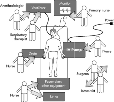
Perioperative care for the patient with ACHD for minor (elective noncardiac procedures) and major procedures, as well as emergency admissions426 requires comprehensive medical, surgical, and nursing management. The team must be knowledgeable about the cardiac anesthesia requirements, the potentially complex hemodynamics, any residual structural abnormalities, anticoagulation needs, and physiologic abnormalities related to each congenital heart lesion. Adults with important co-morbidities such as coronary artery disease or renal failure may require postprocedure recovery in an adult cardiac critical care unit with the consultation of the required adult subspecialists. Adults without significant comorbidities may be treated in a section of the pediatric cardiac critical care unit. Communication among the congenital surgical, interventional cardiology, and adult medical subspecialists is essential to provide optimal care, involving nursing education regarding the defect pathophysiology, hemodynamics, surgical intervention, and likely postoperative complications. The healthcare team must continue to monitor for and document long-term outcomes.
Cardiac catheterization may be required in adults with CHD for hemodynamic assessments. Interventions are commonly performed for complex pulmonary artery stenosis, baffle stenosis, or residual aortic narrowing. Shunting defects, including fenestrations, may be sealed with devices (see Common Diagnostic Tests, Cardiac Catheterization). Pulmonary valves and aortic valves are now replaced by transcatheter interventions in selected cases. Pediatric electrophysiology studies to detect and treat atrial or ventricular arrhythmias may include implantation of a cardiac pacemaker with possible cardioverter defibrillator.388
Comprehensive guidelines are available for the perioperative assessment and management of patients with adult congenital heart disease undergoing noncardiac surgery.196 When cardiovascular surgery is required, reoperations are the most common type of surgery needed.802 High-risk patients require close postoperative surveillance in a critical care or coronary care unit, particularly patients with cyanosis, pulmonary hypertension, ventricular dysfunction, or single ventricle.196
The most common reported complication in the postoperative cardiac surgery adult (occurring in 10.8% of patients) is atrial arrhythmias.1 The postoperative team must carefully monitor the patient's volume status, hemodynamics, and electrolyte balance, and be observant for evidence of hypoxemic pulmonary hypertensive episodes or crisis and be aware of all drug therapies required. Each cardiac lesion carries risks of particular rhythm disturbances, and unexpected arrhythmias may occur at any time.
Conduction anomalies are common in adults with uncorrected cyanotic CHD,802 including ventricular or supraventricular ectopy. Perioperative development of arrhythmias can lead to sudden, severe low cardiac output. Immediate evaluation and prompt treatment are required.802 Monitoring for development of cardiac ischemia includes monitoring for ST segment changes and ventricular ectopy. Atrial and ventricular pacing wires can assist in management (see Arrhythmia Detection and Management).
Bleeding is a potential risk following reoperation, particularly in the patient with longstanding cyanosis.802 Reoperation requires incisions through vascular scar tissue and chronic hypoxemia is associated with thrombocytopenia and thrombocytopathia. In addition, patients with chronic hypoxemia may demonstrate a decrease in vitamin K-dependent clotting factors. For these reasons, bleeding should be anticipated and plenty of blood should be available for postoperative blood replacement.
Cardiac reserve may be limited in many cyanotic patients, and excessive blood loss can rapidly cause hemodynamic instability. Target hemoglobin values and plans for coagulation factor replacement are individualized based on the specific congenital lesion, operative interventions, and associated risk factors. A patient with expected residual cyanosis cannot tolerate anemia, because it decreases oxygen-carrying capacity and reduces oxygen content and likely oxygen delivery.
The risk of perioperative low cardiac output is increased in the presence of chronic hypoxemia, ventricular hypertrophy, chronic volume or pressure overload, ventricular dysfunction, and arrhythmias. In the postoperative period, reactivity of the pulmonary vascular bed is heightened,511 so pulmonary hypertension precautions are vital (see section, Pulmonary Hypertension).
Pulmonary hemorrhage may develop with interventions that increase pulmonary blood flow, particularly after interventional cardiac catheterization. Mechanical ventilation and pulmonary support are required. Development of pneumonia, infection, fever, thrombosis, or pulmonary edema can seriously destabilize the patient with ACHD, even after a minor procedure. Supplementary oxygen can help to decrease the pulmonary vascular resistance, although if cyanotic heart disease is present it may not increase the arterial oxygen saturation.196 Parameters for acceptable and expected oxygen saturation levels and PaO2 are required.
Smoking can compromise pulmonary function preoperatively and postoperatively. Encourage pulmonary toilet hourly while awake with incentive spirometry, and encourage the patient to get out of bed and walk or at least sit in a chair as soon as possible. Intermittent pneumatic compression or the use of antiembolism stockings should be used to reduce the risk of deep vein thrombosis (DVT).196
Risk of DVT is increased with prolonged bedrest, neuromuscular blockade, and limited range of motion. Early ambulation is optimal. Monitor for signs of DVT, including redness or pain in the leg calves. Signs of pulmonary embolus include sudden dyspnea, tachypnea, increased oxygen requirement, and rales. An arterial blood gas will not establish the diagnosis. Massive pulmonary embolism can result in acute right ventricular failure, low cardiac output, and myocardial ischemia with ST segment changes. Diagnosis is confirmed by lung perfusion scan or pulmonary angiogram. Treatment includes immediate, heparinization, and hemodynamic supportive care. In acute situations, fibrinolytics (recombinant tissue plasminogen activator or rTPA) are administered. Cardiopulmonary bypass may be employed to remove the embolus (pulmonary embolectomy).
Renal dysfunction is a known risk in patients with chronical cyanotic hypoxemia.802 Cardiopulmonary bypass can further exacerbate renal dysfunction. Perioperative care includes maintaining optimal cardiac output with careful fluid balance and support of systemic perfusion. Volume replacement requires normal saline infusions typically of 500 cc or more (10 cc/kg), administered as a bolus to optimize cardiac filling pressures. Infusion by syringe injections (often used for pediatric patients) does not allow administration of sufficiently large volumes when resuscitation is required.
Relative hypovolemia can result from rewarming and vasodilation, fever, increased capillary permeability, and use of vasodilators. Signs can include sinus tachycardia, decreased central venous or intracardiac pressures, and signs of decreased perfusion.
Standard medication doses for adults are used, including doses for resuscitation drugs, antiarrhythmic therapy, and antibiotics. Intravenous drug infusions in the adult can be specifically concentrated by the clinical pharmacist to avoid excessive fluid volume administration. Doses must be titrated individually with consideration of any renal or hepatic dysfunction.
Cyanotic patients are at greater risk of postural hypotension because of a greater right-to-left shunt.196 Changes in movement should be gradual.
Effective pain management is essential to minimize catecholamine surges.196 The patient with ACHD may have sensitization to and greater fear of painful procedures because of a previous history of inadequately managed pain during prior surgeries or hospitalization.707 Discussion of plans for pain control and patient-controlled analgesia help to control potential anxiety. Patients with preoperative history of illegal drug use or preoperative prescription narcotics typically require higher doses of analgesia postoperatively. Inadequate analgesia can lead to decreased chest physiotherapy and ambulation.
Many adult survivors of congenital heart disease have unique alterations in neurologic status related to the presence of genetic syndromes (Down, DiGeorge, Williams) or previous neurologic alterations leading to developmental delays and increased dependence on family involvement in care. Rest and privacy are essential. The adult's bedtime ritual can be assessed. Some may use prescriptions, rituals, or over-the-counter medications to promote sleep.
Unique developmental needs and degree of family involvement may be present in the patient with ACHD. Some may rely heavily on a parent and/or significant other for making decisions and daily activities. Survivors require guidance in terms of educational and vocation choices. Some of the survivors of complex congenital heart disease management are at risk for inattention, hyperactivity, and developmental disabilities, and require remedial school services.798
Issues with independence and interdependence may exist, as well as other psychological challenges found in patients with chronic illness, as detailed in Claessens et al180 and Tong et al.867 Providers should assess the patient's knowledge and independence in areas of self-care.
The transitioning process from pediatric to adult-focused CHD care is life-altering for patients, parents, and staff. The act of transfer may be a substantial source of stress for the patient, parent, and longtime caregiver. Therefore all involved parties should be involved in the transitioning process. The concept of transitioning should be discussed early, around age 12 if the patient is emotionally and intellectually ready, and should continue until the patient demonstrates the ability to take responsibility for self-care. It is essential to provide adequate resources for support of the family at this time, including both in-hospital resources and outside patient support resources. The Adult Congenital Heart Association (ACHA) and American College of Cardiology developed a “Passport” document to allow patients to concisely identify their specific cardiac health needs and endocarditis prophylaxis requirements. The healthcare team can provide lesion specific identification of patient conditions, therapies, and the suggested guidelines for ACHD care. A clinic resource list now identifies ACHD clinics nationwide, and is available on the ACHA web site (www.achaheart.org).
Education with respect to employment, insurance needs, and social service resources are also required by many. Continued monitoring and instruction regarding management of noncardiac surgical procedure care and required collaboration with the adult CHD team are essential to optimize care and safety, and minimize morbidity and mortality risks.
Ultimate goals are to optimize ACHD survival and maximize quality of life. It is clear that all patients with congenital heart disease require lifelong collaborative care from pediatric and adult specialists who will provide ongoing screening for known complications and potentially unexpected sequelae of the defect and therapies. These complex patients face potential long-term and possible life-threatening complications. Transition from the pediatric into the ACHD clinic is a vital priority.
• Adult Congenital Heart Association (ACHA): www.achaheart.org
“Passports” for CHD, extensive patient education materials, pamphlets available
• American Heart Association: http://www.heart.org/HEARTORG
• International Society of Adult Congenital Cardiac Disease (ISAACD): http://www.isaccd.org/
Mary Rummell, Patricia Ann E. Schlosser, Sandra Staveski, Winnie Yung
This section provides an overview of postoperative care of the child after cardiovascular surgery. For information about common problems such as congestive heart failure or pulmonary hypertension, please refer to the information in the previous major section of this chapter, Common Clinical Problems. For specific postoperative complications associated with each congenital heart defect, please refer to the information about the defects in the section Specific Diseases.
The transfer (handoff) of patients from the surgical team to the critical care team can result in technical errors (i.e., delivery of inappropriate infusions, incorrect ventilator settings) and errors of omission. In the past several years, the development of standard handoff procedures and checklists, some of which were learned from consultation with managers of Formula One racing teams and aviation models, have been shown to reduce such errors.146,147
Before the patient arrives in the critical care unit, (PCCU), all equipment necessary for the child's care should be set up in working order with appropriate alarm limits established. Whenever possible, the equipment should be transferred with the patient, to avoid the need to change intravenous pumps or monitoring equipment. PCCU personnel should document errors associated with the transfer and standardize this transfer to reduce such errors.
To improve efficiency of patient admission to the critical care unit, each member of the receiving team should be in an assigned position with specific assigned responsibilities (Fig. 8-29). Patient transfers should include a transfer checklist or form, equipment and technology transfer, information transmission, and finally, discussion and plan for the patient.146
Fig. 8-29 Organization of team for transfer of care from surgical to postoperative critical care team.
(Based on Catchpole KR, de Leval MR, Mcewan A, et al: Patient handover from surgery to intensive care: using Formula 1 pit-stop and aviation models to improve safety and quality. Pediatr Anesth 17:470-478, 2007.)
Nursing management after cardiac surgery requires understanding of the child's preoperative and postoperative anatomy and physiology, the surgical intervention itself, intraoperative events, the effects of cardiopulmonary bypass (in many cases), the child's baseline condition, and any co-morbidities. Nurses are in a unique position to optimize care delivery and coordinate care for these complex children by virtue of their knowledge, skills, and extended presence at the bedside. The child's cardiovascular function requires close observation through physical examination, hemodynamic monitoring, and laboratory analysis.
Cardiac output is the amount of blood ejected from the systemic ventricle in one minute. Cardiac output is a product of heart rate and stroke volume. Stroke volume is affected by preload, afterload, and contractility. Cardiac output may be indexed to body surface area, reported as a cardiac index (CI); normal CI is 3.5 to 4.5 L/min per m2 body surface area.
At various stages during the life cycle, there are important developmental components to consider when caring for a patient after cardiac surgery. The infant and child are very dependent on an adequate heart rate to maintain effective cardiac output; bradycardia often is associated with a fall in cardiac output. Children can increase stroke volume in response to volume administration, but the response is limited, particularly during the neonatal period when diastolic ventricular compliance is low with less response to volume loading than in the older infant or adult. It is important to keep these and other developmental considerations in mind as you care for the child after cardiac surgical intervention.
Careful assessment of cardiac output is particularly important during the postoperative period. Adequate postoperative systemic perfusion requires a heart rate appropriate for the child's age and clinical condition, sufficient cardiac preload and intravascular volume, adequate myocardial function, and appropriate ventricular afterload (resistance to ventricular ejection). If cardiac preload or myocardial function is inadequate or if ventricular afterload is excessive, cardiac output may fall. Assessment of cardiac output is accomplished through monitoring heart rate, blood pressure, intracardiac pressures, peripheral perfusion, acid-base status, serum lactate, temperature, urine output, and mixed venous oxygen saturation.73 The moment the child returns from surgery the nurse should form an opinion of the child's systemic perfusion and cardiovascular function. The nurse also should be aware of the “filling” (central venous, atrial) pressures at which systemic perfusion is best and inform the interdisciplinary team of the findings.
Postoperative patients will have one or more invasive monitoring catheters depending on the defect, surgical intervention, and anticipated hemodynamic monitoring requirements. Invasive catheters are used for hemodynamic monitoring and blood sampling, as well as for administration of vasoactive infusions, nutrition, and volume expanders. On admission to the pediatric critical care unit (PCCU), the team should check the chest radiograph to verify appropriate placement of the catheters.
It is imperative that all monitoring catheters be zeroed and calibrated and transducers set at the phlebostatic reference point (level of the right atrium) or as established by unit protocol. Transducers placed at a level (relative to the right atrium) different from the level at which they were zeroed and calibrated will provide inaccurate measurements (see Chapter 21). Transthoracic and intracardiac catheters have been shown to be safe to use, but carry associated risks such as thrombus formation, malfunction, and infection.272
Left atrial catheters are typically used only for monitoring because administration of anything into these catheters may cause the potentially devastating complication of embolization of air, particulate matter, or thrombus into the cerebral or coronary circulation. It is important to note that children with mixing physiology (i.e., systemic venous blood entering the systemic arterial circulation) are at increased risk for embolic events from all intravenous catheters and therefore should have absolutely no air, thrombus, or particulate matter in their intravenous lines. For more information on hemodynamic monitoring, see Chapter 21.
The pressure in a ventricle at the end of diastole (i.e., the end-diastolic pressure) provides the preload to stretch of the child's ventricular fibers. It is affected by the child's intravascular volume as well as ventricular compliance. Inadequate intravascular volume can result from hemorrhage, vasodilatation during rewarming, third spacing of fluids related to systemic inflammatory response following cardiopulmonary bypass (CPB), aggressive diuresis, or inadequate fluid administration (Table 8-21).73
Table 8-21 Potential Causes of Elevated or Reduced Right or Left Atrial Pressure
| Invasive Catheter | Elevated Pressures | Reduced Pressures |
| Right atrial or central venous | Volume overload; decreased right ventricular function; tamponade; artifact; catheter malfunction | Hypovolemia; artifact |
| Left atrial | Volume overload; decreased left ventricular function; tamponade; arrhythmia; artifact | Hypovolemia; artifact; decreased left atrial pressure in combination with high RA and/or CVP can develop with PHTN |
Ventricular compliance (change in volume divided by change in pressure) is the distensibility of the ventricle; it is typically low immediately after cardiovascular surgery, particularly if a ventriculotomy incision was performed or the ventricle is hypertrophied and the surgery resulted in significant alteration of flow patterns.
Preload is evaluated through examination of “filling” pressures (atrial pressures and central venous pressure) and assessment of liver distension, peripheral and pulmonary edema, and fontanelle fullness. The optimal preload is determined at the bedside and varies from patient to patient and in the same patient over the course of postoperative care. A higher preload is likely to be required early in the postoperative period than as the heart recovers. The bedside nurse should note the “filling” pressure (CVP or right or left atrial pressure) at which systemic perfusion is best.
Preload is manipulated through volume administration and diuresis. Fluid boluses and volume resuscitation should be managed by using the appropriate volume expander based on the clinical situation. For example, hypovolemia not related to bleeding is managed by administration of isotonic crystalloid or colloid. By comparison, hypovolemia related to significant bleeding is managed by correction of abnormal coagulation factors and administration of packed red blood cells.
The child's circulating blood volume should be calculated before the child returns from surgery and all blood losses should be considered as a proportion of the child's circulating blood volume:
Please refer to Chapter 6 for fluid administration guidelines in the treatment of shock.
If the child's chest tube output averages 3 mL/kg per hour, these losses will total 10% to 15% of the child's circulating blood volume within 3 hours. If this blood loss is not replaced, significant cardiovascular compromise and shock can result. It is advisable to replace chest tube output once it totals 5% to 10% of the child's circulating blood volume. Replacement should occur as quickly as the output is draining; use atrial filling pressures and other hemodynamic monitoring pressures to guide your fluid management. Chest tube output of 3 to 5 mL/kg per hour constitutes significant hemorrhage, and the source of the bleeding must be identified. If excessive chest tube output continues despite normal clotting function the child typically requires re-exploration, which may occur in the operating room or at the bedside.
Impaired contractility or myocardial dysfunction may influence cardiac output. Drugs, anesthesia, hypoxia, acidosis, ischemia, electrolyte imbalance, extensive ventriculotomy, residual lesions, myocardial resection, and tamponade all affect contractility.73 Hypercapnia and alveolar hypoxia can increase pulmonary vascular resistance and can contribute to left ventricular dysfunction; therefore if they develop, mechanical ventilatory support generally must be instituted or adjusted. Note that if the child has a single functioning ventricle with a bidirectional Glenn, hemi-Fontan, or Fontan-type physiology, support of spontaneous ventilation is preferred to positive pressure ventilation, when possible. Mild hypercapnia may actually improve cardiac output in these patients.935
Acidosis should be treated promptly because it can depress ventricular contractility, leading to decreased perfusion, decreased systemic arterial pressure, and/or increased filling pressures. Treatment (except in tamponade) includes correction of metabolic abnormalities and, if necessary, use of inotropic and afterload reducing agents (see the following).
Afterload is impedance or resistance to ventricular ejection. Increased afterload can result from increased pulmonary or systemic vasoconstriction or residual outflow tract obstruction.
Systemic vasoconstriction may be a compensatory mechanism in a low cardiac output state or from administration of vasoconstrictors. Treatment of increased afterload involves avoidance of precipitating factors, intravenous nonspecific vasodilating agents (SVR and PVR), inhaled vasodilators (PVR), and mechanical ventilator manipulation to optimize cardiopulmonary interactions (e.g., intubation and supporting ventilation for reducing left ventricular afterload).
If systemic vascular resistance (SVR) is high or even normal in the presence of ventricular dysfunction, or the child demonstrates significant peripheral vasoconstriction with poor systemic perfusion, treatment with vasodilators usually is indicated. These agents are nonspecific and may cause dilatory effects in both the systemic and pulmonary beds. When intravenous vasodilators are administered, the child's fluid volume status must be adequate because hypotension may develop, especially during initiation of therapy.
Vasodilators can reduce ventricular preload and afterload because they produce venous and arterial dilation (see Chapter 6). During vasodilator therapy, assess the warmth of the child's extremities, capillary refill, serum lactate, urine output, and blood pressure to determine the effectiveness of therapy. If multiple agents are used, it is also important that the dose of only one medication be changed at any one time so that the patient's response to each change can be determined.
Pulmonary vasoconstriction can result from alveolar hypoxia, acidosis, hyperinflation of the lung, and hypothermia. In addition, cardiopulmonary bypass can affect vascular tone and permeability. Pain may also affect vascular tone.73 Elevated pulmonary vascular resistance (PVR) may be a developmental, acute, or chronic condition. For example, newborns have reactive pulmonary vascular bed and are susceptible elevated PVR. Additionally, some children have a genetic predisposition for pulmonary hypertension, such as children with trisomy 21.652 For further information, see Box 8-1 and Table 8-16.
The effects of cardiopulmonary (CPB), pulmonary leukosequestration, microemboli, and hypothermia have been implicated in pulmonary vascular endothelium dysfunction, and elevated PVR with increased pulmonary vasoreactivity in the postoperative period.73,206,667 Excessive pulmonary blood flow and left-to-right intracardiac shunts before and after CPB, elevated pulmonary venous pressure, lung pathology, blood products, and Protamine are several of the other risk factors associated with postoperative elevation in PVR.73,667,845 Post-CPB pulmonary endothelium dysfunction may be responsible for increased generation of pulmonary vasoconstricting mediators and decreased endogenous nitric oxide (NO) production associated with vasodilatation.71,73
An increase in the child's pulmonary vascular resistance also may be an acute cause of inadequate systemic perfusion postoperatively. An acute rise in PVR with resulting decreased cardiac output, acidosis, hypoxemia, and increased right ventricular (RV) afterload can worsen any preexisting RV dysfunction.4,7,73,206
Pulmonary hypertension (PHTN) is characterized by an elevation in pulmonary artery pressure and PVR. It is defined as a systolic pulmonary artery pressure (PAP) greater than 35 mm Hg or a mean PAP greater than 25 mm Hg at rest. Often in the clinical setting, the severity of PHTN is discussed in relation to systemic arterial pressures. Treatment for PHTN typically begins at approximately half systemic pressures. In the absence of a pulmonary artery catheter, it is possible to assess for effects of PVR by monitoring the systemic venous atrial or central venous filling pressures. A child experiencing PTHN will have a rising right atrial or central venous pressure, falling left atrial (or pulmonary venous atrial) pressure, and falling blood pressure.
PHTN can lead to RV failure, low cardiac output state, impaired oxygen delivery, and shock. The goals of care are to decrease PVR and maximize RV function, and optimize cardiac output.652 Support of right ventricular function and output is important. Milrinone, dopamine, or epinephrine may be used to support cardiac output. Optimizing right ventricular preload can improve cardiac output, because elevated right atrial pressures are required to maintain adequate ventricular filling.845
The child with perioperative pulmonary hypertension requires precise postoperative ventilator management and must remain well oxygenated and ventilated, as well as warm. Providers should make an effort to treat pain and agitation and minimize stimulation (see section, Common Clinical Conditions, Pulmonary Hypertension). Respiratory mechanisms to lower PVR include ensuring adequate gas exchange, maintaining functional residual capacity, and avoiding hypoinflation or hyperinflation. Alveolar hypoxia, such as may be produced by hypoventilation, pneumothorax, airway obstruction, suctioning, and atelectasis, must be avoided. A mild alkalosis will promote pulmonary vasodilation. These children usually require mechanical ventilator support for several days, and then gradual weaning is attempted. During weaning the child's pulmonary pressure (if available) and systemic perfusion should be monitored carefully because hypoventilation produces alveolar hypoxia and can result in pulmonary vasoconstriction.
These children may also require blunting of stress responses with opioid or benzodiazepines, and ventilation with sedation and neuromuscular blockade may be needed. There may be benefit to premedicating the child with an opioid and/or benzodiazepine before suctioning or noxious stimuli to blunt responses to the stimulus. Clustering care to prevent excessive stimulation may reduce the potential for PHTN crisis as well.
Prevention or prompt correction of acidosis is required with either respiratory or metabolic maneuvers to achieve a pH of approximately 7.5.652 Pulmonary vasodilatation with specific and nonspecific agents is frequently accomplished through administration of inhaled nitric oxide (specific) and/or milrinone (nonspecific). If inhaled nitric oxide is administered it should be weaned slowly to avoid rebound effect. Weaning from any PVR therapy should be accomplished in a stepwise, methodical fashion.845
Endotracheal suctioning is a well-known noxious stimulus that can provoke PHTN. Hyperventilation and hyperoxygenation have been shown to reduce suctioning induced hypoxia.669 The use of in-line suctioning catheters may be beneficial as well.442 If open suctioning is required, two nurses should perform the procedure. The first nurse provides hand or mechanical ventilation and maintains oxygenation. If inhaled nitric oxide is being used, ensure that there is delivery of the drug into the hand ventilation circuit. The second nurse provides skilled, gentle suctioning. Children with extremely labile pulmonary vascular beds should have a physician or advanced practice nurse present during suctioning. Please refer to Common Clinical Conditions, Pulmonary Hypertension for further information.
An abnormal heart rate or rhythm can compromise cardiac output postoperatively.
The incidence of arrhythmia after pediatric cardiac surgery has been reported to be as low as 8.8% to as high as 48%; arrhythmias can be a major cause of morbidity.807,961
The most common arrhythmias after cardiac surgery involve rate or conduction abnormalities. The heart rate must always be evaluated in light of age and clinical condition. For example, in neonates a sinus tachycardia to 210/min may be tolerated if needed to increase cardiac output. By comparison, the same heart rate in an older child may curtail diastolic filling and significantly compromise cardiac output.
Preprocedural and procedural factors can contribute to postoperative arrhythmias, and include young age, small size, exposure to anesthesia, CPB, hypothermia, surgical incisions, increased circulating catecholamines, surgical cardiac incisions and suture lines, and hemodynamic lability.807,816
Therapeutic treatment of arrhythmias includes medication (e.g., propranolol, procainamide, amiodarone), pacing (e.g., pacemaker therapy, overdrive pacemaker therapy), and nonpharmacologic interventions (e.g., thermoregulation, sedation, vagal stimulation, cardioversion, defibrillation). Prompt recognition and treatment of arrhythmias can reduce morbidity and mortality (see section, Common Clinical Conditions, Arrhythmias).
Postoperative rhythm disturbances can be characterized by bradyarrhythmias and tachyarrhythmias. Bradyarrhythmias include sinus node dysfunction and atrioventricular block. Tachyarrhythmias include atrial tachycardias, junctional tachycardia, and ventricular tachycardia. The most common arrhythmias after pediatric cardiovascular surgery include supraventricular tachycardia, junctional ectopic tachycardia, and various forms of heart block.807 Loss of atrioventricular synchrony accounts for an approximate 20% to 30% reduction in cardiac output and causes inadequate tissue perfusion.43
Maintaining normal electrolyte balance is very important in the prevention and treatment of many rhythm disturbances. Such balance can be challenging to achieve during aggressive diuretic management (see Chapter 12).
Significant ventricular arrhythmias such as ventricular tachycardia or ventricular fibrillation are relatively uncommon in children after cardiovascular surgery, and the appearance of such arrhythmias usually indicates serious deterioration in the child's acid-base or electrolyte balance, oxygenation, or cardiovascular function (see section, Common Clinical Conditions, Arrhythmias).
Accurate diagnosis and immediate intervention are essential to successful management of arrhythmias. Poor heart rate variability in light of clinical changes is an important finding in the identification of arrhythmias. Additionally, cannon “a” waves visible on atrial tracings can provide clues in detection of rhythm disturbances.73,750 Definitive diagnosis is made by 12-lead electrocardiogram.
Pacemaker wires can also be used to diagnose arrhythmias. An atrial wire tracing performed using the pacemaker wires may be a useful tool in the diagnosis of tachyarrhythmias when the P wave is not easily identified or is obscured by the QRS complex. Please refer to section, Common Clinical Conditions, Arrhythmias for more information.
Temporary pacemaker therapy is common in the postoperative period. Therefore, many children receiving surgical repair involving manipulation near the conduction system have temporary epicardial pacing leads placed during their surgical repair. The leads (or wires) are brought through the child's chest wall. They may be unipolar or bipolar (two terminals in contact with and separated from each other by myocardium). The advantages of bipolar over unipolar leads are: (1) less between-chamber interference, (2) less electrical interference, (3) lower pacing thresholds, and (4) better sensing of local electrograms.815 Each of the pacemaker leads can be attached to an external pacemaker via a cable.
When pacing is provided or available on “standby,” the child's cardiac threshold should be determined and recorded in the nursing records. If heart block is present it is important that the appropriate pacemaker demand rate is set so that the child will receive appropriate pacing support when needed (see Chapter 21). If the child is pacemaker-dependent, the child's pacing threshold should be checked once daily (as opposed to multiple times). In addition, a back-up pacemaker must be readily available with settings programmed and new batteries in place, and the nurse must be able to quickly access transcutaneous pacemaker pads and the device. Assessment of ventricular capture is aided by the presence of a functioning arterial catheter.
If pacing is required and the child does not regain his or her native rhythm within 10 to 14 days, consideration is given to placement of a permanent pacemaker. Pacemakers and defibrillators are now commonly used with children. The diversity and complexity of pediatric patients and those with congenital heart disease makes device management a highly individualized art.178,815 For further information, see Chapter 21.
Cardiac output/index may progressively decrease in the initial postoperative period and nadirs at 9 to 12 h after CPB.937 The progressive decline in cardiac index can be associated with elevations in SVR and PVR. A postoperative cardiac index of less than 2.0 to 2.5 L/min per m2 body surface area indicates the presence of low cardiac output; this is associated with poor systemic perfusion and shock. Early signs of postoperative shock may include tachycardia, decreased intensity of peripheral pulses, cool and pale (or mottled) extremities, prolonged capillary refill time, decreased urine output, and extreme irritability or lethargy. Later signs of shock include hypotension, bradycardia, hypoxemia, and metabolic acidosis. Anticipation, proactive management, and vigilance are important to maintaining stability in the postoperative child.
Potential sources of LCOS include: (1) residual lesion; (2) myocardial ischemia secondary to circulatory arrest, hypothermia, aortic crossclamp and reperfusion injury; (3) insufficient intraoperative myocardial protection and cardioplegia; (4) inflammatory response to CPB; (5) changes in SVR and PVR; (6) arrhythmias; (7) cardiac tamponade; (8) continuation of preoperative ventricular dysfunction, (9) complications of surgery; and (10) infections.43,73,386,939 Cardiac catheterization and echocardiography may be required to explore potential causes of LCOS because residual lesions are unlikely to improve with medical therapies. Surgical or catheterization interventions may be required. Measures to assess and treat LCOS are important in reducing time on mechanical ventilation, hospitalization, and overall mortality and morbidity.73,667 Pharmacologic treatment of LCOS is presented in more detail in Chapter 6.
Extracorporeal membrane oxygenation (ECMO) may be considered for refractory LCOS states or cardiac arrest. Cardiac ECMO can be used as a short-term bridge to recovery, or a bridge to long-term mechanical support or transplantation. A system for rapid deployment ECMO requires a skilled team and resource availability to ensure expeditious initiation of ECMO support. Effective use of rapid deployment ECMO has been associated with decreased mortality in children with heart disease with reversible decompensated states and after cardiac arrest.73,516,940 Please see Chapter 7 for further information about ECMO and ventricular assist devices.
Tamponade is a rare but life-threatening complication and can be a sudden cause of decreased systemic perfusion in the postoperative cardiovascular patient. The pericardial sac surrounding the heart is composed of nonelastic, fibrous tissue. The sac contains a small volume of fluid to cushion and protect the myocardium. Although a slow accumulation of fluid in a relatively noncompliant pericardial sac initially may be tolerated at the expense of ventricular filling,418 the sudden accumulation of pericardial fluid will not be tolerated and generally produces rapid deterioration.
Cardiac tamponade can be associated with the removal of intracardiac catheters in the PCCU.418 Flori and colleagues272 studied the use and associated morbidities of intracardiac catheters and found them to be safe. However, young infants and children with thrombocytopenia or catheters in the left atria or pulmonary artery position had greater need for intervention after catheter removal, so added precautions are warranted in this age group.272 Careful evaluation of hematologic status, patency of chest tubes, blood product availability, pain and agitation status, and the patient's hemodynamics before catheter removal may minimize complications.73 Please refer to Box 8-15 before removing transthoracic and intracardiac catheters.
Box 8-15 Considerations for Transthoracic and Intracardiac Catheter Removal
1. Check the child's most recent coagulation studies (such as: PT, PTT, INR, Platelets)
2. Verify that platelets are at least 70,000/mm3 and INR less than 1.5. If child does not meet or exceed these criteria, obtain order for transfusion, and transfuse with appropriate blood product prior to line removal.
3. Blood products should be double-checked with another nurse. Keep tubing at bedside in case of emergent need for administration.
4. Continuous arterial hemodynamic monitoring should be utilized during the removal procedure and for at least one hour after.
5. Ensure the child has adequate IV access prior to intracardiac catheter removal.
6. Ensure chest tube patency prior to removal.
7. Evaluate the child's need for sedation/analgesia; an active child may elevate pressures and increase the potential for bleeding.
8. Check the child's hematocrit one hour post line removal or sooner if bleeding is suspected.
9. Assess the child for signs of tamponade (such as: tachycardia, narrow pulse pressure and high atrial pressures) and bleeding (presence of blood in chest tubes, decreased blood pressure, pallor, altered mental status)
10. If there is a sudden arterial waveform dampening, assume the child is experiencing cardiac tamponade and initiate resuscitation.
Signs of tamponade may be similar to those of shock, and may include hypotension and tachycardia, a narrowing of the child's pulse pressure, and a high central venous and left atrial pressure. The child's heart sounds may become muffled, or the QRS complexes on the child's ECG may become smaller, but these are inconsistent and often late findings. Signs of tamponade may be indistinguishable from those of low cardiac output. If clots have formed around the right or left side of the heart alone, signs of isolated right or left heart tamponade (and isolated systemic or pulmonary venous hypertension, respectively) may be noted occasionally.
Significant pericardial effusion or tamponade can be visualized quickly using echocardiography. A chest radiograph is often not helpful and may be normal in appearance or may show increased heart size and pulmonary vascular congestion, although these findings may be difficult to differentiate from those caused by congestive heart failure. In children with delayed sternal closure, fluid accumulation is easily diagnosed by assessing the impermeable membrane (e.g., silastic patch) for bulging and fluid retention.
Tamponade should be suspected if the child's systemic perfusion deteriorates and right and left atrial or central venous pressures rise simultaneously and equally. The child may also demonstrate pulsus paradoxus (a fall in systolic blood pressure by 8 to 10 mm Hg during inspiration), but this finding is difficult to appreciate in the tachypneic or hypotensive infant or child. Clotting of the chest tube and resultant tamponade should be suspected if the child has excessive chest tube output that decreases abruptly as the child's systemic perfusion worsens and right and/or left atrial pressures rise.
Excessive bleeding and acute tamponade are life-threatening emergencies in the PCCU and may require emergent mediastinal exploration. This may be a low-volume, high-risk procedure in a PCCU and requires all team members to understand roles and specific duties, sterile technique, proper skin preparation, special equipment, and the importance of sequencing.874 Checklists have been shown to be effective in establishing consistent protocols for opening and closing the chest in places such as the PCCU.811 Please refer to Table 8-22 for an example of a checklist for Preparation for Elective Chest Closure.
Table 8-22 Preparation for Elective Chest Closure—PCCU/CVCCU RN Checklist
| Communication | ||
| Verify OR team is notified and is coming to assist | ||
| Verify if anesthesia is coming; if not, notify PCCU/CVCCU team immediately | ||
| Notify charge nurse | ||
| Labs and Meds | ||
| Pt NPO (4-6 h pre-procedure) | ||
| Meds | Emergency medications (pre-drawn and ready for use) | |
| D/C Heparin drip 4-6 h before procedure (req MD order); after procedure, follow up to see if plan to restart heparin (usually restart 4 h after if no s/s bleeding). | ||
| Antibiotic (cefazolin or vancomycin) ordered by MD | ||
| Labs | Recent coags, Hct, and platelet count (within 6 h of procedure; notify MD immediately if abnormal) | |
| Lab slip for specimen culture: per protocol to indicate “wound culture” and specify “mediastinal” | ||
| Blood | Blood ordered—PRBCs to bedside and placed in ice cooler; perform 2 RN check and have blood filter tubing and administration line ready. | |
| Patient Setup | ||
| Bedside Monitor | ECG electrodes on patient extremities, i.e., away from surgical site | |
| ECG-QRS tone volume is audible | ||
| Pulse-oximeter securely on and accurate | ||
| Chest Tubes | Mark level of chest tube drainage so can account for fluid accumulated during procedure | |
| Pacer | Patient externally paced? Physician may consider asynchronous pacing during cautery | |
| Pacing wires to pacing cables; pacer at bedside and turned on—ensure recent battery change | ||
| Patient | Take down chest tube and sternal wound dressings | |
| Position patient's head to left (away from surgeon's side); small roll under patient's shoulders | ||
| Place Bovie pad on patient and connect to Bovie machine | ||
| IV and Arterial Catheters | Medication administration set with extension tubing and stopcock | |
| Volume administration set with extension tubing and stopcock | ||
| Situate arterial line to ensure accessibility during procedure | ||
| Airway with Respiratory Therapist | Confirm ETT is secure and confirm pt's ETT placement with RT (check morning chest x-ray) | |
| Anesthesia bag and appropriate size mask at bedside | ||
| Secure ETT if necessary and suction pt before procedure if necessary | ||
| Room and Bedside Setup | ||
| Signs on doors “sterile procedure/do not enter”; minimize entry of nonessential personnel during procedure | ||
| Hats/masks—ready to distribute to those in room during procedure | ||
| Clear patient bedside area of all unnecessary equipment and furniture | ||
| Defibrillator, including internal handles and paddles | ||
| Additional wall suction set up × 2; and set up the large portable suction as back-up | ||
| Culture tube with pt's lab label, warm saline to be placed in warmer | ||
| Right Side of Patient | Left Side of Patient | |
| (Surgeon side) | ||
| Light source; with a back-up headlight available | All pleurovacs (preferred at foot of bed, if possible) Urometer (preferred at foot of bed, if possible) |
|
| Bovie/Bovie pad attached to patient | All IV and arterial catheters, pumps, tubing, and transducers (or, head or foot of bed) Pacing cables and pacer |
|
| Enough suction tubing to reach surgeon | Ventilator tubing and ventilator (or, able to suction w/o contaminating surgical field) Large portable suction (foot of bed, if possible) |
|
| Personnel | Sedation performed by: |
|
| OR Team to assist in procedure | ||
| RT notified and at bedside during procedure; safety checks done and ensure ETT secure | ||
| RN to remain at patient bedside to coordinate care and assist as needed | ||
| 2nd RN in addition to patient's nurse | ||
Chest Closure Checklist. Modified from a checklist developed by Winnie Yung, MN, RN, CCRN, for Lucile Packard Children's Hospital at Stanford, Palo Alto, California.
Heart failure is present when cardiac function is incapable of providing sufficient oxygenated blood to meet metabolic demands. Ventricular dysfunction leading to heart failure can be caused by arrhythmias, increased afterload (e.g., aortic stenosis or hypertension), increased preload (e.g., mitral regurgitation or left-to-right shunts), or intrinsic muscle impairment (e.g., ischemia) or a combination of these factors.529 The term congestive heart failure relates to heart failure that is associated with pulmonary congestion and edema that develop because of fluid retention. Heart failure can be present postoperatively, particularly if the child demonstrated it preoperatively, has residual lesions, or if repair of complex heart disease was performed.
Signs of heart failure may include signs of systemic and/or pulmonary venous congestion. Isolated right or left ventricular failure may initially produce signs of systemic or pulmonary venous congestion, although biventricular failure will eventually develop.
Signs of systemic venous congestion in the postoperative patient include a high central venous or right atrial pressure, hepatomegaly, and periorbital edema. Ascites also may be present if systemic venous pressures are high.
Signs of pulmonary venous congestion include a high pulmonary arterial wedge or left atrial pressure. If the child is mechanically ventilated, high-peak inspiratory pressures and decreased lung compliance may be noted. If the child is breathing spontaneously, tachypnea and increased respiratory effort will be present.
The size of the heart on the chest radiograph is usually large, and pulmonary vascular markings will be prominent if pulmonary venous congestion is present.
Management includes maximizing cardiac output while minimizing oxygen demand. The targets of treatment are improving cardiac function (improving cardiac contractility), and reducing cardiac work (decreasing afterload) and diuresis. Therapy includes administration of an inodilator, milrinone, or other vasodilators (e.g., nitroprusside) and diuretics. It may also include administration of digoxin, beta-blockers, and nesiritide, a natriuretic peptide.308,553 Management of congestive heart failure is summarized in Common Clinical Conditions, Congestive Heart Failure.
Postcardiotomy (or postpericardiotomy) syndrome is defined as pericardial inflammation secondary to a surgical cardiotomy, producing pain, a friction rub and occasionally with ECG changes suggestive of ischemia. Other findings include fever (greater than 38.5° C), leukocytosis, pericardial and/or pleural effusion, malaise and arthralgia. The pain typically appears 3 to 6 weeks after a pericardiotomy.199 Because many patients with the syndrome have an elevated C reactive protein (CRP), erythrocyte sedimentation rate, and antiheart antibodies, an autoimmune process has been implicated. In addition, a rise in viral titers also has been documented in many of the patients, suggesting that a viral illness also may be involved.
Treatment of postcardiotomy syndrome involves administration of antiinflammatory agents, observation for and treatment of pleural or pericardial effusion, and general supportive care. Aspirin or indomethacin is typically administered to reduce pericardial inflammation. The nurse should monitor for evidence of fluid retention and should be alert for signs of pleural and pericardial effusion, including signs of cardiac tamponade. The aspirin or indomethacin therapy usually reduces the chest pain and arthralgia, and bedrest may be recommended until the symptoms of pericarditis subside.
Steroids have been implicated in hastening recovery but should be reserved for refractory cases that do not respond to conventional therapies. Surgical intervention may be required for pericardiocentesis or a pericardial window for very difficult cases.199
The general goal of thermoregulation after cardiac surgery is maintenance of normothermia with minimal oxygen consumption. However, there are specific arrhythmias that are blunted with the use of systemic cooling such as junctional ectopic tachycardia (JET). It is important to recognize that children who have not received neuromuscular blockade (with sedation) during systemic cooling may start to shiver in an attempt to raise their core temperature. Continuous temperature monitoring is important if systemic cooling therapies are initiated.
The neonate is unable to shiver to generate heat when exposed to cold stress. Instead, the infant breaks down brown fat to generate heat; this is an energy-requiring process. As a result, cold stress increases the infant's oxygen consumption and must be prevented to minimize the infant's oxygen requirements. Additionally, neonates have a larger body surface area to mass ratio and decreased temperature regulation mechanisms and so can quickly become hypothermic. Cold stress also results in hypoglycemia, hypoxia, and acidosis that are detrimental to the postoperative patient.73
It is important that environmental temperatures be maintained through the use of a radiant warming device. It is important to note that any heating device with a servo control usually is designed with a skin probe in the feedback loop. The heat output of the unit is then increased when the child's skin temperature falls and decreased when the child's skin temperature rises. If the child has decreased skin perfusion resulting from low cardiac output state, the warmer may continue to generate heat whether the infant's core temperature is high or low. Therefore, the nurse should monitor the servo control setting, skin, and patient temperatures whenever the warmers are used in the presence of cardiovascular compromise.
Older children are able to shiver to generate heat. However, they also may require warming if hypothermic bypass was used during cardiovascular surgery. If the child is hypothermic and peripherally vasoconstricted after surgery, fluid administration should be carefully regulated as the child is warmed. While the child is peripherally vasoconstricted, little fluid administration may be required to maintain adequate cardiac filling pressures. Once the child's temperature increases, peripheral vasodilatation results in expansion of the intravascular space, and the child may require additional fluid administration to maintain adequate cardiac filling pressures.
Hyperthermia in the postoperative period can result from activation of inflammatory biomarkers produced by CPB, overzealous warming, and/or low cardiac output state. Elevated body temperature increases metabolic demand and oxygen consumption, exacerbates tachyarrhythmias, and may potentiate neurologic injury risk.73 Treatment includes surface cooling and antipyretics.
Reperfusion-induced pulmonary dysfunction is an important clinical problem in some patients after cardiovascular surgery. There are several blood supply networks in the lungs with extensive connections to support lung tissue oxygenation.922 Postoperative lung dysfunction is observed in some patients after correction of tetralogy of Fallot with pulmonary atresia and major aortopulmonary collaterals or extensive pulmonary artery reconstruction to provide improved pulmonary blood flow.
A phenomenon of “reperfusion pulmonary edema” has also been reported after catheter dilation or surgical intervention to enlarge stenotic blood vessels. The phenomenon is likely associated with insufficient capillary density or arterial smooth muscle to accommodate a sudden elevation in blood flow or hydrostatic pressure.740
Patients with pulmonary reperfusion injury typically develop pulmonary edema with a marked mismatch between ventilation and perfusion. Depending on the extent of the mismatch and reperfusion injury, patients may require sedation and mechanical ventilation for several days. If the patient is profoundly hypoxemic, extracorporeal membrane oxygenation (ECMO) may be required (see Chapter 7).
Most children with congenital cardiac defects should not have baseline parenchymal lung disease. However, several common respiratory problems are associated with congenital cardiac defects, including bronchopulmonary dysplasia, tracheal and/or bronchial malacia, and in some rare cases lung agenesis.
Cardiopulmonary bypass has a profound impact on respiratory mechanics in the postoperative period and may result in excessive pulmonary fluid, decreased lung compliance, or pulmonary hypertension. Respiratory issues presenting in the postoperative period can also be caused by changes with pulmonary blood flow such as reperfusion injury or may include pleural effusion, chylothorax, phrenic nerve injury, airway injury or obstruction (e.g., compression of bronchus), poor nutrition, or weakness associated with prolonged intubation or steroid use. Finally, residual lesions may complicate respiratory mechanics and should be considered when there are several failed attempts to wean and extubate from mechanical ventilation.
The complex relationship between the cardiac and pulmonary system can be especially exaggerated in a patient with a congenital cardiac defect. The mechanics of inhalation and exhalation work in tandem with cardiac filling and output; therefore alterations in intrathoracic pressure associated with respiratory variation and positive pressure ventilation can influence myocardial function.73 Moreover, carbon dioxide and oxygen play an important role in the balancing the pulmonary and systemic circulations and hence, hemodynamic stability in children with mixing physiology. The goal of respiratory management is to optimize gas exchange and tissue oxygenation based on the child's physiology and surgical repair. Cardiopulmonary interactions are presented in detail in Chapter 9.
Careful and continuous assessment and support of the child's airway, oxygenation, and ventilation are required throughout the postoperative period. Assessment should include clinical examination, continuous evaluation of arterial oxyhemoglobin saturation (via pulse oximetry), arterial blood gas analysis as needed, capnometry and capnography (monitoring of end-tidal carbon dioxide pressure), and hemodynamic monitoring.
The postoperative recovery period can be variable; the child may be extubated soon after surgery or remain intubated for days or even months until stable. If an advanced airway is anticipated for more than 48 h postoperatively, consideration should be given to placement of an arterial line for blood gas sampling and continuous hemodynamic monitoring.
General assessment of an intubated patient should be performed on an hourly basis, and more frequently as needed. Once the patient arrives in the PCCU, assessment and support of the child's airway and breathing should be the top priority. Clinical examination of the child for secure placement of endotracheal tube (ETT), symmetric chest rise, equal aeration of all lung fields, and appropriate oxygen saturation should be quickly completed on admission to establish a baseline assessment.
A chest radiograph is performed as soon as patient is settled in the PCCU to confirm ETT depth of insertion and evaluate location of intracardiac catheter(s), central venous catheter(s), and chest tube(s). As part of the multidisciplinary PCCU admission team, the respiratory therapist will connect the patient to the mechanical ventilator and set parameters based on orders from the anesthesia and PCCU teams (per hospital protocol). Further changes in ventilator settings will be based on the initial arterial blood gas result and the child's physiology.
An end-tidal carbon dioxide (PETCO2) sensor is commonly attached to the ventilator circuit to provide a continuous recording of exhaled carbon dioxide. The end-tidal CO2 should be noted during arterial blood gas sampling for tracking its association with PaCO2. By doing so, future ventilator changes may be performed without the need for blood sampling that can lead to iatrogenic blood loss.
Pain and sedation management is ordered based on the projected length of intubation and the child's clinical status. The nurse should understand the overall postoperative plan of care before administrating opioid and benzodiazepines to avoid prolonged intubation. Postoperative physiology dictates the appropriate level of oxygen saturation. Patients with cardiac diseases require detailed investigation for the cause of any compromising respiratory sign or symptom, as respiratory compromise can quickly lead to unstable hemodynamics. Observation of the patient's level of consciousness and color, auscultation of the lung and heart sounds, and palpation of peripheral pulses and temperature of distal extremities should be performed to differentiate the respiratory or cardiac origin of respiratory distress. The child's color may not be the most reliable tool for determining the level of oxygenation because cyanosis is not apparent until severe hypoxemia is present and it may not be observed if the child is anemic.
Patients with complete cardiac repair should have normal oxygen saturation (greater than 95%). Patients with mixing physiology or with residual shunts may have optimal oxygen saturation between 75% and 85%. The fraction of inspired oxygen on the ventilator is set based on postoperative physiology so as to avoid hypoxia or pulmonary overcirculation. Depending on the size of the patient and other known respiratory co-morbidity, physicians may choose to use either volume controlled or pressure controlled mode of ventilation. Although oxygen plays a very important role in pulmonary overcirculation, a child in distress should never have oxygen withheld.
Weaning from mechanical ventilation can be straightforward or complex based on the patient's physiology. In straightforward patients, the sequence of planned extubation is to minimize sedation, observe for respiratory drive, maintain clear lung sounds by suctioning as needed, and employ a trial of constant positive airway pressure (CPAP), with an arterial blood gas result confirming readiness for extubation. However, special precautions must be made for patients with pulmonary hypertension and other complex respiratory issues. Postoperative pulmonary hypertension is anticipated with cardiac defects such as truncus arteriosus, atrioventricular septal defect, multiple or large ventricular septal defect, and obstructed total anomalous pulmonary venous return (see section, Pulmonary Hypertension for specific recommendations). Additionally, suctioning can cause changes in the balance of systemic and pulmonary circulations in single ventricular physiology patients, and therefore should be performed with care, and in the initial postoperative period may require a physician or allied healthcare provider presence.
Nitric oxide, nitrogen hypoxic gas, and carbon dioxide mixtures are specialty gases commonly used to treat congenital cardiac defects. Inhaled nitric oxide is used to treat pulmonary hypertension. Nitric oxide is synthesized endogenously as a signaling compound responsible for vasodilatation. Inhaled nitric oxide localizes vasodilatation in the pulmonary circulation. Daily methemoglobin blood test should be monitored in patients receiving nitric oxide. Methemoglobinemia (methemoglobin greater than 1% to 3%) may cause tissue hypoxia because methemoglobin cannot bind with oxygen to form oxyhemoglobin. See the section on Pulmonary Hypertension for more information.
Inhaled nitrogen hypoxic gas mixture is a specialty gas with increased nitrogen content to provide less than normal fraction of inspired oxygen (FiO2 less than 0.21). Nitrogen increases pulmonary vascular resistance by providing a lower alveolar PO2 for patients with single ventricle physiology who have pulmonary overcirculation. The effect of this hypoxic gas mixture is over an extended period of time; therefore it is not the treatment for acute decompensation. The nurse must also monitor these patients for hypoxemia. Inhaled carbon dioxide may also be used in the treatment of overcirculation.
The child should not be extubated until stability is ensured and so ventilatory support should be continued if hemorrhage, severe heart failure, significant arrhythmias, or shock are present. If right ventricular function is poor, hypoventilation, acidosis, and hypothermia can produce pulmonary vasoconstriction; this will increase right ventricular afterload and can result in right ventricular failure. Therefore, after surgical repair of defects such as severe tetralogy of Fallot, truncus arteriosus, or in the presence of pulmonary hypertension, prolonged mechanical ventilatory support is planned until the child's cardiac function is stable. Weaning is then performed gradually, with careful attention given to both cardiac and respiratory response to weaning. If needed, noninvasive respiratory support, such as noninvasive positive pressure ventilation (e.g., noninvasive pressure support ventilation with CPAP), heated humidified high-flow nasal cannula, and supplementary oxygen through nasal cannula may be provided to bridge respiratory rehabilitation. Chest physiotherapy should not be administered unless the child's cardiovascular function is stable. Chest physiotherapy is not indicated for pulmonary edema. Postural drainage, percussion, vibration, and combinations of hand ventilation and vibration may be helpful for treating atelectasis or consolidation.
Postoperative right upper lobe atelectasis develops frequently in infants, although this complication can also be related to right mainstem bronchus endotracheal (ETT) tube migration. Left lung atelectasis also can develop from inadvertent right mainstem bronchus intubation during mechanical ventilation. After extubation, left lower lobe atelectasis may develop if significant cardiomegaly causes compression of this lobe or the left main bronchus.
Signs of atelectasis include altered pitch or decreased intensity of breath sounds over the involved area, although these may be difficult to appreciate because breath sounds are transmitted easily from other lung areas. Chest expansion may be decreased on the involved side. The involved lung areas are dull to percussion, and atelectasis produces opacity on the chest radiograph. Treatment includes vigorous chest physiotherapy. If a mucous plug is thought to be the cause of persistent atelectasis, bronchoscopy and bronchial lavage may be performed by a physician in the critical care unit.
A pneumothorax can develop postoperatively if the pleural spaces were entered during surgery and if the air is drained inadequately by the pleural chest drainage system. Pneumothorax also can develop spontaneously or during pleural chest tube removal. Signs of pneumothorax include decreased intensity and/or change in pitch of breath sounds over the involved area. If the child with a pneumothorax is receiving mechanical ventilatory support, peak inspiratory pressures often are elevated and the nurse may note increased resistance to hand ventilation. If the child is breathing spontaneously, tachypnea and increased respiratory effort may be noted, and chest expansion may be decreased on the involved side. If the child develops a tension pneumothorax, agitation, hypotension, a shift in the mediastinum, extreme cardiorespiratory distress, and severe hypoxemia will develop.
If the pneumothorax is small, treatment may include administration of supplementary oxygen and frequent assessment to ensure that air accumulation has not increased. If a significant pneumothorax is present, a thoracentesis will be performed or a chest tube will be inserted. The development of a tension pneumothorax constitutes a medical emergency and requires prompt aspiration of the air by thoracentesis or chest tube. With the development of a tension pneumothorax a shift in heart sounds toward the uninvolved side (resulting from a mediastinal shift) may be detected, and pulsus paradoxus (a drop in systolic blood pressure by 10 mm Hg or more during inspiration) may be noted. The hemoglobin saturation will fall if the pneumothorax is significant. This should trigger an alarm if pulse oximetry is used. If a large pneumothorax develops suddenly in the infant, the most significant clinical finding may be the development of hypotension and bradycardia resulting from severe hypoxemia.
A hemothorax can develop from bleeding in the mediastinum (if the pleural spaces are entered and communicate with the mediastinum) or from bleeding from the great vessels. Hemothorax also can result from erosion of the aorta by the tip of a thoracic chest tube. If a chest tube is in place, hemothorax is apparent when a large quantity of blood enters the chest drainage system. If a chest tube is not in place, a hemothorax will cause a decrease in intensity or a change in quality (pitch) of breath sounds over the involved area. If blood accumulation is significant and the child is mechanically ventilated, peak inspiratory pressures may rise and there may be resistance to hand ventilation. If the child is breathing spontaneously, tachypnea and increased respiratory effort usually are noted. Chest expansion on the involved side usually is decreased.
If a significant hemothorax develops acutely, hypotension and signs of hypovolemia will develop. The presence of fluid in the chest will create opacity on the chest radiograph. Treatment requires evacuation of the fluid by means of thoracentesis or chest tube insertion. Surgical exploration of the bleeding site also may be indicated, and administration of packed red blood cells may be required.
Pleural effusions may develop as a result of congestive heart failure or postcardiotomy syndrome. They also may develop in patients with high pulmonary venous pressure such as the Fontan physiology without a fenestration. Accumulation of thoracic fluid can cause tachypnea, increased respiratory effort, and a change in the pitch of breath sounds. Treatment requires thoracentesis or chest tube insertion. The child usually will receive diuretics to minimize accumulation of fluid. If postcardiotomy syndrome is suspected, aspirin or steroids may be ordered (see section Postcardiotomy Syndrome earlier in this section).
Chylothorax is the accumulation of lymph fluid in the chest. It occurs as the result of injury to or obstruction of the thoracic duct or a large lymphatic vessel during cardiac surgery. Surgeries around the aortic arch, such as repair of coarctation of the aorta, or interrupted aortic arch, creation of a subclavian-pulmonary arterial shunt, or ligation of patent ductus arteriosus, are associated with higher incidence of chylothorax. Chylothorax has been reported less frequently after open-heart surgery using a median sternotomy approach. It also may develop in children with high central venous pressure, such as children with tricuspid atresia (especially after a Fontan procedure) or those children who develop vena caval obstruction. However, chylothorax also may also be congenital in origin.
If the surgeon observes lymph in the child's chest at the time of surgery, the healthcare team should be notified so that the chest tubes will be left in place until the presence of chylothorax is confirmed or ruled out. Because the child does not eat for several hours before and after surgery there is often very little fat apparent in lymph drainage during the immediate postoperative period; as a result it may not be apparent that there is lymph fluid in the chest drainage. If the chest tubes are left in place until after the child resumes eating a regular oral diet (one that contains fat), the presence of white or creamy lymphatic drainage from the chest tube will confirm the presence of a chylothorax. If a chest tube is not in place and significant lymphatic drainage is present in the chest, the child can develop severe respiratory distress.
Treatment of chylothorax requires drainage of the lymph fluid by a chest tube or repeat thoracentesis. Many physicians recommend that the child be placed on a medium-chain triglyceride diet because these triglycerides can be absorbed directly in the intestines and passed into portal venous blood, so that they do not enter the lymphatic system and will not contribute to the chylothorax. Administration of these triglycerides and avoidance of long-chain fatty acids is thought to reduce thoracic duct lymph flow and promote healing of the chylothorax. During this conservative management, the child still requires maintenance fluids and calories, and supplementary administration of fat-soluble vitamins (A, D, and E). Parenteral alimentation may be used to provide supplementary caloric intake. If the chylothorax fails to heal after a prolonged period of chest drainage and medium-chain triglyceride diet, octreotide infusion may be used, and finally surgical ligation of the thoracic duct or sclerosis of the chylothorax (with injection of hypertonic fluid or antibiotics into the chest) may be attempted. The child should resume a regular oral diet before discharge so that recurrence or persistence of the chylothorax can be detected promptly and treated.
Temporary or permanent diaphragm paralysis may be a cause of respiratory failure postoperatively. Diaphragm paralysis occurs because of injury to the phrenic nerve during surgery. It is usually temporary in duration, but is likely to produce respiratory failure in young children. Diaphragm paralysis should be suspected in the child who does not tolerate spontaneous ventilation or extubation. The paralyzed hemidiaphragm tends to be drawn up into the ipsilateral chest during spontaneous ventilation, resulting in decreased tidal volume and increased work of breathing. The child's spontaneous ventilation may be improved if the child is placed in a lateral decubitus position, with the paralyzed hemidiaphragm in a dependent position.419 If positioning fails to assist ventilation adequately, nasal continuous positive airway pressure (CPAP) or intubation and positive pressure ventilation usually is required until diaphragm function recovers. If diaphragm paralysis persists for several weeks, surgical plication if the diaphragm may become necessary to prevent billowing of the hemidiaphragm into the ipsilateral chest during inspiration.
The child's neurologic system plays many important roles in the postoperative period, such as the sympathetic nervous system exerting a fight-or-flight response in the cardiac system. Endogenous epinephrine and norepinephrine production increases when body senses the need to increase cardiac output, whereas vagal nerves stimulation can induce bradycardia that may be helpful in the management of tachyarrhythmia. Additionally, the patient's neurologic function plays a crucial role in the child's quality of life after cardiac surgery. Cyanosis, congenital cardiac shunts, surgical shunts, arrhythmias, anticoagulation therapy, poor cardiac output, and other cardiac anatomic defects may increase the risk of thromboembolism, intracranial hemorrhage, and hypoxic insult to the brain.167 Intraoperative and postoperative events associated with cardiac surgery can cause potential neurologic issues. These factors include anesthesia, acidosis, hypoxia, embolic events, CPB, ischemia, and deep hypothermic circulatory arrest.73 Therefore, neurologic assessment in a postoperative cardiac patient is a high priority.
Assess the child's neurologic function as soon as the child returns from surgery. The child's pupils should constrict in response to light and the child's movements should be appropriate for age unless neuromuscular blockers were administered. The child's level of consciousness will vary based on opioid and benzodiazepine use.
Seizure activity may be a sign of brain injury (e.g., perioperative neurologic insult) or may result from fever, hypoglycemia, or electrolyte imbalance. It is very difficult to evaluate the presence of seizures in the child receiving neuromuscular blockade. Nystagmus, sluggish pupil response, unexplained, or wide fluctuation in blood pressure and oxygen saturation may be the only clinical signs of seizures in these patients. In this case, an electroencephalogram may be required to determine if seizure is present. Because status epilepticus causes increased cerebral blood flow and oxygen requirements it must be promptly recognized and treated (see Chapter 11).
Hemiplegia or coma may result from hypoxia, acidosis, shock, or a thromboembolic event. Thrombus, particulate matter, or air bubbles from intravenous therapy entering the circulatory system can be extremely detrimental in mixing physiology and can cause stroke in these children. Paraplegia may result from local injury to spinal cord circulation during crossclamping for coarctation of the aorta repair. It is imperative to evaluate lower as well as upper limb movement bilaterally.
Hypoxic insult to the brain in patients with low cardiac output state or after cardiac arrest is a major concern for this vulnerable population. Avoidance and treatment of hypoxia and acidosis is important in maintaining neurologic integrity. Near infrared spectroscopy technology for continuous regional monitoring of oxygen saturation (rSO2) in the brain and/or the kidneys is commonly used in the operating room. Many PCCUs have adopted the technology by the bedside to improve cerebral protection strategies and for early detection of shock. A probe is placed on the forehead for cerebral rSO2, another probe is placed on the flank area for renal rSO2 monitoring. Interpretation of rSO2 is in the context of pulse oximetry. Trending of rSO2 may provide early warning signs of low cardiac output leading to regional insults. Renal rSO2 is typically higher than cerebral rSO2 because of high blood flow to the kidneys. More research is needed to show the predictability of continuous monitoring of regional oxygen saturation on neurologic outcomes of postoperative children.376
Hypoxic encephalopathy may develop 48 to 72 hours after a significant or prolonged fall in cardiac output. Many studies in the adult and newborn populations have demonstrated the benefit of therapeutic hypothermia for 24 to 48 hours after cardiac arrest (or, in neonates, after hypoxia and ischemia). More research is needed on the benefits of neurologic protection with regional or systemic hypothermia in children.
Careful assessment is needed in neonatal cardiac patients to detect neurologic injury. Routine head ultrasound should be considered for premature infants with neurologic insults, because this population has significant risk of associated intraventricular hemorrhage. Effort must be made on a daily basis to wean FiO2 delivered by mechanical ventilation for the neonatal population so as to minimize the risk of oxygen toxicity in infants.
Horner syndrome may develop after any surgery that requires dissection around the aortic arch and the sympathetic cervical ganglia. The symptoms of Horner syndrome include ipsilateral ptosis of the upper eyelid, pupil constriction, narrowing of the palpebral fissure, and decreased perspiration. These signs appear on the same side as the injury and the ipsilateral pupil constriction may make the contralateral pupil appear to be dilated. When Horner syndrome is present, however, both pupils should still constrict briskly in response to light. Although the signs of Horner syndrome do not disappear, they often become less obvious over a period of months.
If abnormalities in postoperative neurologic function are suspected, a neurologic evaluation usually is ordered so that a specialist can evaluate the extent and severity of the child's injury. If the neurologic dysfunction is temporary and minimal, the parents often are reassured when a neurologist verifies such a conclusion. However, if the child has suffered significant neurologic injury, the early involvement of the neurologist can be very helpful.
The kidneys are very sensitive indicators of cardiac output and intravascular fluid status. Therefore, urine output is monitored as an indicator for renal perfusion and cardiac output. Renal function is also monitored by serum blood urea nitrogen level and serum creatinine level. Renal failure is categorized as pre-renal, renal, and postrenal types. Acute pre-renal failure is the most common type of renal failure in postoperative cardiac patients. The causes of pre-renal failure are decreased cardiac output, dehydration, sepsis, and critical hemorrhage.
Fluid balance is an important aspect of care, and the nurse must be diligent with calculating fluid intake and output and identifying imbalance before it becomes severe. The effects of CPB and osmotic diuresis resulting from elevated glucose levels in the CPB prime may cause generous diuresis in the initial postoperative period. However, if hypothermia is used, renal perfusion may be compromised, causing low urine output. Within several hours after surgery as the cardiac index and serum glucose concentration fall, urine output typically falls as well. Antidiuretic hormone is secreted in response to stress such as surgery, and may cause fluid retention. At any point in the postoperative period, a decrease in renal perfusion can compromise renal function and result in decreased urine output. For these reasons, if perfusion is adequate, fluid administration often is restricted during the immediate postoperative period to 50% to 75% of maintenance fluids. Diuretic therapy is usually started within the first 24 to 48 hours after surgical intervention to reduce volume overload, improve lung compliance, and reduce the workload of the heart.
Early in the child's postoperative period, the nurse should help the healthcare team identify the parameters for central venous pressure, right atrial pressure, and/or left atrial pressure at which the child's systemic perfusion is best. This pressure may be maintained through infusion of packed red blood cells, fresh-frozen plasma, 5% albumin, or any isotonic crystalloid solutions. The type of solution is determined by the child's hematocrit, recent blood loss, presence of coagulopathies, electrolyte balance, acid-base status, urine output, and cardiac physiology. For example, if the child's hematocrit is low or he or she is bleeding, packed red blood cells are administered. Additionally, children with mixing physiology are usually supported with a higher hematocrit level for optimal oxygen transport. If the child's serum albumin is low, 25% albumin may be administered to increase intravascular oncotic pressure and optimize diuretic therapy. The nurse should be aware of fluid replacement strategies in patients with ventricular dysfunction because quick infusion of a large amount of fluid to these patients may exacerbate their dysfunction.
As noted, chest tube output equal to or greater than 3 mL/kg body weight per hour for 3 hours or more is significant. This blood loss must be replaced to prevent hypovolemic shock. In addition, the cause of the bleeding must be identified and corrected. A coagulation panel is usually obtained immediately after arrival in the PCCU to determine if there has been complete reversal of anticoagulation used in CPB, and whenever there is bleeding or possibility of liver dysfunction. Protamine sulfate may need to be administered to patients not completely reversed from their anticoagulation after CPB.
If clotting factors or fibrinogen are low, they are replaced with cryoprecipitate or fresh-frozen plasma as needed. Platelet transfusion may be necessary if thrombocytopenia is present. If platelet count remains low despite repeated platelet transfusion, heparin induced thrombocytopenia should be considered. The presence of ecchymotic lesions, petechiae, or diffuse bleeding from puncture sites also would reinforce the diagnosis of coagulopathy. The use of protamine and platelet replacement should be judicious in shunt-dependent physiology patients.
Children with cyanotic heart disease are likely to demonstrate postoperative bleeding because they may develop a coagulopathy related to their hypoxemia. Any child who requires repeat operations also may demonstrate postoperative bleeding because scar tissue, which is highly vascular, must be dissected to gain cardiac exposure. Cold agglutinins can be triggered by hypothermic surgery or even administration of cold blood (i.e., blood that is not warmed adequately before administration), producing hemolysis and bleeding.
Bleeding that requires reoperation is called a “surgical bleed” and may be caused by oozing from a suture line, a residual atrial or great vessel opening, or a divided collateral vessel. Surgical bleeding should be suspected when the child's chest tube output is sanguineous and totals 3 mL/kg per hour for 3 hours or 5 mL/kg for 1 hour, despite evidence of good clot formation in the chest tubes. Persistent surgical bleeding requires re-operation so that the site of bleeding can be sutured or cauterized and the possibility of tamponade eliminated.
Electrolytes are measured on admission to the PCCU and routinely thereafter depending on the child's condition. Proper electrolyte balance is important to support optimal cardiac contractility, suppress arrhythmias, and reduce the potential for seizure activity in the postoperative period. Infants and young children have a limited ability to maintain normal blood glucose levels and are susceptible to the effects of hypoglycemia. Hypoglycemia can profoundly influence the child's hemodynamics or be the cause of postoperative seizures. Hyperglycemia may also develop as part of a stress response and as the result of glucose used in the bypass prime. Insulin may be administered to keep the blood glucose less than 150 mg/dL (or per unit policy), but careful monitoring is required to avoid hypoglycemia.
Diuretic therapy can cause significant electrolyte imbalances and hyponatremia or a rapid fall in serum sodium concentration may additionally place patients at risk for seizures. Calcium acts as a positive inotrope, increases contractility, and is involved in myocardial depolarization, therefore calcium imbalances may compromise cardiac function and contribute to arrhythmias. Potassium and magnesium derangements may also contribute to arrhythmias. Therefore, meticulous management of electrolytes is important in postoperative cardiac surgical patients. For a complete discussion of electrolyte management, see Chapter 12.
Urine output should remain at least 0.5 mL/kg per hour for the first 24 hours after surgery and then should be maintained at least 1 mL/kg per hour. Neonates and infants should demonstrate a urine volume of approximately 2 mL/kg per hour.
If urine output is inadequate, it is important to separate pre-renal from renal causes. Pre-renal failure occurs when renal perfusion is compromised secondary to heart failure, shock, or inadequate circulating blood volume. Treatment of heart failure or shock may require elimination of excessive intravascular water (diuresis) or inotropic therapy, whereas the treatment of inadequate circulating blood volume requires fluid administration.
The nurse should assess the child's hydration and check for evidence of systemic venous congestion (increased liver span, high central venous pressure, and periorbital edema) or pulmonary venous congestion (tachypnea, decreased lung compliance, increased respiratory effort, high left atrial or pulmonary artery wedge pressure, rales, and pulmonary edema).
Occasionally, children develop significant intravascular hemolysis during or immediately after cardiopulmonary bypass. Signs of hemolysis include excretion of rusty-colored urine that contains cell casts and hemoglobin. In addition, the child may demonstrate bleeding from the gastrointestinal tract, chest tubes, or endotracheal tube because of damage to platelets and erythrocytes. If the child demonstrates hemoglobinuria, it is essential that renal blood flow and urine volume be kept at satisfactory levels so that the hemoglobin can be “flushed out” of the kidneys. In addition, the kidneys should not be required to concentrate urine maximally until cell fragments and hemoglobin have been excreted.
If the child's urine output is inadequate despite the presence of adequate systemic perfusion, adequate hydration, and the administration of diuretics, renal failure should be suspected. Too often, the assumption is made that the child with decreased urine output requires further fluid administration. It is only after several large boluses of fluid are administered without result that the diagnosis of renal failure is made; at this point, the child may be hypervolemic. If renal failure is thought to be present, fluid intake should be restricted and potassium administration should be curtailed. Serum samples usually are sent for analysis of blood urea nitrogen (BUN), creatinine, and potassium. Unless the child is anuric, simultaneous urine sampling for creatinine also is accomplished so that some estimation of urine creatinine clearance can be made. Administration of sodium polystyrene sulfonate, glucose, insulin, or calcium gluconate also may be required to reduce serum potassium concentrations. Peritoneal dialysis may be required to eliminate excess intravascular fluid and control the child's serum potassium concentration; however, this is not a very aggressive modality. If the patient is hemodynamically stable and can tolerate a large fluid volume shift, hemodialysis is the therapy of choice. For patients who are critically ill, continuous renal replacement therapy is the most effective and safe form of therapy.
Nutrition is crucial to the recovery process, especially in the cardiac postoperative period when resting energy expenditure is substantially increased.128 For example, the nutritional needs of an infant with congenital heart disease are typically between 120 and 150 kcal/kg per day.658 Moreover, weight gain is a crucial factor in determining readiness for second-stage palliation in infants with single ventricle physiology.677 Therefore, the importance of adequate nutrition should not be underestimated. A nurse-driven algorithm to advance enteral feeds has been shown to be beneficial for optimizing nutrition without increased risk in a high-risk population.102 Nutritional assessment in these children is done by recording daily weight, tracking daily caloric intake, monitoring albumin and prealbumin level, or performing an indirect calorimetry test.
Trophic enteral feeding (continuous feeds at a rate of 1-5 mL/hour) should be considered as soon as the patient is stabilized, to reduce the risk of translocation of gut flora. Contraindications to enteral feeding include hemodynamic instability, necrotizing enterocolitis (NEC), and/or other gastrointestinal emergencies, and patients with these conditions should have total parental nutrition initiated. Children with congenital heart disease are at increased risk for NEC and should be monitored for symptoms. These symptoms may include temperature instability, lethargy, acidosis, abdominal distension, vomiting, bloody stools, and/or pneumatosis intestinalis.73 Treatment includes nasogastric suction, intravenous fluids, and broad-spectrum antibiotics. Historically, umbilical catheterization has been considered another contraindication to enteral feeding; however, recent research has shown the judicious use of trophic feeds to be acceptable and potentially beneficial.357 When patients have aortic arch abnormalities, feeding should be advanced with careful monitoring of tolerance and monitoring for abdominal distension or bleeding.
Consultation with an occupational therapist should be considered for trialing high-risk infants during their first oral feeding. High-risk infants include those who have never fed before surgery, or have focal cord paralysis, prolonged intubation, or a weak cry. If gastroesophageal reflux or aspiration is suspected, further studies are warranted to determine a safe feeding plan. A nasogastric tube may be left in place to supplement oral feeding and support weight gain in fragile infants. These infants may be limited to several oral feedings daily or to a time limit such as 15 minutes to minimize caloric consumption and promote weight gain. Nasojejunal tube feeding may be used if patients have feeding intolerance with gastric feeds or when aspiration is suspected or confirmed. Approaches to feeding are reviewed in Chapter 14.
Assessment and management of pain and anxiety in children are challenging, particularly in preverbal children. Assessment is further complicated if neuromuscular blockade with sedation is needed. The nurse can use physiologic parameters to support comfort management plans. Hypertension, tachycardia, diaphoresis, and pupil responses can be helpful indicators in measuring discomfort, but it may be difficult to separate signs of cardiovascular compromise from those of anxiety and those produced by pain. If the child is responsive, developmentally appropriate pain and sedation scales should be used consistently (see Chapter 5).
Opioids are used to provide pain control and benzodiazepines are used for anxiolysis. Care should be used when administering opioids such as morphine because they may cause a histamine release with resultant vasodilation and elevation in PVR. Shorter-acting, synthetic opioids such as Fentanyl do not stimulate a histamine response and may be preferable. Acetaminophen and nonsteroidal antiinflammatory drugs may be effective in pain control as well.73 Nonsteroidal drugs may potentate bleeding and nephrotoxicity, so they should be used with care. Insufficient and excessive pain control and sedation should be avoided.
When the child requires long-term mechanical ventilation and sedative and narcotic use, gradual tapering of the dose is required and careful observation for signs of withdrawal is needed. Please refer to Chapter 5 for further discussion.