Bowel Elimination
• Discuss the role of gastrointestinal organs in digestion and elimination.
• Describe three functions of the large intestine.
• Explain the physiological aspects of normal defecation.
• Discuss psychological and physiological factors that influence the elimination process.
• Describe common physiological alterations in elimination.
• Assess a patient’s elimination pattern.
• List nursing diagnoses related to alterations in elimination.
• Describe nursing implications for common diagnostic examinations of the gastrointestinal tract.
• List nursing interventions that promote normal elimination.
• List nursing interventions included in bowel training.
• Discuss nursing care measures required for patients with a bowel diversion.
• Use critical thinking in the provision of care to patients with alterations in bowel elimination.

http://evolve.elsevier.com/Potter/fundamentals/
Regular elimination of bowel waste products is essential for normal body functioning. Alterations in bowel elimination are often early signs or symptoms of problems within either the gastrointestinal (GI) or other body systems. Because bowel function depends on the balance of several factors, elimination patterns and habits vary among individuals.
Understanding normal bowel elimination and factors that promote, impede, or cause alterations in elimination help manage patients’ elimination problems. Supportive nursing care respects the patient’s privacy and emotional needs. Measures designed to promote normal elimination also need to minimize discomfort for the patient.
Scientific Knowledge Base
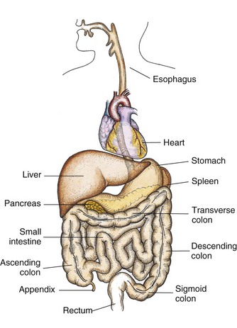
The GI tract is a series of hollow mucous membrane–lined muscular organs. These organs absorb fluid and nutrients, prepare food for absorption and use by body cells, and provide for temporary storage of feces (Fig. 46-1). The GI tract absorbs high volumes of fluids, making fluid and electrolyte balance a key function of the GI system. In addition to ingested fluids and foods, the GI tract also receives secretions from the gallbladder and pancreas.
Mouth
Digestion begins in the mouth and ends in the small intestine. The mouth mechanically and chemically breaks down nutrients into a usable size and form. The teeth masticate food, breaking it down into a size suitable for swallowing. Saliva, produced by the salivary glands in the mouth, dilutes and softens the food in the mouth for easier swallowing.
Esophagus
As food enters the upper esophagus, it passes through the upper esophageal sphincter, a circular muscle that prevents air from entering the esophagus and food from refluxing into the throat. The bolus of food travels down the esophagus and is pushed along by peristalsis, which propels it through the length of the GI tract.
As food moves down the esophagus, it reaches the cardiac or lower esophageal sphincter, which lies between the esophagus and the upper end of the stomach. The sphincter prevents reflux of stomach contents back into the esophagus.
Stomach
The stomach performs three tasks: storing swallowed food and liquid; mixing food, liquid, and digestive juices; and emptying its contents into the small intestine. It produces and secretes hydrochloric acid (HCl), mucus, the enzyme pepsin, and the intrinsic factor. Pepsin and HCl facilitate the digestion of protein. Mucus protects the stomach mucosa from acidity and enzyme activity. The intrinsic factor is essential for the absorption of vitamin B12.
Small Intestine
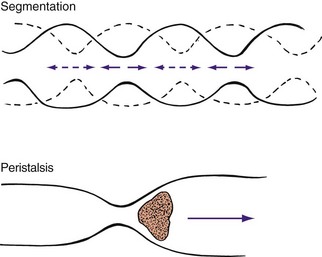
Segmentation and peristaltic movement in the small intestine facilitate both digestion and absorption (Fig. 46-2). Chyme mixes with digestive juices (e.g., bile and amylase). Resorption in the small intestine is so efficient that, by the time the chyme reaches the end of the small intestine, it is pastelike in consistency.
The small intestine has three sections: the duodenum, the jejunum, and the ileum. The duodenum is approximately 20 to 28 cm (8 to 11 inches) long and continues to process the chyme from the stomach. The jejunum is approximately 2.5 m (8 feet) long and absorbs carbohydrates and proteins. The ileum is approximately 3.7 m (12 feet) long and absorbs water, fats, certain vitamins, iron, and bile salts. The duodenum and jejunum absorb most of the nutrients and electrolytes. The intestinal wall also absorbs nutrients across the mucosa and into lymph fluids or blood vessels. Substances, such as plant fiber, that the small intestine cannot digest empty into the cecum at the lower right side of the abdomen. The large intestine begins at the cecum.
Impairment of the small intestine alters the digestive process. For example, conditions such as inflammation, surgical resection, or obstruction disrupt peristalsis, reduce the area of absorption, or block the passage of chyme. Electrolyte and nutrient deficiencies then develop.
Large Intestine
The lower GI tract is called the large intestine (colon) because it is larger in diameter than the small intestine. The large intestine is shorter (1.5 to 1.8 m [5 to 6 feet]) but much wider than the small intestine. The large intestine is divided into the cecum, colon, and rectum (Fig. 46-3). The large intestine is the primary organ of bowel elimination. It is positioned like a question mark, partially encircling the small intestine.
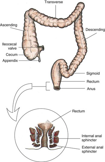
Chyme enters the large intestine by waves of peristalsis through the ileocecal valve, a circular muscular layer that prevents regurgitation. The colon is divided into the ascending, transverse, descending, and sigmoid colons. The muscular tissue of the colon allows it to accommodate and eliminate large quantities of waste and gas (flatus). It has three functions: absorption, secretion, and elimination. The large intestine absorbs water, sodium, and chloride from the digested food that has passed from the small intestine. Healthy adults absorb more than a gallon of water and an ounce of salt from the colon every 4 hours. The amount of water absorbed from chyme depends on the speed at which colonic contents move. Chyme is normally a soft, formed mass. If peristalsis is abnormally fast, there is less time for water to be absorbed, and the stool is watery. If peristaltic contractions slow, water continues to be absorbed; and a hard mass of stool forms, resulting in constipation (JBI, 2008).
The secretory function of the colon aids in electrolyte balance. The colon secretes bicarbonate in exchange for chloride. The colon also excretes about 4 to 9 mEq of potassium daily. Therefore serious alterations in colon function (e.g., diarrhea) cause severe electrolyte disturbances.
Slow peristaltic contractions move contents through the colon. Intestinal content is the main stimulus for contraction. Mass peristalsis pushes undigested food toward the rectum. These mass movements occur only three or four times daily, with the strongest during the hour after mealtime.
The rectum is the final portion of the large intestine. Here bacteria convert fecal matter into its final form. Normally the rectum is empty of waste products (feces) until just before defecation. It contains vertical and transverse folds of tissue that help to temporarily hold fecal contents during defecation. Each fold contains an artery and vein that can become distended from pressure during straining. This distention often results in hemorrhoid formation.
Anus
The body expels feces and flatus from the rectum through the anal canal and anus. Contraction and relaxation of the internal and external sphincters, innervated by sympathetic and parasympathetic stimuli, aid in the control of defecation. The anal canal is richly supplied with sensory nerves that help to control continence.
Defecation
The physiological factors critical to bowel function and defecation include normal GI tract function, sensory awareness of rectal distention and rectal contents, voluntary sphincter control, and adequate rectal capacity and compliance. Normal defecation begins with movement in the left colon, moving stool toward the anus. When stool reaches the rectum, the distention causes relaxation of the internal sphincter and an awareness of the need to defecate. At the time of defecation, the external sphincter relaxes, and abdominal muscles contract, increasing intrarectal pressure and forcing the stool out (Huether and McCance, 2008). Sometimes people use the Valsalva maneuver to assist in stool passage. The Valsalva maneuver exerts pressure to expel feces through a voluntary contraction of the abdominal muscles while maintaining forced expiration against a closed airway. Patients with cardiovascular disease, glaucoma, increased intracranial pressure, or a new surgical wound are at greater risk for cardiac dysrhythmias and elevated blood pressure with the Valsalva maneuver and need to avoid straining to pass the stool. Normal defecation is painless, resulting in passage of soft, formed stool.
Nursing Knowledge Base
Factors Influencing Bowel Elimination
Many factors influence the process of bowel elimination. Knowledge of these factors helps to anticipate measures required to maintain a normal elimination pattern.
Age
Developmental changes affecting elimination occur throughout life. An infant has a small stomach capacity and less secretion of digestive enzymes. Food passes quickly through an infant’s intestinal tract because of rapid peristalsis. The infant is unable to control defecation because of a lack of neuromuscular development. This neuromuscular development usually does not take place until 2 to 3 years of age.
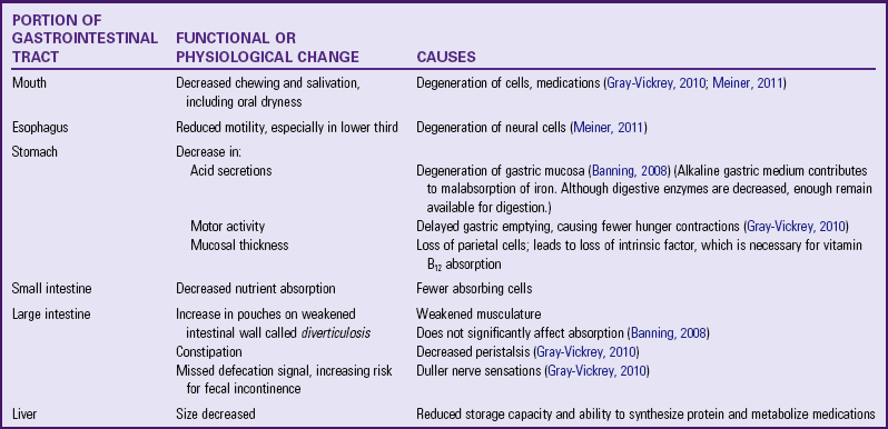
Systemic changes in the function of digestion and absorption of nutrients result from changes in older patients’ cardiovascular and neurological systems rather than their GI system. For example, arteriosclerosis causes decreased mesenteric blood flow, thus decreasing absorption from the small intestine (Meiner, 2011). In addition, peristalsis decreases, and esophageal emptying slows. Older adults often experience changes in the GI system that impair digestion and elimination (JBI, 2008) (Table 46-1).
Older adults also lose muscle tone in the perineal floor and anal sphincter (Holman et al., 2008). Although the integrity of the sphincter remains intact, they often have difficulty controlling bowel evacuation and are at risk for incontinence. In addition, nerve impulses to the anal region slow, causing some individuals to become less aware of the need to defecate. Older adults, especially residents in long-term care facilities, sometimes develop irregular bowel movements and an increased risk for constipation (Kyle, 2007a).
Diet
Regular daily food intake helps maintain a regular pattern of peristalsis in the colon. Fiber, the nondigestible residue in the diet, provides the bulk of fecal material. Bulk-forming foods such as whole grains, fresh fruits, and vegetables help flush the fats and waste products from the body with more efficiency (Holman et al., 2008). The bowel walls stretch, creating peristalsis and initiating the defecation reflex. With stimulation of peristalsis, bulk foods pass quickly through the intestines, keeping the stool soft. Ingestion of a high-fiber diet improves the likelihood of a normal elimination pattern if other factors are normal. Diets high in vegetables and fruits have been linked to decreased risk of colorectal cancer (ACS, 2011b).
Gas-producing foods such as onions, cauliflower, and beans also stimulate peristalsis. The gas formed distends intestinal walls and increases colon motility. Some spicy foods increase peristalsis but also cause indigestion and watery stools.
Food intolerance is not an allergy but rather a particular food that causes the body distress within a few hours of ingestion. The result is diarrhea, cramps, or flatulence. For example, people who drink cow’s milk and have these symptoms are not allergic to milk but lack the enzyme needed to digest the milk sugar lactase and therefore are lactose intolerant. Another condition called celiac disease is a syndrome in which the patient has a hypersensitivity to protein in certain cereal grains and gluten.
Fluid Intake
An inadequate fluid intake or disturbances resulting in fluid loss (such as vomiting) affect the character of feces. Fluid liquefies intestinal contents, easing its passage through the colon. Reduced fluid intake slows passage of food through the intestine and results in hardening of stool contents. Unless there is a medical contraindication, an adult needs to drink at least 1100 to 1400 mL of fluid daily. An increase in fluid intake with the use of fruit juices softens stool and increases peristalsis. Poor fluid intake increases the risk of constipation because of greater resorption of fluid in the colon, resulting in hard, dry stools (Kyle, 2007a).
Physical Activity
Physical activity promotes peristalsis, whereas immobilization depresses it. Encourage early ambulation as illness begins to resolve or as soon as possible after surgery to promote maintenance of peristalsis and normal elimination. Maintaining tone of skeletal muscles used during defecation is important. Weakened abdominal and pelvic floor muscles impair the ability to increase intraabdominal pressure and control the external sphincter. Muscle tone is sometimes weakened or lost as a result of long-term illness, spinal cord injury, or neurological disease that impairs nerve transmission. As a result of these changes in the abdominal and pelvic floor muscles, there is an increased risk for constipation.
Psychological Factors
Prolonged emotional stress impairs the function of almost all body systems (see Chapter 37). During emotional stress the digestive process is accelerated, and peristalsis is increased. Side effects of increased peristalsis are diarrhea and gaseous distention. A number of diseases of the GI tract are associated with stress, including ulcerative colitis, irritable bowel syndrome, certain gastric and duodenal ulcers, and Crohn’s disease. If a person becomes depressed, the autonomic nervous system slows impulses; peristalsis decreases, resulting in constipation.
Personal Habits
Personal elimination habits influence bowel function. Most people benefit from being able to use their own toilet facilities at a time that is most effective and convenient for them. A busy work schedule sometimes prevents the individual from responding appropriately to the urge to defecate, disrupting regular habits and causing possible alterations such as constipation. Individuals need to recognize the best time for elimination.
Chronically ill and hospitalized patients are not always able to maintain privacy during defecation. In a hospital or extended care setting, patients sometimes share bathroom facilities with a roommate with different hygienic habits. In addition, chronic illness limits a patient’s balance, activity tolerance, or physical activity and requires the use of a bedpan or bedside commode. The sights, sounds, and odors associated with sharing toilet facilities or using bedpans are often embarrassing. This embarrassment often causes patients to ignore the urge to defecate, which begins a vicious cycle of constipation and discomfort.
Position During Defecation
Squatting is the normal position during defecation. Modern toilets facilitate this posture, allowing the person to lean forward, exert intraabdominal pressure, and contract the thigh muscles. For the patient immobilized in bed, defecation is often difficult. In a supine position it is impossible to contract the muscles used during defecation. If the patient’s condition permits, raise the head of the bed to assist the patient to a more normal sitting position on a bedpan, enhancing the ability to defecate.
Pain
Normally the act of defecation is painless. However, a number of conditions such as hemorrhoids, rectal surgery, rectal fistulas, and abdominal surgery result in discomfort. In these instances the patient often suppresses the urge to defecate to avoid pain, contributing to the development of constipation.
Pregnancy
As pregnancy advances, the size of the fetus increases, and pressure is exerted on the rectum. A temporary obstruction created by the fetus impairs passage of feces. Slowing of peristalsis during the third trimester often leads to constipation. A pregnant woman’s frequent straining during defecation or delivery results in formation of permanent hemorrhoids.
Surgery and Anesthesia
General anesthetic agents used during surgery cause temporary cessation of peristalsis (see Chapter 50). Inhaled anesthetic agents block parasympathetic impulses to the intestinal musculature. The action of the anesthetic slows or stops peristaltic waves. The patient who receives a local or regional anesthetic is less at risk for elimination alterations because this type of anesthesia generally affects bowel activity minimally or not at all.
Any surgery that involves direct manipulation of the bowel temporarily stops peristalsis. This condition, called paralytic ileus, usually lasts about 24 to 48 hours. If the patient remains inactive or is unable to eat after surgery, return of normal bowel elimination is further delayed.
Medications
Some medications have certain expected actions on the bowel (e.g., there are medications to promote defecation or control diarrhea). In addition, medications prescribed for acute and chronic conditions often have secondary effects on the patient’s bowel elimination patterns (Table 46-2).
TABLE 46-2
Medications and the Gastrointestinal System
| MEDICATIONS | ACTION |
| Dicyclomine HCl (Bentyl) | Suppresses peristalsis and decreases gastric emptying |
| Opioid analgesics | Slow peristalsis and segmental contractions, often resulting in constipation (Lehne, 2010) |
| Anticholinergic drugs such as atropine or glycopyrrolate (Robinul) | Inhibit gastric acid secretion and depress gastrointestinal (GI) motility (Lehne, 2010) (Although useful in treating hyperactive bowel disorders, anticholinergics cause constipation.) |
| Antibiotics | Produce diarrhea by disrupting the normal bacterial flora in the GI tract (An increase in the use of fluoroquinolones in recent years has provided a selective advantage for the epidemic of Clostridium difficile) (Vonberg et al., 2008) |
| Nonsteroidal antiinflammatory drugs | Cause GI irritation that increases the incidence of bleeding with serious consequences to older adults; rectal bleeding is often observed with GI irritation (Lehne, 2010) |
| Aspirin | Prostaglandin inhibitor; interferes with the formation and production of protective mucus and causes GI bleeding (Lehne, 2010) |
| Histamine2 (H2) antagonists | Suppress the secretion of hydrochloric acid and interfere with the digestion of some foods |
| Iron | Causes discoloration of the stool (black), nausea, vomiting, constipation (diarrhea is less commonly reported), and abdominal cramps (Lehne, 2010) |
Laxatives and cathartics soften the stool and promote peristalsis. Although similar, laxatives are milder in action than cathartics. When used correctly, laxatives and cathartics safely maintain normal elimination patterns. However, chronic use of cathartics causes the large intestine to become less responsive to stimulation by laxatives. Laxative overuse can also cause serious diarrhea, leading to dehydration and electrolyte depletion. Mineral oil, a common laxative, decreases fat-soluble vitamin absorption. Laxatives often influence the efficacy of other medications by altering the transit time (i.e., the time the medication remains in the GI tract and is available for absorption).
Diagnostic Tests
Diagnostic examinations involving visualization of GI structures often require a prescribed bowel preparation (e.g., medications, cathartics, and/or enemas) to ensure that the bowel is empty. In addition, the patient cannot eat or drink several hours before the examinations such as an endoscopy, colonoscopy, or other testing that requires visualization of the GI tract. Following the diagnostic procedure, changes in elimination such as increased gas or loose stools often occur until the patient resumes a normal eating pattern.
Common Bowel Elimination Problems
Caring for patients who have or are at risk for elimination problems because of emotional stress (anxiety or depression), physiological changes in the GI tract such as surgical alteration of intestinal structures, inflammatory diseases, prescribed therapy, or disorders impairing defecation is common in the practice of nursing.
Constipation
Constipation is a symptom, not a disease (Box 46-1). Improper diet, reduced fluid intake, lack of exercise, and certain medications can cause constipation. For example, patients receiving opiates for pain after surgery often require a stool softener or laxative to prevent constipation. The signs of constipation include infrequent bowel movements (less than every 3 days), difficulty passing stools, excessive straining, inability to defecate at will, and hard feces (McWilliams, 2010). When intestinal motility slows, the fecal mass becomes exposed over time to the intestinal walls, and most of the fecal water content is absorbed. Little water is left to soften and lubricate the stool. Passage of a dry, hard stool causes rectal pain (Fig. 46-4).
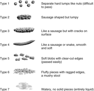
FIG. 46-4 Bristol stool form scale. (Used with permission. Bristol Stool Form Guideline, http://www.aboutconstipation.org/bristol.html, accessed July 31, 2006.)
Constipation is a significant health hazard. Straining during defecation causes problems for the patient with recent abdominal, gynecological, or rectal surgery. The effort to pass a stool often causes sutures to separate, reopening the wound. In addition, patients with histories of cardiovascular disease, diseases causing elevated intraocular pressure (glaucoma), and increased intracranial pressure need to prevent constipation and avoid using the Valsalva maneuver.
Impaction
Fecal impaction results from unrelieved constipation. It is a collection of hardened feces wedged in the rectum that a person cannot expel. In cases of severe impaction the mass extends up into the sigmoid colon. If not resolved or removed, severe impaction often results in intestinal obstruction. Patients who are debilitated, confused, or unconscious are most at risk for impaction. They are dehydrated or too weak or unaware of the need to defecate, and the stool becomes too hard and dry to pass.
An obvious sign of impaction is the inability to pass a stool for several days, despite the repeated urge to defecate (Chien and Bradway, 2010). You suspect impaction when a continuous oozing of diarrhea stool occurs. The liquid portion of feces located higher in the colon seeps around the impacted mass. Loss of appetite (anorexia), nausea and/or vomiting, abdominal distention and cramping, and rectal pain often accompany the condition. If an impaction is suspected, gently perform a digital examination of the rectum and palpate for the impacted mass (Steggall, 2008).
Diarrhea
Diarrhea is an increase in the number of stools and the passage of liquid, unformed feces. It is associated with disorders affecting digestion, absorption, and secretion in the GI tract. Intestinal contents pass through the small and large intestine too quickly to allow for the usual absorption of fluid and nutrients. Irritation within the colon results in increased mucus secretion. As a result, feces become watery, and the patient is unable to control the urge to defecate. Normally an anal bag is safe and effective in long-term treatment of patients with fecal incontinence at home, in hospice, or in the hospital. Fecal incontinence is expensive and a potentially dangerous condition in terms of contamination and risk of skin ulceration (Gray, 2007).
Excess loss of colonic fluid results in serious fluid and electrolyte or acid-base imbalances. Infants and older adults are particularly susceptible to associated complications (see Chapter 41). Because repeated passage of diarrhea stools also exposes the skin of the perineum and buttocks to irritating intestinal contents, meticulous skin care and containment of fecal drainage is necessary to prevent skin breakdown (see Chapter 48).
Many conditions cause diarrhea. Antibiotic use via any route of administration alters the normal flora in the GI tract (Vonberg et al., 2008). Patients receiving enteral nutrition are also at risk for diarrhea. Consult a dietitian when diarrhea occurs (Tabloski, 2009). See Chapter 44 for interventions to decrease diarrhea caused by enteral feedings. Food allergies and intolerances increase peristalsis and cause diarrhea. Surgeries or diagnostic testing of the lower GI tract also cause diarrhea. The aim of treatment is to remove precipitating conditions and slow peristalsis.
Another common causative agent of diarrhea is Clostridium difficile (C. difficile), in which symptoms range from mild diarrhea to severe colitis. C. difficile infection is acquired in one of two ways: by factors that cause an overgrowth of C. difficile, and by contact with the C. difficile organism. A new strain of C. difficile has been identified that is more virulent with more toxic effects (Grossman, 2010). Antibiotics (cephalosporins, ampicillin, amoxicillin, and clindamycin (Calfee, 2008), chemotherapy, and invasive bowel procedures such as surgery or colonoscopy disrupt normal bowel flora and may cause an overgrowth of C. difficile. Some patients acquire the organism from a health care worker’s hands or direct contact with the environmental surfaces contaminated with it. Only hand hygiene with soap and water is effective to physically remove C. difficile spores from the hands. In addition, evidence supports the use of diluted bleach (1 : 10) as an environmental disinfectant to decrease the incidence of C. difficile (Calfee, 2008; Vonberg, 2008). The most common diagnostic test for the bacteria is the enzyme-linked immunosorbent assay (ELISA) test, which detects C. difficile A and B in the stool.
Communicable foodborne pathogens also cause diarrhea. Hand hygiene following the use of the bathroom, before and after preparing foods, and when cleaning and storing fresh produce and meats greatly reduces the risk of foodborne illnesses. When diarrhea is the result of a foodborne virus, the goal usually is to rid the GI system of the pathogen rather than slow peristalsis.
Incontinence
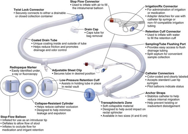
Fecal incontinence is the inability to control passage of feces and gas from the anus. Incontinence harms a patient’s body image (see Chapter 33). In many situations the patient is mentally alert but physically unable to avoid defecation. The embarrassment of soiling clothes often leads to social isolation. Physical conditions that impair anal sphincter function or control cause incontinence. It occurs in a variety of settings. Conditions that create frequent, loose, large-volume, watery stools also predispose to incontinence. Using an anal bag or a bowel management system helps to prevent perineal skin breakdown (Fig. 46-5).
Flatulence
As gas accumulates in the lumen of the intestines, the bowel wall stretches and distends (flatulence). It is a common cause of abdominal fullness, pain, and cramping. Normally intestinal gas escapes through the mouth (belching) or the anus (passing of flatus). However, flatulence causes abdominal distention and severe, sharp pain if intestinal motility is reduced because of opiates, general anesthetics, abdominal surgery, or immobilization.
Hemorrhoids
Hemorrhoids are dilated, engorged veins in the lining of the rectum. They are either external or internal. External hemorrhoids are clearly visible as protrusions of skin. If the underlying vein is hardened, there is usually a purplish discoloration (thrombosis). This causes increased pain and often needs to be excised. Internal hemorrhoids have an outer mucous membrane. Increased venous pressure from straining at defecation, pregnancy, heart failure, and chronic liver disease causes hemorrhoids.
Bowel Diversions
Certain diseases cause conditions that prevent normal passage of feces through the rectum. The treatment for these disorders results in the need for a temporary or permanent artificial opening (stoma) in the abdominal wall. Surgical openings are created in the ileum (ileostomy) or colon (colostomy), with the ends of the intestine brought through the abdominal wall to create the stoma (Durston, 2009).
The standard bowel diversion creates a stoma, or the patient has reconstructive bowel surgery that uses the native sphincter for bowel continence. The reconstructive surgery includes a continent stoma procedure or the ileoanal pouch anastomosis, which is described later in the chapter.
Ostomies
The location of an ostomy determines the consistency of stool. An ileostomy bypasses the entire large intestine. As a result, stools are frequent and liquid. The same is true for a colostomy of the ascending colon. A colostomy of the transverse colon generally results in a more solid, formed stool. The sigmoid colostomy releases near-normal stool. The patient’s medical problem and general condition determine the location of a colostomy. There are three types of colostomy construction: loop, end, and double-barrel.
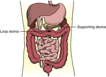
Loop Colostomy: A loop colostomy is usually performed in a medical emergency when health care providers anticipate closure of the colostomy (Fig. 46-6). It is usually a temporary large stoma constructed in the transverse colon. The surgeon pulls a loop of bowel onto the abdomen. An external supporting device such as a plastic rod, bridge, or rubber catheter is temporarily placed under the bowel loop to keep it from slipping back. The surgeon then opens the bowel and sutures it to the skin of the abdomen. A communicating wall remains between the proximal and distal bowel. The loop ostomy has two openings through one stoma. The proximal end drains stool, whereas the distal portion drains mucus. Within 7 to 10 days the surgeon removes the supporting device.
End Colostomy: The end colostomy consists of one stoma formed from the proximal end of the bowel, with the distal portion of the GI tract either removed or sewn closed (called Hartmann’s pouch) and left in the abdominal cavity. For many patients end colostomies are a result of surgical treatment of colorectal cancer. In such cases the rectum is usually removed. Patients with diverticulitis who are treated surgically often have a temporary end stoma with a Hartmann’s pouch (Fig. 46-7).

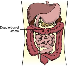
Double-Barrel Colostomy: Unlike the loop colostomy, the surgeon divides the intestine and brings both the proximal and distal ends through the abdominal incision to the abdominal surface when creating a double-barrel colostomy. A small incision is made in the proximal stoma for fecal drainage. The distal stoma leads to the inactive intestine and is left intact. When the intestinal injury has healed, the colostomy is reversed, and the divided ends are anastomosed to restore intestinal integrity (Fig. 46-8).
Alternative Procedures
Ileoanal Pouch Anastomosis: The ileoanal pouch anastomosis is a surgical procedure that is used in patients who need to have a colectomy for treatment of ulcerative colitis or familial polyps (Dorman, 2009). In this procedure the surgeon removes the colon, creates a pouch from the end of the small intestine, and attaches the pouch to the patient’s anus (Fig. 46-9). This pouch provides for the collection of waste material, which is similar to the rectum. The patient is continent of stool because stool is evacuated via the anus. When the ileal pouch is created, the patient has a temporary ileostomy to allow the anastomosis to heal.
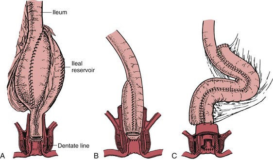
FIG. 46-9 Ileoanal reservoirs (IARs). A, S-shaped configuration for IAR. Three 10-cm limbs of ileum are used, antimesenteric surface of each limb is opened, and adjacent bowel walls are anastomosed. B, J-shaped configuration for IAR. Distal ileum is aligned in J shape, antimesenteric surface of J shape is opened, and adjacent bowel walls are anastomosed. Side-to-end anastomosis of bowel to dentate line is evident. C, Lateral or side-by-side ileoanal pouch configuration. (From Hampton BG, Bryant RA: Ostomies and continent diversions: nursing management, St Louis, 1992, Mosby.)
Kock Continent Ileostomy: The Kock continent ileostomy is created using the patient’s small intestine, changing its cylindrical shape into a spherical reservoir (Gordon, 2007). This procedure is occasionally used in the treatment of ulcerative colitis. The pouch has a continent stoma, a nipple type of valve that is drained with an external catheter, which the patient places intermittently in the stoma (Fig. 46-10).
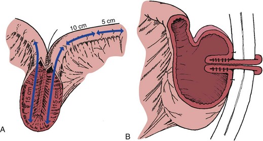
FIG. 46-10 Construction of Kock continent ileostomy—Kock pouch. A, Two 15-cm limbs are used to create a pouch, and one 15-cm limb is used to fashion a nipple valve and stoma. B, Distal limb is intussuscepted into reservoir to create one-way valve and accomplish continence. Sutures or staples, or both, are placed to stabilize and maintain intussuscepted nipple. Anterior surface of reservoir is anchored to anterior peritoneal wall. (From Hampton BG, Bryant RA: Ostomies and continent diversions: nursing management, St Louis, 1992, Mosby.)
Macedo-Malone Antegrade Continence Enema: The Macedo-Malone antegrade continence enema (MACE) procedure improves continence in patients with fecal soiling associated with neuropathic or structural abnormalities of the anal sphincter. This procedure isolates a 3-cm (1.2-inch) flap on the left colon. A Foley catheter placed on the surface of the flap creates a tubular passage. This produces a continence valve mechanism. The surgeon takes the distal end of the tube and makes a V shape to the skin flap (Fig. 46-11). Enema administration begins 7 to 10 days after surgery. Patients receive enemas daily. The volume of the enema varies from 250 to 800 mL and takes 45 to 60 minutes to administer. Colonic evacuation occurs within 30 to 60 minutes (Meurette et al., 2010).
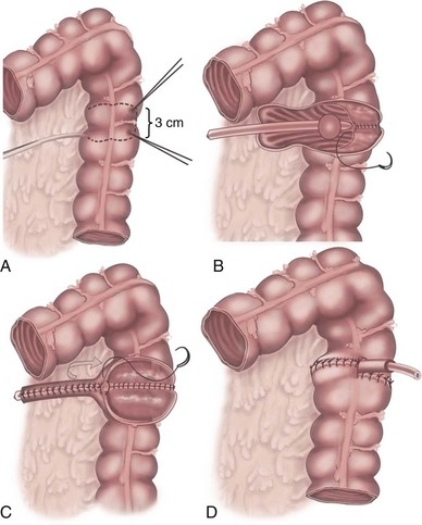
FIG. 46-11 Macedo-Malone antegrade continence enema procedure. A, Isolation of the flap on left colon. B, Foley catheters placed on mucosal surface of flap. C, Tubularization of plate creating efferent tubular conduit. D, Continence valve mechanism is created. (Used with permission. From Calado A et al: The Macedo-Malone antegrade continence enema procedure: early experience, J Urol 173:1340, 2005.)
Psychological Considerations
A stoma causes serious body image changes, particularly if it is permanent. After the surgery, patients face a variety of anxieties and concerns, from learning how to manage their stoma to coping with conflicts of self-esteem and body image. Provide emotional support before and after surgery (Deitz, 2010a). Patients often perceive a stoma as invasive and disfiguring. However, a well-placed stoma usually does not interfere with the patient’s activities and is concealed with clothing. Nonetheless, even though clothing conceals the ostomy, the patient feels different. Many patients have difficulty maintaining or initiating normal sexual relations (see Chapter 34) (Ramirez et al., 2009). Important factors affecting reactions to the stoma include the character of fecal secretions and the ability to control them. Foul odors, spillage, or leakage of liquid stools and inability to regulate bowel movements cause the patient to lose self-esteem (Richbourg et al., 2007). The aging process often affects the ability to manage stomas, even in people who have had them for years. You need to recognize and intervene when problems resulting from advanced age such as skin changes, weight loss or gain, visual impairments, or changes in diet occur (Pearson, 2010). Refer the patient to ostomy support groups such as the United Ostomy Associations of America at http://www.uoaa.org, which has discussion boards for various types of incontinent and continent diversions and networks. The Wound, Ostomy and Continence Nurses Society (http://www.wocn.org) provides information and helps patients locate a wound, ostomy continence nurse (WOCN) (Dorman, 2009).
Critical Thinking
Successful critical thinking requires a synthesis of knowledge, experience, information gathered from patients, critical thinking attitudes, and intellectual and professional standards. Clinical judgments require you to anticipate the information necessary, analyze the data, and make decisions regarding patient care.
In the case of bowel elimination, integrate the knowledge from nursing and other disciplines to understand the patient’s response to bowel elimination alterations. Experience in caring for patients with elimination alterations helps you provide an appropriate plan of care. Use critical thinking attitudes such as fairness, confidence, and discipline when listening to and exploring the patient’s nursing history. Apply relevant standards of practice (e.g., wound care standards, quality and safety standards) when selecting nursing measures.
Nursing Process
Apply the nursing process and use a critical thinking approach in your care of patients. The nursing process provides a clinical decision-making approach for you to develop and implement an individualized plan of care.
Assessment
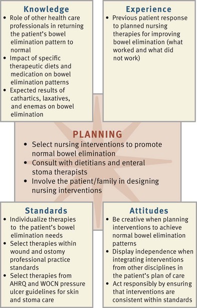
During the assessment process, thoroughly assess each patient and critically analyze findings to ensure you make patient-centered clinical decisions required for safe nursing care. Consider all critical thinking elements that build toward making appropriate diagnoses (Fig. 46-12).
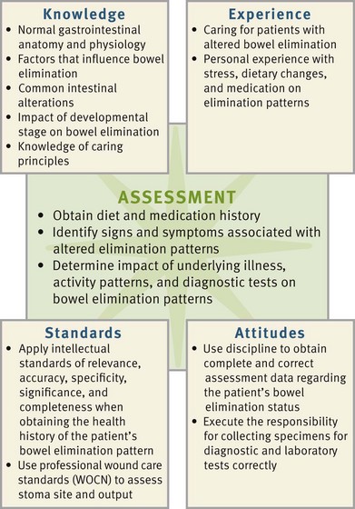
FIG. 46-12 Critical thinking model for elimination assessment. WOCN, Wound, Ostomy and Continence Nurses Society.
Assessment for bowel elimination patterns and abnormalities includes a nursing history, physical assessment of the abdomen, inspection of fecal characteristics, and review of relevant test results. In addition, determine the patient’s medical history, pattern and types of fluid and food intake, chewing ability, medications, and recent illnesses and/or stressors.
Through the Patient’s Eyes
Patients expect the nurse to answer all of their questions regarding diagnostic tests and the preparation for these tests. They are concerned about discomfort and exposure of their more personal areas. Bowel problems are often devastating for the patient and their families. Fecal incontinence in older people, most commonly caused by overflow leakage as a result of constipation, often means the breakdown of care at home, causing them to be admitted to a residential facility (Continence Care Position Statement, 2009). Some older patients who fail to recognize their elimination needs need monitoring for elimination patterns so negative consequences do not occur. Remember that each patient has a unique situation and a perception of what is “right” for him or her. Patients expect a knowledgeable nurse with the ability to teach methods of promoting and maintaining normal bowel elimination patterns. Consider the patient’s cultural practices and preferences because patients of different cultures can have various expectations (Box 46-2).
 Box 46-2 Cultural Aspects of Care
Variables Influencing Colorectal Cancer Screening in African Americans
Biological, psychological, behavioral, and social variables influence colorectal cancer (CRC) screenings in African Americans. African Americans have a 20% higher rate of CRC and 40% higher incidence of disease-related death when compared with Caucasians (ACS, 2011b). Prevention resulting in early detection is key to finding CRC when it is often curable, but screening rates are low in African Americans.
• Lack of routine visits to a primary care provider is one of the strongest predictors for inadequate CRC screening and advanced stage at presentation (Griffin, 2009).
• Patients who do not have health insurance do not frequently seek CRC screening (Griffin, 2009).
• Patients who participate in preventive health practices (e.g., regular physical activity) often seek regular screening for CRC because these patients are often interested in promoting their own health (Griffin, 2009).
• Identification of additional social system predictors such as family support, church affiliation, and geographical access also help in receiving timely CRC screening (Griffin, 2009).
• Removing barriers of safety, quality, and cost-effectiveness improves the involvement of patients in CRC screening (Cronenwett et al., 2007).
Nursing History
The nursing history provides a review of the patient’s usual bowel pattern and habits. What a patient describes as normal or abnormal is often different from factors and conditions that tend to promote normal elimination. Identifying normal and abnormal patterns, habits, and the patient’s perception of normal and abnormal in regard to bowel elimination allows you to accurately determine a patient’s problems. Organize the nursing history around factors that affect elimination (Wisniewski, 2010):
• Determination of the usual elimination pattern: Include frequency and time of day. Having the patient or caregiver complete a bowel elimination diary provides an accurate assessment of a patient’s current bowel elimination pattern.
• Patient’s description of usual stool characteristics: Determines whether the stool is normally watery or formed soft or hard, the typical color, and the presence of blood. Ask the patient to describe the usual shape of the stool and the number of stools per day.
• Identification of routines followed to promote normal elimination: Examples are drinking hot liquids, eating specific foods, or taking time to defecate during a certain part of the day.
• Assessment of the use of artificial aids at home: Assess whether and how often the patient uses enemas, laxatives, or bulk-forming food additives before having a bowel movement.
• Presence and status of bowel diversions: If the patient has an ostomy, assess frequency of fecal drainage, character of feces, appearance and condition of the stoma (color, swelling, and irritation), type of fecal collection device used, and methods used to maintain the function of the ostomy.
• Changes in appetite: Include changes in eating patterns and a change in weight (amount of loss or gain). If a change of weight is present, ask if the patient planned it (e.g., weight loss with a diet).
• Diet history: Determine the patient’s dietary preferences for a day. Determine the intake of fruits, vegetables, cereals, and breads and also if mealtimes are regular or irregular.
• Description of daily fluid intake: This includes the type and amount of fluid. The patient often estimates the amount using common household measurements.
• History of surgery or illnesses affecting the GI tract: This information helps explain symptoms, the potential for maintaining or restoring normal bowel elimination pattern, and whether there is a family history of GI cancer.
• Medication history: Ask whether the patient takes medications (e.g., laxatives, antacids, iron supplements, and analgesics) that alter defecation or fecal characteristics.
• Emotional state: The patient’s emotions significantly alter frequency of defecation. During assessment observation of the patient’s emotions, tone of voice, and mannerisms reveal significant behaviors that indicate stress.
• History of exercise: Ask the patient to specifically describe the type and amount of daily exercise.
• History of pain or discomfort: Ask the patient whether there is a history of abdominal or anal pain. The type, frequency, and location of pain help identify the source of the problem.
• Social history: Patients have many different living arrangements. Where patients live affects their toileting habits. If patients share living quarters, ask how many bathrooms there are. Find out if patients have their own bathroom or if they need to share bathrooms, creating a need to adjust the time they use the bathroom to accommodate others. If patients live alone, can they ambulate safely to the toilet? When patients are not independent in bowel management, determine who assists them and how.
• Mobility and dexterity: Evaluate patients’ mobility and dexterity to determine if they need assistive devices or help from personnel.
Box 46-3 summarizes types of assessment questions to use for gathering a detailed nursing history.
Physical Assessment
Conduct a physical assessment of body systems and functions likely to be influenced by the presence of elimination problems (see Chapter 30).
Mouth: Inspect the patient’s teeth, tongue, and gums. Poor dentition or poorly fitting dentures influence the ability to chew. Sores in the mouth make eating not only difficult but also painful.
Abdomen: Inspect all four abdominal quadrants for contour, shape, symmetry, and skin color. Note masses, peristaltic waves, scars, venous patterns, stomas, and lesions. Normally you do not see peristaltic waves. Observable peristalsis is often a sign of intestinal obstruction.
Abdominal distention appears as an overall outward protuberance of the abdomen. Intestinal gas, large tumors, or fluid in the peritoneal cavity cause distention. A distended abdomen feels tight like a drum; and the skin is taut and appears stretched.
Auscultate the abdomen with a stethoscope to assess bowel sounds in each quadrant (see Chapter 30). Normal bowel sounds occur every 5 to 15 seconds and last a second to several seconds. During auscultation note the character and frequency of bowel sounds. You hear an increase in pitch or a tinkling sound with abdominal distention. Absent (no auscultated bowel sounds) or hypoactive sounds (less than five sounds per minute) occur with paralytic ileus such as after abdominal surgery. High-pitched and hyperactive bowel sounds (35 or more sounds per minute) occur with small intestine obstruction and inflammatory disorders.
Percussion identifies underlying abdominal structures and detects lesions, fluid, or gas within the abdomen. Gas or flatulence creates a tympanic note. Masses, tumors, and fluid are dull to percussion (Wisniewski, 2010).
Gently palpate the abdomen for masses or areas of tenderness. It is important for the patient to relax. Tensing abdominal muscles interferes with palpating underlying organs or masses.
Laboratory Tests
Laboratory and diagnostic examinations yield useful information concerning elimination problems (Table 46-3). Laboratory analysis of fecal contents detects pathological conditions such as tumors, bleeding, parasites, and infection.
TABLE 46-3
Laboratory and Diagnostic Tests for Bowel Function
| MEASUREMENT AND NORMAL VALUES | INTERPRETATION |
| Laboratory Tests | |
| Total bilirubin: 0.3-1 mg/dL | Bilirubin is increased in hepatobiliary diseases, obstructions in bile duct, certain anemias, and following transfusion reactions (Pagana and Pagana, 2011). |
| Alkaline phosphatase: 30-120 units/L | Alkaline phosphatase is elevated in obstructive hepatobiliary diseases, hepatobiliary carcinomas, bone tumors, and healing fractures (Pagana and Pagana, 2011). |
| Amylase: 60-120 Somogyi units/dL | Amylase is elevated in abnormalities of the pancreas such as inflammation or tumors, cholecystitis, necrotic bowel, and diabetic ketoacidosis (Pagana and Pagana, 2011). |
| Carcinoembryonic antigen (CEA): less than 5 ng/mL | CEA is elevated in the presence of cancer or inflammation of the GI tract or hepatobiliary organs (Pagana and Pagana, 2011). |
| Direct Visualization | |
| Endoscopy, colonoscopy | Routine examination such as a colonoscopy is recommended for people after 50 years of age who have no family history of CRC. People with a personal or family history need to talk with their health care provider to determine if they need to start screening at an earlier age and recommended frequency of screening (ACS, 2011b). Normally the GI tract is free of polyps, tumors, inflammation, ulcers, hernias, obstruction, and ulcerations. If a lesion such as a polyp is identified, the health care provider removes the growth or a portion of the growth and sends it to pathology for analysis. If bleeding is present, the health care provider usually attempts to stop it at the source. In some cases the identification of an abnormality indicates the need for follow-up surgery for the patient. |
| Indirect Visualization | |
| X-ray film with contrast medium | X-ray identifies the presence of abnormalities in the GI tract. A series of x-ray films allow for indirect visualization of the entire tract (ACS, 2011b). The presence of tumors, ulcerations, inflammation, or other abnormalities indicates the need for further diagnostic testing and medical or surgical intervention. |
Fecal Specimens: The nurse ensures that specimens are obtained accurately, labeled properly in appropriate containers, and transported to the laboratory on time. Institutions provide special containers for fecal specimens. Some tests require that specimens are placed in chemical preservatives. Use medical aseptic technique during collection of stool specimens (see Chapter 28). Because about 25% of the solid portion of a stool is bacteria from the colon, wear clean gloves when handling specimens.
Hand hygiene is necessary for anyone who comes in contact with the specimen. Often the patient is able to obtain the specimen if properly instructed. Teach the patient to avoid mixing feces with urine or water. The patient defecates into a clean, dry bedpan or a special container under the toilet seat.
Tests performed by the laboratory for occult (microscopic) blood in the stool and stool cultures require only a small sample. Collect about 2.54 cm (1 inch) of formed stool or 15 to 30 mL of liquid diarrhea stool. Tests for measuring the output of fecal fat require a 3- to 5-day collection of stool. You need to save all fecal material throughout the test period.
After obtaining a specimen, label and tightly seal the container and complete all laboratory requisition forms. Record specimen collections in the patient’s medical record. It is important to avoid delays in sending specimens to the laboratory. Some tests such as measurement for ova and parasites require the stool to be warm. When stool specimens remain at room temperature, bacteriological changes that alter test results occur.
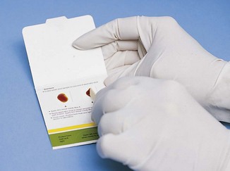
A common laboratory test that patients perform at home or nurses perform at the patient’s bedside is the fecal occult blood test (FOBT), or guaiac test, which measures microscopic amounts of blood in feces (Box 46-4). It is useful as a diagnostic screening tool for colon cancer (Box 46-5). The noninvasive FOBT is one of five colorectal cancer screening regimens recommended by the American Cancer Society (ACS, 2011a; 2011b). Three types of FOBT are available to date. They include the most commonly used guaiac fecal occult blood test (gFOBT), the immunochemical fecal occult blood test (iFOBT), and the stool deoxyribonucleic acid (DNA) test. One positive gFOBT result does not confirm GI bleeding. You need to repeat the test at least three times while the patient refrains from ingesting foods (e.g., some raw vegetables, red meat, poultry, fish) and medications (e.g., vitamin C, aspirin, nonsteroidal anti-inflammatory drugs) that cause false-positive results (Peters, 2008). Patients who take anticoagulants or who have a bleeding disorder or a GI disorder known to cause bleeding (e.g., intestinal tumors, bowel inflammation, or ulcerations) need regular screening for fecal occult blood.
Box 46-4 Procedural Guidelines 
Performing a Guaiac Fecal Occult Blood Test
The skill of performing a guaiac fecal occult blood test (gFOBT) can be delegated to nursing assistive personnel (NAP). However, the nurse must evaluate the significance of the findings. Instruct the NAP to
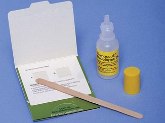
Hemoccult test paper, Hemoccult developer, and wooden applicator (Fig. 46-13)
1. Patient identification: Identify the patient using two identifiers (e.g., name and birth date or name and account number) according to facility policy. Compare identifiers with information on the patient’s medical record (TJC, 2011).
2. Explain purpose of the test and ways for patient to assist. Patient can collect own specimen if possible.
3. Perform hand hygiene and apply clean disposable gloves.
4. Use tip of wooden applicator to obtain a small portion of a stool specimen. Be sure that specimen is free of tissue paper.
5. Perform Hemoccult slide test.
a. Open flap of slide and, using the wooden applicator, thinly smear stool in the first box of the guaiac paper. Apply a second fecal specimen from a different portion of the stool to the second box of the slide (see illustration).
b. Close slide cover and turn the packet over to the reverse side (see illustration). After waiting 3 to 5 minutes, open cardboard flap and apply 2 drops of developing solution on each smear.

c. Interpret the color of the guaiac paper within 60 seconds. A blue color indicates a positive guaiac or presence of fecal occult blood.
d. After determining if the patient’s specimen is positive or negative, apply 1 drop of developer to the quality control section and interpret within 10 seconds.
6. Dispose of test slide in proper receptacle.
7. Wrap wooden applicator in paper towel, remove gloves, and discard in proper receptacle and perform hand hygiene.
8. Note color, changes on guaiac paper, and observe character of stool specimen.
9. Record results of test, noting any unusual fecal characteristics.
Fecal Characteristics: Inspection of fecal characteristics (Table 46-4) reveals information about the nature of elimination alterations. Several factors influence each characteristic. Knowing whether there have been any recent changes is a key to assessment. The patient best provides this information during the nursing history.

Diagnostic Examinations
A variety of radiological and diagnostic tests are used with the patient experiencing altered bowel elimination (Box 46-6). Direct or indirect approaches are used to visualize GI structures. Many facilities use moderate sedation during these procedures. The most common types of drugs used to achieve moderate sedation include benzodiazepines and opiates. It is essential to understand the safety precautions involved concerning this form of anesthesia. In many institutions special training is required. A crash cart must be present at the bedside; and you must monitor the patient continuously with pulse oximetry and frequent vital signs, usually every 15 minutes during and immediately following the procedure (check agency policy).
Nursing Diagnosis
The nursing assessment of the patient’s bowel function reveals data that indicate an actual or potential elimination problem or a problem resulting from elimination alterations. In the examples discussed in the Nursing Care Plan, a patient has constipation as a result of pain medications and decreased fiber intake. Examples of diagnoses that apply to patients with elimination problems include the following:
Associated problems such as age, body-image changes, or skin breakdown require interventions unrelated to bowel function impairment. Ability to identify the correct diagnosis depends not only on the thoroughness of assessment but also on recognition of defining characteristics and factors that impair elimination (Box 46-7). Determine the patient’s risk and institute measures to ensure maintenance of normal bowel function.
Planning
When planning care, synthesize information from multiple resources (Fig. 46-14). Critical thinking ensures that the plan of care integrates everything known about the patient and the clinical problem. Rely on professional standards. The guidelines on incontinence assist in protecting the patient’s skin, promoting continence, and reducing the embarrassment associated with incontinence. In addition, the Agency for Healthcare Research and Quality (AHRQ) provides guidelines on reduction of pressure ulcers that also help you develop a plan of care for patients with bowel incontinence (see Chapter 48).
Goals and Outcomes
Help patients establish goals and outcomes by incorporating their elimination habits or routines as much as possible and reinforcing the routines that promote health (see the Nursing Care Plan). In addition, consider preexisting health concerns. For example, if a patient is at risk for worsening heart failure, you need to individualize an outcome of increased fluid intake to accommodate cardiac function and the patient’s ability to safely handle the increased fluid. In another example, if a patient’s bowel habits caused the elimination problem, help the patient learn new habits. The overall goal of returning the patient to a normal bowel elimination pattern includes the following outcomes:
• Patient sets regular defecation habits.
• Patient is able to list proper fluid and food intake needed to achieve bowel elimination.
• Patient implements a regular exercise program.
• Patient reports daily passage of soft, formed brown stool.
• Patient does not report discomfort associated with defecation.
 Nursing Care Plan
Constipation
Javier, a home care nurse, is visiting Larry Johnston at his home on one of the local cattle ranches. Larry lives 20 miles from town. He is 22 years old and had surgery 6 days ago for repair of a badly broken right leg, after being thrown from a horse. Larry tells Javier that he “just doesn’t feel well.” His past history includes pneumonia at age 12 and a recent traumatic injury to the abdomen. The injury required emergency surgery after he was struck by a bull’s horns.
| Assessment Activities | Findings/Defining Characteristics* |
| Ask Larry about his bowel elimination patterns over the last 5 days. | Larry tells Javier that he has not had a bowel movement since he left the hospital 4 days ago and that his abdomen is tight and sore. |
| Review patient’s medication. | Larry says he is taking one Percocet tablet for pain, up to three tablets a day. |
| Review dietary and fluid intake over last day. | Diet included eggs, bacon, and toast for breakfast; soup for lunch; and chicken, rice, and corn for dinner. He drinks about six cups of coffee each day, no water, but will drink a cola. |
| Ask about any nausea or vomiting. | Denies any nausea or vomiting. |
| Auscultate patient’s abdomen. | Decreased bowel sounds are auscultated throughout all four abdominal quadrants. |
| Palpate abdomen. | While Javier is palpating Larry’s abdomen, Larry tells Javier, “It really hurts.” On palpation left lower quadrant is tender and firm. |
Constipation related to opiate-containing pain medication and decreased fiber intake
| Goals | Expected Outcomes (NOC)† |
| Bowel Elimination | |
| Larry will establish normal defecation. | Larry will report passage of soft, formed stool without straining in next 24 hours. |
| Nutritional Status: Food and Fluid Intake | |
| Larry will make changes to his diet to prevent constipation. | Larry will drink at least 1500 mL of fluid over the next 8 hours. |
| Larry will increase the fiber content of his diet. |
†Outcome classification labels from Moorhead S et al: Nursing outcomes classification (NOC), ed 4, St Louis, 2008, Mosby.
| Interventions (NIC)‡ | Rationale |
| Constipation/Impaction Management | |
| Encourage fluid intake of appropriate fluids, fruit juice, and water. | At least 1500 mL fluid intake daily is necessary to prevent hard, dry stool (JBI, 2008). |
| Encourage activity within patient’s mobility regimen. | Minimal activity (such as leg lifts) increases peristalsis. |
| Instruct Larry to add 20g/day of wheat bran to diet. | Bran along with physical activity prevents constipation (JBI, 2008). |
| Provide laxative or stool softeners as ordered. | Medications soften the stool and prevent straining (Lehne, 2010). |
| Provide privacy during toileting. | Patients need to feel relaxed when moving bowels (Holman et al., 2008). |
‡Intervention classification labels from Bulechek GM, Butcher HK, Dochterman JM: Nursing interventions classification (NIC), ed 5, St Louis, 2008, Mosby.
| Nursing Actions | Patient Response/Finding | Achievement of Outcomes |
| Ask Larry to identify foods high in fiber. | Able to state appropriate foods. | Larry is making excellent progress in introducing high-fiber and low-fat foods into his diet. |
| Review of 24-hour diet diary shows Larry is selecting high-fiber, low-fat foods. | ||
| Ask Larry about physical activity. | Larry states that he has not changed his activity pattern. | Larry did not increase activity pattern and needs to continue to work on this intervention. |
| Observe Larry’s subsequent stool for characteristics such as consistency and color. | Stools are now every 24 to 48 hours. Larry does not “feel regular.” Abdomen is soft and nondistended. Stools are formed and hard, but Larry does report straining. |
Larry did not achieve passage of regular, formed stool. |
Setting Priorities
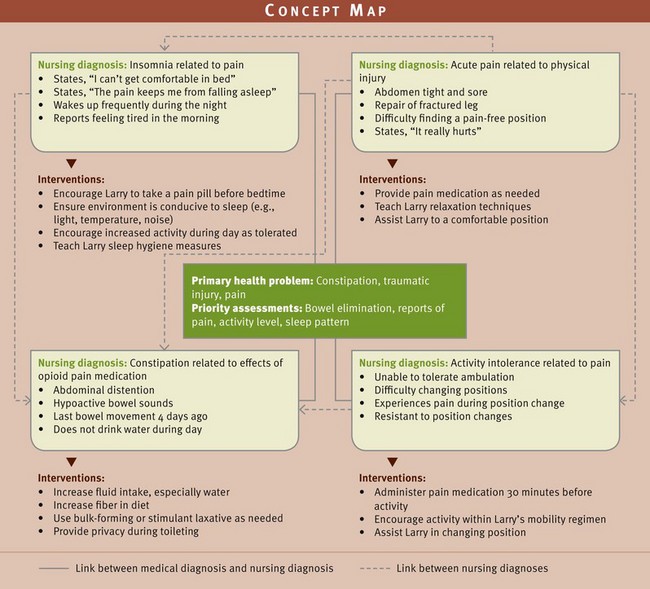
Defecation patterns vary among individuals. For this reason the nurse and patient work together closely to plan effective interventions. Patients often have multiple diagnoses. The concept map (Fig. 46-15) shows an example of how the nursing diagnosis of constipation is related to three other diagnoses and their respective interventions. A realistic time frame to establish a normal defecation pattern for one patient is sometimes very different for another. In the patient with recent abdominal surgery, the priorities of pain management and the avoidance of constipation through increasing fiber in the diet and increasing activity help in the recovery process.
Teamwork and Collaboration
When patients are disabled or debilitated by illness, you need to include the family in the plan of care. In some situations family members have the same ineffective elimination habits as the patient. Thus patient and family teaching is an important part of the care plan. Other health team members such as dietitians and WOCNs are often valuable resources. You coordinate activities of the multidisciplinary health care team.
The patient with alterations in bowel elimination requires intervention from many members of the health care team. Certain tasks such as assisting patients onto the bedpan or bedside commode are appropriate to delegate to nursing assistive personnel (NAP). It is important to remind the NAP to report any abnormal findings or difficulties encountered during the elimination process. Many of the diagnostic tests for evaluation of the GI system are performed by nonnursing personnel. Maintain ongoing communication with these caregivers to ensure that you provide safe and effective patient-centered care and address the patient’s needs, wants, and concerns (Cronenwett et al., 2007).
Implementation
Successful nursing interventions improve the patients’ and family members’ understanding of bowel elimination. Teach the patient and family about proper diet, adequate fluid intake, and factors that stimulate or slow peristalsis such as emotional stress. This is often best done during the patient’s mealtime. Patients also need to learn the importance of establishing regular bowel routines, regular exercise, and taking appropriate measures when elimination problems develop.
Health Promotion
One of the most important habits to teach regarding bowel habits is to take time for defecation. To establish regular bowel habits, a patient needs to know when the urge to defecate normally occurs. Advise the patient to begin establishing a routine during a time when defecation is most likely to occur, usually an hour after a meal. When patients are restricted to bed or need help to ambulate, offer a bedpan or help them reach the bathroom in a timely manner.
Many patients have established routines for defecation. In a hospital or long-term care facility make certain that treatment routines do not interfere with the patient’s routine. It is important to provide privacy. When patients forced to use a bedpan share rooms with other people, pull the curtain around the area so patients are able to relax, knowing that interruptions will not occur. Always place the call light and toilet tissue within the patient’s reach. When patients are at risk for falls, you stand near them or leave the door partially open so you can see them at all times.
Promotion of Normal Defecation: A number of interventions stimulate the defecation reflex, affect the character of feces, or increase peristalsis to help patients evacuate bowel contents normally and without discomfort.
Sitting Position: Assist patients who have difficulty sitting because of muscular weakness and mobility problems. Place an elevated seat on the toilet when patients are unable to lower themselves to a sitting position because of joint- or muscle-wasting diseases. These seats require patients to use less effort to sit or stand.
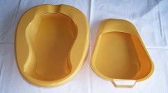
Positioning on Bedpan: Patients restricted to bed use bedpans for defecation. Women use bedpans to pass both urine and feces, whereas men use bedpans only for defecation. Sitting on a bedpan is extremely uncomfortable. Help position patients comfortably. Two types of bedpans are available (Fig. 46-16). The regular bedpan, made of plastic, has a curved smooth upper end and a sharper-edged lower end and is about 5 cm (2 inches) deep. The smaller fracture pan, designed for patients with lower-extremity fractures, has a shallow upper end about 1.3 cm ( inch) deep. The shallow end of the pan fits under the buttocks toward the sacrum; the deeper end, which has a handle, goes just under the upper thighs. The pan needs to be high enough so feces enter it.
When positioning a patient, it is important to prevent muscle strain and discomfort. Never try to lift a patient onto a bedpan. Never place a patient on a bedpan and then leave with the bed flat unless activity restrictions demand it. If the bed is flat, the hips remain hyperextended.
Fig. 46-17 shows proper and improper positions on bedpans. The best method for bedpan placement is to first be sure that the patient is positioned high in bed. Then raise the patient’s head about 30 degrees to prevent hyperextension of the back and provide support to the upper torso. The patient then raises the hips by bending the knees and lifting the hips upward. Place a hand palm up under the patient’s sacrum, resting the elbow on the mattress and using it as a lever to help in lifting while slipping the pan under the patient. Patients who have had abdominal surgery are hesitant to exert strain on suture lines and often have difficulty positioning on a pan. Always wear gloves when handling a bedpan.
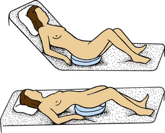
FIG. 46-17 Positions on a bedpan. Top, Proper position reduces patient’s back strain. Bottom, Improper positioning of patient.
When patients are immobile or it is unsafe to allow them to raise their hips, they remain flat and roll onto the bedpan by using the following steps:
1. Lower the head of the bed flat and help the patient roll onto one side, backside toward the nurse.
2. Apply a small amount of powder to back and buttocks or cover bedpan edge with tissue to prevent skin from sticking to the pan.
3. Place the bedpan firmly against the buttocks, down into the mattress with the open rim toward the patient’s feet (Fig. 46-18).
FIG. 46-18 Positioning immobilized patient on bedpan. A, Place bedpan firmly against buttocks. B, Push buttocks down into mattress with open rim toward feet. C, Place one hand against bedpan; place other hand around patient’s fore hip.
4. Keeping one hand against the bedpan, place the other around the patient’s fore hip. Ask the patient to roll back onto the pan, flat in bed. Do not shove the pan under the patient.
5. With the patient positioned comfortably, raise the head of the bed 30 degrees.
6. Place a rolled towel or small pillow under the lumbar curve of the patient’s back for added comfort.
7. Raise the knee gatch or ask the patient to bend the knees to assume a squatting position. Do not raise the knee gatch if contraindicated.
Privacy: Maintain the patient’s privacy during bowel elimination. This is especially important for a patient using a bedpan. The call light and a supply of toilet paper need to be within easy reach. When the patient finishes, respond to the call signal immediately and remove the pan. The patient often requires assistance with wiping. To remove the pan, ask the patient to roll off to the side or raise the hips. While wearing gloves, hold the pan steady to avoid spilling. Avoid pulling or shoving it from under the patient’s hips because this pulls the patient’s skin and causes tissue injury such as shearing (see Chapter 48). Remove the pan and clean the perineum front to back.
After assessing the stool, immediately empty the contents of the bedpan into the toilet or in a special receptacle in the utility room. A spray faucet attached to most toilets allows for the ability to rinse the bedpan thoroughly. The patient uses the same bedpan each time. Finally, document the characteristics of the feces.
Offer the bedpan often. Patients will accidentally soil bedclothes if forced to wait. Many patients try to avoid using a bedpan because it is embarrassing and uncomfortable. They often try to get to the bathroom even though their conditions prohibit ambulation. Warn patients about the risk of falls or accidents.
Acute Care
With some acute illnesses the GI system becomes affected. Changes in the patient’s fluid status, mobility patterns, nutrition, and sleep cycle affect regular bowel habits. Surgical interventions on the GI tract obviously affect bowel elimination. However, surgery on other systems (e.g., the musculoskeletal and cardiovascular systems) sometimes affects the patient’s bowel elimination patterns. Remain sensitive to patients’ elimination needs and intervene to help them maintain as normal bowel elimination habits as possible.
Medications: Some medications initiate and facilitate bowel elimination. Cathartics, laxatives, and occasionally an enema are used to resolve constipation; antidiarrheal preparations help the patient to resolve diarrhea. All of these medications are available over the counter; stronger preparations are available through prescriptions. Caution patients not to use these over-the-counter medications on a prolonged basis without consulting their health care provider.
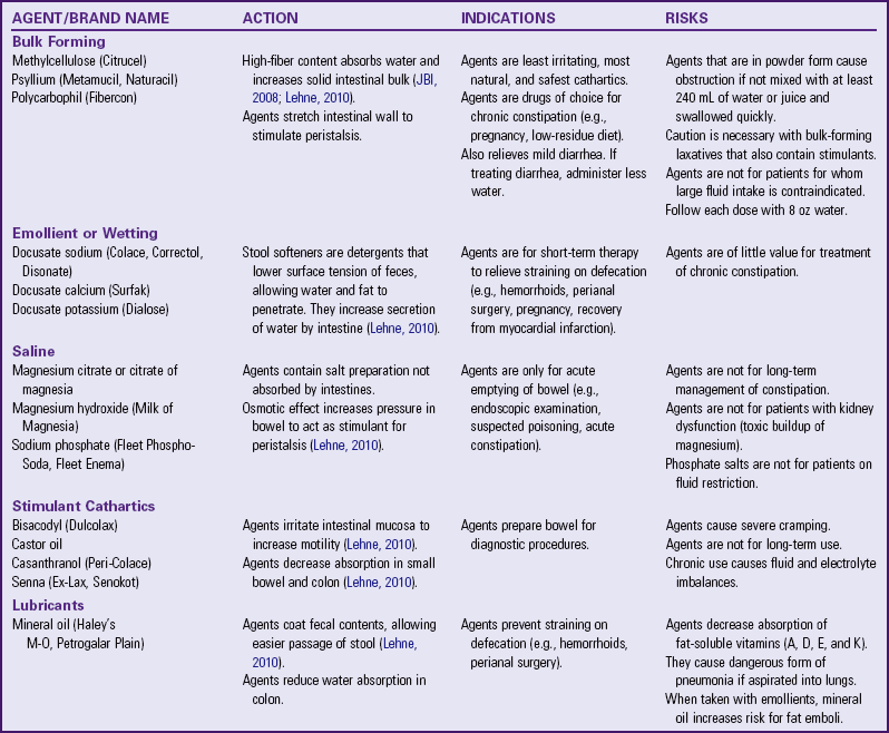
Cathartics and Laxatives: Often a patient is unable to defecate normally because of pain, constipation, or impaction. Cathartics and laxatives have the short-term action of emptying the bowel. They are prescribed for bowel evacuation for patients undergoing GI tests and abdominal surgery. Although the terms cathartic and laxative are often used interchangeably, cathartics have a stronger effect on the intestines. Five types of laxatives and cathartics are available (Table 46-5).
Cathartics and laxatives come in oral, tablet, powder, and suppository dosage forms (see Chapter 31). Although the oral route is most commonly used, suppositories are more effective because of their stimulant effect on the rectal mucosa. Cathartic suppositories such as bisacodyl (Dulcolax) act within 30 minutes. Older adults often get a strong sudden urge to defecate with Dulcolax.
Excessive use of laxatives, enemas, and/or bulk-forming agents increases the patient’s risk for diarrhea and abnormal bowel elimination. It can impair the patient’s normal defecation reflex. In addition, the patient develops altered absorption of nutrients, fluid and electrolyte imbalances, and generalized weakness. In chronically ill or older adult patients weakness and the frequent need to use toilet facilities result in an increased risk for falls and other injuries.
Antidiarrheal Agents: For patients with diarrhea, frequent passage of liquid stools becomes a problem. Many patients use over-the-counter agents such as Imodium to relieve common diarrhea. However, the most effective antidiarrheal agents are prescriptive opiates such as codeine phosphate, opium tincture (Paregoric), and diphenoxylate (Lomotil). Antidiarrheal opiate agents decrease intestinal muscle tone to slow passage of feces. Opiates inhibit peristaltic waves that move feces forward, but they also increase segmental contractions that mix intestinal contents. As a result, the intestinal walls absorb more water. Use antidiarrheal agents with caution because opiates are habit forming. Patients with diarrhea lasting more than 2 days need a stool culture and evaluation of diet and fluid intake for intolerances of food and fluids (e.g., excessive use of fruits, lactose).
Enemas: An enema is the instillation of a solution into the rectum and sigmoid colon. The primary reason for an enema is to promote defecation by stimulating peristalsis. The volume of fluid instilled breaks up the fecal mass, stretches the rectal wall, and initiates the defecation reflex. Enemas are also a vehicle for medications that exert a local effect on rectal mucosa. The most common use for an enema is temporary relief of constipation. Other indications include removing impacted feces, emptying the bowel before diagnostic tests or surgery, and beginning a program of bowel training.
Cleansing Enemas: Cleansing enemas promote the complete evacuation of feces from the colon. They act by stimulating peristalsis through the infusion of a large volume of solution or through local irritation of the mucosa of the colon. They include tap water, normal saline, soapsuds solution, and low-volume hypertonic saline. Each solution has a different osmotic effect, influencing the movement of fluids between the colon and interstitial spaces beyond the intestinal wall. Infants and children receive only normal saline because they are at risk for fluid imbalance.
Tap Water: Tap water is hypotonic and exerts an osmotic pressure lower than fluid in interstitial spaces. After infusion into the colon, tap water escapes from the bowel lumen into interstitial spaces. The net movement of water is low. The infused volume stimulates defecation before large amounts of water leave the bowel. Do not repeat tap-water enemas because water toxicity or circulatory overload develops if the body absorbs large amounts of water.
Normal Saline: Physiologically normal saline is the safest solution to use because it exerts the same osmotic pressure as fluids in interstitial spaces surrounding the bowel. The volume of infused saline stimulates peristalsis. Giving saline enemas does not create the danger of excess fluid absorption.
Hypertonic Solutions: Hypertonic solutions infused into the bowel exert osmotic pressure that pulls fluids out of interstitial spaces. The colon fills with fluid, and the resultant distention promotes defecation. Patients unable to tolerate large volumes of fluid benefit most from this type of enema, which is by design low volume. This type of enema is contraindicated for patients who are dehydrated and young infants. A hypertonic solution of 120 to 180 mL (4 to 6 oz) is usually effective. The commercially prepared Fleet enema is the most common.
Soapsuds: You add soapsuds to tap water or saline to create the effect of intestinal irritation to stimulate peristalsis. Use only pure castile soap that comes in a liquid form included in most soapsuds enema kits. Use soapsuds enemas with caution in pregnant women and older adults because they cause electrolyte imbalance or damage to the intestinal mucosa.
The health care provider sometimes orders a high or low cleansing enema. The terms high and low refer to the height from which, and hence the pressure with which, the fluid is delivered. High enemas cleanse the entire colon. After the enema is infused, ask the patient to turn from the left lateral to the dorsal recumbent, over to the right lateral position. The position change ensures that fluid reaches the large intestine. A low enema cleanses only the rectum and sigmoid colon.
Oil Retention: Oil-retention enemas lubricate the rectum and colon. The feces absorb the oil and become softer and easier to pass. To enhance action of the oil, the patient retains the enema for several hours if possible.
Other Types of Enemas: Carminative enemas provide relief from gaseous distention. They improve the ability to pass flatus. An example of a carminative enema is MGW solution, which contains 30 mL of magnesium, 60 mL of glycerin, and 90 mL of water.
Medicated enemas contain drugs. An example is sodium polystyrene sulfonate (Kayexalate), used to treat patients with dangerously high serum potassium levels. This drug contains a resin that exchanges sodium ions for potassium ions in the large intestine. Another medicated enema is neomycin solution, an antibiotic used to reduce bacteria in the colon before bowel surgery.
Enema Administration: Enemas are available in commercially packaged, disposable units or with reusable equipment prepared before use. Sterile technique is unnecessary because the colon normally contains bacteria. However, wear gloves to prevent the transmission of fecal microorganisms.
Explain the procedure, including the position to assume, precautions to take to avoid discomfort, and length of time necessary to retain the solution before defecation. If the patient needs to take the enema at home, explain the procedure to a family member.
Often the health care provider orders “enemas until clear.” This means that the enema is repeated until the patient passes fluid that is clear and contains no fecal material. It is often necessary to give as many as three enemas, but caution the patient against using more than three. Excess enema use seriously depletes fluids and electrolytes. If the enema fails to return a clear solution after three times (check agency policy) or if the patient seems to not be tolerating the rigors of repeated enemas, notify the health care provider.
Giving an enema to a patient who is unable to contract the external sphincter poses difficulties. Give the enema with the patient positioned on the bedpan. Giving the enema with the patient sitting on the toilet is unsafe because the curved rectal tubing scrapes the rectal wall. Skill 46-1 on pp. 1112-1115 outlines the steps for an enema administration.
Digital Removal of Stool: For a patient with an impaction, the fecal mass is sometimes too large to pass voluntarily. If enemas fail, break up the fecal mass with the fingers and remove it in sections. Digital removal is a last resort in the management of severe constipation and practiced when all other methods have failed. The procedure is very uncomfortable for the patient. Excess rectal manipulation causes irritation to the mucosa; bleeding; and stimulation of the vagus nerve, which results in a reflex slowing of the heart rate. Because of the potential complications of the procedure, a health care provider’s order is necessary to remove a fecal impaction (Kyle, 2007c) (Box 46-8).
Inserting and Maintaining a Nasogastric Tube: A patient’s condition or situation sometimes requires special interventions to decompress the GI tract. Such conditions include surgery (see Chapter 50), infections of the GI tract, trauma to the GI tract, and conditions in which peristalsis is absent.
A nasogastric (NG) tube is a pliable hollow tube that is inserted through the patient’s nasopharynx into the stomach. NG intubation has several purposes (Table 46-6). There are two main categories of NG tubes: Fine- or small-bore tubes and large-bore tubes. Large-bore tubes, 12-Fr and above, are usually used for gastric decompression or removal of gastric secretions. Small-bore tubes are frequently used for medication administration and enteral feedings (see Chapter 44 for enteral feedings). The Levin and Salem sump tubes are the most common for stomach decompression. The Levin tube is a single-lumen tube with holes near the tip. You connect it to a drainage bag or to an intermittent suction device to drain stomach secretions.
TABLE 46-6
Purposes of Nasogastric Intubation
| PURPOSE | DESCRIPTION | TYPE OF TUBE |
| Decompression | Removal of secretions and gaseous substances from gastrointestinal (GI) tract; prevention or relief of abdominal distention (Lewis et al., 2011) | Salem sump, Levin, Miller-Abbott |
| Enteral feeding (see Chapter 44) | Instillation of liquid nutritional supplements or feedings into stomach for patients unable to swallow fluid (Lewis et al., 2011) | Duo, Dobhoff, Levin |
| Compression | Internal application of pressure by means of inflated balloon to prevent internal esophageal or GI hemorrhage (Lewis et al., 2011) | Sengstaken-Blakemore |
| Lavage | Irrigation of stomach in cases of active bleeding, poisoning, or gastric dilation (Lewis et al., 2011) | Levin, Ewald, Salem sump |
The Salem sump tube is preferable for stomach decompression. The tube has two lumina: one for removal of gastric contents and one to provide an air vent. A blue “pigtail” is the air vent that connects with the second lumen. When the main lumen of the sump tube is connected to suction, the air vent permits free, continuous drainage of secretions. Never clamp off the air vent, connect it to suction, or use it for irrigation.
NG tube insertion does not require sterile technique. Simply use clean technique. The procedure is uncomfortable. The patient experiences a burning sensation as the tube passes through the sensitive nasal mucosa. When it reaches the back of the pharynx, the patient sometimes begins to gag. Help him or her relax to make tube insertion easier. Some institutions allow you to use Xylocaine jelly when inserting the tube because it increases patient comfort during the procedure (Skill 46-2 on pp. 1115-1120).
One of the greatest problems in caring for a patient with an NG tube is maintaining comfort. Because the tube constantly irritates the nasal mucosa, you assess the condition of the patient’s nares and mucosa for inflammation and excoriation. The tape or fixation device used to anchor the tube often becomes soiled. Change it every day to lessen irritation. Frequent lubrication of the nares also minimizes excoriation. With one nares occluded, the patient breathes through the mouth. Frequent mouth care (at least every 2 hours) helps minimize dehydration. A glass of cool water for rinsing is useful, but the patient who is allowed nothing by mouth (NPO) should not swallow the water. The patient frequently complains of a sore throat. An ice bag applied externally to the throat helps. If ordered by the health care provider, the patient gargles with topical Xylocaine jelly and/or uses lozenges to minimize the irritation.
After you insert the tube, you need to maintain its patency. Sometimes the tip of the tubing rests against the stomach wall, or the tube becomes blocked with thick secretions. Therefore regular irrigation is necessary. Flushing the tube with normal saline by way of a catheter-tipped syringe clears blockages in the tube. If an NG tube continues to drain improperly after irrigation, reposition it by advancing or withdrawing it slightly. Any change in tube position requires you to verify its placement in the patient’s GI tract.
The NG tube sometimes causes distention. Its presence causes many patients to swallow large volumes of air. Channels of gastric secretions also form along the walls of the stomach and bypass the suction holes. Turning the patient regularly helps to collapse the channels and promotes emptying of stomach contents (Durston, 2009).
Continuing and Restorative Care
Regular elimination patterns need to begin for a patient to recover and return home or to an extended care facility. When patients have a colostomy, they need to learn to care for the ostomy. Other patients require bowel retraining. It is important to remember that you initiate ostomy care and bowel retraining in acute care settings. However, because these are long-term care needs, teaching is usually completed in restorative care settings. Evidence-based interventions reduce the risk of stoma problems in the older adult population over time (Box 46-9).
Care of Ostomies: Patients with temporary or permanent bowel diversions have unique elimination needs. An individual with an ostomy wears a pouch or appliance to collect effluent, stool discharged from the stoma (Dorman, 2009). The patient needs to use meticulous skin care to prevent liquid stool from irritating the skin around the stoma.
Irrigating a Colostomy: Although this practice is not as common as it once was, some patients irrigate their left-sided colostomies to regulate colon emptying. Other patients do not want to spend the additional 60 to 90 minutes in the bathroom every day; thus they empty their pouch as necessary (Dorman, 2009). Only colostomies can be irrigated.
Never use an enema set to irrigate a colostomy. Instead you use specific equipment, which includes a special cone-tipped irrigator to prevent bowel penetration and backflow of the irrigating solution (Fig. 46-19). Help patients to schedule irrigations at times that fit within their daily routine. Before irrigating the stoma, patients usually sit on the toilet and place an irrigating sleeve over the stoma. The end of this sleeve extends into the bowl of the commode. The health care provider orders the amount and type of irrigation solution. For adults the amount typically ranges from 500 to 700 mL of tap water. The patient instills the solution slowly through the lubricated cone tip. Irrigation usually takes 5 to 10 minutes. The patient then removes the cone tip and waits 30 to 45 minutes for the solution and feces to drain out of the irrigation sleeve. Once the drainage stops, the patient applies a stoma cap or a pouch.
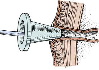
Pouching Ostomies: An ostomy requires a pouch to collect fecal material. An effective pouching system protects the skin, contains fecal material, remains odor free, and is comfortable and inconspicuous. A person wearing a pouch needs to feel secure enough to participate in any activity (Deitz, 2010b).
Many pouching systems are available. To ensure that a pouch fits well and meets the patient’s needs, consider the location of the ostomy, type and size of the stoma, type and amount of ostomy drainage, size and contour of the abdomen, condition of the skin around the stoma, physical activities of the patient, patient’s personal preference, age and dexterity, and cost of equipment. A wound ostomy continence nurse (WOCN) is a nurse specially educated to care for ostomy patients; the WOCN collaborates with staff nurses to be sure that the patient uses the correct pouching system, especially when the patient is ill or is experiencing health changes or problems with the ostomy. For example, you initiate a referral to a WOCN when planning the care of a patient who has a high-output ostomy that requires a pouch modification.
A pouching system consists of a pouch and skin barrier. Some pouching systems such as Squibb-ConvaTec, Hollister, Coloplast, and Smith & Nephew are attached to the patient’s skin from the adhesive surface of the product; whereas other pouching systems such as VIP are nonadhesive. Pouches come in one- and two-piece systems that are disposable or reusable. Some pouches have the opening precut by the manufacturer; others require the stoma opening to be custom cut to the patient’s specific stoma size.
Skin barriers include wafers, pastes, powders, and liquid film applied to the skin around the stoma (Hoeflok et al., 2009). Some wafer skin barriers are permanently attached to the ostomy pouch. These are called one-piece pouch systems. With a two-piece system, the patient detaches the pouch from the skin barrier for emptying or changing. This allows the skin barrier to remain around the patient’s stoma for several days, thus minimizing the chance of skin damage from too-frequent removal of the skin barrier from the peristomal skin. When using a two-piece pouching system, it is important to remember that the skin barrier and pouch need to be the corresponding size and from the same manufacturer. A pouch from one manufacturer does not fit correctly on a skin barrier from another manufacturer. Be sure to use an ostomy pouch made for collecting fecal matter (colostomy or ileostomy) and not one for collecting urine (urostomy).
Assess the stoma color. A normal stoma is bright pink or brick red. Notify the health care provider if the stoma is blue, brown, or black, which indicates circulation problems to the stoma (WOCN et al., 2010). You need to measure the stoma size carefully when selecting and cutting out the opening on the wafer skin barrier. Too tight of an opening constricts the stoma and causes irritation and necrosis. Subtle stoma changes occur over time. Encourage patients to visit their enterostomal nurse at least annually to ensure proper pouching and fit (see Box 46-9). A good skin barrier protects the skin, prevents irritation from repeated removal of the pouch, and is comfortable for the patient to wear (see Fig. 46-21 in Skill 46-3). Skill 46-3 on p. 1121-1124 describes steps for applying a pouching system.
Patients with new stomas often feel vulnerable when they leave the hospital. To provide a smooth transition from hospital to home, offer help for the patient and family caregivers (Richbourg et al., 2007). Effective patient teaching helps patients with a new ostomy transition smoothly to home (Dixon, 2007) (Box 46-10).
Nutritional Considerations for Patients with Ostomies: Nutritional therapy is important for patients with ostomies. During the first weeks after surgery many health care providers recommend low-fiber diets, particularly for patients with ileostomies, because the small bowel requires time to adapt to the diversion. Low-fiber foods include bread, noodles, rice, cream cheese, eggs (not fried), strained fruit juices, lean meats, fish, and poultry. As ostomies heal, patients are able to eat almost any food. High-fiber foods such as fresh fruits and vegetables help ensure a more solid stool. Patients need to avoid blockages of the bowel. The surgical construction of the stoma affects the likelihood of blockage.
A patient with an ileostomy needs to eat slowly and chew food completely. Drinking 10 to 12 glasses of water daily (unless contraindicated) also prevents blockage. High-fiber foods that cause problems include stringy meats, mushrooms, popcorn, fruits such as cherries, and some seafood such as shrimp and crab. Patients with ostomies often benefit from avoiding foods that cause gas and odor, including broccoli, cauliflower, dried beans, and Brussels sprouts.
Bowel Training: The patient with incontinence is unable to maintain bowel control. A bowel training program helps some patients defecate normally, especially those who still have some neuromuscular control.
The training program involves setting up a daily routine. By attempting to defecate at the same time each day and using measures that promote defecation, the patient gains control of bowel reflexes (Mengel, 2009). The program requires time, patience, and consistency. The health care provider determines the patient’s physical readiness and ability to benefit from bowel training. A successful program includes the following:
• Assessing the normal elimination pattern and recording times when the patient is incontinent
• Incorporating principles of gerontological nursing when providing bowel retraining programs for the older adult (Box 46-11)
• Choosing a time in the patient’s pattern to initiate defecation-control measures
• Giving stool softeners orally every day or a cathartic suppository at least half an hour before the selected defecation time (lower colon needs to be free of stool so suppository contacts intestinal mucosa)
• Offering a hot drink (hot tea) or fruit juice (prune juice) (or whatever fluids normally stimulate peristalsis for the patient) before the defecation time
• Helping the patient to the toilet at the designated time
• Avoiding medications such as opioids that increase constipation
• Providing privacy and setting a time limit for defecation (15 to 20 minutes)
• Instructing the patient to lean forward at the hips while sitting on the toilet, apply manual pressure with the hands over the abdomen, and bear down but not strain to stimulate colon emptying
• Not criticizing or conveying frustration if the patient is unable to defecate
• Maintaining normal exercise within the patient’s physical ability
Maintenance of Proper Fluid and Food Intake: In choosing a diet for promoting normal elimination, consider the frequency of defecation, characteristics of feces, and types of foods that impair or promote defecation. The patient with frequent constipation or impaction requires an increased intake of high-fiber foods and more fluids. However, he or she needs to realize that diet therapy provides only long-term relief of elimination problems and does not give immediate relief from problems such as constipation.
When diarrhea is a problem, recommend foods with low-fiber content and discourage foods that typically cause gastric upset or abdominal cramping. Diarrhea caused by illness is sometimes debilitating. If the patient cannot tolerate foods or liquids orally, intravenous therapy (with potassium supplements) is necessary. The patient returns to a normal diet slowly, often beginning with fluids. Excessively hot or cold fluids stimulate peristalsis, causing abdominal cramps and further diarrhea. As tolerance to liquids improves, the patient eats solid foods.
Promotion of Regular Exercise: A daily exercise program helps prevent elimination problems. Walking, riding a stationary bicycle, or swimming stimulates peristalsis. Patients who are sedentary at work are most in need of regular exercise.
For a patient temporarily immobilized, attempt ambulation as soon as possible. If the condition permits, help the patient walk to a chair on the evening of the day of surgery. Have him or her walk farther each day.
Some patients have difficulty passing stool because of weak abdominal and pelvic floor muscles. Exercises help patients who are confined to bed use a bedpan. The patient practices the exercises as follows:
• Lie supine; tighten the abdominal muscles as though pushing them to the floor. Hold the muscles tight to the count of three; relax. Repeat 5 to 10 times as tolerated.
• Flex and contract the thigh muscles by raising one knee slowly toward the chest. Repeat for each leg at least 5 times and increase frequency as tolerated.
Hemorrhoids: Pain results when hemorrhoid tissues are irritated directly. The primary goal for the patient with hemorrhoids is to have soft-formed, painless bowel movements. Proper diet, fluids, and regular exercise improve the likelihood of stools being soft. If the patient becomes constipated, passage of hard stools causes bleeding and irritation. An ice pack or a warm sitz bath (see Chapter 48) provides temporary relief of swollen hemorrhoids.
Maintenance of Skin Integrity: The patient with diarrhea or fecal incontinence is at risk for skin breakdown when fecal contents remain on the skin. The same problem exists for the patient with an ostomy that drains liquid stool. Liquid stool is usually acidic and contains digestive enzymes. Irritation from repeated wiping with toilet tissue aggravates skin breakdown. Bathing the skin after soiling helps, but sometimes it results in more breakdown unless the patient dries the skin thoroughly.
When caring for a patient who is debilitated, incontinent, and unable to ask for assistance, check often for defecation. You can protect the anal areas with petrolatum, zinc oxide, or another ointment that holds moisture in the skin, preventing drying and cracking. Yeast infections of the skin often develop easily. Several powdered antifungal agents are effective against yeast. Do not use baby powder or cornstarch because they have no medical properties, often cake on the skin, are difficult to remove, and enhance fungal infections of the skin.
Evaluation
The effectiveness of care depends on success in meeting the expected outcomes of self-care. Optimally the patient will be able to have regular, pain-free defecation of soft-formed stools. The patient is the only one who is able to determine if the bowel elimination problems have been relieved and which therapies were the most effective (Fig. 46-20).
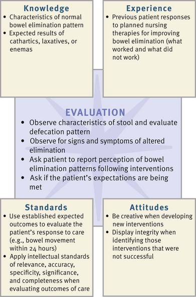
Patient Outcomes
If the nurse establishes a therapeutic relationship with the patient, the patient feels comfortable in discussing the intimate details often associated with bowel elimination. Patients are not embarrassed as nurses help them with elimination needs. Patients relate feelings of comfort and freedom from pain as elimination needs are met within the limits of their condition and treatment. Evaluate a patient’s level of knowledge regarding establishing a normal elimination pattern, caring for an ostomy, and promoting skin integrity. Also determine the extent to which the patient accomplishes normal defection. Ask the patient to describe changes in diet, fluid intake, and activity to promote bowel health. You ask the following questions when a patient’s outcomes are not met:
• Do you use medications such as laxatives or enemas to help you defecate?
• What barriers are preventing you from eating a diet high in fiber and participating in regular exercise?
• How much fluid do you drink in a typical day? What types of fluids do you normally drink?
• What challenges do you encounter when you change your ostomy pouch?
Safety Guidelines for Nursing Skills
Ensuring patient safety is an essential role of the professional nurse. To ensure patient safety, communicate clearly with members of the health care team, assess and incorporate the patient’s priorities of care and preferences, and use the best evidence when making decisions about your patient’s care. When performing the skills in this chapter, remember the following points to ensure safe, individualized, patient care.
• Instruct patients who self-administer enemas to use the side-lying position. Tell them not to self-administer an enema while sitting on the toilet because this position results in curved rectal tubing, causing friction that wears down the rectal wall.
• If a patient has cardiac disease or is taking cardiac or hypertensive medication, obtain pulse rate because manipulation of rectal tissue stimulates the vagus nerve and sometimes causes a sudden decline in pulse rate, which increases the patient’s risk of fainting while on the bedpan, bedside commode, or toilet.
Skill 46-1 Administering a Cleansing Enema
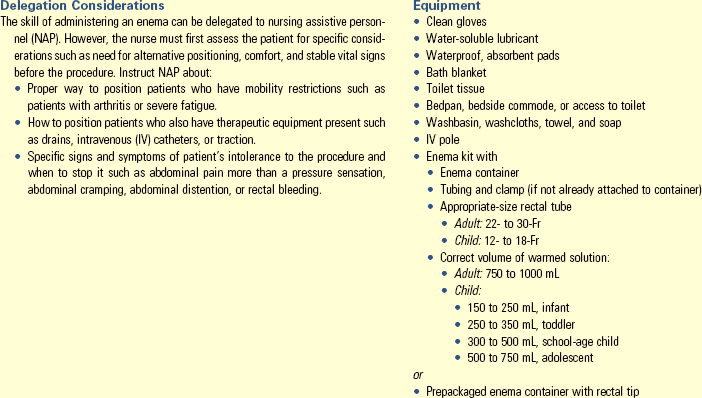
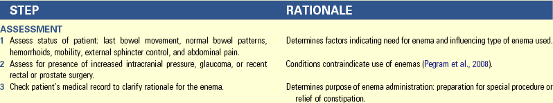
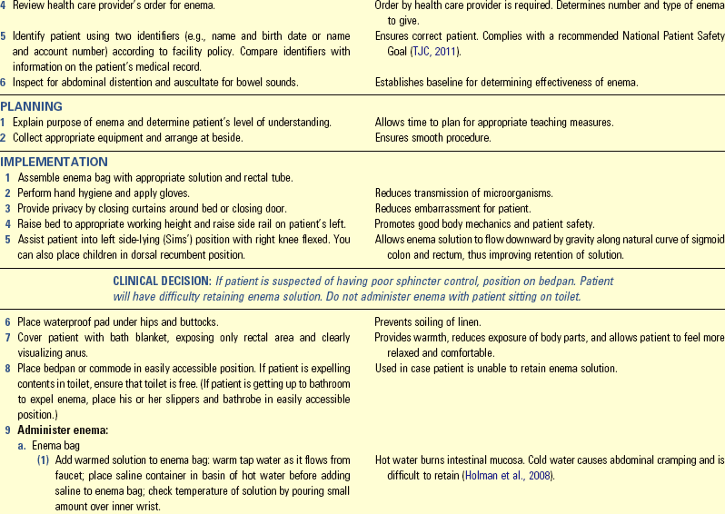
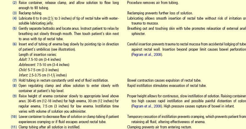

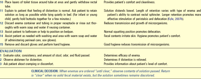
Skill 46-2 Inserting and Maintaining a Nasogastric Tube for Gastric Decompression
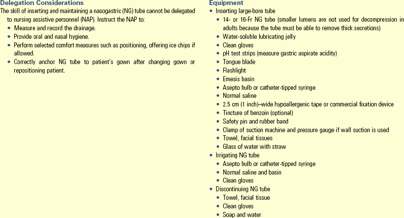
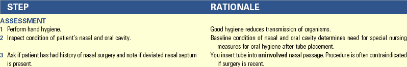
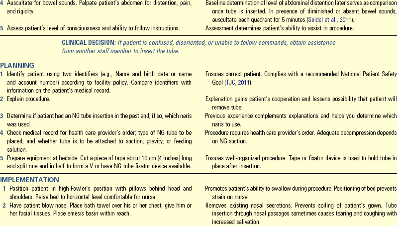
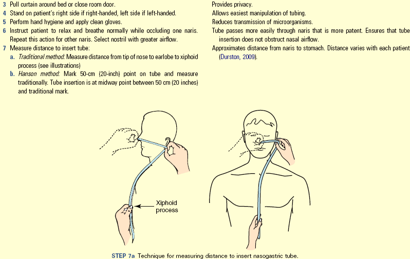
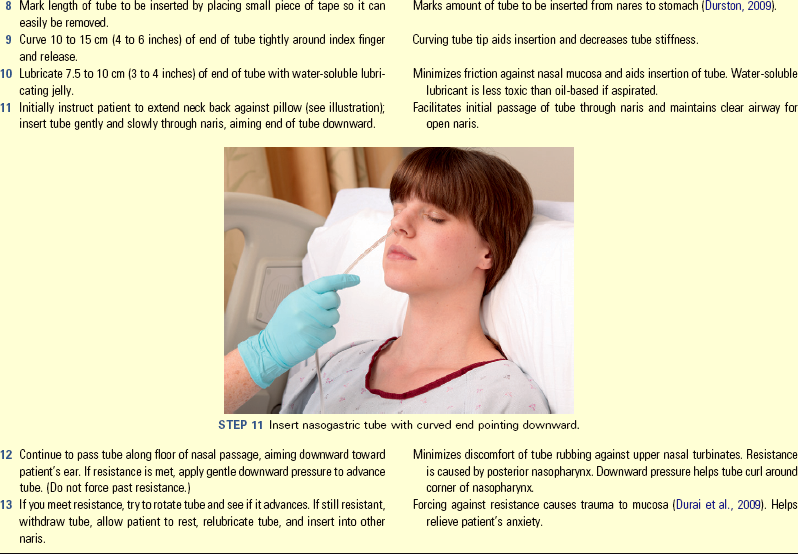
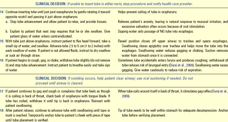
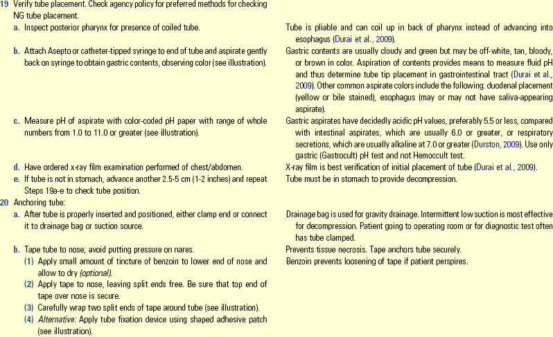
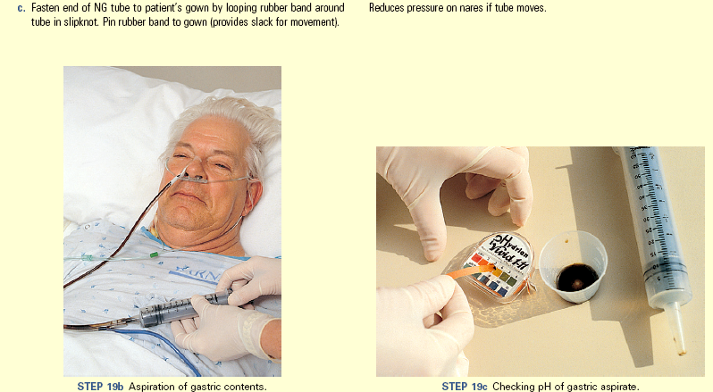
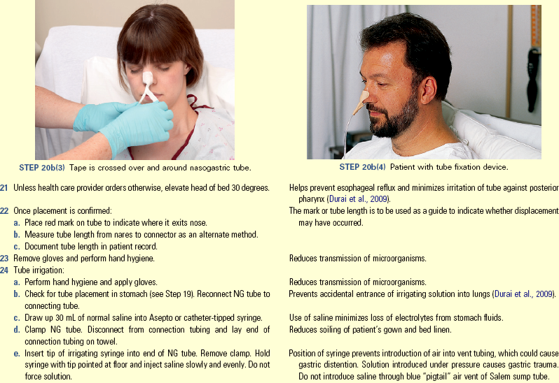
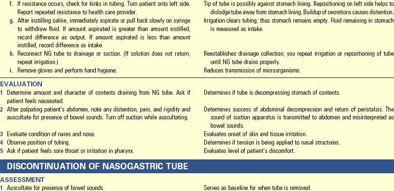
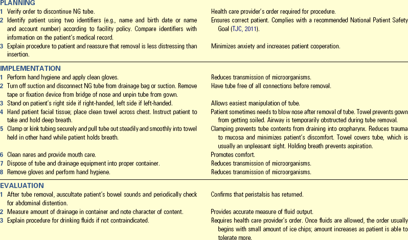
Unexpected Outcomes and Related Interventions:
1. Patient’s abdomen becomes distended and/or painful.
2. Patient complains of sore throat from dry, irritated mucous membranes.
• Increase frequency of oral hygiene.
• Ask health care provider if patient can suck on ice chips or throat lozenges.
3. Patient develops irritation of skin around nares.
4. Patient develops signs of pulmonary aspiration: fever, shortness of breath, pulmonary congestion.
• Record in nurses’ notes time, type, and size of NG tube inserted, patient’s tolerance of procedure, confirmation of placement, character of gastric contents, pH value, whether tube is clamped or connected to drainage device, and amount of suction applied.
• Record in nurses’ notes and/or flow sheet placement checks and amount and character of contents draining from NG tube every shift, unless ordered more often by health care provider.
• Record in nurse’s notes time and date that NG tube was removed, patient’s tolerance of procedure, and his or her status following procedure.
Skill 46-3 Pouching an Ostomy
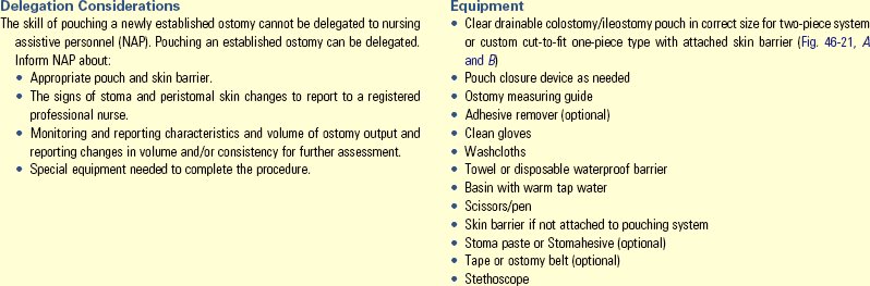
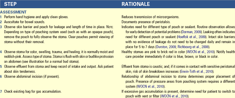
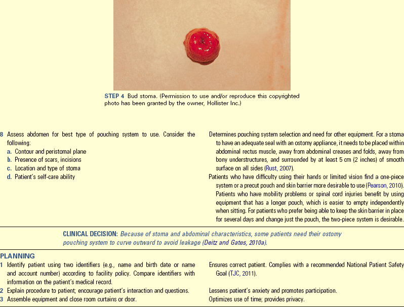
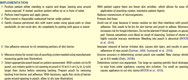
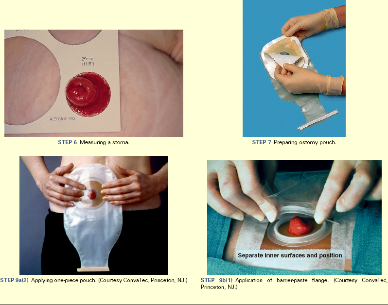
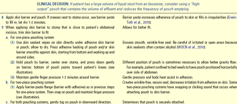
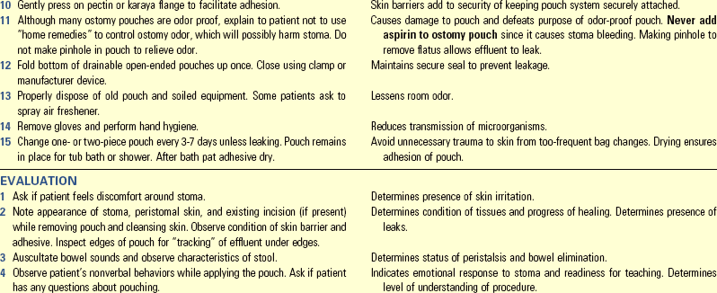
Unexpected Outcomes and Related Interventions:
FIG. 46-21 Ostomy pouches and skin barriers. A, SenSura® one-piece pouch with Velcro closure. B, SenSura® two-piece pouching system with separate skin barrier and attachable pouch. (Note: Skin barriers would need to be custom cut according to stoma size.) (Courtesy Coloplast, Minneapolis, Minn)
1. Patient experiences damage to peristomal skin.
2. Stoma becomes necrotic as manifested by purple or black color, dry instead of moist, failure to bleed, or sloughing tissue.
• Record type of pouch, skin barrier applied, and time; amount and appearance of stool, texture, condition of peristomal skin, and sutures. Record patient teaching.
• Report any of the following to the nurse and/or health care provider:
• Abnormal appearance of stoma (e.g., bleeding, ulcerations, blue or black in color), suture line, peristomal skin, character of output, absence of bowel sounds
• Document abdominal distention and excessive tenderness, nature of bowel sounds.
• Record patient’s level of participation and need for teaching.
• Evaluate patient’s home toileting facilities. This includes presence of adequate toileting facilities, flushable toilet, and number and location of toilets.
• Caution the patient that most ostomy pouches and barriers cannot be flushed down the toilet; they clog the system. Dispose of used ostomy pouch according to local sanitation regulations.
Key Points
• Mechanical breakdown of food elements, gastrointestinal motility, and selective absorption and secretion of substances by the large intestine influence the character of feces.
• Food high in fiber content and an increased fluid intake keep feces soft.
• Ongoing use of cathartics, laxatives, and enemas affects and delays normal defecation reflexes.
• Vagal stimulation, which slows the heart rate, occurs during straining while defecating, taking rectal temperatures, enemas, and digital removal of impacted stool.
• The greatest danger from diarrhea is development of fluid and electrolyte imbalance.
• The location of an ostomy influences consistency of the stool.
• Focus assessment of elimination patterns on bowel habits, factors that normally influence defecation, recent changes in elimination, and a physical examination.
• Indirect and direct visualization of the lower GI tract requires cleansing of the bowel before the procedure.
• Consider frequency of defecation, fecal characteristics, and effect of foods on GI function when selecting a diet promoting normal elimination.
• Proper positioning on a bedpan allows the patient to assume a position similar to squatting without experiencing muscle strain.
• NG intubation decompresses the gastric contents by removing secretions and gaseous products from the GI tract.
• The purposes of gastric decompression are to keep the GI tract free of secretions, reduce nausea and gas, and decrease the risk of vomiting and aspiration.
• Proper selection and use of an ostomy pouching system is necessary to prevent damage to the skin around the stoma.
• Dangers during digital removal of stool include traumatizing the rectal mucosa and promoting vagal stimulation.
• Skin breakdown occurs after repeated exposure to liquid stool.
Clinical Application Questions
Preparing for Clinical Practice
A few days after Javier, the home care nurse, visited Larry in his home, Larry became nauseous, bloated, and had abdominal pain. He took an over-the-counter laxative and went to bed. Around 2:00 am Larry woke with severe abdominal pain. He noticed that his belly was larger than normal, hard, and painful to touch. He felt if he could have a bowel movement, he would feel better. Once Larry was in the bathroom, he began to vomit and passed out from the abdominal pain. His mother heard him and ran to his side, finding him unconscious. She called for an ambulance.
1. Larry arrives in the emergency department of the hospital with vomiting and severe abdominal pain. Which nursing assessment questions does the nurse ask?
2. The nurse receives an order to insert a nasogastric (NG) tube. Once the NG is in place, how does the nurse determine if it is indeed in the stomach?
3. Which additional diagnostic test does the emergency department nurse anticipate that Larry would have?
 Answers to Clinical Application Questions can be found on the Evolve website.
Are You Ready to Test Your Nursing Knowledge?
1. During the nursing assessment a patient reveals that he has diarrhea and cramping every time he has ice cream. He attributes this to the cold nature of the food. However, the nurse begins to suspect that these symptoms are associated with:
2. When assessing a 55-year-old patient who is in the clinic for a routine physical, the nurse instructs the patient about the need to obtain a stool specimen for guaiac fecal occult blood testing (gFOBT):
1. If patient reports rectal bleeding.
2. When there is a family history of polyps.
3. Which of the following medications listed in a patient’s medication history possibly causes gastrointestinal bleeding? (Select all that apply.)
4. Nurses discourage patients from straining on defecation primarily because it causes: (Select all that apply.)
5. A cleansing enema is ordered for a 55-year-old patient before intestinal surgery. The nurse understands that the maximum amount of fluid given is:
6. A patient starts to experience pain while receiving an enema. The nurse notes blood in the return fluid and rectal bleeding. What action does the nurse take first?
1. Administers pain medication
2. Slows down the rate of instillation
7. Number the steps to irrigating a nasogastric tube (NG) in correct order:
1. Slowly aspirate the syringe.
2. Reconnect the NG tube to suction.
3. Clamp and disconnect the NG tube.
4. Perform hand hygiene and apply clean gloves.
5. Insert tip of syringe into NG tube and slowly inject 30 mL saline.
8. List the correct order in which to apply an ostomy pouch:
1. Remove the used pouch and skin barrier.
2. Perform hand hygiene and apply clean gloves.
3. Assess the stoma for color, swelling, and healing.
4. Gently cleanse the peristomal skin with warm tap water.
5. Apply nonallergenic tape around the pectin skin barrier.
6. Cut an opening on the pouch 0.15-0.3 cm ( to
to  inch)larger than the stoma.
7. Press the adhesive backing of the pouch smoothly against the skin.
9. A patient is admitted for lower gastrointestinal (GI) bleeding. What color of stool does the nurse anticipate the patient to have?
10. The nurse is caring for a patient with a colostomy. Which intervention is most important?
1. Cleansing the stoma with hot water
2. Inserting a deodorant tablet in the stoma bag
11. The nurse understands that, when comparing nasogastric tubes used for gastric decompression, a Salem sump is specifically designed to:
1. Minimize the risk of a bowel obstruction.
2. Ensure drainage of the intestines.
12. Before collecting a stool sample for occult blood, the nurse instructs the nursing assistive personnel to:
13. The nurse is taking a health history of a newly admitted patient with a diagnosis Rule/out bowel obstruction. Which of the following is the priority question to ask the patient?
1. Describe your bowel movements.
2. How often do you have a bowel movement?
3. When was the last time you moved your bowels?
4. Do you routinely use stool softeners, laxatives, or enemas?
14. The nurse is caring for a 78-year-old man with diarrhea. Of the following problems, which is the most important to consider?
15. The nurse recognizes which patient needs to use a fracture pan for a bowel movement?
Answers: 1. 3; 2. 3; 3. 1, 4; 4. 3, 4; 5. 4; 6. 4; 7. 4, 3, 5, 1, 2; 8. 2, 1, 4, 3, 6, 7, 5; 9. 1; 10. 3; 11. 3; 12. 1; 13. 3; 14. 2; 15. 4.
References
The American Cancer Society (ACS). Cancer facts and figures. http://www.cancer.org/acs/groups/content/@epidemiologysurveilance/documents/document/acspc-029771.pdf, 2011. [Accessed December 3, 2011].
The American Cancer Society (ACS). Detailed guide: colon and rectum cancer: revised 6/17/2011. http://www.cancer.org/Cancer/ColonandRectumCancer/DetailedGuide/index, 2011. [Accessed December 3, 2011].
Banning, M. Aging and the gut. Nurs Older People. 2008;20(1):17.
Calfee, D. Clostridium difficile: a reemerging pathogen. Geriatrics. 2008;63(9):10.
Chien, D, Bradway, C. Acquired fecal incontinence in community-dwelling adults. Nurse Pract. 2010;35(1):15.
Continence Care Position Statement. Role of the wound, ostomy continence nurse or continence care nurse in continence care. J WOCN. 2009;36(5):529.
Cronenwett, L, et al. Quality and safety education for nurses. Nurs Outlook. 2007;55(1):122.
Deitz, D, Gates, J. Basic ostomy management part 1. Nursing 2010. 2010;40(2):61.
Deitz, D, Gates, J. Basic ostomy management part 2. Nursing 2010. 2010;40(5):62.
Dixon, K. “What really helped me.”: a survey of postoperative patients ostomy education needs. J WOCN. 2007;34(3S):S59.
Dorman, C. Ostomy basics. RN. 2009;72(7):22.
Durai, R, et al. Nasogastric tubes: insertion technique and confirming the correct position. Nurs Times. 2009;105(16):12.
Durston, S. Bowel obstruction: backup along the 750. Nurs Made Incredibly Easy. 2009;7(2):40.
Erwin-Toth, P, et al. Peristomal skin complications. Am J Nurs. 2010;110(2):43.
Gordon, P. Principles and practice of surgery for the colon, rectum, and anus, ed 3. New York: Informa Healthcare; 2007.
Gould, D. Prevention and control of Clostridium difficile infection. Nurs Older People. 2010;22(3):29.
Gray, M. Incontinence-related skin damage: essential knowledge. Ostomy Wound Manage. 2007;53(12):5.
Gray-Vickrey, P. Gathering “pearls” of knowledge for assessing older adults. Nursing 2010. 2010;40(3):34.
Grossman, S, Mager, D. Clostridium difficile: implications for nursing. MedSurg Nurs. 2010;19(3):155.
Herman, A. GI endoscopy: from start to finish. Nurs Made Incredibly Easy. 2010;8(3):5.
Holman, C, et al. Preventing and treating constipation in later life. Nurs Older People. 2008;20(5):22.
Huether, S, McCance, K. Understanding pathophysiology, ed 4. St Louis: Mosby; 2008.
Kyle, G. Bowel Care. Part 1: assessment of constipation. Nurs Times. 2007;103(42):26.
Kyle, G. Bowel Care. Part 4: administering an enema. Nurs Times. 2007;103(45):26.
Kyle, G. Bowel Care. Part 5: a practical guide to digital rectal examination. Nurs Times. 2007;103(46):28.
Lehne, R. Pharmacology for nursing care, ed 7. St Louis: Saunders; 2010.
Lewis, S, et al. Medical-surgical nursing: assessment and management of clinical problems, ed 8. St Louis: Mosby; 2011.
McWilliams, D. Rectal irrigation for patients with functional bowel disorders. Nurs Stand. 2010;24(26):42.
Meiner, S. Gerontologic Nurs, ed 4. St Louis: Mosby; 2011.
Mengel, M. family medicine ambulatory care and prevention, ed 5. New York: McGraw Hill Lange; 2009.
Pagana, KD, Pagana, TJ. Mosby’s diagnostic and laboratory test reference, ed 9. St Louis: Mosby; 2011.
Pearson, T. Older people should be given practical support to effectively manage their stomas. Nurs Times. 2010;106(11):16.
Pegram, A, et al. Safe use of rectal suppositories and enemas with adult patients. Nurs Stand. 2008;22(38):38.
Peters, D. Colon cancer screening: recommendations and barriers to patient participation. Nurse Pract. 2008;33(12):15.
Rust, J. Care of patients with stomas: the pouch change procedure. Nurs Stand. 2007;22(6):43.
Seidel, H, et al. Mosby’s guide to physical examination, ed 7. St Louis: Mosby; 2011.
Steggall, MJ. Digital rectal examination. Nurs Stand. 2008;22(47):46.
Tabloski, P. Gerontological nursing. Upper Saddle River, NJ: Pearson Prentice Hall; 2009.
The Joint Commission (TJC). 2011 National Patient Safety Goals (NPGs). http://www.jointcommission.org/standards_information/npsgs.aspx, 2011. [Accessed December 3, 2011].
Vonberg, R, et al. Infection control measures to limit the spread of Clostridium difficile. Clin Microbiol Infect. 2008;14(suppl 5):2.
Wisniewski, A. Acute abdomen: shaking down the suspects. Nurs Made Incredibly Easy. 2010;8(1):42.
Wound, Ostomy, and Continence Nurses Society (WOCN). Management of the patient with a fecal ostomy: best practice guideline for clinicians. Mount Laurel, NJ: Author; 2010.
Research References
Griffin, K. Biological, psychological and behavioral, and social variables influencing colorectal cancer screening in African Americans. Nurs Res. 2009;58(5):312.
Herlufsen, P, et al. Study of peristomal skin disorders in patients with permanent stomas. Brit J Nurs. 2006;15(16):854.
Hoeflok, J, et al. A prospective multicenter evaluation of a moldable stoma skin barrier. Ostomy/Wound Manage. 2009;55(5):62.
Jacobson, RM, et al. Serum electrolyte shifts following administration of sodium phosphates enema. Gastroenterol Nurs. 2010;33(3):191.
Joanna Briggs Institute (JBI). Management of constipation in older adults. Best Practice. 2008;12(7):1.
Meurette, G, et al. Long-term results of Malone’s procedure with antegrade irrigation for severe chronic constipation. Gastroenterol Clin Biol. 2010;34(3):209.
Ramirez, M, et al. Figuring out sex in a reconfigured body: experiences of female colorectal cancer survivors with ostomies. Women Health. 2009;49(8):608.
Ratliff, CR, Scarano, K, Donovan, AM. Descriptive study of peristomal complications. J Wound Ostomy Continence Nurs. 2005;32(1):33.
Richbourg, L, et al. Ostomy pouch wear time in the United States. J WOCN. 2008;35(5):504.
Richbourg, L, et al. Difficulties experienced by ostomate after hospital discharge. J WOCN. 2007;34(1):70.
Szymanski, K, et al. External stoma and peristomal complications following radical cystectomy and ileal conduit diversion: a systematic review. Ostomy Wound Manage. 2010;56(1):28.
Thompson, H, et al. Matching the skin barrier to the skin type. Brit J Nurs. 2011;20(16):S27.
Williams, J, et al. Evaluating skin care problems in people with stomas. Brit J Nurs. 2010;19(17):S6.