Only a few gram-negative bacteria are considered in this section. A number of important gram-negative pathogens are discussed in the appropriate chapters of organ systems, including bacterial causes of gastrointestinal infections and urinary tract infections. Anaerobic gram-negative organisms are considered later in this chapter. Gram-negative bacterial infections are usually diagnosed by culture.
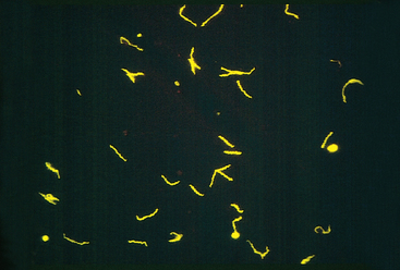
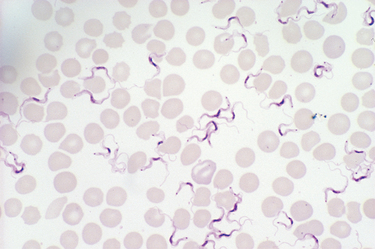
Neisseria are gram-negative diplococci that are flattened on the adjoining sides, giving the pair the shape of a coffee bean (see Fig. 8-3E). These aerobic bacteria have stringent nutritional requirements and grow best on enriched media such as lysed sheep’s blood agar (“chocolate” agar). The two clinically significant Neisseria are N. meningitidis and N. gonorrhoeae.
N. meningitidis is a significant cause of bacterial meningitis, particularly among children younger than 2 years of age. The organism is a common colonizer of the oropharynx and is spread by the respiratory route. Approximately 10% of the population is colonized at any one time, and each episode of colonization lasts, on average, for several months. An immune response leads to elimination of the organism in most people, and this response is protective against subsequent disease with the same serotype of bacteria. There are at least 13 serotypes of N. meningitidis. Invasive disease mainly occurs when people encounter new strains to which they are not immune, as may happen to young children or young adults living in crowded quarters such as military barracks or college dormitories. N. meningitidis disease is endemic in the United States, but epidemics occur periodically in sub-Saharan Africa and cause thousands of deaths.74
Even in the absence of pre-existing immunity, only a small fraction of people infected with N. meningitidis develop meningitis. The bacteria must invade respiratory epithelial cells and travel to the basolateral side of the cells to enter the blood.75 In the blood, the capsule of the bacteria inhibits opsonization and destruction of the bacteria by complement proteins. Despite this, the importance of complement as a first-line defense against N. meningitidis is shown by the increased rates of serious infection among people who have inherited defects in the complement proteins (C5 to C9) that form the membrane attack complex. If N. meningitidis escapes the host response, the consequences can be severe. Although antibiotic treatment of meningitis has greatly reduced mortality of N. meningitidis infection, the death rate is still about 10%. The pathology of pyogenic meningitides is discussed in Chapter 28.
N. gonorrhoeae is an important cause of sexually transmitted disease (STD), infecting about 700,000 people each year in the United States. It is second only to C. trachomatis as a bacterial causative agent of STDs. Infection in men causes urethritis. In women, N. gonorrhoeae infection is often asymptomatic and so may go unnoticed. Untreated infection can lead to pelvic inflammatory disease, which can cause infertility or ectopic pregnancy (Chapter 22). Infection is diagnosed by PCR tests, in addition to culture.
Although N. gonorrhoeae infection usually manifests locally in the genital or cervical mucosa, pharynx, or anorectum, disseminated infections may occur. Like N. meningitidis, N. gonorrhoeae is much more likely to become disseminated in people who lack the complement proteins that form the membrane attack complex. Disseminated infection of adults and adolescents usually causes septic arthritis accompanied by a rash of hemorrhagic papules and pustules. Neonatal N. gonorrhoeae infection causes blindness and, rarely, sepsis. The eye infection, which is preventable by instillation of silver nitrate or antibiotics in the newborn’s eyes, remains an important cause of blindness in some developing nations.
Neisseria use antigenic variation as a strategy to escape the immune response. The existence of multiple serotypes of N. meningitidis results in meningitis in some people on exposure to a new strain, as discussed above. In addition, Neisseria species also generate antigenic variation by special genetic mechanisms, which permit a single bacterial clone to change its expressed antigens (see below) and escape immune defenses.19 Neisseria organisms adhere to and invade nonciliated epithelial cells at the site of entry (nasopharynx, urethra, or cervix). Two surface proteins of Neisseria, both of which bind the bacteria to host cells, undergo antigenic variation through different mechanisms. Although both N. meningitidis and N. gonorrhoeae use these mechanisms, they seem to be more important in N. gonorrhoeae.
Whooping cough, caused by the gram-negative coccobacillus Bordetella pertussis, is an acute, highly communicable illness characterized by paroxysms of violent coughing followed by a loud inspiratory “whoop.” B. pertussis vaccination, whether with killed bacteria or the newer acellular vaccine, has been effective in preventing whooping cough. Since the 1980s, however, rates of pertussis have been increasing in the United States particularly in adolescents and adults, despite continued high rates of vaccination.76 The cause of this increase is not known, but antigenic divergence of clinical strains from vaccine strains and waning immunity in young adults may play a role. In parts of the developing world, where vaccination is not widely practiced, pertussis kills hundreds of thousands of children each year. The diagnosis is best made by PCR, because culture is less sensitive.
Bordetella pertussis colonizes the brush border of the bronchial epithelium and also invades macrophages. Coordinated expression of virulence factors is regulated by the Bordetella virulence gene locus (bvg).77 BVGS is a transmembrane protein that “senses” signals that induce expression of virulence factors. On activation, BVGS phosphorylates the protein BVGA, which regulates transcription of mRNA for adhesins and toxins. The filamentous hemagglutinin adhesin binds to carbohydrates on the surface of respiratory epithelial cells, as well as to CR3 (Mac-1) integrins on macrophages. Pertussis toxin is an exotoxin composed of five distinct proteins, including a catalytic peptide S1 that shows homology with the catalytic peptides of cholera toxin and E. coli heat-labile toxin.78 Like cholera toxin, pertussis toxin ADP-ribosylates and inactivates guanine nucleotide–binding proteins, so these G proteins no longer transduce signals from host plasma membrane receptors. The toxin produced by B. pertussis paralyzes the cilia, thus impairing an important pulmonary defense.
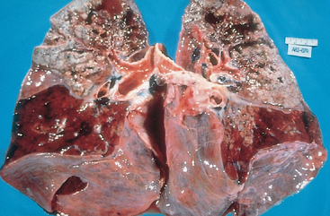
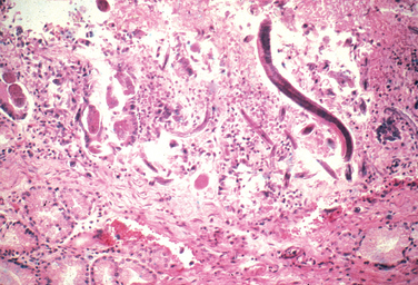
Morphology. Bordetella bacteria cause a laryngotracheobronchitis that in severe cases features bronchial mucosal erosion, hyperemia, and copious mucopurulent exudate (Fig. 8-25). Unless superinfected, the lung alveoli remain open and intact. In parallel with a striking peripheral lymphocytosis (up to 90%), there is hypercellularity and enlargement of the mucosal lymph follicles and peribronchial lymph nodes.
Pseudomonas aeruginosa is an opportunistic aerobic gram-negative bacillus that is a frequent, deadly pathogen of people with cystic fibrosis, severe burns, or neutropenia.93 Many people with cystic fibrosis die of pulmonary failure secondary to chronic infection with P. aeruginosa. P. aeruginosa can be very resistant to antibiotics, making these infections difficult to treat. P. aeruginosa often infects extensive skin burns, which can be a source of sepsis. P. aeruginosa is a common cause of hospital-acquired infections; it has been cultured from washbasins, respirator tubing, nursery cribs, and even antiseptic-containing bottles. P. aeruginosa also causes corneal keratitis in wearers of contact lenses, endocarditis and osteomyelitis in intravenous drug abusers, external otitis (swimmer’s ear) in healthy individuals, and severe external otitis in diabetics.
P. aeruginosa has pili and adherence proteins that bind to epithelial cells and lung mucin, and expresses an endotoxin that causes the symptoms and signs of gram-negative sepsis. Pseudomonas also has a number of distinctive virulence factors. In the lungs of people with cystic fibrosis, these bacteria secrete a mucoid exopolysaccharide called alginate, forming a slimy biofilm that protects bacteria from antibodies, complement, phagocytes, and antibiotics. The organisms also secrete an exotoxin and several other virulence factors. Exotoxin A, like diphtheria toxin, inhibits protein synthesis by ADP-ribosylating the ribosomal protein EF-2.80 P. aeruginosa also releases exoenzyme S, which ADPribosylates RAS and other G proteins that regulate cell growth and metabolism. The organisms also secrete a phospholipase C that lyses red cells and degrades pulmonary surfactant, and an elastase that degrades IgGs and extracellular matrix proteins. These enzymes may be important in tissue invasion and destruction of the cornea in keratitis. Finally, P. aeruginosa produces iron-containing compounds that are extremely toxic to endothelial cells and so may cause the vascular lesions that are characteristic of this infection.81
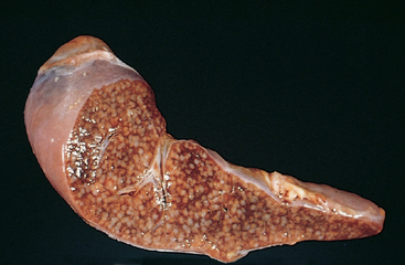
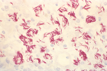
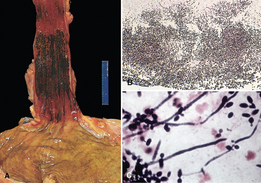
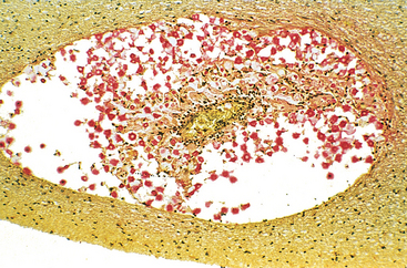
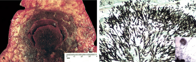
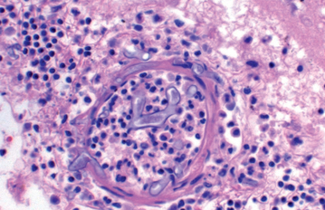
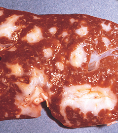
Morphology. Pseudomonas causes a necrotizing pneumonia that is distributed through the terminal airways in a fleur-de-lis pattern, with striking pale necrotic centers and red, hemorrhagic peripheral areas. On microscopic examination, masses of organisms cloud the tissue with a bluish haze, concentrating in the walls of blood vessels, where host cells undergo coagulative necrosis (Fig. 8-26). This picture of gram-negative vasculitis accompanied by thrombosis and hemorrhage, although not pathognomonic, is highly suggestive of P. aeruginosa infection.
Bronchial obstruction caused by mucus plugging and subsequent P. aeruginosa infection are frequent complications of cystic fibrosis. Despite antibiotic treatment and the host immune response, chronic P. aeruginosa infection may result in bronchiectasis and pulmonary fibrosis (Chapter 15).
In skin burns, P. aeruginosa proliferates widely, penetrating deeply into the veins and spreading hematogenously. Well-demarcated necrotic and hemorrhagic oval skin lesions, called ecthyma gangrenosum, often appear. Disseminated intravascular coagulation (DIC) is a frequent complication of bacteremia.
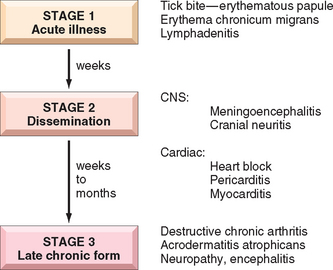
Yersinia pestis is a gram-negative facultative intracellular bacterium that is transmitted from rodents to humans by fleabites or, less often, from one human to another by aerosols. It causes an invasive, frequently fatal infection called plague. Plague, also named Black Death, caused three great pandemics that killed an estimated 100 million people in Egypt and Byzantium in the sixth century; one quarter of Europe’s population in the fourteenth and fifteenth centuries; and tens of millions in India, Myanmar, and China at the beginning of the twentieth century. Currently, 1000 to 3000 cases of plague occur each year worldwide. Wild rodents in the rural western United States are infected with Y. pestis, and 10 to 15 human cases occur per year. Y. enterocolitica and Y. pseudotuberculosis are genetically similar to Y. pestis; these bacteria cause fecal-orally transmitted ileitis and mesenteric lymphadenitis.
Pathogenic Yersinia proliferate within lymphoid tissue. These organisms have a complex of genes, called the Yop virulon, which enable the bacteria to kill host phagocytes.82 The Yop virulon encodes proteins that assemble into a type III secretion system, which is a hollow syringe-like structure that projects from the bacterial surface, binds to host cells, and injects bacterial toxins, called Yops (Yersinia outercoat proteins), into the cell. YopE, YopH, and YopT block phagocytosis by inactivating molecules that regulate actin polymerization. YopJ inhibits the signaling pathways that are activated by LPS, blocking the production of inflammatory cytokines. Y. pestis ensures its own spread by forming a biofilm that obstructs the gut of the infected flea. The flea must regurgitate before it feeds, and thus infects the rodent or human that it is biting.
Morphology. Yersinia pestis causes lymph node enlargement (buboes), pneumonia, or sepsis with a striking neutrophilia. The distinctive histologic features include (1) massive proliferation of the organisms, (2) early appearance of protein-rich and polysaccharide-rich effusions with few inflammatory cells but with marked tissue swelling, (3) necrosis of tissues and blood vessels with hemorrhage and thrombosis, and (4) neutrophilic infiltrates that accumulate adjacent to necrotic areas as healing begins.
In bubonic plague the infected fleabite is usually on the legs and is marked by a small pustule or ulcer. The draining lymph nodes enlarge dramatically within a few days and become soft, pulpy, and plum colored, and may infarct or rupture through the skin. In pneumonic plague there is a severe, confluent, hemorrhagic and necrotizing bronchopneumonia, often with fibrinous pleuritis. In septicemic plague lymph nodes throughout the body as well as organs rich in mononuclear phagocytes develop foci of necrosis. Fulminant bacteremias also induce DIC with widespread hemorrhages and thrombi.
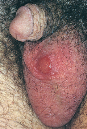
Chancroid is an acute, sexually transmitted, ulcerative infection caused by Hemophilus ducreyi.83 The disease is most common in tropical and subtropical areas among lower socioeconomic groups and men who have regular contact with prostitutes. Chancroid is one of the most common causes of genital ulcers in Africa and Southeast Asia, where it probably serves as an important cofactor in the transmission of HIV infection. Chancroid is uncommon in the United States, with 20 to 50 cases per year reported to the CDC in the past several years. The organism must be cultured in special conditions and PCR-based tests are not widely available, so chancroid may be underdiagnosed.
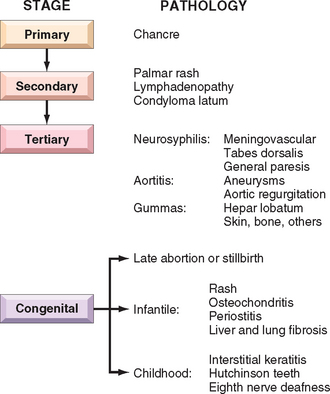
Morphology. Four to seven days after inoculation the person develops a tender, erythematous papule involving the external genitalia. In males the primary lesion is usually on the penis; in females most lesions occur in the vagina or the periurethral area. Over the course of several days the surface of the primary lesion erodes to produce an irregular ulcer, which is more apt to be painful in males than in females. In contrast to the primary chancre of syphilis, the ulcer of chancroid is not indurated, and multiple lesions may be present. The base of the ulcer is covered by shaggy, yellow-gray exudate. The regional lymph nodes, particularly in the inguinal region, become enlarged and tender in about 50% of cases within 1 to 2 weeks of the primary inoculation. In untreated cases the inflamed and enlarged nodes (buboes) may erode the overlying skin to produce chronic, draining ulcers.
Microscopically, the ulcer of chancroid contains a superficial zone of neutrophilic debris and fibrin, with an underlying zone of granulation tissue containing areas of necrosis and thrombosed vessels. A dense, lymphoplasmacytic inflammatory infiltrate is present beneath the layer of granulation tissue. Coccobacilli are sometimes demonstrable in Gram or silver stains, but they are often obscured by other bacteria that colonize the ulcer base.
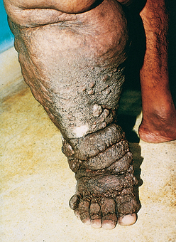
Granuloma inguinale, or donovanosis, is a chronic inflammatory disease caused by Klebsiella granulomatis (formerly called Calymmatobacterium donovani), a minute, encapsulated, coccobacillus. The organism is sexually transmitted. Granuloma inguinale is uncommon in the United States and western Europe but is endemic in rural areas in certain tropical and subtropical regions. Untreated cases are characterized by the development of extensive scarring, often associated with lymphatic obstruction and lymphedema (elephantiasis) of the external genitalia. Culture of the organism is difficult, and PCR assays are still in development, so the diagnosis is made by microscopic examination of smears or biopsy samples of the ulcer.
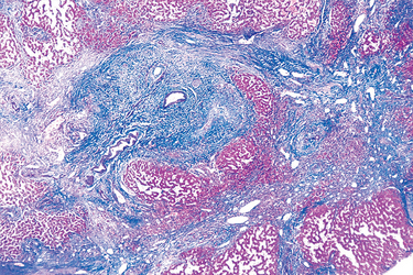
Morphology. Granuloma inguinale begins as a raised, papular lesion on the moist, stratified squamous epithelium of the genitalia or, rarely, the oral mucosa or pharynx. The lesion eventually ulcerates and develops abundant granulation tissue, which is manifested grossly as a protuberant, soft, painless mass. As the lesion enlarges, its borders become raised and indurated. Disfiguring scars may develop in untreated cases and are sometimes associated with urethral, vulvar, or anal strictures. Regional lymph nodes typically are spared or show only nonspecific reactive changes, in contrast to chancroid.
Microscopic examination of active lesions reveals marked epithelial hyperplasia at the borders of the ulcer, sometimes mimicking carcinoma (pseudoepitheliomatous hyperplasia). A mixture of neutrophils and mononuclear inflammatory cells is present at the base of the ulcer and beneath the surrounding epithelium. The organisms are demonstrable in Giemsa-stained smears of the exudate as minute, encapsulated coccobacilli (Donovan bodies) in macrophages. Silver stains (e.g., the Warthin-Starry stain) may also be used to demonstrate the organism.
Bacteria in the genus Mycobacterium are slender, aerobic rods that grow in straight or branching chains. Mycobacteria have a unique waxy cell wall composed of mycolic acid, which makes them acid fast, meaning they will retain stains even on treatment with a mixture of acid and alcohol. Mycobacteria are weakly Gram positive.
Mycobacterium tuberculosis is responsible for most cases of tuberculosis; the reservoir of infection is humans with active tuberculosis. Oropharyngeal and intestinal tuberculosis contracted by drinking milk contaminated with M. bovis is rare in countries where milk is routinely pasteurized, but it is still seen in countries that have tuberculous dairy cows and unpasteurized milk.
Tuberculosis is estimated to affect 1.7 billion individuals worldwide, with 8 to 10 million new cases and 1.6 million deaths each year, a toll second only to HIV disease. Infection with HIV makes people susceptible to rapidly progressive tuberculosis; over 10 million people are infected with both HIV and M. tuberculosis. From 1985 to 1992, the number of tuberculosis cases in the United States rose by 20% because of increase in the disease in people with HIV, immigrants, and those in jail or homeless shelters. Because of public health efforts, the number of cases of tuberculosis has declined since 1993. Currently, there are about 14,000 new cases of active tuberculosis in the United States annually, about half of which occur in foreign-born people.
Tuberculosis flourishes wherever there is poverty, crowding, and chronic debilitating illness. In the United States tuberculosis is mainly a disease of the elderly, the urban poor, and people with AIDS. Certain disease states also increase the risk: diabetes mellitus, Hodgkin lymphoma, chronic lung disease (particularly silicosis), chronic renal failure, malnutrition, alcoholism, and immunosuppression.
It is important that infection with M. tuberculosis be differentiated from disease. Infection is the presence of organisms, which may or may not cause clinically significant disease. Most infections are acquired by person-to-person transmission of airborne organisms from an active case to a susceptible host. In most people primary tuberculosis is asymptomatic, although it may cause fever and pleural effusion. Generally, the only evidence of infection, if any remains, is a tiny, fibrocalcific nodule at the site of the infection. Viable organisms may remain dormant in such lesions for decades. If immune defenses are lowered, the infection may reactivate to produce communicable and potentially life-threatening disease.
Infection typically leads to the development of delayed hypersensitivity to M. tuberculosis antigens, which can be detected by the tuberculin (Mantoux) skin test. About 2 to 4 weeks after infection, intracutaneous injection of purified protein derivative of M. tuberculosis induces a visible and palpable induration that peaks in 48 to 72 hours. A positive tuberculin test result signifies T cell–mediated immunity to mycobacterial antigens. It does not differentiate between infection and disease. False-negative reactions may occur in the setting of certain viral infections, sarcoidosis, malnutrition, Hodgkin lymphoma, immunosuppression, and (notably) overwhelming active tuberculous disease. False-positive reactions may result from infection by atypical mycobacteria or prior vaccination with BCG (Bacillus Calmette-Guerin), an attenuated strain of M. bovis that is used as a vaccine in some countries.
The pathogenesis of tuberculosis in a previously unexposed, immunocompetent person depends on the development of anti-mycobacterial cell-mediated immunity, which confers resistance to the bacteria and also results in development of hypersensitivity to mycobacterial antigens. The pathologic manifestations of tuberculosis, such as caseating granulomas and cavitation, are the result of the hypersensitivity that develops in concert with the protective host immune response. Because the effector cells that mediate immune protection also mediate hypersensitivity and tissue destruction, the appearance of hypersensitivity also signals the acquisition of immunity to the organism. A summary of the pathogenesis of tuberculosis is shown in Figure 8-27.
FIGURE 8-27 The sequence of events in primary pulmonary tuberculosis, commencing with inhalation of virulent Mycobacterium tuberculosis organisms and culminating with the development of cell-mediated immunity to the organism. A, Events occurring in the first 3 weeks after exposure. B, Events thereafter. The development of resistance to the organism is accompanied by the appearance of a positive tuberculin test. γ-IFN, interferon-γ; iNOS, inducible nitric oxide synthase; MHC, major histocompatibility complex; MTB, M. tuberculosis; NRAMP1, natural resistance–associated macrophage protein; TNF, tumor necrosis factor.
Macrophages are the primary cells infected by M. tuberculosis. Early in infection, tuberculosis bacilli replicate essentially unchecked, while later in infection, the cell response stimulates macrophages to contain the proliferation of the bacteria.
In summary, immunity to M. tuberculosis is primarily mediated by TH1 cells, which stimulate macrophages to kill the bacteria. This immune response, while largely effective, comes at the cost of hypersensitivity and accompanying tissue destruction. Reactivation of the infection or re-exposure to the bacilli in a previously sensitized host results in rapid mobilization of a defensive reaction but also increased tissue necrosis. Just as hypersensitivity and resistance are correlated, so, too, the loss of hypersensitivity (indicated by tuberculin negativity in a previously tuberculin-positive individual) may be an ominous sign that resistance to the organism has faded.
The many clinicalpathologic patterns of tuberculosis are shown in Figure 8-28. Primary tuberculosis is the form of disease that develops in a previously unexposed, and therefore unsensitized, person. About 5% of newly infected people develop clinically significant disease. The elderly and profoundly immunosuppressed persons may lose their immunity to M. tuberculosis and so may develop primary tuberculosis more than once. With primary tuberculosis the source of the organism is exogenous.
FIGURE 8-28 The natural history and spectrum of tuberculosis.
(Adapted from a sketch provided by Professor R.K. Kumar, The University of New South Wales, School of Pathology, Sydney, Australia.)
In most people, the primary infection is contained, but in others, primary tuberculosis is progressive. The diagnosis of progressive primary tuberculosis in adults can be difficult. In contrast to secondary tuberculosis (apical disease with cavitation; see below), progressive primary tuberculosis more often resembles an acute bacterial pneumonia, with lower and middle lobe consolidation, hilar adenopathy, and pleural effusion; cavitation is rare, especially in people with severe immunosuppression. Lymphohematogenous dissemination may result in the development of tuberculous meningitis and miliary tuberculosis (discussed below).
Secondary tuberculosis is the pattern of disease that arises in a previously sensitized host. It may follow shortly after primary tuberculosis, but more commonly it appears many years after the initial infection, usually when host resistance is weakened. It most commonly stems from reactivation of a latent infection, but may also result from exogenous reinfection in the face of waning host immunity or when a large inoculum of virulent bacilli overwhelms the host immune system. Reactivation is more common in low-prevalence areas, while reinfection plays an important role in regions of high contagion.
Secondary pulmonary tuberculosis classically involves the apex of the upper lobes of one or both lungs. Because of the pre-existence of hypersensitivity, the bacilli elicit a prompt and marked tissue response that tends to wall off the focus of infection. As a result, the regional lymph nodes are less prominently involved early in secondary disease than they are in primary tuberculosis. On the other hand, cavitation occurs readily in the secondary form. Indeed, cavitation is almost inevitable in neglected secondary tuberculosis, and erosion of the cavities into an airway is an important source of infection because the person now coughs sputum that contains bacteria.
Localized secondary tuberculosis may be asymptomatic. When manifestations appear, they are usually insidious in onset. Systemic symptoms, probably related to cytokines released by activated macrophages (e.g., TNF and IL-1), often appear early in the course and include malaise, anorexia, weight loss, and fever. Commonly, the fever is low grade and remittent (appearing late each afternoon and then subsiding), and night sweats occur. With progressive pulmonary involvement, increasing amounts of sputum, at first mucoid and later purulent, appear. Some degree of hemoptysis is present in about half of all cases of pulmonary tuberculosis. Pleuritic pain may result from extension of the infection to the pleural surfaces. Extrapulmonary manifestations of tuberculosis are legion and depend on the organ system involved.
The diagnosis of pulmonary disease is based in part on the history and on physical and radiographic findings of consolidation or cavitation in the apices of the lungs. Ultimately, however, tubercle bacilli must be identified. Acid-fast smears and cultures of the sputum of patients suspected of having tuberculosis should be performed. Conventional cultures require up to 10 weeks, but culture in liquid media can provide an answer within 2 weeks. PCR amplification of M. tuberculosis DNA allows for even more rapid diagnosis. PCR assays can detect as few as 10 organisms in clinical specimens, compared with more than 10,000 organisms required for smear positivity. However, culture remains the gold standard because it also allows testing of drug susceptibility. Multidrug resistance is now seen more commonly than it was in past years; hence, all newly diagnosed cases in the United States are assumed to be resistant and are treated with multiple drugs. The prognosis is generally good if infections are localized to the lungs, except when they are caused by drug-resistant strains or occur in aged, debilitated, or immunosuppressed individuals, who are at high risk for developing miliary tuberculosis (see below).
All stages of HIV infection are associated with an increased risk of tuberculosis. The use of highly active antiretroviral therapy (HAART) reduces the risk of tuberculosis in people with HIV infection, but even with HAART, people infected with HIV are more likely to get tuberculosis than the uninfected. A low CD4 count before starting HAART is an important risk factor for development of tuberculosis, which underscores the role of the immune response in keeping reactivation of M. tuberculosis in check. The manifestations of tuberculosis differ depending on the degree of immunosuppression. People with less severe immunosuppression (CD4+ T-cell counts >300 cells/mm3) present with usual secondary tuberculosis (apical disease with cavitation). People with more advanced immunosuppression (CD4+ T-cell counts <200 cells/mm3) present with a clinical picture that resembles progressive primary tuberculosis. The extent of immunodeficiency also determines the frequency of extrapulmonary involvement, rising from 10% to 15% in mildly immunosuppressed people to greater than 50% in those with severe immune deficiency. Other atypical features of tuberculosis in HIV-positive people include an increased frequency of false-negative sputum smears and tuberculin tests (the latter due to “anergy”), and the absence of characteristic granulomas in tissues, particularly in the late stages of HIV. The increased frequency of sputum smear-negativity is paradoxical because these immunosuppressed patients typically have higher bacterial loads. The likely explanation is that cavitation and bronchial damage are more in immunocompetent individuals, resulting in more bacilli in expelled sputum. In contrast, the absence of bronchial wall destruction due to reduced T-cell–mediated hypersensitivity results in the excretion of fewer bacilli in the sputum.
Primary Tuberculosis. In countries where infected milk has been eliminated, primary tuberculosis almost always begins in the lungs. Typically, the inhaled bacilli implant in the distal airspaces of the lower part of the upper lobe or the upper part of the lower lobe, usually close to the pleura. As sensitization develops, a 1- to 1.5-cm area of gray-white inflammation with consolidation emerges, known as the Ghon focus. In most cases, the center of this focus undergoes caseous necrosis. Tubercle bacilli, either free or within phagocytes, drain to the regional nodes, which also often caseate. This combination of parenchymal lung lesion and nodal involvement is referred to as the Ghon complex (Fig. 8-29). During the first few weeks there is also lymphatic and hematogenous dissemination to other parts of the body. In approximately 95% of cases, development of cell-mediated immunity controls the infection. Hence, the Ghon complex undergoes progressive fibrosis, often followed by radiologically detectable calcification (Ranke complex), and despite seeding of other organs, no lesions develop.
FIGURE 8-29 Primary pulmonary tuberculosis, Ghon complex. The gray-white parenchymal focus is under the pleura in the lower part of the upper lobe. Hilar lymph nodes with caseation are seen on the left.
Histologically, sites of active involvement are marked by a characteristic granulomatous inflammatory reaction that forms both caseating and noncaseating tubercles (Fig. 8-30A to C). Individual tubercles are microscopic; it is only when multiple granulomas coalesce that they become macroscopically visible. The granulomas are usually enclosed within a fibroblastic rim punctuated by lymphocytes. Multinucleate giant cells are present in the granulomas. Immunocompromised people do not form the characteristic granulomas (Fig. 8-30D).
FIGURE 8-30 The morphologic spectrum of tuberculosis. A characteristic tubercle at low magnification (A) and in detail (B) illustrates central caseation surrounded by epithelioid and multinucleated giant cells. This is the usual response seen in patients who have cell-mediated immunity to the organism. Not all tubercular granulomas might show central caseation (C); hence, irrespective of the presence or absence of caseous necrosis, special stains for acid-fast organisms must be performed when granulomas are present. In immunosuppressed individuals without cellular immunity sheets of foamy macrophages are seen that are packed with mycobacteria (demonstrable with acid-fast stains) (D).
(D, Courtesy of Dr. Dominick Cavuoti, Department of Pathology, University of Texas Southwestern Medical School, Dallas, TX.)
Secondary Tuberculosis. The initial lesion is usually a small focus of consolidation, less than 2 cm in diameter, within 1 to 2 cm of the apical pleura. Such foci are sharply circumscribed, firm, gray-white to yellow areas that have a variable amount of central caseation and peripheral fibrosis (Fig. 8-31). In immunocomptetent individuals, the initial parenchymal focus undergoes progressive fibrous encapsulation, leaving only fibrocalcific scars. Histologically, the active lesions show characteristic coalescent tubercles with central caseation. Tubercle bacilli can often be identified with acid-fast stains in early exudative and caseous phases of granuloma formation but are usually too few to be found in the late, fibrocalcific stages. Localized, apical, secondary pulmonary tuberculosis may heal with fibrosis either spontaneously or after therapy, or the disease may progress and extend along several different pathways.
FIGURE 8-31 Secondary pulmonary tuberculosis. The upper parts of both lungs are riddled with gray-white areas of caseation and multiple areas of softening and cavitation.
Progressive pulmonary tuberculosis may ensue in the elderly and immunosuppressed. The apical lesion expands into adjacent lung and eventually erodes into bronchi and vessels. This evacuates the caseous center, creating a ragged, irregular cavity that is poorly walled off by fibrous tissue. Erosion of blood vessels results in hemoptysis. With adequate treatment the process may be arrested, although healing by fibrosis often distorts the pulmonary architecture. The cavities, now free of inflammation, may persist or become fibrotic. If the treatment is inadequate or if host defenses are impaired, the infection may spread via airways, lymphatic channels, or the vascular system. Miliary pulmonary disease occurs when organisms draining through lymphatics enter the venous blood and circulate back to the lung. Individual lesions are either microscopic or small, visible (2-mm) foci of yellow-white consolidation scattered through the lung parenchyma (the adjective “miliary” is derived from the resemblance of these foci to millet seeds). Miliary lesions may expand and coalesce, resulting in consolidation of large regions or even whole lobes of the lung. With progressive pulmonary tuberculosis, the pleural cavity is invariably involved, and serous pleural effusions, tuberculous empyema, or obliterative fibrous pleuritis may develop.
Endobronchial, endotracheal, and laryngeal tuberculosis may develop by spread through lymphatic channels or from expectorated infectious material. The mucosal lining may be studded with minute granulomatous lesions that may only be apparent microscopically.
Systemic miliary tuberculosis occurs when bacteria disseminate through the systemic arterial system. Miliary tuberculosis is most prominent in the liver, bone marrow, spleen, adrenals, meninges, kidneys, fallopian tubes, and epididymis, but could involve any organ (Fig. 8-32).
FIGURE 8-32 Miliary tuberculosis of the spleen. The cut surface shows numerous gray-white tubercles.
Isolated tuberculosis may appear in any of the organs or tissues seeded hematogenously and may be the presenting manifestation. Organs that are commonly involved include the meninges (tuberculous meningitis), kidneys (renal tuberculosis), adrenals (formerly an important cause of Addison disease), bones (osteomyelitis), and fallopian tubes (salpingitis). When the vertebrae are affected, the disease is referred to as Pott disease. Paraspinal “cold” abscesses in these patients may track along tissue planes and present as an abdominal or pelvic mass.
Lymphadenitis is the most frequent presentation of extrapulmonary tuberculosis, usually occurring in the cervical region (“scrofula”). In HIV-negative individuals, lymphadenitis tends to be unifocal and localized. HIV-positive people, on the other hand, almost always have multifocal disease, systemic symptoms, and either pulmonary or other organ involvement by active tuberculosis.
In years past, intestinal tuberculosis contracted by the drinking of contaminated milk was a fairly common primary focus of disease. In countries where milk is pasteurized, intestinal tuberculosis is more often caused by the swallowing of coughed-up infective material in patients with advanced pulmonary disease. Typically the organisms are seed to mucosal lymphoid aggregates of the small and large bowel, which then undergo granulomatous inflammation that can lead to ulceration of the overlying mucosa, particularly in the ileum.
Mycobacterium avium and M. intracellulare are separate species, but the infections they cause are so similar that they are simply referred to as M. avium-intracellulare complex, or MAC. MAC is common in soil, water, dust, and domestic animals. Clinically significant infection with MAC is uncommon except among people with AIDS and low numbers of CD4+ lymphocytes (<60 cells/mm3).
In AIDS patients MAC causes widely disseminated infections, and organisms proliferate abundantly in many organs, including the lungs and gastrointestinal system. Unchecked by the immune response, the organisms reach very high levels: up to 104 organisms/mL of blood and 106 organisms/gm in tissue. Patients are feverish, with drenching night sweats and weight loss. In the rare case of MAC in a person without HIV, the organisms primarily infect the lung, causing a productive cough and sometimes fever and weight loss.
Morphology. The hallmark of MAC infections in patients with HIV is abundant acid-fast bacilli within macrophages (Fig. 8-33). Depending on the severity of immune deficiency, MAC infections can be widely disseminated throughout the mononuclear phagocyte system, causing enlargement of involved lymph nodes, liver, and spleen, or localized to the lungs. There may be a yellowish pigmentation to these organs secondary to the large number of organisms present in swollen macrophages. Granulomas, lymphocytes, and tissue destruction are rare.
Leprosy, or Hansen’s disease, is a slowly progressive infection caused by Mycobacterium leprae that mainly affects the skin and peripheral nerves and results in disabling deformities. M. leprae is likely to be transmitted from person to person through aerosols from asymptomatic lesions in the upper respiratory tract. Inhaled M. leprae, like M. tuberculosis, is taken up by alveolar macrophages and disseminates through the blood, but replicates only in relatively cool tissues of the skin and extremities. Despite its low communicability, leprosy remains endemic among an estimated 10 to 15 million people living in poor tropical countries.
M. leprae is an acid-fast obligate intracellular organism that grows very poorly in culture but can be propagated in the armadillo. It proliferates best at 32° to 34°C, the temperature of the human skin and the core temperature of armadillos. Like M. tuberculosis, M. leprae secretes no toxins, and its virulence is based on properties of its cell wall. The cell wall is similar enough to that of M. tuberculosis that immunization with BCG confers some protection against M. leprae infection. Cell-mediated immunity is reflected by delayed-type hypersensitivity reactions to dermal injections of a bacterial extract called lepromin.
M. leprae causes two strikingly different patterns of disease. People with the less severe form, tuberculoid leprosy, have dry, scaly skin lesions that lack sensation. They often have asymmetric involvement of large peripheral nerves. The more severe form, lepromatous leprosy, includes symmetric skin thickening and nodules. This is also called anergic leprosy, because of the unresponsiveness (anergy) of the host immune system. Cooler areas of skin, including the earlobes and feet, are more severely affected than warmer areas, such as the axilla and groin. In lepromatous leprosy, widespread invasion of the mycobacteria into Schwann cells and into endoneural and perineural macrophages damages the peripheral nervous system. In advanced cases of lepromatous leprosy, M. leprae is present in sputum and blood. People can also have intermediate forms of disease, called borderline leprosy.
The T-helper lymphocyte response to M. leprae determines whether an individual has tuberculoid or lepromatous leprosy.88 People with tuberculoid leprosy have a TH1 response associated with production of IL-2 and IFN-γ. As with M. tuberculosis, IFN-γ is critical to mobilizing an effective host macrophage response. Lepromatous leprosy is associated with a weak TH1 response and, in some cases, a relative increase in the TH2 response. The net result is weak cell-mediated immunity and an inability to control the bacteria. Occasionally, most often in the lepromatous form, antibodies are produced against M. leprae antigens. Paradoxically, these antibodies are usually not protective, but they may form immune complexes with free antigens that can lead to erythema nodosum, vasculitis, and glomerulonephritis.
Morphology. Tuberculoid leprosy begins with localized flat, red skin lesions that enlarge and develop irregular shapes with indurated, elevated, hyperpigmented margins and depressed pale centers (central healing). Neuronal involvement dominates tuberculoid leprosy. Nerves become enclosed within granulomatous inflammatory reactions and, if small (e.g., the peripheral twigs), are destroyed (Fig. 8-34). Nerve degeneration causes skin anesthesias and skin and muscle atrophy that render the person liable to trauma of the affected parts, leading to the development of chronic skin ulcers. Contractures, paralyses, and autoamputation of fingers or toes may ensue. Facial nerve involvement can lead to paralysis of the eyelids, with keratitis and corneal ulcerations. On microscopic examination, all sites of involvement have granulomatous lesions closely resembling those found in tuberculosis, and bacilli are almost never found, hence the name “paucibacillary” leprosy. The presence of granulomas and absence of bacteria reflect strong T-cell immunity. Because leprosy pursues an extremely slow course, spanning decades, most patients die with leprosy rather than of it.
FIGURE 8-34 Leprosy. A, Peripheral nerve. Note the inflammatory cell infiltrates in the endoneural and epineural compartments. B, Cells within the endoneurium contain acid-fast positive lepra bacilli.
(Courtesy of E.P. Richardson, Jr., and U. De Girolami, Harvard Medical School.)
Lepromatous leprosy involves the skin, peripheral nerves, anterior chamber of the eye, upper airways (down to the larynx), testes, hands, and feet. The vital organs and CNS are rarely affected, presumably because the core temperature is too high for growth of M. leprae. Lepromatous lesions contain large aggregates of lipid-laden macrophages (lepra cells), often filled with masses (“globi”) of acid-fast bacilli (Fig. 8-35). Because of the abundant bacteria, lepromatous leprosy is referred to as “multibacillary”. Macular, papular, or nodular lesions form on the face, ears, wrists, elbows, and knees. With progression, the nodular lesions coalesce to yield a distinctive leonine facies. Most skin lesions are hypoesthetic or anesthetic. Lesions in the nose may cause persistent inflammation and bacilli-laden discharge. The peripheral nerves, particularly the ulnar and peroneal nerves where they approach the skin surface, are symmetrically invaded with mycobacteria, with minimal inflammation. Loss of sensation and trophic changes in the hands and feet follow the nerve lesions. Lymph nodes contain aggregates of bacteria-filled foamy macrophages in the paracortical (T-cell) areas and reactive germinal centers. In advanced disease, aggregates of macrophages are also present in the splenic red pulp and the liver. The testes are usually extensively involved, leading to destruction of the seminiferous tubules and consequent sterility.
Spirochetes are gram-negative, slender corkscrew-shaped bacteria with axial periplasmic flagella wound around a helical protoplasm. The bacteria are covered in a membrane called an outer sheath, which may mask bacterial antigens from the host immune response. Treponema pallidum subsp. pallidum is the microaerophilic spirochete that causes syphilis, a chronic venereal disease with multiple clinical presentations. Other closely related treponemes cause yaws (Treponema pallidum subsp. pertenue) and pinta (Treponema pallidum subsp. carateum).
Syphilis is a chronic venereal disease with multiple presentations. The causative spirochete, T. pallidum subsp. pallidum, hereafter referred to simply as T. pallidum, is too slender to be seen in Gram stain, but it can be visualized by silver stains, dark-field examination, and immunofluorescence techniques (Fig. 8-36). Sexual contact is the usual mode of spread. Transplacental transmission of T. pallidum occurs readily, and active disease during pregnancy results in congenital syphilis. T. pallidum cannot be grown in culture.
FIGURE 8-36 Treponema pallidum (dark-field microscopy) showing several spirochetes in scrapings from the base of a chancre.
(Courtesy of Dr. Paul Southern, Department of Pathology, University of Texas Southwestern Medical School, Dallas, TX.)
Public health programs and penicillin treatment reduced the number of cases of syphilis in the United States from the late 1940s until the 1970s. Cases of syphilis surged upward in the mid-1980s, reaching a total of 50,000 cases in 1990. Renewed public health efforts led to a sharp drop in the incidence of syphilis over the next 10 years, but since 2000 there has been a steady rise in the number of cases reported annually, to ∼10,000 in 2006.
Syphilis is divided into three stages, with distinct clinical and pathologic manifestations (Fig. 8-37).
This stage, occurring approximately 3 weeks after contact with an infected individual, features a single firm, nontender, raised, red lesion (chancre) located at the site of treponemal invasion on the penis, cervix, vaginal wall, or anus. The chancre heals in 3 to 6 weeks with or without therapy. Spirochetes are plentiful within the chancre and can be seen by immunofluorescent stains of serous exudate. Treponemes spread throughout the body by hematologic and lymphatic dissemination even before the appearance of the chancre.
This stage usually occurs 2 to 10 weeks after the primary chancre and is due to spread and proliferation of the spirochetes within the skin and mucocutaneous tissues. Secondary syphilis occurs in approximately 75% of untreated people. The skin lesions, which frequently occur on the palms or soles of the feet, may be maculopapular, scaly, or pustular. Moist areas of the skin, such as the anogenital region, inner thighs, and axillae, may have condylomata lata, which are broad-based, elevated plaques. Silvery-gray superficial erosions may form on any of the mucous membranes but are particularly common in the mouth, pharynx, and external genitalia. All these painless superficial lesions contain spirochetes and so are infectious. Lymphadenopathy, mild fever, malaise, and weight loss are also common in secondary syphilis. The symptoms of secondary syphilis last several weeks, after which the person enters the latent phase of the disease. Superficial lesions may recur during the early latent phase, although they are milder.
This stage is rare where adequate medical care is available, but it occurs in approximately one third of untreated patients, usually after a latent period of 5 years or more. Tertiary syphilis has three main manifestations: cardiovascular syphilis, neurosyphilis, and so-called benign tertiary syphilis. These may occur alone or in combination.
Cardiovascular syphilis, in the form of syphilitic aortitis, accounts for more than 80% of cases of tertiary disease. The aortitis leads to slowly progressive dilation of the aortic root and arch, which causes aortic valve insufficiency and aneurysms of the proximal aorta (see Chapter 11).
Neurosyphilis may be symptomatic or asymptomatic. Symptomatic disease manifests in several ways, including chronic meningovascular disease, tabes dorsalis, and a generalized brain parenchymal disease called general paresis. These are discussed in Chapter 28. Asymptomatic neurosyphilis, which accounts for about one third of neurosyphilis cases, is detected when a patient’s CSF exhibits abnormalities such as pleocytosis (increased numbers of inflammatory cells), elevated protein levels, or decreased glucose. Antibodies stimulated by the spirochetes, discussed below, can also be detected in the CSF, and this is the most specific test for neurosyphilis. Antibiotics are given for a longer time if the spirochetes have spread to the CNS, and so patients with tertiary syphilis should be tested for neurosyphilis even if they do not have neurologic symptoms.
So-called benign tertiary syphilis is characterized by the formation of gummas in various sites. Gummas are nodular lesions probably related to the development of delayed hypersensitivity to the bacteria. They occur most commonly in bone, skin, and the mucous membranes of the upper airway and mouth, although any organ may be affected. Skeletal involvement characteristically causes local pain, tenderness, swelling, and sometimes pathologic fractures. Involvement of skin and mucous membranes may produce nodular lesions or, rarely, destructive, ulcerative lesions that mimic malignant neoplasms. Gummas are now very rare because of the use of effective antibiotics and are seen mainly in individuals with AIDS.
Congenital syphilis occurs when T. pallidum crosses the placenta from an infected mother to the fetus. Maternal transmission happens most frequently during primary or secondary syphilis, when the spirochetes are most numerous. Because the manifestations of maternal syphilis may be subtle, routine serologic testing for syphilis is mandatory in all pregnancies. Intrauterine death and perinatal death each occurs in approximately 25% of cases of untreated congenital syphilis.
Manifestations of congenital disease are divided into early (infantile) and late (tardive) syphilis, depending on whether they occur in the first 2 years of life or later. Early congenital syphilis is often manifested by nasal discharge and congestion (snuffles) in the first few months of life. A desquamating or bullous rash can lead to sloughing of the skin, particularly of the hands and feet and around the mouth and anus. Hepatomegaly and skeletal abnormalities are also common.
Nearly half of untreated children with neonatal syphilis will develop late manifestations, which are discussed below.
Serology remains the mainstay of diagnosis, although microscopy and PCR are also useful. Serologic tests include nontreponemal antibody tests and antitreponemal antibody tests. Nontreponemal tests measure antibody to cardiolipin, a phospholipid present in both host tissues and T. pallidum. These antibodies are detected in the rapid plasma reagin and Venereal Disease Research Laboratory (VDRL) tests. Nontreponemal tests typically become positive 4 to 6 weeks after infection, and so immunofluorescence of exudate from the chancre is important for diagnosis early in infection. The nontreponemal tests are nearly always positive in secondary syphilis, but they usually become negative in tertiary syphilis. The VDRL and rapid plasma reagin tests are used as screening tests for syphilis and to monitor response to therapy, since these tests become negative after successful treatment of infection. False-positive VDRL test results are not uncommon and are associated with certain acute infections, collagen vascular diseases (e.g., systemic lupus erythematosus), drug addiction, pregnancy, hypergammaglobulinemia of any cause, and lepromatous leprosy.
Treponemal antibody tests measure antibodies that specifically react with T. pallidum. These include the fluorescent treponemal antibody absorption test and the microhemagglutination assay for T. pallidum antibodies. These tests also become positive 4 to 6 weeks after infection, but unlike nontreponemal antibody tests, they remain positive indefinitely, even after successful treatment. They are not recommended as primary screening tests because they are significantly more expensive than nontreponemal tests. While more specific than the nontreponemal tests, false-positive treponemal antibody tests can also occur.
Serologic response may be delayed, absent, or exaggerated (false-positive results) in people co-infected with syphilis and HIV. However, in most cases, these tests remain useful in the diagnosis and management of syphilis even in people infected with HIV.
Morphology. In primary syphilis a chancre occurs on the penis or scrotum of 70% of men and on the vulva or cervix of 50% of women. The chancre is a slightly elevated, firm, reddened papule, up to several centimeters in diameter, that erodes to create a clean-based shallow ulcer. The contiguous induration creates a button-like mass directly adjacent to the eroded skin, providing the basis for the designation hard chancre (Fig. 8-38). On histologic examination, treponemes are visible at the surface of the ulcer with silver stains (e.g., Warthin-Starry stain) or immunofluorescence techniques. The chancre contains an intense infiltrate of plasma cells, with scattered macrophages and lymphocytes and a proliferative endarteritis (see Fig. 8-8). The endarteritis, which is seen in all stages of syphilis, starts with endothelial cell activation and proliferation and progresses to intimal fibrosis. The regional nodes are usually enlarged due to nonspecific acute or chronic lymphadenitis, plasma cell–rich infiltrates, or granulomas.
FIGURE 8-38 Syphilitic chancre in the scrotum (see Figure 8-8 for the histopathology of syphilis).
(Courtesy of Dr. Richard Johnson, Beth Israel–Deaconess Hospital, Boston, MA.)
In secondary syphilis widespread mucocutaneous lesions involve the oral cavity, palms of the hands, and soles of the feet. The rash frequently consists of discrete red-brown macules less than 5 mm in diameter, but it may be follicular, pustular, annular, or scaling. Red lesions in the mouth or vagina contain the most organisms and are the most infectious. Histologically, the mucocutaneous lesions of secondary syphilis show the same plasma cell infiltrate and obliterative endarteritis as the primary chancre, although the inflammation is often less intense.
Tertiary syphilis most frequently involves the aorta; the CNS; and the liver, bones, and testes. The aortitis is caused by endarteritis of the vasa vasorum of the proximal aorta. Occlusion of the vasa vasorum results in scarring of the media of the proximal aortic wall, causing a loss of elasticity. There may be narrowing of the coronary artery ostia caused by subintimal scarring with resulting myocardial ischemia. The morphologic and clinical features of syphilitic aortitis are discussed in greater detail with diseases of the blood vessels (Chapter 11). Neurosyphilis takes one of several forms, designated meningovascular syphilis, tabes dorsalis, and general paresis (Chapter 28). Syphilitic gummas are white-gray and rubbery, occur singly or multiply, and vary in size from microscopic lesions resembling tubercles to large tumor-like masses. They occur in most organs but particularly in skin, subcutaneous tissue, bone, and joints. In the liver, scarring as a result of gummas may cause a distinctive hepatic lesion known as hepar lobatum (Fig. 8-39). On histologic examination, the gummas have centers of coagulated, necrotic material and margins composed of plump, palisading macrophages and fibroblasts surrounded by large numbers of mononuclear leukocytes, chiefly plasma cells. Treponemes are scant in gummas and are difficult to demonstrate.
FIGURE 8-39 Trichrome stain of liver shows a gumma (scar), stained blue, caused by tertiary syphilis (the hepatic lesion is also known as hepar lobatum).
The rash of congenital syphilis is more severe than that of adult secondary syphilis. It is a bullous eruption of the palms and soles of the feet associated with epidermal sloughing. Syphilitic osteochondritis and periostitis affect all bones, but lesions of the nose and lower legs are most distinctive. Destruction of the vomer causes collapse of the bridge of the nose and, later on, the characteristic saddle nose deformity. Periostitis of the tibia leads to excessive new bone growth on the anterior surfaces and anterior bowing, or saber shin. There is also widespread disturbance in endochondral bone formation. The epiphyses become widened as the cartilage overgrows, and cartilage is found in displaced islands within the metaphysis.
The liver is often severely affected in congenital syphilis. Diffuse fibrosis permeates lobules to isolate hepatic cells into small nests, accompanied by the characteristic lymphoplasmacytic infiltrate and vascular changes. Gummas are occasionally found in the liver, even in early cases. The lungs may be affected by a diffuse interstitial fibrosis. In the syphilitic stillborn, the lungs appear pale and airless (pneumonia alba). The generalized spirochetemia may lead to diffuse interstitial inflammatory reactions in virtually any other organ (e.g., the pancreas, kidneys, heart, spleen, thymus, endocrine organs, and CNS).
The late manifestations of congenital syphilis include a distinctive triad of interstitial keratitis, Hutchinson teeth, and eighth-nerve deafness. In addition to interstitial keratitis, the ocular changes include choroiditis and abnormal retinal pigmentation. Hutchinson teeth are small incisors shaped like a screwdriver or a peg, often with notches in the enamel. Eighth-nerve deafness and optic nerve atrophy develop secondary to meningovascular syphilis.
There are no good animal models of syphilis, and T. pallidum has never been grown in culture (it lacks genes for making nucleotides, fatty acids, and most amino acids). As a result, our scant knowledge of T. pallidum pathogenesis comes mainly from observations of the disease in humans.
Proliferative endarteritis occurs in all stages of syphilis. The pathophysiology of the endarteritis is not known, although the scarcity of treponemes and the intense inflammatory infiltrate suggest that the immune response plays a role in the development of these lesions. Regardless of the mechanism, much of the pathology of the disease, such as syphilitic aortitis, can be ascribed to the vascular abnormalities.
The immune response to T. pallidum reduces the burden of bacteria, but it may also have a central role in the pathogenesis of the disease. The T cells that infiltrate the chancre are TH1 cells, suggesting that activation of macrophages to kill bacteria may cause resolution of the local infection.89 Although there are many plasma cells in the syphilitic lesions and treponeme-specific antibodies are readily detectable, the antibody response does not eliminate the infection. The outer membrane of T. pallidum seems to protect the bacteria from antibody binding. The mechanism of this effect is not well understood, but either the paucity of bacterial proteins in the membrane or absorption (coating) of the membrane by host proteins may play a role.90 The immune response is ultimately inadequate, since the spirochetes disseminate, persist, and cause secondary and tertiary syphilis.
In passing, it should be noted that antibiotic treatment of syphilis, in patients with a high bacterial load, can cause a massive release of endotoxins, resulting in a cytokine storm that manifests with high fever, rigors, hypotension, and leukopenia. This syndrome, called the Jarisch-Herxheimer reaction, is seen not only in syphilis but in other spirochetal diseases, such as Lyme disease, and can be mistaken for drug allergy.
Relapsing fever is an insect-transmitted disease characterized by recurrent fevers with spirochetemia. Epidemic relapsing fever is caused by body louse-transmitted Borrelia recurrentis, which infects only humans. B. recurrentis, which is associated with overcrowding due to poverty or war, caused multiple large epidemics in Africa, Eastern Europe, and Russia in the first half of the twentieth century, infecting 15 million people and killing 5 million, and is still a problem in some developing countries. Endemic relapsing fever is caused by several Borrelia species, which are transmitted from small animals to humans by Ornithodorus (soft-bodied) ticks.
In both louse- and tick-transmitted borreliosis, there is a 1- to 2-week incubation period after the bite as the spirochetes multiply in the blood. Clinical infection is heralded by shaking chills, fever, headache, and fatigue, followed by DIC and multi-organ failure. Spirochetes are temporarily cleared from the blood by anti-Borrelia antibodies, which target a single major surface protein called the variable major protein.91 After a few days, bacteria bearing a different surface antigen emerge and reach high densities in the blood, and symptoms return until a second set of host antibodies clears these organisms. The lessening severity of successive attacks of relapsing fever and its spontaneous cure in many untreated patients have been attributed to the limited genetic repertoire of Borrelia, enabling the host to build up cross-reactive as well as clone-specific antibodies.
Morphology. The diagnosis can be made by identification of spirochetes in blood smears obtained during febrile periods. In fatal louse-borne disease, the spleen is moderately enlarged (300–400 gm) and contains focal necrosis and miliary collections of leukocytes, including neutrophils, and numerous borreliae. There is congestion and hypercellularity of the red pulp, which contains macrophages with phagocytosed red cells (erythrophagocytosis). The liver may also be enlarged and congested, with prominent Kupffer cells and septic foci. Scattered hemorrhages resulting from DIC may be found in serosal and mucosal surfaces, skin, and viscera. Pulmonary bacterial superinfection is a frequent complication.
Lyme disease is named for the Connecticut town where there was an epidemic of arthritis associated with skin erythema in the mid-1970s. It is caused by several subspecies of the spirochete Borrelia burgdorferi, which is transmitted from rodents to people by Ixodes deer ticks.92,93 Lyme disease is a common arthropod-borne disease in the United States, Europe, and Japan. In the United States the incidence of Lyme disease has risen, with approximately 23,000 cases in 2005. Most cases occur in the Northeastern states and in some parts of Midwestern states. In endemic areas, as many as 50% of ticks are infected with B. burgdorferi, and ticks may also be infected with Ehrlichia and Babesia (discussed later). Serology is the main method of diagnosis, but PCR can be done on infected tissue.
Lyme disease involves multiple organ systems and is divided into three stages (Fig. 8-40). In stage 1, spirochetes multiply and spread in the dermis at the site of a tick bite, causing an expanding area of redness, often with a pale center. This skin lesion, called erythema chronicum migrans, may be accompanied by fever and lymphadenopathy but usually disappears in 4 to 12 weeks. In stage 2, the early disseminated stage, spirochetes spread hematogenously throughout the body and cause secondary skin lesions, lymphadenopathy, migratory joint and muscle pain, cardiac arrhythmias, and meningitis often associated with cranial nerve involvement. In stage 3, the late disseminated stage, 2 or 3 years after the initial bite, Lyme borreliae cause a chronic arthritis sometimes with severe damage to large joints and a polyneuropathy and encephalitis that vary from mild to debilitating.
Borrelia burgdorferi does not produce LPS or exotoxins that damage the host. Much of the pathology associated with B. burgdorferi is thought to be secondary to the immune response against the bacteria and the inflammation that accompanies it. The initial immune response is stimulated by binding of bacterial lipoproteins to TLR2 expressed by macrophages. In response, these cells release proinflammatory cytokines (IL-6 and TNF) and generate bactericidal nitric oxide, reducing but usually not eliminating the infection.
The adaptive immune response to Lyme disease is mediated by CD4+ helper T cells and B cells. Borrelia-specific anti-bodies, made 2 to 4 weeks after infection, drive complement-mediated killing of the bacteria; however, B. burgdorferi escapes the antibody response through antigenic variation. Similar to Borrelia hermsii, a cause of endemic relapsing fever, B. burgdorferi has a plasmid with a single promoter sequence and multiple coding sequences for an antigenic surface protein, VlsE, each of which can shuttle into position next to the promoter and be expressed. Thus, as the antibody response to one VlsE protein is mounted, bacteria expressing an alternate VlsE protein can escape immune recognition. Chronic manifestations of Lyme disease, such as the late arthritis, are probably caused by the immune response against persistent bacteria.
Morphology. Skin lesions caused by B. burgdorferi are characterized by edema and a lymphocytic–plasma cell infiltrate. In early Lyme arthritis, the synovium resembles early rheumatoid arthritis, with villous hypertrophy, lining-cell hyperplasia, and abundant lymphocytes and plasma cells in the subsynovium. A distinctive feature of Lyme arthritis is an arteritis, which produces onionskin-like lesions resembling those seen in lupus (Chapter 6). In late Lyme disease there may be extensive erosion of the cartilage in large joints. In Lyme meningitis the CSF is hypercellular, due to a marked lymphoplasmacytic infiltrate, and contains anti-spirochete IgGs.
Many anaerobic bacteria are normal flora in sites of the body that have low oxygen levels. The anaerobic flora cause disease (abscesses or peritonitis) when they are introduced into normally sterile sites or when the balance of organisms is upset and pathogenic anaerobes overgrow (e.g., Clostridium difficile colitis with antibiotic treatment). Environmental anaerobes also cause disease (tetanus, botulism, and gas gangrene).
Abscesses are usually caused by mixed anaerobic and facultative aerobic bacteria (able to grow with or without oxygen). On average, abscesses have 2.5 species of bacteria, 1.6 of which are anaerobes and 0.9 of which are aerobic or facultative bacteria.94 Commensal bacteria from adjacent sites (oropharynx, intestine, and female genital tract) are the usual cause of abscesses, so the species found in the abscess reflect the normal flora. Since most anaerobes that cause abscesses are part of the normal flora, it is not surprising that these organisms do not produce significant toxins.
The bacteria found in head and neck abscesses reflect oral and pharyngeal flora. Common anaerobes at this site include the gram-negative bacilli Prevotella and Porphyromonas species, often mixed with the facultative S. aureus and S. pyogenes. Fusobacterium necrophorum, an oral commensal, causes Lemierre syndrome, characterized by infection of the lateral pharyngeal space and septic jugular vein thrombosis. Abdominal abscesses are caused by the anaerobes of the gastrointestinal tract, including gram-positive Peptostreptococcus and Clostridium species, as well as the gram-negative Bacteriodes fragilis and E. coli. Genital tract infections in women are caused by anaerobic gram-negative bacilli, including Prevotella species that are found in Bartholin cyst abscesses and tubo-ovarian abscesses, often mixed with E. coli or Streptococcus agalactiae.
Morphology. Abscesses caused by anaerobes contain discolored and foul-smelling pus that is often poorly walled off. Otherwise, these lesions pathologically resemble those of the common pyogenic infections. Gram stain reveals mixed infection with grampositive and gram-negative rods and gram-positive cocci mixed with neutrophils.
Clostridium species are gram-positive bacilli that grow under anaerobic conditions and produce spores that are present in the soil. Four types of disease are caused by Clostridium:
Clostridial infections can be diagnosed by culture (cellulitis, myonecrosis), toxin assays (pseudomembranous colitis), or both (botulism).
Clostridium perfringens does not grow in the presence of oxygen, so tissue death is essential for growth of the bacteria in the host. These bacteria release collagenase and hyaluronidase that degrade extracellular matrix proteins and contribute to bacterial invasiveness, but their most powerful virulence factors are the many toxins they produce. C. perfringens secretes 14 toxins, the most important of which is α-toxin.95 This toxin has multiple actions. It is a phospholipase C that degrades lecithin, a major component of cell membranes, and so destroys red cells, platelets, and muscle cells, causing myonecrosis. It also has a sphingomyelinase activity that contributes to nerve sheath damage.
Ingestion of food contaminated with C. perfringens causes a brief diarrhea. Spores, usually in contaminated meat, survive cooking, and the organism proliferates in cooling food. C. perfringens enterotoxin forms pores in the epithelial cell membranes, lysing the cells and disrupting tight junctions between epithelial cells.96
The neurotoxins produced by C. botulinum and C. tetani both inhibit release of neurotransmitters, resulting in paralysis.26 Botulism toxin, eaten in contaminated foods or absorbed from wounds infected with C. botulinum, binds gangliosides on motor neurons and is transported into the cell. In the cytoplasm, the A fragment of botulism toxin cleaves a protein, called synaptobrevin, that mediates fusion of neurotransmitter-containing vesicles with the neuron membrane. By blocking vesicle fusion, botulism toxin prevents the release of acetylcholine at the neuromuscular junction, resulting in flaccid paralysis. If the respiratory muscles are affected, botulism can lead to death. Indeed, the widespread use of botulism toxin (Botox) in cosmetic surgery is based on its ability to cause paralysis of strategically chosen muscles on the face. The mechanism of tetanus toxin is similar to that of botulism toxin, but tetanus toxin causes a violent spastic paralysis by blocking release of γ-aminobutyric acid, a neurotransmitter that inhibits motor neurons.
Clostridium difficile produces toxin A, an enterotoxin that stimulates chemokine production and thus attracts leukocytes, and toxin B, a cytotoxin, which causes distinctive cytopathic effects in cultured cells. Both toxins are glucosyl transferases and are part of a pathogenicity island that is absent from the chromosomes of nonpathogenic strains of C. difficile.97
Morphology. Clostridial cellulitis, which originates in wounds, can be differentiated from infection caused by pyogenic cocci by its foul odor, its thin, discolored exudate, and the relatively quick and wide tissue destruction. On microscopic examination, the amount of tissue necrosis is disproportionate to the number of neutrophils and gram-positive bacteria present (Fig. 8-41). Clostridial cellulitis, which often has granulation tissue at its borders, is treatable by debridement and antibiotics.
In contrast, clostridial gas gangrene is life-threatening and is characterized by marked edema and enzymatic necrosis of involved muscle cells 1 to 3 days after injury. An extensive fluid exudate, which is lacking in inflammatory cells, causes swelling of the affected region and the overlying skin, forming large, bullous vesicles that rupture. Gas bubbles caused by bacterial fermentation appear within the gangrenous tissues. As the infection progresses, the inflamed muscles become soft, blue-black, friable, and semifluid as a result of the massive proteolytic action of the released bacterial enzymes. On microscopic examination there is severe myonecrosis, extensive hemolysis, and marked vascular injury, with thrombosis. C. perfringens is also associated with dusk-colored, wedge-shaped infarcts in the small bowel, particularly in neutropenic people. Regardless of the site of entry, when C. perfringens disseminates hematogenously there is widespread formation of gas bubbles.
Despite the severe neurologic damage caused by botulinum and tetanus toxins, the neuropathologic changes are subtle and nonspecific.
Obligate intracellular bacteria proliferate only within host cells, although some may survive outside of cells. These organisms are well adapted to the intracellular environment, with membrane pumps to capture amino acids and ATP for energy. Some are unable to synthesize ATP at all (e.g., Chlamydia), while others synthesize at least some of their own ATP (e.g., the rickettsiae).
Chlamydia trachomatis is a small gram-negative bacterium that is an obligate intracellular parasite. C. trachomatis exists in two forms during its unique life cycle. The infectious form, called the elementary body (EB), is a metabolically inactive, sporelike structure. The EB is taken up by host cells by receptor-mediated endocytosis. The bacteria prevent fusion of the endosome and lysosome by an unknown mechanism. Inside the endosome the elementary body differentiates into a metabolically active form, called the reticulate body. Using energy sources and amino acids from the host cell, the reticulate body replicates and ultimately forms new elementary bodies that are capable of infecting additional cells.
The various diseases caused by C. trachomatis infection are associated with different serotypes of the bacteria: urogenital infections and inclusion conjunctivitis (serotypes D through K), lymphogranuloma venereum (serotypes L1, L2, and L3), and an ocular infection of children, trachoma (serotypes A, B, and C). The venereal infections caused by C. trachomatis will be discussed here.
Genital infection by C. trachomatis is the most common sexually transmitted bacterial disease in the world.98 In 2006 approximately a million cases of genital chlamydia were reported to the CDC; this is more than twice the number of cases of gonorrhea. Before the identification of C. trachomatis, people infected with this organism were diagnosed with non-gonococcal urethritis (NGU). Indeed, C. trachomatis is the cause of over half the cases of NGU. Current CDC recommendations call for treatment of both N. gonorrhoeae and C. trachomatis in patients who are diagnosed with either infection, because co-infection with both is common.
Genital C. trachomatis infections (other than lymphogranuloma venereum, discussed below) are associated with clinical features that are similar to those caused by N. gonorrhoeae. Patients may develop epididymitis, prostatitis, pelvic inflammatory disease, pharyngitis, conjunctivitis, perihepatic in-flammation, and proctitis. Unlike N. gonorrhoeae urethritis, C. trachomatis urethritis in men may be asymptomatic and so may go untreated. Both N. gonorrhoeae and C. trachomatis frequently cause asymptomatic infections in women. C. trachomatis urethritis can be diagnosed by culture of the bacteria in human cell lines, but amplified nucleic acid tests performed on genital swabs or urine specimens are more sensitive and have supplanted cultures.
Genital infection with the L serotypes of C. trachomatis causes lymphogranuloma venereum, a chronic, ulcerative disease. Lymphogranuloma venereum is a sporadic disease in the United States and Western Europe, but it is endemic in parts of Asia, Africa, the Caribbean region, and South America. The infection initially manifests as a small, often unnoticed, papule on the genital mucosa or nearby skin. Two to six weeks later, growth of the organism and the host response in draining lymph nodes produce swollen, tender lymph nodes, which may coalesce and rupture. If not treated, the infection can subsequently cause fibrosis and strictures in the anogenital tract. Rectal strictures are particularly common in women.
Morphology. The morphologic features of C. trachomatis urethritis are virtually identical to those of gonorrhea. The primary infection is characterized by a mucopurulent discharge containing a predominance of neutrophils. Organisms are not visible in Gram-stained smears or sections.
The lesions of lymphogranuloma venereum contain a mixed granulomatous and neutrophilic inflammatory response. Variable numbers of chlamydial inclusions are seen in the cytoplasm of epithelial cells or inflammatory cells. Regional lymphadenopathy is common, usually occurring within 30 days of infection. Lymph node involvement is characterized by a granulomatous inflammatory reaction associated with irregularly shaped foci of necrosis and neutrophilic infiltration (stellate abscesses). With time, the inflammatory reaction is dominated by nonspecific chronic inflammatory infiltrates and extensive fibrosis. The latter, in turn, may cause local lymphatic obstruction, lymphedema, and strictures. In active lesions, the diagnosis of lymphogranuloma venereum may be made by demonstration of the organism in biopsy sections or smears of exudate. In more chronic cases, the diagnosis rests with the demonstration of antibodies to the appropriate chlamydial serotypes in the patient’s serum.
Members of the order Rickettsiales are vector-borne obligate intracellular bacteria that cause epidemic typhus (Rickettsia prowazekii), scrub typhus (Orienta tsutsugamushi), and spotted fevers (Rickettsia rickettsii and others).6 These organisms have the structure of gram-negative, rod-shaped bacteria, although they stain poorly with Gram stain. Epidemic typhus, which is transmitted from person to person by body lice, is associated with wars and human deprivation, when individuals are forced to live in close contact without changing clothes. Scrub typhus, transmitted by chiggers, was a major problem for US soldiers in the Pacific in World War II and in Vietnam. Rocky Mountain spotted fever (RMSF), transmitted to humans by dog ticks, is most common in the southeastern and south-central United States. Rickettsiae of RMSF are transmitted after several hours of the tick feeding or, less commonly, when the tick is crushed during removal from the skin.
Ehrlichiosis is a recently discovered, tick-transmitted disease caused by Rickettsiales. The bacteria predominantly infect neutrophils (Anaplasma phagocytophilum and Ehrlichia ewingii) or macrophages (Ehrlichia chaffeensis). Characteristic cytoplasmic inclusions (morulae), occasionally shaped like mulberries and composed of masses of bacteria, can be seen in leukocytes (Fig. 8-42). Ehrlichiosis is characterized by abrupt onset of fever, headache, and malaise, and may progress to respiratory insufficiency, renal failure, and shock. Rash occurs in approximately 40% of people with E. chaffeensis infections.
FIGURE 8-42 Peripheral blood granulocyte (band neutrophil) containing an Ehrlichia inclusion (arrow).
(Courtesy of Dr. Stephen Dumler, Johns Hopkins Medical Institution, Baltimore, MD.)
Rickettsial diseases are usually diagnosed clinically and confirmed by serology.
Rickettsiae do not produce significant toxins. The rickettsiae that cause typhus and spotted fevers predominantly infect vascular endothelial cells, especially those in the lungs and brain. The bacteria enter the endothelial cells by endocytosis, but they escape from the endosome into the cytoplasm before formation of the acidic phagolysosome. The organisms proliferate in the endothelial cell cytoplasm and then either lyse the cell (typhus group) or spread from cell to cell through actin-mobilized motion (spotted fever group). The severe manifestations of rickettsial infection are primarily due to vascular leakage secondary to endothelial cell damage.6 This causes hypovolemic shock with peripheral edema, as well as pulmonary edema, renal failure, and a variety of CNS manifestations that can include coma.
The innate immune response to rickettsial infection is mounted by NK cells, which produce IFN-γ, reducing bacterial proliferation. Subsequent CTL responses are critical for elimination of rickettsial infections. IFN-γ and TNF, from activated NK cells and T cells, stimulate the production of bactericidal nitric oxide. CTLs lyse infected cells, reducing bacterial proliferation. Rickettsial infections are diagnosed by immunostaining of organisms or by detection of antirickettsial antibodies in the serum.
Typhus Fever. In mild cases the gross changes are limited to a rash and small hemorrhages due to the vascular lesions. In more severe cases, there may be areas of necrosis of the skin and gangrene of the tips of the fingers, nose, earlobes, scrotum, penis, and vulva. In such cases, irregular ecchymotic hemorrhages may be found internally, principally in the brain, heart muscle, testes, serosal membrane, lungs, and kidneys.
The most prominent microscopic changes are small-vessel lesions and focal areas of hemorrhage and inflammation in various organs and tissues. Endothelial swelling in the capillaries, arterioles, and venules may narrow the lumens of these vessels. A cuff of mononuclear inflammatory cells usually surrounds the affected vessel. The vascular lumens are sometimes thrombosed. Necrosis of the vessel wall is unusual in typhus (as compared to RMSF). Vascular thromboses lead to gangrenous necrosis of the skin and other structures in a minority of cases. In the brain, characteristic typhus nodules are composed of focal microglial proliferations with an infiltrate of mixed T lymphocytes and macrophages (Fig. 8-43).
Scrub typhus, or mite-borne infection, is usually a milder version of typhus fever. The rash is usually transitory or might not appear. Vascular necrosis or thrombosis is rare, but there may be a prominent inflammatory lymphadenopathy.
Rocky Mountain Spotted Fever. A hemorrhagic rash that extends over the entire body, including the palms of the hands and soles of the feet, is the hallmark of RMSF. An eschar at the site of the tick bite is uncommon with RMSF but is common with R. akari, R. africae, and R. conorii infection. The vascular lesions that underlie the rash often lead to acute necrosis, fibrin extravasation, and occasionally thrombosis of the small blood vessels, including arterioles (Fig. 8-44). In severe RMSF, foci of necrotic skin appear, particularly on the fingers, toes, elbows, ears, and scrotum. The perivascular inflammatory response, similar to that of typhus, is seen in the brain, skeletal muscle, lungs, kidneys, testes, and heart muscle. The vascular lesions in the brain may involve larger vessels and produce microinfarcts. A noncardiogenic pulmonary edema causing adult respiratory distress syndrome is the major cause of death in patients with RMSF.
Fungi are eukaryotes with cell walls that give them their shape. Fungal cells can grow as multicellular filaments called molds or as single cells or chains of cells called yeast. Most yeasts reproduce by budding. Some yeasts, such as Candida albicans, can produce buds that fail to detach and become elongated, producing a chain of elongated yeast cells called pseudohyphae. Molds consist of threadlike filaments (hyphae) that grow and divide at their tips. They can produce round cells, called conidia, that easily become airborne, disseminating the fungus. Many medically important fungi are dimorphic, existing as yeast or molds, depending on environmental conditions (yeast form at human body temperature and a mold form at room temperature).99 Fungal infections can be diagnosed by histologic examination, although definitive identification of some species requires culture.
Fungal infections, also called mycoses, are of four major types: (1) superficial and cutaneous mycoses, which are common and limited to the very superficial or keratinized layers of skin, hair, and nails; (2) subcutaneous mycoses, which involve the skin, subcutaneous tissues, and lymphatics and rarely disseminate systemically; (3) endemic mycoses, which are caused by dimorphic fungi that can produce serious systemic illness in healthy individuals; and (4) opportunistic mycoses, which can cause life-threatening systemic diseases in individuals who are immunosuppressed or who carry implanted prosthetic devices or vascular catheters. Some of the fungi that cause opportunistic mycoses are discussed below; those involving specific organs are discussed in other chapters.
Residing normally in the skin, mouth, gastrointestinal tract, and vagina, Candida species usually live as benign commensals and seldom produce disease in healthy people. However, Candida species, most often C. albicans, are the most frequent cause of human fungal infections. Most types of Candida infections originate when the normal commensal flora breach the skin or mucosal barriers. These infections may be confined to the skin or mucous membranes or disseminate widely.99 In otherwise healthy people Candida organisms cause vaginitis and diaper rash. Diabetics and burn patients are particularly susceptible to superficial candidiasis. In individuals with indwelling intravenous lines or catheters, or undergoing peritoneal dialysis, Candida organisms can spread into the bloodstream. Severe disseminated candidiasis most commonly occurs in patients who are neutropenic due to leukemia, chemotherapy, or bone marrow transplantation, and may cause shock and DIC.
A single strain of Candida can be successful as a commensal or a pathogen. Candida can shift between different phenotypes in a reversible and apparently random fashion. Phenotypic switching involves coordinated regulation of phase-specific genes and provides a way for Candida to adapt to changes in the host environment (produced by antibiotic therapy, the immune response, or altered host physiology). These variants can exhibit altered colony morphology, cell shape, antigenicity, and virulence.100
Candida produce a large number of functionally distinct adhesins that mediate adherence to host cells, some of which also function in Candida morphogenesis or signaling.101 These adhesins include (1) an integrin-like protein, which binds arginine-glycine-aspartic acid (RGD) groups on fibrinogen, fibronectin, and laminin; (2) a protein that resembles transglutaminase substrates and binds to epithelial cells; and (3) several agglutinins that bind to endothelial cells or fibronectin. Adhesion is an important determinant of virulence, since strains with reduced adherence to cells in vitro are avirulent in experimental models in vivo. Differential expression of adhesins by yeast and filamentous forms leads to recognition of distinct receptors on host cells.
Candida produce a number of enzymes that contribute to invasiveness, including at least nine secreted aspartyl proteinases, which may promote tissue invasion by degrading extracellular matrix proteins, and catalases, which may enable the organism to resist oxidative killing by phagocytic cells.101,102 Candida also secrete adenosine, which blocks neutrophil oxygen radical production and degranulation.
The ability of C. albicans to grow as biofilms also contributes to its capacity to cause disease.103 Candida biofilms are microbial communities consisting of mixtures of yeast, filamentous forms, and fungal-derived extracellular matrix. C. albicans can form biofilms on implanted medical devices that reduce susceptibility of the organism to immune responses and antifungal drug therapy.
The immune response to Candida is complex. Innate immunity and T-cell responses are important for protection against Candida infection.104 Neutrophils and macrophages phagocytose Candida, and oxidative killing by these phagocytes is a first line of host defense. The important role of neutrophils and macrophages is illustrated by the increased risk of Candida infections in individuals with neutropenia or defects in NADPH oxidase or myeloperoxidase. Filamentous forms, but not yeast, can escape from phagosomes and enter the cytoplasm and proliferate. Candida yeast activate dendritic cells to produce IL-12 more than do the filamentous forms of the fungi. As a result, the yeast forms elicit a protective antifungal TH1 response, while filamentous forms tend to stimulate a nonprotective TH2 response. Like other fungi, Candida also elicit TH17 responses, which are responsible for recruiting neutrophils and monocytes (Chapter 6). Candida T-cell responses are particularly important for protection against mucosal and cutaneous Candida infection, as shown by recurrent mucocutaneous Candida infections in individuals with HIV infection and low T-cell counts.
Morphology. In tissue sections, C. albicans can appear as yeastlike forms (blastoconidia), pseudohyphae, and, less commonly, true hyphae, defined by the presence of septae (Fig. 8-45). Pseudohyphae, an important diagnostic clue, represent budding yeast cells joined end to end at constrictions. All forms may be present together in the same tissue. The organisms may be visible with routine hematoxylin and eosin stains, but a variety of special “fungal” stains (Gomori methenamine–silver, periodic acid–Schiff) are commonly used to better visualize them.
FIGURE 8-45 The morphology of Candida infections. A, Severe candidiasis of the distal esophagus. B, Silver stain of esophageal candidiasis reveals the dense mat of Candida. C, Characteristic pseudohyphae and blastoconidia (budding yeast) of Candida.
(C, Courtesy of Dr. Dominick Cuvuoti, Department of Pathology, University of Texas Southwestern Medical School, Dallas, TX.)
Most commonly candidiasis takes the form of a superficial infection on mucosal surfaces of the oral cavity (thrush). Florid proliferation of the fungi creates gray-white, dirty-looking pseudomembranes composed of matted organisms and inflammatory debris. Deep to the surface, there is mucosal hyperemia and inflammation. This form of candidiasis is seen in newborns, debilitated people, children receiving oral steroids for asthma, and following a course of broad-spectrum antibiotics that destroy competing normal bacterial flora. The other major risk group includes HIV-positive patients; people with oral thrush for no obvious reason should be evaluated for HIV infection.
Candida esophagitis is commonly seen in AIDS patients and in those with hematolymphoid malignancies. These patients present with dysphagia (painful swallowing) and retrosternal pain; endoscopy demonstrates white plaques and pseudomembranes resembling oral thrush on the esophageal mucosa (see Fig. 8-45).
Candida vaginitis is a common form of vaginal infection in women, especially those who are diabetic, pregnant, or on oral contraceptive pills. It is usually associated with intense itching and a thick, curdlike discharge.
Cutaneous candidiasis can present in many different forms, including infection of the nail proper (“onychomycosis”), nail folds (“paronychia”), hair follicles (“folliculitis”), moist, intertriginous skin such as armpits or webs of the fingers and toes (“intertrigo”), and penile skin (“balanitis”). “Diaper rash” is a cutaneous candidial infection seen in the perineum of infants, in the region of contact with wet diapers.
Invasive candidiasis is caused by blood-borne dissemination of organisms to various tissues or organs. Common patterns include (1) renal abscesses, (2) myocardial abscesses and endocarditis, (3) brain microabscesses and meningitis, (4) endophthalmitis (virtually any eye structure can be involved), and (5) hepatic abscesses. In any of these locations, depending on the immune status of the infected person, the fungus may evoke little inflammatory reaction, cause the usual suppurative response, or occasionally produce granulomas. People with acute leukemias who are profoundly neutropenic after chemotherapy are particularly prone to developing systemic disease. Candida endocarditis is the most common fungal endocarditis, usually occurring in the setting of prosthetic heart valves or in intravenous drug abusers.
Cryptococcus neoformans grows as an encapsulated yeast that causes meningoencephalitis in otherwise healthy individuals but more frequently presents as an opportunistic infection in people with AIDS, leukemia, lymphoma, systemic lupus erythematosus, or sarcoidosis, as well as in transplant recipients. Many of these patients receive high-dose corticosteroids, a major risk factor for Cryptococcus infection.
Cryptococcus neoformans is present in the soil and in bird (particularly pigeon) droppings and infects people when it is inhaled. Several virulence factors enable it to evade host defenses, including (1) a polysaccharide capsule, (2) melanin production, and (3) enzymes.105 These mechanisms are not very effective when C. neoformans infects hosts with intact immune defenses, but they can lead to disseminated disease in immunosuppressed individuals.
Glucuronoxylomannin, the principal capsular polysaccharide of C. neoformans, is a major virulence factor that inhibits phagocytosis by alveolar macrophages, leukocyte migration, and recruitment of inflammatory cells. C. neoformans can undergo phenotypic switching, which leads to changes in the structure and size of the capsule polysaccharide, providing a means to evade immune responses.106
Cryptococcus neoformans makes laccase, which catalyzes the formation of a melanin-like pigment.105 Laccase mutants of C. neoformans have reduced virulence in animal models. The effects of melanin may be related to its antioxidant properties. These fungi also make a number of other enzymes, including a serine proteinase that cleaves fibronectin and other basement membrane proteins, which may aid tissue invasion.107 C. neoformans can establish latent infections accompanied by granuloma formation that can reactivate in immunosuppressed hosts.108
Morphology. Cryptococcus has yeast but not pseudohyphal or hyphal forms. The 5- to 10-μm cryptococcal yeast has a highly characteristic thick gelatinous capsule. Capsular polysaccharide stains intense red with periodic acid–Schiff and mucicarmine in tissues and can be detected with antibody-coated beads in an agglutination assay. India ink preparations create a negative image, visualizing the thick capsule as a clear halo within a dark background. Although the lung is the primary site of infection, pulmonary involvement is usually mild and asymptomatic, even while the fungus is spreading to the CNS. C. neoformans, however, may form a solitary pulmonary granuloma similar to the circumscribed (coin) lesions caused by Histoplasma. The major lesions caused by C. neoformans are in the CNS, involving the meninges, cortical gray matter, and basal nuclei. The host response to cryptococci is extremely variable. In immunosuppressed people, organisms may evoke virtually no inflammatory reaction, so gelatinous masses of fungi grow in the meninges or expand the perivascular Virchow-Robin spaces within the gray matter, producing the so-called soap-bubble lesions (Fig. 8-46). In severely immunosuppressed persons, C. neoformans may disseminate widely to the skin, liver, spleen, adrenals, and bones. In nonimmunosuppressed people or in those with protracted disease, the fungi induce a chronic granulomatous reaction composed of macrophages, lymphocytes, and foreign body–type giant cells. Suppuration also may occur, as well as a rare granulomatous arteritis of the circle of Willis.
Aspergillus is a ubiquitous mold that causes allergies (allergic bronchopulmonary aspergillosis) in otherwise healthy people and serious sinusitis, pneumonia, and invasive disease in immunocompromised individuals. The major conditions that predispose to Aspergillus infection are neutropenia and corticosteroids. Aspergillus fumigatus is the most common species to cause disease, and it produces severe invasive infections in immunocompromised individuals.
Aspergillus species are transmitted by airborne conidia, and the lung is the major portal of entry. The small size of A. fumigatus spores, approximately 2 to 3 μm, enables them to reach alveoli. Conidia germinate into hyphae, which then invade tissues. Neutrophils and macrophages are the major host defenses against Aspergillus. Alveolar macrophages ingest and kill the conidia, while neutrophils produce reactive oxygen intermediates that kill hyphae. Invasive aspergillosis is highly associated with neutropenia and impaired neutrophil defenses.
Aspergillus produces several virulence factors, including adhesins, antioxidants, enzymes, and toxins.108 Conidia can bind to fibrinogen, laminin, complement, fibronectin, collagen, albumin, and surfactant proteins, but receptor-ligand interactions are not well defined. Aspergillus produces several antioxidant defenses, including melanin pigment, mannitol, catalases, and superoxide dismutases. This fungus also produces phospholipases, proteases, and toxins, but their roles in pathogenicity are not yet clear. Restrictocin and mitogillin are ribotoxins that inhibit host-cell protein synthesis by degrading mRNAs. The carcinogen aflatoxin is made by Aspergillus species growing on the surface of peanuts and may be a cause of liver cancer in Africa.109 Sensitization to Aspergillus spores produces an allergic alveolitis110 (Chapter 15). Allergic bronchopulmonary aspergillosis, associated with hypersensitivity arising from superficial colonization of the bronchial mucosa, often occurs in asthmatic people.
Morphology. Colonizing aspergillosis (aspergilloma) usually implies growth of the fungus in pulmonary cavities with minimal or no invasion of the tissues (the nose also is often colonized). The cavities are usually the result of prior tuberculosis, bronchiectasis, old infarcts, or abscesses. Proliferating masses of hyphae form brownish “fungal balls” lying free within the cavities. The surrounding inflammatory reaction may be sparse, or there may be chronic inflammation and fibrosis. People with aspergillomas usually have recurrent hemoptysis.
Invasive aspergillosis is an opportunistic infection that is confined to immunosuppressed hosts. The primary lesions are usually in the lung, but widespread hematogenous dissemination with involvement of the heart valves and brain is common. The pulmonary lesions take the form of necrotizing pneumonia with sharply delineated, rounded, gray foci and hemorrhagic borders; they are often referred to as target lesions (Fig. 8-47A). Aspergillus forms fruiting bodies (usually in lung cavities) and septate filaments, 5 to 10 μm thick, branching at acute angles (40 degrees) (Fig. 8-47B). Aspergillus hyphae cannot be distinguished from Pseudallescheria boydii and Fusarium species by morphology alone. Aspergillus has a tendency to invade blood vessels; therefore, areas of hemorrhage and infarction are usually superimposed on the necrotizing, inflammatory tissue reactions. Rhinocerebral Aspergillus infection in immunosuppressed individuals resembles that caused by Zygomycetes (e.g., mucormycosis).
FIGURE 8-47 Aspergillus morphology. A, Invasive aspergillosis of the lung in a bone marrow transplant patient. B, Histologic sections from this case, stained with Gomori methenamine-silver stain, show septate hyphae with acute-angle branching, features consistent with Aspergillus. Occasionally, Aspergillus may demonstrate fruiting bodies (inset) when it grows in areas that are well aerated (such as the upper respiratory tract).
Zygomycosis (mucormycosis, phycomycosis) is an opportunistic infection caused by “bread mold fungi,” including Mucor, Rhizopus, Absidia, and Cunninghamella, which belong to the class Zygomycetes.111 These fungi are widely distributed in nature and cause no harm to immunocompetent individuals, but they infect immunosuppressed people, albeit somewhat less frequently than do Candida and Aspergillus. Major predisposing factors are neutropenia, corticosteroid use, diabetes mellitus, iron overload, and breakdown of the cutaneous barrier (e.g., as a result of burns, surgical wounds, or trauma).
Similar to Aspergillus, zygomycetes fungi are transmitted by airborne asexual spores. Most commonly, inhaled spores produce infection in the sinuses and the lungs, but percutaneous exposure or ingestion can also lead to infection. The thermotolerance of the spores of some species of zygomycetes might contribute to their spread. Macrophages provide the initial defenses by phagocytosis and oxidative killing of germinating spores.111 Neutrophils have a key role in killing fungi during established infection.
Morphology. Zygomycetes form nonseptate, irregularly wide (6 to 50 μm) fungal hyphae with frequent right-angle branching, which are readily demonstrated in necrotic tissues by hematoxylin and eosin or special fungal stains (Fig. 8-48). The three primary sites of invasion are the nasal sinuses, lungs, and gastrointestinal tract, depending on whether the spores (which are widespread in dust and air) are inhaled or ingested. Most commonly in diabetics, the fungus may spread from nasal sinuses to the orbit and brain, giving rise to rhinocerebral mucormycosis. The zygomycetes cause local tissue necrosis, invade arterial walls, and penetrate the periorbital tissues and cranial vault. Meningoencephalitis follows, sometimes complicated by cerebral infarctions when fungi invade arteries and induce thrombosis.
FIGURE 8-48 Meningeal blood vessels with angioinvasive Mucor species. Note the irregular width and near right-angle branching of the hyphae.
(Courtesy of Dr. Dan Milner, Department of Pathology, Brigham and Women’s Hospital, Boston, MA.)
Lung involvement with zygomycetes may be secondary to rhinocerebral disease, or it may be primary in people with severe immunodeficiency. The lung lesions combine areas of hemorrhagic pneumonia with vascular thrombi and distal infarctions.
Protozoa are unicellular, eukaryotic organisms. The parasitic protozoa are transmitted by insects or by the fecal-oral route and, in humans, mainly reside in the blood or intestine (Table 8-9). Most of these infections are diagnosed by microscopic examination of blood smears or lesions.
TABLE 8-9 Selected Human Protozoal Diseases
Location | Species | Disease |
---|---|---|
Luminal or Epithelial | Entamoeba histolytica | Amebic dysentery; liver abscess |
Balantidium coli | Colitis | |
Giardia lamblia | Diarrheal disease, malabsorption | |
Isospora belli | Chronic enterocolitis or malabsorption or both | |
Cryptosporidium sp. | ||
Trichomonas vaginalis | Urethritis, vaginitis | |
Central Nervous System | Naegleria fowleri | Meningoencephalitis |
Acanthamoeba sp. | Meningoencephalitis or ophthalmitis | |
Bloodstream | Plasmodium sp. | Malaria |
Babesia microti, B. bovis | Babesiosis | |
Trypanosoma sp. | African sleeping sickness | |
Intracellular | Trypanosoma cruzi | Chagas disease |
Leishmania donovani | Kala-azar | |
Leishmania sp. | Cutaneous and mucocutaneous leishmaniasis | |
Toxoplasma gondii | Toxoplasmosis |
Malaria, caused by the intracellular parasite Plasmodium, is a worldwide infection that affects 500 million and kills more than 1 million people each year. According to the World Health Organization, 90% of deaths from malaria occur in sub-Saharan Africa, where malaria is the leading cause of death in children younger than 5 years old. Plasmodium falciparum, which causes severe malaria, and the three other malaria parasites that infect humans (P. vivax, P. ovale, and P. malariae) are transmitted by female Anopheles mosquitoes that are widely distributed throughout Africa, Asia, and Latin America. Nearly all of the approximately 1500 new cases of malaria each year in the United States occur in travelers or immigrants, although rare cases transmitted by Anopheles mosquitoes or blood transfusion do occur. Worldwide public health efforts to control malaria in the 1950s through 1980s failed, leaving mosquitoes resistant to DDT and malathion and Plasmodium resistant to chloroquine and pyrimethamine.
Plasmodium vivax, P. ovale, and P. malariae cause low levels of parasitemia, mild anemia, and, in rare instances, splenic rupture and nephrotic syndrome. P. falciparum causes high levels of parasitemia, severe anemia, cerebral symptoms, renal failure, pulmonary edema, and death. The life cycles of the Plasmodium species are similar, although P. falciparum differs in ways that contribute to its greater virulence.
The infectious stage of malaria, the sporozoite, is found in the salivary glands of female mosquitoes. When the mosquito takes a blood meal, sporozoites are released into the human’s blood and within minutes attach to and invade liver cells by binding to the hepatocyte receptor for the serum proteins thrombospondin and properdin112 (Fig. 8-49). Within liver cells, malaria parasites multiply rapidly, releasing as many as 30,000 merozoites (asexual, haploid forms) when each infected hepatocyte ruptures. P. vivax and P. ovale form latent hypnozoites in hepatocytes, which cause relapses of malaria long after initial infection.
FIGURE 8-49 Life cycle of Plasmodium falciparum. ICAM-1, intercellular adhesion molecule 1; RBC, red blood cell.
(Drawn by Dr. Jeffrey Joseph, Beth Israel–Deaconess Hospital, Boston, MA.)
Once released from the liver, Plasmodium merozoites bind by a parasite lectin-like molecule to sialic acid residues on glycophorin molecules on the surface of red cells. Within the red cells the parasites grow in a membrane-bound digestive vacuole, hydrolyzing hemoglobin through secreted enzymes. The trophozoite is the first stage of the parasite in the red cell and is defined by the presence of a single chromatin mass. The next stage, the schizont, has multiple chromatin masses, each of which develops into a merozoite. On lysis of the red cell, the new merozoites infect additional red cells. Although most malaria parasites within the red cells develop into merozoites, some parasites develop into sexual forms called gametocytes that infect the mosquito when it takes its blood meal.
Plasmodium falciparum causes more severe disease than the other Plasmodium species do. Several features of P. falciparum account for its greater pathogenicity:
There are two general mechanisms of host resistance to Plasmodium. First, inherited alterations in red cells make people resistant to Plasmodium. Second, repeated or prolonged exposure to Plasmodium species stimulates an immune response that reduces the severity of the illness caused by malaria.
Several common mutations in hemoglobin genes confer resistance to malaria. People who are heterozygous for the sickle cell trait (HbS) become infected with P. falciparum, but they are less likely to die from infection. The HbS trait causes the parasites to grow poorly or die because of the low oxygen concentrations. The geographic distribution of the HbS trait is similar to that of P. falciparum, suggesting evolutionary selection of the HbS trait in people by the parasite. HbC, another common hemoglobin mutation, also protects against severe malaria by reducing parasite proliferation. People can also be resistant to malaria due to the absence of proteins to which the parasites bind. P. vivax enters red cells by binding to the Duffy blood group antigen. Many Africans, including most Gambians, are not susceptible to infection by P. vivax because they do not have the Duffy antigen.
Individuals living where Plasmodium is endemic often gain partial immune-mediated resistance to malaria, evidenced by reduced illness despite infection. Antibodies and T lymphocytes specific for Plasmodium reduce disease manifestations, although the parasite has developed strategies to evade the host immune response. P. falciparum uses antigenic variation to escape from antibody responses to PfEMP1. Each haploid P. falciparum genome has about 50 var genes, each encoding a variant of PfEMP1. The mechanism of var regulation is not known, but at least 2% of the parasites switch PfEMP1 genes each generation. CTLs may also be important in resistance to P. falciparum. Despite enormous efforts, there has been little progress in developing a vaccine for malaria.
Morphology. Plasmodium falciparum infection initially causes congestion and enlargement of the spleen, which may eventually exceed 1000 gm in weight. Parasites are present within red cells, which is the basis of the diagnostic test, and there is increased phagocytic activity of the macrophages in the spleen. In chronic malaria infection, the spleen becomes increasingly fibrotic and brittle, with a thick capsule and fibrous trabeculae. The parenchyma is gray or black because of phagocytic cells containing granular, brown-black, faintly birefringent hemozoin pigment. In addition, macrophages with engulfed parasites, red blood cells, and debris are numerous.
With progression of malaria, the liver becomes progressively enlarged and pigmented. Kupffer cells are heavily laden with malarial pigment, parasites, and cellular debris, while some pigment is also present in the parenchymal cells. Pigmented phagocytic cells may be found dispersed throughout the bone marrow, lymph nodes, subcutaneous tissues, and lungs. The kidneys are often enlarged and congested with a dusting of pigment in the glomeruli and hemoglobin casts in the tubules.
In malignant cerebral malaria caused by P. falciparum, brain vessels are plugged with parasitized red cells (Fig. 8-50). Around the vessels there are ring hemorrhages that are probably related to local hypoxia incident to the vascular stasis and small focal inflammatory reactions (called malarial or Dürck granulomas). With more severe hypoxia, there is degeneration of neurons, focal ischemic softening, and occasionally scant inflammatory infiltrates in the meninges.
FIGURE 8-50 Field’s stain of Plasmodium falciparum–infected red cells marginating within a capillary in cerebral malaria.
(Courtesy of Dr. Dan Milner, Department of Pathology, Brigham and Women’s Hospital, Boston, MA.)
Nonspecific focal hypoxic lesions in the heart may be induced by the progressive anemia and circulatory stasis in chronically infected people. In some, the myocardium shows focal interstitial infiltrates. Finally, in the nonimmune patient, pulmonary edema or shock with DIC may cause death, sometimes in the absence of other characteristic lesions.
Babesia microti and Babesia divergens are malaria-like protozoans transmitted by the same deer ticks that carry Lyme disease and granulocytic ehrlichiosis.114 The white-footed mouse is the reservoir for B. microti, and in some areas, nearly all mice have a persistent low-level parasitemia. B. microti survives well in refrigerated blood, and several cases of transfusion-acquired babesiosis have been reported. Babesiae parasitize red blood cells and cause fever and hemolytic anemia. The symptoms are mild except in debilitated or splenectomized individuals, who develop severe and fatal parasitemias.
Morphology. In blood smears, Babesia organisms resemble P. falciparum ring stages, although they lack hemozoin pigment and are more pleomorphic. They form characteristic tetrads (Maltese cross), which are diagnostic if found (Fig. 8-51). The level of B. microti parasitemia is a good indication of the severity of infection (about 1% in mild cases and up to 30% in splenectomized persons). In fatal cases the anatomic findings are related to shock and hypoxia, and include jaundice, hepatic necrosis, acute renal tubular necrosis, adult respiratory distress syndrome, erythrophagocytosis, and visceral hemorrhages.
Leishmaniasis is a chronic inflammatory disease of the skin, mucous membranes, or viscera caused by obligate intracellular, kinetoplast-containing (kinetoplastid) protozoan parasites transmitted through the bite of infected sandflies. Leishmaniasis is endemic throughout the Middle East, South Asia, Africa, and Latin America. It may also be epidemic, as is tragically the case in Sudan, India, Bangladesh, and Brazil, where tens of thousands of people have died of visceral leishmaniasis. Leishmanial infection, like other intracellular organisms (mycobacteria, Histoplasma, Toxoplasma, and trypanosomes), is exacerbated by conditions that interfere with T-cell function, such as AIDS.115 Culture or histologic examination is used to diagnose the infection.
The life cycle of Leishmania involves two forms: the promastigote, which develops and lives extracellularly in the sandfly vector, and the amastigote, which multiplies intracellularly in host macrophages. Mammals, including rodents, dogs, and foxes, are reservoirs of Leishmania. When sandflies bite infected humans or animals, macrophages harboring amastigotes are ingested. The amastigotes differentiate into promastigotes, multiply within the digestive tract of the sandfly and migrate to the salivary gland, where they are poised for transmission by the fly bite. When the infected fly bites a person, the slender, flagellated infectious promastigotes are released into the host dermis along with the sandfly saliva, which potentiates parasite infectivity.116 The promastigotes are phagocytosed by macrophages, and the acidity within the phagolysosome induces them to transform into round amastigotes that lack flagella but contain a single mitochondrion with its DNA massed into a unique sub-organelle, the kinetoplast.117 Amastigotes proliferate within macrophages, and dying macrophages release progeny amastigotes that can infect additional macrophages.
How far the amastigotes spread throughout the body depends on the Leishmania species and host. Cutaneous disease is caused primarily by Leishmania major and Leishmania tropica in the Old World and Leishmania mexicana and Leishmania braziliensis in the New World; mucocutaneous disease (also called espundia) is caused by L. braziliensis in the New World; and visceral disease involving the liver, spleen, and bone marrow is caused by Leishmania donovani and Leishmania infantum in the Old World and Leishmania chagasi in the New World. Tropism of Leishmania species seems to be linked in part to the optimal temperature for their growth. Parasites that cause visceral disease grow better at 37°C in vitro, whereas parasites that cause mucocutaneous disease grow better at lower temperatures. However, “cutaneous” Leishmania species often are viscerotropic in HIV patients.
Leishmania manipulate innate host defenses to facilitate their entry and survival in host macrophages.118 Promastigotes produce two abundant surface glycoconjugates, which seem to be important for their virulence. The first, lipophosphoglycan, forms a dense glycocalyx that both activates complement (leading to C3b deposition on the parasite surface) and inhibits complement action (by preventing membrane attack complex insertion into the parasite membrane). Thus, the parasite becomes coated with C3b but avoids destruction by the membrane attack complex. Instead, the C3b on the surface of the parasite binds to Mac-1 and CR1 on macrophages, targeting the promastigote for phagocytosis. Once inside the cell, lipophosphoglycan protects the parasites within the phagolysosomes by scavenging oxygen radicals and by inhibiting lysosomal enzymes. The second surface glycoprotein, gp63, is a zinc-dependent proteinase that cleaves complement and some lysosomal antimicrobial enzymes. Gp63 also binds to fibronectin receptors on macrophages and promotes promastigote adhesion to macrophages. Leishmania amastigotes also produce molecules that facilitate their survival and replication within macrophages. Amastigotes reproduce in macrophage phagolysosomes, which normally have a pH of 4.5. However, the amastigotes protect themselves from this hostile environment by expressing a proton-transporting ATPase, which maintains the intracellular parasite pH at 6.5.
Much of our knowledge of mechanisms of resistance and susceptibility to Leishmania comes from experimental mouse models.118 Parasite-specific CD4+ helper T lymphocytes of the TH1 subset are needed to control Leishmania in mice and humans. Leishmania evade host immunity by altering macrophage gene expression and impairing the development of the TH1 response. In animal models, mice that are resistant to Leishmania infection produce high levels of TH1-derived IFN-γ, which activates macrophages to kill the parasites through reactive oxygen species. In contrast, in mouse strains that are susceptible to leishmaniasis, there is a dominant TH2 response, and TH2 cytokines such as IL-4, IL-13, and IL-10 prevent effective killing of Leishmania by inhibiting the microbicidal activity of macrophages.
Morphology. Leishmania species produce four different types of lesions in humans: visceral, cutaneous, mucocutaneous, and diffuse cutaneous. In visceral leishmaniasis, L. donovani or L. chagasi parasites invade macrophages throughout the mononuclear phagocyte system (Figure 8-52), and cause severe systemic disease marked by hepatosplenomegaly, lymphadenopathy, pancytopenia, fever, and weight loss. The spleen may weigh as much as 3 kg, and the lymph nodes may measure 5 cm in diameter. Phagocytic cells are enlarged and filled with Leishmania, many plasma cells are present, and the normal architecture of the spleen is obscured. In the late stages the liver becomes increasingly fibrotic. Phagocytic cells crowd the bone marrow and also may be found in the lungs, gastrointestinal tract, kidneys, pancreas, and testes. Often there is hyperpigmentation of the skin in individuals of South Asian ancestry, which is why the disease is called kala-azar or “black fever” in Urdu (the language spoken in India and Pakistan). In the kidneys there may be an immune complex–mediated mesangioproliferative glomerulonephritis, and in advanced cases there may be amyloid deposition. The overloading of phagocytic cells with parasites predisposes the patients to secondary bacterial infections, the usual cause of death. Hemorrhages related to thrombocytopenia may also be fatal.
FIGURE 8-52 Giemsa stain of a tissue macrophage with Leishmania donovani parasites.
(Courtesy of Dr. Dan Milner, Department of Pathology, Brigham and Women’s Hospital, Boston, MA.)
Cutaneous leishmaniasis, caused by L. major, L. mexicana, and L. braziliensis, is a relatively mild, localized disease consisting of ulcer(s) on exposed skin. The lesion begins as a papule surrounded by induration, changes into a shallow and slowly expanding ulcer, often with heaped-up borders, and usually heals by involution within 6 to 18 months without treatment. On microscopic examination, the lesion is granulomatous, usually with many giant cells and few parasites.
Mucocutaneous leishmaniasis, caused by L. braziliensis, is found only in the New World. Moist, ulcerating or nonulcerating lesions, which may be disfiguring, develop in the nasopharyngeal areas. Lesions may be progressive and highly destructive. Microscopic examination reveals a mixed inflammatory infiltrate composed of parasite-containing macrophages with lymphocytes and plasma cells. Later the tissue inflammatory response becomes granulomatous, and the number of parasites declines. Eventually, the lesions remit and scar, although reactivation may occur after long intervals by mechanisms that are not currently understood.
Diffuse cutaneous leishmaniasis is a rare form of dermal infection, thus far found in Ethiopia and adjacent East Africa and in Central and South America. Diffuse cutaneous leishmaniasis begins as a single skin nodule, which continues spreading until the entire body is covered by nodular lesions. Microscopically, they contain aggregates of foamy macrophages stuffed with leishmania.
African trypanosomes are kinetoplastid parasites that proliferate as extracellular forms in the blood and cause sustained or intermittent fevers, lymphadenopathy, splenomegaly, progressive brain dysfunction (sleeping sickness), cachexia, and death. Trypanosoma brucei rhodesiense infections, which occur in East Africa, are often acute and virulent. Trypanosoma brucei gambiense infection tends to be chronic and occurs most frequently in the West African bush. Tsetse flies (genus Glossina) transmit African Trypanosoma to humans either from the reservoir of parasites found in wild and domestic animals (T. brucei rhodesiense) or from other humans (T. brucei gambiense). Within the fly, the parasites multiply in the stomach and then in the salivary glands before developing into nondividing trypomastigotes, which are transmitted to humans and animals.
African trypanosomes are covered by a single, abundant, glycolipid-anchored protein called the variant surface glycoprotein (VSG).119 As parasites proliferate in the bloodstream, the host produces antibodies to the VSG, which, in association with phagocytes, kill most of the organisms, causing a spike of fever. A small number of parasites, however, undergo a genetic rearrangement and produce a different VSG on their surface and so escape the host immune response. These successor trypanosomes multiply until the host mounts an antibody response against their VSG and kills most of them, and another clone with a new VSG takes over. In this way, African trypanosomes escape the immune response to cause waves of fever before they finally invade the CNS.
Trypanosomes have many VSG genes, only one of which is expressed at a time. The parasite uses an elegant mechanism to turn VSG genes on and off.119 Although VSG genes are scattered throughout the trypanosome genome, only VSG genes found within chromosomal regions called bloodstream expression sites, located in telomeres (the ends of chromosomes), are expressed. New VSG genes are moved into the bloodstream expression sites mainly by homologous recombination. A poorly understood transcription apparatus, which includes the RNA polymerase that transcribes VSG genes, associates with a single bloodstream expression site to limit expression to one VSG gene at a time.
Morphology. A large, red, rubbery chancre forms at the site of the insect bite, where large numbers of parasites are surrounded by a dense, predominantly mononuclear, inflammatory infiltrate. With chronicity, the lymph nodes and spleen enlarge due to infiltration by lymphocytes, plasma cells, and macrophages, which are filled with dead parasites. Trypanosomes, which are small and difficult to visualize (Fig. 8-53), concentrate in capillary loops, such as the choroid plexus and glomeruli. When parasites breach the blood-brain barrier and invade the CNS, a leptomeningitis develops that extends into the perivascular Virchow-Robin spaces, and eventually a demyelinating panencephalitis occurs. Plasma cells containing cytoplasmic globules filled with immunoglobulins are frequent and are referred to as Mott cells. Chronic disease leads to progressive cachexia, and patients, devoid of energy and normal mentation, waste away.
Trypanosoma cruzi is a kinetoplastid, intracellular protozoan parasite that causes American trypanosomiasis, or Chagas disease. Chagas disease occurs rarely in the United States and Mexico but is more common in South America, particularly Brazil. T. cruzi parasites infect many animals, including cats, dogs, and rodents. The parasites are transmitted between animals and to humans by “kissing bugs” (triatomids), which hide in the cracks of loosely constructed houses, feed on the sleeping inhabitants, and pass the parasites in the feces; the infectious parasites enter the host through damaged skin or through mucous membranes. At the site of skin entry there may be a transient, erythematous nodule called a chagoma.
While most intracellular pathogens avoid the toxic contents of lysosomes, T. cruzi actually requires brief exposure to the acidic phagolysosome to stimulate development of amastigotes, the intracellular stage of the parasite.120 To gain exposure to lysosomes, T. cruzi trypomastigotes stimulate an increase in the concentration of cytoplasmic calcium in host cells, which promotes fusion of the phagosome and lysosome. In addition to stimulating amastigote development, the low pH of the lysosome activates pore-forming proteins that disrupt the lysosomal membrane, releasing the parasite into the cell cytoplasm. Parasites reproduce as rounded amastigotes in the cytoplasm of host cells and then develop flagella, lyse host cells, enter the bloodstream, and penetrate smooth, skeletal, and heart muscles.
In acute Chagas disease, which is mild in most individuals, cardiac damage results from direct invasion of myocardial cells by the organisms and the subsequent inflammation. Rarely, acute Chagas disease presents with high parasitemia, fever, or progressive cardiac dilation and failure, often with generalized lymphadenopathy or splenomegaly. In chronic Chagas disease, which occurs in 20% of people 5 to 15 years after initial infection, the mechanism of cardiac and digestive tract damage is controversial; it probably results from an immune response induced by T. cruzi parasites, which are still present in small numbers. A striking inflammatory infiltration of the myocardium may be induced by the scant organisms.121 Alternatively, parasites may induce an autoimmune response, such that antibodies and T cells that recognize parasite proteins cross-react with host myocardial cells, nerve cells, and extracellular proteins such as laminin. Damage to myocardial cells and to conductance pathways causes a dilated cardiomyopathy and cardiac arrhythmias, whereas damage to the myenteric plexus causes dilation of the colon (megacolon) and esophagus.
Morphology. In lethal acute myocarditis, the changes are diffusely distributed throughout the heart. Clusters of amastigotes cause swelling of individual myocardial fibers and create intracellular pseudocysts. There is focal myocardial cell necrosis accompanied by extensive, dense, acute interstitial inflammatory infiltration throughout the myocardium, often associated with four-chamber cardiac dilation (Chapter 12).
In chronic Chagas disease the heart is typically dilated, rounded, and increased in size and weight. Often, there are mural thrombi that, in about half of autopsy cases, have given rise to pulmonary or systemic emboli or infarctions. On histologic examination, there are interstitial and perivascular inflammatory infiltrates composed of lymphocytes, plasma cells, and monocytes. There are scattered foci of myocardial cell necrosis and interstitial fibrosis, especially toward the apex of the left ventricle, which may undergo aneurysmal dilation and thinning. In the Brazilian endemic foci, as many as half of the patients with lethal carditis also have dilation of the esophagus or colon, related to damage to the intrinsic innervation of these organs. At the late stages, however, when such changes appear, parasites cannot be found within these ganglia. Chronic Chagas cardiomyopathy is often treated by cardiac transplantation.
Metazoa are multicellular, eukaryotic organisms. The parasitic metazoa are contracted by consuming the parasite, often in undercooked meat, or by direct invasion of the host through the skin or via insect bites. Metazoa dwell in many sites of the body, including the intestine, skin, lung, liver, muscle, blood vessels, and lymphatics. The infections are diagnosed by microscopic identification of larvae or ova in excretions or tissues, and by serology.
Strongyloides stercoralis infects tens of million people worldwide. It is endemic in the southeastern United States, South America, sub-Saharan Africa, and Southeast Asia. The worms live in the soil and infect humans when larvae penetrate the skin, travel in the circulation to the lungs, and then travel up the trachea to be swallowed. Female worms reside in the mucosa of the small intestine, where they produce eggs by asexual reproduction (parthenogenesis). Most of the larvae are passed in the stool and then may contaminate soil to continue the cycle of infection.
In immunocompetent hosts, S. stercoralis may cause diarrhea, bloating, and occasionally malabsorption. Unlike other parasitic worms, S. stercoralis larvae hatched in the gut can invade the colon mucosa and reinitiate infection (autoinfection). Immunocompromised hosts, particularly people on prolonged corticosteroid therapy, can have very high worm burdens due to uncontrolled autoinfection. This hyperinfection can be complicated by sepsis caused by bacteria from the intestine, which are carried into the host’s blood by the invading larvae.
Morphology. In mild strongyloidiasis, worms, mainly larvae, are present in the duodenal crypts but are not seen in the underlying tissue. There is an eosinophil-rich infiltrate in the lamina propria with mucosal edema. Hyperinfection with S. stercoralis results in invasion of larvae into the colonic submucosa, lymphatics, and blood vessels, with an associated mononuclear infiltrate. There are many adult worms, larvae, and eggs in the crypts of the duodenum and ileum (Fig. 8-54). Worms of all stages may be found in other organs, including skin and lungs, and may even be found in large numbers in sputum.
FIGURE 8-54 Strongyloides hyperinfection in a patient treated with high-dose cortisone. A female, her eggs, and rhabditoid larvae are in the duodenal crypts; filariform larvae are entering the blood vessels and muscularis mucosa.
(Courtesy of Dr. Franz C. Von Lichtenberg, Brigham and Women’s Hospital, Boston, MA.)
Taenia solium and Echinococcus granulosus are cestode parasites (tapeworms) that cause cysticercosis and hydatid infections, respectively.122,123 Both diseases are caused by larvae that develop after ingestion of tapeworm eggs. These tapeworms have a complex life cycle requiring two mammalian hosts: a definitive host, in which the worm reaches sexual maturity, and an intermediate host, in which the worm does not reach sexual maturity.
Taenia solium tapeworms consist of a head (scolex) that has suckers and hooklets that attach to the intestinal wall, a neck, and many flat segments called proglottids that contain both male and female reproductive organs. New proglottids develop behind the scolex. The most distal proglottids are mature and contain many eggs, and they can detach and be shed in the feces. T. solium can be transmitted to humans in two ways, with distinct outcomes. (1) Ingestion of undercooked pork containing larval cysts, called cysticerci, leads to development of adult tapeworms in the intestine. Ingested cysticerci attach to the intestinal wall and develop into mature adult tapeworms, which can grow to many meters in length and can produce mild abdominal symptoms. (2) When intermediate hosts (pigs or humans) ingest eggs in food or water contaminated with human feces, the larvae hatch, penetrate the gut wall, disseminate hematogenously, and encyst in many organs. Convulsions, increased intracranial pressure, and neurologic disturbances are caused by T. solium cysts in brain tissue.124 Adult tapeworms are not produced with this mode of infection. Viable T. solium cysts often do not produce symptoms and can evade host immune defenses by producing taeniaestatin and paramyosin, which seem to inhibit complement activation.125 When the cysticerci die and degenerate, an inflammatory response develops. Taenia saginata, the beef tapeworm, and Diphyllobothrium latum, the fish tapeworm, are acquired by eating undercooked meat or fish. In humans these parasites live only in the gut, and they do not form cysticerci.
Hydatid disease is caused by ingestion of eggs of echinococcal species.123 For Echinococcus granulosus the definitive hosts are dogs, and sheep are the usual intermediate hosts. For Echinoccus multilocularis foxes are the most important definitive host, and rodents are intermediate hosts. Humans are accidental intermediate hosts, infected by ingestion of food contaminated with eggs shed by dogs or foxes. Eggs hatch in the duodenum and invade the liver, lungs, or bones.
Morphology. Cysticerci may be found in any organ, but the more common locations include the brain, muscles, skin, and heart. Cerebral symptoms depend on the precise location of the cysts, which may be intraparenchymal, attached to the arachnoid, or freely floating in the ventricular system. The cysts are ovoid and white to opalescent, often grape-sized, and contain an invaginated scolex with hooklets that are bathed in clear cyst fluid (Fig. 8-55). The cyst wall is more than 100 μm thick, is rich in glycoproteins, and evokes little host reaction when it is intact. When cysts degenerate, however, there is inflammation, followed by focal scarring, and calcifications, which may be visible by radiography.
About two thirds of human E. granulosus cysts are found in the liver, 5% to 15% in the lung, and the rest in bones and brain or other organs. In the various organs the larvae lodge within the capillaries and first incite an inflammatory reaction composed principally of mononuclear leukocytes and eosinophils. Many such larvae are destroyed, but others encyst. The cysts begin at microscopic levels and progressively increase in size, so that in 5 years or more they may have achieved dimensions of more than 10 cm in diameter. Enclosing an opalescent fluid is an inner, nucleated, germinative layer and an outer, opaque, non-nucleated layer. The outer non-nucleated layer is distinctive and has innumerable delicate laminations. Outside this opaque layer, there is a host inflammatory reaction that produces a zone of fibroblasts, giant cells, and mononuclear and eosinophilic cells. In time a dense fibrous capsule forms. Daughter cysts often develop within the large mother cyst. These appear first as minute projections of the germinative layer that develop central vesicles and thus form tiny brood capsules. Degenerating scolices of the worm produce a fine, sandlike sediment within the hydatid fluid (“hydatid sand”).
Trichinella spiralis is a nematode parasite that is acquired by ingestion of larvae in undercooked meat from infected animals (usually pigs, boars, or horses) that have themselves been infected by eating T. spiralis–infected rats or meat products. In the United States the number of T. spiralis–infected pigs has been greatly reduced by laws requiring cooking of food or garbage fed to hogs, and this has reduced the number of reported human infections in the United States to about 100 each year. Still, trichinosis is widespread where undercooked meat is eaten.
In the human gut, T. spiralis larvae develop into adults that mate and release new larvae, which penetrate into the tissues. Larvae disseminate hematogenously and penetrate muscle cells, causing fever, myalgias, marked eosinophilia, and periorbital edema. Much less commonly, patients develop dyspnea, encephalitis, and cardiac failure. In striated skeletal muscle, T. spiralis larvae become intracellular parasites, increase dramatically in size, and modify the host muscle cell (referred to as the nurse cell) so that it loses its striations, gains a collagenous capsule, and develops a plexus of new blood vessels around itself.126 The nurse cell–parasite complex is largely asymptomatic, and the worm may persist for years before it dies and calcifies. Antibodies to larval antigens, which include an immunodominant carbohydrate epitope called tyvelose, may reduce reinfection and are useful for serodiagnosis of the disease.127
Trichinella spiralis and other invasive nematodes stimulate a TH2 response, with production of IL-4, IL-5, IL-10, and IL-13. The cytokines produced by TH2 cells activate eosinophils and mast cells, both of which are associated with the inflammatory response to these parasites. In animal models of T. spiralis infection, the TH2 response is associated with increased contractility of the intestine, which expels adult worms from the gut and subsequently reduces the number of larvae in the muscles.128 The mechanism by which the TH2 response increases intestinal motility is unclear, although IL-4, IL-13, and mast cell degranulation have each been implicated. While the TH2 response indirectly reduces the number of larvae in muscle by eliminating adults from the intestine, it is not clear whether the intramuscular inflammatory response, which is composed of mononuclear cells and eosinophils, is effective against the larvae.
Morphology. During the invasive phase of trichinosis, cell destruction can be widespread during heavy infections and may be lethal. In the heart there is a patchy interstitial myocarditis characterized by many eosinophils and scattered giant cells. The myocarditis can lead to scarring. Larvae in the heart do not encyst and are difficult to identify, because they die and disappear. In the lungs, trapped larvae cause focal edema and hemorrhages, sometimes with an allergic eosinophilic infiltrate. In the CNS, larvae cause a diffuse lymphocytic and eosinophilic infiltrate, with focal gliosis in and about small capillaries of the brain.
Trichinella spiralis preferentially encysts in striated skeletal muscles with the richest blood supply, including the diaphragm and the extraocular, laryngeal, deltoid, gastrocnemius, and intercostal muscles (Fig. 8-56). Coiled larvae are approximately 1 mm long and are surrounded by membrane-bound vacuoles within nurse cells, which in turn are surrounded by new blood vessels and an eosinophil-rich mononuclear cell infiltrate. This infiltrate is greatest around dying parasites, which eventually calcify and leave behind characteristic scars, which are useful for retrospective diagnosis of trichinosis.
Schistosomiasis infects approximately 200 million persons and kills over 100,000 individuals annually. Most of the mortality comes from hepatic cirrhosis, caused by Schistosoma mansoni in Latin America, Africa, and the Middle East and Schistosoma japonicum and Schistosoma mekongi in East Asia.129 In addition, Schistosoma haematobium, found in Africa, causes hematuria and granulomatous disease of the bladder, resulting in chronic obstructive uropathy.
Schistosomiasis is transmitted by freshwater snails that live in the slow-moving water of tropical rivers, lakes, and irrigation ditches, ironically linking agricultural development with spread of the disease. Infectious schistosome larvae (cercariae) swim through fresh water and penetrate human skin with the aid of powerful proteolytic enzymes that degrade the keratinized layer. Schistosomes migrate into the peripheral vasculature, travel to the lung, and mature and mate in hepatic vessels, then migrate out as male-female worm pairs and settle in the portal or pelvic venous system. Females produce hundreds of eggs per day, around which granulomas and fibrosis form. Schistosome eggs produce proteases and elicit prominent inflammatory reactions. This inflammatory response is necessary for passive transfer of eggs across the intestine and bladder walls, allowing the eggs to be shed in stool or urine, respectively. Infection of freshwater snails completes the life cycle.
Eggs that are carried by the portal circulation into the hepatic parenchyma cause prominent inflammatory reactions. This immune response to S. mansoni and S. japonicum eggs in the liver causes the severe pathology of schistosomiasis.130 While the immune response does provide some protection in animal models, the price of this response is granuloma formation and hepatic fibrosis. Acute schistosomiasis in humans can be a severe febrile illness that peaks about 2 months after infection. The helper T-cell response in this early stage is dominated by TH1 cells that produce IFN-γ, which stimulates macrophages to secrete high levels of the cytokines TNF, IL-1, and IL-6 that cause fever. Chronic schistosomiasis is associated with a dominant TH2 response, although TH1 cells persist. Stimulation of TH2 cells may be due to proteins in the parasite egg that cause mast cells to produce IL-4, which induces further TH2 differentiation and amplifies the response. Both types of helper T cells contribute to the formation of granulomas surrounding eggs in the liver. Severe hepatic fibrosis is a serious manifestation of chronic schistosomiasis. In animal models, IL-13, produced by TH2 cells, increases fibrosis by stimulating the synthesis of collagen.
Morphology. In mild S. mansoni or S. japonicum infections, white, pinhead-sized granulomas are scattered throughout the gut and liver. At the center of the granuloma is the schistosome egg, which contains a miracidium; this degenerates over time and calcifies. The granulomas are composed of macrophages, lymphocytes, neutrophils, and eosinophils; eosinophils are distinctive for helminth infections (Fig. 8-57). The liver is darkened by regurgitated heme-derived pigments from the schistosome gut, which, like malaria pigments, are iron-free and accumulate in Kupffer cells and splenic macrophages.
FIGURE 8-57 Schistosoma mansoni granuloma with a miracidium-containing egg (center) and numerous adjacent, scattered eosinophils.
In severe S. mansoni or S. japonicum infections, inflammatory patches or pseudopolyps may form in the colon. The surface of the liver is bumpy, and cut surfaces reveal granulomas and widespread fibrosis and portal enlargement without intervening regenerative nodules. Because these fibrous triads resemble the stem of a clay pipe, the lesion is named pipe-stem fibrosis (Fig. 8-58). The fibrosis often obliterates the portal veins, leading to portal hypertension, severe congestive splenomegaly, esophageal varices, and ascites. Schistosome eggs, diverted to the lung through portal collaterals, may produce granulomatous pulmonary arteritis with intimal hyperplasia, progressive arterial obstruction, and ultimately heart failure (cor pulmonale). On histologic examination, arteries in the lungs show disruption of the elastic layer by granulomas and scars, luminal organizing thrombi, and angiomatoid lesions similar to those of idiopathic pulmonary hypertension (Chapter 15). Patients with hepatosplenic schistosomiasis also have an increased frequency of mesangioproliferative or membranous glomerulopathy (Chapter 20), in which glomeruli contain deposits of immunoglobulin and complement but rarely schistosome antigen.
In S. haematobium infection, inflammatory cystitis due to massive egg deposition and granulomas appear early, leading to mucosal erosions and hematuria (see Fig. 8-10). Later, the granulomas calcify and develop a “sandy” appearance, which, if severe, may line the wall of the bladder and cause a dense concentric rim (calcified bladder) on radiographic films. The most frequent complication of S. haematobium infection is inflammation and fibrosis of the ureteral walls, leading to obstruction, hydronephrosis, and chronic pyelonephritis. There is also an association between urinary schistosomiasis and squamous cell carcinoma of the bladder (Chapter 21).
Lymphatic filariasis is transmitted by mosquitoes and is caused by closely related nematodes, Wuchereria bancrofti and Brugia species (B. malayi or B. timori), which are responsible for 90% and 10%, respectively, of the 90 million infections worldwide. In endemic areas, which include parts of Latin America, sub-Saharan Africa, and Southeast Asia, filariasis causes a spectrum of diseases, including (1) asymptomatic microfilaremia, (2) recurrent lymphadenitis, (3) chronic lymphadenitis with swelling of the dependent limb or scrotum (elephantiasis), and (4) tropical pulmonary eosinophilia. As is the case with leprosy and leishmanial infections, some of the different disease manifestations caused by lymphatic filariae are likely related to variations in host T-cell responses to the parasites.131
Infective larvae released by mosquitoes into the tissues during a blood meal develop within lymphatic channels into adult males and females, which mate and release microfilariae that enter into the bloodstream. When mosquitoes bite infected individuals they can take up the microfilariae and transmit the disease. The filarial genome project has led to the identification of a number of filarial molecules that enable the organism to evade or inhibit immune defenses. Brugia malayi produces (1) several surface glycoproteins with antioxidant function, which may protect from superoxide and free oxygen radicals; (2) homologues of cystatins, cysteine protease inhibitors, which can impair the MHC class II antigen-processing pathway; (3) serpins, serine protease inhibitors, which can inhibit neutrophil proteases, critical inflammatory mediators; and (4) homologues of TGF-β, which can bind to mammalian TGF-β receptors and may downregulate inflammatory responses.132,133 In addition, endosymbiotic rickettsia-like Wolbachia bacteria infect filarial nematodes and contribute to pathogenesis of disease.134 Wolbachia seem to be needed for nematode development and reproduction, since antibiotics that eradicate Wolbachia impair nematode survival and fertility. It has been hypothesized that LPS from Wolbachia also stimulates inflammatory responses.
In chronic lymphatic filariasis, damage to the lymphatics is caused directly by the adult parasites and by a TH1-mediated immune response, which stimulates the formation of granulomas around the adult parasites. Microfilariae are most often absent from the bloodstream.
Finally, there may be an IgE-mediated hypersensitivity to microfilariae in tropical pulmonary eosinophilia. IgE and eosinophils may be stimulated by IL-4 and IL-5, respectively, secreted by filaria-specific TH2 helper T cells. Tropical pulmonary eosinophilia is seen most commonly in individuals of Southern Asian descent or in northern Latin America, suggesting that host factors contribute to this disorder (Chapter 15).
Morphology. Chronic filariasis is characterized by persistent lymphedema of the extremities, scrotum, penis, or vulva (Fig. 8-59). Frequently there is hydrocele and lymph node enlargement. In severe and long-lasting infections, chylous weeping of the enlarged scrotum may ensue, or a chronically swollen leg may develop tough subcutaneous fibrosis and epithelial hyperkeratosis, termed elephantiasis. Elephantoid skin shows dilation of the dermal lymphatics, widespread lymphocytic infiltrates and focal cholesterol deposits; the epidermis is thickened and hyperkeratotic. Adult filarial worms—live, dead, or calcified—are present in the draining lymphatics or nodes, surrounded by (1) mild or no inflammation, (2) an intense eosinophilia with hemorrhage and fibrin (recurrent filarial funiculoepididymitis), or (3) granulomas. Over time, the dilated lymphatics develop polypoid infoldings. In the testis, hydrocele fluid, which often contains cholesterol crystals, red cells, and hemosiderin, induces thickening and calcification of the tunica vaginalis.
FIGURE 8-59 Massive edema and elephantiasis caused by filariasis of the leg.
(Courtesy of Dr. Willy Piessens, Harvard School of Public Health, Boston, MA.)
Lung involvement by microfilariae is marked by eosinophilia caused by TH2 responses and cytokine production (tropical eosinophilia) or by dead microfilariae surrounded by stellate, hyaline, eosinophilic precipitates embedded in small epithelioid granulomas (Meyers-Kouvenaar bodies). Typically, these patients lack other manifestations of filarial disease.
Onchocerca volvulus, a filarial nematode transmitted by black flies, affects millions of people in Africa, South America, and Yemen.135 An aggressive campaign of ivermectin treatment has dramatically reduced the incidence of Onchocerca infection in West Africa; however, O. volvulus remains the second most common preventable cause of blindness in sub-Saharan Africa (called “river blindness” because of its prevalence near some rivers). It is estimated that there are half a million people who are blind due to onchocerciasis.
Adult O. volvulus parasites mate in the dermis, where they are surrounded by a mixed infiltrate of host cells that produces a characteristic subcutaneous nodule (onchocercoma). The major pathologic process is caused by large numbers of microfilariae, released by females, that accumulate in the skin and in the eye chambers. Punctate keratitis is caused by inflammation around a degenerating microfilaria. Unfortunately, it is sometimes accentuated by treatment with antifilarial drugs (Mazzotti reaction). Ivermectin kills only immature worms, not adult worms, so parasites repopulate the host a few months after treatment. Doxycycline treatment blocks reproduction of O. volvus for up to 24 months. Doxycycline kills Wolbachia, symbiotic bacteria that live inside adult O. volvulus and are required for the fertility of the worm.136
Morphology. Onchocerca volvulus causes chronic, itchy dermatitis with focal darkening or loss of pigment and scaling, referred to as leopard, lizard, or elephant skin. Foci of epidermal atrophy and elastic fiber breakdown may alternate with areas of hyperkeratosis, hyperpigmentation with pigment incontinence, dermal atrophy, and fibrosis. The subcutaneous onchocercoma is composed of a fibrous capsule surrounding adult worms and a mixed chronic inflammatory infiltrate that includes fibrin, neutrophils, eosinophils, lymphocytes, and giant cells (Fig. 8-60). The progressive eye lesions begin with punctate keratitis along with small, fluffy opacities of the cornea caused by degenerating microfilariae, which evoke an eosinophilic infiltrate. This is followed by a sclerosing keratitis that opacifies the cornea, beginning at the scleral limbus. Microfilariae in the anterior chamber cause iridocyclitis and glaucoma, whereas involvement of the choroid and retina results in atrophy and loss of vision.
1 Minino AM, et al. Deaths: final data for 2004. Natl Vital Stat Rep. 2007;55:1.
2 Lopez AD, et al. Global and regional burden of disease and risk factors, 2001: systematic analysis of population health data. Lancet. 2006;367:1747.
3 Caughey B, Baron GS. Prions and their partners in crime. Nature. 2006;443:803.
4 Aguzzi A, et al. Molecular mechanisms of prion pathogenesis. Annu Rev Pathol. 2008;3:1.
5 Ley RE, Peterson DA, Gordon JI. Ecological and evolutionary forces shaping microbial diversity in the human intestine. Cell. 2006;124:837.
6 Walker DH. Rickettsiae and rickettsial infections: the current state of knowledge. Clin Infect Dis. 2007;45(Suppl 1):S39.
7 Clarke JR. Molecular diagnosis of HIV. Expert Rev Mol Diagn. 2002;2:233.
8 Cinque P, et al. Molecular analysis of cerebrospinal fluid in viral diseases of the central nervous system. J Clin Virol. 2003;26:1.
9 Watson EJ, et al. The accuracy and efficacy of screening tests for Chlamydia trachomatis: a systematic review. J Med Microbiol. 2002;51:1021.
10 Hui EK. Reasons for the increase in emerging and re-emerging viral infectious diseases. Microbes Infect. 2006;8:905.
11 Gillim-Ross L, Subbarao K. Emerging respiratory viruses: challenges and vaccine strategies. Clin Microbiol Rev. 2006;19:614.
12 Quinn TC. Circumcision and HIV transmission. Curr Opin Infect Dis. 2007;20:33.
13 Harrus S, Baneth G. Drivers for the emergence and re-emergence of vector-borne protozoal and bacterial diseases. Int J Parasitol. 2005;35:1309.
14 Centers for Disease Control and Prevention. Biological and chemical terrorism: strategic plan for preparedness and response. Recommendation of the CDC Strategic Planning Workgroup. MMWR Morb Mortal Wkly Rep. 2000;49:1.
15 Curtis LT. Prevention of hospital-acquired infections: review of non-pharmacological interventions. J Hosp Infect. 2008;69:204.
16 Edwards RA, et al. Comparative genomics of closely related salmonellae. Trends Microbiol. 2002;10:94.
17 Irie Y, Parsek MR. Quorum sensing and microbial biofilms. Curr Top Microbiol Immunol. 2008;322:67.
18 Mulvey MA. Adhesion and entry of uropathogenic Escherichia coli. Cell Microbiol. 2002;4:257.
19 Criss AK, et al. The frequency and rate of pilin antigenic variation in Neisseria gonorrhoeae. Mol Microbiol. 2005;58:510.
20 Velasco-Velazquez MA, et al. Macrophage–Mycobacterium tuberculosis interactions: role of complement receptor 3. Microb Pathog. 2003;35:125.
21 Coburn B, et al. Type III secretion systems and disease. Clin Microbiol Rev. 2007;20:535.
22 Portnoy DA, et al. The cell biology of Listeria monocytogenes infection: the intersection of bacterial pathogenesis and cell-mediated immunity. J Cell Biol. 2002;158:409.
23 Pieters J, Gatfield J. Hijacking the host: survival of pathogenic mycobacteria inside macrophages. Trends Microbiol. 2002;10:142.
24 Dobrovolskaia MA, Vogel SN. Toll receptors, CD14, and macrophage activation and deactivation by LPS. Microbes Infect. 2002;4:903.
25 Amagai M, et al. Toxin in bullous impetigo and staphylococcal scalded-skin syndrome targets desmoglein 1. Nat Med. 2000;6:1275.
26 Turton K, et al. Botulinum and tetanus neurotoxins: structure, function and therapeutic utility. Trends Biochem Sci. 2002;27:552.
27 Papageorgiou AC, Acharya KR. Microbial superantigens: from structure to function. Trends Microbiol. 2000;8:369.
28 Karin M, Lawrence T, Nizet V. Innate immunity gone awry: Linking microbial infections to chronic inflammation and cancer. Cell. 2006;124:823.
29 Finlay BB, McFadden G. Anti-immunology: evasion of the host immune system by bacterial and viral pathogens. Cell. 2006;124:767.
30 Fikrig E, Narasimhan S. Borrelia burgdorferi—traveling incognito? Microbes Infect. 2006;8:1390.
31 Hornef MW, et al. Bacterial strategies for overcoming host innate and adaptive immune responses. Nat Immunol. 2002;3:1033.
32 Brown KL, Hancock RE. Cationic host defense (antimicrobial) peptides. Curr Opin Immunol. 2006;18:24.
33 Lodoen MB, Lanier LL. Viral modulation of NK cell immunity. Nat Rev Microbiol. 2005;3:59.
34 Yewdell JW, Hill AB. Viral interference with antigen presentation. Nat Immunol. 2002;3:1019.
35 Lilley BN, Ploegh HL. Viral modulation of antigen presentation: manipulation of cellular targets in the ER and beyond. Immunol Rev. 2005;207:126.
36 Ganem D. KSHV infection and the pathogenesis of Kaposi’s sarcoma. Annu Rev Pathol. 2006;1:273.
37 Lyczak JB, et al. Lung infections associated with cystic fibrosis. Clin Microbiol Rev. 2002;15:194.
38 Moss WJ, Griffin DE. Global measles elimination. Nat Rev Microbiol. 2006;4:900.
39 Yanagi Y, et al. Measles virus: cellular receptors, tropism and pathogenesis. J Gen Virol. 2006;87:2767.
40 Trifilo MJ, et al. Dendritic cell inhibition: memoirs from immunosuppressive viruses. J Infect Dis. 2006;194(Suppl 1):S3.
41 Rima BK, Duprex WP. Molecular mechanisms of measles virus persistence. Virus Res. 2005;111:132.
42 De Jesus NH. Epidemics to eradication: the modern history of poliomyelitis. Virol J. 2007;4:70.
43 Chumakov K, et al. Vaccination against polio should not be stopped. Nat Rev Microbiol. 2007;5:952.
44 Racaniello VR. One hundred years of poliovirus pathogenesis. Virology. 2006;344:9.
45 Blondel B, et al. Poliovirus, pathogenesis of poliomyelitis, and apoptosis. Curr Top Microbiol Immunol. 2005;289:25.
46 Hayes EB, Gubler DJ. West Nile virus: epidemiology and clinical features of an emerging epidemic in the United States. Annu Rev Med. 2006;57:181.
47 Samuel MA, Diamond MS. Pathogenesis of West Nile Virus infection: a balance between virulence, innate and adaptive immunity, and viral evasion. J Virol. 2006;80:9349.
48 Diamond MS, Klein RS. A genetic basis for human susceptibility to West Nile virus. Trends Microbiol. 2006;14:287.
49 Bray M. Pathogenesis of viral hemorrhagic fever. Curr Opin Immunol. 2005;17:399.
50 Taylor TJ, et al. Herpes simplex virus. Front Biosci. 2002;7:d752.
51 Jones C. Herpes simplex virus type 1 and bovine herpesvirus 1 latency. Clin Microbiol Rev. 2003;16:79.
52 Gupta A, et al. Anti-apoptotic function of a microRNA encoded by the HSV-1 latency-associated transcript. Nature. 2006;442:82.
53 Quinlivan M, Breuer J. Molecular studies of Varicella zoster virus. Rev Med Virol. 2006;16:225.
54 Fishman JA, et al. Cytomegalovirus in transplantation—challenging the status quo. Clin Transplant. 2007;21:149.
55 Lehner PJ, Wilkinson GW. Cytomegalovirus: from evasion to suppression? Nat Immunol. 2001;2:993.
56 Guidotti LG, Chisari FV. Immunobiology and pathogenesis of viral hepatitis. Annu Rev Pathol. 2006;1:23.
57 Kutok JL, Wang F. Spectrum of Epstein-Barr virus–associated diseases. Annu Rev Pathol. 2006;1:375.
58 Szakonyi G, et al. Structure of the Epstein-Barr virus major envelope glycoprotein. Nat Struct Mol Biol. 2006;13:996.
59 Soni V, et al. LMP1 TRAFficking activates growth and survival pathways. Adv Exp Med Biol. 2007;597:173.
60 Morra M, et al. X-linked lymphoproliferative disease: a progressive immunodeficiency. Annu Rev Immunol. 2001;19:657.
61 Clarke SR, Foster SJ. Surface adhesins of Staphylococcus aureus. Adv Microb Physiol. 2006;51:187.
62 Menestrina G, et al. Ion channels and bacterial infection: the case of beta-barrel pore-forming protein toxins of Staphylococcus aureus. FEBS Lett. 2003;552:54.
63 Proft T, Fraser JD. Bacterial superantigens. Clin Exp Immunol. 2003;133:299.
64 Daum RS. Skin and soft-tissue infections caused by methicillin-resistant Staphyloccocus aureus. N Eng J Med. 2007;357:380.
65 Bisno AL, et al. Molecular basis of group A streptococcal virulence. Lancet Infect Dis. 2003;3:191.
66 Guilherme L, et al. Molecular mimicry in the autoimmune pathogenesis of rheumatic heart disease. Autoimmunity. 2006;39:31.
67 Jedrzejas MJ. Unveiling molecular mechanisms of bacterial surface proteins: Streptococcus pneumoniae as a model organism for structural studies. Cell Mol Life Sci. 2007.
68 Hadfield TL, et al. The pathology of diphtheria. J Infect Dis. 2000;181(Suppl 1):S116.
69 Seveau S, et al. Molecular mechanisms exploited by Listeria monocytogenes during host cell invasion. Microbes Infect. 2007;9:1167.
70 Swartz MN. Recognition and management of anthrax—an update. N Engl J Med. 2001;345:1621.
71 Young JA, Collier RJ. Anthrax toxin: receptor binding, internalization, pore formation, and translocation. Annu Rev Biochem. 2007;76:243.
72 Grinberg LM, et al. Quantitative pathology of inhalational anthrax I: quantitative microscopic findings. Mod Pathol. 2001;14:482.
73 Torres HA, et al. Nocardiosis in cancer patients. Medicine (Baltimore). 2002;81:388.
74 Stephens DS, et al. Epidemic meningitis, meningococcaemia, and Neisseria meningitidis. Lancet. 2007;369:2196.
75 Pathan N, et al. Pathophysiology of meningococcal meningitis and septicaemia. Arch Dis Child. 2003;88:601.
76 Mooi FR, et al. Adaptation of Bordetella pertussis to vaccination: a cause for its reemergence? Emerg Infect Dis. 2001;7:526.
77 Locht C, et al. Bordetella pertussis, molecular pathogenesis under multiple aspects. Curr Opin Microbiol. 2001;4:82.
78 Ahuja N, et al. The adenylate cyclase toxins. Crit Rev Microbiol. 2004;30:187.
79 Driscoll JA, et al. The epidemiology, pathogenesis and treatment of Pseudomonas aeruginosa infections. Drugs. 2007;67:351.
80 Yates SP, et al. Stealth and mimicry by deadly bacterial toxins. Trends Biochem Sci. 2006;31:123.
81 DeWitte JJ, et al. Assessment of structural features of the pseudomonas siderophore pyochelin required for its ability to promote oxidant-mediated endothelial cell injury. Arch Biochem Biophys. 2001;393:236.
82 Prentice MB, Rahalison L. Plague. Lancet. 2007;369:1196.
83 Lewis DA. Chancroid: clinical manifestations, diagnosis, and management. Sex Transm Infect. 2003;79:68.
84 Fratti RA, et al. Role of phosphatidylinositol 3-kinase and Rab5 effectors in phagosomal biogenesis and mycobacterial phagosome maturation arrest. J Cell Biol. 2001;154:631.
85 Cellier MF, et al. NRAMP1 phagocyte intracellular metal withdrawal defense. Microbes Infect. 2007;9:1662.
86 Flynn JL, Chan J. Immunology of tuberculosis. Annu Rev Immunol. 2001;19:93.
87 Kaufmann SHE. Tuberculosis: back on the immunologists’ agenda. Immunity. 2006;24:351.
88 Ottenhoff TH, et al. Control of human host immunity to mycobacteria. Tuberculosis (Edinb). 2005;85:53.
89 Leader BT, et al. CD4+ lymphocytes and gamma interferon predominate in local immune responses in early experimental syphilis. Infect Immun. 2007;75:3021.
90 Lafond RE, Lukehart SA. Biological basis for syphilis. Clin Microbiol Rev. 2006;19:29.
91 Palmer GH, Brayton KA. Gene conversion is a convergent strategy for pathogen antigenic variation. Trends Parasitol. 2007;23:408.
92 Hengge UR, et al. Lyme borreliosis. Lancet Infect Dis. 2003;3:489.
93 Hoppa E, Bachur R. Lyme disease update. Curr Opin Pediatr. 2007;19:275.
94 Brook I. Microbiology of polymicrobial abscesses and implications for therapy. J Antimicrob Chemother. 2002;50:805.
95 Sakurai J, et al. Clostridium perfringens alpha-toxin: characterization and mode of action. J Biochem (Tokyo). 2004;136:569.
96 McClane BA. Clostridium perfringens enterotoxin and intestinal tight junctions. Trends Microbiol. 2000;8:145.
97 Cloud J, Kelly CP. Update on Clostridium difficile associated disease. Curr Opin Gastroenterol. 2007;23:4.
98 Coonrod DV. Chlamydial infections. Curr Womens Health Rep. 2002;2:266.
99 Latge JP, Calderone R. Host-microbe interactions: fungi invasive human fungal opportunistic infections. Curr Opin Microbiol. 2002;5:355.
100 Whiteway M, Oberholzer U. Candida morphogenesis and host-pathogen interactions. Curr Opin Microbiol. 2004;7:350.
101 Calderone RA, Fonzi WA. Virulence factors of Candida albicans. Trends Microbiol. 2001;9:327.
102 Filler SG. Candida–host cell receptor–ligand interactions. Curr Opin Microbiol. 2006;9:333.
103 Nett J, Andes D. Candida albicans biofilm development, modeling a host-pathogen interaction. Curr Opin Microbiol. 2006;9:340.
104 Romani L. Immunity to fungal infections. Nat Rev Immunol. 2004;4:1.
105 Gomez BL, Nosanchuk JD. Melanin and fungi. Curr Opin Infect Dis. 2003;16:91.
106 Fries BC, et al. Phenotypic switching in Cryptococcus neoformans. Microbes Infect. 2002;4:1345.
107 Rodrigues ML, et al. Cleavage of human fibronectin and other basement membrane–associated proteins by a Cryptococcus neoformans serine proteinase. Microb Pathog. 2003;34:65.
108 Eisenman HC, et al. New Insights on the pathogenesis of invasive Cryptococcus neoformans infection. Curr Infect Dis Rep. 2007;9:457.
109 Groopman JD, et al. Protective interventions to prevent aflatoxin-induced carcinogenesis in developing countries. Annu Rev Public Health. 2008;29:187.
110 Kauffman HF. Immunopathogenesis of allergic bronchopulmonary aspergillosis and airway remodeling. Front Biosci. 2003;8:e190.
111 Greenberg RN, et al. Zygomycosis (mucormycosis): emerging clinical importance and new treatments. Curr Opin Infect Dis. 2004;17:517.
112 Mikolajczak SA, Kappe SH. A clash to conquer: the malaria parasite liver infection. Mol Microbiol. 2006;62:1499.
113 Haldar K, et al. Malaria: mechanisms of erythrocytic infection and pathological correlates of severe disease. Annu Rev Pathol. 2007;2:217.
114 Krause PJ. Babesiosis diagnosis and treatment. Vector Borne Zoonotic Dis. 2003;3:45.
115 Banuls AL, et al. Leishmania and the leishmaniases: a parasite genetic update and advances in taxonomy, epidemiology and pathogenicity in humans. Adv Parasitol. 2007;64:1.
116 Rogers ME, Bates PA. Leishmania manipulation of sand fly feeding behavior results in enhanced transmission. PLoS Pathog. 2007;3:e91.
117 Liu B, et al. Fellowship of the rings: the replication of kinetoplast DNA. Trends Parasitol. 2005;21:363.
118 Sacks D, Noben-Trauth N. The immunology of susceptibility and resistance to Leishmania major in mice. Nat Rev Immunol. 2002;2:845.
119 Pays E. Regulation of antigen gene expression in Trypanosoma brucei. Trends Parasitol. 2005;21:517.
120 Andrews NW. Lysosomes and the plasma membrane: trypanosomes reveal a secret relationship. J Cell Biol. 2002;158:389.
121 Viotti R, Vigliano C. Etiological treatment of chronic Chagas disease: neglected “evidence” by evidence-based medicine. Expert Rev Anti Infect Ther. 2007;5:717.
122 Hoberg EP. Taenia tapeworms: their biology, evolution and socioeconomic significance. Microbes Infect. 2002;4:859.
123 Zhang W, Li J, et al. Concepts in immunology and diagnosis of hydatid disease. Clin Microbiol Rev. 2003;16:18.
124 Kristensson K, et al. Parasites and the brain: neuroinvasion, immunopathogenesis and neuronal dysfunctions. Curr Top Microbiol Immunol. 2002;265:227.
125 White AJ. Neurocysticercosis: updates on epidemiology, pathogenesis, diagnosis, and management. Annu Rev Med. 2000;51:187.
126 Pozio E, Darwin Murrell K. Systematics and epidemiology of trichinella. Adv Parasitol. 2006;63:367.
127 Pozio E, et al. Clinical aspects, diagnosis and treatment of trichinellosis. Expert Rev Anti Infect Ther. 2003;1:471.
128 Finkelman FD, et al. Interleukin-4- and interleukin-13–mediated host protection against intestinal nematode parasites. Immunol Rev. 2004;201:139.
129 Ross AG, et al. Schistosomiasis. N Engl J Med. 2002;346:1212.
130 Pearce EJ, MacDonald AS. The immunobiology of schistosomiasis. Nat Rev Immunol. 2002;2:499.
131 King CL. Transmission intensity and human immune responses to lymphatic filariasis. Parasite Immunol. 2001;23:363.
132 Lawrence RA, Devaney E. Lymphatic filariasis: parallels between the immunology of infection in humans and mice. Parasite Immunol. 2001;23:353.
133 Maizels RM, et al. Helminth parasites—masters of regulation. Immunol Rev. 2004;201:89.
134 Taylor MJ, et al. Wolbachia bacterial endosymbionts of filarial nematodes. Adv Parasitol. 2005;60:245.
135 Hoerauf A, et al. Onchocerciasis. BMJ. 2003;326:207.
136 Hoerauf A, et al. Depletion of Wolbachia endobacteria in Onchocerca volvulus by doxycycline and microfilaridermia after ivermectin treatment. Lancet. 2001;357:1415.