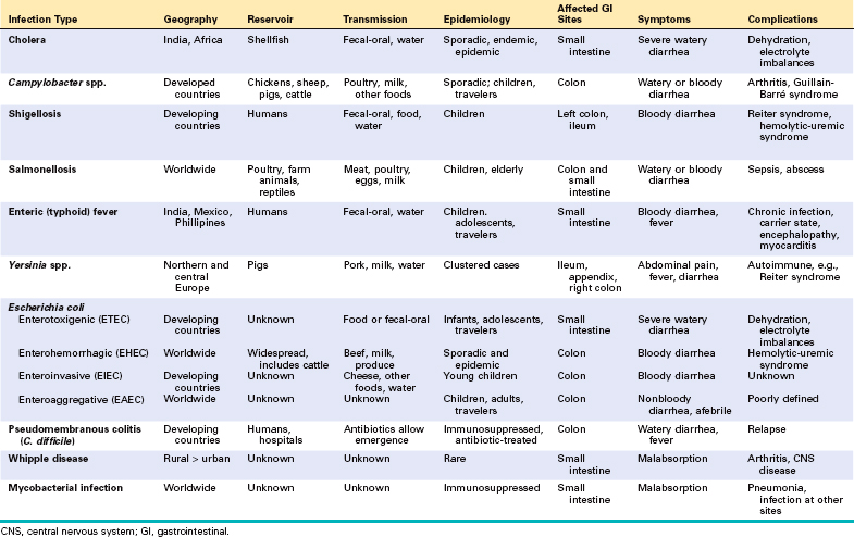
Enterocolitis can present with a broad range of symptoms including diarrhea, abdominal pain, urgency, perianal discomfort, incontinence, and hemorrhage (Table 17-7). This global problem is responsible for more than 12,000 deaths per day among children in developing countries and half of all deaths before age 5 worldwide. Bacterial infections, such as enterotoxigenic Escherichia coli, are frequently responsible, but the most common pathogens vary with age, nutrition, and host immune status as well as environmental influences (Table 17-7). For example, epidemics of cholera are common in areas with poor sanitation, as a result of inadequate public health measures, or as a consequence of natural disasters or war. Pediatric infectious diarrhea, which may result in severe dehydration and metabolic acidosis, is commonly caused by enteric viruses.
Vibrio cholerae are comma-shaped, Gram-negative bacteria that cause cholera, a disease that has been endemic in the Ganges Valley of India and Bangladesh for all of recorded history. Since 1817, seven great pandemics have spread along trade routes to large parts of Europe, Australia, and the Americas,56 but, for unknown reasons these pandemics resolved and cholera retreated back to the Ganges Valley. Cholera also persists within the Gulf of Mexico.
V. cholerae is primarily transmitted by contaminated drinking water. However, it can also be present in food and causes sporadic cases of seafood-associated disease in North America. There is a marked seasonal variation in most climates due to rapid growth of Vibrio bacteria at warm temperatures; the only animal reservoirs are shellfish and plankton. Relatively few V. cholerae serotypes are pathogenic, but other species of Vibrio can also cause disease. For example, V. parahaemolyticus is the most common cause of seafood-associated gastroenteritis in North America.57
Despite the severe diarrhea, Vibrio organisms are non-invasive and remain within the intestinal lumen. A preformed enterotoxin, cholera toxin, encoded by a virulence phage and released by the Vibrio organism, causes disease, but flagellar proteins, which are involved in motility and attachment, are necessary for efficient bacterial colonization. Hemagglutinin, a metalloproteinase, is important for bacterial detachment and shedding in the stool. The mechanism by which cholera toxin induces diarrhea is well understood (Fig. 17-27). Cholera toxin is composed of five B subunits and a single A subunit. The B subunit binds GM1 ganglioside on the surface of intestinal epithelial cells, and is carried by endocytosis to the endoplasmic reticulum, a process called retrograde transport.58 Here, the A subunit is reduced by protein disulfide isomerase, and a fragment of the A subunit is unfolded and released. This peptide fragment is then transported into the cytosol using host cell machinery that moves misfolded proteins from the endoplasmic reticulum to the cytosol. Such unfolded proteins are normally disposed of via the proteasome, but the A subunit refolds to avoid degradation. The refolded A subunit peptide then interacts with cytosolic ADP ribosylation factors (ARFs) to ribosylate and activate the stimulatory G protein Gsα. This stimulates adenylate cyclase and the resulting increases in intracellular cAMP open the cystic fibrosis transmembrane conductance regulator, CFTR, which releases chloride ions into the lumen. This causes secretion of bicarbonate, sodium, and water, leading to massive diarrhea. Chloride and sodium absorption are also inhibited by cAMP. Remarkably, mucosal biopsies show only minimal alterations.
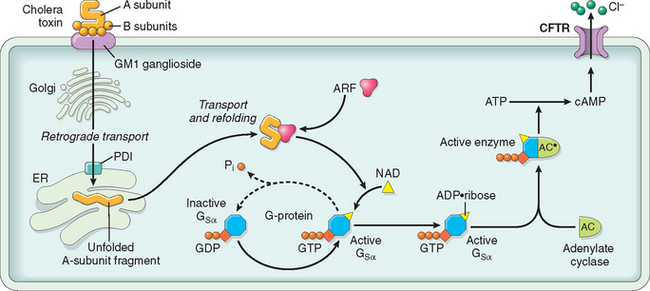
FIGURE 17-27 Mechanisms of cholera toxin transport and signaling. After retrograde toxin transport to the endoplasmic reticulum (ER), the A subunit is released by the action of protein disulfide isomerase (PDI) and is then able to access the epithelial cell cytoplasm. In concert with an ADP-ribosylation factor (ARF), the A subunit then ADP-ribosylates Gsα, which locks it in the active, GTP-bound state. This leads to adenylate cyclase (AC) activation, and the cAMP produced opens CFTR to drive chloride secretion and diarrhea.
Most exposed individuals are asymptomatic or develop only mild diarrhea. In those with severe disease there is an abrupt onset of watery diarrhea and vomiting following an incubation period of 1 to 5 days. The voluminous stools resemble rice water and are sometimes described as having a fishy odor. The rate of diarrhea may reach 1 liter per hour, leading to dehydration, hypotension, muscular cramping, anuria, shock, loss of consciousness, and death. Most deaths occur within the first 24 hours after presentation. Although the mortality for severe cholera is about 50% without treatment, timely fluid replacement can save more than 99% of patients. Oral rehydration is often sufficient.59 Because of an improved understanding of the host and Vibrio proteins involved, new therapies are being developed including CFTR inhibitors that block chloride secretion and prevent diarrhea.60 Prophylactic vaccination is a long-term goal.61
Campylobacter jejuni is the most common bacterial enteric pathogen in developed countries57 and is an important cause of traveler’s diarrhea. Most infections are associated with ingestion of improperly cooked chicken, but outbreaks can also be caused by unpasteurized milk or contaminated water.
The pathogenesis of Campylobacter infection remains poorly defined, but four major virulence properties contribute: motility, adherence, toxin production, and invasion. Flagella allow Campylobacter to be motile. This facilitates adherance and colonization, which are necessary for mucosal invasion. Cytotoxins that cause epithelial damage and a cholera toxin–like enterotoxin are also released by some C. jejuni isolates. Dysentery is generally associated with invasion and only occurs with a small minority of Campylobacter strains. Enteric fever occurs when bacteria proliferate within the lamina propria and mesenteric lymph nodes.
Campylobacter infection can result in reactive arthritis, primarily in patients with HLA-B27. Other extra-intestinal complications, including erythema nodosum and Guillain-Barré syndrome, a flaccid paralysis caused by autoimmune-induced inflammation of peripheral nerves, are not HLA-linked.62 Molecular mimicry has been implicated in the pathogenesis of Guillain-Barré syndrome, as serum antibodies to C. jejuni lipopolysaccharide cross-react with peripheral and central nervous system gangliosides. Moreover, 15% to 50% of individuals with Guillain-Barré syndrome have positive stool cultures or circulating antibodies to Campylobacter.63 Fortunately, Guillain-Barré syndrome develops in 0.1% or less of those infected with Campylobacter.
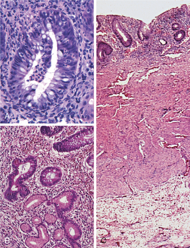
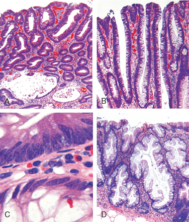
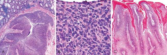
Morphology. Campylobacter are comma-shaped, flagellated, Gram-negative organisms. Diagnosis is primarily by stool culture, since biopsy findings are nonspecific, and reveal acute self-limited colitis with features common to many forms of infectious colitis.64 Mucosal and intraepithelial neutrophil infiltrates are prominent, particularly within the superficial mucosa (Fig. 17-28A); cryptitis (neutrophil infiltration of the crypts) and crypt abscesses (crypts with accumulations of luminal neutrophils) may also be present. Importantly, crypt architecture is preserved (Fig. 17-28D), although this can be difficult to assess in cases with severe mucosal damage.
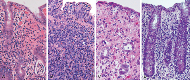
FIGURE 17-28 Bacterial enterocolitis. A, Campylobacter jejuni infection produces acute, self-limited colitis. Neutrophils can be seen within surface and crypt epithelium and a crypt abscess is present at the lower right. B, In Yersinia infection the surface epithelium can be eroded by neutrophils and the lamina propria is densely infiltrated by sheets of plasma cells admixed with lymphocytes and neutrophils. C, Enterohemorrhagic E. coli O157:H7 results in an ischemia-like morphology with surface atrophy and erosion. D, Enteroinvasive E. coli infection is a similar to other acute, self-limited colitides. Note the maintenance of normal crypt architecture and spacing, despite abundant intraepithelial neutrophils.
Ingestion of as few as 500 C. jejuni organisms can cause disease after an incubation period of up to 8 days. Watery diarrhea, either acute or following an influenza-like prodrome, is the primary symptom, and dysentery develops in 15% of patients. Patients may shed bacteria for 1 month or more after clinical resolution. Antibiotic therapy is generally not required.
Shigella are Gram-negative bacilli that were initially isolated during the Japanese red diarrhea epidemic of 1897. Four major strains are now recognized. Shigella are unencapsulated, nonmotile, facultative anaerobes that belong to the Enterobacteriaceae and are closely related to enteroinvasive E. coli. Although humans are the only known reservoir, Shigella spp. remain one of the most common causes of bloody diarrhea. It is estimated that 165 million cases occur worldwide each year.65 Given the infective dose of fewer than several hundred organisms and the presence of as many as 109 organisms in each gram of stool during acute disease, Shigella are highly transmissible by the fecal-oral route or via contaminated water and food.
In the United States and Europe, children in daycare centers, migrant workers, travelers to developing countries, and those in nursing homes are most commonly affected.66,67 Most Shigella infections and deaths occur in children under 5 years of age, and in countries where Shigella is endemic it is responsible for approximately 10% of all pediatric diarrheal disease and as many as 75% of diarrheal deaths.65,68
Shigella are resistant to the harsh acidic environment of the stomach, which translates into an extremely low infective dose. Once in the intestine, organisms are taken up by M, or microfold, epithelial cells, which are specialized for sampling and presentation of luminal antigens. Shigella proliferate intracellularly, escape into the lamina propria, and are phagocytosed by macrophages, in which they induce apoptosis. The ensuing inflammatory process damages surface epithelia and allows Shigella within the intestinal lumen to gain access to the colonocyte basolateral membrane, which is the only surface through which infection can occur in epithelial cells (other than M cells). All Shigella spp. carry virulence plasmids, some of which encode a type III secretion system capable of directly injecting bacterial proteins into the host cytoplasm. S. dysenteriae serotype 1 also release the Shiga toxin Stx, which inhibits eukaryotic protein synthesis resulting in host cell damage and death.69
Morphology. Shigella infections are most prominent in the left colon, but the ileum may also be involved, perhaps reflecting the abundance of M cells in the dome epithelium over the Peyer’s patches. The mucosa is hemorrhagic and ulcerated, and pseudomembranes may be present. The histology of early cases is similar to other acute self-limited colitides, such as Campylobacter colitis, but because of the tropism for M cells, aphthous-appearing ulcers similar to those seen in Crohn disease may occur. The potential for confusion with chronic inflammatory bowel disease is significant, particularly if there is distortion of crypt architecture.
After an incubation period of as long as 4 days, Shigella causes self-limited disease characterized by about 6 days of diarrhea, fever, and abdominal pain. The initially watery diarrhea progresses to a dysenteric phase in approximately 50% of patients, and constitutional symptoms can persist for as long as 1 month. The subacute presentation that develops in a minority of adults is characterized by several weeks of waxing and waning diarrhea that can mimic new-onset ulcerative colitis.68 While duration is typically shorter in children, severity is often much greater. Confirmation of Shigella infection requires stool culture.
Complications of Shigella infection are uncommon and include Reiter syndrome, a triad of sterile arthritis, urethritis, and conjunctivitis that preferentially affects HLA-B27-positive men between 20 and 40 years of age. Hemolytic-uremic syndrome, which is typically associated with enterohemorrhagic E. coli (EHEC), may also occur after infection with S. dysenteriae serotype 1 that secrete Shiga toxin69-71; only Shigella organisms that secrete the toxin are associated with hemolytic-uremic syndrome (Chapter 20). Antibiotic treatment shortens the clinical course and reduces the duration over which organisms are shed in the stool, but antidiarrheal medications are contraindicated because they can prolong symptoms and delay Shigella clearance.
Salmonella, which are classified within the Enterobacteriaceae family of Gram-negative bacilli, are divided into Salmonella typhi, the causative agent of typhoid fever (discussed in the next section) and nontyphoid Salmonella. Nontyphoid Salmonella infection is usually due to S. enteritidis; more than 1 million cases occur each year in the United States, and the prevalence is even greater in other countries. Infection is most common in young children and the elderly, with peak incidence in summer and fall. Transmission is usually through contaminated food, particularly raw or undercooked meat, poultry, eggs, and milk.
Very few viable Salmonella are necessary to cause infection, and the absence of gastric acid, as in individuals with atrophic gastritis or those on acid-suppressive therapy, further reduces the required inoculum. Salmonella possess virulence genes that encode a type III secretion system capable of transferring bacterial proteins into M cells and enterocytes. The transferred proteins activate host cell Rho GTPases, thereby triggering actin rearrangement and bacterial uptake that allow bacterial growth within phagosomes. In addition, flagellin, the core protein of bacterial flagellae, activates TLR5 on host cells and increases the local inflammatory response.72 Similarly, bacterial lipopolysaccharide activates TLR4, although some Salmonella strains express a virulence factor that prevents TLR4 activation from occurring. Salmonella also secrete a molecule that induces epithelial release of the eicosanoid hepoxilin A3, thereby drawing neutrophils into the intestinal lumen and potentiating mucosal damage.73 Mucosal TH17 immune responses limit infection to the colon.
Salmonella infections are clinically indistinguishable from those caused by other enteric pathogens, and symptoms range from loose stools to cholera-like profuse diarrhea to dysentery. Fever often resolves within 2 days, but diarrhea can persist for a week and organisms can be shed in the stool for several weeks after resolution. Antibiotic therapy is not recommended in most cases, because it can prolong the carrier state or even cause relapse and does not typically shorten the duration of diarrhea.74 Most Salmonella infections are self-limited, but deaths do occur. The risk of severe illness and complications is increased in patients with malignancies, immunosuppression, alcoholism, cardiovascular dysfunction, sickle cell disease, and hemolytic anemia.
Typhoid fever, also referred to as enteric fever, is caused by Salmonella typhi and Salmonella paratyphi. It affects up to 30 million individuals worldwide each year. The majority of cases in endemic countries are due to S. typhi, while infection by S. paratyphi is more common among travelers,75 perhaps because travelers tend to be vaccinated against S. typhi (there are no effective S. paratyphi vaccines). In endemic areas children and adolescents are affected most often, but there is no age preference in developed countries. Infection is strongly associated with travel to India, Mexico, the Philippines, Pakistan, El Salvador, and Haiti.76 Like Shigella, humans are the sole reservoir for S. typhi and S. paratyphi and transmission occurs from person to person or via food or contaminated water. Gallbladder colonization with S. typhi or S. paratyphi may be associated with gallstones and the chronic carrier state.
S. typhi are able to survive in gastric acid and, once in the small intestine, are taken up by and invade M cells. Organisms are then engulfed by mononuclear cells in the underlying lymphoid tissue. Unlike S. enteritidis, S. typhi can then disseminate via lymphatic and blood vessels. This causes reactive hyperplasia of phagocytes and lymphoid tissues throughout the body.
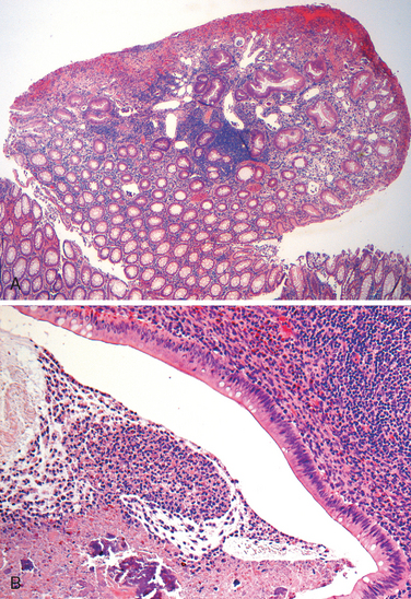
Morphology. Infection causes Peyer’s patches in the terminal ileum to enlarge into sharply delineated, plateau-like elevations up to 8 cm in diameter. Draining mesenteric lymph nodes are also enlarged. Neutrophils accumulate within the superficial lamina propria, and macrophages containing bacteria, red blood cells, and nuclear debris mix with lymphocytes and plasma cells in the lamina propria. Mucosal shedding creates oval ulcers, oriented along the axis of the ileum, that may perforate. The draining lymph nodes also harbor organisms and are enlarged due to phagocyte accumulation.
The spleen is enlarged and soft, with uniformly pale red pulp, obliterated follicular markings, and prominent phagocyte hyperplasia. The liver shows small, randomly scattered foci of parenchymal necrosis in which hepatocytes are replaced by macrophage aggregates, called typhoid nodules, that may also develop in the bone marrow and lymph nodes.
Patients experience anorexia, abdominal pain, bloating, nausea, vomiting, and bloody diarrhea followed by a short asymptomatic phase that gives way to bacteremia and fever with flu-like symptoms.77 Blood cultures are positive in more than 90% of affected individuals during the febrile phase, which may prompt antibiotic treatment and prevent further disease progression.76 In patients who do not receive treatment the febrile phase is followed by up to 2 weeks of sustained high fevers and abdominal tenderness that may mimic appendicitis. Rose spots, small erythematous maculopapular lesions, are seen on the chest and abdomen.77 Symptoms abate after several weeks in those who survive, although relapse can occur.77 Systemic dissemination may cause extra-intestinal complications including encephalopathy, meningitis, seizures, endocarditis, myocarditis, pneumonia, and cholecystitis. Patients with sickle cell disease are particularly susceptible to Salmonella osteomyelitis.
Three Yersinia species are human pathogens. Y. enterocolitica and Y. pseudotuberculosis cause GI disease and are discussed here; Y. pestis, the agent of pulmonic and bubonic plague, is discussed in Chapter 8. GI Yersinia infections are more common in Europe than North America and are most frequently linked to ingestion of pork, raw milk, and contaminated water. Y. enterocolitica is far more common than Y. pseudotuberculosis, and infections tend to cluster in the winter, possibly related to inadequately cooked foods served at holiday gatherings.
Yersinia invade M cells and use bacterial adhesion proteins, adhesins, to bind to host cell β1 integrins. A pathogenicity island encodes an iron uptake system that mediates iron capture and transport; similar iron transport systems are also present in E. coli, Klebsiella, Salmonella, and enterobacteria. In Yersinia, iron enhances virulence and stimulates systemic dissemination, explaining why individuals with hemolytic anemia or hemochromatosis are more likely to develop sepsis and are at greater risk of death.78
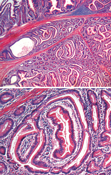
Morphology. Yersinia infections preferentially involve the ileum, appendix, and right colon (Fig. 17-28B). The organisms multiply extracellularly in lymphoid tissue, resulting in regional lymph node and Peyer’s patch hyperplasia and bowel wall thickening.79 The mucosa overlying lymphoid tissue may become hemorrhagic, and aphthous-appearing ulcers may develop, along with neutrophil infiltrates (see Fig. 17-28B) and granulomas, increasing the potential for diagnostic confusion with Crohn disease.
People infected with Yersinia generally present with abdominal pain, but fever and diarrhea may also occur. Nausea, vomiting, and abdominal tenderness are common, and Peyer’s patch invasion with subsequent involvement of regional lymphatics can mimic acute appendicitis in teenagers and young adults. Enteritis and colitis predominate in younger children. Extra-intestinal symptoms of pharyngitis, arthralgia, and erythema nodosum occur frequently. Yersinia can be detected in stool cultures on Yersinia-selective agar or, in cases with extra-intestinal disease, cultures of lymph nodes or blood.80 Postinfectious complications, including sterile arthritis, Reiter syndrome, myocarditis, glomerulonephritis, and thyroiditis, have been reported.
Escherichia coli are Gram-negative bacilli that colonize the healthy GI tract; most are nonpathogeneic, but a subset cause human disease. The latter are classified according to morphology, mechanism of pathogenesis, and in vitro behavior. Subgroups with major clinical relevance include enterotoxigenic E. coli (ETEC), enterohemorrhagic E. coli (EHEC), enteroinvasive E. coli (EIEC), and enteroaggregative E. coli (EAEC).
ETEC organisms are the principal cause of traveler’s diarrhea and spread via contaminated food or water. Infection is common in underdeveloped regions, and children younger than 2 years of age are particularly susceptible. ETEC produce heat-labile toxin (LT) and heat-stable toxin (ST), and both induce chloride and water secretion while inhibiting intestinal fluid absorption. The LT toxin is similar to cholera toxin and activates adenylate cyclase, resulting in increased intracellular cAMP. This stimulates chloride secretion and, simultaneously, inhibits absorption. ST toxins, which have homology to the mammalian regulatory protein guanylin, bind to guanylate cyclase and increase intracellular cGMP with resulting effects on transport that are similar to those produced by LT. Like cholera, the histopathology induced by ETEC infection is limited. Clinical symptoms include secretory, noninflammatory diarrhea, dehydration, and, in severe cases, shock.
EHEC are categorized as E. coli O157:H7 and non-O157:H7 serotypes. Large outbreaks of E. coli O157:H7 in developed countries have been associated with the consumption of inadequately cooked ground beef, sometimes from fast-food establishments. Contaminated milk and vegetables are also vehicles for infection. Both O157:H7 and non-O157:H7 serotypes produce Shiga-like toxins, and the morphology (see Fig. 17-28C) and clinical symptoms are thus similar to S. dysenteriae. O157:H7 strains of EHEC are more likely than non-O157:H7 serotypes to cause large outbreaks, bloody diarrhea, and hemolytic-uremic syndrome.
EIEC organisms are bacteriologically similar to Shigella and are transmitted via food, water, or by person-to-person contact. While EIEC do not produce toxins, they invade epithelial cells and cause nonspecific features of acute self-limited colitis (see Fig. 17-28D). EIEC infections are most common among young children in developing countries and are occasionally associated with outbreaks in developed countries.
EAEC organisms were identified on the basis of their unique pattern of adherence to epithelial cells. These organisms are now recognized as a cause of diarrhea in children and adults in developed as well as developing countries. These can also be a cause of traveler’s diarrhea.81 EAEC attach to enterocytes via adherence fimbriae and are aided by dispersin, a bacterial surface protein that neutralizes the negative surface charge of lipopolysaccharide. While the bacteria do produce enterotoxins related to Shigella enterotoxin and ETEC ST toxin, histologic damage is minimal and the characteristic adherence lesions are only visible by electron microscopy.82 EAEC organisms cause nonbloody diarrhea that may be prolonged in individuals with the acquired immunodeficiency syndrome (AIDS).
Pseudomembranous colitis, generally caused by Clostridium difficile, is also known as antibiotic-associated colitis or antibiotic-associated diarrhea. The latter terms apply to diarrhea developing during or after a course of antibiotic therapy and may be due to C. difficile as well as Salmonella, C. perfringens type A, or Staphylococcus aureus. The latter two organisms produce enterotoxins and are common agents of food poisoning.
It is likely that disruption of the normal colonic flora by antibiotics allows C. difficile overgrowth. Although almost any antibiotic may be responsible, third-generation cephalosporins are implicated most frequently. Immunosuppression is also a predisposing factor for C. difficile colitis. Toxins released by C. difficile cause the ribosylation of small GTPases, such as Rho, and lead to disruption of the epithelial cytoskeleton, tight junction barrier loss, cytokine release, and apoptosis.83 The mechanisms by which these processes lead to the characteristic morphology of pseudomembranous colitis are incompletely understood.
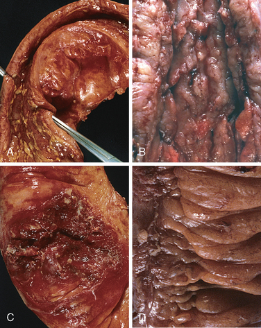
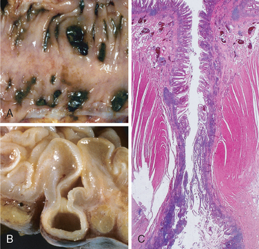
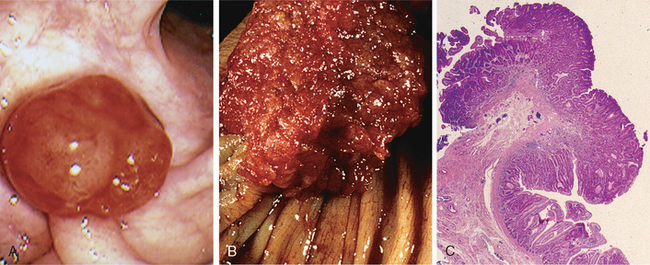
Morphology. Fully developed C. difficile–associated colitis is accompanied by formation of pseudomembranes (Fig. 17-29A, B), made up of an adherent layer of inflammatory cells and debris at sites of colonic mucosal injury. While pseudomembranes are not specific and may occur in ischemia and necrotizing infections, the histopathology of C. difficile–associated colitis is striking. The surface epithelium is denuded, and the superficial lamina propria contains a dense infiltrate of neutrophils and occasional fibrin thrombi within capillaries. Superficially damaged crypts are distended by a mucopurulent exudate that forms an eruption reminiscent of a volcano (Fig. 17-29C). These exudates coalesce to form the pseudomembranes.
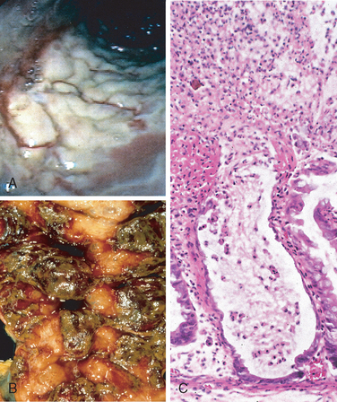
FIGURE 17-29 Clostridium difficile colitis. A, The colon is coated by tan pseudomembranes composed of neutrophils, dead epithelial cells, and inflammatory debris (endoscopic view). B, Pseudomembranes are easily appreciated on gross examination. C, Typical pattern of neutrophils emanating from a crypt is reminiscent of a volcanic eruption.
Risk factors for C. difficile–associated colitis include advanced age, hospitalization, and antibiotic treatment. The organism is particularly prevalent in hospitals; as many as 30% of hospitalized adults are colonized with C. difficile (a rate tenfold greater than the general population), but most colonized patients are free of disease. Individuals with C. difficile–associated colitis present with fever, leukocytosis, abdominal pain, cramps, hypoalbuminemia, watery diarrhea, and dehydration. Fecal leukocytes and occult blood may be present, but grossly bloody diarrhea is rare. Diagnosis of C. difficile–associated colitis is usually accomplished by detection of C. difficile toxin, rather than culture, and is supported by the characteristic histopathology. Metronidazole or vancomycin are generally effective therapies, but antibiotic-resistant and hypervirulent C. difficile strains as well as recurrent disease are increasingly common.84
Whipple disease is a rare, multivisceral chronic disease first described as intestinal lipodystrophy in 1907 by George Hoyt Whipple. A mere 27 years later the pathologist went on to win the Nobel Prize for his work on pernicious anemia. He was a contemporary, but not a relative, of Allen Oldfather Whipple, the surgeon who pioneered the pancreatoduodenectomy.
Whipple’s original case report described an individual with malabsorption, lymphadenopathy, and arthritis of undefined origin. Postmortem examination demonstrated the presence of foamy macrophages and large numbers of argyrophilic rods in the lymph nodes, providing evidence that the disease was infectious.85 The Gram-positive actinomycete, named Tropheryma whippelii, that is responsible for Whipple disease was identified by PCR in 1992 and finally cultured in 2000.86 Clinical symptoms occur because organism-laden macrophages accumulate within the small intestinal lamina propria and mesenteric lymph nodes, causing lymphatic obstruction. Thus, the malabsorptive diarrhea of Whipple disease is due to impaired lymphatic transport.
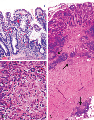
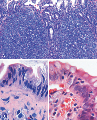
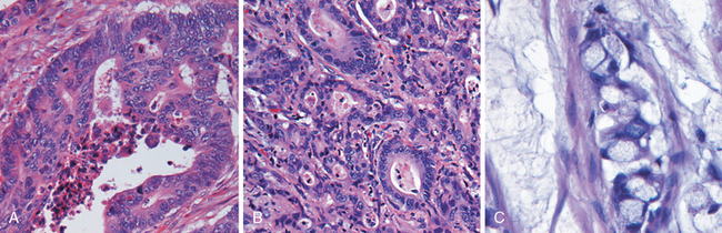
Morphology. The morphologic hallmark of Whipple disease is a dense accumulation of distended, foamy macrophages in the small intestinal lamina propria (Fig. 17-30A). The macrophages contain periodic acid–Schiff (PAS)-positive, diastase-resistant granules that represent lysosomes stuffed with partially digested bacteria (Fig. 17-30B). Intact rod-shaped bacilli can also be identified by electron microscopy (Fig. 17-30C). A similar infiltrate of foamy macrophages is present in intestinal tuberculosis (Fig. 17-30D), and in both diseases the organisms are PAS-positive. However, the acid-fast stain can be used to distinguish between these, since mycobacteria stain positively (Fig. 17-30E) while T. whippelii do not.
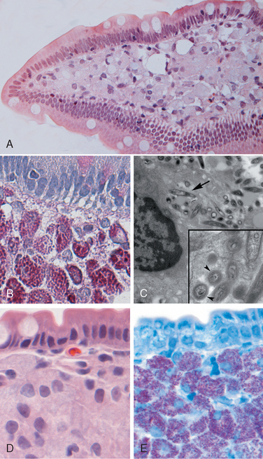
FIGURE 17-30 Whipple disease and mycobacterial infection. A, H&E staining shows effacement of normal lamina propria by a sheet of swollen macrophages. B, PAS stain highlights macrophage lysosomes full of bacilli. Note the positive staining of mucous vacuoles in the overlying goblet cells. C, An electron micrograph of one lamina propria macrophage shows these bacilli within the cell (top) and at higher magnification (inset). D, The morphology of mycobacterial infection can be similar to Whipple disease, particularly in the immunocompromised host. Compare with A. E, Mycobacteria are positive with stains for acid-fast bacteria.
(C, Courtesy of George Kasnic and Dr. William Clapp, University of Florida, Gainesville, FL.)
The villous expansion caused by the dense macrophage infiltrate imparts a shaggy gross appearance to the mucosal surface. Lymphatic dilatation and mucosal lipid deposition account for the common endoscopic detection of white to yellow mucosal plaques. In Whipple disease, bacteria-laden macrophages can accumulate within mesenteric lymph nodes, synovial membranes of affected joints, cardiac valves, the brain, and other sites.
The clinical presentation of Whipple disease is usually a triad of diarrhea, weight loss, and malabsorption. Extraintestinal symptoms, which can exist for months or years before malabsorption, include arthritis, arthralgia, fever, lymphadenopathy, and neurologic, cardiac, or pulmonary disease.
Mycobacterial infections are considered in detail in Chapter 8.
Symptomatic human infection is caused by several distinct groups of viruses. The most common are discussed here.
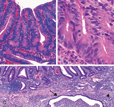
This was previously known as norwalk-like virus and is a common cause of nonbacterial infectious gastroenteritis.87 These are small icosahedral viruses with a single-stranded RNA genome that forms a genus within the Caliciviridae family. Norovirus causes approximately half of all gastroenteritis outbreaks worldwide and is a common cause of sporadic gastroenteritis in developed countries. Local outbreaks are usually related to contaminated food or water, but person-to-person transmission underlies most sporadic cases. Infections spread easily within schools, hospitals, nursing homes, and, most recently, cruise ships. Following a short incubation period, affected individuals develop nausea, vomiting, watery diarrhea, and abdominal pain. Biopsy morphology is nonspecific. When detected, abnormalities are most evident in the small intestine and include mild villous shortening, epithelial vacuolization, loss of the microvillus brush border, crypt hypertrophy, and lamina propria infiltration by lymphocytes (Fig. 17-31A). The disease is self-limited.
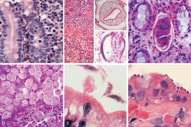
FIGURE 17-31 Infectious enteritis. A, Histologic features of viral enteritis include increased numbers of intraepithelial and lamina propria lymphocytes and crypt hypertrophy. B, Diffuse eosinophilic infiltrates in parasitic infection. This case was caused by Ascaris (upper inset), but a similar tissue reaction could be caused by Strongyloides (lower inset). C, Schistosomiasis can induce an inflammatory reaction to eggs trapped within the lamina propria. D, Entamoeba histolytica in a colon biopsy specimen. Note some organisms ingesting red blood cells. E, Giardia lamblia, which are present in the luminal space over nearly normal-appearing villi, are easily overlooked. F, Cryptosporidia organisms are seen as small blue spheres that appear to lay on top of the brush border but are actually enveloped by a thin layer of host cell cytoplasm.
This encapsulated virus with a segmented double-stranded RNA genome infects 140 million people and causes 1 million deaths each year, making rotavirus the most common cause of severe childhood diarrhea and diarrheal mortality worldwide. Children between 6 and 24 months of age are most vulnerable. Protection in the first 6 months of life is probably due to the presence of antibodies to rotavirus in breast milk, while protection beyond 2 years is due to immunity that develops following the first infection.88 Outbreaks in hospitals and daycare centers are common, and infection spreads easily; the estimated minimal infective inoculum is only 10 viral particles. Rotavirus selectively infects and destroys mature enterocytes in the small intestine, and the villus surface is repopulated by immature secretory cells. This results in loss of absorptive function and net secretion of water and electrolytes that is compounded by an osmotic diarrhea from incompletely absorbed nutrients. Like norovirus, rotavirus has a short incubation period followed by several days of vomiting and watery diarrhea. Vaccines are now available, and their use will probably change the epidemiology of rotavirus infection.
The second most common cause of pediatric diarrhea (after rotavirus), adenovirus also affects immunocompromised patients.89 Small intestinal biopsy specimens can show epithelial degeneration but more often exhibit nonspecific villous atrophy and compensatory crypt hyperplasia. Viral nuclear inclusions are uncommon. Disease typically presents after an incubation period of 1 week with nonspecific symptoms that include diarrhea, vomiting, and abdominal pain. Fever and weight loss may also be present. Symptoms generally resolve within 10 days.
Although viruses and bacteria are the predominant enteric pathogens in the United States, parasitic disease and protozoal infections affect over one half of the world’s population on a chronic or recurrent basis. The small intestine can harbor as many as 20 species of parasites, including nematodes, such as the roundworms Ascaris and Strongyloides; hookworms and pinworms; cestodes, including flatworms and tapeworms; trematodes, or flukes; and protozoa. Parasitic infections are covered in Chapter 8, and we will briefly discuss only those that are common in the intestinal tract.
This nematode infects over a billion individuals worldwide as a result of human fecal-oral contamination. Ingested eggs hatch in the intestine and larvae penetrate the intestinal mucosa, but disease is caused when larvae migrate from the splanchnic circulation to the systemic circulation and create hepatic abscess or Ascaris pneumonitis. In the latter case, larvae migrate up the trachea, are swallowed, and arrive again in the intestine to mature into adult worms. Adult worm masses induce an eosinophil-rich inflammatory reaction (Fig. 17-31B) that can physically obstruct the intestine or biliary tree. Diagnosis is usually made by detection of eggs in stool samples.
The larvae of Strongyloides live in fecally contaminated ground soil and can penetrate unbroken skin. They migrate through the lungs, where they induce inflammatory infiltrates, and then reside in the intestine while maturing into adult worms. Unlike other intestinal worms, which require an ova or larval stage outside the human, the eggs of Strongyloides can hatch within the intestine and release larvae that penetrate the mucosa, causing autoinfection (see Fig. 17-31B). Hence, Strongyloides infection can persist for life, and immunosuppressed individuals can develop overwhelming autoinfection. Strongyloides incite a strong tissue reaction and induce peripheral eosinophilia.
These hookworms infect 1 billion people worldwide and cause significant morbidity. Infection is initiated by larval penetration through the skin and, after further development in the lungs the larvae migrate up the trachea and are swallowed. Once in the duodenum the worms attach to the mucosa, suck blood, and reproduce. This causes multiple superficial erosions, focal hemorrhage, and inflammatory infiltrates and, in chronic infection, iron deficiency anemia. Diagnosis can be made by detection of the eggs in fecal smears.
Also known as pinworms, these parasites infect people in industrialized and developing countries; in the United States more than 60 million people have pinworms. Because they do not invade host tissue and live their entire life within the intestinal lumen, they rarely cause serious illness. Infection by E. vermicularis, or enterobiasis, is primarily by the fecal-oral route. Adult worms living in the intestine migrate to the anal orifice at night, where the female deposits eggs on the perirectal mucosa. The eggs cause intense irritation. Rectal and perineal pruritus ensues and leads to contamination of the fingers, which promotes human-to-human transmission. Both eggs and adult pinworms remain viable outside the body, and repeat infection is common. Diagnosis can be made by applying cellophane tape to the perianal skin and examining the tape for eggs under a microscope.
Whipworms primarily infect young children. Similar to E. vermicularis, Trichuris trichiura does not penetrate the intestinal mucosa and rarely cause serious disease. Heavy infections, however, may cause bloody diarrhea and rectal prolapse.
This disease involving the intestines most commonly takes the form of adult worms residing within the mesenteric veins. Symptoms of intestinal schistosomiasis are caused by trapping of eggs within the mucosa and submucosa (Fig. 17-31C). The resulting immune reaction is often granulomatous and can cause bleeding and even obstruction. More details are presented in Chapter 8.
The three primary species of cestodes that affect humans are Diphyllobothrium latum, fish tapeworms; Taenia solium, pork tapeworms; and Hymenolepsis nana, dwarf tapeworms. They reside exclusively within the intestinal lumen and are transmitted by ingestion of raw or undercooked fish, meat, or pork that contain encysted larvae. Release of the larvae allows attachment to the intestinal mucosa through its head, or scolex. The worm derives its nutrients from the food stream and enlarges by formation of egg-filled segments termed proglottids. Humans are usually infected by a single worm, and, since the worm does not penetrate the intestinal mucosa, peripheral eosinophilia does not generally occur. Nevertheless, the parasite burden can be staggering, since adult worms can grow to many meters in length. Large numbers of proglottids or individual eggs are shed in the feces. Clinical symptoms include abdominal pain, diarrhea, and nausea. Diagnosis is established by stool examination.
This protozoan that causes amebiasis is spread by fecal-oral transmission. E. histolytica infects approximately 500 million people in developing countries such as India, Mexico, and Colombia, and causes 40 million cases of dysentery and liver abscess annually. E. histolytica cysts, which have a chitin wall and four nuclei, are resistant to gastric acid, a characteristic that allows them to pass through the stomach without harm. Cysts then colonize the epithelial surface of the colon and release trophozoites, ameboid forms that reproduce under anaerobic conditions.
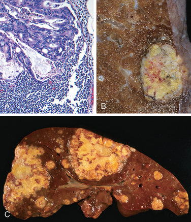
Amebiasis is seen most frequently in the cecum and ascending colon, although the sigmoid colon, rectum, and appendix can also be involved. Dysentery develops when the amebae attach to the colonic epithelium, induce apoptosis, invade crypts, and burrow laterally into the lamina propria. This recruits neutrophils, causes tissue damage, and creates a flask-shaped ulcer with a narrow neck and broad base. Histologic diagnosis can be difficult, since amebae are similar to macrophages in size and general appearance (see Fig. 17-31D). Parasites may penetrate splanchnic vessels and embolize to the liver to produce abscesses in about 40% of patients with amebic dysentery. Amebic liver abscesses, which can exceed 10 cm in diameter, have a scant inflammatory reaction at their margins and a shaggy fibrin lining. The abscesses persist after the acute intestinal illness has passed and may, rarely, reach the lung and the heart by direct extension from the liver. Amebae may also spread via the bloodstream into the kidneys and brain.
Individuals with amebiasis may present with abdominal pain, bloody diarrhea, or weight loss. Occasionally, acute necrotizing colitis and megacolon occur, and both are associated with significant mortality. The parasites lack mitochondria or Krebs cycle enzymes and are thus obligate fermenters of glucose. Therefore, metronidazole, which inhibits the enzyme pyruvate oxidoreductase that is required for fermentation, is the most effective treatment.
These organisms, also referred to as G. duodenalis or G. intestinalis, were initially described by van Leeuwenhoek, the inventor of the microscope, who discovered the pathogen in his own stool. They are the most common pathogenic parasitic infection in humans and are spread by fecally contaminated water or food. Infection may occur after ingestion of as few as 10 cysts. Because cysts are resistant to chlorine, Giardia are endemic in unfiltered public water supplies. They are commonly present in rural streams, explaining infection in campers who use these as a water source. Infection may also occur by the fecal-oral route and, because the cysts are stable, they may be accidentally swallowed while swimming in contaminated water.
Giardia are flagellated protozoans that cause decreased expression of brush-border enzymes, including lactase; microvillous damage; and apoptosis of small intestinal epithelial cells. Secretory IgA and mucosal IL-6 responses are important for clearance of Giardia infections, and immunosuppressed, agammaglobulinemic, or malnourished individuals are often severely affected.90 Giardia can evade immune clearance through continuous modification of the major surface antigen, variant surface protein, and can persist for months or years while causing intermittent symptoms.
Giardia trophozoites can be identified in duodenal biopsies by their characteristic pear shape with two nuclei of equal size, each of which contains a complete copy of the genome. Despite large numbers of trophozoites, which are sickle-shaped in profile and tightly bound to the brush border of villous enterocytes, there is no invasion and small intestinal morphology may be normal by light microscopy (see Fig. 17-31E). Villous blunting with increased numbers of intraepithelial lymphocytes and mixed lamina propria inflammatory infiltrates may be present in patients with heavy infections.
Giardiasis may be subclinical or accompanied by acute or chronic diarrhea, malabsorption, and weight loss.90 Infection is usually documented by immunofluorescent detection of cysts in stool samples. Although oral antimicrobial therapy is effective, recurrence is common.
Like Giardia, Cryptosporidia are an important cause of diarrhea worldwide. Cryptosporidiosis was first discovered in the 1980s as an agent of chronic diarrhea in AIDS patients and is now recognized as a cause of acute, self-limited disease in immunologically normal hosts. Cryptosporidiosis causes traveler’s diarrhea as well as persistent diarrhea in residents of developing countries. The organisms are present worldwide, with the exception of Antarctica, perhaps because the oocysts are killed by freezing. The oocysts are resistant to chlorine and may, therefore, persist in treated, but unfiltered, water. Contaminated drinking water continues to be the most common means of transmission. The largest documented outbreak, a result of inadequate water purification, occurred in 1993 in Milwaukee, Wisconsin, and affected more than 400,000 people. Like giardiasis, cryptosporidiosis can be spread to water sport participants via contaminated water. Food-borne infection occurs less frequently.
Humans are infected by several different Cryptosporidium species, including C. hominis and C. parvum. All are able to go through an entire life cycle, with asexual and sexual reproductive phases, in a single host. The ingested encysted oocyte, of which 10 are sufficient to cause symptomatic infection, is activated by acid in the stomach to produce proteases that allow release of sporozoites from the oocysts. The sporozoites are motile and have a specialized organelle for attachment to the enterocyte brush border, where they induce actin polymerization. This drives extension of the epithelial cell membrane to engulf the parasite and form a vacuole within the microvilli. Sodium malabsorption, chloride secretion, and increased tight junction permeability are responsible for the nonbloody, watery diarrhea that ensues.
Mucosal histology is often only minimally altered, but persistent cryptosporidiosis in children is associated with villous atrophy. Heavy infection in immunosuppressed patients may be associated with villous atrophy, crypt hyperplasia, and variable inflammatory infiltrates. Although the sporozoite is intracellular, it appears, by light microscopy, to sit on top of the epithelial apical membrane (Fig. 17-31F). Organisms are typically most concentrated in the terminal ileum and proximal colon, but can be present throughout the gut, biliary tract, and even the respiratory tract of immunodeficient hosts. Diagnosis is based on finding oocysts in the stool.
Irritable bowel syndrome (IBS) is characterized by chronic, relapsing abdominal pain, bloating, and changes in bowel habits.91 Despite very real symptoms, the gross and microscopic evaluation is normal in most IBS patients. Thus, the diagnosis depends on clinical symptoms.
The pathogenesis of IBS remains poorly defined, although there is clearly an interplay between psychologic stressors, diet, and abnormal GI motility. Data showing disturbances of intestinal motility and enteric sensory function suggest that impairment of signaling in the brain-gut axis contributes to IBS. A small subgroup of IBS patients also relate onset to a bout of infectious gastroenteritis, suggesting an immune or neuroimmune contribution.
The peak prevalence of IBS is between 20 and 40 years of age, and there is a significant female predominance. Variability in diagnostic criteria makes it difficult to establish the incidence, but most authors report a prevalence in developed countries of between 5% and 10%. IBS is presently diagnosed using clinical criteria that require the occurrence of abdominal pain or discomfort at least 3 days per month over 3 months, improvement with defecation, and a change in stool frequency or form. Many patients also report fibromyalgia or other chronic pain disorders, visceral hypersensitivity, backache, headache, urinary symptoms, dyspareunia, lethargy, and depression. In those with diarrhea, microscopic colitis, celiac disease, giardiasis, lactose intolerance, small bowel bacterial overgrowth, bile salt malabsorption, colon cancer, and inflammatory bowel disease must be excluded (although IBS is common in inflammatory bowel disease patients). IBS is not associated with serious long-term sequelae, but affected patients may undergo unnecessary abdominal surgery due to chronic pain and their ability to function socially may be compromised. The prognosis of IBS is most closely related to symptom duration, with longer duration correlating with reduced likelihood of improvement. Ongoing life stressors also reduce the chance of symptom resolution. Consistent with the uncertain mechanisms of disease, diverse treatments are used including psychotherapy, dietary fiber supplementation, tricyclic antidepressants, selective serotonin reuptake inhibitors, probiotics, and antibiotics. In addition, a chloride channel agonist may provide benefit in a subset of patients whose primary manifestation is constipation.
Inflammatory bowel disease (IBD) is a chronic condition resulting from inappropriate mucosal immune activation. The two disorders that comprise IBD are Crohn disease and ulcerative colitis. Descriptions of ulcerative colitis and Crohn disease date back to antiquity and at least the sixteenth century, respectively, but it took modern bacteriologic techniques to exclude conventional infectious etiologies for these diseases.92 As will be discussed below, however, commensal bacteria normally present in the intestinal lumen are probably involved in IBD pathogenesis.
The distinction between ulcerative colitis and Crohn disease is based, in large part, on the distribution of affected sites (Fig. 17-32) and the morphologic expression of disease (Table 17-8) at those sites. Ulcerative colitis is a severe ulcerating inflammatory disease that is limited to the colon and rectum and extends only into the mucosa and submucosa. In contrast, Crohn disease, which has also been referred to as regional enteritis (because of frequent ileal involvement) may involve any area of the GI tract and is typically transmural.
FIGURE 17-32 Distribution of lesions in inflammatory bowel disease. The distinction between Crohn disease and ulcerative colitis is primarily based on morphology.
TABLE 17-8 Features That Differ between Crohn Disease and Ulcerative Colitis
Feature | Crohn Disease | Ulcerative Colitis |
---|---|---|
MACROSCOPIC | ||
Bowel region | Ileum ± colon | Colon only |
Distribution | Skip lesions | Diffuse |
Stricture | Yes | Rare |
Wall appearance | Thick | Thin |
MICROSCOPIC | ||
Inflammation | Transmural | Limited to mucosa |
Pseudopolyps | Moderate | Marked |
Ulcers | Deep, knife-like | Superficial, broad-based |
Lymphoid reaction | Marked | Moderate |
Fibrosis | Marked | Mild to none |
Serositis | Marked | Mild to none |
Granulomas | Yes (∼35%) | No |
Fistulae/sinuses | Yes | No |
CLINICAL | ||
Perianal fistula | Yes (in colonic disease) | No |
Fat/vitamin malabsorption | Yes | No |
Malignant potential | With colonic involvement | Yes |
Recurrence after surgery | Common | No |
Toxic megacolon | No | Yes |
Note: All features may not be present in a single case.
Both Crohn disease and ulcerative colitis are more common in females and frequently present in the teens and early 20s. In Western industrialized nations IBD is most common among Caucasians and, in the United States, occurs 3 to 5 times more often among eastern European (Ashkenazi) Jews. This is at least partly due to genetic factors, as discussed below. The geographic distribution of IBD is highly variable, but it is most common in North America, northern Europe, and Australia. However, IBD incidence worldwide is on the rise, and it is becoming more common in regions such as Africa, South America, and Asia, where the prevalence was historically low. The hygiene hypothesis suggests that this increasing incidence may be related to improved food storage conditions and decreased food contamination. This hypothesis suggests that reduced frequency of enteric infections has resulted in inadequate development of regulatory processes to limit mucosal immune responses, allowing pathogens that should cause self-limited disease to trigger overwhelming immune responses and chronic inflammatory disease in susceptible hosts. Although many details to support this hypothesis are lacking, the observation that helminth infection, which is endemic in regions where IBD incidence is low, can prevent IBD development in animal models and reduce disease in some patients lends support to this idea. The observation that an episode of acute infectious gastroenteritis may precede onset of IBD in some individuals is also consistent with the hygiene hypothesis.
IBD is an idiopathic disorder and the responsible processes are only beginning to be understood. Although there is limited epidemiologic association of IBD with autoimmunity, neither Crohn disease nor ulcerative colitis is thought to be an autoimmune disease. Rather, most investigators believe that the two diseases result from a combination of defects in host interactions with intestinal microbiota, intestinal epithelial dysfunction, and aberrant mucosal immune responses. This view is supported by epidemiologic, genetic, and clinical studies as well as data from laboratory models of IBD (Fig. 17-33).
FIGURE 17-33 One model of IBD pathogenesis. Aspects of both Crohn disease and ulcerative colitis are shown.
Molecular linkage analyses of affected families have identified NOD2 (nucleotide oligomerization binding domain 2) as a susceptibility gene in Crohn disease. Specific NOD2 polymorphisms confer at least a four-fold increase in Crohn disease risk among Caucasians of European ancestry. NOD2 encodes a protein that binds to intracellular bacterial peptidoglycans and subsequently activates NF-κB. It has been postulated that disease-associated NOD2 variants are less effective at recognizing and combating luminal microbes, which are then able to enter the lamina propria and trigger inflammatory reactions. Other data suggest that NOD2 may regulate immune responses to prevent excessive activation by luminal microbes. Whatever the mechanism by which NOD2 polymorphisms contribute to Crohn disease pathogenesis, it should be remembered that fewer than 10% of individuals carrying NOD2 mutations develop disease. Furthermore, NOD2 mutations are uncommon in African and Asian Crohn disease patients. Thus, defective NOD2 signaling is only one of many genetic factors that contribute to Crohn disease pathogenesis.
More recently, the search for IBD-associated genes has used genome-wide association studies (GWAS) that assess single-nucleotide polymorphisms, as described in Chapter 5.94 The number of genes identified by GWAS is increasing rapidly (already numbering more than 30), but along with NOD2, two Crohn disease–related genes of particular interest are ATG16L1 (autophagy-related 16-like), a part of the autophagosome pathway that is critical to host cell responses to intracellular bacteria and, perhaps, epithelial homeostasis, and IRGM (immunity-related GTPase M), which is also involved in autophagy and clearance of intracellular bacteria. NOD2, ATG16L1, and IRGM are expressed in multiple cell types, and their precise roles in Crohn disease pathogenesis have yet to be defined. However, like NOD2, ATG16L1 and IRGM are related to recognition and response to intracellular pathogens, supporting the hypothesis that inappropriate immune reactions to luminal bacteria are an important component of IBD pathogenesis. None of these genes are associated with ulcerative colitis. However, some polymorphisms of the IL-23 receptor are protective in both Crohn disease and ulcerative colitis (discussed later).95
Some data suggest that ulcerative colitis is a TH2-mediated disease, and this is consistent with observations of increased mucosal IL-13 in ulcerative colitis patients. However, the protection afforded by IL-23 receptor polymorphisms and effectiveness of anti-TNF therapy in some ulcerative colitis patients seems to support roles for TH1 and TH17 cells. A recent report linking polymorphisms near the IL-10 gene to ulcerative colitis, but not Crohn disease, further emphasizes the importance of immunoregulatory signals in IBD pathogenesis.96 Overall it is likely that some combination of derangements that activate mucosal immunity and suppress immunoregulation contribute to the development of ulcerative colitis and Crohn disease. The relative roles of innate and adaptive arms of the immune system are presently the subject of intense scrutiny.
One model that unifies the roles of intestinal microbiota, epithelial function, and mucosal immunity suggests a cycle by which transepithelial flux of luminal bacterial components activates innate and adaptive immune responses.106 In a genetically susceptible host, the subsequent release of TNF and other immune-mediated signals direct epithelia to increase tight junction permeability, which causes further increases in the flux of luminal material. These events may establish a self-amplifying cycle in which a stimulus at any site may be sufficient to initiate IBD.107 Although this model is helpful in advancing our understanding of IBD pathogenesis, it is important to recognize that a variety of factors are associated with disease for unknown reasons. For example, an episode of appendicitis is associated with reduced risk of developing ulcerative colitis. Tobacco also modifies IBD epidemiology, but, paradoxically, the risk of Crohn disease is increased by smoking while that of ulcerative colitis is reduced.
Crohn disease, an eponym based on the 1932 description by Crohn, Ginzburg, and Oppenheimer, has existed for centuries. Louis XIII of France (1601–1643) suffered relapsing bloody diarrhea, fever, rectal abscess, small intestinal and colonic ulcers, and fistulae beginning at age 20 years, most likely due to Crohn disease.
Morphology. Crohn disease may occur in any area of the GI tract, but the most common sites involved at presentation are the terminal ileum, ileocecal valve, and cecum. Disease is limited to the small intestine alone in about 40% of cases; the small intestine and colon are both involved in 30% of patients; and the remainder have only colonic involvement. The presence of multiple, separate, sharply delineated areas of disease, resulting in skip lesions, is characteristic of Crohn disease and may help in the differentiation from ulcerative colitis. Strictures are common (Fig. 17-34A).
FIGURE 17-34 Gross pathology of Crohn disease. A, Small-intestinal stricture. B, Linear mucosal ulcers and thickened intestinal wall. C, Perforation and associated serositis. D, Creeping fat.
The earliest Crohn disease lesion, the aphthous ulcer, may progress, and multiple lesions often coalesce into elongated, serpentine ulcers oriented along the axis of the bowel. Edema and loss of the normal mucosal texture are common. Sparing of interspersed mucosa, a result of the patchy distribution of Crohn disease, results in a coarsely textured, cobblestone appearance in which diseased tissue is depressed below the level of normal mucosa (Fig. 17-34B). Fissures frequently develop between mucosal folds and may extend deeply to become fistula tracts or sites of perforation (Fig. 17-34C). The intestinal wall is thickened and rubbery as a consequence of transmural edema, inflammation, submucosal fibrosis, and hypertrophy of the muscularis propria, all of which contribute to stricture formation. In cases with extensive transmural disease, mesenteric fat frequently extends around the serosal surface (creeping fat) (Fig. 17-34D).
The microscopic features of active Crohn disease include abundant neutrophils that infiltrate and damage crypt epithelium. Clusters of neutrophils within a crypt are referred to as crypt abscesses and are often associated with crypt destruction. Ulceration is common in Crohn disease, and there may be an abrupt transition between ulcerated and adjacent normal mucosa. Even in areas where gross examination suggests diffuse disease, microscopic pathology can appear patchy. Repeated cycles of crypt destruction and regeneration lead to distortion of mucosal architecture; the normally straight and parallel crypts take on bizarre branching shapes and unusual orientations to one another (Fig. 17-35A). Epithelial metaplasia, another consequence of chronic relapsing injury, often takes the form of gastric antral-appearing glands, and is called pseudopyloric metaplasia. Paneth cell metaplasia may also occur in the left colon, where Paneth cells are normally absent. These architectural and metaplastic changes may persist even when active inflammation has resolved. Mucosal atrophy, with loss of crypts, may occur after years of disease. Noncaseating granulomas (Fig. 17-35B), a hallmark of Crohn disease, are found in approximately 35% of cases and may occur in areas of active disease or uninvolved regions in any layer of the intestinal wall (Fig. 17-35C). Granulomas may also be present in mesenteric lymph nodes. Cutaneous granulomas form nodules that are referred to as metastatic Crohn disease. The absence of granulomas does not preclude a diagnosis of Crohn disease.
The clinical manifestations of Crohn disease are extremely variable. In most patients disease begins with intermittent attacks of relatively mild diarrhea, fever, and abdominal pain. Approximately 20% of patients present acutely with right lower quadrant pain, fever, and bloody diarrhea that may mimic acute appendicitis or bowel perforation. Periods of active disease are typically interrupted by asymptomatic periods that last for weeks to many months. Disease re-activation can be associated with a variety of external triggers, including physical or emotional stress, specific dietary items, and cigarette smoking. The latter is a strong exogenous risk factor for development of Crohn disease and, in some cases, disease onset is associated with initiation of smoking. Unfortunately, smoking cessation does not result in disease remission.
Iron-deficiency anemia may develop in individuals with colonic disease, while extensive small bowel disease may result in serum protein loss and hypoalbuminemia, generalized nutrient malabsorption, or malabsorption of vitamin B12 and bile salts. Fibrosing strictures, particularly of the terminal ileum, are common and require surgical resection. Disease often recurs at the site of anastamosis, and as many as 40% of patients require additional resections within 10 years. Fistulae develop between loops of bowel and may also involve the urinary bladder, vagina, and abdominal or perianal skin. Perforations and peritoneal abscesses are common.
Extra-intestinal manifestations of Crohn disease include uveitis, migratory polyarthritis, sacroiliitis, ankylosing spondylitis, erythema nodosum, and clubbing of the fingertips, any of which may develop before intestinal disease is recognized. Pericholangitis and primary sclerosing cholangitis occur in Crohn disease but are more common in ulcerative colitis (see Chapter 18). As discussed below, risk of colonic adenocarcinoma is increased in patients with long-standing colonic disease.
Ulcerative colitis is closely related to Crohn disease. However, intestinal disease in ulcerative colitis is limited to the colon and rectum. Common extra-intestinal manifestations of ulcerative colitis overlap with those of Crohn disease and include migratory polyarthritis, sacroiliitis, ankylosing spondylitis, uveitis, skin lesions, pericholangitis, and primary sclerosing cholangitis (Chapter 18). Approximately 2.5% to 7.5% of individuals with ulcerative colitis also have primary sclerosis cholangitis. The long-term outlook for ulcerative colitis patients depends on the severity of active disease and disease duration.
Morphology. Grossly, ulcerative colitis always involves the rectum and extends proximally in a continuous fashion to involve part or all of the colon. Skip lesions are not seen (although focal appendiceal or cecal inflammation may occasionally be present in ulcerative colitis). Disease of the entire colon is termed pancolitis (Fig. 17-36A), while left-sided disease extends no farther than the transverse colon. Limited distal disease may be referred to descriptively as ulcerative proctitis or ulcerative proctosigmoiditis. The small intestine is normal, although mild mucosal inflammation of the distal ileum, backwash ileitis, may be present in severe cases of pancolitis.
FIGURE 17-36 Gross pathology of ulcerative colitis. A, Total colectomy with pancolitis showing active disease, with red, granular mucosa in the cecum (left) and smooth, atrophic mucosa distally (right). B, Sharp demarcation between active ulcerative colitis (right) and normal (left). C, Inflammatory polyps. D, Mucosal bridges.
Grossly, involved colonic mucosa may be slightly red and granular or have extensive, broad-based ulcers, and there can be an abrupt transition between diseased and uninvolved colon (Fig. 17-36B). Ulcers are aligned along the long axis of the colon but do not typically replicate the serpentine ulcers of Crohn disease. Isolated islands of regenerating mucosa often bulge into the lumen to create pseudopolyps (Fig. 17-36C), and the tips of these polyps may fuse to create mucosal bridges (Fig. 17-36D). Chronic disease may lead to mucosal atrophy with a flat and smooth mucosal surface that lacks normal folds. Unlike Crohn disease, mural thickening is not present, the serosal surface is normal, and strictures do not occur. However, inflammation and inflammatory mediators can damage the muscularis propria and disturb neuromuscular function leading to colonic dilation and toxic megacolon, which carries a significant risk of perforation.
Histologic features of mucosal disease in ulcerative colitis are similar to colonic Crohn disease and include inflammatory infiltrates, crypt abscesses (Fig. 17-37A), architectural crypt distortion, and epithelial metaplasia (Fig. 17-37B). However, the inflammatory process is diffuse and generally limited to the mucosa and superficial submucosa (Fig. 17-37C). In severe cases, extensive mucosal destruction may be accompanied by ulcers that extend more deeply into the submucosa, but the muscularis propria is rarely involved. Submucosal fibrosis, mucosal atrophy, and distorted mucosal architecture remain as residua of healed disease but histology may also revert to near normal after prolonged remission. Granulomas are not present in ulcerative colitis.
FIGURE 17-37 Microscopic pathology of ulcerative colitis. A, Crypt abscess. B, Pseudopyloric metaplasia (bottom). C, Disease is limited to the mucosa. Compare to Figure 17-35C.
Ulcerative colitis is a relapsing disorder characterized by attacks of bloody diarrhea with stringy, mucoid material, lower abdominal pain, and cramps that are temporarily relieved by defecation. These symptoms may persist for days, weeks, or months before they subside, and, occasionally, the initial attack may be severe enough to constitute a medical or surgical emergency. More than half of patients have clinically mild disease, although almost all experience at least one relapse during a 10-year period, and up to 30% require colectomy within the first 3 years after presentation because of uncontrollable symptoms. Colectomy effectively cures intestinal disease in ulcerative colitis, but extra-intestinal manifestations may persist.
The factors that trigger ulcerative colitis are not known, but, as noted above, infectious enteritis precedes disease onset in some cases. In other cases the first attack is preceded by psychologic stress, which may also be linked to relapse during remission. The initial onset of symptoms has also been reported to occur shortly after smoking cessation in some patients, and smoking may partially relieve symptoms. Unfortunately, studies of nicotine as a therapeutic agent have been disappointing.
There is extensive pathologic and clinical overlap between ulcerative colitis and Crohn disease (Table 17-8); definitive diagnosis is not possible in approximately 10% of IBD patients. These cases, termed indeterminate colitis, do not involve the small bowel and have colonic disease in a continuous pattern that would typically indicate ulcerative colitis. However, patchy histologic disease, fissures, a family history of Crohn disease, perianal lesions, onset after initiating use of cigarettes, or other features that are not typical of ulcerative colitis may prompt more detailed endoscopic, radiographic, and histologic examination. Serologic studies can be useful in these cases, because perinuclear anti-neutrophil cytoplasmic antibodies are positive in 75% of individuals with ulcerative colitis but only 11% with Crohn disease. In contrast, ulcerative colitis patients tend to lack antibodies to Saccharomyces cerevisiae, which are often present in those with Crohn disease. Even after extensive evaluation, IBD in approximately 10% of patients defies classification. In some of these cases features that develop over time (e.g., strictures or fistulae) ultimately establish the diagnosis; classification remains impossible in other patients. Because of extensive overlap in medical management of ulcerative colitis and Crohn disease, patients carrying a diagnosis of indeterminate colitis can be treated effectively. However, it is preferable to definitively categorize patients, when possible, as evolving medical therapies and surgical management differ in ulcerative colitis and Crohn disease.
One of the most feared long-term complications of ulcerative colitis and colonic Crohn disease is the development of neoplasia. This begins as dysplasia, which, just as in Barrett esophagus and chronic gastritis, represents in situ transformation. The risk of dysplasia is related to several factors:
The role of inflammation in promoting dysplasia is emphasized by the observation that anti-TNF antibody treatment can suppress the development of colitis-associated cancers in experimental animals.
To facilitate early detection of neoplasia, patients are typically enrolled in surveillance programs approximately 8 years after diagnosis of IBD. The major exception to this is patients with primary sclerosing cholangitis, who have an even greater risk of dysplasia and are generally enrolled for surveillance at the time of diagnosis. Surveillance requires regular and extensive mucosal biopsy, making it a costly practice. Research efforts have therefore included a search for molecular markers of dysplasia in nondysplastic mucosa. Genomic instability in rectal mucosa has the potential to be such a marker, but biopsy surveillance remains the best tool currently available.
In many cases dysplasia occurs in flat areas of mucosa that are not grossly recognized as abnormal. Thus, advanced endoscopic imaging techniques including chromoendoscopy and confocal endoscopy are beginning to be used experimentally to increase sensitivity of detection. IBD-associated dysplasia is classified histologically as low grade or high grade (Fig. 17-38A, B) and may be multifocal. High-grade dysplasia may be associated with invasive carcinoma at the same site (Fig. 17-38C) or elsewhere in the colon and, therefore typically prompts colectomy. Low-grade dysplasia may be treated with colectomy or followed closely, depending on a variety of factors including patient age and the number of dysplastic foci present. Colonic adenomas (discussed below) also occur in IBD patients, and in some cases these may be difficult to differentiate from a polypoid focus of IBD-associated dysplasia.108
FIGURE 17-38 Colitis-associated dysplasia. A, Dysplasia with extensive nuclear stratification and marked nuclear hyperchromasia. B, Cribriform glandular arrangement in high-grade dysplasia. C, Colectomy specimen with high-grade dysplasia on the surface and underlying invasive adenocarcinoma. A large cystic, neutrophil-filled space lined by invasive adenocarcinoma is apparent at the bottom right (arrow) beneath the muscularis mucosa, and is surrounded by small invasive glands (arrowhead).
Surgical treatment of ulcerative colitis, Hirschsprung disease, and other intestinal disorders sometimes requires creation of a temporary or permanent ostomy and a blind distal segment of colon, such as a Hartmann’s pouch, from which the normal fecal flow is diverted. Colitis can develop within the diverted segment, particularly in ulcerative colitis patients. Besides mucosal erythema and friability, the most striking feature of diversion colitis is the development of numerous mucosal lymphoid follicles (Fig. 17-39A). Increased numbers of lamina propria lymphocytes, monocytes, macrophages, and plasma cells may also be present. In severe cases the histopathology may resemble IBD and include crypt abscesses, mucosal architectural distortion, or, rarely, granulomas. The pathogenic mechanisms responsible for diversion colitis are not well understood, but changes in the luminal microbiota and diversion of the fecal stream that provides nutrients to colonic epithelial cells have been proposed. Consistent with this, enemas containing short-chain fatty acids, a product of bacterial digestion in the colon and an important energy source for colonic epithelial cells, can promote mucosal recovery in some cases. The ultimate cure is reanastomosis of the diverted segment.
Microscopic colitis encompases two entities, collagenous colitis and lymphocytic colitis. These idiopathic diseases both present with chronic, nonbloody, watery diarrhea without weight loss. Radiologic and endoscopic studies are typically normal. Collagenous colitis, which occurs primarily in middle-aged and older women, is characterized by the presence of a dense subepithelial collagen layer, increased numbers of intraepithelial lymphocytes, and a mixed inflammatory infiltrate within the lamina propria (Fig. 17-39B). Lymphocytic colitis is histologically similar, but the subepithelial collagen layer is of normal thickness and the increase in intraepithelial lymphocytes may be greater, frequently exceeding one T lymphocyte per five colonocytes (Fig. 17-39C). Lymphocytic colitis shows a strong association with celiac disease and autoimmune diseases, including thyroiditis, arthritis, and autoimmune or lymphocytic gastritis.
Graft-versus-host disease occurs following allogeneic bone marrow transplantation. The small bowel and colon are involved in most cases. Although graft-versus-host disease is secondary to donor T cells targeting antigens on the recipient’s GI epithelial cells, the lamina propria lymphocytic infiltrate is typically sparse. Epithelial apoptosis, particularly of crypt cells, is the most common histologic finding. Rarely, total gland destruction occurs, although endocrine cells may persist. Intestinal graft-versus-host disease often presents as a watery diarrhea.
In general, diverticular disease refers to acquired pseudo-diverticular outpouchings of the colonic mucosa and submucosa. Such colonic diverticula are rare in persons under age 30, but the prevalence approaches 50% in Western adult populations over age 60. Diverticulae are generally multiple and the condition is referred to as diverticulosis. This disease is much less common in Japan and nonindustrialized tropical countries, probably because of dietary differences.
Colonic diverticula result from the unique structure of the colonic muscularis propria and elevated intraluminal pressure in the sigmoid colon. Where nerves, arterial vasa recta, and their connective tissue sheaths penetrate the inner circular muscle coat, focal discontinuities in the muscle wall are created. In other parts of the intestine these gaps are reinforced by the external longitudinal layer of the muscularis propria, but, in the colon, this muscle layer is gathered into the three bands termed taeniae coli. Increased intraluminal pressure is probably due to exaggerated peristaltic contractions, with spasmodic sequestration of bowel segments, and may be enhanced by diets low in fiber, which reduce stool bulk, particularly in the sigmoid colon.
Morphology. Anatomically, colonic diverticula are small, flask-like outpouchings, usually 0.5 to 1 cm in diameter, that occur in a regular distribution alongside the taeniae coli (Fig. 17-40A). These are most common in the sigmoid colon, but more extensive areas may be affected in severe cases. Because diverticulae are compressible, easily emptied of fecal contents, and often surrounded by the fat-containing epiploic appendices on the surface of the colon, they may be missed on casual inspection. Colonic diverticula have a thin wall composed of a flattened or atrophic mucosa, compressed submucosa, and attenuated or, most often, totally absent muscularis propria (Fig. 17-40B, C). Hypertrophy of the circular layer of the muscularis propria in the affected bowel segment is common. Obstruction of diverticulae leads to inflammatory changes, producing diverticulitis and peri-diverticulitis. Because the wall of the diverticulum is supported only by the muscularis mucosa and a thin layer of subserosal adipose tissue, inflammation and increased pressure within an obstructed diverticulum can lead to perforation. With or without perforation, diverticulitis may cause segmental diverticular disease–associated colitis, fibrotic thickening in and around the colonic wall, or stricture formation. Perforation can result in pericolonic abscesses, sinus tracts, and, occasionally, peritonitis.
FIGURE 17-40 Sigmoid diverticular disease. A, Stool-filled diverticula are regularly arranged. B, Cross-section showing the outpouching of mucosa beneath the muscularis propria. C, Low-power photomicrograph of a sigmoid diverticulum showing protrusion of the mucosa and submucosa through the muscularis propria.
Most individuals with diverticular disease remain asymptomatic throughout their lives, and the lesions are most often discovered incidentally. About 20% of those affected develop manifestations of disease including intermittent cramping, continuous lower abdominal discomfort, constipation, distention, and a sensation of never being able to completely empty the rectum. Patients sometimes experience alternating constipation and diarrhea. Occasionally there may be minimal chronic or intermittent blood loss, or, in extremely rare cases, massive hemorrhage. Longitudinal studies have shown that diverticulae can regress early in their development or, more commonly, become more numerous and prominent over time. Whether a high-fiber diet prevents such progression or protects against diverticulitis is unclear, but diets supplemented with fiber may provide symptomatic improvement. Even when diverticulitis occurs, it most often resolves spontaneously and relatively few patients require surgical intervention.
Polyps are most common in the colon but may occur in the esophagus, stomach, or small intestine. Most, if not all, polyps begin as small elevations of the mucosa. These are referred to as sessile, a term borrowed from botanists who use it to describe flowers and leaves that grow directly from the stem without a stalk. As sessile polyps enlarge, several processes, including proliferation of cells adjacent to the mass and the effects of traction on the luminal protrusion, may combine to create a stalk. Polyps with stalks are termed pedunculated. In general, intestinal polyps can be classified as non-neoplastic or neoplastic in nature. The most common neoplastic polyp is the adenoma, which has the potential to progress to cancer. The non-neoplastic polyps can be further classified as inflammatory, hamartomatous, or hyperplastic.
The polyp that forms as part of the solitary rectal ulcer syndrome is an example of a purely inflammatory lesion. Patients present with a clinical triad of rectal bleeding, mucus discharge, and an inflammatory lesion of the anterior rectal wall. The underlying cause is impaired relaxation of the anorectal sphincter that creates a sharp angle at the anterior rectal shelf and leads to recurrent abrasion and ulceration of the overlying rectal mucosa. An inflammatory polyp may ultimately form as a result of chronic cycles of injury and healing. Entrapment of this polyp in the fecal stream leads to mucosal prolapse. Thus, the distinctive histologic features are those of a typical inflammatory polyp with superimposed mucosal prolapse and include lamina propria fibromuscular hyperplasia, mixed inflammatory infiltrates, erosion, and epithelial hyperplasia (Fig. 17-41).
FIGURE 17-41 Solitary rectal ulcer syndrome. A, The dilated glands, proliferative epithelium, superficial erosions, and inflammatory infiltrate are typical of an inflamatory polyp. However, the smooth muscle hyperplasia within the lamina propria suggests that mucosal prolapse has also occurred. B, Epithelial hyperplasia. C, Granulation tissue-like capillary proliferation within the lamina propria caused by repeated erosion and re-epithelialization.
Hamartomatous polyps occur sporadically and in the context of various genetically determined or acquired syndromes (Table 17-9). Recall that hamartomas are tumor-like growths composed of mature tissues that are normally present at the site in which they develop. Although hamartomatous polyposis syndromes are rare, they are important to recognize because of associated intestinal and extra-intestinal manifestations and the possibility that other family members are affected.
Juvenile polyps are focal malformations of the mucosal epithelium and lamina propria. These may be sporadic or syndromic, but the morphology of the two forms may be indistinguishable. The vast majority of juvenile polyps occur in children less than 5 years of age. When present in adults, polyps with identical morphology are sometimes confusingly referred to as inflammatory polyps. The majority of juvenile polyps are located in the rectum and most present with rectal bleeding. In some cases prolapse occurs and the polyp protrudes through the anal sphincter. Sporadic juvenile polyps are usually solitary lesions and may be referred to as retention polyps. In contrast, individuals with the autosomal dominant syndrome of juvenile polyposis have from 3 to as many as 100 hamartomatous polyps and may require colectomy to limit the chronic and sometimes severe hemorrhage associated with polyp ulceration. A minority of patients also have polyps in the stomach and small bowel. Pulmonary arteriovenous malformations are a recognized extra-intestinal manifestation of the syndrome.
Morphology. Most juvenile polyps are less than 3 cm in diameter. They are typically pedunculated, smooth-surfaced, reddish lesions with characteristic cystic spaces apparent after sectioning. Microscopic examination shows these cysts to be dilated glands filled with mucin and inflammatory debris (Fig. 17-42). The remainder of the polyp is composed of lamina propria expanded by mixed inflammatory infiltrates. The muscularis mucosa may be normal or attenuated.
Although the morphogenesis of juvenile polyps is incompletely understood, some have suggested that mucosal hyperplasia is the initiating event. This hypothesis is consistent with the discovery that mutations in pathways that regulate cellular growth cause autosomal dominant juvenile polyposis. The most common mutation identified is of SMAD4, which encodes a cytoplasmic intermediate in the TGF-β signaling pathway. BMPR1A, a kinase that is a member of the TGF-β superfamily, may be mutated in other cases (see Table 17-9). However, these mutations account for fewer than half of patients, suggesting that changes in other genes can also cause juvenile polyposis. Dysplasia occurs in a small proportion of juvenile polyps, and the juvenile polyposis syndrome is associated with an increased risk of colonic adenocarcinoma.
This rare autosomal dominant syndrome presents at a median age of 11 years with multiple GI hamartomatous polyps and mucocutaneous hyperpigmentation. The latter takes the form of dark blue to brown macules around the mouth, eyes, nostrils, buccal mucosa, palmar surfaces of the hands, genitalia, and perianal region. These lesions are similar to freckles but are distinguished by their presence in the buccal mucosa. Peutz-Jeghers polyps can initiate intussusception, which is occasionally fatal. Of greater importance, Peutz-Jeghers syndrome is associated with an increased risk of several malignancies, including cancers of the colon, pancreas, breast, lung, ovaries, uterus, and testicles, as well as other unusual neoplasms, such as sex cord tumors.
Germline heterozygous loss-of-function mutations in the gene LKB1/STK11 are present in approximately half of individuals with familial Peutz-Jeghers syndrome as well as a subset of patients with sporadic PeutzJeghers syndrome. LKB1/STK11 is a kinase that regulates cell polarization, growth, and metabolism. The function of the second “normal” copy of LKB1/STK11 is often lost through somatic mutation in cancers occurring in Peutz-Jeghers syndrome, consistent with the view that LKB1/STK11 is a tumor suppressor gene and providing an explanation for the high risk of neoplasia in affected patients. The GI adenocarcinomas arise independently of the hamartomatous polyps, indicating that the hamartomas are not preneoplastic precursor lesions.
Morphology. The polyps of Peutz-Jeghers syndrome are most common in the small intestine, although they may occur in the stomach and colon, and, with much lower frequency, in the bladder and lungs. Grossly, the polyps are large and pedunculated with a lobulated contour. Histologic examination demonstrates a characteristic arborizing network of connective tissue, smooth muscle, lamina propria, and glands lined by normal-appearing intestinal epithelium (Fig. 17-43). The arborization and presence of smooth muscle intermixed with lamina propria are helpful in distinguishing polyps of Peutz-Jeghers syndrome from juvenile polyps.
FIGURE 17-43 Peutz-Jeghers polyp. A, Polyp surface (top) overlies stroma composed of smooth muscle bundles cutting through the lamina propria. B, Complex glandular architecture and the presence of smooth muscle are features that distinguish Peutz-Jeghers polyps from juvenile polyps. Compare to Figure 17-42.
Because the morphology of Peutz-Jeghers polyps can overlap with that of sporadic hamartomatous polyps, the presence of multiple polyps in the small intestine, mucocutaneous hyperpigmentation, and a positive family history are key to the diagnosis. Detection of LKB1/STK11 mutations can be helpful diagnostically in patients with polyps who lack mucocutaneous hyperpigmentation. However, the absence of LKB1/STK11 mutations does not exclude the diagnosis, since mutations in other presently unknown genes can also cause the syndrome. Because of the increased risk of cancer, routine surveillance of the GI tract, pelvis, and gonads is typically recommended.
Cowden syndrome and Bannayan-Ruvalcaba-Riley syndrome are autosomal dominant hamartomatous polyp syndromes associated with loss-of-function mutations in PTEN, a gene encoding a lipid phosphatase that inhibits signaling through the PI3K/AKT pathway. PTEN, a well-characterized tumor suppressor, is also mutated in a small number of patients presenting with juvenile polyposis. The multiple syndromes associated with PTEN mutations are sometimes grouped together under the heading “PTEN hamartoma syndrome.” The basis for the differing presentations of these syndromes is not understood; interaction of PTEN loss-of-function mutations with other unknown modifying genes is suspected.
Cowden syndrome is characterized by macrocephaly, intestinal hamartomatous polyps, and benign skin tumors, typically trichilemmomas, papillomatous papules, and acral keratoses. A variety of other lesions derived from all three embryologic layers, including subcutaneous lipomas, leiomyomas, and hemangiomas, also occur. While individuals with Cowden syndrome do not have increased risk of GI malignancy, they are predisposed to breast carcinoma, follicular carcinoma of the thyroid, and endometrial carcinoma. Bannayan-Ruvalcaba-Riley syndrome can be distinguished from Cowden syndrome on clinical grounds; for example, mental deficiencies and developmental delays are only seen with the Bannayan-Ruvalcaba-Riley syndrome, which also seems to be associated with a lower incidence of neoplasia than Cowden syndrome. Features shared by these two syndromes include GI hamartomatous polyps, lipomas, macrocephaly, hemangiomas, and, in males, pigmented macules on the glans penis.
Cronkhite-Canada syndrome contrasts sharply with other hamartomatous polyposis syndromes in that it is nonhereditary and most often develops in individuals over 50 years of age. The clinical symptoms are nonspecific and include diarrhea, weight loss, abdominal pain, and weakness. The most characteristic feature is the presence of hamartomatous polyps of the stomach, small intestine, and colorectum that are histologically indistinguishable from juvenile polyps. However, the nonpolypoid intervening mucosa also shows cystic crypt dilatation and lamina propria edema and inflammation. Associated abnormalities include nail atrophy and splitting, hair loss, and areas of cutaneous hyperpigmentation and hypopigmentation. The cause of Cronkhite-Canada syndrome is unknown, and no specific therapies are available. Supportive nutritional therapy, which alleviates cachexia and anemia, can occasionally induce remission. Nonetheless, as many as 50% of cases are fatal.
Colonic hyperplastic polyps are common epithelial proliferations that are typically discovered in the sixth and seventh decades of life. The pathogenesis of hyperplastic polyps is incompletely understood, but they are thought to result from decreased epithelial cell turnover and delayed shedding of surface epithelial cells, leading to a “piling up” of goblet cells and absorptive cells. It is now appreciated that these lesions are without malignant potential. Their chief significance is that they must be distinguished from sessile serrated adenomas, histologically similar lesions that have malignant potential, as described later. It is also important to remember that epithelial hyperplasia can occur as a nonspecific reaction adjacent to or overlying any mass or inflammatory lesion and, therefore, can be a clue to the presence of an adjacent, clinically important lesion.
Morphology. Hyperplastic polyps are most commonly found in the left colon and are typically less than 5 mm in diameter. They are smooth, nodular protrusions of the mucosa, often on the crests of mucosal folds. They may occur singly but are more frequently multiple, particularly in the sigmoid colon and rectum. Histologically, hyperplastic polyps are composed of mature goblet and absorptive cells. The delayed shedding of these cells leads to crowding that creates the serrated surface architecture that is the morphologic hallmark of these lesions (Fig. 17-44).
Any neoplastic mass lesion in the GI tract may produce a mucosal protrusion, or polyp. This includes carcinoid tumors, stromal tumors, lymphomas, and even metastatic cancers from distant sites. However, the most common and clinically important neoplastic polyps are colonic adenomas, benign polyps that are precursors to the majority of colorectal adenocarcinomas.
Adenomas are intraepithelial neoplasms that range from small, often pedunculated polyps to large sessile lesions. There is no gender preference, and they are present in nearly 50% of adults living in the Western world by age 50. These polyps are precursors to colorectal cancer and it is recommended that all adults in the United States undergo surveillance colonoscopy by age 50. Because those with a family history are at risk for developing colon cancer earlier in life, these patients are typically screened at least 10 years before the youngest age at which a relative was diagnosed.109 While adenomas are less common in Asia, their frequency has risen (in parallel with an increasing incidence of colorectal adenocarcinoma) in these populations as Western diets and lifestyles become more common.
Colorectal adenomas are characterized by the presence of epithelial dysplasia. Consistent with their role as precursor lesions, the prevalence of colorectal adenomas correlates with that of colorectal carcinoma and the distributions of adenomas and adenocarcinoma within the colon are similar. Large studies have demonstrated that regular surveillance colonoscopy and polyp removal reduces the incidence of colorectal adenocarcinoma. Despite this strong relationship, it must be emphasized that the majority of adenomas do not progress to become adenocarcinoma. However, there are no tools presently available to distinguish between those that will or will not undergo malignant transformation. Most adenomas are clinically silent, with the exception of large polyps that produce occult bleeding and anemia and rare villous adenomas that cause hypoproteinemic hypokalemia by secreting large amounts of protein and potassium.
Morphology. Typical adenomas range from 0.3 to 10 cm in diameter and can be pedunculated (Fig. 17-45A) or sessile, with the surface of both types having a texture resembling velvet (Fig. 17-45B) or a raspberry, due to the abnormal epithelial growth pattern. Histologically, the cytologic hallmark of epithelial dysplasia is nuclear hyperchromasia, elongation, and stratification. These changes are most easily appreciated at the surface of the adenoma and are often accompanied by the presence of large nucleoli, eosinophilic cytoplasm, and a reduction in the number of goblet cells. Notably, the epithelium fails to mature as cells migrate from crypt to surface. Pedunculated adenomas have slender fibromuscular stalks (Fig. 17-45C) containing prominent blood vessels derived from the submucosa. The stalk is usually covered by non-neoplastic epithelium, but dysplastic epithelium is sometimes present.
FIGURE 17-45 Colonic adenomas. A, Pedunculated adenoma (endoscopic view). B, Adenoma with a velvety surface. C, Low-magnification photomicrograph of a pedunculated tubular adenoma.
Adenomas can be classified as tubular, tubulovillous, or villous based on their architecture. These categories, however, have little clinical significance in isolation. Tubular adenomas tend to be small, pedunculated polyps composed of small rounded, or tubular, glands (Fig. 17-46A). In contrast, villous adenomas, which are often larger and sessile, are covered by slender villi (Fig. 17-46B). Tubulovillous adenomas have a mixture of tubular and villous elements. Although villous adenomas contain foci of invasion more frequently than tubular adenomas, villous architecture alone does not increase cancer risk when polyp size is considered.
FIGURE 17-46 Histologic appearance of colonic adenomas. A, Tubular adenoma with a smooth surface and rounded glands. Active inflammation is occasionally present in adenomas, in this case, crypt dilation and rupture can be seen at the bottom of the field. B, Villous adenoma with long, slender projections that are reminiscent of small intestinal villi. C, Dysplastic epithelial cells (top) with an increased nuclear-to-cytoplasmic ratio, hyperchromatic and elongated nuclei, and nuclear pseudostratification. Compare to the nondysplastic epithelium below. D, Sessile serrated adenoma lined by goblet cells without typical cytologic features of dysplasia. This lesion is distinguished from a hyperplastic polyp by extension of the neoplastic process to the crypts, resulting in lateral growth. Compare to the hyperplastic polyp in Figure 17-44A.
Sessile serrated adenomas overlap histologically with hyperplastic polyps, but are more commonly found in the right colon.110 Despite their malignant potential, sessile serrated adenomas lack typical cytologic features of dysplasia that are present in other adenomas (Fig. 17-46C). Histologic criteria include serrated architecture throughout the full length of the glands, including the crypt base, associated with lateral growth and crypt dilation (Fig. 17-46D). In contrast, serrated architecture is typically confined to the surface of hyperplastic polyps.
Intramucosal carcinoma occurs when dysplastic epithelial cells breach the basement membrane to invade the lamina propria or muscularis mucosa. Because lymphatic channels are absent in the colonic mucosa, intramucosal carcinoma has little or no metastatic potential and complete polypectomy is effective therapy (Fig. 17-47A). Invasion beyond the muscularis mucosa, including into the submucosal stalk of a pedunculated polyp (Fig. 17-47B), constitutes invasive adenocarcinoma and carries a risk of spread to other sites. In such cases several factors, including the histologic grade of the invasive component, the presence of vascular or lymphatic invasion, and the distance of the invasive component from the margin of resection, must be considered in planning further therapy. Invasive adenocarcinoma in a polyp requires resection.
Although most colorectal adenomas are benign lesions, a small proportion may harbor invasive cancer at the time of detection. Size is the most important characteristic that correlates with risk of malignancy. For example, while cancer is extremely rare in adenomas less than 1 cm in diameter, some studies suggest that nearly 40% of lesions larger than 4 cm in diameter contain foci of cancer. In addition to size, high-grade dysplasia is a risk factor for cancer in an individual polyp (but not other polyps in the same patient).
Several syndromes characterized by the presence of colonic polyps and increased rates of colon cancer have been described. The genetic basis of these disorders has been established and has greatly enhanced our understanding of sporadic colon cancer (Table 17-10).
Familial adenomatous polyposis (FAP) is an autosomal dominant disorder in which patients develop numerous colorectal adenomas as teenagers. It is caused by mutations of the adenomatous polyposis coli, or APC, gene.
At least 100 polyps are necessary for a diagnosis of classic FAP, and as many as several thousand may be present (Fig. 17-48). Except for their remarkable numbers, these growths are morphologically indistinguishable from sporadic adenomas. In addition, however, flat or depressed adenomas are also prevalent in FAP, and microscopic adenomas, consisting of only one or two dysplastic glands, are frequently observed in otherwise normal-appearing mucosa.
FIGURE 17-48 Familial adenomatous polyposis. A, Hundreds of small polyps are present throughout this colon with a dominant polyp (right). B, Three tubular adenomas are present in this single microscopic field.
Colorectal adenocarcinoma develops in 100% of untreated FAP patients, often before age 30. As a result, prophylactic colectomy is the standard therapy for individuals carrying APC mutations.111 Colectomy prevents colorectal cancer, but patients remain at risk for neoplasia at other sites. For example, adenomas may develop elsewhere in the GI tract, particularly adjacent to the ampulla of Vater and in the stomach.
FAP is associated with a variety of extra-intestinal manifestations including congenital hypertrophy of the retinal pigment epithelium, which can generally be detected at birth and can be an adjunct to early screening. Specific APC mutations have been associated with the development of other manifestations of FAP and explain variants such as Gardner syndrome and Turcot syndrome. In addition to intestinal polyps, Gardner syndrome families have osteomas of mandible, skull, and long bones, epidermal cysts, desmoid tumors, thyroid tumors, and dental abnormalities, including unerupted and supernumerary teeth. Turcot syndrome is rarer and characterized by intestinal adenomas and tumors of the central nervous system. Two thirds of patients with Turcot syndrome have APC gene mutations and develop medulloblastomas. The remaining one third have mutations in one of several genes involved in DNA repair and develop glioblastomas.
Some FAP patients without APC loss have mutations of the base-excision repair gene MUTYH.112 The role of these genes in tumor development is discussed below. In addition, certain APC and MUTYH mutations are associated with attenuated forms of FAP, which are characterized by delayed polyp development, the presence of fewer than 100 adenomas, and the delayed appearance of colon cancer, often to ages of 50 or above.113
Hereditary non-polyposis colorectal cancer (HNPCC), also known as Lynch syndrome, was originally described based on familial clustering of cancers at several sites including the colorectum, endometrium, stomach, ovary, ureters, brain, small bowel, hepatobiliary tract, and skin. Colon cancers in HNPCC patients tend to occur at younger ages than sporadic colon cancers and are often located in the right colon (see Table 17-10). Just as identification of APC mutations in FAP has provided molecular insights into the pathogenesis of the majority of sporadic colon cancers, dissection of the defects in HNPCC has shed light on the mechanisms responsible for most of the remaining sporadic cases. HNPCC is caused by inherited mutations in genes that encode proteins responsible for the detection, excision, and repair of errors that occur during DNA replication (Chapter 7). There are at least five such mismatch repair genes, but the majority of HNPCC cases involve MSH2 and MLH1. Patients with HNPCC inherit one mutated DNA repair gene and one normal allele. When the second copy is lost through mutation or epigenetic silencing, defects in mismatch repair lead to the accumulation of mutations at rates up to 1000 times higher than normal, mostly in regions containing short repeating DNA sequences referred to as microsatellite DNA. The human genome contains approximately 50,000 to 100,000 microsatellites, which are prone to undergo expansion during DNA replication and represent the most frequent sites of mutations in HNPCC. The consequences of mismatch repair deficiency and the resulting microsatellite instability are discussed in the context of colonic adenocarcinoma.
Adenocarcinoma of the colon is the most common malignancy of the GI tract and is a major cause of morbidity and mortality worldwide. In contrast, the small intestine, which accounts for 75% of the overall length of the GI tract, is an uncommon site for benign and malignant tumors. Among malignant small intestinal tumors, adenocarcinomas and carcinoid tumors have roughly equal incidence, followed by lymphomas and sarcomas. Hence, our discussion is focused on colorectal adenocarcinomas.
Each year in the United States there are more than 130,000 new cases and 55,000 deaths from colorectal adenocarcinoma. This represents nearly 15% of all cancer-related deaths, and is second only to lung cancer. Colorectal cancer incidence peaks at 60 to 70 years of age, and fewer than 20% of cases occur before age 50. Males are affected slightly more often than females. Colorectal carcinoma is most prevalent in the United States, Canada, Australia, New Zealand, Denmark, Sweden, and other developed countries. The incidence of this cancer is as much as 30-fold lower in India, South America, and Africa. In Japan, where incidence was previously very low, rates have now risen to intermediate levels (similar to those in the United Kingdom), presumably as a result of changes in lifestyle and diet.
The dietary factors most closely associated with increased colorectal cancer rates are low intake of unabsorbable vegetable fiber and high intake of refined carbohydrates and fat. Although these associations are clear, the mechanistic relationship between diet and risk remains poorly understood. However, it is theorized that reduced fiber content leads to decreased stool bulk and altered composition of the intestinal microbiota. This change may increase synthesis of potentially toxic oxidative by-products of bacterial metabolism, which would be expected to remain in contact with the colonic mucosa for longer periods of time as a result of reduced stool bulk. Deficiencies of vitamins A, C, and E, which act as free-radical scavengers, may compound damage caused by oxidants. High fat intake enhances the hepatic synthesis of cholesterol and bile acids, which can be converted into carcinogens by intestinal bacteria.
In addition to dietary modification, pharmacologic chemoprevention has become an area of great interest. Several epidemiologic studies suggest that aspirin or other NSAIDs have a protective effect. This is consistent with studies showing that some NSAIDs cause polyp regression in FAP patients in whom the rectum was left in place after colectomy. It is suspected that this effect is mediated by inhibition of the enzyme cyclooxygenase-2 (COX-2), which is highly expressed in 90% of colorectal carcinomas and 40% to 90% of adenomas. COX-2 is necessary for production of prostaglandin E2, which promotes epithelial proliferation, particularly after injury.114 Of further interest, COX-2 expression is regulated by TLR4, which recognizes lipopolysaccharide and is also overexpressed in adenomas and carcinomas.115
Studies of colorectal carcinogenesis have provided fundamental insights into the general mechanisms of cancer evolution. These were discussed in Chapter 7; concepts that pertain specifically to colorectal carcinogenesis will be reviewed here.
The combination of molecular events that lead to colonic adenocarcinoma is heterogeneous and includes genetic and epigenetic abnormalities. At least two distinct genetic pathways have been described. In simplest terms, these are the APC/β-catenin pathway, which is associated with WNT and the classic adenoma-carcinoma sequence; and the microsatellite instability pathway, which is associated with defects in DNA mismatch repair (see Table 17-10). Both pathways involve the stepwise accumulation of multiple mutations, but the genes involved and the mechanisms by which the mutations accumulate differ. Epigenetic events, the most common of which is methylation-induced gene silencing, may enhance progression along both pathways.
FIGURE 17-49 Morphologic and molecular changes in the adenoma-carcinoma sequence. It is postulated that loss of one normal copy of the tumor suppressor gene APC occurs early. Individuals may be born with one mutant allele, making them extremely prone to develop colon cancer, or inactivation of APC may occur later in life. This is the “first hit” according to Knudson’s hypothesis (Chapter 7). The loss of the intact copy of APC follows (“second hit”). Other mutations include those on KRAS, losses at 18q21 involving SMAD2 and SMAD4, and inactivation of the tumor suppressor gene p53, lead to the emergence of carcinoma, in which additional mutations occur. Although there seems to be a temporal sequence of changes, the accumulation of mutations, rather than their occurrence in a specific order, is most critical.
FIGURE 17-50 Morphologic and molecular changes in the mismatch repair pathway of colon carcinogenesis. Defects in mismatch repair genes result in microsatellite instability and permit accumulation of mutations in numerous genes. If these mutations affect genes involved in cell survival and proliferation, cancer may develop.
While morphology cannot reliably predict the underlying molecular events that led to carcinogenesis, certain correlations have been associated with mismatch repair deficiency and microsatellite instability. These molecular alterations are common in sessile serrated adenomas. In addition, invasive carcinomas with microsatellite instability often have prominent mucinous differentiation and peritumoral lymphocytic infiltrates. These tumors, as well as those with a CpG island methylator phenotype, are frequently located in the right colon. Tumors with microsatellite instability can be recognized by the absence of immunohistochemical staining for mismatch repair proteins or by molecular genetic analysis of microsatellite sequences. It is important to identify those with HNPCC because of the implications for genetic counseling, the elevated risk of a second malignancy of the colon and other organs, and, in some settings, differences in prognosis and therapy.
Morphology. Overall, adenocarcinomas are distributed approximately equally over the entire length of the colon. Tumors in the proximal colon often grow as polypoid, exophytic masses that extend along one wall of the large-caliber cecum and ascending colon; these tumors rarely cause obstruction. In contrast, carcinomas in the distal colon tend to be annular lesions that produce “napkin-ring” constrictions and luminal narrowing (Fig. 17-51), sometimes to the point that obstruction occurs. Both forms grow into the bowel wall over time and may be palpable as firm masses. The general microscopic characteristics of right- and left-sided colonic adenocarcinomas are similar. Most tumors are composed of tall columnar cells that resemble dysplastic epithelium found in adenomas (Fig. 17-52A). The invasive component of these tumors elicits a strong stromal desmoplastic response, which is responsible for their characteristic firm consistency. Some poorly differentiated tumors form few glands (Fig. 17-52B). Others may produce abundant mucin that accumulates within the intestinal wall, and these are associated with poor prognosis. Tumors may also be composed of signet-ring cells that are similar to those in gastric cancer (Fig. 17-52C). Others may display features of neuroendocrine differentiation.
FIGURE 17-51 Colorectal carcinoma. A, Circumferential, ulcerated rectal cancer. Note the anal mucosa at the bottom of the image. B, Cancer of the sigmoid colon that has invaded through the muscularis propria and is present within subserosal adipose tissue (left). Areas of chalky necrosis are present within the colon wall (arrow).
FIGURE 17-52 Histologic appearance of colorectal carcinoma. A, Well-differentiated adenocarcinoma. Note the elongated, hyperchromatic nuclei. Necrotic debris, present in the gland lumen, is typical. B, Poorly differentiated adenocarcinoma forms a few glands but is largely composed of infiltrating nests of tumor cells. C, Mucinous adenocarcinoma with signet-ring cells and extracellular mucin pools.
The availability of endoscopic screening combined with the knowledge that most carcinomas arise within adenomas presents a unique opportunity for cancer prevention. Unfortunately, colorectal cancers develop insidiously and may therefore go undetected for long periods. Cecal and other right-sided colon cancers are most often called to clinical attention by the appearance of fatigue and weakness due to iron deficiency anemia. Thus, it is a clinical maxim that the underlying cause of iron deficiency anemia in an older man or postmenopausal woman is GI cancer until proven otherwise. Left-sided colorectal adenocarcinomas may produce occult bleeding, changes in bowel habits, or cramping left lower quadrant discomfort.
Although poorly differentiated and mucinous histologies are associated with poor prognosis, the two most important prognostic factors are depth of invasion and the presence or absence of lymph node metastases. Invasion into the muscularis propria confers significantly reduced survival that is decreased further by the presence of lymph node metastases (Fig. 17-53A).118 These factors were originally recognized by Dukes and Kirklin and form the core of the TNM (tumor-nodes-metastasis) classification (Table 17-11) and staging system (Table 17-12) from the American Joint Committee on Cancer. Regardless of stage, it must be remembered that some patients with small numbers of metastases do well for years following resection of distant tumor nodules. This, once again, emphasizes the clinical and molecular heterogeneity of colorectal carcinomas. Metastases may involve regional lymph nodes, lungs (Fig. 17-53B) and bones, but as a result of portal drainage of the colon, the liver is the most common site of metastatic lesions (Fig. 17-53C). The rectum does not drain via the portal circulation, and carcinomas of the anal region that metastasize often circumvent the liver.
FIGURE 17-53 Metastatic colorectal carcinoma. A, Lymph node metastasis. Note the glandular structures within the subcapsular sinus. B, Solitary subpleural nodule of colorectal carcinoma metastatic to the lung. C, Liver containing two large and many smaller metastases. Note the central necrosis within metastases.
TABLE 17-11 American Joint Committee on Cancer (AJCC) TNM Classification of Colorectal Carcinoma
TNM | |
---|---|
Tumor | |
Tis | In situ dysplasia or intramucosal carcinoma |
T1 | Tumor invades submucosa |
T2 | Tumor invades into, but not through, muscularis propria |
T3 | Tumor invades through muscularis propria |
T3a | Invasion <0.1 cm beyond muscularis propria |
T3b | Invasion 0.1 to 0.5 cm beyond muscularis propria |
T3c | Invasion >0.5 to 1.5 cm beyond muscularis propria |
T3d | Invasion >1.5 cm beyond muscularis propria |
T4 | Tumor invades adjacent organs or visceral peritoneum |
T4a | Invasion into other organs or structures |
T4b | Invasion into visceral peritoneum |
Regional Lymph Nodes | |
NX | Lymph nodes cannot be assessed |
N0 | No regional lymph node metastasis |
N1 | Metastasis in one to three regional lymph nodes |
N2 | Metastasis in four or more regional lymph nodes |
Distant Metastasis | |
MX | Distant metastasis cannot be assessed |
M0 | No distant metastasis |
M1 | Distant metastasis or seeding of abdominal organs |
The anal canal can be divided into thirds. The upper zone is lined by columnar rectal epithelium; the middle third by transitional epithelium; and the lower third by stratified squamous epithelium. Carcinomas of the anal canal may have typical glandular or squamous (Fig. 17-54A) patterns of differentiation, recapitulating the normal epithelium of the upper and lower thirds, respectively. An additional differentiation pattern, termed basaloid, is present in tumors populated by immature cells derived from the basal layer of transitional epithelium (Fig. 17-54B). When the entire tumor displays a basaloid pattern, the archaic term cloacogenic carcinoma is still often applied. Alternatively, basaloid differentiation may be mixed with squamous or mucinous differentiation. All are considered variants of anal canal carcinoma. Pure squamous cell carcinoma of the anal canal is frequently associated with HPV infection, which also causes precursor lesions such as condyloma accuminatum (Fig. 17-54C).
FIGURE 17-54 Anal tumors. A, This squamous anal transition zone tumor forms nests with central necrosis. The adjacent rectal mucosa is intact. B, This basaloid anal transition zone tumor is composed of hyperchromatic cells that resemble the basal layer of normal squamous mucosa. C, Condyloma accuminatum with verrucous architecture.
Hemorrhoids affect about 5% of the general population and develop secondary to persistently elevated venous pressure within the hemorrhoidal plexus. The most frequent predisposing influences are straining at stool because of constipation and the venous stasis of pregnancy.
The pathogenesis of hemorrhoids (anal varices) is similar to that of esophageal varices, although anal varices are both more common and less serious. Variceal dilations of the anal and perianal venous plexuses form collaterals that connect the portal and caval venous systems, thereby relieving the venous hypertension.
Morphology. Collateral vessels within the inferior hemorrhoidal plexus are located below the anorectal line and are termed external hemorrhoids, while those that result from dilation of the superior hemorrhoidal plexus within the distal rectum are referred to as internal hemorrhoids. Histologically, hemorrhoids consist of thin-walled, dilated, submucosal vessels that protrude beneath the anal or rectal mucosa. In their exposed position, they are subject to trauma and tend to become inflamed, thrombosed, and, in the course of time, recanalized. Superficial ulceration may occur.
Hemorrhoids often present with pain and rectal bleeding, particularly bright red blood seen on toilet tissue. Except for pregnant women, hemorrhoids are rarely encountered in persons under age 30. Hemorrhoids may also develop as a result of portal hypertension, where the implications are more ominous. Hemorrhoidal bleeding is not generally a medical emergency and can be treated by sclerotherapy, rubber band ligation, or infrared coagulation. Extensive or severe internal or external hemorrhoids may be removed surgically by hemorrhoidectomy.
The appendix is a normal true diverticulum of the cecum that is prone to acute and chronic inflammation. Acute appendicitis is most common in adolescents and young adults, but may occur in any age group. The lifetime risk for appendicitis is 7%; males are affected slightly more often than females. Despite the prevalence of acute appendicitis, the diagnosis can be difficult to confirm preoperatively and may be confused with mesenteric lymphadenitis (often secondary to unrecognized Yersinia infection or viral enterocolitis), acute salpingitis, ectopic pregnancy, mittelschmerz (pain caused by minor pelvic bleeding at the time of ovulation), and Meckel diverticulitis.
Acute appendicitis is thought to be initiated by progressive increases in intraluminal pressure that compromise venous outflow. In 50% to 80% of cases, acute appendicitis is associated with overt luminal obstruction, usually caused by a small stone-like mass of stool, or fecalith, or, less commonly, a gallstone, tumor, or mass of worms (oxyuriasis vermicularis). Ischemic injury and stasis of luminal contents, which favor bacterial proliferation, trigger inflammatory responses including tissue edema and neutrophilic infiltration of the lumen, muscular wall, and periappendiceal soft tissues.
Morphology. In early acute appendicitis subserosal vessels are congested and there is a modest perivascular neutrophilic infiltrate within all layers of the wall. The inflammatory reaction transforms the normal glistening serosa into a dull, granular, erythematous surface. Diagnosis of acute appendicitis requires neutrophilic infiltration of the muscularis propria. Although mucosal neutrophils and focal superficial ulceration are often present, these are not specific markers of acute appendicitis. In more severe cases a prominent neutrophilic exudate generates a serosal fibrinopurulent reaction. As the process continues, focal abscesses may form within the wall (acute suppurative appendicitis). Further appendiceal compromise leads to large areas of hemorrhagic ulceration and gangrenous necrosis that extends to the serosa creating acute gangrenous appendicitis, which is often followed by rupture and suppurative peritonitis.
Typically, early acute appendicitis produces periumbilical pain that ultimately localizes to the right lower quadrant, followed by nausea, vomiting, low-grade fever, and a mildly elevated peripheral white cell count. A classic physical finding is McBurney’s sign, deep tenderness located two thirds of the distance from the umbilicus to the right anterior superior iliac spine (McBurney’s point). Regrettably, these signs and symptoms are often absent, creating difficulty in clinical diagnosis. In some cases, a retrocecal appendix may generate right flank or pelvic pain, while a malrotated colon may give rise to appendicitis in the left upper quadrant. In other cases the peripheral leukocytosis may be minimal or, alternatively, so great that other causes are considered. The diagnosis of acute appendicitis in young children and the very elderly is particularly problematic, since other causes of abdominal emergencies are prevalent in these populations, and the very young and old are also more likely to have atypical clinical presentations. Given these diagnostic challenges, it should be no surprise that even highly skilled surgeons remove normal appendices. This is preferred to delayed resection of a diseased appendix, given the significant morbidity and mortality associated with appendiceal perforation. Other complications of appendicitis include pyelophlebitis, portal venous thrombosis, liver abscess, and bacteremia.
The most common tumor of the appendix is the carcinoid. It is usually discovered incidentally at the time of surgery or examination of a resected appendix. This neoplasm most frequently involves the distal tip of the appendix, where it produces a solid bulbous swelling up to 2 to 3 cm in diameter. Although intramural and transmural extension may be evident, nodal metastases are very infrequent, and distant spread is exceptionally rare. Conventional adenomas or non-mucin-producing adenocarcinomas also occur in the appendix and may cause obstruction and enlargement that mimics acute appendicitis. Mucocele, a dilated appendix filled with mucin, may simply represent an obstructed appendix containing inspissated mucin or be a consequence of mucinous cystadenoma or mucinous cystadenocarcinoma. In the latter instance, invasion through the appendiceal wall can lead to intraperitoneal seeding and spread. In women the resulting peritoneal implants may be mistaken for mucinous ovarian tumors. In the most advanced cases the abdomen fills with tenacious, semisolid mucin, a condition called pseudomyxoma peritoneii (Chapter 22). This disseminated intraperitoneal disease may be held in check for years by repeated debulking but, in most instances, follows an inexorably fatal course.
The peritoneal cavity houses the abdominal viscera and is lined by a single layer of mesothelial cells; these cover the visceral and parietal surfaces and are supported by a thin layer of connective tissue to form the peritoneum. Here we discuss inflammatory, infectious, and neoplastic disorders of the peritoneal cavity and retroperitoneal space. Although they are less common than inflammatory and infectious processes, tumors can carry a grave prognosis and, thus, deserve discussion.
Peritonitis may result from bacterial invasion or chemical irritation and is most often due to:
Bacterial peritonitis occurs when bacteria from the gastrointestinal lumen are released into the abdominal cavity, typically following perforation. This occurs most commonly as a complication of acute appendicitis, peptic ulcer, cholecystitis, diverticulitis, and intestinal ischemia. Acute salpingitis, abdominal trauma, and peritoneal dialysis are other potential sources of contaminating bacteria. Although E. coli, streptococci, S. aureus, enterococci, and C. perfringens are implicated most often, virtually any bacteria can be associated with bacterial peritonitis.
Spontaneous bacterial peritonitis develops in the absence of an obvious source of contamination. It is an uncommon disorder that is seen most often in patients with cirrhosis and ascites; 10% of such individuals develop spontaneous bacterial peritonitis. Children, particularly those with nephrotic syndrome, may also develop spontaneous bacterial peritonitis. The manner by which bacteria gain access to the peritoneal cavity is unknown, but the organisms identified most often are E. coli and pneumococci, suggesting sources in the GI tract or the lungs, respectively.
Morphology. Normally glistening serosal and peritoneal surfaces become dull and lusterless, and serous or slightly turbid fluid begins to accumulate within 2 to 4 hours of infection. As the infection progresses, creamy suppurative material that may be extremely viscous accumulates. The volume of fluid varies enormously; it may be localized by the omentum and viscera to a small area or may fill the abdominal cavity. Exudate may collect around the liver to form subhepatic and subdiaphragmatic abscesses.
The cellular inflammatory response is composed primarily of dense collections of neutrophils and fibrinopurulent debris that coat the viscera and abdominal wall. The reaction usually remains superficial and does not penetrate deeply. One exception is tuberculous peritonitis, which typically studs the serosal and peritoneal surfaces with small, pale granulomas.
While bacterial peritonitis can be fatal, the inflammatory process can also heal, either spontaneously or as a result of therapy. It may resolve completely; undergo organization into fibrous adhesions; or become walled off in abscesses that may persist (potentially serving as new sources of infection) or heal.
Sclerosing retroperitonitis, also known as idiopathic retroperitoneal fibrosis or Ormond disease, is characterized by dense fibrosis that may extend to involve the mesentery. Although the cause of sclerosing retroperitonitis is unknown, it is thought to be an inflammatory process. Because the process frequently compresses the ureters, this entity is described in more detail in Chapter 21.
Cysts may develop within the abdominal cavity and are frequently attached to the peritoneum. They can be quite large, sometimes presenting as palpable abdominal masses. The origins of such cysts are diverse; they can develop from “blind” lymphatic channels; foregut or hindgut diverticulae that pinch off during development; the urogenital ridge or its derivatives (i.e., the urinary tract and male and female genital tracts); walled-off infections; or as a sequela of pancreatitis (pseudocysts).
Most tumors of the peritoneum are malignant and can be divided into primary and secondary forms.
Primary tumors arising from peritoneal lining are mesotheliomas that are similar to tumors of the pleura and pericardium. Peritoneal mesotheliomas are almost always associated with significant asbestos exposure. It has been hypothesized that swallowed asbestos fibers somehow penetrate through the intestinal wall to reach the peritoneum. As with pleural mesothelioma, the histopathologic diagnosis can be difficult. The differential diagnosis includes metastatic adenocarcinoma, which can be distinguished from mesothelioma using a variety of immunohistochemical markers (Chapter 15).
Rarely, primary benign and malignant soft-tissue tumors develop within the peritoneum and retroperitoneum. The most common of these is desmoplastic small round cell tumor. This aggressive tumor occurs in children and young adults. The neoplasm is characterized by a reciprocal chromosomal translocation, t(11;22)(p13;q12) that results in fusion of genes associated with Ewing sarcoma (EWS) and Wilms tumor (WT1). Morphologically, the tumor bears a resemblance to Ewing sarcoma and related tumors.
Secondary tumors of the peritoneum are, in contrast, quite common. In any form of advanced cancer, direct spread to the serosal surface or metastatic seeding (peritoneal carcinomatosis) may occur. The most common tumors producing diffuse serosal implants are ovarian and pancreatic adenocarcinoma. Appendiceal mucinous carcinomas may produce pseudomyxoma peritoneii. However, any intra-abdominal malignancy, as well as a wide variety of tumors of extra-abdominal origin, may spread to the peritoneum.
1 Bueno RC, et al. Intraoperative localization of ectopic gastric mucosa in the nonduplicated intestinal lumen with technetium 99m pertechnetate scanning. J Pediatr Surg. 2001;36:1720.
2 Emison ES, et al. A common sex-dependent mutation in a RET enhancer underlies Hirschsprung disease risk. Nature. 2005;434:857.
3 Gabriel SB, et al. Segregation at three loci explains familial and population risk in Hirschsprung disease. Nat Genet. 2002;31:89.
4 Dogan I, Mittal RK. Esophageal motor disorders: recent advances. Curr Opin Gastroenterol. 2006;22:417.
5 Dantas RO, Aprile LR. Esophageal contractions in Chagas’ disease and in idiopathic achalasia. J Clin Gastroenterol. 2005;39:863.
6 Shaheen NJ, et al. The burden of gastrointestinal and liver diseases, 2006. Am J Gastroenterol. 2006;101:2128.
7 Furuta GT, et al. Eosinophilic esophagitis in children and adults: a systematic review and consensus recommendations for diagnosis and treatment. Gastroenterology. 2007;133:1342.
8 Izzo JG, et al. Molecular mechanisms in Barrett’s metaplasia and its progression. Semin Oncol. 2007;34:S2.
9 Enzinger PC, Mayer RJ. Esophageal cancer. N Engl J Med. 2003;349:2241.
10 Devesa SS, et al. Changing patterns in the incidence of esophageal and gastric carcinoma in the United States. Cancer. 1998;83:2049.
11 Polednak AP. Secular trend in U.S. black-white disparities in selected alcohol-related cancer incidence rates. Alcohol Alcohol. 2007;42:125.
12 Turner JR, et al. Low prevalence of human papillomavirus infection in esophageal squamous cell carcinomas from North America: analysis by a highly sensitive and specific polymerase chain reaction–based approach. Hum Pathol. 1997;28:174.
13 Marshall BJ, Warren JR. Unidentified curved bacilli in the stomach of patients with gastritis and peptic ulceration. Lancet. 1984;1:1311.
14 Wilson KT, Crabtree JE. Immunology of Helicobacter pylori: insights into the failure of the immune response and perspectives on vaccine studies. Gastroenterology. 2007;133:288.
15 Toh BH, et al. Pernicious anemia. N Engl J Med. 1997;337:1441.
16 Suit PF, et al. Gastric antral vascular ectasia. A histologic and morphometric study of “the watermelon stomach”. Am J Surg Pathol. 1987;11:750.
17 Haot J, et al. Lymphocytic gastritis: a newly described entity: a retrospective endoscopic and histological study. Gut. 1988;29:1258.
18 Shapiro JL, et al. A clinicopathologic study of 42 patients with granulomatous gastritis. Is there really an “idiopathic” granulomatous gastritis? Am J Surg Pathol. 1996;20:462.
19 Sandler RS, et al. The burden of selected digestive diseases in the United States. Gastroenterology. 2002;122:1500.
20 Louw JA. Peptic ulcer disease. Curr Opin Gastroenterol. 2006;22:607.
21 Coffey RJ, et al. Menetrier disease and gastrointestinal stromal tumors: hyperproliferative disorders of the stomach. J Clin Invest. 2007;117:70.
22 Burdick JS, et al. Treatment of Menetrier’s disease with a monoclonal antibody against the epidermal growth factor receptor. N Engl J Med. 2000;343:1697.
23 Tomassetti P, et al. Treatment of type II gastric carcinoid tumors with somatostatin analogues. N Engl J Med. 2000;343:551.
24 Ming SC, Goldman H. Gastric polyps: a histogenetic classification and its relation to carcinoma. Cancer. 1965;18:721.
25 Goldstein NS, Lewin KJ. Gastric epithelial dysplasia and adenoma: historical review and histological criteria for grading. Hum Pathol. 1997;28:127.
26 Wingo PA, et al. Long-term trends in cancer mortality in the United States, 1930–1998. Cancer. 2003;97:3133.
27 El-Serag HB, et al. Epidemiological differences between adenocarcinoma of the oesophagus and adenocarcinoma of the gastric cardia in the USA. Gut. 2002;50:368.
28 Hundahl SA, et al. The National Cancer Data Base report on poor survival of U.S. gastric carcinoma patients treated with gastrectomy. Cancer. 2000;88:921.
29 Hansen S, et al. Two distinct aetiologies of cardia cancer; evidence from premorbid serological markers of gastric atrophy and Helicobacter pylori status. Gut. 2007;56:918.
30 Rohatgi PR, et al. Surgical pathology stage by American Joint Commission on Cancer criteria predicts patient survival after preoperative chemoradiation for localized gastric carcinoma. Cancer. 2006;107:1475.
31 Lau M, et al. Noncardia gastric adenocarcinoma remains an important and deadly cancer in the United States: secular trends in incidence and survival. Am J Gastroenterol. 2006;101:2485.
32 Gurbuxani S, Anastasi J. What to do when you suspect gastrointestinal lymphoma: a pathologist’s perspective. Clin Gastroenterol Hepatol. 2007;5:417.
33 Sagaert X, et al. The pathogenesis of MALT lymphomas: where do we stand? Leukemia. 2007;21:389.
34 Suarez F, et al. Infection-associated lymphomas derived from marginal zone B cells: a model of antigen-driven lymphoproliferation. Blood. 2006;107:3034.
35 Hsi ED, et al. Classification of primary gastric lymphomas according to histologic features. Am J Surg Pathol. 1998;22:17.
36 Ye H, et al. High incidence of t(11;18)(q21;q21) in Helicobacter pylori–negative gastric MALT lymphoma. Blood. 2003;101:2547.
37 Ye H, et al. Strong BCL10 nuclear expression identifies gastric MALT lymphomas that do not respond to H. pylori eradication. Gut. 2006;55:137.
38 Ruefli-Brasse AA, et al. Regulation of NF-κB–dependent lymphocyte activation and development by paracaspase. Science. 2003;302:1581.
39 Levy AD, Sobin LH. From the archives of the AFIP: gastrointestinal carcinoids: imaging features with clinicopathologic comparison. Radiographics. 2007;27:237.
40 Lasota J, Miettinen M. KIT and PDGFRA mutations in gastrointestinal stromal tumors (GISTs). Semin Diagn Pathol. 2006;23:91.
41 Hirota S, et al. Gain-of-function mutations of platelet-derived growth factor receptor alpha gene in gastrointestinal stromal tumors. Gastroenterology. 2003;125:660.
42 Hirota S, et al. Gain-of-function mutations of c-KIT in human gastrointestinal stromal tumors. Science. 1998;279:577.
43 Demetri GD, et al. Efficacy and safety of imatinib mesylate in advanced gastrointestinal stromal tumors. N Engl J Med. 2002;347:472.
44 Taylor CT, Colgan SP. Hypoxia and gastrointestinal disease. J Mol Med. 2007;85:1295.
45 Chen LW, et al. The two faces of IKK and NF-κB inhibition: prevention of systemic inflammation but increased local injury following intestinal ischemia-reperfusion. Nat Med. 2003;9:575.
46 Hunt KA, et al. Newly identified genetic risk variants for celiac disease related to the immune response. Nat Genet. 2008;40:395.
47 Wapenaar MC, et al. Associations with tight junction genes PARD3 and MAGI2 in Dutch patients point to a common barrier defect for coeliac disease and ulcerative colitis. Gut. 2008;57:463.
48 Wolters VM, et al. The MYO9B gene is a strong risk factor for developing refractory celiac disease. Clin Gastroenterol Hepatol. 2007;5:1399.
49 Goldstein NS, Underhill J. Morphologic features suggestive of gluten sensitivity in architecturally normal duodenal biopsy specimens. Am J Clin Pathol. 2001;116:63.
50 Fasano A. Clinical presentation of celiac disease in the pediatric population. Gastroenterology. 2005;128:S68.
51 Rostom A, et al. American Gastroenterological Association (AGA) Institute technical review on the diagnosis and management of celiac disease. Gastroenterology. 2006;131:1981.
52 Bennett CL, et al. The immune dysregulation, polyendocrinopathy, enteropathy, X-linked syndrome (IPEX) is caused by mutations of FOXP3. Nat Genet. 2001;27:20.
53 Hori S, et al. Control of regulatory T cell development by the transcription factor Foxp3. Science. 2003;299:1057.
54 Baud O, et al. Treatment of the immune dysregulation, polyendocrinopathy, enteropathy, X-linked syndrome (IPEX) by allogeneic bone marrow transplantation. N Engl J Med. 2001;344:1758.
55 Kuokkanen M, et al. Mutations in the translated region of the lactase gene (LCT) underlie congenital lactase deficiency. Am J Hum Genet. 2006;78:339.
56 Sack DA, et al. Cholera. Lancet. 2004;363:223.
57 Mead PS, et al. Food-related illness and death in the United States. Emerg Infect Dis. 1999;5:607.
58 Lencer WI, Tsai B. The intracellular voyage of cholera toxin: going retro. Trends Biochem Sci. 2003;28:639.
59 Ramakrishna BS, et al. Amylase-resistant starch plus oral rehydration solution for cholera. N Engl J Med. 2000;342:308.
60 Li C, et al. Lysophosphatidic acid inhibits cholera toxin-induced secretory diarrhea through CFTR-dependent protein interactions. J Exp Med. 2005;202:975.
61 Zuckerman JN, et al. The true burden and risk of cholera: implications for prevention and control. Lancet Infect Dis. 2007;7:521.
62 Pope JE, et al. Campylobacter reactive arthritis: a systematic review. Semin Arthritis Rheum. 2007;37:48.
63 Ang CW, et al. Structure of Campylobacter jejuni lipopolysaccharides determines antiganglioside specificity and clinical features of Guillain-Barré and Miller Fisher patients. Infect Immun. 2002;70:1202.
64 Schneider EN, et al. Molecular diagnosis of Campylobacter jejuni infection in cases of focal active colitis. Am J Surg Pathol. 2006;30:782.
65 Kotloff KL, et al. Global burden of Shigella infections: implications for vaccine development and implementation of control strategies. Bull World Health Organ. 1999;77:651.
66 Svenungsson B, et al. Enteropathogens in adult patients with diarrhea and healthy control subjects: a 1-year prospective study in a Swedish clinic for infectious diseases. Clin Infect Dis. 2000;30:770.
67 Crockett CS, et al. Prevalence of shigellosis in the U.S.: consistency with dose-response information. Int J Food Microbiol. 1996;30:87.
68 Niyogi SK. Shigellosis. J Microbiol. 2005;43:133.
69 Fontaine A, et al. Role of Shiga toxin in the pathogenesis of bacillary dysentery, studied by using a Tox- mutant of Shigella dysenteriae 1. Infect Immun. 1988;56:3099.
70 O’Brien AO, et al. Escherichia coli O157:H7 strains associated with haemorrhagic colitis in the United States produce a Shigella dysenteriae 1 (SHIGA) like cytotoxin. Lancet. 1983;1:702.
71 Riley LW, et al. Hemorrhagic colitis associated with a rare Escherichia coli serotype. N Engl J Med. 1983;308:681.
72 Gewirtz AT, et al. Salmonella typhimurium translocates flagellin across intestinal epithelia, inducing a proinflammatory response. J Clin Invest. 2001;107:99.
73 Mrsny RJ, et al. Identification of hepoxilin A3 in inflammatory events: a required role in neutrophil migration across intestinal epithelia. Proc Natl Acad Sci U S A. 2004;101:7421.
74 Thielman NM, Guerrant RL. Clinical practice. Acute infectious diarrhea. N Engl J Med. 2004;350:38.
75 Connor BA, Schwartz E. Typhoid and paratyphoid fever in travellers. Lancet Infect Dis. 2005;5:623.
76 O’Brien D, et al. Fever in returned travelers: review of hospital admissions for a 3-year period. Clin Infect Dis. 2001;33:603.
77 Parry CM, et al. Typhoid fever. N Engl J Med. 2002;347:1770.
78 Rabson AR, et al. Generalized Yersinia enterocolitica infection. J Infect Dis. 1975;131:447.
79 Lamps LW, et al. Molecular biogrouping of pathogenic Yersinia enterocolitica: development of a diagnostic PCR assay with histologic correlation. Am J Clin Pathol. 2006;125:658.
80 Bottone EJ. Yersinia enterocolitica: overview and epidemiologic correlates. Microbes Infect. 1999;1:323.
81 Cohen MB, et al. Prevalence of diarrheagenic Escherichia coli in acute childhood enteritis: a prospective controlled study. J Pediatr. 2005;146:54.
82 Harrington SM, et al. Pathogenesis of enteroaggregative Escherichia coli infection. FEMS Microbiol Lett. 2006;254:12.
83 Farrell RJ, LaMont JT. Pathogenesis and clinical manifestations of Clostridium difficile diarrhea and colitis. Curr Top Microbiol Immunol. 2000;250:109.
84 Kuijper EJ, et al. Clostridium difficile: changing epidemiology and new treatment options. Curr Opin Infect Dis. 2007;20:376.
85 Whipple GH. A hitherto undescribed disease characterized anatomically by deposits of fat and fatty acids in the intestinal mesenteric lymphatic tissue. Johns Hopkins Hosp Bull. 1907;18:382.
86 Raoult D, et al. Cultivation of the bacillus of Whipple’s disease. N Engl J Med. 2000;342:620.
87 Lopman BA, et al. Viral gastroenteritis outbreaks in Europe, 1995–2000. Emerg Infect Dis. 2003;9:90.
88 Anderson EJ, Weber SG. Rotavirus infection in adults. Lancet Infect Dis. 2004;4:91.
89 Forrest G. Gastrointestinal infections in immunocompromised hosts. Curr Opin Gastroenterol. 2004;20:16.
90 Huang DB, White AC. An updated review on Cryptosporidium and Giardia. Gastroenterol Clin North Am. 2006;35:291.
91 Spiller R, et al. Guidelines on the irritable bowel syndrome: mechanisms and practical management. Gut. 2007;56:1770.
92 Sands BE. Inflammatory bowel disease: past, present, and future. J Gastroenterol. 2007;42:16.
93 Cho JH, Weaver CT. The genetics of inflammatory bowel disease. Gastroenterology. 2007;133:1327.
94 Genome-wide association study of 14,000 cases of seven common diseases and 3,000 shared controls. Nature. 2007;447:661.
95 Duerr RH, et al. A genome-wide association study identifies IL23R as an inflammatory bowel disease gene. Science. 2006;314:1461.
96 Franke A, et al. Sequence variants in IL10, ARPC2 and multiple other loci contribute to ulcerative colitis susceptibility. Nat Genet. 2008;40:1319.
97 Turner JR. Molecular basis of epithelial barrier regulation: from basic mechanisms to clinical application. Am J Pathol. 2006;169:1901.
98 Buhner S, et al. Genetic basis for increased intestinal permeability in families with Crohn’s disease: role of CARD15 3020insC mutation? Gut. 2006;55:342.
99 Su L, Turner JR. Got guts? Need nerve! Gastroenterology. 2007;132:1615.
100 An G, et al. Increased susceptibility to colitis and colorectal tumors in mice lacking core 3-derived O-glycans. J Exp Med. 2007;204:1417.
101 Fisher SA, et al. Genetic determinants of ulcerative colitis include the ECM1 locus and five loci implicated in Crohn’s disease. Nat Genet. 2008;40:710.
102 Cadwell K, et al. A key role for autophagy and the autophagy gene Atg16l1 in mouse and human intestinal Paneth cells. Nature. 2008;13:259.
103 Sartor RB. Microbial influences in inflammatory bowel diseases. Gastroenterology. 2008;134:577.
104 Kang SS, et al. An antibiotic-responsive mouse model of fulminant ulcerative colitis. PLoS Med. 2008;5:e41.
105 Bibiloni R, et al. VSL#3 probiotic-mixture induces remission in patients with active ulcerative colitis. Am J Gastroenterol. 2005;100:1539.
106 Su L, et al. Targeted epithelial tight junction dysfunction causes immune activation and contributes to development of experimental colitis. Gastroenterol. 2009;136:551.
107 Clayburgh DR, et al. A porous defense: the leaky epithelial barrier in intestinal disease. Lab Invest. 2004;84:282.
108 Rubin DT, Turner JR. Surveillance of dysplasia in inflammatory bowel disease: the gastroenterologist-pathologist partnership. Clin Gastroenterol Hepatol. 2006;4:1309.
109 Winawer SJ, et al. Guidelines for colonoscopy surveillance after polypectomy: a consensus update by the US Multi-Society Task Force on Colorectal Cancer and the American Cancer Society. Gastroenterology. 2006;130:1872.
110 Farris AB, et al. Sessile serrated adenoma: challenging discrimination from other serrated colonic polyps. Am J Surg Pathol. 2008;32:30.
111 Hes FJ, et al. Somatic APC mosaicism: an underestimated cause of polyposis coli. Gut. 2008;57:71.
112 Sieber OM, et al. Multiple colorectal adenomas, classic adenomatous polyposis, and germ-line mutations in MYH. N Engl J Med. 2003;348:791.
113 Galiatsatos P, Foulkes WD. Familial adenomatous polyposis. Am J Gastroenterol. 2006;101:385.
114 Brown SL, et al. Myd88-dependent positioning of Ptgs2-expressing stromal cells maintains colonic epithelial proliferation during injury. J Clin Invest. 2007;117:258.
115 Fukata M, et al. Toll-like receptor-4 promotes the development of colitis-associated colorectal tumors. Gastroenterology. 2007;133:1869.
116 Shen L, et al. Integrated genetic and epigenetic analysis identifies three different subclasses of colon cancer. Proc Natl Acad Sci U S A. 2007;104:18654.
117 Lassmann S, et al. Array CGH identifies distinct DNA copy number profiles of oncogenes and tumor suppressor genes in chromosomal- and microsatellite-unstable sporadic colorectal carcinomas. J Mol Med. 2007;85:293.
118 O’Connell JB, et al. Colon cancer survival rates with the new American Joint Committee on Cancer Sixth Edition Staging. J Natl Cancer Inst. 2004;96:1420.