Newborns generally exhibit cyanosis within the first few days of life as the ductus arteriosus, which provided pulmonary blood flow, begins to close. Prostaglandin E1, which causes vasodilation and smooth muscle relaxation, thus increasing dilation and patency of the ductus arteriosus, is administered intravenously to reestablish pulmonary blood flow. The use of prostaglandins has been lifesaving for infants with ductus-dependent cardiac defects. The increase in oxygenation allows the infant to be stabilized and have a complete diagnostic evaluation performed before further treatment is needed.
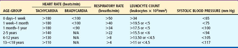
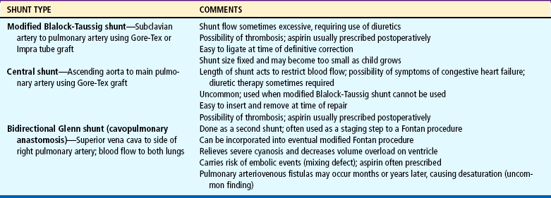
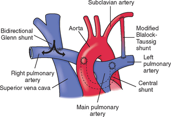
Hypercyanotic spells occur suddenly, and prompt recognition and treatment are essential. In the hospital setting, spells are often seen during blood drawing or IV insertion, when the child is highly agitated, or after cardiac catheterization. Treatment of a hypercyanotic spell is outlined in the Nursing Care Guidelines box. Morphine, administered subcutaneously or through an existing IV line, helps reduce infundibular spasm. A spell indicates the need for prompt surgical treatment if possible. In infants with defects not amenable to surgical repair, a shunt may be created surgically to increase blood flow to the lungs. Several commonly used shunt procedures are described in Table 25-4 and Fig. 25-9.
The cyanotic infant and child are well hydrated to keep the hematocrit and blood viscosity within acceptable limits to reduce the risk of CVAs. Fevers are carefully evaluated because bacteremia can result in bacterial endocarditis. The infant is monitored closely for anemia because of the risk of CVAs and the reduced arterial oxygen-carrying capacity that occurs. Iron supplementation and possibly blood transfusion are used as needed.
Respiratory tract infections or reduced pulmonary function from any cause can worsen hypoxemia in the cyanotic child. Aggressive pulmonary hygiene, chest physical therapy, administration of antibiotics, and use of oxygen to improve arterial saturations are important interventions.
The general appearance of infants and children with significant cyanosis poses unique concerns. Blue lips and fingernails are obvious signs of their hidden cardiac defect. Clubbing and small, thin stature in older children further indicate severe heart disease. Adolescents are especially concerned about their body image; children with cyanosis are often teased about their appearance and singled out as different. Many children, when asked what surgery will do, reply, ‘Make me pink.’ Their joy and excitement after surgery are evident when they see their pink fingers. Parents are often fearful of their child’s bluish color, since cyanosis is usually associated with lack of oxygen and severe illness. They also must deal with comments from relatives, friends, and strangers about their child’s abnormal color. They need a simple explanation of hypoxemia and cyanosis and reassurance that cyanosis does not imply a lack of oxygen to the brain. Their questions and fears need to be addressed in a calm, supportive manner, and positive aspects of their child’s growth and development are emphasized. They are taught the treatment for hypercyanotic spells (see Nursing Care Guidelines box above).
Dehydration must be prevented in hypoxemic children because it potentiates the risk of CVAs. Fluid status is carefully monitored, with accurate intake and output and daily weight measurements. Maintenance fluid therapy is the minimum requirement, supplemental fluids should be readily available, and gavage feeding or IV hydration is given to children unable to take adequate oral fluids. Fever, vomiting, and diarrhea can cause dehydration and require prompt treatment. Parents are instructed in the importance of adequate fluid intake and measures to prevent dehydration. An oral electrolyte solution should be available at home in the event that the infant is unable to tolerate the usual formula. The practitioner should be notified of fever, vomiting, diarrhea, or other problems.
Preventive measures and accurate assessment of respiratory infection are important nursing considerations. Any compromise in pulmonary function will increase the infant’s hypoxemia. Good hand washing and protection from individuals with an obvious respiratory tract infection are important. Aggressive pulmonary hygiene, treatment with antibiotics or antiviral agents as indicated, and supplemental oxygen to decrease hypoxemia are necessary measures. Infants may need to be gavage fed or given parenteral hydration if respiratory distress prevents oral feeding.
When a child is born with a severe cardiac anomaly, the parents are faced with the immense psychologic and physical tasks of adjusting to the birth of a child with special needs. Family issues and nursing interventions to support the family are similar to those discussed in Chapters 11 and 22. The following discussion is primarily directed (1) toward the family of an infant who has a serious heart defect and requires home care before definitive repair and (2) toward preparation and care of the child and family when invasive procedures (catheterization and surgery) are performed. For nursing care related to the child with hypoxemia and CHF, the reader should refer to earlier discussions of these topics.
Nursing care of the child with a congenital heart defect begins as soon as the diagnosis is suspected. Prenatal diagnosis of congenital heart defects is becoming increasingly frequent. New demands are being placed on nurses to counsel and support families as they prepare for the birth of these infants.
Once parents learn of the heart defect, they are initially in a period of shock, followed by high anxiety and fear that the child will die. The family needs time to grieve before they can assimilate the meaning of the defect. Unfortunately, the demands for medical treatment may not allow this, instead necessitating that the parents immediately give informed consent for diagnostic-therapeutic procedures. The nurse can be instrumental in supporting parents in their loss, assessing their level of understanding, supplying information as needed, and helping other members of the health care team understand the parents’ reactions (see Family Focus box).
Severely ill newborns usually remain in the hospital. Parent-infant attachment is supported by encouraging parents to hold, touch, and look at their child and providing time and privacy for the parents to spend with their newborn. (See Chapter 10 for suggestions on promoting attachment between parents and their hospitalized newborn.)
The effect of a child with a serious heart defect on the family is complex. No member, regardless of the degree of positive adjustment, is unaffected. Mothers frequently feel inadequate in their mothering ability because of the more complex care infants with congenital heart defects require. They often feel exhausted from the pressures of caring for these children and the other family members. Fathers and siblings may feel neglected and resentful, a reaction similar to the feelings toward family members with other chronic conditions (see Chapter 18). Often, parents do not feel confident leaving the child in another’s care. This often sets up a trap for parents, especially mothers, who become locked into the child’s care with no relief. Although the fears are justified, they can be minimized by gradually teaching someone (a reliable relative or neighbor) how to care for the child.
The need to maintain discipline and set consistent limits can be difficult for parents. Using behavior modification techniques, in the form of either concrete awards (e.g., a favorite activity) or social reinforcement (e.g., approval), can be effective. However, it is most beneficial if employed before the child learns to control the family. To prevent later problems, it is necessary to begin discussions with parents while the child is in infancy regarding the need for discipline as the child gets older.
Another issue that may develop within family relationships is the child’s overdependency. This is often the result of parental fear that the child may die. Parents need guidance to recognize the eventual hazards of continuing dependency and protectiveness as the child grows older, and the nurse can assist parents in learning ways to foster optimum development. Unless parents are shown what activities the child can do, they may focus on physical limitations and encourage dependency.
The child also needs opportunities for normal social interaction with peers. These children do not need to be prevented from playing with other children because of concern regarding overexertion. Children usually limit their activities if allowed to set their own pace. A child with CHD may constitute a long-term family crisis. Frequently the continuing unremitting stresses of care—physical exhaustion, financial costs, emotional upset, fear of death, and concern for the child’s future–are not fully appreciated by those caring for the family. Even when the child’s condition is stabilized or corrected, the family may need to make adjustments in their lifestyle. Introducing them to other families with similarly affected children can help them adjust to the daily stresses.
When parents are ready to hear about the heart condition, they require a clear explanation based on their level of understanding. A review of the basic structure and function of the heart is helpful before describing the defect. A simple diagram, pictures, or a model of the heart can help parents visualize the heart and the congenital defect.* Parents appreciate receiving written information about the specific condition. A glossary of frequently used terms is also helpful. Parents also require information about prognosis and treatment options.
Increasingly, families are using the Internet as a source of information about heart disease in children.† They are also finding support through contacts with other parents and parent groups. It is important for parents to realize that not all websites offer medically accurate information and that information from other parents might not be applicable to their own situation. Some children with rare, complex heart defects require individualized treatment plans, and general information on the Internet or in books may not apply to their child. Parents should use their health care team, in particular their cardiologist, to discuss information they have received from other sources.
Information given to the child must be tailored to the child’s developmental age. As the child matures, the level of information is revised to meet the child’s new cognitive level. Preschoolers need basic information about what they will experience more than what is actually occurring physiologically. School-age children benefit from a concrete explanation of the defect. Preadolescents and adolescents often appreciate a more detailed description of how the defect affects their heart. Children of all ages need to express their feelings concerning the diagnosis.
Parents are the child’s principal caregivers and need to develop a positive, supportive working relationship with the health care team. Since most children spend the majority of their time at home with episodic trips to the hospital, parents manage their child’s illness on a daily basis. They monitor for signs of illness, give medications and treatments, bring their child to appointments, work with a variety of caregivers, and alert the team about problems. Successful relationships are a partnership between parents and caregivers that is built on mutual trust and respect. Good communication among the family, the cardiology specialists, and the primary care practitioner is essential. As children reach adolescence, they begin to take a larger role in managing their illness and making decisions about their care.
Parents should be aware of the symptoms of their child’s cardiac condition and signs of worsening clinical status. Parents of children who may develop CHF should be familiar with the symptoms (see Box 25-5) and know when to contact the practitioner. Parents of children with cyanosis should be informed about fluid management and hypercyanotic spells (see p. 885). Parents should have an information sheet with their child’s diagnosis, significant treatments such as surgical procedures, allergies, other health care problems, current medications, and health care providers’ contact numbers available in case of emergencies and to share with other caregivers such as teachers, baby-sitters, or daycare providers.
The family also needs to be knowledgeable regarding the therapeutic management of the disorder and the role that surgery, other procedures, medications, and healthy lifestyle play in maintaining good health. Medications play a critical role in managing some cardiac conditions such as dysrhythmias, severe CHF, anticoagulation for artificial valves, and antirejection medications after heart transplantation. Some patients must take multiple medications daily for their lifetime. Many medications can be dangerous if taken incorrectly and require close monitoring. Parents are taught the correct procedure for giving medications and cautioned to keep them in a safe area to prevent accidental ingestion (see Family-Centered Care box, p. 882).
Another area of parental concern is the child’s level of physical activity. Most children do not need to restrict activity, and the best approach is to treat the child normally and allow self-limited activity. Exceptions to self-determined activity primarily involve strenuous recreational and competitive sports in children with specific cardiac problems. Activities and exercise restrictions should be discussed with the child’s cardiologist. Deliberately attempting to prevent crying should be avoided because it can establish a maladaptive parental pattern of relating to the infant.
Infants and children with CHD require good nutrition. Breastfeeding should be possible for many infants with CHD. Providing adequate nutrition to infants with CHF or complex congenital defects is especially difficult because of their high caloric requirements and inability to suck effectively because of fatigue and tachypnea. Instructing parents in feeding methods that decrease the infant’s work and giving high-calorie formula are important interventions (see p. 883 for a discussion on feeding the infant with CHF). Children with severe cardiac defects are often anorexic. Encouraging them to eat can be a tremendous challenge. Consultation with a dietitian is often helpful. The child should be given a choice of available high-nutrient foods.
Infants with heart disease should be immunized according to the current guidelines. Immunization schedules may need to be modified around times of acute illness or surgical procedures (Smith, 2001). Infants and children less than 2 years of age with unrepaired heart defects, cyanotic lesions, pulmonary hypertension, or history of prematurity should receive the vaccine for respiratory syncytial virus (RSV) monthly during RSV season (November to April in North America) (American Academy of Pediatrics, 2006).
Infants and children who have serious heart disease are at risk for developmental delays. Multiple factors can influence neurodevelopmental outcomes, including genetics (chromosomal abnormalities and microdeletions), family background (parental intelligence quotient [IQ] and socioeconomic status), preoperative factors (including prematurity, cyanosis, shock), intraoperative factors (use of cardiopulmonary bypass, deep hypothermic circulatory arrest), and postoperative factors (hemodynamic instability, hypoxia, acidosis, cardiac arrest, stroke, ischemic events). Recent efforts to limit the time of deep hypothermic circulatory arrest and provide better neuroprotection during infant surgery may improve outcomes in the future. Although most children with serious heart disease are within the normal range for IQ, there is a higher incidence of neurodevelopmental deficits in children after heart surgery than in the normal population, specifically in speech and language, fine motor skills, and cognitive processes (Majnemer and Limperopoulos, 1999). Severe neurologic problems such as cerebral palsy, epilepsy, and mental retardation are uncommon.
Chapter 22 provides an extensive discussion of the principles for preparing children for invasive procedures. The American Heart Association published a scientific statement ‘Recommendations for Preparing Children and Adolescents for Invasive Cardiac Procedures’ (LeRoy, Elixson, O’Brien, and others, 2003), which addresses issues specific to the child with heart disease. The following discussion highlights some important aspects of preparation for cardiac catheterization and cardiac surgery.
The expected outcomes for preprocedure preparation include reducing anxiety, improving patient cooperation with procedures, enhancing recovery, developing trust with caregivers, and improving long-term emotional and behavioral adjustments after procedures (LeRoy, Elixson, O’Brien, and others, 2003). Important factors to consider in planning preparation strategies are the child’s cognitive development, previous hospital experiences, child’s temperament and coping style, timing of preparation, and the involvement of the parents. The most beneficial preparation strategies usually combine information giving and coping skills training such as conscious breathing exercises, distraction techniques, guided imagery, or other behavioral interventions.
Outpatient preoperative and precatheterization workups are common for most elective procedures. Children are then admitted on the morning of the procedure. Preprocedure teaching is often done in the clinic setting or at home and may include a tour of the intensive care unit (ICU) and inpatient facilities. Children of different ages and developmental levels require different amounts of information and different approaches. Young children should be prepared close in time to the event, whereas older children and adolescents may benefit from teaching several weeks in advance. Parents should be included in the preparation session to support their child and learn about upcoming events.
Topics to include in preoperative or precatheterization preparation include information on the environment, equipment, and procedures that the child will encounter during and following the procedure. Many information-giving techniques can be used such as verbal and written information, hospital tours, preoperative classes, picture books, or videos. Information about what the child will see, hear, and feel should be included, especially for older children and adolescents. Some of the sensory experiences of being in an ICU or catheterization laboratory include sights (monitors, many people, lots of equipment), sounds (beeping noises, alarms, voices), and sensations (lines and dressings, tape, discomfort, thirst). Familiar aspects of the environment, like BP cuffs, stethoscopes, or oximeter probes, are reviewed; and new equipment, such as monitors, IV lines, and oxygen masks, are described. Comforting aspects of the environment, such as play areas, chairs for parents, and televisions, are emphasized. Many patients who will be sedated during catheterization or receiving narcotic pain relievers after surgery will have minimal recall of that period and will not need detailed information about the equipment or procedures used. Information should be specific to the planned procedure for each patient.
A discussion of ways the child can cope with the experience should be included. For a young child, bringing a familiar stuffed animal or comfort object will help relieve anxiety, whereas advising an older child to bring headphones and favorite music to the catheterization laboratory will help distract him or her during the procedure. Recovery topics after catheterization include lying still to prevent bleeding at the catheter site, advancing diet, controlling pain, and monitoring. After surgery, the nurse reviews the importance of ambulation, coughing, deep breathing, drinking, and eating and describes pain management and monitoring routines. Simple coping strategies for use during painful procedures should be reviewed; these include distraction techniques such as counting, blowing, singing, or telling stories.
Children and their families should have a choice about an ICU tour. Exposure to the ICU environment can actually increase anxiety in some children, particularly young children, those with previous hospital experiences, and those who are highly anxious (LeRoy, Elixson, O’Brien, and others, 2003). Usually the day before the procedure is ample time to allow the child to ask questions and to prevent undue fantasizing about the experience. The child should be protected from the frightening sights in the unit; equipment not in view postoperatively, such as equipment located behind or below the bed, needs less attention. The child and parents are encouraged to ask questions or to explore further any equipment in the room, but they should not be pushed to assimilate more information than they are able.
Preoperative physical care differs little, if any, from that for any other surgery and is discussed in Chapter 22. The child should be assured that the parents will be there when the child wakes up; they should be allowed to accompany their child as far as possible to the operating suite (see Surgical Procedures, p. 694). After all of the equipment and procedures have been explained, it is important to talk about ‘getting well’ and going home.
Immediate postoperative care is usually provided by specially trained nurses in ICUs. Many of the procedures, such as arterial pressure and central venous pressure (CVP) monitoring, and the observations related to vital functions require advanced educational training (the reader should refer to critical care texts for further information). However, nurses caring for the child before surgery and during the convalescent period need to be familiar with the major principles of care. Selected complications that may occur postoperatively are described in Box 25-7.
Vital signs and BP are recorded frequently until stable. Heart rate and respirations are counted for 1 full minute, compared with the ECG monitor, and recorded with activity. The heart rate is normally increased after surgery. The nurse observes cardiac rhythm and notifies the practitioner of any changes in regularity. Dysrhythmias may occur postoperatively secondary to anesthetics, acid-base and electrolyte imbalance, hypoxia, surgical intervention, or trauma to conduction pathways (p. 896).
At least hourly, the lungs are auscultated for breath sounds. Diminished or absent sounds may indicate an area of atelectasis or a pleural effusion or pneumothorax, which necessitates further medical assessment. Temperature changes are typical during the early postoperative period. Hypothermia is expected immediately after surgery from hypothermia procedures, effects of anesthesia, and loss of body heat to the cool environment. During this period the child is kept warm to prevent additional heat loss. Infants may be placed under radiant heat warmers. During the next 24 to 48 hours the body temperature may rise to 37.7° C (100° F) or slightly higher as part of the inflammatory response to tissue trauma. After this period an elevated temperature is most likely a sign of infection and warrants immediate investigation for probable cause.
Intraarterial monitoring of BP is commonly done after open-heart surgery. A catheter is passed into the radial artery or other artery, and the other end is attached to an electronic monitoring system, which provides a continuous recording of the BP. The intraarterial line is maintained with a low-rate, constant infusion of heparinized saline to prevent clotting.
Several IV lines are inserted preoperatively: a peripheral IV to give fluids and medications, and a CVP, which is usually inserted in a large vessel in the neck. Additionally, intracardiac monitoring lines are sometimes placed intraoperatively in the right atrium, left atrium, or pulmonary artery. Intracardiac lines allow assessment of pressures inside the cardiac chambers, providing vital information about volume status, cardiac output, and ventricular function. All lines must be cared for using strict aseptic technique, and patients must be carefully assessed for bleeding at the time of line removal.
Infants usually require mechanical ventilation in the immediate postoperative period. Early extubation in the operating room or early postoperative period is becoming more common. Children, especially those who did not require cardiopulmonary bypass, may be extubated in the operating room or in the first few postoperative hours. Suctioning is performed only as needed and performed carefully to avoid vagal stimulation (which can trigger cardiac dysrhythmias) and laryngospasm, especially in infants. Suctioning is intermittent and maintained for no more than 5 seconds at a time to avoid depleting the oxygen supply. Supplemental oxygen is administered with a manual resuscitation bag before and after the procedure to prevent hypoxia. The heart rate is monitored after suctioning to detect changes in rhythm or rate, especially bradycardia. The child should always be positioned facing the nurse to permit assessment of the child’s color and tolerance of the procedure.
When weaning and extubation are completed, humidified oxygen is delivered by mask, hood, or nasal cannula to prevent drying of mucosa. The child is encouraged to turn and deep breathe at least hourly. Measures are employed to enhance ventilation and decrease pain, such as splinting of the operative site and use of analgesics. Chest tubes are inserted into the pleural or mediastinal space during surgery or in the immediate postoperative period to remove secretions and air to allow reexpansion of the lung. Drainage is checked hourly for color and quantity. Immediately after surgery the drainage may be bright red, but afterward it should be serous. The largest volume of drainage occurs in the first 12 to 24 hours and is greater in extensive heart surgery.
Chest tubes are usually removed on the first to third postoperative day. Removal of chest tubes is a painful, frightening experience. Analgesics such as morphine sulfate, often combined with midazolam (Versed), should be given before the procedure. Older children are forewarned that they will feel a sharp, momentary pain. After the suture is cut, the tubes are quickly pulled out at the end of full inspiration to prevent intake of air into the pleural cavity. A purse-string suture (placed when the tubes were inserted) is pulled tight to close the opening. A petrolatum-covered gauze dressing is immediately applied over the wound and securely taped on all four sides to the skin so that an airtight seal is formed. It is left on for 1 or 2 days. Breath sounds are checked to assess for a pneumothorax.
Intake and output of all fluids must be accurately calculated. Intake is primarily IV fluids; however, a record of fluid used to flush the arterial and CVP lines or to dilute medications is also kept. Output includes hourly recordings of urine (usually a Foley catheter is inserted and attached to a closed collecting device), drainage from chest and nasogastric tubes, and blood drawn for analysis. Urine is analyzed for specific gravity to assess the concentrating ability of the kidneys and to assess approximately the body’s degree of hydration. Renal failure is a potential risk from a transient period of low cardiac output.
Fluids are restricted during the immediate postoperative period to prevent hypervolemia, which places additional demands on the myocardium, predisposing the patient to cardiac failure. To monitor fluid retention, the child is weighed daily, and the same scale is used at approximately the same time each day to avoid errors in measurement. The child is usually given nothing by mouth for the first 24 hours. If an endotracheal (ET) tube is inserted, oral fluids are usually withheld until the child is extubated. Fluid restriction may be imposed even when oral fluids are given. The nurse calculates the distribution over a 24-hour period based on the child’s preoperative weight and drinking habits. The distribution should allow for most fluid to be given during the child’s most wakeful and active periods.
After heart surgery, rest should be provided to decrease the workload of the heart and promote healing. The simplest way to ensure individualized, efficient, high-quality care is to plan at the beginning of the shift the nursing procedures to be done, with periods of rest identified. The schedule should be shared with parents to allow them to visit at the most advantageous times, such as after a rest period when no special treatments are anticipated.
A progressive schedule of ambulation and activity is planned, based on the child’s preoperative activity patterns and postoperative cardiovascular and pulmonary function. Ambulation is initiated early, usually by the second postoperative day, when chest tubes, arterial lines, and assisted ventilatory equipment may be removed. Activity progresses from sitting on the edge of the bed and dangling the legs to standing up and sitting in a chair. Heart rate and respirations are carefully monitored to assess the degree of cardiac demand imposed by each activity. Tachycardia, dyspnea, cyanosis, desaturation, progressive fatigue, or dysrhythmias indicate the need to limit further energy expenditure.
Heart surgery is both painful and frightening for children, and comfort is a primary nursing concern. Several incisions may be used for heart surgery. A median sternotomy is most common, following the sternum down the center of the chest. A ministernotomy opens the lower sternum. A thoracotomy incision is most uncomfortable because it goes through muscle tissue. It allows access to the side of the chest through an incision from under the arm around the back to the scapula.
Most patients need IV analgesics for pain control during the immediate postoperative period. Patient-controlled analgesia may be used with children old enough to understand the concept. Nonsteroidal antiinflammatory drugs (NSAIDs) such as ketorolac (Toradol) may be used intravenously. Paralyzing agents may also be used with the analgesics for children who are agitated or hemodynamically unstable.
After extubation and removal of lines and tubes, pain can be satisfactorily controlled with oral medications such as ibuprofen, codeine with acetaminophen (Tylenol No. 3), or oxycodone and acetaminophen (Tylox). Acetaminophen alone provides adequate pain relief for most children at discharge. Sternotomy incisions are usually well tolerated, with some discomfort when walking and coughing. Thoracotomy incisions are usually more painful because the incision is through muscle; a more aggressive pain management plan with around-the-clock medications for several days is often necessary to allow for adequate rest, ambulation, and pulmonary hygiene.
In addition to pharmacologic pain control, every effort is made to minimize the discomfort of procedures, such as using a firm pillow or favorite stuffed animal placed against the chest incision during movement and performing treatments after pain medication is given, preferably at a time that coincides with the drug’s peak effect. Nonpharmacologic measures are used to lessen the perception of pain, and parents are encouraged to comfort their child as much as possible. (See also Pain Assessment; Pain Management, Chapter 7.)
Children may become depressed after surgery. This is thought to be caused by preoperative anxiety, postoperative psychologic and physiologic stress, and sensory overstimulation. Typically the child’s disposition improves on leaving the ICU.
Children may also be angry and uncooperative after surgery as a response to the physical pain and to the loss of control imposed by the surgery and treatments. They need an opportunity to express feelings, either verbally or through activity. Children often regress in their behavior during the stress of surgery and hospitalization. They also may express feelings of anger or rejection toward parents. The nurse can support the parents by being available for information and explaining all the procedures to them. The first few postoperative days are particularly difficult because parents see their child in pain and realize the potential risks from surgery. They often are overwhelmed by the physical environment of the ICU and feel useless because they can do so little for their child. The nurse can minimize such feelings by including parents in caregiving activities and comfort and play activities, by providing information about the child’s condition, and by being sensitive to their emotional and physical needs. The importance of their presence in making the child feel more secure is stressed, even if they do not provide physical care.
Ideally, discharge planning begins on admission for cardiac surgery and includes an assessment of the parents’ adjustment to the child’s altered state of health. Neonates need additional screening tests (such as newborn metabolic screen and hearing tests) and may need immunizations before discharge (Dodds and Merle, 2005). The family will need both verbal and written instructions on medication, nutrition, activity restrictions, subacute bacterial endocarditis, return to school, wound care, and signs and symptoms of infection or complications (see Family-Centered Care box). Referrals to community agencies may be warranted to assist parents in the transition from hospital to home and to reinforce the teaching.
The parents will also need clear instructions on when to seek medical care for complications and how to contact the health care provider. Follow-up with the cardiologist and primary care provider is also arranged before discharge. Parents should have a summary, including their child’s medical condition, medications, and health care providers available for emergencies. Appropriate identification, such as a MedicAlert device, is indicated for children with a pacemaker or a heart transplant and for those receiving anticoagulation therapy or antidysrhythmic medication.
Although surgical correction of heart defects has improved dramatically, it is still not possible to completely repair many of the complex anomalies. For many children, repeat procedures are required to replace conduits or grafts or to manage complications such as restenosis. Consequently, the long-term prognosis is uncertain, and full recovery is not always possible. For these families, medical follow-up and continued emotional support are essential. The nurse can often serve as an important primary health professional and as a resource for referrals when needed.
BACTERIAL (INFECTIVE) ENDOCARDITIS
Bacterial endocarditis (BE), or infective endocarditis (IE), also referred to as subacute bacterial endocarditis (SBE), is an infection of the valves and inner lining of the heart. Although it can occur without underlying heart disease, it is most often a sequela of bacteremia in the child with acquired or congenital anomalies of the heart or great vessels. It especially affects children with valvular abnormalities, prosthetic valves, shunts, recent cardiac surgery with invasive lines, and rheumatic heart disease with valve involvement. The most common causative agent is Streptococcus viridans; other causative agents are Staphylococcus aureus, gram-negative bacteria, and fungi such as Candida albicans.
Organisms may enter the bloodstream from any site of localized infection. In the past, endocarditis was believed to be highly associated with invasive procedures; however, endocarditis is most likely to occur from routine exposure to bacteremia associated with usual daily activities, although it can also occur after procedures such as dental work (S. viridans); after invasive procedures involving the gastrointestinal-genitourinary tract; after cardiac surgery, especially if synthetic material is used (valves, patches, conduits); or from long-term indwelling catheters. The microorganisms grow on the endocardium, forming vegetations (verrucae), deposits of fibrin, and platelet thrombi. The lesion may invade adjacent tissues, such as aortic and mitral valves, and may break off and embolize elsewhere, especially in the spleen, kidney, and central nervous system.
The diagnosis of IE is suspected on the basis of clinical manifestations (Box 25-8). Several laboratory findings may suggest IE (e.g., ECG changes [prolonged PR interval], radiographic evidence of cardiomegaly, anemia, elevated erythrocyte sedimentation rate, leukocytosis, microscopic hematuria). Vegetations on the valve and abnormal valve function can often be visualized by echocardiography. Definitive diagnosis rests on growth and identification of the causative agent in the blood.
Treatment should be instituted immediately and consists of administration of high doses of appropriate antibiotics intravenously for 2 to 8 weeks. Blood cultures are taken periodically to evaluate response to antibiotic therapy.
Prevention involves administration of prophylactic antibiotic therapy 1 hour before procedures known to increase the risk of entry of organisms in very high–risk patients. New guidelines only require prophylaxis in patients with the highest risk of poor outcome if they develop endocarditis (Box 25-9). Drugs of choice for prophylaxis include amoxicillin, ampicillin, clindamycin, cephalexin, cefadroxil, azithromycin, and clarithromycin.
Ideally, the objective of nursing care is to counsel parents of high-risk children concerning the signs and symptoms of endocarditis and, in certain cases, the need for prophylactic antibiotic therapy before procedures such as dental work. The family’s regular dentist should be advised of the child’s cardiac diagnosis as an added precaution to ensure preventive treatment. SBE prophylaxis is now reserved for very high–risk patients. Many patients who met criteria established in the past may not require prophylaxis under the new guidelines (Wilson, Taubert, Gewitz, and others, 2007) (see Box 25-9). Parents should be counseled regarding the rationale for discontinuing prophylaxis and should be educated as to the fact that their child is still at risk. It is important that all children with congenital or acquired heart disease maintain the highest level of oral health to reduce the chance of bacteremia from oral infections.
Parents should also have a high index of suspicion regarding potential infections. Without unduly alarming them, the nurse stresses that any unexplained fever, weight loss, or change in behavior (lethargy, malaise, anorexia) must be brought to the practitioner’s attention. Such symptoms should not be self-diagnosed as a cold or flu. Early diagnosis and treatment are important in preventing further cardiac damage, embolic complications, and growth of resistant organisms.
Treatment of endocarditis requires long-term parenteral drug therapy. In many cases, IV antibiotics may be administered at home with nursing supervision for part of the treatment course. Nursing goals during this period are (1) preparation of the child for IV infusion, usually with an intermittent-infusion device, and several venipunctures for blood cultures; (2) observation for side effects of antibiotics, especially inflammation along venipuncture sites; (3) observation for complications, including embolism and CHF; and (4) education regarding the importance of follow-up visits for cardiac evaluation, echocardiographic monitoring, and blood cultures.
Rheumatic fever (RF) is a poorly understood inflammatory disease that occurs after infection with group A β-hemolytic streptococcal (GABHS) pharyngitis. It occurs most often in late school-age children or adolescents and is rare in adults. It is a self-limited illness that involves the joints, skin, brain, serous surfaces, and heart. Cardiac valve damage (referred to as rheumatic heart disease) is the most significant complication of RF. The mitral valve is most often affected. In developed countries RF and rheumatic heart disease have become uncommon. However, RF remains a devastating problem in developing (Third World) countries and has reappeared in some parts of the United States (Gentles, Colan, Wilson, and others, 2001).
Strong evidence supports a relationship between upper respiratory tract infection with GABHS and subsequent development of RF (usually within 2 to 6 weeks). Acute RF is the result of an exaggerated immune response to bacteria in a susceptible host (Carapetis, McDonald, and Wilson, 2005). In almost all cases of RF a previous infection with GABHS can be documented by laboratory evidence of rising antibody titers. Prevention or treatment of GABHS infection prevents RF.
Diagnosis is based on a set of guidelines recommended by the American Heart Association (Newburger, Takahashi, Gerber, and others, 2004). These guidelines, known as modifications of the Jones criteria, suggest that the presence of two major manifestations or one major and two minor manifestations, such as fever and arthralgia, with supportive evidence of recent streptococcal infection, indicates a high probability of RF (see Nursing Care Guidelines box).
Children suspected of having RF are tested for streptococcal antibodies. The most reliable and best standardized test is an elevated or rising antistreptolysin O (ASO or ASLO) titer, which occurs in 80% of children with RF.
The goals of medical management are (1) eradication of hemolytic streptococci, (2) prevention of permanent cardiac damage, (3) palliation of the other symptoms, and (4) prevention of recurrences of RF. Penicillin is the drug of choice, with erythromycin as a substitute in penicillin-sensitive children. Salicylates are used to control the inflammatory process, especially in the joints, and reduce the fever and discomfort. Bed rest is recommended during the acute febrile phase but need not be strict.
Prophylactic treatment against recurrence of RF is started after the acute therapy and involves monthly intramuscular injections of benzathine penicillin G (1.2 million units), two daily oral doses of penicillin (200,000 units), or one daily dose of sulfadiazine (1 g). The duration of long-term prophylaxis is uncertain, but 5 years since the last episode or age 18, longer with cardiac involvement, is suggested by the World Health Organization (Carapetis, McDonald, and Wilson, 2005).
Children who have had acute RF are susceptible to recurrent RF for the rest of their lives and should be followed medically for at least 5 years. Repeated infections are likely to result in rheumatic heart disease. Children and families must be aware of the need for continuing antibiotic prophylaxis for dental work, infection, and invasive procedures.
The objectives of nursing care for the child with RF are to (1) encourage compliance with drug regimens, (2) facilitate recovery from the illness, (3) provide emotional support, and (4) prevent the disease. Because compliance is a major concern in long-term drug therapy, every effort is made to encourage adherence to the therapeutic plan (see Compliance, Chapter 22). When compliance is poor, monthly injections may be substituted for daily oral administration of antibiotics, and children need preparation for this often-dreaded procedure.
Interventions during home care are primarily concerned with providing rest and adequate nutrition. Usually, after the febrile stage is over, children can resume moderate activity, and their appetite improves. If carditis is present, the family must be aware of any activity restrictions and may need help in choosing less strenuous activities for the child.
One of the most disturbing and frustrating manifestations of the disease is chorea. The onset is gradual and may occur weeks to months after the illness; it sometimes even occurs in children who have not been diagnosed with RF. It may be mistaken for nervousness, clumsiness, behavioral changes, inattentiveness, and learning disability. It is usually a source of great frustration to the child because the movements, incoordination, and weakness severely limit physical ability. Of utmost importance is stressing to parents and schoolteachers the involuntary, sudden nature of the movements; that the chorea is transitory; and that all manifestations eventually disappear.
Nurses also have a role in prevention, primarily in screening school-age children for sore throats caused by GABHS. This may involve actively participating in throat culture screening programs or in referring children with a possible streptococcal infection for testing.
Hyperlipidemia is a general term for excessive lipids (fat and fatlike substances); hypercholesterolemia refers to excessive cholesterol in the blood. High lipid or cholesterol levels play an important role in producing atherosclerosis (fatty plaque on the arteries), which eventually can lead to coronary artery disease, a primary cause of morbidity and mortality in the adult population. A presymptomatic phase of atherosclerosis can begin in childhood. Preventive cardiology is focusing on the screening and management of lipid levels in childhood. The goal is to identify those children at high risk and intervene early.
Cholesterol is part of the lipoprotein complex in plasma that is essential for cellular metabolism. Triglycerides, natural fats synthesized from carbohydrates, are used for energy. Both are major lipids transported on lipoproteins, a combination of lipids and proteins, which include:
Low-density lipoproteins (LDLs)—These contain low concentrations of triglycerides, high levels of cholesterol, and moderate levels of protein. LDL is the major carrier of cholesterol to the cells. Cells use cholesterol for synthesis of membranes and steroid production. Elevated circulating LDL is a strong risk factor in cardiovascular disease.
High-density lipoproteins (HDLs)—These contain very low concentrations of triglycerides, relatively little cholesterol, and high levels of protein. They transport free cholesterol to the liver for excretion in the bile. High levels of HDL are thought to protect against cardiovascular disease.
Hyperlipidemia is diagnosed on the basis of analysis of blood for a full lipid profile, drawn after a 12-hour fast. A screening thyroid-stimulating hormone (TSH) is useful to rule out hypothyroidism as a cause of secondary hypercholesterolemia. In overweight children, a fasting glucose may be obtained to assess for the potential of metabolic syndrome, which is a combination of multiple symptoms that are associated with increased cardiovascular risk in adults. Blood samples should be collected after having the child sit for 5 minutes, and the tourniquet should be applied immediately before the needle puncture, since posture and vascular stasis may affect results. Diagnostic values for acceptable, borderline, and high total cholesterol and LDL cholesterol levels are listed in Table 25-5.
TABLE 25-5
Classification of Cholesterol Levels in Children from Families with a History of Heart Disease
CATEGORY | TOTAL CHOLESTEROL (mg/dl) | LDL CHOLESTEROL (mg/dl) |
Acceptable | <170 | <110 |
Borderline | 170–199 | 110–129 |
High | ≥200 | ≥130 |
From National Cholesterol Education Program: Report of the Expert Panel on Blood Cholesterol Levels in Children and Adolescents, Pediatrics 89(3 Pt 2):527, 1992.
Screening children for hypercholesterolemia is a controversial issue, with some authorities advocating universal screening and others proposing selective screening. Guidelines from the American Academy of Pediatrics’ Committee on Nutrition (American Academy of Pediatrics, 1998) recommend a strategy that combines two complementary approaches: (1) a population approach that aims to lower the average levels of blood cholesterol among all American children through population-wide changes in nutrient intake and eating patterns, and (2) an individualized approach based on selective screening (see Evidence-Based Practice box).
The first step in the treatment of high cholesterol is oriented to lifestyle modification. The American Academy of Pediatrics (1998) guidelines advocate a heart-healthy diet for all children. Children with known elevated cholesterol should have individual nutritional counseling by a nutritionist with expertise in pediatric lipids.
Research continues to support the benefit of diets low in saturated fats (Van Horn, Obarzanek, Friedman, and others, 2005). Current thinking favors a “Mediterranean”-type diet. Whole grains, fruits, and vegetables form the foundation of this diet. In addition, this diet allows the use of monounsaturated fats, such as olive oil and canola oil, which have beneficial effects on HDL-cholesterol values. The use of these fats also makes the diet more realistic. Daily aerobic exercise of at least 60 minutes a day is also recommended for children with high cholesterol. In addition, patients and parents should be counseled regarding the negative effects of smoking (both first hand and second hand).
For children with severe hypercholesterolemia who fail to respond to dietary modifications (after a 6- to 12-month trial), drug therapy may be necessary. Pharmacologic therapy is recommended for children with LDL cholesterol over 190 mg/dl without other risk factors or over 160 mg/dl in patients with two or more other risk factors. In most situations, medication is reserved for boys over 10 years old and for girls once they begin their menses.
In the past, bile acid–binding resins were the only class of drugs recommended for treatment of children. This class of drug acts by binding bile acids in the intestinal lumen. Because they are not absorbed by the intestine, they do not produce systemic toxicity and are safe for children. Cholestyramine (Questran) and colestipol (Colestid) are both powders that are mixed with water or juice just before ingestion. Unfortunately, the vast majority of patients do not get adequate reduction in LDL-cholesterol from bile acid–binding resins. Many cannot tolerate the medication because of the taste; gritty texture; and side effects, the most significant being constipation, abdominal pain, gastrointestinal bloating, flatulence, and nausea. The most recent findings on lipid abnormalities in children recommends treatment with statins if pharmacologic therapy is indicated, using the previously outlined guidelines for treatment (McCrindle, Urbina, Dennison, and others, 2007). Statins are much more effective at lowering LDL cholesterol and triglycerides and raising HDL cholesterol. Statins work by inhibiting the enzyme necessary for cholesterol synthesis. Statins are most effective when taken in the evening and are started at the lowest possible dose in young people. Blood work should be followed closely and should include a fasting lipid profile, liver function tests, and creatinine kinase repeated at 4- and 8-week intervals initially and with dosage changes.
Patients beginning therapy with a statin should be counseled regarding rare but potentially serious side effects such as rhabdomyolysis, elevated transaminases, and elevated creatinine kinase. Patients should discontinue their medication and contact their practitioner if they develop dark urine or new muscle aches. Lastly, statin medications are not safe during pregnancy and therefore sexually active adolescents need to take adequate birth control measures. Very long–term studies are unlikely to be available over decades; however, in the shorter-term studies that have been completed, statins seem to have a similar safety profile for children as they do for adults (McCrindle, Urbina, Dennison, and others, 2007).
Nurses play an important role in the screening, education, and support of children with hyperlipidemia and their families. When a child is referred to a lipid clinic, it is essential that the family be adequately prepared for the first visit. Generally, the parents will be asked to keep a dietary history of the child before this visit. Sometimes they will need to complete a questionnaire regarding the child’s normal dietary habits during the preceding year. Families should be instructed to keep their child fasting for at least 12 hours before screening. It is important to schedule the blood test early in the morning and to arrange for nourishment immediately thereafter. At the visit, a full family history should be taken, including the health of both parents and all first-degree relatives. Specific questions should be asked regarding early heart disease, hypertension, strokes (CVAs), sudden death, hyperlipidemia, diabetes, and endocrine abnormalities.
Parents and extended families should be informed about cholesterol and hyperlipidemia. This education should include a brief introduction to the different lipoprotein categories, including cholesterol, HDL, LDL, and triglycerides. Also, behavioral risk factors for heart disease, such as smoking and exercise, should be reviewed. For management to be effective, parents need to understand the rationale for dietary or pharmacologic intervention. The key is prevention of future cardiovascular disease.
Stringent dietary guidelines may become an issue of control and a source of great stress for many families. Children should not be viewed as having a disease. Rather, the positive aspects of healthy eating, regularly exercising, and avoiding smoking should be emphasized. Basic dietary changes should be encouraged for the whole family so that the affected child is not singled out. Cultural differences must be considered and recommendations individualized. Substitution rather than elimination needs to be emphasized. Visual aids (e.g., test tubes depicting the amount of fat in a hot dog) are often helpful, especially for children. Diets should be flexible and individually tailored by a nutritionist experienced in combining recommendations that meet both the nutritional demands of the growing child and the lipid modifications. Parents are encouraged to participate in dietary and educational sessions, ask questions, and share ideas and experiences.
Parents often feel guilty about the hereditary component of hyperlipidemia. Many also believe they have failed if the diet alone is not making a significant difference in their child’s lipid profile. They need to be reassured that a dietary approach alone is often not sufficient, especially for children with significantly elevated values.
Parents of children who require pharmacologic therapy need to understand the purpose, dosage, and possible side effects of the various drugs. Medication schedules should remain flexible and should not interfere with the child’s daily activities. Follow-up phone calls by the nurse between visits allow parents to discuss their concerns and ask any questions that have arisen.
Dysrhythmias, or abnormal heart rhythms, can occur in children with structurally normal hearts, as features of some congenital heart defects, and in patients after surgical repair of congenital heart defects. They are also seen in patients with cardiomyopathy and with cardiac tumors. They can occur secondary to metabolic and electrolyte imbalances. They can be classified in several ways, including by heart rate characteristics (bradycardia and tachycardia) and by the origin of the dysrhythmia in the atria or ventricles. Some dysrhythmias are well tolerated and self-limiting. Others may cause decreased cardiac output with associated symptoms. Some dysrhythmias can cause sudden death. Treatment depends on the cause of the dysrhythmia and its severity.
Many advances have been made in the diagnosis and treatment of pediatric dysrhythmias in the past decade. Improvements in technology have allowed better diagnosis, the development of ablation techniques, and the expansion of pacemaker capabilities. New antidysrhythmic medications have proven safe and effective in children. Radiofrequency ablation has offered a cure for some dysrhythmias. Pediatric electrophysiology has become a highly specialized field, and the student should consult more detailed sources for an in-depth discussion. The following sections address diagnostic studies and provide a general discussion of the most common tachycardia (supraventricular tachycardia [SVT]) and the most common bradycardia (complete heart block) that require treatment in the pediatric population.
Nurses must be familiar with the standards of normal heart rate for the particular age-group (see inside back cover). An initial nursing responsibility is recognition of an abnormal heartbeat, either in rate or rhythm. When a dysrhythmia is suspected, the apical rate is counted for a full minute and compared with the radial rate, which may be lower because not all of the apical beats are felt. Consistently high or low heart rates should be regarded as suspicious. The patient should be placed on a cardiac monitor with recording capabilities. A 12-lead ECG yields more information than the monitor recording and should be done as soon as possible.
The basic diagnostic procedure is the ECG, including 24-hour Holter monitoring. Electrophysiologic cardiac catheterization allows for identification of the conduction disturbance and immediate investigation of drugs that may control the dysrhythmia. Another procedure that may be employed is transesophageal recording. An electrode catheter is passed to the lower esophagus and, when in position at a point proximal to the heart, is used to stimulate and record dysrhythmias.
Dysrhythmias can be classified according to various criteria, such as effect on heart rate and rhythm, as follows:
Bradydysrhythmias.: Sinus bradycardia (slower than normal rate) in children can be due to the influence of the autonomic nervous system, as with hypervagal tone, or in response to hypoxia and hypotension. Sinus bradycardias are also known to develop after some complex cardiac surgical repairs involving extensive atrial suture lines such as atrial baffle repairs (Mustard and Senning repairs) and the Fontan procedure.
Complete atrioventricular block (AV block) is also referred to as complete heart block. This can be either congenital (occurring in children with structurally normal hearts) or acquired after surgery to repair cardiac defects. AV blocks are most often related to edema around the conduction system and resolve without treatment. Temporary epicardial wires are placed in most patients at surgery; if a rhythm disturbance occurs, temporary pacing can be employed. Several days after surgery, the health practitioner removes the wires by pulling slowly and deliberately down on them from the site of insertion.
Some children may need a permanent pacemaker. The pacemaker takes over or assists in the heart’s conduction function. The implantation of a pacemaker, in the operating room or possibly the catheterization laboratory, is usually a low-risk procedure. The pacemaker is made up of two basic parts: the pulse generator and the lead. The pulse generator is composed of the battery and the electronic circuitry. The lead is an insulated, flexible wire that conducts the electrical impulse from the pulse generator to the heart. Two types of leads are available: transvenous and epicardial. After the lead has been attached to the heart, a small incision is made and a pocket is formed under the muscle to house and protect the generator. Continuous ECG monitoring is necessary during the recovery phase to assess pacemaker function. The nurse should be aware of the programmed rate and expected individual generator variations. The pacemaker insertion site is monitored for signs of infection. Analgesics are given for pain.
Pacemaker functions have become more sophisticated, and some models can adjust heart rate to activity demands or be programmed for overdrive pacing or cardioversion.
Discharge teaching includes information about the signs and symptoms of infection, general wound care, and activity restrictions. Parents, and patients if they are old enough, should be taught to take a pulse and know the settings of the pacemaker. If the patient’s low rate is set at 80 beats/min and the heart rate is only 68 beats/min, there is a possible problem with the pacemaker that needs to be investigated. Instructions for telephone transmission of ECG readings are also given. Telephone transmission can be used to transmit ECG strips and also to monitor battery life and pacemaker function. The pacemaker generator will have to be replaced periodically because of battery depletion. Children with pacemakers should wear a medical alert device, and their parents should have a paper identification card with specific pacer data in case of an emergency. Cardiopulmonary resuscitation (CPR) instruction is suggested for parents.
Tachydysrhythmias.: Sinus tachycardia (abnormally fast heart rate) secondary to fever, anxiety, pain, anemia, dehydration, or any other etiologic factor requiring increased cardiac output should be ruled out before diagnosing an increased heart rate as pathologic. SVT is the most common tachydysrhythmia found in children and refers to a rapid regular heart rate of 200 to 300 beats/min. The onset of SVT is often sudden, the duration is variable, and the rhythm may end abruptly and convert back to a normal sinus rhythm. Clinical signs in infants and young children are poor feeding, extreme irritability, and pallor. Children may experience palpitations, dizziness, chest pain, and diaphoresis. If SVT is sustained, signs of CHF may be seen.
The treatment of SVT depends on the degree of compromise imposed by the dysrhythmia. In some cases, vagal maneuvers, such as applying ice to the face, massaging the carotid artery (on one side of the neck only), or having an older child perform a Valsalva maneuver (e.g., exhaling against a closed glottis, blowing on a thumb as if it were a trumpet for 30 to 60 seconds), have terminated SVT. If vagal maneuvers fail or the child is hemodynamically unstable, adenosine (a drug that impairs AV conduction) may be used. Adenosine is given by rapid IV push with a saline bolus immediately after the drug because of its very short half-life. If this is unsuccessful or cardiac output is compromised, esophageal overdrive pacing or synchronized cardioversion (delivering an electrical shock to the heart) can be employed in the intensive care setting. Sedation is needed for both procedures. Cardioversion should never be done in a conscious patient. More long-term pharmacologic treatment includes digoxin or possibly propranolol (Inderal) or amiodarone for severe or recurrent SVT.
A primary focus of nursing care is education of the family regarding the symptoms of SVT and its treatment. SVT may occur again despite therapy. Parents should be taught to take a radial pulse for a full minute. If medication is prescribed, instructions regarding accurate dosage and the importance of administering the correct dose at specified intervals are stressed.
Radiofrequency ablation has become first-line therapy for some types of SVT. The procedure is done in the cardiac catheterization laboratory and begins with mapping of the conduction system to identify the dysrhythmia focus. A catheter delivering radiofrequency current is directed at the site, and the area is heated to destroy the tissue in the area. These are lengthy procedures, often 6 to 8 hours, and sedation or general anesthesia is required. Preparation is similar to that for cardiac catheterization.
Pulmonary artery hypertension (PAH) describes a group of rare disorders that result in an elevation of pulmonary artery pressure above 25 mm Hg at rest after the neonatal period (Barst, 1999). These disorders are poorly understood, and until recently there was no treatment beyond supportive care. PAH is a progressive, eventually fatal disease for which there is no known cure. It can be difficult to diagnose in the early stages. Often when patients become symptomatic and a diagnosis is made, their disease is rapidly progressing, treatment is unsuccessful, and death occurs within several years. Significant new information about the disease process, genetic links, diagnosis, and treatment has recently been learned that may improve treatments and outcomes for these patients.
PAH affects the small pulmonary arteries and is characterized by vascular narrowing leading to an increase in pulmonary vascular resistance. Why some children develop the disease and others do not is unclear. There are many possible causes of PAH. Cardiac causes occur primarily in patients with a large left-to-right shunt producing increased pulmonary blood flow. If these defects are not repaired early, the high pulmonary flow will cause changes in the pulmonary artery vessels and the vessels will lose their elasticity. Other causes of PAH include hypoxic lung diseases, thromboembolic diseases causing pulmonary vascular obstruction, collagen vascular diseases, and exposure to toxic substances. Many of the patients have no identifiable cause for PAH and have primary or idiopathic PAH.
The clinical manifestations include dyspnea with exercise, chest pain, and syncope. Dyspnea is the most common symptom and is caused by impaired oxygen delivery. Chest pain is the result of coronary ischemia in the right ventricle from severe hypertrophy. Syncope reflects a limited cardiac output leading to decreased cerebral blood flow. Right-sided heart dysfunction is steadily progressive, and when symptoms of venous congestion and edema are present, prognosis is poor.
Although no cure is known, several therapies have shown promise in slowing the progression of the disease and improving quality of life. In general, situations that may exacerbate the disease and cause hypoxia, such as exercise and high altitudes, are avoided. Supplemental oxygen, especially at night while sleeping, is commonly used to relieve hypoxia. Patients are at risk for thromboembolic events leading to pulmonary emboli, so anticoagulation with warfarin (Coumadin) is often prescribed.
Vasodilator therapy (which relaxes vascular smooth muscle and reduces pulmonary artery pressure) can prolong survival of patients with PAH. Oral calcium channel blockers have been successful in some children. Continuous IV prostacyclin has been used with some success in children who did not respond to oral therapy. Both these therapies, although promising, have been used in only small numbers of patients and are expensive. Lung transplantation may be another treatment option.
Cardiomyopathy refers to abnormalities of the myocardium in which the cardiac muscles’ ability to contract is impaired. Cardiomyopathies are relatively rare in children. Possible etiologic factors include familial or genetic causes, infection, deficiency states, metabolic abnormalities, and collagen vascular diseases. Most cardiomyopathies in children are considered primary or idiopathic, in which the cause is unknown and the cardiac dysfunction is not associated with systemic disease. Some of the known causes of secondary cardiomyopathy are anthracycline toxicity (the antineoplastic agents doxorubicin [Adriamycin] and daunomycin), hemochromatosis (from excessive iron storage), Duchenne muscular dystrophy, Kawasaki disease (KD), collagen diseases, and thyroid dysfunction.
Cardiomyopathies can be divided into three broad clinical categories according to the type of abnormal structure and dysfunction present: dilated cardiomyopathy, hypertrophic cardiomyopathy, and restrictive cardiomyopathy.
Dilated cardiomyopathy is characterized by ventricular dilation and greatly decreased contractility resulting in symptoms of CHF. This is the most common type of cardiomyopathy in children. Its cause is often unknown. The clinical findings are of CHF with tachycardia, dyspnea, hepatosplenomegaly, fatigue, and poor growth. Dysrhythmias may be present and may be more difficult to control with worsening heart failure.
Hypertrophic cardiomyopathy is characterized by an increase in heart muscle mass without an increase in cavity size, usually occurring in the left ventricle and associated with abnormal diastolic filling. It is a familial autosomal dominant genetic abnormality in most cases and is probably the most common genetically transmitted cardiovascular disease (Maron, 2001). The expression of clinical disease varies greatly among patients. Clinical symptoms usually appear in school-age period or adolescence and may include anginal chest pain, dysrhythmias, and syncope. Sudden death is possible. Presentation in infancy includes signs of CHF and has a poor prognosis. The ECG demonstrates left ventricular hypertrophy, often with ST-T changes. The echocardiogram is most helpful and demonstrates asymmetric septal hypertrophy and an increase in left ventricular wall thickness, with a small left ventricle cavity.
Restrictive cardiomyopathy, rare in children, describes a restriction to ventricular filling caused by endocardial or myocardial disease or both. It is characterized by diastolic dysfunction and absence of ventricular dilation or hypertrophy. Symptoms are of CHF (see p. 906).
Treatment is directed toward correcting the underlying cause whenever feasible. However, in most affected children this is not possible, and treatment is aimed at managing CHF (p. 906) and dysrhythmias. Digoxin, diuretics, and aggressive use of afterload reduction agents have been found to be helpful in managing symptoms in those with dilated cardiomyopathy. Practice guidelines for the management of heart failure in children have recently been outlined and provide an in-depth review of available therapies (Rosenthal, Chrisant, Edens, and others, 2004). Digoxin and inotropic agents are usually not helpful in the other forms of cardiomyopathy because increasing the force of contraction may exacerbate the muscular obstruction and actually impair ventricular ejection. Beta blockers such as propranolol or calcium channel blockers such as verapamil (Calan) have been used to reduce left ventricular outflow obstruction and improve diastolic filling in those with hypertrophic cardiomyopathy.
Careful monitoring and treatment of dysrhythmias are essential. The placement of an implantable defibrillator (AICD) should be considered for patients at high risk of sudden death due to ventricular dysrhythmias. Anticoagulants may be given to reduce the risk of thromboemboli, a complication of the sluggish circulation through the heart. For worsening heart failure and signs of poor perfusion, IV inotropic or vasodilating drugs may be needed. Severely ill children may require mechanical ventilation, oxygen administration, and IV medications. Heart transplantation may be a treatment option for patients who have worsening symptoms despite maximum medical therapy.
Because of the poor prognosis in many children with cardiomyopathy, nursing care is consistent with that for any child with a life-threatening disorder (see Chapter 18). One of the most difficult adjustments for the child (especially the normally active youngster with hypertrophic cardiomyopathy) may be the realization of failing health and the need for restricted activity. The child should be included in decisions regarding activity and allowed to discuss feelings, particularly if the disease follows a progressively fatal course. After symptoms of CHF or dysrhythmias develop, the same nursing interventions are implemented as discussed on pp. 877–879. If heart transplantation is considered, the needs of the child and family are great in terms of psychologic preparation and postoperative care. The nurse plays an important role in assessing the family’s understanding of the procedure and long-term consequences. Children of school age and older should be fully informed to give their assent to the procedure (see Informed Consent, Chapter 22).
Heart transplantation has become a treatment option for infants and children with worsening heart failure and a limited life expectancy despite maximum medical and surgical management. Indications for heart transplantation in children are cardiomyopathy and end-stage CHD. It is also an option for patients with some forms of complex congenital cardiac defects, such as hypoplastic left heart syndrome, for whom conventional surgical approaches have a high mortality rate.
The heart transplant procedure may be orthotopic or heterotopic. Orthotopic heart transplantation refers to removing the recipient’s own heart and implanting a new heart from a donor who has had brain death but a healthy heart. The donor and recipient are matched by weight and blood type. Heterotopic heart transplantation refers to leaving the recipient’s own heart in place and implanting a new heart to act as an additional pump, or “piggyback” heart; this type of transplant is rarely done in children.
Before transplantation, potential recipients undergo a careful cardiac evaluation to determine if there are any other medical or surgical options to improve the patient’s cardiac status. Other organ systems are assessed to identify problems that might increase the risk of or preclude transplantation. A psychosocial evaluation of the patient and family is done to assess family function, support systems, and ability to comply with the complex medical regimen after the transplant. Support services to help the family successfully care for their child are provided when possible. Parents and older adolescents need extensive education about the risks and benefits of transplantation so that they can make an informed decision. Patients are listed on a national computer network organized by the United Network for Organ Sharing to match donors and recipients. (See also Organ or Tissue Donation and Autopsy, Chapter 18.)
The number of heart transplants in pediatric patients has been constant for the past decade at about 400 transplants per year internationally (Boucek, Aurora, Edwards, and others, 2007). This likely reflects a limit in the number of available donors. Infants are the largest group of pediatric transplant recipients and account for about a fourth of all procedures. The International Society for Heart and Lung Transplantation registry data for all pediatric heart transplant recipients from 1982 to 2005 demonstrated a 1-year actuarial survival rate of 85%. Early rejection within the first year posttransplant is associated with increased late mortality. There is an ongoing risk of death with time from transplant. The infant group is associated with higher early mortality and adolescents with higher late mortality. Overall survival was approximately 40% for patients up to 20 years after transplantation (Boucek, Aurora, Edwards, and others, 2007). Surviving pediatric patients have excellent functional recovery with less than 10% reporting activity limitations (Boucek, Aurora, Edwards, and others, 2007). The posttransplant course is complex. Although heart function is greatly improved or normal after transplantation, the risk of rejection is serious. The leading cause of death in the first 3 years after heart transplantation is rejection, with the greatest risk in the first 6 months (Blume, 2003). Rejection of the heart is diagnosed primarily by endomyocardial biopsy in older children. Serial echocardiograms are often used in infants and young children to reduce the need for invasive biopsies. Immunosuppressants must be taken for life and have many systemic side effects. Triple drug therapy for immunosuppression with a calcineurin inhibitor (cyclosporine or tacrolimus), steroids, and azathioprine is most commonly used in pediatric patients, although mycophenolate mofetil is being used more frequently and replacing azathioprine. Steroids are weaned in the first year and may be discontinued in some patients.
Infection is always a risk. Potential long-term problems that may limit survival include chronic rejection, causing coronary artery disease; renal dysfunction and hypertension resulting from cyclosporine administration; lymphoma; and infection. Coronary artery disease is the leading cause of death among late survivors of heart transplantation (Boucek, Aurora, Edwards, and others, 2007). In the short term, after successful transplantation, children are able to return to full participation in age-appropriate activities and appear to adapt well to their new lifestyle. Transplantation is not a cure, since patients must live with the lifetime consequences of chronic immunosuppression.
Successfully caring for a child after a heart transplant requires the expertise and dedication of many members of the health care team. Nurses play vital roles in assessment, coordination of care, psychosocial support, and patient and family education. The heart transplant recipient must be carefully monitored for signs of rejection, infection, and the side effects of the immunosuppressant medications. The patient’s and family’s psychosocial well-being also needs to be assessed to identify issues such as increased family stress, depression, substance abuse, and school problems. Noncompliance with an intense medication regimen, especially during adolescence, can lead to serious medical problems and can be fatal. Immunosuppressants and nursing implications are discussed in Chapter 27 in relation to renal transplantation. Care of the immunosuppressed child is reviewed in Chapter 26. Psychosocial concerns and appropriate interventions for the child with a life-threatening disorder are presented in Chapter 18.
The first 6 months to 1 year after the transplant are most intense, since the risk of complications is greatest and the patient and family are adjusting to a new lifestyle. Patients are monitored closely by the health care team, with frequent visits and laboratory tests. Care is usually shared between local health care providers and the transplant center. Many patients are able to return to school and other age-appropriate activities within 2 to 3 months after the transplant.
Hypertension is defined as the consistent elevation of BP beyond values considered to be the upper limits of normal. The two major categories are essential hypertension (no identifiable cause) and secondary hypertension (subsequent to an identifiable cause). In recent years there has been increasing interest in this disorder in adolescents and children. Hypertension in children and adolescents is defined as having a systolic or diastolic BP that consistently falls at or over the 95th percentile. This group is further delineated as follows:
Stage 1 hypertension includes patients who have BP readings between the 95th and 99th percentiles.
Stage 2 hypertension describes patients with BP readings over the 99th percentile plus 5 mm Hg.
An additional group includes children and adolescents who have prehypertension (or high-normal BP). This prehypertensive group includes those with BP readings that fall consistently between the 90th and 95th percentiles. The Fourth Report on the Diagnosis, Evaluation, and Treatment of High Blood Pressure in Children and Adolescents outlines in detail the identification, testing, and treatment recommendations for young people with high BP (National High Blood Pressure Education Program Working Group on High Blood Pressure in Children and Adolescents, 2004).
Most instances of hypertension observed in young children occur secondary to a structural abnormality or an underlying pathologic process, although this is being challenged by screening programs of relatively healthy children. The most common cause of secondary hypertension is renal disease, followed by cardiovascular, endocrine, and some neurologic disorders. As a rule, the younger the child and the more severe the hypertension, the more likely it is to be secondary.
The causes of essential hypertension are undetermined, but evidence indicates that both genetic and environmental factors play a role. The incidence of hypertension has been shown to be higher in children whose parents are hypertensive. African-Americans have a higher incidence of hypertension than Caucasians, and in these persons it develops earlier, is frequently more severe, and results in death at an earlier age. Environmental factors that contribute to the risk of developing hypertension include obesity, salt ingestion, smoking, and stress.
From the increasing numbers of hypertensive or potentially hypertensive children and adolescents being identified, a BP determination should be a routine part of annual assessment in healthy children over 3 years old. BP readings should be done in children less than 3 years old who have high-risk family histories or those with individual risk factors, including CHD, kidney disease, malignancy, transplant, certain neurologic problems, or systemic illnesses known to cause hypertension. Although clinical manifestations associated with hypertension depend largely on the underlying cause, some observations can provide clues to the examiner that an elevated BP may be a factor (Box 25-10). In infants and very young children who cannot communicate symptoms, observation of behavior provides clues, although gross behavioral changes may not be apparent until complications are present.
No definitive cutoff values are used in the diagnosis of hypertension in the pediatric patient. The Fourth Report on the Diagnosis, Evaluation, and Treatment of High Blood Pressure in Children and Adolescents (National High Blood Pressure Education Program Working Group on High Blood Pressure in Children and Adolescents, 2004) provides normative data for children (see inside cover). BP tables now include the 50th, 90th, 95th, and 99th percentiles for BP readings based on age, gender, and height percentiles. These guidelines are based on auscultatory readings, and therefore this is currently the preferred method of assessment. These charts take into account differences in body height. It is therefore important to note that a child who is large for his or her age may normally have a higher BP than a child of average size. Before a diagnosis is made, BP should be measured on at least three separate occasions.
A careful medical history and family history should be obtained to screen for other relatives with hypertension or other cardiovascular risk factors. In children with suspected hypertension, initial laboratory data include a urinalysis, renal function studies such as creatinine and blood urea nitrogen, a lipid profile, complete blood count, and electrolytes. Depending on the severity of hypertension, additional testing may be indicated. Testing may include a renal ultrasound to measure kidney size and Doppler flow to detect the possibility of a renal cause, a cardiac echocardiogram to evaluate the presence of end-organ involvement such as left ventricular hypertrophy, and a retinal examination.
Therapy for secondary hypertension involves diagnosis and treatment of the underlying cause. In cases amenable to surgical repair, the nature of the condition, the type of surgery, and the child’s age are all important considerations. Children or adolescents with consistently elevated BP readings from no known cause or those with secondary hypertension not amenable to surgical correction may be treated with a combination of nonpharmacologic and pharmacologic interventions. Dietary practices and lifestyle changes are important in the control of hypertension both for children and for adults. Nonpharmacologic measures, such as weight control in overweight patients, increased exercise, limited salt intake, and avoidance of stress and smoking, carry no risk and should be instituted first, except in severe cases. Because the long-term effects of antihypertensive agents on children are not known, drug treatment of asymptomatic children with mild or borderline hypertension is not recommended.
Drug therapy is instituted with caution in children with significant elevations of BP resistant to nonpharmacologic intervention. The treatment should begin with one drug and should add other drugs only if control is not obtained. The oral antihypertensive drugs used in children include the beta blockers, ACE inhibitors, calcium channel blockers, angiotensin-receptor blockers, and diuretics. The goal is to achieve a normotensive state throughout the day without accompanying drug side effects.
BP measurement should always be a part of the routine assessment of children over 3 years old and patients under 3 years old who are considered to be at high risk for hypertension. To obtain an accurate reading, care is taken to quiet the child or relax the adolescent while the measurement is recorded to avoid false readings caused by excitement. The chief cause of falsely elevated BP readings is the use of improperly fitting, narrow cuffs. Therefore attention to correct measurement technique is essential (see Blood Pressure, Chapter 6).
Nursing counseling and guidance of affected children are challenges. Education aimed at understanding hypertension and its implication over the life span is essential in promoting patient and family compliance with both nonpharmacologic and pharmacologic therapies (see Compliance, Chapter 22).
Home BP measurements can facilitate surveillance in youngsters with chronic hypertension and can document effectiveness of therapy. A family member can be instructed in how to take and record accurate BP measurements, thus decreasing the number of trips to a health care facility. This individual needs to understand when to contact the practitioner regarding elevated values. The school nurse can often be a valuable resource in monitoring BP. The nurse plays an important role in assessing individual families and providing targeted information regarding nonpharmacologic modes of intervention, such as diet, weight loss, smoking cessation, and exercise programs. If extensive dietary counseling is required, the child should be referred to a nutritionist with expertise in working with children and adolescents. Exercise regimens should be individualized. Schoolchildren and young adolescents generally prefer team sports rather than individual training, which they may view as a burden rather than an enjoyable activity. If peers and family members can be encouraged to participate in any of the management strategies, the child’s compliance is likely to be greater.
Young hypertensive women should avoid oral contraceptives because of their pressor effects. Other options need to be presented before this form of birth control is discontinued (see Contraception, Chapter 17).
If drug therapy is prescribed, the nurse needs to provide information to the family regarding the reasons for it, how the drug works, and possible side effects. General instructions for antihypertensive drugs include:
Rise slowly from a horizontal position and avoid sudden position changes.
Notify practitioner if unpleasant side effects occur, but do not discontinue drug.
The need for follow-up is stressed, especially because antihypertensive therapy can sometimes be safely discontinued if BP remains under control over time.
KD is an acute systemic vasculitis of unknown cause. It is seen in every racial group, and about 75% of the cases occur in children younger than the age of 5 years, with peak incidence in the toddler age-group. The acute disease is self-limited. Without treatment, however, approximately 15% to 25% of children with KD develop coronary artery aneurysms (Belay, Maddox, Holman, and others, 2006). Infants younger than 1 year of age are most seriously affected by KD and are at the greatest risk for heart involvement.
The etiology of KD is unknown. Although it is not spread by person-to-person contact, several factors support infectious etiologic factors. It is often seen in geographic and seasonal outbreaks, with most cases reported in the late winter and early spring (Newburger, Takahashi, Gerber, and others, 2004).
The principal area of involvement is the cardiovascular system. During the initial stage of the illness, extensive inflammation of the arterioles, venules, and capillaries occurs. In addition, segmental damage to the medium-sized muscular arteries, mainly the coronary arteries, can occur, causing the formation of coronary artery aneurysms in some children. When death occurs (in< 0.05% of cases), it is usually the result of myocardial ischemia from coronary thrombosis or, over time, severe scar formation and stenosis in coronary aneurysms (Wilder, Palinkas, Kao, and others, 2007).
Because no specific diagnostic test exists for KD, the diagnosis is established on the basis of clinical findings and associated laboratory results (Box 25-11). These criteria should be used as guidelines. It is important to note that many children with KD do not fulfill standard diagnostic criteria, and infants often have an incomplete presentation. It is therefore important to consider KD as a possible diagnosis in any infant or child with prolonged elevated temperature that is unresponsive to antibiotics and is not attributable to another cause.
KD manifests in three phases: acute, subacute, and convalescent. The acute phase begins with the abrupt onset of high fever that is unresponsive to antibiotics and antipyretics. The child then develops the remaining diagnostic symptoms. During this stage the child is typically very irritable. The subacute phase begins with resolution of the fever and lasts until all clinical signs of KD have disappeared. During this phase the child is at greatest risk for the development of coronary artery aneurysms. Echocardiograms are used to monitor myocardial and coronary artery status. A baseline echocardiogram should be obtained at the time of diagnosis for comparison with future studies. Irritability persists during this phase. In the convalescent phase, all the clinical signs of KD have resolved, but the laboratory values have not returned to normal. This phase is complete when all blood values are normal (6 to 8 weeks after onset). At the end of this stage the child has regained his or her usual temperament, energy, and appetite.
Cardiac Involvement.: Long-term complications of KD include the development of coronary artery aneurysms, disrupting blood flow. In children with aneurysms, there is the potential for myocardial infarction, which can result from thrombotic occlusion of a coronary aneurysm. Over time, as the damaged vessel tries to heal, stenosis of the aneurysm may develop and may lead to myocardial ischemia. Most of the morbidity and mortality occurs in those children affected with the largest aneurysms (giant aneurysms >8 mm). Symptoms of acute myocardial infarction in children may include abdominal pain, vomiting, restlessness, inconsolable crying, pallor, and shock.
The current treatment of KD includes high-dose IV gamma globulin along with salicylate therapy. Gamma globulin has been demonstrated to be effective at reducing the incidence of coronary artery abnormalities when given within the first 10 days of the illness. A single, large infusion of 2 g/kg over 10 to 12 hours is recommended. Retreatment with intravenous gamma globulin is indicated in patients who continue with fever after treatment.
Aspirin is given initially in an antiinflammatory dose (80 to 100 mg/kg/day in divided doses every 6 hours) to control fever and symptoms of inflammation. After fever has subsided, aspirin is continued at an antiplatelet dose (3 to 5 mg/kg/day). Low-dose aspirin is continued in patients without echocardiographic evidence of coronary abnormalities until the platelet count has returned to normal (6 to 8 weeks). If the child develops coronary abnormalities, salicylate therapy is continued indefinitely. Additional anticoagulation (e.g., clopidogrel [Plavix], enoxaparin [Lovenox], or warfarin) may be indicated in children who have medium-sized or giant coronary artery aneurysms.
In the initial phase the nurse must monitor the child’s cardiac status carefully. Intake and output and daily weight measurements are recorded. Although the child may be reluctant to eat and therefore may be partially dehydrated, fluids need to be administered with care because of the usual finding of myocarditis. The child should be assessed frequently for signs of CHF, including decreased urinary output, gallop rhythm (an additional heart sound), tachycardia, and respiratory distress.
Administration of gamma globulin should follow the same guidelines as for any blood product, with frequent monitoring of vital signs. Patients must be watched for allergic reactions. Cardiac status must be monitored because of the large volume being administered to patients with myocarditis and diminished left ventricular function.
Most nursing care focuses on symptomatic relief. To minimize skin discomfort, cool cloths; unscented lotions; and soft, loose clothing are helpful. During the acute phase, mouth care, including lubricating ointment to the lips, is important for mucosal inflammation. Clear liquids and soft foods can be offered.
Patient irritability is perhaps the most challenging problem. These children need a quiet environment that promotes adequate rest. Their parents need to be supported in their efforts to comfort an often inconsolable child. They may need time away from their child, and nurses can often provide respite care for the family. Parents need to understand that irritability is a hallmark of KD and that they need not feel guilty or embarrassed about their child’s behavior.
Discharge Teaching.: Parents need accurate information about the progression of KD, including the importance of follow-up monitoring and when they should contact their practitioner. Irritability is likely to persist for up to 2 months after the onset of symptoms. Peeling of the hands and feet is painless and occurs primarily in the second and third weeks. Arthritis, especially of the larger weight-bearing joints, may persist for several weeks. Children are typically most stiff in the mornings, during cold weather, and after naps. Passive range-of-motion exercises in the bathtub are often helpful in increasing flexibility. Any live immunizations (e.g., measles-mumps-rubella, varicella) should be deferred for 11 months after the administration of gamma globulin because the body might not produce the appropriate amount of antibodies (American Academy of Pediatrics, 2006). The decision to give the varicella (chickenpox) vaccine while the child is receiving aspirin therapy is made individually by the practitioner. Temperature should be recorded after discharge until the child has been afebrile for several days.
All parents should understand the unlikely but real possibility of myocardial infarction, as well as the signs and symptoms of cardiac ischemia, in a child. At discharge the ultimate cardiac sequela is generally not known because vessels do not reach their maximum diameter until 4 to 6 weeks after the onset of KD. In addition, the parents of children with known severe coronary artery sequelae may be taught CPR.
Shock, or circulatory failure, is a complex clinical syndrome characterized by inadequate tissue perfusion to meet the metabolic demands of the body, resulting in cellular dysfunction and eventual organ failure. Although the causes are different, the physiologic consequences are the same: hypotension, tissue hypoxia, and metabolic acidosis. Circulatory failure in children is a result of hypovolemia, altered peripheral vascular resistance, or pump failure. Types of shock are listed in Box 25-12.
A healthy child’s circulatory system is able to transport oxygen and metabolic substrates to body tissues, which require a constant source for these essential needs. The cardiac output and distribution to the various body tissues can change rapidly in response to intrinsic (myocardial and intravascular) or extrinsic (neuronal) control mechanisms. In shock states these mechanisms are altered or challenged.
Reduced blood flow, as in hypovolemic shock, causes diminished venous return to the heart, low CVP, low cardiac output, and hypotension. Vasomotor centers in the medulla are signaled, causing a compensatory increase in the force and rate of cardiac contraction and constriction of arterioles and veins, thereby increasing peripheral vascular resistance. Simultaneously the lowered blood volume leads to the release of large amounts of catecholamines, antidiuretic hormone, adrenocorticosteroids, and aldosterone in an effort to conserve body fluids. This causes reduced blood flow to the skin, kidneys, muscles, and viscera to shunt the available blood to the brain and heart. Consequently, the skin feels cold and clammy, there is poor capillary filling, and glomerular filtration rate and urinary output are significantly reduced.
As a result of impaired perfusion, oxygen is depleted in the tissue cells, causing them to revert to anaerobic metabolism, producing lactic acidosis. The acidosis places an extra burden on the lungs as they attempt to compensate for the metabolic acidosis by increasing respiratory rate to remove excess carbon dioxide. Prolonged vasoconstriction results in fatigue and atony of the peripheral arterioles, which leads to vessel dilation. Venules, less sensitive to vasodilator substances, remain constricted for a time, causing massive pooling in the capillary and venular beds, which further depletes blood volume.
Complications of shock create further hazards. Central nervous system hypoperfusion may eventually lead to cerebral edema, cortical infarction, or intraventricular hemorrhage. Renal hypoperfusion causes renal ischemia with possible tubular or glomerular necrosis and renal vein thrombosis. Reduced blood flow to the lungs can interfere with surfactant secretion and result in acute respiratory distress syndrome (ARDS), characterized by sudden pulmonary congestion and atelectasis with formation of a hyaline membrane. Gastrointestinal tract bleeding and perforation are always a possibility after splanchnic ischemia and necrosis of intestinal mucosa. Metabolic complications of shock may include hypoglycemia, hypocalcemia, and other electrolyte disturbances.
The etiology of shock can be discerned from the history and the physical examination. The severity of the shock is determined by measurements of vital signs, including CVP and capillary filling (Box 25-13). Shock can be regarded as a form of compensation for circulatory failure. Because of the progressive nature of shock, it can be divided into the following three stages or phases:
1. Compensated shock—Vital organ function is maintained by intrinsic compensatory mechanisms; blood flow is usually normal or increased but generally uneven or maldistributed in the microcirculation.
2. Decompensated shock—Efficiency of the cardiovascular system gradually diminishes until perfusion in the microcirculation becomes marginal despite compensatory adjustments. The outcomes of circulatory failure that progress beyond the limits of compensation are tissue hypoxia, metabolic acidosis, and eventual dysfunction of all organ systems.
3. Irreversible, or terminal, shock—Damage to vital organs, such as the heart or brain, is of such magnitude that the entire organism will be disrupted regardless of therapeutic intervention. Death occurs even if cardiovascular measurements return to normal levels with therapy.
At all stages the principal differentiating signs are observed in the (1) degree of tachycardia and perfusion to extremities, (2) level of consciousness, and (3) BP. Additional signs or modifications of these more universal signs may be present depending on the type and cause of the shock. Initially the child’s ability to compensate is effective; therefore early signs are subtle. As the shock state advances, signs are more obvious and indicate early decompensation.
Additional signs may be present, depending on the type and cause of the shock. In early septic shock there are chills, fever, and vasodilation, with increased cardiac output that results in warm, flushed skin (hyperdynamic, or ‘hot,’ shock). A later and ominous development is disseminated intravascular coagulation (see Chapter 26), the major hematologic complication of septic shock. Anaphylactic shock is frequently accompanied by urticaria and angioneurotic edema, which is life threatening when it involves the respiratory passages (see Anaphylaxis, below).
Laboratory tests that assist in assessment are blood gas measurements, pH, and sometimes liver function tests. Coagulation tests are evaluated when there is evidence of bleeding, such as oozing from a venipuncture site, bleeding from any orifice, or petechiae. Cultures of blood and other sites are indicated when there is a high suspicion of sepsis. Renal function tests are performed when impaired renal function is evident.
Treatment of shock consists of three major interventions: (1) ventilation, (2) fluid administration, and (3) improvement of the pumping action of the heart (vasopressor support). The first priority is to establish an airway and administer oxygen. After the airway is ensured, circulatory stabilization is the major concern. Establishment of adequate IV access, ideally with multilumen central lines, is essential to deliver fluids and medications.
Ventilatory Support.: The lung is the organ most sensitive to shock. Decreased distribution or redistribution of blood flow to respiratory muscles plus the increased work of breathing can rapidly lead to respiratory failure. Critically ill patients are unable to maintain an adequate airway. To place the lung at rest and improve ventilation, tracheal intubation is initiated early with positive-pressure ventilation. Supplemental oxygen is always given as soon as possible. Blood gases and pH are monitored frequently.
Increased extravascular lung water caused by edema contributes to the development of respiratory complications. Therapy is directed toward maintaining normal arterial blood gas measurements, normal acid-base balance, and circulation. Efforts are made to remove fluid and prevent its accumulation with the use of diuretics.
Cardiovascular Support.: In most cases, rapid restoration of blood volume is all that is needed for resuscitation of the child in shock. An isotonic crystalloid solution (normal saline or Ringer’s lactate) is the fluid of choice; colloids such as albumin are also used. Successful resuscitation is reflected by an increase in BP and a reduction in heart rate; increased cardiac output will result in improved capillary circulation and skin color. CVP measurements of right atrial pressure help guide fluid therapy, and urinary output measurement is an important indicator of adequacy of circulation. Correction of acidosis, hypoxemia, hypoglycemia, hypothermia, and any metabolic derangements is mandatory.
Temporary pharmacologic support may be required to enhance myocardial contractility, to reverse metabolic or respiratory acidosis, and/or to maintain arterial pressure. The principal agents used to improve cardiac output and circulation are catecholamines, such as dopamine (Intropin) or epinephrine (Adrenalin). Vasodilators that are sometimes used include nitroprusside (Nipride) or milrinone.
The child who is in shock requires intensive observation and care. The initial action is to ensure adequate tissue oxygenation. The nurse should be prepared to administer oxygen by the appropriate route and to assist with any intubation and ventilatory procedures indicated. Other procedures and activities that require immediate attention are establishing an IV line, weighing the child, obtaining baseline vital signs, placing an indwelling catheter, obtaining blood gases and other measurements, and administering medications as indicated. The child is best positioned flat with the legs elevated.
The nurse’s responsibilities are to monitor the IV infusion, intake and output, vital signs (including CVP), and general systems assessments on a routine basis. IV medications are titrated according to patient responses, and vital signs are taken every 15 minutes during the critical periods and thereafter as needed. Urinary output is measured hourly; blood gases, hematocrit, pH, and electrolytes are monitored frequently to assess the child’s status and the efficacy of therapy. An apnea and cardiac monitor is attached and monitored continuously. In the initial stages of acute shock, more than one nurse is often needed to manage all the necessary activities that must be carried out simultaneously (see Emergency Treatment box).
Throughout the intense activity, support for the family must not be overlooked. Someone should contact family members at frequent intervals to inform them about what is being done and whether there is any progress. Ideally, someone should remain with the parents to serve as liaison between them and the intensive care team. However, this is not always feasible in such a critical situation. As soon as possible, the family should be allowed to see the child. A member of the clergy or a social worker may be called to help provide comfort and support.
Anaphylaxis is the acute clinical syndrome resulting from the interaction of an allergen and a patient who is hypersensitive to that allergen. When the antigen enters the circulatory system, a generalized reaction rapidly takes place. Vasoactive amines (principally histamine or a histamine-like substance) are released and cause vasodilation, bronchoconstriction, and increased capillary permeability.
Severe reactions are immediate in onset; are often life threatening; and frequently involve multiple systems, primarily the cardiovascular, respiratory, gastrointestinal, and integumentary systems. Exposure to the antigen can be by ingestion, inhalation, skin contact, or injection. Examples of common allergens associated with anaphylaxis include drugs (e.g., antibiotics, chemotherapeutic agents, radiologic contrast media), latex, foods, venom from bees or snakes, and biologic agents (antisera, enzymes, hormones, blood products).
The onset of clinical symptoms usually occurs within seconds or minutes of exposure to the antigen, and the rapidity of the reaction is directly related to its intensity: the sooner the onset, the more severe the reaction. The reaction may be preceded by symptoms of uneasiness, restlessness, irritability, severe anxiety, headache, dizziness, paresthesia, and disorientation. The patient may lose consciousness. Cutaneous signs of flushing and urticaria are common early signs, followed by angioedema, most notable in the eyelids, lips, tongue, hands, feet, and genitalia.
Bronchiolar constriction may follow, causing narrowing of the airway; pulmonary edema and hemorrhage also may occur. Laryngeal edema with severe acute upper airway obstruction may be life threatening and requires rapid intervention. Shock occurs as a result of mediator-induced vasodilation, which causes capillary permeability and loss of intravascular fluid into the interstitial space. Sudden hypotension and impaired cardiac output with poor perfusion are seen.
Successful outcome of anaphylactic reactions depends on rapid recognition and institution of treatment. The goals of treatment are to provide ventilation, restore adequate circulation, and prevent further exposure by identifying and removing the cause when possible.
A mild reaction with no evidence of respiratory distress or cardiovascular compromise can be managed with subcutaneous administration of antihistamines, such as diphenhydramine (Benadryl) and epinephrine.
Moderate or severe distress presents a potentially life-threatening emergency. Establishing an airway is the first concern, as with all shock states. Epinephrine is given subcutaneously or intravenously as an antihistamine and to support the cardiovascular system and increase BP. Other routes for giving epinephrine are intramuscular and via the airway, either nebulized or injected through an ET tube. In severe anaphylaxis, epinephrine by any route is better than none. Fluids are given to restore blood volume. Additional vasopressors may be given to improve cardiac output.
Prevention of a reaction is preferable. Preventing exposure is more easily accomplished in children known to be at risk, including those with (1) a history of previous allergic reaction to a specific antigen; (2) a history of atopy; (3) a history of severe reactions in immediate family members; and (4) a reaction to a skin test, although skin tests are not available for all allergens. Desensitization may be recommended in certain cases.
When an anaphylactic reaction is suspected, both immediate intervention and preparation for medical therapy are nursing responsibilities. Ventilation is ensured by placing the child in a head-elevated position, unless contraindicated by hypotension, to facilitate breathing and administer oxygen. If the child is not breathing, CPR is initiated and emergency medical services are summoned.
If the cause can be determined, measures are implemented to slow the spread of the offending substance. An IV infusion is established immediately. Emergency medications are given intravenously whenever possible; however, epinephrine may be given subcutaneously (see Emergency Treatment box, above left). Vital signs and urinary output are monitored frequently. Medications are administered as prescribed, with regular assessment to monitor effectiveness and to detect signs of side effects of medication and fluid overload.
To prevent an anaphylactic reaction, parents are always asked about possible allergic responses to foods, latex, medications, and environmental conditions. These are displayed prominently on the patient’s chart. The specific allergen is noted, as is the type and severity of the reaction. Parents are excellent historians, especially when the child has displayed a pronounced reaction to a substance. Drugs, including related drugs (e.g., penicillin, nafcillin), and other items, such as latex, that have produced a reaction previously are never used. If the child is allergic to insect venom, the family is instructed to purchase an emergency kit to be kept with the child at all times. Both the family and the child, if the child is old enough, are taught how to use the equipment. The patient should carry medical identification at all times.
Sepsis and septic shock are caused by an infectious organism (Maar, 2004). Normally an infection triggers an inflammatory response in a local area, which results in vasodilation, increased capillary permeability, and eventually elimination of the infectious agent. The widespread activation and systemic release of inflammatory mediators is called the systemic inflammatory response syndrome (SIRS). Box 25-14 provides the exact definitions for SIRS, infection, sepsis, and severe sepsis. SIRS can occur in response to both infectious and noninfectious (e.g., trauma, burns) causes. When caused by infection, it is called sepsis. Septic shock is defined as sepsis with organ dysfunction and hypotension.
Most of the physiologic effects of shock occur because the exaggerated immune response triggers more than 30 different mediators that result in diffuse vasodilation, increased capillary permeability, and maldistribution of blood flow. This impairs oxygen and nutrient delivery to the cells, resulting in cellular dysfunction. If the process continues, multiple organ dysfunction occurs and may result in death. Table 25-6 includes the age-specific vital signs and laboratory values reflective of septic shock in children.
TABLE 25-6
Age-Specific Vital Signs and Laboratory Variables in Septic Shock*
*Lower values for heart rate, leukocyte count, and systolic blood pressure are for 5th percentile, and upper values for heart rate, respiratory rate, or leukocyte count are for 95th percentile.
From Goldstein B, Giroir B, Randolph A, and others: International pediatric sepsis consensus conference: definitions for sepsis and organ dysfunction in pediatrics, Pediatr Crit Care Med 6(1):2-8, 2005; used with permission.
The incidence of septic shock is increasing in adults and children (Arnal and Stein, 2003), possibly as a result of greater numbers of immunosuppressed patients, more widespread use of invasive devices in the seriously ill, increased awareness of the diagnosis, and a growing number of resistant microorganisms.
Three stages have been identified in septic shock. In early septic shock the patient has chills, fever, and vasodilation with increased cardiac output, which results in warm, flushed skin that reflects vascular tone abnormalities and hyperdynamic, warm, or hyperdynamic-compensated responses. BP and urinary output are normal. The patient has the best chance for survival in this stage. The second stage—the normodynamic, cool, or hyperdynamic-decompensated stage—lasts only a few hours. The skin is cool, but pulses and BP are still normal. Urinary output diminishes, and the mental state becomes depressed. With advancing disease, certain signs of circulatory decompensation that deteriorate to signs of circulatory collapse are indistinguishable from late shock of any cause. In the hypodynamic, or cold, stage of shock, cardiovascular function progressively deteriorates, even with aggressive therapy. The patient has hypothermia, cold extremities, weak pulses, hypotension, and oliguria or anuria. Patients are severely lethargic or comatose. Multiorgan failure is common. This is the most dangerous stage of shock.
Management of septic shock involves measures to provide hemodynamic stability and adequate oxygenation to the tissues and the use of antimicrobials to treat the infectious organism. As with other forms of shock, hemodynamic stability is achieved with fluid volume resuscitation and inotropic agents as needed. Providing adequate oxygenation often requires intubation and mechanical ventilation, supplemental oxygen, sedation, and paralysis to decrease the work of breathing. Septic shock involves activation of complement proteins that promote clumping of the granulocytes in the lung. The granulocytes can release chemicals that can cause direct lung injury to the pulmonary capillary endothelium. This causes a fluid leak into the alveoli, which causes stiff, noncompliant lungs. Disseminated intravascular coagulation and multiorgan dysfunction may also occur and require prompt assessment and management.
Newer therapies are being developed to modify the host immune response by attempting to block various mediators, thereby interrupting the inflammatory cascade.
Early identification of the symptoms of septic shock is critical to patient survival. A high index of suspicion is required in all critically ill patients who are at greater risk for sepsis because of multiple invasive lines and devices, poor nutrition, and impaired immune function. Subtle alterations in tissue perfusion and unexplained tachypnea and tachycardia often are early warning signs. Identification of the infectious agent and prompt treatment are also critical to patient survival. Broad-spectrum antibiotics should be given, and the site of infection should be removed if possible (e.g., drain abscesses, remove indwelling lines). Patients should be managed in an intensive care unit, in which continuous monitoring and sophisticated cardiac and respiratory support are available. Multidisciplinary collaboration is essential in managing these critically ill patients.
Toxic shock syndrome (TSS) is a relatively rare condition caused by the toxins produced by the Staphylococcus bacteria. First described in 1978, TSS can cause acute multisystem organ failure and a clinical picture that resembles septic shock. TSS became well known in 1980 because of the striking relationship between the disease and tampon use (Nakase, 2000). An aggressive health education campaign about the dangers of prolonged tampon use and a change in the chemical composition of tampons has markedly reduced the incidence of TSS in menstruating women. Cases of TSS have also been reported in men, older women, and children.
Diagnosis is established on the basis of the criteria established by the Centers for Disease Control and Prevention’s toxic case definition (Box 25-15). A history of tampon use contributes to the diagnosis. Additional laboratory tests include cultures from blood, vagina, cervix, and any discharge. Other laboratory tests are those that facilitate the management of shock.
The management of TSS is the same as management of shock of any cause and may range from supportive care in mild cases to hospitalization and intensive care in severe cases. Appropriate parenteral antibiotics are usually administered after cultures are obtained.
Because the disease is relatively rare, the major efforts of nursing are directed toward prevention. The association between the disease and the use of tampons provides some direction for education. Avoiding the use of tampons offers the most certain preventive measure, although this approach is probably unacceptable to most adolescent girls, who prefer the freedom, comfort, and inconspicuousness that tampons afford.
Adolescent girls who use tampons can be taught general hygiene measures, such as good hand washing and careful insertion to avoid vaginal abrasion. It is wise to modify their use, alternating with sanitary napkins—perhaps using the napkins during the night, when at home during the day, and when flow is slight. Young girls are advised not to use superabsorbent tampons and not to leave any tampon in the body for more than 4 to 6 hours.
American Academy of Pediatrics, Committee on Infectious Diseases, Pickering L, eds. Red book: 2006 report of the Committee on Infectious Diseases, ed 27, Elk Grove Village, Ill: The Academy, 2006.
American Academy of Pediatrics. Cholesterol in childhood. Pediatrics. 1998;101(1 Pt 1):141–147.
Arnal, LE, Stein, F. Pediatric septic shock: why has mortality decreased? The utility of goal-directed therapy. Semin Pediatr Infect Dis. 2003;14(2):165–172.
Arnold, R, Ley-Zaporozhan, J, Ley, S, et al. Outcome after mechanical aortic valve replacement in children and young adults. Ann Thorac Surg. 2008;85(2):604–610.
Barst, RJ. Recent advances in the treatment of pediatric pulmonary artery hypertension. Pediatr Clin North Am. 1999;46(2):333–345.
Beekman, RH. Coarctation of the aorta. In Allen HD, Driscoll DJ, Shaddy RE, et al, eds.: Moss and Adams’ heart disease in infants, children and adolescents, ed 6, Philadelphia: Lippincott Williams & Wilkins, 2001.
Belay, E, Maddox, R, Holman, R, et al. Kawasaki syndrome and risk factors for coronary artery abnormalities. Pediatr Infect Dis. 2006;25(3):245–249.
Blume, ED. Current status of heart transplantation in children: update 2003. Pediatr Clin North Am. 2003;50:1375–1391.
Boucek, MM, Aurora, P, Edwards, LB, et al. The Registry of the International Society for Heart and Lung Transplantation: tenth official pediatric heart transplantation report—2007. J Heart Lung Transplant. 2007;26(8):796–807.
Carapetis, JR, McDonald, M, Wilson, NJ. Acute rheumatic fever. Lancet. 2005;366:155–168.
Dodds, KM, Merle, C. Discharging neonates with congenital heart disease after cardiac surgery: a practical approach. Clin Perinatol. 2005;32:1031–1042.
Freed, MD. Aortic stenosis. In Allen HD, Driscoll DJ, Shaddy RE, et al, eds.: Moss and Adams’ heart disease in infants, children, and adolescents, ed 6, Philadelphia: Lippincott Williams & Wilkins, 2001.
Gentles, T, Colan, SD, Wilson, NJ, et al. Left ventricular mechanics during and after acute rheumatic fever: contractile dysfunction is closely related to valve regurgitation. J Am Coll Cardiol. 2001;37(1):201–207.
Goldmuntz, E, Clark, BJ, Mitchell, LE, et al. Frequency of 22q11 deletion in patients with conotruncal defects. J Am Coll Cardiol. 1998;32:492–498.
Hoffman, JIE, Kaplan, S. The incidence of congenital heart disease. J Am Coll Cardiol. 2002;39:1890–1900.
Jacobs, JP, Mavroudis, C, Jacobs, ML, et al. Lessons learned from the data analysis of the second harvest (1998-2001) of the Society of Thoracic Surgeons (STS) Congenital Heart Surgery Database. Eur J Cardiothorac Surg. 2004;26(1):18–37.
Latson, LA. Critical pulmonic stenosis. J Intervent Cardiol. 2001;14(3):345–350.
LeRoy, S, Elixson, EM, O’Brien, P, et al. Recommendations for preparing children and adolescents for invasive cardiac procedures: AHA Scientific Statement. Circulation. 2003;108:2550–2564.
Maar, SP. Emergency care in pediatric septic shock. Pediatr Emerg Care. 2004;20(9):617–624.
Majnemer, A, Limperopoulos, C. Developmental progress of children with congenital heart defects requiring open heart surgery. Semin Pediatr Neurol. 1999;6:12–19.
Maron, BJ. Hypertrophic cardiomyopathy. In Allen HD, Driscoll DJ, Shaddy RE, et al, eds.: Moss and Adams’ heart disease in infants, children, and adolescents, ed 6, Philadelphia: Lippincott Williams & Wilkins, 2001.
McCrindle, BW, Urbina, EM, Dennison, BA, et al. Drug therapy of high-risk lipid abnormalities in children and adolescents: a scientific statement from the American Heart Association Atherosclerosis, Hypertension, and Obesity in Youth Committee, Council of Cardiovascular Disease in the Young, with the Council on Cardiovascular Nursing. Circulation. 2007;115:1948–1967.
Nakase, J. Update on emerging infections from the Centers for Disease Control and Prevention. Ann Emerg Med. 2000;36(3):268–270.
National High Blood Pressure Education Program Working Group on High Blood Pressure in Children and Adolescents. The fourth report on the diagnosis, evaluation, and treatment of high blood pressure in children and adolescents. Pediatrics. 2004;114(2):555–576.
Newburger, JW, Takahashi, M, Gerber, MA, et al. Diagnosis, treatment, and long-term management of Kawasaki disease: a statement for health professionals from the Committee on Rheumatic Fever, Endocarditis and Kawasaki Disease, Council on Cardiovascular Disease in the Young, American Heart Association. Circulation. 2004;110(17):2747–2771.
Park, MK. Pediatric cardiology handbook, ed 3. St Louis: Mosby, 2003.
Rome, JJ, Kreutzer, J. Pediatric interventional catheterization: reasonable expectations and outcomes. Pediatr Clin North Am. 2004;51:1589–1610.
Rosenthal, D, Chrisant, MR, Edens, E, et al. International Society for Heart and Lung Transplantation: practice guidelines for management of heart failure in children. J Heart Lung Transplant. 2004;23(12):1313–1333.
Shanmugam, G, MacArthur, K, Pollock, J. Mechanical aortic valve replacement: long-term outcomes in children. J Heart Valve Dis. 2005;14(2):166–171.
Smith, P. Primary care in children with congenital hearth disease. J Pediatr Nurs. 2001;16(5):308–319.
Stetzler, M, Rudd, N, Pick, B. Nutrition care for newborns with congenital heart disease. Clin Perinatol. 2005;32:1017–1030.
Tweddell, JS, Hoffman, GM, Mussatto, KA, et al. Improved survival of patients undergoing palliation of hypoplastic left heart syndrome: lessons learned from 115 consecutive patients. Circulation. 2002;106(12 Suppl 1):182–189.
Uzark, K. Therapeutic cardiac catheterization for congenital heart disease: a new era in pediatric care. J Pediatr Nurs. 2001;16(5):300–307.
Van Horn, L, Obarzanek, E, Friedman, LA, et al. Children’s adaptations to a fat-reduced diet: the dietary intervention study in children. Pediatrics. 2005;115(60):1723–1733.
Wilder, M, Palinkas, L, Kao, A, et al. Delayed diagnosis by physicians contributes to the development of coronary artery aneurysms in children with Kawasaki syndrome. Pediatr Infect Dis. 2007;26(3):256–260.
Wilson, W, Taubert, KA, Gewitz, M, et al. Prevention of infective endocarditis: guidelines from the American Heart Association. Circulation. 2007;116(15):1736–1754.
*The booklet If Your Child Has a Congenital Heart Defect: A Guide for Parents, as well as other information, is available from the American Heart Association, 7272 Greenville Ave., Dallas, TX 75231; (214) 373-6300 or (800) AHA-USA1; http://www.americanheart.org.
†The Congenital Heart Information Network (TCHIN), First Floor, 600 North 3rd Street, Philadelphia, PA 19123-2902; (215) 627-4034; fax: (215) 627-4036; http://tchin.org.