Health Problems of Infants
On completion of this chapter the reader will be able to:
 Identify children at increased risk of developing nutritional disorders.
Outline a nutritional counseling plan for vitamin or mineral deficiency or excess.
Outline a dietary plan for parents when the infant is sensitive to cow’s milk.
List measures that can be used to alleviate colic.
Plan nursing care that meets the physical and emotional needs of the child and family with growth failure.
Provide nursing care that meets the immediate and long-term needs of the family that has lost a child from sudden infant death syndrome.
Identify the stresses and needs of the family whose child is being home monitored for apnea.
RELATED TOPICS and ADDITIONAL RESOURCES
 IN TEXT
Anaphylaxis, Ch. 25
Asthma, Ch. 23
Cardiopulmonary Resuscitation, Ch. 23
Chronic Illness, Disability, or End-of-Life Care for Child and Family, Ch. 18
Gastroesophageal Reflux, Ch. 24
Iron Deficiency Anemia, Ch. 26
Neonatal Loss, Ch. 9
Nutritional Assessment, Ch. 6
Obesity, Ch. 17
Provide Optimum Nutrition (Newborn), Ch. 8
NUTRITIONAL DISORDERS
An American Academy of Pediatrics (2006a) report endorses the Dietary Guidelines for Americans introduced in 2005, yet part of the report highlights the dietary changes that have occurred in households across the United States. These changes include increased dependence on meals not cooked in the home; increased consumption of snack foods; decreased daily activity among children; and poor nutritional intake of a well-balanced source of foods such as fruits, vegetables, and fiber, which could lead to increases in cardiovascular disease. Micronutrient intakes in adolescents are also reported to be substantially below recommended dietary reference intake (DRI) values. Others reported an increased dependence on fortified foods and supplements in toddlers to meet nutritional requirements, rather than meeting such needs with a wide variety of fruits, vegetables, and whole grains (Fox, Reidy, Novak, and others, 2006).
The findings of these studies and other similar reports are important for nurses who work with infants and children. Nurses must endeavor to promote healthy nutrition habits early in the child’s life through proper education of families and children about healthy lifestyle habits, including diet and exercise for health promotion and prevention of morbidities associated with poor micronutrient intake and sedentary lifestyle.
VITAMIN IMBALANCES
Although true vitamin deficiencies are rare in the United States, subclinical deficiencies are commonly seen in population subgroups in which either maternal or child dietary intake of foods containing adequate amounts of vitamins is imbalanced. Vitamin D–deficiency rickets, once rarely seen because of the widespread commercial availability of vitamin D–fortified milk, increased before the turn of the century. Populations at risk include:
Children exclusively breast-fed by mothers with an inadequate intake of vitamin D or who are breast-fed longer than 6 months without adequate maternal vitamin D intake or supplementation
Children with dark skin pigmentation who are exposed to minimal sunlight because of socioeconomic, religious, or cultural beliefs or housing in urban areas with high levels of pollution
Children with diets that are low in sources of vitamin D and calcium
Individuals who use milk products not supplemented with vitamin D (e.g., yogurt, raw cow’s milk) as the primary source of milk
The American Academy of Pediatrics (2003c) now recommends that infants who are exclusively breast-fed begin to receive 200 IU of vitamin D by age 2 months. Furthermore, children who have minimal sun exposure, do not consume at least 500 ml of vitamin D–fortified milk, or are not taking a vitamin supplement with vitamin D should take vitamin D supplements daily to prevent rickets and vitamin D deficiency. The 200 IU of vitamin D may be obtained by taking a multivitamin supplement containing 400 IU vitamin D per ml or per tablet (American Academy of Pediatrics, 2003c). Inadequate maternal ingestion of cobalamin (vitamin B12) may contribute to infant neurologic impairment when exclusive breastfeeding (past 6 months) is the only source of the infant’s nutrition (Centers for Disease Control and Prevention, 2003).
Children may also be at risk secondary to disorders or their treatment. For example, vitamin deficiencies of the fat-soluble vitamins A and D may occur in malabsorptive disorders. Preterm infants may develop rickets in the second month of life as a result of inadequate intake of vitamin D, calcium, and phosphorus. Children receiving high doses of salicylates may have impaired vitamin C storage. Environmental tobacco smoke exposure has been implicated in decreased concentrations of ascorbate in children; therefore increased intake of sources of vitamin C should be encouraged even in children minimally exposed to environmental tobacco smoke (Preston, Rodriguez, and Rivera, 2006; Preston, Rodriguez, Rivera, and others, 2003). Children with chronic illnesses resulting in anorexia, decreased food intake, or possible nutrient malabsorption as a result of multiple medications should be carefully evaluated for adequate vitamin and mineral intake in some form (parenteral or enteral).
Children with sickle cell disease are reported to have suboptimal intakes (according to DRI recommendations) of vitamins E and D, folate, calcium, and fiber, which decrease significantly with increasing age. Poor dietary intake was a significant factor in the study’s findings (Kawchak, Schall, Zemel, and others, 2007).
Vitamin A deficiency has been reported with increased morbidity and mortality in children with measles. However, a Cochrane review of studies wherein a single dose of vitamin A was administered to children with measles found no decrease in mortality. Children with measles under the age of 2 years who received two doses of vitamin A (200,000 IU) on consecutive days did have decreased mortality rates and a reduced rate of pneumonia-specific mortality (Huiming, Chaomin, and Meng, 2005). Complications from diarrhea and infections are often increased in infants and children with vitamin A deficiency. The American Academy of Pediatrics (2006b) recommends that vitamin A supplementation be considered in children hospitalized with measles and associated complications (diarrhea, croup, pneumonia), especially children between the ages of 6 months and 2 years. Although scurvy (caused by a deficiency of vitamin C) is rare in developed countries, cases have been reported in children who were fed an organic diet deficient in vegetables and fruits (Burk and Molodow, 2007).
An excessive dose of a vitamin is generally defined as 10 or more times the recommended dietary allowance (RDA), although the fat-soluble vitamins, especially A and D, tend to cause toxic reactions at lower doses. With the addition of vitamins to commercially prepared foods, the potential for hypervitaminosis has increased, especially when combined with the excessive use of vitamin supplements. Hypervitaminosis of A and D presents the greatest problems, since these fat-soluble vitamins are stored in the body. High intakes of vitamin A have been linked to physeal growth arrest, which can lead to osteoporosis, fracture, and metaphyseal irregularity (Saltzman and King, 2007). Vitamin D is the most likely of all vitamins to cause toxic reactions in relatively small overdoses. The water-soluble vitamins, primarily niacin, B6, and C, can also cause toxicity. Poor outcomes in infants, namely a fatal hypermagnesemia, have been associated with megavitamin therapy with high doses of magnesium oxide (McGuire, Kulkarni, and Baden, 2000), and severe anemia and thrombocytopenia have resulted from megadoses of vitamin A (Perrotta, Nobili, Rossi, and others, 2002).
One vitamin supplement that is recommended for all women of childbearing age is a daily dose of 0.4 mg of folic acid, the usual RDA. Folic acid taken before conception and during early pregnancy can reduce the risk of neural tube defects such as spina bifida by as much as 70%. Drugs such as oral contraceptives and antidepressants may decrease folic acid absorption; thus adolescent females taking such medications should consider supplementation.
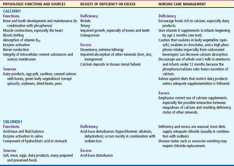
Deficiencies and excesses of vitamins A, B complex, C, D, E, and K are summarized in Table 11-1, and the RDAs and DRIs are listed in Appendix G. General nursing care management is discussed below, and specific interventions are presented in Table 11-1.
TABLE 11-1
Vitamins and Their Nutritional Significance*
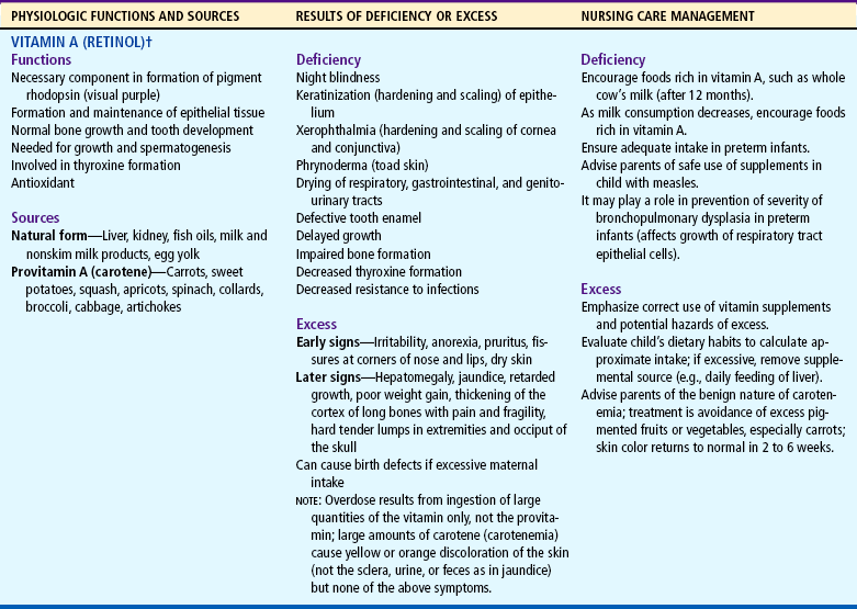
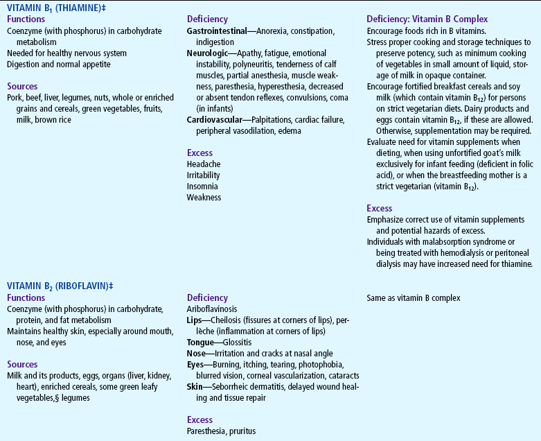
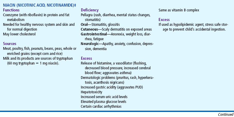
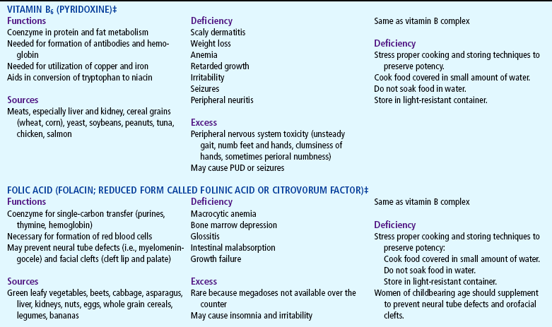
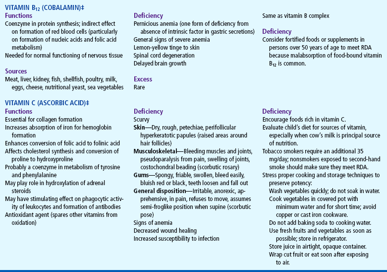
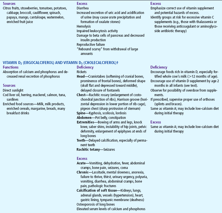
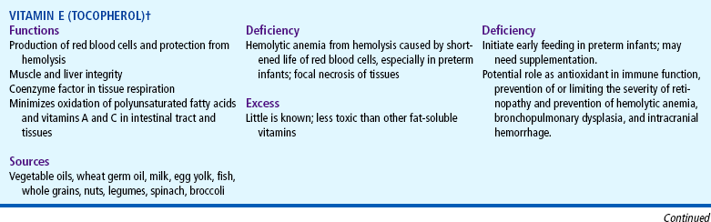
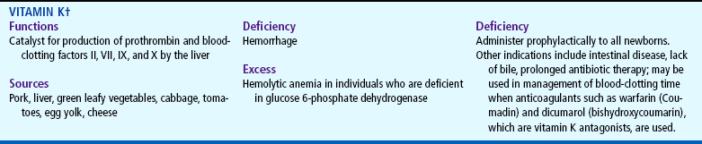
RDA, Recommended dietary allowance; PUD, peptic ulcer disease.
*This listing is not intended to be all inclusive.
§Green leafy vegetables include spinach, broccoli, kale, turnip greens, mustard greens, collards, dandelion greens, and beet greens.
COMPLEMENTARY AND ALTERNATIVE MEDICINE
The misuse or overuse of vitamins as a part of complementary and alternative medicine (CAM) places some children at risk for health problems. One survey found that a relatively small group of parents routinely gave their children megavitamin therapy; however, the researcher recommends further research to ascertain a more realistic number of children using multivitamin preparations (Loman, 2003). Sawni, Ragothaman, Thomas, and others (2007) noted that of persons reportedly using CAM, the most common CAM remedies used in children seen in the emergency department were home or folk remedies (59%), herbs (41%), prayer for healing (14%), and massage therapy (10%). A survey in a WIC (Women, Infants, and Children) clinic found that child herbal use was common, especially among Hispanic children attending the clinic. Some of the herbs used by the children in the survey (St. John’s wort, dong quai, and kava) have questionable safety (Lohse, Stotts, and Priebe, 2006).
There is concern among health care workers that terms often used to market supplements such as megavitamins may mislead parents regarding the actual benefits (or harm) of such therapies. The intention herein is not to discredit the use of CAM such as vitamin supplements; rather, it is to ensure safety and efficacy in children who may experience inadvertent harm. The use of various herbal therapies, or intake of herbs, is also becoming more popular; many of these have been a part of medicine since early days and are beneficial in some cases.
There are reports of an increase in the use of herbs by lactating mothers to increase breast milk supply. The galactogogues fenugreek, blessed thistle, fennel, and chaste tree have been purported to increase maternal milk supply, yet few studies support the efficacy or the safety of these herbs in breastfeeding infants; fenugreek has been the most widely studied, yet it may have adverse effects such as colic and diarrhea in breastfeeding infants (Conover and Buehler, 2004; Lawrence and Lawrence, 2005). For a discussion of galactogogues, including those mentioned above, see Appendix P in Lawrence and Lawrence (2005).
Herbs known to have adverse effects in children include ephedra, comfrey, and pennyroyal; some herbs may not be harmful taken alone but may counteract or potentiate prescription medications when taken together (Loman, 2003). Parents should be fully informed of the use of herbs to ensure that there is more benefit than potential harm in the ingredient being used. Health care workers also need to be knowledgeable of the benefits or potential harm in herbs to appropriately counsel parents and address their concerns. Little research has been performed in children on many over-the-counter herbal medicines, yet some herbs are known to cause harm in children (Kemper and Gardiner, 2004; Lanski, Greenwald, Perkins, and others, 2003; Loman, 2003). Parents should be cautioned not to exceed the upper limits of vitamin intake according to the new DRI (see p. 380 and Appendix G).*
MINERAL IMBALANCES
A number of minerals are essential nutrients. The macrominerals refer to those with daily requirements greater than 100 mg and include calcium, phosphorus, magnesium, sodium, potassium, chloride, and sulfur. Microminerals, or trace elements, have daily requirements of less than 100 mg and include several essential minerals and those whose exact role in nutrition is still unclear. The greatest concern with minerals is deficiency, especially of iron, calcium, phosphorus, magnesium, and zinc. Low levels of zinc can cause nutritional failure to thrive.
The regulation of mineral balance in the body is a complex process. Dietary extremes of mineral intake can cause a number of mineral-mineral interactions that could result in unexpected deficiencies or excesses. For example, excessive amounts of one mineral, such as zinc, can result in a deficiency of another mineral, such as copper, even if sufficient amounts of copper are ingested. Thus megadose intake of one mineral may cause an inadvertent deficiency of another essential mineral by blocking its absorption in the blood or intestinal wall, or by competing with binding sites on protein carriers needed for metabolism.
Deficiencies can also occur when various substances in the diet interact with minerals. For example, iron, zinc, and calcium can form insoluble complexes with phytates or oxalates (substances found in plant proteins), which impair the bioavailability of the mineral. This type of interaction is important in vegetarian diets because plant foods such as soy are high in phytates. Contrary to popular opinion, spinach is not an ideal source of iron or calcium because of its high oxalate content.
Children with certain illnesses are at greater risk for growth failure, especially in relation to bone mineral deficiency as a result of the treatment of the disease, decreased nutrient intake, or decreased absorption of necessary minerals. Those at risk for such deficiencies include children who are receiving or have received radiation and chemotherapy for cancer; children with human immunodeficiency virus (HIV), sickle cell disease, cystic fibrosis, gastrointestinal malabsorption, or nephrosis; and very low–birth-weight preterm infants.
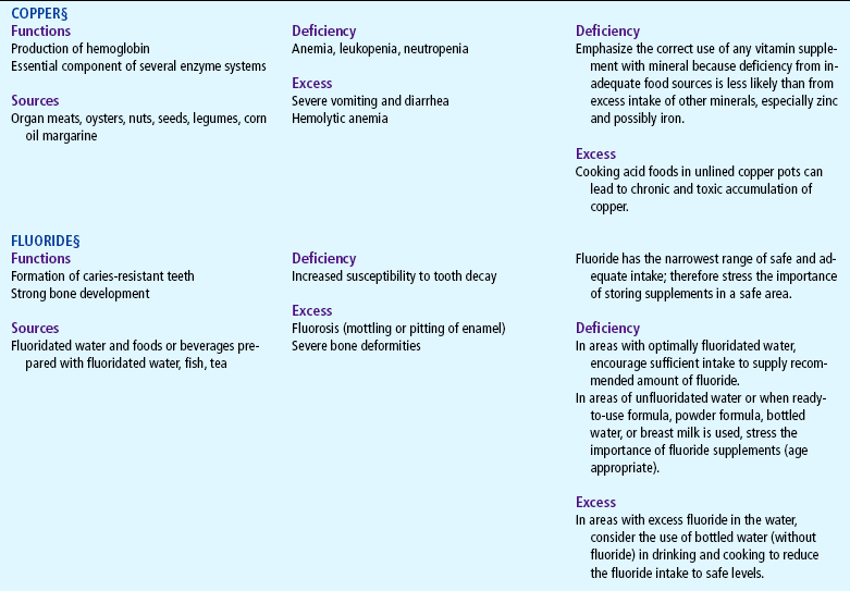
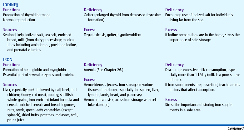
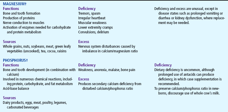
Deficiencies and excesses of the essential macrominerals and microminerals are summarized in Table 11-2. General nursing care management is discussed on p. 380, and specific interventions are discussed in the table.
TABLE 11-2
Minerals and Their Nutritional Significance*
*This listing is not intended to be all inclusive.
†Macrominerals—required intake >100 mg/day.
‡Green leafy vegetables include spinach, broccoli, kale, turnip greens, mustard greens, collards, dandelion greens, and beet greens.
§Microminerals or trace elements—required intake <100 mg/day.
VEGETARIAN DIETS
Vegetarian diets have become increasingly popular in the United States because people are concerned about hypertension; cholesterol; obesity; cardiovascular disease; cancer of the stomach, intestine, and colon; and the influence of the animal rights movement. In one survey, adolescent vegetarians were more likely than nonvegetarians to meet the Healthy People 2010 objectives for overall nutrient consumption (Perry, McGuire, Neumark-Sztainer and others, 2002). The American Dietetic Association and Dietitians of Canada (2003) issued a statement endorsing vegetarian diets for adults and children; the statement further notes that well-planned vegetarian diets are adequate for all stages of the life cycle and promote normal growth. Children and adolescents on vegetarian diets have the potential for lifelong healthy diets and have been shown to have lower intakes of cholesterol, saturated fat, and total fat and higher intakes of fruits, fiber, and vegetables than nonvegetarians (American Dietetic Association and Dietitians of Canada, 2003).
The major types of vegetarianism are:
Lacto-ovo vegetarians, who exclude meat from their diet but consume dairy products and rarely fish.
Lactovegetarians, who exclude meat and eggs but drink milk.
Pure vegetarians (vegans), who eliminate any food of animal origin, including milk and eggs.
Macrobiotics, who are even more restrictive than pure vegetarians, allowing only a few types of fruits, vegetables, and legumes.
Semi-vegetarians, who consume a lacto-ovo vegetarian diet with some fish and poultry. This is an increasingly popular form of vegetarianism and poses little or no nutritional risk to infants unless dietary fat and cholesterol intake is severely restricted.
Many individuals who are concerned about healthy diets subscribe to vegetarian diets that may not be typified by the above categories. Therefore during nutritional assessment it is necessary to clearly list exactly what the diet includes and excludes.*.
The major deficiencies that may occur in the stricter vegan diets are inadequate protein for growth; inadequate calories for energy and growth; poor digestibility of many of the bulky natural, unprocessed foods, especially for infants; and deficiencies of vitamin B6, niacin, riboflavin, vitamin D, iron, calcium, and zinc. Strict vegan diets also require supplements of vitamin B12 and vitamin D. Vitamin D is essential if exposure to sunlight is inadequate (<5 to 15 min/day on the hands, arms, and face of light-skinned persons; slightly more in darker pigmented individuals) or in persons who are dark-skinned or who live in northern latitudes or cloudy or smoky areas. Many of these deficiencies can be avoided in children who are not consuming 100% of the RDA of vitamins and minerals with a multivitamin-mineral supplement (Dunham and Kollar, 2006).
Evaluate for iron deficiency anemia and rickets in children on strict vegetarian and macrobiotic diets; this may occur as a result of consuming plant foods such as unrefined cereals, which impair the absorption of iron, calcium, and zinc. Other factors that affect iron absorption are listed in Box 11-1.
NURSING CARE MANAGEMENT
Identification of adequacy of nutrient intake is the initial nursing goal and requires assessment based on a dietary history and physical examination for signs of deficiency or excess (see Nutritional Assessment, Chapter 6). After assessment data are collected, this information is evaluated against standard intakes to identify areas of concern. The Institute of Medicine (IOM) (2000) has developed guidelines for nutritional intake that encompass the RDAs yet extend their scope to include additional parameters related to nutritional intake. The DRIs* are composed of four categories. These include estimated average requirements (EARs) for age and gender categories, tolerable upper-limit (UL) nutrient intakes that are associated with a low risk of adverse effects, adequate intakes (AIs) of nutrients, and new standard RDAs. The new guidelines present information about lifestyle factors that may affect nutrient function, such as caffeine intake and exercise, and about how the nutrient may be related to chronic disease. An important factor in the development of the DRIs that affects children, particularly infants 0 to 6 months, is that the AIs are based on the nutrient intake of full-term, healthy, breast-fed infants (by well-nourished mothers), which now represents the gold standard for infant nutrition in this age-group.
The American Heart Association (AHA) (2005) (see also Gidding, Dennison, Birch, and others, 2005) Dietary Guidelines, patterned after the 2005 Dietary Guidelines for Americans, may also be used to encourage healthy dietary intakes designed to decrease obesity and cardiovascular risk factors and subsequent cardiovascular disease, which is now known to occur in young children as well as adults. The AHA Guidelines have been endorsed by the American Academy of Pediatrics (2006a), yet it is important to note that these guidelines are for children ages 2 years and older. The guidelines encourage a variety of fruits, vegetables, whole grains, and low-fat dairy and nonfat dairy products, in addition to fish, beans, and lean meat.
MyPyramid, developed by the U.S. Department of Agriculture, replaces the Food Guide Pyramid as a guide for adult and childhood nutrition. This interactive dietary guide aims to simplify food choices designed to decrease fat and empty calorie intake and increase consumption of grains and vegetables. The MyPyramid for Kids (Fig. 11-1) incorporates examples of exercise for children as well as suggested serving sizes. The Internet version of MyPyramid for Kids* offers an interactive game for children (Blast Off). Suggested serving sizes for the five food groups are listed in Box 11-2.

FIG. 11-1 MyPyramid for Kids. (From Food and Nutrition Service, US Department of Agriculture: MyPyramid for kids [FNS-381], Washington, DC, April 19, 2005, The Service, available online at http://www.mypyramid.gov.)
A vegetarian food guide pyramid (rainbow for Canadian vegetarians) developed by the American Dietetic Association and Dietitians of Canada includes guidelines for meeting the minimum recommendations for nutrients, including protein, iron, zinc, calcium, vitamin D, riboflavin, and iodine. The new food guide can be adapted to different types of vegetarian diets according to specific needs (American Dietetic Association and Dietitians of Canada, 2003; Messina, Melina, and Mangels, 2003).
Achieving a nutritionally adequate vegetarian diet is not difficult (except with the strictest diets), but it requires careful planning and knowledge of nutrient sources. For children the lacto-ovo vegetarian diet is nutritionally adequate; however, the vegan diet requires supplementation with vitamins D and B12 for children ages 2 to 12 years. Infants should be breast-fed for the first 6 months and preferably for 1 year, be introduced to some solid foods after about 4 to 6 months, and receive iron-fortified cereal for at least 18 months. Vitamin B12 supplementation is recommended if the breastfeeding mother’s intake of the vitamin is inadequate or if she is not on vitamin supplements (Dunham and Kollar, 2006). The introduction of solids for vegetarian infants may occur using the same guidelines as for other children (see p. 346). The American Dietetic Association and Dietitians of Canada (2003) recommend iron supplementation in infants exclusively breast-fed after 4 to 6 months by vegetarian mothers and no dietary fat restrictions in vegetarian children younger than 2 years. The use of vitamin C juices (in moderate amounts, not as a milk substitute) with foods high in iron will further improve iron absorption. Breast milk from vegetarian mothers can be deficient in vitamin B12; supplementation of both mother and child is advisable. If cow’s or human milk or commercial infant formula is not given, fortified soy formula is recommended. A variety of foods should be introduced during the early years to ensure a well-balanced intake.
To ensure sufficient protein in the diet, foods with incomplete proteins (those that do not have all the essential amino acids) must be eaten at the same meal with other foods that supply the missing amino acids. The three basic combinations of foods consumed by vegetarians that generally provide the appropriate amounts of essential amino acids are:
1. Grains (cereal, rice, pasta) and legumes (beans, peas, lentils, peanuts)
Additional dietary considerations for young children are found in Chapters 12 and 14.
PROTEIN-ENERGY MALNUTRITION
Malnutrition continues to be a major health problem in the world today, particularly in children under 5 years of age. Lack of food, however, is not always the primary cause for malnutrition. In many developing and underdeveloped nations, diarrhea (gastroenteritis) is a major factor in malnutrition. Additional factors are bottle-feeding (in poor sanitary conditions), inadequate knowledge of proper child care practices, parental illiteracy, economic and political factors, climate conditions, cultural and religious food preferences, and simply the lack of adequate food. Müller and Krawinkel (2005) point out that poverty is the underlying cause of malnutrition. The most extreme forms of malnutrition, or protein-energy malnutrition (PEM), are kwashiorkor and marasmus.
In the United States milder forms of PEM are seen as a result of primary malnutrition, although the classic cases of marasmus and kwashiorkor may also occur. Unlike developing countries, where the main reason for PEM is inadequate food, in the United States PEM occurs despite ample dietary supplies (see Growth Failure [Failure to Thrive], p. 396). PEM may also be seen in persons with chronic health problems such as cystic fibrosis, renal dialysis, and gastrointestinal malabsorption; in the elderly who have chronic malnutrition; or in persons with acute illnesses such as prolonged, untreated anorexia nervosa. Kwashiorkor has been reported in the United States in children fed only a rice beverage diet (Rice Dream) and few solid foods (Katz, Mahlberg, Honig, and others, 2005). The rice drink contains 0.13 g of protein per ounce (compared with the 0.5 g found in human milk and infant formulas) and is an inadequate source of nutrition for children. Other reported cases of kwashiorkor in developed countries involved infants who were fed nonstandard infant diets such as flour water, corn porridge, molasses, and nondairy creamer (Katz, Mahlberg, Honig, and others, 2005). Kwashiorkor has also been reported in the United States when infants have been fed inappropriate food as a result of parental (caretaker) nutritional ignorance, a perceived cow milk–based formula intolerance, family social chaos, or cow’s milk intolerance (Liu, Howard, Mancini, and others, 2001). It is therefore important that health care workers not assume that PEM cannot occur in developed countries; a comprehensive dietary history should be obtained in any child with clinical features resembling PEM.
Kwashiorkor
Kwashiorkor has been defined in the past as primarily a deficiency of protein with an adequate supply of calories. A diet consisting mainly of starch grains or tubers provides adequate calories in the form of carbohydrates but an inadequate amount of high-quality proteins. Some evidence, however, supports a multifactorial etiology, including cultural, psychologic, and infective factors that may interact to place the child at risk for kwashiorkor. Penny (2003) suggests that kwashiorkor may result from the interplay of nutrient deprivation and infectious or environmental stresses, which produce an imbalanced response to such insults. Kwashiorkor often occurs subsequent to an infectious outbreak of measles and dysentery. There is further evidence that oxidative stress occurs in children with kwashiorkor, resulting in free radical damage, which may precipitate cellular changes resulting in edema and muscle wasting (Penny, 2003). The role of the essential fatty acid arachidonic acid in lipid metabolism, altered leukotriene production, and oxidative stress in kwashiorkor has yet to be fully understood, yet arachidonic acid seems to have an interactive role in its development (Penny, 2003).
Taken from the Ga language (Ghana), the word kwashiorkor means “the sickness the older child gets when the next baby is born” and aptly describes the syndrome that develops in the first child, usually between 1 and 4 years of age, when weaned from the breast after the second child is born.
The child with kwashiorkor has thin, wasted extremities and a prominent abdomen from edema (ascites). The edema often masks the severe muscular atrophy, making the child appear less debilitated than he or she actually is. The skin is scaly and dry and has areas of depigmentation. Several dermatoses may be evident, partly resulting from the vitamin deficiencies. Permanent blindness often results from the severe lack of vitamin A. Mineral deficiencies are common, especially iron, calcium, and zinc. Acute zinc deficiency is a common complication of severe PEM and results in skin rashes, loss of hair, impaired immune response and susceptibility to infections, digestive problems, night blindness, changes in affective behavior, defective wound healing, and impaired growth. Its depressant effect on appetite further limits food intake. The hair is thin, dry, coarse, and dull. Depigmentation is common, and patchy alopecia may occur.
Diarrhea (persistent diarrhea malnutrition syndrome) commonly occurs from a lowered resistance to infection and further complicates the electrolyte imbalance. Low levels of cytokines (protein cells involved in the primary response to infection) have been reported in children with kwashiorkor, suggesting that such children have a blunted immune response to infection. A large number of fatalities in children with kwashiorkor occur in those who develop HIV infection. Gastrointestinal disturbances occur, such as fatty infiltration of the liver and atrophy of the acini cells of the pancreas. Anemia is also a common finding in malnourished children. Protein deficiency increases the child’s susceptibility to infection, which eventually results in death. Fatal deterioration may be caused by diarrhea and infection or by circulatory failure.
Marasmus
Marasmus results from general malnutrition of both calories and protein. It is a common occurrence in underdeveloped countries during times of drought, especially in cultures where adults eat first; the remaining food is often insufficient in quality and quantity for the children.
Marasmus is usually a syndrome of physical and emotional deprivation and is not confined to geographic areas where food supplies are inadequate. It may be seen in children with failure to thrive in whom the cause is not solely nutritional but primarily emotional. Marasmus may be seen in infants as young as 3 months of age if breastfeeding is not successful and there are no suitable alternatives. Marasmic kwashiorkor is a form of PEM in which clinical findings of both kwashiorkor and marasmus are evident; the child has edema, severe wasting, and stunted growth. In marasmic kwashiorkor the child suffers from inadequate nutrient intake and superimposed infection. Fluid and electrolyte disturbances, hypothermia, and hypoglycemia are associated with a poor prognosis.
Marasmus is characterized by gradual wasting and atrophy of body tissues, especially of subcutaneous fat. The child appears to be very old, with loose and wrinkled skin, unlike the child with kwashiorkor, who appears more rounded from the edema. Fat metabolism is less impaired than in kwashiorkor, so that deficiency of fat-soluble vitamins is usually minimal or absent. In general, the clinical manifestations of marasmus are similar to those seen in kwashiorkor with the following exceptions: with marasmus there is no edema from hypoalbuminemia or sodium retention, which contributes to a severely emaciated appearance; no dermatoses caused by vitamin deficiencies; little or no depigmentation of hair or skin; moderately normal fat metabolism and lipid absorption; and smaller head size and slower recovery after treatment.
The child is fretful, apathetic, withdrawn, and so lethargic that prostration frequently occurs. Intercurrent infection with debilitating diseases such as tuberculosis, parasitosis, HIV, and dysentery is common.
Therapeutic Management
The treatment of PEM includes providing a diet with high-quality proteins, carbohydrates, vitamins, and minerals. When PEM occurs as a result of persistent diarrhea (see also Diarrhea, Chapter 24), three management goals are identified:
1. Rehydration with an oral rehydration solution that also replaces electrolytes
2. Administration of medications such as antibiotics and antidiarrheals
3. Provision of adequate nutrition by either breastfeeding or a proper weaning diet
Local protocols are employed in developing countries to deal with PEM. Penny (2003) proposes a three-phase treatment protocol: (1) acute or initial phase in the first 2 to 10 days, involving initiation of treatment for oral rehydration, diarrhea, intestinal parasites, prevention of hypoglycemia and hypothermia, and subsequent dietary management; (2) recovery or rehabilitation (2 to 6 weeks), focusing on increasing dietary intake and weight gain; and (3) follow-up phase, focusing on care after discharge in an outpatient setting to prevent relapse and promote weight gain, provide developmental stimulation, and evaluate cognitive and motor deficits. In the acute phase care is taken to prevent fluid overload; the child is observed closely for signs of food or fluid intolerance. The refeeding syndrome may occur if intake progresses too rapidly; cardiac failure may cause sudden death in the child who has been malnourished and refed too rapidly.
Vitamin and mineral supplementation are required in most cases of PEM; vitamin A, zinc, and copper are recommended; iron supplementation is not recommended until the child is able to tolerate a steady food source. In addition, the child is observed for signs of skin breakdown, which should be treated to prevent infection. Breastfeeding is encouraged if the mother and child are able to do so effectively; in some cases partial supplementation with a modified cow’s milk–based formula may be necessary (Penny, 2003). In severely malnourished children a modest energy food source is given initially followed by a high protein and energy food source; severely malnourished children will not tolerate a high energy and protein source initially. A number of food sources may be provided to treat PEM and include oral rehydration solutions (ReSoMal), amino acid–based elemental food, and ready-to-feed foods that do not require the addition of water (to minimize contaminated water consumption); parenteral and oral antibiotics are often part of the standard treatment for PEM (Ciliberto, Sandige, Ndekha, and others, 2005; Amadi, Mwiya, Chomba, and others, 2005).
Nursing Care Management
Because PEM appears early in childhood, primarily in children 6 months to 2 years of age, and is associated with early weaning, low protein diet, delayed introduction of complementary foods, and frequent infections (Müller and Krawinkel, 2005), it is essential that nursing care focus on prevention of PEM through parent education about feeding practices during this crucial period. Breastfeeding (provided the mother is HIV-free) is the optimal method of feeding for the first 6 months. The immune properties naturally found in breast milk not only nourish the infant but aid in the prevention of opportunistic infections, which may contribute to PEM. Provision of essential physiologic needs, such as appropriate nutrient intake, protection from infection, adequate hydration, skin care, and restoration of physiologic integrity, is paramount. Additional nursing care focuses on education about and administration of childhood vaccinations to prevent illness, promotion of maternal nutrition and well-being for the lactating mother, encouragement and participation in well-child visits for infants and toddlers, and education regarding sanitation practices to prevent childhood gastrointestinal diseases.
Poor skin integrity further increases the chance of infections, hypothermia, water loss, and skin breakdown. Tube feedings may be required in infants too weak to breastfeed or bottle feed. Oral rehydration with an approved oral rehydration solution is commonly used in cases of PEM where diarrhea and infection are not immediately life threatening.
It is imperative that nurses be at the forefront in educating and reinforcing healthy nutrition habits in parents of small children to prevent malnutrition. Because children with marasmus may suffer from emotional starvation as well, care should be consistent with care of the child with failure to thrive (pp. 396-400).
FOOD SENSITIVITY
Food sensitivity is a general term that includes any type of adverse reaction to food or food additives. Food sensitivities can be divided into two broad categories:
1. Food allergy or hypersensitivity, which refers to reactions involving immunologic mechanisms, usually immunoglobulin E (IgE); the reactions may be immediate or delayed and mild or severe, such as an anaphylactic reaction.
2. Food intolerance, which refers to reactions involving known or unknown nonimmunologic mechanisms; lactose intolerance is an example of a reaction that looks like allergy but is caused by deficiency of the enzyme lactase.
This classification is not universally accepted, however; therefore the terms food sensitivity, hypersensitivity, allergy, and intolerance are often used interchangeably. The American Academy of Allergy, Asthma, and Immunology further suggests defining food-induced reactions according to the following: adverse food reactions, food hypersensitivity (allergy), food anaphylaxis, food intolerance, food idiosyncrasy, food toxicity or poisoning, anaphylactoid reaction to food, pharmacologic food reaction, and metabolic food reaction (American Academy of Pediatrics, 2004).
The clinical manifestations of food hypersensitivity may be divided as follows (American Academy of Pediatrics, 2004):
Systemic—Anaphylactic, failure to thrive
Gastrointestinal—Abdominal pain, vomiting, cramping, diarrhea
Respiratory—Cough, wheezing, rhinitis, infiltrates
Cutaneous—Urticaria, rash, atopic dermatitis
Food hypersensitivities usually occur either as an immunoglobulin E (IgE)–mediated or non-IgE-mediated immune response; some toxic reactions may occur as a result of a toxin found within the food (Sampson, 2004). Food allergy is caused by exposure to allergens, usually proteins (but not the smaller amino acids) that are capable of inducing IgE antibody formation (sensitization) when ingested. Sensitization refers to the initial exposure of an individual to an allergen, resulting in an immune response; subsequent exposure induces a much stronger response that is clinically apparent. Consequently, food hypersensitivity typically occurs after the food has been ingested one or more times. The most common food allergens are listed in Box 11-3.
Allergies in general demonstrate a genetic component: children who have one parent with allergy have a 50% or greater risk of developing allergy; children who have both parents with allergy have up to a 100% risk of developing allergy. Allergy with a hereditary tendency is referred to as atopy. Some infants with atopy can be identified at birth from elevated levels of IgE in cord blood.
Deaths have been reported in children who suffered an anaphylactic reaction to food. Onset of the reactions occurred shortly after ingestion (5 to 30 minutes). In most of the children the reactions did not begin with skin signs, such as hives, red rash, and flushing, but rather mimicked an acute asthma attack (wheezing, decreased air movement in airways, dyspnea). Children with food anaphylaxis should be watched closely because a biphasic response has been recorded in a number of cases in which there is an immediate response, apparent recovery, then acute recurrence of symptoms (Sampson, 2003). Parents, teachers, and child daycare workers should be educated regarding signs and symptoms of food hypersensitivity reactions. People with food sensitivity should avoid unfamiliar foods and restaurants that do not disclose food ingredients. New labeling guidelines require that food additives such as spices and flavoring be clearly labeled on commercially sold, store-bought foods. Hidden ingredients in prepared foods have been implicated as a potential source of food hypersensitivity.
Other symptoms of anaphylaxis to food allergens include wheezing, cough, dyspnea, urticaria, abdominal cramps, vomiting, diarrhea, a drop in systemic blood pressure or shock, and, in small preverbal children, restlessness, urticaria, irritability, listlessness, and unresponsiveness. Oral allergy syndrome occurs when a food allergen is ingested (commonly fruits and vegetables) and there is subsequent edema and pruritus involving the lips, tongue, palate, and throat; recovery from symptoms is usually rapid. Immediate gastrointestinal hypersensitivity is an IgE-mediated reaction to a food allergen, and reactions include nausea, abdominal pain, cramping, diarrhea, vomiting, anaphylaxis, or all of these. Additional food hypersensitivities seen in young children include allergic eosinophilic gastritis, allergic eosinophilic gastroenterocolitis, dietary protein enterocolitis (or milk protein intolerance), and dietary protein proctitis.
Although the reason is unknown, many children “outgrow” their food allergies; children may outgrow milk and egg allergies, but peanut allergies may persist. Children who are allergic to more than one food may develop tolerance to each food at different times. Because of the tendency to lose the hypersensitivity, allergic foods should be reintroduced into the diet after a period of abstinence (usually a year or more) to evaluate whether the food can be safely added to the diet. However, foods that are associated with severe anaphylactic reactions will continue to present a lifelong risk and must be avoided. Because children with food allergies (usually two or more) are at risk for inadequate nutrient intake and growth failure, it is recommended that they have an annual nutritional assessment to prevent such problems (Christie, Hine, Parker, and others, 2002).
Breastfeeding is now considered to be a primary strategy for avoiding atopy in families with known food sensitivities; however, there is some evidence that cow’s milk protein is transferred via breast milk. The breastfeeding mother is encouraged to avoid foods such as peanuts, tree nuts, fish, and shellfish during the first 6 months of breastfeeding. In addition, supplementation, if required, is best with hydrolysated or amino acid formulas, not soy formulas. Additional recommendations to decrease the incidence of food allergies in children at higher risk for allergies is to avoid introduction of selected complementary and solid foods: dairy products should not be introduced until 12 months of age; hen’s eggs at 24 months; and peanut, tree nuts, fish, and seafood at 36 months (Fiocchi, Assaad, Bahna and others, 2006). The strategies listed in the Nursing Care Guidelines box are those recommended by most authorities for infants with a family history of atopy.*
Children with extremely sensitive food allergies should wear medical identification such as a bracelet and have an injectable epinephrine cartridge (EpiPen) readily available and know how to use it. It is also helpful for the child to have a copy of the individualized written treatment plan on hand for prompt diagnosis and treatment (such plans can be downloaded from http://www.foodallergy.org and completed by the practitioner).
Cow’s Milk Allergy
Cow’s milk allergy (CMA) (also referred to as cow’s milk protein allergy [CMPA] or cow milk protein intolerance [CMPI]) is a multifaceted disorder representing adverse systemic and local gastrointestinal reactions to cow’s milk protein. (This discussion is centered on cow’s milk protein found in commercial infant formulas; whole milk is not recommended for infants younger than the age of 12 months.) The hypersensitivity may be manifested within the first 4 months of life through a variety of signs and symptoms that may appear within 45 minutes of milk ingestion or after a period of several days (Box 11-4). In infants who are highly sensitive to the protein, even a small amount of cow’s milk protein may induce anaphylactic reaction. Cases of contamination of non-cow milk–based infant formula during the manufacturing process resulting in severe reactions have been documented (Levin, Motala, and Lopata, 2005). The diagnosis may initially be made from the history, although the history alone is not diagnostic; the timing and diversity of clinical manifestations vary greatly. For example, CMA may be manifested as colic (see p. 395), diarrhea, vomiting, gastrointestinal bleeding, gastroesophageal reflux, chronic constipation, or sleeplessness in an otherwise healthy infant.
The incidence of CMA is reported to range from 2% to 7.5% in developed countries, although the percentage may appear to be higher because of parental report of symptoms rather than actual confirmation of CMA (Host, 2002; Salvatore and Vandenplas, 2002).
Diagnostic Evaluation.: A number of diagnostic tests may be performed, including stool analysis for blood (both frank and occult bleeding can occur from the colitis), serum IgE levels, skin-prick or scratch testing, and radioallergosorbent test (measures IgE antibodies to specific allergens in serum by radioimmunoassay). Both skin and radioallergosorbent testing help identify the offending food, but the results are not always conclusive.
The most definitive diagnostic strategy is elimination of milk in the diet, followed by challenge testing after improvement of symptoms. A clinical diagnosis is made when symptoms improve after removal of milk from the diet and two or more challenge tests produce symptoms (Ewing and Allen, 2005). Challenge testing involves reintroducing small quantities of milk in the diet to detect resurgence of symptoms; at times challenge testing involves the use of a placebo so that the parent is unaware of (or “blind” to) the timing of allergen ingestion. A double-blind placebo-controlled food challenge is the gold standard for diagnosing food allergies such as CMA, yet it may not be used very often for diagnosing CMA because of the expense, time involved, and risk for further exposure and anaphylactic reaction (Ewing and Allen, 2005).
Therapeutic Management.: Treatment of CMA is elimination of cow’s milk–based formula and all other dairy products. For infants fed cow’s milk formula, this primarily involves changing the formula to a casein hydrolysate milk formula, or extensively hydrolyzed formula (Pregestimil, Nutramigen, or Alimentum), in which the protein has been broken down into its amino acids through enzymatic hydrolysis. Although the American Academy of Pediatrics (2004) recommends the use of hydrolyzed formulas for CMA, many practitioners may start a soy formula instead. Approximately 10% of infants who are sensitive to cow’s milk protein will also demonstrate sensitivity to soy (Assaad, 2006), yet soy is less expensive than protein hydrolysate formula. An intolerance to soy is reported to be higher in infants under age 6 months, with a family history of atopy, and with severe gastrointestinal symptoms (Ewing and Allen, 2005). Other choices for children who are intolerant to cow’s milk–based formula are the amino acid–based formulas Neocate or EleCare, yet their cost is a major consideration. Goat’s milk is not an acceptable substitute because it cross-reacts with cow’s milk protein, is deficient in folic acid, and is unsuitable as the only source of calories. Anaphylactic reaction to goat’s milk has been noted in an infant who was also allergic to cow’s milk (Pessler and Nejat, 2004). Infants who are breast-fed but have symptoms of cow’s milk hypersensitivity are treated by eliminating all peanuts and tree nuts from the lactating mother’s diet, and possibly eggs and milk (American Academy of Pediatrics, 2004). If maternal dairy intake is restricted, vitamin D and calcium supplementation should be considered to prevent deficiency. Infants are maintained on the milk-free diet for 1 or 2 years, after which time small quantities of milk are reintroduced; eggs and fish may be introduced at 3 years of age.
Nursing Care Management.: The principal nursing objectives are to prevent and reduce exposure of infants to cow’s milk protein by encouraging exclusive breastfeeding in the first 6 months of life, or even up to 12 months if there is a high risk for atopy. In addition, nurses have an important role in the identification of potential CMA, appropriate counseling of parents regarding signs and symptoms of CMA, and the use of substitute formulas that are appropriate for infants with diagnosed CMA. Parents need much reassurance regarding the needs of nonverbal infants with such an array of symptoms. Endless nights of lost sleep and a crying infant may promote feelings of parenting inadequacy and role conflict, thus aggravating the situation. Nurses can reassure parents that many of these symptoms are common and the reasons are often never found, yet the child does achieve appropriate growth and development; acute symptoms are reported to the practitioner for further evaluation. The protein hydrolysate (partially hydrolyzed and extensively hydrolyzed) formulas tend to be less palatable and more expensive than milk-based formulas. Consequently, the child’s reluctance to accept the new formula may be a problem. This can be overcome by adding nonnutritive, hypoallergenic flavor packets or by introducing the formula gradually over a few days using 1 ounce of new formula to 7 ounces of old formula, then 2 to 6 ounces, 3 to 5 ounces, and as needed. Parents also need to be reassured that the infant will receive complete nutrition from the new formula and will suffer no ill effects from the absence of cow’s milk.
Once solid foods are started, parents need guidance in avoiding milk products (see Box 11-3), although many children reportedly outgrow cow’s milk protein sensitivity by 3 to 4 years of age (Fiocchi and Martelli, 2006).
Lactose Intolerance
Lactose intolerance refers to at least four different entities that involve a deficiency of the enzyme lactase, which is needed for the hydrolysis or digestion of lactose in the small intestine; lactose is hydrolyzed into glucose and galactose. Congenital lactase deficiency occurs soon after birth after the newborn has consumed lactose-containing milk (human milk or commercial formula). This inborn error of metabolism involves the complete absence or severely reduced presence of lactase, is rare, and requires lifelong lactose-free or extremely reduced lactose diet.
Primary lactase deficiency, sometimes referred to as late-onset lactase deficiency, is the most common type of lactose intolerance and is manifested usually after 4 or 5 years of age, although the time of onset is variable. Ethnic groups with a high incidence of lactase deficiency include Asians, southern Europeans, Arabs, Israelis, and African Americans, whereas Scandinavians tend to have the lowest. Lactose malabsorption manifests as lactose intolerance and is characterized by an imbalance between the ability for lactase to hydrolyze the ingested lactose and the amount of lactose ingested (Heyman and American Academy of Pediatrics Committee on Nutrition, 2006).
Secondary lactase deficiency may occur secondary to damage of the intestinal lumen, which decreases or destroys the enzyme lactase. Cystic fibrosis; sprue; celiac disease; kwashiorkor; or infections such as giardiasis, HIV, or rotavirus may cause a temporary or permanent lactose intolerance.
Developmental lactase deficiency refers to the relative lactase deficiency observed in preterm infants of less than 34 weeks gestation (Heyman and American Academy of Pediatrics Committee on Nutrition, 2006).
The primary symptoms of lactose intolerance include abdominal pain, bloating, flatulence, and diarrhea after the ingestion of lactose. The onset of symptoms occurs within 30 minutes to several hours of lactose consumption. Lactose intolerance is often perceived as an allergy, and in several studies with reports of acute gastrointestinal symptoms ascribed to lactose intolerance, measurement of lactase activity is normal (Goldberg, Folta, and Must, 2002).
Lactose intolerance may be diagnosed on the basis of the history and improvement with a lactose-reduced diet. The breath hydrogen test is used to positively diagnose the condition. Breath samples in lactose-deficient individuals will yield a higher percentage of hydrogen (20 ppm [parts per million] or more above baseline). In infants lactose malabsorption may be diagnosed by evaluating fecal pH and reducing substances; fecal pH in infants is usually lower than in older children, but an acidic pH may indicate malabsorption (Heyman and American Academy of Pediatrics Committee on Pediatrics, 2006).
Treatment of lactose intolerance is elimination of offending dairy products; however, some advocate decreasing amounts of dairy products rather than total elimination, especially in small children (Heyman and American Academy of Pediatrics Committee on Nutrition, 2006; Goldberg, Folta, and Must, 2002). In infants, lactose-free or low-lactose formula offers no special advantages over lactose-containing formula except in the severely malnourished (Heyman and American Academy of Pediatrics Committee on Pediatrics, 2006)
One concern is that dairy avoidance in children and adolescents with lactose intolerance will contribute to reduced bone mineral density and osteoporosis (Sibley, 2004). There is evidence that dietary lactose enhances calcium absorption and that lactose-free diets may negatively affect bone mineralization (Heyman and American Academy of Pediatrics Committee on Nutrition, 2006). It has been suggested that individuals with lactose maldigestion who do not experience lactose intolerance symptoms continue to consume small amounts of dairy products with meals to prevent reduced bone mass density and subsequent osteoporosis (Sibley, 2004). There is evidence that probiotics (food preparations containing microorganisms such as Lactobacillus, which alter the gastrointestinal microflora and thus are beneficial to the host) improve lactose intolerance when live cultures are fermented in dairy products (Zeisel and Erickson, 2003). The positive attributes of probiotics for those with lactose maldigestion include delayed gastrointestinal transit (slower than milk), positive effects on intestinal and colonic microflora, and a reduction of maldigestion symptoms (de Vrese, Stegelmann, Richter, and others, 2001).
Most people are able to tolerate small amounts of lactose even in the presence of deficient lactase activity (Heyman and American Academy of Pediatrics Committee on Nutrition, 2006; Goldberg, Folta, and Must, 2002) and should be encouraged to continue their intake of dairy products in small amounts to obtain much-needed nutrients. Milk taken at meals may be better tolerated than when taken alone (see Family-Centered Care box). Pretreated milk (with microbial-derived lactase) is reported to be effective in improving lactose absorption. Because dairy products are a major source of calcium and vitamin D, supplementation of these nutrients is needed to prevent deficiency. Yogurt contains inactive lactase enzyme, which is activated by the temperature and pH of the duodenum; this lactase activity substitutes for the lack of endogenous lactase. Fresh, plain yogurt may be tolerated better than frozen or flavored yogurt; hard cheeses, lactase-treated dairy products, and lactase tablets taken with dairy products are also viable options. An important distinction between lactose intolerance and food hypersensitivity is that lactose intolerance will not manifest as an anaphylactic-type reaction.
 FAMILY-CENTERED CARE
Controlling Symptoms of Lactose Intolerance
In infants substitute lactose-free or soy-based formula for cow’s milk–based formula or human milk.
Limit milk consumption to one to two glasses per day.
Drink milk with other foods rather than alone.
Eat hard cheese, cottage cheese, or yogurt instead of drinking milk.
Use enzyme tablets (Lactaid, Lactrase, Dairy Ease) to metabolize the lactose in milk or supplement the body’s own lactase (add tablets to milk or sprinkle on dairy products such as ice cream).
Eat small amounts of dairy foods daily to help colonic bacteria adapt to ingested lactose.
Include a probiotic (yogurt or cultured [fermented] milk) in meal or as a snack that has Lactobacillus or Bifidobacterium organisms.
Take a calcium supplement if unable to consume any dairy products such as cheese.
Nursing Care Management.: Nursing care is similar to the interventions discussed for CMA: explaining the dietary restrictions to the family; identifying alternate sources of calcium such as yogurt and calcium supplementation; explaining the importance of supplementation; and discussing sources of lactose, especially hidden sources such as its use as a bulk agent in certain medications, and ways of controlling the symptoms (see Family-Centered Care box). Parents are advised to check with the pharmacist regarding this possibility when obtaining medication.
FEEDING DIFFICULTIES
REGURGITATION AND “SPITTING UP”
The return of small amounts of food after a feeding is a common occurrence during infancy. It should not be confused with vomiting, which can be associated with a number of disturbances that may be insignificant or serious. Regurgitation is usually benign, although persistent regurgitation necessitates medical evaluation to rule out gastroesophageal reflux (see Chapter 24). For clarification the terms are defined as:
Regurgitation—Return of undigested food from the stomach, usually accompanied by burping
Spitting up—Dribbling of unswallowed formula from the infant’s mouth immediately after a feeding
The normal occurrence of regurgitation or spitting up should be explained to parents, especially to those who are unduly concerned about it. Regurgitation can be reduced by some simple measures, such as frequent burping during and after feeding, minimum handling during and after feeding, and positioning the child on the right side with the head slightly elevated after feeding. The inconvenience of spitting up can be managed with the use of absorbent bibs on the infant and protective cloths on the parent.
Sometimes frequent dribbling of formula causes excoriation of the corners of the mouth, chin, and neck. Keeping the area dry promotes healing but can be difficult to maintain. Helpful suggestions include applying a thin film of petrolatum or A&D ointment to the affected areas after cleansing and using absorbent nonplastic-lined terry-cloth bibs, which are changed frequently.
PAROXYSMAL ABDOMINAL PAIN (COLIC)
Colic is reported to occur in 5% to 30% of all infants (Neu and Robinson, 2003), yet has no particular affinity in regard to the gender, race, or socioeconomic status of the infant and family (Ellett, 2003). The condition is generally described as paroxysmal abdominal pain or cramping that is manifested by loud crying and drawing the legs up to the abdomen. Other definitions include variables such as duration of cry greater than 3 hours a day, occurring more than 3 days per week, and parental dissatisfaction with the child’s behavior. Some studies report an increase in symptoms (fussiness and crying) in the late afternoon or evening; however, in some infants the onset of symptoms occurs at another time. Colic is more common in young infants under the age of 3 months than in older infants, and infants with “difficult” temperaments are more likely to be colicky. Despite the obvious behavioral indications of pain, the child tolerates breast milk or some type of infant formula well, gains weight, and usually thrives. There is no evidence of a residual effect of colic on older children, except perhaps a strained parent-child relationship in some cases; in other words, infants who are colicky grow up to be normal children and adults.
Among the theories that have been investigated as potential causes are too rapid feeding, overeating, swallowing excessive air, improper feeding technique (especially in positioning and burping), and emotional stress or tension between parent and child. Although all of these may occur, there is no evidence that one factor is consistently present. In some infants colic may be a sign of CMA or cow’s milk intolerance, and eliminating cow’s milk products from the infant’s diet and the diet of lactating mothers can reduce the symptoms; in some infants soy milk may cause the same discomfort as cow’s milk. Parental smoking, strained parent-infant interaction, lactase deficiency, difficult infant temperament, difficulty regulating emotions, central nervous system immaturity, and neurochemical dysregulation in the brain have also been proposed as potential causes of colic (Ellett, 2003; Neu and Robinson, 2003). A positive association between consumption of fruit juices (carbohydrate malabsorption) and colic has been demonstrated in some cases (Duro, Rising, Cedillo, and others, 2002). The final consensus of most experts who study colic is that it is multifactorial in nature and that no single treatment for every colicky infant will be effective in alleviating the symptoms.
Therapeutic Management
Management of colic should begin with an investigation of possible organic causes, such as CMA, intussusception, or other gastrointestinal problem. If a sensitivity to cow’s milk is strongly suspected, a trial substitution of another formula such as an extensively hydrolyzed (Nutramigen, Alimentum, Pregestimil), whey hydrolysate, or amino acid (Neocate, EleCare) formula is warranted. Soy formulas are usually avoided because of the possibility of sensitivity to soy protein as well.
The use of drugs, including sedatives, antispasmodics, antihistamines, and antiflatulents, is sometimes recommended. The most commonly used sedatives are phenobarbital, hydroxyzine hydrochloride (Atarax), and chloral hydrate. Simethicone (Mylicon) may also help allay the symptoms of colic. However, in most controlled studies none of these drugs completely reduced the symptoms of colic. Herbal (chamomile) tea offered at the onset of crying and up to three times daily has proved effective in relieving the symptoms of colic in some infants (Weizman, Alkrinawi, Goldfarb, and others, 1993); however, parents are to be cautioned regarding the unknown safety of this treatment (Crotteau, Wright, and Eglash, 2006). Behavioral interventions have not proved effective at reducing symptoms of colic but have helped parents deal with the crying infant in a more positive manner. The addition of lactase to infant formula has produced mixed results as far as abatement of overall symptoms.
An extensive review of a wide variety of interventions for colic indicates there are no specific safe remedies to alleviate symptoms of colic in every infant; dietary changes such as eliminating cow’s milk protein from the lactating mother’s diet and behavioral interventions were shown to be effective in helping parents reduce stimulation and respond to the infant’s crying, yet these interventions are perceived only as moderately effective (Joanna Briggs Institute, 2004).
Nursing Care Management
The initial step in managing colic is to take a thorough, detailed history of the usual daily events. Areas that should be stressed include (1) the infant’s diet; (2) the diet of the breastfeeding mother; (3) the time of day when crying occurs; (4) the relationship of the crying to feeding time; (5) the presence of specific family members during the crying and habits of family members, such as smoking; (6) activity of the mother or usual caregiver before, during, and after the crying; (7) characteristics of the cry (duration, intensity); (8) measures used to relieve the crying and their effectiveness; and (9) the infant’s stooling, voiding, and sleeping patterns. Of special emphasis is a careful assessment of the feeding process via demonstration by the parent.
If cow’s milk sensitivity is suspected, breastfeeding mothers should follow a milk-free diet for a minimum of 3 to 5 days in an attempt to reduce symptoms in the infant. Mothers need to be cautioned that some nondairy creamers may contain calcium caseinate, a cow’s milk protein. If a milk-free diet is helpful, lactating mothers may need calcium supplements to meet the body’s requirement. Bottle-fed infants may improve with the same dietary modifications as for the child with CMA (see p. 386).
Perhaps the most important nursing intervention (once the diagnosis of colic is established) is reassurance of both parents that they are not doing anything wrong and that the infant is not experiencing any physical or emotional harm. Parents, especially mothers, become easily frustrated with the infant’s crying and perceive this as a sign that there is something horribly wrong. An empathetic, gentle, and reassuring attitude, in addition to suggestions about remedies for treatment, will help allay parents’ anxieties, which are usually exacerbated by loss of sleep and preoccupation over the infant’s welfare. Other support persons and extended family members may be enlisted to help support the parents during this time of difficulty.
When no cause can be identified, helping parents understand the infant’s crying behavior and modifying parent interventions to promptly attend to the infant’s needs can decrease the length of fussiness and crying. Other approaches for managing colic are listed in the Family-Centered Care box. Parents are encouraged to try as many of these approaches as possible, because not all are effective for every infant. One author suggests that a problem-solving discussion with the parents, in addition to acknowledgment that the infant has colic, is an optimal strategy for helping parents manage the infant with colic until a cure is found (Ellett, 2003).
GROWTH FAILURE (FAILURE TO THRIVE)
Growth failure, or failure to thrive (FTT), is a sign of inadequate growth resulting from inability to obtain or use calories required for growth. FTT has no universal definition, although one of the more common parameters is a weight (and sometimes height) that falls below the 5th percentile for the child’s age. Another definition of FTT includes a weight for age (height) z value of less than −2.0 (a z value is a standard deviation value that represents anthropometric data normalizing for sex and age with greater precision than growth percentile curves [Markowitz and Duggan, 2003]). A third way to define FTT is a weight curve that crosses more than 2 percentile lines on the National Center for Health Statistics growth charts after previous achievement of a stable growth pattern. Growth measurements alone are not used to diagnose children with FTT. Rather, the finding of a pattern of persistent deviation from established growth parameters is cause for concern. In addition to lack of consensus on the precise definition of FTT, there are those who advocate for a change in terminology; thus, terms such as growth failure and pediatric undernutrition are used in the literature for failure to thrive (Locklin, 2005).
Place infant prone over a covered hot-water bottle, heated towel, or covered heating pad.
Respond immediately to the crying.
Change infant’s position frequently; walk with child’s face down and with body across parent’s arm, with parent’s hand under infant’s abdomen, applying gentle pressure (Fig. 11-2).

FIG. 11-2 The “colic carry” may be comforting to an infant with colic. (Photo by Paul Vincent Kuntz, Texas Children’s Hospital, Houston.)
Use a front carrier for transporting infant.
Swaddle infant tightly with a soft, stretchy blanket.
Place infant in an electric (or wind-up) infant swing.
Take infant for car rides or outside for a change in environment.
Use bottles that minimize air swallowing (curved bottle or inner collapsible bag).
Use a commercial device* in the crib that stimulates the vibration and sound of a car ride or plays soothing “noise,” in utero sounds, or music.†
Provide smaller, frequent feedings; burp infant during and after feedings using the shoulder position or sitting upright, and place infant in an upright seat after feedings.
Introduce a pacifier for added sucking.
In breast-fed infants, mother should avoid all milk products for a trial period.
If household members smoke, avoid smoking near infant; preferably confine smoking activity to outside of home.
Give appropriate dose of acetaminophen elixir or suppository if suggested by health professional; not recommended for daily use.
If nothing reduces the crying, place infant in crib and allow to cry; periodically hold and comfort child and put down again.
Maintain a brief diary of the time of day the crying starts; events going on in household; time, amount and type of last feeding; length of crying; and characteristics of cry. Although this will not stop the crying, it may help the practitioner identify a possible cause.
*Sweet Dreems, Inc., Sleep Tight Order Department, (800) NO COLIC, (800) 662-6542; http://www.colic.com.
†Suggested infant relaxation music: Heartbeat Lullabies by Terry Woodford. Available from Baby-Go-To-Sleep Center, Audio Therapy Innovations, Inc., PO Box 550, Colorado Springs, CO 80901; (800) 537-7748; website: http://www.babygotosleep.com.
Traditionally the three general categories of failure to thrive have been as follows:
1. Organic failure to thrive—Result of a physical cause, such as congenital heart defects, neurologic lesions, microcephaly, chronic renal failure, gastroesophageal reflux, malabsorption syndrome, endocrine dysfunction, cystic fibrosis, or acquired immunodeficiency syndrome (AIDS).
2. Nonorganic failure to thrive (NFTT)—A definable cause that is unrelated to disease. NFTT is most often the result of psychosocial factors, such as inadequate nutritional information by the parent; deficiency in maternal care or a disturbance in maternal-child attachment; or a disturbance in the child’s ability to separate from the parent, leading to food refusal to maintain attention.
3. Idiopathic failure to thrive—Unexplained by the usual organic and environmental etiologies but may also be classified as NFTT. Both categories of NFTT account for the majority of cases of FTT.
Some experts, however, suggest that the above classifications are too simplistic because most cases of growth failure have mixed causes; they suggest that FTT be classified according to pathophysiology for the following categories: (1) inadequate caloric intake—incorrect formula preparation, neglect, food fads, excessive juice consumption, poverty, behavioral problems affecting eating, or central nervous system problems affecting intake; (2) inadequate absorption—cystic fibrosis, celiac disease, vitamin or mineral deficiencies, biliary atresia, or hepatic disease; (3) increased metabolism–hyperthyroidism, congenital heart defects, or chronic immunodeficiency; and (4) defective utilization—genetic anomaly such as trisomy 21 or 18, congenital infection, or metabolic storage diseases (Krugman and Dubowitz, 2003). The etiology of growth failure is often multifactorial and involves a combination of infant organic disease, dysfunctional parenting behaviors, subtle neurologic or behavioral problems, and disturbed parent-child interactions (Block, Krebs, and American Academy of Pediatrics Committee on Child Abuse and Neglect and Committee on Nutrition, 2005).
Other factors that can lead to inadequate infant caloric intake include:
Poverty—Lack of funds to buy sufficient food; diluting formula to extend available supply
Health or childrearing beliefs—Fad diets; excessive concern with preventing conditions such as obesity, hypercholesterolemia, or nursing caries; strict use of scheduled feedings
Inadequate nutritional knowledge—Cultural confusion of newly arrived immigrants who are unaware of appropriate food selections in American markets; parents with cognitive impairment
Family stress—Overwhelming involvement with another chronically ill child; any number of other stresses (financial, marital, excessive parenting and employment responsibilities, depression, chemical abuse, acute grief)
Feeding resistance—Result of nonoral nutritional therapy early in life
Insufficient breast milk—Result of a number of different causes (fatigue, illness, poor release of milk, insufficient glandular tissue, lack of maternal confidence)
In the following discussion, FTT is used to describe any child with growth failure, whether it be organic, nonorganic, a combination of both, or of unknown etiology.
Diagnostic Evaluation
Diagnosis is initially made from evidence of growth failure. If FTT is recent, the weight, but not the height, is below accepted standards (usually the 5th percentile); if FTT is longstanding, both weight and height are low, indicating chronic malnutrition. Perhaps as important as anthropometric measurements are a complete health and dietary history (including perinatal history), physical examination for evidence of organic causes, developmental assessment, and family assessment. A dietary intake history, either a 24-hour food intake or a history of food consumed over a 3- to 5-day period, is also essential. In addition, the child’s activity level, parental height, perceived food allergies, and dietary restrictions should be explored. An assessment of household organization and mealtime behaviors and rituals is important in the collection of pertinent data. Other tests (lead toxicity, anemia, stool-reducing substances, occult blood, ova and parasites, alkaline phosphatase, and zinc levels) are selected only as indicated to rule out organic problems. In most cases laboratory studies are of little diagnostic value, and hospitalization is avoided except in extreme FTT (American Academy of Pediatrics, 2004). To prevent the overuse of diagnostic procedures, FTT should be considered early in the differential diagnosis. To avoid the social stigma of FTT during the early investigative phase, many health care workers use the term growth delay (or failure) until the actual cause is established.
Therapeutic Management
The primary management of FTT is aimed at reversing the cause of the growth failure. If malnutrition is severe, the initial treatment is directed at reversing the malnutrition. The goal is to provide sufficient calories to support “catch-up” growth’a rate of growth greater than the expected rate for age. Any coexisting medical problems are treated.
In most cases of FTT a multidisciplinary team of physician, nurse, dietitian, child life specialist, occupational therapist, pediatric feeding specialist, and social worker or mental health professional is needed to deal with the multiple problems. Efforts are made to relieve any additional stresses on the family by offering referrals to welfare agencies or supplemental food programs. In some cases family therapy may be required; temporary placement in a foster home may relieve the family’s stress, protect the child, and allow the child some stability if insurmountable obstacles are preventing appropriate family function. Behavior modification aimed at mealtime rituals (or lack thereof) and family social time may be required. Hospitalization admission is indicated for (1) evidence (anthropometric) of severe acute malnutrition, (2) child abuse or neglect, (3) significant dehydration, (4) caretaker substance abuse or psychosis, and (5) outpatient management that does not result in weight gain (American Academy of Pediatrics, 2004; Block, Krebs, and American Academy of Pediatrics Committee on Child Abuse and Neglect and Committee on Nutrition, 2005).
Prognosis
The prognosis for FTT is related to the cause. There are few long-term studies that provide sufficient data for children with FTT; however, some studies indicate that children who had FTT as infants had shorter heights, lower weights, and lower scores on measures of psychomotor development than peers (Rudolf and Logan, 2005). The authors of the analysis caution widespread generalization of these findings. Factors related to poor prognosis are severe feeding resistance, lack of awareness in and cooperation from the parent(s), low family income, low maternal educational level, adolescent mother, and early age of onset of FTT. Because later cognitive and motor function is affected by malnourishment in infancy, many of these children may be below normal in intellectual development, have poorer language development and less well-developed reading skills, attain lower social maturity, and have a higher incidence of behavioral disturbances. Such findings indicate that a long-term plan and follow-up care are needed for the optimum development of these children.
Nursing Care Management
Caring for the child with FTT presents many nursing challenges, whether treatment takes place in the hospital, clinic, or home. Providing a positive feeding environment, teaching the parents successful feeding strategies, and supporting the child and family are essential components of care.
Nurses play a critical role in the diagnosis of FTT through their assessment of the child, parents, and family interactions. Knowledge of the characteristics of children with FTT and their families is essential in helping identify these children and hastening the confirmation of a correct diagnosis (Box 11-5). Accurate assessment of initial weight and height and daily weight, as well as recording of all food intake, is mandatory. The nurse documents the child’s feeding behavior and the parent-child interaction during feeding, other caregiving activities, and play. An excellent feeding observation instrument is the Nursing Child Assessment Satellite Training (NCAST) Feeding Scale, which is designed to assess the feeding interaction of infants up to 12 months of age (Barnard, Hammond, Booth, and others, 1993).* (See Nutritional Assessment, Chapter 6.)
BOX 11-5 Clinical Manifestations of Failure to Thrive
Growth failure (see p. 397 for definitions)
Developmental delays—social, motor, adaptive, language
Feeding or eating disorders, such as vomiting, feeding resistance, anorexia, pica, rumination
No fear of strangers (at age when stranger anxiety is normal)
Wide-eyed gaze and continual scan of the environment (“radar gaze”)
A feature of many children with FTT is their irregularity (low rhythmicity) in activities of daily living. Some children with FTT may typify the “difficult” temperament pattern. However, another type is the passive, sleepy, lethargic infant who does not wake up for feedings. Parents who have been advised to adhere to “demand feeding schedules” may be unsure of whether to wake the child or let the child sleep. Because of their inexperience and lack of guidance, parents may develop a pattern of infrequent feeding that is inadequate to meet the infant’s nutritional needs. Such a pattern is particularly detrimental with the breastfeeding infant, for whom frequent nursing is essential to an adequate milk supply.
Some parents are at increased risk for attachment problems because of (1) isolation and social crisis; (2) inadequate support systems, such as teenage and single mothers; and (3) poor parenting role models as a child. Other factors that should be considered are lack of education; physical and mental health problems such as physical and sexual abuse, depression, or drug dependence; immaturity, especially in adolescent parents; and lack of commitment to parenting, such as giving priority to entertainment or employment. Often these parents and their families are under stress and in multiple chronic emotional, social, and financial crises.
Because part of the difficulty between parent and child is dissatisfaction and frustration, the child should have a primary core of nurses (Fig. 11-3). The nurses caring for the child can learn to perceive the child’s cues and reverse the cycle of dissatisfaction, especially in the area of feeding. Depending on the cause of FTT, children may be treated on an outpatient basis.

Because many of these children are responding to stimuli that have led to the negative feeding patterns, the first goal is to structure the feeding environment to encourage eating. Initially staff members and a feeding specialist may need to feed these children to assess thoroughly the difficulties encountered during the feeding process and to devise strategies that eliminate or minimize such problems. General guidelines for the feeding process are outlined in the Nursing Care Guidelines box.
Four primary goals in the nutritional management of FTT are to (1) correct nutritional deficiencies and achieve ideal weight for height, (2) allow for catch-up growth, (3) restore optimum body composition, and (4) educate the parents or primary caregivers regarding the child’s nutritional requirements and appropriate feeding methods (Corrales and Utter, 2005; Maggioni and Lifshitz, 1995). To increase caloric intake in formula-fed infants, supplements such as Polycose or medium-chain triglycerides may be added slowly. Other carbohydrate additives include fortified rice cereal and vegetable oil. Because vitamin and mineral deficiencies may occur, multivitamin supplementation, including zinc and iron, is recommended. The consumption of fruit juices should be minimized in infants under 6 months. Usually only in extreme cases of malnourishment are tube feedings or intravenous therapy required.
Besides attending to the physical needs of the child, the interdisciplinary team must plan care for appropriate developmental stimulation. After an approximate developmental age is established, a planned program of play is begun. Ideally a child life specialist is involved to implement and supervise the stimulation program. Every effort is made to teach the parent how to play and interact with the child.
Nursing care of these children involves a family systems approach. In other words, for the entire family to become healthy, each member must be helped to change. Care of the parents is aimed at helping them increase their feelings of self-esteem through positive, successful parenting skills. Initially this necessitates providing an environment in which they feel welcomed and accepted. Because these parents are often distrustful of authority figures, it may take some time before they develop any trust toward the nurse. One approach is to empathize with the parent about the difficulties of childrearing. For example, the nurse may state that many parents find adjusting to parenthood a trying time or that the demands of caring for an infant can become overwhelming.
The nurse teaches infant care techniques to the parents through example and demonstration rather than by lecturing. As the nurse perceives the infant’s cues, he or she emphasizes these to the parents. For example, during a feeding the nurse might comment that the infant is still hungry because the child sucks vigorously and looks at the nurse. When the infant is satisfied, the nurse points out that the infant is signaling this by releasing the strong suck, closing the eyes, and breathing deeply and more slowly.
Plans are made to implement these interventions at home. A home health referral is made, and if a foster grandparent was included, this person should also visit the family. Social agencies that can provide financial or housing assistance to lessen the stress of everyday life are also contacted.
POSITIONAL PLAGIOCEPHALY
Since the Back to Sleep campaign began in 1992 advocating nonprone sleeping for infants to prevent sudden infant death syndrome (SIDS), an increase in the incidence of positional plagiocephaly has been observed (American Academy of Pediatrics, 2005; Littlefield, Saba, and Kelly, 2004). The term plagiocephaly connotes an oblique or asymmetric head; positional plagiocephaly, deformational plagiocephaly, or nonsynostotic plagiocephaly implies an acquired condition that occurs as a result of cranial molding during infancy (American Academy of Pediatrics, 2003b; Hummel and Fortado, 2005). Because the infant’s sutures are not closed, the skull is pliable and, when the infant is placed on the back to sleep, the posterior occiput flattens over time (Fig. 11-4, A); a typical bald spot will develop, which is usually transient. As a result of prolonged pressure on one side of the skull, that side becomes misshapen; mild facial asymmetry may develop. The sternocleidomastoid muscle may tighten on the preferential side, and torticollis may also develop. Congenital or acquired torticollis may cause plagiocephaly; this discussion centers only on plagiocephaly caused by supine sleeping position.
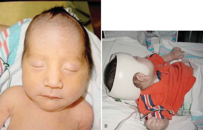
FIG. 11-4 A, Plagiocephaly. B, Helmet used to correct plagiocephaly. (Courtesy Dr. Gerardo Cabrera-Meza, Department of Neonatology, Baylor College of Medicine, Houston.)
Diagnostic Evaluation
The diagnosis of positional plagiocephaly may be made on physical examination of the infant’s head; the infant’s head is viewed frontally and from above. The typical infant’s head shape will resemble a parallelogram, with unilateral flattening of the occiput, frontal and parietal bossing, a prominent cheekbone, and an anterior ear displacement (American Academy of Pediatrics, 2003b). An evaluation of neck movement and range of motion is also made to determine the presence of torticollis. In most cases skull films and further radiologic studies (computed tomographic scan) are used only to rule out craniosynostosis or other cranial deformity that may affect brain growth.
Therapeutic Management
Treatment of torticollis and plagiocephaly initially involved exercises to loosen the tight muscle and switching head position sides during feeding, carrying, and sleep. If the plagiocephaly is not resolved within 4 to 8 weeks of physical therapy, a customized helmet may be worn to decrease the pressure on the affected side of the skull (Biggs, 2003). If no improvement occurs with physical therapy or a molded helmet over a given period, the infant may be referred to a pediatric neurosurgeon or craniofacial surgeon (American Academy of Pediatrics, 2003b). In one study repositioning was not found to be as helpful in reducing plagiocephaly as was the use of an orthotic helmet (see Fig. 11-4, B); those treated with a helmet were older and had a longer treatment period, leading the authors to conclude that early detection and orthotic intervention were likely to be more successful (Graham, Gomez, Halberg, and others, 2005).
Nursing Care Management
Minor skull flattening is not considered significant, but parents should be taught to prevent plagiocephaly by altering the infant’s head position during sleep. Infants should be placed prone on a firm surface during awake time (tummy time), which prevents plagiocephaly and facilitates development of upper shoulder girdle strength; the latter helps in the progressive development of movements such as rolling over and starting to rise up on all fours, which are precursors to crawling and eventually walking. Despite the perceived increase in the incidence of positional plagiocephaly, the supine sleeping position is still recommended because it has led to a significant decrease in loss of infant lives from SIDS (American Academy of Pediatrics, 2005). Additional measures to prevent plagiocephaly include avoiding excessive time spent in car restraint seats or infant seats and bouncers. Alternating the infant’s head position for sleep times can also prevent unilateral molding. When a nurse or parent notices plagiocephaly, a consultation with the primary practitioner is recommended to evaluate the head shape and ascertain the need for early intervention.
Nurses are in a unique position in well-child care settings to encourage parents to follow guidelines for preventing plagiocephaly, to demonstrate alternating head placement for sleeping, to demonstrate sternocleidomastoid muscle exercises (as appropriate to the condition), and to encourage tummy time for infants during awake periods. Most important, nurses should continue to encourage parents to place the infant in a supine sleep position despite the development of plagiocephaly. Parents should not become so alarmed by plagiocephaly that they abandon supine sleeping position for the infant but should consult with the practitioner for further advice.
DISORDERS OF UNKNOWN ETIOLOGY
Sudden infant death syndrome (SIDS) is defined as the sudden death of an infant younger than 1 year of age that remains unexplained after a complete postmortem examination, including an investigation of the death scene and a review of the case history. Since 1992, the incidence of SIDS in the United States has decreased by 53% to an all-time low of 0.57 per 1000 live births in 2002 (American Academy of Pediatrics, 2005). The dramatic decrease is attributed to the Back to Sleep campaign.* SIDS is the third leading cause of infant deaths (birth to 12 months) and the first leading cause of postneonatal (between 1 month and 12 months) deaths. SIDS claimed the lives of 2162 infants in 2003 (Heron and Smith, 2007). Table 11-3 summarizes the major epidemiologic characteristics of SIDS.
TABLE 11-3
Epidemiology of Sudden Infant Death Syndrome (SIDS)
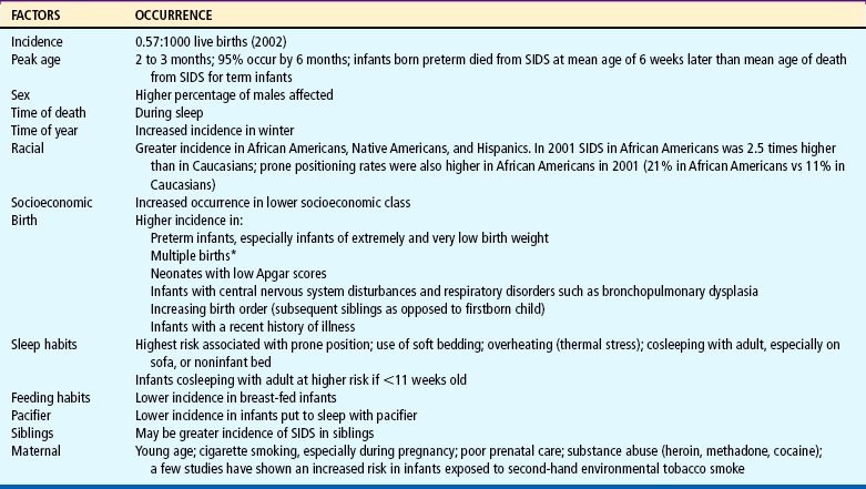
*Although a rare event, simultaneous death of twins from SIDS can occur.
Data from American Academy of Pediatrics, Task Force on Sudden Infant Death Syndrome: The changing concept of sudden infant death syndrome: diagnostic coding shifts, controversies regarding the sleeping environment, and new variables to consider in reducing risk, Pediatrics 116(5):1245-1255, 2005; American Academy of Pediatrics, Task Force on Infant Sleep Position and Sudden Infant Death Syndrome: Changing concepts of sudden infant death syndrome: implications for infant sleeping environment and sleep position, Pediatrics 105(3):650-656, 2000.
Etiology
Numerous theories have been proposed regarding the etiology of SIDS; however, the cause remains unknown. One compelling hypothesis is that SIDS is related to a brainstem abnormality in the neurologic regulation of cardiorespiratory control. Abnormalities include prolonged sleep apnea, increased frequency of brief inspiratory pauses, excessive periodic breathing, and impaired arousal responsiveness to increased carbon dioxide or decreased oxygen. However, sleep apnea is not the cause of SIDS. The vast majority of infants with apnea do not die, and only a minority of SIDS victims have documented apparent life-threatening events (ALTEs) (see Apnea and Apparent Life-Threatening Event, p. 404). Numerous studies indicate that there is no association between SIDS and any childhood vaccine.
A genetic predisposition to SIDS has been postulated as a cause. In one study a genetic mutation on chromosome 6q 22.1-22.31 was positively linked to a syndrome of SIDS and dysgenesis of the testis (Puffenberger, Hu-Lince, Parod, and others, 2004).
Maternal smoking during pregnancy has emerged in numerous epidemiologic studies as a major factor in SIDS, and tobacco smoke in the infant’s environment after birth has also been shown to have a possible relationship with the incidence of SIDS (American Academy of Pediatrics, 2005). Recent data show that exposure to tobacco smoke increased an infant’s risk for SIDS 1.9 times over infants not exposed; 59% of SIDS deaths in smoke-exposed infants were attributed to maternal smoking (Anderson, Johnson, and Batal, 2005). It has been postulated that 12% of all SIDS deaths could be prevented with prenatal maternal smoking cessation (Pollack, 2001). One mechanism that has been proposed as a link between maternal smoking and SIDS is a decrease in the infant’s ability to arouse to auditory stimuli in mothers who smoked prenatally (Franco, Groswasser, Hassid, and others, 1999). Increased nicotine concentrations in lung tissue were found in children who died from SIDS compared with a group of control children (McMartin, Platt, Hackman, and others, 2002).
Cosleeping, or an infant sharing a bed with an adult or older child on a noninfant bed, has been reported to have a positive association with SIDS. One survey found a high association between infant deaths, nonstandard beds (sofa, day bed), and bed sharing; a large percentage of infants were found dead on their backs when bed sharing, suggesting suffocation (Unger, Kemp, Wilkins, and others, 2003). A study from Scotland indicates that the risk for SIDS when bed sharing is significantly increased for infants less than 11 weeks of age (Tappin, Ecob, and Brooke, 2005). Other studies have correlated higher incidences of SIDS and infant cosleeping with maternal smoking, cosleeping with multiple family members, maternal overweight, soft bedding, and unintentional asphyxiation resulting from adult intoxication (overlaying) (American Academy of Pediatrics, 2000, 2005; Hauck, Herman, Donovan, and others, 2003; Carroll-Pankhurst and Mortimer, 2001; Person, Lavezzi, and Wolf, 2002; McGarvey, McDonnell, Chong, and others, 2003).
A study by Hauck, Herman, Donovan, and others (2003) found that bed sharing and SIDS correlated positively only in cases where the infant was sleeping with someone other than the parent; a high number of SIDS cases involved sleeping on a sofa.
Cosleeping with infants in the age range when most SIDS deaths occur has not been shown to be preventive. The latest recommendation for cosleeping from the American Academy of Pediatrics is that the infant’s crib or bassinette be placed in close proximity to the mother’s bed and that the infant be placed in the adult bed only for breastfeeding, then placed to sleep in his or her own crib once the feeding session is completed (American Academy of Pediatrics, 2005).
Mesich (2005) notes that the current scientific literature fails to provide definitive guidance regarding mother-infant sleeping together in relation to safety or nonsafety; certain sleep environments (prone sleeping, tobacco smoke exposure, soft bedding, noninfant bed surface, use of certain drugs by cosleeper, and thermal stress), however, are known to increase the risk for SIDS. (See also Cultural Awareness box, p. 349.)
Studies from countries other than the United States link sleep habits with an increased risk of SIDS. Prone sleeping may cause oropharyngeal obstruction or affect thermal balance or arousal state. One study found that healthy full-term infants had significantly impaired arousal from active and quiet sleep states when sleeping prone (Horne, Ferens, Watts, and others, 2001). Rebreathing of carbon dioxide by infants in the prone position is also a possible cause for SIDS. Infants sleeping prone and on soft bedding may not be able to move their heads to the side, thus increasing the risk of suffocation and lethal rebreathing. Evidence from other countries and the United States shows an increased incidence of SIDS in infants placed in a side-lying position; thus the side-lying position is no longer recommended for infants sleeping at home, daycare, or hospitals (unless medically indicated).
Soft bedding such as waterbeds, sheepskins, beanbags, pillows, or quilts should be avoided for infant sleeping surfaces. Bedding items such as stuffed animals or toys should be removed from the crib while the infant is asleep. Most preterm infants being discharged from the hospital should be placed in a supine sleeping position unless special factors predispose them to airway obstruction. One postulated cause of SIDS has been suggested to be a prolonged Q-T interval; however, at the time of this writing no strong evidence supports universal testing of newborns for prolonged Q-T interval or to support this as a cause of SIDS (American Academy of Pediatrics, 2005).
One study indicated that breastfeeding during the first 16 weeks of life decreased the likelihood of SIDS (Alm, Wennergren, Norvenius, and others, 2002). Some studies have found pacifier use in infants to be a protective factor against the occurrence of SIDS; the data for pacifier use in this population of infants (first year of life) is said to be more compelling than data linking pacifier use to the development of dental complications and the inhibition of breastfeeding (American Academy of Pediatrics, 2005). Therefore recent American Academy of Pediatrics (2005) recommendations are to use a pacifier at naptime and bedtime, use pacifier only if infant is breastfeeding successfully, use no sweetened coating on the pacifier, and avoid forcing the infant to use the pacifier.
The American Academy of Pediatrics (2005) recommends that healthy infants be placed to sleep in the supine (on the back) position. There is an increased risk of SIDS in infants placed in the side-lying position, primarily because of their ability to turn to a prone position; therefore, the side-lying sleep position is no longer recommended.
Although the etiology is unknown, autopsies reveal consistent pathologic findings such as pulmonary edema and intrathoracic hemorrhages that confirm the diagnosis of SIDS. Consequently, autopsies should be performed on all infants suspected of dying of SIDS, and the findings should be shared with the parents as soon as possible after the death.
Whether subsequent siblings of one SIDS infant are at increased risk for SIDS is unclear. Even if the increased risk is correct, families have a 99% chance that their subsequent child will not die of SIDS. A review of recurrent sibling deaths attributed to SIDS in England failed to ascertain a precise risk of recurrence; previous studies suggested a recurrence risk range of 1.7 to 10.1, yet the researchers concluded the studies had too many methodologic flaws to draw any firm conclusions (Bacon, Hall, Stephenson, and others, 2007). Others report recurrence risks for a SIDS death in a family with a previous infant SIDS death range from 2% to 6% (American Academy of Pediatrics, 2005). Home monitoring is not recommended for this group of children, but it is often used by practitioners and may even be requested by parents. Monitoring is best initiated on an individual basis.
Nursing Care Management
Nurses have a vital role in preventing SIDS by educating families about the risk of prone sleeping position in infants from birth to 6 months of age, the use of appropriate bedding surfaces, maternal smoking, and the dangers of cosleeping on noninfant surfaces with adults or other children. Additionally nurses have an important role in modeling behaviors for parents to foster the implementation of practices that decrease the risk of SIDS: placing infants in a supine sleeping position in the hospital and limiting pacifier use to naps and bedtime only; data indicate that a small percentage of nurses still place healthy infants in a side-lying position in the hospital (Bullock, Mickey, Green, and others, 2004; Thompson, 2005). Statistics for infants being placed in a prone sleeping position in the United States decreased from 70% in 1992 to 13% in 2004 (American Academy of Pediatrics, 2005). Nurses must be proactive in further decreasing the incidence of SIDS; postpartum discharge planning, newborn discharges, follow-up home visits, well-baby clinic visits, and immunization visits provide an excellent opportunity to educate parents in these matters.
A concern of many health care workers is that infants placed on the back to sleep will aspirate emesis or mucus; yet studies fail to show an increase in infant deaths, spitting up during sleep, aspiration, asphyxia, or respiratory failure as a result of supine sleep positioning (Malloy, 2002; Tablizo, Jacinto, Parsley, and others, 2007).
Loss of a child from SIDS presents several crises with which the parents must cope. In addition to grief and mourning the death of their child, the parents must face a tragedy that was sudden, unexpected, and unexplained. The psychologic intervention for the family must deal with these additional variables. This discussion focuses primarily on the objectives of care for families experiencing SIDS, rather than on the process of grief and mourning, which is explored in Chapter 18.
Finding the Infant.: Usually it is the mother who finds the child dead in the crib. Typically the child is in a disheveled bed, with blankets over the head, and huddled in a corner. Frothy, blood-tinged fluid fills the mouth and nostrils, and the infant may be lying face down in the secretions, suggesting that he or she bled to death. The diaper is wet and full of stool, which is consistent with a cataclysmic type of death. The hands may be clutching the sheets, as if the child were in distress before death. The initial appearance of the child combined with the shock of such an unexpected event adds to the horror that the parents must face.
The parent who finds the infant initially must deal with her or his initial shock, panic, grief, questions of the other siblings, and the decision of where to find help. The first persons to arrive may be the police and ambulance attendants. Ideally, they will handle the situation by asking few questions; giving no indication of wrongdoing, abuse, or neglect; making sensitive judgments concerning any resuscitation efforts for the child; and comforting the members of the family as much as possible. These individuals should be properly informed about SIDS to recognize its characteristic signs and tell parents that their child probably died of a condition called sudden infant death syndrome, which cannot be predicted or prevented. A compassionate, sensitive approach to the family during the first few minutes can help spare them some of the overwhelming guilt and anguish that commonly follow this type of death.
Arriving at the Emergency Department.: The first contact that nurses typically have with these families is in the emergency department, when the infant is seen by a physician to be pronounced dead. Usually there is no attempt at resuscitation. During the time in the emergency department several aspects warrant special consideration. Parents are asked only factual questions, such as when they found the infant, how he or she looked, and whom they called for help. Any remarks that may suggest responsibility, such as why they did not check on the child earlier, why they did not hear the infant cry out, whether the head was buried in a blanket, or whether other siblings were jealous of this child, are avoided.
A review of 60 studies shows that parents experiencing perinatal death perceive health care workers’ responses as having a significant impact on the parents’ grieving process; many health care workers’ behaviors were perceived as thoughtless or insensitive. The findings suggest that nurses and physicians would benefit from more bereavement training (Gold, 2007).
The discussion of an autopsy should be presented at this time, emphasizing that a diagnosis cannot be confirmed until the postmortem examination is completed. If the mother was breastfeeding, she needs information about abrupt discontinuation of lactation.
Another important aspect of compassionate care for these parents is allowing them to say good-bye to their child. A debriefing session may help health care workers who dealt with the family and deceased infant to cope with feelings that are often engendered when a SIDS victim is brought into the acute care facility. Comprehensive guidelines have been published for health professionals involved in SIDS investigations to assist the family and at the same time to determine that the infant’s death was not the result of other factors such as child maltreatment (American Academy of Pediatrics, 2001).
Returning Home.: When the parents return home, they should be visited by a competent, qualified professional as soon after the death as possible. Printed material that contains excellent information about SIDS (available from several qualified organizations*) should be provided.
Ideally, the number of visits and plans for subsequent intervention need to be flexible. For example, the siblings may initially appear accepting of the explanation and well adjusted but may later refuse to go to sleep or ask questions about graves or funerals, indicating their need for further help in dealing with the death. Parents facing the question of a subsequent child will need support. Both the birth of a subsequent child and the survival of that child, especially past the age of death of the previous child, are important transitional stages for parents.
Because the mourning process continues for at least a year or more, referrals to other parents who have lost a child to SIDS should be considered. The practitioner should be available to answer parents’ questions regarding the infant’s death, which may persist for months to years later until there is some resolution of the loss of the child.
APNEA AND APPARENT LIFE-THREATENING EVENT
Apnea is defined as a cessation of breathing for 20 seconds. Apnea of infancy is defined as an unexplained respiratory pause of 20 seconds or more, or pauses of less than 20 seconds that are accompanied by pallor, cyanosis, bradycardia, or hypotension in the term infant. The latter is a distinct entity from apnea of prematurity (see Chapter 9). An apparent life-threatening event (ALTE), formerly referred to as aborted SIDS death or near-miss SIDS, generally refers to an event that is sudden and frightening to the observer, in which the infant exhibits a combination of apnea, change in color (pallor, cyanosis, redness), change in muscle tone (usually hypotonia), choking, gagging, or coughing, and which usually involves a significant intervention and even cardiopulmonary resuscitation (CPR) by the caregiver who witnesses the event (National Institutes of Health Consensus Development Conference, 1987). The definition of ALTE may include apnea, but ALTE may occur without apnea (Silvestri and Weese-Mayer, 2003).
Apnea during infancy can be a symptom of any one of many disorders, including sepsis, seizures or other neurologic disorder, upper or lower airway infection or abnormality, gastroesophageal reflux, hypoglycemia or other metabolic problems, impaired regulation of breathing during sleep or feeding, or a result of intentional harm by an adult caregiver. Delayed ventilatory responses to hypercapnia and hypoxia were observed in one study of 69 infants with apnea of infancy (Katz-Salamon, 2004). Abusive head injury has been reported in a small percentage (2.5%) of children with ALTE (Altman, Brand, Forman, and others, 2003). Intentional suffocation and Munchausen syndrome by proxy cases have also been reported with ALTE (Hall and Zalman, 2005). However, in about half the cases no cause is identified.
Infants with a history of ALTEs may be at increased risk for SIDS, but these children constitute only approximately 7% to 12% of all SIDS victims. Most infants with ALTE are less than 6 months of age, and although there has been a significant decrease in SIDS since 1992, the incidence of ALTE has not changed (Hall and Zalman, 2005). A diagnosis of apnea of infancy or idiopathic ALTE is often made when no cause is found.
Results from the Collaborative Home Infant Monitoring Evaluation study found that apnea and bradycardia occurred at conventional and extreme alarm thresholds in all groups of infants studied: siblings of SIDS infants, infants with ALTEs, symptomatic (of apnea and bradycardia) and asymptomatic preterm infants weighing less than 1750 g (3.8 pounds) at birth, and healthy term infants. The researchers concluded that many infants experience apnea and bradycardia in each of these groups yet do not die (Jobe, 2001; Ramanathan, Corwin, Hunt, and others, 2001). Furthermore, it was reported that apnea does not appear to be an immediate precursor to SIDS and that cardiorespiratory monitoring is not an effective tool for identifying infants at greater risk for SIDS (American Academy of Pediatrics, 2003a).
Diagnostic Evaluation
An essential component of the diagnostic process includes a detailed description of the event—who witnessed the event, where the infant was during the event, and what, if any, activities were involved (such as during or after a feeding, riding in a car seat restraint, presence of siblings or any minor children, what clothing the infant was wearing). In addition, a prenatal and postnatal history must be obtained. A short period of observation in the emergency department may be appropriate to observe the infant’s respiratory pattern and response to feeding. A careful evaluation of the preterm infant in a car restraint is essential; upper airway occlusion and subsequent apnea and cyanosis may occur if the infant is not positioned properly. Reported diagnoses in infants with ALTE include a neurologic event such as a seizure (30% of cases seen); gastrointestinal problem, including gastroesophageal reflux (50%); respiratory conditions (20%); and metabolic, cardiac, or child abuse (each <5%). In some cases, multiple diagnoses may be made (Hall and Zalman, 2005).
In the event that an underlying diagnosis such as those mentioned previously is not established, home monitoring may be recommended. The most commonly used monitoring is continuous recording of cardiorespiratory patterns (cardiopneumogram, or pneumocardiogram). Four-channel pneumocardiograms (or multichannel pneumogram) monitor heart rate, respirations (chest impedance), nasal airflow, and oxygen saturation. A more sophisticated test, polysomnography (sleep study), also records brain waves, eye and body movements, esophageal manometry, and end-tidal carbon dioxide measurements. However, none of these tests can predict risk. Some children with normal results may still have subsequent apneic episodes.
Therapeutic Management
The treatment of the infant with an ALTE depends on the underlying condition (see above). Treatment of recurrent apnea (without an underlying organic problem) usually involves continuous home monitoring of cardiorespiratory rhythms and in some cases the use of methylxanthines (respiratory stimulant drugs, such as theophylline or caffeine). The decision to discontinue the monitoring is based on the infant’s clinical condition. A general guideline for discontinuation is when infants with ALTEs have gone 2 or 3 months without significant numbers of episodes requiring intervention.
Newer home apnea monitors allow download of information that assists the practitioner in the decision about when home monitoring may be discontinued. It is imperative to remember, however, that the home apnea monitor will not predict or prevent SIDS deaths. Furthermore, impedance-based monitors detect chest wall movement and will not detect obstructive apnea unless the episode involves significant bradycardia (see Family-Centered Care box).
Use the monitor as instructed by the practitioner or home monitor equipment company.
Do not adjust the monitor to eliminate false alarms. Adjustments could compromise the monitor’s effectiveness.
Place the monitor on a firm surface away from the crib and drapes; plug power cord directly into a wall socket with a three-pronged outlet.
Do not sleep in the same bed as the monitored infant.
Keep pets and children away from the monitor and infant.
Keep the monitor away from possible electrical interferences such as appliances (e.g., electric blankets, televisions, air conditioners, remote telephones).
Be aware that strong signals from nearby radio and television stations, airports, ham radios, cellular phones, or police stations could interfere with the monitor. Check for interference if the monitor is to be operated in these areas.
Check the monitor several times a day to be sure the alarm is working and that it can be heard from room to room. Be certain the caregiver can reach the monitor quickly (in less than 30 seconds).
Periodically check the monitor’s breath detection indicator and battery or charger connections.
Read the monitor’s user manual carefully; report problems promptly.
Inform community utility and rescue squads of home monitoring as appropriate.
Data primarily from US Food and Drug Administration: Safety alert: important tips for apnea monitor users, Rockville, Md, 1990, US Department of Health and Human Services.
Nursing Care Management
The diagnosis of an ALTE engenders great anxiety and concern in parents, and the institution of home monitoring presents additional physical and emotional burdens. Parents of infants on home apnea monitors report experiencing emotional distress, especially depression and hostility, during the first few weeks after hospital discharge (Abendroth, Moser, Dracup, and others, 1999). For parents of a SIDS victim who have a new infant on home apnea monitoring, the anxiety is compounded by the uncertainty of the future of the living child and grief for the lost child. Home apnea monitoring may offer some predictability and control over the current child’s survival through the period of uncertainty. If monitoring is required, the nurse can be a major source of support to the family in terms of education about the equipment, observation of the infant’s status, and immediate intervention during apneic episodes, including CPR. Several reports indicate that the first week to month after discharge is the most stressful for parents, particularly when the rate of false alarms is high (Bennett, 2002). To help the family cope with the numerous procedures they must learn, adequate preparation before discharge and written instructions are essential. In the first few weeks after discharge, parents may benefit by having a practitioner readily available to answer questions regarding false alarms and for other technical assistance (Abendroth, Moser, Dracup, and others, 1999).
Several types of home monitors are available and are set up by either a home monitor equipment company or home health staff. Nurses, especially those involved in the care at home, must become familiar with the equipment, including its advantages and disadvantages. Safety is a major concern because monitors can cause electrical burns and electrocution. The following precautions are recommended:
Remove leads from infant when not attached to monitor.
Unplug power cord from electrical outlet when cord is not plugged into monitor.
Use safety covers on electrical outlets to discourage children from inserting objects into a socket.
Siblings should also be supervised when near the infant and taught that the monitor is not a toy. Other safety practices include informing local utility and rescue squads of the home monitoring in case of an emergency. Telephone numbers for these services should be posted near all telephones in the home.
Caregivers need detailed information regarding proper attachment of the electrodes to the infant’s chest with impedance monitors that detect chest movement. The electrodes are placed in the midaxillary line, at a space one or two fingerbreadths below the nipple. For home use, electrodes attached to a belt that is placed around the child’s trunk are preferred (Fig. 11-5). The belt is positioned so that the electrodes contact the skin in the same area. Monitors may have memory chips that allow for event recording, which can be an effective tool in evaluating the use of the monitor, events immediately before and after the event, and reported frequency of alarms.

FIG. 11-5 Placement of electrodes or belt for apnea monitoring. In small infants, one fingerbreadth may be used.
Monitors are effective only if they are used. They do not prevent death but alert the caregiver to the ALTE in time to intervene. The need to use the monitor and to respond appropriately to alarms must be stressed. Noncompliance can result in the infant’s death.
Family Support.: Many of the stresses observed during the monitoring period are characteristic of those of families with chronically ill children. The child with an apnea or cardiorespiratory monitor may have additional health care needs such as a gastrostomy, tracheostomy, ostomy, and myriad medications or treatments that exacerbate the parents’ stress. Parents report increased stress, including concern for the child’s survival, fear of incompetence in assuming home responsibility, inadequate respite care, lack of time for other children and spouse, social isolation from friends and extended family, constant work, and fatigue. The monitored child is at risk for vulnerable child syndrome, which may lead to lack of parental separation and preferential treatment, causing further family disruption (Bennett, 2002). To deal with these potential effects, nurses need to employ the same interventions as those discussed for children with chronic illness (see Chapter 18) and be aware of the need for referral when difficulties are suspected.
To lessen the continuous responsibility of monitoring, other family members, such as grandparents, should be taught how to manipulate the equipment, read and interpret the signals, and administer CPR. They are encouraged to stay with the infant for regular periods to allow parents respite. Support groups of other families who have successfully completed monitoring can also be of benefit. Because baby-sitters are difficult to locate, support group members or nursing students may be potential sources of qualified caregivers.
Common nutritional disorders of infancy and early childhood may result from vitamin and mineral deficiency or excess, some types of vegetarian diets, protein-energy malnutrition, and food intolerance.
Food consumption varies among vegetarians; therefore, a detailed dietary intake is essential for planning adequate intakes, particularly in children and pregnant and lactating women.
Protein-energy malnutrition may occur as a complication of underlying disease, lack of parental education about infant nutrition, inappropriate management of food allergy, or incorrect preparation of formula.
Food intolerance encompasses food allergies and food sensitivities, which can have a number of systemic and local clinical manifestations. CMA and lactose intolerance may occur in some children.
Treatment of colic may involve a change in infant feeding practices, correction of a stressful environment, behavior modification, and support of the parent.
Growth failure or FTT may occur in children who have a chronic illness, or it may occur in a family environment wherein healthy infant feeding practices are poorly managed or understood; FTT is not always associated with a pattern of disturbed maternal-infant relationship.
Positional plagiocephaly can be easily prevented by allowing the awake infant to have periods of tummy time and by alternating the infant’s head position during sleep.
SIDS is the third leading cause of infant death in the United States.
Factors that place the infant at high risk for SIDS include prone sleeping position, soft bedding, sleeping in a noninfant bed with an adult or older child, and maternal prenatal smoking.
The primary nursing responsibility in care associated with sudden infant death is educating the family of newborns about the risks for SIDS, modeling appropriate behaviors in the hospital such as placing the infant in a supine sleep position, and providing emotional support of the family who has experienced a SIDS loss.
Infants with ALTEs are carefully evaluated for clues to the underlying cause.
Home apnea or cardiorespiratory monitors do not prevent SIDS.
REFERENCES
Abendroth, D, Moser, DK, Dracup, K, et al. Do apnea monitors decrease emotional distress in parents of infants at high risk for cardiopulmonary arrest? J Pediatr Health Care. 1999;13(2):50–57.
Alm, B, Wennergren, G, Norvenius, SG, et al. Breast-feeding and the sudden infant death syndrome in Scandinavia, 1992-1995. Arch Dis Child. 2002;86(6):400–402.
Altman, RL, Brand, DA, Forman, S, et al. Abusive head injury as a cause of apparent life-threatening events in infancy. Arch Pediatr Adolesc Med. 2003;157(10):1011–1015.
Amadi, B, Mwiya, M, Chomba, E, et al. Improved nutritional recovery on an elemental diet in Zambian children with persistent diarrhoea and malnutrition. J Trop Pediatr. 2005;51(1):5–10.
American Academy of Pediatrics. Dietary recommendations for children and adolescents: a guide for practitioners. Pediatrics. 2006;117(2):544–559.
American Academy of Pediatrics, Committee on Infectious Diseases, Pickering L, eds. Red book: 2006 report of the Committee on Infectious Diseases, ed 27, Elk Grove Village, Ill: The Academy, 2006.
American Academy of Pediatrics, Task Force on Sudden Infant Death Syndrome. The changing concept of sudden infant death syndrome: diagnostic coding shifts, controversies regarding the sleeping environment, and new variables to consider in reducing risk. Pediatrics. 2005;116(5):1245–1255.
American Academy of Pediatrics, Committee on Nutrition. Pediatric nutrition handbook, ed 5. Elk Grove Village, Ill: The Academy, 2004.
American Academy of Pediatrics, Committee on Fetus and Newborn. Apnea, sudden infant death syndrome, and home monitoring. Pediatrics. 2003;111(4):914–917.
American Academy of Pediatrics. Prevention and management of positional skull deformities in infants (clinical report). Pediatrics. 2003;112(1):199–202.
American Academy of Pediatrics. Prevention of rickets and vitamin D deficiency: new guidelines for vitamin D intake. Pediatrics. 2003;111(4):908–910.
American Academy of Pediatrics, Committee on Child Abuse and Neglect. Distinguishing sudden infant death syndrome from child abuse fatalities. Pediatrics. 2001;107(2):437–441.
American Academy of Pediatrics, Task Force on Infant Sleep Position and Sudden Infant Death Syndrome. Changing concepts of sudden infant death syndrome: implications for infant sleeping environment and sleep position. Pediatrics. 2000;105(3):650–656.
American Dietetic Association and Dietitians of Canada. Position of the American Dietetic Association and Dietitians of Canada: vegetarian diets. J Am Diet Assoc. 2003;103(6):748–765.
American Heart Association. Dietary recommendations for children. http://www.americanheart.org/presenter.jhtml?identifier=53033999, 2005. [retrieved Jan 3, 2008, from].
Anderson, ME, Johnson, DC, Batal, HA. Sudden infant death syndrome and prenatal maternal smoking: rising attributed risk in the Back to Sleep era. BMC Med. 2005;3(1):4.
Assa’ad, AH. Gastrointestinal food allergy and intolerance. Pediatr Ann. 2006;35(10):718–726.
Bacon, CJ, Hall, DBM, Stephenson, TJ, et al, How common is repeat sudden infant death syndrome? Arch Dis Child 2007 10.1136/adc.2006.113969
Barnard, K, Hammond, MA, Booth, CL, et al, Measurement and meaning of parent-child interaction. Morrison, F, Lord, C, Keating, D, eds. Applied developmental psychology, vol 3. New York: Academic Press, 1993.
Bennett, AD. Home apnea monitoring for infants: a discussion of primary care issues. Adv Nurs Pract. 2002;10(3):48–53.
Biggs, WS. Diagnosis and management of positional head deformity. Am Fam Physician. 2003;67(9):1953–1956.
Block, RW, Krebs, NF, American Academy of Pediatrics Committee on Child Abuse and Neglect, Committee on Nutrition. Failure to thrive as a manifestation of child neglect. Pediatrics. 2005;116(5):1234–1237.
Bullock, LFC, Mickey, K, Green, J, et al. Are nurses acting as role models for the prevention of SIDS? MCN. 2004;29(3):172–177.
Burk, CJ, Molodow, R. Infantile scurvy: an old diagnosis revisited with a modern dietary twist. Am J Clin Dermatol. 2007;8(2):103–106.
Carroll-Pankhurst, C, Mortimer, EA. Sudden infant death syndrome, bedsharing, parental weight, and age of death. Pediatrics. 2001;107(3):530–536.
Centers for Disease Control and Prevention. Neurologic impairment in children associated with maternal dietary deficiency of cobalamin—Georgia, 2001. MMWR. 2003;52(4):61–64.
Christie, L, Hine, RJ, Parker, JG, et al. Food allergies in children affect nutrient intake and growth. J Am Diet Assoc. 2002;102(11):1648–1651.
Ciliberto, MA, Sandige, H, Ndekha, MJ, et al. Comparison of a home-based therapy with ready-to-use therapeutic food with standard therapy in the treatment of malnourished Malawin children: a controlled, clinical effectiveness trial. Am J Clin Nutr. 2005;81(4):864–870.
Conover, E, Buehler, BA. Use of herbal agents by breastfeeding women may affect infants. Pediatr Ann. 2004;33(4):235–240.
Corrales, KM, Utter, SL. Growth failure. In Samour PQ, King K, eds.: Handbook of pediatric nutrition, ed 3, Sudbury, Mass: Jones & Bartlett, 2005.
Crotteau, CA, Wright, ST, Eglash, A. What is the best treatment for infants with colic? J Fam Pract. 2006;55(7):634–636.
De Vrese, M, Stegelmann, A, Richter, B, et al. Probiotics: compensation for lactase insufficiency. Am J Clin Nutr. 2001;73(2 Suppl):421S–429S.
Dunham, L, Kollar, L. Vegetarian eating for children and adolescents. J Pediatr Health Care. 2006;20(1):27–34.
Duro, D, Rising, R, Cedillo, M, et al. Association between infantile colic and carbohydrate malabsorption from fruit juices in infancy. Pediatrics. 2002;109(5):797–805.
Ellett, MLC. What is known about colic? Gastroenterol Nurs. 2003;26(2):60–65.
Ewing, WM, Allen, PJ. The diagnosis and management of cow milk protein intolerance in the primary care setting. Pediatr Nurs. 2005;31(6):486–492.
Fiocchi, A, Assa’ad, A, Bahna, S, et al. Food allergy and the introduction of solid foods to infants: a consensus document. Ann Allergy Asthma Immunol. 2006;97(1):10–20.
Fiocchi, A, Martelli, A. Dietary management of food allergy. Pediatr Ann. 2006;35(10):755–763.
Fox, M, Reidy, K, Novak, T, et al. Sources of energy and nutrients in the diets of infants and toddlers. J Am Diet Assoc. 2006;106(1 Suppl 1):S28–S42.
Franco, P, Groswasser, J, Hassid, S, et al. Prenatal exposure to cigarette smoking is associated with a decrease in arousal in infants. J Pediatr. 1999;135(1):34–38.
Gidding, SS, Dennison, BA, Birch, LL, et al. Dietary recommendations for children and adolescents: a guide for practitioners: consensus statement from the American Heart Association. Circulation. 2005;112(13):2061–2075.
Gold, KJ. Navigating care after a baby dies: a systematic review of parent experiences with health providers. J Perinatol. 2007;27(4):230–237.
Goldberg, JP, Folta, SC, Must, A. Milk: can a “good” food be so bad? Pediatrics. 2002;110(4):826–831.
Graham, JM, Gomez, M, Halberg, A, et al. Management of deformational plagiocephaly: repositioning versus orthotic therapy. J Pediatr. 2005;146(2):258–262.
Hall, KL, Zalman, B. Evaluation and management of apparent life threatening events in children. Am Fam Physician. 2005;71(12):2301–2308.
Hauck, FR, Herman, SM, Donovan, M, et al. Sleep environment and the risk of sudden infant death syndrome in an urban population: the Chicago Infant Mortality Study. Pediatrics. 2003;111(5):1207–1214.
Heron, MP, Smith, BL. Deaths: leading causes for 2003. Natl Vital Stat Rep. 2007;55(10):1–92.
Heyman, MB, American Academy of Pediatrics Committee on Nutrition. Lactose intolerance in infants, children, and adolescents. Pediatrics. 2006;118(3):1279–1286.
Horne, RS, Ferens, D, Watts, AM, et al. The prone sleeping position impairs arousability in term infants. J Pediatr. 2001;138(6):793–795.
Host, A. Frequency of cow’s milk allergy in childhood. Ann Allergy Asthma Immunol. 2002;89(6 Suppl 1):33–37.
Huiming, Y, Chaomin, W, Meng, M, Vitamin A for treating measles in children. Cochrane Database Syst Rev 2005;4:CD001479.
Hummel, P, Fortado, D. Impacting infant head shapes. Adv Neon Care. 2005;5(6):329–342.
Institute of Medicine, Food and Nutrition Board. Dietary reference intakes: applications in dietary assessment. Washington, DC: National Academy Press, 2000.
Joanna Briggs Institute. Best practice information sheet: the effectiveness of interventions for infant colic. Best Practice. 2004;8(2):1–6.
Jobe, AH. What do home monitors contribute to the SIDS problem? (editorial). JAMA. 2001;285(17):2244–2245.
Katz, KA, Mahlberg, MA, Honig, PJ, et al. Rice nightmare: kwashiorkor in two Philadelphia-area infants fed Rice Dream beverage. J Am Acad Dermatol. 2005;52(5 Suppl 1):S69–S72.
Katz-Salamon, M. Delayed chemoreceptor responses in infants with apnoea. Arch Dis Child. 2004;89(3):261–266.
Kawchak, DA, Schall, JI, Zemel, BS, et al. Adequacy of dietary intake declines with age in children with sickle cell disease. J Am Diet Assoc. 2007;107(5):843–848.
Kemper, KJ, Gardiner, P. Herbal medicines. In Behrman RE, Kliegman RM, Jenson HB, eds.: Nelson textbook of pediatrics, ed 17, Philadelphia: Saunders, 2004.
Krugman, SD, Dubowitz, H. Failure to thrive. Am Fam Physician. 2003;68(5):879–886.
Lanski, SL, Greenwald, M, Perkins, A, et al. Herbal therapy in a pediatric emergency department population: expect the unexpected. Pediatrics. 2003;111(5 pt 1):981–985.
Lawrence, RA, Lawrence, RM. Breastfeeding: a guide for the medical profession, ed 6. St Louis: Mosby, 2005.
Levin, ME, Motala, C, Lopata, AL. Anaphylaxis in a milk-allergic child after ingestion of soy formula cross-contaminated with cow’s milk protein. Pediatrics. 2005;116(5):1223–1225.
Littlefield, TR, Saba, NM, Kelly, KM. On the current incidence of deformational plagiocephaly: an estimation based on prospective registration at a single center. Semin Pediatr Neurol. 2004;11(4):301–304.
Liu, T, Howard, RM, Mancini, AJ, et al. Kwashiorkor in the United States: fad diets, perceived and true milk allergy, and nutritional ignorance. Arch Dermatol. 2001;137(5):630–636.
Locklin, M. The redefinition of failure to thrive from a case study perspective. Pediatr Nurs. 2005;31(6):474–479. [495,].
Lohse, B, Stotts, JL, Priebe, JR. Survey of herbal use by Kansas and Wisconsin WIC participants reveals moderate, appropriate use and identifies herbal education needs. J Am Diet Assoc. 2006;106(2):227–237.
Loman, DG. The use of complementary and alternative health care practices among children. J Pediatr Health Care. 2003;17(2):58–63.
Maggioni, A, Lifshitz, F. Nutritional management of failure to thrive. Pediatr Clin North Am. 1995;42(4):791–809.
Malloy, MH. Trends in postneonatal aspiration deaths and reclassification of sudden infant death syndrome: impact of the “Back to Sleep” program. Pediatrics. 2002;109(4):661–665.
Markowitz, R, Duggan, C. Failure to thrive: malnutrition in the pediatric setting. In Walker WA, Watkins JB, Duggan C, eds.: Nutrition in pediatrics, ed 3, Hamilton, Ontario: Decker, 2003.
McGarvey, C, McDonnell, M, Chong, A, et al. Factors relating to the infant’s last sleep environment in sudden infant death syndrome in the Republic of Ireland. Arch Dis Child. 2003;88(12):1058–1064.
McGuire, JK, Kulkarni, MS, Baden, HP. Fatal hypermagnesemia in a child treated with megavitamin/megamineral therapy. Pediatrics. 2000;105(2):414.
McMartin, KI, Platt, MS, Hackman, R, et al. Lung tissue concentrations of nicotine in sudden infant death syndrome. J Pediatr. 2002;140(2):205–209.
Mesich, HM. Mother-infant co-sleeping: understanding the debate and maximizing infant safety. MCN. 2005;30(1):30–37.
Messina, V, Melina, V, Mangels, AR. A new food guide for North American vegetarians. J Am Diet Assoc. 2003;103(6):771–775.
Müller, O, Krawinkel, M. Malnutrition and health in developing countries. CMAJ. 2005;173(3):279–286.
National Institutes of Health Consensus Development Conference on Infantile Apnea and Home Monitoring, Sept 29 to Oct 1, 1986. Pediatrics. 1987;79(2):292–299.
Neu, M, Robinson, JA. Infants with colic: their childhood characteristics. J Pediatr Nurs. 2003;18(1):12–20.
Penny, ME. Protein-energy malnutrition: pathophysiology, clinical consequences, and treatment. In Walker WA, Watkins JB, Duggan C, eds.: Nutrition in pediatrics, ed 3, Hamilton, Ontario: Decker, 2003.
Perrotta, S, Nobili, B, Rossi, F, et al. Infant hypervitaminosis A causes severe anemia and thrombocytopenia: evidence of a retinol-dependent bone marrow cell growth inhibition. Blood. 2002;99(6):2017–2022.
Perry, CL, McGuire, MT, Neumark-Sztainer, D, et al. Adolescent vegetarians: how well do their dietary patterns meet the Healthy People 2010 objectives? Arch Pediatr Adolesc Med. 2002;156(5):426–427.
Person, TLA, Lavezzi, WA, Wolf, BC. Cosleeping and sudden unexpected death in infancy. Arch Pathol Lab Med. 2002;126(3):343–345.
Pessler, F, Nejat, M. Anaphylactic reaction to goat’s milk in a cow’s milk-allergic infant. Pediatr Allergy Immunol. 2004;15(2):183–185.
Pollack, HA. Sudden infant death syndrome, maternal smoking during pregnancy and effectiveness of smoking cessation intervention. Am J Public Health. 2001;91(3):432–436.
Preston, AM, Rodriguez, C, Rivera, CE. Plasma ascorbate in a population of children: influence of age, gender, vitamin C intake, and smoke exposure. PR Health Sci J. 2006;25(2):137–142.
Preston, AM, Rodriguez, C, Rivera, C, et al. Influence of environmental tobacco smoke on vitamin C status in children. Am J Clin Nutr. 2003;77(1):167–172.
Puffenberger, EG, Hu-Lince, D, Parod, JM, et al. Mapping of sudden infant death with dysgenesis of the testis syndrome (SIDDT) by a SNP genome scan and identification of TSPYL loss of function. Proc Natl Acad Sci USA. 2004;101(32):11689–11694.
Ramanathan, R, Corwin, MJ, Hunt, DE, et al. Cardiorespiratory events recorded on home monitors: comparison of healthy infants with those at increased risk for SIDS. JAMA. 2001;285(17):2199–2243.
Rudolf, MCJ, Logan, S. What is the long term outcome for children who fail to thrive? A systematic review. Arch Dis Child. 2005;90(9):925–931.
Saltzman, MD, King, EC. Central physeal arrests as a manifestation of hypervitaminosis A. J Pediatr Orthop. 2007;27(3):351–353.
Salvatore, S, Vandenplas, Y. Gastroesophageal reflux and cow milk allergy: is there a link? Pediatrics. 2002;110(5):972–984.
Sampson, HA. Update on food allergy. J Allergy Clin Immunol. 2004;113(5):805–819.
Sampson, HA. Anaphylaxis and emergency treatment. Pediatrics. 2003;111(6 Pt 3):1601–1608.
Sawni, A, Ragothaman, R, Thomas, RL, et al. The use of complementary/alternative therapies among children attending an urban pediatric emergency department. Clin Pediatr. 2007;46(1):36–41.
Sibley, E. Carbohydrate intolerance. Curr Opin Gastroenterol. 2004;20(2):162–167.
Silvestri, JM, Weese-Mayer, D. Disorders of respiratory control: apnea and SIDS. In Rudolph CD, Rudolph AM, Hostetter MK, eds.: Rudolph’s pediatrics, ed 21, New York: McGraw-Hill, 2003.
Tablizo, MA, Jacinto, P, Parsley, D, et al. Supine sleeping position does not cause clinical aspiration in neonates in hospital newborn nurseries. Arch Pediatr Adolesc Med. 2007;161(5):507–510.
Tappin, D, Ecob, R, Brooke, H. Bedsharing, roomsharing, and sudden infant death syndrome in Scotland: a case-control study. J Pediatr. 2005;147(1):32–37.
Thompson, DG. Safe sleep practices for hospitalized infants. Pediatr Nurs. 2005;31(5):400–403. [409,].
Unger, B, Kemp, JS, Wilkins, D, et al. Racial disparity and modifiable risk factors among infants dying suddenly and unexpectedly. Pediatrics. 2003;111(2):e127–e131.
Weizman, Z, Alkrinawi, S, Goldfarb, D, et al. Efficacy of herbal tea preparation in infantile colic. J Pediatr. 1993;122(4):650–652.
Zeisel, SH, Erickson, KE. Dietary supplements (nutraceuticals). In Walker WA, Watkins JB, Duggan C, eds.: Nutrition in pediatrics, ed 3, Hamilton, Ontario: Decker, 2003.
*Helpful websites for health care and consumer information concerning herbs are National Center for Complementary and Alternative Medicine, http://www.nccam.nih.gov; American Botanical Council, http://www.herbalgram.org; and Herb Research Foundation, http://www.herbs.org.
*Further information regarding vegetarian diets may be found at the Vegetarian Resource Group, PO Box 1463, Baltimore, MD 21203; (410) 366-8343; http://www.vrg.org
*For information on the DRIs go to IOM website, http://www.iom.edu, and click on the link for Food and Nutrition; or call (202) 334-2352.
*Further information for parents of infants with food allergies is available from the American Academy of Allergy, Asthma, and Immunology, 555 E. Wells St., Suite 1100, Milwaukee, WI 53202; (414) 272-6071; http://www.aaaai.org. Additional helpful websites for information on food allergy include MedlinePlus (sponsored by U.S. National Library of Medicine and National Institutes of Health), http://medlineplus.gov; Food Allergy and Anaphylaxis Network, (800) 929-4040, http://www.foodallergy.org; National Institute of Allergy and Infectious Diseases, http://www3.niaid.nih.gov; and http://www.allergicchild.com.
*Training is required to use the feeding scale. For information, contact Jean F. Kelly, PhD, Executive Director, NCAST-AVENUW, University of Washington, PO Box 357920, Seattle, WA 98195; (206) 543-8528; e-mail: ncast@u.washington.edu; http://www.ncast.org.
*Back to Sleep materials may be ordered by contacting National Institute of Child Health and Human Development, Back to Sleep, 31 Center Dr., Building 31, Room 2A32, Bethesda, MD 20892; (800) 505-CRIB; fax: (301) 984-1473; http://www.nichd.nih.gov/sids.
*American SIDS Institute, 509 Augusta Dr., Marietta, GA 30067; (800) 232-SIDS, (770) 426-8746; http://www.sids.org; First Candle, 1314 Bedford Ave., Suite 210, Baltimore, MD 21208; (800) 221-7437; http://www.sidsalliance.org; National Sudden Infant Death Resource Center, Georgetown University, box 571272, Washington, DC 20057-1272; (866) 866-7437, (202) 687-7466; http://www.sidscenter.org.