The diagnosis is the molecular genetic marker for the survival motor neuron gene, which is located on chromosome 5q13. Prenatal diagnosis may be made by genetic analysis of circulating fetal cells in maternal blood (Beroud, Karliova, Bonnefont, et al, 2003) or circulating fetal cells in amniotic fluid. Maternal report of decreased fetal movements may also be suggestive of SMA (Markowitz, Tinkle, and Fischbeck, 2004). The risk of subsequent affected offspring in carriers of the mutant gene or in families with known cases of SMA may also be evaluated genetically. Further diagnostic studies include muscle EMG, which demonstrates a denervation pattern, and muscle biopsy; however, the genetic analysis has become the gold standard for diagnosis of the condition. Newborn screening is possible yet is not available on a widespread basis, possibly because there is no treatment (Lunn and Wang, 2008).
There is no cure for the disease, and treatment is symptomatic and preventive, primarily preventing joint contractures and treating orthopedic problems, the most serious of which is scoliosis; hip subluxation and dislocation may also occur. Many children benefit from powered chairs, lifts, special pressure-adjustable mattresses, and accessible environmental controls. Muscle and joint contractures require careful attention and care to prevent further complications. Nutritional failure to thrive may occur in infants and toddlers as a result of poor feeding; supplemental gastrostomy feedings may be required to maintain adequate nutritional status and maintain weight gain (Iannaccone and Burghes, 2002). The use of lower extremity orthoses may assist with ambulation, but eventually the child may be confined to a wheelchair as muscle atrophy progresses.
Restrictive lung disease is the most serious complication of SMA (Iannaccone, 2007). Upper respiratory tract infections often occur and are treated with antibiotic therapy; they are the cause of death in many children. Sleep-disordered breathing is common in children with SMA and often requires noninvasive mechanical ventilation (Iannaccone and Burghes, 2002; Iannaccone, 2007). A polysomnogram may be performed to determine optimal therapeutic intervention with supplemental oxygen or noninvasive ventilation modes. Noninvasive ventilation methods such as bilevel positive airway pressure (BiPAP) have decreased the morbidity and increased the survival rate of children with SMA types 1 and 2. A significant number of infants with SMA require a tracheotomy, and associated medical conditions in survivors include gastroesophageal reflux, scoliosis, early-onset puberty, hip dysplasia, and recurrent oral candidiasis (Bach, 2007).
A number of clinical trials are currently in progress with existing drugs (e.g., valproic acid, phenylbutyrate, creatine) aimed at increasing the SMN mRNA and thus decreasing muscle wasting (Lunn and Wang, 2008).
The infant or small child with progressive muscle weakness requires nursing care similar to that of the immobilized patient. However, the underlying goal of treatment is to assist the child and family in dealing with the illness while progressing toward a life of normalization within the child’s capabilities.
Infants who are able to feed may require special nutritional considerations during breast- and bottle-feeding. Such infants tire easily and may be difficult to feed as a result of a weak suck and an unprotected airway, which cause choking and aspiration. Skin care needs must be addressed because the infant or child is not able to turn over and requires turning to prevent skin breakdown. A compromised nutritional status may further threaten skin integrity.
Preventing muscle and joint contractures, promoting independence in performance of ADLs, and incorporating the child into the mainstream of school when possible should be the focal points of care. In addition, parents need support and resources to be able to provide for the child and remain an intact family. Because children with neuromuscular disease have abnormal breathing patterns that often contribute to early death, it is important to ensure adequate ventilation and oxygenation, especially during sleep when breathing is shallow and hypoxemia may develop. Home pulse oximetry may be used to assess the child during sleep and provide supplemental oxygenation treatment as necessary (Young, Lowe, and Fitzgerald, 2007; Bush, Fraser, and Jardine, 2005) (see section on Duchenne muscular dystrophy [DMD], later in this chapter, for respiratory management). Supportive care also includes management of orthoses and other orthopedic equipment as required.
Because children with SMA are intellectually normal, verbal, tactile, and auditory stimulation are important aspects of developmental care. Supporting them so that they can see the activities around them and transporting them in appropriate devices (e.g., wagon, power wheelchair) for a change of environment provide stimulation and a broader scope of contacts.
Children who are able to sit require proper support and attention to alignment to prevent deformities and other complications. Children who survive beyond infancy need attention to educational needs and opportunities for social interaction with other children. The parents of a child who is chronically ill require much support and encouragement.* (See Chapters 22 and 23.) Parents who have not sought genetic counseling should be encouraged to do so to evaluate further risk potential. (See Chapter 5.)
Juvenile SMA (Kugelberg-Welander disease, or SMA type 3, juvenile proximal hereditary muscular atrophy) is also the result of anterior horn cell and motor nerve degeneration. The disease is characterized by a pattern of muscular weakness similar to that of infantile SMA. Several modes of inheritance have been reported for the disease: autosomal recessive, autosomal dominant, and a rare X-linked recessive form.
The onset occurs from younger than 1 year of age into adulthood, with symptoms resembling those of type 3 infantile SMA. Individuals with type 3 SMA may demonstrate a wide range of clinical symptoms (Lunn and Wang, 2008). Proximal muscle weakness (especially of the lower limbs) and muscular atrophy are the predominant features. The disease runs a slowly progressive course. Some children lose the ability to walk 8 to 9 years after the onset of symptoms, but many can still walk after 30 years or more. Many affected persons have a normal life expectancy (Iannaccone, 1998; Lunn and Wang, 2008).
Management is primarily symptomatic and supportive and related to maintaining mobility as long as possible, preventing complications, and providing child and family support. The discussion of family support in the section for DMD is also applicable to families of children with SMA.
Guillain-Barré syndrome (GBS), also known as infectious polyneuritis, is an uncommon acute demyelinating polyneuropathy with a progressive, usually ascending flaccid paralysis. The hallmark of GBS is acute peripheral motor weakness. The paralysis usually occurs approximately 10 days after a nonspecific viral infection (Sarnat, 2007). Several subtypes of GBS include acute inflammatory demyelinating neuropathy, acute motor axonal neuropathy, acute motor sensory axonal neuropathy, and Miller Fisher syndrome. Children are less often affected than adults; among children, those between ages 4 and 10 years have higher susceptibility. The male/female ratio is reported to be 1.5 : 1. Two peak periods with an increased incidence of GBS have been identified: late adolescence and young adulthood.
Critical Thinking Exercise—Guillain-Barré Syndrome
Congenital GBS is rare yet may occur in the neonatal period and consists of hypotonia, weakness, and decreased or absent reflexes. Maternal neuromuscular disease may or may not be present. Diagnosis is established by the same criteria as in older children, but the symptoms gradually subside over the first few months of life and disappear by 12 months (Sarnat, 2007).
Concerns over the incidence of GBS in those vaccinated for swine influenza in the 1970s prompted a study of the incidence of influenza vaccine–associated GBS in adults. From 1990 through 2003 the incidence of vaccine-associated GBS in adults decreased significantly (fourfold) from 0.17 cases per 100,000 vaccines in 1993 to 1994 to 0.04 per 100,000 vaccines in 2002 to 2003 (Haber, DeStefano, Angulo, et al, 2004). Twenty-two cases of GBS were reported in adolescents ages 11 to 19 years who had received the meningococcal vaccine MCV4 between June 2005 and October 2007. The American Academy of Pediatrics (2009) indicates that there is no conclusive evidence of MCV4 causing GBS, yet suggests that children receive MPSV4 if they have had GBS. The Centers for Disease Control and Prevention notes that since the 1976 swine influenza–associated cases of GBS, there have been no significant increases noted in cases of GBS related to influenza vaccine administration (Fiore, Shay, Broder, et al, 2009). The Centers for Disease Control recommends influenza vaccination for all children ages 6 months to 18 years but cautions against vaccinating persons with GBS or those who have had GBS in the previous 6 weeks.
GBS is an immune-mediated disease often associated with a number of viral or bacterial infections or the administration of vaccines. It has been associated with infectious mononucleosis, measles, mumps, Campylobacter jejuni (gastroenteritis), cytomegalovirus, Borrelia burgdorferi (Lyme disease), Epstein-Barr virus, Helicobacter pylori, and Mycoplasma and Pneumocystis infections. Previous infection with C. jejuni is associated with a severe form of GBS.
Animation—Guillain-Barré Syndrome
Pathologic changes in spinal and cranial nerves consist of inflammation and edema with rapid, segmented demyelination and compression of nerve roots within the dural sheath. Nerve conduction is impaired, producing ascending partial or complete paralysis of muscles innervated by the involved nerves. GBS has three phases (Newswanger and Warren, 2004):
1. Acute or progressive—This phase begins with onset of symptoms and continues until new symptoms stop appearing or deterioration ceases; may last as long as 4 weeks.
2. Plateau—Symptoms remain constant without further deterioration; may last from days to weeks.
3. Recovery—Patient begins to improve and progress to complete recovery; usually lasts a few weeks to a few months.
A mild influenza-like illness or sore throat usually precedes the paralytic manifestations of GBS. The onset can be rapid, reaching peak activity within 24 hours, or there may be a gradual progression of symptoms over days or weeks. Neurologic symptoms initially involve muscle tenderness that sometimes is accompanied by paresthesia and cramps. Proximal muscle weakness progressing to paralysis usually occurs before distal weakness, and there is a tendency toward symmetric involvement. In most patients paralysis ascends from the lower extremities, often involving the muscles of the trunk and upper extremities and those supplied by cranial nerves. The seventh cranial (facial) nerve is often affected.
Tendon reflexes are depressed or absent, and paralysis is flaccid. Paralysis may involve facial, extraocular, labial, lingual, pharyngeal, and laryngeal muscles. Evidence of intercostal and phrenic nerve involvement includes breathlessness in vocalizations and shallow, irregular respirations. There may be variable degrees of sensory impairment. Most patients complain of muscle tenderness or sensitivity to slight pressure. Lower limb pain and back pain are common in children with GBS. Urinary incontinence or retention and constipation are often present. Abdominal pain and fatigue have also been reported in children with GBS (Lyons, 2008).
Autonomic nervous system disturbances may occur in children and adolescents with severe muscle involvement and respiratory muscle paralysis. These include orthostatic hypotension; hypertension; and vagal responses such as bradycardia, asystole, and heart block (Laskowski-Jones, 2007).
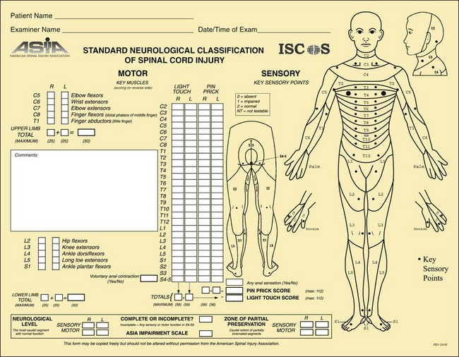
Diagnosis is based on the paralytic manifestations and on EMG. Motor nerve conduction velocities are greatly reduced. Sensory nerve conduction time is often slowed. Cerebrospinal fluid analysis reveals an elevated protein concentration, and normal glucose level, but other laboratory studies are noncontributory. The symmetric nature of the paralysis helps differentiate this disorder from spinal paralytic poliomyelitis, which usually affects sporadic muscles.
Treatment of GBS is primarily supportive. In the acute phase, patients are hospitalized because respiratory and pharyngeal involvement may require assisted ventilation, sometimes with a temporary tracheotomy. Treatment modalities include aggressive ventilatory support, intravenous (IV) administration of immunoglobulin (IVIG), and steroids; plasmapheresis and immunosuppressive drugs may also be used. Plasmapheresis has been shown to decrease the length of recovery in patients with severe GBS yet is expensive, and side effects include hypotension, fever, bleeding disorders, chills, urticaria, and bradycardia. Further evidence reports equal benefits to treatment of GBS with IVIG administration or plasmapheresis; both sped up recovery time in studies reviewed (Hughes and Cornblath, 2005). There is evidence of significant improvement in children with IVIG therapy (versus supportive treatment alone), and IVIG therapy is more cost-effective than plasmapheresis (Harel and Schoenfeld, 2005; Hughes, Raphael, Swan, et al, 2006; Tsai, Wang, Liu, et al, 2007). IVIG is now recommended as the primary treatment of GBS when administered within 2 weeks of diseases onset (Hughes, 2008). Corticosteroids alone do not decrease the symptoms or shorten the duration of the disease.
Medications that may be administered during the acute phase include a low-molecular-weight heparin to prevent deep vein thrombosis (DVT), a mild laxative or stool softener to prevent constipation, pain medication such as acetaminophen, and a histamine-antagonist to prevent stress ulcer formation. Chronic neuropathic pain following GBS may be treated with gabapentin, which is reported to be more effective than carbamazepine (Sarnat, 2007).
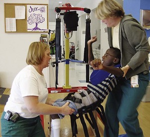
Rehabilitation after the acute phase may involve physical therapy, occupational therapy, and speech therapy. Additional consideration should be given to problems of general weakness and retraining for toileting and feeding (Lyons, 2008).
Prognosis: Recovery usually begins within 2 to 3 weeks, and most patients regain full muscle strength. The recovery of muscle strength progresses in the reverse order of onset of paralysis, with lower extremity strength being the last to recover. There are few long-term outcome studies in children, but in one study 23% of the children had a residual weakness in at least one muscle group (Vajsar, Fehlings, and Stephens, 2003). Poor prognosis with subsequent residual effects in children is reportedly associated with cranial nerve involvement, extensive disability at time of presentation, and intubation (Sarnat, 2007).
The rate of recovery is usually related to the degree of involvement, which may extend from a few weeks to months. The greater the degree of paralysis, the longer the recovery phase.
Nursing care is essentially supportive and is the same as that required for the child with immobilization and respiratory compromise. The emphasis of care is on close observation to assess the extent of paralysis and on prevention of complications, including aspiration, atelectasis, DVT, pressure ulcer, fear and anxiety, autonomic dysfunction, and pain.
During the acute phase of the disease the nurse should carefully observe the child’s condition for possible difficulty in swallowing and respiratory involvement. Closely monitor the child’s respiratory function, and keep the oxygen source, appropriate-sized insufflation bag and mask, endotracheal intubation and suctioning equipment, tracheotomy tray, and vasoconstrictor drugs available. Monitor vital signs frequently, including neurologic signs and level of consciousness. For the child who develops respiratory impairment, the care is the same as that for any child with respiratory distress requiring mechanical ventilation.
Respiratory care, should intubation be required, requires close monitoring of oxygenation status (usually by pulse oximetry and sometimes arterial blood gases), maintenance of an open airway with suctioning, and postural changes to prevent pneumonia. Children with oral and pharyngeal involvement may be fed via a nasogastric or gastrostomy tube to ensure adequate feeding. Immobilization, which occurs with GBS, decreases gastrointestinal function; therefore attention to problems such as decreased gastric emptying, constipation, and feeding residuals require nursing assessment and appropriate collaborative interventions. Temporary urinary catheterization may be required; urinary retention is not uncommon, and appropriate assessment of urinary output is vital. Sensory impairment and paralysis in the lower extremities make the child susceptible to skin breakdown; therefore attention should be given to meticulous skin care. Passive range-of-motion exercises and application of orthoses to prevent muscle contractures are important when paralysis is present. Prevention of DVT is accomplished with pneumatic compression (antiembolism) devices, administration of a low-molecular-weight heparin, and early mobilization and ambulation. Autonomic dysfunction may be life threatening; thus close monitoring of vital signs in the acute phase is essential.
A key to recovery in the child with GBS is the prevention of muscle and joint contractures, so passive range-of-motion exercises must be carried out routinely to maintain vital function. Although the child may have a generalized paralysis, cognitive function remains intact; therefore it is important for nursing care to involve communication with the child regarding procedures and treatments that may be frightening, especially if mechanical ventilation is required. Encourage parents to talk to the child and make eye and physical contact as much as possible to reassure the child during the illness.
Pain management is crucial in the care of children with GBS. Although neuromuscular impairment may make pain perception more difficult to accurately evaluate, use objective pain scales. Carbamazepine and gabapentin may be used to manage neuropathic pain in patients with GBS.
Physical therapy may be limited to passive range-of-motion exercises during the evolving phase of the disease. Later, as the disease stabilizes and recovery begins, an active physical therapy program is implemented to prevent contracture deformities and facilitate muscle recovery. This may include active exercise, gait training, and bracing.
Throughout the course of the illness, child and parent support is paramount. The usual rapidity of the paralysis and the long recovery period greatly tax the emotional reserves of all family members. The parents and child benefit from repeated reassurance that recovery is occurring and from realistic information regarding the possibility of permanent disability. In the event of a residual disability, the family needs assistance in accepting and adjusting to the loss of function. (See Chapter 22.) The GBS/CIDP Foundation International* is a nonprofit organization devoted to support, education, and research. It provides families with support from recovered persons, publishes informational literature and a newsletter, and maintains a list of practitioners experienced with the disease.
Tetanus, or lockjaw, is an acute, preventable, but sometimes fatal disease caused by an exotoxin produced by the anaerobic, spore-forming, gram-positive bacillus Clostridium tetani. It is characterized by painful muscular rigidity primarily involving the masseter and neck muscles. The development of tetanus has four requirements: (1) presence of tetanus spores or vegetative forms of the bacillus, (2) injury to the tissues, (3) wound conditions that encourage multiplication of the organism, and (4) a susceptible host.
Tetanus spores are found in soil, dust, and the intestinal tracts of humans and animals, especially herbivorous animals. The organisms are more prevalent in rural areas but are readily carried to urban areas by wind. They enter the body by way of wounds, particularly a puncture wound, burn, or crushed area. In the newborn, infection may occur through the umbilical cord, usually in situations in which infants are delivered in contaminated surroundings and the mother has not been properly immunized against tetanus. The disease has the greatest incidence during months in which persons are more involved in outdoor activities.
Primary prevention is key and occurs through immunization and boosters (American Academy of Pediatrics, 2009). Once an injury has occurred, further preventive measures are based on the child’s immune status and the nature of the injury. Specific prophylactic therapy after trauma is administration of either tetanus toxoid or tetanus antitoxin. A dose of tetanus toxoid is not necessary for clean, minor wounds in children who have completed the immunization series (see Chapter 12) or who have received a booster within the previous 10 years. Protective levels of antibody are maintained for at least 10 years. Therefore antitoxin is not indicated for the fully immunized child. Children with more serious wounds (e.g., contaminated, puncture, crush, or burn wounds) are given a tetanus toxoid booster prophylactically as soon as possible after injury.
The unprotected or inadequately immunized child who sustains a “tetanus-prone” wound (including wounds contaminated with dirt, feces, soil, and saliva; puncture wounds; avulsions; and wounds resulting from missiles, crushing, burns, and frostbite) should receive tetanus immunoglobulin (TIG). Concurrent administration of both TIG and tetanus toxoid at separate sites is recommended both to provide protection and to initiate the active immune process (American Academy of Pediatrics, 2009). Completion of active immunization is carried out according to the usual pattern. Proper surgical cleansing and débridement of contaminated wounds reduce the chance of infection.
When prevention efforts are not effective and conditions are favorable, the organisms multiply and form two exotoxins: (1) tetanospasmin, a potent toxin that affects the CNS to produce the clinical manifestations of the disease; and (2) tetanolysin, which appears to have no significance. The ideal conditions for growth of the organisms are devitalized tissues without access to air (e.g., puncture wounds); wounds that have not been washed or kept clean; and those that have crusted over, trapping pus beneath. The exotoxin appears to reach the CNS by way of either the neuron axons or the vascular system. The toxin becomes fixed on nerve cells of the brainstem and the anterior horn of the spinal cord. The toxin acts at the neuromuscular junction to produce muscular stiffness and to lower the threshold for reflex excitability.
The incubation period is 3 days to 3 weeks and averages 8 days. Most cases occur within 14 days; in neonates it is usually 5 to 14 days. Shorter incubation periods have been associated with more heavily contaminated wounds, more severe disease, and a worse prognosis (American Academy of Pediatrics, 2009).
There are several forms of the disease. Local tetanus is a less common but severe form characterized by persistent rigidity of muscles near the inoculation site, which may persist for weeks or months. Some cases resolve without sequelae. Neonatal tetanus results from contamination of the umbilical cord, which is rare in the United States but is common and often fatal in developing countries. The first symptom is difficulty in sucking, progressing to total inability to suck, excessive crying, irritability, and nuchal rigidity.
Generalized tetanus is the most common and dangerous form of the disease. The manner of onset varies, but the initial symptoms are usually a progressive stiffness and tenderness of the muscles in the neck and jaw. The characteristic difficulty in opening the mouth (trismus), which is caused by sustained contraction of the jaw-closing muscles, is evident early and gives the disease its common name, lockjaw. Spasm of facial muscles produces the so-called sardonic smile (risus sardonicus). Progressive involvement of the trunk muscles causes opisthotonos and a boardlike rigidity of abdominal and limb muscles. The patient has difficulty swallowing and is highly sensitive to external stimuli. The slightest noise, a gentle touch, or bright light triggers convulsive muscular contractions that last seconds to minutes. The paroxysmal contractions recur with increased frequency until they become almost continuous.
Mentation is unaffected; the patient remains alert, and pain and distress are reflected in a rapid pulse, sweating, and an anxious expression. Laryngospasm and tetany of respiratory muscles and accumulated secretions predispose the child to respiratory arrest, atelectasis, and pneumonia. Fever is usually absent or mild and generally indicates a poor prognosis. As the child recovers from the disease, the paroxysms become less frequent and gradually subside. Survival beyond 4 days usually indicates recovery, but complete recovery may take weeks.
The unprotected or inadequately immunized child who sustains a “tetanus-prone” wound (as described above) should receive TIG. Concurrent administration of both TIG and tetanus toxoid at separate sites is recommended both to provide protection and to initiate the active immune process. Completion of active immunization is carried out according to the usual pattern (American Academy of Pediatrics, 2009). Antibiotic treatment with penicillin G (or erythromycin or tetracycline in older children with allergy to penicillin) is important in the management of tetanus as an adjunct against clostridia (Arnon, 2007).
Aggressive supportive care is necessary to treat tetanus in the acute phase. The acutely ill child is best treated in an intensive care facility, where close and constant observation and equipment for monitoring and respiratory support are readily available.
General supportive care is indicated, including maintaining adequate airway and fluid and electrolyte balance, providing pain management, and ensuring adequate caloric intake. Indwelling oral or nasogastric feedings may be required to maintain adequate fluid and caloric intake; continued laryngospasm may necessitate total parenteral nutrition or gastrostomy feeding. Severe or recurrent laryngospasm or excessive secretions may require advanced airway management such as endotracheal intubation; in some cases a tracheotomy may be performed to provide an adequate airway.
TIG therapy to neutralize toxins is the most specific therapy for tetanus. In countries where TIG is not available, equine tetanus antitoxin (not available in the United States) should be administered. Antibiotics are administered to control the proliferation of the vegetative forms of the organism at the site of infection. When the child recovers, active immunization should take place, since contraction of the disease does not confer a permanent immunity. Standard Precautions for the child with tetanus are recommended; isolation is not recommended.
Local care of the wound by surgical débridement and cleansing with an antiseptic solution helps reduce the number of proliferating organisms at the site of injury. The cleansing should be repeated several times during the first 48 hours. Deep, infected lacerations are usually exposed and débrided.
Diazepam is the drug of choice for seizure control and muscle relaxation (Arnon, 2007), but lorazepam (Ativan) may be used in some cases. Other AEDs may be administered as well. Intrathecal baclofen, magnesium sulfate, dantrolene sodium, and midazolam may also be used in the management of tetanus; intrathecal baclofen may cause apnea and should only be used in the intensive care setting (Arnon, 2007). Patients with severe tetanus and those who do not respond to other muscle relaxants may require the administration of a neuromuscular blocking agent, such as rocuronium or vecuronium. Because of their paralytic effect on respiratory muscles, use of these drugs requires mechanical ventilation with endotracheal intubation or tracheotomy and constant cardiopulmonary monitoring. Endotracheal tube insertion or tracheotomy is often indicated and should be performed before severe respiratory distress develops. Despite the absence of pain manifestation with these drugs, it is important to administer adequate analgesia.
The administration of corticosteroids has met with success in some cases.
The care of the child with tetanus requires supportive management with particular attention to airway and breathing. Carefully evaluate respiratory status for any signs of distress, and keep appropriate emergency equipment available at all times. The location and extent of muscle spasms and the assessment of their severity are important nursing observations. Muscle relaxants, opioids, and sedatives that may be prescribed can also cause respiratory depression; therefore assess the child for excessive CNS depression, apnea, and respiratory failure. At times it may be necessary to completely paralyze the child with a muscle relaxant because of the intensity of the muscle spasms. Attention to hydration and nutrition involves monitoring an IV infusion, monitoring nasogastric or gastrostomy feedings, providing oral hygiene, and suctioning oropharyngeal secretions when indicated.
In caring for the child with tetanus, make every effort to control or eliminate stimulation from sound, light, and touch. A quiet environment is desirable to reduce the amount of stimuli on the CNS. Although a darkened room is ideal, sufficient light is essential so the child can be carefully observed. Light appears to be less irritating than vibratory or auditory stimuli.
If a potent muscle relaxant such as rocuronium or vecuronium is used, total paralysis (including respirations) makes oral communication impossible. The drug is not a sedative, however, and anxiolysis should be considered in children who are intubated. Fentanyl and midazolam may be used to manage pain and anxiety in such children. The nurse must anticipate all of the child’s needs and carefully explain the procedures beforehand to the child and family.
Additional care is focused on preventing the complications associated with prolonged immobility: decreased bowel and bladder tone and subsequent constipation, anorexia, DVT, pneumonia, and skin breakdown.
Because their mental status is often clear, intubated children are aware of what is happening to them and are often extremely anxious. Parents should stay with the child to offer security and support. They also need support, information, and reassurance from the nurse.
Botulism is serious food poisoning that results from ingestion of the preformed toxin produced by the anaerobic bacillus Clostridium botulinum. Botulism toxin exerts its effect by inhibiting the release of acetylcholine at the neuromuscular junction, thereby impairing motor activity of the muscles innervated by the affected nerves. The disease has a wide variation in severity, from constipation to progressive sequential loss of neurologic function and respiratory failure. Human botulism is caused by neurotoxins A, B, E, and rarely F (American Academy of Pediatrics, 2009). Types A and B are the most common causes of infant botulism.
Several forms of botulism are recognized: food borne, infant, wound, man made (for bioterrorism), and botulism from undetermined causes. This chapter only covers the first three forms.
Food-Borne Botulism: This classic form of the disease usually occurs in adults but may occur in children and adolescents. The most common source of the toxin is improperly sterilized home-canned foods (see Community Focus box). CNS symptoms appear abruptly approximately 12 to 36 hours after ingestion of contaminated food and may or may not be preceded by acute digestive disturbance. Early symptoms include blurred vision, diplopia, weakness, dizziness, difficulty talking and speaking, vomiting, and dysphagia. These are followed by descending paralysis and dyspnea. Progressive respiratory paralysis is life threatening.
Infant Botulism: Infant botulism, unlike the disease in older persons, is caused by ingestion of spores or vegetative cells of C. botulinum and the subsequent release of the toxin from organisms colonizing the gastrointestinal tract. C. botulinum types A and B are the most common causative strains of infant botulism. This form of botulism has become more prevalent than any other form. Many cases of infant botulism occur in breast-fed infants who are being introduced to nonhuman milk substances (American Academy of Pediatrics, 2009). There appears to be no common food or drug source of the organisms; however, the C. botulinum organisms have been found in honey. Botulism may occur in infants from 1 week to 12 months of age, with peak incidence between 2 and 4 months of age.
The severity of the disease varies widely, from mild constipation to progressive sequential loss of neurologic function and respiratory failure. The affected infant is usually well before the onset of symptoms. Constipation is a common presenting symptom, and almost all infants exhibit generalized weakness and a decrease in spontaneous movements. Deep tendon reflexes are usually diminished or absent. Cranial nerve deficits are common, as evidenced by loss of head control, difficulty in feeding, weak cry, and reduced gag reflex. SMA type 1 and metabolic disorders are often mistaken for infant botulism in the initial diagnostic phase because of the similarities in clinical manifestations of hypotonia, lethargy, and poor feeding (Francisco and Arnon, 2007). Presenting clinical signs also often mimic those of sepsis in young infants. Botulism toxin exerts its effect by inhibiting the release of acetylcholine at the myoneural junction, thereby impairing motor activity of muscles innervated by affected nerves.
Wound Botulism: Wounds contaminated with C. botulinum and subsequent elaboration of the toxin produce classic symptoms approximately 4 to 14 days after tissue trauma. The disease has been described in a small number of adolescents and adults, and most wounds are sustained in open fields or on farms.
Diagnosis is made on the basis of the clinical history, physical examination, and laboratory detection of the organism in the patient’s stool and, less commonly, blood. However, isolation of the organism may take several days; therefore suspicion of botulism by clinical presentation should require emergent treatment (Arnon, 2007). EMG may be helpful in establishing the diagnosis; however, results may be normal early in the course of the illness.
Treatment consists of immediate administration of botulism immune globulin intravenously (BIG-IV [BabyBIG]) (Francisco and Arnon, 2007), without waiting for laboratory diagnosis. Early administration of BIG-IV neutralizes the toxin and stops the progression of the disease. The human-derived botulism antitoxin (BIG-IV) has been evaluated and is now available nationwide for use only in infant botulism. In one study infants treated with BIG-IV experienced a mean length of hospitalization decrease from 5.7 to 2.6 weeks and also had decreased time spent in intensive care, decreased mean duration of mechanical ventilation, and decreased mean duration of tube or IV feeding. In addition, the infants did not experience any adverse events related to BIG-IV. Most infants received BIG-IV treatment within 3 to 18 days of hospitalization for botulism (Arnon, Schechter, Maslanka, et al, 2006). Studies indicate that treatment with BIG-IV decreased length of hospital stay in affected infants an average of 17 days; infants with botulism requiring mechanical ventilation also had shorter hospitalizations when treated with BIG-IV (Thompson, Filloux, Van Orman, et al, 2005). Potential complications of BIG-IV include hypotension and anaphylaxis (Cirillo, 2008).
Approximately 50% of affected infants require intubation and mechanical ventilation; therefore respiratory support is crucial, as is nutritional support, since the infant is unable to feed. Trivalent equine botulinum antitoxin and bivalent antitoxin, used in adults and older children, is not administered to infants. Antibiotic therapy is not part of the management because the botulinum toxin is an intracellular molecule and antibiotics would not be effective; aminoglycosides in particular should not be administered because they may potentiate the blocking effects of the neurotoxin (Arnon, 2007).
The prognosis is generally good if the patient is adequately treated, although recovery may be slow, requiring a few weeks after severe illness. The average length of stay for infant botulism is 44 days, and the fatality rate is reported to be less than 2%. Untreated patients may require a longer hospitalization.
Nursing responsibilities include observing, recognizing, and reporting signs of poor feeding, constipation, and muscle impairment in the infant with botulism and providing intensive nursing care when an infant is hospitalized. (See Nursing Care Management for the infant with SMA, p. 1704, and Nursing Care of High-Risk Newborns, Chapter 10.) Parental support and reassurance are important. Most infants recover when the disorder is recognized and BIG-IV therapy is implemented. Nursing care of the infant on mechanical ventilation requires observation of oxygenation status and vigilance for any complications. Parents should be aware that, during recovery, infants fatigue easily when muscular action is sustained. This has important implications for timing the resumption of feedings because of the risk of aspiration. They should also be advised that normal bowel activity may not return for several weeks. Therefore a stool softener can be beneficial.
Myasthenia gravis (MG) is relatively uncommon in childhood. The incidence in children under 18 years is 1 per 1 million in North America (Cirillo, 2008). Juvenile MG appears to be identical to that seen in adults and usually has its onset after age 10 years, but it may appear as early as age 2 years. Girls are affected three times more than boys. Juvenile and adult forms of the disease are autoimmune disorders associated with the attack of circulating antibodies on the acetylcholine receptors on the muscle end plate, which blocks their function.
The most common symptoms are general paralysis of the optic muscles with ptosis and diplopia. Difficulty swallowing, chewing, and speaking are also prominent and are accompanied by weakness and paralysis of all skeletal muscles. The signs and symptoms are more pronounced in the late afternoon and evening. Rest can help relieve the symptoms, but exercise and stress worsen them.
The diagnosis is made on the basis of the characteristic distribution of muscle weakness and the progressive weakness on repeated or sustained muscular contraction. The definitive diagnosis is established on the basis of an EMG, which demonstrates a decrease in muscle potentials with repetitive nerve stimulation (Sarnat, 2007). A clinical diagnosis may be established by observation of the response to the anticholinesterase drugs. IV administration of a small test dose of edrophonium (Tensilon) produces a beneficial effect in 1 minute, but the effect lasts less than 5 minutes. Electrophysiologic studies are helpful in diagnosis and help document transmission failure at the neuromuscular junction. Antibodies to human muscle acetylcholine are detected in the serum of almost one third of affected individuals.
Treatment consists of the administration of cholinesterase-inhibiting drugs, such as neostigmine (Prostigmin), given intramuscularly or as oral neostigmine bromide. Pyridostigmine (Mestinon) may be administered because it is considered less toxic, but a higher dose is required to achieve the same results as neostigmine. The starting dosage of neostigmine is usually 0.04 mg/kg, administered intramuscularly every 4 to 6 hours; oral doses may be well tolerated (Sarnat, 2007). The dosage is gradually increased until a satisfactory result is obtained. The child must be observed for signs of parasympathetic stimulation from overmedication. These signs include lacrimation, salivation, abdominal cramps, sweating, diarrhea, vomiting, bradycardia, and weakness of respiratory muscles.
Other therapies directed at the immunologic mechanism include thymectomy (removal of the thymus), IVIG, long-term corticosteroid treatment, and plasmapheresis. Thymectomy may be curative for some individuals, but is not effective for familial and congenital forms of MG.
The prognosis for juvenile MG is relatively good. However, the course of the disease is marked by fluctuating remissions and exacerbations.
Children with MG need ongoing medical and nursing supervision. Teach the parents the importance of accurate administration of medications, with special emphasis on recognizing side effects, including the dangers of choking, aspiration, and respiratory distress.
Counsel parents to promote a lifestyle that minimizes stress and maximizes relaxation. Discourage strenuous activity. Also warn them of the possibility of a sudden exacerbation of symptoms during times of physical or emotional stress (myasthenia crisis), which requires immediate medical attention. They should receive instruction in providing respiratory assistance until help arrives or the child can be transported to medical aid.
A transient form of MG occurs in approximately 10% to 20% of infants born to mothers with MG, who may not know they have the disease. The muscular weakness results from transplacentally acquired maternal acetylcholine receptor antibodies. These infants display generalized muscular weakness and hypotonia at birth with a depressed Moro reflex, ptosis, ineffective sucking and swallowing reflexes, and weak cry. Some infants may require short-term mechanical ventilation (Sarnat, 2007). Symptoms may be evident within a few hours of birth, following a period of normal appearance after delivery. There is no evidence of neurologic damage. Cholinesterase inhibitors may be given on a short-term basis to improve feeding ability. Symptoms usually disappear within 2 to 3 weeks. Infants with transient neonatal MG regain strength once maternal antibodies clear the system, and they are not at increased risk of MG later in life (Cirillo, 2008; Sarnat, 2007).
Congenital MG is a rare familial abnormality of neuromuscular transmission that is not immunologically mediated. It appears indistinguishable from the transient form, but the mother usually does not have the disease. The disease persists throughout life, and more than one sibling may be affected, which suggests a genetic etiology. Gender distribution is equal. The disorder is relatively resistant to drug therapy, and the eyelid and extraocular muscles seem to be the muscles most severely affected.
The prognosis in congenital MG is usually good. Despite gradual worsening of symptoms with age, the life span is not affected significantly.
SCIs with major neurologic involvement are not a common cause of physical disability in children. However, many children with these injuries are admitted to major medical centers, and because of the increased survival rate as a result of improved management, nurses have an important role in the care of children with SCI.
The principles of management and nursing care of the child with a spinal cord lesion apply regardless of cause. In addition to care related to the immobilized child, as discussed in Chapter 39, children with damage to the spinal cord present additional problems—specifically, complications related to the neuropathology of the central and autonomic nervous systems. The extent of paralysis is determined by both neurologic and clinical assessment. Although the majority of children with SCI are paraplegic, some are tetraplegic (quadriplegic). Some children with tetraplegia are able to move only their face and neck muscles, whereas others are able to lift and bend their arms but are unable to perform fine hand movements. Almost every physiologic system is disrupted in a child with high-level tetraplegia. Not only are the central and peripheral nerves impaired, but there is also autonomic nervous system dysfunction. Vital structures such as blood vessels, lungs, bladder, and bowel are affected. Therefore an understanding of neuromuscular physiology is essential to effectively care for the child with damage or injury to the spinal cord.
More males than females experience SCI as children. Cirak, Ziegfeld, Knight, and colleagues (2004) found that the mean age of children with SCI was 9.48 years. Motor vehicle crashes (MVCs) accounted for the majority of infants injured, whereas toddlers and school-aged children up to 9 years were more likely to suffer SCI as a result of a fall. Almost one half (46%) of the pediatric injuries in this study were high cervical injuries. In the United States football injuries accounted for a high percentage of sport-related injuries in adolescents, whereas in Canada such injuries were associated with ice hockey (Mathison, Kadom, and Krug, 2008).
The spinal cord extends from the medulla oblongata to the lower border of the first lumbar vertebra and contains millions of nerve fibers. However, because of its protected location, a considerable amount of direct trauma is required to cause injury. Posteriorly the cord is protected by the spinous processes, which are stabilized by related ligaments and muscles. It is further protected by the spinal fluid, which surrounds it and absorbs some of the shock.
Spinal Nerves: The 31 nerves of the spinal cord are divided into five segments (Fig. 40-5). The cervical cord segments lie within the first seven vertebrae. The remaining cord segments—thoracic (12), lumbar (5), sacral (5), and coccygeal (1)—extend from the first thoracic vertebra to the lower level of the first lumbar vertebra. Therefore the cord constituents do not anatomically match by number the 33 associated vertebrae. However, nerves that arise from the spinal cord exit from the spinal column at the numerically corresponding vertebrae. In describing injuries to the spinal cord, the highest point at which there is normal function is referred to in relation to the vertebra; for example, an intact cord at the sixth cervical vertebra is designated a C6 injury.
Fig. 40-5 Relationships of spinal cord segments and spinal nerves to vertebral bodies. Cervical nerves exit through intervertebral foramina above their respective vertebral bodies (seven cervical vertebrae and eight cervical nerves). Spinal cord ends at L1-L2 vertebral level.
Certain areas of the curved vertebral column are less stable and more prone to damage from severe flexion and twisting. These sites are the cervical area and the junction of the thoracic and lumbar regions. The cervical vertebrae are fractured most often, and this high level of injury causes extensive paralysis and many associated neurologic problems (Table 40-1). Also, traumatic tearing or embolic occlusion of the arteries supplying these areas can markedly jeopardize the cord tissue. Impaired blood supply often produces severe neurologic deficit, which can extend to complete loss of cord function at the level of injury.
Cell bodies of interneurons and motor neurons within the spinal cord are identified as H-shaped gray matter surrounded by columns of white myelinated nerve fibers. Each column serves as a route for a specific type of impulse, such as touch, vibration, pain, and temperature (Fig. 40-6). Nerve pathways in the spinal cord transmit sensory and motor impulses between peripheral receptors and the brain, conduct impulses through the reflex arc, and convey sympathetic and parasympathetic nerve impulses from the brain to peripheral structures.
Fig. 40-6 Main motor and sensory pathways. Perception of touch, passive motion, position, and vibration is transmitted through posterior tract in spinal cord through medial lemniscus in brainstem to thalamus and through internal capsule to cortex (pathway is represented by solid red line). Pain and temperature sensations are transmitted through anterolateral tract and lateral lemniscus to thalamus, then through internal capsule to cortex (blue line). Motor impulses are transmitted by pyramidal tract, descending from cerebral cortex, crossing in medulla to opposite side, and continuing to anterior horns of spinal cord (black line). (From Conway BL: Carini and Owens’ neurological and neurosurgical nursing, ed 7, St Louis, 1978, Mosby.)
Sensory transmission begins when peripheral receptors pick up a wide variety of stimuli and transfer the impulses, by means of peripheral nerves, to the spinal nerves, where they make ganglionic connections and enter the cord posteriorly. At this point the impulses travel in two directions: (1) across the interneuron connection and then to the motor neurons (reflex arc), or (2) up the spinal cord to predetermined areas of the brain. Motor impulses are transmitted from the cerebral cortex to the medulla (where nerve tracts cross) and proceed down descending motor pathways to the desired level within the spinal cord. Here they connect with the anterior horn cells and are transmitted to the muscle fibers by means of the lower motor neurons to complete a meaningful movement.
A network of nerves that serves the major muscle groups constitutes a plexus. Total involvement of any one of these plexuses seriously impairs function to the areas it innervates. Box 40-7 describes the three major plexuses.
Upper Versus Lower Motor Neurons: Upper motor neurons extend from cerebral centers to cells in the spinal column; lower motor neurons consist of anterior horn cells and spinal and peripheral nerves. Motor fibers of the reflex arc are lower motor neurons. This is an important point because relative dominance of the CNS over reflex arcs suppresses some reflex responses. When the higher centers no longer exert an influence in SCI, spastic responses are observed in muscles innervated by the intact lower motor neurons. Most SCIs involve upper motor neurons; children born with spinal cord defects have primarily lower motor neuron deficits (see Fig. 40-1). Box 40-8 outlines manifestations of upper and lower motor neuron syndromes.
Effect on Sensory and Motor Tracts: Voluntary muscle control is lost after complete transection of the cord. In partial transection, function is altered to varying degrees depending on the areas innervated by involved nerves. The crossing of motor tracts at various levels makes it possible for an injured person to have motor paralysis in one leg but retain pain and temperature sensation in that leg, while the opposite leg retains its motor function but loses pain and temperature sensation.
Although a transected cord injury leads to sensory loss, it is not uncommon for the injured person to experience pain. For example, smooth or skeletal muscle spasms, destruction of the myelin sheath (impulses cross to adjacent nerves), and scar formation or irritation of nerve endings may cause pain. Pain suffered by a person with tetraplegia or paraplegia is often intensified because of loss of sensation in other parts. Severe and prolonged pain should be medically evaluated for a treatable pathologic condition.
Effect on Autonomic System: Sympathetic and parasympathetic systems receive both excitatory and inhibitory stimuli from autonomic centers in the cerebral cortex, limbic system, and hypothalamus. The stimuli are transmitted by means of a feedback mechanism within the ascending fibers of the cord that normally controls descending input. Axons of the many CNS neurons synapse with autonomic preganglionic fibers and thus are able to alter their patterned responses. Box 40-9 describes the most significant effects of autonomic disruption.
The most common cause of serious spinal cord damage in children is trauma involving MVCs (including automobile-bicycle, all-terrain vehicles, and snowmobiles), sports injuries (especially from diving, trampoline activities, gymnastics, and football), birth trauma, and child abuse. The increased use of recreational activities involving motorized vehicles such as jet water skis and motorcycles has increased the incidence of SCIs in children.
Congenital defects of the spine such as myelomeningocele (see Chapter 11) also may, in some cases, produce the effects of SCI. Transverse myelitis (inflammation of the spinal cord) has been reported to develop from inadvertent intraarterial administration of long-acting penicillin injected into the buttocks. Damage can be extensive enough to result in paraplegia or even lower limb amputation.
In MVCs most SCIs in children are a result of indirect trauma caused by sudden hyperflexion or hyperextension of the neck, often combined with a rotational force. Trauma to the spinal cord without evidence of vertebral fracture or dislocation (SCI without radiographic abnormality, or SCIWORA) is particularly likely to occur in an MVC when proper safety restraints are not used. An unrestrained child becomes a projectile during sudden deceleration and is subject to injury from contact with a variety of objects inside and outside the vehicle. Individuals who use only a lap seat belt restraint are at greater risk of SCI than those who use a combination lap and shoulder restraint. High cervical spine injuries have been reported in children less than 2 years of age who are restrained in forward-facing car seats. Infants who are improperly restrained in an infant car seat may experience cervical trauma in a car crash. Small children may also be severely injured by front seat air bags. (See Chapter 12.)
Falling from heights occurs less often in children than in adults, but vertebral compression of the spine from blows to the head or buttocks occurs in water sports (diving and surfing) or falls from horses or other athletic injuries. Birth injuries may occur in breech delivery from excessive traction force and rotation on the cord during delivery of the head and shoulders. When shaken, infants commonly sustain cervical cord damage, as well as subdural hematoma and retinal hemorrhage; cognitive impairment and death may occur subsequent to the traumatic event. Infants have weak neck muscles, and during vigorous shaking their large and heavy heads rapidly wobble back and forth. An increasing number of adolescents receive SCIs secondary to gunshot wounds, stabbings, or other violent inflicted injury.
Because of the marked mobility of the neck, fracture or subluxation (partial dislocation) is the most common immediate cause of SCI, particularly in the lower cervical region. Although unusual in adults, SCI without fracture is not uncommon in the child, whose spine is suppler, weaker, and more mobile than that of the adult. Therefore the force is more easily dissipated over a larger number of segments. Upper cervical injuries account for as many as 80% of the SCIs in children under the age of 2 years (Haslam, 2007). In children the vertebral column is composed of cartilaginous rings and is capable of considerable elongation, whereas the cord itself, its meninges, and its vascular supply are unable to withstand the same degree of traction.
The injury sustained can affect any of the spinal nerves; the higher the injury, the more extensive the damage. The child can be left with complete or partial paralysis of the lower extremities (paraplegia) or with damage at a higher level and without functional use of any of the four extremities (tetraplegia). A high cervical cord injury that affects the phrenic nerve paralyzes the diaphragm and leaves the child dependent on mechanical ventilation.
A mild but equally frightening form of cord trauma is spinal cord compression, a temporary neural dysfunction without visible damage to the cord. Complete tetraplegia can result but initially may not be differentiated from serious cord injury.
The severity of the force, the mechanisms of the injury, and the degree of the individual’s muscular relaxation at the time of the injury greatly influence the extent of the trauma. SCIs are classified as either complete or incomplete. In a complete injury there is no motor or sensory function more than three segments below the neurologic level of the injury (Mathison, Kadom, and Krug, 2008). Incomplete lesions have several typical characteristics (Mathison, Kadom, and Krug, 2008):
Central cord syndrome—Central gray matter destruction and preservation of peripheral tracts; tetraplegia with sacral sparing common; some motor recovery gained
Anterior cord syndrome—Complete motor and sensory loss with trunk and lower extremity proprioception and sensation of pressure
Posterior cord syndrome—Loss of sensation, pain, and proprioception with normal cord function, including motor function; able to move extremities but have difficulty controlling such movements
Brown-Séquard syndrome—Unilateral cord lesion with a motor deficit on the opposite side of the body from the primary insult; absence of pain and temperature sensation on the opposite side from the injury
Spinal cord concussion—Transient loss of neural function below the level of the acute spinal cord lesion, resulting in flaccid paralysis and loss of tendon, autonomic, and cutaneous reflex activity; may last hours to weeks
The American Spinal Injury Association (2009) Standards for neurologic classification of SCI worksheet is available online at www.asia-spinalinjury.org/publications/2006_Classif_worksheet.pdf. The ASIA Impairment Scale (Box 40-10) combines motor and sensory function and is used to determine the severity of impairment from the injury (complete or incomplete). It may also be used to measure neurologic changes and functional goals for rehabilitation (Mathison, Kadom, and Krug, 2008).
It is often difficult to determine the extent and severity of damage at first. Immediate loss of function is caused by both anatomic and impaired physiologic function, and improved function may not be evident for weeks or even months. Manifestation of the initial response to acute SCI is flaccid paralysis below the level of the damage. This stage is often referred to as spinal shock syndrome and is caused by the sudden disruption of central and autonomic pathways. Local effects of cord edema and ischemia produce a physiologic transection with or without an anatomic severance. Most children with an SCI experience some spinal shock. Manifestations include the absence of reflexes at or below the cord lesion, with flaccidity or limpness of the involved muscles, loss of sensation and motor function, and autonomic dysfunction (symptoms of hypotension, low or high body temperature, loss of bladder and bowel control, and autonomic dysreflexia).
Autonomic paralysis also affects thermoregulatory functions. Afferent impulses from temperature receptors in the skin are not integrated; therefore the patient is subject to temperature increases or decreases in response to alterations in environmental temperature. Hyperthermia can result from excessive ambient temperature, such as too many covers.
Except in the situations previously mentioned, flaccid paralysis is replaced by spinal reflex activity and increasing spasticity or, in incomplete lesions, greater or lesser degree of neurologic recovery.
The paralytic nature of autonomic function is replaced by autonomic dysreflexia, especially when the lesions are above the midthoracic level. This autonomic phenomenon is caused by visceral distention or irritation, particularly of the bowel or bladder. Sensory impulses are triggered and travel to the cord lesion, where they are blocked, which causes activation of sympathetic reflex action with disturbed central inhibitory control. Excessive sympathetic activity is manifested by a flushing face, sweating forehead, pupillary constriction, marked hypertension, headache, and bradycardia. The precipitating stimulus may be merely a full bladder or rectum or other internal or external sensory input. It can be a catastrophic event unless the irritation is relieved.
Additional clinical findings of SCI may include numbness, tingling, or burning; priapism; weakness; and loss of bowel and bladder control (Hayes and Arriola, 2005).
Neurogenic shock occurs as a result of a disruption in the descending sympathetic pathways with loss of vasomotor tone and sympathetic innervations to the cardiovascular system (Hayes and Arriola, 2005). Hypotension, bradycardia, and peripheral vasodilation occur as a result of neurogenic shock.
Children with suspected SCI may have suffered multiple injuries (e.g., MVC); therefore multiple clinical manifestations may occur that may mask those of an SCI.
In the final stage neurologic signs are stabilized in terms of loss and recovery of function. The major emphasis is on rehabilitation. A problem unique to injury in childhood is progressive spinal deformity usually not seen in adults or in adolescents near the end of the growth period. Scoliosis develops in the majority of children with high thoracic and cervical lesions and is almost certain to occur in children with tetraplegia whose injury occurred in infancy or early childhood.
A history of the injury provides valuable clues regarding the possible type of damage incurred and directions for further assessment without the risk of additional damage. A complete neurologic examination determines whether damage was incurred and, if so, the level and extent of any nerve impairment. A neurologic unit of the CNS is considered normal if reflex arcs are functioning, sensory tracts are intact when each dermatome is examined separately, and voluntary motor response demonstrates an ability to move a body part against gravity on command.
Testing a reflex arc is accomplished by stimulating the peripheral receptors at a specific site, such as eliciting the patellar reflex. Symmetric testing is performed to determine unilateral or bilateral neurologic deficit. A sufficient number of reflexes are examined to test motor function thoroughly. The blunt end of a safety pin is used to assess pressure sensitivity, and the sharp point is used to elicit pain. Hot and cold water, a tuning fork, and cotton may also be used to determine specific sensory loss (e.g., temperature, vibration, and light touch).
The ASIA dermatome classification worksheet is used to determine the extent of neurologic damage (Fig. 40-7). Body surface zones, or dermatomes, accurately correspond to the spinal cord segment receiving the sensory input from the peripheral nerves in that zone. Systematically pinpricking the body surface in each zone determines intactness of sensory pathways. Figure 40-7 illustrates the zones and the spinal cord segments they represent. The examiner tests for each specific sensory fiber in the dermatome areas in which neurologic deficit is suspected.
Fig. 40-7 ASIA classification of spinal cord injury. (Used with permission, American Spinal Injury Association, 2006.)
Matching cord level to vertebra is more difficult in infants and young children than it is in older children and adults because the sacral and several lower lumbar cord segments lie at a lower position, especially during the first 2 years of life. The spinal anatomy approaches adult configuration by the time the child reaches age 7 or 8 years; by late adolescence the conus medullaris has usually reached the level of L1.
Motor system evaluation includes observing gait if the child is able to walk; noting balance maintenance with the child’s eyes open and closed; and noting the ability to lift, flex, and extend the arms and legs. Testing muscle strength with and without resistance and against gravity provides clues to the specific nature and degree of motor dysfunction. The number of muscles in any muscle group that remain completely intact in the upper extremities makes a marked difference in the individual’s ability to provide self-care, especially at high injury levels. Hip movement is necessary for ambulation with braces and crutches.
The degree to which supportive aids are needed for ambulation is determined by the strength, stability, and movement of the pelvis, trunk, hip flexor muscles, and quadriceps muscles. A general guideline for determining the capacity for self-help is that a person with paraplegia who has function down to and including the quadriceps muscle or muscle function below the L3 level will have little difficulty in learning to walk with or without braces and crutches. It is especially vital that children with lumbar levels of injury be taught to walk functionally so that they are weight bearing at least part of the time; this minimizes the risk of osteoporosis and hypercalcemia. The functional significance of the spinal cord lesion level is given in Table 40-1.
If a CNS pathologic disorder is detected, a body system assessment is performed to determine the degree of autonomic impairment. Because the cord and CNS directly influence the function of the autonomic nerves, the specific sympathetically related organ systems are examined for skeletal muscle and vascular tone and body temperature regulation. For example, bladder and gastrointestinal function has sympathetic and parasympathetic innervation and local reflexes.
CT and MRI scans are important for localizing the lesion, but the nature of the spine in childhood often creates difficulty in interpretation. Small children often have no radiographic evidence of vertebral or spinal injury despite significant injuries ranging from complete transection with major hemorrhage to minor hemorrhage, edema, or normal neural findings (Pang, 2004). This condition, SCIWORA, is reported to occur in 19% to 34% of all pediatric SCIs (Mathison, Kadom, and Krug, 2008; Launay, Leet, and Sponseller, 2005). Pang (2004) reports a mean incidence of 34.8% for SCIWORA in children from birth to 17 years. SCIWORA is a common finding in very young children who are victims of abuse (primarily shaken baby syndrome) because of the elasticity and incomplete ossification of the vertebrae. SCIWORA is more common in children under the age of 8 years and injury to the cervical spine is common. Diagnostic scans must be taken carefully and with sufficient help to prevent further damage to the spine.
Initial care begins at the scene of the accident with proper immobilization of the cervical, thoracic, and lumbar spine. Because of the complexity of these injuries, it is usually recommended that these persons be transported to a spinal injury center for care by specially trained health care personnel as soon as possible after the injury for appropriate diagnostic evaluation and intervention. (See The Child and Trauma, Chapter 39.)
The initial management of the child with a suspected SCI should begin with an assessment of the ABCs: airway, breathing, and circulation. Guidelines for the child who is found unconscious with an unknown cause are discussed in Chapter 31 (Cardiopulmonary Resuscitation). The airway should be opened using the jaw-thrust technique to minimize damage to the cervical spine. The child is monitored for cardiovascular instability, and measures are taken to support systemic blood pressure and maintain optimal cardiac output. Because MVC and other trauma in children may involve internal organ damage and potential bleeding, abdominal distention or other signs are acted on immediately to prevent further systemic shock. Once the child is stabilized and transported to a regional trauma center, a thorough evaluation of neurologic status and any other associated trauma is carried out by the multidisciplinary team. Additional interventions are discussed in the Nursing Care Management section below.
A number of special pediatric immobilization devices are now available that make child immobilization more physiologic according to their unique characteristics. A large head (proportionately), weaker neck musculature, and weak tracheal cartilage predispose small children to airway compromise if placed supine; hence a spinal board with a built-in head drop may protect the child’s spine and neck and provide better airway management (DeBoer and Seaver, 2004). Likewise a small pad under the shoulder prevents airway compromise by maintaining the child’s head in a neutral position.
SCI management guidelines and standards of care have been published for adult and pediatric patients with spinal injuries by the American Association of Neurological Surgeons and the Congress of Neurological Surgeons. However, there are no evidence-based guidelines for the management of SCI in children (Mathison, Kadom, and Krug, 2008).
A number of progressive rehabilitation modalities have been developed in recent years that have the potential for increasing the quality of life for children with SCI. One treatment is functional electrical stimulation (FES), also referred to as functional neuromuscular stimulation. With this treatment an electrical stimulator is surgically implanted under the skin in the abdomen, and electrode leads are tunneled to paralyzed leg muscles, enabling the child to sit, stand, and walk with the aid of crutches, a walker, or other orthoses (Spoltore, Mulcahey, Johnston, et al, 2000). The stimulator can also be used to elicit a voluntary grasp and release with the hand. Before the latter can be accomplished, a number of surgical tendon transfers may be required for elbow extension, wrist extension, and finger and thumb flexion. In addition, FES has therapeutic benefits, which include increased muscle strength, improved gait function, and increased cardiovascular fitness (Thrasher and Popovic, 2008). Tendon transfers have been shown to be successful in enhancing hand function, increasing pinch force, and facilitating independence in ADLs (Spoltore, Mulcahey, Johnston, et al, 2000). Restoration of hand and arm function enables children with SCI to perform self-catheterization and achieve greater independence in personal hygiene.
FES is reported to have many benefits for children with SCI, including cardiovascular conditioning, decreasing pressure ulcers, and increasing blood flow (Merenda, Spoltore, and Betz, 2000). Implanted FES has also been reported to enhance bowel function in adolescents with SCI. Subjects were also able to stand independently from a wheelchair and walk 6 m (20 feet) when using the FES (Johnston, Betz, Smith, et al, 2005).
Administration of pharmacologic agents such as clonidine hydrochloride may improve ambulation in patients with partial SCIs, and exercise therapy through interactive locomotor training has helped some individuals with SCI regain ambulatory function (Kalb, 2003).
A number of orthoses or ambulation aids such as crutches may still be necessary to achieve upright mobility, yet, as robotic technology advances, so do the chances for improved mobilization in children with SCI. Mechanical or robotic orthoses may be used in conjunction with FES to enable ambulation in persons with SCI (To, Kirsch, Kobetic, et al, 2005). Gait training may be achieved with a number of different modalities, including a stationary cycle; however, no specific method has proved superior to the others. FES has also been effective in reducing complications from bladder and bowel incontinence and in assisting males in achieving penile erection.
Methylprednisolone has been administered to decrease inflammation, enhance spinal blood flow, and scavenge free radicals; however, many experts suggest there is insufficient evidence to support its use in pediatric SCI at this time (Mathison, Kadom, and Krug, 2008). Methylprednisone is currently considered a treatment option for SCI.
Paralytic scoliosis is a problem for prepubertal children with SCI because thoracic capacity is reduced and pulmonary function hampered. Newer treatments for paralytic scoliosis involve prophylactic bracing from the time of injury until skeletal maturity is achieved. Bracing may delay the need for spinal fusion surgery and in some cases prevented surgical fusion in children with curvatures less than 10 degrees (Mehta, Betz, Mulcahey, et al, 2004). The Boston brace soft body jacket and thoracolumbosacral orthosis (TLSO) are commonly used for bracing; however, compliance with bracing is often difficult to enforce because of the restrictions on children’s activities and independence (Chafetz, Mulcahey, Betz, et al, 2007). (See Chapter 39.). Additional rehabilitative treatments are discussed in other sections as they pertain to bladder and bowel function and sexuality.
Surgical interventions for SCI include early cord decompression (decompression laminectomy) and cervical or thoracic fusion. Crutchfield, Vinke, or Gardner-Wells tongs and skeletal traction may be used for early cervical vertebral stabilization. A halo vest may be suited for ambulation after the acute phase. (See also Cervical Traction, Chapter 39.) After cervical spinal fusion a hard cervical collar or sterno-occipital-mandibular immobilizer brace may be worn until the fusion is solidified.
Prognosis: The ultimate outlook for spinal cord function after injury depends on the completeness of the cord transection, site of injury, complicating damage to the neuronal tissue, and success of treatment regimens aimed at recovery of lost muscle movement and ability. Healing of the injury and the return of neurologic function are related to two factors:
1. Although individual nerve fibers do regenerate, they do not necessarily reconnect or make synaptic connections with the distal portion of the severed fibers; the chance of numerous fibers reconnecting is highly unlikely.
2. The damage resulting from cord ischemia produces necrosis in the gray and white matter of the cord tissue, which does not regenerate if the axon cylinder is not intact.
In children the prognosis for recovery is considered better than in adults because children have rapid healing of bone and ligaments and increased potential for nervous system regeneration. Paraplegia is more common in children under 12 years, whereas older children and adolescents tend to have incomplete injuries (Mathison, Kadom, and Krug, 2008). In one study the mortality rate was 4% and associated injuries contributed to the majority of deaths; cerebral injury was a common factor in the cause of death in this study (Cirak, Ziegfeld, Knight, et al, 2004). Another study reported a mortality rate of 28%, with 66% having long-term neurologic deficits (Platzer, Jaindi, Thalhammer, et al, 2007). Shavelle, DeVivo, Paculdo, and colleagues (2007) reported an increased likelihood of mortality among children less than 16 years of age who suffered an SCI in comparison to adults with similar injuries. Children with incomplete injuries (and who are not ventilator dependent) had a projected 83% chance of normal life expectancy, whereas those with high-level cervical injuries who are not ventilator dependent had a 50% chance of having a normal life expectancy.
In general, recovery of motor function in children with thoracic lesions is variable. Cervical injuries are also variable in the extent of damage. Incomplete lesions produce hemiplegia, whereas complete transection implies some involvement of all extremities—from partial use of the upper extremities to complete paralysis, including the need for some type of assisted ventilation. Lumbar injury may involve partial or complete loss of function in the lower extremities and bladder. With rapidly advancing surgical technology, use of microcomputers in medicine, and newer treatment modalities such as FES, there is increasing hope and evidence that functional mobility and independence can be restored in children with SCI.
The nursing care of the child affected by SCI is complex and challenging. A multidisciplinary SCI team is equipped to manage the acute phase of the injury, and some members, including the nurse, may follow the patient to eventual recovery. Nursing management is concerned with ensuring adequate initial stabilization of the entire spinal column with a rigid cervical collar with supportive blocks on a rigid backboard (Barker and Saulino, 2002). The traumatic event causing the injury may or may not be recalled if the child lost consciousness; such events are extremely frightening to the child. The young child may also be frightened by the immobilization process and the inability to move extremities; therefore it is important to reassure and comfort the child during this process.
During the acute phase of the injury it is imperative that airway patency be ensured, complications prevented, and function maintained. Evaluate the extent of the neurologic damage early to establish a baseline for neurologic function. Continual assessment of sensory and motor function should occur to prevent further deterioration of neurologic status as a result of spinal cord edema. The ASIA Impairment Scale can be used to assess neurologic function on a routine basis during the patient’s recovery. Once the patient is admitted, further evaluation of his or her ability to perform ADLs and need for assistance during recovery can be made with the Functional Independence Measure scale (Barker and Saulino, 2002).
Nursing care during the acute phase should also focus on frequent monitoring of neurologic signs to determine any changes in neurologic function that require further intervention (e.g., level of consciousness using the Glasgow Coma Scale). In addition to airway maintenance, the nurse monitors for changes in hemodynamic status that may require immediate medical attention. Neurogenic shock consists of hypotension, bradycardia, and vasodilation. Inotropic medications may be required to maintain adequate perfusion. Renal function is closely monitored by measuring urinary output and fluids administered. The child with a head injury may experience elevated intracranial pressure; therefore changes in neurologic status are reported to the practitioner. Fluid restriction may be required if intracranial pressure is elevated, so fluid intake should be closely monitored.
Although care of the child with an SCI is, in most aspects, the same as that of any immobilized child, some important differences are discussed here. (See The Immobilized Child, Chapter 39.) Additional aspects of care that should be addressed on an individual basis include hypercalcemia in adolescent males, DVT, latex sensitization, and sleep disordered breathing (Vogel, Hickey, Klaas, et al, 2004).
Respiratory Care: The child with a high-level cervical injury (C3 and above) requires continuous ventilatory assistance. In most instances a tracheostomy is the method of choice for greater ease in clearing secretions and for less trauma to tissues during long-term ventilatory dependence. Patient-triggered synchronous intermittent mandatory ventilation (SIMV–assist/control mode) may be required to maintain adequate oxygenation. In an acute care center, respiratory therapy personnel are often responsible for establishing and maintaining the equipment, but the nurse must understand how it works and recognize mechanical malfunction and deviations from the prescribed rate and volume. In case of malfunction the nurse must be prepared to maintain respirations manually with a self-inflating bag-valve-mask device. In many home care situations the family is responsible for the care of ventilatory assistance devices; therefore adequate family training and availability of the nurse (or durable medical equipment representative) for questions related to the equipment and evaluation of the child’s breathing are essential. For some children, breathing pacemaker devices (phrenic nerve stimulators) are implanted to stimulate the phrenic nerve and produce diaphragmatic contractions and lung expansion without assisted ventilation.
Children with lesions below the C4 level are seldom ventilator dependent, but pulmonary vital capacity is significantly reduced. Position them for optimum chest expansion, and use a variety of breathing exercises and assistive devices to stimulate deep breathing. Chest physiotherapy is performed as needed to mobilize secretions, and flow-by oxygen may be needed occasionally. Regular monitoring of breath sounds to assess for adequate ventilation in all lung fields is part of routine care.
The cough reflex may be markedly diminished, which, combined with weak intercostal muscles, may mean the child has difficulty with secretions. Increasing the elastic qualities of the lung by breathing exercises, mechanical cough assist techniques, and incentive spirometry helps the child achieve a productive cough. (See discussion of airway management and airway clearance devices under Muscular Dystrophies: Therapeutic Management.)
Cardiovascular Care: Children with SCI may experience cardiovascular instability as a result of loss of vagal tone, vagal stimulation during procedures such as oral suctioning or insertion of a nasogastric tube, turning, and endotracheal suctioning. Close monitoring of heart rate and blood pressure is essential to detect any signs of decreased cardiac output. Pneumothorax may occur, resulting in a mediastinal shift and decreased cardiac output. Autonomic dysreflexia may occur and result in decreased cardiac output (see discussion below).
The child with loss of muscle tone and prolonged immobility may be at high risk for the development of DVT. In addition, major reparative surgery for associated injuries and spinal decompression place the child at risk for thrombus formation. DVT is prevented with the use of pneumatic compression devices and low-molecular-weight heparin during the acute phase of care. Fluid and electrolyte balance may be impaired as a result of trauma and associated injuries or decreased fluid intake during the recovery period. Fluid intake should be closely monitored, especially with regard to the development of pulmonary edema and intracranial pressure. The child may require nasogastric tube feedings due to anorexia and immobility.
Temperature Regulation: Temperature regulation usually creates few problems, although environmental conditions can influence body temperature. During the spinal shock stage the dilated capillaries conducting body heat to the subcutaneous tissues cause heat loss. Without the capacity to sweat, the body retains heat in hot weather. An elevated temperature that cannot be corrected by environmental measures should be evaluated to rule out urinary or upper respiratory tract infection. However, excessive perspiration observed in sentient areas usually indicates an elevated ambient temperature. Because the skin is a less reliable indicator in these children, the oral or aural (ear) route is usually the preferred method of temperature measurement.
Skin Care: Children with SCI have unique needs in relation to skin care. Because of decreased sensation and impaired mobility, they depend on others to assess and assist in the management of intact skin. Skin care practices are the same as those for any child who is immobilized. A skin score scale such as the Braden Q Scale can objectively evaluate risks for skin breakdown and skin conditions (Curley, Razmus, Roberts, et al, 2003). Keep an alternating-pressure mattress or other pressure relief/reduction device underneath the child, and inspect the skin thoroughly at least twice a day for signs of pressure, especially over bony prominences. Prevention of skin breakdown is much easier than treatment. A number of factors contribute to the risk of skin breakdown in these children: decreased sensation, inadequate nutrition, muscle spasticity, impaired peripheral circulation, diaphoresis, mechanical shearing from assistive devices, and improper positioning. (See Maintaining Healthy Skin, Chapter 27.)
The areas most likely to be affected are the sacrum, scapulae, heels, and occiput when the child is supine; the trochanters and the lateral aspect of the ankles, heels, and knees when the child is in a side-lying position; and the ischial tuberosities when the child is sitting. The pressure wound may begin in deeper tissues and be visible on the surface only at a later stage. Therefore areas that feel firm, irregular, or warm or that appear to be only slightly reddened require careful evaluation. (See Wounds, Chapter 18.) Keeping the skin clean and dry is particularly important in these children, especially those who are incontinent of urine or stool. When there is any evidence of skin breakdown, treatment to prevent further breakdown is implemented promptly. When orthotic devices such as AFOs and braces are used, skin care and vigilance for pressure areas are also important in the prevention of pressure ulcers. Prolonged use of wheelchairs without special sacral protection may also lead to skin ulceration.
The child who is heavily sedated or who is being given muscle paralytics should receive appropriate eye care to prevent corneal damage (artificial tears, ointment, and impermeable eye shield). Additional nursing care may involve the administration of histamine blockers and proton pump inhibitors to prevent stress ulcer by reducing the secretion of hydrochloric acid.
Physical Therapy: Maintaining proper body alignment, preventing pressure from bed linens, providing proper support, applying splints, and using padded devices such as foam boots to hold the feet in correct position are important in daily care. Range-of-motion, passive, and active exercises are carried out under the guidance of a PT. In children with upper motor neuron involvement, the spasticity that develops may require administration of an antispasmodic medication such as diazepam. Baclofen is considered the drug of choice for reducing muscle spasticity. Gabapentin may be used to treat neuropathic pain (Hayes and Arriola, 2005; Vogel, Hickey, Klaas, et al, 2004). Botulinum toxin type A and α2-adrenergic agonists may be used in older children with SCI to decrease muscle spasticity (see p. 1697).
Unless there are contraindications, exercises during the period of immobilization are aimed at maintaining and increasing the strength of the child’s intact musculature. Upper extremity strengthening is especially important to the paraplegic child, who must rely on these muscle groups for turning, transferring, dressing, parallel bar walking, gait training, and other activities. Children are usually eager to use their muscles and respond to interesting and innovative activities.
Neurogenic Bladder: When the bladder is denervated, as in the acute stage of spinal shock syndrome or after lower motor neuron damage, the bladder wall is flaccid. Lack of muscle tone inhibits the bladder’s ability to respond to changes in passive pressure, causing overdistention. Therefore it is important to prevent distention by periodic emptying, even though there may be dribbling between emptying.
In contrast, an upper motor neuron lesion causes increased bladder tone and contractions that often include the urinary sphincter. Thus, although the bladder empties periodically by reflex action, complete emptying is prevented, resulting in urinary retention and ureteral reflux. Administration of an anticholinergic drug such as dicyclomine (Bentyl) relaxes bladder musculature and promotes increased bladder capacity and more adequate emptying. Intervals of urination depend on many factors, including patterns of fluid intake and perspiration.
In school-age children and adolescents, achieving bladder and bowel continence is a significant developmental issue related to self-esteem and perception of self in relation to peers. Therefore it is imperative to consider options that best meet the child’s physiologic and emotional needs.
Surgical options for children with neurogenic bladder include the creation of a urinary stoma, made possible by removing the appendix and creating a urinary diversion from the bladder to the exterior, usually the umbilicus, thus making self-catheterization more private, especially with the recovery of hand and elbow movement (with tendon transfers). Other options include surgical bladder augmentation to increase capacity and FES to restore micturition on command without a urinary catheter (Merenda and Hickey, 2005; Spoltore, Mulcahey, Johnston, et al, 2000).
Emptying the bladder by clean intermittent catheterization (CIC) is also an option for children with SCI; older children who are functionally capable can learn to perform self-catheterization. Encourage the child to adhere to a schedule for CIC and to maintain a regular pattern of fluid intake throughout the day; they should avoid large intakes of fluid without considering the need for more frequent CIC. Caffeinated beverages and other caffeinated foods are used sparingly to avoid bladder overdistention with increased urine formation (Francis, 2007). Latex catheters should be avoided to prevent the development of latex allergy (if it is not already present). Bladder-training programs usually begin with intermittent bladder emptying at regular intervals that are gradually increased. (See Management of Genitourinary Function under Myelomeningocele [Meningomyelocele], Chapter 11.) The Credé method (applying suprapubic pressure) for emptying the bladder may be used by some individuals with SCI, but this may result in high intravesical pressures, causing further bladder complications (Francis, 2007).
Urinary tract infections are common due to urinary stasis. A regular schedule of CIC may help prevent such infections. Encourage the child to increase fluid intake by approximately 240 ml/day and use CIC every 3 to 4 hours.
Maintenance of bladder dynamics and control of urinary tract infections are of utmost importance. Pyelonephritis and renal failure are the most significant causes of death in longstanding paraplegia.
Bowel Training: The loss of bowel function is considered to be one of the most stressful events when quality-of-life issues are considered in persons with SCI; however, successful bowel training is easier to institute than bladder management. The aim is to control defecation until an appropriate time and place are found. Merenda and Hickey (2005) propose four components in a successful bowel management program: desired stool consistency (i.e., a soft stool), a regular evacuation pattern, upright positioning for planned evacuation, and motivation and commitment from the child and family.
A diet with sufficient fiber (approximately 15 g/day) for adequate stool bulk and insertion of a glycerin or bisacodyl (Dulcolax) suppository at a convenient time, either morning or evening, are often all that are necessary to induce a bowel movement within a short time. The probability of an accident between times diminishes once the bowel is completely evacuated. The key to adequate bowel training is to maintain consistency in the time of day for evacuation. Stool softeners, such as docusate sodium (Colace) and senna (Senokot), may be prescribed, and manual anal stimulation may help initiate evacuation, especially in spastic paraplegia. Sometimes an oral laxative such as bisacodyl may be necessary. Once an appropriate regimen is established, little modification is required.
One surgical option is the antegrade continence enema, which involves the creation of a stoma whereby colonic washouts may be performed with the child sitting on the toilet (Francis, 2007). FES has also been used successfully in some children with SCI to achieve bowel training (Johnston, Betz, Smith, et al, 2005).
Autonomic Dysreflexia: Children with high-level lesions are susceptible to the development of autonomic dysreflexia, which requires prompt action to prevent encephalopathy and shock. Clinical manifestations of autonomic dysreflexia include an increase in systemic blood pressure, headache, bradycardia, profuse diaphoresis, cardiac arrhythmias, flushing, piloerection, blurred vision, nasal congestion, anxiety, spots on the visual field, or absent or minimum symptoms (Vogel, Hickey, Klaas, et al, 2004). A quick assessment may rule out other causes, such as orthostatic intolerance. After that, vital signs, including blood pressure, are taken while the bladder is checked for distention (the usual precipitating cause). The bladder is drained slowly; if this does not relieve symptoms, any tight clothing is loosened, and the bowel is checked for the pressure of impacted feces.
Other potential causes of autonomic dysreflexia in SCI children include bowel impaction and abdominal distention, pressure ulcers, tight clothing, burns, DVT, menses, trauma, fractures, pregnancy, labor, surgery or invasive procedures, any painful stimulus, and hyperthermia (Vogel, Hickey, Klaas, et al, 2004). If removal of the causative agent is unsuccessful in controlling the syndrome, IV administration of an antihypertensive drug is indicated, followed by oral maintenance doses. Antispasmodics may also be administered.
Remobilization: As soon as the condition warrants doing so, the child is moved from a reclining to an upright position. Cardiovascular deconditioning and impaired autonomic responses below the level of injury will cause pooling of blood in the extremities (because of peripheral vasodilation); a drop in blood pressure; and a feeling of lightheadedness, dizziness, or fainting on sudden assumption of an upright posture, often referred to as orthostatic intolerance. Therefore an upright position must be accomplished gradually by first placing the child (who is secured by passive restraint) on a head-up tilt table. The table is slowly elevated from a horizontal to a 30-degree semireclining position. This is performed twice daily for 20 to 30 minutes, with the angle gradually increased until the vertical angle is reached.
During the procedure the vital signs are monitored, and the child’s behavior is observed for subjective symptoms of syncope. The pooling of blood is reduced by using elastic antiembolism stockings and sequential pneumatic compression devices, which consist of inflatable sleeves that fit on the legs and compress the leg muscles for cyclic emptying and filling of leg veins. The process of achieving an upright posture may require several weeks. After tolerance is achieved, the child will be ready to begin using a wheelchair. Getting the child up should be accomplished slowly by gradually elevating the bed over 20 to 30 minutes before placing the child in the wheelchair and then gradually lowering the legs after the child has been in the chair a short time.
All adaptive devices help children increase their mobility, function, and endurance. The child with some lower extremity function progresses to parallel bars and then to a walker. The child with tetraplegia learns to use a wheelchair—among the most valuable aids available to the child with an SCI. The wheelchair should be selected carefully in relation to where it will be used, the architectural barriers, and the child’s functional capacity. For lower extremity paralysis, the wheelchair described on p. 1635 is applicable. For children with severe upper extremity paralysis, a variety of motorized wheelchairs are used; however, the more complex they are, the greater their cost, weight, and tendency to break down. Wheelchair tolerance is gained over time and is accompanied by measures to prevent orthostatic hypotension and pressure sores.
A variety of orthoses and other appliances can be adapted for use by many children. The primary purpose of lower extremity bracing in the child with an SCI is for ambulation, although correction of deformities may be attempted. However, the efficacy is limited because of the tendency to develop pressure lesions over insensate areas. The higher the lesion, the more support required, with the accompanying difficulties of getting into the orthosis and the greater energy expended in using the appliance. The energy required in walking with crutches and braces is two to four times greater than that required for normal walking.
Children, with their natural and overwhelming desire for mobility, usually attain or even surpass the maximum expectation in ambulation. However, as they approach adulthood, the increasing weight and energy cost usually cause them to resort to predominant use of the wheelchair for mobility and the pursuit of more intellectual and vocational interests. Wheelchair mobility has the advantages of requiring no more energy than normal walking and allowing the person with paraplegia to maintain the speed of other pedestrians on level ground.
Physical Rehabilitation: The process of physical rehabilitation usually begins once the child is medically stable and associated problems have been managed. The major aims of physical rehabilitation are to prepare the child and family to achieve normalization and resume life at home and in the community. Additional goals of rehabilitation in children with SCI are to promote independence in mobility and self-care skills, academic achievement, independent living, and employment (Box 40-11).
Members of the multidisciplinary rehabilitation team cooperate with each other and the family to identify the child’s needs and to plan realistic interventions. Integration of activities is coordinated by one team member, most often a specialist in physical medicine and rehabilitation. Members of the team attempt to achieve their collaborative goals through mutual trust, good communication, professional respect, and sincere interest in the child and family. Training in the rehabilitation center promotes maximum achievement commensurate with each child’s physical capacities (Fig. 40-8). Instruction for home routine is stressed and includes all the precautions and management implemented in the acute care center (e.g., skin care, nutrition, bladder and bowel training, gait training) and an exercise program.
Fig. 40-8 Training in a rehabilitation facility can promote achievement and encourage the child to strive to reach his maximum physical abilities. (Courtesy E. Jacob, Texas Children’s Hospital, Houston.)
Inpatient physical rehabilitation of children with tetraplegia takes approximately 2 to 4 months; children with paraplegia can achieve these goals in 1 to 3 months but require constant vigilance to avoid complications. Emotional adjustments take longer, especially in older children and adolescents. In most children the outlook is favorable unless the life-threatening consequences of urinary pathologic condition are severe or the emotional adjustment is poor.
Psychosocial Rehabilitation: Early-acquired or congenital disability is usually more readily accepted by children than paralysis that appears later in childhood. Rehabilitation efforts should include not only the child’s emotional responses but also those of the persons closest to the child. Intensive education is important so that family members understand the nature of the disability, the therapeutic regimen, and complications and are able to provide the physical and emotional support the child needs.
As with any disability, treat children as normally as possible and encourage them in developmental tasks at the age at which they would typically be expected to acquire abilities and perform activities. However, the goals must be realistic, and children should not be forced beyond their capabilities. Vogel, Hickey, Klaas, and colleagues (2004) emphasize the need for children and adolescents with SCI to assume responsibility for their own care. When this is not physically possible, they should direct others in their care. Encouraging self-care is important in the emotional and physical rehabilitation of the child or adolescent with SCI.
Severe depression can be emotionally and intellectually immobilizing, but it indicates that the child is no longer hiding behind denial. In rehabilitation it is desirable for the child to begin to express negative feelings toward the situation because these feelings, redirected by efforts of the rehabilitation team, are the ones that will motivate the child toward learning a new way of life. Anxiety and depression in young children and adolescents with SCI are associated with a poorer quality of life (Anderson, Kelly, Klaas, et al, 2009).
The responses to loss are discussed in Chapter 23; the multiple problems related to altered self-image, especially in older children and adolescents, are discussed in relation to children with disabilities in Chapter 22. Children with severe disabilities need to alter certain concepts about self and social roles. If they perceive adults as persons with complete control over their bodies and the ability to do what they want when they want, they will need to develop a more realistic definition of interdependent adult living.
The needs of young children and adolescents who are permanently disabled must be reevaluated periodically by the total rehabilitation team, including the children and their families. Vocational rehabilitation is important for helping these adolescents find meaningful work activities and enroll in formal educational programs as desired.
The outlook for children and adolescents with SCI is increasingly favorable for integration into society. Increased awareness of the needs of persons with disabilities has removed many structural and occupational barriers. The success of a rehabilitation program is judged not by how well children and adolescents manage within the rehabilitation setting but by how well they function on the outside. In addition to agencies that offer assistance to children with disabilities in general, some agencies provide specific assistance to paralyzed persons, including children.*
Sexuality: Issues related to loss of sexual function also apply to adolescents with debilitating neuromuscular diseases such as DMD and SMA. The problems of self-image are particularly significant when children with SCI reach puberty, especially if the disability was acquired during early adolescence. Sexual development and awareness and changing perceptions of body image are prominent aspects of adolescence; a loss that affects these areas is often devastating. Development of secondary sexual characteristics does not seem to be altered by SCIs, and it is now believed that with comprehensive rehabilitation, motivated young people can look forward to successful participation in marital and family activities.
In females, if the injury occurs after the onset of menstruation, there is usually a temporary cessation and irregularity in menstrual flow, but menstruation resumes in the majority of cases. Ovulation and conception are possible, but only about 50% of females experience vaginal or clitoral orgasms, although they can learn to use other erogenous zones for a sexual experience. This is important to emphasize in sex education, since many females have the misconception that they are unable to conceive because they lack sensation. FES may help some women with SCI achieve orgasm. Education is important because the pregnant paraplegic or tetraplegic patient may be unaware that she is in labor, and those with a high-level injury are subject to autonomic dysreflexia during labor.
More attention has been focused on rehabilitating male sexual function (erection and ejaculation) than female sexual function until the last two decades. A number of pharmacologic (prostaglandin E1) and mechanical devices (penile implants, vacuum devices) now make it possible for males to participate in sexual intercourse and produce offspring, provided that fertility has not been affected by associated complications. Penile injections with vasoactive substances (prostaglandin E1) are reported to be effective in 90% of men (DeForge, Blackmer, Garritty, et al, 2006). However, sildenafil (Viagra) is now considered the treatment of choice for the sexually active male. Adolescents with SCI should be counseled regarding condom use and the symptoms of latex allergy.
As soon as adolescent males become aware of their functional loss, they will be concerned about sexual capacities, regardless of the type of sexual activities experienced before the SCI. The health care professional should take the initiative in discussing sexuality with adolescents and their families. Parents of younger children may want to know about their children’s sexual and reproductive potential. As their interest and understanding increase, adolescents need to know the specifics of physiology, the prognosis, and sexual techniques related to their particular problems. The practitioner should provide them with information about what can be expected regarding erection, ejaculation, and other sexual experiences.
A knowledgeable rehabilitation team is valuable to adolescents as they experience concerns regarding loss as a sexual being. This is especially true in paraplegia or tetraplegia. Most sexual counseling for adolescents with SCI focuses on developing the idea that sex means different things to different people. Most rehabilitation teams have an active counseling program to help adolescents learn intimacy and how to function sexually within their limitations. Through individual and group counseling they gain new attitudes concerning sexuality and experiences exclusive or inclusive of intercourse.
Transition to Adulthood: With the ultimate goal of making an effective transition to adulthood, adolescents with SCI often face challenges similar to others with chronic and debilitating conditions. Issues such as housing, education, personal assistance care, transportation, medical care, and specialized medical care must be addressed in a coordinated transition program (Vogel, Hickey, Klaas, et al, 2004). The concepts of care coordination for children and adolescents requiring home care also apply to adolescents making the transition to adulthood, since different health care services may be needed or requirements may change for benefits for those no longer dependent on parents. (See Chapter 25.)
Juvenile dermatomyositis (JDM) is a relatively rare systemic autoimmune vasculopathy that often occurs after a triggering event such as infection with group A β-hemolytic streptococci, enterovirus (coxsackievirus B), or parvovirus. An environmental trigger such as excessive sun exposure has also been proposed in some children. In children one of the human leukocyte antigens (DQA1*0501, B8, DRB*0301, or DQA1*0301) is present on chromosome 6 and may be associated with increased susceptibility to the disease (Feldman, Rider, Reed, et al, 2008; Pachman, 2007). Caucasian girls are twice as likely to be affected as boys. The average age at onset is 6.9 years. Children with onset before age 7 may experience milder symptoms.
The diagnosis is often established through clinical presentations of bilateral symmetric proximal weakness, a characteristic malar rash (described below), elevated serum enzymes (aldolase, creatine kinase, transaminase, and lactate dehydrogenase), altered EMG, and abnormal muscle biopsy. An alternative to muscle biopsy is an MRI. Nailfold capillaroscopy shows decreased capillary density and presence of disease activity and may be used to diagnose the condition (Feldman, Rider, Reed, et al, 2008).
For approximately half of affected children, the disease is acute and progresses rapidly. Children under 6 years of age often are seen initially with fever and signs of an upper respiratory tract illness. There is proximal limb and trunk muscle weakness and loss of reflexes. Consequently, the child may not be able to rise from the floor to a standing position without walking the hands up the legs (Gower sign). The disease often affects the neck muscles, and the child may have difficulty lifting the head or supporting it in an upright position. Muscles tend to be stiff and sore. A generalized vasculitis of small arteries and capillaries is one prominent feature of the disease. Masseter involvement with atrophy may occur, which makes it difficult to chew food during the active stage of the disease. Soft palate dysfunction may make speech difficult and interfere with breathing. Distal muscle strength and reflex responses remain unaffected. JDM is characterized by a red erythematous rash over the malar areas and nose and a violet discoloration of the eyelids. The skin over extensor muscle surfaces may be erythematous, scaly, and atopic. Calcium deposits develop in muscle tissues as the disease progresses. Dystrophic calcifications may develop over areas exposed to pressure, including the elbows, knees, digits, and buttocks. These lesions may result in skin ulceration with subsequent infection, pain, and functional disability from joint contractures. The vasculitis may cause gastrointestinal, renal, cardiac, and ophthalmologic symptoms as the disease progresses. A common problem in JDM is aspiration pneumonia, and measures should be taken to ensure the child has an adequate airway at all times. If the child has difficulty feeding, a gastrostomy may be used to supplement caloric intake until the drug regimen controls the symptoms.
JDM responds to high-dose oral corticosteroid therapy and methotrexate; in some children high-dose intermittent intravenous methylprednisone may be required. All children with JDM should use a sunscreen to protect against ultraviolet A and B rays. Vitamin D and adequate dietary intake of calcium are also recommended to increase and maintain bone density and minimize osteopenia (Feldman, Rider, Reed, et al, 2008; Pachman, 2007). Some children may respond to cyclophosphamide if methotrexate and IV corticosteroid therapy are not effective (Pachman, 2007). IVIG has been effective in some children who were intolerant of high-dose corticosteroids. Other treatments that have been effective in adult myositis and in isolated cases of JDM include hydroxychloroquine, systemic tacrolimus, etanercept or infliximab, rituximab, and cyclosporin (Feldman, Rider, Reed, et al, 2008).
Physical therapy is essential to prevent contracture deformity and to rebuild muscle strength. Meticulous skin care is an important nursing consideration in the care of these patients.
Although the prognosis for survival has steadily improved, JDM remains a serious chronic illness. Death can occur in the acute phase as a result of myocarditis, progressive unresponsive myositis, perforation of the bowel, or, occasionally, lung involvement. The current mortality rate is approximately 1% (Pachman, 2007).
The MDs constitute the largest and most important single group of muscle diseases of childhood (Table 40-2). They have a genetic origin in which there is gradual, progressive degeneration of muscle fibers, and they are characterized by progressive weakness and wasting of symmetric groups of skeletal muscles, with increasing disability and deformity. In all forms of MD there is insidious loss of strength, but each differs in regard to the muscle groups affected, age of onset, rate of progression, and inheritance patterns.
The basic defect in MD is unknown but appears to be caused by a metabolic disturbance unrelated to the nervous system. Initial sites of muscle involvement are illustrated in Fig. 40-9.
Fig. 40-9 Initial muscle groups involved in muscular dystrophies. A, Pseudohypertrophic (Duchenne). B, Facioscapulohumeral. C, Limb-girdle.
Treatment of the MDs consists mainly of providing supportive measures (including physical therapy; orthopedic procedures to minimize deformity; and ventilatory support, including airway clearance techniques) and assisting the affected child in meeting the demands of daily living. Duchenne muscular dystrophy is discussed in the following sections. Other forms of MD include myotonic dystrophy, scapulohumeral MD, limb-girdle MD, fascioscapulohumeral MD, and congenital MD (Sarnat, 2007).
DMD is the most severe and most common MD of childhood. It is inherited as an X-linked recessive trait, and the single-gene defect is located on the short arm of the X chromosome. DMD has a high mutation rate, with a negative family history in approximately 65% to 75% of all cases; therefore genetic counseling is an important aspect of the care of the family. Approximately 30% of DMD patients are new mutations and the mother is not the carrier (Sarnat, 2007).
As in all X-linked disorders, males are affected almost exclusively. The female carrier may have an elevated serum CK, but muscle weakness is usually not a problem; however, about 10% of female carriers develop cardiomyopathy (Manzur, Kinali, and Muntoni, 2008). In rare instances a female may be identified with DMD disease yet with muscular weakness that is milder than in boys (Sarnat, 2007). The incidence is approximately 1 in 3600 male births for the Duchenne form and approximately 1 in 30,000 live births for the Becker type, a milder variant (Sarnat, 2007). Box 40-12 describes the characteristics of DMD.
At the genetic level, both DMD and Becker MD result from mutations of the gene that encodes dystrophin, a protein product in skeletal muscle. Dystrophin is absent from the muscle of children with DMD and is reduced or abnormal in children with Becker MD. The absence of dystrophin leads to a number of problems in muscle, including muscle fiber degeneration. A deficiency of dystrophin isoforms in brain tissue causes cognitive and intellectual impairment (Manzur, Kinali, and Muntoni, 2008). Children with Becker MD have a later onset of symptoms, which are usually not as severe as those seen in DMD. There is a strong correlation between the clinical severity of these disorders and the type of genetic mutation and dystrophin protein alterations.
Most children with DMD reach the appropriate developmental milestones early in life, although they may have mild, subtle delays. Evidence of muscle weakness usually appears during the third to seventh year, although there may have been a history of delay in motor development, particularly walking. Difficulties in running, riding a bicycle, and climbing stairs are usually the first symptoms noted. Later, abnormal gait on a level surface becomes apparent. In the early years, rapid developmental gains may mask the progression of the disease. Questioning the parents may reveal that the child has difficulty in rising from a sitting or supine position. Occasionally the parents notice enlarged calves.
Typically, affected boys have a waddling gait and lordosis, fall frequently, and develop a characteristic manner of rising from a squatting or sitting position on the floor (Gower sign) (Fig. 40-10). Lordosis occurs as a result of weakened pelvic muscles, and the waddling gait is a result of weakness in the gluteus medius and maximus muscles (Battista, 2010). Muscles, especially in the calves, thighs, and upper arms, become enlarged from fatty infiltration and feel unusually firm or woody on palpation. The term pseudohypertrophy is derived from this muscular enlargement. Profound muscular atrophy occurs in the later stages; contractures and deformities involving large and small joints are common complications as the disease progresses. Ambulation usually becomes impossible by 12 years of age. The loss of mobilization further increases the spectrum of complications, which may include osteoporosis, fractures, constipation, skin breakdown, and psychosocial and behavioral problems. Atrophy of facial, oropharyngeal, and respiratory muscles does not occur until the advanced stage of the disease. Ultimately the disease process involves the diaphragm and auxiliary muscles of respiration, and cardiomegaly is common.
Fig. 40-10 Child with Duchenne muscular dystrophy attains standing posture by kneeling, then gradually pushing his torso upright (with knees straight) by “walking” his hands up his legs (Gower sign). Note marked lordosis in upright position.
Mild to moderate mental impairment is commonly associated with MD. The mean intelligence quotient (IQ) is approximately 20 points below normal, and frank mental deficit is present in 20% to 30% of these children. Verbal IQ is markedly low in males with DMD, and emotional disturbance is more common than in other children with disabilities; however, children with DMD should be involved in early learning programs and eventually moved into regular classrooms as much as possible.
Complications: The major complications of MD include contractures, scoliosis, disuse atrophy, infections, obesity, and respiratory and cardiopulmonary problems.
Contracture deformities of the hips, knees, and ankles occur from early selective muscle involvement and often exaggerate the weakness. Passive range-of-motion exercises, stretching, and active exercises under the supervision of a PT are effective in treating reducible contractures. Nonreducible contractures require wedge casting or surgical reduction. Scoliosis caused by muscle imbalance is common in children who lose ambulatory capability and tends to progress even when the child becomes dependent on a wheelchair. Bracing with an orthosis may be required, but in many cases spinal fusion surgery is performed to prevent complications associated with cardiac and pulmonary restriction.
Atrophy of disuse from prolonged inactivity occurs readily when children are immobilized or confined to bed with illness, injury, or surgery. To minimize this complication, physical therapy should begin if bed rest extends beyond a few days. To maintain muscle strength, a daily goal for well children with moderate disability should be at least 3 hours of ambulation.
Pulmonary infections become increasingly frequent as the dystrophic process produces a progressive decrease in pulmonary vital capacity as a result of weakness of the primary, secondary, and associated muscles of respiration. Consequently even minor upper respiratory tract infections may become serious in these children. The eventual cause of death is usually respiratory tract infection or cardiac failure; however, much progress has been made in providing ventilatory methods to prolong and maintain quality of life. Prompt and vigorous antibiotic therapy, supplemented by postural drainage and aggressive airway clearance methods, is effective. Because of the respiratory musculature weakness, these children are unable to cough effectively and secretions collect easily.
Obesity is a common complication that contributes to premature loss of ambulation. Children who have restricted opportunities for physical activity and who suffer from boredom easily consume calories in excess of their needs. This may be compounded by overfeeding by well-meaning family and friends. Proper dietary intake and a diversified recreational program help reduce the likelihood of obesity and enable children to maintain ambulation and functional independence for a longer time.
Cardiac manifestations are usually late events but may occur in ambulatory children. The most significant of these, cardiac failure, is difficult to correct in advanced cases, but treatment with digoxin and diuretics is often beneficial in the early stages of the disease.
MD is suspected on the basis of clinical manifestations (see Box 40-12) and confirmed by molecular genetic detection of deficient dystrophin by DNA analysis from peripheral blood or in muscle tissue obtained by biopsy. The diagnosis of DMD is primarily established by blood polymerase chain reaction (PCR) for the dystrophin gene mutation (Sarnat, 2007). Diagnostic techniques such as multiplex PCR have made it possible to diagnose 98% of the DMD mutations. Prenatal diagnosis is also possible as early as 12 weeks of gestation. However, ethical questions exist regarding diagnosing a condition in the fetus when no treatment exists.
Serum enzyme measurement, muscle biopsy, and EMG may also be used in establishing the diagnosis. Serum CK levels are extremely high in the first 2 years of life, before the onset of clinical weakness. If the child demonstrates the usual characteristics, has a positive family history for DMD, and the PCR is positive, the muscle biopsy may be deferred.
Muscle biopsy reveals degeneration of muscle fibers, with fibrosis and fatty tissue replacement. EMG readings show a decrease in amplitude and duration of motor unit potentials.
Currently no effective treatment exists for childhood MD. Increased muscle bulk and muscle power have been reported after a course of corticosteroids. Several clinical trials demonstrated increased muscle strength and improved performance and pulmonary function, with significant decrease in the progression of weakness, when prednisone was administered for 6 months to 2 years (Manzur, Kuntzer, Pike, et al, 2008). Corticosteroid administration also prolonged ambulation, preserved respiratory function, and decreased the incidence of scoliosis and cardiomyopathy (Manzur, Kinali, and Muntoni, 2008). Major side effects in these studies included weight gain and a cushingoid facial appearance. The American Academy of Neurology has published a practice parameter for the administration of corticosteroids in the treatment of DMD (Moxley, Ashwal, Pandya, et al, 2005).
Maintaining optimum function in all muscles for as long as possible is the primary goal; secondary is the prevention of contractures. In general, children who remain as active as possible are able to avoid wheelchair confinement for a longer time. Maintenance of function often includes stretching exercises, strength and muscle training, breathing exercises and use of incentive spirometry to increase and maintain vital lung capacity, airway clearance, range-of-motion exercises, surgery to release contracture deformities, bracing, and performance of ADLs. Knee-ankle-foot orthoses have been shown to prolong ambulation for 18 to 24 months beyond the termination of independent ambulation. Serial casting of ankles has proved more effective than surgical release of Achilles tendons in many children with DMD to prevent contractures (Manzur, Kinali, and Muntoni, 2008).
Parents should always be involved in making decisions about the child’s care, and teaching regarding home safety and prevention of falls is important as well (Metules, 2002). Also encourage parents to have the child keep follow-up appointments for medical care and physical and occupational therapy. Because respiratory tract infections are most troublesome in these children, encourage regular influenza and pneumococcal vaccines and avoidance of contact with persons with respiratory tract infections as much as possible. Baseline pulmonary function testing, electrocardiograms, and echocardiograms are also recommended (Metules, 2002).
Eventually, respiratory and cardiac problems become the central focus of the debilitating illness. The child and parents should be involved in a discussion of long-term ventilation options. Cardiac and respiratory assessment during wake-sleep cycles is imperative. Children with neuromuscular disease eventually develop abnormal breathing patterns, particularly during rapid-eye-movement sleep, and hypoxia occurs as a result of inadequate oxygenation. The sleep-disordered breathing of DMD results in symptoms such as frequent night awakenings, morning headache, and daytime sleepiness. Polysomnography should be performed once daytime symptoms of sleep-disordered breathing occur. Noninvasive positive pressure ventilation should be considered in such children to prevent further hypoventilation and cardiorespiratory deterioration (Culebras, 2008). Respiratory care for children with neuromuscular conditions such as SMA and DMD may involve the use of noninvasive ventilation with BiPAP on a temporary or full-time basis, mechanically assisted coughing (MAC), or tracheotomy and relief of airway obstruction with coughing and suctioning devices; the tracheotomy, however, is associated with more complications (Simonds, 2006; Young, Lowe, and Fitzgerald, 2007). Home pulse oximetry may be used to monitor oxygenation during sleep or to aid in decision making regarding the use of MAC to clear the airways. A polysomnogram may be used to evaluate the effectiveness of supplemental oxygen and noninvasive ventilation devices.
Several devices are available for children with neuromuscular disease to assist in clearing the airway when the cough reflex is ineffective or diminished. The mechanical cough in-exsufflator (MIE) (also referred to as cough assist) has been found to be safe and effective in the daily management of respiratory function (Kravitz, 2009; Miske, Hickey, Kolb, et al, 2004). The MIE delivers positive inspiratory pressures at a set rate, followed by negative pressure exsufflation coordinated with the patient’s own breathing rhythm. The exsufflation is designed to mimic a cough reflex so mucus can be effectively cleared. Airway suctioning after exsufflation is accomplished as necessary to clear the airways. In children the MIE device may be connected directly to a tracheostomy or used with a mouthpiece or face mask. The Boitano (2009) reference contains a variety of equipment options, including various masks that can be used to deliver noninvasive positive pressure.
Manual cough-assisting techniques include glossopharyngeal breathing or air stacking (frog breathing); the abdominal thrust, which is similar to the Heimlich maneuver (Kravitz, 2009); and manual hyperinflation with a self-inflating resuscitation bag (without oxygen) and a mouthpiece. Hyperinflation may be used in conjunction with abdominal thrusts to improve peak cough flows (Boitano, 2009).
The use of routine chest physiotherapy for DMD has not been adequately evaluated for its effectiveness in clearing the airway of mucus except when there is focal atelectasis and mucus plugging the airways (Kravitz, 2009).
Survival in individuals with DMD may be prolonged several years with the use of noninvasive ventilation and airway clearance devices such as cough assist as alternatives to tracheotomy and airway suctioning (Simonds, 2006). The American Thoracic Society (2004) has published extensive guidelines for respiratory monitoring and care of children and adults with DMD. See the Finder (2009) reference for an elaboration on the 2004 American Thoracic Society statement.
The American Academy of Pediatrics (2005) recommends an extensive cardiac evaluation of the child diagnosed with either DMD or Becker MD. Patients with neuromuscular conditions may not have the typical signs and symptoms of cardiac dysfunction. Therefore symptoms such as weight loss, nausea and vomiting, cough, increased fatigue on performance of ADLs, and orthopnea should be carefully evaluated to detect early signs of cardiomyopathy.
Research evaluating a number of treatments for DMD is in progress. These include clinical trials with glutamine and creatine monohydrate to preserve muscle strength; utrophin, a protein that is similar to dystrophin and in large quantities may counteract the effects of the dystrophin deficiency (Chakkalakal, Thompson, Parks, et al, 2005; Miura and Jardin, 2006); and the enzyme CT GalNAc transferase, which blocks muscle wasting in mice (Metules, 2002). Oral albuterol administered daily for 12 weeks increased lean body mass and decreased fat mass in a group of 14 ambulatory boys with Becker and DMD; however, overall muscle strength improvement was not observed (Skura, Fowler, Wetzel, et al, 2008).
Genetic counseling is recommended for parents, sisters, and maternal aunts and their daughters. (See Chapter 5.) Long-term care, end-of-life care, and palliative care options are issues that the health care team must discuss with the child and family affected by MD (Finder, 2009). Professional counseling is necessary in some cases to allow frank discussion of these issues, and referrals should be made as appropriate (Finder, Birnkrant, Carl, et al, 2004). (See Chapter 23.)
The care and management of a child with MD involve the combined efforts of a multidisciplinary health care team. Nurses can help clarify the roles of these health care professionals to family and others. The major emphasis of nursing care is to assist the child and family in coping with the progressive, incapacitating, and fatal nature of the disease; to help design a program that will afford a greater degree of independence and reduce the predictable and preventable disabilities associated with the disorder; and to help the child and family deal constructively with the limitations the disease imposes on their daily lives. Because of advances in technology, children with MD may live into early adulthood; therefore the goals of care should also involve decisions regarding quality of life, achievement of independence, and transition to adulthood.
Working closely with other team members, nurses assist the family in developing the child’s self-help skills to give the child the satisfaction of being as independent as possible for as long as possible. It is tempting for parents to overprotect their affected children. Children derive pleasure and build self-esteem from performing actions that visibly please their parents. Therefore parents must be helped to develop a balance between limiting the child’s activity because of muscular weakness and allowing the child to accomplish things alone. This requires continual evaluation of the child’s capabilities, which are often difficult to assess. Most children with MD instinctively recognize the need to be as independent as possible and strive to do so.
Practical difficulties faced by families are the physical limitations of housing, transportation, and mobility. Housing accommodations must be made so the wheelchair-bound child can be mobile in the home setting. Transportation in a car restraint seat adapted for the child with weakened neck and back musculature will be necessary, and eventually a wheelchair-accessible vehicle will be required. Discuss diet, nutritional needs, and nutrition modification according to the needs of the individual child and family. Nutritional needs decrease once the child becomes wheelchair bound, and dietary modifications should be made in conjunction with a pediatric dietitian to ensure the child is receiving an adequate amount of the necessary nutrients to maintain bone health and prevent constipation.
Parents’ social activities may be restricted, and the family’s activities must be continually modified to meet the needs of the affected child. (See Chapter 22.) When the child becomes increasingly incapacitated, the family may consider home care to provide the care needed. The nurse as case manager can assist the family in making this difficult transition. Unless the child is severely incapacitated, he or she should also be involved in the decisions regarding such care. Nurses can assist with decision making by exploring all available options and resources and supporting the child and family in the decision.
Each child’s therapy program is tailored to individual needs and capabilities, and family members should be active participants. Parents often need assistance with the physical therapy program and education regarding a home regimen of exercises and activity. Many parents erroneously believe that by exerting sufficient effort, the child can overcome the weakness and prevent progression of the disease process. They should also be advised to notify the nurse or other designated person when the child becomes even temporarily bedridden so that the exercise program can be modified and continued during this time.
Children with MD tend to become socially isolated as their physical condition deteriorates to the point that they can no longer keep up with friends and classmates. Their physical capabilities diminish, and their dependency increases at the age at which most children are expanding their range of interests and relationships. To gain peer associations, they often learn and employ behaviors that bring them the rewards of other children’s company. These friends are often children who have been rejected by more able-bodied classmates.
Older boys with MD may also need psychiatric or psychologic counseling to deal with issues such as depression, anger, and quality of life (Bothwell, Dooley, Gordon, et al, 2002). Parents need encouragement to become involved in support groups, since there is evidence that adequate social support from family, community, and other parents is crucial to appropriate coping in families with children with chronic illness (Bothwell, Dooley, Gordon, et al, 2002).
Regardless of the success of the program and how well the family adapts to the disorder, superimposed on the physical and emotional problems associated with the child’s long-term disability is the constant specter of the disease’s ultimate outcome. These families encounter all the manifestations of the child with a chronic and fatal illness. (See Chapter 23.)
Nurses are especially valuable health care professionals as they come to know the family and the family’s challenges. Nurses can be alert to the problems and needs of the families and make necessary referrals when supplementary services are indicated. The Muscular Dystrophy Association–USA* has branches in most communities to provide assistance to families that have a member with MD.
• Upper motor neuron lesions produce weakness associated with spasticity, increased deep tendon reflexes, and abnormal superficial reflexes; lower motor neuron lesions interrupt the reflex arc, causing weakness and atrophy of the skeletal muscles.
• The most useful classification of neuromuscular disorders defines the source of the lesion: cerebral cortex, anterior horn cells of the spinal cord, peripheral nerves, neuromuscular junction, and muscles.
• Clinical manifestations of CP include delayed gross motor development; abnormal motor performance; alterations of muscle tone; abnormal posture; reflex abnormalities; and associated disabilities such as developmental and cognitive impairment, seizures, behavioral disorder, speech problems, feeding and growth problems, chronic constipation, and sensory impairment.
• Therapy for CP takes into account the nature of the physical disability, defects associated with the disorder, and interpersonal and social influences encountered by the affected child.
• Werdnig-Hoffmann disease is characterized by progressive weakness and wasting of skeletal muscles caused by degeneration of anterior horn cells.
• Nursing care of the child with GBS consists of monitoring vital signs, monitoring respiratory status, ensuring alignment and positioning, providing physical therapy, managing pain, and providing support to the family.
• Tetanus occurs when tetanus spores or vegetative bacilli enter a wound and multiply in a susceptible host.
• Infant botulism results from toxins produced by C. botulinum; constipation is often a presenting symptom in infants, followed by generalized weakness and poor feeding.
• Management of MG includes administering oral anticholinesterase drugs, ensuring adequate rest periods, and preventing MG crises.
• SCIs represent a major debilitating health problem that is largely preventable in children and adolescents by instituting and following safety measures such as proper car safety restraints and avoiding alcohol ingestion before driving a motor vehicle.
• SCIs usually involve four interrelated pathologic changes: cellular damage to cord tissue; hemorrhage and vascular damage; structural changes of white and gray matter related to vascular disruption, inflammation, and edema; and local biochemical response to trauma.
• Therapeutic management of SCI is directed toward preventing further neuronal damage, managing associated complications, and maintaining vital functions.
• The goals of rehabilitation in SCI are to maximize functional mobility; to help the child cope with the dysfunction and build a positive self-image; to promote independence in performing ADLs (including self-care and hygiene); and to promote education, employment, social relationships, and independent living.
• MDs are the largest and most important group of debilitating muscular dysfunctions in childhood.
• The major complications of MD include contractures, disuse atrophy, respiratory infections, scoliosis, obesity, respiratory compromise, and cardiac failure.
Albright, AL, Gilmartin, R, Swift, D, et al. Long-term intrathecal baclofen therapy for severe spasticity of cerebral origin. J Neurosurg. 2003;98(2):291–295.
American Academy of Pediatrics, Committee on Infectious Diseases 2009. Pickering L, ed. Red book: report of the Committee on Infectious Diseases, ed 28, Elk Grove Village, Ill: The Academy, 2009.
American Academy of Pediatrics. Cardiovascular health supervision for individuals affected by Duchenne or Becker muscular dystrophy. Pediatrics. 2005;116(6):1569–1573.
American Spinal Injury Association, Standard neurological classification of spinal cord injury. The Association; 2009. available at www.asia-spinalinjury.org/publications/2006_Classif_worksheet.pdf [(accessed April 29, 2010)].
American Thoracic Society. Respiratory care of the patient with Duchenne muscular dystrophy: ATS consensus statement. Am J Respir Crit Care Med. 2004;170(4):456–465.
Anderson, CJ, Kelly, EM, Klaas, SJ, et al. Anxiety and depression in children and adolescents with spinal cord injuries. Dev Med Child Neurol. 2009;51(10):826–832.
Arnon, SS. Tetanus (Clostridium tetani). In Kliegman RM, Behrman RE, Jenson HB, et al, eds.: Nelson textbook of pediatrics, ed 18, Philadelphia: Saunders, 2007.
Arnon, SS, Schechter, R, Maslanka, SE, et al. Human botulism immune globulin for the treatment of infant botulism. N Engl J Med. 2006;354(5):462–471.
Ashwal, S, Russman, BS, Blasco, PA, et al. Practice parameter: diagnostic assessment of the child with cerebral palsy: report of the Quality Standards Subcommittee of the American Academy of Neurology and the Practice Committee of the Child Neurology Society. Neurology. 2004;62(6):851–863.
Bach, JR. Medical considerations of long-term survival of Werdnig-Hoffmann disease. Am J Phys Med Rehabil. 2007;86(5):349–355.
Barker, E, Saulino, MF. Special report: first-ever guidelines for spinal cord injuries. RN. 2002;65(10):32–37.
Battista, V. Muscular dystrophy, Duchenne. In Jackson PL, Vessey JA, Schapiro NA, eds.: Primary care of the child with a chronic illness, ed 5, St Louis: Mosby, 2010.
Bax, M, Goldstein, M, Rosenbaum, P, et al. Proposed definition and classification of cerebral palsy. Dev Med Child Neurol. 2005;47(8):571–576.
Berker, AN, Yalçin, MS. Cerebral palsy: orthopedic aspects and rehabilitation. Pediatr Clin North Am. 2008;55(5):1209–1225.
Beroud, C, Karliova, M, Bonnefont, JP, et al. Prenatal diagnosis of spinal muscular atrophy by genetic analysis of circulating fetal cells. Lancet. 2003;361(9362):1013–1014.
Blair, E, Watson, L, Badawi, N, et al. Life expectancy among people with cerebral palsy in Western Australia. Dev Med Child Neurol. 2001;43(8):508–515.
Boitano, LJ. Equipment options for cough augmentation, ventilation, and noninvasive interfaces in neuromuscular respiratory management. Pediatrics. 2009;123(Suppl 4):S226–S230.
Bothwell, JE, Dooley, JM, Gordon, KE, et al. Duchenne muscular dystrophy: parental perceptions. Clin Pediatr. 2002;41(2):105–109.
Bush, A, Fraser, J, Jardine, E. Respiratory management of the infant with type 1 spinal muscular atrophy. Arch Dis Child. 2005;90(7):709–711.
Chafetz, RS, Mulcahey, MJ, Betz, RR, et al. Impact of prophylactic thoracolumbosacral bracing on functional activities and activities of daily living in the pediatric spinal cord injury population. J Spinal Cord Med. 2007;30(Suppl 1):S178–S183.
Chakkalakal, JV, Thompson, J, Parks, RJ, et al. Molecular, cellular, and pharmacological therapies for Duchenne/Becker muscular dystrophies. FASEB J. 2005;19(8):880–891.
Cirak, B, Ziegfeld, S, Knight, VM, et al. Spinal injuries in children. J Pediatr Surg. 2004;39(4):607–612.
Cirillo, ML. Neuromuscular emergencies. Clin Pediatr Emerg Med. 2008;9(2):88–95.
Culebras, A. Sleep-disordered breathing in neuromuscular disease. Sleep Med Clin. 2008;3(3):377–386.
Curley, MAQ, Razmus, IS, Roberts, KE, et al. Predicting pressure ulcer risk in pediatric patients: the Braden Q Scale. Nurs Res. 2003;52(1):22–33.
Dai, AI, Wasay, M, Awan, S. Botulinum toxin type A with oral baclofen versus oral tizanidine: a randomized pilot comparison in patients with cerebral palsy and equines foot deformity. J Child Neurol. 2008;23(12):1464–1466.
DeBoer, SL, Seaver, M. Pediatric spinal immobilization: C-spines, car seats, and color-coded collars. J Emerg Nurs. 2004;30(5):481–484.
DeForge, D, Blackmer, J, Garritty, C, et al. Male erectile dysfunction following spinal cord injury: a systematic review. Spinal Cord. 2006;44(8):465–473.
Feldman, BM, Rider, LG, Reed, AM, et al. Juvenile dermatomyositis and other idiopathic inflammatory myopathies of childhood. Lancet. 2008;371(9631):2201–2212.
Finder, JD. A 2009 perspective on the 2004 American Thoracic Society statement, “Respiratory care of the patient with Duchenne muscular dystrophy,”. Pediatrics. 2009;123(Suppl 4):S239–S241.
Finder, JD, Birnkrant, D, Carl, J, et al. Respiratory care of the patient with Duchenne muscular dystrophy: ATS consensus statement. Am J Respir Crit Care Med. 2004;170(4):456–465.
Fiore, AE, Shay, DK, Broder, K, et al. Prevention and control of seasonal influenza with vaccines. MMWR Early Release. 2009;58(Early release):1–52.
Francis, R. Physiology and management of bladder and bowel continence following spinal cord injury. Ostomy Wound Manage. 2007;53(12):18–27.
Francisco, AM, Arnon, SS. Clinical mimics of infant botulism. Pediatrics. 2007;119(4):826–828.
Gasalberti, D. Alternative therapies for children and youth with special health care needs. J Pediatr Health Care. 2006;20(2):133–136.
Golomb, MR, Saha, C, Garg, BP, et al. Association of cerebral palsy with other disabilities in children with perinatal arterial ischemic stroke. Pediatr Neurol. 2007;37(4):245–249.
Green, L, Greenberg, GM, Hurwitz, E. Primary care of children with cerebral palsy. Clin Fam Pract. 2003;5(2):1–21.
Grether, JK, Nelson, KB, Walsh, E, et al. Intrauterine exposure to infection and risk of cerebral palsy in very preterm infants. Arch Pediatr Adolesc Med. 2003;157(1):26–32.
Haber, P, DeStefano, F, Angulo, FJ, et al. Guillain-Barré syndrome following influenza vaccination. JAMA. 2004;292(20):2478–2481.
Hack, M, Costello, DW. Trends in the rates of cerebral palsy associated with neonatal intensive care of preterm children. Clin Obstet Gynecol. 2008;51(4):763–774.
Harel, M, Schoenfeld, Y. Intravenous immunoglobulin and Guillain-Barré syndrome. Clin Rev Allergy Immunol. 2005;29(3):281–287.
Haslam, RHA. Spinal cord injuries. In Kliegman RM, Behrman RE, Jenson HB, et al, eds.: Nelson textbook of pediatrics, ed 18, Philadelphia: Saunders, 2007.
Hayes, JS, Arriola, T. Pediatric spinal injuries. Pediatr Nurs. 2005;31(6):464–467.
Hermansen, MC, Hermansen, MG. Perinatal infections and cerebral palsy. Clin Perinatol. 2006;33(2):315–333.
Hirtz, D, Thurman, DJ, Gwinn-Hardy, K, et al. How common are the “common” neurologic disorders? Neurology. 2007;68(5):326–337.
Hughes, R. The role of IVIG in autoimmune neuropathies: the latest evidence. J Neurol. 2008;255(Suppl 3):7–11.
Hughes, RA, Cornblath, DR. Guillain-Barré syndrome. Lancet. 2005;366(9497):1653–1666.
Hughes RA, Raphael JC, Swan AV, et al: Intravenous immunoglobulin for Guillain-Barré syndrome, Cochrane Library (1):CD002063, 2006.
Iannaccone, ST. Modern management of spinal muscular atrophy. J Child Neurol. 2007;22(8):974–978.
Iannaccone, ST. Spinal muscular atrophy. Semin Neurol. 1998;18(1):19–26.
Iannaccone, ST, Burghes, A. Spinal muscular atrophies. Adv Neurol. 2002;88:83–98.
Johnston, MV. Cerebral palsy. In Kliegman RM, Behrman RE, Jenson HB, et al, eds.: Nelson textbook of pediatrics, ed 18, Philadelphia: Saunders, 2007.
Johnston, TE, Betz, RR, Smith, BT, et al. Implantable FES system for upright mobility and bladder and bowel function for individuals with spinal cord injury. Spinal Cord. 2005;43(12):713–723.
Jones, MW, Morgan, E, Shelton, JE. Primary care of the child with cerebral palsy: a review of systems (part II). J Pediatr Health Care. 2007;21(4):226–237.
Kalb, RG. Getting the spinal cord to think for itself. Arch Neurol. 2003;60(6):805–808.
Krach, LE. Pharmacotherapy of spasticity: oral medications and intrathecal baclofen. J Child Neurol. 2001;16(1):31–36.
Krageloh-Mann, I, Cans, C. Cerebral palsy update. Brain Dev. 2009;31(7):537–544.
Kravitz, RM. Airway clearance in Duchenne muscular dystrophy. Pediatrics. 2009;123(Suppl 4):S231–S235.
Krigger, KW. Cerebral palsy: an overview. Am Fam Physician. 2006;73(1):91–100. [101-102].
Laskowski-Jones, L. Peripheral nerve and spinal cord problems. In Lewis SL, Heitkemper MM, Dirksen SR, et al, eds.: Medical-surgical nursing: assessment and management of clinical problems, ed 7, St Louis: Mosby/Elsevier, 2007.
Launay, F, Leet, AI, Sponseller, PD. Pediatric spinal cord injury without radiographic abnormality: a meta-analysis. Clin Orthop Related Res. 2005;433:166–170.
Liptak, GS, Accardo, PJ. Health and social outcomes of children with cerebral palsy. J Pediatr. 2004;145(Suppl):S36–S41.
Lovette, B. Safe transportation for children with special needs. J Pediatr Health Care. 2008;22(5):323–328.
Lukban, MB, Rosales, RL, Dressler, D. Effectiveness of botulinum toxin A for upper and lower limb spasticity in children with cerebral palsy: a summary of evidence. J Neural Transm. 2009;116(3):319–331.
Lunn, MR, Wang, CH. Spinal muscular atrophy. Lancet. 2008;371(9630):2120–2133.
Lyons, R. Elusive belly pain and Guillain-Barré syndrome. J Pediatr Health Care. 2008;22(5):310–314.
Manzur, AY, Kinali, M, Muntoni, F. Update on the management of Duchenne muscular dystrophy. Arch Dis Child. 2008;93(11):986–990.
Manzur AY, Kuntzer T, Pike M, et al: Glucocorticoid corticosteroids for Duchenne muscular dystrophy, Cochrane Database Syst Rev 23(1): CD003725, 2008.
Markowitz, JA, Tinkle, MB, Fischbeck, KH. Spinal muscular atrophy in the neonate. JOGNN. 2004;33(1):12–20.
Mathison, DJ, Kadom, N, Krug, SE. Spinal cord injury in the pediatric patient. Clin Pediatr Emerg Med. 2008;9(2):106–123.
McKearnan, KA, Kieckhefer, GM, Engel, JM, et al. Pain in children with cerebral palsy: a review. J Neurosci Nurs. 2004;36(5):252–259.
Mehta, S, Betz, RR, Mulcahey, MJ, et al. Effect of bracing on paralytic scoliosis secondary to spinal cord injury. J Spinal Cord Med. 2004;27(Suppl 1):S88–S92.
Merenda, LA, Hickey, K. Key elements of a bladder and bowel management for children with spinal cord injuries. Sci Nurs. 2005;22(1):8–14.
Merenda, LA, Spoltore, TA, Betz, RR. Progressive treatment options for children with spinal cord injury. Sci Nurs. 2000;17(3):102–109.
Metules, T. Duchenne muscular dystrophy. RN. 2002;65(10):39–47.
Miske, LJ, Hickey, EM, Kolb, SM, et al. Use of the mechanical in-exsufflator in pediatric patients with neuromuscular disease and impaired cough. Chest. 2004;125(4):1406–1412.
Miura, P, Jardin, BJ. Utrophin upregulation for treating Duchenne or Becker muscular dystrophy: how close are we? Trends Mol Med. 2006;12(3):122–129.
Moxley, RT, Ashwal, S, Pandya, S, et al. Practice parameter: corticosteroid treatment on Duchenne dystrophy. Neurology. 2005;64(1):13–20.
Murphy, KP, Molnar, GE, Lankasky, K. Employment and social issues in adults with cerebral palsy. Arch Phys Med Rehabil. 2000;81:807–811.
Nehring, WM. Cerebral palsy. In Jackson PL, Vessey JA, Schapiro NA, eds.: Primary care of the child with a chronic illness, ed 5, St Louis: Mosby, 2010.
Newswanger, DI, Warren, CR. Guillain-Barré syndrome. Am Fam Physician. 2004;69(10):2405–2410.
Nordmark, E, Josenby, AL, Lagergren, J, et al. Long-term outcomes 5 years after selective dorsal rhizotomy. BMC Pediatr. 2008;8:54.
Pachman, LM. Juvenile dermatomyositis. In Kliegman RM, Behrman RE, Jenson HB, et al, eds.: Nelson textbook of pediatrics, ed 18, Philadelphia: Saunders, 2007.
Pang, D. Spinal cord injury without radiographic abnormality in children: two decades later. Neurosurg Online. 2004;55(6):1325–1343.
Platzer, P, Jaindi, M, Thalhammer, G, et al. Cervical spine injuries in pediatric patients. J Trauma. 2007;62(2):389–396.
Rogers, B. Feeding method and health outcomes of children with cerebral palsy. J Pediatr. 2004;145(2 Suppl):S28–S32.
Roscigno, CI. Addressing spasticity-related pain in children with spastic cerebral palsy. J Neurosci Nurs. 2002;34(3):123–131.
Rosenbaum, P, Paneth, N, Leviton, A, et al. A report: the definition and classification of cerebral palsy April 2006. Dev Med Child Neurol. 2007;49(S109):1–44.
Russman, BS. Function changes in spinal muscular atrophy II and III: the DCN/SMA Group. Neurology. 1996;47(4):973–976.
Russman, BS, Iannaccone, ST, Buncher, CR, et al. Spinal muscular atrophy: new thoughts on the pathogenesis and classification schema. J Child Neurol. 1992;7(4):347–353.
Sarnat, HB. Neuromuscular disorders. In Kliegman RM, Behrman RE, Jenson HB, et al, eds.: Nelson textbook of pediatrics, ed 18, Philadelphia: Saunders, 2007.
Shavelle, RM, DeVivo, MJ, Paculdo, DR, et al. Long-term survival after childhood spinal cord injury. J Spinal Cord Med. 2007;30(Suppl 1):S48–S54.
Simonds, AK. Recent advances in respiratory care for neuromuscular disease. Chest. 2006;130(6):1879–1886.
Skura, CL, Fowler, EG, Wetzel, GT, et al. Albuterol increases lean body mass in ambulatory boys with Duchenne or Becker muscular dystrophy. Neurology. 2008;70(2):137–143.
Spoltore, T, Mulcahey, MJ, Johnston, T, et al. Innovative programs for children and adolescents with spinal cord injury. Orthop Nurs. 2000;19(3):55–61.
Thompson, JA, Filloux, FM, Van Orman, CB, et al. Infant botulism in the age of botulism immune globulin. Neurology. 2005;64(12):2029–2032.
Thrasher, TA, Popovic, MR. Functional electrical stimulation of walking: function, exercise and rehabilitation. Ann Readapt Med Phys. 2008;51(6):452–460.
To, CS, Kirsch, RF, Kobetic, R, et al. Simulation of a functional neuromuscular stimulation powered mechanical gait orthosis with coordinated joint locking. IEEE Trans Neural Syst Rehabil Eng. 2005;13(2):227–235.
Tsai, CP, Wang, KC, Liu, CY, et al. Pharmacoeconomics of therapy for Guillain-Barré syndrome: plasma exchange and intravenous immunoglobulin. J Clin Neurosci. 2007;14(7):625–629.
Vajsar, J, Fehlings, D, Stephens, D. Long-term outcome in children with Guillain-Barré syndrome. J Pediatr. 2003;142(3):305–309.
Vogel, LC, Hickey, KJ, Klaas, SJ, et al. Unique issues in pediatric spinal cord injury. Orthop Nurs. 2004;23(5):300–308.
Wu, YW, Escobar, GJ, Grether, JK, et al. Chorioamnionitis and cerebral palsy in term and near-term infants. JAMA. 2003;290(20):2677–2684.
Yeargin-Allsopp, M, Van Naarden Braun, K, Doernberg, NS, et al. Prevalence of cerebral palsy in 8-year-old children in three areas of the United States in 2002: a multisite collaboration. Pediatrics. 2008;121(3):547–554.
Young, HK, Lowe, A, Fitzgerald, DA. Outcome of noninvasive ventilation in children with neuromuscular disease. Neurology. 2007;68(3):198–201.
Zier, JL, Rivard, PF, Krach, LE, et al. Effectiveness of sedation using nitrous oxide compared with enteral midazolam for botulinum toxin A injections in children. Dev Med Child Neurol. 2008;50(11):854–858.
Zuckerbraun, NS, Ferson, SS, Albright, AL, et al. Intrathecal baclofen withdrawal: emergent recognition and management. Pediatr Emerg Care. 2004;20(11):759–764.
*1660 L St., NW, Suite 700, Washington, DC 20036; 800-872-5827; fax: 202-776-0414; e-mail: info@ucp.org; www.ucp.org. The website also has links to each state’s United Cerebral Palsy organization.
*Family resources include Families of SMA, 925 Busse Road, Elk Grove Village, IL 60007; 800-886-1762; www.fsma.org. In Canada: Families of Spinal Muscular Atrophy Canada, PO Box 97, Rivers, Manitoba, Canada R0K 1X0; 800-866-0016; www.curesma.ca. Muscular Dystrophy Association–USA, 3300 E. Sunrise Drive, Tucson, AZ 85718; 800-572-1717; www.mda.org.
*The Holly Building, Forrest Ave., Narberth, PA 19072; 610-667-0131, 866-224-3301; http://gbs-cidp.org.
*Information about organizations and resources can be found through Spinal Cord Injury and Disease Resources, www.makoa.org/sci.htm; and New Mobility, www.newmobility.com. Other helpful resources for families are Spinal Cord Injury Information Network, www.spinalcord.uab.edu; and the Christopher and Dana Reeve Foundation, www.christopherreeve.org.
*3300 E. Sunrise Drive, Tucson, AZ 85718; 800-572-1717 e-mail: mda@mdausa.org; www.mda.org. In Canada: Muscular Dystrophy Canada, 2345 Yonge St., Suite 900, Toronto, Ontario, Canada M4P 2E5; 866-MUSCLE-8; fax: 416-488-7523; www.muscle.ca.