Genetic damage lies at the heart of carcinogenesis. What extrinsic agents can inflict such damage? Three classes of carcinogenic agents have been identified: (1) chemicals, (2) radiant energy, and (3) microbial agents. Chemicals and radiant energy are documented causes of cancer in humans, and oncogenic viruses are involved in the pathogenesis of tumors in several animal models and some human tumors. In the following discussion, each class of agent is considered separately; of note, however, several may act in concert or sequentially to produce the multiple genetic abnormalities characteristic of neoplastic cells.
More than 200 years ago, the London surgeon Sir Percival Pott correctly attributed scrotal skin cancer in chimney sweeps to chronic exposure to soot. On the basis of this observation, the Danish Chimney Sweeps Guild ruled that its members must bathe daily. No public health measure since that time has achieved so much in the control of a form of cancer. Subsequently, hundreds of chemicals have been shown to be carcinogenic in animals.
Some of the major agents are presented in Table 5–4. A few comments on a handful of these are offered next.
Table 5–4 Major Chemical Carcinogens
Direct-Acting Carcinogens |
Alkylating Agents |
Acylating Agents |
Procarcinogens That Require Metabolic Activation |
Polycyclic and Heterocyclic Aromatic Hydrocarbons |
Aromatic Amines, Amides, Azo Dyes |
Natural Plant and Microbial Products |
Others |
Direct-acting agents require no metabolic conversion to become carcinogenic. They are in general weak carcinogens but are important because some of them are cancer chemotherapy drugs (e.g., alkylating agents) used in regimens that may cure certain types of cancer (e.g., Hodgkin lymphoma), only to evoke a subsequent, second form of cancer, usually leukemia. This situation is even more tragic when the initial use of such agents has been for non-neoplastic disorders, such as rheumatoid arthritis or Wegener granulomatosis. The associated risk of induced cancer is low, but its existence dictates judicious use of such agents.
The designation indirect-acting refers to chemicals that require metabolic conversion to an ultimate carcinogen. Some of the most potent indirect chemical carcinogens are polycyclic hydrocarbons, present in fossil fuels. For example, benzo[a]pyrene and other carcinogens are formed in the high-temperature combustion of tobacco in cigarette smoking. These products are implicated in the causation of lung cancer in cigarette smokers. Polycyclic hydrocarbons also may be produced from animal fats during the process of broiling meats and are present in smoked meats and fish. The principal active products in many hydrocarbons are epoxides, which form covalent adducts (addition products) with molecules in the cell, principally DNA, but also with RNA and proteins.
The aromatic amines and azo dyes constitute another class of indirect-acting carcinogens. Before its carcinogenicity was recognized, β-naphthylamine was responsible for a 50-fold increased incidence of bladder cancers in heavily exposed workers in the aniline dye and rubber industries. Many other occupational carcinogens are listed in Table 5–2. Because indirect-acting carcinogens require metabolic activation for their conversion to DNA-damaging agents, much interest is focused on the enzymatic pathways that are involved, such as that mediated by the cytochrome P-450–dependent monooxygenases. The genes that encode these enzymes are polymorphic, and enzyme activity varies among different persons. It is widely believed that the susceptibility to chemical carcinogenesis depends at least in part on the specific allelic form of the enzyme inherited. Thus, it may be possible in the future to assess cancer risk in a given patient by genetic analysis of such enzyme polymorphisms.
A few other agents merit brief mention. Aflatoxin B1 is of interest because it is a naturally occurring agent produced by some strains of Aspergillus, a mold that grows on improperly stored grains and nuts. A strong correlation has been found between the dietary level of this food contaminant and the incidence of hepatocellular carcinoma in some parts of Africa and the Far East. Additionally, vinyl chloride, arsenic, nickel, chromium, insecticides, fungicides, and polychlorinated biphenyls are potential carcinogens in the workplace and about the house. Finally, nitrites used as food preservatives have caused concern, since they cause nitrosylation of amines contained in the food. The nitrosamines thus formed are suspected to be carcinogenic.
Because malignant transformation results from mutations, it should come as no surprise that most chemical carcinogens are mutagenic. Indeed, all direct and ultimate carcinogens contain highly reactive electrophile groups that form chemical adducts with DNA, as well as with proteins and RNA. Although any gene may be the target of chemical carcinogens, the commonly mutated oncogenes and tumor suppressors, such as RAS and TP53, are important targets of chemical carcinogens. Indeed, specific chemical carcinogens, such as aflatoxin B1, produce characteristic mutations in the TP53 gene, such that detection of the “signature mutation” within the TP53 gene establishes aflatoxin as the causative agent. These associations are proving to be useful tools in epidemiologic studies of chemical carcinogenesis.
Carcinogenicity of some chemicals is augmented by subsequent administration of promoters (e.g., phorbol esters, hormones, phenols, certain drugs) that by themselves are nontumorigenic. To be effective, repeated or sustained exposure to the promoter must follow the application of the mutagenic chemical, or initiator. The initiation-promotion sequence of chemical carcinogenesis raises an important question: Since promoters are not mutagenic, how do they contribute to tumorigenesis? Although the effects of tumor promoters are pleiotropic, induction of cell proliferation is a sine qua non of tumor promotion. It seems most likely that while the application of an initiator may cause the mutational activation of an oncogene such as RAS, subsequent application of promoters leads to clonal expansion of initiated (mutated) cells. Forced to proliferate, the initiated clone of cells accumulates additional mutations, developing eventually into a malignant tumor. Indeed, the concept that sustained cell proliferation increases the risk of mutagenesis, and hence promotes neoplastic transformation, also is applicable to human carcinogenesis. For example, endometrial hyperplasia (Chapter 18) and increased regenerative activity that accompanies chronic liver cell injury are associated with the development of cancer in these organs. Were it not for the DNA repair mechanisms discussed earlier, the incidence of chemically induced cancers in all likelihood would be much higher. As mentioned previously, the rare hereditary disorders of DNA repair, including xeroderma pigmentosum, are associated with greatly increased risk of cancers induced by UV light and certain chemicals.
Summary
• Chemical carcinogens have highly reactive electrophile groups that directly damage DNA, leading to mutations and eventually cancer.
• Direct-acting agents do not require metabolic conversion to become carcinogenic, while indirect-acting agents are not active until converted to an ultimate carcinogen by endogenous metabolic pathways. Hence, polymorphisms of endogenous enzymes such as cytochrome P-450 may influence carcinogenesis.
• After exposure of a cell to a mutagen or an initiator, tumorigenesis can be enhanced by exposure to promoters, which stimulate proliferation of the mutated cells.
• Examples of human carcinogens are direct-acting agents (e.g., alkylating agents used for chemotherapy), indirect-acting agents (e.g., benzopyrene, azo dyes, aflatoxin), and promoters or agents that cause hyperplasia of endometrium or regenerative activity in the liver.
Radiation, whatever its source (UV rays of sunlight, x-rays, nuclear fission, radionuclides) is an established carcinogen. Unprotected miners of radioactive elements have a 10-fold increased incidence of lung cancers. Follow-up study of survivors of the atomic bombs dropped on Hiroshima and Nagasaki disclosed a markedly increased incidence of leukemia—principally myelogenous leukemias—after an average latent period of about 7 years, as well as increased mortality rates for thyroid, breast, colon, and lung carcinomas. The nuclear power accident at Chernobyl in the former Soviet Union continues to exact its toll in the form of high cancer incidence in the surrounding areas. More recently, it is feared that radiation release from a nuclear power plant in Japan damaged by a massive earthquake and tsunami will result in significantly increased cancer incidence in the surrounding geographic areas.
Therapeutic irradiation of the head and neck can give rise to papillary thyroid cancers years later. The oncogenic properties of ionizing radiation are related to its mutagenic effects; it causes chromosome breakage, translocations, and, less frequently, point mutations. Biologically, double-stranded DNA breaks seem to be the most important form of DNA damage caused by radiation.
The oncogenic effect of UV rays merits special mention because it highlights the importance of DNA repair in carcinogenesis. Natural UV radiation derived from the sun can cause skin cancers (melanomas, squamous cell carcinomas, and basal cell carcinomas). At greatest risk are fair-skinned people who live in locales such as Australia and New Zealand that receive a great deal of sunlight. Nonmelanoma skin cancers are associated with total cumulative exposure to UV radiation, whereas melanomas are associated with intense intermittent exposure—as occurs with sunbathing. UV light has several biologic effects on cells. Of particular relevance to carcinogenesis is the ability to damage DNA by forming pyrimidine dimers. This type of DNA damage is repaired by the nucleotide excision repair pathway. With extensive exposure to UV light, the repair systems may be overwhelmed, and skin cancer results. As mentioned earlier, patients with the inherited disease xeroderma pigmentosum have a defect in the nucleotide excision repair pathway. As expected, there is a greatly increased predisposition to skin cancers in this disorder.
Summary
• Ionizing radiation causes chromosome breakage, translocations, and, less frequently, point mutations, leading to genetic damage and carcinogenesis.
• UV rays induce the formation of pyrimidine dimers within DNA, leading to mutations. Therefore, UV rays can give rise to squamous cell carcinomas and melanomas of the skin.
Many DNA and RNA viruses have proved to be oncogenic in animals as disparate as frogs and primates. Despite intense scrutiny, however, only a few viruses have been linked with human cancer. The following discussion focuses on human oncogenic viruses. Also discussed is the emerging role of the bacterium H. pylori in gastric cancer.
The study of oncogenic retroviruses in animals has provided spectacular insights into the genetic basis of cancer. However, only one retrovirus, the human T cell lymphotropic virus-1 (HTLV-1), has been demonstrated to cause cancer in humans. HTLV-1 is associated with a form of T cell leukemia/lymphoma that is endemic in certain parts of Japan and the Caribbean basin but is found sporadically elsewhere, including the United States. Similar to the human immunodeficiency virus (HIV), HTLV-1 has tropism for CD4+ T cells, and this subset of T cells is the major target for neoplastic transformation. Human infection requires transmission of infected T cells through sexual intercourse, blood products, or breastfeeding. Leukemia develops only in about 3% to 5% of infected persons after a long latent period of 20 to 50 years.
There is little doubt that HTLV-1 infection of T lymphocytes is necessary for leukemogenesis, but the molecular mechanisms of transformation are not clear. The HTLV-1 genome does not contain a viral oncogene, and in contrast with certain animal retroviruses, no consistent integration site next to a cellular oncogene has been discovered. Indeed, the long latency period between initial infection and development of disease suggests a multistep process, during which many oncogenic mutations are accumulated.
The genome of HTLV-1 contains, in addition to the usual retroviral genes, a unique region called pX. This region contains several genes, including one called TAX. The TAX protein has been shown to be necessary and sufficient for cellular transformation. By interacting with several transcription factors, such as NF-κB, the TAX protein can transactivate the expression of genes that encode cytokines, cytokine receptors, and costimulatory molecules. This inappropriate gene expression leads to autocrine signaling loops and increased activation of promitogenic signaling cascades. Furthermore, TAX can drive progression through the cell cycle by directly binding to and activating cyclins. In addition, TAX can repress the function of several tumor suppressor genes that control the cell cycle, including CDKN2A/p16 and TP53. From these and other observations, the following scenario is emerging (Fig. 5–31): The TAX gene turns on several cytokine genes and their receptors (e.g., the interleukins IL-2 and IL-2R and IL-15 and IL-15R), setting up an autocrine system that drives T cell proliferation. Of these cytokines, IL-15 seems to be more important, but much remains to be defined. Additionally, a parallel paracrine pathway is activated by increased production of granulocyte-macrophage colony-stimulating factor, which stimulates neighboring macrophages to produce other T cell mitogens. Initially, the T cell proliferation is polyclonal, because the virus infects many cells, but because of TAX-based inactivation of tumor suppressor genes such as TP53, the proliferating T cells are at increased risk for secondary transforming events (mutations), which lead ultimately to the outgrowth of a monoclonal neoplastic T cell population.
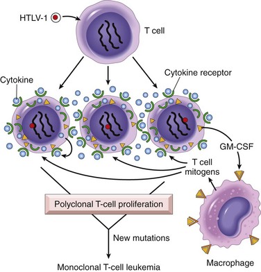
Figure 5–31 Pathogenesis of human T cell lymphotropic virus (HTLV-1)–induced T cell leukemia/lymphoma. HTLV-1 infects many T cells and initially causes polyclonal proliferation by autocrine and paracrine pathways triggered by the TAX gene. Simultaneously, TAX neutralizes growth inhibitory signals by affecting TP53 and CDKN2A/p16 genes. Ultimately, a monoclonal T cell leukemia/lymphoma results when one proliferating T cell suffers additional mutations.
Summary
• HTLV-1 causes a T cell leukemia that is endemic in Japan and the Caribbean.
• The HTLV-1 genome encodes a viral TAX protein, which turns on genes for cytokines and their receptors in infected T cells. This sets up autocrine and paracrine signaling loops that stimulate T cell proliferation. Although this proliferation initially is polyclonal, the proliferating T cells are at increased risk for secondary mutations that lead to the outgrowth of a monoclonal leukemia.
As with RNA viruses, several oncogenic DNA viruses that cause tumors in animals have been identified. Four DNA viruses—HPV, Epstein-Barr virus (EBV), Kaposi sarcoma herpesvirus (KSHV, also called human herpesvirus-8 [HHV-8]), and hepatitis B virus (HBV)—are of special interest because they are strongly associated with human cancer. KSHV and Kaposi sarcoma are discussed in Chapter 4. The others are presented here.
Scores of genetically distinct types of HPV have been identified. Some types (e.g., 1, 2, 4, and 7) cause benign squamous papillomas (warts) in humans (Chapters 18 and 21). Genital warts have low malignant potential and are also associated with low-risk HPVs, predominantly HPV-6 and HPV-11. By contrast, high-risk HPVs (e.g., types 16 and 18) cause several cancers, particularly squamous cell carcinoma of the cervix and anogenital region. In addition, at least 20% of oropharyngeal cancers, particularly those arising in the tonsils, are associated with HPV.
The oncogenic potential of HPV can be related to products of two early viral genes, E6 and E7. Together, they interact with a variety of growth-regulating proteins encoded by proto-oncogenes and tumor suppressor genes. The E7 protein binds to the retinoblastoma protein and releases the E2F transcription factors that normally are sequestered by Rb, promoting progression through the cell cycle. Of interest, E7 protein from high-risk HPV types has a higher affinity for Rb than does E7 from low-risk HPV types. E7 also inactivates the CDKIs CDKN1A/p21 and CDNK1B/p27. The E6 protein has complementary effects. It binds to and mediates the degradation of p53. By analogy with E7, E6 from high-risk HPV types has a higher affinity for p53 than does E6 from low-risk HPV types. Also of interest, in benign warts the HPV genome is maintained in a nonintegrated episomal form, while in cancers the HPV genome is randomly integrated into the host genome. Integration interrupts the viral DNA, resulting in overexpression of the oncoproteins E6 and E7. Furthermore, cells in which the viral genome has integrated show significantly more genomic instability.
To summarize, infection with high-risk HPV types simulates the loss of tumor suppressor genes, activates cyclins, inhibits apoptosis, and combats cellular senescence. Thus, it is evident that many of the hallmarks of cancer discussed earlier are driven by HPV proteins. However, infection with HPV itself is not sufficient for carcinogenesis. For example, when human keratinocytes are transfected with DNA from HPV-16, -18, or -31 in vitro, they are immortalized, but they do not form tumors in experimental animals. Cotransfection with a mutated RAS gene results in full malignant transformation. These data strongly suggest that HPV, in all likelihood, acts in concert with other environmental factors (Chapter 18). However, the primacy of HPV infection in the causation of cervical cancer is attested to by the near-complete protection from this cancer by anti-HPV vaccines.
EBV was the first virus linked to a human tumor, Burkitt lymphoma. Over the last 40 years, however, EBV has been discovered with the cells of a surprisingly diverse list of tumors, including B cell lymphomas in patients with defective T cell immunity (e.g., those infected with HIV), a subset of Hodgkin lymphoma, nasopharyngeal carcinoma, a subset of T cell lymphomas, gastric carcinomas, NK cell lymphomas, and even, in rare instances, sarcomas, mainly in the immunosuppressed.
Burkitt lymphoma is endemic in certain parts of Africa and is sporadic elsewhere. In endemic areas, tumor cells in virtually all affected patients carry the EBV genome. The molecular basis for B cell proliferations induced by EBV is complex. EBV uses the complement receptor CD21 to attach to and infect B cells. In vitro, such infection leads to polyclonal B cell proliferation and generation of B lymphoblastoid cell lines. One of the EBV-encoded genes, called LMP1 (latent membrane protein 1) acts as an oncogene, and its expression in transgenic mice induces B cell lymphomas. LMP1 promotes B cell proliferation by activating signaling pathways, such as NF-κB and JAK/STAT, which mimic B cell activation by the B cell surface molecule CD40. Concurrently, LMP1 prevents apoptosis by activating BCL2. Thus, the virus “borrows” a normal B cell activation pathway to promote its own replication by expanding the pool of cells susceptible to infection. Another EBV-encoded protein, EBNA2, transactivates several host genes, including cyclin D and the src family of proto-oncogenes. In addition, the EBV genome contains a viral cytokine, vIL-10, that was pirated from the host genome. This viral cytokine can prevent macrophages and monocytes from activating T cells and killing virally infected cells.
In immunologically normal persons, EBV-driven polyclonal B cell proliferation is readily controlled, and the affected patient either remains asymptomatic or experiences a self-limited episode of infectious mononucleosis (Chapter 11). Evasion of the immune system seems to be a key step in EBV-related oncogenesis. In regions of the world in which Burkitt lymphoma is endemic, concomitant (endemic) malaria (or other infections) impairs immune competence, allowing sustained B cell proliferation. Of interest, although LMP1 is the primary transforming oncogene in the EBV genome, it is not expressed in EBV-associated Burkitt lymphoma, presumably because it also is one of the major viral antigens recognized by the immune system. Infected cells expressing viral antigens such as LMP-1 are kept in check by the immune system. Lymphoma cells may emerge only when translocations activate the MYC oncogene, a consistent feature of this tumor. MYC may substitute for LMP1 signaling, allowing the tumor cells to downregulate LMP1 and evade the immune system. Of note, in nonendemic areas, 80% of tumors are negative for EBV, but virtually all tumors possess MYC translocations. This observation suggests that although non-African Burkitt lymphomas are triggered by mechanisms other than EBV, these cancers develop by similar pathways.
In patients with deficient T cell function, including those with HIV and organ transplant recipients, EBV-infected B cells undergo polyclonal expansion, producing lymphoblastoid-like cells. In contrast with Burkitt lymphoma, the B lymphoblasts in immunosuppressed patients do express viral antigens, such as LMP-1, that are recognized by T cells. These potentially lethal proliferations can be subdued if T cell immunity can be restored, as may be achieved by withdrawal of immunosuppressive drugs in transplant recipients.
Nasopharyngeal carcinoma is endemic in southern China and some other locales, and the EBV genome is found in all tumors. LMP-1 is expressed in the carcinoma cells and, as in B cells, activates the NF-κB pathway. Furthermore, LMP1 induces the expression of pro-angiogenic factors such as VEGF, FGF-2, MMP-9, and COX-2, which may contribute to oncogenesis. How EBV enters epithelial cells is unclear, as these cells fail to express the CD21 protein that serves as the EBV receptor in B cells.
Summary
• HPV is associated with benign warts, as well as cervical cancer.
• The oncogenicity of HPV is related to the expression of two viral oncoproteins, E6 and E7; they bind to Rb and p53, respectively, neutralizing their function.
• E6 and E7 from high-risk strains of HPV (which give rise to cancers) have higher affinity for their targets than do E6 and E7 from low-risk strains of HPV (which give rise to benign warts).
• EBV is implicated in the pathogenesis of Burkitt lymphomas, lymphomas in immunosuppressed patients (HIV infection or organ transplant recipients), some forms of Hodgkin lymphoma, uncommon T cell and NK cell tumors, nasopharyngeal carcinoma, a subset of gastric carcinoma, and rarely sarcomas.
• Certain EBV gene products contribute to oncogenesis by stimulating a normal B cell proliferation pathway. Concomitant compromise of immune competence allows sustained B cell proliferation, leading eventually to development of lymphoma, with occurrence of additional mutations such as t(8;14) leading to activation of the MYC gene.
The epidemiologic evidence linking chronic HBV and hepatitis C virus (HCV) infection with hepatocellular carcinoma is strong (Chapter 15). It is estimated that 70% to 85% of hepatocellular carcinomas worldwide are due to infection with HBV or HCV. However, the mode of action of these viruses in tumorigenesis is not fully elucidated. The HBV and HCV genomes do not encode any viral oncoproteins, and although the HBV DNA is integrated within the human genome, there is no consistent pattern of integration in liver cells. Indeed, the oncogenic effects of HBV and HCV are multifactorial, but the dominant effect seems to be immunologically mediated chronic inflammation with hepatocyte death leading to regeneration and genomic damage. Although the immune system generally is thought to be protective, recent work has demonstrated that in the setting of unresolved chronic inflammation, as occurs in viral hepatitis or chronic gastritis caused by H. pylori (see further on), the immune response may become maladaptive, promoting tumorigenesis.
As with any cause of hepatocellular injury, chronic viral infection leads to the compensatory proliferation of hepatocytes. This regenerative process is aided and abetted by a plethora of growth factors, cytokines, chemokines, and other bioactive substances produced by activated immune cells that promote cell survival, tissue remodeling, and angiogenesis. The activated immune cells also produce other mediators, such as reactive oxygen species, that are genotoxic and mutagenic. A key molecular step seems to be activation of the nuclear factor-κB (NF-κB) pathway in hepatocytes caused by mediators derived from the activated immune cells. Activation of the NF-κB pathway within hepatocytes blocks apoptosis, allowing the dividing hepatocytes to incur genotoxic stress and to accumulate mutations. Although this seems to be the dominant mechanism in the pathogenesis of virus-induced hepatocellular carcinoma, both HBV and HCV also contain proteins within their genomes that may more directly promote the development of cancer. The HBV genome contains a gene known as HBx, and hepatocellular cancers develop in mice transgenic for this gene. HBx can directly or indirectly activate a variety of transcription factors and several signal transduction pathways. In addition, viral integration can cause secondary rearrangements of chromosomes, including multiple deletions that may harbor unknown tumor suppressor genes.
Although not a DNA virus, HCV also is strongly linked to the pathogenesis of liver cancer. The molecular mechanisms used by HCV are less well defined than those for HBV. In addition to chronic liver cell injury and compensatory regeneration, components of the HCV genome, such as the HCV core protein, may have a direct effect on tumorigenesis, possibly by activating a variety of growth-promoting signal transduction pathways.
Summary
• Between 70% and 85% of hepatocellular carcinomas worldwide are due to infection with HBV or HCV.
• The oncogenic effects of HBV and HCV are multifactorial, but the dominant effect seems to be immunologically mediated chronic inflammation, with hepatocellular injury, stimulation of hepatocyte proliferation, and production of reactive oxygen species that can damage DNA.
• The HBx protein of HBV and the HCV core protein can activate a variety of signal transduction pathways that also may contribute to carcinogenesis.
First incriminated as a cause of peptic ulcers, H. pylori now has acquired the dubious distinction of being the first bacterium classified as a carcinogen. Indeed, H. pylori infection is implicated in the genesis of both gastric adenocarcinomas and gastric lymphomas.
The scenario for the development of gastric adenocarcinoma is similar to that for HBV- and HCV-induced liver cancer. It involves increased epithelial cell proliferation on a background of chronic inflammation. As in viral hepatitis, the inflammatory milieu contains numerous genotoxic agents, such as reactive oxygen species. The sequence of histopathologic changes consists of initial development of chronic inflammation/gastritis, followed by gastric atrophy, intestinal metaplasia of the lining cells, dysplasia, and cancer. This sequence takes decades to complete and occurs in only 3% of infected patients. Like those of HBV and HCV, the H. pylori genome also contains genes directly implicated in oncogenesis. Strains associated with gastric adenocarcinoma have been shown to contain a “pathogenicity island” that contains cytotoxin-associated A gene (CagA). Although H. pylori is noninvasive, CagA is injected into gastric epithelial cells, where it has a variety of effects, including the initiation of a signaling cascade that mimics unregulated growth factor stimulation.
As mentioned previously, H. pylori is associated with an increased risk for the development of gastric lymphomas as well. The gastric lymphomas are of B cell origin, and because the transformed B cells grow in a pattern resembling that of normal mucosa-associated lymphoid tissue (MALT), they also have been referred to as MALT lymphomas (Chapter 11). Their molecular pathogenesis is incompletely understood but seems to involve strain-specific H. pylori factors, as well as host genetic factors, such as polymorphisms in the promoters of inflammatory cytokines such as IL-1β and tumor necrosis factor (TNF). It is thought that H. pylori infection leads to the activation of H. pylori–reactive T cells, which in turn cause polyclonal B cell proliferation. In time, a monoclonal B cell tumor emerges in the proliferating B cells, perhaps as a result of accumulation of mutations in growth regulatory genes. In keeping with this model, early in the course of disease, eradication of H. pylori “cures” the lymphoma by removing antigenic stimulus for T cells.
Summary
• H. pylori infection has been implicated in both gastric adenocarcinoma and MALT lymphoma.
• The mechanism of H. pylori–induced gastric cancers is multifactorial, including immunologically mediated chronic inflammation, stimulation of gastric cell proliferation, and production of reactive oxygen species that damage DNA. H. pylori pathogenicity genes, such as CagA, also may contribute by stimulating growth factor pathways.
• It is thought that H. pylori infection leads to polyclonal B cell proliferations and that eventually a monoclonal B cell tumor (MALT lymphoma) emerges as a result of accumulation of mutations.
The idea that tumors are not entirely “self” was conceived by Ehrlich, who proposed that immune-mediated recognition of autologous tumor cells may be a “positive mechanism” capable of eliminating transformed cells. Subsequently, Lewis Thomas and Macfarlane Burnet formalized this concept by coining the term immune surveillance to refer to recognition and destruction of newly appearing tumor cells, which are seen as foreign by the host immune system. That cancers occur implies that immune surveillance is imperfect; the escape of some tumors from such policing, however, does not preclude the possibility that others may have been aborted. This section addresses certain questions about tumor immunity: What is the nature of tumor antigens? What host effector systems may recognize tumor cells? Is tumor immunity effective against spontaneous neoplasms?
Antigens that elicit an immune response have been demonstrated in many experimentally induced tumors and in some human cancers. Initially, they were broadly classified into two categories based on their patterns of expression: tumor-specific antigens, which are present only on tumor cells and not on any normal cells, and tumor-associated antigens, which are present on tumor cells and also on some normal cells. This classification, however, is imperfect, because many antigens thought to be tumor-specific turned out to be expressed by some normal cells as well. The modern classification of tumor antigens is based on their molecular structure and source.
An important advance in the field of tumor immunology was the development of techniques for identifying tumor antigens that were recognized by cytotoxic T lymphocytes (CTLs), because CTLs are responsible for the major immune defense mechanism against tumors. As described in Chapter 4, CTLs recognize peptides derived from cytoplasmic proteins that are displayed bound to class I major histocompatibility complex (MHC) molecules.
Described next are the main classes of tumor antigens (Fig. 5–32).
Figure 5–32 Tumor antigens recognized by CD8+ T cells.
(Modified from Abbas AK, Lichtman AH: Cellular and Molecular Immunology, 5th ed. Philadelphia, WB Saunders, 2003.)
Neoplastic transformation, as discussed, results from genetic alterations, some of which may lead to the expression of cell surface antigens that are seen as non-self by the immune system. Antigens in this category are derived from mutant oncoproteins and tumor suppressor proteins. Unique tumor antigens arise from β-catenin, RAS, p53, and CDK4, for which the encoding genes frequently are mutated in tumors. Because the mutant genes are present only in tumors, their peptides are expressed only in tumor cells. Since many tumors may carry the same mutation, such antigens are shared by different tumors. Although CTLs can be induced against such antigens, they do not appear to elicit protective responses in vivo. In some cases, unmutated oncogenes are overexpressed in tumors. The best example is that of the HER2/NEU oncogene, whose product is highly expressed in a subset of breast cancers. Antibodies targeted against Her2/Neu protein are used clinically for the treatment of breast cancers.
Because of the genetic instability of tumor cells, many genes are mutated in these cells, including genes whose products are not related to the transformed phenotype and have no known function. Products of these mutated genes are potential tumor antigens. These antigens are extremely diverse, because the carcinogens that induce the tumors may randomly mutagenize virtually any host gene. Mutated cellular proteins are found more frequently in chemical carcinogen- or radiation-induced animal tumors than in spontaneous human cancers. They can be targeted by the immune system, since there is no self-tolerance against them.
Tumor antigens may be normal cellular proteins that are abnormally expressed in tumor cells and elicit immune responses. In a subset of human melanomas, some tumor antigens are structurally normal proteins that are produced at low levels in normal cells and overexpressed in tumor cells. One such antigen is tyrosinase, an enzyme involved in melanin biosynthesis that is expressed only in normal melanocytes and melanomas. T cells from patients with melanoma recognize peptides derived from tyrosinase, raising the possibility that tyrosinase vaccines may stimulate such responses to melanomas; clinical trials with these vaccines are ongoing. It is somewhat surprising that these patients are able to respond to a normal self-antigen. The probable explanation is that tyrosinase normally is produced in such small amounts and in so few cells that it is not recognized by the immune system and fails to induce tolerance.
Another group, the so-called cancer-testis antigens, are encoded by genes that are silent in all normal adult tissues except the testis, and are deregulated in cancer cells—hence their name. Although the protein is present in the testis, it is not expressed on the cell surface in an antigenic form, because sperm do not express MHC class I molecules. Thus, for all practical purposes, these antigens are tumor-specific. Prototypical of this group is the MAGE (melanoma antigen gene) family of genes. Although they are tumor-specific, MAGE antigens are not unique for individual tumors. MAGE-1 is expressed on 37% of melanomas and a variable number of lung, liver, stomach, and esophageal carcinomas. Similar antigens called GAGE, BAGE, and RAGE have been detected in other tumors. Several antigens from this category are now being used in tumor vaccine trials.
As discussed earlier, some viruses are associated with cancers. Not surprisingly, these viruses produce proteins that are recognized as foreign by the immune system. The most potent of these antigens are proteins produced by latent DNA viruses; examples in humans are HPV and EBV. There is abundant evidence that CTLs recognize antigens of these viruses and that a competent immune system plays a role in surveillance against virus-induced tumors because of its ability to recognize and kill virus-infected cells. Indeed, vaccines against HPV antigens have been found to be effective in prevention of cervical cancers in girls and young women.
Oncofetal antigens or embryonic antigens, such as carcinoembryonic antigen (CEA) and alpha fetoprotein, are expressed during embryogenesis but not in normal adult tissues. Derepression of the genes that encode these antigens causes their reexpression in colon and liver cancers. Antibodies can be raised against these antigens and are useful for detection of oncofetal antigens. Although, as discussed later, they are not entirely tumor-specific, they can serve as serum markers for cancer.
Most human and experimental tumors express higher than normal levels and/or abnormal forms of surface glycoproteins and glycolipids, which may be diagnostic markers and targets for therapy. These altered molecules include gangliosides, blood group antigens, and mucins. Although most of the epitopes recognized by antibodies raised against such antigens are not specifically expressed on tumors, they are present at higher levels on cancer cells than on normal cells. This class of antigens is a target for cancer therapy with specific antibodies.
Several mucins are of special interest and have been the focus of diagnostic and therapeutic studies. These include CA-125 and CA-19-9, expressed on ovarian carcinomas, and MUC-1, expressed on breast carcinomas. Unlike many other types of mucins, MUC-1 is an integral membrane protein that normally is expressed only on the apical surface of breast ductal epithelium, a site that is relatively sequestered from the immune system. In ductal carcinomas of the breast, however, the molecule is expressed in an unpolarized fashion and contains new, tumor-specific carbohydrate and peptide epitopes. These epitopes induce both antibody and T cell responses in cancer patients and are therefore candidates for tumor vaccines.
Tumors express molecules that normally are present on the cells of origin. These antigens are called differentiation antigens, because they are specific for particular lineages or differentiation stages of various cell types. Their importance is as potential targets for immunotherapy and in identifying the tissue of origin of tumors. For example, lymphomas may be diagnosed as B cell–derived tumors by the detection of surface markers characteristic of this lineage, such as CD20. Antibodies against CD20 are used for immunotherapy of certain B cell lymphomas. These differentiation antigens typically are normal self-antigens, so they do not induce immune responses in tumor-bearing hosts.
Cell-mediated immunity is the dominant antitumor mechanism in vivo. Although antibodies can be made against tumors, there is no evidence that they play a protective role under physiologic conditions. The cellular effectors that mediate immunity are discussed fully in Chapter 4, so they are characterized only briefly here.
The role of specifically sensitized cytotoxic T lymphocytes (CTLs) in experimentally induced tumors is well established. In humans, they seem to play a protective role, chiefly against virus-associated neoplasms (e.g., EBV-induced Burkitt lymphoma, HPV-induced tumors). The presence of MHC-restricted CD8+ cells that can kill autologous tumor cells within human tumors suggests that the role of T cells in immunity against human tumors may be broader than was previously suspected. In some cases, such CD8+ T cells do not develop spontaneously in vivo but can be generated by immunization with tumor antigen–pulsed dendritic cells.
NK cells are lymphocytes that are capable of destroying tumor cells without previous sensitization; they may provide the first line of defense against tumor cells. After activation with IL-2, NK cells can lyse a wide range of human tumors, including many that seem to be nonimmunogenic for T cells. T cells and NK cells apparently provide complementary antitumor mechanisms. Tumors that fail to express MHC class I antigens cannot be recognized by T cells, but these tumors may trigger NK cells because the latter are inhibited by recognition of normal autologous class I molecules (Chapter 4). Thus, tumors may downregulate MHC class I molecules to avoid recognition by T cells, which then makes them prime targets for NK cells. The triggering receptors on NK cells are extremely diverse and belong to several gene families. NKG2D proteins expressed on NK cells and some T cells are important activating receptors. They recognize stress-induced antigens that are expressed on tumor cells and on cells that have incurred DNA damage and are at risk for neoplastic transformation.
Classically activated macrophages of the M1 type (Chapter 2) exhibit cytotoxicity against tumor cells in vitro. T cells, NK cells, and macrophages may collaborate in antitumor reactivity, because interferon-γ, a cytokine secreted by T cells and NK cells, is a potent activator of macrophages. Activated macrophages may kill tumors by mechanisms similar to those used to kill microbes (e.g., production of reactive oxygen metabolites) (Chapter 2) or by secretion of tumor necrosis factor (TNF).
Although there is no evidence for the protective effects of antitumor antibodies against spontaneous tumors, administration of monoclonal antibodies against tumor cells can be therapeutically effective. A monoclonal antibody against CD20, a B cell surface antigen, is widely used for treatment of certain non-Hodgkin lymphomas.
In view of the host of possible and potential antitumor mechanisms, is there any evidence that they operate in vivo to prevent the emergence of neoplasms? The strongest argument for the existence of immune surveillance is the increased frequency of cancers in immunodeficient hosts. About 5% of persons with congenital immunodeficiencies develop cancers, a rate that is about 200 times reported rates for persons without such immunodeficiencies. By analogy, immunosuppressed transplant recipients and patients with acquired immunodeficiency syndrome have increased numbers of malignancies. Of note, most (but not all) of these neoplasms are lymphomas, often lymphomas of activated B cells. Particularly illustrative is X-linked lymphoproliferative disorder. When affected boys develop an EBV infection, such infection does not take the usual self-limited form of infectious mononucleosis but instead evolves into a fatal form of infectious mononucleosis or, even worse, malignant lymphoma.
Most cancers occur in persons who do not suffer from any overt immunodeficiency. If immune surveillance exists, how do cancers evade the immune system in immunocompetent hosts? Several escape mechanisms have been proposed:
• Selective outgrowth of antigen-negative variants. During tumor progression, strongly immunogenic subclones may be eliminated. This notion is supported by experiments in which tumors arising in immunocompromised mice express antigens that are recognized, with consequent elimination of the tumors by the immune system in normal mice, whereas similar tumors arising in immunocompetent mice are nonimmunogenic.
• Loss or reduced expression of histocompatibility molecules. Tumor cells may fail to express normal levels of human leukocyte antigen (HLA) class I, escaping attack by CTLs. Such cells, however, may trigger NK cells.
• Immunosuppression. Many oncogenic agents (e.g., chemicals, ionizing radiation) suppress host immune responses. Tumors or tumor products also may be immunosuppressive. For example, TGF-β, secreted in large quantities by many tumors, is a potent immunosuppressant. In some cases, the immune response induced by the tumor may inhibit tumor immunity. Several mechanisms of such inhibition have been described. For instance, recognition of tumor cells may lead to engagement of the T cell inhibitory receptor, CTLA-4, or activation of regulatory T cells that suppress immune responses. More insidiously, some tumors express FasL, which can engage Fas on immune cell surfaces and induce the immune cell to enter apoptosis!
• Antigen masking. Many tumor cells produce a thicker coat of external glycocalyx molecules, such as sialic acid–containing mucopolysaccharides, than normal cells. This thick coat may block access of immune cells to antigen-presenting molecules, thereby preventing antigen recognition and cell killing.
• Downregulation of co-stimulatory molecules. Costimulatory molecules are required to initiate strong T cell responses. Many tumors reduce expression of these costimulatory molecules.
Summary
• Tumor cells can be recognized by the immune system as non-self and destroyed.
• Antitumor activity is mediated by predominantly cell-mediated mechanisms. Tumor antigens are presented on the cell surface by MHC class I molecules and are recognized by CD8+ CTLs.
• The different classes of tumor antigens include products of mutated proto-oncogenes, tumor suppressor genes, overexpressed or aberrantly expressed proteins, tumor antigens produced by oncogenic viruses, oncofetal antigens, altered glycolipids and glycoproteins, and cell type–specific differentiation antigens.
• Immunosuppressed patients have an increased risk for development of cancer.
• In immunocompetent patients, tumors may avoid the immune system by several mechanisms, including selective outgrowth of antigen-negative variants, loss or reduced expression of histocompatibility antigens, and immunosuppression mediated by secretion of factors (e.g., TGF-β) from the tumor.
The importance of neoplasms ultimately lies in their effects on patients. Although malignant tumors are of course more threatening than benign tumors, morbidity and mortality may be associated with any tumor, even a benign one. Indeed, both malignant and benign tumors may cause problems because of (1) location and impingement on adjacent structures, (2) functional activity such as hormone synthesis or the development of paraneoplastic syndromes, (3) bleeding and infections when the tumor ulcerates through adjacent surfaces, (4) symptoms that result from rupture or infarction, and (5) cachexia or wasting. The following discussion considers the effects of a tumor on the host, the grading and clinical staging of cancer, and the laboratory diagnosis of neoplasms.
Location is crucial in both benign and malignant tumors. A small (1-cm) pituitary adenoma can compress and destroy the surrounding normal gland, giving rise to hypopituitarism. A 0.5-cm leiomyoma in the wall of the renal artery may encroach on the blood supply, leading to renal ischemia and hypertension. A comparably small carcinoma within the common bile duct may induce fatal biliary tract obstruction.
Hormone production is seen with benign and malignant neoplasms arising in endocrine glands. Adenomas and carcinomas arising in the beta cells of the pancreatic islets of Langerhans can produce hyperinsulinism, sometimes fatal. By analogy, some adenomas and carcinomas of the adrenal cortex elaborate corticosteroids that affect the patient (e.g., aldosterone, which induces sodium retention, hypertension, and hypokalemia). Such hormonal activity is more likely with a well-differentiated benign tumor than with a corresponding carcinoma.
A tumor may ulcerate through a surface, with consequent bleeding or secondary infection. Benign or malignant neoplasms that protrude into the gut lumen may become caught in the peristaltic pull of the gut, causing intussusception (Chapter 14) and intestinal obstruction or infarction.
Many cancer patients suffer progressive loss of body fat and lean body mass, accompanied by profound weakness, anorexia, and anemia—a condition referred to as cachexia. There is some correlation between the size and extent of spread of the cancer and the severity of the cachexia. However, cachexia is not caused by the nutritional demands of the tumor. Although patients with cancer often are anorexic, current evidence indicates that cachexia results from the action of soluble factors such as cytokines produced by the tumor and the host, rather than reduced food intake. In patients with cancer, calorie expenditure remains high, and basal metabolic rate is increased, despite reduced food intake. This is in contrast with the lower metabolic rate that occurs as an adaptive response in starvation. The basis of these metabolic abnormalities is not fully understood. It is suspected that TNF produced by macrophages in response to tumor cells or by the tumor cells themselves mediates cachexia. TNF suppresses appetite and inhibits the action of lipoprotein lipase, inhibiting the release of free fatty acids from lipoproteins. Additionally, a protein-mobilizing factor called proteolysis-inducing factor, which causes breakdown of skeletal muscle proteins by the ubiquitin-proteosome pathway, has been detected in the serum of cancer patients. Other molecules with lipolytic action also have been found. There is no satisfactory treatment for cancer cachexia other than removal of the underlying cause, the tumor.
Symptom complexes that occur in patients with cancer and that cannot be readily explained by local or distant spread of the tumor or by the elaboration of hormones not indigenous to the tissue of origin of the tumor are referred to as paraneoplastic syndromes. They appear in 10% to 15% of patients with cancer, and their clinical recognition is important for several reasons:
• Such syndromes may represent the earliest manifestation of an occult neoplasm.
• In affected patients, the pathologic changes may be associated with significant clinical illness and may even be lethal.
• The symptom complex may mimic metastatic disease, thereby confounding treatment.
The paraneoplastic syndromes are diverse and are associated with many different tumors (Table 5–5). The most common such syndromes are hypercalcemia, Cushing syndrome, and nonbacterial thrombotic endocarditis; the neoplasms most often associated with these and other syndromes are lung and breast cancers and hematologic malignancies. Hypercalcemia in cancer patients is multifactorial, but the most important mechanism is the synthesis of a parathyroid hormone–related protein (PTHrP) by tumor cells. Also implicated are other tumor-derived factors, such as TGF-α, a polypeptide factor that activates osteoclasts, and the active form of vitamin D. Another possible mechanism for hypercalcemia is widespread osteolytic metastatic disease of bone; of note, however, hypercalcemia resulting from skeletal metastases is not a paraneoplastic syndrome. Cushing syndrome arising as a paraneoplastic phenomenon usually is related to ectopic production of ACTH or ACTH-like polypeptides by cancer cells, as occurs in small cell cancers of the lung. Sometimes one tumor induces several syndromes concurrently. For example, bronchogenic carcinomas may elaborate products identical to or having the effects of ACTH, antidiuretic hormone, parathyroid hormone, serotonin, human chorionic gonadotropin, and other bioactive substances.
Table 5–5 Paraneoplastic Syndromes
Clinical Syndrome | Major Forms of Neoplasia | Causal Mechanism(s)/Agent(s) |
---|---|---|
Endocrinopathies | ||
Cushing syndrome | Small cell carcinoma of lung | ACTH or ACTH-like substance |
Pancreatic carcinoma | ||
Neural tumors | ||
Syndrome of inappropriate antidiuretic hormone secretion | Small cell carcinoma of lung; intracranial neoplasms | Antidiuretic hormone or atrial natriuretic hormones |
Hypercalcemia | Squamous cell carcinoma of lung | Parathyroid hormone–related protein, TGF-α, TNF, IL-1 |
Breast carcinoma | ||
Renal carcinoma | ||
Adult T cell leukemia/lymphoma | ||
Ovarian carcinoma | ||
Hypoglycemia | Fibrosarcoma | Insulin or insulin-like substance |
Other mesenchymal sarcomas | ||
Hepatocellular carcinoma | ||
Carcinoid syndrome | Bronchial adenoma (carcinoid) | Serotonin, bradykinin |
Pancreatic carcinoma | ||
Gastric carcinoma | ||
Polycythemia | Renal carcinoma | Erythropoietin |
Cerebellar hemangioma | ||
Hepatocellular carcinoma | ||
Nerve and Muscle Syndrome | ||
Myasthenia | Bronchogenic carcinoma, thymoma | Immunologic |
Disorders of the central and peripheral nervous systems | Breast carcinoma, teratoma | |
Dermatologic Disorders | ||
Acanthosis nigricans | Gastric carcinoma | Immunologic; secretion of epidermal growth factor |
Lung carcinoma | ||
Uterine carcinoma | ||
Dermatomyositis | Bronchogenic and breast carcinoma | Immunologic |
Osseous, Articular, and Soft Tissue Changes | ||
Hypertrophic osteoarthropathy and clubbing of the fingers | Bronchogenic carcinoma | Unknown |
Vascular and Hematologic Changes | ||
Venous thrombosis (Trousseau phenomenon) | Pancreatic carcinoma | Tumor products (mucins that activate clotting) |
Bronchogenic carcinoma | ||
Other cancers | ||
Nonbacterial thrombotic endocarditis | Advanced cancers | Hypercoagulability |
Anemia | Thymoma | Immunologic |
Others | ||
Nephrotic syndrome | Various cancers | Tumor antigens, immune complexes |
ACTH, adrenocorticotropic hormone; IL-1, interleukin-1; TGF-α, transforming growth factor-α; TNF, tumor necrosis factor.
Paraneoplastic syndromes also may manifest as hypercoagulability, leading to venous thrombosis and nonbacterial thrombotic endocarditis (Chapter 10). Other manifestations are clubbing of the fingers and hypertrophic osteoarthropathy in patients with lung carcinomas (Chapter 12). Still others are discussed in the consideration of cancers of the various organs of the body.
Methods to quantify the probable clinical aggressiveness of a given neoplasm and its apparent extent and spread in the individual patient are necessary for making an accurate prognosis and for comparing end results of various treatment protocols. For instance, the results of treating extremely small, highly differentiated thyroid adenocarcinomas that are localized to the thyroid gland are likely to be different from those obtained from treating highly anaplastic thyroid cancers that have invaded the neck organs.
The grading of a cancer attempts to establish some estimate of its aggressiveness or level of malignancy based on the cytologic differentiation of tumor cells and the number of mitoses within the tumor. The cancer may be classified as grade I, II, III, or IV, in order of increasing anaplasia. Criteria for the individual grades vary with each form of neoplasia and are not detailed here. Difficulties in establishing clear-cut criteria have led in some instances to descriptive characterizations (e.g., “well-differentiated adenocarcinoma with no evidence of vascular or lymphatic invasion” or “highly anaplastic sarcoma with extensive vascular invasion”).
Staging of cancers is based on the size of the primary lesion, its extent of spread to regional lymph nodes, and the presence or absence of metastases. This assessment usually is based on clinical and radiographic examination (computed tomography and magnetic resonance imaging) and in some cases surgical exploration. Two methods of staging are currently in use: the TNM system (T, primary tumor; N, regional lymph node involvement; M, metastases) and the AJC (American Joint Committee) system. In the TNM system, T1, T2, T3, and T4 describe the increasing size of the primary lesion; N0, N1, N2, and N3 indicate progressively advancing node involvement; and M0 and M1 reflect the absence and presence, respectively, of distant metastases. In the AJC method, the cancers are divided into stages 0 to IV, incorporating the size of primary lesions and the presence of nodal spread and of distant metastases. Examples of the application of these two staging systems are cited in subsequent chapters. Of note, when compared with grading, staging has proved to be of greater clinical value.
Summary
• Cachexia, defined as progressive loss of body fat and lean body mass, accompanied by profound weakness, anorexia, and anemia, is caused by release of cytokines by the tumor or host.
• Paraneoplastic syndromes, defined as systemic symptoms that cannot be explained by tumor spread or by hormones appropriate to the tissue, are caused by the ectopic production and secretion of bioactive substances such as ACTH, PTHrP, or TGF-α.
• Grading of tumors is determined by cytologic appearance and is based on the idea that behavior and differentiation are related, with poorly differentiated tumors having more aggressive behavior.
• Staging, determined by surgical exploration or imaging, is based on size, local and regional lymph node spread, and distant metastases. Staging is of greater clinical value than grading.
In most instances, the laboratory diagnosis of cancer is not difficult. The two ends of the benign–malignant spectrum pose no problems; in the middle, however, lies a “no man’s land” where the wise tread cautiously. Clinicians tend to underestimate the contributions they make to the diagnosis of a neoplasm. Clinical and radiologic data are invaluable for optimal pathologic diagnosis. Radiation-induced changes in the skin or mucosa can be similar to those of cancer. Sections taken from a healing fracture can mimic an osteosarcoma. The laboratory evaluation of a lesion can be only as good as the specimen submitted for examination. The specimen must be adequate, representative, and properly preserved.
Several sampling approaches are available, including excision or biopsy, fine-needle aspiration, and cytologic smears. When excision of a lesion is not possible, selection of an appropriate site for biopsy of a large mass requires awareness that the margins may not be representative and the center may be largely necrotic. Requesting frozen section diagnosis is sometimes desirable, as, for example, in determining the nature of a mass lesion or in evaluating the regional lymph nodes in a patient with cancer for metastasis. This method, in which a sample is quick-frozen and sectioned, permits histologic evaluation within minutes. In experienced, competent hands, frozen section diagnosis is accurate, but there are particular instances in which the better histologic detail provided by the more time-consuming routine methods is needed. In such instances, it is better to wait a few days, despite the drawbacks, than to perform inadequate or unnecessary surgery.
Fine needle aspiration of tumors is another approach that is widely used. It involves aspiration of cells from a mass, followed by cytologic examination of the smear. This procedure is used most commonly with readily palpable lesions affecting the breast, thyroid, lymph nodes, and salivary glands. Modern imaging techniques permit extension of the method to deeper structures, such as the liver, pancreas, and pelvic lymph nodes. Use of this diagnostic modality obviates surgery and its attendant risks. Although it entails some difficulties, such as small sample size and sampling errors, in experienced hands it can be reliable, rapid, and useful.
Cytologic (Papanicolaou) smears provide another method for the detection of cancer. Historically, this approach has been used widely for discovery of carcinoma of the cervix, often at an in situ stage, but now it is used to investigate many other forms of suspected malignancy, such as endometrial carcinoma, bronchogenic carcinoma, bladder and prostate tumors, and gastric carcinomas; for the identification of tumor cells in abdominal, pleural, joint, and cerebrospinal fluids; and, less commonly, for evaluation of other forms of neoplasia. Neoplastic cells are less cohesive than others and are therefore shed into fluids or secretions (Fig. 5–33). The shed cells are evaluated for features of anaplasia indicative of their origin from a tumor. The gratifying control of cervical cancer is the best testament to the value of the cytologic method.
Figure 5–33 A, Normal Papanicolaou smear from the uterine cervix. Large, flat cells with small nuclei are typical. B, Abnormal smear containing a sheet of malignant cells with large hyperchromatic nuclei. Nuclear pleomorphism is evident, and one cell is in mitosis. A few interspersed neutrophils, much smaller in size and with compact, lobate nuclei, are seen.
(Courtesy of Dr. Richard M. DeMay, Department of Pathology, University of Chicago, Chicago, Illinois.)
Immunocytochemistry offers a powerful adjunct to routine histologic examination. Detection of cytokeratin by specific monoclonal antibodies labeled with peroxidase points to a diagnosis of undifferentiated carcinoma rather than large cell lymphoma. Similarly, detection of prostate-specific antigen (PSA) in metastatic deposits by immunohistochemical staining allows definitive diagnosis of a primary tumor in the prostate. Immunocytochemical detection of estrogen receptors allows prognostication and directs therapeutic intervention in breast cancers.
Flow cytometry is used routinely in the classification of leukemias and lymphomas. In this method, fluorescent antibodies against cell surface molecules and differentiation antigens are used to obtain the phenotype of malignant cells.
Biochemical assays for tumor-associated enzymes, hormones, and other tumor markers in the blood cannot be utilized for definitive diagnosis of cancer; however, they can be useful screening tests and in some instances have utility in quantitating the response to therapy or detecting disease recurrence. The application of these assays is considered with many of the specific forms of neoplasia discussed in other chapters, so only a few examples suffice here. PSA, used to screen for prostatic adenocarcinoma, may be one of the most frequently and successfully used tumor markers in clinical practice. Prostatic carcinoma can be suspected when elevated levels of PSA are found in the blood. However, PSA screening also highlights problems encountered with use of virtually every tumor marker. Although PSA levels often are elevated in cancer, PSA levels also may be elevated in benign prostatic hyperplasia (Chapter 17). Furthermore, there is no PSA level that ensures that a patient does not have prostate cancer. Thus, the PSA test suffers from both low sensitivity and low specificity. PSA assay is extremely valuable, however, for detecting residual disease or recurrence following treatment for prostate cancer. Other tumor markers occasionally used in clinical practice include carcinoembryonic antigen (CEA), which is elaborated by carcinomas of the colon, pancreas, stomach, and breast, and alpha fetoprotein, which is produced by hepatocellular carcinomas, yolk sac remnants in the gonads, and occasionally teratocarcinomas and embryonal cell carcinomas. Unfortunately, like PSA, both of these markers can be produced in a variety of non-neoplastic conditions as well. Thus, CEA and alpha fetoprotein assays lack both specificity and sensitivity required for the early detection of cancers. As with PSA screening, they are still particularly useful in the detection of recurrences after excision. With successful resection of the tumor, these markers disappear from the serum; their reappearance almost always signifies the beginning of the end. CEA is further discussed in Chapter 14 and alpha fetoprotein in Chapter 15.
An increasing number of molecular techniques are being used for the diagnosis of tumors and for predicting their behavior.
• Diagnosis of malignancy: Because each T and B cell exhibits unique rearrangement of its antigen receptor genes, polymerase chain reaction (PCR)–based detection of T cell receptor or immunoglobulin genes allows distinction between monoclonal (neoplastic) and polyclonal (reactive) proliferations. Many hematopoietic neoplasms, as well as a few solid tumors, are defined by particular translocations, so the diagnosis can be made by detection of such translocations. For example, fluorescence in situ hybridization (FISH) or PCR analysis (Chapter 6) can be used to detect translocations characteristic of Ewing sarcoma and several leukemias and lymphomas. PCR-based detection of BCR-ABL transcripts provides the molecular diagnosis of chronic myeloid leukemia.
• Prognosis and behavior: Certain genetic alterations are associated with a poor prognosis, and thus the presence of these alterations determines the patient’s subsequent therapy. FISH and PCR methods can be used to detect amplification of oncogenes such as HER2/NEU and NMYC, which provide prognostic and therapeutic information for breast cancers and neuroblastomas.
• Detection of minimal residual disease: Another emerging use of molecular techniques is for detection of minimal residual disease after treatment. For example, detection of BCR-ABL transcripts by PCR assay gives a measure of residual disease in patients treated for chronic myeloid leukemia. Recognition that virtually all advanced tumors are associated with both intact circulating tumor cells and products derived from tumors (e.g., tumor DNA) has led to interest in following tumor burden through sensitive blood tests.
• Diagnosis of hereditary predisposition to cancer: Germline mutation of several tumor suppressor genes, such as BRCA1, increases a patient’s risk for development of certain types of cancer. Thus, detection of these mutated alleles may allow the patient and the physician to devise an aggressive screening protocol, as well as an opportunity for prophylactic surgery. In addition, such detection allows genetic counseling of relatives at risk.
• Therapeutic decision-making: Therapies that directly target specific mutations are increasingly being developed, and thus detection of such mutations in a tumor can guide the development of targeted therapy, as discussed later. It is now becoming evident that certain targetable mutations may transgress morphologic categories. For example, mutations of the ALK kinase, originally described in a subset of T cell lymphomas, also have been identified in a small percentage of non–small cell carcinomas and neuroblastomas. Clinical trials have shown that lung cancers with ALK mutations respond to ALK inhibitors, whereas other lung cancers do not, leading to recent FDA approval of ALK inhibitors for use in patients with “ALK-mutated” lung cancer. Another recent dramatic example of molecularly “tailored” therapy is seen in melanoma, in which tumors with a valine for glutamate substitution in amino acid 600 (V600E) of the serine/threonine kinase BRAF respond well to BRAF inhibition, whereas melanomas without this mutation show no response. Of some interest, the V600E mutation is also present in a subset of colon cancers, certain thyroid cancers, 100% of hairy cell leukemias, and Langerhans cell histiocytosis (Fig. 5–34). These tumors are morphologically diverse and have distinct cells of origin, but they share identical oncogenic lesions in a common pro-growth pathway.
Molecular profiling of tumors can be done both at the level of mRNA and by nucleotide sequencing. Each of these two is described next.
This technique allows simultaneous measurements of the expression levels of several thousand genes. The principle of this so-called gene chip technology is illustrated in Figure 5–35 and described briefly here.
Figure 5–35 Complementary DNA (cDNA) microarray analysis.
Messenger RNA (mRNA) is extracted from the samples, reverse transcribed to cDNA, and labeled with fluorescent molecules. In the case illustrated, red fluorescent molecules were used for normal cDNA, and green molecules were used for tumor cDNA. The labeled cDNAs are mixed and applied to a gene chip, which contains thousands of DNA probes representing known genes. The labeled cDNAs hybridize to spots that contain complementary sequences. The hybridization is detected by laser scanning of the chip, and the results are read in units of red or green fluorescence intensity. In the example shown, spot A has high red fluorescence, indicating that a greater number of cDNAs from neoplastic cells hybridized to gene A. Thus, gene A seems to be upregulated in tumor cells.
(Courtesy of Dr. Robert Anders, Department of Pathology, University of Chicago, Chicago, Illinois.)
As can be seen, the process begins by extraction of mRNA from any two sources (e.g., normal and malignant, normal and preneoplastic, or two tumors of the same histologic type). Complementary DNA (cDNA) copies of the mRNA are synthesized in vitro with fluorescently labeled nucleotides. The fluorescence-labeled cDNA strands are hybridized to sequence-specific DNA probes linked to a solid support, such as a silicon chip. A 1-cm2 chip can contain thousands of probes arranged in an array of columns and rows. After hybridization, high-resolution laser scanning detects fluorescent signals from each of the spots. The fluorescence intensity of each spot is proportional to the level of expression of the original mRNA used to synthesize the cDNA hybridized to that spot. For each sample, therefore, the expression level of thousands of genes is obtained, and by using bioinformatic tools, the relative levels of gene expression in different samples can be compared. In essence, a molecular profile is generated for each tissue analyzed.
Such analysis has revealed that phenotypically identical large B cell lymphomas (Chapter 11) from different patients are heterogeneous with respect to their gene expression and survival rates. Similar approaches are now being explored in other cancers, such as breast cancers and melanomas.
The progression and development of next-generation sequencing technologies promise even more in-depth analysis of tumors. The advances in such technologies are currently outpacing the famous Moore’s law of microprocessors. Sequencing an entire tumor genome, which just a couple of years ago would have taken months and millions of dollars, now takes days and costs a few thousand dollars. Sequences of the entire tumor genomes, when compared with the normal genome from the same patient, can reveal all the somatic alterations present in a tumor.
Recent results from genomic analyses of tumors have revealed that individual tumors can contain from a handful of somatic mutations (certain childhood leukemias) to tens of thousands of mutations, with the highest mutational burden being found in cancers associated with mutagen exposure, such as lung cancer and skin cancer. Among these are two types of mutations: (1) those that subvert normal control of cell proliferation, differentiation, and homeostasis and (2) those that have no effect on cell phenotype. The first set of mutations is referred to as driver mutations because they may drive the neoplastic process and hence could be therapeutic targets. The other set of mutations, often much more numerous than driver mutations, most often fall in noncoding regions of the genome or have a neutral effect on growth, not conferring any advantage or disadvantage. Such mutations are called passenger mutations. They result from genomic instability of cancer cells and are merely “along for the ride.”
In general, driver mutations are recurrent and are present in a substantial percentage of patients with a particular cancer. Thus, for example, BCR-ABL fusion genes are present in all cases of chronic myelogenous leukemia, and the fusion protein is an excellent drug target. However, driver mutations may be present in only a subset of tumors of a particular type. For example, approximately 4% of non–small cell lung cancers harbor an EML4-ALK tyrosine kinase fusion gene; as already mentioned, in these relatively rare instances, the patient responds well to ALK inhibitors. An additional complication is that some passenger mutations nevertheless have important roles in drug resistance. For example, the mutations in BCR-ABL that confer resistance to imatinib in chronic myelogenous leukemia are present as passenger mutations in rare clones before therapy begins. Because they confer a powerful selective advantage, these mutations are converted from passengers to drivers in the face of drug therapy; it is suspected that the genomic instability of cancer cells sows the seeds of resistance through similar scenarios in many kinds of tumors. Furthermore, in some instances, several distinct and relatively uncommon mutations all converge on the same pathway (such as resistance to apoptosis) and contribute to the cancer phenotype. It is therefore useful to categorize mutations on the basis of their ability to drive the cells along the “hallmarks of cancer” pathways.
It is hoped that identification of all potentially targetable mutations in each individual tumor will refocus the treatment of tumors from the tissue of origin to the molecular lesion, as drugs that target specific mutations are developed (Fig. 5–36). This approach represents a paradigm shift in the classification and therapy of tumors. Perhaps in the future the diverse group of tumors that bear a common mutation such as BRAF will be classified as BRAF-omas (Fig. 5–34), rather than individual types based on morphology or cell of origin!
Figure 5–36 A paradigm shift: Classification of cancer according to therapeutic targets rather than cell of origin and morphology.
Summary
• Several sampling approaches exist for the diagnosis of tumors, including excision, biopsy, fine-needle aspiration, and cytologic smears.
• Immunohistochemistry and flow cytometry studies help in the diagnosis and classification of tumors, because distinct protein expression patterns define different entities.
• Proteins released by tumors into the serum, such as PSA, can be used to screen populations for cancer and to monitor for recurrence after treatment.
• Molecular analyses are used to determine diagnosis, prognosis, the detection of minimal residual disease, and the diagnosis of hereditary predisposition to cancer.
• Molecular profiling of tumors by cDNA arrays and sequencing can determine expression of large segments of the genome and catalog all of the mutations in the tumor genome and thus may be useful in molecular stratification of otherwise identical tumors or those of distinct histogenesis that share a mutation for the purpose of treatment and prognostication.
Ahmed Z, Bicknell R. Angiogenic signalling pathways. Methods Mol Biol. 2009;467:3-24. [Discussion of many signaling pathways in angiogenesis.]
Artandi SE, DePinho RA. Telomeres and telomerase in cancer. Carcinogenesis. 2010;31:9-18. [Review discussing the importance of telomeres and telomerase.]
Barrallo-Gimeno A, Nieto MA. The Snail genes as inducers of cell movement and survival: implications in development and cancer. Development. 2005;132:3151-3161. [Discussion of the genes involved in epithelial-mesenchymal transition in cancer.]
Berx G, van Roy F. Involvement of members of the cadherin superfamily in cancer. Cold Spring Harb Perspect Biol. 1, 2009. [Review discussing the role of cadherins and contact inhibition in cancer.] a003129
Bierie B, Moses HL. Tumour microenvironment: TGFbeta: the molecular Jekyll and Hyde of cancer. Nat Rev Cancer. 2006;6:506-520. [Review discussing the tumor-suppressive and tumor-promoting effects of TGF-β.]
Burkhart DL, Sage J. Cellular mechanisms of tumour suppression by the retinoblastoma gene. Nat Rev Cancer. 2008;8:671-682. [Review of Rb function.].
Ciccia A, Elledge SJ. The DNA damage response: making it safe to play with knives. Mol Cell. 2010;40:179-204. [Review discussing the DNA damage response.]
Coghlin C, Murray GI. Current and emerging concepts in tumour metastasis. J Pathol. 2010;222:1-15. [Discussion of current concepts in metastasis.]
Collado M, Serrano M. Senescence in tumours: evidence from mice and humans. Nat Rev Cancer. 2010;10:51-57. [Update on mechanisms of senescence.]
Feron O. Pyruvate into lactate and back: from the Warburg effect to symbiotic energy fuel exchange in cancer cells. Radiother Oncol. 2009;92:329-333. [An account of the reemergence and molecular pathways of reprogramming of energy metabolism in cancer.]
Grivennikov SI, Greten FR, Karin M. Immunity, inflammation, and cancer. Cell. 2010;140:883-899. [A summary of the links between inflammation and the development of cancer.]
Hanahan D, Weinberg RA. The hallmarks of cancer (2011): the next generation. Cell. 2011;144:646-674. [Reexamination of the hallmarks of cancer.]
Junttila MR, Evan GI. p53—a jack of all trades but master of none. Nat Rev Cancer. 2009;9:821-829. [Update summarizing p53 function.]
Kalluri R, Zeisberg M. Fibroblasts in cancer. Nat Rev Cancer. 2006;6:392-401. [Review discussing the role of stroma in cancer.]
Mathew R, Karantza-Wadsworth V, White E. Role of autophagy in cancer. Nat Rev Cancer. 2007;7:961-967. [A discussion of the mechanisms of autophagy.]
Negrini S, Gorgoulis VG, Halazonetis TD. Genomic instability—an evolving hallmark of cancer. Nat Rev Mol Cell Biol. 2010;11:220-228. [Review on mechanisms of genomic instability, an enabler of malignancy.]
Perona R. Cell signalling: growth factors and tyrosine kinase receptors. Clin Transl Oncol. 2006;8:77-82. [Update on signaling pathways in cancer.]
Stratton MR, Campbell PJ, Futreal PA. The cancer genome. Nature. 2009;458:719-724. [Excellent summary of next-generation sequencing technologies and their application to cancer.]
Willis SN, Adams JM. Life in the balance: how BH3-only proteins induce apoptosis. Curr Opin Cell Biol. 2005;17:617-625. [A review of the mechanisms of apoptosis.]
Witsch E, Sela M, Yarden Y. Roles for growth factors in cancer progression. Physiology (Bethesda). 2010;25:85-101. [An update on the role of growth factors in cancer.]