3 CELLULAR STRUCTURE AND FUNCTION
INTRODUCTION
To truly understand pathophysiology you must first have a fundamental knowledge of the composition of the human body and how it functions. However, to fully appreciate the complexity of the human body you need to understand the structure and function of cells, because all body functions depend on the integrity of cells and their environment. Cells are the smallest functional units in the body. Each cell comprises many different components that work together to allow it to carry out the functions that are vital to the human body. Thus a thorough understanding of human cellular anatomy and physiology is essential in order to comprehend how diseases arise and why they affect the body in particular ways. It is only over the last 200 years or so that our understanding of cellular structure and function has been developed, and this knowledge continues to be expanded today in an effort both to understand how the human body works and to discover how diseases form and what treatments may be beneficial.
This chapter explores the cell — what components it comprises, how these components operate, and how individual cells communicate with other cells and thus coordinate physiological processes. We also look at the different body tissues — that is, groups of specialised cells — to explain how individual cells act in unison with other cells.
CELLULAR STRUCTURE AND FUNCTION
The human body is composed of trillions of individual cells. It is easy to think of these cells as small circular structures that are neatly arranged. The reality is far from this, however; in fact, there are more than 200 different cell types consisting of different shapes and sizes, and each performs quite different functions.
There are a multitude of different cell sizes. For instance, a single nerve cell (neuron) may be up to a massive one metre in length, while a muscle cell (myocyte) may be up to 30 cm and red blood cells (erythrocytes) are only a tiny 6–8 micrometres (1 micrometre is 1/1000 of a millimetre). However, most human cells average about 10–15 micrometres in diameter. Apart from size, there is considerable variation in shape as well. There are disc-shaped (discoid) cells, such as red blood cells; spherical cells such as the human egg (ovum); cube-shaped (cuboidal) cells like hepatocytes (liver cells); squamous cells that are thin and flat, which are typical of skin cells; and column-like (columnar) cells that are elongated, tall but not wide, found in the lining of the intestines. Some cell shapes are spread out, giving the appearance of stars (termed stellate-shaped), like nerve cells. Examples of these cells are shown in Figure 3-1.
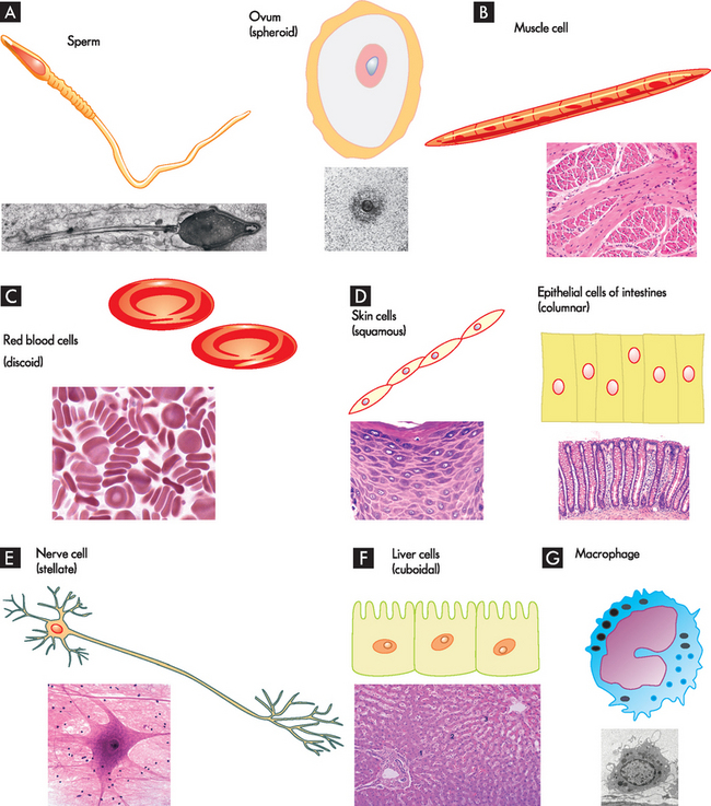
FIGURE 3-1 Different cell forms, including function and shape.
A Cells involved in reproduction, sperm and ovum; B cells that permit movement; C cells that involve transport; D cells that line cavities and provide protection; E cells that transmit information and control physiological functions; F cells that are involved in metabolic processes; and G cells that are involved in inflammation and immune responses. Note that cells are not drawn to scale.
Cells become specialised through the process of differentiation, or maturation, so that some cells eventually perform one kind of function while others perform other functions. For instance, some cells are primarily involved in movement (muscle cells); some transmit information (nerve cells); others secrete hormones (like the pancreatic cells, termed beta cells, which release insulin); some provide a barrier for the body (the epidermal cells of the skin); and some allow continuation of the human species (the sex cells, like sperm).
Despite the variety of cell types with varied morphology (shape and structure) and functions, all cells are made up of the fundamental building blocks of life: carbon, hydrogen, oxygen, nitrogen and small amounts of other elements. Moreover, the typical cell has the following characteristic functions:
CELLULAR COMPONENTS
A useful analogy to remember when trying to understand cellular structures and their respective functions is to consider that cells are like factories. The walls, roof and floors provide boundary limits and determine the size of the factory. Inside the factory, support structures provide structure to the building. The factory has workers and a manufacturing assembly line where products are produced. Raw materials brought into the factory are turned into finished items by the workers using the tools of the assembly line. When the items are completed, they are prepared for shipping, transported out and distributed to the various customers. In addition, the factory managers and executives decide on the types and numbers of items to be produced and relay this information to the factory workers. Furthermore, energy is required at each manufacturing step to produce the items and move them out of the factory.
Similarly for cells, the outer border — termed cell membrane (or plasma membrane) — provides structural integrity from the outside. Within the cell, which is similar to the space inside the factory, a fluid exists called the cytoplasm. The cell also contains many structures called organelles, literally meaning ‘little organs’, which are responsible for all the intracellular activities — these are like the workers and assembly-line equipment. Lastly, the cell has a nucleus, which is the control centre of the cell, housed within the cell and protected by a membrane as well. The nucleus is analogous to the factory managers and executives who control activities by communicating with workers about what is required. These structures are shown in Figure 3-2. While this may seem a simplistic overview of the cell, it will aid your understanding of the cellular components and how they work in unison. We start by examining the components inside the cell, the organelles.
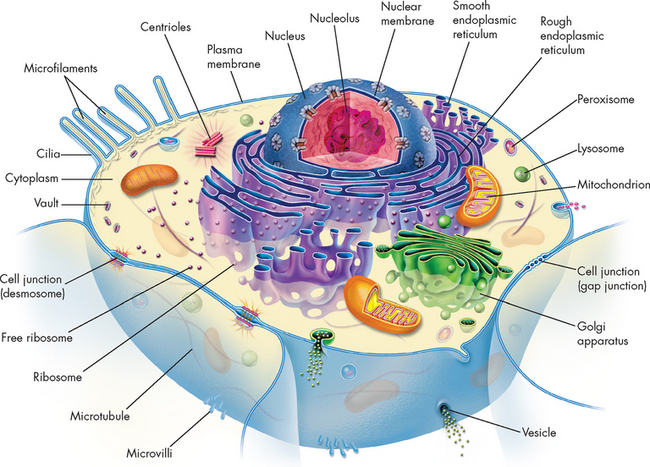
FIGURE 3-2 The structure of a typical cell.
Note that this is a general body cell; not all cells contain all these cellular components, but the components are common in many human body cells.
The organelles
The organelles are all the structures within the cell that are required to maintain normal cellular function. They are specialised structures that have distinct functions and may vary in number according to the primary function of the cell (see Figure 3-2) — for instance, mitochondria, the organelle responsible for energy for the cell, are more numerous in muscle cells than other cells. In addition, the majority of organelles have a membrane that separates the internal structure from the cytoplasm of the cell. This allows the organelles to perform chemical reactions with enzymes (substances that speed up biological processes) that are distinct from the rest of the cellular components. This is very important, because the enzymes are required for specific processes that occur within the internal structure of the organelles, but are separated from other components of the cell which may alter the chemical reactions.
The membranous organelles include the nucleus, endoplasmic reticulum, Golgi apparatus, lysosomes, peroxisomes and mitochondria. The non-membranous organelles, those that do not have a membrane, include the ribosomes and cytoskeleton. We start with the organelle that is found in nearly all cells in the body, the nucleus.
The nucleus
The nucleus is the control centre in most living cells containing genetic material essential for proper functioning of the cell. The cell is dependent on the nucleus; similar to the brain in the human body, the nucleus controls and regulates functions, but also responds to signals that the cell may receive. Almost all cells in the human body have one nucleus. Interestingly, mature red blood cells do not have a nucleus and are referred to as anucleated cells (the ‘a’ at the beginning of the word refers to without; therefore, without a nucleus). These cells cannot reproduce, as the nucleus is responsible for cellular replication. Hence they have only a relatively short life span — approximately 120 days. In contrast, some cells have more than one nucleus; these are referred to as multinucleate (‘multi’ referring to many nuclei, the plural of nucleus). Although it might appear strange to have more than one nucleus in a cell, it is thought that cells which need to produce large amounts of proteins and have numerous intracellular components require multiple nuclei. Examples of multinucleated cells include skeletal muscle cells, some liver cells and bone-dissolving cells (osteoclasts).
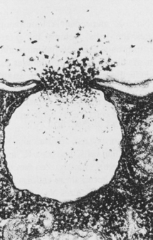
Generally, the nucleus is located in the centre of the cell and is approximately 5 micrometres in diameter. The shape of the nucleus can vary, although most are spherical or oval-shaped; the shape is dependent on the cell type. The nucleus has two main components: the nuclear membrane and the nucleolus (see Figure 3-3). The nuclear membrane, or envelope, comprises two membranes that surround the nucleus and provide protection for its contents. Holes in the membrane, called nuclear pores, allow communication to and from the cell. These pores are vital to the nucleus, because every minute thousands of substances pass through the nuclear pores. Enzymes, hormones and substances required by the nucleus to build genetic material enter the nucleus and processed genetic material is ejected to control the manufacture of new proteins required by the cell. Within the nucleus, deoxyribonucleic acid (commonly abbreviated to DNA) and proteins are arranged to form chromosomes, the genetic material that determines human characteristics. DNA is confined to the nucleus; when genetic information is to be used in the production of new proteins, ribonucleic acid (RNA) is formed. DNA and associated proteins are normally arranged as clumps in the nucleus, termed chromatin. RNA is formed in a spherical dense region, called the nucleolus, composed largely of DNA and the DNA-binding proteins. The nucleolus is mainly responsible for producing a subunit of the ribosomes, essential for the production of new proteins.
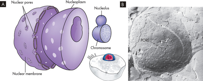
A The nucleus is composed of a double membrane called a nuclear membrane that encloses the fluid-filled interior. B An electron micrograph of a nucleus with the nuclear pores evident.
Source: B Raven PH, Johnson GB. Biology. St Louis: Mosby; 1992.
The primary functions of the nucleus are cell division and control of genetic information. This is achieved through a series of complex tasks which are examined in detail in Chapter 5. Other functions include the replication and repair of DNA and the transcription of information stored in DNA. Genetic information is transcribed into RNA, which can be processed into messenger, transport and ribosomal RNA and introduced into the cytoplasm, where it directs cellular activities. We now look at one of the recipients of the RNA, the ribosomes.
The ribosomes
The ribosomes are small granules of proteins and RNA responsible for protein synthesis — that is, the production of new proteins. The ribosomal RNA and proteins produced in the nucleolus move through the nuclear pores into the cytoplasm. Here they either float freely in the cytoplasm (known as free ribosomes) or form a complex with the membrane (termed the rough endoplasmic reticulum; see below). Both sites are responsible for protein synthesis. These ribosomes combine with another two forms of RNA, messenger RNA (mRNA) and transfer RNA (tRNA), to translate the code for the new protein. Briefly, mRNA has the instructions for the assembly of a specific protein, while tRNA brings the amino acids (the building blocks of a protein) to the ribosome. In this way, amino acids are gathered to form the new protein. The entire process is called translation and is described in more detail in Chapter 5.
Endoplasmic reticulum
The endoplasmic reticulum is a network that is continuous with the nuclear membrane (see Figure 3-4). It facilitates the movement of proteins and is also the site of protein synthesis. The name derives from endo meaning within, plasm meaning fluid and reticulum, which translates as network; therefore, it is a network within the cytoplasm of the cell. There are two types of endoplasmic reticulum: smooth and rough. These are easily differentiated because one looks rough and the other looks smooth!

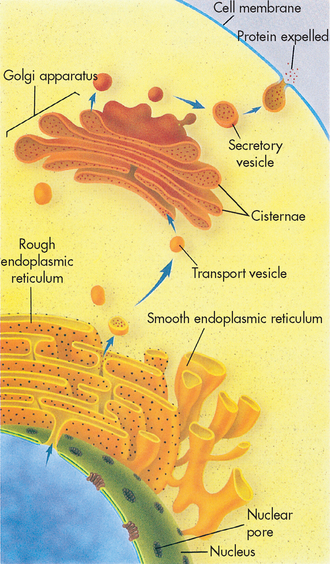
FIGURE 3-4 Endoplasmic reticulum.
A The endoplasmic reticulum is continuous with the nuclear membrane and consists of ribosome-coated cisternae (cavities); the smooth endoplasmic reticulum is attached to the rough component. B An electron micrograph of rough and smooth endoplasmic reticulum.
Source: A Based on McCance KL. Pathophysiology: the biologic basis for disease in adults and children. 6th edn. St Louis: Mosby; 2010; B Courtesy Kelbes C, Farmer M. From Lindsay DT. Functional human anatomy. St Louis: Mosby; 1996.
The rough endoplasmic reticulum comes about from the ribosomes that inhabit the internal surface of the network, while the smooth endoplasmic reticulum does not contain ribosomes. The rough endoplasmic reticulum is primarily responsible for the production of proteins, while the smooth endoplasmic reticulum produces phospholipids (lipids with phosphates attached) required in the cell membrane and performs other functions specific to the cell type. For instance, in the liver the smooth endoplasmic reticulum is involved in the detoxification of drugs, while in the muscle cells it primarily stores calcium, ready to be released during muscle contraction.
The Golgi apparatus
The Golgi apparatus, also referred to as the Golgi complex, is a network of flattened membranes that are folded back on each other (see Figure 3-5). The folds tend to bulge at the ends forming cavities called cisternae, from the Latin meaning box. This is where secretory vesicles are formed (vesicles are small membrane-bound sacs involved in cellular transport). The vesicles either migrate to other cellular organelles or are released from the cell (see Figure 3-6). Essentially, the Golgi apparatus receives lipids and proteins from the endoplasmic reticulum and then modifies, packages and redistributes them to various parts of the cell. In this way, the Golgi apparatus is synonymous with the packaging and distribution centre of the factory.
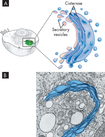
FIGURE 3-5 The Golgi apparatus.
A Schematic representation of the Golgi apparatus showing a stack of flattened sacs (cisternae) and numerous small membranous secretory vesicles. B Transmission electron micrograph showing the Golgi apparatus highlighted with colour.
Source: McCance KL. Pathophysiology: the biologic basis for disease in adults and children. 6th edn. St Louis: Mosby; 2010; based on Thibodeau GA, Patton, KT. Anatomy & physiology. 6th edn. St Louis: Mosby; 2007.
The newly formed proteins are packaged and sent to the Golgi apparatus in the form of a secretory vesicle. This vesicle fuses with the Golgi apparatus and is further modified and then shunted to the ends of the Golgi apparatus (cisternae). In this example, the vesicle is secreted to the cell membrane and released outside the cell.
Source: McCance KL. Pathophysiology: the biologic basis for disease in adults and children. 6th edn. St Louis: Mosby; 2010; based on Raven PH, Johnson GB. Understanding biology. 3rd edn. Dubuque, IA: Brown; 1995.
Lysosomes
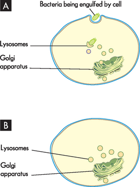
Lysosomes are small storage vesicles that are formed from a pinching of the Golgi apparatus. They are rich in powerful enzymes that act as a digestive system for the cell; that is, they break down and dispose of organelles that do not function properly or are old, and they digest foreign substances such as bacteria (see Figure 3-7). Because these enzymes are so destructive, lysosomes are encased in a membrane to protect the cell. However, in certain situations the enzymes are used to destroy an entire cell, which is like cellular ‘suicide’. This is termed apoptosis, or programmed cell death, and is discussed in Chapter 4.
Peroxisomes
Peroxisomes are similar to lysosomes, but contain different enzymes. These storage vesicles are smaller than lysosomes and are involved in the breakdown of amino acids and fatty acids (those that are responsible for making lipids). The chemical reactions that occur inside peroxisomes result in the creation of hydrogen peroxide (a weak acid, used as a bleach disinfectant or blonde hair dye!), hence the name peroxisome.
Mitochondria
Mitochondria are the powerhouse of the cell. They are commonly small, rod-like structures (see Figure 3-8); however, their shape is quite variable and has been shown to change considerably in living cells.1 Mitochondria contain their own DNA, RNA and ribosomes, meaning that they can self-replicate. But by far their most important role is the synthesis of adenosine triphosphate (ATP), the major energy source of the cell. It must be stressed that ATP is required for most cellular activities to occur. Like the nucleus, mitochondria have a double membrane, with the inner layer folding inwards to form cristae (which means crests). This greatly increases the surface area, which increases the number of sites where ATP can be produced. Energy is not actually produced, but rather is obtained from the breakdown of molecules (glucose) and incorporated into ATP molecules. The vast majority of ATP production occurs within the mitochondria using enzymes located on the inner membrane. The production of ATP is discussed later in the section on cellular metabolism.
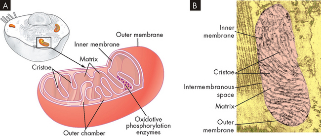
A The inner and outer membranes are shown in the cut-down figure. Note the many folds (cristae) of the inner membrane. B A transmission electron micrograph of a mitochondrion. Although some mitochondria have the capsule shape shown here, many are round or oval.
Source: McCance KL. Pathophysiology: the biologic basis for disease in adults and children. 6th edn. St Louis: Mosby; 2010; Guyton AC, Hall JE. Textbook of medical physiology. 11th edn. Philadelphia: Saunders; 2006; modified from De Robertis EPP, Saez EA, De Robertis EMF. Cell biology. 6th edn. Philadelphia: Saunders; 1975.
The cytoskeleton
The cytoskeleton is exactly what the name implies — a skeleton for the cell. It is easy to assume that cells are quite rigid structures, but this is not the case, as flexibility is allowed. The cytoskeleton provides the structural integrity that maintains the cell’s shape and also allows a pathway for intracellular transport. The cytoskeleton is made up of protein filaments that provide scaffolding that extends throughout the cytoplasm, as well as support for the cell membrane.
The cytoplasm
The cytoplasm is an aqueous solution that fills the cytoplasmic matrix — the space between the nuclear membrane and the outer layer of the cell, the cell membrane. It is also commonly referred to as intracellular fluid, and actually the majority of water in the body is contained inside the cells. The cytoplasm represents about half the volume of the cell and is primarily composed of water with a mixture of dissolved substances such as protein, sugars and waste products. However, the cytoplasm is quite viscous and the organelles are literally suspended in it. In addition, thousands of enzymes are involved in metabolism; thus the cytoplasm is crowded with ribosomes making proteins. Newly synthesised proteins remain in the cytoplasm if they lack a signal for transport to a cell organelle.2 In this way, they are available to be transported into an organelle when later required. The majority of cellular functions use the cytoplasm — for instance, the synthesis of proteins and hormones and their transport out of the cell, the isolation and elimination of waste products from the cell, metabolic processes, the breakdown and disposal of cellular debris, and the maintenance of cellular structure and motility. In addition, the cytoplasm is a storage unit for fat, carbohydrates and secretory vesicles.
THE CELL MEMBRANE
The cell membrane, also called the plasma membrane, is common to all cells and forms a barrier between outside and inside the cell (see Figure 3-9). The cell membrane is exceedingly important to normal physiological function because it controls the composition of the space, or compartment, that it encloses. The membrane can include or exclude various molecules and by controlling the movement of substances from one compartment to another, it exerts a powerful influence on metabolic pathways. The cell membrane also has an important role in cell-to-cell recognition. Other functions of the cell membrane include cellular mobility and the maintenance of cellular shape (see Table 3-1).
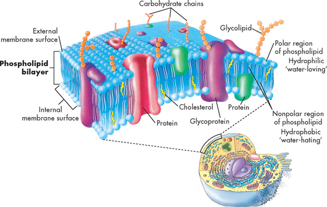
A three-dimensional view of the lipid bilayer that provides the basic cell structure and serves as a relatively impermeable barrier to most water-soluble molecules.
Source: McCance KL. Pathophysiology: the biologic basis for disease in adults and children. 6th edn. St Louis: Mosby; 2010; based on Thibodeau GA, Patton KT. Anatomy & physiology. 6th edn. St Louis: Mosby; 2007.
Table 3-1 CELL MEMBRANE FUNCTIONS
| CELLULAR MECHANISM | MEMBRANE FUNCTION |
|---|---|
| Structure | |
| Protection | |
| Storage | |
| Cell-to-cell interaction |
Source: Modified from King DW et al. General pathology: principles and dynamics. Philadelphia: Lea & Febiger; 1983.
The outer surface of the cell is not smooth but rather is dimpled with cave-like indentations known as caveolae (‘tiny caves’). Caveolae serve as a storage site for many receptors and provide a route for transport into the cell (see later in the chapter).
The major chemical components of all membranes are lipids and proteins, but the percentage of each varies among different membranes.
Lipids
The basic component of the cell membrane is a bilayer of lipid molecules chiefly composed of phospholipids and cholesterol. Phospholipids consist of fatty acids, which make up lipids and a phosphate group. They are integral to the cell membrane as lipids are responsible for the structural integrity of the membrane. Each lipid molecule is said to be polar, meaning that the head is hydrophobic (‘water hating’) and the tail is hydrophilic (‘water loving’) (see Figure 3-9). The membrane organises itself into two layers because of the polar differences. The hydrophobic region (the hydrophobic tail) of each lipid molecule is protected from water, whereas the hydrophilic region (the hydrophilic head) is immersed in it. The bilayer serves as a barrier to the diffusion of water and hydrophilic substances such as glucose, while allowing lipid-soluble molecules such as oxygen and carbon dioxide to diffuse through it readily.
Cholesterol is another major lipid within the cell membrane. High concentrations of cholesterol in the blood are often associated with cardiovascular disease; accordingly, many people believe that cholesterol is a ‘bad’ substance that causes heart disease. Although excessive amounts of cholesterol are a strong risk factor for cardiovascular disease, it is important to realise that the body needs adequate amounts of cholesterol, as it is an essential component of cell membranes and is vitally important in maintaining membrane permeability.
Proteins
About 50% of the total mass of the cell membrane is protein. Proteins can be classified as integral or peripheral. Integral membrane proteins, also called transmembrane proteins, are embedded in the lipid bilayer and span the entire width of the cell membrane. They are involved in many processes but the main ones include acting as a channel or pump to allow water-soluble substances to cross the phospholipid bilayer, and acting as a receptor to allow signals from hormones and chemicals to be transmitted inside the cell (this is especially important for the nervous and endocrine systems, as detailed in Chapters 6 and 10, respectively). Peripheral membrane proteins are not embedded in the bilayer but reside at one surface or the other, bound to an integral protein. They are therefore mostly involved in providing support for the cell membrane and joining cells together.
The interaction of cell membrane proteins with lipids is complex. The role of proteins in the onset and progression of disease is important because of their enzymatic, transport and recognition-receptor functions in cellular physiology.
In the 1960s, the fluid mosaic model for biological membranes was proposed. The model, which is continually being modified, proposes that the cell membrane is dynamic. Lipids and proteins move laterally on the membrane, and ions and other molecules move through it. However, cells can immobilise specific membrane proteins in a region of the membrane to allow certain functions to occur. The fluid mosaic model describes the membrane as existing in a state of change, allowing the cell to protect itself actively against injurious agents. Hormones, bacteria, viruses, drugs, antibodies (produced by the immune system to combat foreign substances) and chemicals that transmit nerve impulses (neurotransmitters) attach to the cell membrane by means of receptors on the outer layer. The number of receptors present may vary at different times and the cell can control the effects of injurious agents by altering receptor numbers and patterns.3 This aspect of the fluid mosaic model has drastically modified previously held concepts concerning the onset of disease.
The concentration of cholesterol in the cell membrane affects membrane fluidity. Increased cholesterol concentration means less fluidity on the membrane’s hydrophilic outer surface and more fluidity at its hydrophobic core. This can impact on the cell’s normal functioning and is a factor in some diseases.
CELLULAR RECEPTORS AND COMMUNICATION
We have examined what constitutes a cell and how the intracellular components function; however, you need to be aware of how cells react to their environment and communicate with each other, including cells close together and ones that are distant. Cells monitor their environment through receptors. These cellular receptors are protein molecules on the cell membrane, in the cytoplasm or in the nucleus that can recognise and bind with specific smaller molecules called ligands. These substances, such as hormones, bind with cell receptors. It should be stressed that there are many substances that can bind with cell receptors and these can either activate or inactivate the receptor. However, recognition and binding depend on the chemical configuration of the molecule and its receptor, which must fit together like pieces of a jigsaw puzzle (see Figure 3-10). In this way, messages can be communicated to the cell or a cellular process can commence.
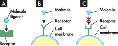
FIGURE 3-10 The chemical configuration of cellular receptors.
A The molecule binds (attaches) to the receptor. B A particular-shaped molecule fits a specific receptor on the cell membrane. C The molecule cannot bind to a different cell receptor due to differences in chemical composition and structure.
Cell membrane receptors protrude from or are exposed at the external surface of the membrane and are often attached to integral proteins (see Figure 3-11). The substances that bind with membrane receptors include hormones, neurotransmitters, infectious agents and metabolites. Many discoveries concerning the specific interactions of cellular receptors with their respective substances have provided a basis for understanding disease.
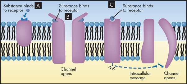
FIGURE 3-11 Cellular receptors.
A Cell membrane receptor for a substance (here, a hormone molecule) on the surface of an integral protein. The substance binds with the receptor and this initiates a cellular process. B Channel-linked receptors, which open when a substance binds with the receptor outside of the cell. C Stimulation of the receptors results in an intracellular message being sent to a channel, causing the channel to open.
It is important to realise that cell receptors often play a vital role in the pharmacological management of diseases and disorders. Fortunately for patients, drugs that are used in clinical practice can interact or bind with receptors too. When drugs bind with a receptor there are two basic types of interaction: drugs that stimulate the receptor are termed agonists, while drugs that block the action are termed antagonists. An example of an agonist is salbutamol, the most common drug used in the management of asthma. Salbutamol binds with receptors in the lung that when stimulated cause bronchodilation, or opening, of the airways. Common examples of antagonists are the beta (β)-blockers. These work by binding to and hence blocking receptor action of beta (β)-adrenergic receptors on the heart (see Chapter 22). This lowers the heart rate and decreases the force of contraction, which reduces blood pressure (hence β-blockers are used in the treatment of high blood pressure, or hypertension).
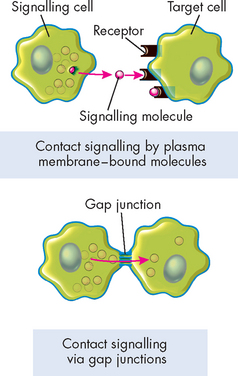
Cells also need to communicate with each other to maintain a stable internal environment (homeostasis), to regulate their growth and division, to oversee their development and organisation into tissues, and to coordinate their functions. Cells communicate in a variety of ways. They form protein channels, called gap junctions, which directly coordinate the activities of adjacent cells, and they have receptors on the cell membrane that respond to signals that affect the cell itself and other cells in direct physical contact with it (see Figure 3-12). There are two primary modes of signalling: hormonal and neural.
 Hormonal signalling involves specialised endocrine cells, which secrete hormone chemicals released by one set of cells that travel through the tissues via the bloodstream to produce a response in another set of cells. This is the basis of the endocrine system and is addressed in detail in Chapter 10. The nervous system also is intimately involved in cell signalling. In neural signalling, the nerve cells (neurons) communicate directly with the cells they innervate by releasing substances called neurotransmitters at specialised junctions called chemical synapses (meaning gap); the neurotransmitters diffuse across the synapses and act on the target cells. The response time of neural signalling is rapid compared with hormonal signalling, and it can produce body-wide effects. For example, when an individual is frightened, the multiple responses are controlled by the nervous system. For more details on the nervous system, see Chapter 6.
MEMBRANE TRANSPORT
The other important area to address is how substances are moved into and out of the cell. Cells continually take in nutrients, fluids and chemical messengers from the extracellular environment and expel metabolites, or the products of metabolism, and the end products of lysosomal digestion. The mechanisms involved depend on the characteristics of the substances to be transported. For example, in passive transport (which does not require energy to move substances), water and small electrically uncharged molecules move easily through pores in the cell membrane’s lipid bilayer. This process occurs naturally through any semi-permeable barrier, such as the cell membrane.
Other molecules are too large to pass through pores. Some of these molecules are moved into and out of the cell by active transport, which requires cellular expenditure of metabolic energy. Other large molecules, called macromolecules, along with fluids, are transported by endocytosis (meaning taking in to the cell) and exocytosis (meaning expelling from the cell). Water and electrically charged molecules are transported by protein channels embedded in the cell membrane.
Movement of water and solutes
Cell membranes are semi-permeable, which simply means that some substances can cross while others cannot. It allows the passage of water and small particles of dissolved substances, called solutes, depending on their size, solubility, electrical properties and concentration on either side of the membrane. Small, lipid-soluble particles, such as oxygen and carbon dioxide, readily pass through the lipid bilayers of the cell membrane. Larger, water-soluble particles may pass through pores in the membranes. Although large protein molecules pass through cell membranes by endocytosis, water is actually drawn towards these large proteins and hence they also cause a movement of water.
Body fluids are composed of electrolytes such as sodium and potassium and non-electrolytes such as glucose. Electrolytes account for approximately 95% of the solute molecules in body water. For example, sodium (Na+) is the predominant extracellular cation (a positively charged ion in the fluid outside the cell) and potassium (K+) is the principal intracellular cation. The difference in intra- and extracellular concentrations of these ions is important for the transmission of electrical impulses across the cell membranes of nerve and muscle cells. Fluid and electrolyte balance between body compartments is discussed in detail in Chapter 29.
Passive transport
Filtration
Filtration is the movement of water and solutes through a membrane because of a greater pushing pressure (force) on one side of the membrane than on the other side. Hydrostatic pressure is the mechanical force of water pushing against cellular membranes. In the vascular system, hydrostatic pressure is the blood pressure generated in the vessels when the heart contracts. The blood reaching the capillary bed has a hydrostatic pressure sufficient to force water across the thin capillary membranes into the spaces between the cells, called the interstitial spaces. Hydrostatic pressure is important for the cardiovascular and urinary systems and is discussed in more detail in Chapters 22 and 28, respectively.
Simple diffusion
Diffusion is the movement of a solute molecule from an area of greater solute concentration to an area of lesser solute concentration. This difference in concentration is known as the concentration gradient (see Figure 3-13). Although particles in a solution move randomly in any direction, if the concentration of particles in one part of the solution is greater than in another part, the particles distribute themselves evenly throughout the solution. According to the same principle, if the concentration of particles is greater on one side of a permeable membrane than on the other side, the particles will diffuse spontaneously from the area of greater concentration to the area of lesser concentration until equilibrium is reached. The higher the concentration on one side, the greater the diffusion rate.
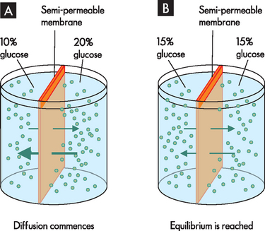
The semi-permeable membrane allows glucose to move from an area of high concentration to an area of low concentration. A The container shows the distinct 10% and 20% glucose solutions, while in B the container shows the result of diffusion over time with an equal distribution of glucose in both compartments.
Source: Based on Patton KT, Thibodeau GA. Anatomy & physiology. 7th edn. St Louis: Mosby; 2010.
The diffusion rate is influenced by many factors, including the size of the substance and its lipid solubility (that is, how well the substance dissolves in lipid). Usually, the smaller the molecule and the more soluble it is in lipid, the more hydrophobic it is and the more rapidly it will diffuse across the lipid bilayer. Oxygen, carbon dioxide and steroid hormones are all hydrophobic molecules. Water-soluble substances such as sugars diffuse very slowly, whereas uncharged lipophilic (‘lipid-loving’) molecules such as fatty acids and steroids diffuse rapidly. Ions and other polar molecules generally diffuse across cellular membranes more slowly than lipid-soluble substances.
Water readily diffuses through cell membranes because water molecules are small and uncharged. The structure of water allows it to rapidly cross the regions of the bilayer containing the lipid head groups. Their groups constitute the two outer regions of the lipid bilayer.
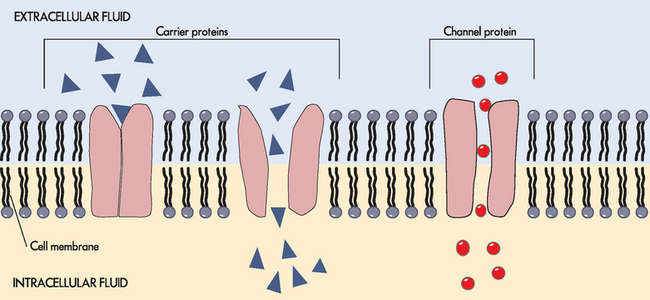
Facilitated diffusion
Facilitated diffusion occurs when substances such as electrolytes and glucose cannot pass directly through the cell membrane but are required to enter the cell. It is called facilitated diffusion because it is aided by integral proteins. The integral proteins transport the substances via a specific channel or carry them through the cell membrane. Importantly, despite the substances moving through specific channels or ports, facilitated diffusion does not require any energy, as the substances are simply transported down the concentration gradient (so they move from a crowded area on one side of the membrane to the other side where there is less of that substance and hence more room is available).
Channels that facilitate the movement of substances allow electrolytes to move down the concentration gradient due to the size and electrical charge of the substances. These channels can be specific for a particular substance. In carrier-mediated facilitated diffusion, the substance binds with the integral protein site and the protein essentially changes shape to allow the substance to pass through. In this way, water-soluble substances can enter and leave the cell without the need for energy expenditure. Examples of facilitated diffusion are presented in Figure 3-14.
Osmosis
Osmosis is the movement of water ‘down’ a concentration gradient — that is, across a semi-permeable membrane from a region of higher water concentration to one of lower concentration (see Figure 3-15). For osmosis to occur: (1) the membrane must be more permeable to water than to solutes; and (2) the concentration of solutes must be greater so that water moves more easily. Osmosis controls the distribution and movement of water between body compartments and is vital to maintaining normal fluid balance. Osmolality controls the distribution and movement of water between the body compartments. It can be measured by the number of milliosmoles per kilogram of water (mOsm/kg) or the concentration of molecules per weight of water.
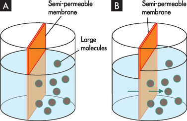
A Water is present in both sides of the chamber. The right side has large molecules that cannot pass through the semi-permeable membrane. B After time water diffuses across the membrane into the right side of the chamber via osmosis.
In addition, osmosis is directly related to both hydrostatic pressure and solute concentration, but not to particle size or weight. The amount of hydrostatic pressure required to oppose the osmotic movement of water is called the osmotic pressure of the solution. Factors that determine osmotic pressure are the type and thickness of the cell membrane, the size of the molecules, the concentration of molecules, or the concentration gradient, and the solubility of molecules within the membrane.
Tonicity is related to the osmotic pressure of a solution, because it relates to how a solution will affect the fluid volume inside the cell. Solutions have relative degrees of tonicity. The normal osmolality of body fluids is approximately 290 mOsm/kg and solutions are classified according to body fluids: an isotonic solution has the same concentration of solute as body fluids (iso refers to equal); a hypotonic solution has a lower concentration of solutes and is thus more dilute than body fluids (hypo meaning below); and a hypertonic solution has a greater concentration than body fluids (hyper meaning above). The concept of tonicity is important in patients when administering intravenous fluids to correct water and solute imbalances.
Active transport
In active transport, the protein transporter moves molecules against, or up, the concentration gradient. Unlike passive and facilitated transport, active transport requires energy. Many active transport systems or pumps have ATP as their primary energy source (see Figure 3-16).
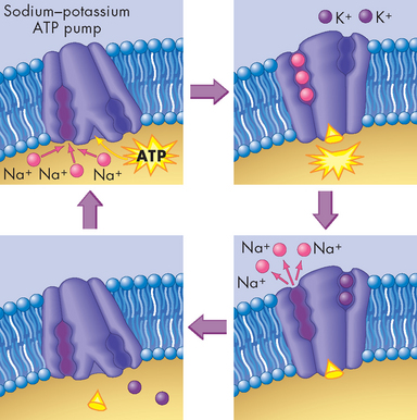
FIGURE 3-16 Active transport and the sodium–potassium pump.
Three sodium (Na+) ions bind to sodium-binding sites on the carrier’s inner face. At the same time, an energy-containing ATP molecule produced by the cell’s mitochondria binds to the carrier. The ATP breaks apart, transferring its stored energy to the carrier. The carrier then changes shape, releases the three Na+ ions to the outside of the cell, and attracts two potassium (K+) ions to its potassium-binding sites. The carrier then returns to its original shape, releasing the two K+ ions and the remnant of the ATP molecule to the inside of the cell. The carrier is now ready for another pumping cycle.
A carrier mechanism in the cell membrane mediates the transport of ions and nutrients. The best-known pump is the sodium–potassium ATP pump, which continuously pumps sodium and potassium against their concentration gradient to maintain ionic concentration gradients vital for nerve conduction and homeostasis. The important aspect is that ATP is required, which means that when energy is available the pump works, but when ATP is depleted, such as during disease, the pump will not be as efficient.
Endocytosis and exocytosis
The active transport mechanisms by which cells move large proteins or polysaccharides (macromolecules) across the cell membrane are very different from those that mediate small solute and ion transport. Transport of macromolecules involves the sequential formation and fusion of membrane-bound vesicles.
In endocytosis, a section of the cell membrane enfolds substances from outside the cell, invaginates (folds inward) and separates from the cell membrane, forming a vesicle that moves into the cell. Two types of endocytosis are designated based on the size of the vesicle formed (see Figure 3-17):
Pinocytosis (meaning cell-drinking) involves the ingestion of fluids and solute molecules through the formation of small vesicles. Phagocytosis (literally cell-eating) involves the ingestion of large particles, such as bacteria, through the formation of large vesicles (vacuoles).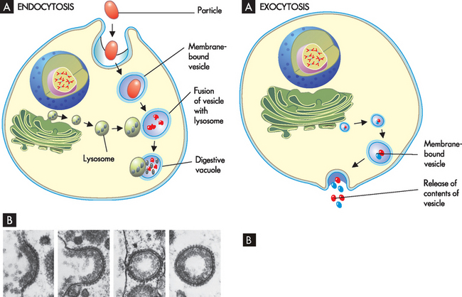
A Endocytosis and fusion with a lysosome. B An electron micrograph of the steps in endocytosis.
Source: B Perry M, Gilbert A. Yolk transport in the ovarian follicle of the hen [Gallus domesticus]: lipoprotein-like particles at the periphery of the oocyte in the rapid growth phase. J Cell Sci 1979; 39: 257–272.
Because most cells continually ingest fluid and solutes by pinocytosis, the terms pinocytosis and endocytosis are often used interchangeably. In pinocytosis, the vesicle containing fluids, solutes or both fuses with a lysosome and lysosomal enzymes digest them for use by the cell. In phagocytosis, the large molecular substances are engulfed by the cell membrane and enter the cell so that they can be isolated and destroyed by lysosomal enzymes. Substances that are not degraded by lysosomes are isolated in residual bodies and released by exocytosis. Both pinocytosis and phagocytosis require metabolic energy and often involve binding of the substance with cell membrane receptors before membrane invagination and fusion with lysosomes in the cell.
Secretion of macromolecules almost always occurs by exocytosis (see Figure 3-18). Exocytosis has two main functions:
A The process of exocytosis, where a vesicle is released out through the cell membrane to the extracellular space. B An electron micrograph of exocytosis.
Source: Raven PH, Johnson GB. Biology. 5th edn. New York: McGraw-Hill; 1999.
CELLULAR METABOLISM
Thus far we have looked at the structure and function of the cellular components, how cells communicate with each other and how substances pass through the cell membrane. Many of these functions require energy. While we may think that we consume food for enjoyment and hunger, the real reason that we eat is to provide nutrients for our cells. By far the most important function of the body is the conversion of foods to ATP, the energy-rich substance that powers the cells. Many chemical reactions are involved in the various stages of ATP production; however, only a rudimentary understanding of chemistry is required to understand these processes. Accordingly, we focus on those processes that are vital to your understanding of the metabolism.
All the chemical tasks of maintaining essential cellular functions are referred to as cellular metabolism. The energy-using process of metabolism is called anabolism (ana meaning upward) and the energy-releasing process is known as catabolism (kata meaning downward). Cellular respiration refers to the catabolic reactions that break down nutrients to ATP. This metabolic process provides cells with the energy they need to produce cellular structures.
Dietary proteins, fats and starches are broken down in the digestive system into amino acids, fatty acids and glucose, respectively. These constituents are then absorbed into the blood, circulated and taken up by the cells, where they are used for various vital cellular processes, including ATP production. The process by which ATP is produced is one example of a series of reactions called a metabolic pathway. A metabolic pathway involves several steps whose end products are not always detectable. A key feature of cellular metabolism is the directing of biological reactions by protein catalysts or enzymes. Each enzyme has a high affinity (strong attraction) for a substrate, a specific substance converted to a product of the reaction.
The role of ATP
For a cell to function it must be able to extract and use the chemical energy in organic molecules — that is, molecules that contain carbon, with the most important organic molecules essential for life being carbohydrates, proteins, lipids and nucleic acids (see Chapter 1). Basically, chemical energy lost by one molecule is transferred to the chemical structure of another molecule by an energy-carrying or transferring molecule such as ATP. The energy stored in ATP can be used in various energy-requiring reactions and in the process is generally converted to adenosine diphosphate (ADP) and an inorganic phosphate. ATP not only stores energy but also transfers it from one molecule to another. The chemical energy stored by carbohydrates, lipids and proteins in food is metabolically broken down to simple smaller molecules through the process of catabolism and then transferred to ATP (see Figure 3-19).
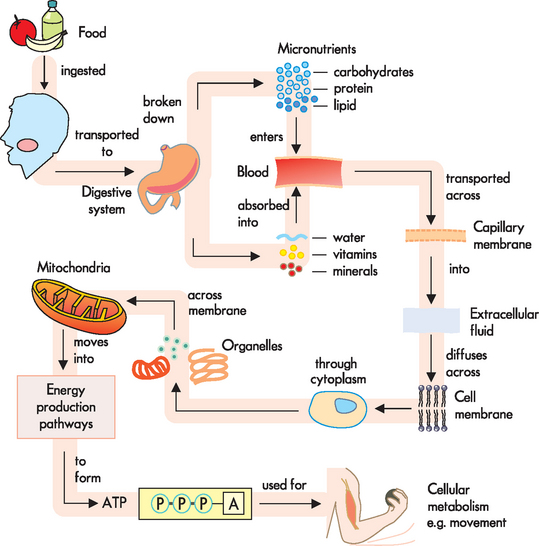
FIGURE 3-19 ATP production from macromolecules.
Food is ingested, broken down into micronutrients in the digestive system and transported to the cells. The nutrients, water and oxygen move through the cell membrane and into the cytoplasm and different organelles. In this example, in the mitochondria energy is transferred by breaking down organic compounds from the nutrients to form ATP. The structure of ATP consists of three phosphate groups that have high-energy bonds that can release chemical energy when required by the cell. The ATP is used for cellular metabolism and energy is released to enable movement.
Note: (1) this is only one example of ATP use; and (2) energy is neither created nor destroyed, but is transferred from one form to another.
As long as ATP is present, cells can perform their functions. The majority of ATP is quickly used by the cells after it is produced. Since the body does not have large reserves of stored ATP it continually produces ATP through cellular respiration. This process can occur with or without oxygen present:
Aerobic respiration refers to the production of ATP in the presence of oxygen — this process produces large amounts of ATP, with water and carbon dioxide as by-products. Anaerobic respiration produces ATP without oxygen by breaking down carbohydrate, with lactic acid as the by-product.Where possible, the body uses aerobic respiration to produce ATP as it is more efficient than anaerobic respiration.
TISSUES
Groups of cells are organised into tissues; different types of tissues compose organs; and organs are integrated to perform complex functions as tracts or systems. All cells are in contact with a network of extracellular macromolecules known as the extracellular matrix. This matrix not only holds cells and tissues together, but also provides an organised latticework within which cells can migrate and interact with one another.
The process by which differentiated cells create tissues and organs is called pattern formation.4 To form tissues, cells must exhibit intercellular recognition and communication, adhesion and memory. Specialised cells sense their environment through signals, such as growth factors, from other cells. This type of communication ensures that new cells are produced only when and where they are required. Different cell types have different adhesion molecules in their cell membranes, sticking selectively to other cells of the same type. They can also adhere to extracellular matrix components.
Types of tissues
There are four primary types of tissues: epithelial, connective, muscle and nervous. The structure and function of these four types underlie the structure and function of each organ system.
Epithelial tissue
Epithelial tissue, also called epithelium, is a protective lining tissue for both inside and outside the body. It lines cavities, forms a lining for organs (such as the heart, lungs and digestive tract) and is the primary tissue of the skin covering our bodies. The cells are typically tightly packed and have no blood supply, relying on the capillaries in the underlying connective tissue to supply oxygen and nutrients and to remove wastes. Epithelial tissue has several functions, including:
The classification of epithelial tissue is based on the shape and layer arrangement of the cells. There are three basic shapes: squamous, cuboidal and columnar. Another type, transitional, refers to cells that can change shape. There are two major divisions based on layers: simple and stratified. Pseudostratified epithelium is a special type of simple epithelium, with the tissue appearing to have multiple layers (stratified) but in fact this is false (hence pseudo). Examples of this type of tissue and some of their respective locations are provided in Figure 3-20.
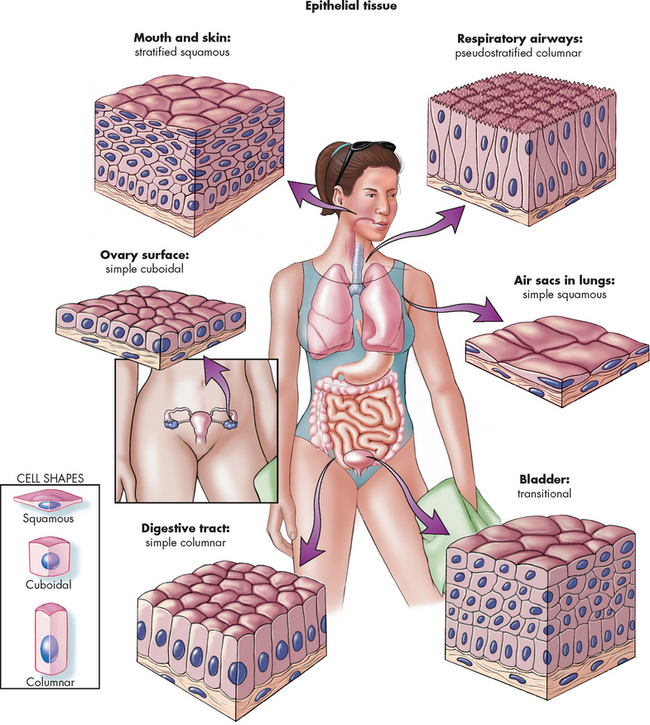
FIGURE 3-20 Classification and locations of epithelial tissue.
The tissues are classified according to the shape and arrangement of the cells.
Source: Patton KT, Thibodeau GA. Anatomy & physiology. 7th edn. St Louis: Mosby; 2010; Herlihy B. The human body in health and illness. 3rd edn. Philadelphia: Saunders; 2007.
Connective tissue
Connective tissue is the most abundant tissue in the body and is widely distributed throughout the body. In contrast to epithelium, the cells are loosely arranged and are not in contact with each other — an extracellular matrix fills the spaces between the cells. Its functions include protection, fat storage, support, transport and, as the name suggests, connecting or binding other tissues and organs so that they are held in place.
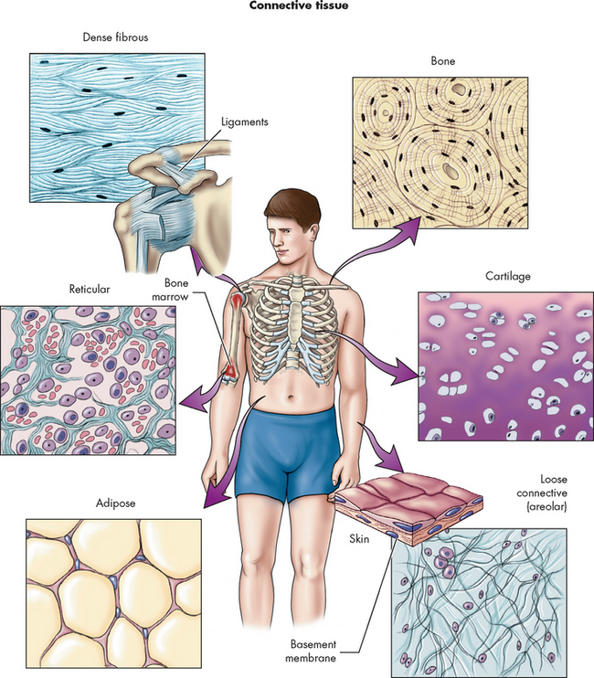
The various tissues that are classified as connective tissue often cause considerable confusion for students. The classification is based on exclusion, with tissues that do not conform to epithelial, muscle or nervous tissues being classified as connective. Accordingly, connective tissue is the most varied of the tissues and its appearance varies considerably. The different types include loose connective tissue, dense fibrous tissue, cartilage, bone, blood and bone marrow. Basically, loose connective tissue includes adipose (fat) and reticular tissue (resembling a netlike formation). Reticular tissue is situated in organs such as the spleen and liver where it forms a frame or scaffold for other cells in these organs. Dense fibrous tissue can be regular or irregular (referring to the appearance of the tissue): regular dense tissue is located in tendons and ligaments; and irregular dense tissue is located in the dermis of the skin and the capsules around the liver, kidneys and spleen. Blood, cartilage and bone are referred to in the haematological and musculoskeletal systems in Chapters 16 and 20, respectively. Connective tissue types are summarised in Figure 3-21.
Muscle tissue
Muscle tissue has long, thin cells that contract and shorten when stimulated, resulting in movement. The other defining characteristic is that the cells are densely packed with proteins, which are responsible for muscle contraction. There are three main types of muscle tissue: skeletal, cardiac and smooth muscle. Briefly:
Skeletal muscle tissue consists of muscle fibres that are arranged attached to bones (hence the name ‘skeletal’) and are responsible for voluntary movement. More details of skeletal muscle tissue are provided in Chapter 20. Cardiac muscle tissue is similar to skeletal muscle tissue, but it is limited to the heart. The tissue contracts involuntarily, which means that it is not under conscious control, and contracts continually throughout your entire life. Further details can be found in Chapter 22. Smooth muscle tissue is found in a variety of locations, such as the digestive system, the airways of the lungs and the blood vessels. The name derives from its appearance and its functions are connected with the organ where it is located. For instance, smooth muscle tissue can change the diameter of blood vessels and so is vitally important in maintaining normal blood pressure.Nervous tissue
Nervous tissue is composed of highly specialised cells called neurons, which receive and transmit electrical impulses rapidly across junctions called synapses. Different types of neurons have special characteristics that depend on their distribution and function within the nervous system and are explored in Chapter 6.
Cellular structure and function
Cellular components
The organelles (little organs) are suspended in the cytoplasm and the majority are enclosed in membranes, except for the ribosomes, centrioles and cytoskeleton. The nucleus is the largest membrane-bound organelle and is found usually in the cell’s centre. The chief functions of the nucleus are cell division and control of genetic information. Ribosomes are composed of ribonucleic acid (RNA) and protein and are located in the cytoplasm and endoplasmic reticulum. They are primarily responsible for protein synthesis. The endoplasmic reticulum is a network of tubular channels (cisternae) that extends throughout the outer nuclear membrane. It specialises in the synthesis and transport of the protein and lipid components of most of the organelles. The Golgi apparatus is a network of smooth membranes and vesicles located near the nucleus. It is responsible for processing and packaging proteins into secretory vesicles. These vesicles break away from the Golgi apparatus and migrate to a variety of intracellular and extracellular destinations, including the cell membrane. Lysosomes are sac-like structures that originate from the Golgi apparatus and contain digestive enzymes. These enzymes are responsible for digesting most cellular substances down to their basic form, such as amino acids, fatty acids and sugars. Peroxisomes are similar to lysosomes but contain several enzymes that either produce or use hydrogen peroxide.The cell membrane
The cell membrane encloses the cell and, by controlling the movement of substances across it, exerts a powerful influence on metabolic pathways. The cell membrane is a bilayer of lipids (phospholipids) and cholesterol, which gives the membrane its structural integrity.Cellular receptors and communication
Cellular receptors are protein molecules on the cell membrane, in the cytoplasm or in the nucleus that are capable of recognising and binding to various substances, such as hormones. Protein receptors on the cell membrane enable the cell to interact with other cells and with extracellular substances.Membrane transport
Two types of solute exist in body fluids: electrolytes and non-electrolytes. Electrolytes are electrically charged and dissociate into constituent ions when placed in solution. Non-electrolytes do not dissociate when placed in solution. Water and small electrically uncharged molecules move through pores in the cell membrane’s lipid bilayer in the process called passive transport. Passive transport does not require the expenditure of energy; rather, it is driven by the physical effects of osmosis, hydrostatic pressure and diffusion. Filtration is the measurement of water and solutes through a membrane because of a greater pushing pressure. Diffusion is the passive movement of a solute from an area of higher solute concentration to an area of lower solute concentration. Facilitated diffusion does not require energy: substances are transported across the cell membrane via an integral membrane, through either a channel or a carrier protein. Osmosis is the movement of water across a semi-permeable membrane from a region of lower solute concentration to a region of higher solute concentration. The amount of hydrostatic pressure required to oppose the osmotic movement of water is called the osmotic pressure of the solution. Active transport requires metabolic energy (ATP) to move molecules against the concentration gradient. Larger molecules and molecular complexes are moved into the cell by active transport, which requires the cell to expend energy (by means of ATP). Active transport occurs also by endocytosis or vesicle formation, in which the substance to be transported is engulfed by a segment of the cell membrane, forming a vesicle that moves into the cell. The largest molecules (macromolecules) and fluids are transported by the processes of endocytosis (ingestion) and exocytosis (expulsion). Pinocytosis is a type of endocytosis in which fluids and solute molecules are ingested through the formation of small vesicles. Phagocytosis is a type of endocytosis in which large particles, such as bacteria, are ingested through the formation of large vesicles, called vacuoles.Cellular metabolism
The chemical tasks of maintaining essential cellular functions are referred to as cellular metabolism. Anabolism is the energy-using process of metabolism, whereas catabolism is the energy-releasing process.Tissues
Cells of one or more types are organised into tissues; different types of tissues compose organs; and organs are organised to function as tracts or systems. Epithelial tissue covers most internal and external surfaces of the body. The functions of epithelial tissue include protection, absorption, secretion and excretion. Connective tissue binds various tissues and organs together, supporting them in their locations and serving as storage sites for excess nutrients.Emma and Amelie are in the laboratory doing an introductory practical on cells. Their tutor has asked them to look at slides of different types of cells under the microscope. They have been told to look at the various structures and to compare them with the illustrations of cells in their textbook. Emma and Amelie notice that parts of the cell look different from the textbook illustrations and that the shapes vary according to the type of cell.
Answer the following questions as though you were participating in the lab session with Emma and Amelie.