Diarrhoea

Diarrhoea is a common clinical problem and there is no uniformly accepted definition of diarrhoea. Organic causes (stool weights >250 g/day) have to be distinguished from functional causes. Sudden onset of bowel frequency associated with crampy abdominal pains, and a fever will point to an infective cause; bowel frequency with loose blood-stained stools to an inflammatory basis; and the passage of pale offensive stools that float, often accompanied by loss of appetite and weight loss, to steatorrhoea. Nocturnal bowel frequency and urgency usually point to an organic cause. Passage of frequent small-volume stools (often formed) points to a functional cause (see Functional gastrointestinal disorders, below).

Mechanisms
Osmotic diarrhoea

The gut mucosa acts as a semipermeable membrane and fluid enters the bowel if there are large quantities of non-absorbed hypertonic substances in the lumen. This occurs because:

image The patient has ingested a non-absorbable substance (e.g. a purgative such as magnesium sulphate or magnesium-containing antacid)

image The patient has generalized malabsorption so that high concentrations of solute (e.g. glucose) remain in the lumen

image The patient has a specific absorptive defect (e.g. disaccharidase deficiency or glucose-galactose malabsorption).

The volume of diarrhoea produced by these mechanisms is reduced by the absorption of fluid by the ileum and colon. The diarrhoea stops when the patient stops eating or the malabsorptive substance is discontinued.

Secretory diarrhoea

In this disorder, there is both active intestinal secretion of fluid and electrolytes as well as decreased absorption. The mechanism of intestinal secretion is shown in Figure 6.45a.

image

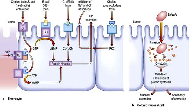
Figure 6.45 Mechanisms of diarrhoea. (a) Small intestinal secretion of water and electrolytes. Cholera toxin binds to its receptor (monosialoganglioside Gi) via fimbria (toxin co-regulated pilus) on its β-subunit. This activates the as subunit (of the Gs protein), which in turn dissociates and activates cyclic AMP (cAMP). The increase in cAMP activates intermediates (e.g. protein kinase and Ca2+) which then act on the apical membrane causing Cl secretion (with water) and inhibition of Na+ and Cl absorption. Heat-labile E. coli enterotoxin shares a receptor with cholera toxin. Heat-stable (HS) E. coli toxin binds to its own receptor and activates guanylate cyclase (cGMP), producing the same effect on secretion. C. difficile activates the protein kinases via Ca2+/calmodulin (Ca2+/CM). Zona occludens toxin is the product of the ZOT gene, which is the gene required for the CTX gene which encodes for cholera toxin. It has enterotoxic activity, producing secretion. Cholera and E. coli cause these effects without invasion of the cell. G, G protein consisting of subunits α, β, γ; i, inhibitory; s, stimulatory; ATP, adenosine triphosphate; GTP, guanosine triphosphate; GMP, guanosine monophosphate; GC, guanyl cyclase; PKC, protein kinase C; VIP, vasoactive intestinal polypeptide. (b) Colonic mucosal cell. This demonstrates one of the mechanisms by which an invasive pathogen (e.g. Shigella) acts. Following penetration, the pathogens generate cytotoxins which lead to mucosal ulceration and cell death.

Common causes of secretory diarrhoea are:

image Enterotoxins (e.g. cholera, E. coli thermolabile or thermostable toxin, C. difficile)

image Hormones (e.g. vasoactive intestinal peptide in the Verner–Morrison syndrome, p. 370)

image Bile salts (in the colon) following ileal resection

image Fatty acids (in the colon) following ileal resection

image Some laxatives (e.g. docusate sodium).

Inflammatory diarrhoea (mucosal destruction)

Diarrhoea occurs because of damage to the intestinal mucosal cell so that there is a loss of fluid and blood (Fig. 6.45b). In addition, there is defective absorption of fluid and electrolytes. Common causes are infective conditions (e.g. dysentery due to Shigella) and inflammatory conditions (e.g. ulcerative colitis and Crohn’s disease).

Abnormal motility

Diabetic, post-vagotomy and hyperthyroid diarrhoea are all due to abnormal motility of the upper gut. Symptoms may be exacerbated by small bowel bacterial overgrowth.

Causes of diarrhoea are shown in Table 6.21. It should be noted that the irritable bowel syndrome, colorectal cancer, diverticular disease and faecal impaction with overflow in the elderly do not cause ‘true’ organic diarrhoea (i.e. >250 g/day), even though the patients may complain of diarrhoea. Worldwide, infection and infestation are a major problem and these are discussed under the causative organisms in Chapter 4.

Table 6.21 Causes of diarrhoea

Infective causes

Bacterial, e.g.
Campylobacter jejuni
Salmonella spp.
Shigella
Escherichia coli (p. 121)
Staphylococcal enterocolitis
Bacillus cereus
Clostridium perfringens, botulinum, difficile
gastrointestinal tuberculosis
Viral, e.g.
rotavirus
Fungal, e.g.
histoplasmosis
Parasitic, e.g.
amoebic dysentery (Entamoeba histolytica)
schistosomiasis
Giardia intestinalis

Non-infective causes of diarrhoea

Inflammatory bowel disease
Radiation proctitis or colitis
Behçet’s disease
Diverticular disease
Ischaemic colitis
Gastrointestinal lymphoma
Carcinoma of the colon (change in bowel habit)
Malabsorption
Gut resection
Bile acid malabsorption
Drugs – many, including
laxatives
metformin
anticancer drugs
statins
proton pump inhibitors
Faecal impaction with overflow
Irritable bowel syndrome and functional diarrhoea

Endocrine

Zollinger–Ellison syndrome
VIPoma
Somatostatinoma
Glucagonoma
Carcinoid syndrome
Thyrotoxicosis
Medullary carcinoma of thyroid
Diabetic autonomic neuropathy

Factitious diarrhoea

Purgative abuse
Dilutional diarrhoea

Acute diarrhoea (excluding cholera – see p. 103)

Diarrhoea of sudden onset is very common, often short-lived and requires no investigation or treatment. Although dietary causes should be considered, diarrhoea due to viral agents may also last 24–48 hours (see p. 103). The causes of other infective diarrhoeas are shown on page 119. Travellers’ diarrhoea, which affects people travelling outside their own countries, particularly to developing countries, usually lasts 2–5 days; it is discussed on page 122. Clinical features associated with the acute diarrhoeas include fever, abdominal pain and vomiting. If the diarrhoea is particularly severe, dehydration can be a problem; the very young and very old are at special risk from this. Investigations are necessary if the diarrhoea has lasted more than 1 week. Stools (up to three) should be sent immediately to the laboratory for culture and examination for ova, cysts and parasites and Clostridium difficile toxin assay. If the diagnosis has still not been made, a sigmoidoscopy and rectal biopsy should be performed and imaging should be considered. Viral and bacterial infective diarrhoeas do not last more than two weeks.

Oral fluid and electrolyte replacement is often necessary. Special oral rehydration solutions (e.g. sodium chloride and glucose powder) are available for use in severe episodes of diarrhoea, particularly in infants. Antidiarrhoeal drugs are thought to impair the clearance of any pathogen from the bowel but may be necessary for short-term relief (e.g. codeine phosphate 30 mg four times daily or loperamide 2 mg three times daily). Antibiotics are occasionally necessary (see p. 125) depending on the organism.

Chronic diarrhoea

This always needs investigation. The flow diagram in Figure 6.46 is illustrative; whether the large or the small bowel is investigated first will depend on the clinical story of, for example, bloody diarrhoea or steatorrhoea. The investigations and treatment are described in detail under the individual diseases. Colonoscopy is usually necessary if stool cultures are negative and small bowel disease is not suspected.

image

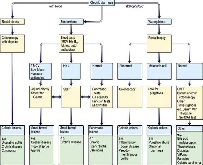
Figure 6.46 Flow diagram for the investigation of chronic diarrhoea. Note: All patients should have had stool cultures with toxin testing for C. difficile. SBFT, small bowel follow-through; VIP, vasoactive intestinal polypeptide; MRCP, magnetic resonance cholangiopancreatography; SeHCAT, 75Se-homocholic acid taurine.

C. difficile-associated diarrhoea (pseudomembranous colitis)

Pseudomembranous colitis (see p. 122) may develop following the use of any antibiotic. Diarrhoea occurs in the first few days after taking the antibiotic or even up to 6 weeks after stopping the drug. The causative agent is Clostridium difficile (see p. 122).

Bile acid malabsorption

Bile acid malabsorption is an underdiagnosed cause of chronic diarrhoea and many patients with this disorder are assumed to have irritable bowel syndrome. Bile acid diarrhoea occurs when the terminal ileum fails to reabsorb bile acids. Bile acids (particularly the dihydroxy bile acids: deoxycholate and chenodeoxycholate) when present in increased concentrations in the colon lead to diarrhoea by reducing absorption of water and electrolytes and, at higher concentrations, inducing secretion as well as increasing colonic motility. A variety of causes of bile acid malabsorption are recognized (Table 6.22).

Table 6.22 Causes of bile acid diarrhoea

Ileal resection

Ileal disease, e.g. active or inactive Crohn’s disease

Primary bile acid diarrhoea

Postinfective gastroenteritis

Rapid small bowel transit

Post-cholecystectomy

Bile acid malabsorption should be considered not only in patients with chronic diarrhoea of unknown cause but also in patients with diarrhoea and associated disease who are not responding to standard therapy (e.g. patients with terminal ileal Crohn’s disease, microscopic inflammatory colitis).

Diagnosis is made using the SeHCAT test in which a radiolabelled bile acid analogue is administered and percentage retention at 7 days calculated (<19% retention abnormal). Treatment is with bile salt sequestrants such as cholestyramine, a resin which binds and inactivates the action of bile acids in the colon. The best results of treatment are obtained in patients with a SeHCAT retention of <5%.

Factitious diarrhoea

Factitious diarrhoea accounts for up to 4% of new patients with diarrhoea attending gastroenterology clinics.

Purgative abuse

This is most commonly seen in females who surreptitiously take high-dose purgatives and are often extensively investigated for chronic diarrhoea. The diarrhoea is usually of high volume (>1 L daily) and patients may have a low serum potassium. Biochemical analysis of the stool may help diagnose laxative abuse. Management is difficult as most patients deny purgative ingestion. Purgative abuse often occurs in association with eating disorders and patients may need psychiatric help.

Dilutional diarrhoea

In this condition, raised stool weights occur as a consequence of patients deliberately diluting their stool with urine or tap water. The diagnosis is made by measuring stool osmolality and electrolyte concentrations in order to calculate the faecal osmolar gap.

Diarrhoea in patients with HIV infection

Chronic diarrhoea is a common symptom in HIV infection, but HIV’s role in the pathogenesis of diarrhoea is unclear. Cryptosporidium (see p. 110) is the pathogen most commonly isolated. Isospora belli and microsporidia have also been found.

The cause of the diarrhoea is often not found and treatment is symptomatic. Table 6.23 shows the conditions affecting the gastrointestinal tract in patients with AIDS.

Table 6.23 Gastrointestinal problems in patients with AIDS

Symptoms Causes

Mouth/oesophagus


 Dysphagia

Herpes simplex virus (HSV)

 Retrosternal discomfort Oral ulceration

Cytomegalovirus (CMV)

 Candidiasis

Small bowel/colon


 Chronic diarrhoea

Parasites:

 Steatorrhoea

 Entamoeba histolytica

 Weight loss

 Giardia intestinalis


 Cryptosporidium


 Blastocysts hominis


 Isospora belli


 Microsporidia


 Cyclospora cayetanensis


Viruses:


 CMV/HSV, adenovirus


Bacteria:


 Salmonella


 Campylobacter


 Shigella


 Mycobacterium avium-intracellulare


Non-infective


 enteropathy – cause unknown

Rectum/colon


 Bloody diarrhoea

Bacterial infection (e.g. Shigella)

Any site


 Weight loss

Neoplasia:

 Diarrhoea

 Kaposi’s sarcoma


 Lymphoma


 Squamous carcinoma


Infection – disseminated, e.g.


 Mycobacterium


 avium-intracellulare


HAART therapy

Functional gastrointestinal disorders

There is a large group of gastrointestinal disorders that are termed ‘functional’ because symptoms occur in the absence of any demonstrable abnormalities in the digestion and absorption of nutrients, fluid and electrolytes and no structural abnormality can be identified in the gastrointestinal tract, although there may be discernible abnormalities in neuromuscular function such as dysmotility and visceral hypersensitivity, which are not routinely investigated.

Table 6.24 lists some of the symptoms that are suggestive of a functional gastrointestinal disorder. Modern classification systems are based on the premise that for each disorder there is a symptom cluster that ‘breeds true’ across clinical and population groups. There is inevitably overlap, with some symptoms being common to more than one disorder.

Table 6.24 Chronic gastrointestinal symptoms suggestive of a functional gastrointestinal disorder (FGID)

Nausea alone

Vomiting alone

Belching

Chest pain unrelated to exercise

Postprandial fullness

Abdominal bloating

Abdominal discomfort/pain (right or left iliac fossa)

Passage of mucus per rectum

Frequent bowel actions with urgency first thing in morning

Table 6.25 lists the common functional gastrointestinal disorders as defined by Rome III criteria. These conditions are extremely common worldwide, comprising 80% of patients seen in the gastroenterology clinic.

Table 6.25 Modified Rome III functional gastrointestinal disorders

A. Functional oesophageal disorders

Heartburn
Chest pain of presumed oesophageal origin
Dysphagia
Globus

B. Functional gastroduodenal disorders

Non-ulcer dyspepsia
Belching disorders
Nausea and vomiting disorders
Rumination syndrome in adults

C. Functional bowel disorders

Irritable bowel syndrome
Functional bloating
Functional constipation
Functional diarrhoea
Unspecified functional bowel disorder

D. Functional abdominal pain syndrome

E. Functional gall bladder and sphincter of Oddi (SO) disorders

Pathophysiology and brain–gut interactions

People with functional gastrointestinal disorders (FGID) are characterized by having a greater gastrointestinal motility response to life events than normal subjects. There is, however, a poor association between measured gastrointestinal motility changes and symptoms in many of the FGIDs. Patients with FGID have been shown to have abnormalities in visceral sensation and have a lower pain threshold when tested with balloon distension (visceral hyperalgesia). Visceral hypersensitivity possibly relates to:

image Altered receptor sensitivity at the viscus itself

image Increased excitability of the spinal cord dorsal horn neurones

image Altered central modulation of sensations.

Symptoms are thought to be due to disturbed gastrointestinal motility that leads to distension with visceral hyperalgesia accentuating the pain but there are no data to confirm this. A systematic review of published studies suggest that 10% of patients who experience an acute infective gastroenteritis develop a degree of FGID. It is not clear whether this is caused by post-infectious bile salt malabsorption, alterations in the mucosal immune system or the use of antibiotics to treat the index infection

The brain–gut axis describes a combination of intestinal motor, sensory and CNS activities (Fig. 6.47). Thus extrinsic (e.g. vision, smell) and intrinsic (e.g. emotion, thought) information can affect gastrointestinal sensation because of the neural connections from higher centres. Conversely, viscero-tropic events can affect central pain perception, mood and behaviour.

image

Figure 6.47 A biopsychosocial conceptualization of the pathogenesis and clinical expression of functional gastrointestinal disorders (FGIDs), showing how genetic, environmental and psychological factors may interact to cause dysregulation of brain-gut function.

(Modified from Drossman DA. The functional gastrointestinal disorders and the Rome III process. Gastroenterology 2006; 130:1377–1390, with permission from Elsevier.)

Psychological stress can exacerbate gastrointestinal symptoms, and psychological disturbances are more common in patients with FGIDs. These disturbances alter attitude to illness, promote healthcare seeking, and often lead to a poor clinical outcome. They have psychosocial consequences with poor quality of life at home and work. Early in life, genetic and environmental influences (e.g. family attitudes towards bowel training, verbal or sexual abuse, exposure to an infection) may affect psychosocial development (susceptibility to life stress, psychological state, coping skills, development of social support) or the development of gut dysfunction (abnormal motility or visceral hypersensitivity). Therefore, FGID should be regarded as a dysregulation of brain–gut function.

Functional oesophageal disorders

The criteria for diagnosis rest mainly on compatible symptoms. However, pathological gastro-oesophageal reflux and eosinophilic oesophagitis may need to be excluded (see p. 244).

Globus

This presents as:

image Persistent or intermittent sensation of a lump or foreign body in the throat

image Occurrence of the sensation between meals

image The absence of dysphagia and pain on swallowing (odynophagia).

Treatment is with explanation and reassurance and a trial of antireflux therapy. Antidepressants may be tried.

Functional chest pain, of presumed oesophageal origin

This is characterized by episodes of mainly midline chest pain, not burning in nature, that are potentially of oesophageal origin and which occur in the absence of a cardiological cause, gastro-oesophageal reflux and achalasia.

More than half of patients will respond to high-dose acid-suppression therapy in the first week; some will respond to nitrates and calcium-channel blockers.

Antidepressant therapy, e.g. amitriptyline or the selective serotonin reuptake inhibitor citalopram, have been shown to be effective.

Functional gastroduodenal disorders

Functional dyspepsia

This is the second most common functional gastrointestinal disorder (after irritable bowel syndrome). Patients can present with a spectrum of symptoms including upper abdominal pain/discomfort, fullness, early satiety, bloating and nausea.

These patients have no structural abnormality as an explanation for their symptoms.

Functional dyspepsia subgroups

Two subgroups based on the predominant (or most bothersome) single symptoms are suggested:

image Epigastric pain syndrome with pain centred in the upper abdomen as the predominant (most bothersome) symptom.

image Postprandial distress syndrome with an unpleasant or troublesome non-painful sensation (discomfort) centred in the upper abdomen being the predominant symptom. This sensation may be associated with upper abdominal fullness, early satiety, bloating and nausea.

There is considerable overlap between these two groups.

Investigations

Helicobacter pylori infection should be serologically excluded, but many young patients (<50) require no further investigation. Older patients or those with alarm symptoms require endoscopy. Gastroscopy often shows gastritis but whether this is the cause of the symptoms is doubtful.

Treatment

The range of therapies prescribed for functional dyspepsia reflects the uncertain pathogenesis and the lack of satisfactory treatment options. Management is further confounded by high placebo response rates (20–60%). A proportion of patients will respond satisfactorily to reassurance, explanations and lifestyle changes. Proton pump inhibitors and prokinetic agents are used for patients with epigastric pain syndrome and postprandial distress syndrome, respectively. Reducing intake of fat, coffee, alcohol and cigarette smoking helps. SSRI medication is tried in refractory cases.

H. pylori eradication therapy has been shown to be effective in some patients with functional dyspepsia.

Aerophagia

Aerophagia refers to a repetitive pattern of swallowing or ingesting air and belching. It is usually an unconscious act unrelated to meals. Usually no investigation is required. Explanation that the symptoms are due to swallowed air and reassurance are necessary, as is treatment of associated psychiatric disease.

Functional vomiting

Functional vomiting is a rare condition in clinical practice, although chronic nausea is a frequent accompaniment in all functional gastrointestinal disorders. CNS pathology and migrainous syndromes (cyclical vomiting syndrome) should be considered and treated Clinically, functional vomiting is characterized by:

image Frequent episodes of vomiting, occurring on at least 1 day a week

image Absence of criteria for an eating disorder, rumination or major psychiatric disease

image Absence of self-induced and medication-induced vomiting

image Absence of abnormalities in the gut or central nervous system and metabolic disease to explain the recurrent vomiting.

Investigation is often not required but always exclude non-GI disorders (see Table 6.1).

Treatment is with anti-nausea drugs and antidepressants; behavioural therapy and psychotherapy are helpful. Dietary changes occasionally help.

Functional bowel disorders

Irritable bowel syndrome (IBS)

IBS is the commonest FGID. In western populations, up to one in five people report symptoms consistent with IBS. Approximately 50% will consult their doctors and of these up to 30% will be referred by their doctor to a hospital specialist. Up to 40% of all patients seen in specialist gastroenterology clinics will have IBS. Estimates in the UK put the annual cost of IBS to healthcare resources as GB£45.6 million; in the USA, the cost is higher at US$8 billion. In the UK, approximately one-quarter of IBS patients lose time off work for periods ranging from 7 to 13 days each year.

The factors that determine whether an IBS sufferer in the community seeks medical advice include higher illness attitude scores and higher anxiety and depression scores than non-consulters. Consulters perceive that their symptoms are severer than non-consulters, and consulting behaviour may be determined by the number of presenting symptoms. Female consulters outnumber male consulters by a factor of 2–3.

IBS – a multisystem disorder

IBS patients suffer from a number of non-intestinal symptoms (Table 6.26), which may be more intrusive than the classical features. IBS co-exists with chronic fatigue syndrome (see p. 1162), fibromyalgia (see p. 509) and temporomandibular joint dysfunction.

Table 6.26 Non-gastrointestinal features of irritable bowel syndrome

Gynaecological symptoms

Painful periods (dysmenorrhoea)
Pain following sexual intercourse (dyspareunia)

Urinary symptoms

Frequency
Urgency
Passing urine at night (nocturia)
Incomplete emptying of bladder

Other symptoms

Joint hypermobility
Back pain
Headaches
Bad breath, unpleasant taste in the mouth
Poor sleeping
Fatigue

The biopsychosocial conceptualization of the pathogenesis and clinical expression of FGIDs (Fig. 6.48) is particularly relevant to IBS and Box 6.11 lists some common factors that have been shown to trigger IBS symptoms. Infectious diarrhoea precedes the onset of IBS symptoms in 7–30% of patients. Whether this is a factor for all patients or just a small subgroup remains controversial. Risk factors in these patients have been shown to include female gender, severity and duration of diarrhoea, pre-existing life events and high hypochondriacal anxiety and neurotic scores at the time of the initial illness. Symptoms of anxiety and depression are more common in IBS patients and stress or adverse life events often precede the onset of chronic bowel symptoms.

image Box 6.11

Some factors that can trigger onset of irritable bowel symptoms

image Affective disorders, e.g. depression, anxiety

image Psychological stress and trauma

image Gastrointestinal infection

image Antibiotic therapy

image Sexual, physical, verbal abuse

image Pelvic surgery

image Eating disorders

FURTHER READING

Chang L. The role of stress on physiologic responses and clinical symptoms in irritable bowel syndrome. Gastroenterology 2011: 140:761–765.

Ford AC, Talley NJ. IBS in 2010: advances in pathophysiology, diagnosis and treatment. Nat Rev Gastroenterol Hepatol 2011; 8: 76–78.

Diagnostic criteria (Rome III 2006)

These criteria state that in the preceding 3 months, there should be at least 3 days/month of recurrent abdominal pain or discomfort associated with two or more of the following:

1. Improvement with defecation

2. Onset associated with a change in frequency of stool

3. Onset associated with a change in form (appearance) of stool.

These are useful for comparative studies.

Subgroups of IBS patients can be identified according to the criteria listed in Table 6.27.

Table 6.27 Subtyping irritable bowel syndrome by predominant stool pattern

IBS with constipation (IBS-C)

Hard lumpy stools >25% and loose (mushy) or watery stools <25% of bowel movements

IBS with diarrhoea (IBS-D)

Loose (mushy) or watery stools >25% and hard or lumpy stools <25% of bowel movements

Mixed IBS (IBS-M)

Hard or lumpy stools >25% and loose (mushy) or watery stools >25% of bowel movements

Unsubtyped IBS

Insufficient abnormality of stool consistency to meet criteria for IBS-C, D or M

The decision as to whether to investigate and the choice of investigations should be based on clinical judgement. Pointers to the need for thorough investigation are the presence of the above symptoms in association with rectal bleeding, nocturnal pain, fever and weight loss and a clinical suspicion of organic diarrhoea. A raised stool calprotectin or lactoferrin would suggest inflammation needing further investigation.

Treatment

Current strategies for treatment of IBS include therapies that target central and end-organ pathways (Box 6.12) and are not mutually exclusive. HT4 receptor agonists for constipation-predominant IBS (prucalopride) are now licensed for use in many countries, although the 5HT3 drugs designed to treat diarrhoea-associated IBS await regulatory approval.

image Box 6.12

Approaches to management of the irritable bowel syndrome (IBS)

  Action

End organ treatment


Explore dietary triggers

Refer to dietitian

High-fibre diet ± fibre supplements for constipationFODMAP diet for bloating

Refer to dietitian

Alter the microbiota

Rifaximin has shown short-term benefit in IBS patients without Pro- and pre-biotics constipation (Target I and II trials)

Anti-diarrhoeal drugs for bowel frequency

Loperamide

Codeine phosphate

Co-phenotrope

Constipation

5HT4 receptor agonist, e.g. prucalopride

Smooth muscle relaxants for pain

Mebeverine hydrochloride

Dicycloverine hydrochloride

Peppermint oil

Central treatment


Explain physiology and symptoms

At consultation (leaflets with diagrams help)

Psychotherapy

Refer to clinical psychologist (see p. 1163)

Hypnotherapy


Cognitive behavioural therapy

Refer to psychiatrist

Antidepressants

Functional diarrhoea – clomipramineDiarrhoea-predominant IBS – tricyclic group, e.g. amitriptylineConstipation-predominant IBS – SSRI, e.g. paroxetine

FURTHER READING

Manabe N, Rao AS, Wong BS et al. Emerging pharmacologic therapies for irritable bowel syndrome. Curr Gastroenterol Rep 2010; 12:8–16.

Probiotics and prebiotics

Probiotics are live or attenuated bacteria or bacterial products that confer a significant health benefit to the host. Recent studies have shown a beneficial effect of specific probiotics such as Bifidobacterium infantis 35624 on IBS symptoms. However, problems with quality control and formulation of probiotics are currently restricting the clinical availability of some probiotics.

Prebiotics are non-digestible food supplements that are fermented by host bacteria thereby altering the microbiota of the host often by stimulating the growth of healthy bacteria. There is early evidence that they may have a beneficial impact on some IBS symptoms.

FURTHER READING

Moayyedi P, Ford AC, Talley NJ et al. The efficacy of probiotics in the treatment of irritable bowel syndrome: a systematic review. Gut 2010; 59:325–332.

Pain/gas/bloat syndrome/midgut dysmotility

There exist a group of patients with functional bowel disease whose abdominal pain and other clinical features are likely to occur as a consequence of disordered motility and visceral sensation that predominantly affects the small intestine or midgut. The symptom-based diagnostic criteria are abdominal pain, often exacerbated by eating and not relieved by opening the bowels and not associated with the passage of more frequent or looser stools than normal and not associated with constipation. Other symptoms include abdominal distension (bloating), postprandial fullness, nausea and, on occasions, anorexia and weight loss.

Treatment of patients with pain/gas/bloat syndrome is not easy; and in some, pain can be chronic and severe. Narcotics should always be avoided. Central and end-organ targeted treatment approaches should be combined, e.g. selective serotonin reuptake inhibitor paroxetine combined with a prokinetic agent domperidone or smooth muscle relaxant, e.g. mebeverine. Patients can be referred to a dietician for a trial of a FODMAP (fermentable oligo-, di- and mono-saccharides and polyols) diet. Small bowel bacterial overgrowth can be a contributory feature.

Some patients with pain/gas/bloat syndrome have particularly severe and chronic symptoms which may be nocturnal. A small subgroup of these has been shown to have manometric features consistent with a diagnosis of chronic idiopathic intestinal pseudo-obstruction (CIIP), and specifically of an enteric neuropathy. Full-thickness small intestinal biopsies confirm this diagnosis by showing a deficiency of α actin staining in the inner circular layer of smooth muscle. More appropriately these patients should be considered to have a gastrointestinal neuromuscular disorder of the gut. About 10% of these patients are subsequently found to have an underlying autoimmune overlap disorder (see p. 541).

Treatment of patients with neuromuscular disorders of the gut requires a multidisciplinary approach, with emphasis on management of pain, psychological state and nutrition. Patients with underlying autoimmune inflammatory mixed connective tissue disorders may benefit from primary treatment of these. Patients with intestinal failure as a result of CIIP need long-term parenteral nutrition.

Functional diarrhoea

In this form of functional bowel disease, symptoms occur in the absence of abdominal pain and commonly are:

image The passage of several stools in rapid succession usually first thing in the morning. No further bowel action may occur that day or defecation only after meals

image The first stool of the day is usually formed, the later ones are mushy, looser or watery

image Urgency of defecation

image Anxiety, uncertainty about bowel function with restriction of movement (e.g. travelling)

image Exhaustion after the defecation.

Chronic diarrhoea without pain is caused by many diseases indistinguishable by history from functional diarrhoea. Features atypical for a functional disorder (e.g. large-volume stools, rectal bleeding, nutritional deficiency and weight loss) call for more extensive investigations.

Treatment of functional diarrhoea is with loperamide often combined with a tricyclic antidepressant prescribed at night (e.g. clomipramine 10–30 mg).

FURTHER READING

Drossman DA. The functional gastrointestinal disorders and the Rome III process. Gastroenterology 2006; 130:1377–1390.

The acute abdomen

This section deals with the acute abdominal conditions that cause the patient to be hospitalized within a few hours of the onset of pain (Table 6.28). If recognized quickly as an emergency, reduction in morbidity and mortality can be achieved. Although a specific diagnosis should be attempted, the immediate problem in management is to decide whether an ‘acute abdomen’ exists and whether surgery is required.

Table 6.28 Common causes of acute abdominal pain

Diagnosis %

Nonspecific abdominal pain

35

Acute appendicitis

30

Gall bladder disease

10

Gynaecological disorders

5

Intestinal obstruction

5

Perforated ulcer/dyspepsia

5

Renal colic

2

Urinary tract infection

2

Diverticular disease

2

Other diagnoses

4

Percentages are approximate and vary in different communities.

History

This should include previous operations, any gynaecological problems and whether any concurrent medical condition is present.

Pain

The onset, site, type and subsequent course of the pain should be determined as accurately as possible. In general, the pain of an acute abdomen can either be constant (usually owing to inflammation) or colicky because of a blocked ‘tube’. The inflammatory nature of a constant pain will be supported by a raised temperature, tachycardia and/or a raised white cell count. If these are normal, then other causes (e.g. musculoskeletal, aortic aneurysm) or rare causes (e.g. porphyria) should be considered. Colicky pain can be due to an obstruction of the gut, biliary system, urogenital system or the uterus. These will probably initially require conservative management along with analgesics. If a colicky pain becomes a constant pain, then inflammation of the organ may have supervened (e.g. strangulated hernia, ascending cholangitis or salpingitis).

A sudden onset of pain suggests:

image Perforation (e.g. of a duodenal ulcer)

image Rupture (e.g. of an ectopic pregnancy)

image Torsion (e.g. of an ovarian cyst)

image Acute pancreatitis

image Infarction (e.g. mesenteric).

Back pain suggests:

image Pancreatitis

image Rupture of an aortic aneurysm

image Renal tract disease.

Inflammatory conditions (e.g. appendicitis) produce a more gradual onset of pain. With peritonitis the pain is continuous and may be made worse by movement. Many inflammatory conditions can progress to those listed in sudden onset due to complications.

Vomiting

Vomiting may accompany any acute abdominal pain but, if persistent, it suggests an obstructive lesion of the gut. The character of the vomit should be asked – does it contain blood, bile or small bowel contents?

Other symptoms

Any change in bowel habit or of urinary frequency should be documented and, in females, a gynaecological history including LMP should be taken.

Physical examination

The general condition of the person should be noted. Does the person look ill? Is he or she shocked? Large volumes of fluid may be lost from the vascular compartment into the peritoneal cavity or into the lumen of the bowel, giving rise to hypovolaemia, i.e. a pale cold skin, a weak rapid pulse and hypotension.

The abdomen

image Inspection. Look for the presence of scars, distension or masses.

image Palpation. The abdomen should be examined gently for sites of tenderness and the presence or absence of guarding. Guarding is involuntary spasm of the abdominal wall and it indicates peritonitis. This can be localized to one area or it may be generalized, involving the whole abdomen.

image Bowel sounds. Increased high-pitch tinkling bowel sounds indicate fluid obstruction; this occurs because of fluid movement within the dilated bowel lumen. Absent bowel sounds suggest peritonitis. In an obstructed patient, absent bowel sounds suggest strangulation, ischaemia or ileus. It is essential that the hernial orifices be examined if intestinal obstruction is suspected.

Vaginal and rectal examination

Vaginal examination can be very helpful, particularly in diagnosing gynaecological causes of an acute abdomen (e.g. a ruptured ectopic pregnancy). Rectal examination is less helpful as localized tenderness may be due to any cause; it may show blood on the glove.

Flexible sigmoidoscopy

If diarrhoea is present, unprepared (without use of laxative bowel preparation or enema) flexible sigmoidoscopy may be indicated to aid exclusion of infective, inflammatory and ischaemic causes of acute pain. A specimen of stool should be taken for stool culture for bacterial pathogens (e.g. Campylobacter, Salmonella, Shigella and Clostridium difficile toxin) (see p. 174).

Other observations

image Mouth. The tongue is furred in some cases and a fetor is present.

image Temperature. Fever is more common in acute inflammatory processes.

image Urine. Examine for:

blood – suggests urinary tract infection or renal colic
glucose and ketones – ketoacidosis can present with acute pain
protein and white cells – to exclude acute pyelonephritis.

image Think of medical causes (Table 6.29).

Table 6.29 Medical causes of acute abdomen

Referred pain

Pneumonia
Myocardial infarction

Functional gastrointestinal disorders

Renal causes

Pelviureteric colic
Acute pyelonephritis

Metabolic causes

Diabetes mellitus
Acute intermittent porphyria
Lead poisoning
Familial Mediterranean fever

Haematological causes

Haemophilia and other bleeding disorders
Henoch–Schönlein purpura
Sickle cell crisis
Polycythaemia vera
Paroxysmal nocturnal haemoglobinaemia

Vasculitis

Embolic
Peri-aortitis
Investigations

image Blood count. A raised white cell count occurs in inflammatory conditions.

image Serum amylase. High levels (more than five times normal) indicate acute pancreatitis. Raised levels below this can occur in any acute abdomen and should not be considered diagnostic of pancreatitis.

image Serum electrolytes. These are not particularly helpful for diagnosis but useful for general evaluation of the patient.

image Pregnancy test. A urine dipstick is used for women of childbearing age.

image X-rays. A chest X-ray is useful to detect air under the diaphragm owing to a perforation (Fig. 6.48). Dilated loops of bowel or fluid levels are suggestive of obstruction (erect and supine abdominal X-ray) (Fig. 6.49).

image Ultrasound. This is useful in the diagnosis of acute cholangitis, cholecystitis and aortic aneurysm, and in expert hands is reliable in the diagnosis of acute appendicitis. Gynaecological and other pelvic causes of pain can be detected.

image CT scan. Spiral CT of abdomen and pelvis is the most accurate investigation in most acute emergencies. It should be used more often to avoid unnecessary laparotomies.

image Laparoscopy. This has gained increasing importance as a diagnostic tool prior to proceeding with surgery, particularly in men and women over the age of 50 years. In addition, therapeutic manoeuvres, such as appendicectomy, can be performed.

image

Figure 6.48 X-ray showing air under the diaphragm.

image

Figure 6.49 X-ray showing small bowel obstruction.

Acute appendicitis

This is a common surgical emergency. It affects all age groups. Appendicitis should always be considered in the differential diagnosis if the appendix has not been removed.

Acute appendicitis mostly occurs when the lumen of the appendix becomes obstructed with a faecolith; however, in some cases there is only generalized acute inflammation. If the appendix is not removed at this stage, gangrene occurs with perforation, leading to a localized abscess or to generalized peritonitis.

Clinical features and management

Most patients present with abdominal pain; in many it starts vaguely in the centre of the abdomen, becoming localized to the right iliac fossa in the first few hours. Nausea, vomiting, anorexia and occasional diarrhoea can occur.

Examination of the abdomen usually reveals tenderness in the right iliac fossa, with guarding due to the localized peritonitis. There may be a tender mass in the right iliac fossa. Although raised white cell counts, ESR and CRP are helpful, other laboratory tests can be unhelpful. An ultrasound scan can detect an inflamed appendix and can also indicate an appendix mass or other localized lesion. CT is highly sensitive (98.5%) and specific (98% negative predictive value; 99.5% positive predictive value), and reduces the incidence of removing the ‘normal’ appendix. With the use of these investigations the incidence of ‘normal’ appendix histology has fallen to 15–20%.

Differential diagnosis

image Nonspecific mesenteric lymphadenitis – may mimic appendicitis

image Acute terminal ileitis due to Crohn’s disease or Yersinia infection

image Gynaecological causes

image Inflamed Meckel’s diverticulum

image Functional bowel disease.

Treatment

The appendix is removed by laparoscopic surgery. If an appendix mass is present, the patient is usually treated conservatively with intravenous fluids and antibiotics. The pain subsides over a few days and the mass usually disappears over a few weeks. Interval appendicectomy is recommended at a later date to prevent further acute episodes.

Gynaecological causes

Ruptured ectopic pregnancy. The fallopian tube is the commonest extrauterine site of implantation. Delayed diagnosis is the major cause of morbidity. Most patients will present with recurrent low abdominal pain associated with vaginal bleeding. Diagnosis is usually made with abdominal and transvaginal ultrasound. Most patients can be managed by laparoscopic salpingostomy or salpingectomy.

Ovarian:

image Rupture of ‘functional’ ovarian cysts in the middle of the cycle (Mittelschmerz)

image Torsion or rupture of ovarian cysts.

Acute salpingitis. Most cases are associated with sexually transmitted infection. Patients commonly present with bilateral low abdominal pain, a fever and vaginal discharge. In the Fitz-Hugh–Curtis syndrome the Chlamydia infection tracks up the right paracolic gutter to cause a perihepatitis. Patients can present with acute right hypochondrial pain, fever and mildly abnormal liver biochemistry.

Acute peritonitis

Localized peritonitis

There is virtually always some degree of localized peritonitis with all acute inflammatory conditions of the gastrointestinal tract (e.g. acute appendicitis, acute cholecystitis). Pain and tenderness are largely features of this localized peritonitis. The treatment is for the underlying disease.

Generalized peritonitis

This is a serious condition, resulting from irritation of the peritoneum owing to infection (e.g. perforated appendix) or from chemical irritation due to leakage of intestinal contents (e.g. perforated ulcer). In the latter case, superadded infection gradually occurs; E. coli and Bacteroides are the most common organisms.

The peritoneal cavity becomes acutely inflamed, with production of an inflammatory exudate that spreads throughout the peritoneum, leading to intestinal dilatation and paralytic ileus.

Clinical features and management

In perforation, the onset is sudden with acute severe abdominal pain, followed by general collapse and shock. The patient may improve temporarily, only to become worse later as generalized toxaemia occurs.

When the peritonitis is secondary to inflammatory disease, the onset is less rapid with the initial features being those of the underlying disease.

Investigations should always include an erect chest X-ray. X-ray is used to detect free air under the diaphragm, and a serum amylase to diagnose acute pancreatitis, which is treated conservatively. Imaging with ultrasound and/or CT should always be performed for diagnosis.

Peritonitis is treated surgically after adequate resuscitation with the re-establishment of a good urinary output. This includes insertion of a nasogastric tube, intravenous fluids and antibiotics. Surgery has a two-fold objective:

image Peritoneal lavage of the abdominal cavity

image Specific treatment of the underlying condition.

Complications

Any delay in treatment of peritonitis produces more profound toxaemia and septicaemia which may lead to development of multiorgan failure (see p. 882). Local abscess formation can occur and should be suspected if a patient continues to remain unwell postoperatively with a swinging fever, high white cell count and continuing pain. Abscesses are commonly pelvic or subphrenic and can be localized and drained using ultrasound and CT scanning techniques.

Intestinal obstruction

Most intestinal obstruction is due to a mechanical block. Sometimes the bowel does not function, leading to a paralytic ileus. This occurs temporarily after most abdominal operations and with peritonitis. Some causes of intestinal obstruction are shown in Table 6.30.

Table 6.30 Causes of intestinal obstruction

Small intestinal obstruction

Adhesions (80% in adults)
Hernias
Crohn’s disease
Intussusception
Obstruction due to extrinsic involvement by cancer

Colonic obstruction

Carcinoma of the colon
Sigmoid volvulus
Diverticular disease

Obstruction of the bowel leads to bowel distension above the block, with increased secretion of fluid into the distended bowel. Bacterial contamination occurs in the distended stagnant bowel. In strangulation, the blood supply is impeded, leading to gangrene, perforation and peritonitis unless urgent treatment of the condition is undertaken.

Clinical features

The patient complains of abdominal colic, vomiting and constipation without passage of wind. In upper gut obstruction the vomiting is profuse but in lower gut obstruction it may be absent.

Examination of the abdomen reveals distension with increased bowel sounds. Marked tenderness suggests strangulation, and urgent surgery is necessary. Examination of the hernial orifices and rectum must be performed. X-ray of the abdomen reveals distended loops of bowel proximal to the obstruction. Fluid levels are seen in small bowel obstruction on an erect film. In large bowel obstruction, the caecum and ascending colon are distended. An instant water-soluble gastrografin enema without air insufflation may help to demonstrate the site of the obstruction. CT can localize the lesion accurately and is the investigation of choice.

Management

Initial management is by resuscitation with intravenous fluids (mainly isotonic saline with potassium) and decompression. Many cases will settle on conservative management, but an increasing temperature, raised pulse rate, increasing pain and a rising white cell count require urgent scanning and possible exploratory laparotomy.

Laparotomy with removal of the obstruction will be necessary in some cases of small bowel obstruction. If the bowel is gangrenous owing to strangulation, gut resection will be required. A few patients (e.g. those with Crohn’s disease) may have recurrent episodes of incomplete intestinal obstruction that can be managed conservatively. In large bowel obstruction due to malignancy, colorectal stents are being used, followed by elective surgery. In critically ill patients, a defunctioning colostomy may be required. Volvulus of the sigmoid colon can be managed by the passage of a flexible sigmoidoscope or a rectal tube to un-kink the bowel, but recurrent volvulus may require sigmoid resection.

Acute colonic pseudo-obstruction

It is now recognized that a clinical picture mimicking mechanical obstruction may develop in patients who do not have a mechanical cause. In more than 80% of cases, it complicates other clinical conditions, e.g.:

image Intra-abdominal trauma, pelvic spinal and femoral fractures

image Postoperatively (abdominal, pelvic, cardiothoracic, orthopaedic, neurosurgical)

image Intra-abdominal sepsis

image Pneumonia

image Metabolic (e.g. electrolyte disturbances, malnutrition, diabetes mellitus, Parkinson’s disease)

image Drugs – opiates (particularly after orthopaedic surgery), antidepressants, antiparkinsonian drugs.

Patients present with rapid and progressive abdominal distension and pain. X-ray shows a gas-filled large bowel. Management is of the underlying problem (e.g. withdraw opiate analgesia) together with a trial of i.v. neostigmine therapy (Box 6.13). Patients should be monitored carefully and consideration should be given to surgery if the diameter of the caecum exceeds 14 cm.

image Box 6.13

Treatment of acute colonic pseudo-obstruction

image Neostigmine 2.0 mg i.v. over 3–5 min in presence of doctor with ECG monitor

image 0.3–1 mg atropine if symptomatically bradycardic. Nurse the patient supine for 60 min

image Monitor abdominal circumference and the diameter of the caecum, ascending, transverse and descending colon on straight abdominal X-ray

The peritoneum

Structure and physiology

The peritoneal cavity is a closed sac lined by mesothelial cells, which produce surfactant that acts as a lubricant within the peritoneal cavity. The cavity contains <100 mL of serous fluid containing <30 g/L of protein.

The mesothelial cells lining the diaphragm have gaps that allow communication between the peritoneum and the diaphragmatic lymphatics. Approximately one-third of fluid drains through these lymphatics, the remainder through the parietal peritoneum. These mechanisms allow particulate matter to be removed rapidly from the peritoneal cavity.

Complement activation is an early defence mechanism followed rapidly by upregulation of the peritoneal mesothelial cells and migration of polymorphonuclear neutrophils and macrophages into the peritoneum.

Mast cells release potent mediators of inflammation including histamine and eicosanoids and interact with T cells to generate an immune response.

The peritoneal-associated lymphoid tissue includes the omental milky spots, the lymphocytes within the peritoneal cavity and the draining lymph nodes. B cells with a unique CD5+ are common. This defence system plays a major role in localizing peritoneal infection.

Disorders affecting the peritoneum

Conditions that can affect the peritoneum are shown in Table 6.31.

Table 6.31 Disease of the peritoneum

Infective (bacterial) peritonitis

Secondary to gut disease, e.g.
appendicitis
perforation of any organ
Chronic peritoneal dialysis
Spontaneous, usually in ascites with liver disease
Tuberculosis

Neoplasia

Secondary deposits (e.g. from ovary, stomach)

Primary mesothelioma

Vasculitis

Rheumatic autoimmune disease
Polyserositis (e.g. familial Mediterranean fever)

Peritonitis can be acute or chronic, as seen in tuberculosis. Most cases of infective peritonitis are secondary to gastrointestinal disease, but it occurs occasionally without intra-abdominal sepsis in ascites due to liver disease. Very rarely, fungal and parasitic infections can also cause primary peritonitis (e.g. amoebiasis, candidiasis). Peritonitis is discussed further on page 300.

The peritoneum can be involved by secondary malignant deposits, and the most common cause of ascites in a young to middle-aged woman is an ovarian carcinoma.

A subphrenic abscess is usually secondary to infection in the abdomen and is characterized by fever, malaise, pain in the right or left hypochondrium and shoulder-tip pain. An erect chest X-ray shows gas under the diaphragm, impaired movement of the diaphragm on screening and a pleural effusion. Ultrasound is usually diagnostic. Percutaneous catheter drainage inserted under CT or ultrasound guidance and antibiotics is highly successful therapy.

Ascites is associated with all diseases of the peritoneum. The fluid that collects is an exudate with a high protein content. It is also seen in liver disease. The mechanism, causes and investigation of ascites are discussed in Chapter 7.

Retroperitoneal fibrosis (periaortitis)

This is a rare condition, in which there is a marked fibrosis over the posterior abdominal wall and retroperitoneum. It is described on page 606.

Tuberculous peritonitis

This is the second most common form of abdominal TB.

Three subgroups can be identified: wet, dry and fibrous.

image In patients with the wet type, ascitic fluid should be examined for protein concentration (>20 g/L) and tubercle bacilli (rarely found).

image In the dry form, patients present with subacute intestinal obstruction which is due to tuberculous small bowel adhesions.

image In the fibrous form, patients present with abdominal pain, distension and ill-defined irregular tender abdominal masses.

The diagnosis of peritoneal TB can be supported by findings on ultrasound or CT screening (mesenteric thickening and lymph node enlargement). A histological diagnosis is not always required before instituting treatment. In some patients careful laparoscopy (to avoid perforation) may have to be performed, and rarely laparotomy.

Treatment

Drug treatment is similar to that for pulmonary TB (see p. 842) and should be supervised by chest physicians who have experience in dealing with contacts.

Bibliography

Feldman M, Friedman LS, Brandt LL. Sleisenger and Fordtran’s Gastrointestinal and Liver Disease, 8th edn. Philadelphia: WB Saunders; 2010.

Significant websites

http://www.bsg.org.uk

British Society of Gastroenterology

http://www.coeliac.co.uk

Coeliac disease

http://www.corecharity.org.uk/Information.html

Gastric ulcer and GORD

http://www.nacc.org.uk

UK National Association for colitis and Crohn’s disease