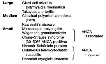
This is a group of conditions affecting the spine and peripheral joints which cluster in families and are linked to certain type 1 HLA antigens (Table 11.17).
The joint involvement is usually more limited than that seen in RA and its distribution is different. There are associated extra-articular and genetic features. These diseases occasionally present in childhood.
Histologically, the synovitis itself is similar to that of RA, but there is no production of rheumatoid factors – hence ‘seronegative’. ACPA is also usually negative. Inflammation of the enthesis (junction of ligament or tendon and bone) and joint ankylosis develop more commonly than in RA. All are associated with an increased frequency of sacroiliitis and an increased frequency of HLA-B27.
The common aetiological thread of these disorders is their striking association with HLA-B27, particularly ankylosing spondylitis (AS). HLA type B27 is present in >90% of Caucasians with AS but only 8% of controls. HLA-B27 exhibits a number of unusual characteristics including a high tendency to mis-fold but its aetiological relevance remains unclear. The role of class I HLA antigens in pathogenesis is supported by the fact that HLA-B27 transgenic mice spontaneously develop arthritis, skin, gut and genitourinary lesions.
There are clues that infections play a role, possibly by molecular mimicry, with parts of the organism which are structurally similar to the HLA molecule triggering cross-reactive antibody formation. This is unproven. AIDS is increasing the prevalence of reactive arthritis and spondylitis in sub-Saharan Africa even in the absence of HLA-B27. The explanation for this changing epidemiology is unclear.
The types of arthritis that follow a precipitating infection are called reactive arthritis (p. 529).
The specialized immune systems of the gut and genitourinary mucous membranes may also play a causal role, perhaps reacting to local infections or to antigens which cross the damaged mucosa.
This is an inflammatory disorder of the spine affecting mainly young adults (late teens to early 30s). It occurs worldwide, with a male to female ratio of 5 : 1. Women present later and are underdiagnosed. The frequency of AS in different populations is roughly paralleled by the incidence of HLA-B27; Africans and Japanese have a low incidence of both HLA-B27 and ankylosing spondylitis, while the North American Haida Indians have a high incidence of both. There are at least 24 subtypes of HLA-B27 (B*2701-B*2724). Some appear to increase risk; others have a protective role. Twin studies indicate a much higher disease concordance in HLA-B27-positive monozygotic (up to 70%) twins than in dizygotic twins (about 20–25%). There are also other genes lying within the major histocompatibility complex (interleukin-1 gene cluster and the gene CYP2D6) which also influence susceptibility to AS but the disease is polygenic.
Environmental factors may also be involved but although Gram-negative organisms, e.g. Yersinia, Klebsiella, Salmonella, Shigella, can cause a reactive arthropathy, there is no conclusive evidence for their involvement in the pathogenesis of AS.
There is lymphocyte and plasma cell infiltration and local erosion of bone at the attachments of the intervertebral and other ligaments (enthesitis). This heals with new bone (syndesmophyte) formation.
Episodic inflammation of the sacroiliac joints in the late teenage years or early 20s is the first manifestation of AS. Pain in one or both buttocks and low back pain and stiffness are typically worse in the morning and relieved by exercise. Initially the diagnosis is often missed because the patient is asymptomatic between episodes and radiological abnormalities are absent. Retention of the lumbar lordosis during spinal flexion is an early sign. Later, paraspinal muscle wasting develops.
Criteria for classifying inflammatory back pain as ankylosing spondylitis are shown in Box 11.14.
Box 11.14
Back pain criteria for diagnosing ankylosing spondylitisa
The presence of four of the five criteria suggests AS with 80% sensitivity. aAll criteria have high sensitivity.
Spinal stiffness can be measured by Schober’s test: a tape measure is placed in the midline 10 cm above the dimples of Venus. Any movement of a marker at 15 cm during flexion is recorded. A reading of <5 cm implies spinal stiffness. Individuals may be able to touch the floor with a stiff back if they have good hip movements but serial measurement of the finger tip to floor distance highlights any change.
Non-spinal complications (uveitis or costochondritis) suggest the diagnosis of spondyloarthritis (Box 11.15). Costochondral junction inflammation causes anterior chest pain. Measurable reduction of chest expansion is due to costovertebral joint involvement.
Peripheral joint involvement is asymmetrical and affects a few, predominantly large joints. Hip involvement leads to fixed flexion deformities of the hips and further deterioration of the posture. Young teenage boys occasionally present with a lower-limb monoarthritis (see p. 546), which later develops into AS.
Acute anterior uveitis is strongly associated with HLA-B27 in AS and related diseases and is occasionally the presenting complaint. Severe eye pain, photophobia and blurred vision are an emergency (see p. 1062).
Overall clinical assessment is based on pain, tenderness, stiffness and fatigue using, e.g. the Bath Ankylosing Spondylitis Disease Activity Index.
Blood. The ESR and CRP are usually raised.
HLA testing is rarely of value because of the high frequency of HLA-B27 in the population, but may give supporting evidence in a difficult case.
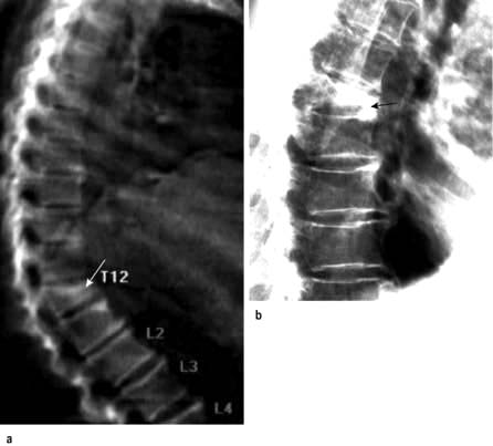
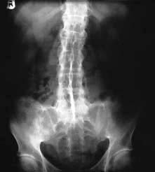
X-rays. The medial and lateral cortical margins of both sacroiliac joints lose definition owing to erosions and eventually become sclerotic (Fig. 11.20). The earliest radiological appearances in the spine are blurring of the upper or lower vertebral rims at the thoracolumbar junction (best seen on a lateral X-ray) caused by an enthesitis at the insertion of the intervertebral ligaments. These changes may eventually affect the whole spine. Persistent inflammatory enthesitis causes bony spurs (syndesmophytes). Syndesmophytes are more vertically oriented than the beak-like osteophytes of spondylosis and the disc is preserved, unlike in spondylosis (see p. 503). Syndesmophytes cause bony ankylosis and permanent stiffening. The sacroiliac joints eventually fuse, as may the costovertebral joints, reducing chest expansion. Calcification of the intervertebral ligaments and fusion of the spinal facet joints and syndesmophytes leads to what is often called a ‘bamboo’ spine (Fig. 11.21).
MRI with gadolinium demonstrates sacroiliitis before it is seen on X-rays, and persistent enthesitis.
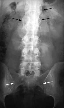
Figure 11.20 X-ray of ankylosing spondylitis. The sacroiliac joints are eroded and show marginal sclerosis (white arrows). There is bridging syndesmophyte formation at the thoracolumbar junction (black arrows).
The key to effective management of AS is early diagnosis so that a regimen of preventative exercises is started before syndesmophytes have formed. Morning exercises aim to maintain spinal mobility, posture and chest expansion.
Failure to control pain and to encourage regular spinal and chest exercises leads to an irreversible dorsal kyphosis and wasted paraspinal muscles. This, along with stiffening of the cervical spine, makes forward vision difficult.
When the inflammation is active and the morning pain and stiffness are too severe to permit effective exercise, an evening dose of a long-acting or slow-release NSAID or an NSAID suppository improves sleep, pain control and exercise compliance. Peripheral arthritis and enthesitis are managed with NSAIDs or local steroid injections.
Methotrexate is effective for peripheral arthritis but not for spinal disease.
The TNF-α blocking drugs adalimumab and etanercept (Table 11.16) have revolutionized the lives of people with AS. They produce a rapid, dramatic and sustained reduction of symptoms and of spinal and peripheral joint inflammation. Around half the patients are able to stop NSAIDs. Relapse occurs on stopping therapy but may be delayed by several months making intermittent treatment feasible. Golimumab is also available for severe active disease unresponsive to conventional therapy. Rituximab does not help spondyloarthritis.
With exercise and pain relief, the prognosis is excellent and over 80% of patients are fully employed. Anti-TNF therapies are likely to reduce the morbidity of severe disease, reducing the risk of permanent spinal stiffness and progressive peripheral joint disease.
Patients should be made aware that they risk passing the HLA-B27 gene to 50% of their children. HLA-B27 positive offspring then have a 30% risk of developing AS.
The prevalence of psoriasis is 2–3% worldwide, and in this population around 10% have arthritis (see p. 506). A family history of psoriasis may be a clue to the diagnosis. The aetiology and pathogenesis is described on page 509.
Patterns of psoriatic arthritis include:
Polyarthritis virtually indistinguishable from RA
Ankylosing spondylitis: uni- or bilateral sacroiliitis and early cervical spine involvement; only 50% are HLA-B27 positive
Distal interphalangeal arthritis, which is the most typical pattern of joint involvement in psoriasis, often with adjacent nail dystrophy (see p. 1209) reflecting enthesitis extending into the nail root
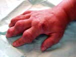
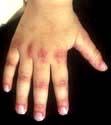
Arthritis mutilans, which affects about 5% of patients who have psoriatic arthritis and causes marked periarticular osteolysis and bone shortening (’telescopic’ fingers) (Fig. 11.22).
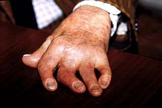
Figure 11.22 Hand showing psoriatic arthritis mutilans. All the fingers are shortened and the joints unstable, owing to underlying osteolysis.
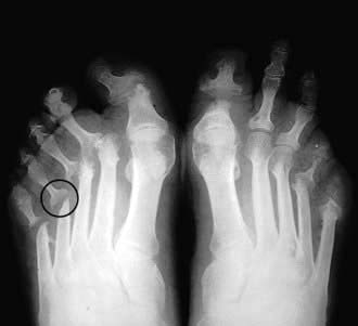
Radiologically, psoriatic arthritis is erosive but the erosions are central in the joint, not juxta-articular, and produce a ‘pencil in cup’ appearance (Fig. 11.23). The skin and nail disease can be mild and may develop after the arthritis.
NSAIDs and/or analgesics help the pain but they can occasionally worsen the skin lesions. Local synovitis responds to intra-articular corticosteroid injections.
In milder, polyarticular cases, sulfasalazine or methotrexate slows the development of joint damage.
When the disease is severe, methotrexate or ciclosporin is given because they control both the skin lesions and the arthritis. Anti-TNF-α agents, e.g. etanercept and golimumab (see p. 1210) and ustekinumab, are highly effective and safe for severe skin and joint disease. They should be given when methotrexate has failed. Corticosteroids orally may destabilize the skin disease and are best avoided but are valuable when injected into a single inflamed joint. Rituximab has no role in treating psoriatic arthritis.
The prognosis for the joint involvement is generally better than in RA.
Reactive arthritis is a sterile synovitis, which occurs following an infection (see also post-streptococcal arthritis, p. 546).
Spondyloarthritis develops in 1–2% of patients after an acute attack of dysentery, or a sexually acquired infection – nonspecific urethritis (NSU) in the male, nonspecific cervicitis in the female. In male patients who are HLA-B27 positive, the relative risk is 30–50. Not all patients are HLA-B27 positive. Women are less commonly affected.
A variety of organisms can be the trigger, including some strains of Salmonella or Shigella spp. in bacillary dysentery. Yersinia enterocolitica causes diarrhoea and a reactive arthritis. In NSU, the organisms are Chlamydia trachomatis or Ureaplasma urealyticum.
People with reactive arthritis are not more susceptible to infection but appear to respond differently. Bacterial antigens or bacterial DNA have been found in the inflamed synovium of affected joints, suggesting that this persistent antigenic material is driving the inflammatory process. The methods by which HLA-B27 increases susceptibility to reactive arthritis may include:
T cell receptor repertoire selection
Molecular mimicry causing autoimmunity against HLA-B27 and/or other self antigens
Mode of presentation of bacteria-derived peptides to T lymphocytes.
These are not mutually exclusive.
There are other organisms that also trigger reactive arthritis but have a different genetic basis; see post-streptococcal arthritis (p. 533), gonococcal arthritis (p. 546) and brucellosis (p. 533). In these, the borderline between reactive arthritis and septic arthritis is more indistinct and they can cause both.
The arthritis is typically an acute, asymmetrical, lower-limb arthritis, occurring a few days to a couple of weeks after the infection. The arthritis may be the presenting complaint if the infection is mild or asymptomatic. Enthesitis is common, causing plantar fasciitis or Achilles tendon enthesitis (see p. 509). Seventy per cent recover fully within 6 months but many have a relapse.
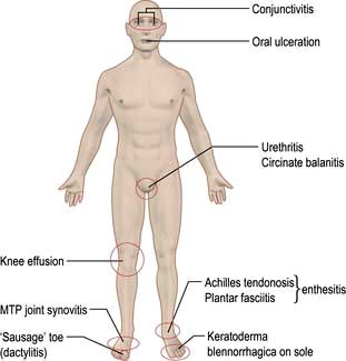
In susceptible individuals with reactive arthritis, sacroiliitis and spondylitis may also develop. Sterile conjunctivitis occurs in 30%. Acute anterior uveitis complicates more severe or relapsing disease but is not synchronous with the arthritis.
The skin lesions resemble psoriasis:
Circinate balanitis in the uncircumcised male causes painless superficial ulceration of the glans penis. In the circumcised male the lesion is raised, red and scaly. Both heal without scarring.
Keratoderma blennorrhagica – the skin of the feet and hands develops painless, red and often confluent raised plaques and pustules histologically similar to pustular psoriasis.
Treating persisting infection with antibiotics alters the course of the arthritis, once it has developed. Cultures should be taken and any infection treated. Sexual partners must be screened.
Pain responds well to NSAIDs and locally injected or oral corticosteroids. The majority of individuals with reactive arthritis have a single attack which settles, but a few develop a disabling relapsing and remitting arthritis. Relapsing cases are sometimes treated with sulfasalazine or methotrexate (Table 11.16). TNF-α blocking agents remain the drugs of next choice in severe and persistent disease but are rarely necessary.
Enteropathic synovitis occurs in up to 10–15% of patients who have ulcerative colitis and Crohn’s disease (see p. 275). The link between the bowel disease and the inflammatory arthritis is not clear. Selective mucosal leakiness may expose the individual to antigens that trigger synovitis.
The arthritis is asymmetrical and predominantly affects lower-limb joints. An HLA-B27-associated sacroiliitis or spondylitis also occurs. The joint symptoms may predate the development of bowel disease and lead to its diagnosis.
Remission of ulcerative colitis or total colectomy usually leads to remission of the joint disease, but arthritis can persist even in well-controlled Crohn’s disease.
The inflammatory bowel disease should be treated (see p. 272). In all cases of enteropathic arthritis, the symptoms are helped by NSAIDs, although they may make diarrhoea worse. A monoarthritis is best treated by intra-articular corticosteroids. Sulfasalazine is more frequently prescribed than mesalazine as it may help both bowel and joint disease. The TNF-α blocking drug infliximab is used in inflammatory bowel disease (p. 272) and can help the arthritis.
Two main types of crystal account for the majority of crystal-induced arthritis. They are sodium urate and calcium pyrophosphate and are distinguished by their different shapes and refringence properties under polarized light with a red filter. Rarely, crystals of calcium apatite (see p. 515) or cholesterol cause acute synovitis.
Gout is an inflammatory arthritis associated with hyperuricaemia and intra-articular sodium urate crystals.
The prevalence of gout is increasing mainly in developed countries, due to changing diets – purine rich foods, high saturated fats, fructose containing drinks and alcohol. The prevalence is 1.4% in the UK (increasing with age to 3% in women and 7% in men) and 2.7% in the USA. Asian populations are more at risk as their diet becomes more Western. Gout develops in men more than women (10 : 1) and rarely occurs before young adulthood (when it suggests a specific genetic defect), and seldom in premenopausal females. Some 85–90% of cases are idiopathic. The prevalence in older females is increasing with increased diuretic use. Hyperuricaemia is common in certain ethnic groups (e.g. Maoris).
The last two steps of purine metabolism in humans are the conversion of hypoxanthine to xanthine and of xanthine to uric acid, catalysed by the enzyme xanthine oxidase. Primates lost the gene for uricase during their evolution about 10–20 million years ago. Hyperuricaemia possibly offered an evolutionary advantage.
Uric acid levels are higher in men than in women. There is a normal distribution of serum uric acid in the population with a skewed distribution at the upper end of the range. Hyperuricaemia is defined as a serum uric acid level greater than two standard deviations from the mean (420 µmol/L in males; 360 µmol/L in females). This is close to the limit of solubility – 360 µmol/L at 35°C and 300 µmol/L at 30°C.
Most people with hyperuricaemia are asymptomatic. Osteoarthritic joints are more prone to gouty attacks. The range for gouty individuals is higher than for normals, but the curves overlap (Fig. 11.25). Serum uric acid levels increase with age, obesity, a ‘Western’ diet (see above) and combined hyperlipidaemia, diabetes mellitus, ischaemic heart disease and hypertension (metabolic syndrome, p. 218). There is often a family history of gout.
Figure 11.25 Serum uric acid levels in controls and in those with gout. 7.9 mg/dL is equivalent to 474 µmol/L, 5.1 mg/dL is equivalent to 306 µmol/L.
(From Snaith ML, Scott JT. Uric acid clearance in patients with gout and normal subjects. Annals of Rheumatic Disease 1971; 30:285–289, with permission from the BMJ Publishing Group.)
Uric acid is the final product of endogenous and dietary purine metabolism in humans and levels in the blood depend on the balance between purine synthesis and the ingestion of dietary purines, and the elimination of urate by the kidney (66%) and intestine (33%).
Some 90% of people with gout have impaired excretion of uric acid (10% have increased production due to high cell turnover and <1% due to an inborn error of metabolism). Renal excretion is coordinated by a group of renal tubular urate transport molecules and a complex process of glomerular filtration, proximal tubule reabsorption via the urate transporter-1 (URAT-1) and active resecretion (Fig. 11.26). GLUT9 transports uric acid along with glucose and fructose into the cell from the tubule and back into the circulation. Both the entry of uric acid via the URAT-1 mechanism and the exit into circulation by GLUT9 can be blocked by uricosuric drugs such as probenecid. The body pool is about 1000 mg and 60% is turned over daily. Low-dose aspirin blocks uric acid secretion. Insulin resistance enhances uric acid resorption. Causes of hyperuricaemia are shown in Table 11.18.
Figure 11.26 Urate renal transport. The net result is that about 5–10% of the glomerular load is excreted in the urine under normal circumstances.
Table 11.18 Causes of hyperuricaemia
Increased production of uric acid |
The roles of innate immunity and of the inflammasome suggest that crystal arthritis is an autoinflammatory disease, similar to the hereditary periodic fevers (p. 548). A series of receptors recognize bacteria and viruses as ‘foreign’ and eliminate them by activating the cytokine cascade. One such receptor (NLRP3) has recently been implicated in crystal-triggered inflammation. The activation of the inflammasome (p. 63) activates interleukin-1β, which in turn activates more cells and triggers an IL-8 mediated influx of neutrophils. Ingestion by polymorphonuclear leucocytes of sodium urate crystals causes the release of pro-inflammatory cytokines, particularly interleukin-1β and complement. Colchicine works by inhibition of microtubule formation necessary for this to occur. The involvement of IL-1β indicates a potential role for the IL-1β blocking agent anakinra in gout, in patients resistant to usual treatments.
Hyperuricaemia may be asymptomatic. It also causes:
Acute gout, followed by an asymptomatic intercritical phase; a second acute attack likely within 2 years
Chronic interval gout, with acute attacks superimposed on low grade inflammation and potential joint damage
Chronic polyarticular gout is rare, except in elderly people on longstanding diuretic treatment, in renal failure, or when allopurinol is started too soon after an acute attack
Urate renal stone formation (p. 600).
Acute gout presents typically in a middle-aged male with sudden onset of agonizing pain, swelling and redness of the first MTP joint. The attack occurs at any time, but may be precipitated by too much food or alcohol, by dehydration or by starting a diuretic. Untreated attacks last about 7 days. Recovery is typically associated with desquamation of the overlying skin. In 25% of attacks, a joint other than the great toe is affected.
In severe attacks, overlying crystal cellulitis makes gout difficult to distinguish clinically from infective cellulitis. A family or personal history of gout and the finding of a raised serum urate suggest the diagnosis but, if in doubt, blood and joint fluid cultures should be taken.
The clinical picture is often diagnostic, as is the rapid response to NSAIDs or colchicine.
Joint fluid microscopy is the most specific and diagnostic test but is technically difficult.
Serum uric acid is usually raised (>600 µmol/L). If it is not, recheck it several weeks after the attack, as the level falls immediately after an acute attack. Acute gout rarely occurs with a serum uric acid in the lower half of the normal range below the saturation point of 360 µmol/L.
Serum urea, creatinine and eGFR are monitored for signs of renal impairment.
The use of NSAIDs or coxibs in high doses rapidly reduces the pain and swelling. The first dose should be taken at the first indication of an attack:
Naproxen: 750 mg immediately, then 500 mg every 8–12 hours
Diclofenac: 75–100 mg immediately, then 50 mg every 6–8 hours
Indometacin: 75 mg immediately, then 50 mg every 6–8 hours. For some, the frequency of side-effects is unacceptably high with indometacin.
After 24–48 hours, reduced doses are given for a further week. Caution: NSAIDs may cause renal impairment. In individuals with renal impairment or a history of peptic ulceration, alternative treatments include:
Colchicine: 1000 µg immediately, then 500 µg every 6–12 hours, but this causes diarrhoea or colicky abdominal pain
Corticosteroids: oral prednisolone or intramuscular or intra-articular depot methylprednisolone.
The first attacks may be separated by up to 2 years and are managed symptomatically. Individuals should be advised to reduce their alcohol intake, especially beer, which is high in purines and fructose. Non-diet carbonated soft drinks are also high in fructose. A diet which reduces total calorie and cholesterol intake and avoids such foods as offal, some fish and shellfish and spinach, all of which are rich sources of purines, is advised. This can reduce serum urate by 15% and delay the need for drugs that reduce serum urate levels. Dietary advice is readily available on the internet.
The aim of treatment is to reduce the uric acid level below the 360 µmol/L level; some guidelines recommend below 300 µmol/L.
Allopurinol should only be used when the attacks are frequent and severe (despite dietary changes), associated with renal impairment or tophi, or when the patient finds NSAIDs or colchicine difficult to tolerate. Allopurinol (300–600 mg) blocks the enzyme xanthine oxidase, which converts xanthine into uric acid (see Fig. 16.20). It reduces serum uric acid levels rapidly and is relatively non-toxic but should be used at low doses (50–100 mg) in renal impairment. It should never be started within a month of an acute attack and always be started under cover of a course of NSAID or colchicine for the first 2–4 weeks before and 4 weeks after starting allopurinol, as it may induce acute gout. The dose can be increased gradually from 100 mg every few weeks until the uric acid level is below the 360 µmol/L level. Skin rashes and gastrointestinal intolerance are the most common side-effects. A hypersensitivity reaction is the most serious adverse event. This is rare, as is bone marrow suppression.
Febuxostat (80–120 mg) is a non-purine analogue inhibitor of xanthine oxidase but not other enzymes in the purine and pyrimidine pathway. It is well tolerated and as effective as allopurinol in trials and is safer in renal impairment as it is metabolized in the liver and not renally excreted. It has been approved by the FDA and is helpful in patients who cannot tolerate allopurinol but there are anxieties that it may increase cardiovascular risks. At time of writing, most doctors advise trying allopurinol first unless there are strong contraindications to its use.
Pegloticase, a pegylated recombinant uricase given intravenously, lowers urate levels dramatically but its place in therapy is unclear.
Uricosuric agents also lower the serum uric acid but their use is restricted throughout Europe by the very rare occurrence of serious hepatotoxicity. Benzbromarone acts on the URAT-1 transporter and is well tolerated. Sulphinpyrazone and probenecid are best avoided in renal impairment. Availability of these drugs varies between countries – in the UK benzbromarone and probenecid can be obtained for treating named patients.
Losartan is an angiotensin I-receptor antagonist and is uricosuric in hypertensive patients with gout. It may reduce the risk of gout in patients with the metabolic syndrome.
Anakinra blocks IL-1β and canakinumab is a human monoclonal antibody with specific cross-reactivity for IL-1β but not other members of the IL-1 family. Their role in treatment-resistant gout is still subject to trials to establish when their use is justified in gout which has not responded to the more conventional agents.
Individuals with very high levels of uric acid can present with chronic tophaceous gout, as sodium urate forms smooth white deposits (tophi) in skin and around joints. They occur on the ear, the fingers (Fig. 11.27) or the Achilles tendon. Large deposits are unsightly and ulcerate. There is chronic joint pain and sometimes superimposed acute gouty attacks.
Periarticular deposits lead to a halo of radio-opacity and clearly defined (‘punched-out’) bone cysts on X-ray.
Tophaceous gout is often associated with renal impairment and/or the long-term use of diuretics. There may be acute or chronic urate nephropathy or renal stone formation. Whenever possible, stop the diuretics or change to less urate-retaining ones, such as bumetanide. Treat with allopurinol and/or uricosuric agents (see above). Pegylated uricase (Pegloticase), a pegylated recombinant uricase given intravenously, is used preventatively in people undergoing chemotherapy for malignancies (tumour lysis syndrome). Pegloticase has an important but, as yet, unproven role in those rare individuals who have refractory tophaceous gout.
Calcium pyrophosphate deposits in hyaline and fibrocartilage produce the radiological appearance of chondrocalcinosis (see p. 515). Shedding of crystals into a joint precipitates acute synovitis which resembles gout, except that it is more common in elderly women and usually affects the knee or wrist. The attacks are often very painful. In young people it may be associated with haemochromatosis, hyperparathyroidism, Wilson’s disease or alkaptonuria.
The diagnosis is made by detecting rhomboidal, weakly positively birefringent crystals in joint fluid, or deduced from the presence of chondrocalcinosis on X-ray. The joint fluid looks purulent. Septic arthritis must be excluded and joint fluid should be sent for culture. The attacks may be associated with fever and a raised white blood cell count.
Joints become infected by direct injury or by blood-borne infection from an infected skin lesion or other site.
Chronically inflamed joints (e.g. in rheumatoid arthritis) are more prone to infection than are normal joints. Individuals who are immunosuppressed, by AIDS or by immunosuppressive agents, are particularly at risk, as are infants, the elderly and those who use excess alcohol. Artificial joints are also potential sites for infection.
The organism that most commonly causes septic arthritis is Staphylococcus aureus. Other organisms include streptococci, other species of staphylococcus, Neisseria gonorrhoeae, Haemophilus influenzae in children, and these and other Gram-negative organisms in the elderly or complicating RA.
Suspected septic arthritis is a medical emergency. In young and previously fit people, the joint is hot, red, swollen and agonizingly painful and held immobile by muscle spasm. In the elderly and immunosuppressed and in RA the clinical picture is less dramatic, so a high index of suspicion is needed to avoid missing treatable but potentially severely destructive and occasionally fatal septic arthritis.
In 20% of patients, the sepsis affects more than one joint. Chronic destructive arthritis due to tuberculosis is rare.
Aspirate the joint and send the fluid for urgent Gram-staining and culture. The fluid is usually frankly purulent. The culture techniques should include those for gonococci and anaerobes.
Blood cultures are often positive.
Leucocytosis is usual, unless the person is severely immunosuppressed.
X-rays are of no value in diagnosis in acute septic arthritis.
Skin wound swabs, sputum and throat swab or urine may be positive and indicate the source of infection.
This should be started immediately on diagnosis because joint destruction may occur within weeks. The joint should be immobilized initially and then physiotherapy started early to prevent stiffness and muscle wasting. Intravenous antibiotics should be given for 1–2 weeks. It is usual to give two antibiotics to which the organism is sensitive for 6 weeks, then one for a further 6 weeks, orally. Monitor clinically and with the ESR and CRP.
Empirical treatment in septic arthritis
This is started before the results of culture are obtained. Discuss the case with a microbiologist. Intravenous flucloxacillin 1–2 g is given 6-hourly, plus fusidic acid 500 mg orally 8-hourly. If the patient is allergic to penicillin, replace flucloxacillin with erythromycin 1 g i.v. 6-hourly or clindamycin 600 mg i.v. 8-hourly. In immunosuppressed patients, flucloxacillin 1–2 g i.v. 6-hourly plus gentamicin (to cover Gram-negative organisms) should be used. Teicoplanin i.v. should replace flucloxacillin if MRSA is likely. Change the antibiotics if the organism is not sensitive. Drainage of the joint and arthroscopic joint washouts are helpful in relieving pain.
This is the most common cause of a septic arthritis in previously fit young adults; more commonly affecting women and men who have sex with men.
Initially the patient becomes febrile and develops characteristic pustules on the distal limbs. Polyarthralgia and tenosynovitis are common at this stage and about 40% have a gonococcaemia. This phase settles and blood cultures usually become negative. Nucleic acid amplification tests are a useful adjunct to cultures and may be positive even when cultures are negative. Later, large-joint mono- or pauciarticular arthritis may follow. Culture is usually positive from the genital tract, although the joint fluid may be sterile. It is not clear whether this is simply a septic arthritis – although it responds rapidly to antibiotics, or whether there is also a reactive element to bacterial lipopolysaccharide.
Treatment consists of oral penicillin, ciprofloxacin or doxycycline for 2 weeks, and joint rest. Resistance to antibiotics is increasing.
Around 1% of people with tuberculosis develop joint and/or bone involvement. It occurs as the primary disease in children. In adults, it is usually due to haematogenous spread from secondary pulmonary or renal lesions. The onset is insidious and diagnosis often delayed.
The organism invades the synovium or intervertebral disc. There are caseating granulomas and rapid destruction of cartilage and adjacent bone. Some patients develop a reactive polyarthritis (Poncet’s disease).
The hip or knee (30%) is quite commonly affected, but around 50% develop spinal disease. The patient is febrile, has night sweats, is anorexic and loses weight. The usual risk factors for tuberculosis apply – debility, excess alcohol use or immunosuppression. HIV-positive/AIDS patients are at particular risk.
Investigations should include culture of fluid, and culture and biopsy of the synovium. M. tuberculosis is the usual organism, but atypical mycobacteria are occasionally implicated. A chest X-ray should be performed. Initially joint or spinal X-rays may be normal but joint-space reduction and bone destruction develop rapidly if treatment is delayed. MRI shows the abnormality earlier in the spine and CT-guided biopsy from the affected disc is often necessary to obtain cultures.
Treatment is as for pulmonary tuberculosis with therapy for 9 months (see p. 820). The joint should be rested and the spine immobilized in the acute phase.
This may complicate a meningococcal septicaemia and presents as a migratory polyarthritis. Organisms can only rarely be cultured from the joint and most cases are due to immune complex deposition. Treatment is urgent with immediate penicillin therapy (p. 75).
This may present with arthralgia, polymyalgia rheumatica-like symptoms or an infective arthritis. It is discussed on page 687.
About 25% of people with Lyme disease develop arthralgia, less commonly an acute pauciarticular arthritis (see p. 130). This usually resolves but 10% of untreated cases go on to develop a chronic arthritis. There are no positive markers in these patients of an ongoing infection (p. 130).
Diagnosis is by the detection of IgM antibodies against the spirochaete Borrelia burgdorferi.
Treatment with antibiotics (amoxicillin or doxycycline) is highly effective in early disease. The response of chronic arthritis to antibiotic treatment is discussed on page 130.
Brucellosis (see p. 129) has a worldwide distribution. The most common cause of chronic brucellosis and of arthritis is Brucella melitensis. There is usually a migratory large joint mono- or oligoarticular arthritis, which is septic or reactive. Arthritis is more common in chronic infections of more than 6 months.
Congenital syphilis (see p. 166) can cause an acute painful epiphysitis or osteochondritis sometimes associated with para-articular swelling in the first few weeks of life. Later, at age 8–16 years, painless effusion of the knees may occur (Clutton’s joints).
In acquired syphilis, arthralgia and arthritis occur in the secondary stage. Charcot’s (neuropathic) joints usually involve the knees in tabes dorsalis (see p. 1129).
Acute or chronic symmetrical polyarthritis resembling RA, swollen hands and feet due to lepra reactions, tenosynovitis and thickened nerves with or without cutaneous manifestations are seen in leprosy.
A transient polyarthritis or arthralgia can occur before, during or after many viral illnesses. These include infectious mononucleosis, chickenpox, mumps, adenovirus, rubella, erythrovirus B19, hepatitis B and C, arboviral infections and HIV. In most of these it is due to a direct toxic effect or immune complex deposition.
In rubella (see p. 104) the virus can occasionally be isolated from the joint. This arthritis is rare in countries where rubella vaccination is routine. It occurs most commonly in up to 50% of young adult females a few days after rubella infection (6% of men). It is a symmetrical polyarthritis involving the MCP or PIP joints most commonly, but many joints can be affected. It closely resembles rheumatoid arthritis. IgM rubella antibodies are present. It resolves within a few weeks in most cases. A mild arthritis occurs rarely 2–4 weeks after rubella vaccination.
Erythrovirus B19 (p. 101) causes an acute, self-limiting arthritis and is associated with erythema infectiosum (‘slapped cheek disease’).
In hepatitis B infection (see p. 318), a sudden symmetrical polyarticular arthritis of the small joints of the hands occurs in approximately one-third of patients, often in the prodromal phase and mostly resolving before the onset of jaundice. Hepatitis C infection causes type II mixed cryoglobulinaemia (see p. 323).
Arbovirus infections (see p. 104) which are endemic in many parts of the world, give rise to an arthralgia and/or arthritis. For example, the Ross River virus causes an epidemic polyarthritis in Australia and the South Pacific; it involves the small joints of the hands and clears in 2–4 weeks. Other viral infections causing epidemic arthritis include chikungunya (p. 105) and O’nyong-nyong.
The clinical features seen in these patients are due to a number of causes such as opportunistic infections and drug therapy and are not usually caused directly by HIV. Infective arthritis seen in these immunosuppressed patients often has minimal symptoms and signs. Some of the antiviral agents cause an acute arthritis, possibly because of crystallization in the joint.
Arthralgia is common in AIDS. There is a seronegative, predominantly lower-limb arthritis, similar to psoriasis or Reiter’s disease. Spondylitis also occurs but is not HLA-B27 associated. Avascular necrosis, possibly associated with corticosteroids or alcohol, is seen.
Non-articular diseases such as Sjögren- and lupus-like syndromes, systemic vasculitis of the necrotizing and hypersensitivity types and myositis also occur.
Fungal infections of joints occur rarely. Bone abscesses may be seen. Destructive joint lesions can also occur with blastomycosis. A benign polyarthritis accompanied by erythema nodosum occasionally occurs in coccidioidomycosis and histoplasmosis. Culture of purulent synovial fluid and skin tests for fungi may help the diagnosis.
Osteomyelitis can be due either to metastatic haematogenous spread (e.g. from a boil) or to local infection. Malnutrition, debilitating disease and decreased immunity may play a part in the pathogenesis.
Staphylococcus is the organism responsible for 90% of cases of acute osteomyelitis. Other organisms include Haemophilus influenzae and Salmonella; infection with the latter may occur as a complication of sickle cell anaemia. The classic presentation is with fever and localized bone pain with overlying tenderness and erythema.
Treatment of osteomyelitis is with immobilization and antibiotic therapy with intravenous teicoplanin or intravenous flucloxacillin 1–2 g every 6 hours and oral fusidic acid. Switch to oral antibiotics after 2 weeks and continue for a further 4 weeks. Surgical drainage and removal of dead bone (sequestrum) may be necessary but recurrence is common.
Delayed treatment leads to chronic osteomyelitis. In chronic osteomyelitis sinus formation is usual. Subacute osteomyelitis is associated with a chronic abscess within the bone (Brodie’s abscess). Symptoms may be limited to local pain.
This is usually due to haematogenous spread from a reactivated primary focus in the lungs or gastrointestinal tract. The disease starts in intra-articular bone. The spine is commonly involved (Pott’s disease), with damage to the bodies of two neighbouring vertebrae leading to vertebral collapse and acute angulation of the spine (gibbus). Later an abscess forms (‘cold abscess’). Pus can track along tissue planes and discharge at a point far from the affected vertebrae. Symptoms consist of local pain and later swelling if pus has collected. Systemic symptoms of malaise, fever and night sweats occur.
Treatment is as for pulmonary tuberculosis but extended to 9 months (see p. 811), together with initial immobilization.
Autoimmune diseases are conditions in which the immune system attacks tissues of the body. The antigens can be present in multiple organs so the clinical manifestations are systemic and diverse. In some diseases, such as Graves’ disease, Hashimoto’s thyroiditis, and insulin-dependent diabetes mellitus only a single organ is affected. The term ‘autoimmune rheumatic disease’ (ARD) is preferable to the older term ‘connective tissue disease’ because the clinical effects of ARD are not limited to connective tissues. Each individual ARD has a characteristic pattern of symptoms and signs, which are used to make the diagnosis. In some ARD there are also characteristic autoantibodies (i.e. antibodies that recognize antigens which are normal constituents of the body, such as DNA and phospholipids). Positive blood tests for autoantibodies are useful but not essential in the diagnosis of ARD (Table 11.3, p. 497).
SLE is an inflammatory, multisystem autoimmune disorder with arthralgia and rashes as the most common clinical features, and cerebral and renal disease as the most serious problems.
SLE occurs worldwide and is about nine times as common in women as in men, with a peak age of onset between 20 and 40 years. The prevalence varies between ethnic groups, being highest (at 1 : 250) in African/Caribbean women. In other populations, the prevalence varies between 1 : 1000 and 1 : 10 000.
The cause is unknown but there are several predisposing factors.
Heredity. There is a higher concordance rate in monozygotic twins (up to 25%) compared with dizygotic twins (3%). First-degree relatives have a 3% chance of developing the disease, but approximately 20% have autoantibodies.
Genetics. Recent research, including three whole genome analyses, has led to the identification of approximately 20 genes linked to the development of SLE. These include some HLA genes as well as genes involved in T and B lymphocyte function. Homozygous deficiencies of the complement genes C1q, C2 or C4 are very rare but convey a high risk of developing SLE.
Sex hormone status. Premenopausal women are most frequently affected.
Drugs such as hydralazine, isoniazid, procainamide and penicillamine can induce a form of SLE which is usually mild in that kidneys and the CNS are not affected.
Ultraviolet light can trigger flares of SLE, especially in the skin.
Exposure to Epstein–Barr virus has been suggested as a trigger for SLE.
When cells die by apoptosis, the cellular remnants appear on the cell surface as small blebs which carry self-antigens. These antigens include nuclear constituents (e.g. DNA and histones), which are normally hidden from the immune system. In people with SLE, removal of these blebs by phagocytes is inefficient so that they are transferred to lymphoid tissues where they can be taken up by antigen-presenting cells. The self-antigens from these blebs can then be presented to T cells which in turn stimulate B cells to produce autoantibodies directed against these antigens (p. 69). It has been shown that in some patients the autoantibodies are present in stored blood samples that were taken years before the patient developed clinical features of SLE. The combination of availability of self-antigens and failure of the immune system to inactivate B cells and T cells which recognize these self-antigens (i.e. a breakdown of tolerance, see Ch. 3, p. 69) leads to the following immunological consequences.
Development of autoantibodies that either form circulating complexes or deposit by binding directly to tissues
This leads to activation of complement and influx of neutrophils causing inflammation in those tissues
Abnormal cytokine production: increased blood levels of IL-10 and alpha-interferon are particularly closely linked to high activity of inflammation in SLE.
SLE of the skin and kidneys is characterized by deposition of complement and IgG antibodies and influx of neutrophils and lymphocytes. Biopsies of other tissues are carried out less frequently but can show vasculitis affecting capillaries, arterioles and venules. The synovium of joints can be oedematous and may contain immune complexes. Haematoxylin bodies (rounded blue homogeneous haematoxylin-stained deposits) are seen in inflammatory infiltrates and are thought to result from the interaction of antinuclear antibodies and cell nuclei.
The pathology of lesions in other organs is described in the appropriate chapters.
The manifestations of SLE vary greatly between patients. Most patients suffer fatigue, arthralgia and/or skin problems. Involvement of major organs is less common but more serious (Fig. 11.28).
Fever is common in exacerbations. Patients complain of marked malaise and tiredness and these symptoms do not correlate with disease activity or severity of organ-based complications.
Joint involvement is the most common clinical feature (>90%). Patients often present with symptoms resembling RA with symmetrical small joint arthralgia. Joints are painful but characteristically appear clinically normal, although sometimes there is slight soft tissue swelling surrounding the joint. Deformity because of joint capsule and tendon contraction is rare, as are bony erosions. Rarely, major joint deformity resembling RA (known as Jaccoud’s arthropathy) is seen. Avascular necrosis affecting the hip or knee is a rare complication of the disease or of treatment with corticosteroids.
Myalgia is present in up to 50% of patients but a true myositis is seen only in <5%. If myositis is prominent, the patient may well have an overlap ARD with both polymyositis and SLE.
The skin (see p. 1219) is affected in 85% of cases. Erythema, in a ‘butterfly’ distribution on the cheeks of the face and across the bridge of the nose (see Fig. 24.26), is characteristic. Vasculitic lesions on the finger tips and around the nail folds, purpura and urticaria occur. In 40–50% of cases there is photosensitivity (especially in patients positive for anti-Ro antibodies). Prolonged exposure to sunlight can lead to exacerbations of the disease. Livedo reticularis, palmar and plantar rashes, pigmentation and alopecia are seen. Scarring alopecia can lead to irreversible bald patches which are especially upsetting for women, who form the majority of people with SLE. Raynaud’s phenomenon (see p. 788) is common and may precede the development of other clinical problems by years.
Discoid lupus is a benign variant of lupus in which only the skin is involved. The rash is characteristic and appears on the face as well-defined erythematous plaques that progress to scarring and pigmentation (see p. 1198). Subacute cutaneous lupus erythematosus, a rare variant, is described on page 1198.
Up to 50% of patients will have lung involvement sometime during the course of the disease (see p. 848). Recurrent pleurisy and pleural effusions (exudates) are the most common manifestations and are often bilateral. Pneumonitis and atelectasis are seen; eventually a restrictive lung defect develops with loss of lung volumes and raised hemidiaphragms. This ‘shrinking lung syndrome’ is poorly understood but may have a neuromuscular basis. Rarely, pulmonary fibrosis occurs, more commonly in overlap syndromes. Intrapulmonary haemorrhage associated with vasculitis is a rare but potentially life-threatening complication.
The heart and cardiovascular system
The heart is involved in 25% of cases. Pericarditis, with small pericardial effusions detected by echocardiography, is common. A mild myocarditis also occurs, giving rise to arrhythmias. Aortic valve lesions and a cardiomyopathy can rarely be present. A non-infective endocarditis involving the mitral valve (Libman–Sacks syndrome) is very rare. Raynaud’s, vasculitis, arterial and venous thromboses can occur, especially in association with the antiphospholipid syndrome (see below). There is an increased frequency of ischaemic heart disease and stroke in people with SLE. This is partly due to altered levels of common risk factors such as hypertension and lipid levels but the presence of chronic inflammation over many years may also play a role. It is not known whether intensive treatment of cardiovascular risk factors in SLE will alter the risk of developing coronary disease or stroke. The benefit of statin therapy in the absence of significant hypercholesterolaemia remains to be proved.
A classification of types of nephritis is on page 574. Autopsy studies suggest that histological changes are very frequent, but clinical renal involvement occurs in only approximately 30% of cases. All patients should have regular screening of urine for blood and protein. An asymptomatic patient with proteinuria may be in the early stages of lupus nephritis, and treatment may prevent progression to renal impairment. Proteinuria should be quantified and haematuria should prompt examination for urinary casts or fragmented red cells that suggest glomerulonephritis. Renal vein thrombosis can occur in nephrotic syndrome or associated with antiphospholipid antibodies.
Involvement of the nervous system occurs in up to 60% of cases and symptoms often fluctuate. There may be a mild depression but occasionally more severe psychiatric disturbances occur. Epilepsy, migraines, cerebellar ataxia, aseptic meningitis, cranial nerve lesions, cerebrovascular disease or a polyneuropathy may be seen. The pathogenic mechanism for cerebral lupus is complex. Lesions may be due to vasculitis or immune-complex deposition, thrombosis or non-inflammatory microvasculopathy. The commonest finding on MRI scan is of increased white matter signal abnormality. In people with cerebral lupus, infection should be excluded or treated in parallel with administration of corticosteroids and immunosuppression.
Retinal vasculitis can cause infarcts (cytoid bodies) which appear as hard exudates, and haemorrhages. There may be episcleritis, conjunctivitis or optic neuritis, but blindness is uncommon. Secondary Sjögren’s syndrome is seen in about 15% of cases.
Histology. Characteristic histological and immunofluorescent abnormalities (deposition of IgG and complement) are seen in biopsies from the kidney and skin.
Diagnostic imaging. CT scans of the brain sometimes show infarcts or haemorrhage with evidence of cerebral atrophy. MR can detect lesions in white matter which are not seen on CT. However, it can be very difficult to distinguish true vasculitis from small thrombi.
Box 11.16
Antinuclear autoantibodies and disease associations
Antibody | Disease | Prevalence |
---|---|---|
ds DNA |
SLE |
70% |
Anti-histone |
Drug-induced lupus |
– |
Anti-centromeric |
Limited SS |
70% |
Anti-Ro (SS-A) |
SLE |
40–60% |
Primary Sjögren |
60–90% |
|
Anti-La (SS-B) |
SLE |
15% |
Primary Sjögren |
35–85% |
|
Anti-Sm |
SLE |
10–25% (Caucasian) |
30–50% (Black African) |
||
Anti-UI-RNP |
SLE |
30% |
Overlap syndrome |
||
Anti-Jo-1 (antisynthetase) |
Polymyositis |
30% |
Dermatomyositis |
||
Anti-topoisomerase-1 (Scl-70) |
Diffuse cutaneous SSc |
30% |
SS-A, SS-B, Sjögren’s syndrome -A and -B; -Ro, -La, first two letters of name of patients; Sm, Smith, patient’s name; RNP, ribonucleoprotein; SSc, systemic scleroderma.
The disease and its management should be discussed with the patient, particularly the effect upon the patient’s lifestyle, e.g. appearance and debility due to fatigue. Patients are advised to avoid excessive exposure to sunlight and it is also necessary to reduce cardiovascular risk factors (p. 727).
Many patients do not need treatment with corticosteroid tablets or immunosuppressive agents. Arthralgia, arthritis, fever and serositis all respond well to standard doses of NSAIDs (p. 511). Topical corticosteroids are effective and widely used in cutaneous lupus. Antimalarial drugs (chloroquine or hydroxychloroquine) help mild skin disease, fatigue and arthralgias that cannot be controlled with NSAIDs but patients require regular eye checks because of rare retinal toxicity (1 in 2000).
Corticosteroids and immunosuppressive drugs
Single intramuscular injections of long-acting corticosteroids or short courses of oral corticosteroids are useful in treating severe flares of arthritis, pleuritis or pericarditis. In some cases, these symptoms can only be kept under control using long-term oral corticosteroids.
Renal (p. 578) or cerebral disease and severe haemolytic anaemia or thrombocytopenia must be treated with high- dose oral corticosteroids and the first two of these require immunosuppressive drugs in addition. Cyclophosphamide was most commonly used to achieve remission in these severe forms of lupus but is being replaced by mycophenolate mofetil, which has fewer side-effects. Azathioprine is also used to maintain remission. Newer agents, which specifically target cells or cytokines in the immune system, are coming into use, especially in refractory cases. These include rituximab (anti-CD20) and belimumab, which are both monoclonal antibodies acting against B lymphocytes.
An episodic course is characteristic, with exacerbations and complete remissions that may last for long periods. However, SLE can also be a chronic persistent condition. The mortality rate in SLE has fallen dramatically over the last 50 years; the 10-year survival rate is about 90%, but this is lower if major organ-based complications are present. Deaths early in the course of disease are mainly due to renal or cerebral disease or infection. Later coronary artery disease and stroke become more prevalent. Chronic progressive destruction of joints as seen in RA and OA occurs rarely, but a few patients develop deformities such as ulnar deviation. People with SLE have an increased long-term risk of developing some cancers, especially lymphoma.
Fertility is usually normal except in severe disease and there is no major contraindication to pregnancy. Recurrent miscarriages can occur, especially in women with antiphospholipid antibodies. Exacerbations can occur during pregnancy with frequent exacerbations of the disease postpartum. The patient’s medications should be reviewed. Mycophenolate should be stopped whereas azathioprine, hydroxychloroquine and low-dose oral corticosteroids are safe. Hypertension must be controlled. People with anti-Ro or anti-La antibodies have a 2% risk of giving birth to babies with neonatal lupus syndrome (rash, hepatitis and fetal heart block).
Patients who have thrombosis (arterial or venous) and/or recurrent miscarriages and who also have persistently positive blood tests for antiphospholipid antibodies (aPL) have the antiphospholipid syndrome (APS). aPL can be detected by several different tests:
The anticardiolipin test, which detects antibodies (IgG or IgM) that bind the negatively charged phospholipid, cardiolipin
The lupus anticoagulant test, which detects changes in the ability of blood to clot in a test tube. Despite the name, this is not a test for lupus. It is a test for APS. The anticoagulant effect caused by aPL in the test tube causes an opposite procoagulant effect inside the body, because the balance of factors stimulating thrombosis is different there.
The anti-β2-glycoprotein I test, which detects antibodies that bind β2-glycoprotein I, a molecule that interacts closely with phospholipids.
A persistently positive test (i.e. positive on at least two occasions, ≥12 weeks apart) in one or more of these assays is needed to diagnose APS. However, some people who test positive for aPL will never get APS, i.e. not all aPLs are harmful. APS can present in patients who already have another ARD, especially SLE. APS can also occur on its own (primary APS).
Negatively charged phospholipids and β2-glycoprotein I are present on the outer surface of apoptotic blebs and so aPLs are believed to arise by a similar mechanism to the lupus autoantibodies described above. Pathogenic aPLs bind to the N-terminal domain of β2-glycoprotein I and this interaction is facilitated when the protein is bound to phospholipid on the surface of cells such as endothelial cells, platelets, monocytes and trophoblasts. This alters the functioning of those cells leading to thrombosis and/or miscarriage.
Since APS is defined by the presence of thrombosis and/or pregnancy loss it is not surprising that these are the most common features. Ischaemic strokes occur in about 20% of patients and deep vein thrombosis in about 40%. Unlike most causes of thrombophilia, APS can cause either arterial or venous thrombosis (though rarely both in the same patient). Of women who have had two or more spontaneous miscarriages, 27% have APS.
However, large studies show that people with APS can also have many other features including:
Cutaneous manifestations (e.g. livedo reticularis)
Renal impairment due to ischaemia in the small renal vessels.
Occasionally, APS is catastrophic. Catastrophic APS is a rare variant (about 1% of cases) in which multiple infarcts in different organs of the body cause failure of multiple organs simultaneously. There is a high mortality from catastrophic APS.
In people with APS who have had one or more thrombosis, the recommended treatment to prevent further thrombosis is long-term anticoagulation with warfarin. The optimal target INR is unclear and many patients are managed with lower target INR. Pregnant women with APS are given oral aspirin and subcutaneous heparin from early in gestation. This reduces the chance of a miscarriage but pre-eclampsia and poor fetal growth remain common. There are no definite guidelines for managing people with aPL who have never had thrombosis. Aspirin or clopidogrel are sometimes given prophylactically, especially in those with high IgG aPL. Warfarin is given much more rarely in these circumstances.
Systemic sclerosis (SSc) (see p. 1218), is a multisystem disease. This distinguishes it from localized scleroderma syndromes, such as morphea, that do not involve internal organ disease and are rarely associated with vasospasm (Raynaud’s phenomenon). SSc has the highest case-specific mortality of any of the autoimmune rheumatic diseases. SSc occurs worldwide but there may be racial or ethnic differences in clinical features. For example, renal involvement is less frequent in Japanese cases.
The incidence of SSc is 10/million population per year with a 3 : 1 female to male ratio. The peak incidence is between 30 and 50 years of age. It is rare in children.
Environmental risk factors for scleroderma-like disorders include: exposure to vinyl chloride, silica dust, adulterated rapeseed oil and trichloroethylene. Drugs such as bleomycin also produce a similar picture. Although unusual, familial cases are reported and twin cohorts suggest higher concordance in monozygotic pairs, consistent with genetic determinants of aetiology.
An early lesion is widespread vascular damage involving small arteries, arterioles and capillaries. There is initial endothelial cell damage with release of cytokines including endothelin-1, which causes vasoconstriction. There is continued intimal damage with increasing vascular permeability, leading to cellular activation, activation of adhesion molecules (E-selectin, VCAM, ICAM-1), with migration of cells into the extracellular matrix. Migrating lymphocytes are IL-2-producing cells, expressing surface antigens such as CD3, CD4 and CD5. All these factors cause release of other mediators (e.g. interleukin-1, -4, -6 and -8, transforming growth factor-β and platelet-derived growth factor) with activation of fibroblasts.
The damage to small blood vessels also produces widespread obliterative arterial lesions and subsequent chronic ischaemia.
Fibroblasts synthesize increased quantities of collagen types I and III, as well as fibronectin and glycosaminoglycans, producing fibrosis in the lower dermis of the skin as well as the internal organs. It is possible that antibodies to platelet-derived growth factor receptor, which have been found in blood of people with SSc, stimulate fibroblasts to cause fibrosis.
Raynaud’s phenomenon is seen in almost 100% of cases and can precede the onset of the full-blown disease by many years.
Limited cutaneous scleroderma (LcSSc): 70% of cases
This usually starts with Raynaud’s phenomenon many years (up to 15) before any skin changes. The skin involvement is limited to the hands, face, feet and forearms. The skin is tight over the fingers and often produces flexion deformities of the fingers. Involvement of the skin of the face produces a characteristic ‘beak’-like nose and a small mouth (microstomia). Painful digital ulcers and telangiectasia with dilated nail-fold capillary loops are seen. Digital ischaemia may lead to gangrene. Gastrointestinal tract involvement is common. Pulmonary hypertension (PHT) develops in 21% of people with LcSSc and pulmonary interstitial disease also occurs.
Diffuse cutaneous scleroderma (DcSSc): 30% of cases
Initially oedematous in onset, skin sclerosis rapidly follows. Raynaud’s phenomenon usually starts just before or concomitant with the oedema.
Diffuse swelling and stiffness of the fingers is rapidly followed by more extensive skin thickening, which can involve most of the body in the severest cases. Later the skin becomes atrophic. Early involvement of other organs occurs with general symptoms of lethargy, anorexia and weight loss.
Heartburn, reflux or dysphagia due to oesophageal involvement is almost invariable and anal incontinence occurs in many patients. Malabsorption from bacterial overgrowth due to dilatation and atony of the small bowel is not infrequent, and more rarely dilatation and atony of the colon occurs. Pseudo-obstruction is a known complication.
Renal involvement is acute or chronic. Acute hypertensive renal crisis used to be the most common cause of death in systemic sclerosis. ACE inhibitors and better care along with dialysis and renal transplantation have changed this.
Lung disease, both fibrosis (in 41% of cases) and pulmonary hypertension (17% of cases), contributes significantly to mortality in SSc. PHT can be isolated or secondary to fibrosis, and high plasma levels of endothelin-1 are seen.
Myocardial fibrosis leads to arrhythmias and conduction defects. Pericarditis is found occasionally.
Sometimes, these systemic features occur without skin involvement (SSc sine scleroderma).
Full blood count. A normochromic, normocytic anaemia occurs and a microangiopathic haemolytic anaemia is seen in some people with renal disease.
Urea and electrolytes. Urea and creatinine rise in acute kidney injury.
Autoantibodies (Box 11.16):
Urine microscopy and, if there is proteinuria, urine albumin/creatinine ratio should be measured.
Other investigations of gastrointestinal tract (e.g. see Fig. 6.5), lung, renal and cardiac as appropriate.
Treatment should be organ-based in order to try to control the disease. Currently, there is no cure. In contrast to many other ARDs, corticosteroids and immunosuppressants are rarely used in SSc, with the exception of SSc-related pulmonary fibrosis.
Education, counselling and family support are essential.
Regular exercises and skin lubricants may limit contractures but no treatment has proven efficacy in reducing skin fibrosis.
Raynaud’s may be improved by hand warmers and oral vasodilators (calcium-channel blockers, ACE inhibitors, angiotensin receptor blockers). In severe cases, parenteral vasodilators (prostacyclin analogues and calcitonin gene-related peptide) are used. Lumbar sympathectomy can help foot symptoms. Radical micro-arteriolysis (digital sympathectomy) can be used where individual fingers or toes are severely ischaemic and thoracic sympathectomy under video assisted thoracic surgery is now performed.
Oesophageal symptoms can almost always be improved by proton pump inhibitors, but prokinetic drugs are rarely helpful.
Symptomatic malabsorption requires nutritional supplements and rotational antibiotics to treat small intestinal bacterial overgrowth.
Renal involvement requires intensive control of hypertension. First drug of choice is an ACE inhibitor. Vigilance for hypertensive scleroderma renal crisis (SRC) is critical, especially in early-stage dcSSc with rapidly progressive skin and tendon friction rubs. High-dose corticosteroids (above 10 mg prednisolone daily) may increase the risk of SRC.
Pulmonary hypertension is treated with oral vasodilators, oxygen and warfarin. Advanced cases should receive prostacyclin therapy (inhaled, subcutaneous or intravenous) or the oral endothelin-receptor antagonists (bosentan and sitaxsentan). Right heart failure is treated conventionally and transplantation (heart-lung or single lung) is used in eligible cases.
Pulmonary fibrosis is currently treated with immunosuppression, most often with cyclophosphamide or azathioprine combined with low-dose oral prednisolone.
In limited cutaneous scleroderma the disease is often milder, with much less severe internal organ involvement and a 70% 10-year survival. Pulmonary hypertension is a significant later cause of death. Lung fibrosis and severe gut involvement also determine mortality. In diffuse disease, where organ involvement is often severe at an earlier stage, many patients die of pulmonary, cardiac or renal involvement. Overall, pulmonary involvement (vascular or interstitial) accounts for around 50% of scleroderma-related deaths.
Localized forms of scleroderma occur either in patches (morphea, p. 1218) or linear forms. These are more commonly seen in children and adolescents and do not convert into systemic forms, although ANA may occur in localized scleroderma and very occasionally there is co-existence of localized and systemic forms.
Polymyositis is a rare disorder of unknown cause, in which the clinical picture is dominated by inflammation of striated muscle, causing proximal muscle weakness. When the skin is involved it is called ‘dermatomyositis’. The incidence is about 2–10/million population per annum and it occurs in all races and at all ages. The aetiology is unknown, although viruses (e.g. Coxsackie, rubella, influenza) have been implicated and persons with HLA-B8/DR3 appear to be genetically predisposed.
Women are affected three times more commonly than men.
The onset can be insidious, over months, or acute. General malaise, weight loss and fever can develop during the acute phase, but the cardinal symptom is proximal muscle weakness. The shoulder and pelvic girdle muscles become wasted but are not usually tender. Face and distal limb muscles are not usually affected. Movements such as squatting and climbing stairs become difficult. As the disease progresses, involvement of pharyngeal, laryngeal and respiratory muscles can lead to dysphonia and respiratory failure. These severe complications are rare if the disease is treated early.
This is also more common in women. Apart from muscle weakness these patients often suffer from myalgia, polyarthritis and Raynaud’s phenomenon but DM is primarily distinguished from PM by the characteristic rash. This typically affects the eyelids, where heliotrope (purple) discoloration is accompanied by periorbital oedema, and the fingers where one sees purple-red raised vasculitic patches. These patches occur over the knuckles (Gottron’s papules) in 70% of patients, and this appearance is highly specific for DM. Ulcerative vasculitis and calcinosis of the subcutaneous tissue occurs in 25% of cases. In the long term, muscle fibrosis and contractures of joints occur.
Other organ involvement (antisynthetase syndrome)
Some 20–30% of people with PM or DM have antibodies to tRNA synthetase enzymes. These people are more likely to develop pulmonary interstitial fibrosis, Raynaud’s phenomenon, arthritis and hardening and fissuring of skin over the pulp surface of the fingers (mechanic’s hands). This variant of PM/DM is sometimes called antisynthetase syndrome and often has a poor outcome. Respiratory muscles are affected in PM/DM and this compounds the effects of interstitial fibrosis. Dysphagia is seen in about 50% of patients owing to oesophageal muscle involvement.
There is an association with other ARD (e.g. SLE, RA and SSc) with their associated clinical features such as deforming arthritis, malar rash and skin sclerosis.
The relative risk of cancer is 2.4 for male and 3.4 for female patients, and a wide variety of cancers have been reported. The onset and clinical picture does not differ from that of typical DM/PM. The associated cancer may not become apparent for 2–3 years, and recurrent, refractory or ANA-negative DM should prompt a search for occult malignancy.
Malignancy (e.g. lung, ovary, breast, stomach) can also predate the onset of myositis, particularly in males with DM.
This most commonly affects children between the ages of 4 and 10 years. The typical rash of DM is usually accompanied by muscle weakness. Muscle atrophy, subcutaneous calcification and contractures may be widespread and severe. Ulcerative skin vasculitis is common and recurrent abdominal pain due to vasculitis is also a feature.
Serum creatine kinase (CK), aminotransferases, lactate dehydrogenase (LDH) and aldolase are usually raised and are useful guides to muscle damage but may not reflect activity.
Serum autoantibody studies. Antinuclear antibody testing is usually positive in people with DM. Rheumatoid factor is present in up to 50% and many myositis-specific antibodies (MSAs) have been recognized and correlate with certain subsets. Antisynthetase antibodies have been described above.
Electromyography (EMG) shows a typical triad of changes with myositis: spontaneous fibrillation potentials at rest; polyphasic or short-duration potentials on voluntary contraction; and salvos of repetitive potentials on mechanical stimulation of the nerve.
MRI can be used to detect abnormally inflamed muscle.
Needle muscle biopsy shows fibre necrosis and regeneration in association with an inflammatory cell infiltrate with lymphocytes around the blood vessels and between muscle fibres. Open biopsy allows more thorough assessment.
Screening for malignancy is usually limited to relatively noninvasive investigations such as CXR, mammography, pelvic/abdominal ultrasound, urine microscopy and a search for circulating tumour markers.
Bed rest may be helpful but must be combined with an exercise programme. Prednisolone is the mainstay of treatment; 0.5–1.0 mg/kg body weight as initial therapy continued until at least 1 month after myositis has become clinically and enzymatically inactive. Tapering of steroids must be slow. Early intervention with steroid-sparing agents such as methotrexate, azathioprine, ciclosporin, cyclophosphamide and mycophenolate mofetil is common, especially where there is clinical relapse or rise in CK as the dose of steroids is reduced. Intravenous immunoglobulin therapy (IVIG) is helpful in some recalcitrant cases. Treatment of childhood DM tends to be more intensive with earlier use of immunosuppressive agents. Use of biological agents such as rituximab has been described but they are not commonly used.
Inclusion body myositis is an idiopathic inflammatory myopathy occurring usually in men over 50 years. Weakness of the pharyngeal muscles causes difficulty in swallowing in over 50%. It is a slowly progressive weakness of mainly distal muscles. In contrast to polymyositis, the creatine kinase is only slightly elevated; the EMG shows both myopathic and neuropathic changes. On MRI, the changes are often more distal but can be similar to polymyositis. A muscle biopsy shows inflammation and basophilic rimmed vacuoles with diagnostic filamentous inclusions and vacuoles on electron microscopy. A trial of corticosteroids is worthwhile but generally the response is poor.
The syndrome of dry eyes (keratoconjunctivitis sicca) in the absence of rheumatoid arthritis or any of the autoimmune diseases is known as ‘primary Sjögren’s syndrome’. There is an association with HLA-B8/DR3. Dryness of the mouth, skin or vagina may also be a problem. Salivary and parotid gland enlargement is seen. In the majority of cases dryness and fatigue are the only symptoms, and Sjögren’s syndrome is irritating and inconvenient rather than dangerous. However, in a minority there may be systemic symptoms such as:
Arthralgia and occasional non-progressive polyarthritis, like that seen in SLE (but much less common)
Dysphagia and abnormal oesophageal motility as seen in systemic sclerosis (but less common)
Other organ-specific autoimmune disease, including thyroid disease, myasthenia gravis, primary biliary cirrhosis, autoimmune hepatitis and pancreatitis
Renal tubular defects (uncommon) causing nephrogenic diabetes insipidus and renal tubular acidosis
Pulmonary diffusion defects and fibrosis
Biopsies of the salivary gland or of the lip show a focal infiltration of lymphocytes and plasma cells.
Schirmer tear test. A standard strip of filter paper is placed on the inside of the lower eyelid; wetting of <10 mm in 5 min indicates defective tear production.
Rose Bengal staining of the eyes shows punctate or filamentary keratitis.
Laboratory abnormalities. These include raised immunoglobulin levels, circulating immune complexes and autoantibodies. Rheumatoid factor is usually positive. Antinuclear antibodies are found in 80% of cases and anti-mitochondrial antibodies in 10%. Anti-Ro (SSA) antibodies are found in 60–90%, compared with 10% of cases of RA and secondary Sjögren’s syndrome. This antibody is of particular interest because it can cross the placenta and cause congenital heart block.
An overlap syndrome is one where the patient shows the characteristic clinical features of more than one ARD. Treatment of each ARD is usually the same as if they occurred separately.
Undifferentiated ARD is a term used for patients who have evidence of autoimmunity (e.g. positive autoantibody test) and some clinical features of such diseases (commonly Raynaud’s phenomenon and/or arthralgia) but not enough to make a clear diagnosis of any individual ARD. These patients sometimes develop a clearer ARD over time, but some always remain undifferentiated and tend to have relatively mild disease without major organ problems.
Vasculitis is a histological term describing inflammation of the vessel wall. Vasculitis can be seen in many diseases (Tables 11.19, 11.20). The group of diseases described in this section (systemic inflammatory vasculitides) is characterized by widespread vasculitis leading to systemic symptoms and signs, generally requiring treatment with corticosteroids and/or immunosuppressive drugs. Two main features are helpful in classifying these vasculitides; the size of the blood vessels involved and the presence or absence of anti-neutrophil cytoplasmic antibodies (ANCA) in the blood (Fig. 11.30 and Table 11.19).
Large vessel vasculitis refers to the aorta and its major tributaries.
Medium vessel vasculitis refers to medium and small-sized arteries and arterioles.
Small vessel vasculitis refers to small arteries, arterioles, venules and capillaries.
Table 11.19 Types of systemic vasculitis
Table 11.20 Other conditions associated with vasculitis (see also Table 11.19)
Infective |
e.g. Subacute infective endocarditis |
Non-infective |
Vasculitis with rheumatoid arthritis |
Systemic lupus erythematosus |
|
Scleroderma |
|
Polymyositis/dermatomyositis |
|
Drug-induced Behçet’s disease |
|
Goodpasture’s syndrome |
|
Hypocomplementaemia |
|
Serum sickness |
|
Paraneoplastic syndromes |
|
Inflammatory bowel disease |
Figure 11.30 Clinical features of systemic inflammatory vasculitides. These illnesses all frequently present with joint pain as well as systemic symptoms such as fatigue, malaise and weight loss. The commonest organ-specific manifestations are shown. Giant cell arteritis is usually seen in people over 50, whereas Takayasu’s disease is seen in people under 50. Kawasaki’s disease is usually seen in children under 5.
FURTHER READING
Lane SE, Watts RA, Shepstone L et al. Primary systemic vasculitis: clinical features and mortality. QJM 2005; 98:97–111.
Warrell DA, Cox TM, Firth JD, eds. Oxford Textbook of Medicine, 5th edn. Oxford: Oxford University Press; 2010: Section 19.11 Autoimmune rheumatic disorders and vasculitides; Section 21.10.2 The kidney in systemic vasculitis.
Polymyalgia rheumatica (PMR) and giant cell (temporal) arteritis are systemic illnesses of the elderly. Both are associated with the finding of a giant cell arteritis on temporal artery biopsy.
PMR causes a sudden onset of severe pain and stiffness of the shoulders and neck, and of the hips and lumbar spine; a limb girdle pattern. These symptoms are worse in the morning, lasting from 30 minutes to several hours. The clinical history is usually diagnostic and the patient is always over 50 years old.
Approximately one-third of patients develop systemic features of tiredness, fever, weight loss, depression and occasionally nocturnal sweats especially if PMR is not diagnosed and treated early. A differential diagnosis is shown in Box 11.17.
A raised ESR and/or CRP is a hallmark of this condition. It is rare to see PMR without an acute-phase response. If it is absent, the diagnosis should be questioned and the tests repeated a few weeks later before treatment is started.
Serum alkaline phosphatase and γ-glutamyl-transpeptidase may be raised as markers of the acute inflammation.
Anaemia (mild normochromic, normocytic) is often present.
Temporal artery biopsy shows giant cell arteritis in 10–30% of cases, but is rarely performed unless GCA is also suspected.
GCA is inflammatory granulomatous arteritis of large cerebral arteries which occurs in association with PMR. The patient may have current PMR, a history of recent PMR, or be on treatment for PMR. It is extremely rare under 50 years of age. Presenting symptoms of GCA include severe headaches, tenderness of the scalp (combing the hair may be painful) or of the temple, claudication of the jaw when eating, tenderness and swelling of one or more temporal or occipital arteries. The most feared manifestation is sudden painless temporary or permanent loss of vision in one eye due to involvement of the ophthalmic artery (see p. 1105). Systemic manifestations of severe malaise, tiredness and fever occur.
Normochromic, normocytic anaemia
ESR is usually raised (in the region of 50–120 mm/h) and the CRP very high
Liver biochemistry. Abnormalities occur, as in PMR. The albumin may be low
A temporal artery biopsy from the affected side is the definitive diagnostic test. This should be taken before, or within 7 days of starting, high doses of corticosteroids. The lesions are patchy and the whole length of the biopsy (>1 cm long) must be examined; even so, negative biopsies occur.
The histological features of GCA are:
Cellular infiltrates of CD4+ T lymphocytes, macrophages and giant cells in the vessel wall. Note that giant cells are not visible in all cases
Granulomatous inflammation of the intima and media
Breaking up of the internal elastic lamina
Giant cells, lymphocytes and plasma cells in the internal elastic lamina.
Corticosteroids produce a dramatic reduction of symptoms of PMR within 24–48 hours of starting treatment, provided the dose is adequate. If this improvement does not occur, the diagnosis should be questioned and an alternative cause sought, such as RA, vasculitis, infection or malignancy. This treatment should reduce the risk of patients who have PMR developing GCA. NSAIDs are less effective and should not be used.
In GCA, corticosteroids are obligatory because they significantly reduce the risk of irreversible visual loss and other focal ischaemic lesions, but much higher doses are needed than in PMR. If GCA is suspected, it may not be possible to arrange a temporal artery biopsy rapidly. In these circumstances, treatment should not be delayed, especially if there have already been episodes of visual loss or stroke.
Starting daily doses of prednisolone are:
The dose should then be reduced gradually in weekly or monthly steps. While the dose is above 20 mg, the step reductions are 5 mg, reducing the evening doses first. Between 20 mg and 10 mg the reduction can be in 2.5 mg steps, but below 10 mg the rate should be slower and the steps each of 1 mg. Most patients will eventually be able to stop corticosteroids after 12–18 months but up to 25% may need low doses long-term. Steroid-sparing immunosuppressive agents are used in refractory cases where it is hard to reduce the corticosteroid dose without causing a flare of disease or a rise in ESR or CRP.
Calcium and vitamin D supplements and sometimes bisphosphonates are necessary to prevent osteoporosis while high-dose steroids are being used (p. 556).
This is a granulomatous inflammation of the aorta and its major branches and is discussed on page 789.
Classical PAN is a rare condition which usually occurs in middle-aged men. It is accompanied by severe systemic manifestations, and its occasional association with hepatitis B antigenaemia suggests a vasculitis secondary to the deposition of immune complexes. Pathologically, there is fibrinoid necrosis of vessel walls with microaneurysm formation, thrombosis and infarction.
These include fever, malaise, weight loss and myalgia. These initial symptoms are followed by dramatic acute features that are due to organ infarction.
Neurological: mononeuritis multiplex is due to arteritis of the vasa nervorum.
Abdominal: pain due to arterial involvement of the abdominal viscera, mimicking acute cholecystitis, pancreatitis or appendicitis. Gastrointestinal haemorrhage occurs because of mucosal ulceration.
Renal: presents with haematuria and proteinuria. Hypertension and acute/chronic kidney disease occur.
Cardiac: coronary arteritis causes myocardial infarction and heart failure. Pericarditis also occurs.
Skin: subcutaneous haemorrhage and gangrene occur. A persistent livedo reticularis is seen in chronic cases. Cutaneous and subcutaneous palpable nodules occur, but are uncommon.
Blood count. Anaemia, leucocytosis and a raised ESR occur.
Biopsy material from an affected organ shows features listed above.
Angiography. Demonstration of microaneurysms in hepatic, intestinal or renal vessels if necessary.
Other investigations as appropriate (e.g. ECG and abdominal ultrasound), depending on the clinical problem. ANCA is positive only rarely in classic PAN.
Treatment is with corticosteroids, usually in combination with immunosuppressive drugs such as azathioprine.
This is an acute systemic vasculitis involving medium-sized vessels, affecting mainly children under 5 years of age. It is very frequent in Japan, and an infective trigger is suspected. It occurs worldwide and is also seen in adults.
Bilateral conjunctival congestion 2–4 days after onset
Dryness and redness of the lips and oral cavity 3 days after onset
Acute cervical lymphadenopathy accompanying the fever
Polymorphic rash involving any part of the body
Redness and oedema of the palms and soles 2–5 days after onset.
The persistent fever plus at least four of the other five features should be present to make the diagnosis, or fewer than four if coronary aneurysms can be seen on two-dimensional echocardiography, MRI or angiography.
Cardiovascular changes in the acute stage include pancarditis and coronary arteritis leading to aneurysms or dilatation. Other features include diarrhoea, albuminuria, aseptic meningitis and arthralgia and, in most, there is a leucocytosis, thrombocytosis and a raised CRP. Anti-endothelial cell autoantibodies are often detectable.
Treatment is with a single dose of high-dose intravenous immunoglobulin (2 g/kg), which prevents the coronary artery disease, followed after the acute phase by aspirin 200–300 mg daily. There is no evidence that steroid treatment improves the outcome.
This can be separated into those that are positive or negative for anti-neutrophil cytoplasmic antibody (ANCA) (see p. 498).
Cutaneous leucocytoclastic vasculitis is the characteristic acute purpuric lesion which histologically involves the dermal post-capillary venules. This lesion affects only the skin and should be differentiated from similar lesions produced in systemic vasculitis. The purpura may be accompanied by arthralgia and glomerulonephritis. Hepatitis C infection is common and may be an aetiological agent. The condition can also be caused by drugs such as sulphonamides and penicillin.
The treatment depends on the organs involved. Vasculitis confined to the skin may not require systemic treatment whereas involvement of major organs (e.g. lungs or kidneys in Wegener’s granulomatosis) requires high-dose corticosteroids, immunosuppression and sometimes plasma exchange. Two recent clinical trials have shown that depletion of B cells with rituximab is as effective as cyclophosphamide in treating ANCA-associated vasculitis and it is likely that this will become a common form of treatment in the near future.
Behçet’s disease is an inflammatory disorder of unknown cause. There is a striking geographical distribution, it being most common in Turkey, Iran and Japan. The prevalence per 100 000 is 10–15 in Japan and 80–300 in Turkey. There is a link to the HLA-B51 allele, with a relative risk of 5–10; this association is not seen in patients in the USA and Europe.
The cardinal clinical feature is recurrent oral ulceration. The international criteria for diagnosis require oral ulceration and any two of the following: genital ulcers, defined eye lesions, defined skin lesions, or a positive skin pathergy test (see below). Oral ulcers can be aphthous or herpetiform. The eye lesions include an anterior or posterior uveitis or retinal vascular lesions. Cutaneous lesions consist of erythema nodosum, pseudofolliculitis and papulopustular lesions.
Other manifestations include a self-limiting peripheral mono- or oligoarthritis affecting knees, ankles, wrists and elbows; gastrointestinal symptoms of diarrhoea, abdominal pain and anorexia; pulmonary and renal lesions; thrombophlebitis (especially in the legs); vasculitis; a brainstem syndrome, organic confusional states and a meningoencephalitis. All the common manifestations are self-limiting except for the ocular attacks. Repeated attacks of uveitis can cause blindness.
The pathergy reaction is highly specific to Behçet’s disease. Skin injury, by a needle prick for example, leads to papule or pustule formation within 24–48 hours. Blood tests usually show raised ESR and CRP but not autoantibodies.
Corticosteroids, immunosuppressive agents and ciclosporin are used for chronic uveitis and the rare neurological complications. Colchicine helps erythema nodosum and joint pain. Thalidomide may be useful in some cases although side-effects of drowsiness and peripheral neuropathy are common. It should not be used in pregnant women because of phocomelia (limb abnormalities). Anti-TNF agents can be used to control severe uveitis and serious manifestations such as neurological and gastrointestinal Behçet’s disease.
Joint and limb pains are common in children but arthritis is fortunately rare. Babies and young children may present with immobility of a joint or a limp, but the diagnosis can be extremely difficult. Figure 11.31 summarizes the differential diagnosis.
For chronic conditions, the child and family often need a great deal of support from physiotherapists, occupational therapists, psychologists, teachers, social workers and orthopaedic surgeons. These are best obtained in specialist paediatric centres.
Still’s disease (which accounts for 10% of cases of JIA) affects boys and girls equally up to 5 years of age; then girls are more commonly affected. Adult-onset Still’s disease is extremely rare.
Clinical features include a high (>39°) fever with an evanescent pink maculopapular rash and arthralgia, arthritis, myalgia and generalized lymphadenopathy. Hepatosplenomegaly, pericarditis and pleurisy occur. The differential diagnoses include malignancy, in particular leukaemia and neuroblastoma, and infection. Laboratory tests show a high ESR and CRP, neutrophilia and thrombocytosis. Autoantibodies are negative. Macrophage activation syndrome (an excessive proliferation of T cells and macrophages) is a rare but potentially fatal complication. It can follow infection (often viral) or a change in medication.
This is the most common form of JIA (50–60%) but is still a relatively uncommon condition. It affects, by definition, four or fewer joints, especially knees, ankles and wrists, often in an asymmetrical pattern. It affects mainly girls, with a peak age of 3 years. The prognosis is generally good with most going into remission. Uveitis (often with a positive ANA) occurs and requires regular screening by slit-lamp examination. Blindness can occur if it is untreated. Prognosis is generally good, with remission occurring eventually in most patients.
In approximately 25% of patients, oligoarthritis extends to affect many more joints after around 6 months. This form of arthritis can be very destructive.
The rheumatoid factor-positive form (usually also ACPA positive) occurs in older girls, usually over 8 years. It is a systemic disease; the arthritis commonly involves the small joints of the hands, wrists, ankles and feet initially, and eventually larger joints. It can be a very destructive arthritis and needs aggressive treatment.
The rheumatoid factor-negative form is commoner. It usually affects girls under 12 years but can occur at any age. The arthritis is often asymmetrical, with a distribution similar to that seen in the RF-positive form. It may also affect the cervical spine, temporomandibular joints and elbows. Patients may be ANA positive, with a risk of chronic uveitis. All children must have regular ophthalmologic examination.
This affects teenage and younger boys mainly, producing an asymmetrical arthritis of lower-limb joints and enthesitis. It is associated with HLA-B27 and a risk of iritis. It is the childhood equivalent of adult ankylosing spondylitis but spinal involvement is rare in childhood. Approximately one in three develops spinal disease in adulthood.
Early recognition and aggressive treatment prevents joint damage and allows normal growth and development. There is no cure but clinical remission is an achievable goal. JIA should always be referred to a specialist paediatric rheumatology unit with facilities to assess and design treatment plans which aim to prevent long-term disability. These units also need facilities for rehabilitation, education and surgical intervention. NSAIDs reduce pain and stiffness but disease-modifying agents such as methotrexate are used to control moderate and severe disease. Corticosteroids are often required in systemic disease: intravenous pulsed methylprednisolone is used, followed by methotrexate (10–15 mg/m2) weekly to control disease and prevent growth suppression.
Cytokine modulators (see Table 11.16) are used if methotrexate fails, and are highly effective in all types except systemic-onset JIA where the results are variable. Etanercept and adalimumab are the commonest drugs used but anakinra, tocilizumab and abatacept are being used in systemic-onset JIA. Anakinra (p. 526), an IL-1β receptor antagonist, helps in methotrexate-resistant systemic onset disease. Sulfasalazine is used only in enthesitis-related JIA. Aspirin may be a cause of Reye’s syndrome and should not be used under the age of 12 years.
Before cytokine modulators, up to 50% of children developed long-term disability; 25% continued to have active arthritis into adult years. Death was due to infection or systemic disease with pericarditis or amyloidosis. The prognosis has much improved but long-term studies, particularly on safety, are awaited.
This is the commonest systemic vasculitis seen in children. Skin biopsy findings are pathognomonic, with IgA immune complexes deposit in the small vessels and leucocytotoxic vasculitis in host capillary venules. It often occurs after upper respiratory tract infections. Other manifestations include lower limb purpura, a transient non-migratory polyarthritis, and abdominal pain. Some 50% of these patients will have haematuria and proteinuria, due to a glomerulonephritis; treatment of this is discussed on page 583. The prognosis is excellent, although 1% develop chronic renal damage.
Rheumatic fever still occurs occasionally in developed countries but is more common in developing countries. It is described on page 127.
The arthritis affects large joints and migrates between joints, each being affected for a few days at a time. This is unlike systemic onset JIA, where arthritis is usually much more persistent in each affected joint. The fever is persistent but rarely as high as in systemic onset JIA, and the temperature often remains above normal. A child may not volunteer a history of sore throat and the carditis may be silent. Isolated arthritis is the presenting symptom in 14–42%. The disease is easily missed if not included in the differential diagnosis of acute childhood arthritis.
Treatment is described on page 128.
Around 5–10% of children are hypermobile. A proportion of them will develop various musculoskeletal complaints in early childhood, such as late walking, flat feet or nocturnal leg pains, probably due to hypermobile ankles and knees suffering recurrent sprains and strains after exercise. Joint effusions, subluxation, dislocation and ligamentous injuries may occur throughout childhood. Low back pain may develop in affected adolescents. There is a risk of the early development of osteoarthritis in adulthood. More severe hypermobility is also seen in Ehlers–Danlos and Marfan’s syndromes (see pp. 743) and in the joint hypermobility syndrome.
Treatment is with exercise directed at improving the strength of muscles that cross affected joints, as well as overall fitness and endurance. It may be necessary to reduce or change sporting and other activities. Cognitive behavioural therapy helps in teenagers.
Idiopathic musculoskeletal pain can become chronic in children. Management requires exclusion of the causes shown in Figure 11.31, but without performing unnecessary laboratory investigations. Nocturnal musculoskeletal pains are episodic and may be associated with hypermobility. They are called ‘growing pains’. They often last 15–30 minutes and awaken the child from sleep, and may require physiotherapy and analgesics, together with advice and support to the parents.
Low back pain in children may reflect psychosocial problems at home or school as much as any obvious musculoskeletal pathology.
Osteochondritis can affect the ossification centre of the ends of bones. A typical condition is Osgood–Schlatter disease, which is characterized by localized pain and swelling over the tibial tubercle or at the patellar tendon insertion. It is usually seen in athletic teenagers and responds to local treatment and changes of sporting activities. Sever’s disease is an osteochondritis of the insertion of the Achilles tendon into the calcaneum.
Perthes’ disease is an idiopathic, possibly avascular, necrosis of the proximal femoral epiphysis, of unknown aetiology. It presents as a painless limp, usually in boys aged 3–12 years, and is occasionally bilateral. If severe it may require surgical correction.
Transient synovitis of the hip (irritable hip) causes painful limitation of movement, usually of one hip, after an upper respiratory infection in young children (usually boys). Symptoms usually resolve within a few weeks (2–3% develop Perthes’ disease) but other more serious causes of hip pain should be excluded. Treatment is with rest and analgesia until the pain resolves.
Autoimmune hepatitis (see p. 326) may be accompanied by an arthralgia similar to that seen in systemic lupus erythematosus. Joint pain occurs in a bilateral, symmetrical distribution, with the small joints of the hands being predominantly affected. Joints usually look normal but sometimes there is a slight soft tissue swelling. These patients often have positive tests for antinuclear antibodies.
Primary biliary cirrhosis patients occasionally have a symmetrical arthropathy.
Hereditary haemochromatosis is associated with arthritis in 50% of cases; this is often the first sign of the disease and chondrocalcinosis is common.
Whipple’s disease (see p. 268) is accompanied by fever and arthralgia.
It is not uncommon for malignant diseases to present with musculoskeletal symptoms. Bone pain may be due to multiple myeloma, lymphoma, a primary tumour of bone or secondary deposits. The pain is typically unremitting, worse at night and there are other clinical clues such as weight loss or ill-health. Secondary gout occurs in conditions such as chronic myeloid leukaemia.
Malignant tumours of bone are shown in Table 11.21. The most common tumours are metastases from the bronchus, breast and prostate. Metastases from kidney and thyroid are less common. Primary bone tumours are rare and usually seen only in children and young adults.
Table 11.21 Malignant neoplasms of bone
Metastases (osteolytic) |
Symptoms are usually related to the anatomical position of the tumour, with local bone pain. Systemic symptoms (e.g. malaise and pyrexia) and aches and pains occur and are occasionally related to hypercalcaemia (see p. 544). The diagnosis of metastases can often be made from the history and examination, particularly if the primary tumour has already been diagnosed. Symptoms from bony metastases may, however, be the first presenting feature.
Skeletal isotope scans show bony metastases as ‘hot’ areas before radiological changes occur.
X-rays may show metastases as osteolytic areas with bony destruction. Osteosclerotic metastases are characteristic of prostatic carcinoma.
MRI is used extensively, particularly for vertebral lesions.
Serum alkaline phosphatase (from bone) is usually raised.
Hypercalcaemia is seen in 10–20% of patients who have metastatic malignancies or is due to ectopic parathormone or parathyroid hormone-related protein secretion.
Prostate-specific antigen (PSA) and serum acid phosphatase are raised in the presence of prostatic metastases.
Treatment is usually with analgesics and anti-inflammatory drugs. Local radiotherapy to bone metastases relieves pain and reduces the risk of pathological fracture. Some tumours respond to chemotherapy; others are hormone-dependent and respond to hormonal therapy. Bisphosphonates (p. 555) can help symptomatically. Occasionally, pathological fractures require internal fixation.
Hypertrophic osteoarthropathy is most often associated with carcinoma of the bronchus. It is a paraneoplastic, non-metastatic complication and may be the presenting feature of the disease. It occurs only rarely with other conditions that also cause clubbing. It is seen most often in middle-aged men, who present with pain and swelling of the wrists and ankles. Other joints are involved occasionally. The mechanism is unclear. One suggestion is the release of vascular endothelial growth factor (VEGF) into the circulation. Primary HPO is a hereditary condition involving a mutation in the HPGD gene that degrades prostaglanding E2 (PGE2). The mutation therefore allows over-production of PGE2, which may cause clubbing.
The diagnosis is made on the presence of clubbing of the fingers, which is usually gross, and periosteal new bone formation along the shafts of the distal ends of the radius, ulna, tibia and fibula on X-ray. A chest X-ray usually shows the malignancy.
Treatment should be directed at the underlying carcinoma; if this can be removed, the arthropathy disappears. NSAIDs relieve the symptoms.
This is accompanied by arthritis in over 50% of cases. The knees and ankles are particularly affected, being swollen, red and tender. The arthritis subsides, along with the skin lesions, within a few months. Treatment is with NSAIDs or occasionally steroids.
Neuropathic joints (Charcot’s joints) are joints damaged by trauma as a result of the loss of the protective pain sensation. They were first described by Charcot in relation to tabes dorsalis. They are also seen in syringomyelia, diabetes mellitus and leprosy. The site of the neuropathic joint depends upon the localization of the pain loss:
In tabes dorsalis, the knees and ankles are most often affected.
In diabetes mellitus, the joints of the tarsus are involved.
Neuropathic joints are not painful, although there may be painful episodes associated with crystal deposition. Presentation is usually with swelling and instability. Eventually severe deformities develop.
The characteristic finding is a swollen joint with abnormal but painless movement. This is associated with neurological findings that depend upon the underlying disease (e.g. dissociated sensory loss in syringomyelia or polyneuropathy in diabetes). X-ray changes are characteristic, with gross joint disorganization and bony distortion.
Treatment is symptomatic. Surgery may be required in advanced cases.
Arthritis due to haemarthrosis is a common presenting feature of people with haemophilia (see p. 411). Attacks begin in early childhood in most cases and are recurrent. The knee is the most commonly affected joint but the elbows and ankles are sometimes involved. The arthritis can lead to bone destruction and disorganization of joints. Apart from replacement of factor VIII, affected joints require initial immobilization followed by physiotherapy to restore movement and measures to prevent and correct deformities.
Sickle cell crises (p. 408) are often accompanied by joint pain that particularly affects the hands and feet in a bilateral, symmetrical distribution. Affected joints usually look normal but are occasionally swollen. This condition may also be complicated by avascular necrosis (see p. 556) and by osteomyelitis.
Arthritis can also occur in acute leukaemia; it may be the presenting feature in childhood. The knee is particularly affected and is very painful, warm and swollen. Treatment is directed at the underlying leukaemia. Arthritis may also occur in chronic leukaemia, with leukaemic deposits in and around the joints.
Individuals with thalassaemia major (see p. 374) are living longer and are presenting with back pain due to premature disc degeneration, secondary spondylosis and crush fractures due to osteoporosis. There is marked discal calcification.
Hypothyroid patients may complain of pain and stiffness of proximal muscles, resembling polymyalgia rheumatica. They may also have carpal tunnel syndrome. Less often, there is an arthritis accompanied by joint effusions, particularly in the knees, wrist and small joints of the hands and feet. These problems respond rapidly to thyroxine.
Hyperparathyroidism may be complicated by chondrocalcinosis and acute pseudogout.
In acromegaly, arthralgia occurs in about 50% of patients. It particularly affects the small joints of the hands and knees. There may be carpal tunnel syndrome.
In Cushing’s disease, back pain is common.
Joint disorders related to diabetes mellitus are described on page 1026.
Familial hypercholesterolaemia is associated with oligo-or polyarthritis usually with tendon xanthomata. Arthritis also occurs in combined hyperlipidaemia.
FMF is inherited as an autosomal recessive condition and occurs in certain ethnic groups, particularly Arabs, Turks, Armenians and Sephardic Jews. The gene, called MEFV, has been localized to chromosome 16. It encodes for pyrin (or marenostrin), a suppressor of the activation of caspase 1, which stimulates the biosynthesis of interleukin-1β, which drives inflammation. Failure of suppression leads to FMF attacks.
These are characterized by recurrent attacks of fever, arthritis and serositis. Abdominal or chest pain due to peritonitis or pleurisy occurs. The arthritis is usually monoarticular and attacks last up to 1 week. The CRP is markedly raised during the attacks. The condition may be mistaken for palindromic rheumatism (p. 519), but such attacks are not usually accompanied by fever.
The diagnosis can be made by PCR, if available, but usually it is based on the clinical picture and exclusion of other conditions.
Treatment. Regular colchicine 1000–1500 µg daily can usually prevent the attacks. In resistant patients, thalidomide (p. 445) and anakinra can be tried. In general, the disorder is benign but in 25% of cases, renal amyloidosis develops.
Sarcoidosis (see p. 847) is a multisystem granulomatous disease and is associated with erythema nodosum, which occurs in 20% of cases at or soon after the onset of the disease. The most useful diagnostic test is a chest X-ray, which shows hilar lymphadenopathy in 80% of cases. The serum ACE may be raised.
Other patterns of arthritis occur later in the disease. These include a transient rheumatoid-like polyarthritis and an acute monoarthritis that can be mistaken for gout. Bone cysts can also develop.
Treatment is with NSAIDs, but if these fail to control the symptoms, corticosteroids are usually very effective.
This rare syndrome appears to be a reaction to chronic Propionibacterium acnes infection. It produces chronic multifocal osteitis with anterior chest wall pain and peripheral synovitis. There is inflammatory cytokine release and global neutrophil activation. Etanercept (p. 524) may help.
In this condition, foci of cartilage form within the synovial membrane. These foci become calcified and then ossified (osteochondromas). They may give rise to loose bodies within the joint. The condition occurs in a single joint of a young adult and X-rays are usually diagnostic.
This is characterized by exuberant synovial proliferation that occurs either in joints or in tendon sheaths. The main manifestation in joints is recurrent haemarthrosis. It may produce progressive local bone destruction. A malignant form is seen occasionally.
Treatment is synovectomy or radiotherapy. In tendon sheaths, the condition gives rise to a nodular mass that requires excision.
Relapsing polychondritis is a rare inflammatory condition of cartilage. It occurs equally in males and females, usually the elderly. Tenderness, inflammation and eventual destruction of cartilage occur, mainly in the ear, nose, larynx or trachea. A seronegative polyarthritis occurs, as well as episcleritis and evidence of a vasculitis (e.g. glomerulonephritis). The diagnosis is clinical with laboratory evidence of acute inflammation.
Treatment involves corticosteroids and immunosuppressive agents.
Bone is a specialized connective tissue serving three major functions:
Mechanical – structure and muscle attachment for movement
Metabolic – providing the body’s primary store of calcium and phosphate
Bone is comprised of cells and a matrix of organic protein and inorganic mineral. Long bones (femur, tibia, humerus) and flat bones (skull, scapula) have different embryological templates, with varying proportions of cortical and trabecular bone.
Cortical (compact) bone forms the shaft of long bones and the outer shell of flat bones. Formed of concentric rings of bone, it is particularly adapted to withstand bending strain.
Trabecular (cancellous) bone is found at the ends of long bones and inside flat bones. Comprised of a network of interconnecting rods and plates of bone, it offers resistance to compressive loads. It is also the main site of bone remodelling for mineral homeostasis.
Woven bone lacks an organized structure. It appears in the first few years of life, at sites of fracture repair and in high-turnover bone disorders such as Paget’s disease.
Lamellar bone, in which the collagen fibres are arranged in parallel bundles, forms the bone in adult life.
Type I collagen is the main protein, forming parallel lamellae of differing density (which impairs spreading of cracks). In cortical bone, concentric lamellae form around a central blood supply (Haversian system) which communicates via transverse (Volkmann’s) canals. Non-collagen proteins include osteopontin, osteocalcin and fibronectin. Bone mineral largely consists of calcium and phosphate in the form of hydroxyapatite.
These are small cells, derived from osteoblasts, embedded in bone and interconnected with each other and with bone lining cells through cytoplasmic processes. They respond to mechanical strain by undergoing apoptosis or through altered cell signalling, which in turn activates bone formation with or without prior resorption.
Derived from local mesenchymal stem cells, these cells synthesize matrix (osteoid) and regulate its mineralization. After bone formation, the majority of osteoblasts are removed by apoptosis, others remaining at the bone/marrow interface as lining cells or within the bone as osteocytes. Osteoblasts critically regulate bone resorption through the balance in expression of the stimulatory RANKL (the ligand for receptor activator of nuclear factor kappaB) and its antagonist, osteoprotegerin (OPG). Osteoblasts are rich in alkaline phosphatase and express receptors for PTH, oestrogen, glucocorticoids, vitamin D, inflammatory cytokines and the transforming growth factor-β family, all of which may therefore influence bone remodelling.
These are cells with the unique capacity to resorb bone and are derived from haematopoietic precursors of the macrophage lineage. In response to RANKL and macrophage colony stimulating factor (M-CSF), they attach to bone, creating a ruffled border which forms a number of extracellular lysosomal compartments. Hydrogen ions are actively secreted into these spaces and the acid environment removes the mineral phase before specialized cysteine proteases (e.g. cathepsin K) resorb the collagen matrix.
Longitudinal growth occurs at the epiphyseal growth plate, a cartilage structure between the epiphysis and metaphysis (Fig. 11.32). Cartilage production is tightly regulated, with subsequent mineralization and growth finally arrested at between 18 and 21 years when the epiphysis and metaphysis fuse.
In adults, bone is regularly remodelled to ensure repair of microdamage and turnover of calcium and phosphate for homeostasis. Signals regulating initiation of remodelling include changes in osteocytes (apoptosis or altered signalling of sclerostin, prostaglandins and other molecules), resulting in altered balance of RANKL and OPG expression by adjacent osteoblasts. Regulation of bone formation involves reciprocal effects of wnt versus dickkopf (Dkk) and sclerostin on the LRP5/6-β-catenin pathway.
Remodelling is carried out by the basic multicellular unit (BMU) (Fig. 11.33). Retraction of bone lining cells precedes binding of multinucleate osteoclasts to the bone surface, resulting in bone resorption. Unknown factors limit the amount of bone resorbed, after which osteoblasts fill in the resorption cavity. Bone remodelling is said to be coupled as formation normally follows resorption. New bone formation without resorption may, however, occur in the adult skeleton in response to anabolic therapy such as parathyroid hormone peptides. Additional influences include systemic hormones of which oestrogen (in both sexes) is particularly involved, promoting survival of osteocytes and inhibiting osteoclastogenesis.
Figure 11.33 Bone is remodelled in response to alterations in two reciprocal systems. Quiescent bone (centre), may experience reduced load (left); in response, the osteocyte increases expression of sclerostin (SOST+), inhibiting the response to wnt via the LRP5/frizzled co-receptor complex (orange). Microdamage (left) causes osteocyte apoptosis, with direct osteoclast-generating effects via RANKL, and loss of the inhibitory effect of SOST on later bone formation (dashed line, SOST−). The osteoblastic lining cells retract from an area of bone, forming a bone remodelling unit, while increased expression of RANKL and reduced osteoprotegerin (OPG) activates formation of a bone-resorbing multinucleate osteoclast from circulating precursors; the resorbed area is then replaced by new osteoid formed by cuboidal osteoblasts. As new osteocytes are formed, SOST levels rise and the osteoblasts cease formation, the majority undergoing apoptosis. If bone experiences loading without damage (right), sclerostin expression is reduced (SOST−), increasing signal response to wnt, activation of osteoblast bone formation, and increased OPG expression. Anabolic therapy with PTH is likely to act in a similar fashion. Corticosteroids may increase osteocyte SOST expression, and stimulate expression of Dkk, another inhibitor of the wnt/LRP5/frizzled axis. Myeloma cells have dual lytic effects, with enhanced expression of RANKL and expression of Dkk. In rheumatoid arthritis, similarly, RANKL and Dkk are increased, whereas in spondyloarthritis, characterized by new bone formation alongside erosion, Dkk is inhibited, with the increased wnt activity also increasing OPG relative to RANKL.
Calcium homeostasis is regulated by the effects of parathyroid hormone (PTH) and 1,25-dihydroxyvitamin D (1,25(OH)2D3) on gut, kidney and bone. Calcium-sensing receptors are present in the parathyroid glands, kidney, brain and other organs.
Daily calcium consumption (Fig. 11.34), primarily from dairy foods, should ideally be around 20–25 mmol (800–1000 mg). The combined effect of calcium and vitamin D deficiency contributes to bone fragility in some older persons. Intestinal absorption of calcium is reduced by vitamin D deficiency, and in malabsorption states.
The primary source of vitamin D (Fig. 11.35) in humans is photoactivation in the skin of 7-dehydrocholesterol to cholecalciferol, which is then converted first in the liver to 25-hydroxyvitamin D (25(OH)D3) and subsequently in the kidney (by the enzyme 1α hydroxylase) to 1,25(OH)2D3. Regulation of the latter step is by PTH, phosphate and feedback inhibition by 1,25(OH)2D3.
PTH, an 84 amino-acid hormone, is secreted from the chief cells of the parathyroid gland, which bear calcium-sensing and vitamin D receptors. PTH increases renal phosphate excretion and increases plasma calcium by:
increasing osteoclastic activity (a rapid response)
increasing intestinal absorption of calcium (a slower response)
increasing 1α-hydroxylation of vitamin D (the rate-limiting step)
Hypomagnesaemia can suppress the normal PTH response to hypocalcaemia.
Calcitonin is produced by thyroid C cells. Although calcitonin inhibits osteoclastic bone resorption and increases the renal excretion of calcium and phosphate, neither excess calcitonin (in medullary carcinoma of the thyroid) nor its deficiency following thyroidectomy has significant skeletal effects in humans.
About 40% is ionized and physiologically active: the remainder is complexed or protein bound. As ionized calcium is difficult to measure, normal practice is to measure total calcium, correcting the value to allow for protein binding according to the following formula: add or subtract 0.02 mmol/L for each gram per litre of a simultaneous albumin level below or above 40 g/L. For critical measurements, samples should be taken in the fasting state and without a tourniquet (the latter may increase local plasma calcium concentration).
Phosphate is essential to most biological systems. High levels are found in renal failure and hypoparathyroidism, while low levels are associated with primary hyperparathyroidism, hypophosphataemic rickets and osteomalacia and other disorders associated with reduced renal tubular phosphate reabsorption.
The PTH assay measures the intact hormone. In hypercalcaemia due to causes other than hyperparathyroidism, serum PTH levels are suppressed. Lithium toxicity may be associated with raised PTH levels, and in familial hypocalciuric hypercalcaemia (FHH) serum PTH may be normal or marginally elevated.
Vitamin D status is best assessed using serum 25-(OH)D3, as 1,25(OH)2D3 has a short half-life and does not accurately reflect true vitamin D status. Levels are only measured if disorders of vitamin D metabolism are suspected. Whilst rickets and osteomalacia occur with vitamin D deficiency, vitamin D insufficiency may increase the risk of a wide range of conditions, including ischaemic heart disease and a number of cancers.
This is increased where renal tubular reabsorption of calcium is decreased, and in hypercalcaemia. One exception is familial hypocalciuric hypercalcaemia where the genetic defect leads to inappropriately reduced calcium excretion. Measurement of 24-hour urinary calcium excretion should be performed in the assessment of hypercalcaemic patients.
While these are available in many laboratories, their use is limited by large biovariability and measurement variance. Serial measurements at the same time of day in individual patients are useful in assessing response to treatment of metabolic bone diseases. In addition, measurements of bone turnover markers may have a role in the assessment of fracture risk.
Bone-specific alkaline phosphatase. Circulating alkaline phosphatase is derived from bone, liver and placenta. The bone-specific isoenzyme can be measured as a marker of formation, although there is some overlap with the liver isoenzyme. Elevated serum levels occur during bone growth, fracture repair, and in high bone turnover states.
Type 1 collagen propeptides are by-products of collagen synthesis. Serum levels of both the carboxyterminal (P1CP) and aminoterminal (P1NP) propeptides reflect bone formation.
Serum osteocalcin is another bone formation marker.
Serum or urine levels of N-terminal (NTX) and C-terminal (CTX) cross-linked telopeptides reflect bone resorption. They may change rapidly in response to anti-resorptive drugs or in disease states.
Plain radiographs identify fractures, tumours and infections. Other specific features may be seen (see following sections).
Radionucleotide imaging. Technetium-99m-labelled methylene bisphosphonate uptake in bone reflects bone turnover and blood flow. Increased uptake is therefore seen in fractures, tumour and metastatic deposits, infection and Paget’s disease of bone.
Magnetic resonance imaging is the most sensitive and specific test for the diagnosis of osteomyelitis. It is also useful in the detection of stress fractures, which may not be demonstrated on plain radiographs. A technique to suppress the high signal associated with bone marrow (such as STIR sequences, see p. 498) allows highly sensitive recognition of ‘bone marrow oedema’, a nonspecific feature of a number of bone disorders including avascular necrosis. High-resolution MRI provides information about bone microarchitecture, but this is not yet applied in the clinical setting.
Bone biopsy (Fig. 11.36). A core of bone is removed, including both cortices of the iliac crest, using a trephine. The non-decalcified specimen is examined for static and dynamic (bone turnover) indices. An oral tetracycline is given to the patient prior to the biopsy, for 2 days on two occasions 10 days apart, allowing assessment of the rate of bone turnover and mineralization. Biopsy is most commonly used in assessment of suspected renal bone disease and osteomalacia.
Bone densitometry measurements (p. 553).
Figure 11.36 Computerized images of bone biopsies in osteoporosis demonstrate changes in bone architecture over time. Upper pair without treatment, the bone cortex becomes thinner, and numerous trabecular rods and plates are lost. Centre pair in response to bisphosphonate treatment, resorption is reduced, with preservation of bone mineral and structure. Lower pair in response to anabolic therapy, bone is increased at both trabecular and cortical sites.
(From Jiang Y, Zhao JJ, Mitlak BH et al. Recombinant human parathyroid hormone (1–34) improves both cortical and cancellous bone structure. Journal of Bone and Mineral Research 2003; 18:1932–1941, with permission of the American Society for Bone and Mineral Research.)
Osteoporosis is defined as ‘a disease characterized by low bone mass and micro-architectural deterioration of bone tissue, leading to enhanced bone fragility and an increase in fracture risk’.
The World Health Organization (WHO) defines osteoporosis as a bone density of 2.5 standard deviations (SDs) below the young healthy adult mean value (T-score ≤−2.5) or lower. Values between −1 and −2.5 SDs below the young adult mean are termed ‘osteopenia’. The rationale for this definition is the inverse relationship between bone mineral density and fracture risk in postmenopausal women and also older men. However, this definition should not be applied to younger women, men or children.
Fractures due to osteoporosis are a major cause of morbidity and mortality in elderly populations, with osteoporotic fractures of the spine causing acute pain or deformity and postural back pain. One in two women and one in five men aged 50 years will have an osteoporotic fracture during their remaining lifetime. Caucasian and Asian races are particularly at risk. As the risk of fracture increases exponentially with age, changing population demographics will increase the burden of disease.
Osteoporosis results from increased bone breakdown by osteoclasts and decreased bone formation by osteoblasts leading to loss of bone mass.
Bone mass decreases with age (Fig. 11.37) but will depend on the ‘peak’ mass attained in adult life and on the rate of loss in later life. Genetic factors are the single most significant influence on peak bone mass, but multiple genes are involved, including collagen type 1A1 (p. 494), vitamin D receptor and oestrogen receptor genes. Nutritional factors, sex hormone status and physical activity also affect peak mass.
Figure 11.37 Lifetime changes in bone mineral density (BMD). Peak bone mass is achieved between 20 and 30 years of age (gain), and consolidated up to around the age of 40 years. Then, age-related bone loss occurs in both men and women, with an accelerated loss in women starting around the time of menopause which lasts between 5 and 10 years.
Oestrogen deficiency is a major factor in the pathogenesis of accelerated bone loss. In the elderly, vitamin D insufficiency and consequent hyperparathyroidism are pathogenetic factors.
Additional risk factors are associated with increased bone loss, or with increased bone fragility but are independent of effects on the bone mineral density (Table 11.23). For example, hyperparathyroidism, hyperthyroidism and malabsorption each increase the risk of the person having a low bone mass and are BMD dependent. However, other risk factors such as previous fracture, increasing age, glucocorticoid therapy, smoking and falls increase the risk of fracture, on top of the risk associated with their particular low bone mass. The effect of many of these risk factors is particularly notable in terms of hip fracture risk.
Table 11.23 Risk factors for fragility fractures
BMD-dependent | BMD-independent |
---|---|
Female sex |
Increasing age |
Caucasian/Asian |
Previous fragility fracture |
Gastrointestinal disease |
Family history of hip fracture |
Hypogonadism |
Low body mass index |
Immobilization |
Smoking |
Chronic liver disease |
Excess alcohol use |
Chronic kidney disease |
Glucocorticoid therapy |
Low dietary calcium intake |
High bone turnover |
Vitamin D insufficiency |
Increased risk of falling |
Chronic obstructive pulmonary disease |
Rheumatoid arthritis |
Drugs |
|
Heparin |
|
Calcineurin inhibitors, e.g. ciclosporin |
|
Anticonvulsants |
|
Thiazolidinediones |
|
Aromatase inhibitors |
|
Anti-androgens |
|
GnRH analogues |
|
Proton pump inhibitors |
|
?Selective serotonin reuptake inhibitors |
|
Endocrine disease |
|
Cushing’s syndrome |
|
Hyperthyroidism |
|
Hyperparathyroidism |
|
Other diseases |
|
Diabetes mellitus |
|
Mastocytosis |
|
Multiple myeloma |
|
Osteogenesis imperfecta |
|
Treatment also can depend on the type of risk factors, for if they are recognized as ‘skeletal’, they respond to bone-directed treatment and if as ‘non-skeletal’, they require other intervention (e.g. reduction of falls risk).
Not all causes of osteoporosis affect bone remodelling and architecture in the same way. For example, oestrogen deficiency results in increased numbers of remodelling units, and increased resorption depth exceeding osteoblast synthetic capacity, with a loss of resistance to fracture that is not fully reflected in the bone density measurement. Glucocorticoids induce a high turnover state initially, with increased fracture risk evident within three months of starting therapy. More prolonged use leads to a reduced turnover state but with a net loss due to reduced synthesis (through increased inhibition of the wnt-LRP5/6 axis).
Fracture is the only cause of symptoms in osteoporosis. Sudden onset of severe pain in the spine, often radiating around to the front, suggests vertebral crush fracture. However, only about one in three vertebral fractures is symptomatic. Pain from mechanical derangement, increasing kyphosis, height loss and abdominal protuberance follow crushed vertebrae. Colles’ fractures typically follow a fall on an outstretched arm. Fractures of the proximal femur usually occur in older individuals falling on their side or back. Other causes of low-trauma fractures must not be overlooked, including metastatic disease and myeloma.
Plain radiographs usually show a fracture and may reveal previously asymptomatic vertebral deformities. Such clinically silent fractures may also be detected during the DXA scan with an additional analysis (called lateral vertebral assessment, Fig. 11.38) carried out with a much lower radiation dose than conventional imaging.
Figure 11.38 Vertebral fracture as seen on lateral vertebral assessment (LVA) and plain radiograph. (a) The LVA image shows a vertebra with reduced height, at T12. (b) The radiograph shows a typical wedge compression fracture.
Dual energy X-ray absorptiometry (DXA) measures areal bone density (mineral per surface area rather than a true volumetric density), usually of the lumbar spine and proximal femur. It is precise, accurate, uses low doses of radiation and is the ‘gold standard’ in osteoporosis diagnosis (Fig. 11.39). Because of osteophytes, spinal deformity and vertebral fractures, spinal values may be artefactually elevated and should be interpreted with caution in the elderly.
Quantitative ultrasound of the calcaneum. This does not require ionizing radiation and is cheaper than other methods. It cannot be used for diagnostic purposes but is useful as a screening procedure prior to DXA assessment. The WHO T-score definition of osteoporosis should only be applied to DXA carried out at the hip and spine, and not to ultrasound, where a term such as ‘low bone mass’ is preferable.
Quantitative CT scanning allows true volumetric assessment, and distinction between trabecular and cortical bone. However, it is more expensive, requires higher radiation than other techniques, and to date offers no clinical advantage.
Investigations to exclude other diseases or identify contributory factors associated with osteoporosis should be performed and are particularly necessary in men, in whom secondary causes are more common (Table 11.23).
The purpose of treatment is to reduce the risk of fractures (Box 11.18). Thus, assessment of absolute fracture risk should be made in every case. Although bone mineral density measurements in the spine and proximal femur provide useful information about fracture risk, they have a relatively low sensitivity and the majority of fragility fractures occur in women with a T-score ≥−2.5. Prediction of fracture risk can be improved by the addition of risk factors that are at least partially independent of bone mineral density (Table 11.23). This forms the basis of a ‘fracture risk tool’ for clinical practice that has been developed under the auspices of the WHO, which provides an algorithm for calculation of 10-year fracture probabilities, based on independent clinical risk factors with and without bone mineral density values. The intervention threshold can then be determined by the cost-effectiveness of treatment and by clinical judgement.
Box 11.18 Management of osteoporosis
summary
Treatment is guided by risk of fracture, not BMD alone.
If intermediate risk from clinical factors, request DXA scan (see: www.shef.ac.uk/FRAX or other risk calculator until familiar with assessments).
Do not underestimate the risk from steroids or previous fracture.
Many guidelines (e.g. NICE) recommend bisphosphonate as first-line drugs in most cases.Other options include:
All postmenopausal women and older men with a history of fragility fracture should be reviewed for treatment. In those aged >75 years, DXA is often not necessary prior to treatment but in those <75 years, DXA is useful in guiding treatment decisions (Table 11.24). However, other risk factors should also be taken into account when deciding whether to treat, particularly age and a history of previous fracture. Thus individuals with higher BMD values but with other clinical risk factors have a greater fracture probability than those with low BMD values in the absence of risk factors (Fig. 11.40).
Table 11.24 Indications for DXA scanning
Previous fragility fracture (in those aged <75 years) Glucocorticoid therapy (in those aged <65 years) Body mass index below 19 (kg/m2) Maternal history of hip fracture BMD-dependent risk factors in Table 11.23 |
In patients presenting with height loss and/or kyphosis, lateral thoracic spine X-ray should be the initial investigation.
Figure 11.40 A graph illustrating the combined effects of age (x-axis) and reduced bone mass (expressed as T-scores on the z-axis) on the 10-year probability of fracture (y-axis) in a population of women. Reduced bone mass osteopenia, T-scores between –1 and –2.5. Osteoporotic bone (T-scores <−2.5) is shown in pale green. Note that the risk of fracture may be greater in an older woman with osteopenia than in a young woman with osteoporosis.
(Data from Kanis JA. Diagnosis of osteoporosis and assessment of fracture risk. Lancet 359:1929–1936.)
SIGNIFICANT WEBSITE
FRAX tool for 10-year probability of hip fracture and 10-year probability of major osteoporotic fracture: www.shef.ac.uk/FRAX
Symptomatic management. New vertebral fractures may require bed rest for 1–2 weeks with strong analgesia, muscle relaxants (e.g. diazepam 2 mg three times daily) and gradual physiotherapy to restore confident mobilization (p. 505). Intravenous pamidronate helps severe pain. Non-spinal fractures should be treated by conventional orthopaedic means.
Calcium and vitamin D. Daily intakes of 800–1000 mg of calcium and 400–800 IU of vitamin D are recommended, throughout life. Vitamin D and calcium supplements should also be given to patients receiving bone protective medication. Calcium given with vitamin D has been shown to reduce non-vertebral fractures, including hip fractures, in elderly women living in residential care (but not in those living in the community).
Lifestyle measures. Weight-bearing exercise for 30 minutes three times a week may increase BMD, while gentle exercise in the elderly may reduce the risk of falls and improve the protective responses to falling. Smoking is associated with lower BMD and increased fracture risk. Excess alcohol use (>3 units/day) should be avoided.
Reduction of falls. Physiotherapy and assessment of home safety are helpful. Hip protectors do reduce fractures in the elderly in residential care when worn correctly, but compliance is poor.
Most interventions used act by inhibiting bone resorption (anti-resorptives) although parathyroid hormone peptides stimulate bone formation. The mechanism of action of strontium ranelate remains incompletely defined.
The evidence base for anti-fracture efficacy of interventions varies. Adequately powered randomized controlled trials, with fracture as the primary endpoint, exist for alendronate, risedronate, ibandronate, zoledronate, raloxifene, hormone replacement therapy, strontium ranelate, denosumab, teriparatide (recombinant human PTH peptide 1–34), human recombinant parathyroid hormone 1–84 and combined calcium/vitamin D (the latter in frail older individuals only).
Some interventions have been shown to reduce fracture at vertebral and non-vertebral sites, including the hip, whereas others have not been demonstrated to be effective at all sites (Table 11.25). Since a fracture at one site increases the risk of subsequent fracture at any site, treatments with efficacy at all major fracture sites (particularly spine and hip) are preferable. Hence, the bisphosphonates and strontium ranelate are generally regarded as first-line options in the majority of postmenopausal women with osteoporosis.
Bisphosphonates, synthetic analogues of bone pyrophosphate, adhere to hydroxyapatite and inhibit osteoclasts. Alendronate and risedronate are given most commonly as once weekly doses (70 mg and 35 mg, respectively) and zoledronate is given as a once yearly infusion of 5 mg. Ibandronate is available as a once monthly oral therapy (150 mg per month) or as a three-monthly intravenous injection (3 mg per 3 months). However, anti-fracture efficacy at non-vertebral sites has only been shown in high-risk subgroups with this latter choice.
Strontium ranelate consists of two stable strontium atoms linked to an organic acid, ranelic acid. Its mechanism of action remains uncertain although it has weak anti-resorptive activity whilst maintaining bone formation. It reduces the risk of vertebral, hip and other non-vertebral fractures in postmenopausal women with osteoporosis. The dose is 2 g daily, given as granules dissolved in water at night. Nausea, diarrhoea and headaches are infrequent side-effects, and there is a small increase in the risk of venous thromboembolism.
Denosumab is a fully human monoclonal antibody to RANKL. It is administered as a single subcutaneous injection every 6 months. Denosumab is an anti-resorptive agent which increases bone mineral density and reduces fractures at the spine, hip and other non-vertebral sites. Fracture risk reduction is equivalent to bisphosphonates. In addition to promoting osteoclastogenesis, RANKL has a role in the immune system and denosumab has been associated with exacerbations of eczema and a small increase in severe cases of cellulitis.
Raloxifene 60 mg daily is a selective oestrogen-receptor modulator (SERM). It has no stimulatory effect on the endometrium but activates oestrogen receptors in bone. It prevents BMD loss at the spine and hip in postmenopausal women, though fracture rates were only reduced in the spine. It also reduces the incidence of oestrogen-receptor-positive breast carcinoma in women treated for up to 4 years. Leg cramps and flushing may occur and the risk of thromboembolic complications is also increased to a degree similar to that seen with HRT. Its use is associated with a small increase in the risk of stroke. Lasofoxifene is a new SERM undergoing clinical trials.
Recombinant human parathyroid hormone peptide 1–34 (teriparatide) and recombinant human parathyroid hormone 1–84 are anabolic agents that stimulate bone formation. Teriparatide reduces vertebral and non-vertebral fractures in postmenopausal women with established osteoporosis, although data on hip fracture are not available. It is given by daily subcutaneous injection in a dose of 20 µg for 18–24 months. Recombinant human parathyroid hormone 1–84 is also administered by once daily subcutaneous injection of 100 µg and has been only shown to reduce vertebral, but not non-vertebral fractures. An anti-resorptive drug should be given after parathyroid hormone peptide therapy to maintain the increase in bone mineral density. Non-osteoporotic bone diseases such as osteomalacia should be excluded prior to treatment. Parathyroid hormone peptide therapy is mainly indicated in severe cases of vertebral osteoporosis or in women who fail to respond to other therapies. Teriparatide causes only mild transient hypercalcaemia and routine monitoring is not required. Nausea and headache may occur. Recombinant human parathyroid hormone 1–84 is associated with a higher incidence of hypercalcaemia and hypercalciuria and routine monitoring is advised. Neither agent should be used in people with skeletal metastases or osteosarcoma.
Hormone replacement therapy (HRT). Because of adverse effects on breast cancer and cardiovascular disease risk, HRT is a second-line option for osteoporosis except in early postmenopausal women at high fracture risk who also have perimenopausal symptoms.
Calcitriol (1,25-(OH)2D3) and calcitonin may reduce vertebral fracture rate, although the data are inconsistent.
Combination therapies, either with two anti-resorptive agents or an anti-resorptive and an anabolic agent, often produce larger increases in BMD than monotherapy but have not been shown to result in greater fracture reduction than with monotherapy and combination therapy is not advised.
FURTHER READING
Cummings SR, Martin J, McClung MR et al. Denosumab for prevention of fractures in postmenopausal women with osteoporosis. N Engl J Med 2009; 361:756–757.
Favus MJ. Bisphosphonates for osteoporosis. N Engl J Med 2010; 363:2027–2035.
National Osteoporosis Foundation Clinical Guidelines; http://www.nof.org/professionals/clinical-guidelines.
Rachner TD, Khosla S, Hofbauer LC. Osteoporosis: now and the future. Lancet 2011; 377:1276–1287.
Individuals requiring continuous oral glucocorticoid therapy for 3 months or more (at any dose) should be assessed for co-existing risk factors (age, previous fracture, hormone status). Postmenopausal women, men aged over 50 years and any individuals who have sustained a fragility fracture should receive treatment without waiting for DXA scanning. DXA results and fracture risk assessment guide treatment for other patients (Table 11.24). For these individuals, bisphosphonates and teriparatide are the approved agents. Calcium and vitamin D supplementation should also be given.
This is also known as aseptic, avascular or ischaemic necrosis of the bone. The pathogenesis is uncertain but it is thought to result from a temporary interruption in the blood supply to the bone. There are a multitude of risk factors including glucocorticoid treatment, sickle cell disease, systemic lupus erythematosus (SLE), deep sea diving (Caisson’s disease), endocrine disorders (e.g. Cushing’s, diabetes mellitus), trauma, HIV infection, irradiation and excess alcohol use.
Osteonecrosis usually presents with joint pain but can be asymptomatic. If undiagnosed, it can lead to bone collapse.
MRI best confirms the diagnosis by showing bone marrow oedema. If advanced, it can be seen on plain X-rays. Treatment depends on the cause; joint replacement is often required. Risk may be reduced with statin therapy in steroid-associated osteonecrosis.
Osteitis deformans or Paget’s disease is a focal disorder of bone remodelling. The initial event of excessive resorption is followed by a compensatory increase in new bone formation, increased local bone blood flow and fibrous tissue in adjacent bone marrow. Ultimately, formation exceeds resorption but the new bone is structurally abnormal.
Epidemiological studies are difficult because most affected individuals are asymptomatic. Paget’s disease is most often seen in Europe and particularly in northern England. It affects men and women (2 : 3) over the age of 40 years. The incidence approximately doubles per decade thereafter, with up to 10% of individuals radiologically affected by the age of 90. A positive family history is noted in about 14%.
A number of genes have been implicated in Paget’s disease, including nuclear factor kappa B (NF-κB), sequestrosome p62 (which results in activation of NF-κB), osteoprotegerin and 6Cl2 (an anti-apoptotic gene). Intracellular inclusions in the osteoclasts in pagetic lesions are believed to be paramyxovirus nucleocapsid (e.g. canine distemper virus, measles or respiratory syncytial virus). However, similar microfilaments are seen in other bone disorders, and theories of a viral aetiology in Paget’s remain contentious. Altered expression of c-fos (an oncogene) is one suggested mechanism linking viral infection with the pathogenic changes in osteoclasts, which are more numerous and contain an increased number of nuclei (up to 100). Increased osteoclastic bone resorption is followed by formation of woven bone, which is weaker than normal bone, which leads to deformity and increased fracture risk. Unaffected bone remains normal throughout the disease course (i.e. Paget’s disease does not spread, but can become symptomatic at previously silent sites).
Most (60–80%) people with radiologically identified Paget’s disease are asymptomatic. Diagnosis often follows the finding of an asymptomatic elevation of serum alkaline phosphatase, or a plain X-ray performed for other indications. The disease may involve one bone (monostotic, in 15%) or many (polyostotic). The most common sites in order of frequency are pelvis, lumbar spine, femur, thoracic spine, sacrum, skull and tibia. Small bones of the feet and hands are rarely involved.
Figure 11.41 Paget’s disease. (a) Clinical features. (b) X-ray appearance of the pelvis, showing osteolytic and osteosclerotic lesions. (c) Legs showing bowing of the tibia caused by increased bone growth. Note the erythema abigne on the medial aspect of the thigh.
Symptoms include the following:
Bone pain, most often in the spine or the pelvis
Joint pain when an involved bone is close to a joint, leading to cartilage damage and osteoarthritis
Deformities, in particular bowed tibia and skull changes
Neurological complications: nerve compression (deafness from VIIIth cranial nerve involvement; also cranial nerves II, V, VII may be involved); spinal stenosis; hydrocephalus due to blockage of the aqueduct of Sylvius
High-output cardiac failure and myocardial hypertrophy due to increased bone blood flow
Osteogenic sarcoma in pagetic bone (fewer than 1% of cases, but a 30-fold increased risk compared with non-pagetic patients).
X-ray features (Fig. 11.41b) vary from predominantly lytic lesions (osteoporosis circumscripta in the skull is characteristic), through a mixed phase, to a mainly sclerotic phase of bone expansion, thickening of trabeculae and loss of distinction between cortex and trabeculae (de-differentiation).
Isotope bone scans show the extent of skeletal involvement, but are unable to distinguish between Paget’s disease and sclerotic metastatic carcinoma (especially breast and prostate).
Increased serum alkaline phosphatase with normal serum calcium and phosphate reflects increased bone turnover. Levels may be normal with limited or monostotic Paget’s. Levels are reduced with treatment and increase during relapse. Mild hypercalcaemia follows immobilization only when there is extensive disease.
Urinary hydroxyproline excretion is increased and may also be used as a marker of disease activity.
Bisphosphonates are the mainstay of treatment. New bone formed after treatment is lamellar, not woven (reflecting normalization of bone turnover rather than a direct effect on osteoblasts). Treatment is interrupted and repeat courses are guided by symptoms and by recurrence in elevation of alkaline phosphatase or urinary hydroxyproline. In addition to treating symptomatic patients, treatment of asymptomatic lesions is appropriate if there is a significant risk of potential complications, e.g. fracture in weight-bearing long bones or the spine, nerve entrapment or deafness with skull involvement, and before orthopaedic procedures in involved bone (to reduce vascularity).
Zoledronate is the most commonly used agent for Paget’s disease, administered as a single infusion over 15 min. Pamidronate is an alternative but takes longer to infuse, is less potent and some patients develop drug resistance for unknown reasons. Both drugs can be associated with a first-dose reaction characterized by ‘flu-like’ symptoms, including transient pyrexia over 24–48 hours, which can be ameliorated with paracetamol.
Oral bisphosphonates are typically used at doses higher than those for osteoporosis. They are less effective than zoledronate but at least as effective as pamidronate. Alendronate is given at a dose of 40 mg daily for 6 months, repeated after a further 6 months if necessary. Risedronate is given at a dose of 30 mg daily for 2 months, repeated after a further 2 months if necessary.
Joint replacement or osteotomy is sometimes necessary to correct deformity or pain due to associated degenerative joint disease (osteoarthrosis). Intra-articular injection of lidocaine can be useful to differentiate joint or bone disease. Neurosurgery may be required where there is spinal disease. Osteosarcoma usually requires amputation, though wide excision and limb-salvage can be successful at distal sites.
Osteomalacia is defective mineralization of newly formed bone matrix or osteoid. Rickets is defective mineralization at the epiphyseal growth plate and is found in association with osteomalacia in children.
Many factors can result in defective mineralization of the osteoid. For normal mineralization, adequate levels of vitamin D, calcium and phosphate, adequate activity of alkaline phosphatase, a normal pH at the osteoid surface and normal osteoid composition are all necessary (Table 11.26).
Table 11.26 Causes of rickets/osteomalacia
The most common cause of osteomalacia is hypophosphataemia due to hyperparathyroidism secondary to vitamin D deficiency. The most common cause of vitamin D deficiency worldwide is dietary deficiency. Bread, milk and cereals in First World countries are now fortified with vitamin D. This has led to a greatly decreased incidence of osteomalacia and rickets.
Vitamin D is produced in the skin through the action of sunlight on 7-dehydrocholesterol (Fig. 11.35). Lack of sun exposure can lead to vitamin D deficiency, especially in individuals living in temperate regions who keep large parts of the skin covered throughout the year.
Vitamin D is a fat soluble vitamin so gastrointestinal disease can result in malabsorption of the vitamin. Gastrectomy, cystic fibrosis, coeliac disease, Crohn’s disease and primary biliary cirrhosis are all well-recognized causes.
Due to the intimate involvement of the kidney in phosphate balance a number of causes of osteomalacia are mediated by the kidney. Primary renal phosphate wasting occurs in tumour-induced osteomalacia, multiple myeloma and the Fanconi’s syndrome. Proximal (type 2) renal tubular acidosis can cause osteomalacia both due to renal phosphate wasting and due to abnormal osteoid pH secondary to metabolic acidosis.
Osteomalacia may be asymptomatic and identified incidentally on routine investigations. When symptomatic it characteristically causes muscle weakness and widespread bone pain. Muscle weakness is due to a multifactorial proximal myopathy, with low vitamin D, hypophosphataemia and high PTH levels all contributing. It results in a characteristic waddling gait with difficulty climbing stairs and getting out of a chair. Generalized bone pain and tenderness is thought to be caused by hydration of the demineralized matrix; the resultant swelling pushes against the periosteum. The pain is typically a dull ache that is worse on weight-bearing and walking. It can be reproduced by pressure on the sternum or tibia. Insufficiency fractures can occur.
At birth, neonatal rickets may present as craniotabes (thin deformed skull). In the first few years of life, there may be widened epiphyses at the wrists and beading at the costochondral junctions, producing the ‘rickety rosary’, or a groove in the rib cage (Harrison’s sulcus). In older children, lower limb deformities are seen. A myopathy may also occur. Hypocalcaemic tetany may occur in severe cases.
Serum alkaline phosphatase is elevated in 90% of cases.
Low serum calcium, low phosphate and elevated PTH are each present in approximately half of the cases.
Serum 25-(OH)D3 is low, usually less than 25 nmol/L (10 ng/mL).
Serum FGF-23 is elevated in many people with tumour-induced osteomalacia.
Plain radiographs demonstrate decreased bone mineralization. The characteristic finding in osteomalacia is Looser’s pseudofractures. These are narrow radiolucent lines with sclerotic borders running perpendicular to the cortex. They can be found at any site but are most commonly seen in the femur and pelvis.
Tetracycline-labelled bone biopsy is the gold standard diagnostic test. This is not practical in most clinical settings and is mainly used in research studies.
Vitamin D replacement is the cornerstone of treatment. Treatment involves two stages: an initial loading stage to replenish body stores of vitamin D and a subsequent maintenance phase to avoid repeat deficiency. All patients should also receive supplementary calcium of 1000–1200 mg/day. In nutritional deficiency recommended initial replacement is with calciferol 50 000 units per week orally. The initial replacement dose should be continued for 3 months. Calciferol is also available as an intramuscular injection; two doses of 300 000 units are usually enough to replenish body stores. Adequacy of vitamin D replacement should be evaluated by re-assaying vitamin D levels, or PTH levels if initially abnormal. This should be followed by regular supplementation with 800–1000 units of vitamin D per day.
Doses for children are lower and are age dependent. People with gastrointestinal disease and vitamin D deficiency due to malabsorption need higher doses of 10 000–50 000 units per day of ergocalciferol.
Tumour-induced osteomalacia is best treated by removal of the causative neoplasm which is usually occult and frequently benign. This leads to rapid resolution of symptoms.
These include a large group of heterogeneous disorders of bone and connective tissue.
Collagen is responsible for many of the structural, tensile and load-bearing properties in the various tissues where it is found. (The structure of collagen is discussed on page 494.) Thirty or more dispersed genes encode for more than 19 different types of collagen (Table 11.27).
Joint laxity, which often starts in childhood (p. 546), can later produce widespread soft tissue lesions which are often caused initially by trauma. Mechanical back pain is common (p. 503). An arthralgia lasting over 3 months in 4 or more joints also occurs. The Beighton score (Box 11.19) is used in epidemiological studies to assess hypermobility.
Box 11.19
The Beighton hypermobility scorea and diagnostic criteria for joint hypermobility syndrome (JHS)
Joint | Finding | Score |
---|---|---|
Little (5th) finger |
Passive dorsiflexion >90° |
1 for each side |
Thumb |
Passive dorsiflexion to the flexor aspect of the forearm |
1 for each side |
Elbow |
Hyperextends >10°, extends ≤10° |
1 for each side |
Knee |
Hyperextends >10°, extends ≤10° |
1 for each side |
Forward flexion of trunk with the knees fully extended |
Palms can rest flat on the floor |
1 |
Diagnostic criteria for JHS: Beighton score ≥4; arthralgia for ≥3 months in ≥4 joints. aIn this 9-point system, the higher the score the higher the laxity. Young adults can score 4–6.
Management is multidisciplinary, ensuring that the patient understands the nature of the problem so that injury can be prevented.
This is a heterogeneous group of disorders of collagen. Ten different types have been recognized with varying degrees of skin fragility, skin hyperextensibility and joint hypermobility.
Types I, II and III are inherited in an autosomal dominant fashion; the biochemical basis is unknown. No abnormalities in COL1A1, COL1A2 and COL2A1 genes have been found.
Type IV (vascular type) is also autosomal dominant and involves arteries, the bowel and uterus, as well as the skin. Mutations in COL3A1 gene produce abnormalities in structure, synthesis or secretion of type III collagen.
Type VI is a recessively inherited disorder and results from a mutation in the gene that encodes lysyl hydroxylase.
Type VII is an autosomal dominant disorder in which there is a defect in the conversion of procollagen to collagen; COL1A1 and COL1A2 mutations delete the N-proteinase cleavage sites.
The other forms of Ehlers–Danlos are very rare and their defects have not been elucidated here. The clinical features are described on page 1229.
This is a heterogeneous group of mainly autosomally dominant inherited disorders with mutations in COL1A1, COL1A2 genes. There are four main types of osteogenesis imperfecta, and clinical subtypes are also described (V, VI and VII). The major clinical feature is bone fragility but other collagen-containing tissues are also involved, such as tendons, the skin and the eyes.
Type I: has mild bony deformities, blue sclerae, defective dentine, early-onset deafness, hypermobility of joints, and heart valve disorders
Type II: death in the perinatal period
Treatment with bisphosphonates (particularly intravenous pamidronate) has improved bone cortical thickness and skeletal development. Prognosis is variable, depending on the severity of the disease. Stem cell therapy is being used.
This condition may be inherited in either an autosomal dominant or a typically severe, autosomal recessive pattern. Another recessive form associated with renal tubular acidosis is due to carbonic anhydrase II deficiency.
The severe form is due to a mutation in the gene encoding a chloride channel necessary for osteoclast activity. Bone density is increased throughout the skeleton but bones tend to fracture easily. Encroachment on the marrow space leads to a leucoerythroblastic anaemia. There is mental retardation and early death. In the mild form there may be only X-ray changes, but fractures and infection can occur. The acid phosphate level is raised. Stem cell transplantation has been successful.
Achondroplasia (‘dwarfism’) is diagnosed in the first years of life. The disease is inherited in an autosomal dominant manner and is caused by a defect in the fibroblast growth factor receptor-3 gene. The trunk is of normal length but the limbs are very short and broad due to abnormal endochondrial ossification. The vault of the skull is enlarged, the face is small and the nose bridge is flat. Intelligence is normal.
American College of Rheumatology
http://www.rheumatology.org.uk/
British Society of Rheumatology – useful, patient-oriented information
UK National Osteoporosis Society – useful information and reviews of ongoing research
http://courses.washington.edu/bonephys/ophome.html
Movie and histological imaging to explain bone physiology in health and disease