Classical PAN is a multisystem disorder (see also p. 543). Aneurysmal dilatation of medium-sized arteries may be seen on renal arteriography. The condition is more common in men and in the elderly and, typically, the patient is ANCA negative. Hypertension, polyneuropathy and features indicating ischaemic infarction of various organs including the kidney are presenting features. It may be associated with drug use and hepatitis B infection. This form of polyangiitis is associated with slowly progressive CKD, often accompanied by severe hypertension. Rapidly progressive kidney failure is rare. Treatment with immunosuppression is less effective than it is for microscopic polyangiitis.
Systemic sclerosis (scleroderma) is a chronic, multisystem disease characterized by fibrosis and vasculopathy of the skin and visceral organs. Of the patients, 10% develop scleroderma renal crisis, which is characterized by accelerated hypertension, rapidly progressive kidney failure and proteinuria.
Histopathological changes occur in the arcuate and interlobular arteries. Characteristically in the acute stage there are fibrin thrombi and areas of fibrinoid necrosis; these are followed by ‘onion skin’ hypertrophy of the arteries in the chronic stage. The treatment of choice is ACE inhibitors, which have led to remarkable improvement of outcomes in scleroderma renal crisis. Death is now rare, and <50% of patients progress to ESKD.
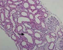
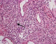
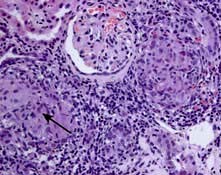
HUS is characterized by intravascular haemolysis with red-cell fragmentation (microangiopathic haemolysis), thrombocytopenia and acute kidney injury due to thrombosis in small arteries and arterioles (Fig. 12.25). These features are also seen in disseminated intravascular coagulation, but coagulation tests are typically normal in HUS.
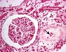
Figure 12.25 Typical haemolytic uraemic syndrome (HUS) renal lesion – light microscopy. Arrow shows microthrombi.
Diarrhoea-associated HUS (D+HUS)
This often follows a febrile illness, particularly gastroenteritis and usually associated with Escherichia coli, notably strain O157. This strain of E. coli produces verocytotoxin (shiga toxin), which has an A unit and five B units. The A unit is pathogenic by inhibiting protein synthesis and initiating endothelial damage. The role of B units is to facilitate the entry of the A unit into the endothelial cells by binding to a receptor (Gb3) on the endothelial cell. The toxins are transported to endothelial cells from the gut on neutrophils. Most patients with D+HUS recover renal function, but supportive care including maintenance of fluid and electrolyte balance, antihypertensive medication, nutritional support and dialysis is commonly required. Plasmapheresis is not beneficial but is usually tried as a last resort. About 5% die during the acute episode, 5% develop ESKD and 30% exhibit evidence of long-term damage with persistent proteinuria. Antibiotic and antimotility agents for the diarrhoea increase the risk of HUS and its complications.
Recently, an outbreak caused by shiga toxin-producing Escherichia coli O104:H4 (new strain) was reported in Germany and other European countries. In this outbreak, HUS cases were unusually high and associated with significant morbidity and mortality (see p. 121). Severe neurological complications were seen; immunoadsorption was successful in many cases.
Recurrent episodes of HUS have been described in the same individual, and familial forms of the disease (with both recessive and dominant inheritance) exist.
Also called atypical HUS (aHUS), this probably is a complement-driven illness due to a deficiency of complement factor H (CFH) or complement factor I (CFI). Factor H is a soluble protein produced by the liver, which regulates the activity of the alternative complement activation pathway; in particular, it protects host cell surfaces from complement-mediated damage. In some families with D–HUS, a mutation has been traced to another complement regulatory protein, CD46 (previously known as membrane cofactor protein, MCP). This protein is highly expressed in the kidney and normally prevents glomerular C3 activation. A loss of function mutation in CD46 results in unopposed complement activation and development of HUS. Functional deficiency of these factors can be acquired due to autoantibody formation either as an isolated phenomenon or as part of a rheumatic autoimmune disease such as SLE. A loss of function mutation in thrombomodulin (a membrane-bound anticoagulant glycoprotein) has been identified as an alternative complement pathway. Rarely gain of function mutations can affect genes encoding the alternative pathway C3 convertase components, CFB and C3. CFB mutations, which lead to chronic alternative-pathway activation, occur in only 1–2% of patients with D–HUS. About 4–10% of patients have heterozygous mutations in C3, usually with low C3 levels. Most mutations reduce C3b binding to CFH and CD46, which severely impairs degradation of mutant C3b.
Treatment is often very difficult because of severe hypertension and the possibility of frequent recurrences. The course of the disease is often indolent and progressive. Plasmapheresis or plasma infusion is still the only therapy used in the majority of patients. C5 activation is one of the critical steps in the activation of complement cascade. Eculizumab, a monoclonal humanized anti-C5 antibody, has shown success in patients with D–HUS either dependent or refractory to plasmapheresis therapy. Liver transplantation is potentially the only curative treatment in patients harbouring CFH and CFI mutations.
These can be associated with pregnancy, SLE, scleroderma, malignant hypertension, metastatic cancer, HIV infection and various drugs including oral contraceptives, ciclosporin, tacrolimus, chemotherapeutic agents (e.g. cisplatin, mitomycin C, bleomycin) and heparin. Treatment is supportive with removal of the offending agent or specific treatment of the underlying cause. There is no evidence in favour of plasma infusion or plasmapheresis in these sporadic cases but it is tried, usually as a last resort.
This rare complication of Streptococcus pneumoniae infection was previously associated with a high morbidity and mortality. This organism produces an enzyme (possibly neuroaminidase) which can expose an antigen (Thomsen antigen) present on RBCs, platelets and glomeruli. Antibodies to the Thomsen antigen result in an antigen-antibody reaction and can lead to HUS and anaemia. The improved outcome is due to increasing awareness of this complication, judicious use of blood products (washed blood products) and avoiding plasma infusion or plasmapheresis.
TTP (see p. 420) is characterized by microangiopathic haemolysis, renal failure and evidence of neurological disturbance. Young adults are most commonly affected.
In antiphospholipid syndrome (APS, see p. 538), the binding of antiphospholipid antibodies (aPL) to beta 2 glycoprotein I (β2GPI) induces endothelial cell–leukocyte adhesion and thrombus formation by the inhibition of eNOS. The inhibition of eNOS is caused by antibody recognition and dimerization of β2GPI and impairment of eNOS phosphorylation.
The central feature of APS is recurrent thrombosis (both venous and arterial) and fetal loss in the presence of antiphospholipid antibodies. Such antibodies may be primary or secondary to infections (HIV, hepatitis C) or autoimmune disease (SLE). Some 50% have renal involvement with proteinuria. Thrombotic microangiopathy is a rare but well-recognized presentation. In some cases, a lupus nephritis-like (usually membranous GN) lesion is seen. The only proven treatment for APS is warfarin with an INR of 3–4. Use of steroids or plasmapheresis is reserved for patients with APS and life-threatening renal involvement with thrombotic microangiopathy. Treatment is variably successful (30–70%).
Acute kidney injury (AKI) is relatively common in myeloma, occurring in 20–30% of affected individuals at the time of diagnosis, and is mainly due to the nephrotoxic effects of the abnormal immunoglobulins. It is often irreversible. The following types of renal lesions are associated with myeloma.
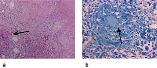
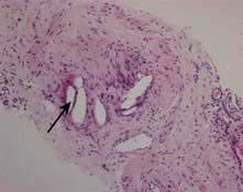
Light chain cast nephropathy – intratubular deposition of light chains, particularly kappa chains facilitated by Tamm–Horsfall glycoprotein, which characteristically appear on renal histology as fractured casts with giant cell reaction (Fig. 12.26)
AL amyloidosis – deposition of amyloid fibrils of light chains (Congo red positive)
Light chain deposition disease – nodular glomerulosclerosis with granular deposits of usually lambda light chains (Congo red negative)
Plasma cell infiltration – often incidental finding at autopsy
Fanconi’s syndrome – tubular toxicity due to light chains
Hypercalcaemic nephropathy – bone resorption causing hypercalcaemia
Hyperuricaemic nephropathy – tumour lysis causing tubular crystallization of uric acid
Radiocontrast nephropathy – interaction between light chains and radiocontrast.
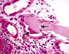
Figure 12.26 Cast nephropathy in a patient with multiple myeloma. Light microscopy picture showing characteristic fractured cast and giant cell reaction (arrows).
Treatment of underlying myeloma is indicated (p. 471). If a patient with cast nephropathy and severe AKI remains dialysis-dependent, the prognosis is poor. Commencement of effective bortezomib based chemotherapy, which decreases light chain production, and a high cut-off haemodialysis has shown some promise in relapsed myeloma.
Urinary tract infection (UTI) is common in women, in whom it usually occurs in an anatomically normal urinary tract. Conversely, it is uncommon in men and children, and the urinary tract is often abnormal and requires investigation. The incidence of UTI is 50 000 per million persons per year and accounts for 1–2% of patients in primary care. Recurrent infection causes considerable morbidity; if complicated, it can cause severe renal disease including ESKD. It is also a common source of life-threatening Gram-negative septicaemia.
Infection is most often due to bacteria from the patient’s own bowel flora (Table 12.9). Transfer to the urinary tract is most often via the ascending transurethral route but may be via the bloodstream, the lymphatics or by direct extension (e.g. from a vesicocolic fistula).
Table 12.9 Organisms causing urinary tract infection in domiciliary practice
Organism | Approximate frequency (%) |
---|---|
Escherichia coli and other ‘coliforms’ |
68+ |
Proteus mirabilis |
12 |
Klebsiella aerogenesa |
4 |
Enterococcus faecalisa |
6 |
Staphylococcus saprophyticus or epidermidisb |
10 |
a More common in hospital practice.
b More common in young women (20–30%).
Symptomatic infection is related to the virulence of the organisms, which compete with the innate host defence system. However, inflammation and injury are determined by the host response and not by the bacterium.
Virulence. Ability to adhere to epithelial cells determines the degree of virulence of the organism. For E. coli, these adhesive factors include flagellae (for motility), aerobactin (for iron acquisition in the iron-poor environment of the urinary tract), haemolysin (for pore forming) and above all, the presence of adhesins on the bacterial fimbriae and on the cell surface.
There are two types of E. coli: those with type 1 fimbriae (with adhesin known as FimH) associated with cystitis; and those with type P fimbriae (with adhesin known as PapG) commonly responsible for pyelonephritis. Bacterial adhesins are necessary for attachment of bacteria to the mucous membranes of the perineum and urothelium. There are several molecular forms of adhesins. The most studied is the PapG adhesin, which is located on the tip of P fimbriae. This lectin (one of the P blood group antigens) structure recognizes binding sites consisting of oligosaccharide sequences present on the mucosal surface.
Innate host defence. The following host defence mechanisms are necessary to prevent UTI:
Neutrophils – adhesins activate receptors, e.g. Toll receptor 4, on the mucosal surface, resulting in IL-8 production and expression of its receptor CXCR1 on neutrophil surfaces. Activation of neutrophils is essential for bacterial killing. Defective IL-8 production or reduced expression of CXCR1 results in impaired function of neutrophils predisposing an individual to severe UTI.
Urine osmolality and pH – urinary osmolality >800 mOsm/kg and low or high pH reduce bacterial survival.
Complement – complement activation with IgA production by uroepithelium (acquired immunity) also plays a major role in defence against UTI.
Commensal organisms – such as lactobacilli, corynebacteria, streptococci and bacteroides are part of the normal host defence. Eradication of these commensal organisms by spermicidal jelly or disruption by certain antibiotics results in overgrowth of E. coli.
Urine flow – urine flow and normal micturition wash out bacteria. Urine stasis promotes UTI.
Uroepithelium – mannosylated proteins such as Tamm–Horsfall proteins (THP), which are present in the mucus and glycocalyx covering uroepithelium, have antibacterial properties. These proteins interfere with bacterial binding to uroepithelium. Disruption of this uroepithelium by trauma (e.g. sexual intercourse or catheterization) predisposes to UTI. Cranberry juice and blueberry juice contain a large-molecular-weight factor (proanthrocyanidins) that prevents binding of E. coli to the uroepithelium (see p. 591).
Blood group antigens – women who are non-secretors of ABH blood group antigens are three to four times more likely to have recurrent UTIs.
UTI is commonly an isolated, rather than a repeated, event (Fig. 12.27).
It is necessary to distinguish between UTI occurring in patients with functionally normal urinary tracts and in those with abnormal tracts.
Functionally normal urinary tracts (with normal renal imaging). Here, persistent or recurrent infection seldom results in serious kidney damage (uncomplicated UTI).
Abnormal urinary tracts. Tracts with stones, or associated diseases such as diabetes mellitus which themselves cause kidney damage, may be made worse with infection (complicated UTI). UTI, particularly with Proteus, may predispose to stone formation. The combination of infection and obstruction results in severe, sometimes rapid, kidney damage (obstructive pyonephrosis) and is a major cause of Gram-negative septicaemia.
The combination of fever, loin pain with tenderness and significant bacteriuria usually implies infection of the kidney (acute pyelonephritis). Small renal cortical abscesses and streaks of pus in the renal medulla are often present. Histologically, there is focal infiltration by polymorphonuclear leucocytes and many polymorphs in tubular lumina.
Although, with antibiotics, significant permanent kidney damage in adults with normal urinary tracts is rare, CT scanning can show wedge-shaped areas of inflammation in the renal cortex (Fig. 12.29) and hence damage to renal function.
This was called chronic pyelonephritis or atrophic pyelonephritis, and it results from a combination of:
Normally, the vesicoureteric junction acts as a one-way valve (Fig. 12.30), urine entering the bladder from above; the ureter is shut off during bladder contraction, thus preventing reflux of urine. In some infants and children – possibly even in utero – this valve mechanism is incompetent, bladder voiding being associated with variable reflux of a jet of urine up the ureter. A secondary consequence is incomplete bladder emptying, as refluxed urine returns to the bladder after voiding. This latter event predisposes to infection, and the reflux of infected urine leads to kidney damage.
Typically, there is papillary damage, tubulointerstitial nephritis and cortical scarring in areas adjacent to ‘clubbed calyces’.
Diagnosis is based on CT scan of the kidneys, which shows irregular renal outlines, clubbed calyces and a variable reduction in renal size. The condition may be unilateral or bilateral and affect all or part of the kidney.
Reflux usually ceases around puberty with growth of the bladder base. Damage already done persists and progressive renal fibrosis and further loss of function occur in severe cases even though there is no further infection.
Reflux nephropathy cannot occur in the absence of reflux and therefore does not begin in adult life. Consequently, adult females with bacteriuria and a normal urogram can be reassured that kidney damage will not develop.
Chronic reflux nephropathy acquired in infancy predisposes to hypertension in later life and, if severe, is a relatively common cause of ESKD in childhood or adult life. Meticulous early detection and control of infection, with or without ureteral reimplantation to create a competent valve, can prevent further scarring and allow normal growth of the kidneys. No proof exists, however, that reimplantation surgery confers long-term benefit.
When UTI is recurrent, it is necessary to distinguish between relapse and reinfection.
Relapse is diagnosed by recurrence of bacteriuria with the same organism within 7 days of completion of antibacterial treatment and implies failure to eradicate infection (Fig. 12.31) usually in conditions such as stones, scarred kidneys, polycystic disease or bacterial prostatitis.
Reinfection is when bacteriuria is absent after treatment for at least 14 days, usually longer, followed by recurrence of infection with the same or different organisms. This is not due to failure to eradicate infection, but is the result of reinvasion of a susceptible tract with new organisms. Approximately 80% of recurrent infections are due to reinfection.
The most typical symptoms of UTI are:
These symptoms relate to bladder and urethral inflammation, commonly called ‘cystitis’, and suggest lower urinary tract infection. Loin pain and tenderness, with fever and systemic upset, suggest extension of the infection to the pelvis and kidney, known as pyelitis or pyelonephritis. However, localization of the site of infection on the basis of symptoms alone is unreliable.
UTI also presents with minimal or no symptoms or may be associated with atypical symptoms such as abdominal pain, fever or haematuria in the absence of frequency or dysuria.
In small children, who cannot complain of dysuria, symptoms are often ‘atypical’. The possibility of UTI must always be considered in the fretful, febrile sick child who fails to thrive.
This is based on quantitative culture of a clean-catch midstream specimen of urine and the presence or absence of pyuria. The criteria for the diagnosis of UTI, particularly in symptomatic women, are shown in Table 12.10. Most Gram-negative organisms reduce nitrates to nitrites and produce a red colour in the reagent square. False-negative results are common. Dipsticks that detect significant pyuria depend on the release of esterases from leucocytes. Dipstick tests positive for both nitrite and leucocyte esterase are highly predictive of acute infection (sensitivity of 75% and specificity of 82%).
Table 12.10 Criteria for diagnosis of bacteriuria
|
Causes of truly abacteriuric frequency/dysuria include postcoital bladder trauma, vaginitis, atrophic vaginitis or urethritis in the elderly, and interstitial cystitis (Hunner’s ulcer). In symptomatic young women with ‘sterile pyuria’, Chlamydia infection and tuberculosis must be excluded.
Interstitial cystitis is an uncommon but distressing complaint, most often affecting women over the age of 40 years. It presents with frequency, dysuria and often severe suprapubic pain. Urine cultures are sterile. Cystoscopy shows typical inflammatory changes with ulceration of the bladder base. It is commonly thought to be an autoimmune disorder. Various treatments are advocated with variable success. These include oral prednisolone therapy, bladder instillation of sodium cromoglycate or dimethyl sulphoxide and bladder stretching under anaesthesia.
Predominant frequency and passage of small volumes of urine (‘irritable bladder’) is possibly consequent on previous UTI or conditioned by psychosexual factors. Such patients must be distinguished from those with frequency due to polyuria. Repeated courses of antibiotics in patients with genuine abacteriuric frequency or dysuria are quite inappropriate and detract from identifying the true nature of the problem.
Uncomplicated UTI usually does not require radiological evaluation unless it is recurrent or affecting males and children or there are unusually severe symptoms. Patients with predisposing conditions such as diabetes mellitus or immunocompromised states benefit from early imaging.
Ultrasound is used in the assessment of patients with suspected pyelonephritis that requires drainage. This allows the detection of calculi, obstruction and also incomplete emptying.
CT is a more sensitive modality for diagnosis and follow-up of complicated renal tract infection. Contrast-enhanced CT allows different phases of excretion to be studied and can define the extent of disease and identify significant complications or obstruction.
MRI is particularly useful in those with iodinated contrast allergies, offering an ionizing radiation-free alternative in the diagnosis of both medical and surgical diseases of the kidney.
Nuclear medicine has a limited role in the evaluation of UTI in adults. Its main role is in the assessment of renal function and detection of scars by DMSA scan, often prior to surgery.
Pre-treatment urine culture is desirable.
Antibiotics for 3–5 days with amoxicillin (250 mg three times daily), nitrofurantoin (50 mg three times daily), trimethoprim (200 mg twice daily) or an oral cephalosporin. The treatment regimen is modified in light of the result of urine culture and sensitivity testing, and/or the clinical response.
For resistant organisms the alternative drugs are co-amoxiclav or ciprofloxacin.
Single-shot treatment with 3 g of amoxicillin or 1.92 g of co-trimoxazole is used for patients with bladder symptoms of less than 36 hours’ duration who have no previous history of UTI.
A high (2 L daily) fluid intake should be encouraged during treatment and for some subsequent weeks. Urine culture should be repeated 5 days after treatment.
If the patient is acutely ill with high fever, loin pain and tenderness (acute pyelonephritis), antibiotics are given intravenously, e.g. aztreonam, cefuroxime, ciprofloxacin or gentamicin (2–5 mg/kg daily in divided doses), switching to a further 7 days’ treatment with oral therapy as symptoms improve. Intravenous fluids may be required to achieve a good urine output.
In patients presenting for the first time with high fever, loin pain and tenderness, urgent renal ultrasound examination is required to exclude an obstructed pyonephrosis. If this is present it should be drained by percutaneous nephrostomy (p. 570).
Pre-treatment and post-treatment urine cultures are necessary to confirm the diagnosis and identify whether recurrent infection is due to relapse or reinfection.
Relapse. A search should be made for a cause (e.g. stones or scarred kidneys), and this should be eradicated. Intense or prolonged treatment – intravenous or intramuscular aminoglycoside for 7 days or oral antibiotics for 4–6 weeks – is required. If this fails, long-term antibiotics are required.
Reinfection implies that the patient has a predisposition to periurethral colonization or poor bladder defence mechanisms. Contraceptive practice should be reviewed and the use of a diaphragm and spermicidal jelly discouraged. Atrophic vaginitis should be identified in postmenopausal women, who should be treated (see below). All patients must undertake prophylactic measures:
Voiding at 2–3-hour intervals with double micturition if reflux is present
Voiding before bedtime and after intercourse
Avoidance of spermicidal jellies and bubble baths and other chemicals in bath water
Avoidance of constipation, which may impair bladder emptying.
Evidence of impaired bladder emptying on excretion urography/ultrasound requires urological assessment. If UTI continues to recur, treatment for 6–12 months with low-dose prophylaxis (trimethoprim 100 mg, co-trimoxazole 480 mg, cefalexin 125 mg at night or macrocrystalline nitrofurantoin) is required; it should be taken last thing at night when urine flow is low. Intravaginal oestrogen therapy has been shown to produce a reduction in the number of episodes of UTI in postmenopausal women. Cranberry juice is said to reduce the risk of symptoms and reinfection by 12–20% but studies are limited.
Colonization of the bladder by a urinary pathogen is common after a urinary catheter has been present for more than a few days, partly due to organisms forming biofilms. So long as the bladder catheter is in situ, antibiotic treatment is likely to be ineffective and will encourage the development of resistant organisms. Treatment with antibiotics is indicated only if the patient has symptoms or evidence of infection, and should be accompanied by replacement of the catheter. When changing catheters, a single injection of gentamicin is recommended.
Infection by Candida is a frequent complication of prolonged bladder catheterization. Treatment should be reserved for patients with evidence of invasive infection or those who are immunosuppressed, and should consist of removal or replacement of the catheter. In severe infections continuous bladder irrigation with amphotericin 50 µg/mL is used.
The urine of all pregnant women must be cultured, as 2–6% have asymptomatic bacteriuria. While asymptomatic bacteriuria in the non-pregnant female seldom leads to acute pyelonephritis and often does not require treatment, acute pyelonephritis frequently occurs in pregnancy under these circumstances. Failure to treat may thus result in severe symptomatic pyelonephritis later in pregnancy, with the possibility of premature labour. Asymptomatic bacteriuria, in the presence of previous renal disease, may predispose to pre-eclamptic toxaemia, anaemia of pregnancy, and small or premature babies. Therefore bacteriuria must always be treated and be shown to be eradicated. Reinfection may require prophylactic therapy. Tetracycline, trimethoprim, sulphonamides and 4-quinolones must be avoided in pregnancy. Amoxicillin and ampicillin, nitrofurantoin and oral cephalosporins may safely be used in pregnancy.
Bacterial prostatitis is a relapsing infection which is difficult to treat. It presents as perineal pain, recurrent epididymo-orchitis and prostatic tenderness, with pus in expressed prostatic secretion. Treatment is for 4–6 weeks with drugs that penetrate into the prostate, such as trimethoprim or ciprofloxacin. Long-term low-dose treatment may be required. Prostadynia (prostatic pain in the absence of active infection) may be a very persistent sequel to bacterial prostatitis. Amitriptyline and carbamazepine may alleviate the symptoms.
Renal carbuncle is an abscess in the renal cortex caused by a blood-borne Staphylococcus, usually from a boil or carbuncle of the skin. It presents with a high swinging fever, loin pain and tenderness, and fullness in the loin. The urine shows no abnormality, as the abscess does not communicate with the renal pelvis, more often extending into the perirenal tissue. Staphylococcal septicaemia is common. Diagnosis is by ultrasound or CT scanning. Treatment involves antibacterial therapy with flucloxacillin and surgical drainage.
Tuberculous infection is on the increase worldwide, partly due to the reservoir of infection in susceptible HIV-infected individuals and the emergence of drug-resistant strains. Tuberculosis of the urinary tract presents with frequency, dysuria or haematuria. In the UK, it is mainly seen in the Asian immigrant population. Cortical lesions result from haematogenous spread in the primary phase of infection. Most heal, but in some, infection persists and spreads to the papillae, with the formation of cavitating lesions and the discharge of mycobacteria into the urine. Infection of the ureters and bladder commonly follows, with the potential for the development of ureteral stricture and a contracted bladder. Rarely, cold abscesses may form in the loin. In males the disease may present with testicular or epididymal discomfort and thickening.
Diagnosis depends on constant awareness, especially in patients with sterile pyuria. Imaging may show cavitating lesions in the renal papillary areas, commonly with calcification. There may also be evidence of ureteral obstruction with hydronephrosis. Diagnosis of active infection depends on culture of mycobacteria from early-morning urine samples. Imaging may be normal in diffuse interstitial renal tuberculosis when diagnosis is made by renal biopsy demonstrating caseating granuloma with multinucleate giant cells and acid-fast bacilli on Ziehl–Neelsen staining (Fig. 12.32). Some patients present with small unobstructed kidneys, when the diagnosis is easy to miss.
Figure 12.32 Renal tuberculosis. (a) Caseating granulomatous interstitial nephritis showing multinuclear giant cell (arrow). (b) Ziehl–Neelsen staining showing myobacteria (arrow).
Treatment. The treatment is as for pulmonary tuberculosis (see p. 842). Renal ultrasonography and/or CT scanning should be carried out 2–3 months after initiation of treatment as ureteric strictures may first develop in the healing phase.
This is an uncommon chronic interstitial infection of the kidney, most often due to Proteus, in which there is fever, weight loss, loin pain and a palpable enlarged kidney. It is usually unilateral and associated with staghorn calculi and urinary tract infection. CT scanning shows up intrarenal abscesses as lucent areas within the kidney. Nephrectomy is the treatment of choice; antibacterial treatment rarely, if ever, eradicates the infection.
Diseases of the kidney primarily affect the glomeruli, vasculature, or the remainder of the renal parenchyma that consists of the tubules and interstitium. Although the tubules and the interstitium are distinct functional entities, they are intimately related. Injury involving one of them invariably results in damage to the other.
In approximately 70% of the cases, acute TIN is due to a hypersensitivity reaction to drugs (Table 12.11), most commonly drugs of the penicillin family and non-steroidal anti-inflammatory drugs (NSAIDs).
Table 12.11 Common causes of acute tubulointerstitial nephritis
Drugs (70%) |
|
Antibiotics |
Diuretics |
Cephalosporins |
Furosemide |
Ciprofloxacin |
Thiazides |
Erythromycin |
Miscellaneous |
Penicillin |
Cimetidine |
Rifampicin |
Phenytoin |
Sulphonamides |
Valproate |
Analgesics |
Carbamazepine |
Non-steroidal anti-inflammatory drugs |
AllopurinolProton pump inhibitors |
Infection (15%) |
|
Viruses, e.g. hantavirus |
|
Bacteria, e.g. streptococci |
|
Idiopathic (8%) |
|
Tubulointerstitial nephritis with uveitis (TINU) (5%) |
|
Systemic inflammatory disorders, e.g. systemic lupus erythematosus (SLE) (2%) |
Drug induced acute TIN. Patients present with fever, arthralgia, skin rashes and acute oliguric or non-oliguric kidney injury. Many have eosinophilia and eosinophiluria. Renal histology shows an intense interstitial cellular infiltrate, often including eosinophils, with variable tubular necrosis (Fig. 12.33). Rarely, NSAIDs can cause a glomerular minimal-change lesion in addition to TIN and present as the nephrotic syndrome. Treatment involves withdrawal of offending drugs. High-dose steroid therapy (prednisolone 60 mg daily) is commonly given but its efficacy has not been proven. Patients may require dialysis for management of the acute kidney injury. Most patients make a good recovery in kidney function, but some may be left with significant interstitial fibrosis and CKD.
Figure 12.33 Tubulointerstitial nephritis showing diffuse interstitial infiltrate with red staining (arrow).
Infection causing acute TIN. Acute pyelonephritis leads to inflammation of the tubules, producing a neutrophilic cellular infiltrate. TIN can complicate systemic infections with viruses (Hantavirus, Epstein–Barr virus, HIV, measles, adenovirus), bacteria (Legionella, Leptospira, streptococci, Mycoplasma, Brucella, Chlamydia) and others (Leishmania, Toxoplasma). Hantavirus causes haemorrhagic fever with TIN and can be fatal. Epstein–Barr virus DNA has been found in renal biopsy tissue of cases of idiopathic TIN. In immunocompromised patients such as post-renal transplantation, CMV, polyoma, and HSV can cause acute TIN in the renal graft. Treatment involves eradication of infection by appropriate antibiotics or antiviral agents and in renal transplantation modifying immunosuppressive regimen.
Acute TIN as part of multisystem inflammatory diseases. Several non-infectious inflammatory disorders such as Sjögren’s syndrome, SLE and Wegener’s granulomatosis can cause acute or chronic TIN rather than glomerulonephritis. Sjögren’s syndrome may additionally present as renal tubular acidosis. Sarcoidosis presents as granulomatous TIN in up to 20% of patients. Associated hypercalcaemia causes acute kidney injury. These heterogeneous conditions with TIN generally respond to steroids.
TINU syndrome. In this syndrome, uveitis generally coincides with acute TIN. It is common in childhood, but has been reported in adulthood. Among adults it is more common in females, but its cause remains unknown. Available evidence suggests that it is associated with autoantibodies directed against modified C-reactive protein. Patients present with weight loss, anaemia and raised ESR. A prolonged course of steroids leads to improvement in both renal function and uveitis.
The major causes of chronic tubulointerstitial nephritis are given in Table 12.12. It is characterized by generalized chronic inflammatory cellular infiltration of the interstitium with tubular atrophy and generalized interstitial oedema or fibrosis. In many cases no cause is found. Chronic TIN changes evolve into progressive primary glomerular or vascular disease of the kidney, where its severity is a better predictor of long-term renal survival than the primary site of insult.
Table 12.12 Causes of chronic tubulointerstitial
Drugs and toxins |
e.g. All causes of ATNa, e.g. analgesics |
Ciclosporin |
|
Cadmium, lead, titanium |
|
Irradiation |
|
Diseases |
e.g. Diabetes mellitus |
Sickle cell disease or trait |
|
SLE/vasculitis |
|
Sarcoidosis |
|
Metabolic |
e.g. Hyperuricaemia |
Nephrocalcinosis |
|
Hyperoxaluria |
|
Infection |
e.g. HIV |
EBVb |
|
Miscellaneous |
Hypertension |
Balkan nephropathy |
|
Herbal nephropathy |
|
Alport’s syndrome |
aAcute tubulointerstitial nephritis. bEpstein–Barr virus.
The patient usually presents with either polyuria and nocturia, or is found to have proteinuria or uraemia. Proteinuria is usually slight (<1 g daily). Papillary necrosis with ischaemic damage to the papillae occurs in a number of tubulointerstitial nephritides, for example in analgesic abuse, diabetes mellitus, sickle cell disease or trait. The papillae can separate and be passed in the urine. Microscopic or overt haematuria or sterile pyuria also occurs, and occasionally a sloughed papilla may cause ureteric colic or produce acute ureteric obstruction. The radiological appearances must be distinguished from those of reflux nephropathy (Fig. 12.34) or obstructive uropathy which is usually accompanied by tubular dilatation and atrophy and intense interstitial fibrosis with patchy inflammatory cellular infiltrate in the scarred areas.
Tubular damage to the medullary area of the kidney leads to defects in urine concentration and sodium conservation with polyuria and salt wasting. Fibrosis progressing into the cortex leads to loss of excretory function and uraemia.
The chronic consumption of large amounts of analgesics (especially those containing phenacetin) and NSAIDs leads to chronic tubulointerstitial nephritis and papillary necrosis. Analgesic nephropathy is twice as common in women as in men and presents typically in middle age. Patients are often depressed or neurotic. Presentation may be with anaemia, CKD, UTIs, haematuria or urinary tract obstruction (owing to sloughing of a renal papilla). Salt and water-wasting renal disease may occur. Chronic analgesic abuse also predisposes to the development of uroepithelial tumours. Diagnosis is usually made on clinical grounds combined with the non-pathognomonic appearance on imaging (such as ultrasonography or CT scan), which demonstrates smallish irregularly outlined kidneys.
The consumption of the above analgesics should be discouraged. If necessary, dihydrocodeine or paracetamol is a reasonable alternative. This may result in the arrest of the disease and even in improvement in function. UTI, hypertension (if present) and saline depletion will require appropriate management. The development of flank pain or an unexpectedly rapid deterioration in renal function should prompt ultrasonography to screen for urinary tract obstruction due to a sloughed papilla.
Chinese herbal medicines have been increasingly used in the West, e.g. for slimming, and have caused a nephropathy. The renal histology is similar to Balkan nephropathy but the clinical course is very aggressive. The causal agent has been identified as aristolochic acid produced as a result of fungal contamination of the herbal medicine. It is characterized by relentless progression to ESKD. There is a high incidence of uroepithelial tumours.
This is a chronic TIN endemic in areas along the tributaries of the River Danube. Inhabitants of the low-lying plains, which are subjected to frequent flooding and where the water supply comes from shallow wells, are affected, whereas the disease does not occur in hillside villages where surface water provides the water supply. Its cause was essentially unknown. However, new research suggests that chronic dietary poisoning by aristolochic acid (AA) is responsible for BN and its associated urothelial cancer. AA-DNA adduct is found in tissue biopsies both from BN and associated urothelial cancers. This research suggests that AA is the environmental agent responsible for BN and its associated transitional-cell cancer. The disease is insidious in onset, with mild proteinuria progressing to ESKD in 3 months to 10 years. There is no treatment.
These are rare (Table 12.12). Diagnosis of all forms depends on a history of drug ingestion or industrial exposure to nephrotoxins. In patients with unexplained renal impairment with normal-sized kidneys, renal biopsy must always be undertaken to exclude a treatable tubulointerstitial nephritis such as granulomatous TIN due to renal sarcoidosis (Fig. 12.35), which may be the first presentation of sarcoidosis (see p. 846). Renal sarcoidosis generally responds rapidly to steroids.
Acute hyperuricaemic nephropathy (see p. 503) is a well-recognized cause of acute kidney injury in patients with marked hyperuricaemia that is usually due to lymphoproliferative or myeloproliferative disorders. It may occur prior to treatment but most often follows commencement of treatment, when there is rapid lysis of malignant cells, release of large amounts of nucleoprotein and increased uric acid production. Renal failure is due to intrarenal and extrarenal obstruction caused by deposition of uric acid crystals in the collecting ducts, pelvis and ureters. The condition is manifest as oliguria or anuria with increasing uraemia. There may be flank pain or colic. Plasma urate levels are above 0.75 mmol/L and may be as high as 4.5 mmol/L. Diagnosis is based on the hyperuricaemia and the clinical setting. Ultrasound may demonstrate extrarenal obstruction due to stones, but a negative scan does not exclude this where there is coexistent intrarenal obstruction.
Allopurinol 100–200 mg three times daily for 5 days is given prior to and throughout treatment with radiotherapy or cytotoxic drugs. A high rate of urine flow must be maintained by oral or parenteral fluid and the urine kept alkaline by the administration of sodium bicarbonate 600 mg four times daily and acetazolamide 250 mg three times daily, since uric acid is more soluble in an alkaline than in an acid medium. Febuxostat, a non-purine-analogue inhibitor of xanthine oxidase, can be used if allopurinol cannot be tolerated and eGFR is >30 mL/min. Rasburicase, a recombinant urate oxidase (p. 449), and pegloticase, a pegylated uricase in development, are occasionally used. In severely oliguric or anuric patients, dialysis is required to lower the plasma urate.
There is no convincing direct evidence for chronic hyperuricaemia nephropathy. However, a few observational studies have recently suggested that elevated levels of uric acid independently increase the risk for new-onset CKD, and that lowering plasma urate reduction with allopurinol has a beneficial effect in slowing the rate of progression of CKD.
Hypertension can be the cause or the result of renal disease. It is often difficult to differentiate between the two on clinical grounds. Routine tests (as described on p. 780) should be performed on all hypertensive patients, but renal imaging is usually unnecessary.
The mechanisms responsible for the normal regulation of arterial blood pressure and the development of essential primary hypertension are unclear (p. 777). One basic concept is that the long-term regulation of arterial pressure is closely linked to the ability of the kidneys to excrete sufficient salt to maintain normal sodium balance, extracellular fluid volume, and normal blood volume at normotensive arterial pressures. Cross-transplantation experiments suggest that hypertension travels with the kidney, in that hypertension will develop in a normotensive recipient of a kidney genetically programmed for hypertension. Similarly, patients with ESKD due to hypertension become normotensive after receiving a renal allograft from normotensive donors, provided the new kidney works.
One of many renal factors involved in the genesis of hypertension is the total number of nephrons in the kidney. Patients with hypertension and normal renal function have a significantly reduced number of nephrons in each kidney alongside enlargement of the remaining glomeruli due to glomerular hyperfiltration. Moreover, certain races (black, Hispanics) with predilection for hypertension have increased glomerular volume, a surrogate marker for a reduced number of nephrons.
Whether the reduced number of nephrons is caused by genetic or environmental factors is unclear. Changes in the intrauterine environment may lead to retarded renal growth before birth and low birth weight and hypertension during adult life. In humans, an association has been found between low birth weight and reduced renal volume, possibly indicating reduced numbers of nephrons.
In benign essential hypertension, arteriosclerosis of major renal arteries and changes in the intrarenal vasculature (nephrosclerosis) occur as follows:
In small vessels and arterioles, intimal thickening with reduplication of the internal elastic lamina occurs and the vessel wall becomes hyalinized.
In large vessels, concentric reduplication of the internal elastic lamina and endothelial proliferation produce an ‘onion skin’ appearance.
Reduction in size of both kidneys occurs; this may be asymmetrical if one major renal artery is more affected than the other.
The proportion of sclerotic glomeruli is increased compared with age-matched controls.
Deterioration in excretory function accompanies these changes, but severe CKD is unusual in whites (1 in 10 000). In black Africans, by contrast, hypertension much more often results in the development of CKD with a fourfold higher incidence of ESKD in blacks compared to whites. This racial difference in incidence of hypertensive renal disease may be due to overestimation of diagnosis on clinical grounds, poor compliance with medication, higher incidence of hypertension, which is usually a salt-sensitive type, reduced number of nephrons and possibly resulting from higher frequency of susceptibility alleles for ESKD in the West African than the European gene pool.
A recent genome-wide study found statistically stronger associations between two independent sequence variants in the apolipoprotein L1 gene (APOL1) and non-diabetic nephropathy in African Americans, in hypertension-attributed ESKD. These kidney disease risk variants most likely arose due to positive selection for evolutionary advantage these variants in APOL1 conferred against trypanosomal infection and protection from African sleeping sickness. These observations provide some evidence, similar to findings with sickle cell anaemia, that natural selection might protect from one disease but allow another one to develop.
In accelerated or malignant-phase hypertension:
Arteriolar fibrinoid necrosis occurs, probably as a result of plasma entering the media of the vessel through splits in the intima. It is prominent in afferent glomerular arterioles.
Fibrin deposition within small vessels is often associated with thrombocytopenia and red-cell fragmentation seen in the peripheral blood film (microangiopathic haemolytic anaemia).
Microscopic haematuria, proteinuria, usually of modest degree (1–3 g daily), and progressive uraemia occur. If untreated, fewer than 10% of patients survive 2 years.
The management of benign essential and malignant hypertension is described on page 781.
If treatment is begun before CKD has developed, the prognosis for renal function is good. Stabilization or improvement in renal function with healing of intrarenal arteriolar lesions and resolution of microangiopathic haemolysis occur with effective treatment of malignant phase hypertension. In blacks with hypertensive nephrosclerosis and CKD, a blood pressure target of <140/85 mmHg should be achieved.
FURTHER READING
Genovese G, Friedman DJ, Ross MD et al. Association of trypanolytic ApoL1 variants with kidney disease in African Americans. Science 2010; 329(5993):841–845.
Kao WH, Klag MJ, Meoni LA et al. MYH9 is associated with nondiabetic end-stage renal disease in African Americans. Nat Genet 2008; 40(10):1185–1192.
Hypertension commonly complicates bilateral renal disease such as chronic glomerulonephritis, bilateral reflux nephropathy, polycystic disease and analgesic nephropathy. Two main mechanisms are responsible:
Activation of the renin-angiotensin-aldosterone system
Retention of salt and water owing to impairment in excretory function, leading to an increase in blood volume and hence blood pressure.
The second of these assumes greater significance as renal function deteriorates.
Hypertension occurs earlier, is more common and tends to be more severe in patients with renal cortical disorders, such as glomerulonephritis, than in those with disorders affecting primarily the renal interstitium, such as reflux or analgesic nephropathy.
Management is described on page 781. Meticulous control of the blood pressure is necessary to prevent further deterioration of renal function secondary to vascular changes produced by the hypertension itself. There is good evidence that ACE-inhibitor drug treatment confers an additional renoprotective effect for a given degree of blood pressure control than other hypotensive drugs. In a study of African Americans with hypertension, intensive blood pressure control (130/78) was not superior to standard control (141/86) in the prevention of ESKD. However, in the same study patients with proteinuria (protein creatinine ratio >0.22) benefited more from intensive blood pressure control.
Renal ischaemia results in a reduction in the pressure in afferent glomerular arterioles. This leads to an increase in the production and release of renin from the juxtaglomerular apparatus (see p. 567) with a consequent increase in angiotensin II, a very potent vasoconstrictor. Angiotensin II also causes hypertension by upregulating NADPH oxidase enzyme with excessive superoxide generation. Superoxide chelates nitric oxide (a potent vasodilator) resulting in reduced vasodilator activity and also hypertension.
In renal artery stenosis, renal perfusion pressure is reduced and nephron transit time is prolonged on the side of the stenosis; salt and water reabsorption is therefore increased. As a result, urine from the ischaemic kidney is more concentrated and has a lower sodium concentration than urine from the contralateral kidney. Creatinine clearance is decreased on the ischaemic side.
Narrowing of the renal arteries (renal artery stenosis) is caused by one of two pathological entities: fibromuscular disease or atherosclerotic renovascular disease (ARVD).
Fibromuscular disease (FMD) accounts for 20–40% of renal vascular disease and encompasses four distinct types: (1) medial fibroplasia (65–85%); (2) perimedial fibroplasia (10–15%); (3) intimal fibroplasia (5–10%); and (4) medial hyperplasia (5%). Medial fibroplasia usually follows a benign course and never follows a progressive course after the age of 40 years. The other two types of fibroplasia follow a progressive course and may lead to total occlusion.
Medial hyperplasia is a distinct but rare entity, which accounts for only 1% of renovascular disease. It commonly affects young females, who exhibit elevated blood pressures but with well-preserved renal function.
MR angiography (gadolinium enhanced) reveals a characteristic string of beads appearance in fibroplasia.
Angioplasty (occasionally stent insertion) or surgery is usually performed in affected individuals. Cure rates were only 36% and 54% after angioplasty and surgery, respectively, in a recent study of over 2000 patients (defining cure as blood pressure <140/90 mm Hg without treatment), and the blood pressure outcome was strongly age related. Furthermore, the incidence of complications was substantial: combined risks of periprocedural complications were 12% after angioplasty and 17% after surgery, with fewer major complications after angioplasty (6%) than after surgery (15%). Given the efficacy of current medical antihypertensive therapy, intervention in these patients is usually not warranted.
This is a common cause of hypertension and CKD due to ischaemic nephropathy. Its incidence increases with age, rising from 5% under 60 years to 16% in those over 60 years old. In most patients, the atherosclerotic lesion is ostial (within 1 cm of the origin of the renal artery) and usually associated with symptomatic atherosclerotic vascular disease elsewhere. Patients with peripheral vascular disease (39%), coronary artery disease (10–29%), congestive cardiac failure (34%) and aortic aneurysm (38%) are at high risk of developing significant renal artery stenosis.
Many patients are asymptomatic and are discovered incidentally during investigation for other conditions. Aortography experience from the USA shows 11% of asymptomatic patients have significant unilateral stenosis and 4% have bilateral disease. The renal consequences of ARVD are functional, such as hypertension (present in 50%), sodium retention (ankle and flash pulmonary oedema), proteinuria (usually sub-nephrotic range) and decreased GFR. The morphological features of the affected kidneys include vascular sclerosis, tubular atrophy, interstitial fibrosis with inflammatory cellular infiltrate, atubular glomeruli, cholesterol emboli and secondary focal segmental glomerulosclerosis (FSGS) changes. Baseline renal function is related to the extent of renal parenchymal injury rather than to the degree of stenosis, as is the response (improvement in hypertension and renal function) to revascularization.
Renovascular disease should be looked for in the following: patients with hypertension and/or CKD; patients with abdominal audible bruits, as well as bruits over carotid arteries suggestive of generalized arterial disease; Doppler ultrasonography showing >1.5 cm renal asymmetry; recurrent flash pulmonary oedema without cardiopulmonary disease; and lastly, progressive CKD in patients with evidence of generalized atherosclerosis.
The aim of treatment is to correct hypertension and improve renal perfusion and excretory function. Renal artery stenosis can progress to occlusion, particularly in patients with stenosis >75% as shown by serial angiography, necessitating revascularization in ARVD.
The options in renal artery stenosis include transluminal angioplasty to dilate the stenotic region, insertion of stents across the stenosis (sometimes the only endoscopic option when the stenosis occurs close to the origin of the renal artery from the aorta, rendering angioplasty technically difficult or impossible), reconstructive vascular surgery and nephrectomy.
Indications for revascularization. Vessels with stenosis >75% and recurrent flash pulmonary oedema, drug-resistant severe hypertension, ARVD affecting solitary functioning kidney, patients with cardiac failure needing ACE inhibitors, unexplained progressive CKD and dialysis-dependent renal failure. Generally, endovascular procedures are considered better than medical therapy alone and, with good selection of patients, hypertension is cured or improved by intervention in more than 50%. Occasional dramatic improvements in renal function ensue but results are generally disappointing. Deterioration of the renal function occurs in 20–30% of the patients after renal angioplasty stenting. Atheroembolism seems to play a role and is probably the main cause of this renal function deterioration.
Medication. All patients with ARVD should be treated with a combination of aspirin, statins and optimal control of blood pressure as prophylaxis against progression of atherosclerosis.
Prognosis. Mortality is high because of other associated co-morbidities, and ARVD patients have generalized endothelial dysfunction. ARVD patients with ESKD have higher rates than those with good renal function. Five-year survival is only 18% in patients with ESKD due to ARVD.
Radionuclide studies (see p. 571). These can demonstrate decreased renal perfusion on the affected side. In unilateral renal artery stenosis, a disproportionate fall in uptake of isotope on the affected side following administration of captopril or aspirin is suggestive of the presence of significant renal artery stenosis. A completely normal result renders the diagnosis unlikely.
Doppler ultrasound. This method is very sensitive but highly operator-dependent and time-consuming. Measurement of renal-artery velocity by Doppler ultrasound provides a functional assessment of the severity of stenosis; higher velocity usually means higher pressure differential across the stenosis. It also generates useful data about intrarenal vascular resistance, which can be valuable in predicting the success of revascularization procedures. A resistive index of >80 is a predictor of poor response following intervention.
Magnetic resonance angiography. MRA can be used to visualize the renal arteries and there is a good – though not perfect – correlation between MRA findings and those of renal arteriography.
CT scanning. This permits non-invasive imaging of the renal arteries. It is much less expensive than MRA but does expose the patient to ionizing radiation and to contrast injection and is less reliable than MRA.
Renal arteriography (see this chapter) is used to confirm the diagnosis of renal arterial disease.
FURTHER READING
Tullus K, Brennan E, Hamilton G. Renovascular hypertension in children. Lancet 2008; 371:1453–1463.
Williams GJ, Macaskill P, Chan SF et al. Comparative accuracy of renal duplex sonographic parameters in the diagnosis of renal artery stenosis: paired and unpaired analysis. Am J Roentgenol 2007; 188(3):798–811.
This occurs from thrombosis in situ, usually in a severely damaged atherosclerotic vessel, or more commonly from embolization, e.g. in atrial fibrillation. Both lead to renal infarction, resulting in a wide spectrum of clinical manifestations depending on the size of the artery involved. Occlusion of a small branch artery may produce no effect, but occlusion of larger vessels results in dull flank pain and varying degrees of CKD.
Intra-arterial thrombolytic therapy has been tried with mixed results.
Showers of cholesterol-rich atheromatous material from ulcerated plaques reach the kidney from the aorta and/or renal arteries, particularly after catheterization of the abdominal aorta or attempts at renal artery angioplasty. Anticoagulants and thrombolytic agents also precipitate cholesterol embolization. Renal failure from cholesterol emboli may be acute or slowly progressive. Clinical features include fever, eosinophilia, back and abdominal pain, and evidence of embolization elsewhere, e.g. to the retina or digits. The diagnosis can be confirmed by renal biopsy (Fig. 12.36). It is more common in males, the elderly (>70 years) and patients with cardiovascular disease. Over 80% have abnormal renal function at baseline. AERD occurs spontaneously in 25% of the cases. The 2-year mortality is 30% and a similar percentage of patients develop CKD. Baseline co-morbidities, i.e. reduced renal function, presence of diabetes, history of heart failure, acute/subacute presentation, and gastrointestinal tract involvement, are significant predictors of event occurrence. The risk of dialysis and death is 50% lower among those receiving statins.
This is usually of insidious onset, occurring in patients with the nephrotic syndrome, with a renal cell carcinoma, and in thrombophilia (p. 424) with an increased risk of venous thrombosis. Anticoagulation is indicated.
Renal stones are very common worldwide, with a lifetime risk of about 10%. Prevalence of stone disease is much higher in the Middle East. Most stones occur in the upper urinary tract.
Most stones are composed of calcium oxalate and phosphate; these are more common in men (Table 12.13). Mixed infective stones, which account for about 15% of all calculi, are twice as common in women as in men. The overall male to female ratio of stone disease is 2:1.
Table 12.13 Type and frequency of renal stones in the UK
Type of renal stone | Percentage of stones |
---|---|
Calcium oxalate usually with calcium phosphate |
65 |
Calcium phosphate alone |
15 |
Magnesium ammonium phosphate (struvite) |
10–15 |
Uric acid |
3–5 |
Cystine |
1–2 |
Stone disease is frequently a recurrent problem. More than 50% of patients with a history of nephrolithiasis will develop a recurrence within 10 years. The risk of recurrence increases if a metabolic or other abnormality predisposing to stone formation is present and is not modified by treatment. Nephrolithiasis is not a benign condition as several observational studies have demonstrated its association with increased risk of ESKD, bone diseases, hypertension and myocardial infarction.
Inhibitors of crystal formation are present in normal urine preventing the formation of stones, as the concentrations of stone-forming substances in many cases exceed their maximum solubility in water. Many stone-formers have no detectable metabolic defect, although microscopy of warm, freshly passed urine reveals both more and larger calcium oxalate crystals than are found in normal subjects. Factors predisposing to stone formation in these so-called ‘idiopathic stone-formers’ are:
Chemical composition of urine that favours stone crystallization
Production of a concentrated urine as a consequence of dehydration associated with life in a hot climate or work in a hot environment
Impairment of inhibitors that prevent crystallization in normal urine. Postulated inhibitors include inorganic magnesium, pyrophosphate and citrate. Organic inhibitors include glycosaminoglycans and nephrocalcin (an acidic protein of tubular origin). Tamm–Horsfall protein may have a dual role in both inhibiting and promoting stone formation.
Recognized causes of stone formation are listed in Table 12.14.
Table 12.14 Causes of urinary tract stones
If the GFR is normal, hypercalcaemia almost invariably leads to hypercalciuria. The common causes of hypercalcaemia leading to stone formation are:
Of these, primary hyperparathyroidism (see p. 994) is the most common cause of stones.
This is by far the most common metabolic abnormality detected in calcium stone-formers.
Approximately 8% of men excrete in excess of 7.5 mmol of calcium in 24 hours. Calcium stone formation is more common in this group, but as the majority of even these individuals do not form stones the definition of ‘pathological’ hypercalciuria is arbitrary. A reasonable definition is 24-hour calcium excretion of >7.5 mmol in male stone-formers and >6.25 mmol in female stone-formers.
The kidney is the major site for plasma calcium regulation. Approximately 90% of the ionized calcium filtered by the kidney is reabsorbed. Renal tubular reabsorption is controlled largely by parathyroid hormone (PTH).
Approximately 65% of the filtered calcium is absorbed in the proximal convoluted tubule, 20% by the thick ascending limb of the loop of Henle, and 15% by the distal convoluted tubule and collecting ducts.
An excessive dietary intake of calcium
Excessive resorption of calcium from the skeleton, such as occurs with prolonged immobilization or weightlessness
Idiopathic hypercalciuria is a common risk factor for the formation of stones, and uncontrolled hypercalciuria is a cause of recurrences. The majority of patients with idiopathic hypercalciuria have increased absorption of calcium from the gut. Moreover, studies have shown that animal protein and salt also have a considerable influence on calcium excretion.
There are two inborn errors of glyoxalate metabolism that cause increased endogenous oxalate biosynthesis and are inherited in an autosomal recessive manner:
Type 1: alanine-glyoxylate aminotransferase deficiency
Type 2: glyoxylate reductase hydroxypyruvate reductase deficiency.
In both types, calcium oxalate stone formation occurs.
The prognosis is poor owing to widespread calcium oxalate crystal deposition in the kidneys. CKD typically develops in the late teens or early twenties. Successful liver transplantation has been shown to cure the metabolic defect.
Much more common causes of mild hyperoxaluria are:
Excess ingestion of foodstuffs high in oxalate, such as spinach, rhubarb and tea
Dietary calcium restriction, with compensatory increased absorption of oxalate
Gastrointestinal disease (e.g. Crohn’s), usually with an intestinal resection, associated with increased absorption of oxalate from the colon.
Dehydration secondary to fluid loss from the gut also plays a part in stone formation.
Uric acid stones account for 3–5% of all stones in the UK, but in Israel the proportion is as high as 40%. Uric acid is the endpoint of purine metabolism. Hyperuricaemia (see p. 530) can occur as a primary defect in idiopathic gout, and as a secondary consequence of increased cell turnover, e.g. in myeloproliferative disorders. Increased uric acid excretion occurs in these conditions, and stones will develop in some patients. Some uric acid stone-formers have hyperuricosuria (>4 mmol/24 hours on a low-purine diet), without hyperuricaemia.
Dehydration alone may also cause uric acid stones to form. Patients with ileostomies are at particular risk both from dehydration and from the fact that loss of bicarbonate from gastrointestinal secretions results in the production of an acid urine (uric acid is more soluble in an alkaline than in an acid medium).
Some patients with calcium stones also have hyperuricaemia and/or hyperuricosuria; it is believed the calcium salts precipitate upon an initial nidus of uric acid in such patients.
Mixed infective stones are composed of magnesium ammonium phosphate together with variable amounts of calcium. Such struvite stones are often large, forming a cast of the collecting system (staghorn calculus). These stones are usually due to UTI with organisms such as Proteus mirabilis that hydrolyse urea, with formation of the strong base ammonium hydroxide. The availability of ammonium ions and the alkalinity of the urine favour stone formation. An increased production of mucoprotein from infection also creates an organic matrix on which stone formation can occur.
Cystinuria results in the formation of cystine stones. About 1–2% of all stones are composed of cystine.
Polycystic renal disease (see p. 632) shows a high prevalence of stone disease.
Medullary sponge kidney is also associated with stones. There is dilatation of the collecting ducts with associated stasis and calcification (Fig. 12.37). Approximately 20% of these patients have hypercalciuria and a similar proportion have a renal tubular acidification defect.
Renal tubular acidoses, both inherited and acquired, are associated with nephrocalcinosis and stone formation, owing, in part, to the production of a persistently alkaline urine and reduced urinary citrate excretion.
Some drugs promote calcium stone formation (e.g. loop diuretics, antacids, glucocorticoids, theophylline, vitamins D and C, acetazolamide); some promote uric acid stones (e.g. thiazides, salicylates,); and some precipitate into stones (e.g. indinavir, triamterene, sulphadiazine).
Bladder stones are endemic in some developing countries but the incidence is declining. The cause of this is unknown but dietary factors probably play a role. Bladder stones may be the result of:
bladder outflow obstruction (e.g. urethral stricture, neuropathic bladder, prostatic obstruction)
the presence of a foreign body (e.g. catheters, non-absorbable sutures).
Significant bacteriuria is usually found in patients with bladder stones. Some stones found in the bladder have been passed down from the upper urinary tract.
Stones may be single or multiple and vary enormously in size from minute, sand-like particles to staghorn calculi or large stone concretions in the bladder. They may be located within the renal parenchyma or within the collecting system. Pressure necrosis from a large calculus can cause direct damage to the renal parenchyma, and stones regularly cause obstruction, leading to hydronephrosis. They may ulcerate through the wall of the collecting system, including the ureter. A combination of obstruction and infection accelerates damage to the kidney.
Most people with urinary tract calculi are asymptomatic. Pain is the most common symptom and may be sharp or dull, constant, intermittent or colicky (Table 12.15).
Table 12.15 Clinical features of urinary tract stones
Asymptomatic |
Urinary tract infection |
Pain: renal colic |
Urinary tract obstruction |
Haematuria |
|
When urinary tract obstruction is present, measures that increase urine volume, such as copious fluid intake or diuretics, including alcohol, make the pain worse. Physical exertion may cause mobile calculi to move, precipitating pain and, occasionally, haematuria. Ureteric colic occurs when a stone enters the ureter and either obstructs it or causes spasm during its passage down the ureter. This is one of the most severe pains known. Radiation from the flank to the iliac fossa and testis or labium in the distribution of the first lumbar nerve root is common. Pallor, sweating and vomiting often occur and the patient is restless, trying to obtain relief from the pain. Haematuria often occurs. Untreated, the pain of ureteric colic typically subsides after a few hours.
When urinary tract obstruction and infection are present, the features of acute pyelonephritis or of a Gram-negative septicaemia may dominate the clinical picture.
Vesical calculi associated with bladder bacteriuria present with frequency, dysuria and haematuria; severe introital or perineal pain may occur if trigonitis is present. A calculus at the bladder neck or an obstruction in the urethra may cause bladder outflow obstruction, resulting in anuria and painful bladder distension.
A history of possible aetiological factors should be obtained, including:
Occupation and residence in hot countries likely to be associated with dehydration
Calcified papillae may mimic ordinary calculi, so that causes of papillary necrosis such as analgesic abuse should be considered.
Physical examination should include a search for corneal or conjunctival calcification, gouty tophi and arthritis and features of sarcoidosis.
A mid-stream specimen of urine for culture
Serum urea, electrolyte, creatinine (eGFR) and calcium levels.
CT-KUB is the best diagnostic test available. Ureteric stones can be missed by ultrasound.
CT-KUB (CT of kidney, ureter and bladder) is carried out during the episode of pain; a normal CT excludes the diagnosis of pain due to calculous disease. The CT-KUB appearances in a patient with acute left ureteric obstruction are shown in Figure 12.38.
Figure 12.38 CT-KUB in ureteric stone obstruction. (a) Left ureteric calculus. (b) A dilated renal pelvis (arrow) proximal to the ureteric stone in (a).
Pure uric acid stones are radiolucent and show as a filling defect after injection of contrast medium if excretion urography is performed. Such stones are readily seen on CT scanning (Fig. 12.39). Mixed infective stones in which organic matrix predominates are barely radiopaque.
Figure 12.39 CT scan, showing a uric acid stone, which appears as a bright lesion in the left kidney (arrow).
The urine of the patient should be passed through a sieve to trap any calculi for chemical analysis.
Adequate analgesia should be given. An NSAID, e.g. diclofenac 75 mg by i.v. infusion, compares favourably with pethidine and does not cause nausea. Stones less than 0.5 cm diameter usually pass spontaneously. Alpha blockers (e.g. tamsulosin) facilitate spontaneous expulsion of distal ureteral stones of <6 mm size and should be tried first as alpha receptors are predominantly present in distal ureter and detrusor and this strategy is very cheap and has a high safety profile.
Stones >1 cm diameter usually require urological or radiological intervention. Extracorporeal shock wave lithotripsy (ESWL) will fragment most stones, which then pass spontaneously. Ureteroscopy with a YAG laser can be used for larger stones. Percutaneous nephrolithotomy is also used. Open surgery is rarely needed.
In an elderly patient who has had a single episode with one stone, only limited investigation is required. Younger patients and those with recurrent stone formation require detailed investigation.
Renal imaging is necessary to define the presence of a primary renal disease predisposing to stone formation.
Significant bacteriuria may indicate mixed infective stone formation, but relapsing bacteriuria may be a consequence of stone formation rather than the original cause.
Chemical analysis of any stone passed is of great value and all that is required in the diagnosis of cystinuria or uric acid stone formation.
Serum calcium concentration should be estimated and corrected for serum albumin concentration (see p. 634). Hypercalcaemia, if present, should be investigated further (see p. 995).
Serum urate concentration is often, but not invariably, elevated in uric acid stone-formers.
A screening test for cystinuria should be carried out by adding sodium nitroprusside to a random unacidified urine sample; a purple colour indicates that cystinuria may be present. Urine chromatography is required to define the diagnosis precisely.
Urinary calcium, oxalate and uric acid output should be measured in two consecutive carefully collected 24-hour urine samples. After withdrawing aliquots for estimation of uric acid, it is necessary to add acid to the urine in order to prevent crystallization of calcium salts upon the walls of the collection vessel, which would give falsely low results for urinary calcium and oxalate.
Plasma bicarbonate is low in renal tubular acidosis. The finding of a urine pH that does not fall below 5.5 in the face of metabolic acidosis is diagnostic of this condition (see p. 664).
The age of the patient and the severity of the problem affect both the need for and the type of prophylaxis.
Where no metabolic abnormality is present, the mainstay of prevention is maintenance of a high intake of fluid throughout the day and night. The aim should be to ensure a daily urine volume of 2–2.5 L, which requires a fluid intake in excess of this, substantially so in the case of those who live in hot countries or work in a hot environment.
Severe dietary calcium restriction is inappropriate (see p. 627) and patients should be encouraged to consume a normal-calcium (30 mmol/day) diet. Dietary calcium restriction results in hyperabsorption of oxalate, and so foods containing large amounts of oxalate should also be limited. A high fluid intake should be advised as for idiopathic stone-formers. Patients who live in a hard-water area may benefit from drinking softened water.
If hypercalciuria persists and stone formation continues, a thiazide is used (e.g. bendroflumethiazide 2.5 or 5 mg each morning). Thiazides reduce urinary calcium excretion by an indirect effect due to mild volume contraction resulting in increased calcium absorption in the proximal renal tubule. They may precipitate diabetes mellitus or gout and worsen hypercholesterolaemia. Reduction of animal proteins to 50 g/day and sodium intake to 50 mmol/day is also advisable.
Recurrent stones should be prevented by maintenance of a high fluid intake and meticulous control of bacteriuria. This will require long-term follow-up and often the use of long-term low-dose prophylactic antibacterial agents.
Dietary measures are probably of little value and are difficult to implement. Effective prevention can be achieved by the long-term use of allopurinol to maintain the serum urate and urinary uric acid excretion in the normal range. A high fluid intake should also be maintained. Uric acid is more soluble at alkaline pH, and long-term sodium bicarbonate supplementation to maintain an alkaline urine is an alternative approach in those few patients unable to take allopurinol (see p. 995). However, alkalinization of the urine facilitates precipitation of calcium oxalate and phosphate.
These can be prevented and indeed will dissolve slowly with a high fluid intake. Five litres of water is drunk each 24 hours, and the patient must wake twice during the night to ingest 500 mL or more of water. Many patients cannot tolerate this regimen. Alkalinization to a pH of 7 requires high doses of potassium citrate or bicarbonate. An alternative option is the long-term use of the chelating agent penicillamine; this causes cystine to be converted to the more soluble penicillamine-cysteine complex. Side-effects include drug rashes, blood dyscrasias and immune complex-mediated glomerulonephritis. However, it is especially effective in promoting dissolution of cystine stones already present. Other cystine-binding drugs include tiopronin which may lower incidence of side-effects compared with penicillamine and is sometimes preferred.
Hyperoxaluria can result from rare monogenetic conditions. Type 1 can be managed with oral high-dose pyridoxine, but type 2 is unlikely to respond to it. Unfortunately, there is currently no proven pharmacotherapy to effectively treat the more common form of ‘idiopathic’ hyperoxaluria present in up to 40% of stone formers. Probiotic Oxalobacter formigenes has shown some promise.
Current advice for idiopathic hyperoxaluria patients includes high fluid intake and dietary oxalate restriction. Dietary advice as in hypercalciuria is also advisable.
The term ‘nephrocalcinosis’ means diffuse renal parenchymal calcification that is detectable radiologically (Fig. 12.40). The condition is typically painless. Hypertension and CKD commonly occur. The main causes of nephrocalcinosis are listed in Table 12.16.
Table 12.16 Causes of nephrocalcinosis
Dystrophic calcification occurs following renal cortical necrosis. In hypercalcaemia and hyperoxaluria, deposition of calcium oxalate results from the high concentration of calcium and oxalate within the kidney.
In renal tubular acidosis (see p. 664) failure of urinary acidification and a reduction in urinary citrate excretion both favour calcium phosphate and oxalate precipitation, since precipitation occurs more readily in an alkaline medium and the calcium-chelating action of urinary citrate is reduced.
Treatment and prevention of nephrocalcinosis consists of treatment of the cause.
The urinary tract can be obstructed at any point between the kidney and the urethral meatus. This results in dilatation of the tract above the obstruction. Dilatation of the renal pelvis is known as hydronephrosis.
Obstructing lesions may lie within the lumen, or in the wall of the urinary tract, or outside the wall, causing obstruction by external pressure. The major causes of obstruction are shown in Table 12.17. Overall, the frequency is the same in men and women. However, in the elderly, urinary tract obstruction is more common in men owing to the frequency of bladder outflow obstruction from prostatic disease.
Table 12.17 Causes of urinary tract obstruction
Obstruction with continuing urine formation results in:
Progressive rise in intraluminal pressure
Dilatation proximal to the site of obstruction
Compression and thinning of the renal parenchyma, eventually reducing it to a thin rim and resulting in a decrease in the size of the kidney.
Acute obstruction is followed by transient renal arterial vasodilatation succeeded by vasoconstriction, probably mediated mainly by angiotensin II and thromboxane A2. Ischaemic interstitial damage mediated by free oxygen radicals and inflammatory cytokines compounds the damage induced by compression of the renal substance.
Loin pain occurs which can be dull or sharp, constant or intermittent. It is often provoked by measures that increase urine volume and hence distension of the collecting system, such as a high fluid intake or diuretics, including alcohol. Complete anuria is strongly suggestive of complete bilateral obstruction or complete obstruction of a single kidney.
Conversely, polyuria may occur in partial obstruction owing to impairment of renal tubular concentrating capacity. Intermittent anuria and polyuria indicates intermittent complete obstruction.
Infection complicating the obstruction may give rise to malaise, fever and septicaemia.
Symptoms may be minimal. Hesitancy, narrowing and diminished force of the urinary stream, terminal dribbling and a sense of incomplete bladder emptying are typical features (see p. 635).
Infection commonly occurs, causing increased frequency, urgency, urge incontinence, dysuria and the passage of cloudy smelly urine. It may precipitate acute retention.
Loin tenderness may be present. An enlarged hydronephrotic kidney is often palpable. In acute or chronic retention the enlarged bladder can be felt or percussed. Examination of the genitalia, rectum and vagina is essential, since prostatic obstruction and pelvic malignancy are common causes of urinary tract obstruction. However, the apparent size of the prostate on digital examination is a poor guide to the presence of prostatic obstruction.
Routine blood and biochemical investigations show a raised serum urea or creatinine (eGFR), hyperkalaemia, anaemia of chronic disease or blood in the urine.
Plain abdominal X-ray may detect radiolucent stones/calcification but can miss stones lying over the bone.
CT scanning has a high sensitivity and can visualize uric acid (radiolucent) stones as small as 1 mm, as well as details of the obstruction.
Ultrasonography (see p. 570) can rule out upper urinary tract dilatation. Ultrasound cannot distinguish a baggy, low-pressure unobstructed system from a tense, high-pressure obstructed one, so that false-positive scans are seen. Stones in the ureter can be missed.
Excretion urography is seldom used. The nephrogram is delayed on the obstructed side, owing to a reduction in the GFR. With time, the nephrogram on the affected side becomes denser than normal, owing to the prolonged nephron transit time, and the site of obstruction with proximal dilatation is seen (Fig. 12.41).
Radionuclide studies (see p. 571). These have no place in the initial investigation of acute obstruction. Their main role is in possible longstanding obstruction to differentiate true obstructive nephropathy from retention of tracer in a baggy, low-pressure, unobstructed pelvicalyceal system.
Antegrade pyelography and ureterography (see p. 570) defines the site and cause of obstruction. It can be combined with drainage of the collecting system by percutaneous needle nephrostomy.
Retrograde ureterography (see p. 571) is indicated if antegrade examination cannot be carried out or if there is the possibility of dealing with ureteric obstruction from below at the time of examination. The technique carries the risk of introducing infection into an obstructed urinary tract. In obstruction due to neuromuscular dysfunction at the pelviureteric junction or retroperitoneal fibrosis, the collecting system may fill normally from below.
Cystoscopy, urethroscopy and urethrography can visualize obstructing lesions within the bladder and urethra directly. Urethrography involves introducing contrast medium into the bladder by catheterization or suprapubic bladder puncture, and taking X-ray films during voiding to show obstructing lesions in the urethra. It is of particular value in the diagnosis of urethral valves and strictures.
The aim is to relieve symptoms and preserve renal function by:
Temporary external drainage of urine by nephrostomy may be valuable, as this allows time for further investigation when the site and nature of the obstructing lesion are uncertain, doubt exists as to the viability of the obstructed kidney, or when immediate definitive surgery would be hazardous.
Recent, complete upper urinary tract obstruction demands urgent relief to preserve kidney function, particularly if infection is present.
In contrast, with partial urinary tract obstruction, particularly if spontaneous relief is expected – such as by passage of a calculus – there is no immediate urgency.
Surgical management depends on the cause of the obstruction (see below) and local expertise. Dialysis may be required in the ill patient prior to surgery.
Diuresis usually follows relief of obstruction at any site in the urinary tract. Massive diuresis may occur following relief of bilateral obstruction owing to previous sodium and water overload and the osmotic effect of retained solutes combined with a defective renal tubular reabsorptive capacity (as in the diuretic phase of recovering acute tubular necrosis). This diuresis is associated with increased blood volume and high levels of atrial natriuretic peptide (ANP). Defective renal tubular reabsorptive capacity cannot be the sole mechanism of severe diuresis since this phenomenon is not observed following relief of unilateral obstruction. The diuresis is usually self-limiting, but a minority of patients will develop severe sodium, water and potassium depletion requiring appropriate intravenous replacement. In milder cases, oral salt and potassium supplements together with a high water intake are sufficient.
This results from a functional disturbance in peristalsis of the collecting system in the absence of mechanical obstruction. Surgical attempts at correction of the obstruction by open or percutaneous pyeloplasty are indicated in patients with recurrent loin pain and those in whom serial scans or measurements of GFR indicate progressive kidney damage. Nephrectomy to remove the risk of developing pyonephrosis and septicaemia is indicated if longstanding obstruction has destroyed kidney function.
This childhood condition may become evident only in adult life. It results from the presence of a region of defective peristalsis at the lower end of the ureter adjacent to the ureterovesical junction. The condition is more common in males. It presents with UTI, flank pain or haematuria. The diagnosis is made on imaging with ultrasound, CT or, if necessary, ascending ureterography.
Excision of the abnormal portion of ureter with reimplantation into the bladder is always indicated in children and in adults when the condition is associated with evidence of progressive deterioration in renal function, bacteriuria that cannot be controlled by medical means, or recurrent stone formation.
The incidence and prevalence are 0.1 per 100 000 and 1.38 per 100 000, respectively. It is three times more common in men than in women.
The ureters become embedded in dense retroperitoneal fibrous tissue with resultant unilateral or bilateral obstruction. Obstruction is usually due to loss of peristalsis rather than occlusion. The condition may extend from the level of the second lumbar vertebra to the pelvic brim. In up to 15% of patients, the fibrotic process can extend outside the retroperitoneum, consistent with it being a systemic condition. Mediastinal fibrosis, Riedel fibrosing thyroiditis, sclerosing cholangitis, fibrotic orbital pseudotumour, fibrotic arthropathy, pleural, pericardial and lung fibrosis have been reported with increasing frequency.
Aetiology is either an autoallergic response to leakage of material, probably ceroid, from atheromatous plaques producing an inflammatory reaction, or a systemic autoimmune disease. There is an association with HLA-DRB1*03, an allele linked to various autoimmune diseases. RPF is possibly initiated as a vasa vasorum vasculitis in the aortic wall which is often seen in chronic periaortitis. This inflammatory process can cause medial wall thinning and promote atherosclerosis, and also extends into the surrounding retroperitoneum with a fibro-inflammatory reaction typical of chronic periaortitis. The autoimmune reaction to plaque antigens could be an epiphenomenon of this immune-mediated process. Activating antibodies against fibroblasts (detectable in one-third of patients) have also been implicated in the pathogenesis, as has the presence of IgG4-bearing plasma cells; the latter is a common finding in autoimmune chronic pancreatitis, a disorder sometimes associated with idiopathic retroperitoneal fibrosis. In addition, several infiltrating B cells show clonal or oligoclonal immunoglobulin heavy chain rearrangement. These findings raise the possibility of RPF being a primary B-cell disorder.
Pathology. The hallmark of idiopathic RPF is background sclerosis with myofibroblasts associated with a diffuse and perivascular infiltrate mainly consisting of T and B lymphocytes and IgG4 isotype bearing plasma cells. Small vessel vasculitis may be found in approximately 50% of the patients.
Causes are many but 66% are idiopathic. Secondary causes include drugs (methysergide, lysergic acid, ergot derived dopamine receptor agonists (cabergoline, bromocriptine, pergolide), ergotamine, methyldopa, hydralazine, beta-blockers, malignant diseases (carcinomas of the colon, prostate, breast, stomach, carcinoid, Hodgkin’s and non-Hodgkin’s lymphomas, sarcomas), infections (tuberculosis, syphilis, histoplasmosis, actinomycosis and fungal infections), and surgery/radiotherapy (lymph node resection, colectomy, hysterectomy, aortic aneurysm repair). Recognized associations include untreated abdominal aortic aneurysm, smoking and asbestosis.
Clinical. Malaise, low back pain, weight loss, testicular pain, claudication and haematuria occur.
Laboratory tests show normochromic anaemia, uraemia and a raised erythrocyte sedimentation rate (ESR) and CRP.
Imaging with ultrasound will show a poorly circumscribed periaortic mass. The test of choice is a contrast-enhanced CT, which will show the mass, lymph nodes and tumour (Fig. 12.43). MRI will show similar findings but does not require contrast. Positron emission tomography (PET) is helpful (see below).
Figure 12.43 Retroperitoneal fibrosis (periaortitis). Note the large mass surrounding the abdominal aorta on this CT scan (arrow).
Treatment. Obstruction is relieved surgically by ureterolysis. A biopsy is performed to exclude an underlying lymphoma or carcinoma. Corticosteroids are of benefit, and in bilateral obstruction in frail patients it may be best to free only one ureter and to rely upon steroid therapy to induce regression of fibrous tissue on the contralateral side, since bilateral ureterolysis is a major operation. An alternative approach is placement of a ureteric stent or stents and corticosteroid therapy, but regular (usually 6-monthly) changes of the stent or stents are required if the periaortic mass does not regress.
Management. Response to treatment and disease activity are assessed by serial measurements of ESR and eGFR supplemented by isotopic and imaging techniques including CT scanning. Fluorodeoxyglucose-PET (FDG-PET), a functional imaging modality, assesses the metabolic activity of the retroperitoneal mass. FDG-PET also allows whole-body imaging and can detect occult malignant or infectious foci, particularly in secondary retroperitoneal fibrosis. In idiopathic RPF, FDG-PET can be used to monitor the residual inflammatory component following medical therapy. Relapse after withdrawal of steroid therapy may occur and treatment may need to be continued for years. Mycophenolate or tamoxifen is also effective. Long-term follow-up is mandatory.
Benign prostatic hypertrophy is a common cause of urinary tract obstruction. It is described on page 635.
The prognosis depends upon the cause and the stage at which obstruction is relieved. In obstruction, four factors influence the rate at which kidney damage occurs, its extent and the degree and rapidity of recovery of renal function after relief of obstruction. These are:
Complete obstruction for several weeks will lead to irreversible or only partially reversible kidney damage. If the duration of complete obstruction is several months, total irreversible destruction of the affected kidney will result. Partial obstruction carries a better prognosis, depending upon its severity. Bacterial infection coincident with obstruction rapidly increases kidney damage. Obstruction at or below the bladder neck may induce hypertrophy and trabeculation of the bladder without a rise in pressure within the upper urinary tract, in which case the kidneys are protected from the effects of back-pressure.
Impaired perfusion of the kidneys can result from drugs that cause:
Decrease in cardiac output, which impairs renal perfusion (e.g. beta-blockers)
Decreased renal blood flow (e.g. ACE inhibitors particularly in the presence of renovascular disease).
Several mechanisms of drug-induced renal damage exist and may co-exist.
Acute tubular necrosis produced by direct nephrotoxicity. Examples include prolonged or excessive treatment with aminoglycosides (e.g. gentamicin, streptomycin), amphotericin B, heavy metals or carbon tetrachloride. The combination of aminoglycosides with furosemide is particularly nephrotoxic.
Acute tubulointerstitial nephritis (see p. 595) with interstitial oedema and inflammatory cell infiltration. This cell-mediated hypersensitivity nephritis occurs with many drugs, including penicillins, sulphonamides and NSAIDs (which have many other effects on the kidney; Box 12.3).
Chronic tubulointerstitial nephritis due to drugs, see page 596.
Membranous glomerulonephritis, e.g. penicillamine, gold, anti-TNF (p. 577).
Box 12.3
Non-steroidal anti-inflammatory drugs and the kidney
Problem | Cause |
---|---|
Sodium and water retention |
Reduction of prostaglandin production |
Acute tubulointerstitial nephritis |
Hypersensitivity reaction |
Nephrotic syndrome |
Membranous glomerulopathy |
Analgesic nephropathy |
Papillary necrosis after chronic use |
Acute kidney injury |
Acute tubular necrosis |
Hyperkalaemia |
Decreased renal excretion of K+ |
Retroperitoneal fibrosis with urinary tract obstruction can result from the use of drugs (p. 606).
Many aspects of drug handling are altered in patients with CKD.
Box 12.4
Safe prescribing in kidney disease
Safe prescribing in CKD demands knowledge of the clinical pharmacology of the drug and its metabolites in normal individuals and in uraemia. The clinician should ask the following questions when prescribing, and discuss with the patient:
1. Is treatment mandatory? Unless it is, it should be withheld.
2. Can the drug reach its site of action? For example, there is little point in prescribing the urinary antiseptic, nitrofurantoin, in severe CKD since bacteriostatic concentrations will not be attained in the urine.
3. Is the drug’s metabolism altered in uraemia?
4. Will accumulation of the drug or metabolites occur? Even if accumulation is a potential problem owing to the drug or its metabolites being excreted by the kidneys, it is not necessarily an indication to change the drug given. The size of the loading dose will depend upon the size of the patient and is unrelated to renal function. Avoidance of toxic levels of drug in blood and tissues subsequently requires the administration of normal doses of the drug at longer time intervals than usual or smaller doses at the usual time intervals.
5. Is the drug toxic to the kidney?
6. Are the effective concentrations of the drug in biological tissues similar to the toxic concentrations? Should blood levels of the drug be measured?
7. Will the drug worsen the uraemic state by means other than nephrotoxicity, e.g. steroids, tetracycline?
8. Is the drug a sodium or potassium salt? These are potentially hazardous in uraemia.
Not surprisingly, adverse drug reactions are more than twice as common in CKD as in normal individuals. Elderly patients, in whom unsuspected CKD is common, are particularly at risk. Attention to the above and titration of the dose of drugs employed should reduce the problem.
Oxidative metabolism of drugs by the liver is altered in uraemia, although this is rarely of clinical significance.
The rate of drug metabolism by the kidney is reduced as a result of two factors:
Reduced drug catabolism. Insulin, for example, is in part catabolized by the normal kidney. In renal disease, insulin catabolism is reduced. The insulin requirements of diabetics decline as renal function deteriorates, for this reason.
Reduced conversion of a precursor to a more active metabolite, such as the conversion of 25-hydroxycholecalciferol to the more active 1,25-(OH)2D3. The 1α-hydroxylase enzyme responsible for this conversion is located in the kidney. In renal disease, production of the enzyme declines and deficiency of 1,25-(OH)2D3 results.
Reduced protein binding of a drug potentiates its activity and increases the potential for toxic side-effects. Measurement of the total plasma concentration of such a drug can give misleading results. For example, the serum concentration of phenytoin required to produce an antiepileptic effect is much higher in normal individuals than in those with CKD, since in the latter proportionately more drug is present in the free form.
Some patients with renal disease are hypoproteinaemic and reduced drug-binding to protein results. This is not the sole mechanism of reduced drug-binding in such patients. For example, hydrogen ions, which are retained in CKD, bind to receptors for acidic drugs such as sulphonamides, penicillin and salicylates, thus enhancing their potential for causing toxicity.
Salt and water overload or depletion may occur in patients with renal disease. This affects the concentration of drug obtained from a given dose.
The renal response to drug treatment may be reduced in renal disease. For example, mild thiazide diuretics have little diuretic effect in patients with severe CKD.
The major problem in the use of drugs in CKD concerns the reduced elimination of many drugs normally excreted by the kidneys.
Water-soluble drugs such as gentamicin that are poorly absorbed from the gut, typically given by injection and not metabolized by the liver, give rise to far more problems than lipid-soluble drugs such as propranolol, which are well absorbed and principally metabolized by the liver. Metabolites of lipid-soluble drugs, however, may themselves be water-soluble and potentially toxic.
Tetracyclines, with the exception of doxycycline, have a catabolic effect and as a result the concentration of nitrogenous waste products is increased. They may also cause impairment of GFR by a direct effect. Corticosteroids have a catabolic effect and so also increase the production of nitrogenous wastes. A patient with moderate impairment of renal function may therefore become severely uraemic if given tetracyclines or corticosteroid therapy.
Drugs and toxic agents causing specific renal tubular syndromes include mercury, lead, cadmium and vitamin D.
Acute kidney injury has variably been defined as an abrupt deterioration in parenchymal renal function, which is usually, but not invariably, reversible over a period of days or weeks. In clinical practice, such deterioration in renal function is sufficiently severe to result in uraemia. Oliguria is usually, but not invariably, a feature. Acute kidney injury may cause sudden, life-threatening biochemical disturbances and is a medical emergency. The distinction between acute and CKD or even acute-on chronic kidney disease, cannot be readily apparent in a patient presenting with uraemia. In view of these difficulties, the Acute Dialysis Quality Initiative group proposed the RIFLE (Risk, Injury, Failure, Loss, End-stage renal disease; Box 12.5) criteria utilizing either increases in serum creatinine or decreases in urine output. It characterizes three levels of renal dysfunction (R, I, F) and two outcome measures (L, E). These criteria indicate an increasing degree of renal damage and have a predictive value for mortality.
Box 12.5
RIFLE classification for acute kidney injury
Grade | GFR criteria | UO criteria |
---|---|---|
Risk |
SCr × 1.5 within 48 hr |
UO <0.5 mL/kg per h × 6 h |
Injury |
SCr × 2−3 |
UO <0.5 mL/kg per h × 12 h |
Failure |
SCr × 3 or SCr |
UO <0.3 mL/kg per h × 24 h |
>350 µm with an acute rise >40 µmol/L |
|
|
|
||
Loss |
Persistent AKI >4 weeks |
|
ESKD |
Persistent renal failure >3 months |
|
SCr, serum creatinine; UO, urinary output; ESKD, end-stage kidney disease; RIFLE, Risk, Injury, Failure, Loss, End-stage renal disease.
The Acute Kidney Injury Network (AKIN) has proposed a modification of the RIFLE criteria. It now includes less severe AKI, a time constraint of 48 hours, and gives a correction for volume status before classification. ‘R’ in RIFLE is stage 1 (a serum creatinine of ≥26.4 µmol/L, i.e. a 1.5-fold increase within 48 hours); stage 2 is ‘I’, i.e. a 2–3-fold increase in serum creatinine; and ‘F’ is stage 3, i.e. an increase in serum creatinine of >300% (equal to ≥354 µmol/L). Urine output data are the same.
The observed incidence of AKI is highly dependent upon the populations studied and the definition of AKI employed, e.g.:
Community-acquired AKI on admission to hospital: approximately 1% in the UK) (superimposed on CKD in half of these patients)
Severe AKI (creatinine >500 µmol/L): about 130–140/million population per year
Less severe AKI (creatinine levels of up to 177 µmol/L or a 50% rise from baseline): about 200/million/year.
The incidence of hospital-acquired AKI has increased.
The outcome of AKI also varies greatly. Uncomplicated AKI can usually be managed outside the intensive care unit (ITU) setting and carries a good prognosis, with mortality rates <5–10%. In contrast, AKI complicating non-renal organ system failure (in the ITU setting) is associated with mortality rates of 50–70%, which have not changed for several decades. Sepsis-related AKI has a significantly worse prognosis than AKI in the absence of sepsis.
Renal failure results in reduced excretion of nitrogenous waste products, of which urea is the most commonly measured. A raised serum urea concentration (uraemia) is classified as:
More than one category may be present in an individual patient. Other causes of altered serum urea and creatinine concentration are shown in Table 12.18.
Table 12.18 Causes of altered serum urea and creatinine concentration other than altered renal function
Decreased concentration | Increased concentration | |
---|---|---|
Urea |
Low protein intake |
Corticosteroid treatment |
Liver failure |
Tetracycline treatment |
|
Sodium valproate treatment |
Gastrointestinal bleeding |
|
Creatinine |
Low muscle mass |
High muscle mass |
Red meat ingestion |
||
Muscle damage (rhabdomyolysis) |
||
Decreased tubular secretion (e.g. cimetidine, trimethoprim therapy) |
In pre-renal uraemia, there is impaired perfusion of the kidneys with blood. This results either from hypovolaemia, hypotension, impaired cardiac pump efficiency or vascular disease limiting renal blood flow, or combinations of these factors. Usually the kidney is able to maintain glomerular filtration close to normal despite wide variations in renal perfusion pressure and volume status – so-called ‘autoregulation’. Further depression of renal perfusion leads to a drop in glomerular filtration and development of pre-renal uraemia. Drugs which impair renal autoregulation, such as ACE inhibitors and NSAIDs, increase the tendency to develop pre-renal uraemia.
All causes of pre-renal uraemia may lead to established parenchymal kidney damage and the development of AKI. By definition, excretory function in pre-renal uraemia improves once normal renal perfusion has been restored.
A number of criteria have been proposed to differentiate between pre-renal and intrinsic renal causes of uraemia (Table 12.19).
Urine specific gravity and urine osmolality are easily obtained measures of concentrating ability but are unreliable in the presence of glycosuria or other osmotically active substances in the urine.
Urine sodium is low if there is avid tubular reabsorption, but may be increased by diuretics or dopamine.
Fractional excretion of sodium (FENa) (Table 12.19), the ratio of sodium clearance to creatinine clearance, increases the reliability of this index but may remain low in some ‘intrinsic’ renal diseases, including contrast nephropathy and myoglobinuria.
Table 12.19 Criteria for distinction between pre-renal and intrinsic causes of renal dysfunction
Pre-renal | Intrinsic | |
---|---|---|
Urine specific gravity |
>1.020 |
<1.010 |
Urine osmolality (mOsm/kg) |
>500 |
<350 |
Urine sodium (mmol/L) |
<20 |
>40 |
![]() |
<1% |
>1% |
FE, fractional excretion; P, plasma; U, urine; Cr, creatinine; Na, sodium.
Laboratory tests, however, are no substitute for clinical assessment. A history of blood or fluid loss, sepsis potentially leading to vasodilatation, or of cardiac disease may be helpful. Hypotension (especially postural), a weak rapid pulse and a low jugular venous pressure will suggest that the uraemia is pre-renal. In doubtful cases, measurement of central venous pressure is often invaluable, particularly with fluid challenge (see p. 873).
If the pre-renal uraemia is a result of hypovolaemia and hypotension, prompt replacement with appropriate fluid is essential to correct the problem and prevent development of ischaemic renal injury and acute kidney disease (p. 885). Since pre-renal and renal uraemia may co-exist, and fluid challenge in the latter situation may lead to volume overload with pulmonary oedema, careful clinical monitoring is vital. Blood pressure should be checked regularly and signs of elevated jugular venous pressure and of pulmonary oedema sought frequently. Central venous pressure monitoring is usually advisable (see p. 872). If the problem relates to cardiac pump insufficiency or occlusion of the renal vasculature, appropriate measures – albeit often unsuccessful – need to be taken.
Here, uraemia results from obstruction of the urinary tract at any point from the calyces to the external urethral orifice. The causes and presentation of urinary tract obstruction are dealt with on page 604. Screening for urinary tract obstruction is by renal ultrasonography. Urinary tract obstruction may present in an acute fashion (if obstruction of a single functioning kidney by, e.g. a calculus occurs) but typically is of insidious onset.
This is most commonly due to acute renal tubular necrosis (Table 12.20). Other causes include disease affecting the intrarenal arteries and arterioles as well as glomerular capillaries, such as a vasculitis (Chapter 11), accelerated hypertension, cholesterol embolism, haemolytic uraemic syndrome, thrombotic thrombocytopenic purpura (TTP), Pre-eclampsia and crescentic glomerulonephritis. Acute tubulointerstitial nephritis (p. 595) may also cause AKI. This also occurs when renal tubules are acutely obstructed by crystals, for example following sulphonamide therapy in a dehydrated patient (sulphonamide crystalluria) or after rapid lysis of certain malignant tumours following chemotherapy (acute hyperuricaemic nephropathy). Acute bilateral suppurative pyelonephritis or pyelonephritis of a single kidney can cause acute uraemia.
Table 12.20 Some causes of acute tubular necrosis
Diarrhoea and vomiting, fluid loss from fistulae Haemoglobinaemia (due to haemolysis, e.g. in falciparum malaria, ‘blackwater fever’) Radiological contrast agents (see p. 614) Drugs, e.g. aminoglycosides, NSAIDs, ACE inhibitors, platinum derivatives |
Acute tubular necrosis (ATN) is common, particularly in hospital practice. It results most often from renal ischaemia but can also be caused by direct renal toxins including drugs such as the aminoglycosides, lithium and platinum derivatives (Table 12.20).
Kidneys are particularly vulnerable to ischaemic injury when cholestatic jaundice is present, and more than one ischaemic factor appears to be present in some situations. For example, disseminated intravascular coagulation complicating Gram-negative septicaemia and complications of pregnancy such as placental rupture, pre-eclampsia and eclampsia may result in occlusion or partial occlusion of intrarenal vessels, exacerbating the ischaemic insult resulting from hypotension associated with the underlying condition.
Myoglobinaemia and haemoglobinaemia consequent upon muscle injury (rhabdomyolysis) complicating trauma, pressure necrosis or heroin use predispose to ATN, perhaps in part owing to occlusion of renal tubules by myoglobin and haemoglobin casts. In liver failure, AKI appears to result from rapidly reversible vasomotor abnormalities within the kidney. A kidney removed from a patient with hepatic cirrhosis and liver failure dying with oliguric renal failure may function normally immediately after transplantation into a normal individual. Efferent glomerular arteriolar dilatation resulting from ACE-inhibitor drug therapy, with consequent lowering of glomerular filtration pressure, may cause acute deterioration in excretory function if renal arterial disease is also present (see Fig. 12.48). The effect is compounded by concomitant use of non-steroidal anti-inflammatory agents which reduce prostaglandin production, opposing this effect.
Factors postulated to be involved in the development of ATN include:
Intrarenal microvascular vasoconstriction:
Tubular cell injury. Ischaemic injury results in rapid depletion of intracellular ATP stores resulting in cell death either by necrosis or apoptosis, due to the following:
Tubular cellular recovery. Tubular cells have the capacity to regenerate rapidly and to reform the disrupted tubular basement membrane, which explains the reversibility of ATN. Multiple growth factors, including insulin-like growth factor 1, epidermal growth factor and hepatocyte growth factor, and their receptors are upregulated during the regenerative process after injury.
In established ATN, renal blood flow is much reduced, particularly blood flow to the renal cortex. Ischaemic tubular damage contributes to a reduction in glomerular filtration by a number of interrelated mechanisms:
Glomerular contraction reducing the surface area available for filtration, due to reflex afferent arteriolar spasm mediated by increased solute delivery to the macula densa. Increased solute delivery is due to impaired sodium absorption in the proximal tubular cells because of loss of cell polarity with mislocalization of the Na+/K+-ATPase and impaired tight junction integrity, resulting in decreased apical-to-basal transcellular sodium absorption.
‘Back leak’ of filtrate in the proximal tubule owing to loss of function of the tubular cells.
Obstruction of the tubule by debris shed from ischaemic tubular cells; these appear on renal biopsy as flat rather than the normal tall appearance (Fig. 12.44).
The clinical course of AKI associated with ATN is variable depending on the severity and duration of the renal insult. Oliguria is common in the early stages: non-oliguric AKI is usually a result of a less severe renal insult. Recovery of renal function typically occurs after 7–21 days, although recovery is delayed by continuing sepsis. In the recovery phase, GFR may remain low while urine output increases, sometimes to many litres a day owing to defective tubular reabsorption of filtrate. The clinical course is variable and ATN may last for up to 6 weeks, even after a relatively short-lived initial insult. Eventually renal function usually returns to almost normal or to normal, although exceptions exist (e.g. in renal cortical necrosis, see below).
No treatment is, as yet, known which will reduce the duration of acute renal tubular necrosis once it has occurred. The use of intravenous mannitol, furosemide or ‘renal-dose’ dopamine is not supported by controlled trial evidence, and none of these treatments is without risk.
These are the features of the causal condition together with features of rapidly progressive uraemia. The rate at which serum urea and creatinine concentrations increase is dependent upon the rate of tissue breakdown in the individual patient. This is increased in the presence of trauma, sepsis and following surgery. Hyperkalaemia is common, particularly following trauma to muscle and in haemolytic states. Metabolic acidosis is usual unless hydrogen ion loss by vomiting or aspiration of gastric contents is a feature. Hyponatraemia may be present owing to water overload if patients have continued to drink in the face of oliguria, or if overenthusiastic fluid replacement with 5% glucose has been carried out. Pulmonary oedema owing to salt and water retention is not uncommon, particularly after inappropriate attempts to initiate a diuresis by infusion of 0.9% saline without adequate monitoring of the patient’s volume status. Hypocalcaemia due to reduced renal production of 1,25-dihydroxycholecalciferol and hyperphosphataemia due to phosphate retention are common.
Symptoms of uraemia such as anorexia, nausea, vomiting and pruritus develop, followed by intellectual clouding, drowsiness, fits, coma and haemorrhagic episodes. Epistaxes and gastrointestinal haemorrhage are relatively common. Severe infection may have initiated the AKI or have complicated it owing to the impaired immune defences of the uraemic patient or ill-considered management, such as the insertion and retention of an unnecessary bladder catheter with complicating urinary tract infection and bacteraemia.
Investigations are aimed at defining whether the patient has acute or chronic uraemia, whether uraemia results from pre-renal, renal or post-renal factors, and establishing the cause.
The distinction between acute and chronic uraemia depends in part on the history, duration of symptoms and previous urinalysis or measurements of renal function.
A rapid rate of change of serum urea and creatinine with time suggests an acute process. A normochromic, normocytic anaemia suggests chronic disease, but anaemia may complicate many of the diseases that cause AKI, owing to a combination of haemolysis, haemorrhage and deficient erythropoietin production.
Ultrasound assessment of renal echogenicity and size is helpful. Small kidneys of increased echogenicity are diagnostic of a chronic process, although the reverse is not true; the kidney may remain normal in size in diabetes and amyloidosis, for instance.
Evidence of renal osteodystrophy (e.g. digital subperiosteal erosions due to hyperparathyroid bone disease) is indicative of CKD.
Bladder outflow obstruction is ruled out by insertion of a urethral catheter or flushing of an existing catheter, which should then be removed unless a large volume of urine is obtained. Absence of upper tract dilatation on renal ultrasonography will, with very rare exceptions, rule out urinary tract obstruction.
The distinction between pre-renal and renal uraemia may be difficult (see p. 610). Assessment of the patient’s volume status is essential and central venous pressure measurement is extremely helpful. If volume status is low, appropriate corrective measures are indicated. If no diuresis ensues, AKI is present.
Urinalysis, urine microscopy, particularly for red cells and red-cell casts (indicative of glomerulonephritis) and urine culture. Urine should be tested for free haemoglobin and myoglobin, where appropriate.
In AKI it takes 48–72 hours before creatinine rises in the plasma; by that time cell injury is well established and irreversible. Urinary and plasma biomarkers (e.g. kidney injury molecule 1, neutrophil gelatinase associated lipocalin) rise within few hours of AKI and may allow earlier treatment.
Blood tests include measurement of serum urea, electrolytes, creatinine, calcium, phosphate, albumin, alkaline phosphatase and urate concentrations, as well as full blood count and examination of the peripheral blood film where necessary. Coagulation studies, blood cultures and measurements of nephrotoxic drug blood levels should be carried out.
The aim of management of acute renal tubular necrosis is to keep the patient alive until spontaneous recovery of renal function occurs. Ideally patients should be managed by a nephrologist or intensivist with access to facilities for blood purification and fluid removal (see below). Early specialist referral is advisable. Poor initial management and late referral result in the arrival in the specialist centre of a patient who is severely uraemic, acidotic and hyperkalaemic.
Good nursing and physiotherapy are vital. Regular oral toilet, chest physiotherapy and consistent documentation of fluid intake and output, and where possible measurement of daily bodyweight to assess fluid balance changes, all have a role. The patient should be confined to bed only if essential.
This is a life-threatening complication owing to the risk of cardiac dysrhythmias, particularly ventricular fibrillation. Treatment is outlined in Emergency Box 13.1.
Correction of acidosis with intravenous sodium bicarbonate will also reduce serum potassium concentration, but administration of sodium is inappropriate if the patient is salt and water overloaded. Rapid correction of acidosis in a hypocalcaemic patient may also trigger tetany, since hydrogen ions displace calcium from albumin-binding sites, thus increasing the physiologically active calcium concentration in blood. Ion exchange resins are used to prevent subsequent hyperkalaemia. In many patients, hyperkalaemia will be controlled only by dialysis or haemofiltration.
Unless a diuresis can be induced with intravenous furosemide, dialysis or haemofiltration will be required.
Infections, when detected, should be treated promptly, bearing in mind the need to avoid nephrotoxic drugs and to use drugs with appropriate monitoring and drug levels (e.g. gentamicin, vancomycin). Prophylactic antibiotics or barrier nursing is not recommended in all cases.
Twice-daily clinical assessment is needed. In general, once the patient is euvolaemic, daily fluid intake should equal urine output plus losses from fistulae and from vomiting, plus an allowance of 500 mL daily for insensible loss. Febrile patients will require an additional allowance. Sodium and potassium intake should be minimized. If abnormal losses of fluid occur, e.g. in diarrhoea, additional fluid and electrolytes will be required. The development of signs of salt and water overload (peripheral oedema, basal crackles, elevation of jugular venous pressure) or of hypovolaemia should prompt reappraisal of fluid intake. Large changes in daily weight reflecting change in fluid balance status should also prompt a reappraisal of volume stasis.
With rare exceptions, sodium and potassium restriction are appropriate. The place of dietary protein restriction is controversial. If it is hoped to avoid dialysis or haemofiltration, protein intake is sometimes restricted to approximately 40 g daily. This poses the risk of a negative nitrogen balance despite attempts to reduce endogenous protein catabolism by maintenance of a high energy intake in the form of carbohydrate and fat. Patients treated by blood purification techniques are more appropriately managed by providing 70 g protein daily or more. Hypercatabolic patients will require an even higher nitrogen intake to prevent negative nitrogen balance.
Routes of intake are, in preferred order, enteral by mouth, enteral by nasogastric tube, and parenteral. The last of these is, however, only necessary if vomiting or bowel dysfunction render the enteral route inappropriate.
Vitamin supplements are usually supplied. Vitamin D analogue therapy and pharmacological doses of erythropoietin are not employed routinely.
The main indications for blood purification and/or excess fluid removal by these techniques are:
Complications of uraemia, such as pericarditis
Severe biochemical derangement in the absence of symptoms (especially if a rising trend is observed in an oliguric patient and in hypercatabolic patients)
Hyperkalaemia not controlled by conservative measures
For removal of drugs causing the AKI, e.g. gentamicin, lithium, severe aspirin overdose.
The main options are intermittent haemodialysis (HD) combined with ultrafiltration if necessary, intermittent haemofiltration, continuous arteriovenous or venovenous haemofiltration, haemodiafiltration and peritoneal dialysis. For reasons that are incompletely understood, adverse cardiovascular effects are much less during haemofiltration than during haemodialysis. Continuous treatments are superior to intermittent ones in this respect.
Blood flow is achieved either by using the patient’s own blood pressure to generate arterial blood flow through a filter or by the use of a blood pump to draw blood from the lumen of a dual-lumen catheter placed in the jugular, subclavian or femoral vein.
Continuous arteriovenous or venovenous haemofiltration (CAVH, CVVH) refers to the continuous removal of ultrafiltrate from the patient, usually at rates of up to 1000 mL/h, combined with simultaneous infusion of replacement solution. For instance, in a fluid-overloaded patient one might remove filtrate at 1000 mL/h and replace at a rate of 900 mL/h, achieving a net fluid removal of 100 mL/h.
Continuous haemodiafiltration (CAVHDF, CVVHDF) is a combination of haemofiltration and haemodialysis, involving both the net removal of ultrafiltrate from the blood and its replacement with a replacement solution, together with the countercurrent passage of dialysate (which may be identical to the replacement solution). Both the ultrafiltrate and the spent dialysate appear as ‘waste’.
Peritoneal dialysis (PD) is used infrequently in the management of AKI. Drawbacks to the use of PD in AKI are:
Low efficiency in fluid and solute removal compared to CRRT or intermittent HD
AKI complicating intra-abdominal pathology is unsuitable for PD
Increasing intraabdominal pressure can compromise lung function
Use of dialysis fluids with a high glucose content may produce hyperglycaemia and other metabolic derangements. Data suggest that PD is significantly less effective than CRRT in the management of AKI and should be reserved for situations where other modalities of therapy are not available.
There are thus insufficient data to favour either HD or CRRT as a superior mode of therapy in AKI. However, there is consensus that in haemodynamically unstable patients, CRRT is better tolerated.
The concept of membrane ‘biocompatibility’ relates to the activation of cellular (neutrophils, platelets) and humoral (complement system and coagulation cascade) components upon contact between blood and dialysis membranes. As a general rule, unsubstituted cellulose membranes (cuprophane) are the least biocompatible, with biocompatibility improving with substitution of free hydroxyl groups by tertiary amino groups (hemophan), acetate (cellulose acetate, diacetate, triacetate) or the use of synthetic polymers (e.g. polysulphone, polyamide, polyacrylonitrile and polymethylmethacrylate).
Synthetic membranes appear to confer a significant survival advantage over unsubstituted cellulose (cuprophane)-based membranes but no benefit on recovery of renal function.
Increasing numbers of patients with AKI are managed in the setting of an intensive care unit. Many such patients have multiorgan failure, sepsis or both, with associated cardiovascular instability.
The appropriate replacement fluid for plasma losses is 0.9% saline (e.g. burns, pancreatitis); 4% albumin or 0.9% saline is used for fluid resuscitation in ICU patients. There are no significant differences between them with respect to death rates, organ failure, the need for RRT or the duration of hospitalization. Serum K+ and acid-base status should be monitored in all subjects. K+ supplementation of replacement fluids should not be given unless there is hypokalemia.
Diuretics may be useful in volume overload in AKI. Non-oliguric patients with AKI fare better than oliguric patients. However, conversion of oliguria to non-oliguria has not been shown to decrease mortality. Diuretics have not been shown to prevent AKI or improve outcomes in AKI. In fact, in AKI patients in the ICU, diuretic use is associated with a significant increase in the risk of death or non-recovery of renal function.
Enteral feeding is the preferred means of nutritional supplementation.
Protein and non-protein calories should be provided to meet calculated energy expenditures and at a protein intake not exceeding 1.5 g/kg per day.
Total parenteral nutrition should be administered only to patients who are severely malnourished or patients expected to be unable to eat for >14 days.
Timing of initiation of dialysis is essentially an unresolved issue. However, there appears to be some evidence that early initiation of RRT (BUN levels of <76 mg/dL) may be associated with better outcomes. Continuous methods of blood purification and control of fluid balance, such as venovenous haemofiltration, are preferable to intermittent haemodialysis or peritoneal dialysis in such patients. Advantages include:
Much less disturbance of cardiovascular stability
The ability to generate as much ‘space’ for fluid administration as is required, which can be adjusted flexibly to the needs of the patient (many patients require large volumes of fluid to be administered for nutritional and other reasons)
The removal of potentially harmful substances such as inflammatory cytokines via the more porous membrane employed in haemofiltration.
Two large trials have failed to show a survival benefit of augmented doses of renal replacement therapy in critically ill patients. These essentially negative studies underscore the urgent need for strategies in the early detection and prevention of AKI.
Acute respiratory distress syndrome (ARDS) is not uncommon in patients with multiorgan failure, including acute kidney injury, requiring intensive therapy. In such patients the wish to remove as much fluid from the patient as possible to reduce pulmonary congestion must be balanced against the need of organs, including the kidneys, for an adequate blood flow, if recovery is to occur. Anaemia is relatively common in patients admitted to ITU and is managed by blood transfusions. In a study, erythropoietin (EPO) given weekly did not reduce the blood transfusion requirements but unexpectedly increased survival in trauma patients. This also showed that EPO has non-haemopoietic pleiotrophic effects by which it reduces the risk of acute ischaemia and reperfusion injury in multiple organs.
Usually, after 1–3 weeks, renal function improves, as evidenced by an increase in urine volume and improvement in serum biochemistry. Dialysis or haemofiltration, if they have been required, can be discontinued. A careful watch on clinical state, salt and water balance, and serum chemistry is required at this stage, particularly if a major diuretic phase develops owing to recovery of glomerular filtration at a time when renal tubular reabsorptive capacity for sodium, potassium and water remains impaired. Intravenous fluid replacement is sometimes required together with supplements of sodium chloride and potassium. Typically, the diuretic phase lasts for only a few days.
Renal hypoperfusion results in diversion of blood flow from the cortex to the medulla, with a drop in GFR. Medullary ischaemic damage is largely reversible owing to the capacity of the tubular cells for regeneration. In contrast, glomerular ischaemic injury does not heal with regeneration but with scarring – glomerulosclerosis. Prolonged cortical ischaemia may lead to irreversible loss of renal function termed ‘cortical necrosis’. This may be patchy or complete. Any cause of acute tubular necrosis, if sufficiently severe or prolonged, may lead to cortical necrosis. This outcome is particularly common if acute kidney injury has been accompanied by derangements of the vascular endothelial system or coagulation system, such as occurs in the haemolytic uraemic syndrome and with complications of pregnancy.
In patients with impaired renal function, iodinated radiological contrast media may be nephrotoxic, possibly by causing renal vasoconstriction and by a direct toxic effect upon renal tubules. The effect is dose-dependent and therefore more commonly seen in procedures that require large amounts of contrast media, such as angiography with or without angioplasty. In many patients, the effect is mild, transient, fully reversible and was thought of as benign and of no clinical significance. However, even transient elevation of creatinine following contrast administration particularly as part of coronary angiography is associated with long-term consequences such as increased risk of cardiac events, end-stage renal disease and mortality. The risk and severity of contrast nephropathy is amplified by the presence of hypovolaemia and severity of CKD, especially if due to diabetic nephropathy. Diabetes per se is not a risk factor. However, metformin can precipitate acidosis and should be stopped and not restarted until renal function returns to the baseline level.
Prevention involves minimization, as far as possible, of the dose of contrast employed and use of an iso-osmolar or low-osmolality contrast medium. Superiority of iso-osmolar agents has been questioned by randomized trials in patients with severe CKD (eGFR <30 mL/min) undergoing coronary angiogram, where no differences were seen between the two types of contrast agent. Pre-hydration with intravenous saline is of proven benefit. A popular regimen involves infusion of 1 L of 0.9% saline during the 12 h before and 12 h after contrast exposure. Administration of 1 L of sodium bicarbonate 1.4% peri-procedure and up to 8–12 h post-procedure has been shown to be more effective than saline in some studies. Care must be taken to avoid volume overload in susceptible patients.
Acetylcysteine (potent antioxidant) given 48 hours prior to radiological intervention may be of borderline benefit in preventing worsening of pre-existing CKD following intravenous contrast, but the beneficial effects on morbidity and mortality are not detected. Routine use of dopamine, theophylline (an adenosine antagonist) and prophylactic haemodialysis (removing contrast agent from circulation) is of no benefit. In patients with advanced CKD who are undergoing coronary angiography, periprocedural haemofiltration given in an ITU setting appears to be effective in preventing the deterioration of renal function due to contrast agent-induced nephropathy and is associated with improved in-hospital and long-term outcomes.
When deterioration in renal function occurs after intra-arterial injection of contrast (e.g. after coronary angiography) it may be difficult to differentiate the effects of contrast-induced damage from those of atheromatous embolization (see p. 599). The latter carries a worse prognosis.
Note: Gadolinium used in contrast MRI causes problems in CKD (see p. 598).
Administration of oral sodium phosphate solution as bowel preparation for gastrointestinal investigations has recently been recognized as a cause of AKI. Risk factors for the development of APN include CKD, dehydration, older age, hypertension treated with ACE inhibitors and or ARBs and or loop diuretics, female gender and NSAIDs. Oral phosphate solution is contraindicated in patients with CKD, congestive heart failure, gastrointestinal obstruction, and pre-existing electrolyte disorders like hypercalcemia. The diagnosis of acute phosphate nephropathy is made by:
Recent exposure to oral phosphate
Renal biopsy findings of acute and chronic tubular injury with abundant calcium phosphate deposits (usually involving more than 40 tubular lumina in a single biopsy),
No other significant pattern of kidney injury on renal biopsy.
Treatment is usually supportive and dialysis if necessary with good renal recovery.
The renal failure observed in HRS results from profound renal vasoconstriction with histologically normal kidneys (p. 337). Although many of the features of HRS resemble pre-renal AKI, the defining feature is a lack of improvement in renal function with volume expansion. Renal recovery is usually observed after restoration of hepatic function after successful liver transplantation.
FURTHER READING
Bosch X, Esteban Poch E, Grau JM. Rhabdomyolysis and acute kidney injury. N Engl J Med 2009; 361:62–72.
Coca SG, Yalavarthy R, Concato J et al. Biomarkers for the diagnosis and risk stratification of acute kidney injury: a systematic review. Kidney Int 2008; 73:1008–1016.
Levey AS, Coresh J. Chronic kidney disease. Lancet 2012; 379:165–180.