Sexually transmitted infections (STIs) are among the most common causes of illness in the world and remain epidemic in all societies. The public health, social and economic consequences are extensive, both for acute infections and for their longer-term sequelae.
Those most likely to acquire STIs are young people, homo- and bisexual men and black and ethnic minority populations. Changes in incidence reflect high-risk sexual behaviour and inconsistent use of condoms. Increased travel both within and between countries, recreational drug use, alcohol and more frequent partner change are also implicated. Multiple infections frequently co-exist, some of which may be asymptomatic and facilitate spread. Many people attend genito-urinary medicine (GUM) clinics to seek information, advice and checks of their sexual health, but have no active STI.
Patients presenting with possible STIs are frequently anxious, embarrassed and concerned about confidentiality. Staff must be alert to these issues and respond sensitively. The clinical setting must ensure privacy and reinforce confidentiality.
The history of the presenting complaint frequently focuses on genital symptoms, the three most common being vaginal discharge (Table 4.45), urethral discharge (Table 4.46) and genital ulceration (Table 4.47). Details should be obtained of any associated fever, pain, itch, malodour, genital swelling, skin rash, joint pains and eye symptoms. All patients should be asked about dysuria, haematuria and loin pain. A full general medical, family and drug history, particularly of any recent antibacterial or antiviral treatment, allergies and use of oral contraceptives, must be obtained. In women, menstrual, contraception and obstetric history should be obtained. Any past or current history of drug and/or alcohol misuse should be explored.
Table 4.45 Causes of vaginal discharge
Infective | Non-infective |
---|---|
Candida albicans |
Cervical polyps |
Trichomonas vaginalis |
Neoplasms |
Bacterial vaginosis |
Retained products (e.g. tampons) |
Neisseria gonorrhoeae |
Chemical irritation |
Chlamydia trachomatis |
|
Herpes simplex virus |
|
Table 4.46 Causes of urethral discharge
Infective | Non-infective |
---|---|
Neisseria gonorrhoeae |
Physical or chemical trauma |
Chlamydia trachomatis |
Urethral stricture |
Mycoplasma genitalium |
Nonspecific (aetiology unknown) |
Ureaplasma urealyticum |
|
Trichomonas vaginalis |
|
Herpes simplex virus |
|
Human papillomavirus (meatal warts) |
|
Urinary tract infection (rare) |
|
Treponema pallidum (meatal chancre) |
|
Table 4.47 Causes of genital ulceration
Infective | Non-infective |
---|---|
Syphilis: |
Behçet’s syndrome |
Primary chancre |
Toxic epidermal necrolysis |
Secondary mucous patches |
Stevens–Johnson syndrome |
Tertiary gumma |
Carcinoma |
Chancroid |
Trauma |
Lymphogranuloma venereum |
|
Donovanosis |
|
Herpes simplex: |
|
Primary |
|
Recurrent |
|
Herpes zoster |
|
A detailed sexual history should be taken and include the number and types of sexual contacts (genital/genital, oral/genital, anal/genital, oral/anal) with dates, partner’s sex, whether regular or casual partner, use of condoms and other forms of contraception, previous history of STIs including dates and treatment received, HIV testing and results and hepatitis B vaccination status.
Enquiries should be made concerning travel abroad to areas where antibiotic resistance is known or where particular pathogens are endemic.
Risk assessment of STI acquisition should be routinely undertaken. Those identified as being at high risk (e.g. frequent partner change, unprotected sex, use of drugs and/or alcohol to a level that reduces safer sex) should be offered risk reduction advice based on motivational interview techniques.
General examination must include the mouth, throat, skin and lymph nodes in all patients. Signs of HIV infection are covered on page 175. The inguinal, genital and perianal areas should be examined with a good light source. The groins should be palpated for lymphadenopathy and hernias. The pubic hair must be examined for nits and lice. The external genitalia must be examined for signs of erythema, fissures, ulcers, chancres, pigmented or hypopigmented areas and warts. Signs of trauma may be seen.
In men, the penile skin should be examined and the foreskin retracted to look for balanitis, ulceration, warts or tumours. The urethral meatus is located and the presence of discharge noted. Scrotal contents are palpated and the consistency of the testes and epididymis noted. A rectal examination/proctoscopy should be performed in patients with rectal symptoms, those who practise anoreceptive intercourse and patients with prostatic symptoms. A search for rectal warts is indicated in patients with perianal lesions.
In women, Bartholin’s glands must be identified and examined. The cervix should be inspected for ulceration, discharge, bleeding and ectopy and the walls of the vagina for warts. A bimanual pelvic examination is performed to elicit adnexal tenderness or masses, cervical tenderness and to assess the position, size and mobility of the uterus. Rectal examination and proctoscopy are performed if the patient has symptoms or practises anoreceptive intercourse.
Although the history and examination will guide investigation, it must be remembered that multiple infections may co-exist, some being asymptomatic. Full screening is indicated in any patient who may have been in contact with an STI.
A guide for the investigation of asymptomatic patients is given in Table 4.48.
HIV antibody testing should be performed on an ‘opt out’ basis. If declined, the reasons why should be documented (see p. 178).
Asymptomatic screening for hepatitis viruses in patients in groups at increased risk and who, if susceptible, should be offered vaccination.
Table 4.48 Recommendations for screening and testing for sexually transmitted infections
GC, gonococcus; NAAT, nucleic acid amplification test; EIA, enzyme immunoassay; LGV lymphogranuloma venereum; TPHA, Treponema pallidum haemagglutination; TPPA, Treponema pallidum particle agglutination assay.
Adapted from UK national guidance, BASHH 2006: http://www.bashh.org/documents/59/59.pdf
Screening tests for hepatitis B. Tests should include hepatitis B surface antigen (HBsAg), which enables identification of currently infected individuals, and IgG anti-hepatitis B core, a marker of past infection and hence natural immunity. Screening is recommended for: men who have sex with men (MSM) and their sexual partners; sex workers; injecting drug users and their contacts; recipients of blood/blood products; needle-stick recipients; people who have been sexually assaulted; HIV-positive people; sexual partners of those who are HBsAg-positive and individuals from areas where hepatitis B is endemic.
Hepatitis A immunity screening and vaccination should be offered to MSM men in regions where an outbreak of hepatitis A has been reported, injecting drug users and patients with chronic hepatitis B or C, or other causes of chronic liver disease.
Hepatitis C. Hepatitis C screening is recommended in injecting drug users, recipients of blood/blood products, needle-stick recipients, patients with HIV and the sexual partners of HCV-positive people, as treatment is frequently required.
Investigations for symptomatic patients
The investigations will depend on the clinical presentation. Common presentations include urethral discharge in men, vaginal discharge in women and genital ulceration. Recommended investigations are shown in Table 4.48.
The treatment of specific conditions is considered in the appropriate section. Many GUM clinics keep basic stocks of medication and dispense directly to the patient. Treatment, particularly for those conditions that may be asymptomatic, is a major way to prevent onward transmission.
Tracing the sexual partners of patients is crucial in controlling the spread of STIs. The aims are to prevent the spread of infection within the community and to ensure that people with asymptomatic infection are properly treated. Appropriate antibiotic therapy may be offered to those who have had recent intercourse with someone known to have an active infection (epidemiological treatment). Interviewing people about their sexual partners requires considerable tact and sensitivity and specialist health advisers are available in GUM clinics.
Over half of all STI diagnoses in the UK are in young adults (16–24 years). Prevention starts with education and information. People begin sexual activity at ever-younger ages and education programmes need to include school pupils as well as young adults. Education of health professionals is also crucial. Public health campaigns aimed at informing groups at particular risk are being implemented at a national level. The National Chlamydia Screening Programme in England (NCSP) aims to provide earlier detection and treatment for Chlamydia by providing easy access for under 25s to Chlamydia testing in community settings.
Avoiding multiple partners, correct and consistent use of condoms and avoiding sex with people who have symptoms of infection may reduce the risks of acquiring an STI. For those who change their sexual partners frequently, regular check-ups (approximately 3-monthly) are advisable. Once people develop symptoms they should be encouraged to seek medical advice as soon as possible to reduce complications and spread to others.
The medical and psychological management of people who have been sexually assaulted requires particular sensitivity and should be undertaken by an experienced clinician in ways that reduce the risks of further trauma. Specialist sexual assault centres exist, providing multidisciplinary medical, forensic and psychological care in secure and sensitive settings. Post-traumatic stress disorder is common. Although most frequently reported by women, both women and men may suffer sexual assault. Investigations for and treatment of sexually transmitted infections in people who have been raped can be carried out in GUM departments. Collection of material for use as evidence, however, should be carried out within 7 days of the assault by a physician trained in forensic medicine and must take place before any other medical examinations are performed.
In addition to the general medical, gynaecological and contraceptive history, full details of the assault, including the exact sites of penetration, ejaculation by the assailant and condom use should be obtained, together with details of the sexual history both before and after the event.
Any injuries requiring immediate attention must be dealt with prior to any other examination or investigations. Accurate documentation of any trauma is necessary. Forced oral penetration may result in small palatal haemorrhages. In cases of forced anal penetration, anal examination including proctoscopy should be carried out, noting any trauma.
The risk of STI acquisition from rape is variable and depends on the incidence of STI in the population. Ideally, a full STI screen at presentation, with appropriate specimens collected from all sites of actual or attempted penetration and a second examination 2 weeks later, is recommended. Nucleic acid amplification tests (NAATs) for Neisseria gonorrhoeae and for Chlamydia trachomatis can be carried out on urine specimens, avoiding the need for invasive examinations. A positive test should be confirmed by another method for medicolegal purposes. Gram-stained slides of urethral, cervical and rectal specimens for microscopy for gonococci should be performed. Bacterial vaginosis, yeasts and Trichomonas vaginalis (TV) tests should be carried out on vaginal material. Syphilis serology should be requested and a serum saved. Hepatitis B, HIV and hepatitis C testing should be offered. Specimens should be identified as having potential medicolegal implications.
Preventative therapy for gonorrhoea and Chlamydia is advised using a single dose combination of ciprofloxacin 500 mg and azithromycin 1 g. An accelerated course of Hep B vaccine should be offered and may be of value up to 3 weeks after the event. HIV prophylaxis may be offered within 72 h of the assault, based upon risk assessment. Post-coital oral contraception may be given within 72 h of assault. Psychological care provision and appropriate referral to support agencies should be arranged. Sexual partners should be screened and treated if necessary. Follow-up at 2 weeks should be arranged to review the patient’s needs and the prophylaxis regimens that are in place, with further follow-up as needed (e.g. to confirm or exclude acquisition of HBV, HIV or HCV infection). Following sexual assault, most people have a range of emotional and psychological reactions and will require varying levels of psychological support. Referral to a specialist centre is recommended.
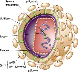
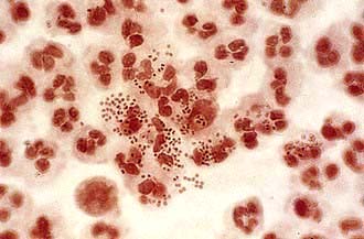
Neisseria gonorrhoeae (Gonococci, GC) is a Gram-negative intracellular diplococcus (Fig. 4.37), which infects epithelium particularly of the urogenital tract, rectum, pharynx and conjunctivae. Humans are the only host and the organism is spread by intimate physical contact. It is very intolerant to drying and although occasional reports of spread by fomites exist, this route of infection is extremely rare.
Up to 50% of women and 10% of men are asymptomatic. The incubation period is 2–14 days with most symptoms occurring between days 2 and 5.
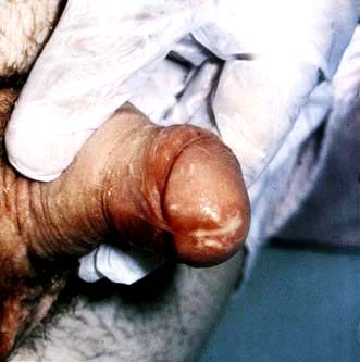
In men, the most common syndrome is one of anterior urethritis causing dysuria and/or urethral discharge (Fig. 4.38). Complications include ascending infection involving the epididymis or prostate leading to acute or chronic infection. In MSM rectal infection may produce proctitis with pain, discharge and itch.
In women, the primary site of infection is usually the endocervical canal. Symptoms include an increased or altered vaginal discharge, pelvic pain due to ascending infection, dysuria and intermenstrual bleeding. Complications include Bartholin’s abscesses and in rare cases a perihepatitis (Fitzhugh–Curtis syndrome) can develop. On a global basis GC is one of the most common causes of female infertility. Rectal infection, due to local spread, occurs in women and is usually asymptomatic, as is pharyngeal infection. Conjunctival infection is seen in neonates born to infected mothers and is one cause of ophthalmia neonatorum.
Disseminated GC leads to arthritis (usually monoarticular or pauciarticular) (see p. 533) and characteristic papular or pustular rash with an erythematous base in association with fever and malaise. It is more common in women.
N. gonorrhoeae can be identified from infected areas by culture on selective media with a sensitivity of at least 95%. Nucleic acid amplification tests (NAATs) using urine specimens are non-invasive and highly sensitive, although they may give false-positive results. Microscopy of Gram-stained secretions may demonstrate intracellular, Gram-negative diplococci, allowing rapid diagnosis. The sensitivity ranges from 90% in urethral specimens from symptomatic men to 50% in endocervical specimens. Microscopy should not be used for pharyngeal specimens. Blood culture and synovial fluid investigations should be performed in cases of disseminated GC. Co-existing pathogens such as Chlamydia, Trichomonas and syphilis must be sought.
Treatment is indicated in those patients who have a positive culture for GC, positive microscopy or a positive NAAT (this should be repeated because of false positives and all positives should be confirmed by cultures). Treatment is given to patients who have had recent sexual intercourse with someone with confirmed GC infection. Although N. gonorrhoeae is sensitive to a wide range of antimicrobial agents, antibiotic-resistant strains have shown a recent significant increase. Immediate therapy based on Gram-stained slides is usually initiated in the clinic, prior to culture and sensitivity results. Antibiotic choice is influenced by travel history or details known from contacts.
Single-dose ceftriaxone i.m. (500 mg) is recommended in the UK. Single-dose oral amoxicillin 3 g with probenecid 1 g, ciprofloxacin (500 mg) or ofloxacin (400 mg) may be used in areas with low prevalence of antibiotic resistance. (The CDC has reported high resistance, however, and recommends the addition of azithromycin 1 g orally.) Resistance to all antibiotics has recently been reported.
Longer courses of antibiotics are required for complicated infections. There should be at least one follow-up assessment and culture tests should be repeated at least 72 h after treatment is complete. All sexual contacts should be notified and then examined and treated as necessary.
This organism has a worldwide distribution. Genital infection with CT is common, with up to 14% of sexually active people below the age of 25 years in the UK infected. It is regularly found in association with other pathogens: 20% of men and 40% of women with gonorrhoea have been found to have co-existing Chlamydia infections. In men 40% of non-gonococcal and post-gonococcal urethritis is due to Chlamydia. As CT is often asymptomatic much infection goes unrecognized and untreated, which sustains the infectious pool in the population. The long-term complications associated with Chlamydia infection, especially infertility, impose significant morbidity in the UK. The UK government has introduced a national Chlamydia screening programme in an attempt to decrease the rates of asymptomatic infection.
In men, CT gives rise to an anterior urethritis with dysuria and discharge; infection is asymptomatic in up to 50% and detected by contact tracing. Ascending infection leads to epididymitis. Rectal infection leading to proctitis occurs in men practising anoreceptive intercourse. In women, the most common site of infection is the endocervix, where it may go unnoticed; up to 80% of infection in women is asymptomatic. Symptoms include vaginal discharge, post-coital or intermenstrual bleeding and lower abdominal pain. Ascending infection causes acute salpingitis. Reactive arthritis (see p. 529) has been related to infection with C. trachomatis. Neonatal infection, acquired from the birth canal, can result in mucopurulent conjunctivitis and pneumonia.
CT is an obligate intracellular bacterium, which complicates diagnosis. The nucleic acid amplification test (NAAT) is the diagnostic test of choice and is used as the ‘standard of care’ with a 90–95% sensitivity. Cell culture techniques provide the ‘gold standard’ and are 100% specific, but are expensive and require considerable expertise. Indirect diagnostic tests include direct fluorescent antibody (DIF) tests and enzyme immunoassays (EIA), but they are less reliable and none is diagnostic.
In men, first-voided urine samples are tested, or urethral swabs obtained. In women endocervical swabs are the best specimens and up to 20% additional positives will be detected if urethral swabs are also taken. Urine specimens are less reliable than endocervical swabs in women but are useful if a speculum examination is not possible.
Ligase chain reaction (LCR) can be used as an alternative to PCR.
Tetracyclines or macrolide antibiotics are most commonly used to treat Chlamydia. Azithromycin 1 g as a single dose or doxycycline 100 mg 12-hourly for 7 days or are both effective for uncomplicated infection. Tetracyclines are contraindicated in pregnancy. Other effective regimens include erythromycin 500 mg four times daily. Routine test of cure is not necessary after treatment with doxycycline or azithromycin, although if symptoms persist or reinfection is suspected then further tests should be taken. NAATs may remain positive for up to 5 weeks after treatment as they pick up material from non-viable organisms. Sexual contacts must be traced, notified and treated, particularly as so many infections are clinically silent.
Urethritis is usually characterized in men by a discharge from the urethra, dysuria and varying degrees of discomfort within the penis. In 10–15% of cases there are no symptoms. A wide array of aetiologies can give rise to the clinical picture and are divided into two broad bands: gonococcal or non-gonococcal urethritis (NGU). NGU occurring shortly after infection with gonorrhoea is known as postgonococcal urethritis (PGU). Gonococcal urethritis and Chlamydia urethritis (a major cause of NGU) are discussed above.
Trichomonas vaginalis, Mycoplasma genitalium, Ureaplasma urealyticum and Bacteroides spp. are responsible for a proportion of cases. HSV can cause urethritis in about 30% of cases of primary infection, considerably fewer in recurrent episodes. Other causes include syphilitic chancres and warts within the urethra. Non-sexually transmitted NGU may be due to urinary tract infections, prostatic infection, foreign bodies and strictures.
The urethral discharge is often mucoid and worse in the mornings. Crusting at the meatus or stains on underwear occur. Dysuria is common but not universal. Discomfort or itch within the penis may be present. The incubation period is 1–5 weeks with a mean of 2–3 weeks. Asymptomatic urethritis is a major reservoir of infection. Reactive arthritis (see p. 529) causing conjunctivitis with arthritis occurs, particularly in HLA B27-positive individuals.
NAATs for gonorrhoea and Chlamydia should be performed in all men with symptoms of urethritis. In those who are negative, smears should be taken from the urethra when the patient has not voided urine for at least 4 h and should be Gram-stained and examined under a high-power (×1000) oil-immersion lens. The presence of five or more polymorphonuclear leucocytes per high-power field or a Gram-stained preparation from a centrifuged sample of a first passed urine specimen, containing >10 PMNL per high-power is diagnostic. Men who are symptomatic but have no objective evidence of urethritis should be re-examined and tested after holding urine overnight. Cultures for gonorrhoea must be taken together with specimens for Chlamydia testing.
Therapy for NGU is with either azithromycin 1 g orally as a single dose or doxycycline 100 mg 12-hourly for 7 days. Sexual intercourse should be avoided. The vast majority of patients will show partial or total response. Sexual partners must be traced and treated; C. trachomatis can be isolated from the cervix in 50–60% of the female partners of men with PGU or NGU, many of whom are asymptomatic. This causes long-term morbidity in such women, acts as a reservoir of infection for the community and may lead to reinfection in the index case if not treated.
This is a common and difficult clinical problem which is empirically defined as persistent or recurrent symptomatic urethritis occurring 30–90 days following treatment of acute NGU. It can occur in 20–60% of men treated for acute NGU. The usual time for patients to re-present is 2–3 weeks following treatment. Tests for organisms, e.g. Mycoplasma, Chlamydia and Ureaplasma, are usually negative. It is necessary to document objective evidence of urethritis, check adherence to treatment and establish any possible contact with untreated sexual partners. A further 1 week’s treatment with erythromycin 500 mg four times a day for 2 weeks plus metronidazole 400 mg twice daily for 5 days may be given and any specific additional infection treated appropriately. If symptoms are mild and all partners have been treated, patients should be reassured and further antibiotic therapy avoided. In cases of frequent recurrence and/or florid unresponsive urethritis, the prostate should be investigated and urethroscopy or cystoscopy performed to investigate possible strictures, periurethral fistulae or foreign bodies.
Chlamydia trachomatis types LGV 1, 2 and 3 (different biovars or variant prokaryotic strain) are responsible for this sexually transmitted infection. It is endemic in the tropics, with the highest incidences in Africa, India and South-east Asia. There has been a recent upsurge in UK-acquired LGV, particularly among HIV-infected men who have sex with men (MSM), presenting with rectal symptoms and associated genital ulceration. Many have also been hepatitis C co-infected. The L2 serovar (different antigenic properties) has been the predominant strain. The Health Protection Agency has enhanced LGV surveillance to track the current UK outbreak.
There are three characteristic stages. The primary lesion is a painless ulcerating papule on the genitalia occurring 7–21 days following exposure. It is frequently unnoticed. A few days to weeks after this heals, regional lymphadenopathy develops. The lymph nodes are painful and fixed and the overlying skin develops a dusky erythematous appearance. Finally, nodes may become fluctuant (buboes) and can rupture. Acute LGV also presents as proctitis with perirectal abscesses, the appearances sometimes resembling anorectal Crohn’s disease. The destruction of local lymph nodes can lead to lymphoedema of the genitalia.
The diagnosis is often made on the basis of the characteristic clinical picture after other causes of genital ulceration or inguinal lymphadenopathy have been excluded. Syphilis and genital herpes must be excluded. The laboratory investigations have become more sensitive and specific:
Detection of nucleic acid (DNA). Nucleic acid amplification tests for LGV serovar are now available. Positive samples should be confirmed by real-time PCR for LGV-specific DNA.
Isolation of C. trachomatis from clinical lesions in tissue culture remains the most specific test; however, sensitivity is only 75–85%.
Chlamydia trachomatis serology. Complement fixation (CF) tests, single L-type immunofluorescence test and micro-immunofluorescence test (micro-IF) (the latter being the most accurate). A four-fold rise in antibody titre in the course of the illness or a single point titre of >1/64 is considered to be diagnostic.
Early treatment is critical to prevent the chronic phase. Doxycycline (100 mg twice daily for 21 days) or erythromycin (500 mg four times daily for 21 days) is efficacious. Follow-up should continue until signs and symptoms have resolved, usually 3–6 weeks. Chronic infection may result in extensive scarring and abscess and sinus formation. Surgical drainage or reconstructive surgery is sometimes required. HIV and hepatitis C screening should be recommended. Sexual partners in the 30 days prior to onset should be notified, examined and treated if necessary.
Syphilis is a chronic systemic disease, which is acquired or congenital. In its early stages diagnosis and treatment are straightforward but untreated it can cause complex sequelae in many organs and eventually lead to death. There has been a marked increase in the incidence of syphilis over the past decade. A majority of these infections occur in MSM, many of whom are co-infected with HIV.
The causative organism, Treponema pallidum (TP), is a motile spirochaete that is acquired either by close sexual contact or can be transmitted transplacentally. The organism enters the new host through breaches in squamous or columnar epithelium. Primary infection of non-genital sites may occasionally occur but is rare.
Both acquired and congenital syphilis have early and late stages, each of which has classic clinical features (Table 4.49).
Primary. Between 10 and 90 days (mean 21 days) after exposure to the pathogen a papule develops at the site of inoculation. This ulcerates to become a painless, firm chancre. There is usually painless regional lymphadenopathy in association. The primary lesion may go unnoticed, especially if it is on the cervix or within the rectum. Healing occurs spontaneously within 2–3 weeks.
Secondary. Between 4 and 10 weeks after the appearance of the primary lesion constitutional symptoms with fever, sore throat, malaise and arthralgia appear. Any organ may be affected – leading, for example, to hepatitis, nephritis, arthritis and meningitis. In a minority of cases the primary chancre may still be present and should be sought.
Table 4.49 Classification and clinical features of syphilis
Clinical features | |
---|---|
Acquired |
|
Early stages |
|
Primary |
Hard chancre |
Painless, regional lymphadenopathy |
|
Secondary |
General: Fever, malaise, arthralgia, sore throat and generalized lymphadenopathy |
Skin: Red/brown maculopapular non-itchy, sometimes scaly rash; condylomata lata |
|
Mucous membranes: Mucous patches, ‘snail-track’ ulcers in oropharynx and on genitalia |
|
Late stages |
|
Tertiary |
Late benign: Gummas (bone and viscera) |
Cardiovascular: Aortitis and aortic regurgitation |
|
Neurosyphilis: Meningovascular involvement, general paralysis of the insane (GPI) and tabes dorsalis |
|
Congenital |
Stillbirth or failure to thrive |
Early stages |
‘Snuffles’ (nasal infection with discharge) |
Skin and mucous membrane lesions as in secondary syphilis |
|
Late stages |
‘Stigmata’: Hutchinson’s teeth, ‘sabre’ tibia and abnormalities of long bones, keratitis, uveitis, facial gummas and CNS disease |
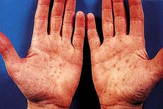
Generalized lymphadenopathy (50%)
Generalized skin rashes involving the whole body including the palms and soles but excluding the face (75%) – the rash, which rarely itches, may take many different forms, ranging from pink macules, through coppery papules, to frank pustules (Fig. 4.39)
Condylomata lata: warty, plaque-like lesions found in the perianal area and other moist body sites
Superficial confluent ulceration of mucosal surfaces – found in the mouth and on the genitalia, described as ‘snail track ulcers’
Acute neurological signs in less than 10% of cases (e.g. aseptic meningitis).
Untreated early syphilis in pregnant women leads to fetal infection in at least 70% of cases and may result in stillbirth in up to 30%.
Latent. Without treatment, symptoms and signs abate over 3–12 weeks, but in up to 20% of individuals, may recur during a period known as early latency, a 2-year period in the UK (1 year in USA). Late latency is based on reactive syphilis serology with no clinical manifestations for at least 2 years. This can continue for many years before the late stages of syphilis become apparent.
Tertiary. Late benign syphilis, so called because of its response to therapy rather than its clinical manifestations, generally involves the skin and the bones. The characteristic lesion, the gumma (granulomatous, sometimes ulcerating, lesions), can occur anywhere in the skin, frequently at sites of trauma. Gummas are commonly found in the skull, tibia, fibula and clavicle, although any bone may be involved. Visceral gummas occur mainly in the liver (hepar lobatum) and the testes.
Cardiovascular and neurosyphilis are discussed on pages 669, 1129.
Congenital syphilis usually becomes apparent between the 2nd and 6th week after birth, early signs being nasal discharge, skin and mucous membrane lesions and failure to thrive. Signs of late syphilis generally do not appear until after 2 years of age and take the form of ‘stigmata’ relating to early damage to developing structures, particularly teeth and long bones. Other late manifestations parallel those of adult tertiary syphilis.
Treponema pallidum is not amenable to in vitro culture – the most sensitive and specific method is identification by dark-ground microscopy. Organisms may be found in variable numbers, from primary chancres and the mucous patches of secondary lesions. Individuals with either primary or secondary disease are highly infectious.
Serological tests used in diagnosis are either treponemal-specific or nonspecific (cardiolipin test) (Table 4.50):
Treponemal specific. The T. pallidum enzyme immunoassay (EIA). T. pallidum haemagglutination or particle agglutination assay (TPHA/TPPA) and fluorescent treponemal antibodies absorbed (FTA-abs) test are both highly specific for treponemal disease but will not differentiate between syphilis and other treponemal infection such as yaws. These tests usually remain positive for life, even after treatment.
Treponemal nonspecific. The Venereal Disease Research Laboratory (VDRL) and rapid plasma reagin (RPR) tests are nonspecific, becoming positive within 3–4 weeks of the primary infection. They are quantifiable tests which can be used to monitor treatment efficacy and are helpful in assessing disease activity. They generally become negative by 6 months after treatment in early syphilis. The VDRL may also become negative in untreated patients (50% of patients with late-stage syphilis) or remain positive after treatment in late stage. False-positive results may occur in other conditions – particularly infectious mononucleosis, hepatitis, Mycoplasma infections, some protozoal infections, cirrhosis, malignancy, autoimmune disease and chronic infections.
The EIA is the screening test of choice and can detect both IgM and IgG antibodies. A positive test is then confirmed with the TPHA/TPPA and VDRL/RPR tests. All serological investigations may be negative in early primary syphilis; the EIA IgM and the FTA-abs being the earliest tests to be positive. The diagnosis will then hinge on positive dark-ground microscopy. Treatment should not be delayed if serological tests are negative in such situations.
Examination of the CSF for evidence of neurosyphilis is indicated in those patients with syphilis who demonstrate neurological signs and symptoms, have a high titre of non-treponemal tests (usually taken to be >1:32), those who have any evidence of treatment failure and HIV co-infected patients with late latent syphilis or syphilis of unknown duration. A chest X-ray should also be performed.
Treponemocidal levels of antibiotic must be maintained in serum for at least 7 days in early syphilis to cover the slow division time of the organism (30 h). In late syphilis treponemes may divide even more slowly requiring longer therapy.
Early syphilis (primary or secondary) should be treated with long-acting penicillin such as procaine benzylpenicillin (procaine penicillin) (e.g. Jenacillin A, which also contains benzylpenicillin) 600 mg intramuscularly daily for 10 days.
For late-stage syphilis, particularly when there is cardiovascular or neurological involvement, the treatment course should be extended for a further week.
For patients sensitive to penicillin, either doxycycline 200 mg daily or erythromycin 500 mg four times daily is given orally for 2–4 weeks depending on the stage of the infection. Non-compliant patients can be treated with a single dose of benzathine penicillin G 2.4 g intramuscularly. Azithromycin is not recommended following evidence of resistance. The diagnosis and management of syphilis in HIV co-infected patients remains unaltered; however, if untreated it may advance more rapidly than in HIV-negative patients and have a higher incidence of neurosyphilis. The Jarisch–Herxheimer reaction, which is due to release of TNF-α, IL-6 and IL-8, is seen in 50% of patients with primary syphilis and up to 90% of patients with secondary syphilis. It occurs about 8 h after the first injection and usually consists of mild fever, malaise and headache lasting several hours. In cardiovascular or neurosyphilis the reaction, although rare, may be severe and exacerbate the clinical manifestations. Prednisolone given for 24 h prior to therapy may ameliorate the reaction but there is little evidence to support its use. Penicillin should not be withheld because of the Jarisch–Herxheimer reaction; since it is not a dose-related phenomenon, there is no value in giving a smaller dose.
The prognosis depends on the stage at which the infection is treated. Early and early latent syphilis have an excellent outlook but once extensive tissue damage has occurred in the later stages the damage will not be reversed although the process may be halted. Symptoms in cardiovascular disease and neurosyphilis may therefore persist.
All patients treated for early syphilis must be followed up at regular intervals for the first year following treatment. Serological markers with a fall in titre of the VDRL/RPR of at least four-fold is consistent with adequate treatment for early syphilis. The sexual partners of all patients with syphilis and the parents and siblings of patients with congenital syphilis must be contacted and screened. Babies born to mothers who have been treated for syphilis in pregnancy are retreated at birth.
Chancroid or soft chancre is an acute STI caused by Haemophilus ducreyi. Although a less common cause of genital ulceration than HSV-2, it is prevalent in parts of Africa and Asia. It is rare in the UK with cases usually associated with travel to or partners from endemic areas. Epidemiological studies in Africa have shown an association between genital ulcer disease, frequently chancroid and the acquisition of HIV infection. A new urgency to control chancroid has resulted from these observations.
The incubation period is 3–10 days. At the site of inoculation an erythematous papular lesion forms which then breaks down into an ulcer. The ulcer frequently has a necrotic base, a ragged edge, bleeds easily and is painful. Several ulcers may merge to form giant serpiginous lesions. Ulcers appear most commonly on the prepuce and frenulum in men and can erode through tissues. In women the most commonly affected site is the vaginal entrance and the perineum and lesions sometimes go unnoticed.
At the same time, inguinal lymphadenopathy develops (usually unilateral) and can progress to form large buboes which suppurate.
Chancroid must be differentiated from other genital ulcer diseases (see Table 4.47). Co-infection with syphilis and herpes simplex is common. Isolation of H. ducreyi, a fastidious organism, in specialized culture media is definitive but difficult. Swabs should be taken from the ulcer and material aspirated from the local lymph nodes for culture. Polymerase chain reaction (PCR) techniques are available. Gram stains of clinical material may show characteristic coccobacilli, but this is an insensitive test. Detection of antibody to H ducreyi using EIA may be useful for population surveillance but, at an individual level, lacks sensitivity and specificity. A ‘probable diagnosis’ may be made if the patient has the appropriate clinical picture, without evidence of T. pallidum or herpes simplex infection.
Single-dose regimens include azithromycin 1 g orally or ceftriaxone 250 mg i.m. Other regimens include ciprofloxacin 500 mg twice daily for 3 days, erythromycin 500 mg four times daily for 7 days. Clinically significant plasmid-mediated antibiotic resistance in H. ducreyi is developing.
Patients should be followed up at 3–7 days, when the ulcers should be responding if the treatment is successful.
Sexual partners should be notified, examined and treated epidemiologically, as asymptomatic carriage has been reported.
HIV-infected patients should be closely monitored, as healing may be slower. Multiple-dose regimens are needed in HIV patients since treatment failures have been reported with single-dose therapy.
Donovanosis is the least common of all STIs in North America and Europe, but is endemic in the tropics and subtropics, particularly the Caribbean, South-east Asia and South India. Infection is caused by Klebsiella granulomatis, a short, encapsulated Gram-negative bacillus. The infection was also known as granuloma inguinale. Although sexual contact appears to be the most usual mode of transmission, the infection rates are low, even between sexual partners of many years’ standing.
In the vast majority of patients, the characteristic, heaped-up ulcerating lesion with prolific red granulation tissue appears on the external genitalia, perianal skin or the inguinal region within 1–4 weeks of exposure. It is rarely painful. Almost any cutaneous or mucous membrane site can be involved, including the mouth and anorectal regions. Extension of the primary infection from the external genitalia to the inguinal regions produces the characteristic lesion, the ‘pseudo-bubo’.
The clinical appearance usually strongly suggests the diagnosis but K. granulomatis (Donovan bodies) can be identified intracellularly in scrapings or biopsies of an ulcer. Successful culture has only recently been reported and PCR techniques and serological methods of diagnosis are being developed, but none is routinely available.
Antibiotic treatment should be given until the lesions have healed. A minimum of 3 weeks’ treatment is recommended. Regimens include doxycycline 100 mg twice daily, co-trimoxazole 960 mg twice daily, azithromycin 500 mg daily or 1 g weekly, or ceftriaxone 1 g daily.
Sexual partners should be notified, examined and treated if necessary.
Genital herpes is one of the most common STIs worldwide and the most common ulcerative STI in the UK. The peak incidence is in 16- to 24-year-olds of both sexes, with women having higher rates of diagnosis than men. Infection, which is lifelong, may be either primary or recurrent. Transmission occurs during close contact with a person who is shedding virus (who may well be asymptomatic). Historically, most genital herpes is due to HSV type 2. However, genital contact with oral lesions caused by HSV-1 can also produce genital infection and rates of HSV-1-associated genital herpes are approaching 50% in some parts of the world.
Susceptible mucous membranes include the genital tract, rectum, mouth and oropharynx. The virus has the ability to establish latency in the dorsal root ganglia by ascending peripheral sensory nerves from the area of inoculation. It is this ability which allows for recurrent attacks.
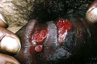
Primary genital herpes is usually accompanied by systemic symptoms of varying severity including fever, myalgia and headache. Multiple painful shallow ulcers develop, which may coalesce (Fig. 4.40). Atypical lesions are common. Tender inguinal lymphadenopathy is usual. Over a period of 10–14 days the lesions develop crusts and dry. In women with vulval lesions the cervix is almost always involved. Rectal infection may lead to a florid proctitis. Neurological complications can include aseptic meningitis and/or involvement of the sacral autonomic plexus leading to retention of urine. Asymptomatic primary infection has been reported but is rare. Serological surveys indicate that only about 10% of individuals with antibodies to HSV-2 give a clinical history of genital lesions, indicating that asymptomatic primary infection is the norm.
Recurrent attacks occur in a significant proportion of people following the initial episode and are more likely with HSV type 2 infections. Precipitating factors vary, as does the frequency of recurrence. A symptom prodrome is present in some people prior to the appearance of lesions. Systemic symptoms are rare in recurrent attacks.
The clinical manifestations in immunosuppressed patients (including those with HIV) may be more severe, asymptomatic shedding increased and recurrences occurring with greater frequency. Systemic spread has been documented (see p. 191).
The history and examination can be highly suggestive of HSV infection.
A firm diagnosis can routinely be made only on the basis of detection of virus from within lesions.
Swabs should be taken from the base of lesions and placed in viral transport medium. Virus is most easily isolated from new lesions.
HSV DNA detection by polymerase chain reaction (PCR) has increased the rates of detection compared with virus culture.
Real-time PCR assays are highly specific although not available in all laboratories.
Type-specific immune responses can be found 8–12 weeks following primary infection and indicate HSV infection at some time. False negatives may be found early after infection. IgM assays are unreliable.
Primary. Saltwater bathing or sitting in a warm bath is soothing and may allow the patient to pass urine with some degree of comfort. Aciclovir 200 mg five times daily, famciclovir 250 mg three times daily or valaciclovir 500 mg twice daily, all for 5 days, are useful if patients are seen while new lesions are still forming. If lesions are already crusting, antiviral therapy will do little to change the clinical course. Secondary bacterial infection occasionally occurs and should be treated. Rest, analgesia and antipyretics should be advised.
Recurrence. Recurrent attacks tend to be less severe and can be managed with simple measures such as saltwater bathing. Psychological morbidity is associated with recurrent genital herpes and frequent recurrences impose strains on relationships; patients need considerable support. Long-term suppressive therapy is given in patients with frequent recurrences. An initial course of aciclovir 400 mg twice daily or valaciclovir 250 mg twice daily for 6–12 months significantly reduces the frequency of attacks, although there may still be some breakthrough. Therapy should be discontinued after 12 months and the frequency of recurrent attacks reassessed.
The potential risk of infection to the neonate needs to be considered in addition to the health of the mother. Infection occurs either transplacentally (very rare) or via the birth canal. If HSV is acquired for the first time during pregnancy, transplacental infection of the fetus may, rarely, occur. Management of primary HSV in the 1st or 2nd trimester will depend on the woman’s clinical condition and aciclovir can be prescribed in standard doses. Aciclovir, given at a dose of 400 mg three times a day to accommodate altered pharmacokinetics of the drug in late pregnancy, during the last 4 weeks of pregnancy, may prevent recurrence at term.
Symptomatic primary acquisition in the 3rd trimester or at term with high levels of viral shedding usually leads to delivery by caesarean section.
For women with previous infection, the risk for the baby acquiring HSV from the birth canal is very low in recurrent attacks. Only those with genital lesions at the onset of labour are delivered by caesarean section. Sequential cultures during the last weeks of pregnancy to predict viral shedding at term are no longer indicated.
Patients must be advised that they are infectious when lesions are present; sexual intercourse should be avoided during this time or during prodromal stages. Condoms may not be effective as lesions may occur outside the areas covered. Sexual partners should be notified, examined and given information on avoiding infection. Asymptomatic viral shedding is a cause of onward transmission. It is most common in HSV-2, during the first 12 months following infection and in those with frequent symptomatic HSV. Antiviral drugs reduce shedding and onward transmission.
Anogenital warts are among the most common sexually acquired infections. The causative agent is human papillomavirus (HPV), especially types 6 and 11 with types 16 and 18 causing a majority of cases of cervical carcinoma (see p. 100). HPV is acquired by direct sexual contact with a person with either clinical or subclinical infection. Genital HPV infection is common, with only a small proportion of those infected being symptomatic. Neonates may acquire HPV from an infected birth canal, which may result either in anogenital warts or in laryngeal papillomas. The incubation period ranges from 2 weeks to 8 months or even longer.
Warts develop around the external genitalia in women, usually starting at the fourchette, and involve the perianal region. The vagina may be infected. Flat warts may develop on the cervix and are not easily visible on routine examination. Such lesions are associated with cervical intraepithelial neoplasia. In men, the penile shaft and subpreputial space are the most common sites, although warts involve the urethra and meatus. Perianal lesions are more common in men who practise anoreceptive intercourse but they can be found in any patient. The rectum may become involved. Warts become more florid during pregnancy or in immunosuppressed patients.
The diagnosis is essentially clinical. It is critical to differentiate condylomata lata of secondary syphilis. Unusual lesions should be biopsied if the diagnosis is in doubt. Up to 30% of patients have co-existing infections with other STIs and a full screen must be performed.
Significant failure and relapse rates are seen with current treatment modalities. Choice of treatment will depend on the number and distribution of lesions. Local agents, including podophyllin extract (15–25% solution, once or twice weekly), podophyllotoxin (0.5% solution or 1.5% cream in cycles) and trichloroacetic acid, are useful for non-keratinized lesions. Those that are keratinized respond better to physical therapy, e.g. cryotherapy, electrocautery or laser ablation. Imiquimod, an immune response modifier, induces a cytokine response when applied to skin infected with HPV (5% cream used three times a week) and is indicated in both types of lesion. Podophyllin, podophyllotoxin and imiquimod are not advised in pregnancy. Patients co-infected with HIV may have a poorer response to treatment and higher rates of intraepithelial neoplasia. Sexual contacts should be examined and treated if necessary. In view of the difficulties of diagnosing subclinical HPV, condoms should be used for up to 8 months after treatment. Because of the association of HPV with cervical intraepithelial neoplasia, women with warts and female partners of men with warts are advised to have regular cervical screening, i.e. every 3 years. Colposcopy may be useful in women with vaginal and cervical warts.
Two vaccines exist against HPV. One is effective against HPV types 16 and 18 and the second against types 6, 11, 16 and 18. Both vaccines, based on a DNA-free HPV capsid, are strongly immunogenic and act by inducing a neutralizing antibody response and enhancing CD4 memory cell function. They are given over 6 months in three divided doses, with an excellent safety profile. Up to 98% serological responses, maintained for over 4 years, have been reported from early trials. In placebo-controlled clinical trials of the quadrivalent vaccine a highly significant reduction in low- and high-grade cervical dysplasia, vulval pre-cancers and external genital warts has been demonstrated. Vaccination is most beneficial in those who have not yet been exposed to HPV infection, with a recommendation that it be given before people become sexually active. Although initially considered to be most useful for women and girls, vaccination of boys to prevent both genital warts and the transmission of oncogenic HPV strains is under consideration. Routine HPV vaccination commenced in the UK in late 2008, initially for girls of about 12 years of age, with a second catch-up opportunity at about 18 years of age.
This is discussed in Chapter 7. Sexual contacts should be screened and given vaccine if they are not immune (see p. 318).
Trichomonas vaginalis (TV) is a flagellated protozoon which is predominantly sexually transmitted. It is able to attach to squamous epithelium and can infect the vagina and urethra. Trichomonas may be acquired perinatally in babies born to infected mothers.
Infected women may, unusually, be asymptomatic. Commonly the major complaints are of vaginal discharge, which is offensive and of local irritation. Men usually present as the asymptomatic sexual partners of infected women, although they may complain of urethral discharge, irritation or urinary frequency.
Examination often reveals a frothy yellowish vaginal discharge and erythematous vaginal walls. The cervix may have multiple small haemorrhagic areas which lead to the description ‘strawberry cervix’. Trichomonas infection in pregnancy has been associated with pre-term delivery and low birth weight.
Phase-contrast, dark-ground microscopy of a drop of vaginal discharge shows TV swimming with a characteristic motion in 40–80% of female patients. Similar preparations from the male urethra will only be positive in about 30% of cases. Many polymorphonuclear leucocytes are also seen. Culture techniques are good and confirm the diagnosis. Trichomonas is sometimes observed on cervical cytology with 60–80% accuracy in diagnosis. New, highly sensitive and specific tests based on polymerase chain reactions are being developed.
Metronidazole is the treatment of choice, either 2 g orally as a single dose or 400 mg twice daily for 7 days. There is some evidence of metronidazole resistance and nimorazole may be effective in these cases. Topical therapy with intravaginal tinidazole can be effective, but if extravaginal infection exists this may not be eradicated and vaginal infection reoccurs. Male partners should be treated, especially as they are likely to be asymptomatic and more difficult to detect.
Vulvovaginal infection with Candida albicans is extremely common. The organism is also responsible for balanitis in men. Candida can be isolated from the vagina in a high proportion of women of childbearing age, many of whom will have no symptoms.
The role of Candida as pathogen or commensal is difficult to disentangle and it may be changes in host environment which allow the organism to produce pathological effects. Predisposing factors include pregnancy, diabetes and the use of broad-spectrum antibiotics and corticosteroids. Immunosuppression can result in more florid infection.
In women, pruritus vulvae is the dominant symptom. Vaginal discharge is present in varying degrees. Many women have only one or occasional isolated episodes. Recurrent candidiasis (four or more symptomatic episodes annually) occurs in up to 5% of healthy women of reproductive age. Examination reveals erythema and swelling of the vulva with broken skin in severe cases. The vagina may contain adherent curdy discharge. Men may have a florid balanoposthitis. More commonly, self-limiting burning penile irritation immediately after sexual intercourse with an infected partner is described. Diabetes must be excluded in men with balanoposthitis.
Microscopic examination of a smear from the vaginal wall reveals the presence of spores and mycelia. Culture of swabs should be undertaken but may be positive in women with no symptoms. Trichomonas and bacterial vaginosis must be considered in women with itch and discharge.
Topical. Pessaries or creams containing one of the imidazole antifungals such as clotrimazole 500 mg single dose used intravaginally are usually effective. Nystatin is also useful.
Oral. The triazole drugs such as fluconazole 150 mg as a single dose or itraconazole 200 mg twice in 1 day are used systemically where topical therapy has failed or is inappropriate. Recurrent candidiasis may be treated with fluconazole 100 mg weekly for 6 months or clotrimazole pessary, 500 mg weekly for 6 months.
The evidence for sexual transmission of Candida is slight and there is no evidence that treatment of male partners reduces recurrences in women.
Bacterial vaginosis (BV) is a disorder characterized by an offensive vaginal discharge. The aetiology and pathogenesis are unclear but a mixed flora of Gardnerella vaginalis, anaerobes including Bacteroides, Mobiluncus spp. and Mycoplasma hominis replaces the normal lactobacilli of the vagina. Amines and their breakdown products from the abnormal vaginal flora are thought to be responsible for the characteristic odour associated with the condition. As vaginal inflammation is not part of the syndrome, the term vaginosis is used rather than vaginitis. The condition has been shown to be more common in black women than in white. It is not regarded as a sexually transmitted disease.
Vaginal discharge and odour are the commonest complaints, although a proportion of women are asymptomatic. A homogeneous, greyish white, adherent discharge is present in the vagina, the pH of which is raised (>5). Associated complications are ill-defined but may include chorioamnionitis and an increased incidence of premature labour in pregnant women. Whether BV predisposes non-pregnant women to upper genital tract infection is unclear.
Different authors have differing criteria for making the diagnosis of BV. In general, it is accepted that three of the following should be present for the diagnosis to be made:
Characteristic vaginal discharge
The amine test: raised vaginal pH using narrow-range indicator paper (>4.7)
A fishy odour on mixing a drop of discharge with 10% potassium hydroxide
The presence of ‘clue’ cells on microscopic examination of the vaginal fluid.
Clue cells are squamous epithelial cells from the vagina, which have bacteria adherent to their surface, giving a granular appearance to the cell. A Gram stain gives a typical reaction of partial stain uptake.
Metronidazole given orally in doses of 400 mg twice daily for 5–7 days is usually recommended. A single dose of 2 g metronidazole is less effective. Topical 2% clindamycin cream 5 g intravaginally once daily for 7 days is effective.
Recurrence is high, with some studies giving a rate of 80% within 9 months of completing metronidazole therapy. There is debate over the treatment of asymptomatic women who fulfil the diagnostic criteria for BV. Until the relevance of BV to other pelvic infections is elucidated, the treatment of asymptomatic women with BV is not to be recommended. Simultaneous treatment of the male partner does not influence the rate of recurrence of BV and routine treatment of male partners is not indicated.
The pubic louse (Phthirus pubis) is a blood-sucking insect which attaches tightly to the pubic hair. They may also attach to eyelashes and eyebrows. It is relatively host-specific and is transferred only by close bodily contact. Eggs (nits) are laid at hair bases and usually hatch within a week. Although infestation may be asymptomatic, the most common complaint is of itch.
Lice may be seen on the skin at the base of pubic and other body hairs. They resemble small scabs or freckles but if they are picked up with forceps and placed on a microscope slide, will move and walk away. Blue macules may be seen at the feeding sites. Nits are usually closely adherent to hairs. Both are highly characteristic under the low-power microscope.
As with all sexually transmitted infections, the patient must be screened for co-existing pathogens.
Both lice and eggs must be killed with 0.5% malathion, 1% permethrin or 0.5% carbaryl. The preparation should be applied to all areas of the body from the neck down and washed off after 12 h. In a few cases, a further application after 1 week may be necessary. For severe infestations, antipruritics may be indicated for the first 48 h. All sexual partners should be seen and screened.
This is discussed in Chapter 24.
FURTHER READING
British Association for Sexual Health and HIV (BASHH). BASHH Clinical Effectiveness Guidelines. London: BASHH; 2008. Also online. Available at: http://www.bashh.org/guidelines.asp
Gupta R, Warren T, Wald A. Genital herpes. Lancet 2007; 370:2127–2137.
Low N, Broutet N, Adu-Sarkodie Y et al. Global control of sexually transmitted infections. Lancet 2006; 368:2001–2016.
Robertson C, Jayasuriya A, Allan PS. Sexually transmitted infections: where are we now? J R Soc Med 2007; 100:414–417.
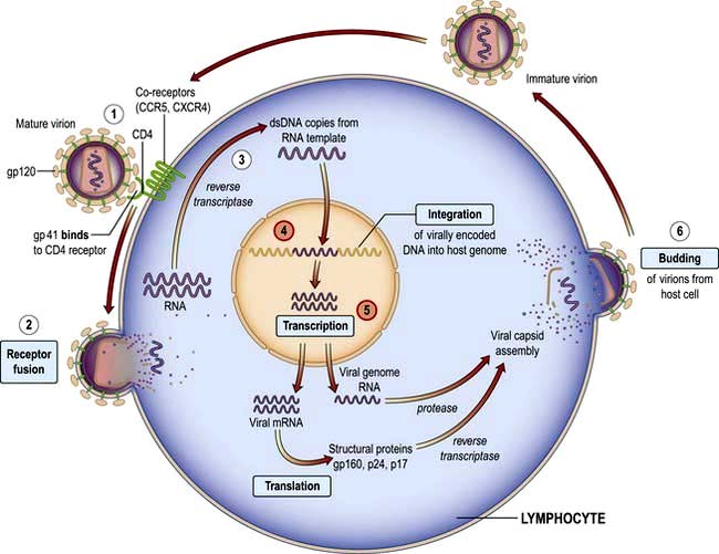
Since the first description of AIDS in 1981 and the identification of the causative organism HIV in 1984, more than 20 million people have died. At least 33 million people worldwide are living with HIV infection. Sub-Saharan Africa remains the most seriously affected, but in some areas, the number of new diagnoses has stabilized. However, in Eastern Europe and parts of central Asia, infection rates are rising exponentially. The human, societal and economic costs are huge: 33% of 15-year-olds in high-prevalence countries in Africa will die of HIV. Demographics of the epidemic have varied greatly, influenced by social, behavioural, cultural and political factors. Highly active antiretroviral therapy (HAART) has dramatically reduced mortality for those who are able to access care. Current global estimates suggest that about a quarter of those who need HAART are on treatment but that for each individual starting therapy there are two new infections, highlighting the size of the problem and the global inequalities that exist in healthcare.
In the UK falling death rates and continued new infections mean that the total number of people living with HIV continues to rise. Men who have sex with men (MSM) and culturally diverse heterosexual populations from sub-Saharan Africa, are the two largest groups of people living with HIV in the UK, and accessing treatment and care. Of those diagnosed with HIV in the UK, 30% are women. As mortality rates fall so the population of people with HIV is becoming older, further changing the clinical picture.
Approximately one-quarter of those with HIV infection in the UK are undiagnosed and unaware of their infection, which contributes to late diagnosis, poorer clinical outcomes and onward transmission. Late diagnosis is now the most common cause of HIV-related morbidity and mortality in the UK. Reducing undiagnosed HIV through wider testing, particularly in patients presenting with clinical conditions that are associated with HIV and in areas with high seroprevalence, is critical to both the individual and public health (Box 4.21).
Box 4.21 HIV testing
UK guidelines on where and who to test
All patients diagnosed with a sexually transmitted infection
Sexual partners of men and women known to be HIV positive
Men who have disclosed sexual contact with other men
Female sexual contacts of men who have sex with men
People reporting a history of injecting drug use
Men and women known to be from a country of high HIV prevalence (>1%)
Men and women who report sexual contact abroad or in the UK with individuals from countries of high HIV prevalence
Patients presenting for healthcare where HIV enters the differential diagnosis (see below for table of indicator conditions)
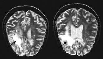
Cerebral toxoplasmosisa; primary cerebral lymphomaa; cryptococcal meningitisa; progressive multifocal leucoencephalopathya; aseptic meningitis/encephalitis; cerebral abscess; space-occupying lesion of unknown cause; Guillain–Barré syndrome; transverse myelitis; peripheral neuropathy; dementia.
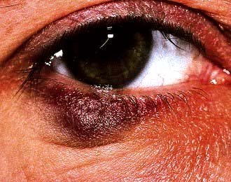
Kaposi’s sarcomaa; severe/recalcitrant seborrhoeic dermatitis; severe/recalcitrant psoriasis; multidermatomal/recurrent herpes zoster.
Persistent cryptosporidiosisa; oral candidiasis; oral hairy leukoplakia; chronic diarrhoea of unknown cause; weight loss of unknown cause; Salmonella; Shigella; Campylobacter; hepatitis B infection; hepatitis C infection.
Non-Hodgkin’s lymphomaa; anal cancer; anal intraepithelial dysplasia; lung cancer; seminoma; head and neck cancer; Hodgkin’s lymphoma; Castleman’s disease.
Cervical cancera; vaginal intraepithelial neoplasia; cervical intraepithelial neoplasia, grade 2 or above.
Any unexplained blood dyscrasia including thrombocytopenia, neutropenia and lymphopenia.
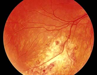
Cytomegalovirus retinitisa; infective retinal diseases including herpesviruses and toxoplasma; any unexplained retinopathy.
http://guidance.nice.org.uk/PH33; http://www.nice.org.uk/guidance/PH34; http://www.bhiva.org/HIVTesting2008.aspx
a AIDS-defining condition.
Despite the fact that HIV can be isolated from a wide range of body fluids and tissues, the majority of infections are transmitted via semen, cervical secretions and blood.
Sexual intercourse (vaginal and anal). Globally, heterosexual intercourse accounts for the vast majority of infections and co-existent STIs, especially those causing genital ulceration, enhance transmission. Passage of HIV appears to be more efficient from men to women and to the receptive partner in anal intercourse, than vice versa. In the UK, sex between men accounts for over half the infections reported, but there is an increasing rate of heterosexual transmission. In Central and sub-Saharan Africa, the epidemic has always been heterosexual and more than half the infected adults in these regions are women. South-east Asia and the Indian subcontinent are experiencing an explosive epidemic, driven by heterosexual intercourse and a high incidence of other sexually transmitted diseases.
Mother-to-child (transplacental, perinatal, breast-feeding). Vertical transmission is the most common route of HIV infection in children. European studies suggest that, without intervention, 15% of babies born to HIV-infected mothers are likely to be infected, although rates of up to 40% have been reported from Africa and the USA. Increased vertical transmission is associated with advanced disease in the mother, maternal viral load, prolonged and premature rupture of membranes and chorioamnionitis. Transmission can occur in utero, although the majority of infections take place perinatally. Breast-feeding has been shown to double the risk of vertical transmission. In the developed world, interventions to reduce vertical transmission, including the use of antiretroviral agents and the avoidance of breast-feeding, have led to a dramatic fall in the numbers of infected children. The lack of access to these interventions in resource-poor countries in which 90% of infections occur is a major issue.
Contaminated blood, blood products and organ donations. Screening of blood and blood products was introduced in 1985 in Europe and North America. Prior to this, HIV infection was associated with the use of clotting factors (for haemophilia) and with blood transfusions. In some parts of the world where blood products may not be screened and in areas where the rate of new HIV infections is very high, transfusion-associated transmission continues to occur.
Contaminated needles (intravenous drug misuse, injections, needle-stick injuries). The practice of sharing needles and syringes for intravenous drug use continues to be a major route of transmission of HIV in both developed countries and parts of South-east Asia, Latin America and the states of the former Soviet Union. In some areas, including the UK, successful education and needle exchange schemes have reduced the rate of transmission by this route. Iatrogenic transmission from needles and syringes used in developing countries is reported. Healthcare workers have a risk of approximately 0.3% following a single needle-stick injury with known HIV-infected blood.
There is no evidence that HIV is spread by social or household contact or by blood-sucking insects such as mosquitoes and bed bugs.
HIV belongs to the lentivirus group of the retrovirus family. There are two types, HIV-1 and HIV-2. HIV-1 is the most frequently occurring strain globally. HIV-2 is almost entirely confined to West Africa, although there is some spread to Europe, particularly France and Portugal. HIV-2 has only 40% structural homology with HIV-1 and although it is associated with immunosuppression and AIDS, appears to take a more indolent course than HIV-1. Many of the drugs that are used in HIV-1 are ineffective in HIV-2. The structure of HIV is shown in Figure 4.41.
Figure 4.41 Structure of HIV. Two molecules of single-stranded RNA are shown within the nucleus. The reverse transcriptase polymerase converts viral RNA into DNA (a characteristic of retroviruses). The protease includes integrase (p32 and p10). The p24 (core protein) levels can be used to monitor HIV disease. p17 is the matrix protein; gp120 is the outer envelope glycoprotein which binds to cell surface CD4 molecules; gp41, a transmembrane protein, influences infectivity and cell fusion capacity.
Retroviruses are characterized by the possession of the enzyme reverse transcriptase, which allows viral RNA to be transcribed into DNA and thence incorporated into the host cell genome. Reverse transcription is an error-prone process with a significant rate of mis-incorporation of bases. This, combined with a high rate of viral turnover, leads to considerable genetic variation and a diversity of viral subtypes or clades. On the basis of DNA sequencing, HIV-1 is divided into four distinct strains, which represent four independent cross species transfers, three (M, N and O) based on the chimpanzee related strains of SIV and 1 (P) that may represent chimpanzee to gorilla to human transmission.
Group M (major) subtypes (98% of infections worldwide) exhibit a high degree of diversity, with subtypes (or clades), denoted A–K. There is a predominance of subtype B in Europe, North America and Australia, but areas of Central and sub-Saharan Africa have multiple M subtypes, with clade C being the commonest subtype. Recombination of viral material generates an array of circulating recombinant forms (CRFs), which increases the genetic diversity that may be encountered.
Group N (new) is mostly confined to parts of West Central Africa (e.g. Gabon).
Group O (outlier) subtypes are highly divergent from group M and are largely confined to small numbers centred on Cameroon.
Group P related to gorilla strains of SIV has been identified from a patient from Cameroon.
Connections between genetic diversity and biological effects, in particular pathogenicity, rates of transmission and response to therapy are being sought. Diversity in the viral subtypes and recombinant forms encountered in the UK is increasing.
The interrelationship between HIV and the host immune system is the basis of the pathogenesis of HIV disease. At the time of initial exposure virus is transported by dendritic cells from mucosal surfaces to regional lymph nodes where permanent infection is established. The host cellular receptor that is recognized by HIV surface glycoprotein gp120 is the CD4 molecule, which defines the cell populations that are susceptible to infection (Fig. 4.42). The interaction between CD4 and HIV gp120 surface glycoprotein, together with host chemokine CCR5 co-receptors, is responsible for HIV entry into cells. Although CCR5 CD4 memory T lymphocytes within all body systems are susceptible to infection and depletion those found in the gastrointestinal tract are heavily infected early in the process and become rapidly depleted leading to compromised mucosal immune function.
Figure 4.42 HIV entry and replication in CD4 T lymphocytes. (1) Binding: the virus binds to host CD4 receptor molecules via the envelope glycoprotein gp 120 and co-receptors CCR5 and CXCR4. (2) Fusion: a subsequent conformational change results in the fusion between gp41 and the cell membrane. (3) Reverse transcription: entry of the viral capsid is followed by the uncoating of the RNA. DNA copies are made from both RNA templates. DNA polymerase from the host cell leads to formation of dsDNA. (4) Integration: in the nucleus, virally encoded DNA is inserted into the host genome. (5) Transcription: regulatory proteins control transcription (an RNA molecule is now synthesized from the DNA template). (6) Budding: the virus is reassembled in the cytoplasm and budded out from the host cell.
Studies of viral turnover have demonstrated a virus half-life in the circulation of about 6 hours. To maintain observed levels of plasma viraemia, 108–109 virus particles need to be released and cleared daily. Virus production by infected cells lasts for about 2 days and is probably limited by the death of the cell, owing to direct HIV effects, linking HIV replication to the process of CD4 destruction and depletion. Loss of activated CD4 T lymphocytes is a key factor in the immunopathogenesis of HIV, but debate continues about the exact mechanisms of cell depletion.
Resulting cell-mediated immunodeficiency leaves the host open to infections with intracellular pathogens, while co-existing antibody abnormalities predispose to infections with capsulated bacteria. HIV is associated with a long-term inflammatory state, which is a key driver of disease progression. T-cell activation is observed from the earliest stages of infection which in turn leads to an increase in the numbers of susceptible CD4 bearing target cells that can become infected and destroyed. This inflammatory state is associated with HIV itself, with co-pathogens such as cytomegalovirus and with the translocation of microbial products, in particular lipopolysaccharides, from the gut into the systemic circulation following HIV destruction of normal mucosal immunity. Raised levels of inflammatory cytokines and coagulation system activation occur. These inflammatory responses play a role in HIV-associated end organ damage.
HIV is now a manageable chronic condition, but only in those who are aware of their diagnosis and who start effective antiretroviral therapy early enough. Starting treatment with more advanced disease compromises clinical outcomes. In 2009, 26% of those living with HIV in the UK were unaware of their infection. More than half of those newly diagnosed in the UK in 2009 were ‘late presenters’, i.e. had a CD4 count below the threshold to start therapy (<350 cells/mm3), and in one-third, the CD4 count was below 200 cells/mm3 putting them at high risk of HIV-associated pathology. Increasing the uptake of HIV testing is a major public health objective. Guidelines on HIV testing from The British HIV Association and the National Institute for Health and Clinical Excellence (NICE) include clinical settings in which HIV testing should be universally offered, together with a list of clinical situations and diagnoses (indicator conditions) that are highly predictive of HIV infection and where HIV testing should be recommended (Box 4.21). All new registrants in primary care and patients admitted to acute medical care should be recommended to test in areas of the UK where HIV seroprevalence is >2/1000 population.
Discussion about HIV testing and the consent required is straightforward and should be within the competencies of a wide range of healthcare professionals. Sensitive and specific point-of-care HIV antibody tests using either blood or oral fluids can give results within minutes and have extended the possibilities for diagnosis. All reactive point-of-care tests should be followed up with confirmatory serological assays, appropriate arrangements made to ensure patients receive test results and those who are found to be HIV positive have rapid routes into specialist care.
HIV infection is diagnosed either by the detection of virus-specific antibodies (anti-HIV) or by direct identification of viral material. The recommended UK first-line assay is one which tests for HIV antibody AND p24 antigen simultaneously. These fourth generation assays have the advantage of reducing the time between infection and an HIV-positive test result to one month which is several weeks earlier than with sensitive third generation (antibody only detection) assays.
Detection of IgG antibody to envelope components. This is the most commonly used marker of infection. The routine tests used for screening are based on ELISA techniques, which may be confirmed with Western blot assays. Up to 3 months (mean 6 weeks) may elapse from initial infection to antibody detection (serological latency, or window period). These antibodies to HIV have no protective function and persist for life. As with all IgG antibodies, anti-HIV will cross the placenta. All babies born to HIV-infected women will thus have the antibody at birth. In this situation, anti-HIV antibody is not a reliable marker of active infection and in uninfected babies will be gradually lost over the first 18 months of life.
Simple and rapid HIV antibody assays are increasingly available, giving results within minutes. Assays that can utilize alternative body fluids to serum/plasma such as oral fluid, whole blood and urine are now available and home testing kits are being developed. These tests are extremely sensitive and may give false-positive results, making it necessary to perform a confirmatory test.
A Serologic Testing Algorithm for Recent HIV Serocon-versions (STARHS) can be used to identify recently acquired infection. A highly sensitive ELISA that is able to detect HIV antibodies 6–8 weeks after infection is used on blood in patients with positive oral fluid test, in parallel with a less sensitive (detuned) test that identifies later HIV antibodies within 130 days. A positive result on the sensitive test and a negative ‘detuned’ test are indicative of recent infection, while positive results on both tests point to an infection that is more than 130 days old. The major application of this is in epidemiological surveillance and monitoring.
IgG antibody to p24 (anti-p24). This can be detected from the earliest weeks of infection and through the asymptomatic phase. It is frequently lost as the disease progresses.
Genome detection assays. Nucleic acid-based assays that amplify and test for components of the HIV genome are available. These assays are used to aid diagnosis of HIV in the babies of HIV-infected mothers or in situations where serological tests may be inadequate, such as in early infection when antibody may not be present, or in subtyping HIV variants for medicolegal reasons. (See the discussion of viral load monitoring, on p. 179.)
Viral p24 antigen (p24ag). This is detectable shortly after infection but has usually disappeared by 8–10 weeks after exposure. It can be a useful marker in individuals who have been infected recently but have not had time to mount an antibody response.
Isolation of virus in culture. This is a specialized technique available in some laboratories to aid diagnosis and as a research tool.
The spectrum of illnesses associated with HIV infection is broad and is the result of direct HIV effects, HIV-associated immune dysfunction, the drugs used to treat the condition, as well as co-existing morbidity and co-infections.
Several classification systems exist; the most widely used being the 1993 Centers for Disease Control (CDC) classification (Box 4.22). This classification depends to a large extent on definitive diagnoses of infection, which makes it more difficult to use in those areas of the world without sophisticated laboratory support.
As immunosuppression progresses, the patient is susceptible to an increasing range of opportunistic infections and tumours, certain of which meet the criteria for the diagnosis of AIDS (Box 4.23).
Box 4.23
AIDS-defining conditions
Candidiasis of bronchi, trachea or lungs
Coccidioidomycosis, disseminated or extrapulmonary
Cryptococcosis, extrapulmonary
Cryptosporidiosis, chronic intestinal (1-month duration)
Cytomegalovirus (CMV) disease (other than liver, spleen or nodes)
CMV retinitis (with loss of vision)
Herpes simplex, chronic ulcers (1-month duration); or bronchitis, pneumonitis or oesophagitis
Histoplasmosis, disseminated or extrapulmonary
Isosporiasis; chronic intestinal (1-month duration)
Lymphoma, immunoblastic (or equivalent term)
Mycobacterium avium-intracellulare complex or M. kansasii, disseminated or extrapulmonary
Mycobacterium tuberculosis, any site
Mycobacterium, other species or unidentified species, disseminated or extrapulmonary
Pneumocystis jiroveci pneumonia
Progressive multifocal leucoencephalopathy
The definition of AIDS differs between the USA and Europe. The USA definition includes individuals with CD4 counts below 200 in addition to the clinical classification based on the presence of specific indicator diagnoses shown in Box 4.22. In Europe, the definition remains based on the diagnosis of specific clinical conditions with no inclusion of CD4 lymphocyte counts. Where highly active antiretroviral therapy (HAART) is available and started before the development of severe immunosuppression, progression to AIDS is now uncommon.
Primary HIV infection (PHI) refers to the first 6-month period following HIV acquisition. This is a period of uncontrolled viral replication resulting in high levels of HIV circulating in the plasma and genital tract and consequently of high infectiousness. At a population level PHI is increasingly recognized as a contributor to onward transmission. In the UK up to 20% of all newly diagnosed individuals are recently infected. The 2–4 weeks immediately following infection may be silent both clinically and serologically. In a number of people, a self-limiting nonspecific illness occurs 3–6 weeks after exposure. Symptoms include fever, arthralgia, myalgia, lethargy, lymphadenopathy, sore throat, mucosal ulcers and occasionally a transient faint pink maculopapular rash. Neurological symptoms are common, including headache, photophobia, myelopathy and neuropathy and in rare cases encephalopathy. The illness lasts up to 3 weeks and recovery is usually complete.
Laboratory abnormalities include lymphopenia with atypical reactive lymphocytes noted on blood film, thrombocytopenia and raised liver transferases. CD4 lymphocytes may be markedly depleted and the CD4:CD8 ratio reversed. Antibodies to HIV may be absent during this early stage of infection, although the level of circulating viral RNA is high and p24 core protein may be detectable. HIV RNA NAAT assays may be diagnostic 7 days before a p24 antigen test and 12 days before a sensitive HIV antibody test. If PHI is suspected but standard diagnostic tests are negative then repeat testing in 7 days and referral for expert advice is recommended.
The rate of clinical progression of untreated HIV is variable. The majority of people with HIV infection are asymptomatic for a substantial but variable length of time. However, the virus continues to replicate and the person is infectious. Most people with HIV have a gradual decline in CD4 count over a period of approximately 10 years before progression to AIDS. Others progress much more rapidly, with continued high levels of viral RNA and a rapid decline in CD4 count over 2–5 years. Other, long-term non-progressors, may continue with a normal CD4 count over many years. Within this group, a small subpopulation of elite controllers maintain a viral load below 2000 copies/mL or even to undetectable levels without therapy.
Older age is associated with more rapid progression. Gender and pregnancy per se do not appear to influence the rate of progression, although women may fare less well for a variety of reasons. A subgroup of patients with asymptomatic infection have persistent generalized lymphadenopathy (PGL), defined as lymphadenopathy (>1 cm) at two or more extra-inguinal sites for more than 3 months in the absence of causes other than HIV infection. The nodes are usually symmetrical, firm, mobile and non-tender. There may be associated splenomegaly. The architecture of the nodes shows hyperplasia of the follicles and proliferation of the capillary endothelium. Biopsy is rarely indicated. Similar disease progression has been noted in asymptomatic patients with or without PGL. Nodes may disappear with disease progression.
As HIV infection progresses, the viral load rises, the CD4 count falls and the patient develops an array of symptoms and signs. The clinical picture is the result of direct HIV effects and of the associated immunosuppression.
In an individual patient, the clinical consequences of HIV-related immune dysfunction will depend on at least three factors:
The microbial exposure of the patient throughout life. Many clinical episodes represent reactivation of previously acquired infection, which has been latent. Geographical factors determine the microbial repertoire of an individual patient. Those organisms requiring intact cell-mediated immunity for their control are most likely to cause clinical problems.
The pathogenicity of organisms encountered. High-grade pathogens such as Mycobacterium tuberculosis, Candida and the herpesviruses are clinically relevant even when immunosuppression is mild and will thus occur earlier in the course of the disease. Less virulent organisms occur at later stages of immunodeficiency.
The degree of immunosuppression of the host. When patients are severely immunocompromised (CD4 count <100/mm3) disseminated infections with organisms of very low virulence such as M. avium-intracellulare (MAI) and Cryptosporidium are able to establish themselves. These infections are very resistant to treatment, mainly because there is no functioning immune response to clear organisms. This hierarchy of infection allows for appropriate intervention with prophylactic drugs.
Infection of the nervous tissue occurs at an early stage but clinical neurological involvement increases as HIV advances. This includes AIDS dementia complex (ADC), sensory polyneuropathy and aseptic meningitis (see p. 1130). These conditions are much less common since the introduction of HAART (highly active antiretroviral therapy). The pathogenesis is thought to be due both to the release of neurotoxic products by HIV itself and to cytokine abnormalities secondary to immune dysregulation.
ADC has varying degrees of severity, ranging from mild memory impairment and poor concentration through to severe cognitive deficit, personality change and psychomotor slowing. Changes in affect are common and depressive or psychotic features may be present. The spinal cord may show vacuolar myelopathy histologically. In severe cases brain CT scan shows atrophic change of varying degrees. MRI changes consist of white matter lesions of increased density on T2-weighted sections. EEG shows non-specific changes consistent with encephalopathy. The CSF is usually normal, although the protein concentration may be raised. Patients with mild neurological dysfunction may be unduly sensitive to the effects of other insults such as fever, metabolic disturbance or psychotropic medication, any of which may lead to a marked deterioration in cognitive functioning.
Sensory polyneuropathy is seen in advanced HIV infection, mainly in the legs and feet, although hands may be affected. Severe forms cause intense pain, usually in the feet, which disrupts sleep, impairs mobility and generally reduces the quality of life.
Autonomic neuropathy may also occur with postural hypotension and diarrhoea. Autonomic nerve damage is found in the small bowel. Didanosine and stavudine produce a similar neuropathy as a major toxic side-effect and are best avoided in patients with HIV neuropathy.
HAART, using agents that penetrate the CNS can lead to significant improvements in cognitive function in many patients with ADC. It may also have a neuroprotective role.
Eye pathology is usually seen in the later stages. The most serious is cytomegalovirus retinitis (see p. 81), which is sight-threatening. Retinal cotton wool spots due to HIV per se are rarely troublesome but they can be confused with CMV retinitis. Anterior uveitis can present as acute red eye associated with rifabutin therapy for mycobacterial infections in HIV. Steroids used topically are usually effective but modification of the dose of rifabutin is required to prevent relapse. Pneumocystis, toxoplasmosis, syphilis and lymphoma can all affect the retina and the eye may be the site of first presentation.
The skin is a common site for HIV-related pathology as the function of dendritic and Langerhans’ cells, both target cells for HIV, is disrupted. Delayed-type hypersensitivity, a good indicator of cell-mediated immunity, is frequently reduced or absent even before clinical signs of immunosuppression appear. Pruritus is a common complaint at all stages of HIV. Generalized dry, itchy, flaky skin is typical and the hair may become thin and dry. An intensely pruritic papular eruption favouring the extremities may be found, particularly in patients from African backgrounds. Eosinophilic folliculitis presents with urticarial lesions particularly on the face, arms and legs.
Table 4.51 Some mucocutaneous manifestations of HIV infection (see also Ch. 24)
Skin | Mucous membranes |
---|---|
Dry skin and scalp Onychomycosis Seborrhoeic dermatitis Tinea: cruris pedis Pityriasis: versicolor rosea Folliculitis Acne Molluscum contagiosum Warts Herpes zoster: multidermatomal disseminated Papular pruritic eruption Scabies Ichthyosis Kaposi’s sarcoma |
Candidiasis: oral vulvovaginal Oral hairy leucoplakia Aphthous ulcers Herpes simplex: genital oral labial Periodontal disease Warts: oral genital |
Drug reactions with cutaneous manifestations are frequent; with rashes developing notably to sulphur-containing drugs amongst others (see Fig. 24.39). Recurrent aphthous ulceration, which is severe and slow to heal, is common and can impair the patient’s ability to eat. Biopsy may be indicated to exclude other causes of ulceration. Topical steroids are useful and resistant cases may respond to thalidomide. In addition to the above the skin is a common site of opportunistic infections (see p. 127).
These are common in advanced HIV infection.
Lymphopenia progresses as the CD4 count falls.
Anaemia of chronic HIV infection is usually mild, normochromic and normocytic.
Neutropenia is common and usually mild.
Isolated thrombocytopenia may occur early in infection and be the only manifestation of HIV for some time. Platelet counts are often moderately reduced but can fall dramatically to 10–20 × 109/L producing easy bleeding and bruising. Circulating antiplatelet antibodies lead to peripheral destruction. Megakaryocytes are increased in the bone marrow but their function is impaired. Effective antiretroviral therapy usually produces a rise in platelet count. Thrombocytopenic patients undergoing dental, medical or surgical procedures may need therapy with human immunoglobulin, which gives a transient rise in platelet count, or be given platelet transfusion. Steroids are best avoided.
Pancytopenia occurs because of underlying opportunistic infection or malignancies, in particular Mycobacterium avium-intracellulare, disseminated cytomegalovirus and lymphoma.
Other complications. Myelotoxic drugs include zidovudine (megaloblastic anaemia, red cell aplasia, neutropenia); lamivudine (anaemia, neutropenia); ganciclovir (neutropenia); systemic chemotherapy (pancytopenia) and co-trimoxazole (agranulocytosis).
Weight loss and diarrhoea are common in untreated HIV-infected patients. Wasting is a common feature of advanced HIV infection, which although originally attributed to direct HIV effects on metabolism, is usually a consequence of anorexia. There is a small increase in resting energy expenditure in all stages of HIV, but weight and lean body mass usually remain normal during periods of clinical latency when the patient is eating normally.
Gastrointestinal infections are common. An HIV enteropathy with varying degrees of villous atrophy has been described with chronic diarrhoea when no other pathogen has been found.
Hypochlorhydria is reported in patients with advanced HIV disease and may have consequences for drug absorption and bacterial overgrowth in the gut.
Rectal lymphoid tissue cells are the targets for HIV infection during penetrative anal sex and may be a reservoir for infection to spread through the body.
HIV-associated nephropathy (HIVAN) (see p. 588), although rare, can cause significant renal impairment, particularly in more advanced disease. It is most frequently seen in black male patients and can be exacerbated by heroin use.
Nephrotic syndrome subsequent to focal glomerulosclerosis is the usual pathology, which may be a consequence of HIV cytopathic effects on renal tubular epithelium. The course is usually relentlessly progressive and dialysis may be required.
Many nephrotoxic drugs are used in the management of HIV-associated pathology, particularly foscarnet, amphotericin B, pentamidine and sulfadiazine. Tenofovir is associated with Fanconi’s syndrome (p. 1040).
The upper airway and lungs serve as a physical barrier to air-borne pathogens and any damage will decrease the efficiency of protection, leading to an increase in upper and lower respiratory tract infections. The sinus mucosa may also function abnormally in HIV infection and is frequently the site of chronic inflammation. Response to antibacterial therapy and topical steroids is usual but some patients require surgical intervention. A similar process is seen in the middle ear, which can lead to chronic otitis media.
Lymphoid interstitial pneumonitis (LIP) is well described in paediatric HIV infection but is uncommon in adults. There is an infiltration of lymphocytes, plasma cells and lymphoblasts in alveolar tissue. Epstein–Barr virus may be present. The patient presents with dyspnoea and a dry cough, which may be confused with pneumocystis infection (see p. 1040). Reticular nodular shadowing is seen on chest X-ray. Therapy with steroids may produce clinical and histological benefit in some patients.
Various endocrine abnormalities have been reported, including reduced levels of testosterone and abnormal adrenal function. The latter assumes clinical significance in advanced disease when intercurrent infection superimposed upon borderline adrenal function precipitates clear adrenal insufficiency requiring replacement doses of gluco- and mineralocorticoid. CMV is also implicated in adrenal-deficient states.
Cardiovascular pathology is increasingly recognized as a cause of morbidity in people with HIV. Although lipid dysregulation has been associated with antiretroviral medication (ARV), the observation has been made that HDL levels are lower in those with untreated HIV infection than in HIV-negative controls. In a large international study (SMART), ischaemic heart disease was more common in those who took intermittent ARV therapy than in those who maintained viral suppression. Cardiomyopathy, although rare, is associated with HIV and may lead to congestive cardiac failure. Lymphocytic and necrotic myocarditis have been described. Ventricular biopsy should be performed to ensure other treatable causes of myocarditis are excluded.
Immunodeficiency (see p. 81) allows the development of opportunistic infections (OI) (Table 4.52 and see also Table 4.56). These are diseases caused by organisms that are not usually considered pathogenic, unusual presentations of known pathogens and the occurrence of tumours that may have an oncogenic viral aetiology. Susceptibility increases as the patient becomes more immunosuppressed. CD4 T lymphocyte numbers are used as markers to predict the risk of infection. Patients with CD4 counts above 200 are at low risk for the majority of AIDS-defining OIs. A hierarchy of thresholds for specific infectious risks can be constructed. Mechanisms include defective T cell function against protozoa, fungi and viruses, impaired macrophage function against intracellular bacteria such as Mycobacteria and Salmonella and defective B-cell immunity against capsulated bacteria such as Strep. pneumoniae and Haemophilus. Many of the organisms causing clinical disease are ubiquitous in the environment or are already carried by the patient.
Table 4.52 Major HIV-associated pathogens
Protozoa
|
Diagnosis in an immunosuppressed patient may be complicated by a lack of typical signs, as the inflammatory response is impaired. Examples are lack of neck stiffness in cryptococcal meningitis or minimal clinical findings in early Pneumocystis jiroveci pneumonia. Multiple pathogens may co-exist. Indirect serological tests are frequently unreliable. Specimens should be obtained from the appropriate site for examination and culture in order to make a diagnosis.
Newly diagnosed patients should be reviewed by an HIV clinician within 2 weeks of diagnosis, or earlier if the patient is symptomatic or has other acute needs A full medical history, physical examination and laboratory evaluation should be undertaken in all newly diagnosed patients to determine the stage of infection, the presence of co-morbidities and co-infections and to assess overall physical, mental and sexual health. The initial assessment should also include details of socioeconomic situation, relationships, family and social support networks and substance misuse. Baseline investigations will depend on the clinical setting, but those appropriate for an asymptomatic person in the UK are shown in Box 4.24.
Patients are regularly monitored (approximately 3-monthly) to assess the progression of the infection and the need for treatment. Decisions about appropriate intervention can be made.
CD4 lymphocytes. The absolute CD4 count and its percentage of total lymphocytes falls as HIV progresses. These figures bear a relationship to the risk of the occurrence of HIV-related pathology, with patients with counts below 200 cells at greatest risk. Rapidly falling CD4 counts and those below 350 are an indication for HAART. Factors other than HIV (e.g. smoking, exercise, intercurrent infections and diurnal variation) also affect CD4 numbers. CD4 counts are performed at approximately 3-monthly intervals unless values are approaching critical levels for intervention, in which case they are performed more frequently.
HIV replicates at a high rate throughout the course of infection, with many billion new virus particles being produced daily. The rate of viral clearance is relatively constant in any individual and thus the level of viraemia is a reflection of the rate of virus replication. This has both prognostic and therapeutic value.
The commonly used term ‘viral load’ encompasses viraemia and HIV RNA levels. Three HIV RNA assays for viral load are in current use:
Results are given in copies of viral RNA/mL of plasma, or converted to a logarithmic scale and there is good correlation between tests. The most sensitive test is able to detect as few as 20 copies of viral RNA/mL. Transient increases in viral load are seen following immunizations (e.g. for influenza and Pneumococcus) or during episodes of acute intercurrent infection (e.g. tuberculosis); and viral load measurements should not be carried out within a month of these events.
By about 6 months after seroconversion to HIV, the viral set-point for an individual is established and there is a correlation between HIV RNA levels and long-term prognosis, independent of the CD4 count. Those patients with a viral load consistently >100 000 copies/mL have a 10 times higher risk of progression to AIDS over the ensuing 5 years than those consistently below 10 000 copies/mL.
HIV RNA is the standard marker of treatment efficacy (see below). Both duration and magnitude of virus suppression are pointers to clinical outcome. None of the available therapies appears to be able to suppress viral replication indefinitely and a rising viral load, in a patient where compliance is assured, indicates drug failure.
Various guidelines exist for viral load monitoring in clinical practice. Baseline measurements are followed by repeat estimations at intervals of 3–4 months, ideally in conjunction with CD4 counts to allow both pieces of evidence to be used together in decision-making. Following initiation of antiretroviral therapy or changes in therapy, effects on viral load should be seen by 4 weeks, reaching a maximum at 10–12 weeks, when repeat viral load testing should be carried out (Fig. 4.43).
Clear genotype variations exist within HIV, not only between viral subtypes but also with well-identified point mutations associated with resistance to antiretroviral drugs. New infections with drug-resistant variants of HIV may be seen. Viral genotype analysis is recommended for all newly diagnosed patients with HIV. The most appropriate sample is the one closest to the time of diagnosis and the results are used to guide the selection of HAART agents.
Effective ART has transformed the management of HIV infection with a movement away from treating opportunistic conditions in immunosuppressed patients towards delivering long-term, effective suppressive therapy. With access and adherence to potent, tolerable antiretroviral drugs within a managed clinical setting, a 35-year-old initiating therapy in the UK in 2011 has a life expectancy of about 40 years. Nevertheless, there is still no cure for HIV and patients live with a chronic, infectious and unpredictable condition. Limitations to efficacy include the inability of current drugs to clear HIV from certain intracellular pools, the occurrence of serious drug side-effects, adherence requirements, complex drug–drug interactions and the emergence of resistant viral strains.
Box 4.25
An approach to sick HIV-positive patients
Full blood count and differential count
Liver and renal function tests
Blood gases including acid–base balance
Blood cultures, including specimens for mycobacterial culture
Microscopy and culture of available/appropriate specimens: stool, sputum, urine, CSF
Malaria screen in recent travellers from malaria areas
Serological tests for cryptococcal antigen, toxoplasmosis: save serum for viral studies
CT/MR scan of brain if focal neurological signs and ALWAYS before lumbar puncture
Note: Lymphocyte subsets and HIV viral load assays may yield misleading results during intercurrent illness.
The aims of management in HIV infection are to maintain physical and mental health, improve the quality of life, increase survival rates, restore and improve immune function, avoid onward transmission of the virus and to provide appropriate palliative support as needed. This requires long-term, maximal suppression of HIV activity using antiretroviral medication and management via a multidisciplinary team approach. Regular assessment is needed for details of intercurrent medical problems, medications, vaccinations, any recreational drug use, sexual history, reproductive decision-making, cervical cytology, social situation to include support networks, employment, benefits and accommodation. Depression and anxiety are common among people living with HIV and mood and cognitive function should be routinely assessed. Psychological support may be needed not only for the patient but also for family, friends and carers. Regular reviews of sexual and reproductive health together with advice on reducing the risk of HIV transmission must be provided and future sexual practices discussed. Information to allow people to make informed choices about childbearing is required. The implications for existing family members should be considered and diagnostic testing offered as necessary. Regular monitoring of weight, body mass index, blood pressure and cardiovascular risk is required. Dietary assessment and advice should be freely accessible. General health promotion advice on smoking, alcohol, diet, drug misuse and exercise should be given, particularly in the light of the cardiovascular, metabolic and hepatotoxicity risks associated with HIV and its treatment.
The treatment of HIV using antiretroviral therapy (HAART) continues to evolve and improve. Increased potency, reduced toxicity, greater convenience of formulation, compounds with different mechanisms of action coupled with improved understanding of drug resistance have combined to consistently improve HIV clinical outcome over time. An increase in the numbers of compounds, the array of drug-drug interactions, for example, combine to make HIV treatment complex and better clinical outcomes have been linked closely to physician expertise and the numbers of patients under direct care. Regularly updated treatment guidelines are produced in the UK by the British HIV Association (www.BHIVA.org.uk) and in the USA by the Department of Health and Human Services (http://www.aidsinfo.nih.gov/ContentFiles/AdultandAdolescentGL.pdf). The most up-to-date versions can be found on these websites and the current version must be used.
Table 4.53 Antiretroviral drugs generally used in clinical practice
Drugs | Comments | |
---|---|---|
Nucleoside and nucleotide reverse transcriptase inhibitors (NRTIs) |
Tenofovir, abacavir, zidovudine,a stavudine,a lamivudine, emtricitabine |
Tenofovir is associated with renal and perhaps bone dysfunction. Abacavir is associated with hypersensitivity reactions in at-risk individuals (HLA B5701) and is associated in some studies with an increased risk of cardiovascular disease. Abacavir might be less potent than tenofovir in patients with high viral loads. Zidovudine and stavudine are associated with profound fat redistribution (lipoatrophy). All NRTIs are associated with potential to cause risk of severe lactic acidosis. The combination of tenofovir and emtricitabine is the preferred first-line regimen in most regions |
Non-nucleoside reverse transcriptase inhibitors (NNRTIs) |
Efavirenz, nevirapine,a etravirine |
Efavirenz can cause CNS toxicity (which is usually time limited). Efavirenz has teratogenic potential and should be used with caution in woman who might become pregnant. Nevirapine can cause severe hepatoxicity when used in patients with higher CD4 cell counts (more than 250 cells per µL for women and more than 400 cells per µL for men). Etravine is given twice daily and has generally been used as second-line regimen |
Integrase inhibitors |
Raltegravir |
Raltegravir has no short-term and no known long-term toxic effects, although data are scarce |
Protease inhibitors |
Fosamprenavir, atazanavir, darunavir, lopinavir, saquinavir (ritonavir) |
Most protease inhibitors are extensively metabolised by the P450 CYP3A system; ritonavir is generally given at low doses (100–200 mg per day) to inhibit P450 and boost the co-administered protease inhibitors. Most protease inhibitors are associated with hyperlipidaemia and other metabolic abnormalities such as insulin resistance. Long-term protease inhibitor exposure has been associated with increased risk of cardiovascular disease |
CCR5 inhibitors |
Maraviroc |
Maraviroc is only active in patients who do not have virions that use CXCR4 for cell entry. A specialised assay is therefore needed to screen for co-receptor tropism. By contrast with other antiretroviral drugs, maraviroc binds to a host rather than a viral target. Maraviroc has an immunomodulatory effect that is independent of its effect on HIV replication; the clinical significance of this activity is unknown |
Fusion inhibitors |
Enfuvirtide |
Enfuvirtide must be given subcutaneously twice daily and is very expensive. The drug is generally used only in patients with no other therapeutic options |
a Some drugs such as zidovudine, stavudine, and nevirapine are generally used in resource-limited regions because of cost considerations. These drugs are generally not recommended as preferred agents in resource-rich regions in view of their potential toxic effects.
(From: Volberding PA, Deeks SG. Antiretroviral therapy and management of HIV infection. Lancet 2010; 376:496–472.)
The key practical principles of prescribing ARVs are given in Box 4.26.
Box 4.26 Prescribing antiretroviral drugs
Practice points
Characteristics of antiretroviral agents | Practice points |
---|---|
Must be taken exactly as prescribed |
|
Should not be stopped suddenly |
Make sure that mechanisms are in place to ensure adequate drug supplies, e.g. regular clinic appointments, repeat prescriptions, home delivery of medications Beware unexpected time away from home, e.g. holidays, intercurrent hospital admissions, immigration detention, police detention If there is an urgent medical indication to stop, obtain advice from specialist physician or pharmacist |
Can be compromised by the introduction of other medications, including other ARVs and vice versa |
Be careful with enzyme inducers, e.g. rifampicin, rifabutin, warfarin, nevirapine, which will reduce the effective levels of some ARVs Methadone levels may be reduced by efavirenz Some ARVs block the metabolism of other agents which may reach toxic levels Check potential interactions before adding new agents, see: www.hiv-druginteractions.org |
Can adversely interact with some herbal, complementary and recreational agents |
Herbal remedies that induce cytochrome P450, e.g. St John’s wort; Chinese herbal remedies will reduce levels of some ARVs Check potential interactions before adding new agents, see: www.hiv-druginteractions.org |
May produce additive toxicities when given with other medications |
|
Are associated with a range of adverse drug reactions which may be confused with other pathology |
|
May exacerbate co-morbidities |
Reverse transcriptase inhibitors
Nucleoside/nucleotide analogues. Nucleoside reverse transcriptase inhibitors (NRTIs) inhibit the synthesis of DNA by reverse transcription and also act as DNA chain terminators. NRTIs need to be phosphorylated intracellularly for activity to occur. These were the first group of agents to be used against HIV, initially as monotherapy and later as dual drug combinations. Usually, two drugs of this class are combined to provide the ‘backbone’ of a HAART regimen. Several fixed-dose NRTI combinations are available, which helps reduce the pill burden. NRTIs have been associated with mitochondrial toxicity (see p. 186), a consequence of their effect on the human mitochondrial DNA polymerase. Lactic acidosis is a recognized complication of this group of drugs. Nucleotide analogues (nucleotide reverse transcriptase inhibitors (NtRTIs)) have a similar mechanism of action but only require two intracellular phosphorylation steps for activity (as opposed to the three steps for nucleoside analogues).
Non-nucleoside analogues. Non-nucleoside reverse transcriptase inhibitors (NNRTIs) interfere with reverse transcriptase by direct binding to the enzyme. They are generally small molecules that are widely disseminated throughout the body and have a long half-life. NNRTIs affect cytochrome P450. They are ineffective against HIV-2. The level of cross-resistance across the class is very high. All have been associated with rashes and elevation of liver enzymes. Second-generation NNRTIs, such as etravirine and rilpivirine, with fewer adverse effects have some activity against viruses resistant to other compounds of the NNRTI class.
These act competitively on the HIV aspartyl protease enzyme, which is involved in the production of functional viral proteins and enzymes. As a consequence, viral maturation is impaired and immature dysfunctional viral particles are produced. Most of the protease inhibitors are active at very low concentrations and in vitro are found to have synergy with reverse transcriptase inhibitors. However, there are differences in toxicity, pharmacokinetics, resistance patterns and also cost, which influence prescribing. Cross-resistance can occur across the PI group. There appears to be no activity against human aspartyl proteases (e.g. renin), although there are clinically significant interactions with the cytochrome P450 system. This is used to therapeutic advantage, ‘boosting’ blood levels of PI by blocking drug breakdown with small doses of ritonavir. PIs have been linked with abnormalities of fat metabolism and control of blood sugar and some have been associated with deterioration in clotting function in people with haemophilia. Second generation PIs (e.g. darunavir, tipranavir) with activity against viruses resistant to the first generation drugs are available.
These drugs act as a selective inhibitor of HIV integrase, which blocks viral replication by preventing insertion of HIV DNA into the human DNA genome. Three compounds have so far been developed, although only one, raltegravir, is used. Raltegravir is metabolized by glucuronidation and does not require retroviral drug boosting. It is effective in treatment of both experienced and naive patients.
Maraviroc is a chemokine receptor antagonist which blocks the cellular CCR5 receptor entry by CCR5 tropic strains of HIV. These strains are found in earlier HIV infection and, with time adaptations (against which maraviroc is ineffective) allow the CXCR4 receptor to become the more dominant form. The drug is metabolized by CYP p450 (3A), giving the potential for drug–drug interactions. Tropism assays to establish that the patient is carrying a CCR5 tropic virus are required before treatment is used.
Enfurvitide is the only licensed compound in this class of agents. It is an injectable peptide derived from HIV gp41 that inhibits gp41-mediated fusion of HIV with the target cell. It is synergistic with NRTIs and PIs. Although resistance to enfurvitide has been described, there is no evidence of cross-resistance with other drug classes. Because it has an extracellular mode of action there are few drug–drug interactions. Side-effects relate to the subcutaneous route of administration in the form of injection site reactions.
Although the benefits of HAART in HIV infection are now indisputable, treatment regimens require a long-term commitment to high levels of adherence. Risks of therapy include short- and longer-term side-effects, drug–drug interactions and the potential for development of resistant viral strains. The full involvement of patients in therapeutic decision-making is essential for success. Various national guidelines and treatment frameworks exist (e.g. BHIVA Guidelines, DHHS Guidelines and IAS Recommendations). Laboratory marker data, including viral load and CD4 counts, together with individual circumstances, should guide therapeutic decision-making. The current UK recommendations are shown in Table 4.54. In situations where therapy is recommended but the patient elects not to start, then more intensive clinical and laboratory monitoring is advisable.
Table 4.54 When to start antiretroviral therapy
Primary HIV infection |
Treatment in clinical trial |
or if there is neurological involvement |
|
or if the CD4 <200 for >3/12 |
|
or if an AIDS-defining illness is present |
|
Established HIV infection |
|
CD4 <250 |
Treat |
CD4 251–350 |
Treat as soon as patient ready |
CD4 >350 |
Consider enrolment into ‘when to start’ trial |
AIDS diagnosis/CDC stage C |
Treat (except for TB when CD4 >350) |
Modified from Williams I, et al. British HIV Association guidelines for the treatment of HIV-1-positive adults with antiretroviral therapy 2012.
Questions still remain about the best time to start therapy. Clear clinical benefit has been demonstrated with the use of antiretroviral drugs in advanced HIV disease. All patients with symptomatic HIV disease, AIDS or a CD4 count that is consistently below 200 cells/mm3 should initiate treatment as soon as possible. In such situations, there is a significant risk of serious HIV-associated morbidity and mortality and the longer-term prognosis for patients initiating therapy below 200 CD4 cells/mm3 is not as good as for those who start at higher counts.
In asymptomatic patients, the absolute CD4 count is the key investigation used to guide treatment decisions. The UK recommendation is that therapy should be started at or around a CD4 count of 350 and patients with CD4 counts between 200 and 350 cells/mm3 should start therapy as soon as they are ready.
Debate remains about starting therapy at higher CD4 counts. The risk of disease progression for individuals with a count >350 is low and has to be balanced against ARV therapy toxicity and development of resistance. Earlier intervention at higher CD4 counts may be considered in those with a higher risk of disease progression, e.g. with high viral loads (>60 000 copies/mL) or rapidly falling CD4 count (losing more than 80 cells/year). Co-infection with hepatitis C virus is also a factor for earlier intervention (see p. 324).
Treatment for primary HIV infection is only recommended either within a clinical trial or to alleviate symptoms. Special situations (seroconversion, pregnancy, post-exposure prophylaxis) in which antiretroviral agents may be used are described on page 186.
The drug regimen used for starting therapy must be individualized to suit each patient’s needs. As differences in drug efficacy become less marked, fitting the drugs to the patient’s needs and lifestyle are key to success. Treatment is initiated with three drugs, two NRTIs in combination, with either an NNRTI or a boosted protease inhibitor (Table 4.53 and Table 4.55). The development of fixed-dose coformulations reduces pill burden, increases convenience and facilitates adherence. Newer drug classes such as integrase inhibitors and regimens without an NRTI backbone are still in the trial process and are likely to be the next development in first-line therapy.
Table 4.55 Initial HAART regimens – choice of initial therapy. Preferred regimens
Therapy naïve patients should start with a regimen that contains two NRTIs and a third agent, either a ritonavir-boosted protease inhibitor, or a NNRTI or an integrase inhibitor |
Preferred | Alternative | |
---|---|---|
NRTI |
||
Third agent |
Atazanavir/ritonavir |
Lopinavir/ritonavir |
Darunavir/ritonavir |
Fosamprenavir/ritonavir |
|
Efavirenz |
Nevirapine2 |
|
Raltegravir |
Rilpiverine3 |
Drugs listed in alphabetical order
1. Abacavir is contraindicated if HLA B*5701 positive
2. Nevirapine is contra-indicated if baseline CD4 greater than 250 cells/µL in women or greater than 400 cells/µL in men
3 Use recommended only if baseline viral load less than 100,000 copies/ml
Modified from Williams, I et al. British HIV Association guidelines for the treatment of HIV-1-positive adults with antiretroviral therapy 2012. April 2012 http://www.bhiva.org/TreatmentofHIV1_2012.aspx
Nucleoside reverse transcriptase inhibitor (NRTI)
Which two NRTIs form the backbone is influenced by efficacy, toxicity and ease of administration. The availability of once daily one-tablet fixed-dose combinations, Truvada (TDF/FTC) and Kivexa (ABC/3TC), has led to the majority of patients who are naive to medication being prescribed one of these as their 2NRTI backbone. Kivexa should only be used in those who are HLAB*5701 negative. Combivir (ZDV/3TC) has lower efficacy than Truvada, a twice-daily dosing schedule and poorer toxicity profile. Data comparing Truvada and Kivexa in naive patients has demonstrated non-inferiority of Kivexa at viral levels below 100 000 copies/mL and there is concern over an association of abacavir with cardiovascular events. In patients with high viral levels Kivexa should be reserved for use when Truvada is contraindicated. Stavudine is associated with lipodystrophy and peripheral neuropathy and is no longer used as first-line therapy in the UK, although it continues to be used widely in other parts of the world.
Non-nucleoside reverse transcriptase inhibitor (NNRTI)
The decision about use of NNRTI or boosted PI will depend on the particular circumstances of each patient but in the UK, an NNRTI-based regimen is most commonly prescribed to naive patients.
Efavirenz is the recommended option in the UK, having demonstrated good durability over time, potency at low CD4 counts and in high viral loads. Efavirenz has the advantage of once-daily dosing but is associated with CNS side-effects such as dysphoria and insomnia and is contraindicated in pregnancy. The fixed-dose preparation of efavirenz co-formulated with Truvada (Atripla) allows for a ‘one pill once a day’ regimen.
Nevirapine is of equivalent potency to efavirenz but has a higher incidence of hepatoxicity and rash. Toxicity is greater in women and in those with higher CD4 counts. It is contraindicated in women with CD4 counts above 250 cells and in men with counts above 400. It can be a useful alternative to efavirenz if CNS side-effects are troublesome and in women with lower CD4 counts who wish to conceive.
Etravirine and rilpivirine are second generation NNRTIs, active against drug-resistant strains and useful in treatment of experienced patients.
This class of drugs has demonstrated excellent efficacy in clinical practice. PIs are usually combined with a low dose of ritonavir (a ‘boosting’ PI), which provides a pharmacokinetic advantage by blocking cytochrome P450 metabolism. Using this approach, the half-life of the active drug is increased, allowing greater drug exposure, fewer pills, enhanced potency and the risk of resistance minimized. The disadvantages include a greater pill burden and increased risk of greater lipid abnormalities, particularly raised fasting triglycerides. Cobicistat, a novel cytochrome P450 inhibitor with no intrinsic anti-HIV activity, is in clinical trials.
Atazanavir, darunavir or lopinavir, boosted with ritonavir, are most commonly used as first-line therapy. All three can cause GI disturbance and lipid abnormalities. Atazanavir increases unconjugated bilirubin levels and may produce icterus. All have interactions with cytochrome P450.
Success rates for initial therapy using modern ARVs judged by virological response are very high. By 4 weeks of therapy, the viral load should have dropped by at least 1 log10 copies/mL and by 12–24 weeks should be below 50 copies/mL. A suboptimal response at either time point demands a full assessment and possible change in therapy. Once stable on therapy the viral load should be routinely measured every 3–4 months. CD4 count should be repeated at 1 and 3 months after starting HAART and then every 3–4 months. Once both the viral load is below 50 copies/mL and the CD4 count has been above 350 cells for at least 12 months frequency may fall to 6 monthly. Impaired immunological recovery is associated with treatment initiation in advanced infection (a low CD 4 count and late presentation) and with older age.
Box 4.27
Monitoring patients on highly active antiretroviral therapy (HAART)
Regular clinical assessment should include review of adherence to and tolerability of the regimen, weight, blood pressure and urinalysis. Patients should be monitored for drug toxicity, including full blood count, liver and renal function and fasting lipids and glucose levels.
Resistance to ARVs (Box 4.28) results from mutations in the protease reverse transcriptase and integrase genes of the virus. HIV has a rapid turnover with 108 replications occurring per day. The error rate is high, resulting in genetic diversity within the population of virus in an individual, which will include drug-resistant mutants. When drugs only partially inhibit virus replication there will be a selection pressure for the emergence of drug-resistant strains. The rate at which resistance develops depends on the frequency of pre-existing variants and the number of mutations required. Resistance to most NRTIs and PIs occurs with an accumulation of mutations, whilst a single point mutation will confer high-level resistance to NNRTIs. There is evidence for the transmission of HIV strains that are resistant to all or some classes of drugs. Studies of primary HIV infection have shown prevalence rates between 2% and 20%. Prevalence of primary mutations associated with drug resistance in chronically infected patients not on treatment ranges from 3% to 10% in various studies.
Box 4.28
Mechanisms and implications of HIV drug resistance
1. HIV replicates rapidly and inaccurately. Replication in the presence of antiretroviral drugs leads to a selection pressure for those mutations which can survive, i.e. selects for drug resistance
2. Specific point mutations in the viral reverse transcriptase, protease and integrase genes correlate with reduced drug sensitivity and can be identified by genotyping the virus
3. Inadequate antiretroviral (ARV) drug levels both fail to suppress viral replication/viral load and precipitate drug resistance
4. Inadequate drug levels can result from poor adherence, altered GI tract absorption, increased drug breakdown and drug-drug interactions
5. Some ARVs, especially NNRTIs, have a low genetic barrier, i.e. a small number of mutations that occur rapidly can quickly result in high levels of resistance
6. Stopping drugs with a long half-life can leave a subtherapeutic drug tail for long enough for resistant strains to develop
7. In patients on stable ARV therapy, the introduction of new drugs that have an impact on cytochrome P450 pathways can lead to dangerous alterations in ARV drug levels
8. Therapeutic drug monitoring (TDM) may be a useful investigation for some drugs in some circumstances
9. Without drug selection pressure wild type virus reasserts itself and resistant variants no longer make up the major circulating viral strains and may not be found on investigation. This means that genotyping should if possible be carried out on specimens obtained whilst on therapy
10. Resistant variants survive and are archived. They reappear if the drug selection pressure is reintroduced. This means that all previous genotypes need to be considered when assessing virological failure and planning new therapy
HIV antiretroviral drug resistance testing has become routine clinical management in patients at diagnosis/before starting therapy and for whom therapy is failing. The tests are based on PCR amplification of virus and give an indirect measure of drug susceptibility in the predominant variants. Such assays are limited both by the starting concentration of virus and their ability to detect minority strains.
For results to be useful in situations where therapy is failing, samples must be analysed when the patient is on therapy, as once the selection pressure of therapy is withdrawn, wild type virus becomes the predominant strain and resistance mutations present earlier may no longer be detectable.
Databases containing nearly all published HIV (amongst others) and protease sequences and associated resistance patterns are maintained in real time by Stanford University (see: http://hivdb.stanford.edu).
Phenotypic assays provide a more direct measure of susceptibility but the complexity of the assays limits availability.
Drug therapy in HIV is complex and the potential for clinically relevant drug interactions is substantial. Both Pls and NNRTIs are able to variably inhibit and induce cytochrome P450, influencing both their own and other drug metabolic rates. Both inducers and inhibitors of cytochrome P450 are sometimes prescribed simultaneously. Induction of metabolism may result in subtherapeutic antiretroviral drug levels with the risk of treatment failure and development of viral resistance, whilst inhibition can raise drug levels to toxic values and precipitate adverse reactions.
Conventional (e.g. rifamycins) and complementary therapies (e.g. St John’s wort) affect cytochrome P450 activity and may precipitate substantial drug interactions. Therapeutic drug monitoring (TDM) indicating peak and trough plasma levels may be useful in certain settings.
Potential interactions can be checked using the online tool maintained by Liverpool University at www.hiv-druginteractions.org.
Patients’ beliefs about their personal need for medicines and their concerns about treatment affect how and whether they take them. Adherence to treatment is pivotal to success. Levels of adherence below 95% have been associated with poor virological and immunological responses although some of the newer ARVs are more forgiving. Poor absorption and low bioavailability mean that for some compounds trough levels are barely adequate to suppress viral replication and missing even a single dose will result in plasma drug levels falling dangerously low. Patchy adherence facilitates the emergence of drug-resistant variants, which in time will lead to virological treatment failure.
Factors implicated in poor adherence may be associated with the medication, with the patient or with the provider. The former include side-effects associated with medications, the degree of complexity and pill burden and inconvenience of the regimen. Patient factors include the level of motivation and commitment to the therapy, psychological wellbeing, the level of available family and social support and health beliefs. Supporting adherence is a key part of clinical care and specific guidelines are available (BHIVA 2004). Education of patients about their condition and treatment is a fundamental requirement for good adherence, as is education of clinicians in adherence support techniques. The acceptability and tolerability of the regimen together with an assessment of adherence should be documented at each visit. Provision of acute and ongoing multidisciplinary support for adherence within clinical settings should be universal. Medication-alert devices may be useful for some patients.
Failure of antiretroviral treatment, i.e. persistent viral replication causing immunological deterioration and eventual clinical evidence of disease progression, is caused by a variety of factors, e.g. poor adherence, limited drug potency and food or other medication may compromise drug absorption. There may be drug interactions or limited penetration of drug into sanctuary sites such as the CNS, permitting viral replication. Side-effects and other patient-related elements contribute to poor adherence.
A rise in viral load, a falling CD4 count or new clinical events that imply progression of HIV disease are all reasons to review therapy. Reasons for treatment failure include the emergence of resistant viral strains, poor patient adherence or intolerance/adverse drug reactions. Virological failure, i.e. two consecutive viral loads of >400 copies/mL in a previously fully suppressed patient, requires investigation. Viral genotyping should be used to help select future therapy, choosing at least two new agents to which the virus is fully sensitive. If a new suitable treatment option is available it should be started as soon as possible.
Treatment failure in highly treatment-experienced patients poses considerable challenges, but new classes of antiretroviral agents, with activity against drug-resistant strains of HIV, make long-term virological suppression a realistic objective, even in heavily pre-treated patients. However, in some situations it may be better to hold back a new drug and await development of another new agent to give the maximum chance of success.
If the patient has a viral load below the limit of detection and a change needs to be made because of intolerance of a particular drug, then a switch to another sensitive drug within the same class should be made. Simplification of complex regimens may be considered if adherence is problematic.
Antiretroviral drugs may have to be stopped in, for example, cumulative toxicity, or potential drug interactions with medications needed to deal with another more pressing problem. If adherence is poor, stopping completely may be preferable to continuing with inadequate dosing, in order to reduce the development of viral resistance. Poor quality of life and the view of the patient should be discussed.
NNRTIs efavirenz and nevirapine have long half-lives and therefore should be stopped before the other drugs in the mixture to reduce the risk of drug resistance. If this is not possible, lopinavir/ritonavir may be used either in substitution of the NNRTI or as monotherapy for several weeks to cover the period of subtherapeutic levels.
Data from a large international trial, the SMART study, on intermittent antiretroviral therapy (even with CD4 counts above 250 cells/mL) suggests these patients do less well than those on continuous therapy. Current British guidance does not support treatment interruption as a standard of care.
Side-effects are a common problem in HAART (see Table 4.53). Some are acute and associated with initiation of medication, whilst others emerge after longer-term exposure to drugs.
Allergic reactions occur with greater frequency in HIV infection and have been documented with all the antiretroviral drugs. Abacavir is associated with a potentially fatal hypersensitivity reaction, strongly associated with the presence of HLAB*5701, usually within the first 6 weeks of treatment. There may be a discrete rash and often a fever coupled with general malaise and gastrointestinal and respiratory symptoms. The diagnosis is clinical and symptoms resolve when abacavir is withdrawn. Re-challenge with abacavir can be fatal and is contraindicated. In the UK routine screening for the HLAB*5701 allele has reduced the incidence of abacavir hypersensitivity. Allergies to NNRTIs (often in the second or third week of treatment) usually present with a widespread maculopapular pruritic rash, often with a fever and disordered liver biochemical tests. Reactions can resolve even with continuing therapy but drugs should be stopped immediately in any patient with mucous membrane involvement or severe hepatic dysfunction.
Lipodystrophy and metabolic syndrome
A syndrome of lipodystrophy occurs in patients with HIV on HAART comprising characteristic morphological changes and metabolic abnormalities. The main characteristics include a loss of subcutaneous fat in the arms, legs and face (lipoatrophy), deposition of visceral, breast and local fat, raised total cholesterol, HDL cholesterol and triglycerides and insulin resistance with hyperglycaemia. The syndrome is potentially associated with increased cardiovascular morbidity. The aetiology is unclear, although PIs and NRTIs have been implicated. The highest incidence occurs in those taking combinations of NRTIs and PIs. Stavudine and zidovudine are associated with the lipoatrophy component of the process. Dietary advice and increasing exercise may improve some of the metabolic problems and help body shape. Statins and fibrates are recommended to reduce circulating lipids. In 2010, the US FDA approved tesamorelin to help treat HIV patients with lipodystrophy. Simvastatin is contraindicated as it has high levels of drug interactions with PIs.
Mitochondrial toxicity and lactic acidosis
Mitochondrial toxicity, mostly involving the older drugs stavudine and didanosine in nucleoside analogue class, leads to raised lactate and lactic acidosis, which has in some cases been fatal. NRTIs inhibit gamma DNA polymerase and other enzymes that are necessary for normal mitochondrial function. Symptoms are often vague and insidious and include anorexia, nausea, abdominal pain and general malaise. Venous lactate is raised and the anion gap is typically widened. This is a serious condition requiring immediate cessation of antiretroviral therapy and provision of appropriate supportive measures until normal biochemistry is restored. All patients should be alerted to possible symptoms and encouraged to attend hospital promptly.
A variety of bone disorders have been reported in HIV, in particular osteopenia, osteoporosis and avascular necrosis. The prevalence of these conditions has varied widely in different studies. Antiretroviral agents, particularly PIs, have been implicated in the aetiology, although untreated HIV is believed to have a direct impact on bone metabolism.
Paradoxical inflammatory reactions (immune reconstitution inflammatory syndrome, IRIS) may occur on initiating HAART. This occurs usually in people who have been profoundly immunosuppressed and begin therapy. As their immune system recovers, they are able to mount an inflammatory response to a range of pathogens, which can include exacerbation of symptoms with new or worsening of clinical signs. Examples include unusual mass lesions or lymphadenopathy associated with mycobacteria, including deteriorating radiological appearances associated with TB infection. Inflammatory retinal lesions in association with cytomegalovirus, deterioration in liver function in chronic hepatitis B carriers and vigorous vesicular eruptions with herpes zoster have also been described.
Antiretroviral therapy in patients presenting with an acute seroconversion illness is controversial. This stage of disease may represent a unique opportunity for therapy as there is less viral diversity and the host immune capacity is still intact. There is evidence to show that the viral load can be reduced substantially by aggressive therapy at this stage, although it rises when treatment is withdrawn. The longer-term clinical sequelae of treatment at this stage remain uncertain. People with severe symptoms during primary HIV infection may gain a clinical improvement on antiretrovirals. If treatment is contemplated in this situation, entry into a clinical trial is sensible.
In the UK, the mother-to-child HIV transmission rate is 1% for all women diagnosed prior to delivery and 0.1% for women on HAART with a viral load below 50 copies/mL. Management of HIV-positive pregnant women requires close collaboration between obstetric, medical and paediatric teams. The management aim is to deliver a healthy, uninfected baby to a healthy mother without prejudicing the future treatment options of the mother. Although considerations of pregnancy must be factored into clinical decision-making, pregnancy per se should not be a contraindication to providing optimum HIV-related care for the woman. HIV-positive women are advised against breast-feeding, which doubles the risk of vertical transmission. Delivery by caesarean section reduced the risk of vertical transmission in the pre HAART era but if the woman is on effective HAART and the labour is uncomplicated vaginal delivery carries no additional risk. For women eligible for treatment of their own HIV disease, whether pregnant or not, triple therapy is the regimen of choice. Risk of vertical transmission increases with viral load. Although the fetus will be exposed to more drugs, the chances of reducing the viral load and hence preventing infection are greatest with a potent triple therapy regimen in the mother.
Treatment should start as soon as possible and continue during delivery. The baby should receive zidovudine for 4 weeks postpartum and the mother remain on ARVs with appropriate monitoring and support.
Women who do not need treatment for themselves should be prescribed a short course of antiretroviral therapy initiated at approximately 20 weeks of pregnancy to reduce vertical transmission. Efavirenz has been associated with developmental abnormalities in primate models and several retrospective cases of neural tube defects in babies born to women taking efavirenz have been reported. Women who have conceived on the drug should discuss the risks and benefits of continuing or switching with an expert clinician.
Details of adverse effects associated with ARVs in pregnancy are maintained by the antiretroviral pregnancy registry, which holds prospective international data and is regularly updated (see: www.apregistry.com).
The time taken for HIV infection to become established after exposure offers an opportunity for prevention. Animal models provide support for the use of triple ARVs for post-exposure prophylaxis (PEP) but there are no prospective trials to inform the best approach and each situation should be evaluated on a case by case basis to estimate the potential risk of infection and potential treatment benefit. Healthcare workers may be treated following occupational exposure to HIV as may those exposed sexually. The risk of acquisition of HIV following exposure is dependent upon the risk that the source is HIV positive (if this is unknown in a sexual exposure) and the risk of transmission of the particular exposure. PEP may be useful up to 72 hours after possible exposure. In the UK, the standard regimen is Truvada plus Kaletra although this may be varied depending on what is known about the source. Treatment is given for 4 weeks and the recipient should be monitored for toxicity. The at-risk patient should be tested for established HIV infection before PEP is dispensed. Rapid point-of-care tests are particularly useful in this setting. PEP following sexual exposure should not be seen as a substitute for other methods of prevention. Pre-exposure prophylaxis is discussed on page 193.
Although effective antiretroviral therapy has resulted in a remarkable decline in opportunistic infections in patients with HIV, not all those at risk may be either on or adhering to effective treatment. In 2009, 19% of those newly diagnosed with HIV in the UK had a CD4 count below 200 cells/µL at presentation and over 25% of those living with HIV remain undiagnosed, making late presentation and OI more likely. In parallel, the types of infections seen in the context of HIV have altered with fewer episodes of the ‘classic’ OIs, such as pneumocystis pneumonia and cytomegalovirus, but an increase in community-acquired infections such as Strep. pneumoniae and Haemophilus influenzae (Table 4.56).
Table 4.56 Some causes of opportunistic pneumonia in immunocompromised patients (see Table 15.16)
Pathogen | Affected patient population | Clinical and radiographic features |
---|---|---|
Pneumocystis jiroveci |
||
Non-tuberculous mycobacterial species |
Varied clinical presentation (see p. 736): Nonspecific fevers, cough, malaise Lymphadenopathy or hepatosplenomegaly CT findings: nodules, cavitation, thickened airways, ‘tree-in-bud’ small airways |
|
Nocardia spp. |
||
Aspergillus spp. |
||
Cryptococcus spp. |
||
Histoplasma capsulatum |
||
Coccidioidomycosis |
||
Cytomegalovirus |
||
Respiratory syncytial virus, Human metapneumovirus, influenza, parainfluenza |
||
Toxoplasma gondii |
Immune reconstitution with HAART may produce unusual responses to opportunistic pathogens and confuse the clinical picture. Thus prevention and treatment of OIs remains an integral part of the management of HIV infection.
Exposure to certain organisms can be avoided in those known to be HIV-infected. Attention to food hygiene will reduce exposure to Salmonella, toxoplasmosis and Cryptosporidium and protected sexual intercourse will reduce exposure to herpes simplex virus (HSV), hepatitis B and C and papillomaviruses. Cytomegalovirus (CMV)-negative patients should be given CMV-negative blood products. Travel-related infection can be minimized with appropriate advice.
Immunization strategies (Box 4.29)
Guidance on the appropriate use of vaccines in HIV is available from: www.bhiva.org/files. Immunization may not be as effective in HIV-infected individuals.
Box 4.29
Use of vaccines in HIV-infected adults
Vaccines that can be used in all HIV-infected adults (all inactivated) | Vaccines that are contraindicated in all HIV-infected adults | Vaccines that can be used only in asymptomatic HIV-infected adults with a current CD4 count >200 cells/mm3 |
---|---|---|
Based on BHIVA Guidelines 2006: www.bhiva.org.
Hepatitis A and B vaccines should be given for those without natural immunity who are at risk, particularly if there is co-existing liver pathology, e.g. hepatitis C.
In the absence of a normal immune response, many OIs are hard to eradicate using antimicrobials and the recurrence rate is high. Primary and secondary chemoprophylaxis has reduced the incidence of many OIs. Advantages must be balanced against the potential for toxicity, drug interactions and cost, with each medication added to what are often complex drug regimens.
Primary prophylaxis is effective in reducing the risk of Pneumocystis jiroveci, toxoplasmosis and Mycobacterium avium-intracellulare.
Primary prophylaxis is not normally recommended against cytomegalovirus, herpesviruses or fungi.
With the introduction of HAART and immune reconstitution, ongoing chemoprophylaxis can be discontinued in those patients with CD4 counts that remain consistently above 200. In areas where effective HAART may not be available, long-term secondary prophylaxis still has a role. Other less severe but recurrent infections may also warrant prophylaxis (e.g. herpes simplex, candidiasis).
This organism most commonly causes pneumonia (PCP) but can cause disseminated infection. It is not usually seen until patients are severely immunocompromised with a CD4 count below 200. The introduction of effective antiretroviral therapy and primary prophylaxis in patients with CD4 <200 has significantly reduced the incidence in the UK. The organism damages alveolar epithelium, which impedes gas exchange and reduces lung compliance.
The onset is often insidious over a period of weeks, with a prolonged period of increasing shortness of breath (usually on exertion), non-productive cough, fever and malaise. Clinical examination reveals tachypnoea, tachycardia, cyanosis and signs of hypoxia. Fine crackles are heard on auscultation, although in mild cases there may be no auscultatory abnormality. In early infection the chest X-ray is normal but the typical appearances are of bilateral perihilar interstitial infiltrates, which can progress to confluent alveolar shadows throughout the lungs. High-resolution CT scans of the chest demonstrate a characteristic ground-glass appearance even when there is little to see on the chest X-ray. The patient is usually hypoxic and desaturates on exercise. Definitive diagnosis rests on demonstrating the organisms in the lungs via bronchoalveolar lavage or by PCR amplification of the fungal DNA from a peripheral blood sample. As the organism cannot be cultured in vitro it must be directly observed either with silver staining or immunofluorescent techniques.
Treatment should be instituted as early as possible. First-line therapy is with intravenous co-trimoxazole (120 mg/kg daily in divided doses) for 21 days. Up to 40% of patients receiving this regimen will develop some adverse drug reaction, including a typical allergic rash. If the patient is sensitive to co-trimoxazole, intravenous pentamidine (4 mg/kg per day) or dapsone and trimethoprim are given for the same duration. Atovaquone or a combination of clindamycin and primaquine is also used. In severe cases (PaO2 <9.5 kPa), systemic corticosteroids reduce mortality and should be added. Continuous positive airways pressure (CPAP) or mechanical ventilation (see p. 894) is required if the patient remains severely hypoxic or becomes too tired. Pneumothorax may complicate the clinical course in an already severely hypoxic patient. If not already on antiretroviral therapy, HAART should be initiated early in the course of infection.
Secondary prophylaxis is required in patients whose CD4 count remains below 200, to prevent relapse, the usual regimen being co-trimoxazole 960 mg three times a week. Patients sensitive to sulphonamide are given dapsone, pyrimethamine or nebulized pentamidine. The last only protects the lungs and does not penetrate the upper lobes particularly efficiently; hence if relapses occur on this regimen they may be either atypical or extrapulmonary.
The most common presentation of cryptococcus infection (see p. 141) in the context of HIV is meningitis, although pulmonary and disseminated infections can also occur. The organism, C. neoformans, is widely distributed – often in bird droppings – and is usually acquired by inhalation. The onset may be insidious with nonspecific fever, nausea and headache. As the infection progresses the conscious level is impaired and changes in affect may be noted. Fits or focal neurological presentations are uncommon. Neck stiffness and photophobia may be absent as these signs depend on the inflammatory response of the host, which in this setting is abnormal.
Diagnosis is made on examination of the CSF (perform CT scan before lumbar puncture to exclude space-occupying pathology). Indian ink staining shows the organisms directly and CSF cryptococcal antigen is positive at variable titre. It is unusual for the cryptococcal antigen to become negative after treatment, although the levels should fall substantially. Cryptococci can also be cultured from CSF and/or blood.
Factors associated with a poor prognosis include a high organism count in the CSF, a low white cell count in the CSF and an impaired consciousness level at presentation.
Treatment. Initial treatment is usually with intravenous liposomal amphotericin B (4.0 mg/kg per day) ± flucytosine as induction, although intravenous fluconazole (400 mg daily) is useful if renal function is impaired or if amphotericin side-effects are troublesome.
Patients diagnosed with cryptococcal disease should receive HAART, starting at approximately 2 weeks after commencement of cryptococcal treatment, to minimize the risk of IRIS.
Mucosal infection, particularly oral, with Candida (see p. 139) is common in HIV-infected patients. C. albicans is the usual organism, although C. krusei and C. glabrata occur. Pseudomembranous candidiasis consisting of creamy plaques in the mouth and pharynx is easily recognized but erythematous Candida appears as reddened areas on the hard palate or as atypical areas on the tongue. Angular cheilitis can occur in association with either form. Vulvovaginal Candida may be problematic.
Oesophageal Candida infection produces odynophagia (see p. 237). Fluconazole or itraconazole are the agents of choice. Disseminated Candida is uncommon in the context of HIV infection but if present, fluconazole is the preferred drug with amphotericin, voriconazole or caspofungin also used. C. krusei may colonize patients who have been treated with fluconazole, as it is fluconazole-resistant. Amphotericin is useful in the treatment of this infection and an attempt to type Candida from clinically azole-resistant patients should be made. The most successful strategy for managing HIV-positive patients with candidiasis is effective HAART.
Infection with Aspergillus fumigatus (see p. 141) is rare in HIV, unless there are co-existing factors such as lung pathology, neutropenia, transplantation or glucocorticoid use. Spores are air-borne and ubiquitous. Following inhalation, lung infection proceeds to haematogenous spread to other organs. Sinus infection occurs.
Voriconazole is the preferred therapy with liposomal amphotericin B (3 mg/kg i.v. daily) as an alternative. Caspofungin is also effective.
These fungal infections are geographically restricted but should be considered in HIV-positive patients who have travelled to areas of high risk. The most common manifestation is with pneumonia, which may be confused with Pneumocystis jiroveci in its presentation (see above), although systemic infection is reported, particularly with Penicillium, which can also produce papular skin lesions. Treatment is with amphotericin B.
Toxoplasma gondii (see p. 149) most commonly causes encephalitis and cerebral abscess in the context of HIV, usually as a result of reactivation of previously acquired infection. The incidence depends on the rate of seropositivity to toxoplasmosis in the particular population. High antibody levels are found in France (90% of the adult population). About 25% of the adult UK population is seropositive to toxoplasma.
Clinical presentation is of a focal neurological lesion with convulsions, fever, headache and possible confusion. Examination reveals focal neurological signs in more than 50% of cases. Eye involvement with chorioretinitis may also be present. In most but not all cases of Toxoplasma serology is positive. Typically, contrast-enhanced CT scan of the brain shows multiple ring-enhancing lesions. A single lesion on CT may be found to be one of several on MRI. A solitary lesion on MRI, however, makes a diagnosis of toxoplasmosis unlikely.
Diagnosis. Characteristic radiological findings on CT and MRI. Single photon emission computed tomography (SPECT) may also be helpful differentiating toxoplasma from primary CNS lymphoma. In most cases, an empirical trial of anti-toxoplasmosis therapy is instituted and if this leads to radiological improvement within 3 weeks this is considered diagnostic. The differential diagnosis includes cerebral lymphoma, tuberculoma or focal cryptococcal infection.
Treatment is with pyrimethamine for at least 6 weeks (loading dose 200 mg, then 50 mg daily) combined with sulfadiazine and folinic acid. Clindamycin and pyrimethamine may be used in patients allergic to sulphonamide.
HAART should be initiated as soon as the patient is clinically stable, approximately 2 weeks after acute treatment has begun to minimize the risk of IRIS.
Cryptosporidium parvum (see p. 151) can cause a self-limiting acute diarrhoea in an immunocompetent individual. In HIV infection it can cause severe and progressive watery diarrhoea which may be associated with anorexia, abdominal pain, nausea and vomiting. In the era of HAART the infection is rare. Cysts attach to the epithelium of the small bowel wall, causing secretion of fluid into the gut lumen and failure of fluid absorption. It is also associated with sclerosing cholangitis (see p. 338). The cysts are seen on stool specimen microscopy using Kinyoun acid-fast stain and are readily identified in small bowel biopsy specimens. HAART is associated with complete resolution of infection following restoration of immune function; otherwise treatment is largely supportive. Nitazoxanide may have some effect.
Enterocytozoon bieneusi and Septata intestinalis are a cause of diarrhoea. Spores can be detected in stools using a trichrome or fluorescent stain that attaches to the chitin of the spore surface. HAART and immune restoration is the treatment of choice and can have a dramatic effect.
Leishmaniasis (see p. 148) occurs in immunosuppressed HIV-infected individuals who have been in endemic areas, which include South America, tropical Africa and much of the Mediterranean. Symptoms are frequently nonspecific, with fever, malaise, diarrhoea and weight loss. Splenomegaly, anaemia and thrombocytopenia are significant findings. Amastigotes may be seen on bone marrow biopsy or from splenic aspirates. Serological tests exist for Leishmania but they are not reliable in this setting.
Treatment. Liposomal amphotericin is the drug of choice and HAART once the patient is stable. Relapse is common without HAART, in which case long-term secondary prophylaxis may be given.
Because of the comparable routes of transmission of hepatitis viruses (see p. 323 and p. 318) and HIV, co-infection is common, particularly in MSM, drug users and those infected by blood products. A higher prevalence of hepatitis viruses is found in those with HIV infection than in the general population. With the striking improvement in prognosis in HIV since the introduction of HAART, morbidity and mortality of HBV and HCV co-infection has become increasingly significant with a greater mortality and more rapid development of liver cirrhosis than is seen in monoinfection. In co-infected patients the hepatotoxicity associated with certain antiretroviral agents may be potentiated. Advice on alcohol use should be given to all co-infected patients (see p. 323).
Hepatitis B infection does not appear to influence the natural history of HIV; however, in HIV co-infected patients, there is a significantly reduced rate of hepatitis B e antigen (HBeAg) clearance and the risk of developing chronic infection is increased. HBV reactivation and reinfection is also seen. Liver disease occurs most commonly in those with high HBV DNA levels indicative of continuing replication. In HBV infection, detection and quantification of HBV DNA acts as a marker of viral activity, while the significance of viral genotype is still uncertain. Liver biopsy is useful to obtain a histological staging of disease.
Treatment for HBV should be initiated in co-infected patients with evidence of fibrotic liver damage or active HBV replication. Treatments for HBV include agents with concomitant anti-HIV activity, including lamivudine, tenofovir and emtricitabine. These must be used within an effective anti-HIV regimen. Initiation of HAART may result in a flare of HBV resulting from the improved immune function. If not already immune patients should be given vaccine against hepatitis A.
Hepatitis C is associated with more rapid progression of HIV infection and the CD4 responses to HAART in co-infected patients may be blunted. Hepatitis C progression is both more likely and more rapid in the presence of HIV infection and the hepatitis C viral load tends to be elevated. The drug-related hepatotoxicity may be worse in those with HCV co-infection.
Assessment of co-infected patients requires full clinical and laboratory evaluation and staging of both infections. For HCV, both viral load and genotype will influence therapeutic decision-making.
Treatment options in HCV co-infection are similar for those infected with HCV alone, depending on stage of disease and HCV genotype. Pegylated interferon has greater efficacy than standard interferon and is combined with ribavirin. In HIV/HCV co-infected patients those with a CD4 count above 200 have a better chance of success. In general it is preferable to treat HCV first if HIV infection is stable, as this minimizes hepatotoxicity associated with HAART. However, if the CD4 count is low or the patient is at risk of HIV progression, HIV therapy should be instituted first. Protease inhibitors are being used in HIV/HCV co-infected patients. Patients should be vaccinated against both HBV and hepatitis A if not already immune.
CMV (see p. 99) has been the cause of considerable morbidity in HIV-infected individuals, especially in the later stages of disease when the CD4 count is consistently below 100. The availability of HAART has dramatically altered the epidemiology, as a majority of patients start antiretroviral drugs (ARVs) before they are at risk for CMV disease. The major problems encountered are retinitis, colitis, oesophageal ulceration, encephalitis and pneumonitis. CMV infection is associated with an arteritis, which may be the major pathogenic mechanism. CMV also causes polyradiculopathy and adrenalitis.
This occurs once the CD4 count is below 50. Although usually unilateral to begin with, the infection may progress to involve both eyes. Presenting features depend on the area of retina involved (loss of vision being most common with macular involvement) and include floaters, loss of visual acuity, field loss and scotomata, orbital pain and headache.
Examination of the fundus (Fig. 4.44) reveals haemorrhages and exudates, which follow the vasculature of the retina (so-called ‘pizza pie’ appearances). The features are highly characteristic and the diagnosis is made clinically. Retinal detachment and papillitis may occasionally occur. If untreated, retinitis spreads within the eye, destroying the retina within its path. Routine fundoscopy should be carried out on all HIV-infected patients to look for evidence of early infection. Any patient with symptoms of visual disturbance should have a thorough examination with pupils dilated and if no evident pathology is seen a specialist ophthalmologic opinion should be sought.
Treatment for CMV should be started as soon as possible, with either oral valganciclovir (900 mg twice daily), i.v. ganciclovir (5 mg/kg twice daily) or foscarnet (90 mg/kg twice daily) given intravenously for at least 3 weeks or until retinitis is quiescent. If immunosuppression is not reversed, reactivation is common, leading to blindness. The major side-effect of ganciclovir is myelosuppression and foscarnet is nephrotoxic. Maintenance therapy may be required until HAART is instituted and has improved immune competence. Valganciclovir, an oral prodrug of ganciclovir, is available which has some long-term benefit when used as maintenance therapy, but has a lower efficacy than intravenous ganciclovir. Ganciclovir can be given directly into the vitreous cavity but regular injections are required. A sustained-release implant of ganciclovir can be surgically inserted into the affected eye. Cidofovir is available for use when the above drugs are contraindicated. It has renal toxicity.
CMV gastrointestinal conditions
CMV colitis usually presents with abdominal pain, often generalized or in the left iliac fossa, diarrhoea which may be bloody, generalized abdominal tenderness and a low-grade fever. Dilated large bowel may be seen on abdominal X-ray. Sigmoidoscopy shows a friable or ulcerated mucosa; histology shows the characteristic ‘owl’s eye’ cytoplasmic inclusion bodies (Fig. 4.16).
Treatment. Intravenous ganciclovir (5 mg/kg twice daily) for 14–28 days and when stable, optimization of HAART improves symptoms and the histological changes are reversed. Restoration of immune competence with HAART removes the need for maintenance therapy.
Other sites along the gastrointestinal tract are also prone to CMV infection, e.g. ulceration of the oesophagus, usually in the lower third, causes odynophagia. CMV can also cause hepatitis.
CMV encephalopathy has clinical similarities to that caused by HIV itself, although it tends to be more aggressive in its course. CMV polyradiculopathy can affect the lumbosacral roots, leading to muscle weakness and sphincter disturbance. The CSF has an increase in white cells, which surprisingly are almost all neutrophils. Although progression may be arrested by anti-CMV medication, functional recovery may not occur. Diagnosis may be based on MRI imaging and CSF PCR. Therapy is with ganciclovir. HAART should be started after anti-CMV treatment.
Herpes simplex primary infection (see p. 97) occurs with greater frequency and severity, presenting in an ulcerative rather than vesicle form in profoundly immunosuppressed individuals. Genital, oral and occasionally disseminated infection is seen. Viral shedding may be prolonged in comparison with immunocompetent patients.
Varicella zoster can occur at any stage of HIV but tends to be more aggressive and longer-lasting in the more immunosuppressed patient. Multidermatomal zoster may occur.
Therapy with aciclovir is usually effective. Frequent recurrences need suppressive therapy. Aciclovir-resistant strains (usually due to thymidine kinase-deficient mutants) in HIV-infected patients have become more common. Such strains may respond to foscarnet.
Herpesvirus 8 (HHV-8) is the causative agent of Kaposi’s sarcoma (see p. 97).
Patients with HIV have been shown to have high levels of EBV colonization (see p. 99). There are increased EBV titres in oropharyngeal secretions and high levels of EBV-infected B cells. The normal T-cell response to EBV is depressed in HIV. EBV is strongly associated with primary cerebral lymphoma and non-Hodgkin’s lymphoma (see below). Oral hairy leucoplakia caused by EBV is a sign of immunosuppression first noted in HIV but now also recognized in other conditions. It appears intermittently on the lateral borders of the tongue or the buccal mucosa as a pale ridged lesion. Although usually asymptomatic, patients may find it unsightly and occasionally painful. The virus can be identified histologically and on electron microscopy. There is a variable response to aciclovir.
HPV (see p. 100) produces genital, plantar and occasionally oral warts, which may be slow to respond to therapy and recur repeatedly. HPV is associated with the more rapid development of cervical and anal intraepithelial neoplasia, which in time may progress to squamous cell carcinoma of the cervix or rectum in HIV-infected individuals. HPV vaccination is now available (see p. 169).
JC virus, a member of the papovavirus family, which infects oligodendrocytes, causes progressive multifocal leucoencephalopathy (PML) (see p. 101). This leads to demyelination particularly within the white matter of the brain. The features are of progressive neurological and/or intellectual impairment, often including hemiparesis or aphasia. The course is usually inexorably progressive but a stuttering course may be seen. Radiologically the lesions are usually multiple and confined to the white matter. They do not enhance with contrast and do not produce a mass effect. MRI (Fig. 4.45) is more sensitive than CT and reveals enhanced signal on T2-weighted images of the lesions. MRI appearances and JC virus detection by PCR in a CSF sample are usually sufficient for diagnosis and avoid the need for a tissue diagnosis requiring brain biopsy. There is no specific therapy. Effective HAART is the only intervention that has been shown to deliver both clinical and radiological remission.
Bacterial infection in HIV is common. Cell-mediated immune responses normally control infection against intracellular bacteria, e.g. mycobacteria. The abnormalities of B-cell function associated with HIV lead to infections with encapsulated bacteria, as reduced production of IgG2 cannot protect against the polysaccharide coat of such organisms. These functional abnormalities may be present well before there is a significant decline in CD4 numbers and so bacterial sepsis may be seen at early stages of HIV infection. Streptococcus pneumoniae, Haemophilus influenzae and Moraxella catarrhalis infections are examples. Bacterial infection is often disseminated and, although usually amenable to standard antibiotic therapy, may reoccur. Long-term prophylaxis is required if recurrent infection is frequent.
Many parts of the world with a high prevalence of TB (see p. 859) also have high rates of HIV infection, e.g. Africa, both of which are increasing. The respiratory transmission of TB means both HIV-positive and HIV-negative people are being infected. TB can cause disease when there is only minimal immunosuppression and thus often appears early in the course of HIV infection. HIV-related TB frequently represents reactivation of latent TB, but there is also clear evidence of newly acquired infection and hospital-acquired spread in HIV-infected populations.
The pattern of disease differs with immunosuppression:
Patients with relatively well preserved CD4 counts have a clinical picture similar to that seen in HIV-negative patients with pulmonary infection.
In more advanced HIV disease, atypical pulmonary presentations without cavitation and prominent hilar lymphadenopathy, or extrapulmonary TB affecting lymph nodes, bone marrow or liver occur. Bacteraemia may be present.
Diagnosis depends on demonstrating the organisms in appropriate tissue specimens. The response to tuberculin testing is blunted in HIV-positive individuals and is unreliable. Sputum microscopy may be negative even in pulmonary infection and culture techniques are the best diagnostic tool.
Treatment. M. tuberculosis infection usually responds well to standard treatment regimens, although the duration of therapy may be extended, especially in extrapulmonary infection. Multidrug resistance and extensive drug-resistant TB (see p. 845) is becoming a problem, particularly in the USA where it is becoming a health danger. Cases from HIV units in the UK have been reported. Compliance with antituberculous therapy needs to be emphasized. Treatment of TB (see below) is not curative and long-term isoniazid prophylaxis may be given. In patients from TB-endemic areas, primary prophylaxis may prevent emergence of infection.
Drug–drug interactions between antiretroviral and antituberculous medications are complex and are a consequence of enzyme induction or inhibition. Rifampicin is a potent inducer of cytochrome P450, which is also the route for metabolism of HIV protease inhibitors. Using both drugs together results in a reduction in circulating protease inhibitor with reduced efficacy and increased potential for drug resistance. Some protease inhibitors themselves block cytochrome P450, which leads to potentially toxic levels of rifampicin and problems such as uveitis and hepatotoxicity. The non-nucleoside reverse transcriptase inhibitor class also interacts variably with rifamycins, requiring dose alterations. Additionally, there are overlapping toxicities between HAART regimens and antituberculous drugs, in particular hepatotoxicity, peripheral neuropathy and gastrointestinal side-effects. Rifabutin has a weaker effect on cytochrome P450 and may be substituted for rifampicin. Dose adjustments must be made for drugs used in this situation to take account of these interactions.
Paradoxical inflammatory reactions (immune reconstitution inflammatory syndrome, IRIS) which can include exacerbation of symptoms, new or worsening clinical signs and deteriorating radiological appearances have been associated with the improvement of immune function seen in HIV-infected patients starting HAART in the face of M. tuberculosis infection. They are most commonly seen in the first few weeks after initiation of HAART in patients recovering from TB and can last several weeks or months. The syndrome does not reflect inadequate TB therapy and is not confined to any particular combination of antiretroviral agents. It is vital to exclude new pathology in this situation. However, delaying antiretroviral therapy increases the risks of further opportunistic events. Allowing at least 2 weeks of antituberculous therapy before commencing HAART allows some reduction in the burden of mycobacteria. If the CD4 count is <100 then antiretrovirals should be started at about 2 weeks of anti-TB medication, If the CD4 count is above 200 then initiation of HAART may wait for at least 6 weeks after the start of antituberculous therapy.
Mycobacterium avium-intracellulare
Atypical mycobacteria, particularly M. avium-intracellulare (MAI), generally appear only in the very late stages of HIV infection when patients are profoundly immunosuppressed. It is a saprophytic organism of low pathogenicity that is ubiquitous in soil and water. Entry may be via the gastrointestinal tract or lungs with dissemination via infected macrophages.
The major clinical features are fevers, malaise, weight loss, anorexia and sweats. Dissemination to the bone marrow causes anaemia. Gastrointestinal symptoms may be prominent with diarrhoea and malabsorption. At this stage of disease patients frequently have other concurrent infections, so differentiating MAI is difficult on clinical grounds. Direct examination and culture of blood, lymph node, bone marrow or liver give the diagnosis most reliably.
Treatment. MAI is typically resistant to standard antituberculous therapies, although ethambutol may be useful. Drugs such as rifabutin in combination with clarithromycin or azithromycin reduce the burden of organisms and in some ameliorate symptoms. A common combination is ethambutol, rifabutin and clarithromycin. Addition of amikacin to a drug regimen may produce a good symptomatic response. Primary prophylaxis with rifabutin or azithromycin may delay the appearance of MAI, but no corresponding increase in survival has been shown.
FURTHER READING
Madhi SA, Nachman S, Violari A et al., for the P1041 Study Team. Primary isoniazid prophylaxis against tuberculosis in HIV-exposed children. N Engl J Med 2011; 365:21–31.
Martinson NA, Barnes GL, Moulton LH et al. New regimens to prevent tuberculosis in adults with HIV infection. N Engl J Med 2011; 365:11–20.
Nardell E, Churchyard G. What is thwarting tuberculosis prevention in high-burden settings? N Engl J Med 2011; 365:79–81.
Torok ME, Farrar JJ. When to start. Antiretroviral tuberculosis. N Engl J Med 2011; 365:1538–1540.
Influenza virus A. Although no more frequent in HIV-infected peoples, influenza A (IVA) has been associated with an increased severity and complication rate in people with HIV, in particular those with low CD4 counts. Oseltamivir oral 75 mg twice daily for 5 days should be used as treatment. People with HIV should be vaccinated annually against IVA. Prophylaxis with oseltamivir is used in unvaccinated individuals with a CD4 count below 200 cells/µL.
Salmonellae (non-typhoidal) (see p. 120) are much less frequent pathogens in HIV infection if effective HAART is being used. Salmonellae are able to survive within macrophages, this being a major factor in their pathogenicity. Organisms are usually acquired orally and frequently result in disseminated infection. Gastrointestinal disturbance may be disproportionate to the degree of dissemination and once the pathogen is in the bloodstream any organ may be infected. Salmonella osteomyelitis and cystitis have been reported. Diagnosis is from blood and stool cultures. Despite increasing reports of resistance, a majority of isolates are sensitive to oral ciprofloxacin 500 mg twice daily for 5 days. Education on food hygiene should be provided.
Skin conditions such as folliculitis, abscesses and cellulitis are common and are usually caused by Staph. aureus. Periodontal disease, which may be necrotizing, causes pain and damage to the gums. It is more common in smokers, but no specific causative agent has been identified. Therapy is with local debridement and systemic antibiotics.
Strongyloides (see p. 156), a nematode found in tropical areas, may produce a hyperinfection syndrome in HIV-infected patients. Larvae are produced which invade through the bowel wall and migrate to the lung and occasionally to the brain. Albendazole or ivermectin may be used to control infection. Gram-negative septicaemia can develop (see Chapter 16).
Scabies (see Chapter 24) may be much more severe in HIV infection. It may be widely disseminated over the body and appear as atypical, crusted papular lesions known as ‘Norwegian scabies’, from which mites are readily demonstrated. Superadded staphylococcal infection may occur. Treatment with conventional agents such as lindane may fail and ivermectin has been used to good effect in some patients.
HIV infection is associated with an increased risk of cancer, the most tightly linked being Kaposi’s sarcoma, non-Hodgkin’s lymphoma and cervical cancer, which are AIDS defining. Other cancers that have an association with infectious agents such as liver and anal cancer and Hodgkin’s lymphoma, although not AIDS defining, are significantly more common in HIV-infected patients. Patterns are changing and there has been a marked reduction in AIDS-defining malignancy in association with effective antiretroviral therapy. The increased longevity for people with HIV increases the risks for development of cancers that are associated with older age. Some data suggests that HAART may provide some prevention benefits for non AIDS-defining malignancies.
Kaposi’s sarcoma (KS) (see Chapter 24) in association with HIV (epidemic KS) behaves more aggressively than that associated with HIV-negative populations (endemic KS). The incidence has fallen significantly since the introduction of HAART. Human herpesvirus 8 (HHV-8) is involved in pathogenesis. KS skin lesions are characteristically pigmented, well circumscribed and occur in multiple sites. It is a multicentric tumour consisting of spindle cells and vascular endothelial cells, which together form slit-like spaces in which red blood cells become trapped. This process is responsible for the characteristic purple hue of the tumour. In addition to the skin lesions, KS affects lymphatics and lymph nodes, the lung and gastrointestinal tract, giving rise to a wide range of symptoms and signs. Most patients with visceral involvement also have skin or mucous membrane lesions. Visceral KS carries a worse prognosis than that confined to the skin. Kaposi’s sarcoma is seen around the eye (Fig. 4.46), particularly in the conjunctivae, which can lead to periorbital oedema.
HAART leads to regression of lesions. Local radiotherapy gives good results in skin lesions and is helpful in lymph node disease. In aggressive disease, systemic chemotherapy is indicated.
A significant proportion of patients with HIV develop lymphoma, mostly of the non-Hodgkin’s, large B-cell type. These are frequently extranodal, often affecting the brain, lung and gastrointestinal tract. Many of these tumours are strongly associated with Epstein–Barr virus (EBV), with evidence of expression of latent gene nuclear antigens such as EBNA 1–6, some of which are involved in the immortalization of B cells and drive a neoplastic pathway.
HIV-associated lymphomas are frequently very aggressive. Patients often present with systemic ‘B’ syndromes and progress rapidly despite chemotherapy. Primary cerebral lymphoma is variably responsive to radiotherapy but overall carries a poor prognosis. Lymphomas occurring early in the course of HIV infection tend to respond better to therapy and carry a better prognosis, occasionally going into complete remission.
High rates of new HIV infection continue and prevention interventions remain fundamental to the control of the epidemic. Combinations of behavioural, biomedical and structural approaches with interventions appropriate for the particular population at risk are required. Vaccine development has been hampered by the genetic variability of the virus and the complex immune response that is required from the host with disappointing results from trials of candidate agents.
Consistent condom use and education for behaviour change have been key strategies. Provision of clean injecting equipment has been successful in those countries where it has been implemented.
Medically performed circumcision has been shown in African studies of HIV-negative heterosexual men to reduce the female-to-male transmission of HIV by at least 50%. A more modest reduction in HIV incidence in HIV-negative women following circumcision of HIV-positive male partners has been demonstrated but only at 2 years after the procedure.
Reduction of viral load in HIV-infected people by effective antiretroviral therapy has been shown to prevent onward transmission. Initiation of ART by HIV-infected individuals provided 96% reduction in risk of HIV transmission to HIV-uninfected heterosexual sexual partners. Vertical transmission can be reduced to negligible levels by the use of effective antiretroviral therapy in pregnant women.
Post-exposure prophylaxis (PEP) following sexual or occupational exposure can reduce the risk of infection if implemented promptly. Results from two trials of pre-exposure prophylaxis (PrEP) have demonstrated a modest reduction in risk in heterosexual women using intravaginal tenofovir gel and in MSM who take daily Truvada, although these drugs are not licensed for these indications.
Partner notification schemes are helpful but are sensitive and controversial. Availability and accessibility of confidential HIV testing provides an opportunity for individual health education and risk reduction to be discussed.
Understanding and changing behaviour is crucial but notoriously difficult, especially in areas that carry as many taboos as sex, HIV and AIDS. Poverty, social unrest and war all contribute to the spread of HIV. Political will, not always readily available, is required if progress in these areas is to occur.
FURTHER READING
Barnett T, Whiteside A. AIDS in the Twenty-First Century, Disease and Globalisation, 2nd edn. London: Palgrave; 2006.
CDC. Revised recommendations for HIV testing of adults, adolescents and pregnant women in healthcare settings. MMWR Morb Mortal Wkly Rep 2006; 55:1–17.
Centers for Disease Control and Prevention, Panel on Antiretroviral Guidelines for Adults and Adolescents. Guidelines for the Use of Antiretroviral Agents in HIV-1-infected Adults and Adolescents. Department of Health and Human Services, 2008. Updated 2008. Available at: ContentFiles/AdultandAdolescentGL.pdf.
Chadwick DR, Geretti AM. Immunization of the HIV infected traveller. AIDS 2007; 21:787–794.
Palella FJ Jr, Baker RK, Moorman AC et al. Mortality in the highly active antiretroviral therapy era: changing causes of death and disease in the HIV outpatient study. J Acquir Immune Defic Syndr 2006; 43(1):27–34.
de Ruiter A, Mercey D anderson J et al. The British HIV Association and Children’s HIV Association guidelines for the management of HIV in pregnant women. HIV Med 2008; 9:452–502.
Simoni JM, Pearson CR, Pantalone DW et al. Efficacy of interventions in improving highly active antiretroviral therapy adherence and HIV-1 RNA viral load. A meta-analytic review of randomized controlled trials. J Acquir Immune Defic Syndr 2006; 43(suppl 1):S23–35.
Taylor BS, Carr JK, Salminen MO et al. The challenge of HIV-1 subtype diversity. N Engl J Med 2008; 358:1590–1603.
The UK Collaborative Group for HIV and STI Surveillance. Testing Times: HIV and other Sexually Transmitted Infections in the United Kingdom. London: Health Protection Agency Centre for Infections; November 2007.
UK National Guidelines for HIV testing 2008: http://www.bhiva.org.
Williams I, et al. British HIV Association guidelines for the treatment of HIV-1-positive adults with antiretroviral therapy 2012, http://www.bhiva.org.
Young T, Arens F, Kennedy G et al. Antiretroviral post-exposure prophylaxis (PEP) for occupational HIV exposure. Cochrane Database Syst Rev 2007; Jan 21(1):CD002835.
http://www.aidsinfo.nih.gov/HealthTopics/HealthTopicDetails
aspx?MenuItem=HealthTopics&Search=Off&HealthTopicID=100&ClassID=62
National Institutes of Health: AIDS Info-HIV Treatment Guidelines
British Association for Sexual Health and HIV
University of Stanford. HIV Drug Resistance Database
National AIDS Manual. Aidsmap Information on HIV and AIDS
BHIVA. British HIV Association
http://www.hiv-druginteractions.org/