Chapter 16 Critical care medicine
Introduction
Critical care medicine (or ‘intensive care medicine’) is concerned predominantly with the management of patients with acute life-threatening conditions (’the critically ill’) in specialized units. As well as emergency cases, such units admit high-risk patients electively after major surgery (Table 16.1). Intensive care medicine also encompasses the resuscitation and transport of those who become acutely ill, or are injured in the community. Management of seriously ill patients throughout the hospital (e.g. in coronary care units, acute admissions wards, postoperative recovery areas or emergency units), including critically ill patients who have been discharged to the ward (‘outreach care’), is also undertaken. Teamwork and a multidisciplinary approach are central to the provision of intensive care and are most effective when directed and coordinated by committed specialists.
Table 16.1 Some common indications for admission to intensive care
Intensive care units (ICUs) are usually reserved for patients with established or potential organ failure and provide facilities for the diagnosis, prevention and treatment of multiple organ dysfunction. They are fully equipped with monitoring and technical facilities, including an adjacent laboratory (or ‘near patient testing’ devices) for the rapid determination of blood gases and simple biochemical data such as serum potassium, blood glucose and blood lactate levels. Patients receive continuous expert nursing care and the constant attention of appropriately trained medical staff. High dependency units (HDUs) offer a level of care intermediate between that available on the general ward and that provided in an ICU. They provide monitoring and support for patients with acute (or acute-on-chronic) single organ failure and for those who are at risk of developing organ failure. These units are a more comfortable environment for less severely ill patients who are often conscious and alert. They can also provide a ‘step-down’ facility for patients being discharged from intensive care.
The provision of staff and the level of technical support must match the needs of the individual patient and resources are used more efficiently when they are combined in a single critical care facility rather than being divided between physically and managerially separate units.
In the UK, only around 3.4% of hospital beds are designated for intensive care (3.5 ICU beds per 100 000 population), whereas in many other developed economies, the proportion is much higher.
General aspects of managing the critically ill
Recognition and diagnosis of critical illness
Early recognition and immediate resuscitation are fundamental to the successful management of the critically ill. In order to facilitate identification of ‘at risk’ patients on the ward and early referral to the critical care emergency or outreach team a number of early warning systems have been devised (e.g. the Modified Early Warning Score, MEWS; see Box 16.1). These are based primarily on bedside recognition of deteriorating physiological variables and can be used to supplement clinical intuition. A MEWS score of ≥5 is associated with an increased risk of death and warrants immediate admission to ICU. Another example of a system used to trigger referral to a Medical Emergency Team (MET) is also shown in Box 16.1 (see also ‘Management of shock and sepsis’ and ‘Clinical assessment of respiratory failure’, below).
 Box 16.1
Early warning systems for referral of ‘at risk’ patients to the critical care team
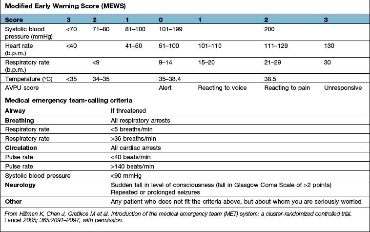
Medical emergency team-calling criteria
Airway |
If threatened |
Breathing |
All respiratory arrests |
Respiratory rate |
<5 breaths/min |
Respiratory rate |
>36 breaths/min |
Circulation |
All cardiac arrests |
Pulse rate |
<40 beats/min |
Pulse rate |
>140 beats/min |
Systolic blood pressure |
<90 mmHg |
Neurology |
Sudden fall in level of consciousness (fall in Glasgow Coma Scale of >2 points) |
|
Repeated or prolonged seizures |
Other |
Any patient who does not fit the criteria above, but about whom you are seriously worried |
From Hillman K, Chen J, Cretikos M et al. Introduction of the medical emergency team (MET) system: a cluster-randomized controlled trial. Lancet 2005; 365:2091–2097, with permission.
In some of the most seriously ill patients, the precise underlying diagnosis is initially unclear but in all cases, the immediate objective is to preserve life and prevent, reverse or minimize damage to vital organs such as the lungs, brain, kidneys and liver. This involves a rapid assessment of the physiological derangement followed by prompt institution of measures to support cardiovascular and respiratory function in order to restore perfusion of vital organs, improve delivery of oxygen to the tissues and encourage the removal of carbon dioxide and other waste products of metabolism (following the ABC approach: Airway, Breathing, Circulation, see Fig. 16.25, below). The patient’s condition and response to treatment should be closely monitored throughout. The underlying diagnosis may only become clear as the results of investigations become available, a more detailed history is obtained and a more thorough physical examination is performed. In practice resuscitation, assessment and diagnosis usually proceed in parallel.
Critically ill patients require multidisciplinary care with:
 Intensive skilled nursing care (usually 1 : 1 or 1 : 2 nurse/patient ratio in the UK).
Specialized physiotherapy including mobilization and rehabilitation.
Management of pain and distress with judicious administration of analgesics and sedatives (see p. 893).
Constant reassurance and support (critically ill patients easily become disorientated and delirium is common.
H2-receptor antagonists or proton pump inhibitors in selected cases to prevent stress-induced ulceration.
Compression stockings (full-length and graduated), pneumatic compression devices and subcutaneous low-molecular-weight heparin to prevent venous thrombosis.
Care of the mouth, prevention of constipation and of pressure sores.
Nutritional support (see p. 222). Protein energy malnutrition is common in critically ill patients and is associated with muscle wasting, weakness, delayed mobilization, difficulty weaning from ventilation, immune compromise and impaired wound healing. There is also an association between malnutrition and increased mortality. It is therefore recommended that nutritional support should be instituted as soon as is practicable in those unable to meet their nutritional needs orally, ideally within 1–2 days of the acute episode. Enteral nutrition, which is less expensive, preserves gut mucosal integrity, is more physiological and is associated with fewer complications, is preferred. Recently, the value of early feeding has been questioned, apart from giving small amounts to ensure gut viability. Parenteral nutrition is sometimes indicated at a later stage for those unable to tolerate or absorb enteral nutrition and should be initiated without delay, at least within 3 days. Persistent attempts at enteral nutrition in those with gastrointestinal intolerance leads to underfeeding and malnutrition.
Critically ill patients commonly require intravenous insulin infusions, often in high doses, to combat hyperglycaemia and insulin resistance (see p. 1006). Although the use of intensive insulin therapy to achieve ‘tight glycaemic control’ (blood glucose level between 4.4 and 6.1 mmol/L) was shown to improve outcome (at least when combined with aggressive nutritional support), more recent studies have failed to confirm this finding and have indicated that this approach is associated with an unacceptably high incidence of hypoglycaemia, and possibly an increase in mortality. Current recommendations suggest that blood glucose levels should be maintained at <8–10 mmol/L.
Discharge of patients from intensive care should normally be planned in advance and should ideally take place during normal working hours. Planned discharge often involves a period in a ‘step-down’ intermediate care area. Premature or unplanned discharge, especially during the night, has been associated with higher hospital mortality rates. A summary including ‘points to review’ should be included in the clinical notes and there should be a detailed handover to the receiving team (medical and nursing). The intensive care team should continue to review the patient, who might deteriorate following discharge, on the ward and should be available at all times for advice on further management (e.g. tracheostomy care, nutritional support). In this way, deterioration and readmission to intensive care (which is associated with a particularly poor outcome) or even cardiorespiratory arrest might be avoided.
This chapter concentrates on cardiovascular and respiratory problems. Many patients also have failure of other organs such as the kidney and liver; treatment of these is dealt with in more detail in the relevant chapters.
FURTHER READING
Casaer MP, Mesotten O, Hermans G et al. Early versus late parenteral nutrition in critically ill adults. N Engl J Med 2011; 365:506–517.
The NICE-SUGAR Study Investigators. Intensive versus conventional glucose control in critically ill patients. N Engl J Med 2009; 360:1293–1297.
Ziegler TR. Parenteral nutrition in the critically ill patient. N Engl J Med 2009; 361:1088–1097.
Applied cardiorespiratory physiology
Oxygen delivery and consumption
Oxygen delivery (DO2) (Fig. 16.1) is defined as the total amount of oxygen delivered to the tissues per unit time. It is dependent on the volume of blood flowing through the microcirculation per minute (i.e. the total cardiac output,  ) and the amount of oxygen contained in that blood (i.e. the arterial oxygen content, CaO2). Oxygen is transported both in combination with haemoglobin and dissolved in plasma. The amount combined with haemoglobin is determined by the oxygen capacity of haemoglobin (usually taken as 1.34 mL of oxygen per gram of haemoglobin) and its percentage saturation with oxygen (SO2), while the volume dissolved in plasma depends on the partial pressure of oxygen (PO2). Except when hyperbaric oxygen is administered, the amount of dissolved oxygen in plasma is insignificant.
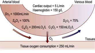
Figure 16.1 Tissue oxygen delivery and consumption in a normal 70 kg person breathing air. Oxygen delivery (DO2) = cardiac output × (haemoglobin concentration × oxygen saturation (SaO2) × 1.34). In normal adults, oxygen delivery is roughly 1000 mL/min, of which 250 mL is taken up by tissues. Mixed venous blood is thus 75% saturated with oxygen.  , mixed venous oxygen content;
  , mixed venous oxygen saturation; CaO2, arterial oxygen content.
Clinically, however, the utility of this global concept of oxygen delivery is limited because it fails to account for changes in the relative flow to individual organs and the distribution of flow through the microcirculation (i.e. the efficiency with which oxygen delivery is matched to the metabolic requirements of individual tissues or cells). Furthermore, some organs (such as the heart) have high oxygen requirements relative to their blood flow and will receive insufficient oxygen, even if the overall oxygen delivery is apparently adequate. Lastly, microcirculatory flow is influenced by blood viscosity.
Oxygenation of the blood
Oxyhaemoglobin dissociation curve
The saturation of haemoglobin with oxygen is determined by the partial pressure of oxygen (PO2) in the blood, the relationship between the two being described by the oxyhaemoglobin dissociation curve (Fig. 16.2). The sigmoid shape of this curve is significant for a number of reasons:
Modest falls in the partial pressure of oxygen in the arterial blood (PaO2) may be tolerated (since oxygen content is relatively unaffected) provided that the percentage saturation remains above 92%.
Increasing the PaO2 to above normal has only a minimal effect on oxygen content unless hyperbaric oxygen is administered (when the amount of oxygen in solution in plasma becomes significant).
Once on the steep ‘slippery slope’ of the curve (percentage saturation below about 90%), a small decrease in PaO2 can cause large falls in oxygen content, whereas increasing PaO2 only slightly, e.g. by administering 28% oxygen to a patient with chronic obstructive pulmonary disease (COPD), can lead to a useful increase in oxygen saturation and content.
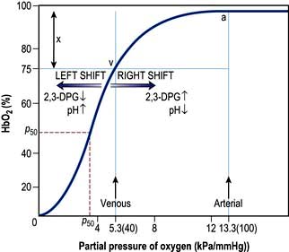
Figure 16.2 The oxyhaemoglobin dissociation curve. a, arterial point; v, venous point; x, arteriovenous oxygen content difference. HbO2 (%) is the percentage saturation of haemoglobin with oxygen. The curve will move to the right in the presence of acidosis (metabolic or respiratory), pyrexia or an increased red cell 2,3-DPG concentration. For a given arteriovenous oxygen content difference, the mixed venous PO2 will then be higher. Furthermore, if the mixed venous PO2 is unchanged, the arteriovenous oxygen content difference increases and more oxygen is off-loaded to the tissues (see p. 374). P50 (the PO2 at which haemoglobin is half saturated with O2) is a useful index of these shifts – the higher the P50 (i.e. shift to the right), the lower the affinity of haemoglobin for O2.
The PaO2 is in turn influenced by the alveolar oxygen tension (PAO2), the efficiency of pulmonary gas exchange, and the partial pressure of oxygen in mixed venous blood  .
.
Alveolar oxygen tension (PAO2)
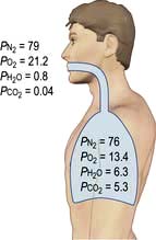
The partial pressures of inspired gases are shown in Figure 16.3. By the time the inspired gases reach the alveoli they are fully saturated with water vapour at body temperature (37°C), which has a partial pressure of 6.3 kPa (47 mmHg) and contain CO2 at a partial pressure of approximately 5.3 kPa (40 mmHg); the PAO2 is thereby reduced to approximately 13.4 kPa (100 mmHg).
The clinician can influence PAO2 by administering oxygen or by increasing the barometric pressure.
Pulmonary gas exchange
In normal subjects there is a small alveolar-arterial oxygen difference (PA–aO2). This is due to:
a small (0.133 kPa, 1 mmHg) pressure gradient across the alveolar membrane
a small amount of blood (2% of total cardiac output) bypassing the lungs via the bronchial and thebesian veins
Pathologically, there are three possible causes of an increased PA–aO2 difference:
Diffusion defect. This is not a major cause of hypoxaemia even in conditions such as lung fibrosis, in which the alveolar-capillary membrane is considerably thickened. Carbon dioxide is also not affected, as it is more soluble than oxygen.
Right-to-left shunts. In certain congenital cardiac lesions or when a segment of lung is completely collapsed, a proportion of venous blood passes to the left side of the heart without taking part in gas exchange, causing arterial hypoxaemia. This hypoxaemia cannot be corrected by administering oxygen to increase the PAO2, because blood leaving normal alveoli is already fully saturated; further increases in PO2 will not, therefore, significantly affect its oxygen content. On the other hand, because of the shape of the carbon dioxide dissociation curve (Fig. 16.4), the high PCO2 of the shunted blood can be compensated for by over-ventilating patent alveoli, thus lowering the CO2 content of the effluent blood. Indeed, many patients with acute right-to-left shunts hyperventilate in response to the hypoxia and/or to stimulation of mechanoreceptors in the lung, so that their PaCO2 is normal or low.
Ventilation/perfusion mismatch (see p. 796). Diseases of the lung parenchyma (e.g. pulmonary oedema, acute lung injury) result in  mismatch, producing an increase in alveolar deadspace and hypoxaemia. The increased deadspace can be compensated for by increasing overall ventilation. In contrast to the hypoxia resulting from a true right-to-left shunt, that due to areas of low
  can be partially corrected by administering oxygen and thereby increasing the PAO2 even in poorly ventilated areas of lung.
Mixed venous oxygen tension  and saturation (
 )
)
The  is the partial pressure of oxygen in pulmonary arterial blood that has been thoroughly mixed during its passage through the right heart. Assuming PaO2 remains constant,
  and
and  will fall if more oxygen has to be extracted from each unit volume of blood arriving at the tissues. A low
  therefore indicates either that oxygen delivery has fallen or that tissue oxygen requirements have increased without a compensatory rise in cardiac output. If
  falls, the effect of a given degree of pulmonary shunting on arterial oxygenation will be exacerbated. Thus, worsening arterial hypoxaemia does not necessarily indicate a deterioration in pulmonary function but might instead reflect a fall in cardiac output and/or a rise in oxygen consumption.
Conversely, a rise in  and
and  may reflect impaired tissue oxygen extraction (due to microcirculatory dysfunction) and/or reduced oxygen utilization (e.g. due to mitochondrial dysfunction) as seen in severe sepsis (see below).
Monitoring the oxygen saturation in central venous ( ), rather than pulmonary artery blood is less invasive and has been shown to be a valuable guide to the resuscitation of critically ill patients (see p. 891).
Cardiac output
Cardiac output is the product of heart rate and stroke volume, and is affected by changes in either of these (see Fig. 16.5).
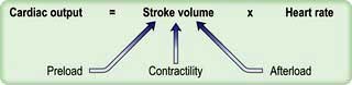
Heart rate
When heart rate increases, the duration of systole remains essentially unchanged, whereas diastole, and thus the time available for ventricular filling, becomes progressively shorter, and the stroke volume eventually falls. In the normal heart this occurs at rates greater than about 160 beats per minute, but in those with cardiac pathology, especially when this restricts ventricular filling (e.g. mitral stenosis), stroke volume may fall at much lower heart rates. Furthermore, tachycardias cause a marked increase in myocardial oxygen consumption ( ) and this may precipitate ischaemia in areas of the myocardium with restricted coronary perfusion. When the heart rate falls, a point is reached at which the increase in stroke volume is insufficient to compensate for bradycardia and again cardiac output falls.
Alterations in heart rate are often caused by disturbances of rhythm (e.g. atrial fibrillation, complete heart block) in which ventricular filling is not augmented by atrial contraction, exacerbating the fall in stroke volume.
Stroke volume
The volume of blood ejected by the ventricle in a single contraction is the difference between the ventricular end-diastolic volume (VEDV) and end-systolic volume (VESV) (i.e. stroke volume = VEDV – VESV). The ejection fraction describes the stroke volume as a percentage of VEDV (i.e. ejection fraction = (VEDV − VESV)/VEDV × 100%) and is an indicator of myocardial performance.
Three interdependent factors determine the stroke volume (see p. 671).
Preload
This is defined as the tension of the myocardial fibres at the end of diastole, just before the onset of ventricular contraction, and is therefore related to the degree of stretch of the fibres. As the end-diastolic volume of the ventricle increases, tension in the myocardial fibres is increased and stroke volume rises (Fig. 16.6). Myocardial oxygen consumption ( ) increases only slightly with an increase in preload (produced, for example, by a ‘fluid challenge’, see below) and this is therefore the most efficient way of improving cardiac output.
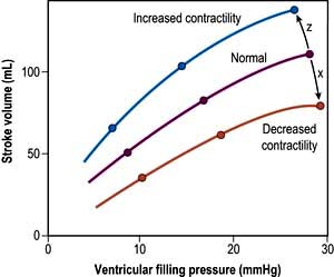
Figure 16.6 The Frank–Starling relationship: as preload is increased, stroke volume rises. If the ventricle is overstretched, stroke volume will fall (x). In myocardial failure, the curve is depressed and flattened. Increasing contractility, e.g. due to sympathetic stimulation, shifts the curve upwards and to the left (z).
Myocardial contractility
This refers to the ability of the heart to perform work, independent of changes in preload and afterload. The state of myocardial contractility determines the response of the ventricles to changes in preload and afterload. Contractility is often reduced in critically ill patients, as a result of either pre-existing myocardial damage (e.g. ischaemic heart disease), or the acute disease process itself (e.g. sepsis). Changes in myocardial contractility alter the slope and position of the Starling curve; worsening ventricular performance is manifested as a depressed, flattened curve (Fig. 16.6 and Fig. 14.5). Inotropic drugs can be used to increase myocardial contractility (see below).
Afterload
This is defined as the myocardial wall tension developed during systolic ejection. In the case of the left ventricle, the resistance imposed by the aortic valve, the peripheral vascular resistance and the elasticity of the major blood vessels are the major determinants of afterload. Ventricular wall tension will also be increased by ventricular dilatation, an increase in intraventricular pressure or a reduction in ventricular wall thickness.
Decreasing the afterload (exercise, sepsis, vasodilator agents) can increase the stroke volume achieved at a given preload (Fig. 16.7), while reducing  . The reduction in wall tension also leads to an increase in coronary blood flow, thereby improving the myocardial oxygen supply/demand ratio. Excessive reductions in afterload will cause hypotension.
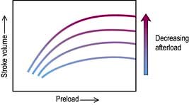
Figure 16.7 The effect of changes in afterload on the ventricular function curve. At any given preload, decreasing afterload increases the stroke volume.
Increasing the afterload (increased sympathetic activity, vasoconstrictor agents), on the other hand, can cause a fall in stroke volume and is a potent cause of increased  . Right ventricular afterload is normally negligible because the resistance of the pulmonary circulation is very low but is increased in pulmonary hypertension.
Monitoring critically ill patients
As well as allowing immediate recognition of changes in the patient’s condition, monitoring can also be used to establish or confirm a diagnosis, to gauge the severity of the condition, to follow the evolution of the illness, to guide interventions and to assess the response to treatment. Invasive monitoring is generally indicated in the more seriously ill patients and in those who fail to respond to initial treatment. These techniques are, however, associated with a significant risk of complications, as well as additional costs and patient discomfort and should therefore only be used when the potential benefits outweigh the dangers. Likewise, invasive devices should be removed as soon as possible.
Assessment of tissue perfusion
Pale, cold skin, delayed capillary refill and the absence of visible veins in the hands and feet indicate poor perfusion. Although peripheral skin temperature measurements can help clinical evaluation, the earliest compensatory response to hypovolaemia or a low cardiac output, and the last to resolve after resuscitation is vasoconstriction in the splanchnic region.
Metabolic acidosis with raised lactate concentration suggests that tissue perfusion is sufficiently compromised to cause cellular hypoxia and anaerobic glycolysis. Persistent, severe lactic acidosis is associated with a very poor prognosis. In many critically ill patients, especially those with sepsis, however, lactic acidosis can also be caused by metabolic disorders unrelated to tissue hypoxia and can be exacerbated by reduced clearance owing to hepatic or renal dysfunction as well as the administration of adrenaline (epinephrine).
Urinary flow is a sensitive indicator of renal perfusion and haemodynamic performance.
Blood pressure
Alterations in blood pressure are often interpreted as reflecting changes in cardiac output. However, if there is vasoconstriction with a high peripheral resistance, the blood pressure may be normal, even when the cardiac output is reduced. Conversely, the vasodilated patient may be hypotensive, despite a very high cardiac output.
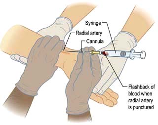
Hypotension jeopardizes perfusion of vital organs. The adequacy of blood pressure in an individual patient must always be assessed in relation to the premorbid value. Blood pressure is traditionally measured using a sphygmomanometer but if rapid alterations are anticipated, continuous monitoring using an intra-arterial cannula is indicated (Practical Box 16.1; Fig. 16.8).
 Practical Box 16.1
Radial artery cannulation
Technique
1. The procedure is explained to the patient and, if possible, consent obtained.
2. The arm is supported, with the wrist extended, by an assistant. (Gloves should be worn.)
3. The skin should be cleaned with chlorhexidine.
4. The radial artery is palpated where it arches over the head of the radius.
5. In conscious patients, local anaesthetic is injected to raise a weal over the artery, taking care not to puncture the vessel or obscure its pulsation.
6. A small skin incision is made over the proposed puncture site.
7. A small parallel-sided cannula (20 gauge for adults, 22 gauge for children) is used in order to allow blood flow to continue past the cannula.
8. The cannula is inserted over the point of maximal pulsation and advanced in line with the direction of the vessel at an angle of approximately 30°.
9. ‘Flashback’ of blood into the cannula indicates that the radial artery has been punctured.
10. To ensure that the shoulder of the cannula enters the vessel, the needle and cannula are lowered and advanced a few millimetres into the vessel.
11. The cannula is threaded off the needle into the vessel and the needle withdrawn.
12. The cannula is connected to a non-compliant manometer line filled with saline. This is then connected via a transducer and continuous flush device to a monitor, which records the arterial pressure.
Central venous pressure (CVP)
This provides a fairly simple, but approximate method of gauging the adequacy of a patient’s circulating volume and the contractile state of the myocardium. The absolute value of the CVP is not as useful as its response to a fluid challenge (the infusion of 100–200 mL of fluid over a few minutes) (Fig. 16.9). The hypovolaemic patient will initially respond to transfusion with little or no change in CVP, together with some improvement in cardiovascular function (falling heart rate, rising blood pressure, increased peripheral temperature and urine output). As the normovolaemic state is approached, the CVP usually rises slightly and reaches a plateau, while other cardiovascular values begin to stabilize. At this stage, volume replacement should be slowed, or even stopped, in order to avoid overtransfusion (indicated by an abrupt and sustained rise in CVP, often accompanied by some deterioration in the patient’s condition). In cardiac failure, the venous pressure is usually high; the patient will not improve in response to volume replacement, which will cause a further, sometimes dramatic, rise in CVP.
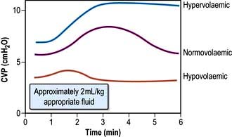
Figure 16.9 The effects on the central venous pressure (CVP) of a rapid administration of a ‘fluid challenge’ to patients with a CVP within the normal range.
(From Sykes MK. Venous pressure as a clinical indication of adequacy of transfusion. Annals of Royal College of Surgeons of England 1963; 33:185–197.)
Central venous catheters are usually inserted via a percutaneous puncture of the subclavian or internal jugular vein using a guidewire technique (Practical Box 16.2; Figs 16.10, 16.11). The guidewire techniques can also be used in conjunction with a vein dilator for inserting multilumen catheters, double lumen cannulae for haemofiltration or pulmonary artery catheter introducers. The routine use of ultrasound to guide central venous cannulation reduces complication rates.
Practical Box 16.2
Internal jugular vein cannulation
Technique
1. The procedure is explained to the patient and, if possible, consent obtained.
2. The patient is placed head-down to distend the central veins (this facilitates cannulation and minimizes the risk of air embolism but may exacerbate respiratory distress and is dangerous in those with raised intracranial pressure).
3. The skin is cleaned with an antiseptic solution such as chlorhexidine. Sterile precautions are taken throughout the procedure.
4. Local anaesthetic (1% plain lidocaine) is injected intradermally to raise a weal at the apex of a triangle formed by the two heads of sternomastoid with the clavicle at its base.
5. A small incision is made through the weal.
6. The cannula or needle is inserted through the incision and directed laterally downwards and backwards in the direction of the nipple until the vein is punctured just beneath the skin and deep to the lateral head of sternomastoid.
Ultrasound-guided puncture is recommended to reduce the incidence of complications.
7. Check that venous blood is easily aspirated.
8. The cannula is threaded off the needle into the vein or the guidewire is passed through the needle (see Fig. 16.11).
9. The CVP manometer line is connected to a manometer/transducer.
10. A chest X-ray should be taken to verify that the tip of the catheter is in the superior vena cava and to exclude pneumothorax.
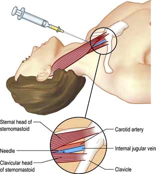
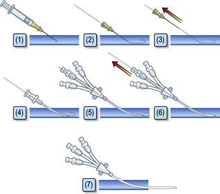
Figure 16.11 Seldinger technique – insertion of a catheter over guidewire. (1) Puncture vessel; (2) advance guidewire; (3) remove needle; (4) dilate vessel; (5) advance catheter over guidewire; (6) remove guidewire; (7) catheter in situ.
The CVP should be read intermittently using a manometer system or continuously using a transducer and bedside monitor. It is essential that the pressure recorded always be related to the level of the right atrium. Various landmarks are advocated (e.g. sternal notch with the patient supine, sternal angle or mid-axilla when the patient is at 45°), but which is chosen is largely immaterial provided it is used consistently in an individual patient. Pressure measurements should be obtained at end-expiration.
The following are common pitfalls in interpreting central venous pressure readings:
Blocked catheter. This results in a sustained high reading, with a damped or absent waveform, which often does not correlate with clinical assessment.
Transducer wrongly positioned. Failure to level the system is a common cause of erroneous readings.
Catheter tip in right ventricle. If the catheter is advanced too far, an unexpectedly high pressure with pronounced oscillations is recorded. This is easily recognized when the waveform is displayed.
Left atrial pressure
In uncomplicated cases, careful interpretation of the CVP provides a reasonable guide to the filling pressures of both sides of the heart. In many critically ill patients, however, there is a disparity in function between the two ventricles. Most commonly, left ventricular performance is worse, so that the left ventricular function curve is displaced downward and to the right (Fig. 16.12). High right ventricular filling pressures, with normal or low left atrial pressures, are less common but occur with right ventricular dysfunction and in cases where the pulmonary vascular resistance (i.e. right ventricular afterload) is raised, such as in acute respiratory failure and pulmonary embolism.
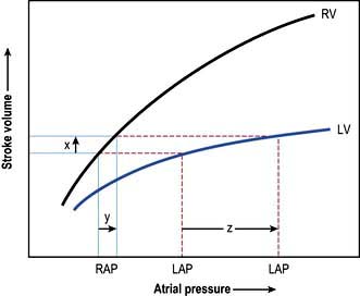
Figure 16.12 Left ventricular (LV) and right ventricular (RV) function curves in a patient with left ventricular dysfunction. Since the stroke volume of the two ventricles must be the same (except perhaps for a few beats during a period of circulatory adjustment), left atrial pressure (LAP) must be higher than right atrial pressure (RAP). Moreover, an increase in stroke volume (x) produced by expanding the circulatory volume may be associated with a small rise in RAP (y) but a marked increase in LAP (z).
Pulmonary artery pressure
A ‘balloon flotation catheter’ enables reliable catheterization of the pulmonary artery. These ‘Swan–Ganz’ catheters can be inserted centrally (Fig. 16.10) or through the femoral vein, or via a vein in the antecubital fossa. Passage of the catheter from the major veins, through the chambers of the heart, into the pulmonary artery and into the wedged position is monitored and guided by the pressure waveforms recorded from the distal lumen (Practical Box 16.3; Fig. 16.13). A chest X-ray should always be obtained to check the final position of the catheter. In difficult cases, screening with an image intensifier may be required.
Practical Box 16.3
Passage of a pulmonary artery balloon flotation catheter through the chambers of the heart into the ‘wedged’ position
Consent should be obtained if possible from the patient after explanation of the procedure.
Note: (a), (b), (c), (d) refer to Figure 16.13.
1. A balloon flotation catheter is inserted through a large vein (see text).
2. Once in the thorax, respiratory oscillations are seen. The catheter should be advanced further towards the lower superior vena cava/right atrium (a), where pressure oscillations become more pronounced. The balloon should then be inflated and the catheter advanced.
3. When the catheter is in the right ventricle (b), there is no dicrotic notch and the diastolic pressure is close to zero. The patient should be returned to the horizontal, or slightly head-up, position before advancing the catheter further.
4. When the catheter reaches the pulmonary artery (c) a dicrotic notch appears and there is elevation of the diastolic pressure. The catheter should be advanced further with the balloon inflated.
5. Reappearance of a venous waveform indicates that the catheter is ‘wedged’. The balloon is deflated to obtain the pulmonary artery pressure. The balloon is inflated intermittently to obtain the pulmonary artery occlusion (also known as pulmonary artery, or capillary, ‘wedge’) pressure (d).
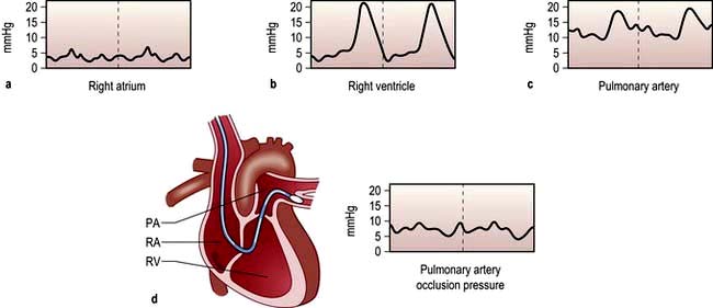
Figure 16.13 Passage of pulmonary artery balloon flotation catheter through the chambers of the heart into the ‘wedged’ position to measure the pulmonary artery occlusion pressure. (See Practical Box 16.3.)
Once in place, the balloon is deflated and the pulmonary artery mean, systolic and end-diastolic pressures (PAEDP) can be recorded. The pulmonary artery occlusion pressure (PAOP, previously referred to as the pulmonary artery or capillary ‘wedge’ pressure) is measured by reinflating the balloon, thereby propelling the catheter distally until it impacts in a medium-sized pulmonary artery. In this position there is a continuous column of fluid between the distal lumen of the catheter and the left atrium, so that PAOP is usually a reasonable reflection of left atrial pressure.
The technique is generally safe – the majority of complications such as ‘knotting’, valve trauma and pulmonary artery rupture (which can be fatal) are related to user inexperience. Pulmonary artery catheters should preferably be removed within 72 h, since the incidence of complications, especially infection, then increases progressively
Cardiac output
Cardiac output can be continuously monitored using a modified pulmonary artery catheter which transmits low heat energy into the surrounding blood and constructs a ‘thermodilution curve’. These catheters also optically measure and continuously display  .
.
In general, pulmonary artery catheters enable the clinician to optimize cardiac output and oxygen delivery, while minimizing the risk of volume overload. They can also be used to guide the rational use of inotropes and vasoactive agents and are particularly helpful in patients with pulmonary hypertension. There is, however, a considerable body of evidence to suggest that the unselective use of this monitoring device in the absence of evidence-based haemodynamic goals does not lead to improved outcomes and less invasive techniques are increasingly preferred.
Less invasive techniques for assessing cardiac function and guiding volume replacement
Arterial pressure variation as a guide to hypovolaemia
Systolic arterial pressure decreases during the inspiratory phase of intermittent positive pressure ventilation (p. 894). The magnitude of this cyclical variability has been shown to correlate more closely with hypovolaemia than other monitored variables, including CVP. Systolic pressure (or pulse pressure) variation during mechanical ventilation can therefore be used as a simple and reliable guide to the adequacy of the circulatory volume. The response to fluid loading can also easily be predicted by observing the changes in pulse pressure during passive leg raising.
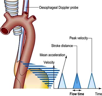
Oesophageal Doppler
Stroke volume, cardiac output and myocardial function can be assessed non-invasively using Doppler ultrasonography. A probe is passed into the oesophagus to continuously monitor velocity waveforms from the descending aorta (Fig. 16.14). Although reasonable estimates of stroke volume, and hence cardiac output can be obtained, the technique is best used for trend analysis rather than for making absolute measurements. Oesophageal Doppler probes can be inserted quickly and easily and are particularly valuable for perioperative optimization of the circulating volume and cardiac performance in the unconscious patient. They are contraindicated in patients with oropharyngeal/oesophageal pathology.
Pulse contour analysis
Lithium dilution/pulse contour analysis does not require pulmonary artery catheterization or instrumentation of the oesophagus and is suitable for use in conscious patients. A bolus of lithium chloride is administered via a central venous catheter and the change in arterial plasma lithium concentration is detected by a lithium-sensitive electrode. This sensor can be connected to an existing arterial cannula via a three-way tap. A small battery-powered peristaltic pump is used to create a constant blood flow through the sensor and over the electrode tip. The cardiac output determined in this way can be used to calibrate an arterial pressure waveform (‘pulse contour’) analysis programme that will continuously monitor changes in cardiac output. Devices that use uncalibrated pulse contour analysis to estimate cardiac output are also available. As with pulse pressure variation, stroke volume variation using these devices can accurately predict fluid replacements.
Echocardiography
Echocardiography is being used increasingly often to provide immediate diagnostic information about cardiac structure and function (myocardial contractility, ventricular filling) in the critically ill patient. Transoesophageal echocardiography (TOE) is preferred because of its superior image clarity (Fig. 16.15).
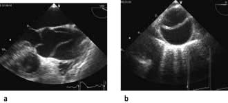
Figure 16.15 Aortic dissection (transoesophageal echocardiography, TOE). (a) Mid-oesophageal, long axis view showing Type A aortic dissection. (b) Short axis view of descending aorta showing intimal flap with false and true lumen.
(From Hinds CJ, Watson JD. Intensive Care: A Concise Textbook, 3rd edn. Edinburgh: Saunders; 2008. Courtesy of Dr C. Rathwell.)
If there is disagreement between clinical signs and a monitored variable, it should be assumed that the monitor is incorrect until all sources of potential error have been checked and eliminated. Changes and trends in monitored variables are more informative than a single reading.
Disturbances of acid–base balance
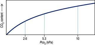
The physiology of acid–base control is discussed on page 660. Acid–base disturbances can be described in relation to the diagram illustrated in Figure 13.13, p. 663 (which shows PaCO2 plotted against arterial [H+]).
Both acidosis and alkalosis can occur, each of which are either metabolic (primarily affecting the bicarbonate component of the system) or respiratory (primarily affecting PaCO2). Compensatory changes may also be apparent. In clinical practice, arterial [H+] values outside the range 18–126 nmol/L (pH 6.9–7.7) are rarely encountered.
Blood gas and acid–base values (normal ranges) are shown in Table 16.2. (For blood gas analysis, see p. 891.)
Table 16.2 Arterial blood gas and acid–base values (normal ranges)
H+ |
35–45 nmol/L |
pH 7.35–7.45 |
PO2 (breathing room air) |
10.6–13.3 kPa |
(80–100 mmHg) |
PCO2 |
4.8–6.1 kPa |
(36–46 mmHg) |
Base deficit |
±2.5 |
|
Plasma HCO3– |
22–26 mmol/L |
|
O2 saturation |
95–100% |
|
Respiratory acidosis. This is caused by retention of carbon dioxide. The PaCO2 and [H+] rise. A chronically raised PaCO2 is compensated by renal retention of bicarbonate, and the [H+] returns towards normal. A constant arterial bicarbonate concentration is then usually established within 2–5 days. This represents a primary respiratory acidosis with a compensatory metabolic alkalosis (see p. 666). Common causes of respiratory acidosis include ventilatory failure and COPD (type II respiratory failure where there is a high PaCO2 and a low PaO2, see p. 814).
Respiratory alkalosis. In this case, the reverse occurs and there is a fall in PaCO2 and [H+], often with a small reduction in bicarbonate concentration. If hypocarbia persists, some degree of renal compensation may occur, producing a metabolic acidosis, although in practice this is unusual. A respiratory alkalosis may be produced, intentionally or unintentionally, when patients are mechanically ventilated; it may also be seen with hypoxaemic (type I) respiratory failure (see Ch. 15, p. 817), spontaneous hyperventilation and in those living at high altitudes.
Metabolic acidosis (p. 664). This may be due to excessive acid production, most commonly lactate and H+ (lactic acidosis) as a consequence of anaerobic metabolism during an episode of shock or following cardiac arrest. A metabolic acidosis may also develop in chronic renal failure or in diabetic ketoacidosis. It can also follow the loss of bicarbonate from the gut or from the kidney in renal tubular acidosis. Respiratory compensation for a metabolic acidosis is usually slightly delayed because the blood–brain barrier initially prevents the respiratory centre from sensing the increased blood [H+]. Following this short delay, however, the patient hyperventilates and ‘blows off’ carbon dioxide to produce a compensatory respiratory alkalosis. There is a limit to this respiratory compensation, since in practice values for PaCO2 less than about 1.4 kPa (11 mmHg) are rarely achieved. Spontaneous respiratory compensation cannot occur if the patient’s ventilation is controlled or if the respiratory centre is depressed, for example by drugs or head injury.
Metabolic alkalosis. This can be caused by loss of acid, for example from the stomach with nasogastric suction, or in high intestinal obstruction, or excessive administration of absorbable alkali. Overzealous treatment with intravenous sodium bicarbonate is sometimes implicated. Respiratory compensation for a metabolic alkalosis is often slight, and it is rare to encounter a PaCO2 above 6.5 kPa (50 mmHg), even with severe alkalosis.
Shock, sepsis and acute disturbances of haemodynamic function
Shock is the term used to describe acute circulatory failure with inadequate or inappropriately distributed tissue perfusion resulting in generalized cellular hypoxia and/or an inability of the cells to utilize oxygen.
Causes of shock
Abnormalities of tissue perfusion can result from:
The causes of shock are shown in Table 16.3. Often shock can result from a combination of these factors (e.g. in sepsis, distributive shock is frequently complicated by hypovolaemia and myocardial depression).
| Hypovolaemic | Obstructive |
|---|---|
Pathophysiology
The sympatho-adrenal response to shock (Fig. 16.16)
Hypotension stimulates the baroreceptors, and to a lesser extent the chemoreceptors, causing increased sympathetic nervous activity with ‘spill-over’ of noradrenaline (norepinephrine) into the circulation. Later this is augmented by the release of catecholamines (predominantly, adrenaline (epinephrine)) from the adrenal medulla. The resulting vasoconstriction, together with increased myocardial contractility and heart rate, help to restore blood pressure and cardiac output.
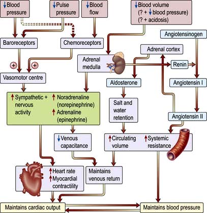
Figure 16.16 The sympatho-adrenal response to shock showing the effect of increased catecholamines on the left of the diagram and the release of angiotensin and aldosterone on the right. Both mechanisms help to maintain the cardiac output and blood pressure in shock.
Reduction in perfusion of the renal cortex stimulates the juxtaglomerular apparatus to release renin. This converts angiotensinogen to angiotensin I, which in turn is converted in the lungs and by the vascular endothelium to the potent vasoconstrictor angiotensin II. Angiotensin II also stimulates secretion of aldosterone by the adrenal cortex, causing sodium and water retention (p. 566). This helps to restore the circulating volume (see p. 639).
Neuroendocrine response
There is release of pituitary hormones such as adrenocorticotrophic hormone (ACTH), vasopressin (antidiuretic hormone, ADH) and endogenous opioid peptides. (In septic shock there may be a relative deficiency of vasopressin.)
There is release of cortisol, which causes fluid retention and antagonizes insulin.
There is release of glucagon, which raises the blood sugar level.
Although absolute adrenocortical insufficiency (e.g. due to bilateral adrenal haemorrhage or necrosis) is rare, there is evidence that patients with septic shock have a blunted response to exogenous ACTH (so-called ‘relative’ or ‘occult’ adrenocortical insufficiency) and that this could be associated with an impaired pressor response to noradrenaline (norepinephrine) and a worse prognosis. The diagnosis, causes and clinical significance of this phenomenon remain unclear.
Release of pro- and anti-inflammatory mediators
Severe infection (often with bacteraemia or endotoxaemia), the presence of large areas of damaged tissue (e.g. following trauma or extensive surgery), hypoxia or prolonged/repeated episodes of hypoperfusion can trigger an exaggerated inflammatory response with systemic activation of leucocytes and release of a variety of potentially damaging ‘mediators’ (see also Ch. 3). Although beneficial when targeted against local areas of infection or necrotic tissue, dissemination of this ‘innate immune’ response can produce shock and widespread tissue damage. Characteristically the initial episode of overwhelming inflammation is followed by a period of immune suppression, which in some cases may be profound and during which the patient is at increased risk of developing secondary infections. It also seems that pro- and anti-inflammatory elements of the host response may co-exist.
Microorganisms and their toxic products (Fig. 16.17)
In sepsis/septic shock the innate immune response and inflammatory cascade are triggered by the recognition of pathogen-associated molecular patterns (PAMPs), including cell wall components (e.g. endotoxin) and/or exotoxins (antigenic proteins produced by bacteria such as staphylococci, streptococci and Pseudomonas).
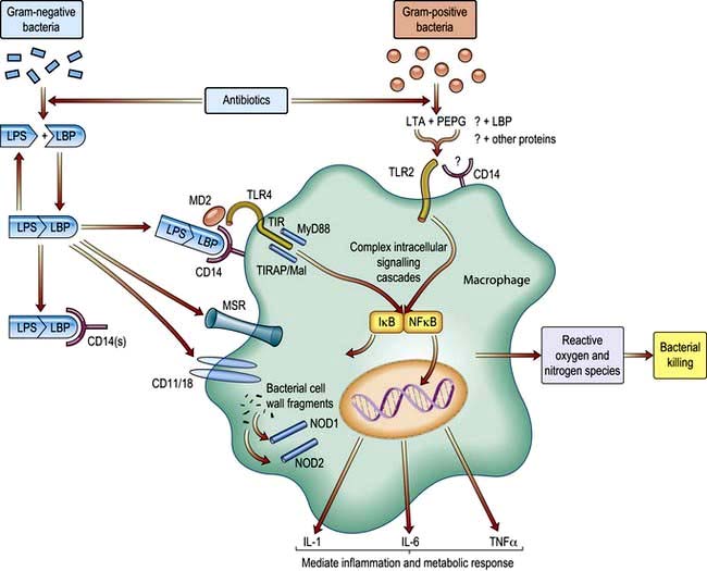
Figure 16.17 Induction of the innate immune response by the lipopolysaccharide–lipopolysaccharide-binding protein (LPS-LBP) complex. This simplified figure illustrates the intracellular events initiated by Gram-negative and Gram-positive bacteria, which eventually lead to bacterial killing. LPS, lipopolysaccharide; LBP, lipopolysaccharide binding protein; LTA, lipoteichoic acid; NFκB, nuclear factor kappa B; IκB, inhibitory factor kappa B; PEPG, peptidoglycan-N; TLR, toll-like receptors; MSR, macrophage scavenger receptor; MyD88, myeloid differentiation factor 88; TIR, toll-interleukin receptor; TIRAP, toll-interleukin 1 receptor adaptor protein; MD2 is a secreted protein involved in binding liposaccharide with TLR4; TIRAP/Mal, an adaptor protein for TLR2 and TLR4.
Endotoxin is a lipopolysaccharide (LPS) derived from the cell wall of Gram-negative bacteria and is a potent trigger of the inflammatory response. The lipid A portion of LPS can be bound by a protein normally present in human serum known as lipopolysaccharide binding protein (LBP). The LBP/LPS complex attaches to the cell surface marker CD14 and, combined with a secreted protein (MD2), this complex then binds to a member of the toll-like receptor family (TLR4), which transduces the activation signal into the cell. These receptors act through a critical adaptor molecule, myeloid differentiation factor 88 (MyD 88), to regulate the activity of NFκB pathways. Intracellular pattern recognition receptors such as nucleotide-binding oligomerization domain (NOD) 1 may also be involved. Another mechanism in this complex area involves TREM-I (triggering receptor expressed in myeloid cells, see p. 54), which triggers secretion of pro-inflammatory cytokines.
Specific kinases then phosphorylate inhibitory kappa B (IκB), releasing the nuclear transcription factor NFκB, which passes into the nucleus where it binds to DNA and promotes the synthesis of a wide variety of inflammatory mediators. Gram-positive bacteria have cell wall components which are similar in structure to LPS (e.g. lipoteichoic acid), and can also trigger a systemic inflammatory response, probably through similar (TLR2) but not identical pathways (Fig. 16.17). Following traumatic or surgical tissue injury, inflammatory pathways may be triggered by damage-associated molecular patterns (DAMPS) such as DNA fragments.
Activation of complement cascade
Fragments of C3 act as opsonins and co-stimulatory molecules that assist lymphocytes with the adaptive immune response, while small peptides derived from C3, C4 and C5 cause leucocyte chemotaxis, release of cytokines and increased vascular permeability (see p. 51).
Cytokines
Pro-inflammatory cytokines (see also p. 49) such as the interleukins (ILs) and tumour necrosis factor (TNF) are also mediators of the systemic inflammatory response. TNF release initiates many of the responses to endotoxin, for example, and acts synergistically with IL-1, in part through induction of cyclo-oxygenase, platelet-activating factor (PAF) and nitric oxide synthase (see below). Subsequently, other cytokines including IL-6 and IL-8 appear in the circulation. IL-6 is the major stimulant for hepatic synthesis of acute phase proteins and is involved in the induction of fever, anaemia and cachexia, while IL-8 is a chemoattractant. The cytokine network is extremely complex, with many endogenous self-regulating mechanisms. For example, naturally occurring soluble TNF receptors are shed from cell surfaces during the inflammatory response, binding to TNF and thereby reducing its biological activity. An endogenous inhibitory protein that binds competitively to the IL-1 receptor has also been identified.
In addition to pro-inflammatory mediators such as TNF, anti-inflammatory cytokines, e.g. IL-10, are released. When excessive, this anti-inflammatory response is associated with an inappropriate immune hyporesponsiveness.
Products of arachidonic acid metabolism
Arachidonic acid, derived from the breakdown of membrane phospholipid, is metabolized to form prostaglandins and leukotrienes, which are key inflammatory mediators (see Fig. 15.30) and p. 826).
Heat shock proteins (HSPs)
HSPs are synthesized after exposure to various harmful stimuli such as heat, cytokines, hypoxia, endotoxin, various chemicals and oxygen free radicals. They appear to be protective in sepsis, probably because they recognize and form complexes with denatured proteins, thus inducing correct protein folding and, where necessary, proteolytic degradation. They also protect normal, functional proteins against degradation and inhibit apoptosis. HSPs are therefore often referred to as ‘molecular chaperones’.
Adhesion molecules
Adhesion of activated leucocytes to the vessel wall and their subsequent extravascular migration is a key component of the sequence of events leading to endothelial injury, tissue damage and organ dysfunction (see also p. 23). This process is mediated by inducible intercellular adhesion molecules (ICAMs) found on the surface of leucocytes and endothelial cells. Expression of these molecules can be induced by endotoxin and pro-inflammatory cytokines. Several families of molecules are involved in promoting leucocyte-endothelial interaction. The selectins are ‘capture’ molecules and initiate the process of leucocyte rolling on vascular endothelium, while members of the immunoglobulin superfamily (ICAM-1 and vascular cell adhesion molecule-1) are involved in the formation of a more secure bond which leads to leucocyte migration into the tissues (see Fig. 3.13).
Endothelium-derived vasoactive mediators
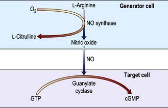
Endothelial cells synthesize a number of mediators which contribute to the regulation of blood vessel tone and the fluidity of the blood; these include nitric oxide, prostacyclin and endothelin (a potent vasoconstrictor). Nitric oxide (NO) is synthesized from the terminal guanidino-nitrogen atoms of the amino acid L-arginine under the influence of nitric oxide synthase (NOS). NO inhibits platelet aggregation and adhesion and produces vasodilatation by activating guanylate cyclase in the underlying vascular smooth muscle to form cyclic guanosine monophosphate (cGMP) from guanosine triphosphate (GTP) (Fig. 16.18). There are several distinct NOS enzymes.
Constitutive or endothelial NOS (cNOS or eNOS) present in endothelial cells is responsible for the basal release of NO and is involved in the physiological regulation of vascular tone, blood pressure and tissue perfusion.
Neuronal NOS (nNOS). The role of nerves containing nNOS is uncertain but they probably provide neurogenic vasodilator tone. In the central nervous system nNOS may be a regulator of local cerebral blood flow as well as fulfilling a number of other physiological functions, such as the acute modulation of neuronal firing behaviour.
Inducible NOS (iNOS) is induced in vascular endothelial smooth muscle cells and monocytes within 4–18 h of stimulation with endotoxin and certain cytokines, such as TNF. The resulting prolonged increase in NO formation is believed to be a cause of the sustained vasodilatation, hypotension and reduced reactivity to adrenergic agonists (‘vasoplegia’) that characterizes septic shock. This mechanism is also involved in severe prolonged haemorrhage/traumatic shock. The NO generated by macrophages contributes to their role as highly effective killers of intracellular and extracellular pathogens, in part as a consequence of its ability to bind to cytochrome oxidase and inhibit electron transport, but also via the production of the highly reactive radical peroxynitrite.
Redox imbalance
In health, the balance between reducing and oxidizing conditions (redox) is controlled by antioxidants which either prevent radical formation (e.g. transferrin and lactoferrin which bind iron, a catalyst for radical formation) or remove/inactivate reactive oxygen and nitrogen species (e.g. enzymes such as superoxide dismutases, vitamins C and E and sulphydryl group donors such as glutathione). There are also mechanisms to remove and repair oxidatively damaged molecules and in particular to preserve DNA integrity. In severe systemic inflammation the uncontrolled production of oxygen-derived free radicals and reactive nitrogen species, e.g. superoxide (O2•−), hydroxyl radicals (OH•), hydrogen peroxide (H2O2) and peroxynitrite (ONOO−), particularly by activated polymorphonuclear leucocytes, can overwhelm these defensive mechanisms and cause:
lipid and protein peroxidation
increased capillary permeability
impaired mitochondrial respiration
apoptosis (see p. 32) (which may contribute to the organ damage and immune hyporesponsiveness associated with sepsis).
Influence of genetic variation
Individuals vary considerably in their susceptibility to infection, as well as their ability to recover from apparently similar infections, illnesses or traumatic insults. There is evidence to suggest that interindividual variations in susceptibility to, and outcome from, sepsis can be partly explained by genetic variation.
Haemodynamic and microcirculatory changes
The dominant haemodynamic feature of severe sepsis/septic shock is peripheral vascular failure with:
maldistribution of regional blood flow
abnormalities in the microcirculation (Fig. 16.19):
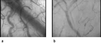
Figure 16.19 Still side-stream dark field (SDF) image of the microcirculation in (a) a normal subject and (b) septic shock.
Although these vascular and microvascular abnormalities may partly account for the reduced oxygen extraction often seen in septic shock, there is also a primary defect of cellular oxygen utilization caused by mitochondrial dysfunction (see above). Initially, before hypovolaemia supervenes, or when therapeutic replacement of the circulating volume has been adequate, cardiac output is usually high and peripheral resistance is low. These changes may be associated with impaired oxygen consumption, a reduced arteriovenous oxygen content difference, an increased  and a lactic acidosis (so-called ‘tissue dysoxia’). Vasodilatation and increased vascular permeability also occur in anaphylactic shock.
In the initial stages of other forms of shock, and sometimes when hypovolaemia and myocardial depression supervene in sepsis and anaphylaxis, cardiac output is low and increased sympathetic activity causes vasoconstriction. This helps to maintain the systemic blood pressure.
Activation of the coagulation system
The inflammatory response to shock, tissue injury and infection is frequently associated with systemic activation of the clotting cascade, leading to platelet aggregation, widespread microvascular thrombosis and inadequate tissue perfusion.
Initially the production of PGI2 by the capillary endothelium is impaired. Cell damage (e.g. to the vascular endothelium) leads to exposure to tissue factor (p. 416), which triggers coagulation. In severe cases these changes are compounded by elevated levels of plasminogen activation inhibitor type 1, which impairs fibrinolysis, as well as by deficiencies in physiological inhibitors of coagulation (including antithrombin, proteins C and S and tissue factor-pathway inhibitor). Antithrombin and protein C have a number of anti-inflammatory properties, whereas thrombin is pro-inflammatory.
Plasminogen is converted to plasmin, which breaks down thrombus, liberating fibrin/fibrinogen degradation products (FDPs). In some cases there is increased fibrinolysis. Circulating levels of FDPs are therefore increased, the thrombin time, PTT and PT are prolonged and platelet and fibrinogen levels fall. Activation of the coagulation cascade can be confirmed by demonstrating increased plasma levels of D-dimers. The development of disseminated intravascular coagulation (DIC) often heralds the onset of multiple organ failure. Because clotting factors and platelets are consumed in DIC, they are unavailable for haemostasis elsewhere and a coagulation defect results – hence the alternative name for DIC is ‘consumption coagulopathy’. DIC presents with microvascular bleeding or generalized ‘oozing’ of blood, e.g. from surgical or traumatic wounds and skin puncture sites. In some cases, a microangiopathic haemolytic anaemia develops. DIC is relatively uncommon but is particularly associated with septic shock, especially when due to meningococcal infection (see p. 127). Management of the underlying cause is most urgent. Supportive treatment may include infusions of fresh frozen plasma, platelets, cryoprecipitate when fibrinogen levels are low and occasionally factor VIII concentrates.
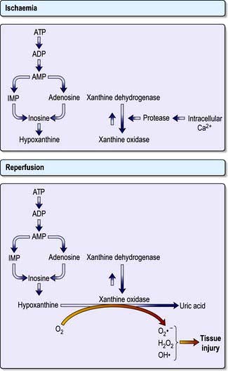
Reperfusion injury
Restoration of flow to previously hypoxic tissues can exacerbate cell damage through the generation of large quantities of reactive oxygen species and activation of polymorphonuclear leucocytes (see above) (Fig. 16.20). The gut mucosa seems to be especially vulnerable to this ‘ischaemia-reperfusion injury’.
Metabolic response to trauma, major surgery and severe infection
This is initiated and controlled by the neuroendocrine system and various cytokines (e.g. IL-6) acting in concert, and is characterized initially by an increase in energy expenditure (‘hypermetabolism’) (see also p. 201). Gluconeogenesis is stimulated by increased glucagon and catecholamine levels, while hepatic mobilization of glucose from glycogen is increased. Catecholamines inhibit insulin release and reduce peripheral glucose uptake. Combined with elevated circulating levels of other insulin antagonists such as cortisol, and downregulation of insulin receptors, these changes mean that the majority of patients are hyperglycaemic (‘insulin resistance’). Later hypoglycaemia may be precipitated by depletion of hepatic glycogen stores and inhibition of gluconeogenesis. Free fatty acid synthesis is also increased, leading to hypertriglyceridaemia.
Protein breakdown is initiated to provide energy from amino acids, and hepatic protein synthesis is preferentially augmented to produce the ‘acute phase reactants’. The amino acid glutamine (which is indispensable in this situation) is mobilized from muscle for use as a metabolic fuel in rapidly dividing cells such as leucocytes and enterocytes. Glutamine is also required for hepatic production of the free radical scavenger glutathione. When severe and prolonged, this catabolic response can lead to considerable weight loss. Protein breakdown is associated with wasting and weakness of skeletal and respiratory muscle, prolonging the need for mechanical ventilation and delaying mobilization. Tissue repair, wound healing and immune function also are compromised.
Clinical features of shock and sepsis
Although many clinical features are common to all types of shock, there are certain aspects in which they differ (Box 16.2).
Box 16.2
Haemodynamic changes in shock
Hypovolaemic shock
Extreme hypovolaemia may be associated with bradycardia.
Additional clinical features may occur in the following types of shock.
Cardiogenic shock (see p. 722)
Signs of myocardial failure, e.g. raised jugular venous pressure (JVP), pulsus alternans, ‘gallop’ rhythm, basal crackles, pulmonary oedema.
Obstructive shock
Pulsus paradoxus and muffled heart sounds in cardiac tamponade
Signs of pulmonary embolism (see p. 764).
Anaphylactic shock (see p. 69)
Signs of profound vasodilatation:
Erythema, urticaria, angio-oedema, pallor, cyanosis
Oedema of the face, pharynx and larynx
Ideally, 10 mL of clotted blood should be taken within 45–60 minutes after the reaction for confirmation of the diagnosis, e.g. by measurement of mast cell tryptase. Serum should be separated and stored at −20°C. Follow-up of these patients is essential.
Sepsis, severe sepsis and septic shock
Pyrexia and rigors, or hypothermia (unusual)
Vasodilatation, warm peripheries
The diagnosis of sepsis is easily missed, particularly in the elderly when the classical signs may not be present. Mild confusion, tachycardia and tachypnoea may be the only clues, sometimes associated with unexplained hypotension, a reduction in urine output, a rising plasma creatinine and glucose intolerance.
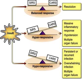
The clinical signs of sepsis (triggered by PAMPS) are not always associated with bacteraemia and can occur with non-infectious processes such as pancreatitis, cardiopulmonary bypass or severe trauma (triggered by DAMPS). The term ‘systemic inflammatory response syndrome’ (SIRS) describes the disseminated inflammation that can complicate this diverse range of disorders (Box 16.3). Patterns of systemic inflammatory response are shown in Figure 16.21, which illustrates the pro-inflammatory response (SIRS) and the counter-regulatory anti-inflammatory response syndrome (CARS).
Box 16.3
Terminology used in systemic inflammation and sepsis
Systemic inflammatory response syndrome (SIRS)
The systemic inflammatory response to a variety of severe clinical insults. The response is manifested by two or more of the following:
Severe sepsis
Sepsis associated with organ dysfunction, hypoperfusion or hypotension. Hypoperfusion and perfusion abnormalities include, but are not limited to, lactic acidosis, oliguria or an acute alteration in mental state.
Septic shock
Severe sepsis with hypotension (systolic BP <90 mmHg or a reduction of >40 mmHg from baseline) in the absence of other causes for hypotension and despite adequate fluid resuscitation.
(Patients receiving inotropic or vasopressor agents may not be hypotensive when perfusion abnormalities are documented.)
Sepsis and multiple organ failure (MOF) (also known as multiple organ dysfunction syndrome, MODS)
Sepsis is being diagnosed with increasing frequency and is now the commonest cause of death in non-coronary adult intensive care units. The estimated incidence of severe sepsis has varied from 77 to 300 cases per 100 000 of the population. Mortality rates are high (between 20% and 60%) and are closely related to the severity of illness and the number of organs that fail. Those who die are overwhelmed by persistent or recurrent sepsis, with fever, intractable hypotension and failure of several organs (Fig. 16.22).
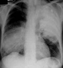
Figure 16.22 Bilateral pneumococcal pneumonia. Community acquired pneumonia is the commonest cause of sepsis requiring admission to intensive care.
(From Hinds CJ, Watson JD. Intensive Care: A Concise Textbook, 3rd edn. Edinburgh: Saunders; 2008. Courtesy of Dr SPG Padley.)
Sequential failure of vital organs occurs progressively over weeks, although the pattern of organ dysfunction is variable. In most cases the lungs are the first to be affected (acute lung injury, ALI; acute respiratory distress syndrome, ARDS; see below) in association with cardiovascular instability and deteriorating renal function. Damage to the mucosal lining of the gastrointestinal tract, as a result of reduced splanchnic flow followed by reperfusion, allows bacteria within the gut lumen, or their cell wall components, to gain access to the circulation. The liver defences, which are often compromised by poor perfusion, are overwhelmed and the lungs and other organs are exposed to bacterial toxins and inflammatory mediators released by liver macrophages. Some have therefore called the gut the ‘motor of multiple organ failure’. Secondary pulmonary infection, complicating ALI/ARDS, also frequently acts as a further stimulus to the inflammatory response. Later, kidney injury and liver dysfunction develop (see p. 884). Gastrointestinal failure, with an inability to tolerate enteral feeding and paralytic ileus, is common. Ischaemic colitis, acalculous cholecystitis, pancreatitis and gastrointestinal haemorrhage may also occur. Features of central nervous system dysfunction include impaired consciousness and disorientation, progressing to coma. Characteristically, these patients initially have a hyperdynamic circulation with vasodilatation and a high cardiac output, associated with an increased metabolic rate. Eventually, however, cardiovascular collapse supervenes. It is now often possible to support such patients for weeks or months; many now die following a decision to withdraw or not to escalate treatment (see p. 897).
Acute lung injury/acute respiratory distress syndrome
Definition and causes (Table 16.4)
Acute lung injury (ALI) and the more severe acute respiratory distress syndrome (ARDS) are diagnosed in an appropriate clinical setting with one or more recognized risk factors. ALI/ARDS can be defined as follows:
Stiff lungs (reduced pulmonary compliance resulting in high inflation pressures)
Chest radiograph: new bilateral, diffuse, patchy or homogeneous pulmonary infiltrates
Cardiac: no apparent cardiogenic cause of pulmonary oedema (pulmonary artery occlusion pressure <18 mmHg if measured or no clinical evidence of left atrial hypertension)
Gas exchange abnormalities: ALI – arterial oxygen tension/fractional inspired oxygen (PaO2/FIO2) ratio <40 kPa (<300 mmHg); ARDS – PaO2/FIO2 <26.6 kPa (<200 mmHg) (in both cases, despite normal arterial carbon dioxide tension and regardless of positive end-expiratory pressure). The criterion for arterial oxygen tension/fractional inspired oxygen is arbitrary and the value of differentiating ALI from ARDS has been questioned.
Table 16.4 Disorders associated with acute respiratory distress syndrome
| Direct lung injury | Indirect lung injury |
|---|---|
Common causes |
|
Less common causes |
|
ALI/ARDS can occur as a nonspecific reaction of the lungs to a wide variety of direct pulmonary and indirect non-pulmonary insults. By far the commonest predisposing factor is sepsis, and 20–40% of patients with severe sepsis will develop ALI/ARDS (Table 16.4).
Pathogenesis and pathophysiology of ALI/ARDS
Acute lung injury can be viewed as an early manifestation of a generalized inflammatory response with endothelial dysfunction and is therefore frequently associated with the development of multiple organ dysfunction syndrome (MODS) (see p. 882).
Non-cardiogenic pulmonary oedema
This is the cardinal feature of ALI and is the first and clinically most evident sign of a generalized increase in vascular permeability caused by the microcirculatory changes and release of inflammatory mediators described previously (see p. 877), with activated neutrophils playing a particularly key role. The pulmonary epithelium is also damaged in the early stages, reducing surfactant production and lowering the threshold for alveolar flooding.
Pulmonary hypertension
Pulmonary hypertension sometimes complicated by right ventricular failure (p. 762) is a common feature of ALI/ARDS. Initially, mechanical obstruction of the pulmonary circulation may occur as a result of vascular compression by interstitial oedema, while local activation of the coagulation cascade leads to thrombosis and obstruction in the pulmonary microvasculature. Later, pulmonary vasoconstriction may develop in response to increased autonomic nervous activity and circulating substances such as catecholamines, serotonin, thromboxane and complement. Those vessels supplying alveoli with low oxygen tensions constrict (the ‘hypoxic vasoconstrictor response’), diverting pulmonary blood flow to better oxygenated areas of lung, thus limiting the degree of shunt.
Haemorrhagic intra-alveolar exudate
This exudate is rich in platelets, fibrin, fibrinogen and clotting factors and may inactivate surfactant and stimulate inflammation, as well as promoting hyaline membrane formation and the migration of fibroblasts into the air spaces.
Resolution, fibrosis and repair
Within days of the onset of lung injury, formation of a new epithelial lining is underway and activated fibroblasts accumulate in the interstitial spaces. Subsequently, interstitial fibrosis progresses, with loss of elastic tissue and obliteration of the lung vasculature, together with lung destruction and emphysema. In those who recover, the lungs are substantially remodelled.
Physiological changes
Shunt and deadspace increase, compliance falls, and there is evidence of airflow limitation. Although the lungs in ALI and ARDS are diffusely injured, the pulmonary lesions, when identified as densities on a CT scan, are predominantly located in dependent regions (Fig. 16.23). This is partly explained by the effects of gravity on the distribution of extravascular lung water and areas of lung collapse. Pleural effusions are common.
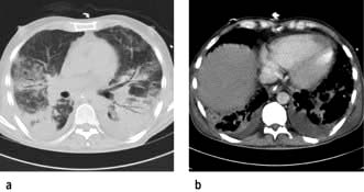
Figure 16.23 Acute respiratory distress syndrome. (a) Lung computed tomography scan showing ground-glass opacification in non-dependent regions with atelectasis and consolidation in dependent regions. There are small pleural effusions. (b) Same patient as shown in (a) using soft-tissue window settings to demonstrate small bilateral effusions layering in the dependent region of both hemithoraces.
(From Hinds CJ, Watson JD. Intensive Care: A Concise Textbook, 3rd edn. Edinburgh: Saunders; 2008, with permission. Courtesy of Dr SPG Padley.)
Clinical presentation of ali/ards
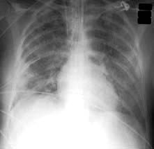
The first sign of the development of ALI/ARDS is often an unexplained tachypnoea, followed by increasing hypoxaemia, with central cyanosis, and breathlessness. Fine crackles are heard throughout both lung fields. Later, the chest X-ray shows bilateral diffuse shadowing, interstitial at first, but subsequently with an alveolar pattern and air bronchograms (Fig. 16.24). The differential diagnosis includes cardiac failure and lung fibrosis.
Management of ali/ards
This is based on treatment of the underlying condition (e.g. eradication of sepsis), supportive measures and avoidance of complications such as ventilator-associated pneumonia.
Mechanical ventilation
Strategies designed to minimize ventilator-associated lung injury and encourage lung healing should be used (see p. 895).
Pulmonary oedema limitation. Pulmonary oedema formation should be limited by minimizing left ventricular filling pressure with fluid restriction, the use of diuretics and, if these measures fail, preventing fluid overload by haemofiltration. The aim should be to achieve a consistently negative fluid balance. Cardiovascular support and the reduction of oxygen requirements are also necessary.
Prone position. When the patient is changed from the supine to the prone position, lung densities in the dependent region are redistributed and shunt fraction is reduced. More uniform alveolar ventilation, caudal movement of the diaphragm, redistribution of perfusion and recruitment of collapsed alveoli all contribute to the improvement in gas exchange. Body position changes can be achieved with minimal complications despite the presence of multiple indwelling vascular lines. Repeated position changes between prone and supine allow reductions in airway pressures and the inspired oxygen fraction. The response to prone positioning is, however, variable and it seems that this strategy does not improve overall outcome (and perhaps therefore should be reserved for those with severe refractory hypoxaemia).
Inhaled nitric oxide. This vasodilator, when inhaled, may improve  matching by increasing perfusion of ventilated lung units, as well as reducing pulmonary hypertension. It has been shown to improve oxygenation in so-called ‘responders’ with ALI/ARDS but has not been shown to increase survival. Its administration requires specialized monitoring equipment, as products of its combination with oxygen include toxic nitrogen dioxide.
Aerosolized prostacyclin. This appears to have similar effects to inhaled NO and is easier to monitor and deliver. As with inhaled NO, the response to aerosolized prostacyclin is, however, variable and although it has been shown to improve oxygenation its effect on outcome has yet to be established.
Aerosolized surfactant. Surfactant replacement therapy reduces morbidity and mortality in neonatal respiratory distress syndrome and is beneficial in animal models of ALI/ARDS. In adults with ARDS, however, the value of surfactant administration remains uncertain.
Steroids. Administration of steroids to patients with persistent ALI/ARDS does not appear to improve outcome.
Prognosis
Mortality from ALI/ARDS has fallen over the last decade, from around 60% to between 30% and 40%, perhaps as a consequence of improved general care, the increasing use of management protocols, and attention to infection control and nutrition, as well as the introduction of novel treatments and lung-protective strategies for respiratory support. Prognosis is, however, still very dependent on aetiology. When ARDS occurs in association with intra-abdominal sepsis, mortality rates remain very high, whereas much lower mortality rates are to be expected in those with ‘primary’ ARDS (pneumonia, aspiration, lung contusion). Mortality rises with increasing age and failure of other organs. Most of those dying with ARDS do so as a result of MODS and haemodynamic instability rather than impaired gas exchange.
FURTHER READING
Hall JP, Kress JP. The burden of functional recovery from ARDS. N Engl J Med 2011; 364:1360–1366.
Herridge MS et al. Functional disability 5 years after acute respiratory distress syndrome. N Engl J Med 2011; 364:1293–1304.
The RENAL Replacement Therapy Study Investigators. Intensity of continuous renal-replacement therapy in critically ill patients. N Engl J Med 2009; 361:1627–1638.
Wheeler AP, Bernard GR. Acute lung injury and the acute respiratory distress syndrome: a clinical review. Lancet 2007; 369:1556–1565.
Acute kidney injury
Acute kidney injury (AKI) is a common and serious complication of critical illness which adversely affects the prognosis. A modification of the RIFLE classification has been proposed and covers the spectrum of severity and consequences of acute kidney injury (see Ch. 12; Box 12.5).
The importance of preventing renal injury by rapid and effective resuscitation, as well as the avoidance of nephrotoxic drugs (especially NSAIDs), and control of infection cannot be overemphasized. Shock and sepsis are the most common causes of AKI in the critically ill, but diagnosis of the cause of renal dysfunction is necessary to exclude reversible pathology, especially obstruction (see Ch. 12).
Oliguria is usually the first indication of renal impairment and immediate attempts should be made to optimize cardiovascular function, particularly by expanding the circulating volume and restoring blood pressure. Restoration of the urine output is a good indicator of successful resuscitation. Evidence now suggests that dopamine is not an effective means of preventing or reversing renal impairment and this agent should not be used for renal protection in sepsis (p. 888). If these measures fail to reverse oliguria, administration of diuretics such as furosemide by bolus or infusion, or less often mannitol (for example in rhabdomyolysis) may be indicated (see Ch. 12). If oliguria persists, it is necessary to reduce fluid intake and review drug doses.
Intermittent haemodialysis has a number of theoretical disadvantages in the critically ill. In particular, it is frequently complicated by hypotension and it may be difficult to remove sufficient volumes of fluid. Nevertheless, provided that strict guidelines are used to improve tolerance and metabolic control, almost all patients with acute kidney injury can be managed successfully with daily haemodialysis. The use of continuous veno-venous haemofiltration, often with dialysis (CVVHD), is however generally preferred in the critically ill (see Ch. 12) and is indicated for fluid overload, electrolyte disturbances (especially hyperkalaemia), severe acidosis and, less often uraemia. The intensity of renal replacement therapy does not seem to influence outcome. Peritoneal dialysis is unsatisfactory in critically ill patients and is contraindicated in those who have undergone intra-abdominal surgery.
If the underlying problems resolve, renal function almost invariably recovers a few days to several weeks later.
Management of shock and sepsis (Fig. 16.25)
Delays in making the diagnosis and in initiating treatment (especially antibiotics), as well as inadequate resuscitation, are associated with increased morbidity and mortality and should be avoided.
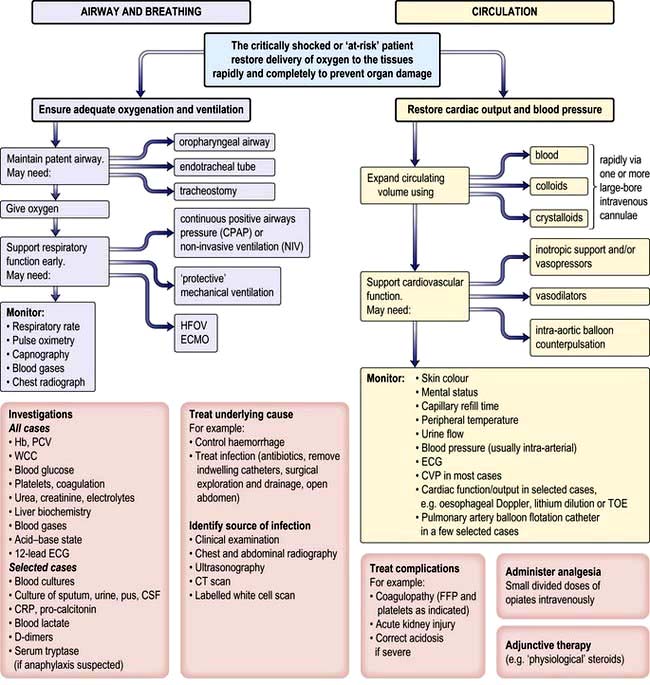
Figure 16.25 Management of the critically ill, shocked or ‘at-risk’ patient. CSF, cerebrospinal fluid; CT, computed tomography; CVP, central venous pressure; FFP, fresh frozen plasma. ECMO, extracorporeal membrane oxygenation; HFOV, high frequency oscillatory ventilation. TOE, Transoesophageal echocardiography.
A patent airway must be maintained and oxygen must be given. If necessary, an oropharyngeal airway or an endotracheal tube is inserted. The latter has the advantage of preventing aspiration of gastric contents. Very rarely, emergency tracheostomy is indicated (see below). Some patients may require immediate mechanical ventilation.
The underlying cause of shock should be corrected, e.g. haemorrhage should be controlled or infection eradicated. In patients with septic shock, every effort must be made to identify the source of infection and isolate the causative organism. As well as a thorough history and clinical examination, X-rays, ultrasonography or CT scanning may be required to locate the origin of the infection. Appropriate samples (urine, sputum, cerebrospinal fluid, pus drained from abscesses) should be sent to the laboratory for microscopy, culture and sensitivities. Several blood cultures should be performed and empirical, broad-spectrum antibiotic therapy (p. 85) should be commenced within the first hour of recognition of sepsis. If an organism is isolated later, therapy can be adjusted appropriately. The choice of antibiotic depends on the likely source of infection, previous antibiotic therapy and known local resistance patterns, as well as on whether infection was acquired in hospital or in the community. Abscesses must be drained and infected indwelling catheters removed.
Whatever the aetiology of the haemodynamic abnormality, tissue blood flow must be restored by achieving and maintaining an adequate cardiac output, as well as ensuring that arterial blood pressure is sufficient to maintain perfusion of vital organs. Published guidelines for adult patients suffering severe sepsis or septic shock advocate targeting an MAP >65 mmHg, CVP 8–12 mmHg (>12 mmHg if mechanically ventilated), urine output >0.5 mL/kg per hour and an  ≥70% (or
  ≥65%) as the initial goals of resuscitation. Administration of fluids can also be targeted at abolishing arterial pressure variation and/or optimizing stroke volume.
Preload and volume replacement
Optimizing preload is the most efficient way of increasing cardiac output. Volume replacement is obviously essential in hypovolaemic shock but is also required in anaphylactic and septic shock because of vasodilatation, sequestration of blood and loss of circulating volume because of vascular leak.
In obstructive shock, high filling pressures may be required to maintain an adequate stroke volume. Even in cardiogenic shock, careful volume expansion may, on occasions, lead to a useful increase in cardiac output. On the other hand, patients with severe cardiac failure, in whom ventricular filling pressures are markedly elevated, often benefit from measures to reduce preload (and afterload) – such as the administration of vasodilators and diuretics (see below). Adequate perioperative volume replacement also reduces morbidity and mortality in high-risk surgical patients.
The circulating volume must be replaced quickly (in minutes not hours) to reduce tissue damage and prevent acute kidney injury. Fluid is administered via wide-bore intravenous cannulae to allow large volumes to be given quickly, and the effect is continuously monitored.
You must prevent volume overload, which leads to cardiac dilatation, a reduction in stroke volume, and a rise in left atrial pressure with a risk of pulmonary oedema. Pulmonary oedema is more likely in seriously ill patients because of a low colloid osmotic pressure (usually due to a low serum albumin) and disruption of the alveolar–capillary membrane (e.g. in acute lung injury).
Choice of fluid for volume replacement
Blood
This is conventionally given for haemorrhagic shock as soon as it is available. In extreme emergencies, group-specific crossmatch can be performed in minutes (see p. 408). When available and not contraindicated, blood salvage should be employed for those with severe on-going bleeding.
Although red cell transfusion will augment oxygen-carrying capacity, and hence global oxygen delivery, tissue oxygenation is also dependent on microcirculatory flow. This is influenced by the viscosity of the blood and hence the packed cell volume (PCV). Conventionally, a PCV of 30–35% has been considered to provide the optimal balance between oxygen-carrying capacity and tissue flow, although it is well recognized that previously fit people with haemorrhagic shock can tolerate extremely low Hb concentrations, provided their circulating volume and cardiac output are maintained. Transfusion of old stored red cells, which become spherical rather than biconcave and poorly deformable, with increased adhesiveness, can compromise microvascular flow and worsen tissue hypoxia. Whole blood has now been largely replaced by red cell concentrates (see p. 412)
Massive blood transfusion can be defined as a volume of >8–10 units of red cells transfused within a 24-hour period, and massive haemorrhage as a loss of 50% of blood volume within 3 hours or a rate of blood loss exceeding 150 mL/min.
Complications of blood transfusion are discussed on page 408.
Special problems arise as a result of massive transfusion:
Temperature changes. Bank blood is stored at 4°C; transfusion may result in hypothermia, peripheral venoconstriction (which slows the rate of the infusion) and arrhythmias. If possible, blood should be warmed during massive transfusion and in those at risk of hypothermia (e.g. during prolonged major surgery with open body cavity).
Coagulopathy. Stored blood has virtually no effective platelets or clotting factors. Massive transfusions that often include large volumes of colloid/crystalloid can therefore be associated with a coagulopathy. This often needs to be treated by replacing clotting factors with fresh frozen plasma and administering platelet concentrates. Occasionally cryoprecipitate is required. There is some evidence that a higher ratio of FFP to blood transfused is associated with improved survival, especially in the military trauma setting. Recombinant factor VIIa may occasionally be indicated in those with uncontrollable bleeding, although the safety of this product has been questioned. Prothrombin complex concentrates have some advantages compared with FFP, in that they do not need to be crossmatched or thawed.
Hypocalcaemia. Citrate in stored blood binds calcium ions. During rapid transfusion total body ionized calcium levels may be reduced, causing myocardial depression and exacerbating coagulation defects. This is uncommon in practice but can be corrected by administering 10 mL of 10% calcium chloride intravenously. Routine treatment with calcium is not recommended.
Increased oxygen affinity. In stored blood, the red cell 2,3-disphosphoglycerate (2,3-DPG) content is reduced, so that the oxyhaemoglobin dissociation curve is shifted to the left. The oxygen affinity of haemoglobin is therefore increased and oxygen unloading is impaired. Red cell levels of 2,3-DPG are substantially restored within 12 h of transfusion.
Hyperkalaemia. Plasma potassium levels rise progressively as blood is stored. However, hyperkalaemia is rarely a problem as rewarming of the blood increases red cell metabolism – the sodium pump becomes active and potassium levels fall.
Microembolism. Microaggregates in stored blood may be filtered out by the pulmonary capillaries. This process is thought by some to contribute to ALI.
Concern about the supply, cost and safety of blood, including the risk of disease transmission and immune suppression, has encouraged a more conservative approach to transfusion. There is some evidence to suggest that in normovolaemic critically ill patients a restrictive strategy of red cell transfusion (Hb maintained at >70 g/L) is at least as effective, and may be safer than a liberal transfusion strategy (Hb maintained at 100–120 g/L). However, in some groups of patients (e.g. the elderly and those with significant cardiac or respiratory disease and patients who are actively bleeding) it is preferable to maintain Hb closer to the higher level. The use of leucodepleted blood is considered to be safer in terms of disease transmission and immune suppression.
Crystalloids and colloids
The choice of intravenous fluid for resuscitation and the relative merits of crystalloids or colloids has long been controversial. Crystalloid solutions such as Hartmann’s solution are cheap, convenient to use and free of side-effects.
It has been generally accepted that volumes of crystalloid several times that of colloid are required to achieve an equivalent haemodynamic response and that colloidal solutions produce a greater and more sustained increase in circulating volume, with associated improvements in cardiovascular function and oxygen transport. This traditional view has been challenged, however, and a large, prospective, randomized, controlled trial has demonstrated that in a heterogeneous group of critically ill patients the use of either physiological saline or 4% albumin for fluid resuscitation resulted in similar outcomes.
Polygelatin solutions have an average molecular weight of 35 000, which is iso-osmotic with plasma. They are cheap and do not interfere with crossmatching. Large volumes can be administered, as clinically significant coagulation defects are unusual and renal function is not impaired. However, because they readily cross the glomerular basement membrane, their half-life in the circulation is only approximately 4 h and they can promote an osmotic diuresis. These solutions are useful during the acute phase of resuscitation, especially when volume losses are continuing. Allergic reactions can, however, occur.
Hydroxyethyl starches (HES). Numerous preparations are now available, characterized by their concentrations (3%, 6%, 10%) and low, medium or high molecular weight. The half-life of high and medium molecular weight solutions is between 12 and 24 h, while that of the low-molecular-weight solutions is 4–6 h. Elimination of HES occurs primarily via the kidneys following hydrolysis by amylase. HES are stored in the reticuloendothelial system, apparently without causing functional impairment, but skin deposits have been associated with persistent pruritus. HES, especially the higher-molecular-weight fractions, have anticoagulant properties and many therefore recommend limiting the volume administered. HES have been implicated in the development of acute kidney injury.
Human albumin solution (HAS) is a natural colloid which has been used for volume replacement in shock and burns, and for the treatment of hypoproteinaemia. HAS is not generally recommended for routine volume replacement, because supplies are limited and other cheaper solutions are equally effective. Some use HAS to expand the circulating volume in patients who are hypoalbuminaemic. There is some suggestion that the administration of HAS may improve outcome from sepsis.
Myocardial contractility and inotropic agents
Myocardial contractility can be impaired by many factors such as hypoxaemia and hypocalcaemia, as well as by some drugs (e.g. beta-blockers, antiarrhythmics and sedatives).
Severe lactic acidosis conventionally is said to depress myocardial contractility and limit the response to vasopressor agents. Attempted correction of acidosis with intravenous sodium bicarbonate, however, generates additional carbon dioxide which diffuses across cell membranes, producing or exacerbating intracellular acidosis. Other disadvantages of bicarbonate therapy include sodium overload and a left shift of the oxyhaemoglobin dissociation curve. Ionized calcium levels may be reduced and, combined with the fall in intracellular pH, this may impair myocardial performance. Treatment of lactic acidosis should therefore concentrate on correcting the cause. Bicarbonate should only be administered to correct extreme persistent metabolic acidosis (see Chapter 13).
If the signs of shock persist despite adequate volume replacement, and perfusion of vital organs is jeopardized, pressor agents should be administered to improve cardiac output and blood pressure. Vasopressor therapy may also be required to maintain perfusion in those with life-threatening hypotension, even when volume replacement is incomplete. All inotropes increase myocardial oxygen consumption, particularly if a tachycardia develops, and this can lead to an imbalance between myocardial oxygen supply and demand, with the development or extension of ischaemic areas. Inotropes should therefore be used with especial caution, particularly in cardiogenic shock following myocardial infarction and in those known to have ischaemic heart disease.
Many of the most seriously ill patients become increasingly resistant to the effects of pressor agents, an observation attributed to ‘downregulation’ of adrenergic receptors and NO-induced ‘vasoplegia’ (p. 879).
All inotropic agents should be administered via a large central vein, and their effects continually monitored (Table 16.5).
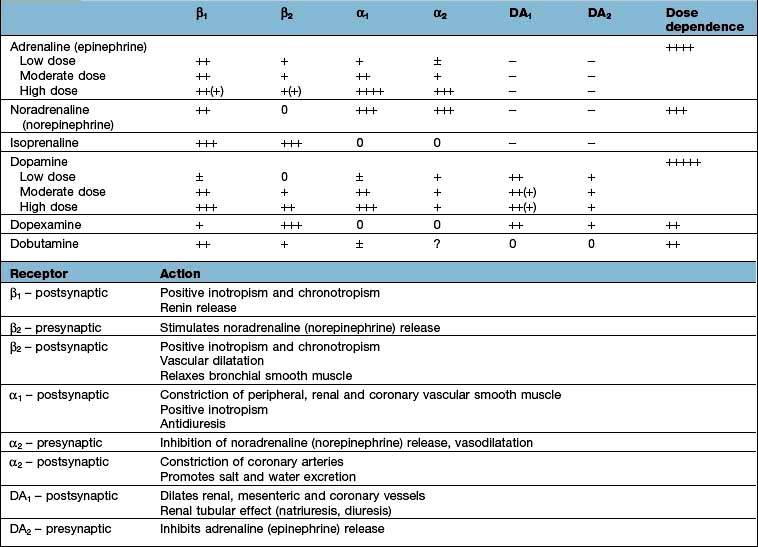
Adrenaline (epinephrine)
Adrenaline stimulates both α- and β-adrenergic receptors, but at low doses, β effects seem to predominate. Heart rate and cardiac index increase, while peripheral resistance is reduced. If there is an associated increase in perfusion pressure, urine output may improve. Adrenaline at higher doses can cause excessive (α-mediated) vasoconstriction, with reductions in splanchnic flow, and cardiac output may fall. Prolonged high-dose administration can cause peripheral gangrene and lactic acidosis. The minimum effective dose of adrenaline should therefore be used for as short a time as possible.
Noradrenaline (norepinephrine)
This is predominantly an α-adrenergic agonist. It is particularly useful in those with hypotension associated with a low systemic vascular resistance, e.g. in septic shock. There is a risk of producing excessive vasoconstriction with impaired organ perfusion and increased afterload. Noradrenaline administration should normally therefore be guided by comprehensive haemodynamic monitoring, including invasive or non-invasive determination of cardiac output (see p. 875) and calculation of systemic vascular resistance.
FURTHER READING
Annane D, Vignon P, Renault A et al. Norepinephrine plus dobutamine versus epinephrine alone for management of septic shock: a randomized trial. Lancet 2007; 370:676–684.
De Backer D, Biston P, Devriendt J et al. Comparison of dopamine and norepinephrine in the treatment of shock. N Engl J Med 2010; 362:779–789.
Russell JA, Walley KR, Singer J et al. Vasopressin versus norepinephrine infusion in patients with septic shock. N Engl J Med 2008; 358:877–887.
Dopamine
Dopamine is a natural precursor of adrenaline (epinephrine) which acts on β receptors and α receptors, as well as dopaminergic DA1 and DA2 receptors.
In low doses (e.g. 1–3 µg/kg per min), dopaminergic vasodilatory receptors in the renal, mesenteric, cerebral and coronary circulations are activated. DA1 receptors are located on postsynaptic membranes and mediate vasodilatation, while DA2 receptors are presynaptic and potentiate these vasodilatory effects by preventing the release of adrenaline (epinephrine). Renal and hepatic flow increase and urine output is improved. The significance of the renal vasodilator effect of dopamine has, however, been questioned and it has been suggested that the increased urine output is largely attributable to the rise in cardiac output and blood pressure, combined with a decrease in aldosterone and inhibition of tubular sodium reabsorption mediated via DA1 stimulation.
In moderate doses (e.g. 3–10 µg/kg per min), dopamine increases heart rate, myocardial contractility and cardiac output. In some patients the dose of dopamine is limited by β-receptor effects such as tachycardia and arrhythmias.
In higher doses (e.g. >10 µg/kg per min) the increased noradrenaline (norepinephrine) produced is associated with vasoconstriction. This increases afterload and raises ventricular filling pressures.
Dopexamine
Dopexamine is an analogue of dopamine which activates β2 receptors as well as DA1 and DA2 receptors. Dopexamine is a weak positive inotrope, but is a powerful splanchnic vasodilator, reducing afterload and improving blood flow to vital organs, including the kidneys. Dopexamine has been used as an adjunct to the perioperative management of high-risk surgical patients (see below).
Dobutamine
Dobutamine is closely related to dopamine and has predominantly β1 activity. Dobutamine has no specific effect on the renal vasculature but urine output often increases as cardiac output and blood pressure improve. It reduces systemic vascular resistance, as well as improving cardiac performance, thereby decreasing afterload and ventricular filling pressures. Dobutamine is therefore useful in patients with cardiogenic shock and cardiac failure. In septic shock, dobutamine can be used to increase cardiac output and oxygen delivery.
Phosphodiesterase inhibitors (e.g. milrinone, enoximone)
These agents have both inotropic and vasodilator properties. Because the phosphodiesterase type III inhibitors bypass the β-adrenergic receptor they do not cause tachycardia and are less arrhythmogenic than β agonists. They are useful in patients with receptor ‘downregulation’, those receiving beta-blockers, for weaning patients from cardiopulmonary bypass and for patients with cardiac failure. In vasodilated septic patients, however, they can precipitate or worsen hypotension.
Vasopressin
Patients with septic shock have inappropriately low circulating levels of vasopressin. Low-dose vasopressin can increase blood pressure and systemic vascular resistance in patients with vasodilatory septic shock and a high cardiac output unresponsive to other vasopressors (‘vasoplegia’). A 2008 randomized controlled trial suggests that low-dose vasopressin added to conventional vasopressors may have some value in less severe septic shock.
Levosimendan
Levosimendan is a myofilament calcium sensitizer and novel inotrope. Unlike other inotropes, levosimendan does not exert its action through increases in intracellular Ca2+ and as a result does not impair diastolic relaxation of the heart. Levosimendan binds to troponin C with high affinity but only during systole when the intracellular calcium concentration is high. Levosimendan has phosphodiesterase inhibitor actions but these are not thought to be clinically significant. Significantly, a long-acting metabolite of levosimendan has similar calcium sensitizing actions, maintaining the inotropic effect of levosimendan once an infusion is stopped. Adverse cardiovascular effects of levosimendan include tachycardia and hypotension; as a consequence the addition of a vasopressor may be required.
Guidelines for use of inotropic and vasopressor agents
Some still consider dopamine in low to moderate doses to be the first-line agent for restoring blood pressure, although evidence suggests that dopamine is associated with a greater number of adverse events (especially arrhythmias) than norepinephrine. Certainly high-dose dopamine is best avoided. Dobutamine is particularly indicated in patients in whom the vasoconstriction caused by dopamine could be dangerous (i.e. patients with cardiac disease and septic patients with fluid overload or myocardial failure). The combination of dobutamine and noradrenaline (norepinephrine) is popular for the management of patients who are shocked with a low systemic vascular resistance (e.g. septic shock). Dobutamine is given to achieve an optimal cardiac output, while noradrenaline (norepinephrine) is used to restore an adequate blood pressure by reducing vasodilatation. In vasodilated septic patients with a high cardiac output, noradrenaline (norepinephrine) can be used alone. At time of writing, there is evidence to suggest that adrenaline (epinephrine) may be equally safe and effective as a dobutamine/noradrenaline combination. Because of its potency, adrenaline is particularly useful in patients with refractory hypotension. The role of levosimendan in the management of shock has yet to be established.
Targeting haemodynamics and oxygen transport
Although resuscitation has conventionally aimed at achieving normal haemodynamics, survival of many critically ill patients is associated with raised values for cardiac output, DO2 and VO2. However, elevation of DO2 and VO2 to these ‘supranormal’ levels following admission to intensive care produces no benefit and may be harmful. By contrast, early goal-directed therapy to resuscitate patients in the emergency room, aimed at maintaining a central venous oxygen saturation of more than 70%, significantly improves outcome in patients with severe sepsis or septic shock, as does therapy targeted at lactate clearance.
High-risk surgical patients (Box 16.4)
These patients benefit from intensive perioperative monitoring and circulatory support, in particular maintenance of an adequate circulating volume, and postoperative admission to a critical care area. Volume replacement and administration of inotropes or vasopressors should be guided by monitoring of stroke volume/cardiac output, usually using an oesophageal Doppler or pulse contour analysis. The value of the routine use of inodilators such as dopexamine remains unclear.
Box 16.4
Patients at risk of developing perioperative multiorgan failure
Patients with co-morbidity, especially limited cardiorespiratory reserve
Patients with trauma to two body cavities requiring multiple blood transfusions
Patients undergoing surgery involving extensive tissue dissection, e.g. oesophagectomy, pancreatectomy, aortic aneurysm surgery
Patients undergoing emergency surgery for intra-abdominal or intrathoracic catastrophic states, e.g. faecal peritonitis, oesophageal perforation.
Modified from Shoemaker WC, Appel PL, Kram HB et al. Prospective trial of supranormal values of survivors as therapeutic goals in high-risk surgical patients. Chest 1988; 94:1176–1186.
Vasodilator therapy
In selected cases, afterload reduction is used to increase stroke volume and decrease myocardial oxygen requirements by reducing the systolic ventricular wall tension. Vasodilatation (see p. 720) also decreases heart size and the diastolic ventricular wall tension so that coronary blood flow is improved. The relative magnitude of the falls in preload and afterload depends on the pre-existing haemodynamic disturbance, concurrent volume replacement and the agent selected (see below). Vasodilators also improve microcirculatory flow.
Vasodilator therapy can be particularly helpful in patients with cardiac failure in whom the ventricular function curve is flat (Fig. 16.6) so that falls in preload have only a limited effect on stroke volume. This form of treatment, combined in selected cases with inotropic support, is therefore useful in cardiogenic shock and in the management of patients with cardiogenic pulmonary oedema or mitral regurgitation.
Nitrate vasodilators are usually used. Nitrates, because of their ability to improve the myocardial oxygen supply/demand ratio, also help to control angina and limit ischaemic myocardial injury.
Sodium nitroprusside (SNP) dilates arterioles and venous capacitance vessels, as well as the pulmonary vasculature by donating nitric oxide. SNP therefore reduces the afterload and preload of both ventricles and can improve cardiac output and the myocardial oxygen supply/demand ratio. The effects of SNP are rapid in onset and spontaneously reversible within a few minutes of discontinuing the infusion. A large overdose of SNP can cause cyanide poisoning, with intracellular hypoxia caused by inhibition of cytochrome oxidase, the terminal enzyme of the respiratory chain. This is manifested as a metabolic acidosis and a fall in the arteriovenous oxygen content difference.
Nitroglycerine (NTG). At low doses, NTG is predominantly a venodilator, but as the dose is increased, it also causes arterial dilatation, thereby decreasing both preload and afterload. Nitrates are particularly useful in the treatment of cardiac failure with pulmonary oedema and are usually used in combination with intravenous furosemide. NTG reduces pulmonary vascular resistance, an effect that can be exploited in patients with a low cardiac output secondary to pulmonary hypertension.
Mechanical support of the myocardium
Intra-aortic balloon counterpulsation (IABCP) is the technique used most widely for mechanical support of the failing myocardium. It is discussed on page 696.
Adjunctive treatment
Initial attempts to combat the high mortality associated with sepsis concentrated on cardiovascular and respiratory support in the hope that survival could be prolonged until surgery, antibiotics and the patient’s own defences had eradicated the infection and injured tissues were repaired. Despite some success, mortality rates remained unacceptably high. So far, attempts to improve outcome by modulating the inflammatory response (including high-dose steroids) or neutralizing endotoxin (Table 16.6) have proved disappointing and in some cases may even have been harmful.
Table 16.6 Some of the therapeutic strategies tested in randomized, controlled phase II or III trials in human sepsis
The administration of relatively low, ‘stress’ doses of hydrocortisone to patients with refractory vasopressor-resistant septic shock may assist shock reversal and perhaps improve outcome. Careful control of the blood sugar level to between 8 and 10 mmol/L is also recommended.
The aim of current sepsis guidelines is to combine these, and other evidence-based interventions, with early effective resuscitation (aimed especially at achieving an adequate circulating volume, combined with the rational use of inotropes and/or vasoactive agents to maintain blood pressure, cardiac output and oxygen transport) in order to create ‘bundles of care’ delivered within specific time limits (see http://www.survivingsepsis.org).
FURTHER READING
Dellinger RP, Levy MM, Carlet JM et al. for the International Surviving Sepsis Campaign Guidelines Committee. Surviving Sepsis Campaign: International Guidelines for Management of Severe Sepsis and Septic Shock. Crit Care Med 2008; 36:296–327.
Sprung CL, Annane D, Keh D et al. for the CORTICUS Study Group. Hydrocortisone therapy for patients with septic shock. N Engl J Med 2008; 358:111–124.
Toussaint S, Gerlach H. Activated protein C for sepsis. N Engl J Med 2009; 361:2640–2652.
Respiratory failure (see Ch. 15)
Types and causes
The respiratory system consists of a gas-exchanging organ (the lungs) and a ventilatory pump (respiratory muscles/thorax), either or both of which can fail and precipitate respiratory failure. Respiratory failure occurs when pulmonary gas exchange is sufficiently impaired to cause hypoxaemia with or without hypercarbia. In practical terms, respiratory failure is present when the PaO2 is <8 kPa (60 mmHg) or the PaCO2 is >7 kPa (55 mmHg). It can be divided into:
Type I respiratory failure, in which the PaO2 is low and the PaCO2 is normal or low
Type II respiratory failure, in which the PaO2 is low and the PaCO2 is high.
Type I or ‘acute hypoxaemic’ respiratory failure occurs with diseases that damage lung tissue. Hypoxaemia is due to right-to-left shunts or  mismatch. Common causes include cardiogenic pulmonary oedema, pneumonia, acute lung injury and lung fibrosis.
Type II or ‘ventilatory failure’ occurs when alveolar ventilation is insufficient to excrete the volume of carbon dioxide being produced by tissue metabolism. Inadequate alveolar ventilation is due to reduced ventilatory effort, inability to overcome an increased resistance to ventilation, failure to compensate for an increase in deadspace and/or carbon dioxide production, or a combination of these factors. The most common cause is chronic obstructive pulmonary disease (COPD). Other causes include chest-wall deformities, respiratory muscle weakness (e.g. Guillain–Barré syndrome) and depression of the respiratory centre (e.g. overdose).
Deterioration in the mechanical properties of the lungs and/or chest wall increases the work of breathing and the oxygen consumption/carbon dioxide production of the respiratory muscles. The concept that respiratory muscle fatigue (either acute or chronic) is a major factor in the pathogenesis of respiratory failure is controversial.
Clinical assessment of respiratory failure
A clinical assessment of respiratory distress should be made on the following criteria (those marked with an asterisk* may be indicative of respiratory muscle fatigue):
the use of accessory muscles of respiration
pulsus paradoxus (rarely present)
inability to speak, unwillingness to lie flat
agitation, restlessness, diminished conscious level
asynchronous respiration (a discrepancy in the timing of movement of the abdominal and thoracic compartments)*
paradoxical respiration (abdominal and thoracic compartments move in opposite directions)*
respiratory alternans (breath-to-breath alteration in the relative contribution of intercostal/accessory muscles and the diaphragm)*.
Blood gas analysis should be performed to guide oxygen therapy and to provide an objective assessment of the severity of the respiratory failure. The most sensitive clinical indicator of increasing respiratory difficulty is a rising respiratory rate. Measurement of tidal volume is a less sensitive indicator.
Vital capacity is often a better guide to deterioration and is particularly useful in patients with respiratory inadequacy that is due to neuromuscular problems such as the Guillain–Barré syndrome or myasthenia gravis, in which the vital capacity decreases as weakness increases.
Monitoring of respiratory failure
Pulse oximetry
Lightweight oximeters can be applied to an ear lobe or finger. They measure the changing amount of light transmitted through the pulsating arterial blood and provide a continuous, non-invasive assessment of arterial oxygen saturation (SpO2). These devices are reliable, easy to use and do not require calibration, although remember that pulse oximetry is not a sensitive guide to changes in oxygenation. An SpO2 within normal limits in a patient receiving supplemental oxygen does not exclude the possibility of hypoventilation with carbon dioxide retention. Readings are occasionally inaccurate in those with poor peripheral perfusion.
Blood gas analysis
Normal values of blood gas analysis are shown in Table 16.2.
Errors can result from malfunctioning of the analyser or incorrect sampling of arterial blood. Disposable preheparinized syringes are available for blood gas analysis.
The sample should be analysed immediately. Alternatively, the syringe should be immersed in iced water (the end having first been sealed with a cap) to prevent the continuing metabolism of white cells causing a reduction in PO2 and a rise in PCO2.
Air almost inevitably enters the sample. The gas tensions within these air bubbles will equilibrate with those in the blood, thereby lowering the PCO2 and usually raising the PO2 of the sample. However, provided the bubbles are ejected immediately by inverting the syringe and expelling the air that rises to the top of the sample, their effect is insignificant.
Interpretation of the results of blood gas analysis can be considered in two separate parts:
Correct interpretation requires a knowledge of the clinical history, the age of the patient, the inspired oxygen concentration and any other relevant treatment (e.g. the ventilator settings for those on mechanical ventilation or the administration of sodium bicarbonate). The oxygen content of the arterial blood is determined by the percentage saturation of haemoglobin with oxygen. The relationship between the latter and the PaO2 is determined by the oxyhaemoglobin dissociation curve (Fig. 16.2).
Capnography
Continuous breath-by-breath analysis of expired carbon dioxide concentration can be used to:
continuously monitor end-tidal PCO2, which approximates to PaCO2 in normal subjects. Continuous capnography is now recommended for all mechanically ventilated patients to detect acute airway problems, e.g. tracheal tube/tracheostomy blocked/dislodged (also essential when transporting critically ill patients)
Management of respiratory failure
Standard management of patients with respiratory failure includes:
administration of supplemental oxygen through a patent airway
treatment for distal airways obstruction
The load on the respiratory muscles should be reduced by improving lung mechanics. Correction of abnormalities which may lead to respiratory muscle weakness, such as hypophosphataemia and malnutrition, is also necessary.
Oxygen therapy
Methods of oxygen administration
Oxygen is initially given via a facemask. In the majority of patients (except those with COPD and chronically elevated PaCO2) the concentration of oxygen given is not vital and oxygen can therefore be given by a ‘variable performance’ device such as a simple facemask or nasal cannulae (Fig. 16.26).
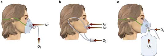
Figure 16.26 Methods of administering supplemental oxygen to the unintubated patient. (a) Simple facemask. (b) Nasal cannulae. (c) Mask with reservoir bag and non-rebreathing value.
With these devices, the inspired oxygen concentration varies from about 35% to 55%, with oxygen flow rates of between 6 and 10 L/min. Nasal cannulae are often preferred because they are less claustrophobic and do not interfere with feeding or speaking, but they can cause ulceration of the nasal or pharyngeal mucosa. Higher concentrations of oxygen can be administered by using a mask with a reservoir bag attached (Fig. 16.26c). Figure 16.26 should be compared with the fixed-performance mask shown in Figure 15.25, with which the oxygen concentration can be controlled. This latter type of mask is used in patients with COPD and chronic type II failure; the hazards of reducing hypoxic drive can be overemphasized and are less dangerous when the patient is in a critical care unit – remember, severe hypoxaemia is more dangerous than hypercapnia.
Oxygen toxicity
Experimentally, mammalian lungs have been shown to be damaged by continuous exposure to high concentrations of oxygen, but oxygen toxicity in humans is less well proven. Nevertheless, it is reasonable to assume that high concentrations of oxygen might damage the lungs, and so the lowest inspired oxygen concentration compatible with adequate arterial oxygenation should be used. Dangerous hypoxia should never be tolerated through a fear of pulmonary oxygen toxicity. There has been concern that in some circumstances (e.g. following myocardial infarction) routine administration of supplemental oxygen may be harmful, perhaps because of associated vasoconstriction.
Respiratory support
If, despite the above measures, the patient continues to deteriorate or fails to improve, the institution of some form of respiratory support is necessary (Table 16.7). Non-invasive ventilation via a mask or hood (see p. 895) can be used, particularly in respiratory failure due to COPD, but in critically ill patients invasive ventilation through an endotracheal tube or tracheostomy is more usual.
Table 16.7 Techniques for respiratory support
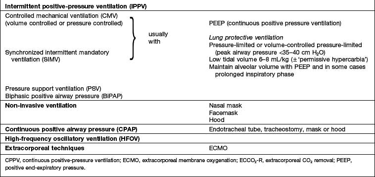
Intermittent positive pressure ventilation (IPPV) is achieved by intermittently inflating the lungs with a positive pressure delivered by a mechanical ventilator. Over the last few decades there have been a number of refinements and modifications to the manner in which positive pressure is applied to the airway and in the interplay between the patient’s respiratory efforts and mechanical assistance (p. 894).
Controlled mechanical ventilation (CMV), with the abolition of spontaneous breathing, rapidly leads to atrophy of respiratory muscles so that assisted modes that are triggered by the patient’s inspiratory efforts (see below) are preferred.
The rational use of mechanical ventilation depends on a clear understanding of its potential beneficial effects, as well as the dangers.
Beneficial effects of mechanical ventilation
Relief from exhaustion. Mechanical ventilation reduces the work of breathing, ‘rests’ the respiratory muscles and relieves the extreme exhaustion present in patients with respiratory failure. In some cases, if ventilation is not instituted, this exhaustion may culminate in respiratory arrest.
Effects on oxygenation. Application of positive pressure can prevent or reverse atelectasis. In those with severe pulmonary parenchymal disease, the lungs may be very stiff and the work of breathing is therefore greatly increased. Under these circumstances, the institution of respiratory support significantly reduces total body oxygen consumption; consequently  and thus PaO2 may improve. Because ventilated patients are connected to a leak-free circuit, it is possible to administer high concentrations of oxygen (up to 100%) accurately and to apply a positive end-expiratory pressure (PEEP). In selected cases, the latter may reduce shunting and increase PaO2 (see below).
Improved carbon dioxide elimination. By increasing the volume of ventilation, the PaCO2 can be controlled.
Indications for mechanical ventilation
Acute respiratory failure, with signs of severe respiratory distress (e.g. respiratory rate >40/min, inability to speak, patient exhausted) persisting despite maximal therapy. Confusion, restlessness, agitation, a decreased conscious level, a rising PaCO2 (>8 kPa) and extreme hypoxaemia (<8 kPa), despite oxygen therapy, are further indications.
Acute ventilatory failure due, for example, to myasthenia gravis or Guillain–Barré syndrome. Mechanical ventilation should usually be instituted when the vital capacity has fallen to 10 mL/kg or less. This will avoid complications such as atelectasis and infection as well as preventing respiratory arrest. The tidal volume and respiratory rate are relatively insensitive indicators of respiratory failure in the above conditions and change late in the course of the disease. A high PaCO2 (particularly if rising) is an indication for urgent mechanical ventilation.
By no means all patients with respiratory failure and/or a reduced vital capacity require ventilation; clinical assessment of each individual case is essential. The patient’s general condition, degree of exhaustion, level of consciousness and ability to protect their airway are often more useful than blood gas values.
Postoperative ventilation in high-risk patients
Head injury: to avoid hypoxia and hypercarbia, which increase cerebral blood flow and intracranial pressure
Trauma: chest injury and lung contusion
Severe left ventricular failure with pulmonary oedema
Coma with breathing difficulties, e.g. following drug overdose.
Institution of invasive respiratory support
This requires tracheal intubation. If the patient is conscious, the procedure must be fully explained and consent obtained before anaesthesia is induced. The complications of tracheal intubation are given in Table 16.8.
Table 16.8 Complications of endotracheal intubation
| Complication | Comments |
|---|---|
Immediate |
|
Trauma to the upper airway |
Lips, teeth, gums, trachea |
Tube in one or other (usually the right) main bronchus |
Avoid by checking both lungs are being inflated, i.e. both sides of the chest move and air entry is heard bilaterally on auscultation |
Early |
|
Migration of the tube out of the trachea |
Dangerous complications |
Manual inflation with 100% oxygen |
|
Tracheal suction |
|
Check position of tube |
|
Deflate cuff |
|
Check tube for ‘kinks’ or blockage with secretions or blood (common) |
|
If no improvement, remove tube, ventilate with facemask and then insert new endotracheal tube |
|
Late |
|
Sinusitis |
|
Intubating patients in severe respiratory failure is a hazardous undertaking and should only be performed by experienced staff. In extreme emergencies, it may be preferable to ventilate the patient by hand using an oropharyngeal airway and a facemask with added oxygen until experienced help arrives. An alternative is insertion of a laryngeal mask airway.
The patient is usually hypoxic and hypercarbic, with increased sympathetic activity; the stimulus of laryngoscopy and intubation can precipitate dangerous arrhythmias, bradycardia and even cardiac arrest. Except in an extreme emergency, therefore, the ECG and oxygen saturation should be monitored, and the patient preoxygenated with 100% oxygen before intubation. Resuscitation drugs should be immediately available. If time allows, the circulating volume should be optimized and, if necessary, inotropes commenced before attempting intubation. In some cases, it is appropriate to establish intra-arterial and central venous pressure monitoring before instituting mechanical ventilation, although many patients will not tolerate the supine or head-down position. In some deeply comatose patients, no sedation is required, but in the majority of patients, a short-acting intravenous anaesthetic agent, usually with an opiate followed by muscle relaxation will be necessary. When available, capnography must be used to confirm tracheal intubation.
Sedation, analgesia and muscle relaxation
Most critically ill patients will require analgesia and many will receive sedatives. The combination of an opiate with a benzodiazepine or propofol is often used to facilitate mechanical ventilation and to obtund the physiological response to stress. Heavy sedation is indicated in those with severe respiratory failure, especially since ‘lung protective’ ventilatory strategies (see p. 895) are inherently uncomfortable. A few may require neuromuscular blockade, indeed evidence suggests that early administration of atracurium improves outcomes for mechanically ventilated patients with severe ARDS. It is now recognized, however, that minimizing sedation levels using ‘sedation scores’ and ‘daily wakening’, or even the avoidance of sedatives altogether, often in combination with spontaneous breathing modes of respiratory support (see p. 895) is associated with reductions in the duration of mechanical ventilation and more rapid discharge from the ICU and hospital. It is also now recognized that benzodiazepines predispose to the development of delirium (which is an independent predictor of increased mortality and length of hospital stay) in critically ill patients. The use of dexmedetomidine (an α2 agonist) rather than a benzodiazepine has been shown to be associated with less delirium and reduced time on the ventilator.
Tracheostomy
Tracheostomy may be required for the long-term control of excessive bronchial secretions, particularly in those with a reduced conscious level, and/or to maintain an airway and protect the lungs in those with impaired pharyngeal and laryngeal reflexes. Tracheostomy is also performed when intubation is likely to be prolonged, for patient comfort, to reduce sedation requirements and to facilitate weaning from mechanical ventilation.
Tracheostomy can be performed in the ICU or in an operating theatre. A percutaneous dilatational approach, which is quick and can be performed at the bedside, is a suitable technique for most critically ill patients (and can be used in an emergency). The alternative surgical approach, opening the trachea vertically through the second, third and fourth tracheal rings via a transverse skin incision, involves transferring the patient to an operating theatre.
A life-threatening obstruction of the upper respiratory tract that cannot be bypassed with an endotracheal tube can be relieved by a cricothyroidotomy, which is safer, quicker and easier to perform than a formal tracheostomy.
Tracheostomy has a small but significant mortality rate. Complications of tracheostomy are shown in Table 16.9.
Table 16.9 Complications of tracheostomy
(As for tracheal intubation, see Table 16.8), plus:
|
|
Complications associated with mechanical ventilation
Airway complications. There may be complications associated with tracheal intubation or tracheostomy (see above) (Tables 16.8, 16.9).
Disconnection, failure of gas or power supply, mechanical faults. These are unusual but dangerous. A method of manual ventilation, a facemask and oxygen must always be available by the bedside.
Cardiovascular complications. The application of positive pressure to the lungs and thoracic wall impedes venous return and distends alveoli, thereby ‘stretching’ the pulmonary capillaries and causing a rise in pulmonary vascular resistance. Both these mechanisms can produce a fall in cardiac output.
Respiratory complications. Mechanical ventilation can be complicated by a deterioration in gas exchange because of  mismatch, fluid retention and collapse of peripheral alveoli. Traditionally, the latter was prevented by using high tidal volumes (10–12 mL/kg) but high inflation pressures, with overdistension of compliant alveoli, perhaps exacerbated by the repeated opening and closure of distal airways, can disrupt the alveolar–capillary membrane. There is an increase in microvascular permeability and release of inflammatory mediators leading to ‘ventilator-associated lung injury’. Extreme overdistension of the lungs during mechanical ventilation with high tidal volumes and PEEP can rupture alveoli and cause air to dissect centrally along the perivascular sheaths. This ‘barotrauma’ may be complicated by pneumomediastinum, subcutaneous emphysema, pneumoperitoneum, pneumothorax, and intra-abdominal air. The risk of pneumothorax is increased in those with destructive lung disease (e.g. necrotizing pneumonia, emphysema), asthma or fractured ribs.
A tension pneumothorax can be rapidly fatal in ventilated patients. Suggestive signs include the development or worsening of hypoxia, hypercarbia, respiratory distress, an unexplained increase in airway pressure, as well as hypotension and tachycardia, sometimes accompanied by a rising CVP. Examination may reveal unequal chest expansion, mediastinal shift away from the side of the pneumothorax (deviated trachea, displaced apex beat) and a hyperresonant hemithorax. Although breath sounds are often diminished over the pneumothorax, this sign can be misleading in ventilated patients. If there is time, the diagnosis can be confirmed by chest X-ray prior to definitive treatment with chest tube drainage.
Ventilator-associated pneumonia. Hospital-acquired pneumonia occurs in as many as one-third of patients receiving mechanical ventilation and this is associated with a significant increase in mortality. It can be difficult to diagnose. The measurement of serum procalcitonin, a specific marker of severe bacterial infections, can be helpful. Various organisms can be isolated, such as aerobic Gram-negative bacilli, e.g. Pseudomonas aeruginosa, Klebsiella pneumoniae, E. coli, Acinetobacter spp. and Staphylococcus aureus, including MRSA. Leakage of infected oropharyngeal secretions past the tracheal tube cuff is thought to be largely responsible. Bacterial colonization of the oropharynx may be promoted by regurgitation of colonized gastric fluid and the risk of ventilator-associated pneumonia can be reduced by nursing patients in the semi-recumbent, rather than the supine, position and by oropharyngeal decontamination.
Techniques for respiratory support (Table 16.7)
Controlled mechanical ventilation (CMV)
This technique is used in patients in whom respiratory efforts are absent or have been abolished.
Ventilation involves one of two types:
Volume controlled ventilation. The tidal volume and respiratory rate are preset on the ventilator. The airway pressure varies according to both the ventilator setting and the patient’s lung mechanics (airways resistance and compliance).
Pressure controlled ventilation. Both the inspiratory pressure and the respiratory rate are preset but the tidal volume varies according to the patient’s lung mechanics.
Positive end-expiratory pressure (PEEP)
A positive airway pressure can be maintained at a chosen level throughout expiration by attaching a threshold resistor valve to the expiratory limb of the circuit. PEEP re-expands underventilated lung units, and redistributes lung water from the alveoli to the perivascular interstitial space, thereby reducing shunt and increasing PaO2. The inevitable rise in mean intrathoracic pressure associated with the application of PEEP may, however, further impede venous return, increase pulmonary vascular resistance and reduce cardiac output. The fall in cardiac output can be ameliorated by expanding the circulating volume, although in some cases inotropic or vasopressor support is required. Thus, although arterial oxygenation is often improved by the application of PEEP, a simultaneous fall in cardiac output can lead to a reduction in total oxygen delivery.
Traditionally, the application of PEEP was only considered if it proved difficult to achieve adequate oxygenation of arterial blood despite raising the inspired oxygen concentration above 50%. Many now use low levels of PEEP (5–7 cmH2O) in the majority of mechanically ventilated patients in order to maintain lung volume, as well as for those with basal atelectasis and in selected cases with airways obstruction.
Continuous positive airway pressure (CPAP)
The application of CPAP achieves for the spontaneously breathing patient what PEEP does for the ventilated patient. Oxygen and air are delivered under pressure via an endotracheal tube, a tracheostomy, a tightly fitting facemask or a hood (Fig 16.27). Not only can CPAP improve oxygenation, but the lungs become more compliant, and the work of breathing is reduced.
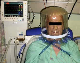
Pressure support ventilation (PSV)
Spontaneous breaths are augmented by a preset level of positive pressure (usually between 5 and 20 cmH2O) triggered by the patient’s spontaneous respiratory effort and applied for a given fraction of inspiratory time or until inspiratory flow falls below a certain level. Tidal volume is determined by the set pressure, the patient’s effort and pulmonary mechanics. The level of pressure support can be reduced progressively as the patient improves.
Intermittent mandatory ventilation (IMV)
This technique allows the patient to breathe spontaneously between the ‘mandatory’ tidal volumes delivered by the ventilator. These mandatory breaths are timed to coincide with the patient’s own inspiratory effort (synchronized IMV or SIMV). SIMV can be used with or without CPAP, and spontaneous breaths may be assisted with pressure support ventilation.
‘Lung-protective’ ventilation
This is designed to avoid exacerbating or perpetuating lung injury by avoiding overdistension of alveoli, minimizing airway pressures and preventing the repeated opening and closure of distal airways. Alveolar volume is maintained with PEEP, and sometimes by prolonging the inspiratory phase, while tidal volumes are limited to 6–8 mL/kg ideal bodyweight. Peak airway pressures should not exceed 35–40 cmH2O. An alternative is to deliver a constant preset inspiratory pressure for a prescribed time in order to generate a low tidal volume at reduced airway pressures (‘pressure-limited’ mechanical ventilation). Respiratory rate can be increased to improve CO2 removal and avoid severe acidosis (pH <7.2), but hypercarbia is frequent and should be accepted (‘permissive hypercarbia’). Both techniques can be used with SIMV. Ventilation with low tidal volumes has been shown to improve outcome in patients with acute lung injury (ALI) or the acute respiratory distress syndrome (ARDS) (see p. 884). Lung protective ventilation should now be used in almost all patients undergoing mechanical ventilation.
FURTHER READING
Nava S, Hill N. Non-invasive ventilation in acute respiratory failure. Lancet 2009; 374:250–259.
Peek GJ, Mugford M, Tinwoipati R et al. Efficacy and economic assessment of conventional ventilatory support versus extracorporeal membrane oxygenation for severe adult respiratory failure (CESAR): a multicentre randomised controlled trial. Lancet 2009; 374:1351–1363.
High frequency oscillation (HFO)
With HFO, there is no bulk flow of gas; rather gas oscillates to and fro at rates of 60–3000 cycles/min with a VT of 1–3 mL/kg. Both inspiration and expiration are actively controlled with a sine wave pump. The mechanism of gas exchange is not fully understood but lung volume is well maintained and oxygenation may be improved. There is some evidence to suggest that HFO is a safe and effective intervention for patients with severe ARDS.
Extracorporeal gas exchange (ECGE)
In patients with severe refractory respiratory failure pumped veno-venous bypass through a membrane lung (extracorporeal membrane oxygenation, ECMO or extracorporeal carbon dioxide removal, ECCO2-R) has been used to reduce ventilation requirements, thereby minimizing further ventilation-induced lung damage and encouraging resolution of the lung injury. A recent (2010) randomized controlled trial suggested that ECMO might improve outcome in adult patients with severe respiratory failure. There is also some indication that the technique is particularly valuable in young patients with acute respiratory distress syndrome caused by influenza.
Non-invasive ventilation (NIV)
NIV is suitable for patients who are conscious, cooperative and able to protect their airway; they must also be able to expectorate effectively. Positive pressure is applied to the airways using a tight-fitting full-face/nasal mask or a hood. The most popular ventilators for this purpose are those that deliver bilevel positive airway pressure (BiPAP), which are simple to use, cheap and flexible. With the latter technique, inspiratory and expiratory pressure levels and times are set independently and unrestricted spontaneous respiration is possible throughout the respiratory cycle. BiPAP can also be patient triggered. There is a reduced risk of ventilator-associated pneumonia and improved patient comfort, with preservation of airway defence mechanisms, speech and swallowing (which allows better nutrition). Spontaneous coughing and expectoration are not hampered, allowing effective physiotherapy, and sedation is usually unnecessary. Institution of non-invasive respiratory support can rest the respiratory muscles, reduce respiratory acidosis and breathlessness, improve clearance of secretions and re-expand collapsed lung segments. The intubation rate, length of ICU and hospital stay and, in some categories of patient, mortality, may all be reduced. NIV is particularly useful in acute hypercapnic respiratory failure associated with COPD, provided the patient is not profoundly hypoxic. NIV may also be useful as a means of avoiding tracheal intubation in immunocompromised patients with acute respiratory failure. Evidence suggests that early NIV after extubation of hypercapnic patients with respiratory disorders can reduce the risk of subsequent respiratory failure and mortality. Box 16.5 shows some indications for the use of NIV when standard medical treatment has failed. Remember, NIV should not be used as a substitute for invasive ventilation when the latter is clearly more appropriate.
Box 16.5
Some indications for the use of non-invasive ventilation (NIV)
Acute exacerbation of COPD (pH <7.35)
Chest wall deformity/neuromuscular disease (hypercapnic respiratory failure)
Severe pneumonia (see Box 15.7)
Contraindications include facial or upper airway surgery, reduced conscious level, inability to protect the airway.
Modified from BTS guidelines after 1997, http://www.british thoracic society.co.uk.
Weaning
Weakness and wasting of respiratory muscles is an inevitable consequence of the catabolic response to critical illness and is often exacerbated by the reduction in respiratory work during mechanical ventilation (‘disuse atrophy’). Often abnormalities of gas exchange and lung mechanics persist. Not surprisingly, therefore, many patients experience difficulty in resuming spontaneous ventilation. In a significant proportion of patients who have undergone a prolonged period of respiratory support the situation is further complicated by the development of a neuropathy, a myopathy or both.
Neuromuscular weakness complicating critical illness
Polyneuromyopathies have most often been described in association with persistent sepsis and multiple organ failure (see below). Critical illness polyneuropathy is characterized by a primary axonal neuropathy involving both motor and, to a lesser extent, sensory nerves. Clinically, the initial manifestation is often difficulty in weaning the patient from respiratory support. There is muscle wasting, the limbs are weak and flaccid, and deep tendon reflexes are reduced or absent. Cranial nerves are relatively spared. Nerve conduction studies confirm axonal damage. The cerebrospinal fluid (CSF) protein concentration is normal or minimally elevated. These findings differentiate critical illness neuropathy from Guillain–Barré syndrome, in which nerve conduction studies nearly always show evidence of demyelination and CSF protein is usually high.
The cause of critical illness polyneuropathy is not known and there is no specific treatment. Weaning from respiratory support and rehabilitation are likely to be prolonged. With resolution of the underlying critical illness, recovery can be expected after 1–6 months but muscle weakness and fatigue frequently persist.
Myopathies can also occur, often in association with a neuropathy. A severe quadriplegic myopathy has been particularly associated with the administration of steroids and muscle relaxants to mechanically ventilated patients with acute, severe asthma.
Criteria for weaning patients from mechanical ventilation
Clinical assessment is the best way of deciding whether a patient can be weaned from the ventilator. The patient’s conscious level, mood, the effects of drugs and cardiovascular performance must all be taken into account. A subjective evaluation by an experienced clinician of the patient’s response to a short period of spontaneous breathing (spontaneous breathing trial) is the most reliable predictor of weaning success or failure. Objective criteria are based on an assessment of pulmonary gas exchange (blood gas analysis), lung mechanics and muscular strength.
Techniques for weaning
Patients who have received mechanical ventilation for <24–48 hours, e.g. after elective major surgery, can usually resume spontaneous respiration immediately and no weaning process is required. This procedure can also be adopted for those who have been ventilated for longer periods but who tolerate a spontaneous breathing trial and clearly fulfil objective criteria for weaning. Techniques of weaning include the following:
The traditional method is to allow the patient to breathe entirely spontaneously for a short time, following which respiratory support is reinstituted. The periods of spontaneous breathing are gradually increased and the periods of respiratory support are progressively reduced. Initially it is usually advisable to ventilate the patient throughout the night. This method can be stressful and tiring for both patients and staff, although it is sometimes successful when other methods have failed.
SIMV with progressive reduction in the frequency of mandatory breaths. Spontaneous breaths are usually pressure supported.
Gradual reduction of the level of pressure support is currently considered by many to be the preferred technique.
CPAP can prevent the alveolar collapse, hypoxaemia and fall in compliance that might otherwise occur when patients start to breathe spontaneously. It is therefore used during weaning with SIMV or pressure support, during spontaneous breathing trials and in spontaneously breathing patients prior to extubation.
Tracheostomy is often used to facilitate weaning from mechanical ventilation.
Non-invasive respiratory support (BiPAP, CPAP) can be used following extubation to prevent respiratory failure and re-intubation.
Extubation and tracheostomy decannulation
This should not be performed until patients can cough, swallow, protect their own airway and are sufficiently alert to be cooperative. Patients who fulfil these criteria can be extubated provided their respiratory function has improved sufficiently to sustain spontaneous ventilation indefinitely. Similar considerations guide the elective removal of tracheostomy tubes.
Outcomes: withholding and withdrawing treatment
(See also Ch. 10.)
For many critically ill patients, intensive care is undoubtedly life-saving and resumption of a normal lifestyle is to be expected. It is also widely accepted that the elective admission of high-risk patients into an ICU or HDU, particularly in the immediate postoperative period, can minimize morbidity and mortality and reduce costs, as well as reducing the demands on medical and nursing personnel on general wards. In the most seriously ill patients, however, immediate mortality rates are high and a significant number die soon after discharge from the intensive care unit. Mortality rates are particularly high in those who require readmission to intensive care. Moreover, the quality of life for some of those who do survive is poor and longer-term mortality rates (up to 5 years post discharge) are also higher than in the general population. Some centres have established specialist follow-up clinics to address long-term sequelae of critical illness.
Inappropriate use of intensive care facilities has other implications. The patient may experience unnecessary suffering and loss of dignity, while relatives may also have to endure considerable emotional pressures. In some cases, treatment may simply prolong the process of dying, or sustain life of dubious quality, and in others the risks of interventions outweigh the potential benefits. Lastly, intensive care is expensive, particularly for those with the worst prognosis, and resources are limited.
Both for a humane approach to the management of critically ill patients and to ensure that limited resources are used appropriately, it is necessary to:
avoid admitting patients who cannot benefit from intensive care and
limit further aggressive therapy when the prognosis is clearly hopeless.
Such decisions are extremely difficult; every case must be assessed individually, taking into account previous health and quality of life, primary diagnosis, medium- and long-term prognosis of the underlying condition, and survivability of the acute illness. Age alone should not be a consideration. When in doubt, active measures should continue but with regular review in the light of response to treatment and any other changes.
Decisions to limit therapy, not to resuscitate or to withdraw treatment should be made jointly by the medical staff of the unit, the primary physician or surgeon, the nurses and if possible the patient, normally in consultation with the patient’s family. Limitation of active treatment is not the cessation of medical or nursing care: rather, a caring approach must be adopted to ensure a dignified death, free of pain and distress, with support for family and friends (see Ch. 10).
Scoring systems
A variety of scoring systems have been developed that can be used to evaluate the severity of a patient’s illness. Some have included an assessment of the patient’s previous state of health and the severity of the acute disturbance of physiological function (acute physiology, age, chronic health evaluation, APACHE and simplified acute physiology score, SAPS). Other systems have been designed for particular categories of patient (e.g. the injury severity score for trauma victims).
The APACHE and SAPS scores are widely applicable and have been extensively validated. They can quantify accurately the severity of illness and predict the overall mortality for large groups of critically ill patients, and are therefore useful for defining the ‘casemix’ of patients when auditing a unit’s clinical activity, for comparing results nationally or internationally, and as a means of characterizing groups of patients in clinical studies. Although the APACHE and SAPS methodologies can also be used to estimate risks of mortality, no scoring system has yet been devised that can predict with certainty the outcome in an individual patient. They must not, therefore, be used in isolation as a basis for limiting or discontinuing treatment.
FURTHER READING
Herridge MS, Tansey CM, Matte A et al. Functional disability 5 years after acute respiratory distress syndrome. N Engl J Med 2011; 364:1293–1304.
Lautrette A, Darmen M, Megarlane B et al. A communication strategy and brochure for relatives of patients dying in the ICU. N Engl J Med 2007; 356:469–478.
Brain death
Brain death means ‘the irreversible loss of the capacity for consciousness combined with the irreversible loss of the capacity to breathe’. Both of these are essentially functions of the brainstem. Death, if thought of in this way, can arise either from causes outside the brain (i.e. respiratory and cardiac arrest) or from causes within the cranial cavity. With the advent of mechanical ventilation it became possible to support such a dead patient temporarily, although in all cases cardiovascular failure eventually supervenes and progresses to asystole.
Before deciding on a diagnosis of brainstem death, it is essential that certain preconditions and exclusions are fulfilled.
Exclusions
The possibility that unresponsive apnoea is the result of poisoning, sedative drugs or neuromuscular blocking agents must be excluded.
Hypothermia must be excluded as a cause of coma. The central body temperature should be more than 35°C.
There must be no significant metabolic or endocrine disturbance that could produce or contribute to coma or cause it to persist.
There should be no profound abnormality of the plasma electrolytes, acid–base balance or blood glucose levels.
Diagnostic tests for the confirmation of brainstem death
All brainstem reflexes are absent in brainstem death.
Tests
The following tests should not be performed in the presence of seizures or abnormal postures.
Oculocephalic reflexes should be absent. In a comatose patient whose brainstem is intact, the eyes will rotate relative to the orbit (i.e. doll’s eye movements will be present). In a brainstem dead patient, when the head is rotated from side to side, the eyes move with the head and therefore remain stationary relative to the orbit.
The pupils are fixed and unresponsive to bright light. Both direct and consensual light reflexes are absent. The size of the pupils is irrelevant, although most often they will be dilated.
There are no vestibulo-ocular reflexes on caloric testing (see p. 1078).
There is no motor response within the cranial nerve territory to painful stimuli applied centrally or peripherally. Spinal reflex movements may be present.
There is no gag or cough reflex in response to pharyngeal, laryngeal or tracheal stimulation.
Spontaneous respiration is absent. The patient should be ventilated with 100% O2 (or 5% CO2 in 95% O2) for 10 minutes and then temporarily disconnected from the ventilator for up to 10 minutes. Oxygenation is maintained by insufflation with 100% oxygen via a catheter placed in the endotracheal tube. The patient is observed for any signs of spontaneous respiratory efforts. A blood gas sample should be obtained during this period to ensure that the PaCO2 is sufficiently high to stimulate spontaneous respiration (>6.7 kPa; 50 mmHg).
The examination should be performed and repeated by two senior doctors.
In the UK, it is not considered necessary to perform confirmatory tests such as EEG and carotid angiography.
The primary purpose of establishing a diagnosis of brainstem death is to demonstrate beyond doubt that it is futile to continue mechanical ventilation and other life-supporting measures.
In suitable cases, and provided the assent of relatives has been obtained (easier if the patient was carrying an organ donor card or is on the organ donor register), the organs of those in whom brainstem death has been established may be used for transplantation. In the UK, each region has a transplant coordinator who can help with the process, as well as providing information, training and advice about organ donation. They should be informed of all potential donors. In all cases in the UK, the coroner’s consent must be obtained.
Hinds CJ, Watson D. Intensive Care: A Concise Textbook. Edinburgh: Elsevier; 2008.
Society of Critical Care Medicine. Online. Available at http://www.sccm.org/professional_resources/guidelinestable-of-contents/index.asp
Faculty of Intensive Care Medicine
European Society of Intensive Care Medicine