The lymphomas are malignancies of the lymphoid system and hence may arise at any site where lymphoid tissue is present. Certain subtypes have increased in frequency over the past 50 years for reasons which are not clear, the overall incidence being 15–20 per 100 000 population making them the fifth most common malignancy in the Western world. Most commonly patients have peripheral lymphadenopathy or symptoms due to occult lymph nodes, although approximately 20% arise at primary extra-nodal sites. A relatively small proportion present with lymphoma-associated ‘B’ symptoms of weight loss, fever and sweats. The natural history and clinical course are determined by the pathological subtype, classified by histological, immunological and molecular criteria, the distribution of the disease (‘Stage’), nonspecific prognostic features and general co-morbidity.
A significant proportion of patients are cured and many others are helped both in terms of quality and length of life.
The WHO classification of Tumours of Haematopoetic and Lymphoid Tissues primarily distinguishes Hodgkin’s lymphoma from non-Hodgkin’s lymphoma, an umbrella term covering a multiply subclassified spectrum of B- and T-cell malignancies, reflecting the stage of lymphoid development at which they arise. Thus, lymphoblastic lymphoma and lymphoblastic leukaemia are considered as a single entity, as are small lymphocytic lymphoma and chronic lymphatic leukaemia, both discussed in the section above.
FURTHER READING
Canellos GP, Lister TA, Young BD (eds). The Lymphomas, 2nd edn. Philadelphia: Saunders; 2006.
Magrath IT, Boffetta P, Potter M (eds). The Lymphoid Neoplasms, 3rd edn. London: Hodder Arnold; 2010.
Swerdlow SH, Campo E, Harris NL et al. WHO Classification of Tumours of Haematopoietic and Lymphoid Tissues, 4th edn. Geneva: WHO; 2008.
A suspected diagnosis of lymphoma should always be confirmed by an excision biopsy of the relevant tissue large enough to allow histological, immunological and molecular analysis. Cutting needle biopsy is an acceptable substitute for biopsy of impalpable, ‘occult’ disease, but fine needle aspiration is inadequate. The opinion of an expert haemato-pathologist is essential.
The diagnosis having been established, the treatment strategy and details will depend on the outcome of investigations that are common to all the lymphomas. These investigations are conducted to provide a basis for prognostication and treatment decisions, against which the outcome of treatment may be assessed (Table 9.22), ‘stage’ being assigned notionally to the modification of the Ann Arbor classification for all nodal lymphomas despite the fact it was planned only for Hodgkin’s disease.
Table 9.22 Investigation of the patient with lymphoma
The tests in Table 9.22 are essential for planning specific therapy. The serum uric acid is helpful, particularly in those lymphomas in which there is risk of tumour lysis syndrome (see p. 449), tests of cardiac function when potentially cardiotoxic chemotherapy is to be recommended, as well as HIV, hepatitis B and C status.
Upon completion of these investigations, within a maximum of 2 weeks, a treatment plan should be presented to the patient. Central to this is the expectation of success and whether treatment is given with curative or palliative intent. The options will range from expectant management to immediate intervention with intensive therapy, itself potentially life-threatening. A decision to ‘treat’ having been made, a course of treatment will begin, during which benefit will be assessed at appropriate intervals and subsequent to which complete re-evaluation (‘re-staging’) will be performed. Depending on the outcome, future plans will be made. In the event of a decision to stop treatment, surveillance will in the first instance, be close, the interval between attendances being extended with the passage of time. Beyond 5 years, the focus of attention is upon the possible long-term consequences of therapy, rather than the disease itself. For those for whom the initial therapy has been less successful than wished, management will be dictated by individual circumstances. As conventional treatment becomes exhausted, experimental therapy may be broached.
HL occurs with an incidence of approximately 3 per 100 000 in the Western world; there is a male predominance of approximately 1.3 : 1. The majority of cases occur between the ages of 16 and 65, with a peak in the 3rd decade. The incidence is stable.
There is epidemiological evidence linking previous infectious mononucleosis with HL; up to 40% of patients with HL have increased EBV antibody titres at the time of diagnosis and EBV DNA has been demonstrated in tissue from patients with HL. These data suggest a role for EBV in pathogenesis. Other viruses have not been detected. Other environmental and occupational exposures to pathogens have been postulated.
Hodgkin’s lymphoma is subclassified according to the WHO classification (Table 9.23) into:
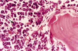
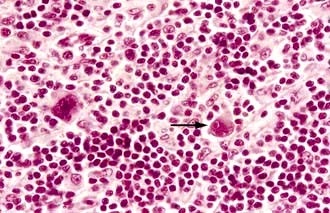
1. Classical Hodgkin’s lymphoma (cHL), the hallmark of which is the Reed–Sternberg cell (Fig. 9.17), accounting for 90–95% of cases and which is further subdivided into four distinct categories
2. Nodular lymphocyte predominant HL (NLPHL), characterized by the Reed–Sternberg cell variant, the ‘popcorn cell’.
Table 9.23 Hodgkin’s lymphoma – pathological classification
5% |
From Harris NL, Jaffe ES, Diebold J et al. 1999 World Health Organization classification of neoplastic diseases of the hematopoietic and lymphoid tissues: report of the clinical advisory committee meeting – Airlie House, Virginia, November 1997. J Clin Oncol 17:3835–49, with permission.
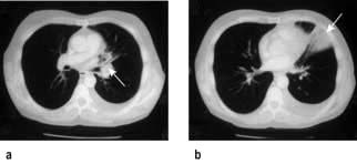
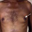
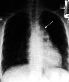
The commonest presentation of HL is painless cervical lymphadenopathy, commonly described in examination as ‘rubbery’. Other causes of cervical lymphadenopathy are shown in Table 9.24. A smaller proportion present with disease localized to the mediastinum (often young women), with cough due to mediastinal lymphadenopathy (Fig. 9.18), others with ‘generalized disease’, including hepatosplenomegaly and constitutional ‘B’ symptoms. Other less common symptoms, undoubtedly associated with Hodgkin’s lymphoma, but not recognized in the staging classification, are pruritus and alcohol-related pain at the site of lymphadenopathy.
Table 9.24 Cervical lymph node enlargement–differential diagnosis
Infections Acute Pyogenic infections Infective mononucleosis Toxoplasmosis Cytomegalovirus infection Infected eczema Cat scratch fever Acute childhood exanthema Chronic Tuberculosis Syphilis Sarcoidosis HIV infectionAutoimmune rheumatic disease Rheumatoid arthritisDrug reactions Phenytoin |
Primary lymph node malignancies Hodgkin’s lymphoma Non-Hodgkin’s lymphoma Chronic lymphocytic leukaemia Acute lymphoblastic leukaemiaSecondary malignancies Nasopharyngeal, oropharyngeal Thyroid Laryngeal Lung Melanoma Breast StomachMiscellaneous Kawasaki’s syndrome Kikuchi’s disease (histiocytic necrotizing lymphadenitis) Castleman’s disease |
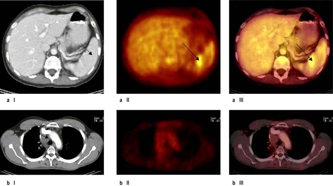
This is summarized in Table 9.22. Bone marrow biopsy is only indicated in patients with clinically advanced disease (stage III, IV), those with ‘B’ symptoms and those who are HIV-positive. The clinical utility of the PET scan is becoming established in the management of Hodgkin’s lymphoma (Fig. 9.19).
‘Stage’ is currently assigned according to the Cotswolds modification of the Ann Arbor Classification, although this is under review (Table 9.25). The Hasenclever score is used for prognostication (Box 9.8); however its relevance to treatment planning is limited because of the very small number of patients at high risk of standard treatment failure. On the basis of ‘stage’ and other prognostic factors, patients with HL are divided into three groups (Table 9.26).
Table 9.25 Cotswolds modification of Ann Arbor staging classification of Hodgkin’s lymphoma
Stage | Description |
---|---|
Stage I |
Involvement of a single lymph-node region or lymphoid structure (e.g. spleen, thymus, Waldeyer’s ring) or involvement of a single extralymphatic site |
Stage II |
Involvement of two or more lymph-node regions on the same side of the diaphragm (hilar nodes, when involved on both sides, constitute stage II disease); localized contiguous involvement of only one extranodal organ or site and lymph-node region(s) on the same side of the diaphragm (IIE). The number of anatomic regions involved should be indicated by a subscript (e.g. II3) |
Stage III |
Involvement of lymph-node regions on both sides of the diaphragm (III), which may also be accompanied by involvement of the spleen (IIIS) or by localized involvement of only one extranodal organ site (IIIE) or both (IIISE) |
III1 |
With or without involvement of splenic, hilar, coeliac or portal nodes |
III2 |
With involvement of para-aortic, iliac and mesenteric nodes |
Stage IV |
Diffuse or disseminated involvement of one or more extranodal organs or tissues, with or without associated lymph-node involvement |
Designations applicable to any disease state |
|
A |
No symptoms |
B |
Fever (temperature >38°C), drenching night sweats, unexplained loss of >10% of body weight within the previous 6 months |
X |
Bulky disease (a widening of the mediastinum by more than one-third of the presence of a nodal mass with a maximal dimension >10 cm) |
E |
Involvement of a singe extranodal site that is contiguous or proximal to the known nodal site |
From Diehl V, Thomas RK, Re D et al. Hodgkin’s lymphoma – diagnosis and treatment. Lancet Oncology 2004; 5:19–26 with permission from Elsevier.
Box 9.8
Advanced stage Hodgkin’s lymphoma (Hasenclever score)
Score | 0 | 1 |
---|---|---|
Serum albumin |
Normal |
<40 g/L |
Haemoglobin |
>105 g/L |
<105 g/L |
Age |
<45 |
>45 |
Sex |
Female |
Male |
Stage |
<IV |
IV |
Leucocytosis |
<15 × 109/L |
>15 × 109/L |
Lymphopenia |
>0.6 × 10⁹/L |
<0.6 × 10⁹/L |
Cumulative score |
Freedom from progression at 5 years |
Frequency |
Score 0 |
84% |
7% (of all patients) |
Score 1 |
77% |
22% |
Score 2 |
67% |
29% |
Score 3 |
60% |
28% |
Score 4 |
51% |
12% |
Score 5 |
42% |
7% |
Table 9.26 Hodgkin’s lymphoma prognostic groups
Early favourable |
Stage I + II without unfavourable prognostic factors |
Early unfavourable |
Stage I + II with unfavourable factors |
Advanced |
The remainder |
Treatment is aimed towards a curative intent with expectation of success. However, in patients with NLPHL who usually present with stage I disease with longstanding lymphadenopathy, an expectant policy with close surveillance, is followed. Older patients, with or without co-morbidity, require considerable modification of therapy and there is an expectation of success. Patients with HIV infection should be managed, in conjunction with their HIV clinicians, in the same way as those who are seronegative.
‘Moderate’ chemotherapy, comprising 2–4 cycles of doxorubicin, bleomycin, vinblastine and dacarbazine (ABVD), non-sterilizing and of a low second cancer risk, followed by involved field irradiation (20–30 Gy) has replaced large field irradiation (p. 435), with 90% being cured. Current trials are evaluating the role of PET scanning to see if patients who become ‘PET’ negative’ after chemotherapy can be spared irradiation altogether.
This is also curable for a significant proportion of patients with a median survival well exceeding 5 years for 50–60% of patients. Cyclical chemotherapy with 6–8 cycles of ABVD with involved field irradiation to sites which were initially bulky, or at which there is ‘persistent disease’ after chemotherapy, is standard. Increasingly, the data from studies incorporating PET scanning both part way through and after planned chemotherapy have shown that PET–ve masses are likely to represent fibrous tissue and that in this situation irradiation may be omitted.
The major short-term toxicity relates to myelosuppression and mucositis, the mortality being no more than 1% and the long-term risks being to the heart and lungs. Infertility and second malignancy are uncommon.
The above approach fails for about 25% of patients. More intensive treatment programmes, e.g. BEACOPP with additional etoposide (E) procarbazine (P) and prednisolone (P), have been tested with an overall increase in efficacy, but with greater toxicity profiles (and expense). It remains a challenge to identify those who will be ‘ABVD failures’ at initial presentation. An alternative is to develop ‘risk-adapted’ therapy, based on the response to the first two cycles of therapy, again with PET scanning and escalate to more intensive therapy in those in whom the response is deemed inadequate.
This has become a declining problem because of improvements in the outcome of first-line therapy. The median survival from first recurrence is more than 10 years, possibly being influenced by the duration of the first remission; it may not be so good if failure occurs after very intensive therapy. Second and third remissions may be achieved with ‘appropriate’ re-induction therapy, consolidated, if possible with an autograft. Registry data suggest this may be curative for up to 50%, but follow-up only extends to 15 years.
With such excellent results of first- and second-line conventional therapy, experimental treatment is seldom required. The antigen-targeted immunoconjugate, anti-CD-30-auristatin (SGN-35, brentuximab) has shown such efficacy in phase II trials and the histone deacetylase inhibitor (HDAC) panobinostat also has potential. Allogeneic haemopoietic stem cell transplantation (HSCT) following myeloablative conditioning has high treatment-related mortality and morbidity. Reduced intensity conditioning HSCT, followed if necessary by donor lymphocyte infusion, is being investigated.
Defined by the WHO classification (Table 9.27), approximately 80% of NHL are of B-cell origin and 20% of T-cell origin, there being considerable geographical variation. The incidence has increased, not necessarily for all subtypes, from 5 to 15 per 100 000 per year in the last half century.
Table 9.27 Modified WHO classification of lymphoid neoplasms other than ALL (2008)
B-cell lymphomas |
|
Precursor B-cell neoplasm |
B-cell lymphoblastic lymphoma/leukaemia (highly aggressive) |
Mature B-cell lymphoma |
Chronic lymphocytic leukaemia/small lymphocytic lymphoma |
B-cell prolymphocytic leukaemia |
|
Splenic marginal zone lymphoma |
|
Hairy cell leukaemia |
|
Lymphoplasmacytic lymphoma |
|
Extranodal marginal zone B-cell lymphoma of mucosa-associated lymphoid tissue (MALT-lymphoma) |
|
Nodal marginal zone B-cell lymphoma |
|
Follicular lymphoma (aggressive) |
|
Mantle cell lymphoma |
|
Diffuse large B-cell lymphoma (aggressive) |
|
Mediastinal (thymic) large B-cell lymphoma |
|
Intravascular large B-cell lymphoma |
|
Primary effusion lymphoma |
|
Burkitt’s lymphoma/leukaemia (highly aggressive) |
|
T/NK cell lymphomas |
|
Precursor T-cell neoplasm |
T-cell lymphoblastic leukaemia/lymphoma (highly aggressive) |
Mature T/NK cell lymphoma |
T-cell prolymphocytic leukaemia |
T-cell large granular lymphocytic leukaemia |
|
Chronic/lymphoproliferative disorder of NK cells |
|
Aggressive NK cell leukaemia |
|
Adult T-cell leukaemia/lymphoma (very aggressive) |
|
Extranodal NK/T-cell lymphoma, nasal type |
|
Enteropathy-type T-cell lymphoma |
|
Hepatosplenic T-cell lymphoma |
|
Subcutaneous panniculitis-like T-cell lymphoma |
|
Mycosis fungoides |
|
Sézary syndrome |
|
Primary cutaneous CD30+ peripheral T-cell lymphoproliferative disorders |
|
Peripheral T-cell lymphoma, unspecified (aggressive) |
|
Angioimmunoblastic T-cell lymphoma |
|
Anaplastic large cell lymphoma (aggressive) ALK positive |
|
Anaplastic large cell lymphoma (aggressive) ALK negative |
NK, natural killer.
Modified from: Jaffe ES, Harris NL, Stein H et al. (eds) World Health Organization Classification of Tumours. Pathology and Genetics of Tumours of Haematopoietic and Lymphoid Tissues. Lyon: IARC Press; 2008, with permission from the World Health Organization.
A family history is associated with a minor increase in risk of lymphoma and common genetic polymorphisms with only a small risk for an individual may be significant in population terms. Certain inherited syndromes, e.g. ataxia-telangiectasia and Wiskott–Aldrich syndrome, are associated with an increased risk of lymphoma. The human T-cell leukaemia virus type 1 (HTLV-1) is causally related to adult T-cell lymphoma/leukaemia. Helicobacter pylori is known to ‘cause’ extranodal marginal zone lymphoma in the stomach. There is a very strong epidemiological relationship between EBV and endemic Burkitt’s lymphoma and a lesser one with sporadic Burkitt’s lymphoma and Hodgkin’s lymphoma.
Immune suppression, immunosuppressant drugs, particularly as used for solid organ transplantation, and HIV infection are all associated with an increased incidence of lymphoma. Agricultural work is associated with lymphoma, but no other data show any convincing evidence of an association between occupation or lifestyle. In the majority of individual cases, the cause is unknown.
Malignant clonal expansion of lymphocytes occurs at different stages of lymphocyte development, leading to the different subtypes of lymphoma (Fig. 9.20). In general, neoplasms of non-dividing mature lymphocytes are ‘indolent’, whereas those of proliferating cells (e.g. lymphoblastic) are much more ‘aggressive’. Malignant transformation is usually due to errors in gene rearrangements which occur during the class switch, or gene recombinations for immunoglobulin and T-cell receptors. Thus, many of the errors occur within immunoglobulin loci or T- cell receptor loci. For example, an abnormal gene translocation may lead to the activation of a proto-oncogene, by moving it next to a promoter sequence for the immunoglobulin heavy chains (Ig-H).
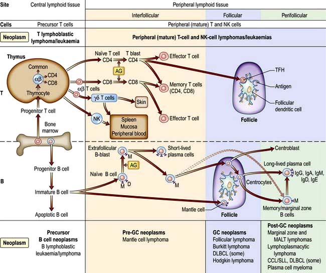
Figure 9.20 Differentiation of T and B lymphocytes and their relationship to neoplasms. Top. T-cell differentiation. Progenitor T cells from the bone marrow enter the thymus and develop into different naive T cells. αβ T cells leave the thymus, are exposed to antigen (AG) and undergo blast transformation. They then develop into CD4+ and CD8+ effector and memory T cells. T regulatory cells are the major type of CD4+ effector cells. The other specific effector T cells are the follicular helper T cell (TFM) in the germinal centres (GC). Upon antigenic stimulation, T-cell responses to antigenic stimulation pathways of natural killer cells (NK) and γδ T cells is unknown. Bottom. B cell differentiation. Precursor B cells mature in the bone marrow and undergo apoptosis or mature to naive B cells. Following exposure to antigen and blast transformation, they develop into short-lived plasma cells or enter the germinal centre (GC). Somatic hypermutation and heavy chain class switching occur here (not shown). The transformed cells of the GC (centroblasts) undergo apoptosis or develop into centrocytes. Post-GC cells include long-lived plasma cells and memory/marginal cells. AG, antigen; DLBLC, diffuse large B-cell lymphoma; CLL/SLL, chronic lymphocytic leukaemia/small lymphocytic lymphoma.
(Redrawn from information in Swerdlow SH, Campo E, Harris NL et al. WHO Classification of Tumours of Haematopoietic and Lymphoid Tissues, 4th edn. Geneva: WHO; 2008.)
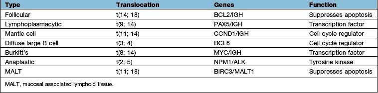
Burkitt’s lymphoma was the first tumour in which a cytogenetic change was shown to involve the translocation of a specific gene (Table 9.28). The most frequent change is a translocation between chromosomes 8 and 14 in which the MYC oncogene is translocated from chromosome 8 to a position near the constant region of the immunoglobulin heavy chain gene on chromosome 14, resulting in upregulation of myc. Similar rearrangements involving the light chain loci are seen in the alternative Burkitt’s lymphoma translocations between chromosome 8 and either chromosome 2 or 22. Other somatic cytogenetic abnormalities associated with human lymphoma are the t(14;18) in follicular lymphoma, involving upregulation of BCL2 or the upregulation of the cell cycle regulator cyclin D1, as a result of t(11;14) in mantle cell lymphoma. Gene expression profiling and other molecular techniques are identifying new molecular subclasses of lymphoma with prognostic significance.
The more common subtypes of non-Hodgkin’s lymphomas are described below.
FURTHER READING
Cheson BD, Leonard JP. Monoclonal antibody therapy for B-cell non-Hodgkin’s lymphoma. N Engl J Med 2008; 359:613–626.
Good DJ, Gascoyne RD. Classification of non-Hodgkin’s lymphoma. Hematol Oncol Clin North Am 2008; 22:781–805.
Sehn LH. Optimal use of prognostic factors in non-Hodgkin lymphoma. Hematology (American Society of Hematology Education Program) 2006: 295–302.
This is the second commonest non-Hodgkin’s lymphoma (comprising approximately 20% of the lymphomas in the world).
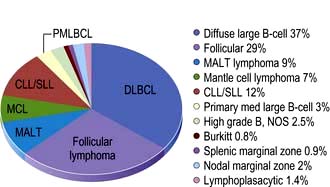
Figure 9.21 Relative frequencies of B-cell lymphoma subtypes in adults.
(Reproduced with permission from the WHO. Swerdlow SH, Campo E, Harris NL et al. WHO Classification of Tumours of Haematopoietic and Lymphoid Tissues, 4th edn. Geneva: WHO; 2008: Fig 8.06, p. 164.)
Follicular lymphoma occurs in middle to late life, being rare in childhood. The majority of patients will present with painless lymphadenopathy at more than one site, although a small proportion will be ill, some with ‘B’ symptoms. In the latter, there should be suspicion that the diagnostic biopsy was not representative. This may be the case when the presenting symptom relates to an abdominal mass, but a peripheral node is biopsied. Percutaneous needle biopsy of the abdominal mass may reveal transformation to diffuse large B-cell lymphoma, with potentially different management. Bone marrow infiltration is common in certain subtypes.
There have been dramatic improvements in the outcome of therapy since the introduction of antibody therapy (rituximab), targeting the CD20 antigen expressed on almost all B-cell lymphomas, leading to the development of chemoimmunotherapy, but to date, however, the proportion of patients cured has been small. The illness has been shown to regress spontaneously in some cases which has led to an expectant policy of treatment for many patients. The clinical course following initiation of treatment to date has been that of a remitting recurring disease, often with several biopsy-proven episodes of lymphadenopathy which are, albeit usually transiently, responsive to therapy. Transformation to diffuse large B-cell lymphoma occurs in up to 25% of patients over 15 years and usually heralds a grave prognosis, although this may be improving. Death occurs because of resistant disease, transformed or not, the complications of therapy, or unrelated causes. The median survival now exceeds 10 years. Prognostic factor are shown in Box 9.9.
Lymph node biopsy accompanied by appropriate further investigation should precede any treatment decision. The ‘well’ patient variously defined, but certainly without symptoms, organ impairment, ‘bulky disease’ or evidence of rapid progression or transformation should, after a careful explanation of the rationale, be managed expectantly. This approach is followed by progression mandating therapy in about two and a half years for half the patients, with 20% having had some spontaneous regression and 15% having had no treatment, more than 10 years since diagnosis. A large trial comparing expectant management with immunotherapy with rituximab, however, shows a very considerable delay to having the first treatment in the rituximab group. The implications of this are unclear. Indications for treatment of low-grade non-Hodgkin’s lymphoma are shown in Box 9.10.
Box 9.10
Indications for treatment of low-grade NHL at presentation or progression
Stage I (possibly stage II): This is treated with involved field megavoltage irradiation, which almost always induces complete remission, with 50% being disease-free after 10–15 years. There are no randomized trials to show this is better in terms of overall survival than expectant management (i.e. observe and treat if progression occurs). Functional imaging, i.e. PET scanning, may identify patients who are in ‘surgical CR’ post-biopsy for whom no therapy is indicated.
Chemoimmunotherapy incorporating rituximab is the treatment of choice having been shown in randomized trials to be superior to chemotherapy alone, in terms of disease-free, progression-free and overall survival. ‘CHOP-R’ (cyclophosphamide, doxorubicin, vincristine and prednisolone + rituximab) and the less intensive ‘R-CVP’ (rituximab + cyclophosphamide, vincristine and prednisolone) are both widely used and R-Bendamustine is gaining popularity. Over the next few years, it will become clear which chemotherapy is the best for which group of patients. It has been shown that continuing rituximab ‘maintenance’ for 2 years has a dramatic effect on progression-free survival, although as yet not on overall survival. If rituximab maintenance is not given, patients in complete or (‘good’) partial remission are managed expectantly, until progression occurs. This is challenged by data showing that consolidation with radio-immunotherapy (90yttrium anti-CD20) also prolongs disease-free survival. Trials are in progress to determine whether consolidation with myeloablative chemotherapy, with or without rituximab maintenance, improves outcome further.
Two studies have shown the efficacy of prolonged rituximab alone; the role of intensive therapy in standard practice is being questioned.
Patients are managed expectantly in the first instance, provided full re-evaluation, including repeat biopsy, reveals no evidence of transformation. A number of options are available, including re-induction of remission with combined chemoimmunotherapy, followed by rituximab maintenance in those not ‘rituximab resistant’. Myeloablative consolidation chemotherapy is used in younger patients, particularly those in whom the first remission was short. Reduced-intensity conditioning allogeneic haematopoetic stem cell transplantation (HSCT) has yielded very impressive results in selected patients that may be curative despite the toxicity of the treatment.
The biggest challenges lie in patients with ‘resistant disease’ and in the treatment of ‘transformation’. A number of experimental agents with new antibodies, immunomodulatory agents and drugs targeted to specific pathways are showing promise.
There has been a dramatic improvement in the overall survival pattern of follicular lymphoma as the result of introducing anti-CD20 (rituximab) in the treatment of advanced disease. The median survival has been extended, well beyond 10 years in several series, albeit possibly selected patients. Improvements in disease-free survival, both after initial and second-line therapy, are encouraging. It is reasonable to anticipate that further improvement will be seen with the selective use of allogeneic stem cell transplantation and the new targeted therapies under investigation.
FURTHER READING
Relander T, Johnson NA, Farinha P et al. Prognostic factors in follicular lymphoma. J Clin Oncol 2010; 28(17):2902–2913.
Salles G, Seymour JF, Offner F et al. Rituximab maintenance for 2 years in patients with high tumour burden follicular lymphoma responding to rituximab plus chemotherapy (PRIMA): a phase 3 randomised controlled trial. Lancet 2011; 377:42–51.
This is the commonest lymphoma worldwide in the adult population (increasing in incidence with age) and the second commonest in childhood, accounting for approximately 30% of all cases. There is a slight male preponderance.
The majority of patients present with painless lymphadenopathy, clinically at one or several sites. Intra-abdominal disease presents with bowel symptoms due to compression or infiltration of the gastrointestinal tract. In a small proportion there is a primary mediastinal presentation, most often in men, with symptoms and signs akin to those of Hodgkin’s lymphoma. There may be ‘B symptoms’, which should not be confused with symptoms related to the site of involvement. Investigation will lead to the demonstration of either locally or systematically advanced disease in the majority of cases. The illness is itself rapidly progressive, without intervention, death occurring within months rather than years. Approximately 30% present at an extranodal site as opposed to having nodal disease with extranodal spread.
Treatment should be initiated immediately after the diagnosis is confirmed and in younger patients without co-morbidity there is a high expectation of cure. Treatment is assigned on the basis of the International Prognostic Index (Box 9.9, Fig. 9.22).
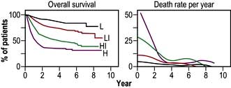
Figure 9.22 A predictive model for aggressive non-Hodgkin’s lymphoma. Survival among the 1274 younger patients (≤60 years) according to risk group defined by the age-adjusted international index. L, low risk; LI, low–intermediate risk; HI, high–intermediate risk; H, high risk.
(Reproduced with permission from: The International Non-Hodgkin’s Lymphoma Prognostic Factors Project. A predictive model for aggressive non-Hodgkin’s lymphoma. New England Journal of Medicine 1993; 329(14):987–994.)
Although this was constructed in the pre-rituximab era, it remains broadly applicable. Further refinement using gene expression profiling has identified at least two distinct subtypes of DLBCL (Fig. 9.23). Allopurinol is given routinely and also in some cases with different prognoses – germinal centre cell (GC) and activated B-cell (AB). A high proliferative index, aggressive prophylaxis against tumour lysis is indicated.
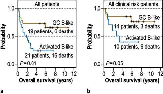
Figure 9.23 Kaplan–Meier plot of overall survival of DLBCL patients grouped on the basis of gene expression profiling.
(Reproduced with permission from Alizadeh AA. Distinct types of diffuse large B-cell lymphoma identified by gene expression profiling. Nature 2000; 403(6769):503–511.)
At present, ‘R-CHOP’ followed by involved field irradiation, or ‘more R-CHOP’, are both used. Interim PET scanning may be used to inform individualization of therapy.
Two studies conducted in France suggest that the prognosis is better with chemotherapy alone provided ‘enough’ is given, with more than 80% of younger patients being alive 10 years after therapy. A trial comparing ‘R-chemo’ with ‘chemo’ in all patients with low-risk disease showed a marked advantage for those receiving chemoimmunotherapy. Further studies are awaited (Fig. 9.24).
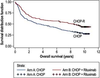
Figure 9.24 Overall survival in patients treated with CHOP and R-CHOP. Median overall survival (OS) was 3.5 years (95% CI: 2.2–5.5) in the CHOP arm and 8.4 years (95% CI: 5.4–not reached) in the R-CHOP arm (p<0.0001). DLBCL, diffuse large B-cell lymphoma.
(Reproduced with permission from Coiffier B, Thieblemont C, Van Den Neste et al. Long-term outcome of patients in the LNH-98.5 trial, the first randomized study comparing rituximab-CHOP to standard CHOP chemotherapy in DLBCL patients: a study by the Groupe d’Etudes des Lymphomes de l’Adulte. Blood 2010; 116(12):2040–2045.)
The age of the patient and co-morbidity are critical for both the selection of treatment and the prognosis. Recognition of this fact has led to the use of an age-adjusted prognostic index for patients over the age of 60. Chemoimmunotherapy was established to be superior to chemotherapy alone for older patients in this category. ‘R-CHOP’, to a total of 6 or 8 cycles, has become standard care for the large majority of patients of all ages with DLBCL.
Many trials of increasing intensity of therapy for selected patients have yet to yield convincing results. However, there is an increasing consensus that the small group of patients having DLBCL with ‘Burkitt-like features’ should be treated as for Burkitt’s lymphoma. The ability to distinguish between the molecularly distinct, ‘germinal centre’ and ‘activated B cell’, DLBCL with immunohistochemistry, has made it possible to explore different therapeutic approaches in the two groups, with appropriate targeted agents
Involvement of the central nervous system, most often meningeal, is an uncommon but devastating complication, confirming a very poor prognosis overall. Patients with a high IPI score and particularly those with specific extranodal sites of involvement (testis, paranasal sinuses, bone marrow) and those who are HIV-positive should receive intrathecal methotrexate with each cycle of therapy. The management of overt leptomeningeal or parenchymal involvement, often in the context of generalized lymphoma, is difficult and usually unsuccessful in the long term. Most strategies involve high doses of systemic methotrexate and cytosine arabinoside, both of which cross the blood–brain barrier. Cranial or craniospinal irradiation is also used.
Although there may be responsiveness initially to alternative chemotherapy, after failure of initial therapy or subsequent progression, the prognosis is very poor. Patients entering a partial remission do better than those who recur after entering an initial complete remission.
The major issues to be addressed are whether treatment is to be undertaken with palliative or curative intent and if the latter, the expectations of success. The palliative approach involves both chemotherapy and irradiation. The curative approach involves complete re-evaluation followed by second-line chemotherapy, with a proven response rate of approximately 50%. If at least a further partial remission is achieved, in the younger, fitter patient, peripheral blood stem cell harvest is undertaken, followed, if successful, by an autograft. Overwhelmingly, the best results are achieved in those entering an unequivocal second complete remission. Even so, the proportion of patients in a prolonged second remission does not exceed 25%.
The outlook for patients with diffuse large B-cell lymphoma has improved by at least 15% in terms of cure, with the incorporation of rituximab into the initial therapy, the expectation of cure now being between 40% and 80% depending on the presenting features. The challenge of progressive disease following initial treatment is great, with overall less than 20% of patients alive long term.
This is the most rapidly proliferating lymphoma with a doubling time approaching 100% and a very rapid evolution. The commonest childhood malignancy worldwide, it has a male : female preponderance of approximately 3 : 1 and occurs in all ages. There are three types:
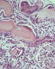
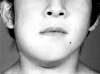
The commonest presenting feature in the endemic type is a rapidly growing jaw tumour in a young child (Fig. 9.25). Otherwise, the next commonest is an abdominal mass often associated with bone marrow involvement. Other common sites are the central nervous system, the kidney and the testis. Investigation is along conventional lines for lymphoma, at least in the Western world, but must be conducted as a matter of urgency. A different staging classification is applied to children.
Burkitt’s lymphoma should be treated with curative intent whenever feasible, regardless of HIV status. Investigation having been completed, it is essential that the patient is haemodynamically and metabolically stable prior to the initiation of specific therapy. Particular attention must be paid to the risk of the tumour lysis syndrome. Whenever possible, rasburicase prophylaxis should be given. If this is not available, other standard measures based on fluids and allopurinol to minimize the risk of tumour lysis syndrome should be pursued. Very frequent monitoring of electrolyte balance is essential for at least 72 hours after treatment is commenced, with particular attention to potassium and phosphate levels.
Standard treatment comprises if possible, intensive, cyclical combination chemotherapy, including cyclophosphamide, methotrexate and cytosine arabinoside in high doses. Rituximab is now included, although the evidence base for this is minimal. The details and number of cycles administered will be determined by the perceived level of ‘risk’. Prophylactic central nervous system therapy is essential, intrathecal methotrexate or cytosine arabinoside often being given in addition to high-dose systemic administration. The chances of cure are very high for ‘low-risk’ patients and exceed 50% for ‘poor-risk’ patients as well, provided all treatment can be administered.
Failure to achieve complete remission is a very poor prognostic factor as is recurrence, which does so within the first year after completion of initial therapy if it is to happen. Although there may be further chemo-responsiveness, it is rare for second-line therapy to be more than transiently beneficial, regardless of whether it is followed by consolidation, with either myeloablative chemotherapy or allogeneic haematopoeitic stem cell transplantation.
This is one of the less common B-cell lymphomas, presenting usually in later life, with a male to female preponderance of 3 : 1. The commonest presentation is with painless lymphadenopathy, often generalized. There may be nonspecific symptoms of tiredness, or those related to the gastrointestinal tract. ‘B’ symptoms occur in <50%. Examination and standard investigation usually confirm generalized lymphadenopathy with or without hepatosplenomegaly (Fig. 9.26). Patients with bowel symptoms frequently have multiple lesions found on endoscopy. The bone marrow is usually involved and there may well be lymphoma cells in the peripheral blood.
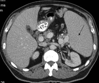
Figure 9.26 CT of the abdomen of a patient with mantle cell lymphoma showing significant splenomegaly.
In the majority of patients, therapy is started after investigation has been completed. It is usual for there to be regression of disease with chemotherapy, although it is not often that complete remission is achieved. A prognostic index is used to help determine whether more or less ‘aggressive’ treatment is best employed. In reality, the major determining factors are the age of the patient and the presence or absence of co-morbidities. The outcome is likely to be best when ‘more’ treatment is given. Hence, younger, fitter patients are now treated with relatively intensive chemoimmunotherapy, incorporating rituximab, followed by, depending on its efficacy, myeloablative chemotherapy, with autologous stem cell rescue. Older, less fit patients are treated with less intensive therapy. In general, the strategy is to stop treatment after the planned initial course of treatment, provided the patient is well and at least a partial remission has been achieved. Almost inevitable progression for both older and younger patients occurs; palliation being the expectation for most. Experimental approaches include reduced intensity allogeneic haematopoietic stem cell transplantation and novel drugs targeting both the NFκB and mTOR pathways.
Mantle cell lymphoma is a lymphoma with a natural history untreated which lies between that of diffuse large B-cell lymphoma and follicular lymphoma. The prognosis has improved in recent years with the introduction of treatment strategies which have prolonged the period of remission without curing the patient and with the discovery of new ‘effective’ agents, making the overall median survival nearer 5 years.
This is an uncommon, B-cell malignancy, which when associated with an IgM paraprotein and bone marrow infiltration is known as Waldenström’s macroglobulinaemia. It usually occurs in later life, the incidence being approximately the same in men and women. It may be preceded by a pre-lymphomatous phase, in which a small, monoclonal IgM band is present, often an incidental finding, for many years. The presentation is with lymphadenopathy or alternatively with symptoms of anaemia or hyperviscosity due to the paraprotein (e.g. headaches, visual disturbance). Examination and investigation usually reveal little beyond minimal adenopathy and commonly splenomegaly.
Following completion of investigation, the critical decision is whether to initiate specific therapy or not. A prognostic score is assigned, the critical decision being whether to initiate specific therapy or not. In an emergency, with severe symptoms of hyperviscosity, it is most appropriate to lower the paraprotein by plasmapheresis. In some circumstances, particularly in the otherwise less fit, it may be best to use maintenance pheresis and blood transfusion as the primary therapy. ‘Responses’ occur in about 50% of cases, with a fall in the paraprotein to 50% of the baseline level with single agent therapy and higher with combination chemotherapy and rituximab. The general strategy is to treat when it is clinically indicated and to stop after a conventional course of treatment. Treatment, either repeating the initial therapy or changing, is re-instated only when progression is clearly documented, most often by fall in the haemoglobin or significant rise in the M-band and the illness is a threat to quality and quantity of life. In the very small proportion of younger patients for whom complete remission is achieved, consolidation with a bone marrow transplant is used. Similarly, in the same group of patients who have recurrent disease, which is again responsive, allogeneic haematopoietic stem cell transplantation is also used.
Lymphoma may arise anywhere in the body where there is lymphoid tissue and therefore the clinical presentation is that of a lesion or mass at the relevant site. In practice the majority occur in the central nervous system, the stomach, or the skin.
This diffuse large B-cell lymphoma occurs in both the immunocompetent (predominantly the elderly) and the immunosuppressed, in the context of post-solid organ transplant or HIV infection. It presents with symptoms relating to single or multiple parenchymal mass lesions. The diagnosis needs to be made on the basis of a biopsy, particularly in the immunocompromised, in whom an infectious aetiology of the symptoms is possible, e.g. toxoplasmosis. MRI scan is the first choice investigation; cerebrospinal fluid is usually normal. Further investigation is necessary to exclude the cerebral lesion being a manifestation of generalized disease.
In some patients in the post-transplant setting, reduction of immune suppression may be beneficial. Chemotherapy alone, with high-dose methotrexate and cytosine arabinoside, is used. Possibly, the best ‘disease-free’ results have been obtained by the use of chemotherapy and irradiation but toxicity is greater, with a risk of irreversible loss of cerebral function. The overall results are disappointing with only a small proportion of patients alive long-term without disability. The situation in the HIV-positive patient with cerebral lymphoma (fortunately a declining problem) is much worse and palliative irradiation is the best option.
This B-cell lymphoma, either extranodal marginal zone lymphoma of mucosa-associated tissue (MALT), or diffuse large B-cell lymphoma arising on a background of MALT lymphoma, is closely related to Helicobacter infection. It presents with symptoms of gastric ulceration or a mass, indigestion or bleeding and the diagnosis is made by endoscopic biopsy to include both the confirmation of lymphoma and H. pylori status. This is followed by investigation as for nodal lymphoma, which reveals local nodal involvement in a proportion of patients and distant spread in only a small number. Treatment is entirely dependent on whether or not there is any evidence of diffuse large B-cell lymphoma. If only ‘low-grade’ gastric ‘MALT’ lymphoma is present, Helicobacter eradication therapy is the treatment of choice (p. 249). This almost invariably alleviates the symptoms. Re-evaluation after 3 months with endoscopy, repeat biopsy and in some circumstances, endoscopic ultrasound is carried out. In general, a conservative approach is followed, as responses may take many months to achieve and rapid progression is very unlikely. Regular endoscopy 6-monthly should be continued for at least 2 years. Failure is not common and if it occurs, it is rarely rapid. If it occurs, the biopsy should be repeated. If the histology is unchanged after further Helicobacter eradication therapy (if necessary) either irradiation to the gastric bed or chemoimmunotherapy is likely to be effective or possibly curative. Overall, the prognosis is very good, the very large proportion of patients being alive 10 years after diagnosis.
Any evidence of diffuse large B-cell lymphoma is an indication for chemoimmunotherapy. Helicobacter eradication therapy should also be given, but should not be considered definitive treatment. The potential risk of gastric perforation or haemorrhage because of therapy is not a contraindication to treatment. Surgery is rarely needed, but irradiation is used for persistent disease. The prognosis is approximately the same as for nodal DLBCL of equivalent extent.
Lymphomas of both B- or T-cell type may arise singly and multiply in the skin and pursue a very long natural history even though they may give rise to considerable discomfort.
This is the commonest cutaneous lymphoma, predominantly arising in and confined to the skin, although later in the disease spreading to other organs. It has a long natural history, being sometimes preceded by a scaly ‘pre-mycotic phase’. This T-cell lymphoma presents with multiple erythematous lesions, plaques and tumours, which when associated with spread to the blood, become the Sézary syndrome. Generalized erythema may occur (erythroderma). The likelihood over time of disease extending beyond the skin is highest in patients with tumours.
Treatment is palliative and there is little indication that the disease is ever eradicated. Many treatments result in regression of disease. Antibiotics are used for infection. Phototherapy (PUVA), topical steroids and topical chemotherapy all lead to response. Radiation is effective and total skin electron beam therapy particularly so, although attention must be given to potential side-effects including erythroderma. Systemic chemotherapy, either at conventional or high doses, has been disappointing. Newer approaches include anti-T-cell antibodies and histone deacetylase inhibitors The median survival is approximately 10 years, there being close correlation with the extent of disease at presentation. Interaction between oncologist and dermatologist is essential.
The two major subtypes are extranodal marginal zone and follicle centre in origin. Both usually present with either single or clustered cutaneous lesions, biopsy of which confirms the diagnosis. All conventional staging investigations are negative. Treatment is either expectant, surgical excision or irradiation, which may be used repeatedly over time. Antibiotics are used for marginal zone lymphoma if there is evidence of Borrelia burgdorferi infection. Only if there are lesions at multiple sites should systemic chemotherapy be used. The long-term prognosis is excellent.
These are much less common than their B-cell counterparts, although they are relatively more frequent in the East than the West. The commonest presentations are nodal or cutaneous (p. 1226) and specific subtypes involve the liver and subcutaneous tissue. Peripheral T-cell lymphomas with nodal presentation have a poor prognosis. They are treated as for DLBCL without rituximab.
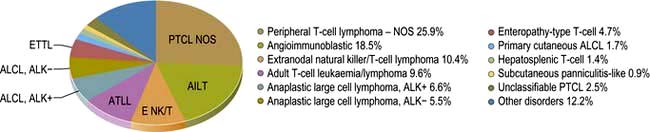
Figure 9.27 Relative frequencies of T-cell lymphoma subtypes in adults. NOS, not otherwise specified.
(Reproduced with permission from WHO. Swerdlow SH, Campo E, Harris NL et al. WHO Classification of Tumours of Haematopoietic and Lymphoid Tissues, 4th edn. Geneva: WHO; 2008: Fig 8.07.)
The two ‘commonest subtypes’ of ‘nodal’ T-cell lymphoma are peripheral T-cell lymphoma, ‘not otherwise specified’ (NOS) and angioimmunoblastic T-cell lymphoma, which together account for about 50% of T-cell lymphomas. Both occur in the middle-aged to elderly population, the primary presentation being lymphadenopathy. In contrast to the B-cell lymphomas, ‘B symptoms’ are common. Patients with angioimmunoblastic T-cell lymphoma also present with features akin to inflammatory disease, with fevers, rashes and electrolyte abnormalities, which in the first instance may be rapidly responsive to corticosteroids or low doses of alkylating agents.
Following standard investigation, which usually reveals widespread disease, patients are treated with cyclical combination chemotherapy as used for diffuse large B-cell lymphoma. CD20 is not expressed on T cells so rituximab is not used and, as yet, there is no equivalent drug for T-cell lymphoma. Resolution of symptoms almost invariably occurs, although they may recur between cycles. Overall, the outcome of treatment is worse than for DLBCL, in terms of quality of response, duration of response and overall survival. Second-line therapy is rarely satisfactory, although a small proportion of patients may benefit from myeloablative therapy to consolidate a second response.
Myeloma is a malignant disease of bone marrow plasma cells, accounting for 1% of all malignant disease. There is a clonal expansion of abnormal, proliferating plasma cells producing a monoclonal paraprotein, mainly IgG (55%) or IgA (20%) and rarely IgM and IgD. The paraproteinaemia may be associated with excretion of light chains in the urine (Bence Jones protein), which are either kappa or lambda. In approximately 20%, there is no paraproteinaemia, only light chains in the urine. Rarely, patients produce no paraprotein or light chains – ‘non-secretory myeloma’ (<5%).
Myeloma is a disease of the elderly, the median age at presentation being over 60 years. It is rare under 40 years of age. The annual incidence is 4 per 100 000 and it is commoner in males and in black Africans but less common in Asians. The clinical features include:
Bone destruction, often causing fractures of long bones or vertebral collapse (which can cause spinal cord compression) and hypercalcaemia. Soft tissue plasmacytomas also occur and they are the usual cause of spinal cord compression in myeloma
Bone marrow infiltration with plasma cells, resulting in anaemia, neutropenia, thrombocytopenia, together with production of the paraprotein which may (rarely) result in symptoms of hyperviscosity
Kidney injury (see p. 1043) owing to a combination of factors – deposition of light chains in the renal tubules, hypercalcaemia, hyperuricaemia, use of NSAIDs and rarely the deposition of AL amyloid.
Kidney biopsy of a patient with myeloma and acute kidney injury due to light chain cast nephropathy.
(Courtesy of Professor Michael Sheaff, Barts and The London NHS Trust.)
In addition, there is a reduction in the normal immunoglobulin levels (immune paresis), contributing to the tendency for patients with myeloma to have recurrent infections, particularly of the respiratory tract. The WHO classification (2008) of plasma cell neoplasms is shown in Table 9.29.
Table 9.29 WHO (2008) classification of plasma cell neoplasms
With fluorescence in situ hybridization and microarray techniques abnormalities are found in most cases of myeloma. Abnormalities of chromosome 13 and hypodiploidy (<45 chromosomes) have been shown to be associated with poor survival, as have t(4; 14), t(14; 16) and p53 (17p) deletions; t(11 : 14) and hyperdiploidy (>50 chromosomes) are associated with a better prognosis.
There is dysregulation of bone remodelling, which leads to the typical lytic lesions, usually seen in the spine, skull, long bones and ribs. In myeloma there is increased osteoclastic activity with no increased osteoblast formation of bone. Bisphosphonates that inhibit osteoclast activity are useful in myeloma but surprisingly there is no increase in bone deposition (see below).
Adhesion of stromal cells to myeloma cells stimulates the production of RANKL, IL-6 and also VEGF, which plays a role in angiogenesis. RANKL also stimulates osteoclast formation and the lytic lesions. Myeloma cells also produce dickkopf-1 (DKK1), which inhibits osteoblast activity and therefore production of new bone. This occurs because DKK1 binds to the Wnt co-receptor, lipoprotein receptor-related protein 5 (LRP5), inhibiting Wnt signalling (see p. 26) and osteoblast differentiation.
Bone pain – most commonly backache owing to vertebral involvement (60%)
Symptoms of renal failure (20–30%)
Rarely symptoms of hyperviscosity and bleeding due to thrombocytopenia.
Patients can be asymptomatic, the diagnosis being suspected by ‘routine’ abnormal blood tests. Life-threatening complications are shown in Box 9.11.
Box 9.11
Life-threatening complications of myeloma
Renal impairment – often a consequence of hypercalcaemia – requires urgent attention and patients may need to be referred for long-term peritoneal or haemodialysis
Hypercalcaemia should be treated by rehydration and use of bisphosphonates such as pamidronate
Spinal cord compression due to myeloma is treated with dexamethasone, followed by radiotherapy to the lesion delineated by a magnetic resonance imaging (MRI) scan
Hyperviscosity due to high circulating levels of paraprotein may be corrected by plasmapheresis.
Full blood count. Hb, WCC and platelet count are normal or low.
Blood film. There may be rouleaux formation as a consequence of the paraprotein and circulating plasma cells in the aggressive plasma cell leukaemia variant of myeloma.
Urea and electrolytes. There may be evidence of kidney injury (see above).
Serum calcium is normal or raised. Serum alkaline phosphatase is usually normal.
Total protein is normal or raised.
Serum protein electrophoresis and immunofixation characteristically shows a monoclonal band and immune paresis (Fig. 9.28). The serum free light chain assay, more sensitive than urine electrophoresis, may show an abnormal ratio and an increased total amount of free kappa or lambda chains and is often abnormal earlier than routine electrophoresis.
24-hour urine electrophoresis and immunofixation is used for assessment of light-chain excretion.
Figure 9.28 Immunofixation and electrophoresis. (a) Immunofixation IgG kappa monoclonal paraprotein. (b) Quantitation and serum electrophoresis IgG paraprotein. Serum electrophoresis and immunofixation of a patient with an IgG K paraprotein (left). The column on the far left is the total serum electrophoresis stained with anti-protein reagents. Albumin is at the top with the increased immunoglobulins at the bottom. These are shown to stain with anti-IgG and anti-kappa light chain reagents. The paraprotein is quantified (right) by calculating the area under the paraprotein spike and comparing it with the albumin concentration.
Skeletal survey. This may show characteristic lytic lesions, most easily seen in the skull (Fig. 9.29). CT, MRI and PET are used in plasmacytomas (bone or soft tissue deposits). MRI spine is useful if there is back pain – may show imminent compression/collapse.
Reduced serum albumin, increased serum β2-microglobulin and increased serum lactate dehydrogenase (LDH).
Symptomatic myeloma (SMM) can be diagnosed if either of the following are present:
with evidence of end organ failure, i.e. hypercalcaemia, renal impairment, anaemia, lytic bone lesions (CRAB).
An international prognostic index based on serum albumin + β2 microglobulin (β2M) at diagnosis is used for prognostic information (Box 9.12).
Box 9.12
International prognostic index based on serum albumin + β2 microglobulin (β2M)
Stage | Median survival (months) |
---|---|
Stage 1 – β2M <3.5 mg/L and serum albumin ≥35 g/L |
62 |
Stage 2 – not stage 1 or 3 |
44 |
Stage 3 – β2M >5.5 mg/L |
29 |
Asymptomatic myeloma (AMM) (10% cases) has a significant paraprotein (IgG or IgA >30 g/dL or urinary light chain excretion >1 g/day) and/or a marrow plasmacytosis >10% but no end organ damage. The median time to progression for these patients is 2–3 years. The risk is highest for those with IgA isotype and light chains in the urine
Monoclonal gammopathy of unknown significance (MGUS). MGUS describes an isolated finding of a monoclonal paraprotein in the serum, usually in the elderly, that does not fulfil the diagnostic criteria for SMM or AMM. 20–30% go on to develop multiple myeloma over a 25-year period. Low-risk MGUS is characterized by those with an IgG subtype, a paraprotein <15 g/dL and a normal serum free light chain ratio.
Plasmacytoma. Patients may present with an isolated tumour of neoplastic plasma cells called a plasmacytoma and no evidence of multiple myeloma. This may be a ‘solitary plasmacytoma of bone’ within the skeleton or an ‘extramedullary plasmacytoma’ outside the marrow cavity typically in the upper aerodigestive tract.
For paraprotein associated neuropathy and POEMS, see page 1079.
With good supportive care and chemotherapy, median survival is now 5 years with some patients surviving to 10 years. Young patients receiving more intensive therapy may live longer.
Anaemia should be corrected; blood transfusion may be required. Erythropoietin often helps. Transfusion should be undertaken slowly in patients with hyperviscosity.
Hypercalcaemia, kidney injury and hyperviscosity should be treated as indicated (see Acute oncology p. 448 and Acute kidney injury p. 609).
Infection should be treated promptly with antibiotics. Give yearly flu vaccinations.
Bone pain can be helped most quickly by radiotherapy and systemic chemotherapy or high-dose dexamethasone. NSAIDs are usually avoided because of the risk of acute kidney injury. Bisphosphonates, e.g. zoledronate, which inhibit osteoclast activity, help ensure rapid normocalcaemia and, given long term, reduce skeletal events such as pathological fracture, cord compression and bone pain.
Pathological fractures may also be prevented by prompt orthopaedic surgery with pinning of lytic bone lesions at critical sites seen on the skeletal survey, e.g. femoral shaft. Kyphoplasty and vertebroplasty may be useful in treating vertebral fractures (p. 505).
Myeloma remains incurable. Therapy is aimed at treatment of specific complications, prevention of these and prolongation of overall survival. Thalidomide has activity as a single agent and is widely used in first-line and relapsed settings. It is teratogeneic and associated with neuropathy, somnolence, constipation and an increased risk of venous thrombosis.
In younger patients (<65–70 years), an orally active cyclophosphamide, thalidomide and dexamethasone-based induction (CTD), followed by a high-dose melphalan autograft, has a significantly higher response rate with 40% of patients achieving a CR with median survival increasing to 6 years. The role of allogeneic transplant is currently unclear. In older or less fit patients, melphalan and prednisolone was used, with a median survival of 29–37 months; complete remissions are rare. Recent phase III studies have suggested that this, combined with thalidomide, results in improved response rates and overall survival, albeit with increased toxicity.
For relapsed patients, a second autograft may be considered if there was a favourable response duration to the first (>12–18 months). Bortezomib is a proteosome inhibitor, which is licensed for relapsed myeloma. Thalidomide, lenalidomide and bortezomib all show synergy with dexamethasone and chemotherapy and several phase II studies have shown very promising activity in newly diagnosed patients. One phase III study has shown that bortezomib plus melphalan and prednisolone (VMP) is superior to the latter two alone. Lenalidomide is a thalidomide analogue, which is also used for relapsed myeloma. It has greater potency than thalidomide with less toxicity.
FURTHER READING
Kyle RA, Durie BG, Rajkumar SV et al. Monoclonal gammopathy of undetermined significance (MGUS) and smoldering (asymptomatic) multiple myeloma: IMWG consensus perspectives risk factors for progression and guidelines for monitoring and management. Leukemia 2010; 24:1121–1127.
Kyle RA, Remstein ED, Therneau TM et al. Clinical course and prognosis of smoldering (asymptomatic) multiple myeloma. N Engl J Med 2007; 356:2582–2590.
Palumbo A, Anderson K. Multiple myeloma. N Engl J Med 2011; 364:1046–1060.
Common solid cancer mortality is listed in Table 9.1; the improvements in survival over the past 10 years have come from advances in both prevention, diagnosis and treatment (Fig. 9.31). The presentation, diagnosis, natural history and systemic treatment of the common cancers are described in the relevant chapters. The decision to treat and the aim of that treatment, whether for palliation or cure, require knowledge of the natural history of the disease, prognostic and predictive factors, the patient’s performance status and the potential efficacy of treatment. Management should be carried out by multidisciplinary teams, usually led by an oncologist.
Figure 9.31 Ten-year relative survival from selected cancers in adults in England and Wales, 1971–2007.
The presentation and diagnosis of lung cancer (Fig. 9.32) are more fully covered in the chapter on respiratory disease (see p. 856). The current treatment reflects the fact that the majority of patients are diagnosed at an advanced stage with a poor prognosis and therefore there are many trials assessing different screening strategies to try and achieve earlier diagnosis and more curative treatment.
Figure 9.32 CT showing (a) Non-small cell lung cancer of the left main bronchus with (b) associated hilar lymphadenopathy and distal atelectasis of the lung.
Lung cancer histology is divided into two main types: small-cell (neuroendocrine) lung cancers (SCLC), which tend to disseminate early in their development, and non-small-cell lung cancers (NSCLC), which are more likely to be diagnosed in a localized form. Tumour stage and patient performance status are used in selecting treatment and predicting response and prognosis. While overall 5-year survival has remained approximately 15%, treatment is beginning to have an impact in selected groups and the multidisciplinary team can greatly aid in the appropriate application of treatment and the avoidance of nihilism.
The staging has been improved by CT and PET scanning and is classified according to the TNM system (see Table 15.29; Fig. 15.44), by which the disease can be divided into local, locally advanced and advanced stages with 5-year survival varying from 55–67%, to 23–40%, to 1–3%, respectively.
Surgery can be curative in non-small-cell lung cancer (T1, N0, M0) but only 5–10% of all cases are suitable for resection; about 70% of these survive for 5 years. Surgery is rarely appropriate in patients over 65 years, as the operative mortality rate exceeds the 5-year survival rate. Trial data suggest that neoadjuvant chemotherapy may downstage tumours to render them operable and may also improve 5-year survival in patients whose tumours are operable at presentation.
Preoperative assessment: lung function tests, including walking oximetry, are used to predict postoperative potential. An active life after pneumonectomy is unlikely if the gas transfer is reduced below 50%.
In operable disease stages T1N0 to T3N2 (stage I to IIIa), adjuvant radiotherapy and chemotherapy following surgery can improve prognosis in patients of good performance status as shown by the international adjuvant lung cancer trial and a meta-analysis of 12 randomized controlled trials. Cisplatin-based combination chemotherapy induced a response in 60% and produced a relative risk reduction of 11% with an absolute improvement in 5-year survival for stage II and IIIa disease of 4% from 40.4% to 44.5%.
A practical molecular assay to predict survival in resected non-squamous lung cancer is becoming available.
In patients who are fit and who have a stage 1 NSCLC, high-dose radiotherapy (65 Gy or 6500 rads) can result in a 27-month median survival and a 22% 5-year survival. It is the treatment of choice if surgery is not appropriate; however, poor lung function can also be a relative contraindication to radiotherapy. Radiation pneumonitis (defined as an acute infiltrate precisely confined to the radiation area and occurring within 3 months of radiotherapy) develops in 10–15% of cases. Radiation fibrosis, a fibrotic change occurring within a year or so of radiotherapy and not precisely confined to the radiation area, occurs to some degree in all cases but is usually asymptomatic.
For unresectable disease, the combination of concurrent cisplatin with radiotherapy (chemoradiation) when compared with radiotherapy alone has increased the resection rate and 3-year survival from 11% to 23%, at the expense of greater oesophageal toxicity.
In advanced disease, cisplatin or carboplatin in combination with one other drug such as paclitaxel or gemcitabine for 12 weeks, produces a symptomatic improvement in 40% and increases median survival from 6 to 10 months, compared with best supportive care, with 10–20% alive at 1 year.
In the future, molecularly targeted treatment for lung cancer is becoming a possibility. Activating mutations (del exon 19 and L858R point mutation) in EGFR in 10% of adenocarcinomas confer sensitivity to erlotinib and gefitinib and 5% of NSCLC (mostly non-smoking associated adenocarcinoma) carry mutated anaplastic lymphoma kinase (ALK) genes conferring sensitivity to the receptor tyrosine kinase inhibitor crizotinib.
The staging of small-cell lung cancer is divided into limited and extensive disease according to whether or not it is confined to a single anatomical area or radiation field. Systemic therapy is the primary therapeutic modality because of the usually disseminated nature of the disease.
Limited disease is present in approximately 30% of patients and is best treated with concurrent chemo- and radiotherapy using a co-mbination of cisplatin and etoposide or irinotecan, which increases the survival at 5 years from 15% to 25%, compared with radiotherapy alone. A similar degree of improvement can also be achieved with hyperfractionated radiotherapy. Prophylactic whole-brain radiation to prevent cerebral metastases can reduce symptomatic CNS disease and improve overall survival by 5%.
Extensive disease can be palliated with the combination of carboplatin and etoposide or irinotecan, which when compared with best supportive care, can increase median survival from 6 months to 9–13 months and the 2-year survival to 20%.
The prognosis for the majority of patients remains poor because the disease is diagnosed in an advanced stage and the co-morbidity from other smoking-related disease compromises treatment. Therefore, much of the treatment whether symptomatic or anticancer is delivered with palliative intent. (General palliative care is discussed in Chapter 10.) Specific issues for lung cancer are the relief of bronchial obstruction and breathlessness and the relief of local pain for which radiotherapy is often employed alongside appropriate opiate analgesia. Laser therapy, endobronchial irradiation and tracheobronchial stents are used (see p. 862).
Metastases are very common and usually present as round shadows (1.5–3.0 cm diameter). They are usually detected on chest X-ray in patients already diagnosed as having carcinoma, but may be the first presentation. Typical sites for the primary tumour include the kidney, prostate, breast, bone, gastrointestinal tract, cervix or ovary.
Metastases develop in the parenchyma or pleura usually and are often relatively asymptomatic even when the chest X-ray shows extensive disease. Rarely, metastases may develop within the bronchi and present with haemoptysis.
Carcinoma, particularly of the stomach, pancreas and breast, can involve mediastinal glands and spread along the lymphatics of both lungs (lymphangitis carcinomatosa), leading to progressive and severe breathlessness. Chest X-ray signs of hilar lymphadenopathy and basal shadowing are unreliable compared with the characteristic signs on CT scan of irregular thickening of the interlobular septa in a polygonal pattern around a thick-walled central vessel.
Occasionally, a pulmonary metastasis may be detected as a solitary round shadow on chest X-ray in an asymptomatic patient. The most common primary tumour to do this is a renal cell carcinoma.
The differential diagnosis includes:
Single pulmonary metastases can be removed surgically but, as CT scans usually show the presence of small metastases undetected on chest X-ray, detailed imaging including PET scanning and assessment is essential before undertaking surgery
FURTHER READING
Bradbury PA, Shepherd FA. Chemotherapy for operable NSCLC. Lancet 2007; 369:1903–1904.
Kwak EL, Bang YJ, Camidge DR et al. Anaplastic lymphoma kinase inhibition in non-small-cell lung cancer. N Engl J Med 2010; 363:1693–1703.
Maemondo M, Inoue A, Kobayashi K et al. Gefitinib or chemotherapy for non-small-cell lung cancer with mutated EGFR. N Engl J Med 2010; 362:2380–2388.
Morita K, Fuwa N, Suzuki Y et al. Radical radiotherapy for medically inoperable non-small cell lung cancer in clinical stage I: a retrospective analysis of 149 patients. Radiother Oncol 1997; 42:31–36.
Breast cancer is the most common cancer in women who do not smoke. The screening programme in the UK, with biplanar digital mammography every 3 years in women aged 50–70, and improvements in multimodality treatment have improved overall survival and rates of cure, while breast-conserving surgery has greatly ameliorated the psychosexual impact of the disease.
The majority of breast cancers arise from the epithelial cells of the milk ducts and reproduce their histological features in a variety of patterns (Table 9.30), of which the most common is an infiltrating ductal carcinoma. The molecular biology of breast cancer has revealed that further subdivision based on resemblance to basal, luminal A, or luminal B type ductal cells is associated with differing behaviour. For example the oestrogen receptor(ER), negative, progesterone receptor (PR) negative, mutated EGFR (Her2) negative, triple negative phenotype corresponds to a portion of the basal cell population and is associated with a worse prognosis than the luminal types. Some triple negative cancers also resemble the BRCA1 mutated cancers with an associated DNA repair deficiency which has been targeted with new drugs such as PARP inhibitors. For many cancers it is thought that there is an identifiable precursor in situ stage which is confined within the basement membrane and is still truly localized and detectable by its trademark microcalcification on a screening mammogram. For others, this stage may be so brief or non-existent as to not be detectable and invasive disease is present from very early in development with a consequently worse prognosis. Approximately 10% of women have familial breast cancer (Table 9.31) and 3% have detectable mutations in the BRCA1 and 2 genes and TP53. The hormonal environment exerts a major effect on the expression of the breast cancer potential and is related to reproductive behaviour, diet, exercise, weight and exogenous hormones from oral contraception and postmenopausal hormone replacement therapy.
Table 9.30 Breast cancer histology
Table 9.31 Familial patterns of breast and ovarian cancer with increased risk of inherited BRCA mutations
Most women with symptomatic rather than screen-detected breast cancer present with a painless increasing mass which may also be associated with nipple discharge, skin tethering, ulceration and, in inflammatory cancers, oedema and erythema. In developing countries, 80% are likely to present with advanced disease and metastases.
The triple assessment of any symptomatic breast mass by palpation, radiology (mammography, ultrasound and MRI scan) and fine-needle aspiration cytology is the most reliable way to differentiate breast cancer from the 15 times more common benign breast masses. Large bore core needle biopsy should follow to provide histological confirmation and predictive factors such as grade, Ki-67 proliferation index and oestrogen, progesterone and Her2 receptor status to inform the subsequent decision-making process. Assessment should be carried out in a dedicated one-stop clinic able to provide the appropriate support and referral. Staging is both surgical with respect to tumour size and axillary lymph node status and, in advanced disease, by investigation of common sites of metastasis by chest X-ray and CT scan of lungs and liver and bone scan. At present, only 20% of patients are diagnosed with no evidence of microscopic nodal metastases.
The following are all significant independent predictors of risk of recurrence: size of the primary tumour; the histological subtype (most are infiltrating ductal carcinoma); histological grade/differentiation; oestrogen and progesterone receptor (ER, PR) status; patient age and menopausal status. Expression of Her2 is an adverse factor for small otherwise good prognosis tumours and like ER and PR is a predictor of treatment response.
Gene expression profiles have identified sets of between 20 and 70 genes, the expression pattern of which can identify low-and high-risk subsets independent of the clinical risk factors. Clinical trials are in progress to test whether this leads to better decision-making and outcome than the traditional clinical factors.
Survival probability and benefit from adjuvant treatment can be calculated using the website: www.adjuvantonline.com, which is based on the American Surveillance Epidemiology and End Results (SEER) database and has been validated on independent datasets from British Columbia and Finland. The estimated 10-year survival probability following surgery alone will vary from 93–99% for small (<1 cm) low-grade node-negative tumours to only 10% for large high-grade tumours with more than nine axillary nodes involved.
Surgery may vary from wide local excision or segmental mastectomy and breast conservation for masses <3 cm in diameter, to simple mastectomy with or without reconstruction. The choice is dictated by the location and extent of the breast mass in relation to the breast size and patient preferences. In the absence of clinical or radiological (usually ultrasound) evidence of lymphadenopathy, surgery of the axilla can be minimized by sentinel lymph node guided sampling (after dye or radioactive tracer injection); otherwise dissection to level 3 is required if there are clinically involved nodes in order to gain local control and provide prognostic information to guide adjuvant treatment. The greater the amount of axillary surgery, the greater the risk of postoperative lymphoedema.
Radiotherapy is given to the conserved breast after wide local excision to reduce local recurrence and to the chest wall after mastectomy if there are risk factors for local recurrence to complete the local control measures. The indications for breast cancer adjuvant radiotherapy are:
Radiotherapy to the axilla can be added after sampling but not after full dissection of the axilla because the combination raises the risk of severe lymphoedema to 30%. Adjuvant radiotherapy reduces the risk of local recurrence by 25% and improves 10-year survival by 3%.
Recent data suggest that women over 70 years with oestrogen receptor positive cancers up to 2 cm diameter may be offered surgery and tamoxifen alone without radiotherapy, without compromising outcome.
In premenopausal women, a reduction in oestrogens can be achieved by oophorectomy or via pituitary downregulation using a gonadotrophin-releasing hormone (GnRH) analogue such as goserelin or leuprorelin.
Tamoxifen is a mixed agonist and antagonist of oestrogen action on the ER while the more recent drug fulvestrant is a more selective oestrogen receptor modulator (SERM).
Synthetic progestogens, e.g. medroxyprogesterone acetate and megestrol acetate, have a direct effect on breast tumour cells through progesterone receptors, as well as effects on the pituitary/ovarian (premenopausal) and adrenal/pituitary axis (postmenopausal). They can be as effective as tamoxifen in metastatic breast cancer
In postmenopausal women, androgens are synthesized by the adrenal glands and converted in subcutaneous fat to estrone by the enzyme aromatase. The aromatase inhibitors, anastrozole, letrozole and exemestane, reduce circulating oestrogen levels and oestrogen synthesis in tumour cells and have shown greater efficacy than tamoxifen in the treatment of metastatic breast cancer and equivalence in the adjuvant setting.
Side-effects (Table 9.32) are those of oestrogen deprivation and women need support in managing them if they are to be able to complete the standard 5-year course.
Table 9.32 Side-effects of endocrine agents for breast cancer in order of frequency
Tamoxifen | Aromatase inhibitors |
---|---|
Hot flushes |
Hot flushes |
Weight gain |
Vaginal dryness |
Mood changes |
Arthralgia |
Vaginal discharge |
Skin rash |
Thromboembolism |
Osteoporosis |
Endometrial hyperplasia and neoplasia |
Adverse lipid profile |
In about one-third of patients, the breast cancer will express receptors for oestrogen and progesterone. For premenopausal women tamoxifen adjuvant therapy immediately following surgery for receptor-positive disease reduces the 10-year relative risk of women dying from breast cancer by about 25% and the absolute 10-year death rate by an average of 12% for all stages.
For postmenopausal women with oestrogen and/or progesterone receptor-positive disease, adjuvant tamoxifen or aromatase inhibitors (AIs) such as anastrozole, letrozole, or exemestane all given for 5 years, reduce the risk of death from breast cancer by a similar 25%.
Aromatase inhibitors, however, are the treatment of choice for postmenopausal women because they avoid the adverse effects of tamoxifen on the uterus and venous thromboembolism and achieve a greater reduction in contralateral breast cancers and in distant metastases contributing to an overall improvement in relapse-free survival though not an overall survival advantage. Both tamoxifen and aromatase inhibitors cause symptoms of oestrogen deprivation (see above), but whereas tamoxifen has a beneficial effect upon serum lipid profiles the reverse is true of AIs leading to concerns over their long-term effects on women’s cardiovascular health. It is recommended that the choice should be discussed for each patient taking the individual co-morbidities into account. The effects of endocrine therapy are additive to those of chemotherapy and most effective given following, not concurrently with, the chemotherapy.
A meta-analysis of all randomized trials of adjuvant therapy in breast cancer has shown that for women with high-risk features (Box 9.13), adjuvant chemotherapy with first generation regimens such as CMF (cyclophosphamide, 5-fluorouracil plus methotrexate) for 6 months, reduces the absolute 10-year death rate by about 10% and the relative risk of death by 20%. Ovarian ablation by a GnRH analogue for 2 years is equally as effective as CMF chemotherapy in premenopausal women. More effective second generation regimens with an anthracycline such as epirubicin (Epi-CMF) or FEC100 (5-fluorouracil, epirubicin 100 mg/m2, cyclophosphamide) increase this to 25%. A third generation regimen with a taxane, e.g. FEC-D (fluorouracil, epirubicin, cyclophosphamide followed by docetaxel) (Table 9.11), gives a 33% relative risk reduction but has increased toxicity.
Chemotherapy is less effective in hormone receptor positive disease and although menopausal status does not affect the relative efficacy of chemotherapy the risk of recurrence is lower after the menopause and thus the absolute improvement in survival is less. Toxicity may also be higher in this age group, especially over the age of 70, so that treatment decisions may need to be more individualized in discussion between the patient and her doctors. The combined effect of radiotherapy, chemotherapy and tamoxifen or aromatase inhibitor approximately halves the risk of dying of breast cancer for appropriately selected patients.
The addition of adjuvant i.v. trastuzumab for a further year to chemotherapy for the treatment of the 15–20% of patients in whom the breast cancer overexpresses Her2 further reduces the risk of mortality by 25% for trastuzumab alone and by 33% when administered concurrently with a taxane. Trastuzumab is the standard of care for these patients but has a direct toxic effect upon the myocardium that is additive to pre-existing myocardial damage, especially that caused by anthracyclines, and should not be given concurrently. An alternative regimen with docetaxel, carboplatin and trastuzumab is equally as effective and can avoid much of the myocardial toxicity. Left ventricular ejection fraction must be monitored before and during treatment to avoid potentially severe congestive heart failure.
Alternative approaches which block the Her2 signalling with a different monoclonal antibody pertuzumab or the receptor tyrosine kinase inhibitor lapatinib are also proving successful and dual combination with trastuzumab holds promise for preventing the development of resistance to these targeted therapies.
There is no survival advantage for preoperative endocrine or chemotherapy treatment when compared with postoperative treatment, i.e. the advantage over surgery alone is the same; however, there is a significant benefit from either rendering inoperable tumours operable (called primary systemic therapy) or large tumours smaller and suitable for breast conserving surgery in about one-third of such patients (called neoadjuvant therapy), in addition to the reduction in risk of death from distant metastases.
Patients with established metastatic disease may require endocrine therapy, chemotherapy and radiotherapy. The treatment is not curative but is of great palliative benefit and consistent often with many years of good-quality life. Little additional benefit has been gained by adding endocrine and chemotherapy together, although the addition of anti-HER2 (antibodies) to chemotherapy has produced a survival advantage. Prolonging treatment can delay relapse but at the expense of treatment toxicity and, therefore, the serial use of intermittent courses of the different endocrine and chemotherapies starting with the least toxic, most effective treatment seems most consistent with maintaining a good quality of life for as long as possible.
Women who have high levels of oestrogen receptors (ER) and progesterone receptors (PR) in their tumour have a greater chance of responding to endocrine treatments (i.e. 60% versus 10% for ER negative disease). Endocrine responsive disease characteristics are:
Endocrine therapy is usually tried first in those patients who have characteristics suggesting they are likely to respond and who do not have immediately life-threatening organ failure. Remission lasts on average 2 years and is consistent with an excellent quality of life. When relapse occurs, further treatment with different agents may produce another remission.
Chemotherapy is used for patients who lack the above features of hormone responsive disease or who fail to respond to endocrine therapy or who require a rapid response if at risk of, e.g. liver or respiratory failure. Chemotherapy can provide good-quality palliation and prolongation of life. The drugs listed below are all able to induce objective responses in metastatic disease and patients in whom the disease responds are likely to experience further serial responses to subsequent treatment at relapse. There is no advantage in combining more than two drugs at a time and considerable uncertainty over the advantages of combinations with higher response rates but only rarely better survival compared with single agent regimens which have the advantage of preserving more options for future use. There is very little difference in efficacy between the different regimens for metastatic disease, with response rates varying from 40% to 60% for median duration of 8–10 months. The most common regimens with either single agents or doublet combinations include:
MM: mitoxantrone and methotrexate
AC/EC: doxorubicin or epirubicin and cyclophosphamide
DC: docetaxel and capecitabine
The multiple regimens provide the possibility of avoiding drug resistance over several episodes of treatment interspersed with treatment-free periods so that the disease can be palliated, often for several years.
The addition of trastuzumab and more recently lapatinib to the cytotoxic drugs (except the anthracyclines, see above) has significantly improved survival for those women whose tumour overexpresses the c-erbB2/Her2 oncogene. Inhibition of VEGFR by bevacizumab may be effective in a small proportion of patients but it is without as yet a predictive biomarker. Markers of DNA repair deficiency such as BRCA1 mutations render the breast cancer especially sensitive to the PARP inhibitor olaparib.
Bone metastases are a common problem in the management of breast cancer and the bisphosphonates have a major role in reducing the incidence of osteolytic deposits, bone pain and fracture when used preventatively and in treating pain and hypercalcaemia from established metastases. Second generation bisphosphonates pamidronate and oral clodronate are often sufficient. The more potent third generation drugs such as zoledronate and oral ibandronate may require less frequent administration but can be associated with impaired bone healing and osteonecrosis of the jaw (see Chapter 11). Denosumab is a MAb to the RANK ligand that inhibits osteolysis and is equally effective as bisphosphonates.
FURTHER READING
Punglia RS, Morrow M, Winer EP et al. Local therapy and survival in breast cancer. N Engl J Med 2007; 356:2399–2405.
Tutt A , Robson M, Garber JE et al. Oral poly(ADP-ribose) polymerase inhibitor olaparib in patients with BRCA1 or BRCA2 mutations and advanced breast cancer: a proof-of-concept trial. Lancet 2010; 376:235–244.
Presentation and diagnosis are described in Gastrointestinal disease, see Chapter 6.
Early diagnosis as pioneered in Japan, where there is a particularly high incidence, has shown that it is possible to improve the prognosis of a disease otherwise typically only diagnosed when local metastases have already occurred.
Histology, stage, age and performance status are critical prognostic factors for treatment decisions, which should be made by a multidisciplinary team in designated units with the surgical expertise to avoid treatment mortality. The prognosis for the majority of symptomatic patients is poor, 50% have distant metastases at the time of diagnosis and the majority of the remainder will have loco-regional spread into adjacent mediastinal structures. Staging with endoscopic ultrasound and CT-PET scans has improved the selection of patients of good performance status with truly localized disease for whom curative treatment may be attempted.
Surgery provides the best chance of a cure and should be used only when imaging (see above) has shown that the tumour has not infiltrated outside the oesophageal wall (stage 1). Five-year survival following surgery for stage 1 is 80% (T1/T2, N0, M0). For those with stage 2 it is 30%, stage 3, 18% and stage 4, 4%. Some 70% of patients present with stage ≥3 disease, so overall survival is 27% at 1 year and around 10% at 5 years.
Neoadjuvant therapy for potentially resectable squamous carcinomas with cisplatin, 5-fluorouracil and concurrent radiotherapy achieves complete remission in 20–40% with a median survival of 19 months and 25–35% of patients alive 5 years after surgery. There is an increased perioperative mortality. Pre- and postoperative chemotherapy with epirubicin, cisplatin and 5FU for adenocarcinomas at the oesophagogastric junction has improved overall survival for those patients with resectable disease and good performance status with a relative risk reduction of 25% and absolute improvement in 5-year survival by 13%, from 23% to 36%.
Locally advanced or metastatic disease can be palliated with 5-fluorouracil or capecitabine chemotherapy in approximately 30%, increasing to 45–55% with the addition of oxaliplatin or irinotecan for a median duration of 6–8 months. Distressing symptomatic problems with dysphagia can be partially relieved by endoscopic insertion of expanding metal stents or percutaneous endoscopic gastrostomy tubes to support liquid enteral feeding and endoscopic ablation to help control bleeding. In deciding upon nutritional measures the patient and their family need considerable support and explanation to understand that feeding including parenteral feeding does not improve survival beyond that dictated by the underlying cancer and may introduce its own complications with adverse effects upon quality of life.
Presentation and diagnosis of gastric cancer are described in Chapter 6.
The majority of patients are still diagnosed at an advanced stage except in Japan, which has an active surveillance policy. Thus, in the West, the overall prognosis has not improved above 10% survival at 5 years. Selected groups may do much better and the histological grade and staging with respect to the presence of serosal involvement (T3), nodal involvement (N1–2) and performance status are the main factors in determining prognosis and selecting treatment.
Early non-ulcerated mucosal lesions can be removed endoscopically, but suitable lesions are rare outside Japan.
Surgery remains the most effective form of treatment if the patient is operable. Careful selection has reduced the numbers undergoing surgery and has improved the overall surgical 5-year survival rates from 20% to 30%. Five-year survival rates in ‘curative’ operations are as high as 50%.
Adjuvant treatment trials of perioperative chemotherapy with epirubicin, cisplatin and infusional 5-fluorouracil (ECF) improved 5-year survival in operable gastric and lower oesophageal adenocarcinomas from 23% to 36%. Adjuvant postoperative treatment with cisplatin, 5-fluorouracil and radiotherapy compared with surgery alone significantly increased median survival from 28 to 35 months and 3-year survival from 41% to 50%. This was achieved mainly through improvement in loco-regional control but is suitable only for good performance status patients.
Advanced disease may be palliated with chemotherapy such as epirubicin or docetaxel combined with cisplatin and infusional 5-fluorouracil, or oxaliplatin and capecitabine with response in 40–50% of patients for a median of 8–10 months in good performance status patients. The addition of trastuzumab for tumours overexpressing Her2 has improved response as in breast cancer.
Supportive care for patients with upper GI cancers more than any other site must include careful attention to nutrition with the use of endoscopic stents to relieve obstruction, nasojejunal and percutaneous gastrostomy feeding tubes and occasionally parenteral nutrition with the same caveats as above. When the disease progresses beyond active anticancer treatment, management of the distressing obstructive symptoms can be helped with octreotide to reduce secretions and if necessary a venting gastrostomy.
These rare slow growing tumours may arise in the stomach, small or large intestine and carry a mutation of the cKit oncogene, which renders them sensitive to the receptor tyrosine kinase inhibitor imatinib (see Ch. 6). Surgery is potentially curative for localized disease and Imatinib can induce remissions in 50–80% by PET-CT criteria for a median of 2 years with further responses to dasatinib.
Predisposing factors of small intestine presentation and treatment are described in Chapter 6.
Presentation and diagnosis of colorectal cancer are described in Chapter 6.
Diet. A low-fat, high-fibre diet for the prevention of sporadic colorectal cancer and endoscopic screening is recommended for at-risk patients with a strong family history and for inherited syndromes (e.g. FAP, HNPCC).
NSAIDs or aspirin may play a role in prevention and after 5 years daily aspirin, there is a 35% reduction in all GI cancers (used for those not at risk of gastric erosions).
See page 436.
Treatment should be undertaken by multidisciplinary teams working in specialist units. About 80% of patients with colorectal cancer undergo surgery, though fewer than half of these survive more than 5 years. The operative procedure depends on the cancer site. Long-term survival is determined by the stage of the primary tumour, the achievement of clear surgical margins and the presence of metastatic disease (Table 9.33). There has been a gradual move from using Dukes’ classification to using the TNM classification system.
Table 9.33 Staging and survival of colorectal cancers
The site of the disease, above or below the pelvic peritoneal reflection, surgical margins, TNM stage and performance status are the main clinical prognostic factors Gene profiles are being developed to identify patients at risk of recurrence and who may benefit from adjuvant chemotherapy.
Rectal cancer. Total mesorectal excision (TME) is used to carefully remove the entire package of mesorectal tissue surrounding the cancer. A low rectal anastomosis is then performed. Abdomino-perineal excision which requires a permanent colostomy is reserved for very low tumours within 5 cm of the anal margin. TME combined with preoperative radiotherapy reduces local recurrence rates in rectal cancer to around 8% and improves survival. Local transanal surgery is very occasionally used for early superficial rectal cancers.
Colon cancer. A segmental resection and restorative anastomosis with removal of the draining lymph nodes as far as the root of the mesentery, is used for cancer elsewhere in the colon. Surgery in patients with obstruction carries greater morbidity and mortality. Where technically possible, preoperative decompression by endoscopic stenting with a mesh-metal stent relieves obstruction so surgery can be elective rather than emergency and is probably associated with a decrease in morbidity and mortality.
Neoadjuvant chemo-radiation treatment of rectal cancers using cisplatin and 5-fluorouracil with radiotherapy has increased the proportion of locally advanced tumours able to be resected with clear surgical margins (long course radiotherapy) and reduced local relapse rate (both short and long course radiotherapy) but has not improved distant metastatic relapse and overall survival. Preoperative rather than postoperative treatment has reduced toxicity to the other pelvic structures due to the preservation of the normal anatomical relations.
Adjuvant chemotherapy with 6 months of infusional 5-fluorouracil and folinic acid or oral capecitabine for rectal and colonic adenocarcinoma reduces the risk of death by 30% and significantly increases 5-year survival for node-positive disease stage III (Dukes’ C) by 10–15%, from 40% to 55%. A further 4% improvement in one trial was achieved by the addition of oxaliplatin but with additional toxicity. Less benefit is seen for stage II node-negative patients. The initial promising results from the addition of biological agents such as bevacizumab have yet to be demonstrated in adjuvant studies and remain experimental.
All patients who have surgery should have a total colonoscopy performed before surgery to look for additional lesions. If total colonoscopy cannot be achieved before surgery, a second ‘clearance’ colonoscopy within 6 months of surgery is essential. Patients with stage II or III disease should be followed up with regular colonoscopy and CEA measurements; rising levels of CEA suggest recurrence. CT scanning to detect operable liver metastases should be performed for up to 5 years post-surgery.
FURTHER READING
Cunningham D, Atkin W, Lenz HJ, et al. Colorectal cancer. Lancet 2010; 375:1030–1047.
Fiorica F, Cartei F, Licata A et al. Can chemotherapy concomitantly delivered with radiotherapy improve survival of patients with resectable rectal cancer? A meta-analysis of literature data. Cancer Treat Rev 2010; 36:539–549.
O’Connell MJ, Lavery I, Yothers G et al. Relationship between tumor gene expression and recurrence in four independent studies of patients with stage II/III colon cancer treated with surgery alone or surgery plus adjuvant fluorouracil plus leucovorin. J Clin Oncol 2010; 28:3937–3944.
Advanced colorectal cancer is successfully palliated with little toxicity by 5-fluorouracil and folinic acid regimens or oral capecitabine in approximately 30% of patients for a median of 12–14 months. The addition of irinotecan or oxaliplatin increases the proportion who benefit to 55% and extended median survival to 18 months but with increased toxicity. The anti-VEGFR monoclonal antibody bevacizumab and anti-EGFR cetuximab or panitumumab (for those with wild type KRAS and BRAF genes) increase the response rate with chemotherapy to 68% and the median survival from 14 to 24 months.
Liver and lung metastases are a common problem with colorectal cancers. With appropriate selection of patients with a good performance status and in whom MRI and PET-CT scans do not demonstrate extrahepatic disease, local treatment can prolong good quality survival. This can be accomplished with a variety of methods from surgical resection to gamma knife irradiation, radiofrequency, or cryo-ablation, or hepatic artery embolization. Small lesions can be ablated, larger lesions are best managed by partial hepatectomy or a combination approach so that embolization is followed by hepatic regeneration before final resection. Long-term survival without recurrence is reported in up to 20% of patients at 5 years with a single <4 cm lesion amenable to resection presenting more than a year from initial diagnosis and may be further extended by perioperative chemotherapy.
HCC arises in a cirrhotic liver in 95% of cases and can be screened and detected by cross-sectional imaging and a rise in serum α-fetoprotein. Surgical resection of isolated lesions <5 cm diameter or up to three lesions <3 cm diameter is associated with a median survival of 5 years although the remaining liver remains at risk of further recurrence. Liver transplantation offers the only opportunity for cure for patients with a small primary but is limited often by the underlying cause of the hepatitis and cirrhosis. Conventional chemotherapy and radiotherapy are unsuccessful, but transarterial embolization or radiofrequency ablation in patients with small primaries as above and adequate liver function prolongs survival though less successfully than surgery. Antiangiogenic compounds are being evaluated: sorafenib prolongs survival in patients with non-resectable tumours to 10 months.
Cancer of the biliary tree may be intra- or extrahepatic. These malignancies represent approximately 1% of all cancers. A number of associations have been identified such as that with choledochal cyst and chronic infection of the biliary tree with, for example, Clonorchis sinensis. There are also associations with autoimmune disease processes such as primary sclerosing cholangitis.
Cholangiocarcinoma of the gall bladder represents 1% of all cancers. The mean age of occurrence is in the early 60s with a ratio of 3 women to 1 man. Gallstones have been suggested as an aetiological factor but this relationship remains unproven. Diffuse calcification of the gall bladder (porcelain gall bladder), considered to be the end stage of chronic cholecystitis, has also been associated with cancer of the gall bladder and is an indication for early cholecystectomy. Adenomatous polyps of the gall bladder in excess of 1 cm in diameter are also recognized as premalignant lesions.
Carcinoma of the gall bladder is often detected at the time of planned cholecystectomy for gallstones and in such circumstances resection of an early lesion may be curative. Early lymphatic spread to the liver and adjacent biliary tract precludes curative resection in more advanced lesions.
Cholangiocarcinoma of the bile ducts usually presents with jaundice and is detected by imaging, initially ultrasound and thereafter CT and in particular magnetic resonance cholangiopancreatography (MRCP). The disease spread is usually by local lymphatics or local extension. Cholangiocarcinoma of the common bile duct may be resectable at presentation but local extension precludes such management in the majority of more proximal lesions. Localized disease justifies an aggressive surgical approach including partial hepatic resection. Hepatic transplantation for selected stage 1 and 2 disease has achieved 80% 5-year survival.
Palliative chemotherapy for good performance status patients with advanced disease with gemcitabine and cisplatin achieves a response in 50% with a median survival of 12 months. Chemoradiation has been used to treat localized small hilar cholangiocarcinoma and radiotherapy can provide good analgesia.
The 5-year survival rate for carcinoma of the pancreas is approximately 2–5%, with surgical intervention representing the only chance of long-term survival. Approximately 20% of all cases have a localized tumour suitable for resection and a median survival of 2 years but in an elderly population, many of these have co-morbid factors that preclude such major surgery.
There is no proven adjuvant therapy for pancreatic cancer following surgery. To optimize the percentage of patients undergoing possible surgical resection it is necessary to review each case in a multidisciplinary meeting. This approach also allows formulation of treatment strategies for those considered unsuitable for surgery.
In the majority of cases, the management is palliative. Obstruction of the biliary tree and jaundice is a debilitating complication, often associated with severe pruritus but also the cause of nonspecific malaise, lethargy and anorexia. Endoscopic placement of endoprostheses (stents) offers excellent palliation.
Palliative surgery has a role in duodenal obstruction (a complication seen in 10% of cases) but in advanced disease self-expanding metal stents can be placed across the duodenal obstruction with excellent short-term results.
Chemoradiotherapy with gemcitabine for small locally advanced disease can achieve response in 30% and a median survival of 17 months. Palliative chemotherapy for advanced disease with gemcitabine and cisplatin can achieve a response in approximately 20% of patients with advanced disease with an improvement in median survival from 6 to 12 months.
With disease progression, abdominal pain is a frequent complicating factor which may prove extremely difficult to treat but can be helped sometimes by radiotherapy.
Treatment options for pancreatic neuroendocrine tumours require a multidisciplinary approach and depend upon the presence or absence of metastatic (usually hepatic) disease. Surgical resection of the pancreatic lesion is the only potential curative approach. Aggressive surgical intervention including a resection of the primary lesion as well as liver resection for metastasis has been used in selected cases. Somatostatin analogues such as octreotide and lanreotide have been used specifically for the control of symptoms secondary to the hormonal secretion. Radionuclide labelled octreotide can also be used to induce responses in 23% for a median of 17 months.
The chemotherapeutic agents streptozotocin, 5-fluorouracil and cisplatin produce partial remission in 33% for a median of 9 months, while median survival was 31 months reflecting their indolent natural history. Sunitinib, the VEGFR inhibitor, can provide palliative benefit with prolongation of progression-free survival from 5.5 to 11 months and increased survival.
In patients with extensive liver metastasis, occlusion of the arterial blood flow by hepatic arterial embolization may control hormone-related symptoms. In most cases the tumours are slowly progressive and may allow a reasonable quality of life for several years.
FURTHER READING
Cwikla JB, Sankowski A, Seklecka N et al. Efficacy of radionuclide treatment DOTATATE Y-90 in patients with progressive metastatic gastroenteropancreatic neuroendocrine carcinomas (GEP-NETs): a phase II study. Ann Oncol 2010; 21:787–794.
Raymond E, Dahan L, Raoul JL et al. Sunitinib malate for the treatment of pancreatic neuroendocrine tumors. N Engl J Med 2011; 364:501–513.
Turner NC, Strauss SJ, Sarker D et al. Chemotherapy with 5-fluorouracil, cisplatin and streptozocin for neuroendocrine tumours. Br J Cancer 2010; 102:1106–1112.
There is uncertainty over the tissue of origin that gives rise to the 80% of all ovarian cancers that are epithelial (Table 9.34). The ovarian surface epithelium of the serosal peritoneum or the epithelial lining of the fallopian tubes are most likely. There is a consistent relationship between the risk of epithelial ovarian cancer (EOC) and the frequency and duration of ovulation. While not mechanistically explained this has provided a successful rationale for reducing risk of EOC by up to one-third through early pregnancy and the use of the oral contraceptive pill. The non-epithelial cancers are of germ cell or stromal origin although molecular biological markers have shown that the category of mixed Müllerian sarcoma is an entirely epithelial tumour with metaplastic stromal elements.
Table 9.34 Ovarian cancer pathology
Ovarian cancer typically causes few specific symptoms, often leading to late diagnosis. However, the following are most often associated and need investigation:
Persistent abdominal distension (women often refer to this as bloating)
Feeling full when eating or early satiety and loss of appetite
Sometimes, there is a sensation of a pelvic mass, which may become (acutely) painful, often there is only vague abdominal distension and epigastric discomfort. Symptoms of irritable bowel syndrome can be confused with those of ovarian cancer but rarely present for the first time over the age of 50 when they should stimulate investigation for ovarian cancer.
On examination, the majority of patients present with a pelvic mass and advanced stage III (spread within the peritoneal cavity) or IV (extraperitoneal) disease. Screening (see p. 436) by serum CA125 tumour marker and transvaginal ultrasound scan does detect some early cancers with improved survival but is being further refined with serial tests to avoid too many negative laparotomies and is thus still considered a research tool.
Pelvic examination should be complemented by a serum CA125 tumour marker in primary care and transvaginal ultrasound. MRI is the definitive imaging technique for the pelvis while CT-PET scans assist in staging the patient.
An RMI >250 (risk of malignancy index, Table 9.35) should trigger investigation by a specialist gynaecological cancer team.
Table 9.35 Risk of ovarian malignancy index (RMI)
Histological subtype (clear cell and mucinous are worse), grade/differentiation, stage, extent of residual disease following surgery (macroscopic versus microscopic or none) and performance status are all significant independent prognostic factors for survival (Table 9.36).
Table 9.36 Invasive epithelial ovarian cancer
Stage | Relative 5-year survival rate |
---|---|
I |
89% |
IA |
94% |
IB |
91% |
IC |
80% |
II |
66% |
IIA |
76% |
IIB |
67% |
IIC |
57% |
III |
34% |
IIIA |
45% |
IIIB |
39% |
IIIC |
35% |
IV |
18% |
From Heintz APM, et al 2006 Carcinoma of the ovary. FIGO 26th Annual Report on the Results of Treatment in Gynecological Cancer. Int J Gyneacol Obstet 95(Suppl 1):S161–192, with permission.
Surgery (with total abdominal hysterectomy, bilateral salpingo-oophorectomy and omentectomy) has a major role in the treatment of ovarian cancer. For patients in whom the disease is confined to the ovary, i.e. stage I, the surgery can be curative in 80–90% if the histology is well to moderately differentiated. For patients with poorly differentiated or more advanced disease, with spread throughout the peritoneal cavity, surgery still has a major role in staging the patient and improving survival when it is possible to debulk optimally to no visible residual disease. Primary chemotherapy and delayed surgery is an alternative approach when the disease is too advanced to permit primary debulking surgery and is able to render approximately one-third of inoperable patients fit for optimal debulking surgery. Survival of patients with such advanced disease is equal whether chemotherapy is given before or after surgery but preoperative downstaging can avoid much surgical morbidity.
Carboplatin, which is associated with fewer side-effects than cisplatin, has become the mainstay of epithelial ovarian cancer chemotherapy. Response is achieved in approximately two-thirds of patients. Paclitaxel has been shown to increase the response rate and improve the survival of many patients when added to a platinum-based treatment.
Adjuvant treatment with carboplatin and paclitaxel for stage I high-risk disease increases the absolute 5-year survival by 9%, from 70% to 79% and for combined stages I–III completely debulked disease by 19%, from 60% to 79%, a relative risk reduction of 29%.
In more advanced disease, 75% of patients will respond to combination chemotherapy and the median survival is approximately 3 years. Treatment at recurrence can be delayed until the disease is symptomatic. Further palliative responses can be achieved with carboplatin and paclitaxel if the treatment-free interval is >6 months, or liposomal doxorubicin, etoposide, gemcitabine and trabectedin are all active drugs for the palliation of recurrent disease. Up to 30% of those with metastatic disease may be alive after 5 years, although this falls to 5–10% if the cancer is not able to be debulked at operation or has spread outside the peritoneal cavity. It has proven difficult to develop more tageted biological agents for ovarian cancer. Bevacizumab when added to chemotherapy has increased disease-free survival by a few months and BRCA-positive tumours are sensitive to PARP inhibitors such as olaparib.
Epithelial ovarian cancer tends to remain within the peritoneal cavity often exclusively which has led to new intraperitoneal routes of administration of treatment with potentially improved survival. However, at recurrence bulk disease in the peritoneum commonly causes progressive bowel obstruction which may require palliative operation or expert palliative care support to manage the terminal phases of the illness.
FURTHER READING
Audeh MW, Carmichael J, Penson RT et al. Oral poly(ADP-ribose) polymerase inhibitor olaparib in patients with BRCA1 or BRCA2 mutations and recurrent ovarian cancer: a proof-of-concept trial. Lancet 2010; 376:245–251.
Rustin GJ, van der Burg ME, Griffin CL et al. Early versus delayed treatment of relapsed ovarian cancer (MRC OV05/EORTC 55955): a randomised trial. Lancet 2010; 376:1155–1163.
Vergote I, Tropé CG, Amant F et al. Neoadjuvant chemotherapy or primary surgery in stage IIIC or IV ovarian cancer. N Engl J Med 2010; 363:943–953.
See Chapter 12.
At diagnosis, renal cancers are staged using CT and/or MRI scans to assess operability and the presence of distant metastases. Prognosis depends upon the TNM stage, histological subtype and grade, performance status and serum LDH, calcium and haemoglobin levels.
A nephrectomy is performed unless bilateral tumours are present or the contralateral kidney functions poorly, in which case conservative surgery such as partial nephrectomy may be indicated. If metastases are present, nephrectomy may still be warranted when the primary tumour constitutes the main bulk of disease since regression of metastases has been reported after removal of the main tumour mass. Adjuvant treatments are unproven and being investigated. The 5-year survival rate is 60–70% with tumours confined to the renal parenchyma, 15–35% with lymph node involvement and only approximately 5% in those who have distant metastases.
Metastases are present in 20–30% of patients at diagnosis and are likely to develop in 40% of patients following surgery for apparently localized disease. Prognosis without treatment depends upon the extent of the disease as measured by the Memorial Sloan-Kettering Cancer Center (MSKCC) index comprising: performance status, high LDH, low haemoglobin, high calcium and primary in situ. Patients with no metastases and a score of 0 have a median survival of 20 months, those with a score of 1–2 have median survival 10 months and those with a score of >2 have median survival of 4 months.
Immunotherapy for renal cancer has been pursued since the observation of the occasional regression of metastases either spontaneously or following nephrectomy. In good performance status patients interferon-α produces a response in 16% with a median survival of 15 months. Interleukin-2 and interferon with 5FU is associated with a higher response rate (23%) but no longer survival. Other approaches such as tumour cell vaccines and stem cell transplants continue to be investigated.
Agents that target the VEGF (and other) receptors have been investigated because the inactivation of the VHL pathway in renal cancers leads to an increase in VEGF and PDGF production which is inversely associated with prognosis. The tyrosine kinase inhibitors sunitinib and sorafenib, the monoclonal antibody bevacizumab and the mTOR inhibitor temsirolimus have all demonstrated an ability to delay progression of disease and temsirolimus an improvement in survival. However, the incidence of cardiovascular and cerebrovascular toxicity may limit their use.
FURTHER READING
Escudier B, Szczylik C, Hutson TE et al. Randomized phase II trial of first-line treatment with sorafenib versus interferon alfa-2a in patients with metastatic renal cell carcinoma. J Clin Oncol 2009; 27:1280–1289.
Gore ME, Griffin CL, Hancock B et al. Interferon alfa-2a versus combination therapy with interferon alfa-2a, interleukin-2 and fluorouracil in patients with untreated metastatic renal cell carcinoma (MRC RE04/EORTC GU 30012): an open-label randomised trial. Lancet 2010; 375:641–648.
Prognosis depends upon the performance status of the patient, the TNM stage of the tumour (in particular in the bladder whether it has penetrated the bladder muscle) and its degree of differentiation. After surgery, the surgical margins, lymphovascular invasion and nodal involvement are further prognostic factors.
Early stage tumours are treated by nephroureterectomy. Adjuvant radiotherapy and chemotherapy appear to be of little or no value. The remainder of the urothelium is at risk of further primary transitional cell cancers (TCC) in about half the patients and requires surveillance cystoscopy. Metastatic TCC is treated as for bladder below.
Superficial bladder TCC within the basement membrane are treated by transurethral resection or local diathermy. The risk of recurrence varies with the differentiation and follow-up check cystoscopies and cytological examination of the urine are required. Recurrent superficial TCC can be treated with bladder instillation of BCG (bacille Calmette-Guérin) or alternatively with chemotherapy agents such as doxorubicin, or mitomycin to delay further recurrence for on average 18 months.
Patients with muscle invasive bladder tumours are treated with radical cystectomy in patients under 70 years and radical radiotherapy in those over 70 years with salvage cystectomy for recurrences. The prognosis ranges from a 5-year survival rate of 80–90% for lesions not involving bladder muscle to 5% for those presenting with metastases.
Alternatively, those with T1 grade 3 to T4 tumours can be offered the chance of bladder preservation with chemo-radiotherapy with cisplatin and 5FU. This can achieve complete response at subsequent transurethral resection in 66%, bladder preservation and a good quality of life in 67% and a comparable long-term survival (30–50% 5 years) to cystectomy. Cystectomy requires a new bladder to be made out of small bowel, joining this to the urethra if possible, or an ileal conduit.
Presentation and diagnosis are described on page 635.
Diagnosis is usually made on a raised serum PSA (prostate-specific antigen) followed by a transrectal ultrasound-guided needle biopsy. The histological appearances are graded and accorded a Gleason Score, which together with the height of the serum PSA plus accurate staging of the local extent of disease with pelvic MRI and transrectal ultrasound can identify prognostic groups (Table 9.37). Treatment decisions must balance the age and performance status of the patient with the predicted behaviour of the cancer, which is becoming more objectively identifiable with advances in gene profiling. This allows the selection of patients with good prognosis for no active treatment who may reasonably choose to be kept under surveillance and, like 75% of men over the age of 80, die with, but not because of, their prostate cancer.
Table 9.37 Prostate cancer prognostic factors
|
Patients with disease localized to the prostate requiring treatment can be managed by curative surgery (radical prostatectomy), or external beam radiotherapy, or brachytherapy implants which can achieve equivalent survival rates but differ in the spectrum of unwanted side-effects with respect to incontinence and sexual dysfunction. Radiotherapy tends to be more used in older patients who wish to avoid surgery. In appropriately selected series of patients a 5-year survival of 85% can be achieved. Adjuvant radiotherapy after radical prostatectomy can reduce PSA relapse but has not increased overall survival. Discussion between patient and clinician is vital to enable a treatment choice that is most appropriate to the patient’s circumstances; many of the patients are elderly and the side-effects of treatment are significant with often no improvement in mortality.
GnRH agonists, e.g. goserelin and leuprorelin, and orchidectomy are equally effective in lowering circulating androgens and inducing responses in prostate cancer. However, in the first week GnRH agonists produce a rise in LH and testosterone which can result in a tumour flare in metastatic disease and require combination with an antiandrogen, e.g. flutamide, in the initial phases.
Androgen receptor blockers such as flutamide and abiraterone, an inhibitor of CYP17 which is necessary for androgen production, are also effective and abiraterone has been effective after failure of androgen suppression.
Adjuvant androgen deprivation treatment such as monthly depot goserelin has not improved the survival following surgery but when given before and during radiotherapy can improve the overall survival at 3 years for T1–T3 tumours from 62% to 78%.
Locally advanced prostate cancer (T3 N0) without distant metastases is best treated with combined androgen deprivation and radiotherapy, which improves 10-year survival compared with endocrine treatment alone from 61% to 71%.
Metastatic prostate cancer with either local or most often osteoblastic skeletal spread, is rapidly and effectively palliated in 70% of patients by androgen deprivation through orchidectomy, monthly depot injection of GnRH analogues goserelin or leuprorelin, or androgen receptor blockade with oral flutamide. The median duration of response is 2 years. However, on progression despite androgen suppression, prostate cancer management must be further tailored to the patient’s age, performance status and bone marrow function. These measures include simple steroids and bisphosphonates, or docetaxel chemotherapy which can be guided by PSA response to provide some further palliation. Abiraterone is an inhibitor of adrenal androgen synthesis which has achieved responses in 50% of castration-resistant post docetaxel patients for a median duration of 6 months. Radiotherapy provides very effective palliation for the common problem of painful skeletal metastases and can be delivered by external beam therapy or systemically by intravenous bone-seeking strontium-labelled bisphosphonate for patients with multiple affected sites.
Bone pain can also be reduced with bisphosphonates such as zoledronate, as the metastatic sites comprise a combination of increased osteoclastic and osteoblastic activity.
FURTHER READING
Morote J, Del Amo J, Borque A et al. Improved prediction of biochemical recurrence after radical prostatectomy by genetic polymorphisms. J Urol 2010; 184:506–511.
Reid AH, Attard G, Danila DC et al. Significant and sustained antitumor activity in post-docetaxel, castration-resistant prostate cancer with the CYP17 inhibitor abiraterone acetate. J Clin Oncol 2010; 28:1489–1495.
Wiegel T, Bottke D, Steiner U et al. Phase III postoperative adjuvant radiotherapy after radical prostatectomy compared with radical prostatectomy alone in pT3 prostate cancer with postoperative undetectable prostate-specific antigen: ARO 96–02/AUO AP 09/95. J Clin Oncol 2009; 27:2924–2930.
Germ cell tumours are the most common cancers in men aged 15–35 years but comprise only 1–2% of all cancers. They are much less common in women. There are two main histological types, seminoma (dysgerminoma in women) and teratoma. Teratomas may comprise varying proportions of mature and immature elements. Mature teratomas in women present as dermoid cysts with low malignant potential. Germ cell tumours may rarely occur in extragonadal sites in the midline from pituitary, mediastinum or retroperitoneum but should be treated in a similar manner (Table 9.38).
Table 9.38 Germ cell histology: WHO classification
Most men present with a testicular mass which is often painful, some with symptoms of metastases to the para-aortic lymph nodes with back pain and gynaecomastia if hCG secreting. In women, the mass presents with vague pelvic symptoms but at a younger age than the more common epithelial ovarian cancers.
Ultrasound or MRI scanning of the testicle or ovary is required.
Assay of serum tumour markers, α-fetoprotein (AFP) and beta-human chorionic gonadotrophin (β-hCG) and lactic dehydrogenase (LDH).
A urinary pregnancy test for hCG in A&E has saved the lives of young men with metastatic germ cell cancer.
CT or MRI scan for distant metastases.
Surgery for men is by the inguinal approach to avoid spillage of highly metastatic tumour in the scrotum.
Surgery in women for diagnosis and staging should always be conservative compared to the approach in epithelial ovarian cancer with preservation of fertility because of the efficacy of chemotherapy.
Seminomas are the least common of these tumours and are very radiosensitive and chemosensitive. Seminomas are associated with a raised serum LDH but only rarely a mildly raised β-hCG and never a raised AFP. Stage I disease limited to the gonad is associated with a 30% 5-year risk of recurrence with surgery alone. Adjuvant therapy with either chemotherapy or radiotherapy to the para-aortic lymph nodes leads to greater than 95% cure in early-stage disease but chemotherapy with single-agent cisplatin or carboplatin does not have the long-term risks of secondary malignancy associated with radiotherapy. Alternatively, intensive surveillance can be undertaken with treatment reserved for those who relapse with an equally high cure rate since combination chemotherapy (e.g. cisplatin, etoposide and bleomycin) will cure 90% of those with visible metastatic disease.
The risk of relapse with stage I disease varies from 5% to 40% depending upon the prognostic factors of histological differentiation and extent of local invasion.
Adjuvant chemotherapy for those at moderate to high risk with cisplatin, etoposide and bleomycin leads to a 95% cure rate. In those without vascular invasion a single cycle of treatment can be given instead of retroperitoneal lymph node dissection.
Metastatic disease commonly involves para-aortic lymph nodes and lungs but may spread rapidly (especially if there are trophoblastic (β-hCG-producing) elements present) and cause life-threatening respiratory or other organ failure. A rapid diagnosis can be made in the presence of gynaecomastia and a positive urinary pregnancy test before the institution of potentially life-saving treatment. About 80% of teratomas will express either β-hCG or AFP and almost all metastatic disease will be associated with an elevation of the less-specific serum marker LDH.
Chemotherapy cure for metastatic teratoma varies from over 90% for those with small-volume to 40% for those with large-volume metastases and associated rises of AFP >10 000 and β-hCG >100 000 IU/L.
Although approximately 20% of men will be infertile due to azoospermia at the time of diagnosis, the majority of the remainder will retain their fertility after chemotherapy and be able to father normal children. Similarly, most women retain their fertility, although less is known about the association with infertility at presentation owing to the much lower frequency of germ cell tumours in women.
FURTHER READING
Albers P, Siener R, Krege S et al. Randomized phase III trial comparing retroperitoneal lymph node dissection with one course of bleomycin and etoposide plus cisplatin chemotherapy in the adjuvant treatment of clinical stage I nonseminomatous testicular germ cell tumors. J Clin Oncol 2008; 26:2966–2972.
Grimison PS, Stockler MR, Thomson DB et al. Comparison of two standard chemotherapy regimens for good-prognosis germ cell tumors: updated analysis of a randomized trial. J Nat Cancer Inst 2010; 102:1253–1262.
Tandstad T, Dahl O, Cohn-Cedermark G et al. Risk-adapted treatment in clinical stage I nonseminomatous germ cell testicular cancer: the SWENOTECA management program. J Clin Oncol 2009; 27:2122–2128.
Patients presenting with symptoms of their metastases or with an incidental finding on imaging without a clinically obvious primary after investigation represent a common clinical problem and comprise 5–10% of patients in a specialist oncological centre. As a result of several systematic studies, some with post-mortem follow-up, the following guidance should aid the choice of appropriate investigation and treatment. Poor prognosis patients identified by performance status, histology, site and extent of disease on the other hand can be spared the discomfort of intensive investigation and given more appropriate palliative care.
Diagnosis requires histology first and foremost, as it will lead to the identification of several distinct groups.
Squamous cancers: mostly presenting in the lymph nodes of the cervical region, 80% will be associated with an occult head and neck primary, the remainder arising from the lung. Inguinal nodes point usually to a primary of the genital tract or anal canal. Treatment with radiotherapy and chemotherapy may have curative potential especially in the head and neck area even in the absence of an identifiable primary on pan-endoscopy.
Poorly differentiated or anaplastic cancers: this group will contain the majority of the curable cancers such as high-grade lymphomas and germ cell tumours and should be suspected in all young patients with midline masses. They are identifiable by their immunocytochemistry and tumour markers. Gene markers such as ip12 for germ cell tumours and Bcl-2 for lymphomas are increasingly available to aid this diagnosis. Treatment and prognosis are as outlined for lymphoma and germ cell tumours.
Adenocarcinomas form the majority of cases and their investigation should be guided by the desire to identify the most treatable options and the knowledge that the largest proportion will have arisen from the lung or pancreas, with relatively poor treatment prospects.
Tissue tumour markers can be helpful (Table 9.39) and increasingly, gene profiles can identify the primary site of origin by gene expression microarray and RT-PCR.
Table 9.39 Adenocarcinoma of unknown primary (ACUP): immunohistochemistry markers of most probable but not exclusive tissue of origin
Immunohistochemistry markers | Probable tissue of origin |
---|---|
EMA (epithelial membrane antigen) |
Epithelial |
LCA (leucocyte common antigen) |
Lymphoid |
Cytokeratin 7+ 20+ |
Pancreas 65%, cholangiocarcinoma 65% |
Gastric 40%, transitional cell 65%, ovarian mucinous 90% |
|
Cytokeratin 7+ 20− |
Ovarian (except mucinous) 100%, breast 90%, lung adeno 90%, uterus endometrioid 85%, transitional cell 35%, pancreas adeno 30%, cholangiocarcinoma 30%, thyroid 100%, mesothelioma 65% |
Cytokeratin 7− 20+ |
Colorectal adeno 80%, gastric adeno 35%, Merkel cell 70% |
Cytokeratin 7− 20− |
Hepatocellular 80%, carcinoid 80%, lung small cell and squamous 75%, prostate 85%, renal adeno 80%, adrenal 100%, germ cell 95%, squamous cancer of head and neck and oesophagus 70% mesothelioma 35% |
TTF-1 (thyroid transcription factor) |
Lung and thyroid cancer |
Thyroglobulin |
Thyroid cancer |
ER and PR and Her2 receptors |
Breast cancer |
PSA |
Prostate and breast cancer |
CEA (carcinoembryonic antigen) |
Gastrointestinal cancer |
CDX2 |
Colorectal and small intestinal |
Villin |
Gastrointestinal |
CA125 peritoneal antigen |
Ovarian, fallopian tube and primary peritoneal and breast cancer |
WT1 |
Ovarian serous cancer, mesothelioma, desmoplastic tumours, Wilms’ tumours |
S100, melanin and HMB45 |
Melanoma |
Myosin, desmin and factor VIII |
Soft tissue sarcoma |
Chromogranin and NSE (neurone specific enolase) |
Neuroendocrine cancers |
AFP (α-fetoprotein) |
Germ cell tumours and hepatocellular carcinoma |
β-hCG (beta human chorionic gonadotrophin) |
Germ cell and trophoblastic tumours |
CD117 |
Gastrointestinal stromal tumours |
Investigations should therefore always start with a review of the histology, a chest X-ray and CT scan of chest, abdomen and pelvis, with, in men, serum PSA and rectal ultrasound to identify prostate cancers and in women, mammography and breast MRI to identify occult breast cancer and pelvic MRI to identify ovarian cancer.
For good prognosis patients wishing to have palliative chemotherapy, investigations such as endoscopy to identify lung, colon or stomach primaries are indicated to guide the choice of chemotherapy agents, although the diagnostic yield of 4–5% must be set against the discomfort and risks. Serum tumour markers for other solid cancers, although highly sensitive, are too nonspecific and unreliable to be useful as diagnostic aids in this situation.
Further investigation may require PET-CT for head and neck, lung and possibly other primaries and radioisotope scans for thyroid and carcinoid tumours. PET-CT may also be used to seek other metastatic sites if considering surgery for unifocal disease.
The histological type and extent of the disease and performance status of the patient are the key factors. Most large series report an overall median survival of 12 weeks but considerably better survival amongst the special subgroups such as patients presenting with isolated nodal metastases who have a significantly better prognosis than the majority with visceral and/or bone metastases and may warrant more extensive investigation.
If investigations have not identified a primary site, surgery may be considered for unifocal ACUP metastases in lymph nodes, lung, liver and brain especially if due to melanoma with potential for long-term survival.
In women, an isolated axillary lymph node metastasis should be treated as for lymph-node-positive breast cancer with a similar prospect for long-term cure though without the need for breast surgery. Malignant ascites in women should have a trial of chemotherapy as for primary peritoneal, fallopian tube or epithelial ovarian cancer. The prognosis for those responding to the therapeutic trial is similar to the disease of known primary origin. Primary chemotherapy that achieves an excellent response by imaging and CA125 criteria should be followed by debulking surgery with, if successful, a median survival in excess of 4 years.
For men, the occasional occult prostatic cancer found from a raised serum PSA offers some palliative treatment prospects (Box 9.14).
For the patient presenting with hepatic metastases most commonly associated with an occult gastrointestinal primary there is an increasing choice and efficacy of chemotherapy agents for gastrointestinal cancers that have the potential to improve their palliation.
If there is an excellent response, suitable patients may even be considered for hepatic ablation or resection. If after all efforts no primary has been identified, palliative chemotherapy treatment can achieve responses in 20–40% in highly selected series, with median survivals of 9–10 months and 5–10% surviving to 5 years.
FURTHER READING
Greco FA, Spigel DR, Yardley DA et al. Molecular profiling in unknown primary cancer: accuracy of tissue of origin prediction. Oncologist 2010; 15:500–506.
Monzon FA, Lyons-Weiler M, Buturovic LJ et al. Multicenter validation of a 1,550-gene expression profile for identification of tumor tissue of origin. J Clin Oncol 2009; 27:2503.
Pavlidis N, Pentheroudakis G. Cancer of unknown primary site. Lancet 2012; 379:1428–1435.