Complications of diabetes

People with diabetes still have a considerably reduced life expectancy. The major cause of death in treated patients is due to cardiovascular problems (60–70%) followed by renal failure (10%) and infections (6%). There is no doubt that the duration and degree of hyperglycaemia play a major role in the production of complications. Better diabetic control can reduce the rate of progression of both nephropathy and retinopathy and the DCCT showed a 60% reduction in developing complications over 9 years when the HbA1c was kept at around 7% in type 1 diabetes.

Pathophysiology

The mechanisms leading to damage are ill-defined. The following are consequences of hyperglycaemia and may play a role:

image Non-enzymatic glycosylation of a wide variety of proteins, e.g. haemoglobin, collagen, LDL and tubulin in peripheral nerves. This leads to an accumulation of advanced glycosylated end-products causing injury and inflammation via stimulation of pro-inflammatory factors, e.g. complement, cytokines.

image Polyol pathway. The metabolism of glucose by increased intracellular aldose reductase leads to accumulation of sorbitol and fructose. This causes changes in vascular permeability, cell proliferation and capillary structure via stimulation of protein kinase C and TGF-β.

image Abnormal microvascular blood flow impairs supply of nutrients and oxygen. Microvascular occlusion is due to vasoconstrictors, e.g. endothelins and thrombogenesis, and leads to endothelial damage.

image Other factors include the formation of reactive oxygen species and growth factors stimulation (TGF-β) and vascular endothelial growth factor (VEGF). These growth factors are released by ischaemic tissues and cause endothelial cells to proliferate.

image Haemodynamic changes, e.g. in kidney (see p. 1025).

It has been proposed that all of the above mechanisms stem from a single hyperglycaemia-induced process of overproduction of superoxide by the mitochondrial electron chain. This paradigm offers an integrated explanation of how complications of diabetes develop.

Macrovascular complications (Table 20.10)

Diabetes is a risk factor for the development of atherosclerosis. This risk is related to that of the background population. For example, people with diabetes in Japan are less likely than European patients to develop atherosclerosis, but more likely to develop it than non-diabetic Japanese.

image Stroke is twice as likely.

image Myocardial infarction is 3–5 times as likely, and women with diabetes lose their premenopausal protection from coronary artery disease.

image Amputation of a foot for gangrene is 50 times as likely.

Table 20.10 Diabetic risk factors for macrovascular complications

Duration

Increasing age

Systolic hypertension

Hyperinsulinaemia due to insulin resistance associated with obesity and the metabolic syndrome

Hyperlipidaemia, particularly hypertriglyceridaemia/low HDL

Proteinuria (including microalbuminuria)

Other factors are the same as for the general population

Several large trials have shown that intensive glucose-lowering treatment of diabetes has a relatively minor effect upon cardiovascular risk. Since the effect of other cardiovascular risk factors is enhanced in diabetes, it is vital to tackle all cardiovascular risk factors together in diabetes, and not just to focus on glucose levels.

image Hypertension. The UKPDS demonstrated that aggressive treatment of hypertension produces a marked reduction in adverse cardiovascular outcomes, both microvascular and macrovascular. To achieve the target for blood pressure (Table 20.8), the UKPDS found that one-third of patients needed three or more antihypertensive drugs in combination, and two-thirds of treated patients needed two or more.

image Smoking: the avoidable risk factor (see p. 806). Never give up efforts to help diabetic patients stop smoking.

image Lipid abnormalities. Clinical trials suggest that there is no ‘safe’ cut-off for serum cholesterol. The lowest achievable level seems best to aim for, and in practice this means that almost all people with type 2 diabetes will be treated with a statin.

image Low-dose aspirin can reduce macrovascular risk, but is associated with a morbidity and mortality from bleeding. The benefits of aspirin outweigh the bleeding risk when the risk of a cardiovascular end-point is >30% in the next 10 years. This risk is reached in patients aged under 45 with three strong additional cardiovascular risk factors, aged 45–54 with three additional risk factors, aged 54–65 with two additional risk factors or aged over 65 with just one additional risk factor.

image ACE inhibitors/angiotensin II receptor antagonists. Treating people with diabetes and at least one other major cardiovascular risk factor with an ACE inhibitor produces a 25–35% lowering of the risk of heart attack, stroke, overt nephropathy or cardiovascular death. Angiotensin II receptor antagonists are sometimes preferred initially and are also used for those intolerant to ACE inhibitors.

Microvascular complications

In contrast to macrovascular disease, which is prevalent in the West as a whole, microvascular disease is specific to diabetes. Small blood vessels throughout the body are affected but the disease process is of particular danger in three sites:

image Retina

image Renal glomerulus

image Nerve sheaths.

Diabetic retinopathy, nephropathy and neuropathy tend to manifest 10–20 years after diagnosis in young patients, but may present earlier in older patients, probably because these have had unrecognized diabetes for months or even years prior to diagnosis. Genetic factors appear to contribute to the susceptibility to microvascular disease. Diabetic siblings of diabetic patients with renal and eye disease have a three- to five-fold increased risk of the same complication in both type 1 and type 2 patients. There are racial differences in the overall prevalence of nephropathy. In the USA, prevalence is: Pima American Indians > Hispanic/Mexican > US black > US white patients.

Diabetic eye disease

At least 90% of young patients with type 1 diabetes will develop retinal changes, but these only progress to sight-threatening retinopathy in a minority. Some 30–50% will require laser photocoagulation to prevent or limit progression to proliferative retinopathy, and good control of blood pressure is essential. Diabetes is still the commonest cause of blindness in under 65 year olds. It affects the eye in a variety of ways:

image Diabetic retinopathy is damage to the retina and iris caused by diabetes, which can lead to blindness.

image Cataract is denaturation of the protein and other components of the lens of the eye which render it opaque.

image External ocular palsies (p. 1081). The sixth and the third nerve are the most commonly affected. Third nerve palsy is not associated with pain. These nerve palsies usually recover spontaneously within a period of 3–6 months.

Natural history

Cataracts

Cataract develops earlier in people with diabetes than in the general population. Sustained very poor diabetes control with a degree of ketosis can cause an acute cataract (snowflake cataract), which comes on rapidly. Fluctuations in blood glucose concentration can cause refractive variability, as a result of osmotic changes within the lens (the absorption of water into the lens causes temporary hypermetropica). This presents as fluctuating difficulty in reading. It resolves with better metabolic control of the diabetes.

Diabetic retinopathy

Diabetic retinopathy (Fig. 20.13) is the most commonly diagnosed diabetes-related complication. Its prevalence increases with the duration of diabetes (Fig. 20.14). Some 20% of people with type 1 diabetes will have retinal changes after 10 years, rising to >95% after 20 years (see Table 20.11); 20–30% of people with type 2 diabetes have retinopathy at diagnosis. The metabolic consequences of poorly-controlled diabetes cause intramural pericyte death, and thickening of the basement membrane in the small blood vessels of the retina. This leads initially to incompetence and increased permeability of the vascular walls, and later to occlusion of the vessels (capillary closure). This process has somewhat different consequences in the peripheral retina and in the macular area.

image image image image image image image image image image

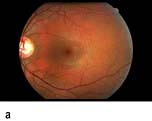
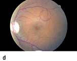
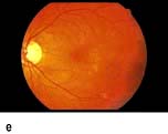
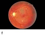
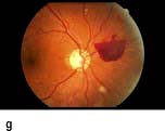
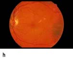
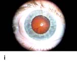
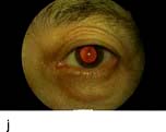
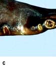
Figure 20.13 Features of diabetic eye disease. (a) The normal macula (centre) and optic disc (to left). (b) Microaneurysms (small circles) and blot haemorrhage (larger circle) – early background retinopathy. (c) Hard exudates (circled) and single cotton wool spot (arrowed) in addition to multiple blot haemorrhages in background retinopathy. (d) Intra-retinal microvascular abnormalities (IRMA) – pre-proliferative retinopathy (circled). (e) Venous loop (circled) also indicates pre-proliferative change. (f) Fronds of new vessels on the disc and elsewhere (proliferative). (g) Pre-retinal haemorrhage in proliferative disease. (h) Hard exudates within a disc-width of the macula (maculopathy). (i) Cortical and (j) central cataracts can be seen against the red reflex with the ophthalmoscope.

image

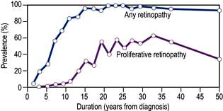
Figure 20.14 Prevalence of retinopathy in relation to duration of the disease in patients with type 1 diabetes mellitus diagnosed under the age of 33 years. Almost all eventually develop background change and 60% progress to proliferative retinopathy.

(Data from Archives of Ophthalmology 1984; 102:520.)

Table 20.11 Grading of pathological changes in the retina in diabetic retinopathy: the action needed

Retinopathy grade Retinal abnormality (cause) Action needed

Peripheral retina


 Background (R1)

Dot haemorrhages (capillary microaneurysms) (usually appear first)

Blot haemorrhages (leakage of blood into deeper retinal layers)

Hard exudates (exudation of plasma rich in lipids and protein)

Cotton wool spots/Cytoid bodies

Annual screening only

 Pre-proliferative (R2)

Venous beading/loops

Intraretinal microvascular abnormalities – IRMAs

Multiple deep round haemorrhages

Non-urgent referral to an ophthalmologist

 Proliferative (R3)

New blood vessel formation/neovascularization

Preretinal or subhyaloid haemorrhage

Vitreous haemorrhage

Urgent referral to an ophthalmologist

 Advanced retinopathy

Retinal fibrosis

Traction retinal detachment

Urgent referral to an ophthalmologist – but much vision already lost

Central retina


 Maculopathy (M1)

Hard exudates within one disc-width of macula

Lines or circles of hard exudates within 2 disc-widths of macula

Microaneurysms or retinal haemorrhages within 1 disc-width of macula if associated with an unexplained visual acuity 6/12 or worse

Referral to an ophthalmologist soon

Note: Hard exudates have a bright yellowish-white colour and are often irregular in outline with a sharply defined margin. Cotton-wool spots are greyish-white, have indistinct margins and a dull matt surface, unlike the glossy appearance of hard exudates. R, retinopathy; M, maculopathy.

Peripheral retina

Damage to the wall of small vessels causes microaneurysms (small red dots) within the retina. When vessel walls are breached superficial (blot) haemorrhages occur in the ganglion cell and outer plexiform layers. Damaged blood vessels leak fluid into the retina. The fluid is cleared into the retinal veins leaving behind protein and lipid deposits causing hard exudates. These are eventually cleared by macrophages.

Micro-infarcts within the retina due to occluded vessels cause cotton wool spots. The spot itself is due to the accumulation of axoplasmic debris. This debris is removed by macrophages. As this occurs, there may be white dots at the site of the previous cotton wool spot (cytoid bodies). Damage to the walls of veins causes their calibre to vary (venous beading), and elongation to occur causing venous loops. Blockage of blood vessels leads to areas of capillary non-perfusion. Ischaemia in these areas causes the release of vascular growth factors such as VEGF (vascular endothelial growth factor). These factors cause new blood vessels to grow in the retina (neovascularization). Some of these new blood vessels are inside the retina and are helpful. These new intraretinal vessels, and other vessels whose walls are damaged and dilated, give the appearance of intraretinal microvascular abnormalities (IRMAs).

Other new vessels emerge through the retina and lie on its surface, usually at the margin of an area of capillary closure. The normal shearing stresses that occur within the eye can cause these poorly supported new vessels to bleed. Small haemorrhages give rise to pre-retinal haemorrhages (boat-shaped haemorrhages). With further bleeding vitreous haemorrhage occurs with consequent sudden loss of vision. Later collagen tissue grows along the margins of the new vessels and giving rise to fibrotic bands. These bands may contract and pull on the retina causing further haemorrhage and retinal detachment. Sometimes vessels may be induced to grow on the pupil margin (rubeosis) and in the angle of the anterior chamber of the eye giving rise to rapid increase in intraocular pressure (rubeotic glaucoma). These features of retinopathy in the peripheral retina are grouped, according to the risk of visual loss, into three stages (Table 20.11).

FURTHER READING

Antonetti DA et al. Diabetic retinopathy. N Engl J Med 2011; 366:1227–1239.

Macular area

Fluid from leaking vessels is cleared poorly in the macular area due to its anatomy differing from the rest of the retina. Above a certain rate of formation, clearance fails and macular oedema occurs. This distorts and thickens the retina at the macula. If sustained, this distortion causes loss of central vision. Macular oedema is not visible with the ophthalmoscope or with retinal photography. For this reason surrogate markers for the presence of macular oedema are used (Table 20.11). Capillary occlusion in the macular area will also cause loss of central vision.

Examination

Bedside examination of the eye. Visual acuity should be checked using both a pinhole and the patient’s distance spectacles. The ocular movements are assessed to detect any ocular motor palsies. The iris is examined for rubeosis and then the pupils dilated with 1% tropicamide. About 20 minutes later the eye is examined for the presence of a cataract by looking at the lens with a +10.00 lens in the ophthalmoscope and viewing the lens against the red reflex. The retina is then examined systematically looking at the disc, then all four quadrants, and finally the macula. The macula is examined last because this induces the greatest discomfort, and pupillary constriction.

Eye screening

Screening for sight-threatening eye disease with universal access is seen as offering the best hope of displacing diabetes as the commonest cause of blindness in those under 65 years of age. The National Screening Committee in the UK has helped establish digital photography-based screening across the country, based on a national set of standards. All people with diabetes, over the age of 12 are offered annual measurement of their acuity, and photographs of their retina. Box 20.5 shows standardized criteria for screening schemes; these are regularly inspected.

image Box 20.5

Criteria for a successful local screening scheme for sight-threatening diabetic retinopathy

Clearly defined geographical area for the screening programme

Adequate number of people with diabetes for viability (>12 000)

An identified screening programme manager

An identified clinical screening lead

An identified hospital eye service for diagnosis and laser treatment

Computer software capable of supporting call/recall of patients and image grading

Centralized appointment administration

Single collated list of all people with diabetes in the area over the age of 12

Equipment to obtain adequate disc and macula centred images of each eye

Single image grading centre

Process to manage people with poor quality images

Clear route of referral for treatment, and for feedback from treatment centre to screening unit

Accreditation of screening staff

Annual reporting of service performance

Management of diabetic eye disease (Table 20.11)

Cataract

Extraction and intraocular lens implantation is indicated if the cataract is causing visual disability to the patient or is giving rise to inability to view the retina adequately. Cataract extraction is straightforward if there is no retinopathy present. Pre-existing retinopathy may worsen after cataract extraction.

Retinopathy

The DCCT and UKPDS show that the risk of developing diabetic eye disease, and the risk of established retinopathy developing further, can be reduced by tight metabolic control of both diabetes and blood pressure. Development or progression of retinopathy may be accelerated by rapid improvement in glycaemic control, pregnancy and in those with nephropathy, and these groups need frequent monitoring. Fluorescein angiography (a fluorescent dye is injected into an arm vein and photographed in transit through the retinal vessels) is used to define the extent of the potentially sight-threatening diabetic retinopathy. Ocular coherence tomography (OCT) is used to image the content of the layers of the retina at the macula, and in particular to measure retinal thickness. It can detect macular oedema and other macular abnormalities.

Treatment of proliferative retinopathy

New vessels are an indication for laser photocoagulation therapy. New vessels on the disc carry the worst prognosis and warrant urgent laser therapy. The laser should be directed at the new vessels and, in addition, to the associated areas of capillary non-perfusion (ischaemia). If the proliferative retinopathy has progressed to new vessels developing on the optic disc then a technique known as panretinal photocoagulation (PRP) is carried out. This involves multiple laser burns to the peripheral retina, especially in the areas of capillary non-perfusion. Rubeosis is also treated with panretinal photocoagulation. If some bleeding has occurred but there is a good view then laser treatment should be applied. Vitreoretinal surgery is used if bleeding is recurrent and preventing laser therapy. It is also used to try to salvage some vision after vitreous haemorrhage and to treat fibrotic traction retinal detachment in advanced retinopathy.

Treatment of maculopathy

Extrafoveal exudates can be watched. However, if they are beginning to encroach on the fovea then the centre of any rings of exudates, should be treated by laser photocoagulation. If oedema has spread into the centre of the macula, then a technique known as grid photocoagulation is used, where a number of laser burns are scattered around the macula. This limits deterioration in vision, and on occasion results in some visual improvement. Ischaemic maculopathy is not treatable, and leads to central visual loss.

The future

Anti-VEGF drugs, such as bevacizumab and ranibizumab (see p. 1064) are being used to control diabetic retinopathy and diabetic maculopathy, particularly that which involves the centre of the macula and is causing sight loss. Recent studies have shown benefit over laser for this type of maculopathy.

FURTHER READING

Googe J, Brucker AJ, Bressler NM et al.; of the Diabetic Retinopathy Clinical Research Network. Randomized trial evaluating short-term effects of intravitreal ranibizumab or triamcinolone acetonide on macular edema after focal/grid laser for diabetic macular edema in eyes also receiving panretinal photocoagulation. Retina 2011; 31:1009–1027.

Michaelides M, Kaines A, Hamilton RD et al. A prospective randomized trial of intravitreal bevacizumab or laser therapy in the management of diabetic macular edema (BOLT study) 12-month data: report 2. Ophthalmology 2010; 117:1078–1086.

The diabetic kidney

The kidney may be damaged by diabetes in three main ways:

image Glomerular damage

image Ischaemia resulting from hypertrophy of afferent and efferent arterioles

image Ascending infection.

Diabetic nephropathy
Epidemiology

Clinical nephropathy secondary to glomerular disease usually manifests 15–25 years after diagnosis of diabetes and affects 25–35% of patients diagnosed under the age of 30 years. It is the leading cause of premature death in young diabetic patients. Older patients also develop nephropathy, but the proportion affected is smaller. The incidence of end-stage kidney disease has fallen in recent decades, probably due to better control of blood glucose and blood pressure, but this benefit has been cancelled out by the rising incidence of both types of diabetes.

Pathophysiology

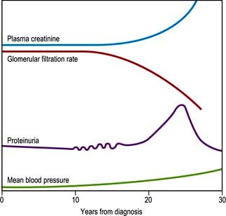
The earliest functional abnormality in the diabetic kidney is renal hypertrophy associated with a raised glomerular filtration rate. This appears soon after diagnosis and is related to poor glycaemic control. As the kidney becomes damaged by diabetes, the afferent arteriole (leading to the glomerulus) becomes vasodilated to a greater extent than the efferent glomerular arteriole. This increases the intraglomerular filtration pressure, further damaging the glomerular capillaries. This increased intraglomerular pressure also leads to increased local shearing forces which are thought to contribute to mesangial cell hypertrophy and increased secretion of extracellular mesangial matrix material. This process eventually leads to glomerular sclerosis. The initial structural lesion in the glomerulus is thickening of the basement membrane. Associated changes result in disruption of the protein cross-linkages which normally make the membrane an effective filter. In consequence, there is a progressive leak of large molecules (particularly protein) into the urine.

Albuminuria

The earliest evidence of this is ‘microalbuminuria’ – amounts of urinary albumin so small as to be undetectable by standard dipsticks (see p. 309). Microalbuminuria may be tested for by radioimmunoassay or by using special dipsticks. It is a predictive marker of progression to nephropathy in type 1 diabetes, and of increased cardiovascular risk in type 2 diabetes. Microalbuminuria may, after some years, progress to intermittent albuminuria followed by persistent proteinuria. Light-microscopic changes of glomerulosclerosis become manifest; both diffuse and nodular glomerulosclerosis can occur. The latter is sometimes known as the Kimmelstiel–Wilson lesion. At the later stage of glomerulosclerosis, the glomerulus is replaced by hyaline material.

At the stage of persistent proteinuria, the plasma creatinine is normal but the average patient is only some 5–10 years from end-stage kidney disease. The proteinuria may become so heavy as to induce a transient nephrotic syndrome, with peripheral oedema and hypoalbuminaemia.

Patients with nephropathy typically show a normochromic normocytic anaemia and a raised erythrocyte sedimentation rate (ESR). Hypertension is a common development and may itself damage the kidney still further. A rise in plasma creatinine is a late feature that progresses inevitably to renal failure, although the rate of progression may vary widely between individuals.

The natural history of this process is shown in Figure 20.15.

image

Figure 20.15 Schematic representation of the natural history of nephropathy. The typical onset is 15 years after diagnosis. Intermittent proteinuria leads to persistent proteinuria. In time, the plasma creatinine rises as the glomerular filtration rate falls.

Ischaemic lesions

Arteriolar lesions, with hypertrophy and hyalinization of the vessels, can occur in patients with diabetes. The appearances are similar to those of hypertensive disease and lead to ischaemic damage to the kidneys.

Infective lesions

Urinary tract infections are relatively more common in women with diabetes, but not in men. Ascending infection may occur because of bladder stasis resulting from autonomic neuropathy, and infections more easily become established in damaged renal tissue. Autopsy material frequently reveals interstitial changes suggestive of infection, but ischaemia may produce similar changes and the true frequency of pyelonephritis in diabetes is uncertain. Untreated infections in diabetics can result in renal papillary necrosis, in which renal papillae are shed in the urine, but this complication is now very rare.

Diagnosis

The urine of all diabetic patients should be checked regularly (at least annually) for the presence of protein. Many centres also screen younger patients for microalbuminuria since there is evidence that meticulous glycaemic control and early antihypertensive treatment, particularly with ACE inhibitors and angiotensin 2 blockers, may delay the onset of frank proteinuria. The albumin creatinine ratio (ACR) (tested on a mid-stream first morning urine sample) is <2.5 in healthy men, <3.5 mg/mmol in healthy women. Once proteinuria is present, other possible causes should be considered (see below), but once these are excluded, a presumptive diagnosis of diabetic nephropathy can be made. For practical purposes this implies inevitable progression to end-stage kidney disease, although the time course can be markedly slowed by early aggressive antihypertensive therapy. Clinical suspicion of a non-diabetic cause of nephropathy may be provoked by an atypical history, the absence of diabetic retinopathy (usually but not invariably present with diabetic nephropathy) and the presence of red-cell casts in the urine. Renal biopsy should be considered in such cases, but is rarely necessary or helpful. A 24-hour urine collection is often performed to quantify protein loss. Regular measurement is made of the plasma creatinine level with estimated glomerular filtration rate (eGFR).

FURTHER READING

Sun W et al. Intensive diabetes therapy and glomerular filtration rate in type 1 diabetes mellitus. N Engl J Med 2011; 365:2366–2376.

Management

The management of diabetic nephropathy is similar to that of other causes of chronic kidney disease, with the following provisos:

image Aggressive treatment of blood pressure with a target below 130/80 mmHg has been shown to slow the rate of deterioration of renal failure considerably. Angiotensin-converting enzyme inhibitors or an angiotensin receptor II antagonist are the drugs of choice (see Chapter 8). These drugs should also be used in normotensive patients with persistent microalbuminuria. Reduction in albuminuria occurs with this treatment.

image Oral hypoglycaemic agents partially excreted via the kidney (e.g. glibenclamide and metformin) should be avoided.

image Insulin sensitivity increases and drastic reductions in insulin dosage may be needed.

image Associated diabetic retinopathy tends to progress rapidly, and frequent ophthalmic supervision is essential.

Management of end-stage disease is made more difficult by the fact that patients often have other complications of diabetes such as blindness, autonomic neuropathy or peripheral vascular disease. Vascular shunts tend to calcify rapidly and hence chronic ambulatory peritoneal dialysis may be preferable to haemodialysis. The failure rate of renal transplants is somewhat higher than in non-diabetic patients. A segmental pancreatic or islet graft is sometimes performed under cover of the immunosuppression needed for the renal graft, and this has been shown to improve survival as well as offering freedom from insulin injections.

Diabetic neuropathy

Diabetes can damage peripheral nervous tissue in a number of ways. The vascular hypothesis postulates occlusion of the vasa nervorum as the prime cause. This seems likely in isolated mononeuropathies, but the diffuse symmetrical nature of the common forms of neuropathy implies a metabolic cause. Since hyperglycaemia leads to increased formation of sorbitol and fructose in Schwann cells, accumulation of these sugars may disrupt function and structure.

The earliest functional change in diabetic nerves is delayed nerve conduction velocity; the earliest histological change is segmental demyelination, caused by damage to Schwann cells. In the early stages axons are preserved, implying prospects of recovery, but at a later stage irreversible axonal degeneration develops.

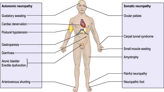
The following varieties of neuropathy occur (Fig. 20.16):

image Symmetrical mainly sensory polyneuropathy (distal)

image Acute painful neuropathy

image Mononeuropathy and mononeuritis multiplex

cranial nerve lesions
isolated peripheral nerve lesions

image Diabetic amyotrophy (asymmetrical motor diabetic neuropathy)

image Autonomic neuropathy.

image

Figure 20.16 The neuropathic man.

Symmetrical mainly sensory polyneuropathy

This is often unrecognized by the patient in its early stages. Early clinical signs are loss of vibration sense, pain sensation (deep before superficial) and temperature sensation in the feet. At later stages patients may complain of a feeling of ‘walking on cotton wool’ and can lose their balance when washing the face or walking in the dark owing to impaired proprioception. Involvement of the hands is much less common and should prompt a search for non-diabetic causes. Complications include unrecognized trauma, beginning as blistering due to an ill-fitting shoe or a hot-water bottle, and leading to ulceration.

Sequelae of neuropathy. Involvement of motor nerves to the small muscles of the feet gives rise to interosseous wasting. Unbalanced traction by the long flexor muscles leads to a characteristic shape of the foot, with a high arch and clawing of the toes, which in turn leads to abnormal distribution of pressure on walking, resulting in callus formation under the first metatarsal head or on the tips of the toes and perforating neuropathic ulceration. Neuropathic arthropathy (Charcot’s joints) may sometimes develop in the ankle. The hands show small-muscle wasting as well as sensory changes, but these signs and symptoms must be differentiated from those of the carpal tunnel syndrome, which occurs with increased frequency in diabetes and may be amenable to surgery.

Acute painful neuropathy

A diffuse, painful neuropathy is less common. The patient describes burning or crawling pains in the feet, shins and anterior thighs. These symptoms are typically worse at night, and pressure from bedclothes may be intolerable. It may present at diagnosis or develop after sudden improvement in glycaemic control (e.g. when insulin is started). It usually remits spontaneously after 3–12 months if good control is maintained. A more chronic form, developing later in the course of the disease, is sometimes resistant to almost all forms of therapy. Neurological assessment is difficult because of the hyperaesthesia experienced by the patient, but muscle wasting is not a feature and objective signs can be minimal.

Management is firstly to explore for non-diabetic causes (see p. 1146). Explanation and reassurance about the high likelihood of remission within months may be all that is needed. Duloxetine (NICE recommend as first-line therapy), tricyclics, gabapentin or pregabalin, mexiletine, valproate and carbamazepine all reduce the perception of neuritic pain somewhat, but usually not as much as patients hope for. Transepidermal nerve stimulation (TENS) benefits some patients. Topical capsaicin-containing creams help occasionally. A few report that acupuncture has helped.

FURTHER READING

Tesfaye S. Advances in management of painful diabetic neuropathy. Curr Opin Support Palliat Care 2009; 3:136–143.

Mononeuritis and mononeuritis multiplex (multiple mononeuropathy)

Any nerve in the body can be involved in diabetic mononeuritis; the onset is typically abrupt and sometimes painful. Radiculopathy (i.e. involvement of a spinal root) may also occur.

Isolated palsies of nerves to the external eye muscles, especially the third and sixth nerves, are more common in diabetes. A characteristic feature of diabetic third nerve lesions is that pupillary reflexes are retained owing to sparing of pupillomotor fibres. Full spontaneous recovery is the rule for most episodes of mononeuritis over 3–6 months. Lesions are more likely to occur at common sites for external pressure palsies or nerve entrapment (e.g. the median nerve in the carpal tunnel, see p. 1074).

Diabetic amyotrophy

This condition is usually seen in older men with diabetes. Presentation is with painful wasting, usually asymmetrical, of the quadriceps muscles or occasionally in the shoulders. The wasting may be very marked and knee reflexes are diminished or absent. The affected area is often extremely tender. Extensor plantar responses sometimes develop and CSF protein content is elevated. Diabetic amyotrophy is usually associated with periods of poor glycaemic control and may be present at diagnosis. It often resolves in time with careful metabolic control of the diabetes.

Autonomic neuropathy

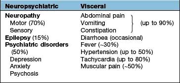
Asymptomatic autonomic disturbances can be demonstrated on testing in many patients (Box 20.6), but symptomatic autonomic neuropathy is rare. It affects both the sympathetic and parasympathetic nervous systems and can cause disabling postural hypotension.

image Box 20.6

Bedside testing of autonomic function

  Normal Abnormal

Supine to erect blood pressure: systolic BP fall (mmHg)

10

≥30

Heart rate responses to:


Deep breathing (6 breaths over 1 min) max to min HR

≥15

≤10

Valsalva manoeuvre (15 s): ratio of longest to shortest R–R interval

≥1.21

≤1.20

Lying to standing ratio of R–R interval of 30th to 15th beats

≥1.04

≤1.00

R–R, time between R and next R on ECG; HR, heart rate.

The cardiovascular system

Vagal neuropathy results in tachycardia at rest and loss of sinus arrhythmia. At a later stage, the heart may become denervated (resembling a transplanted heart). Cardiovascular reflexes such as the Valsalva manoeuvre are impaired. Postural hypotension occurs owing to loss of sympathetic tone to peripheral arterioles. A warm foot with a bounding pulse is sometimes seen in a polyneuropathy as a result of peripheral vasodilatation.

Gastrointestinal tract

Vagal damage can lead to gastroparesis, often asymptomatic, but sometimes leading to intractable vomiting. Implantable devices which stimulate gastric emptying, and injections of botulinum toxin into the pylorus (to partly paralyse the sphincter), have each shown benefit in cases of this previously intractable problem. Autonomic diarrhoea characteristically occurs at night accompanied by urgency and incontinence. Diarrhoea and steatorrhoea may occur owing to small bowel bacterial overgrowth; treatment is with antibiotics such as tetracycline.

Bladder involvement

Loss of tone, incomplete emptying and stasis (predisposing to infection) can occur, and may ultimately result in an atonic, painless, distended bladder. Treatment is with intermittent self-catheterization, permanent catheterization if that fails and prophylactic antibiotic therapy for those prone to recurrent infection.

Male erectile dysfunction

This is common. The first manifestation is incomplete erection which may in time progress to total failure; retrograde ejaculation also occurs in patients with autonomic neuropathy. Erectile dysfunction in diabetes has many causes including anxiety, depression, alcohol excess, drugs (e.g. thiazides and beta-blockers), primary or secondary gonadal failure, hypothyroidism and inadequate vascular supply owing to atheroma in pudendal arteries. The history and examination should focus on these possible causes. Blood is taken for LH, FSH, testosterone, prolactin and thyroid function. Treatment should ideally include sympathetic counselling of both partners.

Phosphodiesterase type-5 inhibitors (sildenafil, tadalafil, vardenafil, avanafil), which enhance the effects of nitric oxide on smooth muscle and increase penile blood flow, are used in those who do not take nitrates for angina. Some 60% of patients can be expected to benefit from this therapy.

Alternatives for those who fail to improve with a phosphodiesterase inhibitor, who dislike the side-effects (headache and a green tinge to vision the next day) or those in whom it is contraindicated, are:

image Apomorphine 2 or 3 mg sublingually 20 min before sexual activity

image Alprostadil (prostaglandin E1 preparation) given as a small pellet inserted with a device into the urethra (125 µg initially with a maximum of 500 µg). If the partner is pregnant, barrier contraception must be used to keep prostaglandin away from the fetus

image Intracavernosal injection or insertion of a pellet of alprostadil urethra into the urethra (2.5 µg initially with a maximum of 40 µg). Side-effects include priapism which needs urgent treatment should erection last more than 3 hours

image Vacuum devices.

The diabetic foot

A total of 10–15% of diabetic patients develop foot ulcers at some stage in their lives. Diabetic foot problems are responsible for nearly 50% of all diabetes-related hospital admissions. Many diabetic limb amputations could be delayed or prevented by more effective patient education and medical supervision. Ischaemia, infection and neuropathy combine to produce tissue necrosis. Although all these factors may co-exist, the ischaemic and the neuropathic foot (Table 20.12) can be distinguished. In rural India, foot ulcers are commonly due to neuropathic and infective causes rather than vascular causes.

Table 20.12 Distinguishing features between ischaemia and neuropathy in the diabetic foot

  Ischaemia Neuropathy

Symptoms

Claudication

Usually painless

Rest pain

Sometimes painful neuropathy

Inspection

Dependent rubor

High arch

Trophic changes

Clawing of toes


No trophic changes

Palpation

Cold

Warm

Pulseless

Bounding pulses

Ulceration

Painful

Painless

Heels and toes

Plantar

image image image

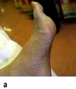
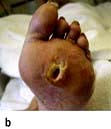
Diabetic foot. (a) High arch and clawing of toes. (b) Typical neuropathic plantar ulceration. (c) Vascular pattern of ulceration.

Management

Many diabetic foot problems are avoidable, so patients need to learn the principles of foot care (Table 20.13). Older patients should visit a chiropodist/podiatrist regularly and should not cut their own toenails. Once tissue damage has occurred in the form of ulceration or gangrene, the aim is preservation of viable tissue. The four main threats to the skin and subcutaneous tissues are:

image Infection. This can take hold rapidly in a diabetic foot. Early antibiotic treatment is essential, with antibiotic therapy adjusted in the light of culture results. The organisms grown from the skin surface may not be the organism causing deeper infection. Collections of pus are drained and excision of infected bone is needed if osteomyelitis develops and does not respond to appropriate antibiotic therapy. Regular X-rays of the foot are needed to check on progress.

image Ischaemia. The blood flow to the feet is assessed clinically and with Doppler ultrasound. Femoral angiography is used to localize areas of occlusion amenable to bypass surgery or angioplasty. Relatively few patients fall into this category.

image Abnormal pressure. An ulcerated site must be kept non-weight-bearing. Resting the affected leg may need to be supplemented with special deep shoes and insoles to move pressure away from critical sites, or by removable or non-removable casts of the leg. After healing, special shoes and insoles are likely to continue to be needed to protect the feet and prevent abnormal pressure repeating damage to a healed area. In neuropathic feet particularly, sharp surgical debridement by a chiropodist is necessary to prevent callus distorting the local wound architecture and causing damage through abnormal pressure on normal skin nearby.

image Wound environment. Dressings are used to absorb or remove exudate, maintain moisture, and protect the wound from contaminating agents, and should be easily removable. Expensive new dressings containing growth factors and other biologically active agents may have a role to play in future, but their place is still being assessed.

Table 20.13 Principles of diabetic foot care

Inspect feet daily

Moisturize dry skin

Seek early advice for any damage

Check shoes inside and out for sharp bodies/areas before wearing

Use lace-up shoes with plenty of room for the toes

Keep feet away from sources of heat (hot sand, hot-water bottles, radiators, fires)

Check the bath temperature before stepping in

Regular podiatrist care

Good liaison between physician, chiropodist/podiatrist and surgeon is essential if periods in hospital are to be used efficiently. When irreversible arterial insufficiency is present, it is often quicker and kinder to opt for an early major amputation rather than subject the patient to a debilitating sequence of conservative procedures.

FURTHER READING

Jude EB, Eleftheriadou I, Tentolouris N. Peripheral arterial disease in diabetes – a review. Diabetes Med 2010; 27:4–14.

Infections

There is no evidence that diabetic patients with good glycaemic control are more prone to infection than normal subjects. However, poorly-controlled diabetes entails increased susceptibility to the following infections:

image Skin

staphylococcal infections (boils, abscesses, carbuncles)
mucocutaneous candidiasis

image Gastrointestinal tract

periodontal disease
rectal and ischiorectal abscess formation (when control very poor)

image Urinary tract

urinary tract infections (in women)
pyelonephritis
perinephric abscess

image Lungs

staphylococcal and pneumococcal pneumonia
Gram-negative bacterial pneumonia
tuberculosis

image Bone

spontaneous staphylococcal spinal osteomyelitis.

One reason why poor control leads to infection is that chemotaxis and phagocytosis by polymorphonuclear leucocytes are impaired because at high blood glucose concentrations neutrophil superoxide generation is impaired.

Conversely, infections may lead to loss of glycaemic control, and are a common cause of ketoacidosis. Insulin-treated patients need to increase their dose by up to 25% in the face of infection, and non-insulin-treated patients may need insulin cover while the infection lasts. Patients should be told never to omit their insulin dose, even if they are nauseated and unable to eat; instead they should test their blood glucose frequently and seek urgent medical advice. Diabetic patients should receive pneumococcal vaccine and yearly influenza vaccine.

Diabetes and cancer

Certain types of cancer are more common in type 2 diabetes. The risk of carcinoma of the uterus and of the pancreas is approximately doubled, and there is a 20–50% increase in the risk of colorectal and breast cancer. These associations appear to be mediated by obesity, which confers similar levels of risk in the absence of hyperglycaemia, although there is also an element of reverse causation with carcinoma of the pancreas, which can precipitate or cause diabetes. Metformin-treated patients have been reported to have a lower cancer risk than those on other therapies, and this agent is under investigation for possible anti-tumour properties.

Skin and joints

Joint contractures in the hands are a common consequence of childhood diabetes. The sign may be demonstrated by asking the patient to join the hands as if in prayer; the metacarpophalangeal and interphalangeal joints cannot be opposed. Thickened, waxy skin can be noted on the backs of the fingers. These features may be due to glycosylation of collagen and are not progressive (see also p. 1220). The condition is sometimes referred to as diabetic cheiroarthropathy.

Osteopenia in the extremities is also described in type 1 diabetes but rarely leads to clinical consequences.

FURTHER READING

The ADVANCE Collaborative Group. Intensive blood glucose control and vascular outcomes in patients with type 2 diabetes. N Engl J Med 2008; 358:2560–2572.

Camilleri M. Diabetic gastroparesis. N Engl J Med 2007; 356:820–829.

The Diabetes Control and Complications Trial/Epidemiology of Diabetes Interventions and Complications Research Group. Prolonged effect of intensive therapy on the risk of retinopathy complications in patients with type 1 diabetes mellitus. Arch Ophthalmol 2008; 126:1707–1715.

Duckworth W, Abraira C, Moritz T et al. Glucose control and vascular complications in veterans with type 2 diabetes. N Engl J Med 2009; 360:129–39.

McVary KT. Erectile dsyfunction. N Engl J Med 2007; 357:2473–2481.

Notes on special situations in diabetes

Surgery

Smooth control of diabetes minimizes the risk of infection and balances the catabolic response to anaesthesia and surgery. The procedure for insulin-treated patients is simple:

image Long-acting and/or intermediate insulin should be stopped the day before surgery, with soluble insulin substituted.

image Whenever possible, diabetic patients should be first on the morning theatre list.

image An infusion of glucose, insulin and potassium is given during surgery. The insulin can be mixed into the glucose solution or administered separately by syringe pump. A standard combination is 16 U of soluble insulin with 10 mmol of KCl in 500 mL of 10% glucose, infused at 100 mL/hour.

image Postoperatively, the infusion is maintained until the patient is able to eat. Other fluids needed in the perioperative period must be given through a separate intravenous line and must not interrupt the glucose/insulin/potassium infusion. Glucose levels are checked every 2–4 hours and potassium levels are monitored. The amount of insulin and potassium in each infusion bag is adjusted either upwards or downwards according to the results of regular monitoring of the blood glucose and serum potassium concentrations.

The same approach is used in the emergency situation, with the exception that a separate variable-rate insulin infusion may be needed to bring blood glucose under control before surgery.

Non-insulin-treated patients should stop medication 2 days before the operation. Patients with mild hyperglycaemia (fasting blood glucose below 8 mmol/L) can be treated as non-diabetic. Those with higher levels are treated with soluble insulin prior to surgery, and with glucose, insulin and potassium during and after the procedure, as for insulin-treated patients.

Pregnancy in established diabetes

Pregnancy in diabetes was in the past associated with high fetal mortality, which has been dramatically reduced by meticulous metabolic control of the diabetes and careful obstetric management. Despite this, the rates of congenital malformation and perinatal mortality remain several times higher than in the non-diabetic population. Type 2 diabetes is now much more prevalent in the maternal population as a result of the changing natural history of this condition.

Metabolic control of diabetes in pregnancy

Congenital malformations are associated with poor glucose control in the early weeks of pregnancy, and good control should therefore be in place before conception wherever possible. The mother should perform daily home blood glucose profiles, recording blood tests before and 2 hours after meals. The renal threshold for glucose falls in pregnancy, and urine tests are therefore of little value. Insulin requirements rise progressively, and intensified insulin regimens are generally used. The aim is to maintain blood glucose and fructosamine (or HbA1c) levels as close to the normal range as can be tolerated. Oral antidiabetic therapy should be avoided, except for metformin which is recognized to be safe in pregnancy.

General management

The patient is seen at intervals of 2 weeks or less at a clinic managed jointly by physician and obstetrician. Circumstances permitting, the aim should be outpatient management with a spontaneous vaginal delivery at term. Retinopathy and nephropathy may deteriorate during pregnancy. Digital photographic eye screening and urine testing for protein should be undertaken at booking, at 28 weeks and before delivery.

Obstetric problems associated with diabetes

Congenital malformations associated with maternal diabetes affect cardiac and skeletal development, of which the caudal regression syndrome is an example. Poorly-controlled diabetes later in gestation is associated with stillbirth, mechanical problems in the birth canal owing to fetal macrosomia, hydramnios and pre-eclampsia. Ketoacidosis in pregnancy carries a 50% fetal mortality, but maternal hypoglycaemia, although highly undesirable, is relatively well tolerated by the fetus.

Neonatal problems

Maternal diabetes, especially when poorly controlled, is associated with fetal macrosomia. The infant of a diabetic mother is more susceptible to hyaline membrane disease than non-diabetic infants of similar maturity. In addition, neonatal hypoglycaemia may occur. The mechanism is as follows: maternal glucose crosses the placenta, but insulin does not; the fetal islets hypersecrete to combat maternal hyperglycaemia, and a rebound to hypoglycaemic levels occurs when the umbilical cord is severed. These complications are due to hyperglycaemia in the third trimester.

Gestational diabetes

This term refers to glucose intolerance that develops or is first recognized in the course of pregnancy; it is typically asymptomatic and usually remits following delivery. Gestational diabetes has been estimated to complicate about 7% of all pregnancies, with wide variation due to differences between populations and diagnostic criteria. Women with a previous history of gestational diabetes, older or overweight women, those with a history of large for gestational age babies and women from certain ethnic groups are at particular risk, but many affected women are not in any of these categories. For this reason some advocate screening of all pregnant women on the basis of random plasma glucose testing in each trimester and by oral glucose tolerance testing if the glucose concentration is, for example, 7 mmol/L or more. The Hyperglycemia and Adverse Pregnancy Outcomes (HAPO) study found that the risk of adverse outcomes increased as a function of maternal glucose levels at 24–28 weeks of pregnancy, even when these were within the normal reference range. This has added to the controversy concerning the appropriate cut-off levels for screening and intervention, since the benefits of intervention are marginal at lower glucose levels, while labelling a mother as diabetic may have unwanted consequences such as a higher rate of caesarean section.

Those who meet the diagnostic criteria for diabetes at first presentation are treated with insulin. Treatment for the remainder is with diet in the first instance, although most patients require insulin cover at some stage during pregnancy (target levels: fasting <4.9 mmol/L; postprandial <6.5 mmol/L). Insulin does not cross the placenta. Many oral agents cross the placenta and are usually avoided because of the potential risk to the fetus, although metformin has been used with success when healthcare facilities are limited.

Gestational diabetes has been associated with all the obstetric and neonatal problems described above for preexisting diabetes, except that there is no increase in the rate of congenital abnormalities. Gestational diabetes typically remits after delivery but signals an increased risk of type 2 diabetes in later life; maintaining a low bodyweight and keeping physically active reduce this risk.

Not all diabetes presenting in pregnancy is gestational. True type 1 diabetes may develop, and swift diagnosis is essential to prevent the development of ketoacidosis. Hospital admission is required if the patient is symptomatic, or has ketonuria or a markedly elevated blood glucose level.

FURTHER READING

Ecker JL, Greene MF. Gestational diabetes mellitus. N Engl J Med 2008; 358:2061–2063.

The HAPO Study Cooperative Research Group. Hyperglycemia and adverse pregnancy outcomes. N Engl J Med 2008; 358:1991–2002.

International Association of Diabetes in Pregnancy Study Groups Consensus Panel. International association of diabetes and pregnancy study groups recommendations on the diagnosis and classification of hyperglycemia in pregnancy. Diabetes Care 2010; 33:676–682.

Landon MB, Spong CY, Thom E. A multicenter, randomized trial of treatment for mild gestational diabetes. N Engl J Med 2009; 361:1339–1348.

Unstable diabetes

This term is used to describe patients with recurrent ketoacidosis and/or recurrent hypoglycaemic coma. Of these, the largest group is made up of those who experience recurrent severe hypoglycaemia.

Recurrent severe hypoglycaemia

This affects 1–3% of insulin-dependent patients. Most are adults who have had diabetes for >10 years. By this stage, endogenous insulin secretion is negligible in the great majority of patients. Pancreatic α cells are still present in undiminished numbers, but the glucagon response to hypoglycaemia is virtually absent. Long-term patients are thus subject to fluctuating hyperinsulinaemia owing to erratic absorption of insulin from injection sites, and lack a major component of the hormonal defence against hypoglycaemia. In this situation adrenaline (epinephrine) secretion becomes vital, but this too may become impaired in the course of diabetes. Loss of adrenaline (epinephrine) secretion has been attributed to autonomic neuropathy, but this is unlikely to be the sole cause; central adaptation to recurrent hypoglycaemia may also be a factor.

The following factors may also predispose to recurrent hypoglycaemia:

image Overtreatment with insulin. Frequent biochemical hypoglycaemia lowers the glucose level at which symptoms develop. Symptoms often reappear when overall glucose control is relaxed.

image An unrecognized low renal threshold for glucose. Attempts to render the urine sugar-free will inevitably produce hypoglycaemia.

image Excessive insulin doses. A common error is to increase the dose when a patient needs more frequent injections to overcome a problem of timing.

image Endocrine causes. These include pituitary insufficiency, adrenal insufficiency and premenstrual insulin sensitivity.

image Alimentary causes. These include exocrine pancreatic failure and diabetic gastroparesis.

image Chronic kidney disease. Clearance of insulin is diminished.

image Patient causes. Patients may be unintelligent, uncooperative or may manipulate their therapy.

Recurrent ketoacidosis

This usually occurs in adolescents or young adults, particularly girls. Metabolic decompensation may develop very rapidly. A combination of chaotic food intake and insulin omission, whether conscious or unconscious, is now regarded as the primary cause of this problem. It almost always occurs in the context of considerable psychosocial problems, particularly eating disorders. This area needs careful and sympathetic exploration in any patient with recurrent ketoacidosis. It is perhaps not surprising that in an illness where much of one’s life is spent thinking of and controlling food intake, 30% of women with diabetes have had some features of an eating disorder at some time. Other causes include:

image Iatrogenic. Inappropriate insulin combinations may be a cause of swinging glycaemic control. For example, a once-daily regimen may cause hypoglycaemia during the afternoon or evening and pre-breakfast hyperglycaemia due to insulin deficiency.

image Intercurrent illness. Unsuspected infections, including urinary tract infections and tuberculosis, may be present. Thyrotoxicosis can also manifest as unstable glycaemic control.

Hypoglycaemia in the non-diabetic

Hypoglycaemia develops when hepatic glucose output falls below the rate of glucose uptake by peripheral tissues. Hepatic glucose output may be reduced by:

image The inhibition of hepatic glycogenolysis and gluconeogenesis by insulin

image Depletion of hepatic glycogen reserves by malnutrition, fasting, exercise or advanced liver disease

image Impaired gluconeogenesis (e.g. following alcohol ingestion).

In the first of these categories, insulin levels are raised, the liver contains adequate glycogen stores and the hypoglycaemia can be reversed by injection of glucagon. In the other two situations, insulin levels are low and glucagon is ineffective. Peripheral glucose uptake is accelerated by high insulin levels and by exercise, but these conditions are normally balanced by increased hepatic glucose output.

The most common symptoms and signs of hypoglycaemia are neurological. The brain consumes about 50% of the total glucose produced by the liver. This high energy requirement is needed to generate ATP used to maintain the potential difference across axonal membranes.

Insulinomas

Insulinomas are pancreatic islet cell tumours that secrete insulin. Most are sporadic but some patients have multiple tumours arising from neural crest tissue (multiple endocrine neoplasia). Some 95% of these tumours are benign. The classic presentation is with fasting hypoglycaemia, but early symptoms may also develop in the late morning or afternoon. Recurrent hypoglycaemia is often present for months or years before the diagnosis is made, and the symptoms may be atypical or even bizarre; the presenting features in one series are given in Table 20.14. Common misdiagnoses include psychiatric disorders, particularly pseudodementia in elderly people, epilepsy and cerebrovascular disease. Whipple’s triad remains the basis of clinical diagnosis. This is satisfied when:

image Symptoms are associated with fasting or exercise

image Hypoglycaemia is confirmed during these episodes

image Glucose relieves the symptoms.

Table 20.14 Presenting features of insulinoma

Diplopia

Sweating, palpitations, weakness

Confusion or abnormal behaviour

Loss of consciousness

Grand mal seizures

A fourth criterion – demonstration of inappropriately high insulin levels during hypoglycaemia – may usefully be added to these.

The diagnosis is confirmed by the demonstration of hypoglycaemia in association with inappropriate and excessive insulin secretion. Hypoglycaemia is demonstrated by:

image Measurement of overnight fasting (16 hours) glucose and insulin levels on three occasions. About 90% of patients with insulinomas will have low glucose and non-suppressed (normal or elevated) insulin levels.

image A prolonged 72-hour supervised fast if overnight testing is inconclusive and symptoms persist.

Autonomous insulin secretion is demonstrated by lack of the normal feedback suppression during hypoglycaemia. This may be shown by measuring insulin, C-peptide or proinsulin during a spontaneous episode of hypoglycaemia.

Treatment of insulinoma

The most effective therapy is surgical excision of the tumour, but insulinomas are often very small and difficult to localize. Many techniques can be used to attempt to localize insulinomas. Sensitivity and specificity vary between centres and between operators. These include highly selective angiography, contrast-enhanced high-resolution CT scanning, scanning with radiolabelled somatostatin (some insulinomas express somatostatin receptors) and endoscopic and intraoperative ultrasound scanning. Venous sampling for the detection of ‘hot spots’ of high insulin concentration in the various intra-abdominal veins is still used occasionally.

Medical treatment with diazoxide is useful when the insulinoma is malignant, in patients in whom a tumour cannot be located and in elderly patients with mild symptoms. Symptoms may also remit on treatment with a somatostatin analogue (octreotide or lanreotide).

Hypoglycaemia with other tumours

Hypoglycaemia may develop in the course of advanced neoplasia and cachexia, and has been described in association with many tumour types. Certain massive tumours, especially sarcomas, may produce hypoglycaemia owing to the secretion of insulin-like growth factor-1. True ectopic insulin secretion is extremely rare.

Postprandial hypoglycaemia

If frequent venous blood glucose samples are taken following a prolonged glucose tolerance test, about one in four subjects will have at least one value below 3 mmol/L. The arteriovenous glucose difference is quite marked during this phase, so that very few are truly hypoglycaemic in terms of arterial (or capillary) blood glucose content. Failure to appreciate this simple fact led some authorities to believe that postprandial (or reactive) hypoglycaemia was a potential ‘organic’ explanation for a variety of complaints that might otherwise have been considered psychosomatic. An epidemic of false ‘hypoglycaemia’ followed, particularly in the USA. Later work showed a poor correlation between symptoms and biochemical hypoglycaemia. Even so, a number of otherwise normal people occasionally become pale, weak and sweaty at times when meals are due, and report benefit from advice to take regular snacks between meals.

True postprandial hypoglycaemia may develop in the presence of alcohol, which ‘primes’ the cells to produce an exaggerated insulin response to carbohydrate. The person who substitutes alcoholic beverages for lunch is particularly at risk. Postprandial hypoglycaemia sometimes occurs after gastric surgery, owing to rapid gastric emptying and mismatching of nutrient absorption and insulin secretion. This is referred to as ‘dumping’ but it is now rarely encountered (see p. 251).

Hepatic and renal causes of hypoglycaemia

The liver can maintain a normal glucose output despite extensive damage, and hepatic hypoglycaemia is uncommon. It is particularly a problem with fulminant hepatic failure.

The kidney has a subsidiary role in glucose production (via gluconeogenesis in the renal cortex), and hypoglycaemia is sometimes a problem in terminal renal failure.

Hereditary fructose intolerance occurs in 1 in 20 000 live births and can cause hypoglycaemia (see p. 1040).

Endocrine causes of hypoglycaemia

Deficiencies of hormones antagonistic to insulin are rare but well-recognized causes of hypoglycaemia. These include hypopituitarism, isolated adrenocorticotrophic hormone (ACTH) deficiency and Addison’s disease.

Drug-induced hypoglycaemia

Many drugs have been reported to produce isolated cases of hypoglycaemia, but usually only when other predisposing factors are present:

image Sulfonylureas may be used in the treatment of diabetes or may be taken by non-diabetics in suicide attempts.

image Quinine may produce severe hypoglycaemia in the course of treatment for falciparum malaria.

image Salicylates may cause hypoglycaemia; usually accidental ingestion by children.

image Propranolol can induce hypoglycaemia in the presence of strenuous exercise or starvation.

image Pentamidine used in the treatment of resistant pneumocystis pneumonia (see p. 188).

Alcohol-induced hypoglycaemia

Alcohol inhibits gluconeogenesis. Alcohol-induced hypoglycaemia occurs in poorly-nourished chronic alcoholics, binge drinkers and in children who have taken relatively small amounts of alcohol, since they have a diminished hepatic glycogen reserve. They present with coma and hypothermia (hypothermia is a feature of hypoglycaemia, due to the suppression of central thermoregulation, particularly the shivering response: children manifest hypothermia more frequently due to their high surface area to body mass ratio).

Factitious hypoglycaemia

This is a relatively common variant of self-induced disease and is more common than an insulinoma. Hypoglycaemia is produced by surreptitious self-administration of insulin or sulfonylureas. Many patients in this category have been extensively investigated for an insulinoma. Measurement of C-peptide levels during hypoglycaemia should identify patients who are injecting insulin; sulfonylurea abuse can be detected by chromatography of plasma or urine.

Disorders of lipid metabolism

Lipid physiology

Lipids are insoluble in water, and are transported in the bloodstream as macromolecular complexes. In these complexes, lipids (principally triglyceride, cholesterol and cholesterol esters) are surrounded by a stabilizing coat of phospholipid. Proteins (called apoproteins) embedded into the surface of these ‘lipoprotein’ particles exert a stabilizing function and allow the particles to be recognized by receptors in the liver and the peripheral tissues. The structure of a chylomicron (one type of lipoprotein particle) is illustrated in Figure 20.17.

image

Figure 20.17 Schematic diagram of a chylomicron particle (75–1200 nm) showing apoproteins lying in the surface membrane.

Five principal types of lipoprotein particles are found in the blood (Fig. 20.18). They are structurally different and can be separated in the laboratory by their density and electrophoretic mobility. The larger particles give postprandial plasma its cloudy appearance. More than half of all patients aged under 60 with angiographically confirmed coronary artery disease have a lipoprotein disorder.

image

Figure 20.18 Schematic representation of the sites of origin, interaction between, and fate of, the major lipoprotein particles.

The genes for all the major apoproteins and that for the low-density lipoprotein (LDL) receptor have been isolated, sequenced and their chromosomal sites mapped. Production of abnormal apoproteins is known to produce, or predispose to, several types of lipid disorder, and it is likely that others will be discovered.

Chylomicrons

Chylomicrons (Fig. 20.18) are synthesized in the small intestine postprandially, passing initially into the intestinal lymphatic drainage, then along the thoracic duct into the bloodstream. They contain triglyceride and a small amount of cholesterol and its ester, and provide the main mechanism for transporting the digestion products of dietary fat to the liver and peripheral tissues. Each newly formed chylomicron contains several different apoproteins (B-48, A-I, A-II), and acquires apoproteins C-II and E by transfer from high-density lipoprotein (HDL) particles in the bloodstream. Apoprotein C-II binds to specific receptors in adipose tissue and skeletal muscle and the liver, where the endothelial enzyme, lipoprotein lipase, hydrolyses most of the triglyceride into fatty acids which are used as an energy source or stored. The remaining chylomicron remnant particle, which contains the bulk of the original cholesterol, is taken up by the liver. Apoprotein E on the particle’s surface binds with liver clearance receptors.

Very-low-density lipoprotein (VLDL) particles

These are synthesized continuously in the liver and contain most of the body’s endogenously synthesized triglyceride and a smaller quantity of cholesterol. They are the body’s main source of energy during prolonged fasting. Apoprotein B-100 is an essential component of VLDL. Apoproteins C-II and E are incorporated later into VLDL by transfer from HDL particles. As they pass round the circulation, VLDL particles bind through apoprotein C-II allowing triglyceride to be progressively removed by lipoprotein lipase in the capillary endothelium. This leaves a particle, now depleted of triglyceride and apoprotein C-II, called an intermediate-density lipoprotein (IDL) particle.

Intermediate-density lipoprotein (IDL) particles

These have apoprotein B-100 and apoprotein E molecules on the particle surface. Most IDL particles bind to liver LDL receptors through the apoprotein E molecule and are then catabolized. Some IDL particles have further triglyceride removed (by the enzyme hepatic lipase), producing LDL particles.

Low-density lipoprotein (LDL) particles

LDL particles are the main carrier of cholesterol, and deliver it both to the liver and to peripheral cells. The surface of the LDL particle contains apoprotein B-100, and also apoprotein E. The apoprotein B-100 is the principal ligand for the LDL clearance receptor. This receptor lies within coated pits on the surface of the hepatocyte. Once bound to the receptor, the coated pit invaginates and fuses with liposomes which destroy the LDL particle (Fig. 20.19). The number of hepatic LDL clearance receptors regulates the circulating LDL concentration, which is also influenced by controlling the activity of the rate-limiting enzyme in the cholesterol synthetic pathway, hydroxymethylglutaryl coenzyme A (HMG-CoA) reductase.

image

Figure 20.19 Receptor-mediated endocytosis. LDL receptors are formed in the endoplasmic reticulum and transported via the Golgi apparatus to the surface of a hepatocyte. LDLs bind to these receptors, are internalized and taken up by the endosome. The receptor is recycled back to the surface, while the LDL is broken down by the lysosomes, freeing cholesterol needed for membrane synthesis.

LDL particles can deposit lipid into the walls of the peripheral vasculature. Not all the cholesterol synthesized by the liver is packaged immediately into lipoprotein particles. Some is oxidized into bile salts. Both bile salts and cholesterol are excreted in the bile: both are then reabsorbed through the terminal ileum and recirculated (enterohepatic circulation).

LDL particles become Lp(a) lipoproteins as a result of the linkage of apoprotein (a) to apoprotein B-100 with a single disulphide bond. Raised levels of Lp(a) lipoprotein are a risk factor for cardiovascular disease.

High-density lipoprotein (HDL) particles

Nascent HDL particles are produced in both the liver and intestine. They are disc shaped, seemingly inert and contain apoprotein A-I. They are transmuted into mature particles by the acquisition of phospholipids, and the E and C apoproteins from chylomicrons and VLDL particles in the circulation. The more mature HDL particles take up cholesterol from cells in the peripheral tissues aided by cholesterol-efflux regulatory protein – a product of the ATP-binding cassette transporter 1 gene (ABC1 gene). As it is taken up, the enzyme lecithin cholesterol acyldtransferase (LCAT), activated by the apoprotein A on the particle’s surface, esterifies the sequestered cholesterol. The HDL particle transports cholesterol away from the periphery and may transfer it indirectly to other particles such as VLDL in the circulation or deliver its cholesterol directly to the liver (reverse cholesterol transport) and steroid-synthetic tissues (ovaries, testes, adrenal cortex).

This direct delivery takes place through scavenger-receptor B1. In experimental animals the absence of scavenger-receptor B1 dramatically accelerates the development of atheroma, and genetically programmed overproduction suppresses atheroma formation.

Measurement

When a laboratory measures fasting serum lipids, the majority of the total cholesterol concentration consists of LDL particles with a 20–30% contribution from HDL particles. The triglyceride concentration largely reflects the circulating number of VLDL particles, since chylomicrons are not normally present in the fasted state. If the patient is not fasted, the total triglyceride concentration will be raised owing to the additional presence of triglyceride-rich chylomicrons.

Epidemiology and lipids

LDL and total cholesterol

Population studies have repeatedly demonstrated a strong association between both total and LDL cholesterol concentration and coronary heart risk. There is a strong link between mean fat consumption, mean serum cholesterol concentration and the prevalence of coronary heart disease between countries. The exception is France where the cardiovascular risk is only moderate – perhaps owing to high alcohol consumption. Studies of migrants, particularly of Japanese men migrating to Hawaii, have shown that as diet changes, and cholesterol concentrations rise, so does the cardiovascular risk. Such studies show the role of the environment rather than the genetic make-up of a population.

The Multiple Risk Factor Intervention Trial (MRFIT) screened one-third of a million American men for various cardiovascular risk factors and then followed them for 6 years. Data from this study have shown that although cardiovascular risk rises progressively as total cholesterol concentration increases (Fig. 20.20), the risk increase is modest for individuals with no other cardiovascular risk factors. With each additional risk factor the effect produced by the same difference in cholesterol concentration becomes greatly magnified. The Framingham Study has reproduced these findings in a separate population.

image

Figure 20.20 The Multiple Risk Factor Intervention Trial. Relationship between levels of serum cholesterol and risks of fatal coronary artery disease in a longitudinal study of >361 000 men screened for entry into the trial.

(Data from Stamler J, Wentworth D, Neaton JD. Is relationship between serum cholesterol and risk of death from coronary heart disease continuous and graded? Findings in 356 222 primary screenees of the Multiple Risk Factor Intervention Trial (MRFIT). JAMA 1986; 256:2823.)

HDL cholesterol

Epidemiological studies have shown that higher HDL concentrations protect against cardiovascular disease. Raising HDL by pharmacological means does not, however, necessarily reduce cardiovascular risk. HDL also has effects on the function of platelets and of the haemostatic cascade. These properties may favourably influence thrombogenesis.

VLDL particles (triglycerides)

There is a relatively weak independent link between raised concentrations of (triglyceride-rich) VLDL particles and cardiovascular risk. Very raised triglyceride concentrations (>6 mmol/L) cause a greatly increased risk of acute pancreatitis and retinal vein thrombosis. Hypertriglyceridaemia tends to occur in association with a reduced HDL concentration. Much of the cardiovascular risk associated with ‘hypertriglyceridaemia’ turns out on multivariate analysis to be due to the associated low HDL levels and not to the hypertriglyceridaemia itself.

Chylomicrons

Excess chylomicrons do not confer an excess cardiovascular risk, but raise the total plasma triglyceride concentration.

Hyperlipidaemia

Hyperlipidaemia results from genetic predisposition interacting with an individual’s diet.

Secondary hyperlipidaemia

If a lipid disorder has been detected it is vital to carry out a clinical history, examination and simple special investigations to detect causes of secondary hyperlipidaemia (Table 20.15), which may need treatment in their own right. Hypothyroidism, diabetes, renal disease and abnormal liver function can all raise plasma lipid levels.

Table 20.15 Causes of secondary hyperlipidaemia

Hypothyroidism

Diabetes mellitus (when poorly controlled)

Obesity

Renal impairment

Nephrotic syndrome

Dysglobulinaemia

Hepatic dysfunction

Alcohol in susceptible individuals

Drugs:

Oral contraceptives in susceptible individuals
Retinoids, thiazide diuretics, corticosteroids, op’DDD (used in the treatment of Cushing’s syndrome), sirolimus (and other immunosuppressive agents)

Classification, clinical features and investigation of primary hyperlipidaemias

We have used the functional/genetic classification which has the advantage that the genetic disorders may be grouped by the results of simple lipid biochemistry into causes of:

image Disorders of VLDL and chylomicrons – hypertriglyceridaemia alone

image Disorders of LDL – hypercholesterolaemia alone

image Disorders of HDL

image Combined hyperlipidaemia.

Disorders of VLDL and chylomicrons – hypertriglyceridaemia alone

The majority of cases appear to be due to multiple genes acting together to produce a modest excess of circulating concentration of VLDL particles, such cases being termed polygenic hypertriglyceridaemia.

In a proportion of cases, there will be a family history of a lipid disorder or its effects (e.g. pancreatitis). Such cases are often classified as familial hypertriglyceridaemia. The defect underlying the majority of such cases is not understood. Apoprotein A5 deficiency underlies some cases. The main clinical feature is a history of attacks of pancreatitis or retinal vein thrombosis in some individuals.

Lipoprotein lipase deficiency and apoprotein C-II deficiency

These are rare diseases which produce greatly elevated triglyceride concentrations owing to the persistence of chylomicrons (and not VLDL particles) in the circulation. The chylomicrons persist because the triglyceride within cannot be metabolized if the enzyme lipoprotein lipase is defective, or because the triglycerides cannot gain access to the normal enzyme owing to deficiency of the apoprotein C-II on their surface. Patients present in childhood with eruptive xanthomas, lipaemia retinalis and retinal vein thrombosis, pancreatitis and hepatosplenomegaly. If not identified in childhood, it can present in adults with gross hypertriglyceridaemia resistant to simple measures. The presence of chylomicrons floating like cream on top of fasting plasma suggests this diagnosis. It is confirmed by plasma electrophoresis or ultracentrifugation. An abnormality of apoprotein C can be deduced if the hypertriglyceridaemia improves temporarily after infusing fresh frozen plasma, and lipoprotein lipase deficiency is likely if it does not.

Disorders of LDL – hypercholesterolaemia alone

Heterozygous familial hypercholesterolaemia is an autosomal dominant monogenic disorder present in 1 in 500 of the normal population. The average primary care physician would therefore be expected to have four such patients, but because of clustering within families the prevalence varies. There is an increased prevalence in some racial groups (e.g. French Canadians, Finns, South Africans). Surprisingly, most individuals with this disorder remain undetected. Patients may have no physical signs, in which case the diagnosis is made on the presence of very high plasma cholesterol concentrations which are unresponsive to dietary modification and are associated with a typical family history of early cardiovascular disease. Diagnosis can more easily be made if typical clinical features are present. These include xanthomatous thickening of the Achilles tendons and xanthomas over the extensor tendons of the fingers. Xanthelasma may be present, but is not diagnostic of familial hypercholesterolaemia.

The genetic defect is the underproduction or malproduction of the LDL cholesterol clearance receptor in the liver (Table 20.16). Over 150 different mutations in the LDL receptor have been described to date. Fifty per cent of men with the disease will die by the age of 60, most from coronary artery disease, if untreated.

Table 20.16 The genetic defects underlying some lipoprotein disorders

image

Homozygous familial hypercholesterolaemia is very rare indeed. Affected children have no LDL receptors in the liver. They have a hugely elevated LDL cholesterol concentration, and massive deposition of lipid in arterial walls, the aorta and the skin. The natural history is for death from ischaemic heart disease in late childhood or adolescence. Repeated plasmapheresis has been used to remove LDL cholesterol with some success. Liver transplantation is a ‘cure’. Plasma lipids normalize and xanthomas regress after transplantation, but the number of patients having undergone this procedure is small.

Mutations in the apoprotein B-100 gene cause another relatively common single gene disorder. Since LDL particles bind to their clearance receptor in the liver through apoprotein B-100, this defect also results in high LDL concentrations in the blood, and a clinical picture which closely resembles classical heterozygous familial hypercholesterolaemia. The two disorders can be distinguished clearly only by genetic tests. The approach to treatment is the same.

Polygenic hypercholesterolaemia is a term used to lump together patients with raised serum cholesterol concentrations, but without one of the monogenic disorders above. They exist in the right-hand tail of the normal distribution of cholesterol concentration. The precise nature of the polygenic variation in plasma cholesterol concentration remains unknown. Variations in the apoprotein E gene (chromosome 19), and in sterol-regulatory element-binding protein (SREBP)-2 gene, are involved in some individuals in this heterogeneous group.

Disorders of HDL (very low HDL, low total cholesterol)

Tangier disease is an autosomal recessive disorder characterized by a low HDL cholesterol concentration. Cholesterol accumulates in reticuloendothelial tissue and arteries causing enlarged orange-coloured tonsils and hepatosplenomegaly. Cardiovascular disease, corneal opacities and a polyneuropathy also occur. It is due to a gene mutation (ABC1 gene, see HDL physiology above and Table 20.16) which normally promotes cholesterol uptake from cells by HDL particles.

Other mutations in this gene have been found in a few families with autosomal dominant HDL deficiency. It is as yet unknown whether abnormalities of this gene contribute to the low HDL cholesterol concentrations commonly seen in cardiovascular disease patients.

Combined hyperlipidaemia (hypercholesterolaemia and hypertriglyceridaemia)

The most common patient group is a polygenic combined hyperlipidaemia. Patients have an increased cardiovascular risk due to both high LDL concentrations and suppression of HDL by the hypertriglyceridaemia.

Familial combined hyperlipidaemia

This is relatively common, affecting 1 in 200 of the general population. The genetic basis for the disorder has not yet been characterized. It is diagnosed by finding raised cholesterol and triglyceride concentrations in association with a typical family history. There are no typical physical signs.

Remnant hyperlipidaemia

This is a rare (1 in 5000) cause of combined hyperlipidaemia. It is due to accumulation of LDL remnant particles and is associated with an extremely high risk of cardiovascular disease. It may be suspected in a patient with raised total cholesterol and triglyceride concentrations by finding xanthomas in the palmar creases (diagnostic) and the presence of tuberous xanthomas, typically over the knees and elbows (Fig. 20.21). Remnant hyperlipidaemia is almost always due to the inheritance of a variant of the apoprotein E allele (apoprotein E2) together with an aggravating factor such as another primary hyperlipidaemia. When suspected clinically the diagnosis can be confirmed using ultracentrifugation of plasma, or phenotyping apoprotein E.

image

Figure 20.21 Tuberous xanthomas behind the elbow, and lipid deposits in the hand creases in a patient with remnant hyperlipidaemia.

Therapies available to treat hyperlipidaemia

The lipid-lowering diet

Studies have shown that dieticians helping patients to adjust their own diet to meet the nutritional targets set out below produce a better lipid-lowering effect than does the issuing of standard diet sheets and advice from a doctor. The main elements of a lipid-lowering diet are similar to those for people with diabetes (Table 20.5). Additional specific measures are to:

image reduce consumption of liver, offal and fish roes to reduce dietary cholesterol

image reduce alcohol consumption, since this may worsen primary lipid disorders at doses that would not affect normal individuals

image include foods containing plant stanols in the diet. Plant stanols reduce the absorption of cholesterol from the intestine by competing for space in the micelles that deliver lipid to the mucosal cells of the gut. They are largely unabsorbed and excreted in the stool. Increasing the amount of plant stanol in the diet 10-fold by using a margarine (e.g. Benecol) containing added stanol esters lowers LDL cholesterol by approximately 0.35–0.5 mmol/L. A reduction in the risk of heart disease of about 25% would be expected if this reduction in LDL cholesterol was applied to a population.

Drugs

The classes of drugs used to treat hyperlipidaemia are described in Table 20.17. How they are used is set out in Table 20.18. Statins are the most widely used lipid-lowering agents. Generalized muscular aches are the commonest adverse effect, occasionally leading to a frank myopathy. These adverse events occur more frequently in people with a SNP in a gene region coding for a liver-specific organic anion transporter protein (SoLute Carrier Organic anion transporter IBI – SLCOIBI), which leads to a decreased hepatic uptake of the statin and higher statin levels in the serum.

Table 20.17 Drugs used in the management of hyperlipidaemia

image image

Table 20.18 Classes of drugs used to treat hyperlipidaemia

image

Other specific issues in the management of hyperlipidaemias

Screening

Most patients with hyperlipidaemia are asymptomatic and have no clinical signs. Many are discovered during the screening of high-risk individuals. Whose lipids should be measured?

There are great doubts as to whether blanket screening of plasma lipids is warranted. Selective screening of people at high risk of cardiovascular disease should be undertaken:

image A family history of coronary heart disease (especially below 50 years of age)

image A family history of lipid disorders

image The presence of a xanthoma

image The presence of xanthelasma or corneal arcus before the age of 40 years

image Obesity

image Diabetes mellitus

image Hypertension

image Acute pancreatitis

image Those undergoing renal replacement therapy.

Where one family member is known to have a monogenic disorder such as familial hypercholesterolaemia (1 in 500 of the population), siblings and children must have their plasma lipid concentrations measured. It is also worth screening the prospective partners of any patients with this heterozygous monogenic lipid disorder because of the small risk of producing children homozygous for the condition.

Acute severe illnesses such as myocardial infarction can derange plasma lipid concentrations for up to 3 months. Plasma lipid concentrations should be measured either within 48 hours of an acute myocardial infarction (before derangement has had time to occur) or 3 months later.

Serum cholesterol concentration does not change significantly after a meal and as a screening test a random blood sample is sufficient. If the total cholesterol concentration is raised, HDL cholesterol, triglyceride and LDL cholesterol concentrations should be quantitated on a fasting sample. If a test for hypertriglyceridaemia is needed, a fasting blood sample is mandatory.

Treatment of hypertriglyceridaemia

A serum triglyceride concentration below 2.0 mmol/L is normal. In the range 2.0–6.0 mmol/L, no specific intervention will be needed unless there are coincident cardiovascular risk factors, and in particular a strong family history of early cardiovascular death. In general, patients should be advised that they have a minor lipid problem, offered advice on weight reduction if obese, and advice on correcting other cardiovascular risk factors.

If the triglyceride concentration is above 6.0 mmol/L, there is a risk of pancreatitis and retinal vein thrombosis. Patients should be advised to reduce their weight if overweight and start a formal lipid-lowering diet (see below). A proportion of individuals with hypertriglyceridaemia have livers which respond to even moderate degrees of alcohol intake by allowing accumulation or excess production of VLDL particles. If hypertriglyceridaemia persists, lipid measurements should be repeated before and after a 3-week interval of complete abstinence from alcohol. If a considerable improvement results, lifelong abstinence may prove necessary. Other drugs, including thiazides, oestrogens and glucocorticoids, can have a similar effect to alcohol in susceptible patients.

If the triglyceride concentration remains above 6.0 mmol/L, despite the above measures, drug therapy is warranted (Table 20.18). The severe hypertriglyceridaemia associated with the rare disorders of lipoprotein lipase deficiency and apoprotein C-II deficiency may require restriction of dietary fat to 10–20% of total energy intake and the use of special preparations of medium-chain triglycerides in cooking in place of oil or fat. Medium-chain triglycerides are not absorbed via chylomicrons (see p. 260).

Treatment of hypercholesterolaemia (without hypertriglyceridaemia)

Familial hypercholesterolaemia

Individuals often require treatment with diet and more than one cholesterol-lowering drug. The cholesterol absorption inhibitor ezetimibe is a logical addition to a statin and has a low side-effect profile (Tables 20.17 and 20.18). Bile acid sequestrants are an alternative to ezetimibe, but there are problems with tolerability. Concurrent therapy with statins and fibrates, particularly fenofibrate, can be used in severe cases. Checking for muscle symptoms and measuring creatine kinase is necessary.

FURTHER READING

Link E, Parish S, Armitage J et al.; SEARCH Collaborative Group. SLCO 1B1 variants and statin-induced myopathy – a genomewide study. N Engl J Med 2008; 359:789–799.

Primary prevention for people with risk factors

Lipid-lowering therapy using a statin, or alternatives as above is used in asymptomatic individuals irrespective of the total or LDL cholesterol level in type 2 diabetes alone or with two or more of: positive family history of cardiovascular disease, albuminuria, hypertension, smoking.

Primary prevention for people without risk factors

In the absence of risk factors, lipid-lowering therapy should be used in asymptomatic men with LDL cholesterol levels persistently above 6.5 mmol/L despite dietary change. The situation for women is less clear.

Secondary prevention

As a generality, statin treatment is warranted for any patient with known macrovascular disease (coronary artery disease, TIA or stroke, peripheral vascular disease), irrespective of the total or LDL cholesterol level (treatment target is total cholesterol under 4.0, LDL under 2 mmol/L). If a statin is not tolerated, combinations of other agents are tried (Table 20.18).

Risk prevention tables

An array of risk prediction tables (see p. 669) are available to allow quantification of the risk of a patient having a cardiovascular event within the next 10 years. Some advocate the use of these risk tables as a guide to treatment, e.g. if the 10-year risk reaches the 15% or the 30% level. The authors side with those who have reservations over their use. Such risk analyses are a useful approach in helping to decide whether to use treatments such as aspirin. Aspirin probably has no effect until the day an atherosclerotic plaque ruptures, when it may then prevent thrombosis leading to a heart attack or stroke. Furthermore, it has a significant associated morbidity and mortality (from bleeding). At a 10-year cardiovascular risk level of 15% the benefit : risk ratio for aspirin becomes favourable. At the 30% level the benefits are clear.

By contrast, the use of lipid-lowering agents, if initially tolerated, has a low associated morbidity and mortality. These agents probably reduce the rate of atheroma accumulation over a period of decades. When a patient is young, the chance that atheroma will be bad enough to cause a heart attack or stroke within the next 10 years will be small, even if atheroma is accumulating at a swift rate. The level of cardiovascular risk will only rise to the 15% or 30% level when the patient gets older. Yet it seems bizarre not to treat the gradual accumulation of atheroma when the patient is young with a low 10-year risk, and then to start treatment when age causes the 10-year risk levels to rise to a particular threshold, if all other factors are the same. In choosing whether or not to prescribe, we prefer to consider, ‘will he/she live long enough to collect some pension and see his/her grandchildren?’, rather than, ‘will he/she have a heart attack or stroke within the next 10 years?’ The answers to these two questions are very different.

Combined hyperlipidaemia (hypercholesterolaemia and hypertriglyceridaemia)

Treatment is the same for all varieties of combined hyperlipidaemia. For any given cholesterol concentration the hyper-triglyceridaemia found in the combined hyperlipidaemias increases the cardiovascular risk considerably. Treatment is aimed at reducing serum cholesterol below 4.0 mmol/L and triglycerides below 2.0 mmol/L. Therapy is with diet in the first instance and with drugs if an adequate response has not occurred. Fibrates are the treatment of choice since these reduce both cholesterol and triglyceride concentrations, and also have the benefit of raising cardioprotective HDL concentrations. Combination with other agents is often needed (Table 20.18).

Other lipid disorders

Hypolipidaemia

Low lipid levels can be found in severe protein-energy malnutrition. They are also seen occasionally with severe malabsorption and in intestinal lymphangiectasia.

Hypobetalipoproteinaemia (Table 20.16) is a benign familial condition which is being increasingly recognized. The cholesterol levels are in the range 1–3.5 mmol/L.

Abetalipoproteinaemia

This is described on page 270.

FURTHER READING

Ford I, Murray H, Packard CJ et al. Long-term follow-up of the West of Scotland Coronary Prevention Study. N Engl J Med 2007; 357:1477–1486.

Heart Protection Study Collaborative Group. MRC/BHF Heart Protection Study of cholesterol lowering with simvastatin in 20 536 high risk individuals: a randomized placebo-controlled trial. Lancet 2004; 363:757–767.

Inborn errors of carbohydrate metabolism

Glycogen storage disease

All mammalian cells can manufacture glycogen, but the main sites of its production are the liver and muscle. Glycogen is a high-molecular-weight glucose polymer made up of 1–4 linked glucose units, with a 1–6 branch point every 4–10 residues. In glycogen storage disease there is either an abnormality in the molecular structure or an increase in glycogen concentration owing to a specific enzyme defect. Almost all these conditions are autosomal recessive in inheritance and present in infancy, except for McArdle’s disease, which presents in adults.

Table 20.19 shows the classification and clinical features of some of these diseases.

Table 20.19 Some glycogen storage diseases (GSDs)

image

Galactosaemia

Galactose is normally converted to glucose. However, a deficiency of the enzyme galactose-1-phosphate uridyl-transferase or, less commonly, uridine diphosphate galactase-4-epimerase, results in accumulation of galactose-1-phosphate in the blood. The transferase deficiency, inherited as an autosomal recessive, is due in 70% of patients to a glutamine to arginine missense mutation in Q188R. Galactose ingestion (i.e. milk) leads to inanition, failure to thrive, vomiting, hepatomegaly and jaundice, diabetes, cataracts and developmental delay. A lactose-free diet stops the acute toxicity but poor growth and problems with speech and mental development still occur with the transferase deficiency. A newborn screening programme to detect galactosaemia is used in parts of the USA and other countries.

Prenatal diagnosis and diagnosis of the carrier state are possible by measurement of red cell galactose-1-phosphate activity. DNA analysis is available for common mutations.

Galactokinase deficiency also results in galactosaemia and early cataract formation.

Defects of fructose metabolism

Absorbed fructose is chiefly metabolized in the liver to lactic acid or glucose. Three defects of metabolism in the liver and intestine occur; all are inherited as autosomal recessive traits:

image Fructosuria is due to fructokinase deficiency. It is a benign asymptomatic condition.

image Hereditary fructose intolerance is due to fructose-1-phosphate aldolase deficiency. Fructose-1-phosphate accumulates after fructose ingestion, inhibiting both glycogenolysis and gluconeogenesis, resulting in symptoms of severe hypoglycaemia. Hepatomegaly and renal tubular defects occur but are reversible on a fructose- and sucrose-free diet. Intelligence is normal and there is an absence of dental caries.

image Hereditary fructose-1,6-diphosphatase deficiency leads to a failure of gluconeogenesis. Infants present with hypoglycaemia, ketosis and lactic acidosis. Dietary control can lead to normal growth.

Inborn errors of amino acid metabolism

Inborn errors of amino acid metabolism are chiefly inherited as autosomal recessive conditions. The major ones are shown in Table 20.20.

Table 20.20 The major inborn errors of amino acid metabolism

image
Amino acid transport defects

Amino acids are filtered by the glomerulus, but 95% of the filtered load is reabsorbed in the proximal convoluted tubule by an active transport mechanism. Aminoaciduria results from:

image abnormally high plasma amino acid levels (e.g. phenylketonuria)

image any inherited disorder that damages the tubules secondarily (e.g. galactosaemia)

image tubular reabsorptive defects, either generalized (e.g. Fanconi’s syndrome) or specific (e.g. cystinuria).

Amino acid transport defects can be congenital or acquired.

FURTHER READING

Blau N, van Spronsen FJ, Levy HL. Phenylketonuria. Lancet 2010; 376:1417–1427.

Generalized aminoacidurias

Fanconi’s syndrome

This occurs in a juvenile form (De Toni–Fanconi–Debré syndrome); in adult life it is often acquired through, for example, heavy metal poisoning, drugs or some renal diseases. There is a generalized defective proximal tubular reabsorption of: most amino acids, glucose, urate, phosphate, resulting in hypophosphataemic rickets and bicarbonate, with failure to transport hydrogen ions, causing a renal tubular acidosis that then produces a hyperchloraemic acidosis (see p. 664).

Other abnormalities include potassium depletion, primary or secondary to the acidosis, polyuria, increased excretion of immunoglobulins and other low-molecular-weight proteins.

Various combinations of the above abnormalities have been described.

The juvenile form begins at the age of 6–9 months, with failure to thrive, vomiting and thirst. The clinical features are as a result of fluid and electrolyte loss and the characteristic vitamin D-resistant rickets.

In the adult, the disease is similar to the juvenile form, but osteomalacia is a major feature.

Treatment of the bone disease is with large doses of vitamin D (e.g. 1–2 mg of 1 α-hydroxycholecalciferol with regular blood calcium monitoring). Fluid and electrolyte loss needs to be corrected.

Specific aminoacidurias

Cystinuria

There is a defective tubular reabsorption and jejunal absorption of cystine and the dibasic amino acids, lysine, ornithine and arginine. Cystinuria is either a completely or incompletely autosomal recessive disorder with mutations in two genes, SLC3A1 and SLC7A9. Cystine absorption from the jejunum is impaired but, nevertheless, cystine in peptide form can be absorbed. Cystinuria leads to urinary stones and is responsible for approximately 1–2% of all urinary calculi. The disease often starts in childhood, although most cases present in adult life.

Treatment is described on page 603.

The condition cystinosis must not be confused with cystinuria. Cystinosis is characterized by the accumulation of cystine in different organs leading to organ dysfunction, e.g. photophobia and ocular problems, chronic kidney disease. Treatment is with cysteamine.

Hartnup’s disease

There is defective tubular reabsorption and jejunal absorption of most neutral amino acids but not their peptides. The resulting tryptophan malabsorption produces nicotinamide deficiency (see p. 210). Patients can be asymptomatic, but others develop evidence of pellagra (p. 210). Treatment is with nicotinamide.

Other aminoacidurias

Tryptophan malabsorption syndrome (blue diaper syndrome), familial iminoglycinuria and methionine malabsorption syndrome have all been described.

SIGNIFICANT WEBSITE

Human gene mutation database: www.hgmd.cf.ac.uk

Lysosomal storage diseases

Lysosomal storage diseases are due to inborn errors of metabolism which are mainly inherited in an autosomal recessive manner (see Table 20.19).

Glucosylceramide lipidoses: Gaucher’s disease

This is the most prevalent lysosomal storage disease and is due to a deficiency in glucocerebrosidase, a specialized lysosomal acid β-glucosidase. This results in accumulation of glucosylceramide in the lysosomes of the reticuloendothelial system, particularly the liver, bone marrow and spleen. Over 200 mutations have been characterized in the glucocerebrosidase gene (1g21), the most common being a single base change (N370S) causing the substitution of arginine for serine; this is seen in 70% of Jewish patients. The typical Gaucher cell, a glucocerebroside-containing reticuloendothelial histiocyte, is found in the bone marrow, producing many cytokines such as CD14.

There are three clinical types, the most common presenting in childhood or adult life with an insidious onset of hepatosplenomegaly (Type 1). There is a high incidence in Ashkenazi Jews (1 in 3000 births), and patients have a characteristic pigmentation on exposed parts, particularly the forehead and hands. The clinical spectrum is variable, with patients developing anaemia, evidence of hypersplenism and pathological fractures that are due to bone involvement. Nevertheless, many have a normal life-span.

The diagnosis is made on finding reduced glucocerebrosidase in leucocytes. Mutational analysis will confirm the diagnosis. Plasma chitotriosidase (an enzyme secreted by activated macrophages) is grossly elevated in Gaucher’s disease and other lysosomal disorders: it is used to monitor enzyme replacement therapy.

Acute Gaucher’s disease (Type 2) presents in infancy with rapid onset of hepatosplenomegaly, with neurological involvement owing to the presence of Gaucher cells in the brain. The outlook is very poor.

Type 3 presents in childhood or adolescence with a variable progression of hepatosplenomegaly, neurodegeneration and bone disease. Again, the outlook is poor.

Some patients with non-neuropathic (Type 1) Gaucher’s disease show considerable improvement with infusion of human recombinant glucocerebrosidase (imiglucerase – a human recombinant enzyme). Velaglucerase alfa is also used. Oral miglustat (an inhibitor of glucosylceramide synthase) is used for mild to moderate type 1 Gaucher’s disease.

FURTHER READING

Fletcher J, Wilcken B. Neonatal screening for lysosomal storage disorders. Lancet 2012; 379:294–295.

Sphingomyelin cholesterol lipidoses: Niemann–Pick disease

The disease is due to a deficiency of lysosomal sphingomyelinase which results in the accumulation of sphingomyelin cholesterol and glycosphingolipids in the reticuloendothelial macrophages of many organs, particularly the liver, spleen, bone marrow and lymph nodes. The disease usually presents within the first 6 months of life with mental retardation and hepatosplenomegaly; a particular type (11c) presents in adults with dementia. The gene frequency is 1 : 100 in Ashkenazi Jews, the diagnosis being made in the group by targeted mutation analysis. Typical foam cells are found in the marrow, lymph nodes, liver and spleen.

The mucopolysaccharidoses (MPSs)

This is a group of disorders caused by the deficiency of lysosomal enzymes (e.g. α-L-iduronidase) required for the catabolism of glycosaminoglycans (mucopolysaccharides).

The catabolism of dermatan sulphate, heparan sulphate, keratin sulphate or chondroitin sulphate may be affected either singularly or together.

Accumulation of glycosaminoglycans in the lysosomes of various tissues results in the disease. Ten forms of MPS have been described; all are chronic but progressive, and a wide spectrum of clinical severity can be seen within a single enzyme defect. The MPS types show many clinical features though in variable amounts, with dysostosis, abnormal facies, poor vision and hearing and joint dysmobility (either stiff or hypermobile) being frequently seen. Mental retardation is present in, e.g. Hurler (MPS IH) and San Filippo A (MPS IIIA) types, but normal intelligence and life-span are seen in Scheie (MPS IS). L-aronidase infusion reduces lysosomal storage, resulting in clinical improvement.

FURTHER READING

Grabouski GA. Phenotype, diagnosis and treatment of Gaucher’s disease. Lancet 2008; 372:1263–1271.

Shiffman R. Enzyme replacement in Fabry’s disease. Am Int Med 2007; 146:142–151.

Van der Ploeg AT, Reuser AJ. Pompe’s disease. Lancet 2008; 372:1342–1353.

Zarate YA, Hopkin RJ. Fabry’s disease. Lancet 2008; 372:1427–1435.

The GM2 gangliosidoses

In these conditions there is accumulation of GM2 gangliosides in the central nervous system and peripheral nerves. It is particularly common (1 in 2000) in Ashkenazi Jews. Tay–Sachs disease is the severest form, where there is a progressive degeneration of all cerebral function, with fits, epilepsy, dementia and blindness, and death usually occurs before 2 years of age. The macula has a characteristic cherry spot appearance.

Fabry’s disease

This X-linked recessive condition involves the glycosphingolipid pathway. There is a deficiency of lysosomal hydrolase (α-galactosidase A). This enzyme is encoded by the gene on the Xq22.1 region of the X chromosome. Many mutations have been described and genetic analysis is used in the diagnosis, causing an accumulation of globotriaosylceramide with terminal α-galactosyl moieties in the lysosomes of various tissues including the liver, kidney, blood vessels and the ganglion cells of the nervous system. The patients present with peripheral nerve involvement, gastrointestinal symptoms/abdominal pain, diarrhoea and early satiety, but eventually most patients have a cardiomyopathy, strokes and kidney disease in adult life. An absent or very low level of α-gal A in leucocytes confirms the diagnosis. Genetic testing is available. Treatment is with agalsidase α-β infusions.

SIGNIFICANT WEBSITE

Information on genetic testing: www.genetests.org

Diagnosis

Many of the sphingolipidoses can be diagnosed by demonstrating the enzyme deficiency, usually in peripheral blood leucocytes.

Prenatal diagnosis is possible in a number of the conditions by obtaining specimens of amniotic cells. Carrier states can also be identified, so that sensible genetic counselling can be given.

Amyloidosis

Amyloidosis is a disorder of protein metabolism in which there is an extracellular deposition of pathological insoluble fibrillar proteins in organs and tissues. Characteristically, the amyloid protein consists of β-pleated sheets that are responsible for its insolubility and resistance to proteolysis.

Amyloidosis can be acquired or inherited. Classification is based on the nature of the precursor plasma proteins (at least 20) that form the fibrillar deposits. The process for the production of these fibrils appears to be multifactorial and differs amongst the various types of amyloid.

AL amyloidosis (immunoglobulin light chain-associated)

This is a plasma cell dyscrasia, related to multiple myeloma, in which clonal plasma cells in the bone marrow produce immunoglobulins that are amyloidogenic. This may be the outcome of destabilization of light chains owing to substitution of particular amino acids into the light chain variable region. There is a clonal dominance of amyloid light (AL) chains – either the dominant κ or γ isotype – which are excreted in the urine (Bence Jones proteins). This type of amyloid is often associated with lymphoproliferative disorders, such as myeloma, Waldenstrom’s macroglobulinaemia or non-Hodgkin’s lymphoma. It rarely occurs before the age of 40 years.

The clinical features are related to the organs involved. These include the kidneys (presenting with proteinuria and the nephrotic syndrome) and the heart (presenting with heart failure). Autonomic and sensory neuropathies are relatively common, and carpal tunnel syndrome with weakness and paraesthesia of the hands may be an early feature. Sensory neuropathy is common. There is an absence of central nervous system involvement.

On examination, hepatomegaly and rarely splenomegaly, cardiomyopathy, polyneuropathy and bruising may be seen. Macroglossia occurs in about 10% of cases and periorbital purpura in 15%.

Familial amyloidoses (transthyretin-associated, ATTR)

These are autosomally dominant transmitted diseases where the mutant protein forms amyloid fibrils, starting usually in middle age. The most common form is due to a mutant – transthyretin – which is a tetrameric protein with four identical subunits. It is a transport protein for thyroxine and retinol-binding protein and mainly synthesized in the liver. Over 80 amino acid substitutions have been described; for example, a common substitution is that of methionine for valine at position 30 (Met 30) in all racial groups, and alanine for threonine (Ala 60) in the English and Irish. These substitutions destabilize the protein, which precipitates following stimulation, and can cause disorders such as familial amyloidotic polyneuropathy (FAP), cardiomyopathy or the nephrotic syndrome. Major foci of FAP occur in Portugal, Japan and Sweden.

Other less common variants include mutations of apoprotein A-I, gelsolin, fibrinogen Aα and lysozyme.

Clinically, peripheral sensorimotor and autonomic neuropathy are common, with symptoms of autonomic dysfunction, diarrhoea and weight loss. Renal disease is less prevalent than with AL amyloidosis. Macroglossia does not occur. Cardiac problems are usually those of conduction. There may be a family history of unidentified neurological disease.

Other hereditary systemic amyloidoses include other familial amyloid polyneuropathies (e.g. Portuguese, Icelandic, Dutch). There is a familial Creutzfeldt–Jakob disease. In familial Mediterranean fever, renal amyloidosis is a common serious complication.

Reactive systemic (secondary AA) amyloidoses

These are due to amyloid formed from serum amyloid A (SAA), which is an acute phase protein. It is, therefore, related to chronic inflammatory disorders and chronic infection.

Clinical features depend on the nature of the underlying disorder. Chronic inflammatory disorders include rheumatoid arthritis, inflammatory bowel disease and untreated familial Mediterranean fever. In developing countries, it is still associated with infectious diseases such as tuberculosis, bronchiectasis and osteomyelitis. AA amyloidosis often presents with chronic kidney disease, with hepatomegaly and splenomegaly. Macroglossia is not a feature and cardiac involvement is rare. The degree of renal failure correlates with the SAA level in a more favourable outcome in patients with low normal levels.

Other amyloids

Cerebral amyloidosis, Alzheimer’s disease and transmissible spongiform encephalopathy

The brain is a common site of amyloid deposition, although it is not directly affected in any form of acquired systemic amyloidosis. Intracerebral and cerebrovascular amyloid deposits are seen in Alzheimer’s disease. Most cases are sporadic, but hereditary forms caused by mutations have been reported. In hereditary spongiform encephalopathies several amyloid plaques have been seen.

Amyloid deposits are frequently found in the elderly, particularly cerebral deposits of A4 protein. This is also seen in Down’s syndrome. Apoprotein E (involved in LDL transport, see p. 1033) interacts directly with β-A4 protein in senile plaques and neurofibrillary tangles in the brain. The gene for apoprotein E is on chromosome 19 and may be a susceptibility factor in the aetiology of Alzheimer’s disease.

Local amyloidosis

Deposits of amyloid fibrils of various types can be localized to various organs or tissues (e.g. skin, heart and brain).

Dialysis-related amyloidosis

This is due to the β2-microglobulin producing amyloid fibrils in chronic dialysis patients (see p. 628). It frequently presents with the carpal tunnel syndrome.

Diagnosis

This is based on clinical suspicion and, if possible, on tissue histology. Amyloid in tissues appears as an amorphous, homogeneous substance that stains pink with haematoxylin and eosin and stains red with Congo red. It also has a green fluorescence in polarized light. Tissue can be obtained from the rectum, gums or fat pad. The bone marrow may show plasma cells in amyloidosis or a lymphoproliferative disorder. A paraproteinaemia and proteinuria with light chains in the urine may also be seen in AL amyloidosis. In secondary or reactive amyloidosis there will be an underlying disorder. Scintigraphy using 123I-labelled serum amyloid P component is useful for the assessment of AL, ATTR and AA amyloidosis, but it is not widely available and is expensive.

Treatment

This is symptomatic or the treatment of the associated disorder. The nephrotic syndrome and congestive cardiac failure require the relevant therapies. Treatment of any inflammatory source or infection should be instituted. Colchicine may help familial Mediterranean fever. Eprodisate, which interferes with interactions between amyloid proteins and glycosaminoglycans, inhibits polymerization of amyloid fibroids; it slows the fall in renal function in AA amyloidosis. Chemotherapy is with melphalan plus dexamethasone in AL amyloidosis. Stem cell therapy is also used. In ATTR amyloidosis where transthyretin is predominantly synthesized in the liver, liver transplantation (when there would be a disappearance of the mutant protein from the blood) is considered as the definitive therapy.

FURTHER READING

Merlini G, Bellotti V. Molecular mechanisms of amyloidosis. N Engl J Med 2003; 349:583–596.

Rajkumar SV, Gertz MA. Advances in the treatment of amyloidosis. N Engl J Med 2007; 356:2413–2415.

The porphyrias

This heterogeneous group of rare inborn errors of metabolism is caused by abnormalities of enzymes involved in the biosynthesis of haem, resulting in overproduction of the intermediate compounds called ‘porphyrins’ (Fig. 20.22). The porphyrias show extreme genetic heterogeneity. For example, in acute intermittent porphyria, more than 90 mutations have been identified in the porphobilinogen deaminase gene. One mutation has a high prevalence in patients in northern Sweden, suggesting a common ancestor.

image

Figure 20.22 Porphyrin metabolism showing the various porphyrias. ALA synthase is the rate-limiting enzyme. Deficiencies of the other seven enzymes (crosses) cause the different porphyrias: (1) X-linked sideroblastic anaemia; (2) ALA dehydratase porphyria (ADP); (3) Acute intermittent porphyria (AIP); (4) Congenital erythropoietic porphyria (CEP); (5) Porphyria cutanea tarda (PCT) and hepatoerythropoietic porphyria (HEP); (6) Hereditary coproporphyria (HCP); (7) Variegate porphyria (VP); (8) Erythropoietic protoporphyria (EPP).

Structurally, porphyrins consist of four pyrrole rings. These pyrrole rings are formed from the precursors glycine and succinyl-CoA, which are converted to δ-aminolaevulinic acid (δ-ALA) in a reaction catalysed by the enzyme δ-ALA synthase. Two molecules of δ-ALA condense to form a pyrrole ring.

Porphyrins can be divided into uroporphyrins, coproporphyrins or protoporphyrins depending on the structure of the side-chain. They are termed type I if the structure is symmetrical and type III if it is asymmetrical. Both uroporphyrins and coproporphyrins can be excreted in the urine.

The sequence of enzymatic changes in the production of haem is shown in Figure 20.22. The chief rate-limiting step is the enzyme δ-ALA synthase. This has two isoforms, ALA-N (non-erythroid) and ALA-E (erythroid). ALA-N is under negative feedback by haem but is upregulated by drugs and chemicals; there is no known inherited deficiency and the gene is on 3p21. Conversely ALA-E, encoded by Xp11.21, is unaffected by drugs or haem, and an inherited deficiency causes X-linked sideroblastic anaemia (Fig. 20.22).

Consequently:

image Haem (endogenous or exogenous) produces remission of hepatic porphyria.

image Chemicals and drugs can produce disease.

image Erythropoietic porphyria gives constant symptoms and is affected by sunlight.

FURTHER READING

Puy H et al. Porphyrias. Lancet 2010; 375:924–937.

Clinical features

All of the haem intermediates shown in Figure 20.22 are potentially toxic. Three patterns of symptoms occur in the various types of porphyria:

image Neurovisceral (Table 20.21)

image Photosensitive

image Haemolytic anaemia.

Table 20.21 Porphyria: neurovisceral symptoms

image

The most common types of porphyria are acute intermittent (AIP), porphyria cutanea tarda (PCT) and erythropoietic porphyria (EPP).

Neurovisceral

Acute intermittent porphyria (AIP)

AIP is an autosomal dominant disorder (Fig. 20.22). Presentation is in early adult life, usually around the age of 30 years, and women are affected more than men. It may be precipitated by alcohol and drugs such as barbiturates and oral contraceptives, but a wide range of lipid-soluble drugs have also been incriminated. Acute attacks present with neurovisceral symptoms (Table 20.21). Symptoms of the rare, autosomal recessive aminolaevulinic acid dehydrogenase porphyria (ADP) are similar.

Investigations

A rapid semi-quantitative spot urine test for porphobilinogen (PBG) is often available.

The urine turns red–brown or red on standing.

image Blood count. This is usually normal, with occasional neutrophil leucocytosis.

image Liver biochemical tests. There is elevated bilirubin and aminotransferases.

image Serum urea is often raised.

image ALA and PBG are raised (24 h collection).

image Erythrocyte PBG deaminase is decreased.

Screening

Family members should be screened to detect latent cases. Urinalysis is not adequate but measurement of erythrocyte porphobilinogen deaminase and ALA synthase is extremely sensitive.

Mixed neurovisceral and photocutaneous

Variegate porphyria (VP)

This combines neurovisceral symptoms with those of a cutaneous photosensitive porphyria (Fig. 20.22). A bullous eruption develops on exposure to sunlight owing to the activation of porphyrins deposited in the skin.

Investigation shows an elevated urinary ALA and PBG. Fluorescence emission spectroscopy of plasma differentiates this from other cutaneous porphyrias.

Hereditary coproporphyria (HCP)

This is extremely rare and broadly similar in presentation to variegate porphyria.

Photocutaneous

Porphyria cutanea tarda (cutaneous hepatic porphyria) (PCT)

This condition, which has a genetic predisposition, presents with a bullous eruption on exposure to sunlight; the eruption heals with scarring. Alcohol is the most common aetiological agent but hepatitis C, iron overload or HIV can also precipitate the disease. Evidence of biochemical or clinical liver disease may also be present. Polychlorinated hydrocarbons have been implicated and porphyria cutanea tarda has been seen in association with benign or malignant tumours of the liver.

Hepatoerythropoietic porphyria (HEP, Fig. 20.22) is a rare disease clinically very similar to congenital erythropoietic porphyria presenting in childhood; haemolytic anaemia occurs. The defect of HEP is similar to that of PCT.

The diagnosis depends on demonstration of increased levels of urinary uroporphyrin. Histology of the skin shows subepidermal blisters with perivascular deposition of periodic acid–Schiff-staining material. The serum iron and transferrin saturation are often raised. Liver biopsy shows mild iron overload as well as features of alcoholic liver disease.

Congenital erythropoietic porphyria (CEP)

This is extremely rare and is transmitted as an autosomal recessive trait. Its victims show extreme sensitivity to sunlight and develop disfiguring scars. Dystrophy of the nails, blindness due to lenticular scarring, and brownish discoloration of the teeth also occur.

Erythropoietic protoporphyria (EPP)

This is more common than congenital erythropoietic porphyria and is inherited as an autosomal dominant trait. It presents with irritation and a burning pain in the skin on exposure to sunlight. The liver is usually normal but protoporphyrin deposition can occur. Diagnosis is made by fluorescence of the peripheral red blood cells and by increased protoporphyrin in the red cells and stools.

Management of porphyrias

Neurovisceral

Acute. The management of acute episodes is largely supportive. Precipitating factors, e.g. drugs, should be stopped. Analgesics should be given (avoiding drugs that may aggravate an attack). Intravenous carbohydrates, e.g. glucose, inhibit ALA synthase activity. Intravenous haem arginate (human haemin) infusion reduces ALA and PBG excretion by having a negative effect on ALA synthase-N activity (Fig. 20.22) and decreases the duration of an attack; this is useful in a severe attack. Calorie and fluid intake should be maintained.

Prevention in remission period. This is by avoidance of possible precipitating factors, e.g. drugs and alcohol. Stopping smoking, treatment of infections and stress avoidance are helpful. Surgery can precipitate attacks. A high-carbohydrate diet should be maintained and haemin infusions may also help.

Photocutaneous episodes

Acute attacks following exposure to UV light can only be treated symptomatically. However, venesection, which reduces urinary porphyria, can be used for PCT in both acute and remission phases. Chloroquine can also aid excretion by forming a water-soluble complex with uroporphyrins. Liver transplantation is used for severe cases.

Prevention is with avoidance of sunlight, and use of zinc-containing sunscreens and protective clothing.

Oral β-carotene, which quenches free radicals, provides effective protection against solar sensitivity in EPP.

Bibliography

National Collaborating Centre for Chronic Conditions. Type 1 diabetes in adults – national clinical guidelines for diagnosis and management in primary and secondary care. London: Royal College of Physicians; 2004.

Scriver CR, Beaudet AL, Sly WS, et al. The Metabolic Basis of Inherited Disease, 9th edn. New York: McGraw-Hill; 2008.

Significant Websites

http://www.porphyriafoundation.com

American Porphyria Foundation

http://medweb.bham.ac.uk/easdec/

Diabetic retinopathy

http://www.diabetes.org

American Diabetes Association – heavyweight and authoritative, with an American flavour

http://www.diabetes.ca

Canadian Diabetes Association site – well designed practical site with many links to other diabetes-related sites; a good starting point

http://www.diabetes.org.uk

Diabetes UK charity – information for patients, researchers and health professionals

http://www.doh.gov.uk/nsf

UK Government forthcoming National Service Framework draft information

http://www.dtu.ox.ac.uk

Diabetes Trials Unit (University of Oxford) – research information, particularly the UK Prospective Diabetes Study results

http://www.eatlas.idf.org

International Diabetes Federation

http://www.sign.ac.uk/guidelines/published/index.html

Scottish Intercollegiate Guidelines Network – guidelines on a range of subjects including diabetes