Chapter 9 Malignant disease
Introduction
The term ‘malignant disease’ encompasses a wide range of illnesses, including common ones such as lung, breast and colorectal cancer (Table 9.1) as well as rare ones, like the acute leukaemias. Malignant disease is widely prevalent and, in the West, almost one-third of the population will develop cancer at some time during their life. It is second only to cardiovascular disease as the cause of death. Although the mortality of cancer is high, many advances have been made, both in terms of treatment and in understanding the biology of the disease at the molecular level.
Table 9.1 Relative 5-year survival estimates based on survival probabilities observed during 2000–2001, by sex and site, England and Wales
| 5-year survival (%) | ||
|---|---|---|
| Men | Women | |
Pancreas |
3 |
2 |
Lung |
6 |
6 |
Oesophagus |
7 |
8 |
Stomach |
12 |
13 |
Brain |
13 |
15 |
Multiple myeloma |
24 |
22 |
Ovary |
|
34 |
Leukaemia |
38 |
36 |
Kidney |
45 |
43 |
Colon |
46 |
45 |
Rectum |
45 |
48 |
Non-Hodgkin’s lymphoma |
51 |
52 |
Prostate |
61 |
|
Larynx |
67 |
|
Bladder |
71 |
61 |
Cervix |
|
68 |
Melanoma |
78 |
90 |
Breast |
|
79 |
Hodgkin’s lymphoma |
84 |
83 |
Testis |
95 |
|
The biology of cancer
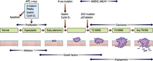
Most human neoplasms are clonal in origin, i.e. they arise from a single population of precursor or cancer stem cells. This process is typically initiated by genetic aberrations within this precursor cell. Cancer is increasingly common the older we get and can be related to a time dependent accumulation of DNA damage that is not repaired by the normal mechanisms of genome maintenance, damage tolerance and checkpoint pathways. Malignant transformation may result from a gain in function as cellular proto-oncogenes become mutated (e.g. ras), amplified (e.g. HER2) or translocated (e.g. BCR-ABL). However, these mutations are insufficient to cause malignant transformation by themselves. Alternatively, there may be a loss of function of tumour suppressor genes such as P53 that normally suppress growth. Loss or gain of function may also involve alterations in the genes controlling the transcription of the oncogenes or tumour suppressor genes (p. 46). Over subsequent cell divisions, heterogeneity develops with the accumulation of further genetic abnormalities (Fig. 9.1).
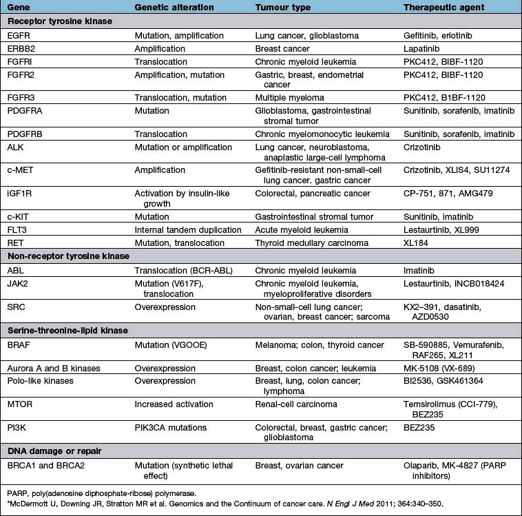
The genes most commonly affected can be characterized as those controlling cell cycle checkpoints, DNA repair and DNA damage recognition, apoptosis, differentiation, growth factor receptors and signalling pathways and tumour suppressor genes (Table 9.2). Recognition of critical genetic alterations has enabled extensive development of new targeted drugs such as imatinib that inhibits the growth signals of the abnormal tyrosine kinase BCR/ABL. Proliferation may continue at the expense of differentiation which, together with the failure of apoptosis, leads to tumour formation with the accumulation of morphologically abnormal cells varying in size, shape and cytoplasmic or nuclear maturity.
Table 9.2 Common genetic abnormalities in cancer
| Gene | Example |
|---|---|
Control cell cycle checkpoints |
Cyclin D1, p15, p16 |
DNA repair |
FANCA, ATM |
Apoptosis |
Bcl2 |
Differentiation |
PML/RARA |
Growth factor receptors |
EGF, VEGF, FGF, BCR/ABL, TGF-B, KIT, L-FLT3 |
Signalling pathways |
RAS, BRAF, JAK2, NF1, PTCH |
Hedgehog signalling pathway |
See p. 26 |
Tumour suppressor genes |
P53, Rb, WT1, VHL |
The hallmarks in developing cancer are shown in Figure 9.2.
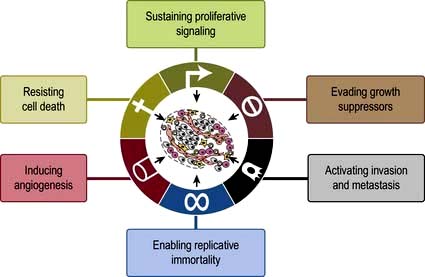
Figure 9.2 The hallmarks of cancer: the next generation.Six biological capabilities acquired during the multistep development of human tumours have been identified as shown in figure. Two others have been identified, viz reprogramming of energy metabolism and evading immune destruction.
(From Hanagan D and Weinberg PA The Hallmarks of cancer: the Next Generation. Cell 2011;144:646–474 with permission.)
Tumour immunology
Tumour cells are usually not recognized and killed by the immune system. There are two main reasons. The first is failure to express molecules such as HLA and co-stimulatory B7 molecules that are required for activation of cytotoxic, or ‘killer’, T lymphocytes. Second, tumours may also actively secrete immunosuppressive cytokines and cause a generalized immunosuppression. Successful strategies for tumour vaccines that overcome these obstacles are developing in renal cancer and prostate cancer. The monoclonal antibody ipilimumab against the inhibitory cytotoxic T lymphocyte-associated antigen 4 (CTLA-4) molecule that is expressed after T-cell activation, is used in melanoma (p. 479)
Angiogenesis
For many tumours, there is a progressive slowing of the rate of growth as the tumours become larger. This occurs for many reasons, but outgrowing the blood supply is paramount. New vessel formation (angiogenesis) is stimulated by a variety of peptides produced both by tumour cells and by host inflammatory cells, such as basic fibroblast growth factor (bFGF), angiopoietin 2 and vascular endothelial growth factors (VEGFs), which are stimulated by hypoxia. The anti-VEGF-receptor monoclonal bevacizumab has had some success in colorectal and ovarian cancer.
Invasion and metastasis
Solid cancers spread by both local invasion and by distant metastasis through the vessels of the blood and lymphatic systems. Infiltration into surrounding tissues is associated with loss of cell–cell cohesion, which is mediated by active homotypic cell adhesion molecules (CAMs). Epithelial cadherin (E-cadherin) is expressed by many carcinomas and mutated in some such as familial gastric carcinoma (see p. 252).
Invasion is also determined by the balance of activators to inhibitors of proteolysis. The matrix metalloproteinases (MMPs) and their tissue inhibitors (TIMPs) are involved in tumour growth, invasion, metastasis and angiogenesis and are being targeted in new therapeutic drugs for cancer treatment.
Dissemination of tumour cells occurs through intravasation into the vascular and lymphatic vessels and dissemination to distant sites, partly by chance, but also because of specific interactions between receptors and cytokines found on stromal and tumour cells such as TNF, IL-6 and chemokines.
Aetiology and epidemiology
For most patients, the cause of their cancer is unknown, probably representing a multifactorial interaction between individual genetic predispositions and environmental factors.
Genetic factors
Rather than occurring by somatic mutation in response to mutagens, germline mutations in the genes that predispose to the development of cancer may be inherited and therefore present in all tissues. Examples of such cancer syndromes are given in Table 9.3. Expression of the mutation and hence carcinogenesis, will depend upon the penetrance (due to the level of expression and the presence of other genetic events) of the gene and whether the mutated allele has a dominant or recessive effect. There is a small group of autosomal dominant inherited mutations such as RB (in retinoblastoma), and a small group of recessive mutations (Table 9.3). Carriers of the recessive mutations are at risk of developing cancer if the second allele becomes mutated, leading to ‘loss of heterozygosity’ within the tumour, although this is seldom sufficient as carcinogenesis is a multistep process.
Table 9.3 Familial cancer syndromes
| Gene | Neoplasms | |
|---|---|---|
Autosomal dominant |
|
|
Retinoblastoma |
RB1 |
Eye |
Wilms’ tumour |
WT1 |
Kidney |
Li–Fraumeni |
p53 |
Sarcoma/brain/leukaemia |
Neurofibromatosis type 1 |
NF1 |
Neurofibromas/ leukaemia |
Familial adenomatous polyposis (FAP) |
APC |
Colon |
Hereditary non-polyposis colon cancer (HNPCC) |
MLH1 and MSH2 |
Colon, endometrium |
Hereditary diffuse gastric cancer syndrome |
E-cadherin |
Stomach |
Breast ovary families |
BRCA1 |
Breast/ovary |
BRCA2 |
||
p53 |
||
Melanoma |
p16 |
Skin |
Von Hippel–Lindau |
VHL |
Renal cell carcinoma and haemangioblastoma |
Multiple endocrine neoplasia type 1 |
MEN1 |
Pituitary, pancreas, parathyroid |
Multiple endocrine neoplasia type 2 |
RET |
Thyroid, adrenal medulla |
Autosomal recessive |
|
|
Xeroderma pigmentosa |
XP |
Skin |
Ataxia telangiectasia |
AT |
Leukaemia, lymphoma |
Fanconi’s anaemia |
FA |
Leukaemia, lymphoma |
Bloom’s syndrome |
BS |
Leukaemia, lymphoma |
Environmental factors
A wide range of environmental factors have been identified as being associated with the development of malignancy (Table 9.4) and may be amenable to preventative action such as smoking cessation, dietary modification and antiviral immunization (Box 9.1). Environmental factors interact with genetic predisposition. For example, subsequent generations of people moving from countries with a low incidence to those with a high incidence of breast or colon cancer acquire the cancer incidence of the country to which they have moved while northern European people exposed to strong UV radiation have the highest risk of developing melanoma.
Table 9.4 Some causative factors associated with the development of cancer
Smoking |
Mouth, pharynx, oesophagus, larynx, lung, bladder, lip |
Alcohol |
Mouth, pharynx, larynx, oesophagus, colorectal |
Iatrogenic |
|
Alkylating agents |
Bladder, bone marrow |
Oestrogens |
Endometrium, vagina, breast, cervix |
Androgens |
Prostate |
Radiotherapy (e.g. mantle radiotherapy) |
Carcinoma of breast and bronchus |
Diet |
|
High-fat diet |
Colorectal cancer |
Environmental/occupation |
|
Vinyl chloride |
Liver (angiosarcoma) |
Polycyclic hydrocarbons |
Skin, lung, bladder, myeloid leukaemia |
Aromatic amines |
Bladder |
Asbestos |
Lung, mesothelium |
Ultraviolet light |
Skin, lip |
Radiation |
e.g. leukaemia, thyroid cancer |
Aflatoxin |
Liver |
Biological agents |
|
Hepatitis B virus |
Liver (hepatocellular carcinoma) |
Hepatitis C virus |
Liver (hepatocellular carcinoma) |
Human T-cell leukaemia virus |
Leukaemia/lymphoma |
Epstein–Barr virus |
Burkitt’s lymphoma |
Hodgkin’s lymphoma |
|
Nasopharyngeal carcinoma |
|
Human papillomavirus types 16, 18 |
Cervix |
Oral cancer (type 16) |
|
Schistosoma japonicum |
Bladder |
Helicobacter pylori |
Stomach |
Tobacco
The incidence of lung cancer in both men and women increased dramatically in the last 25 years worldwide, but is now falling in many developed countries. The association of smoking with lung cancer is indisputable and causative mechanisms have been identified: cigarette tobacco is responsible for one-third of all deaths from cancer in the UK. Smoking not only causes lung cancer, it is also associated with cancer of the mouth, larynx, oesophagus and bladder. Smoking is discussed on page 806.
Alcohol
Alcohol is associated with cancers of the upper respiratory and gastrointestinal tracts, and it also interacts with tobacco in the aetiology of these tumours. It may be associated with an increased risk of breast cancer.
Diet
Dietary factors have been attributed to account for one-third of cancer deaths, although it is often difficult to differentiate these from other epidemiological factors. For example, the incidence of stomach cancer is particularly high in the Far East, while breast and colon cancers are more common in the Western, economically more developed countries. Many associations have been observed without a causative mechanism being identified between the incidence of cancer and the consumption of dietary fibre, red meat, saturated fats, salted fish, vitamin E, vitamin A and many others. Food and its role in the causation of gastrointestinal cancer is discussed in Chapter 5 (see p. 218). Increasing levels of obesity in the developed world have been associated with increases in women of cancers associated with oestrogenic stimulation of the breast and endometrium.
Environmental/occupational
Ultraviolet light is known to increase the risk of skin cancer (basal cell, squamous cell and melanoma). The incidence of melanoma is therefore particularly high in the white Anglo-Celtic population of Australia, New Zealand and South Africa, where exposure to UV light is combined with a genetically predisposed population.
Arsenical contamination of water supplies has been linked to high incidence of lung and colon cancers in Southeast Asia particularly where bore holes are the main water source.
Occupational factors. In 1775, Percival Pott described the association between carcinogenic hydrocarbons in soot and the development of scrotal epitheliomas in chimney sweeps. The principal causes now are asbestos (lung and mesothelial cancer) and polycyclic hydrocarbons from fossil fuel combustion (skin, lung, bladder cancers). Organic chemicals, such as benzene, may cause the development of bone marrow conditions such as myelodysplastic syndrome or acute myeloid leukaemia.
Infectious agents
The geographical distribution of a rare malignancy suggests that it might be caused by, or associated with, an infective agent. Chronic persistent infection provides growth stimulation while many viruses contain transforming viral oncogenes.
T-cell leukaemia, seen almost exclusively in residents of the southern island of Japan and in the West Indies, is caused by infection with the locally endemic retrovirus HTLV-1 (human T-cell leukaemia virus) and integration of the oncogene, TAX, into the cellular genome.
Hepatocellular carcinoma occurs in patients with hepatitis B and C virus infections and Burkitt’s lymphoma and nasopharyngeal carcinoma are associated with the Epstein–Barr virus. EBV is also linked with Hodgkin’s lymphoma (see p. 459).
Patients with HIV infection or immunosuppression from organ transplantation have an increased incidence of EBV-related lymphoma and herpesvirus-8-associated Kaposi’s sarcoma.
The incidence of cervical cancer had increased among younger women in association with sexually transmitted HPV (human papillomavirus) infection types 16 and 18, for which an effective vaccine is now available.
Bacterial infection with Helicobacter pylori predisposes to the development of gastric cancer and gastric lymphoma, while Schistosoma japonicum infection predisposes to the development of squamous cell carcinomas in the bladder.
Medications
Oestrogens have been implicated in the development of vaginal, endometrial and breast carcinoma. Certain cytotoxic drugs given, e.g. for Hodgkin’s lymphoma (see later) are themselves associated with an increased incidence of secondary acute myelogenous leukaemia (AML), bladder and lung cancer. Androgens have been associated with both benign and malignant liver tumours.
Radiation
Accidental. The nuclear disasters of Hiroshima, Nagasaki and Chernobyl led to an increased incidence of leukaemia after 5–10 years in the exposed population as well as increased incidences of thyroid and breast cancer. Radiation workers are at an increased risk of malignancy due to occupational exposure unless precautions are taken to minimize this using personal and environmental shielding and to record and limit the amount of personal exposure.
Therapeutic. Long-term survivors following radiotherapy, e.g. for Hodgkin’s lymphoma, have an increased incidence of cancer, particularly at the radiation field margins.
Diagnostic. Imaging procedures involving radiation exposure are associated with an increased risk of cancer. This risk is cumulative, dose dependent and time dependent, i.e. children are at higher risk than adults. The cancer risk of various common investigations is shown in Table 9.5. All doctors should strive to minimize diagnostic exposure to radiation where possible using alternative modalities such as ultrasound or MRI. Good documentation of radiation doses is required. This is particularly so in children and pregnant women.
Table 9.5 Radiation exposure from common diagnostic radiological procedures
| Procedure | mSv |
|---|---|
CXR |
0.02 |
IVU |
3 |
CT chest |
7 |
CT abdomen |
8–10 |
Whole body CT |
20 |
Percutaneous coronary intervention |
15 |
Myocardial perfusion imaging |
15.6 |
UK background radiation is 2.6 mSv per year. 1 mSv carries a lifetime cancer risk of 1 in 17 500 and 5 mSv a risk of 1 in 3500.
Modified from: Smith-Bindman R, Lipson J, Marcus R et al. Archives of Internal Medicine 2009; 169:2078–2086 and Fazel R, Krumholz HM, Wang Y et al. New England Journal of Medicine 2009; 361:849–857.
Epidemiology
The incidence and mortality from cancer varies by tumour type and geographical region across the world.
Geographical distribution
The incidence of cancer across the world is dependent on the local environmental factors, the diet and the genetics of the population (see above) (Figs 9.3, 9.4). Age is also a factor as most cancers occur in those over the age of 65 who comprise 3.3% of the population in Africa compared with 15.2% in Europe. Reproductive patterns also influence breast cancer. Migrating individuals often take on the risks of the local environmental factors.
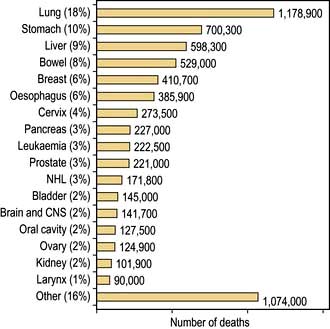
Figure 9.3 The most common causes of death from cancer worldwide, excluding non-melanoma skin cancers (NMSC) 2002 estimates.
(From: http://info.canceresearchuk.org/cancerstats/world/the-global-picture/)
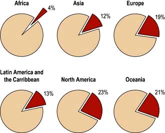
Figure 9.4 Percentage of all deaths due to cancer in the different regions of the world.
(From: http://info.canceresearchuk.org/cancerstats/world/the-global-picture/)
Other factors
Incidence and mortality are closely linked for cancers for which treatment has yet to make significant improvements such as lung, stomach and liver, while in countries with effective screening programmes, there is an increasing incidence and decreasing mortality for breast, cervix, bowel and prostate cancers.
The clinical presentation of malignant disease
Asymptomatic detection through screening
Most common cancers start as focal microscopic clones of transformed cells and diagnosis only becomes likely once sufficient tumour bulk has accumulated to cause symptoms or signs. In order to try to make an earlier diagnosis and increase the curative possibilities, an increasing number of screening programmes are being developed which target the asymptomatic or preinvasive stages of the cancer as in cervix, breast and colon or use serum tumour markers as in prostate and ovarian cancers. Genetic screening can be used to target screening to groups at most risk of developing cancer, e.g. BRCA1 positive and breast cancer (see Table 9.3). The aim of screening programmes is to improve individual and/or population survival by detecting cancer at its very early stages when the patient is asymptomatic. This strategy is dependent upon finding tests that are sufficiently sensitive and specific, using detection methods that identify cancer before it has spread and having curative treatments that are practical and consistent with maintenance of a normal lifestyle and quality of life.
Screening is provided to populations, e.g. for breast, cervical and colon cancer in the UK, and also to individuals via annual check-ups, or opportunistic when patients see their doctor for other reasons.
Unfortunately, earlier diagnosis does not necessarily mean longer survival and randomized trials are necessary to prove benefit. With lead time bias, the patient is merely treated at an earlier date and hence the survival appears longer; death still occurs at the same time from the point of genesis of the cancer (Fig. 9.5). With length time bias, a greater number of slowly growing tumours are detected when screening asymptomatic individuals leading to a false impression of an improvement in survival.
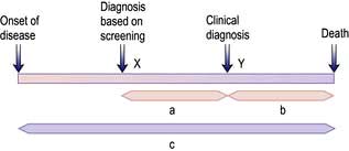
Figure 9.5 Lead time bias. Earlier diagnosis, at X, made by screening tests before the clinical diagnosis, at Y, suggests an increased survival time of A + B. The actual survival time (C) remains unchanged.
An effective screening procedure should:
 be affordable to the healthcare system
be acceptable to all social groups so that they attend for screening
have a good discriminatory index between benign and malignant lesions
Cervical cancer. The smear test is cheap and safe but requires a well-trained cytologist to identify the early changes (dyskaryosis and cervical intraepithelial neoplasia, CIN). However, developments in liquid cytology and DNA testing for human papillomavirus (HPV) may overcome this. Effective treatment for high-risk preinvasive malignant changes reduces the incidence and mortality from cervical cancer, although there are no randomized trials. Screening will continue to be required despite the introduction of vaccination against HPV infection for women before they become sexually active because the lag time between infection and the appearance of disease can be in the order of 40–50 years.
Breast cancer. The UK NHS Breast Screening Programme (i.e. biplanar mammography every 3 years) for women aged 50–70 years has been shown to reduce mortality from breast cancer in randomized controlled studies. The test is acceptable to most women with 50–75% of women attending for screening when sufficiently educated about the benefits. In North America, there is continuing debate about whether annual mammography from a younger age is more effective.
The cost is estimated to be between £250 000 and £1.3 million per life saved, money which, according to critics of screening, could be used more appropriately in better treatment.
Women from families with BRCA1, BRCA2 and p53 mutations require intensive screening starting at an earlier age when mammography is inaccurate due to greater breast density and MRI scanning is preferred.
Colorectal cancer (CRC). Faecal occult blood is a cheap test for the detection of CRC. Large randomized studies have shown a reduction in cancer-related mortality of 15–33%. However, the false-positive rates are high, meaning many unnecessary colonoscopies (see p. 291). The UK has recently introduced a national screening programme using faecal occult blood in patients aged 60–64 years, in which positive tests have identified that 10% have cancer and 40% adenomas. A randomized trial in Norway has found an increased number of early stage cancers in the screened population but a high incidence of interval cancers between biennial screens.
Colonoscopy is the ‘gold-standard’ technique for the examination of the colon and rectum and is the investigation of choice for high-risk patients. Universal screening strategies have been recommended in the USA, but the shortage of skilled endoscopists, the expense, the need for full bowel preparation and the small risk of perforation make colonoscopy impractical as a population screening tool at present and CT colonography (‘virtual colonoscopy’) (see Fig. 6.5) may become an alternative along with genetic testing and stool DNA tests.
Other population-based screening programmes that are being used or are in trials are:
Prostate cancer. Serum prostate-specific antigen (PSA) can be used for the detection of this cancer, which is on the increase. Many men over 70 have evidence of prostate cancer at post mortem with no symptoms of the disease and it has been suggested that over 75-year-olds should not have screening PSAs. The test must be interpreted with caution due to the natural increase in PSA with age, benign prostatic hypertrophy and with prostatitis. The early results of screening for prostate cancer have varied greatly from no benefit in a low-risk population to a halving of deaths from prostate cancer in a general population study but with no overall reduction in mortality. Currently national screening programmes are not recommended.
Epithelial ovarian cancer. Serum CA125 can be used for the early detection of this cancer and is the subject of ongoing trials. An improvement in survival of a screened population can be shown but at the cost of many unnecessary laparotomies so that further enhancements are being investigated by serial testing and in combination with transvaginal ultrasound scans.
FURTHER READING
Menon U, Gentry-Maharaj A, Hallett R et al. Sensitivity and specificity of multimodal and ultrasound screening for ovarian cancer and stage distribution of detected cancers: results of the prevalence screen of the UK Collaborative Trial of Ovarian Cancer Screening (UKCTOCS). Lancet. Oncology 2009; 10:327–340.
Paimela H, Malila N, Palva T et al. Early detection of colorectal cancer with faecal occult blood test screening. British Journal of Surgery 2010; 97:1567–1571.
Schroder FH et al. Prostate cancer mortality at 11 years of follow-up. N Engl J Med 2012; 366:981–990.
The symptomatic patient with cancer
Patients may offer information of predisposing conditions and family history that alerts the clinician to the likelihood of a cancer diagnosis. Many present with a history of tumour site-specific symptoms, e.g. pain, and physical signs, e.g. a mass, which readily identify the primary site of the cancer. However, some only seek medical attention when more systemic and nonspecific symptoms occur such as weight loss, night sweats, fever, fatigue, recurrent infections and anorexia. These usually indicate a more advanced stage of the disease, except in some paraneoplastic and ectopic endocrine syndromes (see below). Other patients are only diagnosed upon the discovery of established metastases such as the abdominal distension of ovarian cancer, the back pain of metastatic prostatic cancer or the liver enlargement of metastatic gastrointestinal cancer (Table 9.6).
Table 9.6 Symptoms and signs of malignant disease
| Degree of spread | Anatomical location | Examples of clinical problems |
|---|---|---|
Local |
Mass |
Thyroid nodule, pigmented naevus, breast lump, abdominal mass, testicular mass |
Local infiltration of skin |
Dermal nodules, peau d’orange, ulceration |
|
Local infiltration of nerve |
Neuropathic pain and loss of function |
|
Local infiltration of vessel |
Venous thrombosis, tumour emboli, haemorrhage, e.g. GI |
|
Obstruction of viscera or duct |
Small or large bowel obstruction, dysphagia, SVC obstruction |
|
Nodal |
Peripheral |
Supraclavicular fossa, Virchow’s node, lymphoedema |
Central |
Mediastinum – SVC obstruction, porta hepatis – obstructive jaundice, para-aortic nodes and back pain |
|
Metastatic |
Lung |
Pleuritic pain, cough, shortness of breath, lymphangitis and respiratory failure, recurrent pneumonia |
Liver |
RUQ pain, anorexia, fever, raised serum liver enzymes, jaundice |
|
Brain |
Headache and vomiting of raised intracranial pressure, focal deficit, coma, seizure |
|
Bone |
Bone pain, cord compression, fracture, hypercalcaemia |
|
Pleura |
Effusion, pain, shortness of breath |
|
Peritoneum |
Ascites, Krukenberg tumours |
|
Adrenal |
Addison’s disease (hypoadrenalism) |
|
Umbilicus |
Sister Mary Joseph’s nodule |
Paraneoplastic syndromes are indirect effects of cancer (Box 9.2, Fig. 9.6) that are often associated with specific types of cancer and may be reversible with treatment of the cancer. The effects and mechanisms can be very variable. For example in the Lambert–Eaton syndrome (see p. 1152), there is cross-reactivity between tumour antigens and the normal tissues, e.g. the acetylcholine receptors at neuromuscular junctions.
 Box 9.2
Paraneoplastic syndromes
| Syndrome | Tumour | Serum antibodies |
|---|---|---|
Neurological |
|
|
Lambert–Eaton syndrome |
Lung (small-cell) lymphoma |
Anti-VGLC |
Peripheral sensory neuropathy |
Lung (small-cell), breast and ovary lymphoma |
Anti-Hu |
Cerebellar degeneration |
Lung (particularly small-cell) lymphoma |
Anti-Yo |
Opsoclonus/myoclonus |
Breast, lung (small-cell) |
Anti-Ri |
Stiff person syndrome |
Breast, lung (small-cell) |
Anti-amphiphysin |
Limbic, hypothalamic, brain stem encephalitis |
Lung |
Anti-Ma protein |
|
Testicular |
Anti-NMDAR |
Endocrine/metabolic |
|
|
SIADH |
Lung (small-cell) |
|
Ectopic ACTH secretion |
Lung (small-cell) |
|
Hypercalcaemia |
Renal, breast, myeloma, lymphoma |
|
Fever |
Lymphoma, renal |
|
Musculoskeletal |
|
|
Hypertrophic pulmonary osteoarthropathy |
Lung (non-small-cell) |
|
Clubbing |
Lung |
|
Skin |
|
|
Dermatomyositis/polymyositis |
Lung and upper GI |
|
Acanthosis nigricans |
Mainly gastric |
|
Velvet palms |
Gastric, lung (non-small cell) |
|
Hyperpigmentation |
Lung (small-cell) |
|
Pemphigus |
Non-Hodgkin’s lymphoma, CLL |
|
Haematological |
|
|
Erythrocytosis |
Renal cell carcinoma, hepatocellular carcinoma, cerebellar haemangioblastoma |
|
Thrombocytosis |
Ovarian cancer |
|
Migratory thrombophlebitis |
Pancreatic adenocarcinoma |
|
DVT |
Adenocarcinoma |
|
DIC |
Adenocarcinoma |
|
Renal |
|
|
Nephrotic syndrome |
Myeloma, amyloidosis |
|
Membranous glomerulonephritis |
Lymphoma |
|
SIADH, syndrome of inappropriate antidiuretic hormone secretion; ACTH, adrenocorticotrophic hormone; CLL, chronic lymphocytic leukaemia; DIC, disseminated intravascular coagulation; NMDAR, N-methyl-D-aspartate receptors.

The coagulopathy of cancer may present with thrombophlebitis, deep venous thrombosis and pulmonary emboli, particularly in association with cancers of pancreas, stomach and breast. Some 18% of patients with recurrent pulmonary embolus will be found to have an underlying cancer and many cancer patients are at increased risk of venous thromboembolism (VTE) following diagnosis. Trousseau’s syndrome – superficial thrombophlebitis migrans – refers to this process in the superficial venous system. All patients with active cancer admitted to hospital are at high risk of VTE and should be given prophylaxis with subcutaneous LMW heparin in the absence of any contraindications (see p. 429). Dabigatran, an oral direct thrombin inhibitor, is an alternative therapy.
Other symptoms are related to peptide or hormone release, e.g. carcinoid or Cushing’s syndrome.
Cachexia of advanced cancer is thought to be due to release of chemokines such as tumour necrosis factor (TNF), as well as the fact that patients have a loss of appetite. The unexplained loss of >10% of body weight in a patient should always stimulate a search for an explanation.
Cancer-associated immunosuppression can lead to reactivation of latent infections such as herpes zoster and tuberculosis.

Serum tumour markers
Tumour markers are intracellular proteins or cell surface glycoproteins released into the circulation and detected by immunoassays. Examples are given in Table 9.7. Values in the normal range do not necessarily equate with the absence of disease and a positive result must be corroborated by histology as these markers can be seen in many benign conditions. They are most useful in the serial monitoring of response to treatment. As discussed in subsequent sections, a proportion of low-grade B-cell lymphomas and a majority of cases of myeloma will produce a monoclonal paraprotein of intact immunoglobulin molecule or light chains. This acts as a valuable tumour marker in the diagnosis and assessment of response.
Table 9.7 Serum tumour markers
α-Fetoprotein |
Hepatocellular carcinoma and non-seminomatous germ cell tumours of the gonads |
β-Human chorionic gonadotrophin (β-hCG) |
Choriocarcinomas, germ cell tumours (testicular) and lung cancers |
Prostate-specific antigen (PSA) |
Carcinoma of prostate |
Carcinoma embryonic antigen (CEA) |
Gastrointestinal cancers |
CA125 |
Ovarian cancer |
CA19–9 |
Gastrointestinal cancers, particularly pancreatic cancer |
CA15–3 |
Breast cancer |
Osteopontin |
Many cancers including mesothelioma |
M-band (Ig or light chain) |
Myeloma, chronic lymphocytic leukaemia, small lymphocytic lymphoma, lymphoplasmacytic lymphoma, amyloid |
Cancer imaging
Radiological investigation by experts is required at various stages: at initial diagnosis and staging of the disease, during the monitoring of treatment efficacy, at the detection of recurrence and for the diagnosis and treatment of complications.
The choice of investigations needs to be guided by the patient’s symptoms and signs, site and histology of the cancer, the curative or palliative potential of treatment and the utility of the information in guiding treatment. The investigations are described under each tumour type.
Contrast agents are used for increased structural discrimination and can be further enhanced with functional specificity for metabolically active tissue with 19fluorodeoxy-glucose uptake and CT-positron emission tomography (CT-PET scan) as used extensively in head and neck cancer, lung cancer and lymphoma. Radionuclide imaging of sentinel lymph nodes is used to guide lymphatic surgery in breast cancer and melanoma. Tumour targeted contrast agents can improve detection rates such as the radiolabelled MAb rituximab for lymphoma or radiolabelled small molecules such as octreotide for neuroendocrine tumours. Research into the use of reporter agents which become visible only upon activation within the tumour environment holds the promise of greater sensitivity and specificity in the future.
Biopsy and histological examination
The diagnosis of cancer may be suspected by both patient and doctor but advice about treatment can usually only be given on the basis of a tissue diagnosis. This may be obtained by endoscopic, radiologically-guided or surgical biopsy or on the basis of cytology (e.g. lung cancer diagnosed by sputum cytology). Malignant lesions can be distinguished morphologically from benign ones by the pleiomorphic nature of the cells, increased numbers of mitoses, nuclear abnormalities of size, chromatin pattern and nucleolar organization and evidence of invasion into surrounding tissues, lymphatics or vessels. The degree of differentiation (or conversely of anaplasia) of the tumour has prognostic significance: generally speaking, more differentiated tumours have a better prognosis than poorly-differentiated ones. In some tumours where the surgical procedure will vary depending on the presence of malignancy, an intraoperative histological opinion can be rapidly obtained using a tissue sample processed using ‘frozen section’ techniques, which requires the availability of a histopathologist. This obviates the need for the sample to be paraffin embedded, which takes hours to days.
Tissue tumour markers. Immunocytochemistry, using monoclonal antibodies against tumour antigens, is very helpful in differentiating between lymphoid and epithelial tumours and between some subsets of these, for example T- and B-cell lymphomas, germ cell tumours, prostatic tumours, neuroendocrine tumours, melanomas and sarcomas. However, there is much overlap in the expression of many of the markers and some adenocarcinomas and squamous carcinomas do not bear any distinctive immunohistochemical markers that are diagnostic of their primary site of origin.
Molecular markers of genetic abnormalities have long been available in the haematological cancers and are increasingly available in solid cancers. For example, fluorescent in situ hybridization (FISH, see p. 40) can be used to look for characteristic chromosomal translocations, e.g. in lymphoma and leukaemia, as well as deletions or amplifications, e.g. in breast cancer (see genetic basis of cancer, p. 45). Tissue microarrays can identify patterns of multiple genomic alterations and single nucleotide polymorphisms (SNPs), e.g. in breast cancer and lymphoma (see p. 35), and RNA assays with RT-PCR can be used to identify tissue of origin with prognostic and predictive relevance.
Genomics and proteomics are being investigated in order to target new (and expensive) therapies, e.g. imatinib in CML and GIST, trastuzumab and lapatinib in breast cancer and erlotinib in lung cancer.
Cancer treatment
Aims of treatment
Optimal cancer treatment is delivered by a multidisciplinary team which coordinates the delivery of the appropriate anticancer treatment (surgery, chemotherapy, radiotherapy and biological/endocrine therapy), supportive and symptomatic care and psychosocial support. While all members will have the patient’s care as their central concern, someone, often the oncologist, has to take responsibility for the coordination of the many professionals involved.
The organization across multiple departments and coordination from primary to secondary and tertiary care has become known as a patient pathway. Establishment of agreed patient pathways has enabled more effective and timely delivery of care and post-treatment rehabilitation. The aim is to provide optimal treatment and for the patient to experience seamless and high quality care and to allow audit and continuing improvement against agreed standards. Central to this endeavour is the involvement of the patient, through education as to the nature of their disease and the treatment options available. An informed choice can then be made, even if in the end it is simply to abide by the decisions made by the professionals. Good communication embodies a humane approach which preserves hope at an appropriate level through empathy and understanding of the patient’s position (see p. 14).
A curative approach
For most solid tumours local control is necessary, but not sufficient, for cure because of the presence of systemic (microscopic) disease, while haematological cancers are usually disseminated from the outset. Improvement in the rate of cure of most cancers is thus dependent upon earlier detection to increase the success of local treatment and effective systemic treatment. The likelihood of cure of the systemic disease depends upon the type of cancer and its expression of appropriate treatment targets, its drug sensitivity and tumour bulk (microscopic or clinically detectable). A few rare cancers are so chemosensitive that even bulky metastases can be cured, e.g. leukaemia, lymphoma, gonadal germ cell tumours and choriocarcinoma. For most common solid tumours such as lung, breast and colorectal cancer, there is no current cure of bulky (clinically detectable) metastases, but micrometastatic disease treated by adjuvant systemic therapy (see below) after surgery can be cured in 10–20% of patients.
Adjuvant therapy for solid tumours
This is defined as treatment given, in the absence of macroscopic evidence of metastases, to patients at risk of recurrence from micrometastases, following treatment given for the primary lesion. ‘Neoadjuvant’ therapy, alternatively, is given before primary surgery, to both shrink the tumour to improve the local excision and treat any micrometastases as soon as possible.
Micrometastatic spread by lymphatic or haematological dissemination often occurs early in the development of the primary tumour and can be demonstrated by molecular biological methods capable of detecting the small numbers (1 in 106) of circulating cells. Studies correlating prognosis with histological features of the primary cancer, e.g. differentiation, invasion of blood vessels or regional lymph nodes and molecular markers, e.g. Her2 in breast cancer, enable risk stratification and increasing individualization of therapy.
The success of adjuvant treatment across many tumour types relies upon careful selection of patients according to defined risk criteria and the reduction of treatment toxicity to reach a balanced risk/benefit ratio. Relative risk reductions in the order of 12–33% and absolute improvements in 5–10-year survival of 5–25% (dependent upon the pre-existing risk) have been achieved in common epithelial cancers such as lung, bowel, breast and prostate, with greater absolute improvements in the more sensitive germ cell tumours.
While these improvements currently translate into many lives saved from common diseases at a public health level, the majority who receive such treatment do not benefit because they were already cured, or because the cancer is resistant to the treatment. Better tests, e.g. gene arrays and circulating tumour cells, are being developed to identify those with the micrometastases who really need treatment. On an individual patient basis the decision on whether adjuvant treatment will be worthwhile must include consideration of other factors such as the patient’s life expectancy, concurrent medical conditions and lifestyle priorities.
A palliative approach
When cure is no longer possible, palliation, i.e. relief of tumour symptoms, preservation of quality of life and prolongation of life, is possible in many cancers in proportion to their drug and radiation sensitivity. There is on average a 2–18-month prolongation in median life expectancy with treatments for solid tumours (see specific tumour types for details) and up to 5–8 years for some leukaemias and lymphomas, with those with the most responsive tumours experiencing the greatest benefit. The development of more effective chemotherapeutic drugs, targeted biological agents and better supportive care has done much to reduce the side-effects of systemic therapy and to improve the cost/benefit ratio for the patient receiving palliative treatment. In addition, through early assessment during treatment, it is possible to stop if no evidence of benefit is demonstrable early on, so as to minimize exposure to toxic and unsuccessful treatment.
Assessments before treatment
Staging
Before a decision about treatment can be made, not only the type of tumour but also its extent and distribution need to be established. Various ‘staging investigations’ are therefore performed before a treatment decision is made. To be useful clinically the staging system must subdivide the patients into groups of different prognosis which can guide treatment selection.
The staging systems vary according to the type of tumour and may be site specific (see Hodgkin’s lymphoma, p. 461), or the TNM (tumour, node, metastases) classification shown in Table 15.29, which can be adapted for application to most common cancers.
Performance status
In addition to anatomical staging, the person’s age and general state of health need to be taken into account when planning treatment. The latter has been called ‘performance status’ and is of great prognostic significance for all tumour types (Table 9.8). Performance status reflects the effects of the cancer on the patient’s functional capacity. An alternative performance rating scale is by Karnowsky. With a performance status of 2, response to and survival following treatment are greatly reduced for most tumour types.
Table 9.8 Eastern Cooperative Oncology Group (ECOG) performance status scale
| Status | Description |
|---|---|
0 |
Asymptomatic, fully active and able to carry out all predisease performance without restrictions |
1 |
Symptomatic, fully ambulatory but restricted in physically strenuous activity and able to carry out performance of a light or sedentary nature, e.g. light housework, office work |
2 |
Symptomatic, ambulatory and capable of all self-care but unable to carry out any work activities. Up and about >50% of waking hours: in bed <50% of day |
3 |
Symptomatic, capable of only limited self-care, confined to bed or chair >50% of waking hours, but not bedridden |
4 |
Completely disabled. Cannot carry out any self-care. Totally bedridden |
Assessing the benefits of treatment
A measurable response to treatment can serve as a useful early surrogate marker when assessing whether to continue a given treatment for an individual patient. Trials to assess response to treatment in advanced disease have identified active agents for use in the more curative setting of adjuvant treatment of early stage disease
Response to treatment can be subjective or objective.
A subjective response is one perceived by the patient in terms of, for example, relief of pain and dyspnoea, or improvement in appetite, weight gain or energy. Such subjective response is a major aim of most palliative treatments. Quantitative measurements of these subjective symptoms (Patient Reported Outcome Measures, PROMs) form a part of the assessment of response to chemotherapy, especially in those situations where cure is not possible and where the aim of treatment is to provide prolongation of good-quality life. In these circumstances, measures of quality of life enable an estimate of the balance of benefit and side-effects to be made.
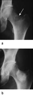
Chemotherapy radiological response. Bone metastasis responding to chemotherapy with sclerotic new bone formation. (a) Tumour (arrow). (b) Following radiotherapy.
An objective response to treatment is assessed clinically and radiologically. The term ‘remission’ is often used synonymously with ‘response’, which if complete, means an absence of detectable disease without necessarily implying a cure of the cancer. The terms used to evaluate the responses of tumours are given in Box 9.3. For a complete response all previous clinical abnormalities should have resolved and this needs to be confirmed by clinical examination or sampling of the primary disease site, e.g. by bone marrow examination in leukaemia. Where a tumour marker exists, such as a paraprotein in myeloma or β-hCG in testicular cancer, reductions in the level of tumour markers are useful surrogates for evidence of tumour response. They are also useful predictors for disease recurrence. Radiologically, a complete response, is the complete disappearance of all detectable disease and a partial response, defined since 1999 by the Response Evaluation Criteria in Solid Tumors (RECIST) convention, is a ≥30% reduction in the sum of all measurable lesion diameters.
Box 9.3
RECIST criteria for assessing response to treatment
Complete response |
Disappearance of all target lesions |
Partial response |
≥30% decrease in the baseline sum of the longest diameters of all target lesions |
Progression |
≥20% increase in the sum of the longest diameters of all target lesions compared with the smallest recorded sum since treatment started, or the appearance of any new lesions |
Stable disease |
All values between partial response and progressive disease |
Target lesions |
Measurable lesions up to 5 per organ and 10 in total, selected on size and replicable measurement |
Non-target lesions |
Non-measurable lesions recorded as present or absent at baseline and on follow-up. |
Measurable |
Lesions with the longest diameter in one dimension ≥2.0 cm or ≥1.0 cm if assessed by spiral CT scan |
Non-measurable |
e.g. bone, meningeal, ascites, pleural effusion, inflammatory breast cancer, lymphangitis cutis and pulmonis, cystic lesions |
The final assessment of treatment outcome is the impact of the therapy on remission duration and survival, i.e. the cure rate. Such survival figures are increasingly incorporating quality of life assessments and a health economic assessment to calculate the number of quality adjusted life years (QALY) gained for the cost of the treatment and judgements made about health system affordability.
When counselling, the individual patient’s interpretation of the trial evidence for benefit must be adjusted for the degree to which they resemble the population from which it is derived and the cost interpreted in view of their co-morbidities and lifestyle choices.
Principles of chemotherapy
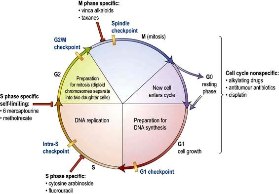
Cytotoxic chemotherapy employs systemically administered drugs that directly damage cellular DNA (and RNA). It kills cells by promoting apoptosis and sometimes frank necrosis. Different cytotoxic drugs work at different stages in the cell cycle (Fig. 9.7; see also Fig. 2.13).
There is a narrow therapeutic window between effective treatment of the cancer and normal tissue toxicity, because cytoxic drugs are not cancer specific (unlike some of the targeted biological agents) and the increased proliferation in cancers is not much greater than in normal tissues (see tumour growth and failure of apoptosis, p. 32, 33). The dose and schedule of the chemotherapy is limited by the normal tissue tolerance, especially in those more proliferative tissues of the bone marrow and gastrointestinal tract mucosa. All tissues can be affected, however, depending upon the pharmacokinetics of the drug and affinity for particular tissues (e.g. heavy metal compounds for kidneys and nerves).
The therapeutic effect on the cancer is achieved by a variety of mechanisms which seek to exploit differences between normal and transformed cells. While most of the drugs have been derived in the past by empirical testing of many different compounds, e.g. alkylating agents, the new molecular biology is leading to targeting of particular genetic defects in the cancer (see tyrosine kinase inhibitors, p. 445).
Toxicity to normal tissue can be limited in some instances by supplying growth factors such as granulocyte colony-stimulating factor (G-CSF) or by the infusion of stem cell preparations to diminish myelotoxicity. The use of more specific targeted biological agents with relatively weak pro-apoptotic effects in combination with the general cytotoxics has also improved the therapeutic ratio (see trastuzumab and breast cancer, p. 475). Certain cytotoxic therapies may also be administered into the pleural space, the peritoneum, the CSF or into the arterial supply of a tumour.
Most tumours rapidly develop resistance to single agents given on their own through changes in membrane transport and DNA repair pathways. For this reason, the principle of intermittent combination chemotherapy was developed. Several drugs are combined together, chosen on the basis of differing mechanisms of action and non-overlapping toxicities. These drugs are given over a period of a few days, followed by a rest of a few weeks, during which time the normal tissues have the opportunity for regrowth. If the normal tissues are more proficient at DNA repair than the cancer cells, it may be possible to deplete the tumour while allowing the restoration of normal tissues between chemotherapy cycles (Fig. 9.8).
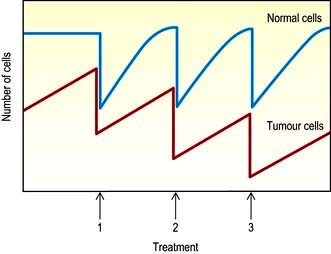
Figure 9.8 Effects of multiple courses of cytotoxic chemotherapy, showing the decrease in the number of cells with each course.
In many experimental tumours, it has been shown that there is a log–linear relationship between drug dose and number of cancer cells killed and that the maximum effective dose is very close to the maximum tolerated dose at which dose-limiting toxicity is reached. With a chemosensitive tumour, relatively small increases in dose may have a large effect on tumour cell kill. It is therefore apparent that where cure is a realistic option the dose administered is critical and may need to be maintained despite toxicity. In situations where cure is not a realistic possibility and palliation is the aim, a sufficient dose to exceed the therapeutic threshold, but not cause undue toxicity, is required as the short-term quality of life becomes a major consideration.
Classification of cytotoxic drugs (Table 9.9)
DNA damaging drugs
Alkylating agents act by covalently binding alkyl groups and their major effect is to cross-link DNA strands, interfering with DNA synthesis and causing strand breaks. Despite being among the earliest cytotoxic drugs developed, they maintain a central position in the treatment of cancer. Melphalan is one of the original nitrogen mustards and is used in multiple myeloma. Chlorambucil is used in Hodgkin’s lymphoma and chronic lymphocytic leukaemia. Other common alkylating agents include cyclophosphamide and ifosfamide, as well as the nitrosoureas, carmustine (BCNU), bendamustine, lomustine (CCNU) and busulfan used in chronic myeloid leukaemia. Tetrazines also alkylate DNA; dacarbazine is used in Hodgkin’s lymphoma and temozolomide in malignant gliomas.
Table 9.9 Chemotherapy: some cytotoxic drugs
|
|
Platinum compounds. Cisplatin, carboplatin and oxaliplatin cause interstrand cross-links of DNA and are often regarded as non-classical alkylating agents. They have transformed the treatment of testicular cancer (cisplatin) and have a major role against many other tumours, including lung, ovarian and head and neck (cis or carboplatin) and gastrointestinal (oxaliplatin) cancer. Toxicity, as for other heavy metals, includes renal and peripheral nerve damage.
Antimetabolites
Antimetabolites are usually structural analogues of naturally occurring metabolites that interfere with normal synthesis of nucleic acids by falsely substituting purines and pyrimidines in metabolic pathways. Antimetabolites can be divided into:
Folic acid antagonists, e.g. methotrexate. This is structurally very similar to folic acid and binds preferentially to dihydrofolate reductase, the enzyme responsible for the conversion of folic acid to folinic acid. It is used widely in the treatment of solid tumours and haematological malignancies. Folinic acid is often given to ‘rescue’ normal tissues from the effects of high doses of methotrexate.
Pyrimidine antagonists. 5-Fluorouracil (5FU) consists of a uracil molecule with a substituted fluorine atom. It acts by blocking the enzyme thymidylate synthase, which is essential for pyrimidine synthesis. 5-Fluorouracil has a major role in the treatment of solid tumours, particularly gastrointestinal cancers. Oral capecitabine is metabolized to 5FU and tegafur with uracil and calcium folinate are used in gastrointestinal and breast cancers.
Arabinosides inhibit DNA synthesis by inhibiting DNA polymerase. Cytosine arabinoside (cytarabine, Ara-C) is used almost exclusively in the treatment of acute myeloid leukaemia where it remains the backbone of therapy, while its analogue gemcitabine is proving useful in a number of solid cancers such as lung, breast, pancreas and ovary. Fludarabine is used in the treatment of B cell chronic lymphocytic leukaemia; it is also used in reduced intensity stem cell transplantation (see this chapter) because of its immunosuppressive effect. Other related drugs have found niche applications in acute leukaemia (cladribine, clofarabine, nelarabine/AraG) and myelodysplasia (azacytidine).
Purine antagonists, e.g. 6-mercaptopurine and 6-tioguanine, which are both used almost exclusively in the treatment of acute leukaemia.
DNA repair inhibitors
Epipodophyllotoxins. These are semisynthetic derivatives of podophyllotoxin which inhibit topoisomerase. Topoisomerase enzymes allow unwinding and uncoiling of supercoiled DNA. Etoposide is a drug used in a wide range of cancers and works by maintaining DNA strand breaks by inhibiting the enzyme topoisomerase II. Topoisomerase I inhibitors such as irinotecan and topotecan have also proved active against lung, colon, ovary and cervix cancer.
Cytotoxic antibiotics. The anthracyclines act by intercalating adjoining nucleotide pairs on the same strand of DNA and by inhibiting topoisomerase II DNA repair. They have a wide spectrum of activity in haematological and solid tumours. Doxorubicin and its congener epirubicin are two of the most widely used of all cytotoxic drugs but have cumulative toxicity to the myocardium. Pegylated liposomal doxorubicin is used for Kaposi’s sarcoma and as second-line treatment for advanced ovarian cancer with reduction of cardiotoxicity, but increased toxicity to the skin on the palms of the hands and soles of the feet. Amsacrine is a similar drug used occasionally in acute myeloid leukaemia. Bleomycin and mitomycin are also intercalating agents which promote the cleavage of DNA and RNA. Bleomycin has a particular toxicity to the lung causing interstitial fibrosis.
Antitubulin agents
Vinca alkaloids. Drugs such as vincristine, vinblastine and vinorelbine act by binding to tubulin and inhibiting microtubule formation during mitosis (see p. 20). They are used in the treatment of haematological (vincristine and vinblastine) and non-haematological cancers (vinorelbine). They are associated with neurotoxicity due to their anti-microtubule effect and must never be given intrathecally as this is lethal.
Taxanes. Paclitaxel and docetaxel bind to tubulin dimers and prevent their assembly into microtubules. They are active drugs against many cancers such as ovarian, breast and lung cancer. Taxanes can cause neurotoxicity and hypersensitivity reactions and patients should be premedicated with steroids, H1 and H2 histamine antagonists prior to treatment.
Side-effects of chemotherapy
Chemotherapy carries many potentially serious side-effects and should be used only by trained practitioners; however, an appreciation of its common potential side-effects is necessary for any general physician who is called to see a cancer patient on chemotherapy. The five most common side-effects are vomiting, hair loss, tiredness, myelosuppression and mucositis (Table 9.10). Side-effects are much more directly dose related than anticancer effects and it has been the practice to give drugs at doses close to their maximum tolerated dose, although this is not always necessary to achieve their maximum anticancer effect. Common combination chemotherapeutic regimens are shown in Table 9.11.
Table 9.10 Side-effects of chemotherapy
|
|
Table 9.11 Some common chemotherapy regimens
Hodgkin’s lymphoma |
ABVD |
Doxorubicin, bleomycin, vinblastine, dacarbazine |
Non-Hodgkin’s lymphoma |
CHOP |
Cyclophosphamide, hydroxy-doxorubicin, vincristine, prednisolone |
Breast |
AC |
Adriamycin and cyclophosphamide |
Lung |
PE |
Cisplatin, etoposide |
Stomach |
ECF |
Epirubicin, cisplatin, 5-fluorouracil |
Colorectal |
FolFOx |
Oxaliplatin, 5FU, folinic acid |
Note: Some abbreviations are related to trade names.
Extravasation of intravenous drugs. Cytotoxic drugs should only be given by trained personnel. They cause severe local tissue necrosis if leakage occurs outside the vein. Stop the infusion immediately and institute local measures, e.g. aspirate as much of the drug from the cannula, infiltrate area with 0.9% saline and apply warm compresses. Antihistamines and corticosteroids may give symptomatic relief. Dexrazoxane is used for anthracycline extravasation.
Nausea and vomiting. The severity of this common side-effect varies with the cytotoxic and it can be eliminated in 75% of patients by using modern antiemetics. Nausea and vomiting are particular problems with platinum analogues. A stepped policy with antiemetics such as metoclopramide and domperidone followed by 5-HT3 serotonin antagonists (e.g. ondansetron, granisetron) combined with dexamethasone should be used to match the emetogenic potential of the chemotherapy. Aprepitant, a neurokinin receptor antagonist, is helpful in preventing acute and delayed nausea and vomiting. It is used with dexamethasone and a 5-HT3 antagonist. Drugs such as cyclizine, haloperidol and levomepromazine and benzodiazepines can be used to control persistent nausea.
Hair, skin and nails. Many but not all cytotoxic drugs are capable of causing hair loss. Scalp cooling can sometimes be used to reduce hair loss but in general this side-effect can only be avoided by selection of drugs where this is possible. Hair regrows on completion of chemotherapy. Nails will demonstrate banding reflecting periods of cessation of growth during each chemotherapy cycle and skin toxicity may be particularly pronounced with 5FU, capecitabine and docetaxel (Fig. 9.9).


Fatigue is often significant and may continue beyond completion of therapy. Other problems such as anaemia or depression may exacerbate this. Attention should be paid to nutrition, hydration, sleep hygiene, gentle exercise, task prioritization, pacing, realistic target setting and scheduling rest within the day.
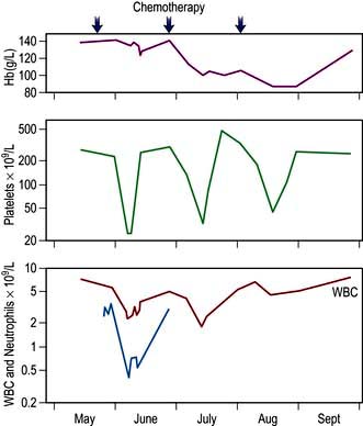
Bone marrow suppression and immunosuppression. Suppression of the production of red blood cells, white blood cells and platelets occurs with most cytotoxic drugs and is a dose-related phenomenon (Fig. 9.10). Severely myelosuppressive chemotherapy may be required if treatment is to be given with curative intent despite the potential for rare but fatal infection or bleeding. Anaemia and thrombocytopenia are managed by red cell or platelet transfusions. (Neutropenic infection is discussed on p. 448.) The risk of infective problems can be ameliorated by the use of prophylactic antimicrobials, such as ciprofloxacin, or the use of GCSF as primary prophylaxis in those chemotherapy regimens with a significant risk of febrile neutropenia or those patients on less intensive therapies who are at higher risk due to age or co-morbidity.
Mucositis. This common side-effect of chemotherapy reflects the sensitivity of the mucosa to antimitotic agents. It causes severe pain in the oropharyngeal region and problems with swallowing and nutrition. Mucositis can be generalized throughout the intestinal tract when it can cause life-threatening diarrhoea. Treatment is with antiseptic and anti-candidal mouthwash and, if severe, fluid and antibiotic support, as the mucosa is a portal for entry of enteric organisms. Palifermin, a recombinant keratinocyte derived growth factor, may ameliorate severe chemotherapy and radiotherapy induced mucositis.
Other toxicities
Cardiotoxicity. This is a rare side-effect of chemotherapy, usually associated with anthracyclines such as doxorubicin, and can present as an acute arrhythmia during administration or cardiac failure due to cardiomyopathy after chronic exposure. This effect is dose-related and can largely be prevented by restricting the cumulative total dose of anthracyclines within the safe range (equivalent to 450 mg/m2 body surface area cumulative doxorubicin dose). The risk of anthracycline cardiomyopathy is also dependent on other treatments such as trastuzumab or radiotherapy as well as other cardiac risk factors such as hypertension, smoking and hypercholesterolaemia. Cardiotoxicity can also be reduced by using the analogue epirubicin or by reducing peak drug concentrations through delayed release preparations such as liposomal doxorubicin. 5-Fluorouracil and its prodrug capecitabine can cause cardiac ischaemia.
Neurotoxicity. This occurs predominantly with the vinca alkaloids, taxanes and platinum analogues (but not carboplatin). It is dose-related and cumulative. Chemotherapy is usually stopped before the development of a significant polyneuropathy, which once established is only partially reversible. Vinca alkaloids such as vincristine must never be given intrathecally as the neurological damage is progressive and fatal.
Nephrotoxicity. Cisplatin (but not oxaliplatin or carboplatin), methotrexate and ifosfamide can potentially cause renal damage. This can usually be prevented by maintaining an adequate diuresis during treatment to reduce drug concentration in the renal tubules and careful monitoring of renal function.
Sterility and premature menopause. Some anticancer drugs, particularly alkylating agents, but also anthracyclines and docetaxel, may cause gonadal damage resulting in sterility and in women the loss of ovarian oestrogen production, which may be irreversible.
In males, the storage of sperm prior to chemotherapy should be offered to the patient when chemotherapy is given with curative intent.
In females, collection of oocytes to be fertilized in vitro and cryopreserved as embryos for subsequent implantation is most successful; however it is also possible to collect and freeze by vitrification of unstimulated oocytes. Cryopreservation of ovarian tissue and retrieval of viable oocytes for subsequent fertilization is still experimental. The recovery of gonadal function is dependent upon the status before treatment and in women this is mostly related to age since menarche, with those under the age of 40 having significantly more ovarian reserve.
Secondary malignancies. Anticancer drugs have mutagenic potential and the development of secondary malignancies, predominantly acute leukaemia, is an uncommon but particularly unwelcome long-term side-effect in patients otherwise cured of their primary malignancies. The alkylating agents, anthracyclines and epipodophyllotoxins are particularly implicated in this complication.
Haemopoietic stem cell transplantation (HSCT)
HSCT is used in a range of malignancies and some non-malignant disorders such as sickle cell disease. It relies on the ability of transfused haemopoietic stem cells to repopulate the marrow niche that has been rendered temporarily or permanently hypoplastic from chemotherapy with or without additional radiotherapy. Such procedures vary in the source of the stem cells and in the type and intensity of the preparatory conditioning regimen (Box 9.4).
Autologous stem cell transplantation
Most anticancer drugs have a sigmoid dose–response relationship which suggests that, up to a point, a higher dose of a cytotoxic drug will induce a greater response. However, increasing cytotoxic drug dose is often not possible, owing to toxicity. For those chemotherapeutic agents with a dose-limiting toxicity of bone marrow failure, infusion of previously harvested haemopoietic stem cells is able to ‘rescue’ the haemopoietic system and permits the use of higher doses to overcome tumour drug resistance. Haemopoietic stem cells are either collected from the patient’s bone marrow, or more commonly by leucopheresis from the peripheral blood following stem cell mobilization from the marrow niche by the administration of the growth factor granulocyte colony-stimulating factor (G-CSF) with or without chemotherapy. These stem cells are stored by cryopreservation and then reinfused intravenously after an intensive, chemotherapy regimen. This approach has been particularly effective in relapsed leukaemias, lymphomas, myeloma and germ cell tumours. However, tumour contamination of the reinfused stem cells remains a reality. Treatment-related mortality is in the region of 1–5%. Such an approach is also being evaluated for the treatment of severe autoimmune disease, such as Crohn’s.
Allogeneic stem cell transplantation
Conventional ablative allogeneic transplants
These combine the cytotoxic effect of high-dose therapy with a potent immunotherapy effect. Historically, the transplantation of donor haemopoietic cells has been combined with myeloablative chemotherapy with or without radiotherapy with the dual effects of treating the malignancy as well as causing temporary immunosuppression that allows the graft ‘to take’. Donors are usually fully matched at the major HLA antigens. Thus siblings are more likely to be found to be potential donors than unrelated volunteers. Allogeneic transplantation has been successfully used in acute and chronic leukaemias and myeloma. The engraftment of the donor immune system, with antitumour activity (graft-versus-tumour), is primarily responsible for the increased effectiveness of this approach. Complications include graft-versus-host disease (GVHD), an allo-immune reaction of the donor cells against normal host organs, which can affect 30–50% of transplant recipients and is potentially fatal in some cases. Immunosuppression, both from conditioning therapy and from the immunosuppressive drugs (ciclosporin or tacrolimus) given to prevent graft-versus-host disease, results in a high incidence of opportunistic infection and viral reactivation, e.g. CMV. All patients receive prophylactic antibacterial, antifungal and antiviral drugs. Mortality therefore from conventional allogeneic stem cell transplantation is a major problem, with 20–40% at risk of dying from the procedure, depending on the age and status of the recipient and the degree of HLA compatibility of the donor. The use of donor lymphocyte infusions following allogeneic bone marrow transplantation (BMT), while losing some of the specificity, has produced the strongest evidence for the efficacy of immunotherapy via graft-versus-tumour activity with clinical remissions observed, albeit with a risk of triggering GVHD.
Non-myeloablative allogeneic stem cell transplantation
This has been developed using conditioning therapy containing drugs such as fludarabine which are primarily immunosuppressive rather than myelosuppressive. This maintains the anticancer ‘graft-versus-leukaemia’ effect of the transplant without the toxicity of conventional allogeneic stem cell transplantation. Treatment-related mortality is lower and the technique can be used successfully particularly in the elderly and those with co-morbidities. GVHD remains an obstacle to success however.
Principles of endocrine therapy
Oestrogens are capable of stimulating the growth of breast and endometrial cancers and androgens the growth of prostate cancer. Removal of these growth factors by manipulation of the hormonal environment may result in apoptosis and regression of the cancer. Endocrine therapy can be curative in a proportion of patients treated for micrometastatic disease in the adjuvant setting for breast and prostate cancer and provides a minimally toxic non-curative (palliative) treatment in advanced/metastatic disease. The presence of detectable cellular receptors for the hormone is strongly predictive of response. However, this is also modified by the many molecular interactions between the activation pathways of, for example, EGFR and oestrogen receptor (ER). (See breast cancer p. 475 and prostate cancer p. 481.)
Principles of biological and targeted therapy
Biological therapies
Interferons
Interferons are naturally occurring cytokines that mediate the cellular immune response. They are both antiproliferative and stimulate humoral and cell-mediated immune responses to the tumour that can result in an antitumour effect if the host effector mechanisms are fully competent.
Alpha-interferon (IFN-α) has been used to treat advanced melanoma and renal cell carcinoma.
Treatment with IFN has side-effects (see p. 321), most commonly flu-like symptoms, which tend to diminish with time, and fatigue, which generally does not, and can be treatment limiting. IFN was given as a daily subcutaneous injection, but conjugation with polyethylene glycol (PEG interferon) has led to a reduction in frequency of injection and severity of side-effects.
Interleukins
Interleukin 2, a recombinant protein, is used to activate T-cell responses, often in conjunction with interferon-stimulated B-cell activation. Antitumour activity has been observed in renal cell carcinoma and melanoma with responses in 10–20% of patients, occasionally for prolonged periods. Toxicity is common; acutely this includes the capillary leak syndrome with hypotension and pulmonary oedema, while autoimmune thyroiditis and vitiligo occur later.
Immunotherapy
While the most dramatic evidence for immunotherapy is seen in the allogeneic HSCT above, activation of the immune system using Bacille Calmette–Guérin (BCG) for bladder cancer induces responses in 60% of patients. Certain antigens that are specific to cancer cells, such as sequences of tumour immunoglobulin from B-cell lymphomas, or melanoma antigens, have been used as tumour vaccines together with manipulation of the immune system to overcome tolerance.
Immunomodulatory drugs (IMIDs) thalidomide, lenalidomide
This family of immunomodulatory drugs are being increasingly used in a range of malignancies, such as myeloma, chronic lymphocytic leukaemia (CLL) and myelodsyplastic syndrome (MDS) as well some solid tumours. They have anti-angiogenic functions as well as affecting cytokine production, tumour/stromal interactions and T-cell co-stimulatory functions. They are all considered teratogenic and should be avoided in women of reproductive potential unless extra precaution is taken against conceiving.
Proteasome inhibitors
The proteasome degrades redundant or damaged proteins that have been labelled by a process called ubiquitination. Such proteins include cyclins and cyclin-dependent kinases as well as factors in the NFκB pathway. Inhibition of the proteasome leads to apoptosis in cancer cells and is synergistic with other treatments such as steroids and chemotherapy. Bortezomib is the first of such inhibitors to reach clinical practice and is used in myeloma as well as some types of NHL.
Targeted therapies
Monoclonal antibodies
Monoclonal antibodies (MAb) directed against tumour cell surface antigens are ‘humanized’ by being genetically engineered and have a range of functions:
As direct treatment for B cell lymphoid malignancy (e.g. rituximab anti-CD20 surface antigen). Tumour cell lysis occurs by both complement- and antibody-dependent cellular cytotoxicity
As a carrier molecule to target toxins or radioisotopes to the tumour cells, e.g. anti-CD20 conjugated to radioactive yttrium or iodine has been used as treatment for non-Hodgkin’s lymphoma
As anti-growth factor agents added to chemotherapy. They act by inhibiting dimerization of the extracellular receptor molecules, e.g. trastuzumab and pertuzumab target a member of the epidermal growth factor receptor (EGFR) family, the Her2/Neu or c-erbB2 antigen to increase the apoptotic response to cytotoxics in breast cancer. Others include bevacizumab anti-VEGF receptor in colorectal and breast cancer, cetuximab anti-EGFR for head and neck and colorectal cancers and ipilimumab for melanoma.
Side-effects are those of hypersensitivity to the foreign protein and specific cross-reactivities, e.g. trastuzumab for the myocardium, bevacizumab for the mucosa and renal tubule and cetuximab for the skin follicles.
Intracellular signal inhibitors (Table 9.12)
Many cancer cells are transformed by the activity of the protein products of oncogenes that signal growth by phosphorylation of tyrosine residues on the intracellular portion of growth factor receptors. Small molecule inhibitors have many pharmacokinetic advantages over the MAb inhibitors. The first example was the tyrosine kinase inhibitor (TKi) imatinib, which specifically inhibits the BCR-ABL fusion oncoprotein and c-Kit. This compound is an extremely effective treatment for chronic myeloid leukaemia and gastrointestinal stromal tumours (GIST), which are characterized by the presence of the c-Kit target. Lapatinib, which inhibits Her2, has increased survival in breast cancer. The less specific TKIs sunitinib and sorafenib which inhibit signalling by EGFR and VEGFR have proved effective in metastatic renal cancer, while erlotinib and gefitinib have shown activity in lung cancer. Vemurafenib is a kinase inhibitor and has a specificity for BRAF-V600 as used in malignant melanoma ( p. 1226). Many other similar molecules are in preclinical or early clinical development but heterogeneity within a single tumour occurs and genomic anomalies may not all be represented in a single biopsy specimen.
Gene therapy
Antisense oligonucleotides are short sequences of DNA bases which specifically inhibit complementary sequences of either DNA or RNA. As a result, they can be generated against genetic sequences, which are specific for tumour cells. Their clinical development has been hampered by poor uptake by tumour cells and rapid degradation by natural endonucleases. However, one antisense sequence directed against the Bcl-2 oncogene has been shown to have an antitumour effect in patients with non-Hodgkin’s lymphoma. Viral vectors for the transfection of tumour cells in vivo are being tested as a way of delivering specific replacement gene therapy in head and neck cancers.
Principles of radiation therapy
Theoretical background
Radiation delivers energy to tissues, causing ionization and excitation of atoms and molecules. The biological effect is exerted through the generation of single- and double-strand DNA breaks, inducing apoptosis of cells as they progress through the cell cycle and through the generation of short-lived free radicals, particularly from oxygen, which damage proteins and membranes. The generation of free radicals depends upon the degree of oxygenation/hypoxia in the target tissues. This can affect the biological effect by up to threefold and is the subject of continuing research for hypoxic cell sensitizers to overcome the reduced efficacy of radiation for hypoxic tumours. Hypoxia, however, may also drive a more malignant potential further, so reduced efficacy is only part of the solution to the hypoxia problem.
The radiation effect will also depend upon the intensity of the radiation source, measured as the linear energy transfer or frequency of ionizing events per unit of path, which is subject to the inverse square law as the energy diminishes with the distance from the source. The depth of penetration of biological tissues by the photons depends upon the energy of the beam. Low-energy photons from an 85 kV source are suitable for superficial treatments, while high-energy 35 MV sources produce a beam with deeper penetration, less dose at the initial skin boundary (skin sparing), sharper edges and less absorption by bone. Superficial radiation may be also delivered by electron beams from a linear accelerator that has had the target electrode that generates the X-rays removed.
The radiation dose is measured in Gray (Gy), where 1 Gray = 1 joule (J) absorbed per kilogram of absorbing tissue. The biological effect is dependent upon the dose rate, duration, volume irradiated and the tissue sensitivity. Sensitivity to photon damage is greatest during the G2–M phase of the cell cycle and is also dependent upon the DNA repair capacity of the cell.
Types of radiation therapy
External beam (or teletherapy) from a linear accelerator source produces X-rays. The energy is transmitted as photons and is the most commonly used form of radiotherapy. Cobalt-60 generators can also provide γ-rays and high-energy photons, but are being gradually phased out. Most external beam treatments that are given with curative intent are delivered in 1.5–2 Gy fractions daily for 5 days per week.
Fractionation is the delivery of the radiation dose in increments separated by at least 4–6 hours to try to exploit any advantage in DNA repair between normal and malignant cells.
Hyperfractionation is when more than one fraction per day is given and this approach has been shown to improve outcome in head and neck and lung cancer. The treatment can also be accelerated, i.e. the total dose is given in a shorter overall time. For example a standard curative treatment taking 6.5 weeks can be accelerated so that the same dose is delivered in 5.5 weeks. Radiation dose is thus described by three factors:
Brachytherapy is the use of radiation sources in close contact with the tissue to provide intense exposure over a short distance to a restricted volume. Such techniques have been used to treat localized breast, prostatic and cervical carcinoma.
Systemic radionuclides, e.g. iodine-131, or radioisotope-labelled monoclonal antibodies (e.g. anti-CD20 for lymphoma) and hormones (e.g. somatostatin for carcinoid tumours), can be administered by intravenous or intracavitary routes to provide radiation targeted to particular tissue uptake via surface antigens or receptors.
Clinical application of radiation therapy
Radiotherapy treatment planning involves both detailed physics of the applied dose and knowledge of the biology of the cancer and whether the intention is to treat the tumour site alone, or include the likely loco-regional patterns of spread. Normal tissue tolerance will determine the extent of the side-effects and therefore the total achievable dose. A balanced decision is made according to the curative or palliative intent of the treatment and the likely early or late side-effects.
The cancers for which radiotherapy is usually employed in a primary curative approach, when the tumour is anatomically localized, are listed in Table 9.13, along with those in which radiotherapy has curative potential when used in addition to surgery (adjuvant radiotherapy). Palliative treatments are frequently used to provide relief of symptoms to improve quality if not duration of survival (Box 9.5). Palliative treatment is usually given in as few fractions as possible over as short a time as possible. Radiotherapy planning, by the use of CT scanning guidance, has been complemented by the introduction of 3-dimensional planning and intensity modulated radiotherapy (IMRT) which can deliver curved dose distributions to enable an improved therapeutic ratio. This allows a greater differential in dose between the tumour and critical normal structures, in turn allowing dose escalation or a reduced risk of toxicity. 4D radiotherapy planning is also becoming widely used varying radiation dose over time, e.g. the respiratory cycle during lung cancer treatment. Stereotactic focused irradiation using the γ-knife or Cyberknife can concentrate gamma radiation from multiple sources onto a small volume to generate an ablative dose for treating tumours of the CNS and isolated metastases.
Table 9.13 Curative radiotherapy treatment
| Primary modality | Adjuvant to primary surgery |
|---|---|
Retina |
Lung |
CNS |
Breast |
Skin |
Uterus |
Pharynx and larynx |
Bladder |
Cervix and vagina |
Rectum |
Prostate |
Testis-seminoma |
Lymphoma |
Sarcoma |
Box 9.5
Palliative benefits of radiotherapy
Pain relief, e.g. bone metastases
Reduction of headache and vomiting in raised intracranial pressure from CNS metastases
Relief of obstruction of bronchus, oesophagus, ureter and lymphatics
Preservation of skeletal integrity from metastases in weight-bearing bones
Reversal of neurological impairment from spinal cord or optic nerve compression by metastases
Combination chemoradiotherapy
The local efficacy of radiotherapy can be increased by the simultaneous but not serial addition of chemotherapy with agents such as cisplatin, mitomycin and 5FU for cancers of the head and neck, lung, oesophagus, stomach, rectum, anus and cervix. Reduced local recurrence rates have translated into survival benefits and further research is investigating the concurrent use of biological agents (e.g. epidermal growth factor receptor inhibitors) with radiation.
Side-effects of radiotherapy
Early radiotherapy side-effects may occur within days to weeks of treatment when they are usually self-limiting but associated with general systemic disturbance (Table 9.14). The side-effects will depend upon tissue sensitivity, fraction size and treatment volume and are managed with supportive measures until normal tissue repair occurs. The toxicity may also be enhanced by exposure to other radiation-sensitizing agents, especially some cytotoxics, e.g. bleomycin, actinomycin, anthracyclines, cisplatin and 5-fluorouracil.
Table 9.14 Side-effects of radiotherapy
Acute temporary side-effects/dependent on region being treated |
|
Anorexia, nausea, malaise |
|
Mucositis, oesophagitis, diarrhoea |
|
Alopecia |
|
Myelosuppression |
|
Late side-effects |
|
Skin |
Ischaemia, ulceration |
Bone |
Necrosis, fracture, sarcoma |
Mouth |
Xerostomia, ulceration |
Bowel |
Stenosis, fistula, diarrhoea |
Bladder |
Fibrosis |
Vagina |
Dyspareunia, stenosis |
Lung |
Fibrosis |
Heart |
Pericardial fibrosis, cardiomyopathy, vasculopathy |
CNS |
Myelopathy |
Gonads |
Infertility, menopause |
Second malignancies |
e.g. leukaemia, cancer, e.g. thyroid |
Other |
Carotid artery stenosis |
Later side-effects occur from months to years later, unrelated to the severity of the acute effects because of their different mechanism. Late effects reflect both the loss of slowly proliferating cells and a local endarteritis which produces ischaemia and proliferative fibrosis. The risks of late side-effects are related to the fraction size and total dose delivered to the tissue.
Growth may be arrested if bony epiphyses are not yet fused and are irradiated, leading to distorted skeletal growth in later life.
Secondary malignancies following radiotherapy may appear 10–20 years after the cure of the primary cancer. Haematological malignancies tend to occur sooner than solid tumours from the irradiated tissues. The latter are very dependent upon the status of the tissue at the time of treatment, e.g. the pubertal breast is up to 300 times more sensitive to malignant transformation than the breast tissues of a woman in her thirties. Patients who smoke are more liable to develop lung cancer. Treatment of these secondary cancers can be successful providing there is normal bone marrow to reconstitute the haemopoietic system or the whole tissue at risk can be resected (e.g. thyroid after mantle radiotherapy for lymphoma).
Acute oncology
The acute care of all patients admitted to hospital with a cancer diagnosis has become known as acute oncology, in order to ensure a coordinated patient care, with access to all facets of the multidisciplinary team and thus the most efficient use of high cost inpatient facilities. In addition, there are a number of common oncological emergencies for which urgent treatment is critical for success (Table 9.15).
Table 9.15 Acute oncology problems and common causes
Fever |
Neutropenic sepsis |
Breathlessness |
Pulmonary embolus, pleural effusion |
Neutropenic sepsis |
|
Bronchial obstruction and lobar collapse |
|
Tense ascites |
|
Hypotension |
Neutropenic sepsis |
Embolus, pericardial tamponade |
|
Swollen facies |
Superior vena caval obstruction |
Leg weakness |
Spinal cord compression |
Mental deterioration |
Hypercalcaemia |
Raised intracranial pressure |
|
Renal failure |
Obstructive uropathy, sepsis |
Drugs; NSAIDs, methotrexate, cisplatin |
|
Metabolic; calcium, uric acid, myeloma protein, tumour lysis |
|
Haemorrhage |
Tumour erosion, thrombocytopenia, DIC |
Bone pain |
Pathological fracture |
Acute abdomen |
Intestinal obstruction and perforation |
Jaundice |
Obstructing mass, parenchymal destruction by tumour or drugs |
Neutropenic sepsis
This is the most common cause of attendance in the emergency department for any cancer patient and must be always considered in any patient who is unwell within a month of chemotherapy. Neutropenic patients are at high risk of bacterial and fungal infection, most often from enteric bowel flora. Patients must be warned of the possibility of neutropenic fever occurring. Nonspecific symptoms are also common, e.g. nausea, diarrhoea, drowsiness, breathlessness. A fever >37.5°C may not always be present. The critical test is the full blood count and patients with neutrophils <1.0 × 10⁹/L are managed by the immediate introduction of broad-spectrum antibiotics and fluid resuscitation. Treatment can be risk stratified and low-risk patients managed with oral antibiotics such as co-amoxiclav. However, signs of systemic illness such as tachycardia, hypotension, oliguria mandate urgent admission and resuscitation with intravenous treatment (Box 9.6). Initial empirical therapy should be reviewed following microbiological results. All such patients need to be discussed with the appropriate specialist oncology team and hospitals should have clear protocols for the rapid institution of antibiotics in such patients within an hour of arrival in the A&E department. In units practised in the assessment of febrile neutropenia it is possible to follow a more risk-stratified antibiotic policy and avoid or curtail admission with oral co-amoxiclav plus ciprofloxacin when low-risk features are present: i.e. absence of tachycardia, hypotension, hypoxia and mucositis and an expected short duration of myelosuppression.
Box 9.6
Treatment of febrile neutropenia*
One-off reading ≥38.5°C or a reading ≥38°C sustained for 1 hour
Patients on antipyretics or steroids and elderly patients may not mount a febrile response
Immediate intervention is essential
Resuscitation with intravenous fluids to restore circulatory function, monitor urine output, GCS and central venous pressure
Cultures of blood, urine, sputum and stool
Empirical antibiotics as per local policy and sensitivities
Intensive care review and consideration for inotropic support at an early stage
If the patient deteriorates clinically and/or temperature still elevated after 48 hours, change antibiotics according to culture results or empirically increase Gram-negative and consider adding Gram-positive cover. Discuss with Microbiology.
If fever not responding to broad-spectrum antibiotics – consider imaging, e.g. chest CT to detect occult source for fever, and adding treatment for opportunistic infections
* For further information, see Dellinger RP et al. Surviving sepsis campaign: International guidelines for management of severe sepsis and septic shock. Critical Care Medicine 2008; 36:296–327.
Pulmonary embolus
This is a common complication of the coagulopathy of cancer and as a side-effect of chemotherapy (Fig. 9.11). It often presents with unexplained breathlessness and episodic exacerbations from multiple small emboli, rather than chest pain. A high level of suspicion should be kept in any cancer patient with hypoxia or chest pain. CT pulmonary angiogram is the investigation of choice. Prophylactic anticoagulation is given to all immobilized patients (see p. 429). Warfarin is ineffective in reversing the coagulopathy of cancer and heparin is required as long as treatment continues or the cancer is active.
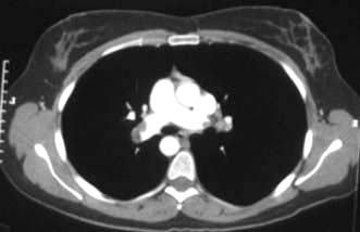
Superior vena caval obstruction
Superior vena caval obstruction (Fig. 9.12) can arise from any upper mediastinal mass but is most commonly associated with lung cancer and lymphoma. The patient presents with difficulty breathing and/or swallowing, with stridor, swollen, oedematous facies and arms with venous congestion in the neck and dilated veins in the upper chest and arms. Treatment is with immediate steroids, vascular stents, anticoagulation and mediastinal radiotherapy or chemotherapy. Some tumours, e.g. lymphomas, small-cell lung cancers and germ cell tumours, are so sensitive to chemotherapy that this is preferred to radiotherapy, as the masses are likely to be both large and associated with more disseminated disease elsewhere. An early decision is necessary on the patient’s likely prognosis, as ventilatory support may be required until treatment has had time to relieve the obstruction.
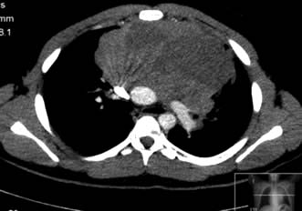
Spinal cord compression
Spinal cord compression (see p. 1135) needs to be rapidly diagnosed and urgent treatment arranged within 24 hours of onset of paresis to salvage as much functional capacity as possible. Early neurological clinical features may be incomplete, more subjective than objective and gradual in onset. MR scanning is the investigation of choice. Treatment should begin with high-dose steroids and a joint neurosurgical and oncological consultation. Good prognosis patients with limited disease require surgical decompression and radiotherapy to the affected vertebrae to achieve the best disease control and palliation.
Tumour lysis syndrome
This occurs if treatment triggers a massive breakdown of tumour cells, leading to increased serum levels of urate, potassium and phosphate and a secondary hypocalcaemia. These biochemical changes can lead to cardiac arrhythmias and seizures. Urate deposition in the renal tubules can cause renal failure (hyperuricaemic nephropathy). Vigorous hydration, often with diuretics, is crucial to maintain high urine outputs in such patients; however, a proportion will require dialysis for uraemia, oliguria or severe electrolyte disturbances. The xanthine oxidase inhibitor allopurinol should be given before treatment is started in low-risk patients. Intravenous rasburicase, a recombinant urate oxidase, is used for prophylaxis in high-risk patients and in the treatment of tumour lysis syndrome.
Acute hypercalcaemia
This presents with vomiting, confusion, constipation and oliguria. Treatment is by resuscitation with intravenous fluids to establish a saline diuresis and i.v. bisphosphonate, e.g. pamidronate or the more potent zoledronic acid (see Emergency Box 19.2). Treating the cause is crucial. Denosumab and calcitonin can be used in intractable cases.
Raised intracranial pressure
Raised intracranial pressure due to intracerebral metastases presents classically with headache, nausea and vomiting. There are often no localizing neurological signs and almost never papilloedema until very late in the disease. However, for many there is a slower onset with nonspecific symptoms such as drowsiness or mental deterioration. Treatment is by high-dose steroids and investigation by MRI as to whether surgery is appropriate if unifocal and/or threatening the fourth ventricle, or whole brain or local stereotactic Cyberknife radiotherapy are required.
Hyperviscosity
This can affect those with a very high haematocrit (Hb >180 g/L), white cell count (>100 × 109/L) or platelet cell count (>1000 × 109/L) from untreated leukaemia, or a myeloproliferative disorder. Viscosity can also be increased by high levels of monoclonal immunoglobulin molecules seen in myeloma or Waldenstrom’s macroglobulinaemia. IgA and IgM are more commonly implicated due to their respective dimeric and pentameric structures. Clinical features include hypoxia, pulmonary infiltrates, confusion, headache, visual disturbances, papilloedema and retinal venous dilation as well as rarely cardiac failure or priapism. Treatment is by leucopheresis or plasmapheresis followed by urgent treatment for the underlying malignancy.
Malignant bile duct obstruction
This will present with cholestatic jaundice. Lymphomatous obstruction will respond very well to prompt initiation of therapy. A small proportion of pancreatic and bile duct tumours are surgically resectable, more commonly those in the distal bile duct as compared to the hilar region. However, in the greater proportion of patients the treatment is palliative. In recent years endoscopic techniques have allowed the insertion of stents into the biliary tree to re-establish bile flow. The initial use of plastic stents has largely been replaced by self-expanding metal stents which have considerably longer periods of patency though at the risk of ascending infection. In the small proportion of patients in whom bile duct drainage is not possible endoscopically, the percutaneous route offers an alternative. Endoscopic photodynamic laser therapy with a photoporphyrin sensitizer can also prolong patency.
FURTHER READING
Hoeijmakers JH. DNA damage, aging and cancer. N Engl J Med 2009; 361:1475–1485.
McDermott U, Downing JR, Stratton MR. Genomics and the continuum of cancer care. N Engl J Med 2011; 364:340–350.
Smith TJ, Khatcheressian J, Lyman GH et al. 2006 update of recommendations for the use of white blood cell growth factors: an evidence-based clinical practice guideline. J Clin Oncol 2006; 24:3187–3205.
Haematological malignancies
The leukaemias, the lymphomas and multiple myeloma are an interrelated spectrum of malignancies of the myeloid and lymphoid systems. They are uncommon but not rare, the lymphomas alone being the fifth commonest cancer in the UK. The aetiology of these diseases for the most part is unknown, although viruses, irradiation, cytotoxic poisons and immune suppression have been implicated in a small proportion of cases (see p. 434). The pathogenesis involves at least one or usually more molecular abnormalities and non-random chromosomal abnormalities have been detected in several leukaemias and lymphomas. Classification has become increasingly complex, with the universally applied WHO scheme demanding morphological, cytogenetic and sometimes molecular criteria to be fulfilled. Transformation from low-grade to high-grade pathologic subtype may occur. Treatment options are multiple. Patients need to be supported through treatment involving prolonged myelosuppression and immunosuppression. These are potentially life-threatening but can also be curative. This has given rise to the need for highly skilled staff and specialist facilities; patients should be referred to these centres for treatment.
Haematological malignancies can be divided on the basis of the speed of evolution of the disease, according to the cell of origin (myeloid or lymphoid) and according to whether the presentation is primarily a marrow-based leukaemic presentation or a nodal or extranodal lymphomatous presentation where soft tissue masses predominate. This is summarized in Table 9.16. This is an arbitrary division and there is movement across the divisions, e.g. CML can transform to either AML or ALL, myeloproliferative neoplasms (MPN) and MDS can transform to AML, CLL and low-grade NHL can transform to a high-grade form ‘Richter’s transformation’.
Table 9.16 Classification of leukaemia and lymphoma
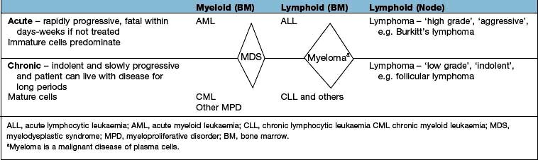
The myelodysplastic syndromes are considered pre-leukaemic and are discussed on page 405. Similarly the myeloproliferative disorders may also transform to acute leukaemia and are discussed on page 402.
In the management of these diseases, it is critical that patients are apprised of the natural history, its potential modification by treatment and the risks of both severe morbidity and mortality. It must be made clear from the outset whether a curative or palliative strategy is most appropriate and why. If cure is to be pursued, the patient must be apprised of the approximate probability of success and its potential price. The possibility of failure needs to be addressed at the outset.
The leukaemias
There are four main subtypes, as discussed above:
1. Acute myeloid leukaemia (AML)
2. Acute lymphoblastic leukaemia (ALL)
These are relatively uncommon diseases with an incidence of about 10/100 000 per year, which can occur at any age. The type of leukaemia varies with age; acute lymphoblastic leukaemia (ALL) is mainly seen in childhood and chronic lymphocytic leukaemia (CLL) is a disease of the elderly.
Leukaemia can be diagnosed by examination of a stained slide of peripheral blood and bone marrow, but immunophenotyping, cytogenetics and molecular genetics are essential for complete subclassification and prognostication. The lineage and degree of maturity of the leukaemic clone can be assessed by the expression of cytosolic enzymes and expression of surface antigens.
Aetiology
In the majority of patients, this is unknown but several factors have been associated:
Radiation. This can induce genetic damage to haemopoietic precursors and ALL, AML and CML have been seen in increased incidences in survivors of Hiroshima and Nagasaki and in patients treated with ionizing radiation.
Chemical and drugs. Exposure to benzene used in industry may lead to marrow damage. AML occurs after treatment with alkylating agents (e.g. melphalan) and topoisomerase II inhibitors (e.g. etoposide).
Genetic. Leukaemia risk is highly elevated in a number of germline conditions that result in genetic instability or bone marrow failure. These include Fanconi anaemia, ataxia telangiectasia and Li–Fraumeni syndrome. The risk is elevated some 30 times in people with trisomy 21. There is a high degree of concordance among monozygotic twins. Several genes have also been associated with familial AML such as CEBPA and RUNX1.
Viruses. A type of leukaemias is associated with human T cell lymphotropic retrovirus type 1 (HTLV-1), which is found particularly in Japan and the Caribbean.
Genetic abnormalities in leukaemia
Leukaemic cells often have a somatically acquired cytogenetic abnormality, which may be of prognostic, as well as diagnostic, significance.
These genetic alterations change the normal cell regulating process by interfering with the control of normal proliferation, blocking differentiation, maintaining an unlimited capacity for self-renewal and, lastly, promoting resistance to death signals, i.e. decreased apoptosis.
The first non-random chromosomal abnormality to be described was the Philadelphia (Ph) chromosome, which is associated with CML in 97% of cases (see Fig. 9.16a). The Ph chromosome is also found in ALL, the incidence in the latter illness increasing with age. The translocation is shown schematically in Figure 9.16a. The Ph chromosome is an abnormal chromosome 22, resulting from a reciprocal translocation between part of the long arm of chromosome 22 and chromosome 9. The resulting karyotype is described as t(9; 22)(q34; q11). The molecular consequences of the translocation are that part of the Abelson proto-oncogene (c-ABL) normally present on chromosome 9 is translocated to chromosome 22, where it comes into juxtaposition with a region of chromosome 22 named the ‘breakpoint cluster region’ (BCR). The new ‘fusion’ gene BCR-ABL is capable of being expressed as a chimeric messenger RNA, which has been identified in cells from patients with CML. When translated, this produces a fusion protein that has tyrosine kinase activity and enhanced phosphorylating activity compared with the normal protein, resulting in altered cell growth, stromal attachment and apoptosis. The breakpoint differs in CML and Ph-positive ALL, leading to the production of two different tyrosine kinase proteins with molecular weights of 210 kDa and 190 kDa, respectively. It is unclear whether the presence of BCR-ABL is sufficient for the development of the disease. It has been shown that normal subjects can carry low levels of the BCR-ABL fusion gene in their blood without developing leukaemia. Other genetic and cytogenetic abnormalities are often seen in leukaemic cells (Table 9.17).
Table 9.17 WHO classification of acute leukaemia
|
|
a The entities included in this group are defined almost identically to the corresponding entity in the French–American–British (FAB) classification.
Modified from: Jaffe ES, Harris NL, Stein H et al. (eds) World Health Organization Classification of Tumours. Pathology and Genetics of Tumours of Haematopoietic and Lymphoid Tissues. Lyon: IARC Press; 2008, with permission from the World Health Organization.
As well as cytogenetic and molecular aberrations, epigenetic modification via abnormal methylation patterns and chromatin modification due to histone acetylation is increasingly understood to be involved in oncogenesis and may represent potential therapeutic targets.
Acute leukaemias
The acute leukaemias increase in incidence with advancing age. Acute myeloid (myeloblastic, myelogenous) leukaemia (AML) has a median age at presentation of 65 years and may arise de novo or against a background of myelodysplasia or prior cytotoxic chemotherapy (‘secondary’). Acute lymphoid (lymphoblastic) leukaemia (ALL) has a substantially lower median age at presentation and in addition is the commonest malignancy in childhood. The WHO classification is shown in Table 9.17.
Clinical features
The majority of patients with acute leukaemia, regardless of subtype, present with symptoms reflecting inadequate haematopoiesis secondary to infiltration of the bone marrow by leukaemic cells, symptoms due to tissue infiltration by leukaemic cells, the consequences of a high WBC or substance release from the tumour cells (Table 9.18).
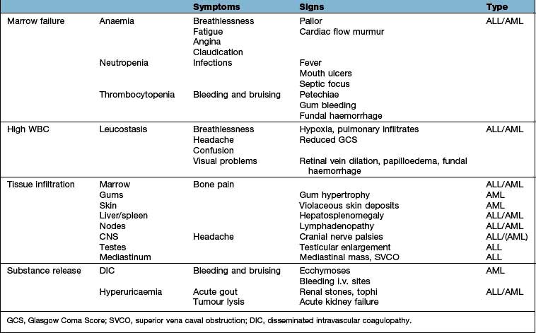
Investigations
Confirmation of diagnosis (Fig. 9.13)
Blood count. Hb low, WBC raised usually (sometimes low), platelets low.
Blood film. Blast cells almost invariably seen (Fig. 9.13a), lineage may be identified morphologically, e.g. presence of Auer rods is consistent with a diagnosis of AML (Box 9.7).
Bone marrow aspirate. Increased cellularity, reduced erythropoiesis, reduced megakaryocytes. Replacement by blast cells >20% (often approaching 100%) (Fig. 9.13b). Lineage confirmation by immunophenotyping, e.g. AML – CD33 or CD13, B lineage ALL – CD10 and CD19 and T lineage – CD3. Cytogenetic FISH analysis in real-time PCR and molecular genetics for prognostication.
Chest X-ray. Mediastinal widening often present in T lymphoblastic leukaemia.
Cerebrospinal fluid examination. Performed in all patients with ALL, as the risk of CNS involvement is high. It is less critical in AML.
Coagulation profile to exclude presence of DIC (raised PT, APTT, reduced fibrinogen, increased fibrinogen degradation products, e.g. D-dimers).
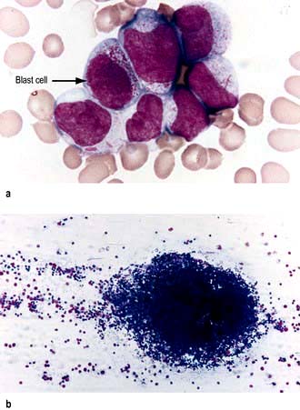
Figure 9.13 Acute leukaemia. (a) Peripheral blood film showing characteristic blast cells. The arrow points to the abnormal blast cell. (b) Bone marrow aspirate showing particle with increased cellularity.
(Courtesy of Dr Manzoor Mangi.)
Box 9.7
Characteristics of blast cells
A blast is an immature precursor of myeloid cells (myeloblasts) or lymphoid cells (lymphoblast)
Bigger than normal counterpart
Immature nucleus (nucleolus, open chromatin)
Cytoplasmic appearances often atypical
Rarely ever seen in normal individuals
If present, are highly suggestive of an acute leukaemia or a chronic disorder that is beginning to transform into an acute disease, e.g. transformed MPN, MDS-RAEB, CML blast crisis.
For planning therapy
Biochemistry, serum urate, renal and liver biochemistry.
Cardiac function; ECG and direct tests of left ventricular function, e.g. echocardiogram (see p. 685).
Principles of management
Untreated acute leukaemia is invariably fatal, most often within a few months, though with judicious palliative care, it may be extended to perhaps a year. Treatment with curative intent may be successful, or may fail, either because the leukaemia does not respond (i.e. refractory to treatment), because the disease returns after an initial favourable response (relapse) or because the patient succumbs to complications of the therapy (treatment-related mortality). At initial presentation, acute leukaemias range from being probably curable (e.g. childhood ‘good risk’ ALL) through to probably incurable (e.g. AML with adverse cytogenetic features in the elderly). Since curative treatment carries considerable morbidity and potential mortality, it is essential that the ‘risk/benefit’ ratio is clearly understood by physician and patient alike.
Palliative therapy
Every attempt should be made to ensure that the patients are at home as much as possible, while making available the full range of supportive care. Palliation may well include both low-dose chemotherapy and irradiation in addition to blood product support and antimicrobials.
Curative therapy
The decision to treat with curative intent, particularly if successful, implies severe disruption of normality for the patient and family for at least 6 months and often up to a year. In the short term, it may demand transfer to another hospital, as acute leukaemia should only be treated in units seeing at least 10 such cases per year. It is highly likely to involve admission to hospital for up to a month in the first instance, with further, partly predictable, subsequent admissions of several days’ to weeks’ duration, requiring discussions and decisions about work or education.
The decision to treat with curative intent implies that cure is possible and that the chance of cure justifies the risks of the therapy. It does not imply that cure is guaranteed or even expected. The failure rate may be high and the patient must know that he or she will be told if cure becomes an unrealistic goal.
Active therapy
Supportive care
This forms the basis of treatment whether for cure or palliation:
Avoidance of symptoms of anaemia (haemoglobin >80–100 g/L) – repeated transfusion of packed red cells (sometimes irradiation of cells is required)
Prevention or control of bleeding (platelet count <10 × 109/L in the stable and <20 × 109/L in the septic patient or <50 if a procedure is planned, e.g. a lumbar puncture). Coagulation abnormalities should be corrected with FFP to keep APTR/INR <1.5 × normal and with cryoprecipitate to keep the fibrinogen level >1.5 g/dL. Norethisterone is given to women of menstrual age to avoid menorrhagia during their thrombocytopenic phase.
Leucopheresis may be required to reduce the white cell count rapidly before the chemotherapy has started to be effective.
Treatment of infection (see Box 9.6):
Control of hyperuricaemia with hydration, prophylactic allopurinol and occasionally rasburicase (see p. 479).
Indwelling venous devices, e.g. Hickman line, are required to allow easy access to the blood for tests and administration of therapy. Sperm banking is offered to postpubertal men and oocyte collection to women, if there is time before treatment.
Specific treatment
The initial requirement of therapy is to return the peripheral blood and bone marrow to normal (complete remission; CR). This ‘induction chemotherapy’ is tailored to the particular leukaemia and the individual patient’s risk factors. Since this treatment is not leukaemia specific but also impairs normal bone marrow function, it leads to a major risk of life-threatening infection, which increases the risk of early death in the short term.
Successful remission induction is always followed by further treatment (consolidation). The details of this are determined by the type of leukaemia, the patient’s risk factors and the patient’s tolerance of treatment. Recurrence is almost invariable if ‘consolidation’ therapy is not given. This reflects the lack of sensitivity of the definition of ‘complete remission’, which has been solely morphological. Cytogenetics and molecular genetic techniques can however identify residual leukaemic cells not detected morphologically and they are highly predictive of recurrence. Recommendations have recently been made to modify the definition of remission to reflect this. Failure to achieve morphological CR with two cycles of therapy (‘refractory’) carries almost as bad a prognosis as the untreated leukaemia. If CR can be achieved, e.g. by new experimental approaches, cure may still be possible with stem cell transplantation (see p. 444). A small proportion of patients with refractory disease may also be cured by a myeloablative allograft.
Acute myeloid leukaemia (AML)
In AML, the prognosis is dependent on a range of key variables, the two main ones being age and cytogenetics (Fig. 9.14; Table 9.19).
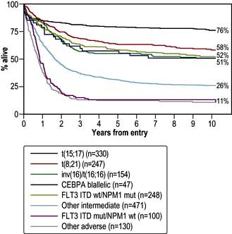
Figure 9.14 Prognosis related to cytogenetics and molecular data in AML Outcome of younger adults with AML treated in the MRC AML10 and AML12 trials stratified according to cytogenetic and molecular abnormalities.
(Reproduced with permission from Smith ML, Hills RK, Grimwade D. Independent prognostic variables in acute myeloid leukaemia. Blood Reviews 2011; 25(1):39–51.)
| Good risk | Poor risk |
|---|---|
Treatment with curative intent is undertaken in the majority of adults below the age of 60 years, provided there is no significant co-morbidity. Treatment success reflects the cytogenetic pattern. Those at ‘low risk’ are treated with moderately intensive combination chemotherapy. This always includes an anthracycline such as daunorubicin and cytosine arabinoside (cytarabine) and consolidation with a minimum of four cycles of treatment given at 3–4-week intervals. Patients with low-risk disease do not benefit from allogeneic stem cell transplantation during their first complete remission because the risks outweigh benefits. Those at ‘intermediate risk’ are a heterogeneous group. When possible, they should be given consolidation chemotherapy after an initial remission has been achieved followed by allogeneic transplantation only in those who are deemed at increased risk of relapse because of other risk factors, e.g. mutational data. Patients with high-risk disease should proceed to a stem cell transplant in CR1 because they respond poorly to conventional chemotherapy and have a high risk of relapse.
Complete remission (CR) will be achieved in about 80% of patients under the age of 60. Failure is due to either resistant leukaemia (10%) or death due to infection or bleeding (10%). Approximately 50% of those entering complete remission will be cured (i.e. approximately 40% overall) although this varies from 60–70% in the favourable cytogenetic group to 10–20% in the adverse cytogenetic group.
The initial treatment of the older patient is much more contentious. Older patients tolerate cytotoxic therapy less well than younger patients due to co-morbidities and their disease is often more aggressive in its biology, e.g. adverse cytogenetics are more common with increasing age. Reduced intensity allogeneic transplantation is increasingly being used for this group but is limited by the toxicity of this treatment.
The management of recurrence is undertaken on an individual basis, since the overall prognosis is very poor despite the fact that second remissions may be achieved. Long survival following recurrence is rarely achieved without allogeneic transplantation. Experimental therapy should be considered. The use of minimal residual disease monitoring may allow the detection of a subgroup of patients during initial therapy who require treatment intensification, e.g. allograft, as well as patients in CR who are at the early stages of relapse and need preemptive therapy before frank marrow relapse occurs.
Newer agents that target the FLT3 mutation, present in a significant proportion of cases of AML, are in clinical trials in conjunction with conventional chemotherapy. Other novel therapies that are being considered in AML include chemotherapy labelled monoclonal antibodies (gemtuzumab ozogamicin) and hypomethylating agents (azacytidine).
Acute promyelocytic leukaemia (APML)
This is a variant of AML, occurring in 10–15% of cases, that is characterized by the translocation t(15; 17) and with particular morphological features. There is an almost invariable coagulopathy, which was a major cause of early death. The empirical discovery that all-trans-retinoic acid (ATRA) causes differentiation of promyelocytes and rapid reversal of the bleeding tendency was a major breakthrough. APML is treated with ATRA combined with several courses of chemotherapy. Complete remission and molecular remission occur in at least 90% of younger adults with APML and at least 70% will expect to be cured. Transplantation may be necessary either if the leukaemia is not eliminated at the molecular level, or following recurrence and reinduction therapy. Arsenic trioxide, which induces apoptosis via activation of the caspase cascade (see p. 33), is used with resistant or relapsed disease and is under investigation as first-line therapy.
Acute lymphoblastic leukaemia (ALL)
This condition may present in leukaemic phase with significant marrow involvement (ALL) or may present as localized disease, typically a mediastinal mass (lymphoblastic lymphoma). The tumour cells in each condition are indistinguishable and similar therapies are therefore used. The overall strategy for the treatment of ALL differs in detail from that for AML. Remission induction is undertaken with combination chemotherapy including vincristine, a glucocorticoid, an anthracycline and asparaginase. Once remission is achieved, the details of consolidation will be determined by the anticipated risk of failure. Intensive consolidation of remission with variable numbers of chemotherapy cycles comprising cytotoxics with different mechanisms of action has been standard practice, including the administration of high-dose methotrexate. In those patients with high-risk features (see below) or an HLA-matched sibling donor, allogeneic transplantation is recommended upon achieving first complete remission (CR1).
A major difference between therapy for ALL and AML is the need for central nervous system directed therapy. Prophylaxis should be given with intrathecal chemotherapy under platelet cover if necessary, as soon as blasts are cleared from the blood. Depending upon risk this may be continued for up to 2 years and complemented by high doses of systemic cytosine arabinoside (cytarabine) or methotrexate. Cranial irradiation was previously given to all patients to reduce the risk of relapse within the central nervous system; some risk adapted strategies reserve this only for those patients at very high risk.
Additionally, after intensive induction and consolidation, maintenance therapy for 2 years is required to reduce the risk of disease recurrence. This typically comprises 2 years of treatment with methotrexate and mercaptopurine, although more intensive regimens are used by many groups.
Prognostic factors
A number of clinical and laboratory features are determinants of treatment response and survival in ALL (Table 9.20). Increasingly, therapeutic strategies based upon prognostic risk are being used in the management of the disease.
Table 9.20 Acute lymphoblastic leukaemia (ALL) risk factors
| Risk factor | Good | Poor |
|---|---|---|
Age |
Younger age |
Older age |
WBC |
<50 × 109/L for B-lineage <100 × 109/L for T-lineage |
>50 × 109/L for B-lineage >100 × 109/L for T-lineage |
Immunophenotype |
CD10 + common ALL |
Pro-B ALL |
Cytogenetic aberrations |
t(12;21) hyperdiploidy |
t(9;22) or t(4;11) hypodiploidy |
Time to response |
Early clearance of blasts |
Failure to achieve a CR within 3–4 weeks |
Minimal residual disease |
MRD negative |
MRD positive |
Extramedullary disease |
CSF clear |
CSF positive |
Prognosis
The prognosis (Fig. 9.15) of ALL in childhood is now excellent: complete remission is achieved in almost all, with up to 80% being alive without recurrence at 5 years. Failure occurs most frequently in those with high presentation blast counts or a t(9;22) translocation. Current treatment strategies are to lessen therapy for ‘good risk’ children in order to avoid some of the long-term consequences of therapy such as avascular necrosis of bone, infertility, neurotoxicity and cardiotoxicity.
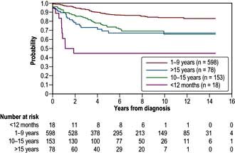
Figure 9.15 Overall survival in 2628 children of different ages with newly diagnosed ALL participating in consecutive studies conducted at St Jude Children’s Research Hospital from 1962 to 2005.
(From Pui C-M, Robinson LL, Lock T. Acute lymphoblastic leukaemia. Lancet 2008; 371:1030–1043, Figure 3, with permission.)
The situation is far less satisfactory for adults, the prognosis getting worse with advancing years. Co-morbidity and t(9; 22) translocation increases in frequency with age. Overall, the complete remission rate is 70–80%. Disease that is refractory to first-line therapy carries a very poor prognosis. Between 30% and 40% of patients continue in durable first remissions, resulting in approximately 25–30% overall patient cure. Increasing numbers of patients are being transplanted for this condition, including those with sibling donors and those high-risk patients with an available unrelated donor. Imatinib used in conjunction with chemotherapy increases the response rate and quality of response in patients with the t(9;22) translocation and ALL. As with AML, most recurrences occur within the first 3 years and the outcome is extremely poor. Second remissions, though usually achieved, are rarely durable except following allogeneic transplantation. Isolated extramedullary recurrences, however, may be cured.
Chronic leukaemias
Chronic myeloid leukaemia (CML)
Chronic myeloid leukaemia (CML), which accounts for about 14% of all leukaemias, is one of the family of myeloproliferative neoplasms (MPNs) and is almost exclusively a disease of adults with the peak of presentation being between 40 and 60 years. It is defined by the presence of the Philadelphia chromosome (Fig. 9.16), either demonstrated cytogenetically (95%) or molecularly (5%). Unlike the acute leukaemias which are either rapidly reversed or rapidly fatal, CML has a more slowly progressive course, which if not initially cured, will be followed eventually by ‘blast crisis’ transformation to acute leukaemia (75% myeloid, 25% lymphoid) or myelofibrosis with death in a median of 3–4 years.
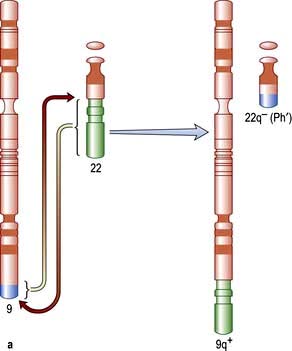
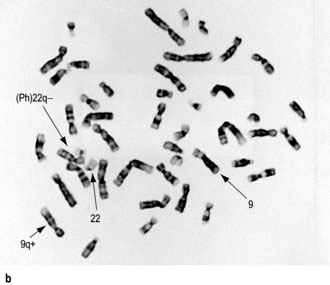
Figure 9.16 The Philadelphia chromosome (Ph). (a) The long arm (q) of chromosome 22 has been shortened by the reciprocal translocation with chromosome 9. (b) The Philadelphia chromosome is formed by a reciprocal translocation of part of the long arm (q) of chromosome 22 to chromosome 9. It is seen in 90–95% of patients with chronic myeloid leukaemia. The karyotype is expressed as 46XX, (9;22)(q34;q11).
Clinical features
CML usually presents in the chronic phase and some patients have no symptoms. Symptoms will include:
Investigations
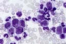
Blood count. Hb low (normochromic and normocytic) or normal, WBC raised (usually >100 × 109/L), platelets low, normal or raised.
Blood film. Neutrophilia with the whole spectrum of mature myeloid precursors. Elevated basophils and eosinophils. Increased numbers of blasts are suggestive of accelerated phase or blast crisis.
Bone marrow aspirate. Increased cellularity, increased myeloid precursors. Cytogenetics reveals t(9; 22) translocation (the Philadelphia chromosome) (Fig. 9.16b).
Fluorescence in situ hybridization (FISH) or reverse transcriptase polymerase chain reaction (RT-PCR) are used to demonstrate the cytogenetic/molecular abnormality. These are also used to quantitatively monitor response to therapy.
Management
Treatment has been transformed by the advent of imatinib, a tyrosine kinase inhibitor that specifically blocks the enzymatic action of the BCR-ABL fusion protein, which is now first-line treatment for the chronic phase. Imatinib produces a complete haematological response in over 95% of patients and 70–80% of these have no cytogenetically detectable BCR-ABL translocation in the marrow (complete cytogenetic remission). A proportion will lose molecularly detectable BCR/ABL transcripts from the blood (complete molecular remission). Event-free and overall survival appear to be better than for other treatments. Side-effects of imatinib include nausea, headache, rashes and cytopenia. Imatinib can be continued indefinitely although it should be stopped before attempting to conceive. Resistance to imatinib as a single agent may develop as a result of secondary BCR/ABL kinase mutations beyond the t(9;22). The use of second-generation tyrosine kinase inhibitors, dasatinib and nilotinib, may restore haematological or molecular remissions in those patients in the chronic phase that have primary or acquired resistance to imatinib or who are intolerant to imatinib. Both dasatinib and nilotinib have demonstrated promise in the first-line setting although it remains to be seen whether any advantage over imatinib in speed or depth of response is sustained over time. Bosutinib and ponatinib are also under evaluation.
In the acute phase (blast transformation) most patients have only a short-lived response to imatinib and other chemotherapy, as for acute leukaemia, and stem cell transplantation is used in the hope of achieving a durable remission.
Chronic lymphocytic leukaemia (CLL)
This is the most common leukaemia, occurring predominantly in later life and increasing in frequency with advancing years (median age of presentation between 65 and 67 years). It results from the clonal expansion of small lymphocytes and is almost invariably (95%) B cell in origin. The majority of patients are asymptomatic, identified as a chance finding on a blood count performed for another indication. Other patients, however, present with the features of marrow failure or immunosuppression. The median survival is about 10 years and prognosis correlates with clinical stage at presentation (Table 9.21). A number of cytogenetic and molecular abnormalities are now recognized as being of prognostic significance (see below). This condition may present in leukaemic phase with significant marrow involvement (CLL) or may present as localized disease (small lymphocytic lymphoma, SLL). The tumour cells in each condition are indistinguishable and a similar therapeutic approach is therefore used. A pre-malignant condition, monoclonal B cell lymphocytosis (MBL) exists where there are less than the 5 × 10⁹/L B-cells required for a diagnosis of CLL. Some of these have CLL phenotype and may progress to CLL.
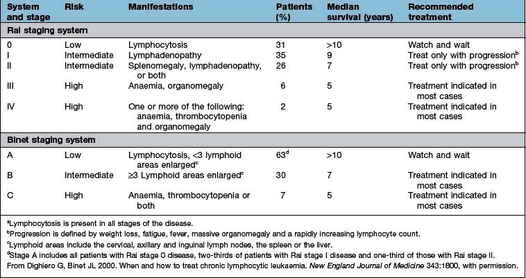
Clinical features
The majority of patients are asymptomatic at presentation. Common symptoms will be:
Recurrent infection because of (functional) leucopenia and immune failure (reduced immunoglobulins)
Investigations
Blood count. Hb normal or low; WBC raised and may be very high; with lymphocytosis (criteria for diagnosis >5 × 10⁹/L), platelets normal or low.
Blood film. Small or medium sized mature and normal appearing lymphocytes. May see smudge cells in vitro. No immature blasts are evident.
Bone marrow. Reflects peripheral blood, often very heavily infiltrated with lymphocytes.
Immunophenotyping shows mainly CD19+, CD5+, CD23+ B cells with weak expression of CD20, CD79b and surface immunoglobulin (kappa and lambda light chains).
Cytogenetics/FISH analyses are not essential for diagnosis but help in the assessment of prognosis.
Direct Coombs’ test. May be positive if there is haemolysis.
Prognostic biomarkers
The clinical course of CLL is variable. Several markers are used to supplement clinical stage and have been shown to predict progression and survival. Variations in predictor cut-off levels have limited their widespread application.
Cytogenetic abnormalities are detected in >90% of cases. Patients with an isolated deletion of 13q have an excellent prognosis, in contrast to those with either 11q deletion or 17p deletion (the sites of the tumour suppressor genes ATM and TP53, respectively) who tend to have a rapidly evolving clinical course. In those tumours that demonstrate a high level of mutation within the variable region of the rearranged immunoglobulin heavy chain (IgVH), the clinical course is more indolent than those where the IgVH sequence more closely resembles that of the germline. Expression of ZAP70, a 70 kDa tyrosine kinase protein, correlates well with mutational status. Patients with <20% expression of ZAP70 have median 10-year survival of >50%; in >20% expression the median survival was <5 years. High expression of CD38 on leukaemic cells may also indicate adverse prognosis.
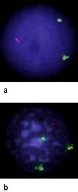
Fluorescence in situ hybridization photomicrograph of a patient with CLL. (a) 17p (green probe) and 11q (red probe) shows two green signals (TP53 deletion) with normal diploid complement of 11q. (b) 12 centromere (green probe) and 13q14 (red probe) shows three green signals (trisomy 12) with normal diploid complement of 13q.
(Courtesy of Debra Lillington, Barts and the London NHS Trust.)
Management
In CLL, the major consideration is when to treat, indeed 30% of patients will never require intervention. Treatment depends on the ‘stage’ (Table 9.21) of the disease. Choice of therapy will depend upon patient-related factors such as age and co-morbidity, adverse prognostic features and anticipated response and toxicities to therapy. Intervention, when indicated, usually causes improvement in symptoms and in the blood count. The effect on survival is unclear.
Early-stage disease is usually managed expectantly, advanced-stage disease is always treated immediately and the approach to the intermediate stage is variable. The absolute indications for treatments are:
Marrow failure manifest by worsening anaemia and/or thrombocytopenia
Massive or progressive splenomegaly or lymphadenopathy
Progressive disease manifest by doubling of the lymphocyte count in 6 months
General/supportive treatment
Anaemia due to haemolysis is treated with steroids. Anaemia and thrombocytopenia due to marrow infiltration is treated with chemotherapy and, when necessary, transfusion. Erythropoietin (see p. 374) may avoid the need for transfusions, particularly in patients receiving chemotherapy.
Infection is treated as indicated, with prophylactic antibiotic, antiviral, anti-PCP therapy and antifungal therapy potentially being given during periods of chemotherapy. Immunoglobulin replacement may be helpful.
Specific treatment
Chlorambucil usually reduces the white cell count and decreases lymphadenopathy and splenomegaly and successfully palliates the disease. In asymptomatic patients without an indication for therapy, early use of chlorambucil does not provide a survival advantage over expectant management.
Combination therapy with rituximab shows a dramatic improvement in the response rate and has become standard of care as first-line therapy – FCR (fludarabine, cyclophosphamide, rituximab).
Purine analogues, fludarabine alone or in combination with cyclophosphamide or mitoxantrone (with or without steroids), have had a much greater impact on the bone marrow and can induce complete or molecular complete remission although they are not helpful in patients with 17p deletion or TP53 mutation.
Alemtuzumab, a humanized monoclonal antibody targeting CD52 which is highly expressed on B-CLL, is used in those patients that progress after fludarabine or who have 17p deletion or TP53 mutation
Ofatumumab is a new generation anti-CD20 monoclonal antibody that binds to a different epitope to that of rituximab and is used for treatment of fludarabine or alemtuzumab refractory CLL
Allogeneic stem cell transplantation with non-myeloablative conditioning regimens is increasingly performed.
Hairy cell leukaemia (HCL)
HCL is a clonal proliferation of abnormal B (or very rarely T) cells which, as in CLL, accumulate in the bone marrow and spleen. It is a rare disease, median age at presentation is 52 years old and the male to female ratio is 4 : 1. The bizarrename relates to the appearance of the cells on a blood film and in the bone marrow – they have an irregular outline owing to the presence of filament-like cytoplasmic projections.
Clinical features
Clinical features include anaemia, fever and weight loss. Splenomegaly occurs in 80%, lymphadenopathy is uncommon. Anaemia, neutropenia, thrombocytopenia and low monocyte counts are found.
Treatment
The purine analogues 2-chloroadenosine acetate (2-CDA; cladribine) and pentostatin have specific activity in this condition; complete remission is achieved in 90% with just one cycle of treatment. The remissions can last for many years and patients can be successfully retreated. Rituximab is used in cases who do not respond to the above drugs.