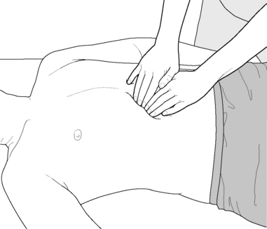
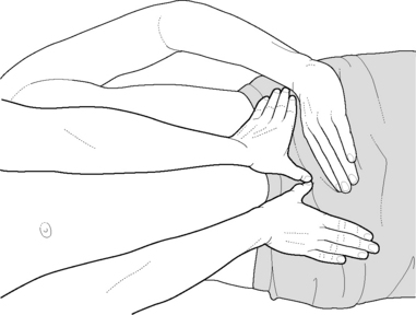
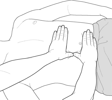
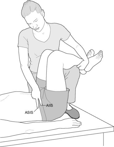
Box 10.7 Lief’s NMT of lower thoracic and lumbar area

Refer to Volume 1, Box 14.8 and Fig. 14.24 for Lief’s NMT approach to the proximal aspects of latissimus. The descriptions in Volume 1, Box 14.8 focus on intercostal NMT assessment but it is clear that superficial to the intercostal muscles, latissimus fibers would be being evaluated when attempting to access the tissues between the ribs, as would the attachments of serratus posterior inferior at the lower four ribs.

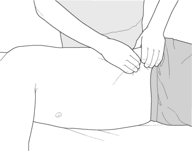
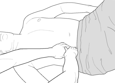
The practitioner stands on the left side at the level of the patient’s waist, initially facing caudad (Fig. 10.29).

A pattern of strokes, as illustrated, should now be carried out on the patient’s left side by the practitioner’s right hand. A series of slow searching strokes should be applied as follows while involving two or more glides of the thumb in each location, the first more superficial than the second. In this way paraspinal as well as tissue slightly lateral to these tissues will be sequentially searched for evidence of aberrant soft tissue changes, both superficially and at greater depth. The series begins by:

running inferiorly alongside the spine and then

commencing slightly more laterally, involving the levels of T8 to T11

followed by the levels of T11 to L1 then

searching the tissues at the levels of L1 to L4.

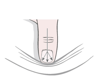
The pressure of the thumb (or finger if thumb is unstable) should be downward into the tissues, meeting and matching tension and angled so that the medial aspect of the thumb (or distal pad of the finger) is applying the most force, for precise localization of dysfunction.

The lower intercostal areas should be worked so that the searching thumb or finger contact evaluates for altered tissue texture throughout the lumbar and lower thoracic soft tissues, with particular focus on attachment sites. Strokes that run from the spinous processes laterally across the transverse processes of the lower thoracic vertebrae toward the lower ribs may access somatic changes (i.e. trigger points) in muscles such as serratus posterior inferior or latissimus dorsi. Care should be taken on the lower ribs due to their relative fragility and lack of anterior osseous support.

In this way, the lumbodorsal fascia, erector spinae and latissimus dorsi will be effectively evaluated for localized soft tissue dysfunction. The practitioner should then glide the thumb along the superior iliac crest, from just above the hip to the sacroiliac joint. Tissues just inferior to the crest, as well as over it and, if possible, just under its anterior rim, should all receive attention, so ensuring that the origin of latissimus is ‘combed’ for evidence of local dysfunction. Several such strokes may be applied into the heavy musculature above the crest of ilium. To assess and/or treat the opposite side the practitioner may need to change sides.

If the practitioner is tall enough she may be able to apply finger strokes contralaterally in order to achieve the same effects (see Volume 1, Chapter 9, for details of finger stroke).

image

Figure 10.29 Suggested locations of thumb or finger strokes in the lumbar and pelvic areas, using Lief’s NMT evaluation and treatment methods, as described in the text

(adapted from Chaitow (2010)).

Facing the patient’s waist and half-turned toward the feet, the fingers of the left hand can deal with the right lower dorsal and upper lumbar area and the iliac crest, in the manner described above, while the right hand is used to stabilize and/or distract the tissues being assessed.

One or two light but searching strokes should also be applied running caudad or cephalad, alongside and between the tips of the spinous processes, from the mid-dorsal area to the sacrum, to evaluate attachment dysfunction. If trigger points are located, especially active ones which reproduce recognizable symptoms to the patient, these should be charted and treated, if appropriate, using INIT methods or any combination of ischemic compression, MET, PRT, MFR or other similarly useful modalities.

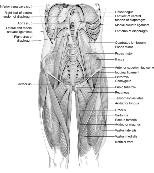
Quadratus lumborum (Figs 10.25d, 10.31)

image image

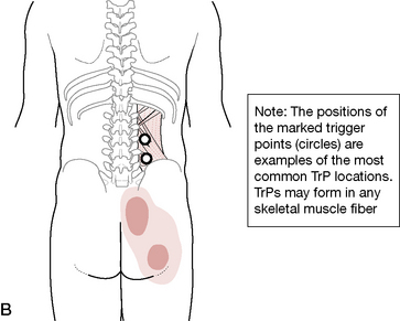
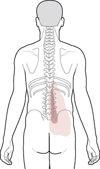
Figure 10.31 Quadratus lumborum trigger points refer into SI joint, lower buttocks and wrap laterally along the iliac crest and hip region. A referral pattern into the lower abdominal region is not illustrated

(adapted from Travell & Simons (1992), Fig 4.1 A, B).

Attachments: Iliocostal fibers (posterior plane): extend nearly vertically from the 12th rib to the iliac crest and iliolumbar ligament; iliolumbar fibers (intermediate plane): diagonally oriented from the iliac crest to the anterior surfaces of the transverse processes of L1–3 or L4; lumbocostal fibers (anterior plane): diagonally oriented from the 12th rib to the transverse processes of L2–4 or L5.

Innervation: Lumbar plexus (T12-L3 or L4)

Muscle type: Postural (type 1), shortens when stressed

Function: Ipsilateral flexion of the trunk, eccentrically controls the lumbar spine during contralateral side-flexion, stabilizes the lumbar spine, elevates ipsilateral hip, assists forced exhalation (coughing), stabilizes the attachments of the diaphragm during inspiration. QL contracting bilaterally extends the lumbar spine

Synergists: For lateral trunk flexion: ipsilateral external and internal obliques

Antagonists: For lateral trunk flexion: contralateral QL, external and internal obliques

Indications for treatment

Low back pain, especially when weight bearing

Lower back pain when coughing or sneezing

Misdiagnosed as radicular pain of lumbar origin

Muscular guarding of lumbar region

Compensatory scoliosis

Pain in iliac crest, hip region, SI joint, lower buttocks and lower quadrant of abdomen and groin (Travell & Simons 1992)

  Page 256 

Functional short leg (hypertonic QL elevates ilium in non-weightbearing position)

Restricted forward bending

Special notes

Quadratus lumborum (QL) forms a quadrilateral-shaped muscle that extends from the iliac crest to the 12th rib as well as additional sets of fibers running from both the 12th rib and iliac crest to the transverse processes of most of the lumbar vertebrae. It has a free lateral border, which is usually palpable when placed under light tension (see sidelying position, p. 361). A sheet of thoracolumbar fascia lies both anterior and posterior to QL, thereby wrapping it in a fascial casing. These fascial extensions merge laterally and attach to the transverse abdominis, thereby providing a tensional element of support for the lumbar region.

The QL is often reasonably grouped with the psoas muscles as a deep lateral muscle of the trunk, providing a portion of the deep abdominal wall. However, it has been placed in this text with the muscles of the lumbar region so that it is addressed while the patient is in a prone position. Additionally, its direct action on the lumbar vertebrae is unquestioned, as is its ability to deform lumbar discs. (The psoas muscle is discussed with the deep abdomen on p. 287.)

While QL’s most obvious task is that of lateral flexion of the trunk and lumbar spine, its less obvious roles include elevation of the hip (especially important during gait), extension of the lumbar spine when contracting bilaterally, (possibly) to provide flexion of the spine or perhaps to stabilize it during contralateral flexion (Travell & Simons 1992), to (possibly) offer assistance in normal inhalation (stabilizing diaphragm’s rib attachment) as well as forced exhalation (coughing, sneezing), and to assist in unilateral trunk rotation on a fixed pelvis.

As mentioned earlier in this chapter, Norris (2000a) has described the divided roles in which quadratus is involved.

The quadratus lumborum has been shown to be significant as a stabilizor (sic) in lumbar spine movements (McGill et al 1996) while tightening has also been described (Janda 1983). It seems likely that the muscle may act functionally differently in its medial and lateral portions, with the medial portion being more active as a stabilizor (sic) of the lumbar spine, and the lateral more active as a mobilizer (sic). (See stabilizer/mobilizer discussion in Box 2.2, and in Volume 1, Chapter 2).

Janda (1983) observes that when the patient is sidebending, ‘when the lumbar spine appears straight, with compensatory motion occurring only from the thoracolumbar region upwards, tightness of quadratus lumborum may be suspected’. This ‘whole lumbar spine’ involvement differs from a segmental restriction, which would probably involve only a part of the lumbar spine.

Quadratus fibers merge with the diaphragm (as do those of psoas), which makes involvement in respiratory dysfunction a possibility, both via this merging and its attachment to the 12th rib. It plays a role in forced exhalation (such as coughing) and during speech and singing, by fixing a base for controlled relaxation of the diaphragm (Gray’s anatomy 2005). It also stabilizes the rib, and therefore the diaphragm, during inhalation.

The lumbodorsal junction (LDJ) is biomechanically important because it is the only transitional juncture where these two mobile structures meet. Dysfunction may result from alteration of the quality of motion between these structures (upper and lower trunk/dorsal and lumbar spines). In dysfunction, there is often a degree of spasm or tightness in the muscles that stabilize the region, notably psoas and erector spinae of the thoracolumbar region, as well as quadratus lumborum and rectus abdominis.

Symptomatic differential diagnosis of muscle involvement at the LDJ is possible, as follows.

Psoas involvement usually triggers abdominal pain if severe and produces flexion of the hip and the typical antalgic posture of lumbago (Lewit 1985).

Erector spinae involvement produces low back pain at its caudad end of attachment and interscapular pain at its thoracic attachment (as far up as the mid-thoracic level) (Liebenson 2007).

Quadratus lumborum involvement causes lumbar pain and pain at the attachment of the iliac crest and lower ribs (Lewit 1985).

Rectus abdominis contraction may mimic abdominal pain and result in pain at the attachments at the pubic symphysis and the xyphoid process, as well as forward bending of the trunk and restricted ability to extend the spine (Lewit 1985) though its TrPs refer posteriorly.

There is seldom pain at the site of the lesion in LDJ dysfunction. Lewit (1985) points out that even if a number of the associated muscles are implicated it is seldom necessary, using PIR methods [MET], to treat them all since, as the muscles most involved (discovered by tests for shortness, overactivity, sensitivity and direct palpation) are stretched and normalized, others will also begin to normalize ‘automatically’.

Trigger points in QL may be activated by persistent structural inadequacies (such as lower leg length differential or developmental anomalies of the lumbar spine), overload (especially from an awkward, twisting position), trauma including auto accidents, postural strain during leisure (see Chapter 4) or sport (see Chapter 5) or work activities, or even while putting clothes on the lower body (socks, pants, pantyhose, etc.), walking on slanted surfaces or when straining during gardening, housework or other repetitive tasks (Travell & Simons 1992).

Travell & Simons (1992) provide an extensive list for differential diagnosis and a more extensive discussion than that which is offered in the following summation. These listed conditions should be ruled out in patients with associated symptoms but the reader is also reminded to examine the patient for trigger points in quadratus lumborum and associated tissues, when there exists a diagnosis of one (or more) of these conditions. Even though a diagnosis of a listed condition may be accurate and other pathological or dysfunctional conditions may exist, trigger points may be a readily remediable secondary perpetuating factor. Consideration should also be given to the possibility that a trigger point’s referral pattern may be the entire source of a painful condition that is ‘masquerading’ as the diagnosed condition. It is essential, however, not to disregard the possibility of organ or structural pathology mimicking QL dysfunction, which may have the potential to progress to an irreversible degree if neglected. Conditions that should be ruled out when confronted with apparent QL dysfunction include (Travell & Simons 1992):

  Page 257 

trochanteric bursitis

sciatica

radiculopathy

osteoarthritic spurs or narrowing of lumbar disc space

translatory movement between lumbar vertebrae

SI joint dysfunction

fractured lumbar transverse process

thoracolumbar articular dysfunction (including facet dysfunction)

spinal tumors

myasthenia gravis

aortic aneurysm

multiple sclerosis

and organ pathologies, including gallstones, liver disease, kidney stones, urinary tract problems, intraabdominal infections, intestinal parasites and diverticulitis.

Functional assessment for shortness of QL

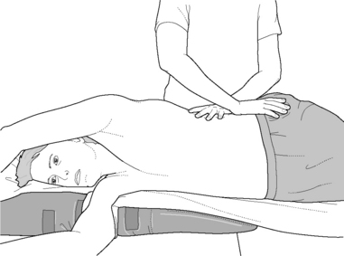
Quadratus lumborum test 1: Janda’s functional hip abduction test (see also Volume 1, Chapter 5)

The patient is sidelying and is asked to take his upper arm over his head to grasp the top edge of the table, ‘opening out’ the lumbar area.

The practitioner stands facing the front or the back of the patient, in order to palpate quadratus lumborum’s lateral border – a major trigger point site (Travell & Simons 1992).

Activity of gluteus medius and also tensor fascia latae is tested (palpated for) with the other hand, as the leg is slowly abducted.

If the muscles act simultaneously or if quadratus fires first, then QL is stressed (probably short) and will usually benefit from stretching.

The normal firing sequence should involve gluteus medius and TFL, with QL not being actively involved in contracting until 25° of lateral excursion of the leg has occurred.

Quadratus lumborum test 2

The patient stands with his back toward the crouching practitioner.

Any leg length disparity (based on pelvic crest height) is equalized by using a book or pad under the short leg side heel.

With the patient’s feet shoulder width apart, a pure sidebending is requested, so that the patient runs a hand down the contralateral thigh. Normal levels of side-bending excursion should allow the patient’s finger tips to reach to just below his contralateral knee.

If sidebending to one side is limited then QL on the opposite side is probably short.

Combined evidence from palpation (test 1) and this sidebending test indicates whether it is necessary to treat quadratus or not.

NMT for quadratus lumborum

The patient lies in a prone position and the practitioner stands at the level of the hip on the side to be treated. A light amount of lubrication is applied to the skin over the QL fibers. Only a portion of QL lies lateral to the erector spinae; however, the gliding strokes described here will influence tissues that are superficial to and lateral to QL, which may also influence QL’s ability to relax.

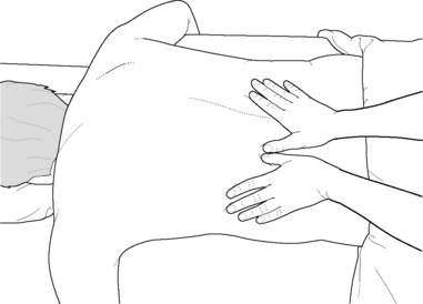
Gliding strokes are applied with both thumbs, from the crest of the ilium to the 12th rib, while remaining immediately lateral to the erector spinae (Fig. 10.32). The gliding process is repeated 4–5 times on this first section of tissue. The practitioner should avoid undue stress on her thumbs by pointing the tips of the thumbs toward the direction of the glide rather than placing the tips toward each other during the stroke, which can strain the thumb joints. (See correct hand positioning in Chapter 9, p. 197, Box 9.7)

image

Figure 10.32 Although only a small portion of quadratus lumborum is palpable, this gliding stroke can be valuable in assessing as well as producing lengthening of surrounding tissues in addition to QL.

  Page 258 

The thumbs are then moved laterally onto the next section of tissue and the gliding process is repeated 4–5 times. A third strip of tissue is usually available before encountering the fibers of external oblique. These gliding strokes can also be applied to the external oblique, if needed.

Gentle friction can be used to examine the attachments of QL on the ‘floating’ 12th rib, which varies in length. Excessive pressure should be avoided, especially in patients with known or suspected osteoporosis, and the potentially sharp end of the rib should be carefully palpated.

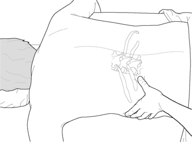
With the fingers of the cephalad hand wrapping around the rib cage and the thumb pointed toward the spine at a 45° angle (Fig. 10.33), the thumb is slid medially on the inferior surface of the 12th rib until it is just lateral to the erector spinae. Special care is taken to avoid pressing on the sharp lateral edge of the 12th rib or the lateral ends of the transverse processes. Static pressure or mild friction is applied to the transverse process of L1 to assess for tenderness or referred pain patterns.

image

Figure 10.33 Care must be taken to avoid pressing on the sharp lateral edge of the 12th rib or the lateral ends of the transverse processes while palpating near or on them.

The treating thumb is then moved inferiorly at approximately 1-inch intervals and the palpation step is repeated to search for L2–4. The transverse processes are not always palpable and are usually more palpable at the level of L2 and L3. If rotoscoliosis of the lumbar spine exists, the transverse processes are usually more palpable on the side to which the spine is rotated.

The practitioner now turns to face the patient’s feet while standing at the level of the mid-chest. Caudally oriented repetitive gliding strokes are applied to the most medial section of the quadratus lumborum, from the 12th rib to the iliac crest, while remaining lateral to the erector spinae. These gliding strokes are applied in sections in the same manner as the cranially oriented strokes were applied previously and can also be continued onto the oblique fibers, which lie lateral to QL.

While continuing to face the patient’s feet, the practitioner applies transverse friction to the attachment of QL on the uppermost edge of the iliac crest while assessing for tender attachments and taut or fibrotic fibers. This frictional assessment can be continued through the oblique fibers as well.

Additional NMT applications to quadratus lumborum may involve the patient in a sidelying position with the treatment of muscles attaching to the pelvis (See p. 280).

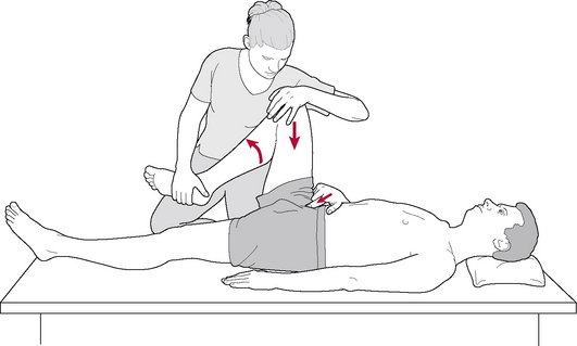
MET for quadratus lumborum 1

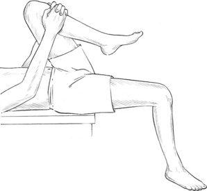
Note: The positioning of patient and practitioner is almost identical for the quadratus lumborum MET (‘banana’) stretch as it is for MET latissimus stretch (see p. 252 and Volume 1, Fig. 13.56). The only differences are in the instructions given to the patient regarding the isometric contraction and the direction of stretch, which for QL is into pure contralateral sideflexion.

An alternative QL stretch is suggested for those practitioners who find that size and/or weight considerations prevent safe application of the ‘banana’ stretch.

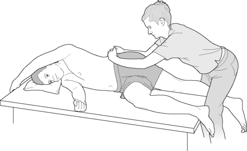
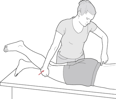
MET for quadratus lumborum 2 (Fig. 10.34)

image

Figure 10.34 MET treatment of quadratus lumborum. Note that it is important after the isometric contraction (sustained raised/abducted leg) that the muscle be eased into stretch, avoiding any defensive or protective resistance which sudden movement might produce. For this reason, body weight – rather than arm strength – should be used to apply traction

(reproduced with permission from Chaitow (1996)).

The practitioner stands behind the sidelying patient, at waist level.

The patient has the uppermost arm extended over the head to firmly grasp the top end of the table and, on an inhalation, abducts the uppermost leg until the practitioner palpates strong quadratus activity (abduction to around 30°, usually).

The patient holds the leg isometrically contracted, allowing gravity to provide resistance, for 10 seconds.

The patient then allows the leg to hang slightly behind himself, over the back of the table.

The practitioner straddles this suspended leg and, cradling the pelvis with both hands (fingers interlocked over crest of pelvis), transfers weight backward to take out all slack and to ‘ease the pelvis away from the lower ribs’, as the patient exhales.

  Page 253 

The stretch should be held for not less than 10, and ideally up to 30, seconds.

The method will be more successful if the patient is grasping the top edge of the table, so providing a fixed point from which the practitioner can induce stretch.

The contraction followed by stretch is repeated once or twice more with raised leg in front of and once or twice with raised leg behind the trunk, in order to activate different fibers.

  Page 259 

The direction of stretch should also be varied so that it is always in the same direction as the long axis of the abducted leg. This clearly calls for the practitioner changing position from the back to the front of the table, as appropriate.

PRT for quadratus lumborum (two variations)

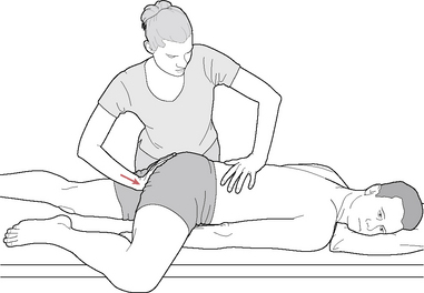
The patient is prone and the practitioner stands on the side contralateral to that being treated.

The tender points for quadratus lie close to the transverse processes of L1–5. Medial pressure (toward the spine) is usually required to access the tender points, which should be pressed lightly as pain in the area is often exquisite. Once the most sensitive tender point has been identified this should be lightly compressed and the patient asked to register the discomfort as a ‘10’.

One of two variations can then be employed.

Variation 1 (Fig. 10.35)

image

Figure 10.35 Prone PRT treatment of quadratus lumborum

(adapted from Deig (2001)).

While the practitioner maintains the monitoring contact on the tender point, the patient is asked to externally rotate, abduct and flex the ipsilateral hip to a position that reduces the ‘score’ significantly. The limb, flexed at hip and knee, should lie supported on the treatment table. The patient turns his head ipsilaterally and slides his ipsilateral hand beneath the flexed thigh, easing the hand very slowly toward the foot of the treatment table until a further reduction in the pain score is noted. This combination of hip flexion/abduction/rotation and arm movement effectively laterally flexes the lumbar spine, so slackening quadratus fibers. If further reduction is required in the pain score (i.e. it is not already at ‘3’ or less), the practitioner’s caudad hand should apply gentle cephalad pressure from the ipsilateral ischial tuberosity. This final compressive force usually reduces the score to ‘0’. This position should be held for at least 30 and, ideally, up to 90 seconds before a slow return to the starting position.

Variation 2

Practitioner is standing on the same side of the table as the QL being treated. With the cephalad hand applying monitoring pressure to the tender point, the practitioner’s caudad hand grasps the patient’s ipsilateral thigh, just proximal to the knee, and eases it into slight extension until there is a reduction in reported sensitivity. The patient’s thigh may then be supported by the practitioner’s caudad thigh as she rests her knee on the table. The practitioner then gradually abducts the leg until the pain is reported to reduce by at least 70%. Fine-tuning may involve slight internal or external rotation of the thigh (whichever eases the pain most) and a final degree of compression should be added (if it effects a pain reduction) by easing the thigh in a cephalad direction. This final position should be held for between 30 (if compression is added) and 90 seconds, before a slow return to the starting position.

  Page 260 

The thoracolumbar paraspinal muscles (erector spinae)

A complex array of short and long extensors and rotators lies deep to the more superficial latissimus dorsi and thoracolumbar fascia. Many of these muscles extend vertically onto the thorax and as far as the cranium, while others lie deeply placed in an oblique orientation. Confusion abounds in anatomy classes when attempting to sort the terminology as some or all of these muscles may collectively be called the paravertebral group, the paraspinal muscles or the erector spinae or may be described by individual names, such as longissimus, semispinalis and iliocostalis. This confusion subsides when one realizes that many of these terms are simply alternative descriptors for the same structures and that the names have specific meanings that help to explain their roles and locations.

The term ‘erector spinae’ represents a group of muscles, all of which are innervated by the dorsal rami of spinal nerves. This group is divided into lateral (superficial) and medial (deep) tracts, with each tract having two further subdivisions of straight and obliquely oriented fibers.

‘Paravertebral group’ and ‘paraspinal muscles’ are both terms that describe the combination of lateral and medial tract fibers.

Those muscles that support and laterally flex the spinal column are oriented for the most part vertically while those more diagonally oriented rotate or finely control rotation of the column.

Their names frequently offer clues to their attachment sites (capitis, cervicis, thoracis and lumborum).

The deeper the fibers lie, the shorter their course.

In the lateral (superficial) tract, there are two long vertical muscular columns (the longissimus and the iliocostalis) as well as a transversospinal set (splenius capitis and splenius cervicis). The longissimus column is more medially placed than the iliocostalis column.

In the medial (deep) tract, there are vertical fibers (interspinales, intertransversarii, spinalis) as well as an obliquely oriented group (rotatores, multifidus and semispinalis).

Superficial paraspinal muscles (lateral tract) (see Fig. 10.25c and Volume 1, Fig. 14.14)

Attachments: Iliocostalis lumborum extends from the iliac crest, sacrum, thoracolumbar fascia and the spinous processes of T11-L5 to attach to the inferior borders of the angles of the lower 6–9 ribs

Iliocostalis thoracis fibers run from the superior borders of the lower six ribs to the upper six ribs and the transverse process of C7

Iliocostalis cervicis fibers arise from ribs 3–6 and insert on the transverse processes of C4–6

Longissimus thoracis shares a broad thick tendon with iliocostalis lumborum and fiber attachments to the transverse and accessory processes of the lumbar vertebrae and thoracolumbar fascia, which then attaches to the tips of the transverse processes and between the tubercles and angles of the lower 9–10 ribs

Longissimus cervicis fibers run from the transverse processes of T1–6 to the transverse processes of C2–5

Longissimus capitis fibers run from the transverse processes of C5-T5 to the mastoid process

Innervation: Dorsal rami of spinal nerves

Muscle type: Postural (type 1), shortens when stressed

Function: Unilaterally flexes the vertebral column and bilaterally extends it. Iliocostalis lumborum depresses lower ribs and is active at the end of inhalation and during (maximum) exhalation (Simons et al 1999)

Synergists: For lateral flexion: oblique abdominal muscles, rectus abdominis, quadratus lumborum

For extension: contralateral fibers of the same muscles, quadratus lumborum, serratus posterior inferior

Antagonists: To lateral flexion of lumbar region: contralateral fibers of oblique abdominal muscles, rectus abdominis, quadratus lumborum

To extension: rectus abdominis, oblique abdominal muscles

Indications for treatment:

Pain in the back and/or buttocks

Restricted spinal motion

Difficulty rising from seated position or in climbing stairs

Deep, steady ache in the spine

Hypertrophy of one or both sides of the lower back

Scoliosis

Special notes

William Kuchera (1997a) describes the erector spinae as one of the four major muscles (along with latissimus dorsi, gluteus maximus and biceps femoris) involved in stabilizing the sacroiliac joint by means of inducing force closure. He also highlights the need to maintain a sense of the interconnectedness of spinal and general bodily biomechanics and of the role of the erector spinae in this.

The first three lumbar vertebrae serve as primary posterior attachments for the crura of the abdominal diaphragm. These vertebrae also supply attachments for the erector spinae mass of muscles that extend from the pelvis all the way to the neck and head. The latissimus dorsi muscle connects the pelvis with the upper extremity. Through the lumbar aponeurosis and fascia, the lumbar region is functionally attached to the hamstrings, the gluteal muscles, and the iliotibial band into the lower extremity; through the oblique abdominal muscles, the posterior lumbar region is functionally related to the anterior abdominal wall.

  Page 261 

Everything connects to everything and much can be gained by keeping this constantly in mind.

Vleeming et al (2007) describe the erector spinae as being:

‘Pivotal muscles that load and extend the spine and pelvis. The sacral connections of the erector/multifidus induce nutation in the SIJ, tensing ligaments such as the interosseous, sacrotuberous and sacrospinal. …These muscle has a double function since their iliac connections pull the posterior sides of the iliac bones towards each other, constraining nutation.’

This means that during the process of nutation (see Chapter 11), the erector spinae ensure that the cephalad aspect of the SIJ is compressed, while the caudal aspect widens.

If the erector spinae are weak this will lead to (and may result from) what Vleeming et al (2007) term ‘insufficient nutation’; that is, there would be a reduced ability for the sacral base to move anteriorly between the ilia.

If, additionally, gluteus maximus is weak, with implications for reduced sacrotuberous ligament activity and therefore inadequate sacroiliac compression (‘self-locking’), a chain reaction of negative influences involving the thoracolumbar fascia ensues, possibly also involving latissimus dorsi.

This pattern of dysfunction is likely to result in increased compensating tension in biceps femoris, ultimately rotating the pelvis posteriorly (so engineering counternutation at the SI joint) and flattening the lumbar spine.

This in turn may lead to an unstable low back.

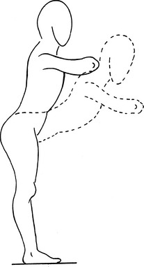
An exercise that encourages a stable lordosis, including a strengthened erector spinae, is suggested. See Fig. 10.40 below, and its accompanying text description, for rehabilitation exercise for weak erector spinae.

image

Figure 10.40 A suggested exercise with lordosis of the lumbar spine and nutation of the SIJ. The biceps femoris, gluteus maximus and erector spinae are simultaneously activated

(reproduced with permission from Vleeming et al (2007)).

Snijders et al (1997) have shown that the slightly stooped (reduced lordosis) posture adopted by many low back pain patients results in reduced psoas activity and an unloading of the SI joint, so easing discomfort. This posture may, however, increase load on possibly painful structures, such as the long dorsal SI ligaments, via increased activity from the erector spinae muscles. Interestingly, the wearing (by the low back pain patient) of a small rucksack (backpack) weighing approximately 6 lbs/3 kg, diminishes erector spinae activity, while still allowing the pain-reducing slight stoop to be maintained. Reduction in low back pain in males with back problems as well as females, peripartum, have been noted using these tactics, according to Snijders et al (1997).

Norris (2000b) discusses the flexion relaxation response, which occurs with flexion of the spine during a lifting effort. When the spine is in almost full flexion (during lifting), the erector spinae become electrically ‘silent’, as an elastic recoil occurs involving the posterior ligaments and musculature. ‘During the final stages of flexion, and from 2° to 10° of extension, movement occurs by recoil of the stretched tissues rather than by active muscle work’. However, in a case of chronic low back pain, if the erector spinae are in spasm, the flexion relaxation response is likely to be obliterated.

Braggins (2000) reports on a very significant perspective on the possible etiology of sudden low back pain/dysfunction that occurs on bending to lift a light object.

Cholewicki [1997] and McGill [1998] found that stability of the lumbar spine diminished during periods of low muscular activity, making it vulnerable to injury in the presence of sudden unexpected loading. The spine will buckle if the activity of lumbar multifidus [see p. 270] and the erector spinae is zero, even when the forces in large muscles are substantial… for example a person could work all day on a demanding job and then ‘put his back out’ stooping to pick up a pencil from the floor in the evening. Buckling behavior can be limited to a single level from inappropriate activation of muscles.

It is obvious from the current knowledge of trigger point activity that such entities in the erector spinae could be part of a scenario that induced single-level (or more widespread) muscular weakness or inappropriate activation. (See discussion on trigger points in the Essential Information section of this volume, and in Volume 1, Chapter 6. See also Box 10.4 for discussion on lifting.)

Trigger points located in these vertical muscular columns refer caudally and cranially across the thorax and lumbar regions, into the gluteal region and anteriorly into the chest and abdomen (see Volume 1, Fig. 14.16).

The erector spinae system is discussed in Volume 1, Chapter 14, due to its substantial attachments in the thoracic region where its numerous attachments onto the ribs require that it be released before intercostals are examined. When the intercostal muscles are examined as described in that chapter, the practitioner may encounter tender attachment sites that appear to lie in the erectors. Marking each tender spot with a skin-marking pencil may reveal vertical or horizontal patterns of tenderness. Clinical experience suggests that horizontal patterns often represent intercostal involvement, as these structures are segmentally innervated, while vertically oriented patterns of tenderness usually relate to the erector spinae muscles.

Vertical lines of tension imposed by the erector system can dysfunctionally distort the torso and contribute significantly to scoliotic patterns, especially when unilaterally hypertonic. Leg length differential, whether functional or structural, may need attention in order to sustain any long-term improvement in the myofascial tissue brought about by treatment or exercise.

  Page 262 

The posterior fascial lines (of potential tension), which run from above the brow (over the head and down the back) to the soles of the feet, are a critical line of reference to altered biomechanics of the spine and thorax (see fascial chains, Volume 1, Chapter 1). There may be widespread effects on postural adaptation mechanisms following any substantial release, for example, of the middle portion (erector group) of that posterior line. If the lamina myofascial tissues are also released, the tensegrity tower (the spine) could potentially adapt and rebalance more effectively. However, the practitioner should note that following such a series of releases, a requirement for structural adaptations will have been imposed on the body as a whole, as the arms move to new positions of balance and the body’s center of gravity is altered. The patient’s homecare use of stretching, applied to the neck, shoulder girdle, lower back, pelvis and legs, coupled with postural exercises should be designed to facilitate and stabilize the induced adaptational changes.

While release of excessive tension might appear to be always desirable, it is important to consider the demands for compensation imposed by induced soft tissue release. Local tissues, and the individual as a whole, will be obliged to adapt biomechanically, neurologically, proprioceptively and possibly emotionally. Engineering any substantial release of postural muscles, before other areas of the body (and the body as a whole) are prepared, may overload compensatory adaptation potentials, possibly creating new areas of pain, structural distress or myofascial dysfunction (‘The part you treated is better, but now I hurt here and here’). Other osseous and myofascial elements may already be adapting to preexisting stresses and may become dysfunctional under such an increased load.

However, if treatment has been carefully planned and executed, the process of adaptation to a new situation, following local soft tissue treatment, while almost inevitably producing symptoms of stiffness and discomfort, should be recognized as a probable indication of desirable change and not necessarily ‘bad’. The patient should, therefore, be forewarned to anticipate such symptoms for a day or two following NMT or other appropriate soft tissue manipulation.

Erector spinae inappropriate firing (prone extension) sequence test (see Volume 1, Fig. 5.5)

The patient lies prone and the practitioner stands to the side at waist level with her cephalad hand spanning the lower lumbar musculature and assessing erector spinae activity.

The caudad hand is placed so that the heel lies on the gluteal muscle mass with the finger tips on the hamstrings.

The patient is asked to raise his leg into extension as the practitioner assesses the firing sequence.

The normal activation sequence is (1) gluteus maximus, (2) hamstrings, followed by (3) erector spinae contralateral, then (4) ipsilateral. (Note: not all clinicians agree with this sequence definition; some believe hamstrings fire first or that there should be a simultaneous contraction of hamstrings and gluteus maximus.)

If the hamstrings and/or erectors take on the role of gluteus maximus as the prime mover, they wil become shortened.

Janda says: ‘The poorest pattern occurs when the erector spinae on the ipsilateral side, or even the shoulder girdle muscles, initiate the movement and activation of gluteus maximus is weak and substantially delayed … the leg lift is achieved by pelvic forward tilt and hyperlordosis of the lumbar spine, which undoubtedly stresses this region.’

Variation: When the hip extension movement is performed there should be a sense of the lower limb ‘hinging’ from the hip joint. If, instead, the hinge seems to exist in the lumbar spine, the indication is that the lumbar spinal extensors have adopted much of the role of gluteus maximus and that these extensors (and probably hamstrings) will have shortened.

Erector spinae muscle shortness test 1 (Fig. 10.36)

The patient is seated on a treatment table so that the extended legs are also lying on the table and the pelvis is vertical. Flexion is introduced in order to approximate the forehead to the knees.

In a normal, flexible individual, an even ‘C’-shaped kyphotic curve should be observed, as well as a distance of about 4 in/10 cm between the knees and the forehead.

No knee flexion should occur and the movement should be a spinal one, not involving pelvic tilting.

Erector spinae muscle shortness test 2

The previous assessment position is then modified to remove hamstring shortness from the picture, by having the patient sit at the end of the table, knees flexed over it with feet and lower legs hanging down toward the floor.

Once again the patient is asked to perform full flexion, without strain, so that forward bending is introduced to bring the forehead toward the knees.

The pelvis should be fixed by the placement of the patient’s hands on the pelvic crest, applying light pressure toward the table.

If bending of the trunk is greater in this position than in test 1 above, then there is probably shortened hamstring involvement.

During these assessments, areas of shortening in the spinal muscles may be observed as ‘flattening’ of the curve or even, in the lumbar area, a reversed curve. For example, on forward bending, a lordosis may be maintained in the lumbar spine or flexion may be very limited even without such lordosis. There may be evidence of obvious overstretching of the upper back and relative tightness of the lower back.

  Page 263 

All areas of ‘flatness’ are charted since these represent an inability of those segments to flex, which involves the erector spinae muscles as a primary or secondary feature.

If the flexion restriction relates to articular factors, the erector group will nevertheless benefit from MET or other forms of release. If the erector spinae are primary causes of the flexion restriction then MET attention is even more indicated.

Lewit (1999) points out that patients with a long trunk and short thighs may perform the movement without difficulty, even if the erectors are short, whereas if the trunk is short and the thighs long, even if the erectors are supple, flexion will not allow the head to approximate the knees.

In the modified position, with the patient’s hands on the crest of the pelvis and the patient ‘humping’ his spine, Lewit suggests observation of the presence or absence of lumbar kyphosis for evidence of muscular shortness in that region. If it fails to appear, erector spinae shortness in the lumbar region is likely and this, together with the presence of flat areas, provides significant evidence of general shortness of erector spinae.

Breathing wave: evaluation of spine’s response to breathing (see Volume 1, Fig. 14.7)

Once all flat areas have been noted and charted following shortness tests 1 and 2 (above), the patient is placed in a prone position.

The practitioner squats at the side and observes the spinal ‘wave’ as deep breathing is performed.

There should be a wave of movement, commencing from the sacrum and finishing at the base of the neck on inhalation.

Areas of restriction (‘flat areas’ in the previous tests) are often seen to move as ‘blocks’, rather than in a wave-like manner. Lack of spinal movement, or where motion is not in sequence, should be noted and compared with findings from tests 1 and 2 above.

This assessment is not diagnostic but offers a picture of the current response of the spine to a full cycle of breathing.

Periodic review of the relative normality of this wave is a useful guide to progress (or lack of it) in normalization of the functional status of both spinal and respiratory structures.

Additional assessments for erector spinae

Liebenson (2000c) has described the changes in longissimus (thoracis) related to trigger point activity and a variety of low back and coccygeal pain, pseudo-visceral pain, known as the Silvertolpe reflex (see discussion on p. 225 and Fig. 10.12). Direct perpendicular palpation produces a reflex twitch response. Liebenson reports that treatment of the sacrotuberous ligament is usually successful in obliterating the trigger point activity.

Liebenson (2007, 2001) further suggests careful visual and palpatory evaluation of the paraspinal musculature. ‘The bulk of the erector spinae should be compared from side to side as well as from the lumbar to the thoracolumbar region. There should be no evident difference between sides and regions’. Overactivity of the thoracolumbar erector spinae may lead to visible hypertrophy (see Chapter 2, Fig. 2.13)

  Page 264 

Assessment for weakness in erector spinae

Janda (1983) describes precise evaluation of strength in the muscles that extend the spine from a prone position. See the Essential Information section of this volume for details of muscle strength grading, where ‘5’ represents strong normal and ‘0’ complete lack of function.

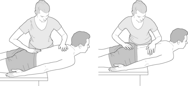
Patient lies prone with thorax extending over the edge of the table so that the edge of the table lies level with the upper abdomen, and the upper body is flexed to 30°. The arms lie alongside the trunk.

The practitioner stabilizes the buttocks, pelvis and lumbar spine, holding these toward the table with one arm while offering mild resistance to the patient between the shoulder blades, as the patient attempts to extend the spine.

The cervical spine should remain in neutral (i.e. in line with the thoracic spine) throughout the procedure (Fig. 10.37).

image

Figure 10.37 Positions of patient and practitioner for assessment of erector spinae strength. Note the position of the practitioner’s hands (which offer resistance to extension), on the upper thoracic spine for Grade 5 and the lower thoracic spine for Grade 4

(adapted from Janda (1983)).

Once the horizontal position has been achieved the practitioner’s resisting hand is placed against the lower ribs as extension continues.

If the patient can achieve maximal extension at the lumbar level against both gravity and the practitioner’s resistance, grade 5 is merited. If complete lumbar extension is not achieved, grade 4 is appropriate.

Grade 3 is appropriate if the process commences in the same starting position but no manual resistance is offered and there is an even degree of back extension through the full range.

For Grade 2 the trunk is fully on the table, arms at the sides, with the same practitioner stabilization and no resistance. The patient is able to extend the thorax as the head and shoulders are lifted from the table.

For Grade 1 the patient is prone and is unable to lift the thorax or head into extension.

See rehabilitation exercise for weak erector spinae in this section.

Preparation for NMT treatment

Having evaluated where a restricted area exists, MFR techniques can be applied to the tissues before any lubrication is used as MFR methods are most effectively employed when applied to dry skin. MFR calls for the application of a sustained gentle pressure, usually in line with the fiber direction of the tissues being treated, which engages the elastic component of the elastico-collagenous complex, stretching this until it commences to and then (eventually) ceases to release (this can take several minutes). A more complete description is presented on p. 204.

Functional technique In situations where the erector spinae are particularly sensitive, functional positional release technique may be applied to provide an opportunity for reduction in hypertonicity and sensitivity prior to NMT application (see Fig. 10.7 and also notes on functional methodology in chapter 9, p. 203). Alternatively, functional methods may be used following NMT to further ease sensitive tissues.

  Page 265 

NMT for erector spinae

The erector spinae are lubricated from C7 to the top of the sacrum. Gliding strokes are applied repetitiously with the thumbs (tips pointing caudally) or with the palm of the hands from the C7 area to the sacrum. Alternating from side to side after several strokes have been applied (while gradually increasing pressure) will warm the tissues and begin the lengthening treatment of the erector spinae. These sets of 10–12 gliding strokes can be repeated several times while alternating between the two sides of the back. Clinically there appear to be postural benefits (for example, in reducing anterior pelvic positioning) when glides directed toward the pelvis are applied over lines of normal myofascial tension, such as those provided by the erector group. Lengthening these lines, between the upper thorax and sacroiliac areas, may result in reductions of anterior pelvic tilt, lumbar lordosis and forward head posture.

These sets of strokes are applied alternately to each side, until each has been treated 4–5 times, while avoiding excessive pressure on the bony protuberances of the pelvis and the spinous processes. Progressive applications usually encounter less tenderness and a general relaxation of the myofascial tissues, especially if heat is applied to the tissues while the contralateral side is being treated. Unless contraindicated (for example, by recent injury, inflammation or excessive tenderness) a hot pack may be moved back and forth between the two sides between the gliding strokes in order to ‘flush’ the tissues.

The connective tissues may become more supple or the myofascial tensional lines induced by (or inducing) trigger points, ischemia or connective tissue adaptations may be released and softened by the gliding strokes, as described above. Trigger points may become more easily palpable as excessive ischemia is reduced or completely released by these gliding strokes. Palpation of the deeper tissues is usually more defined and tissue response to applied pressure is usually enhanced by this sequence of strokes.

The powerful influence of effleurage strokes, when applied repeatedly to the erector spinae or to the thoracic and lumbar lamina groove, should not be underestimated. Clinical experience strongly suggests that the application of this form of repetitive NMT effleurage can significantly influence layer upon layer of fibers, attaching into the lamina. Such strokes are among the most important tools in neuromuscular therapy. Treatment of this sort can beneficially influence segmental spinal mobility, postural integrity and the potential for tensegrity processes to function more effectively in dealing with the stresses and strains to which the body is exposed.

A repeat of these gliding strokes at the end of the session will allow a comparative assessment, which often demonstrates the changes in the tissues (and discomfort levels) to the practitioner as well as the patient.

For a broader and, if appropriate, deeper effleurage stroke, the blade of the proximal forearm can be used. See Volume 1, Fig. 14.17 for assistance in achieving the following position, which is critical to non-straining application of the stroke. When facing the table, one of the practitioner’s arms and legs is determined as the cephalad side and the other as the caudad.

The practitioner places her feet so that the caudad foot is level with the patient’s waist and the cephalad foot is level with the patient’s shoulder. The practitioner then turns to face toward the patient’s head. This position should be comfortable for the practitioner throughout the gliding stroke and if not, the foot positions should be switched to see if strain is relieved.

The practitioner’s caudad elbow is bent with the forearm placed perpendicular to the spine. The olecranon process is placed next to (but never onto) the spinous process of L5. To accomplish this position, about 90% of the practitioner’s body weight must be dropped into the waist-level foot. The knee will be bent slightly to achieve this position.

While exercising caution to avoid pressing on spinous processes, a broad gliding stroke of a moderate speed is applied by the proximal one third of the ulna (not the pointed tip of the olecranon) to the erector spinae from the crest of the ilium to C7. The movement of the gliding stroke results from transfer of body weight from the caudad leg to cephalad leg, not as a result of motion of the practitioner’s shoulder. Pressure is slightly reduced in the thoracic area and the angle of the elbow decreased slightly to work in the narrower interscapular space.

The practitioner now turns to face the patient’s feet and reverses her foot positions. The forearm of the opposite arm is used to glide down the erector spinae from C7 to the iliac crest, similarly using the transfer of body weight, not arm motion (tai chi-like movement). Care is taken to avoid gliding on the spinous processes, the iliac crest or the sacrum.

A snapping palpation (see description on p. 196, p. 424, Fig. 12.31 and in Volume 1, Chapter 9) can be applied across the erector spinae fibers to produce a vibrational effect. If tolerable, the thumbs, finger tips, knuckles or elbow (carefully used) can also apply (laterally oriented) unidirectional transverse snapping strokes to particularly fibrotic or taut bands of erector spinae so long as care is taken to avoid striking the spinous processes. A similar method is described in Chapter 12 in the treatment of the iliotibial band.

CAUTION: This snapping technique is useful on fibrotic tissues and taut fibers of a more chronic nature, as it creates a vibrational effect that may affect the connective tissue’s ground substance, changing it from a gel to a sol. It should not be applied to acute spasms or tissues that tend to be ‘neurologically excitable’, as it may tend to increase their reactivity.

  Page 266 

The practitioner’s fingers can glide along taut fibers of erector spinae to discover localized tender spots consistent with the location of trigger points in myofascial tissues (usually at mid-fiber or attachment sites). It should be noted that tendons are elongated in erector spinae and, therefore, the muscle bellies (where central trigger points form) lie in the lower half of the muscle’s length. When trigger point pressure release (ischemic compression) is applied to these tender spots, nodular or locally dense tissues in palpably taut bands (entrapping them against deeper structures, in this case), with referred phenomena to a predictable target zone of referral confirm the location of a trigger point. Trigger point pressure release, MET, PR or other techniques such as INIT (as described in Chapter 9) can be applied to these tissues in an attempt to reduce their tensional elements as well as their patterns of referral. Elongation of the tissues through precisely applied myofascial release or by active or passive stretching should follow the release techniques.

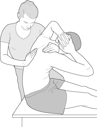
MET for erector spinae

The patient sits on the treatment table with his back to the practitioner, knees flexed and hands clasped behind his neck.

The practitioner places a knee on the table close to the patient, on the side toward which sidebending and rotation will be introduced (Fig. 10.38).

image

Figure 10.38 Muscle energy procedure for the thoracolumbar region of the erector spinae

(adapted from Lewit (1992)).

The practitioner passes a hand in front of the patient’s axilla on the side to which the patient is to be sideflexed and/or rotated, across the front of the patient’s upper chest to rest on the contralateral shoulder.

The practitioner’s free hand monitors an area of ‘tightness’ involving the erector spinae musculature (as evidenced by ‘flatness’ in the ‘C’ curve flexion tests described above) and ensures that the various forces localize at this area of maximum contraction/tension in the erector spinae musculature. The patient is drawn (by the practitioner’s anteriorly placed arm) into flexion, sidebending and rotation to a point of soft endpoint resistance (i.e. not a strained position).

When the patient has been taken to the comfortable limit of flexion, sidebending and rotation, he is asked to breathe in and, while holding the breath for 7–10 seconds, to very lightly attempt to return toward the upright sitting position against firm resistance offered by the practitioner. This engages the agonists (the shortened structures) in an isometric contraction which incorporates the principles of a postisometric relaxation (PIR) response.

The held breath can usefully be focused into the tight spinal area by the patient while this is being palpated and monitored by the practitioner. This results in an increase in isometric contraction of the shortened musculature.

The patient is then asked to release the breath and to completely relax.

The practitioner waits for the patient’s second full exhalation and then takes the patient further in all the directions of restriction, toward the new barrier, but not through it.

The new position of slight stretch is held for at least 30 seconds.

This whole process is repeated several times, at each level of restriction/flatness, to both the right and the left.

At the end of each sequence of repetition the patient may usefully be asked to breathe in and to gently attempt to rotate further against resistance, in the direction in which he is being held, i.e. toward the restriction barrier, while holding the breath for 7–10 seconds.

This involves contraction of the antagonists and incorporates the principles of reciprocal inhibition. After relaxation, the new barrier is again engaged and held.

MET variation using slow isotonic eccentric stretching

Isotonic eccentric contraction/stretches have attracted various labels. If performed rapidly there is a degree of (controlled) tissue damage and the descriptor used is of an ‘isolytic’ contraction. Such strategies are rarely (but effectively) used in treatment, for example, of chronic TFL shortness, where the objective is to stretch the tissues forcefully and to then encourage remodeling by means of patient-applied stretching as homework (Chaitow 2006).

  Page 267 

Slowly applied isotonic eccentric contraction/stretches have been named SEIS (slow eccentric isotonic stretch) (Chaitow 2010) as well as ‘eccentric MET’ (Liebenson 2001). The method involves the patient engaging a restriction barrier and then attempting to maintain that barrier position (using between 40% and 80% of available strength) while the practitioner slowly overcomes this resistance and lengthens the contracting muscle, so achieving an eccentric contraction.

The purpose of this technique is to facilitate (tone) the muscles being slowly isotonically stretched and at the same time to reciprocally inhibit the antagonistic muscles, without producing significant degrees of tissue damage such as would occur in a rapid isotonic eccentric stretch. The indication for this approach is when there is a need to release tension in individual or multiple muscles (ideally hypertonic postural muscles), while simultaneously toning their weakened/inhibited antagonists.

To treat the erector spinae the patient should be placed on a fixed stool or chair, in a seated, slumped position, feet flat on the floor and with the head approximating the knees.

The practitioner stands behind and to the side and passes an arm across the anterior upper chest from shoulder to shoulder, while her other hand maintains a contact with the lower back (lumbodorsal junction region).

The patient is asked to maintain the forward bend with about half the available muscle strength (a little less if the patient is bulky and the practitioner slight) while the practitioner slowly introduces a force which extends the patient’s spine, thereby overcoming resistance of the patient’s flexion attempt.

If the exertion is too great for the practitioner, the patient should be asked to maintain the flexion position with a reduced effort. (‘Resist my pressure toward sitting you upright, but allow me to overcome your effort’)

Following this the patient should reengage the (new) flexion barrier and the procedure should be repeated.

In this way weakened abdominal structures will be toned and tight extensors will be released.

A slow, controlled, passive stretch of previously shortened structures might then be usefully carried out, ideally in an antigravity position, such as seated.

Ruddy’s ‘pulsed’ MET and the erector spinae muscles

Osteopathic physician TJ Ruddy developed a method that utilized a series of rapid pulsating contractions against resistance, which he termed ‘rapid resistive duction’. Ruddy’s method (now known as ‘pulsed MET’) called for a series of muscle contractions against resistance, at a rhythm a little faster than the pulse rate. This approach can be applied in all areas where isometric contractions are suitable. Its simplest use involves the dysfunctional tissues (or joint) being held at their resistance barrier, at which time the patient, against the resistance of the practitioner, is asked to introduce a series of rapid (2 per second), minute efforts toward the barrier. The barest initiation of effort is called for with, to use Ruddy’s term, ‘no wobble and no bounce’.

To treat the erector spinae group the patient engages a restriction barrier that places the muscle at its elastic barrier, i.e. in flexion, or some combination of flexion, sidebending and rotation (as in the MET method described above for the erector spinae).

The patient is coached as to the rhythm as well as the amplitude of the pulsation needed.

This requires initiation of a series of 20 (twice per second for 10 seconds) very slight attempts to move further in the direction of the restriction barrier, pulsing against the firm resistance of the practitioner.

After the series and a brief rest, the barrier is reassessed and reengaged, and the process repeated.

In this painless procedure the patient is rhythmically activating the antagonist muscles to those which are restricted and preventing full range of movement. The series of pulsing contractions tones the inhibited antagonists while reciprocally inhibiting hypertonic agonists, so increasing the range of motion.

PRT for erector spinae (and extension strains of the lumbar spine)

In the strain-counterstrain (SCS) variation of PRT methodology, tender areas in the extensor muscles are related to stresses that have been imposed onto these structures, whether acutely or chronically, and the positions that produce a release of hypertonicity. Restoration of more normal function needs to involve an exaggeration of the shortness in the muscles and/or a reproduction of the strain positions that exacerbated or caused their distress.

Areas of particular tenderness should be sought in the erector spinae and adjacent musculature, which will be used as monitors during the SCS application. The tender points relating to extension strains in the region of L1 and L2 are found near the tips of the transverse processes of the respective vertebrae.

Extension strains should be treated with the patient prone. The practitioner stands on the side of the table opposite the tissues to be treated, while grasping the patient’s leg on the side of the dysfunction just proximal to the knee and bringing the leg into slight extension and adduction, in a scissor-like movement (Fig. 10.39). As these movements are slowly performed the palpated tender point should be monitored and the patient should report on the pain score out of ‘10’ (which is the initial pain level before repositioning commences) until it is less painful (the objective is to reduce the self-reported score by 70% at least).

image

Figure 10.39 Position of ease for a tender point associated with an extension strain of the lumbar spine involves use of the legs of the prone patient as means of achieving extension and fine tuning

(reproduced with permission from Chaitow (2007)).

  Page 268 

Fine tuning to reduce the pain further is accomplished by slightly modifying the leg position, using rotation, increased extension (but not sufficient to cause lumbar spine distress) or by the addition of a compression force through the long axis of the femur toward the painful tender point. This final position of ease should be held for 30–90 seconds.

The tender point for extension strain at the level of L3 is usually located approximately 3 inches (8 cm) lateral to the posterior superior iliac spine and the tender point for extension strains of the L4 level is usually located 1–2 inches (2.5–5 cm) lateral to this, on or close to and following the contour of the crest of the pelvis.

Treatment of L3 and L4 extension strains is accomplished with the patient prone, with the practitioner standing on the side of dysfunction. The operator’s knee or thigh can be usefully placed under the elevated thigh of the patient to hold it in extension (or a bolster might be used) while fine tuning progresses. This is usually accomplished by means of abduction and external rotation of the leg while the tender point is being monitored.

Rotation of the limb, introduction of small degrees of adduction or abduction and positioning of the patient’s leg in a more anterior or posterior plane, always in a degree of extension, are the fine-tuning mechanisms used to reduce pain from the palpated tender point. ‘Crowding’ by lightly applied pressure through the long axis should complete the process of reducing reported pain by at least 70%. This final position of ease should be held for 30–90 seconds.

Jones reports various tender points for extension strains in the region of L5. One of the key points, known as the upper pole L5 strain, is found bilaterally between the spinous process of L5 and the spinous process of S1 and is treated as in extension strains of the LI and L2 level (using scissor-like extension of the prone patient’s leg on the side of the dysfunction and fine tuning by variations in position).

Anterior tender points/flexion strains

Strains in the lumbar region, including those involving the erector spinae, which occur in flexion, reflect as areas of tenderness in the muscles that are shortened, acutely or chronically, in relation to the dysfunction. These are usually found on the anterior trunk, i.e. for the lumbar area these tender points are mainly located in the abdominal musculature.

Positional release methods for treating flexion dysfunction of the lumbar region are described on p. 286 with the abdominal muscles.

Rehabilitation exercise for weak erector spinae (Fig. 10.40)

Vleeming et al (2007) describe a simple exercise that will effectively encourage nutation at the sacroiliac joint as well as toning gluteus maximus, the hamstrings and the erector spinae, while simultaneously encouraging a lengthening of shortened hamstrings.

The patient stands erect, arms folded onto the chest, and hollows the low back, creating a lordosis.

  Page 269 

A slow forward bend is initiated at the hips without allowing any loss of the (slightly exaggerated) lordosis, until approximately 70° of hip flexion has been achieved (see Fig. 10.40) or until a sense of tightness (but not pain) is noted in the hamstrings.

This position is maintained for 30–60 seconds or until a sense of fatigue is noted, at which time a slow return is made to the upright position, with lumbar lordosis being maintained throughout.

Repeat several times daily.

Additional rehabilitation exercises are presented in Chapter 7 with other self-help techniques.

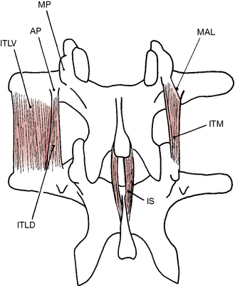
Deep paraspinal muscles (medial tract): lumbar lamina (see Fig. 10.25d)

Multifidi

Attachments: From the superficial aponeurosis of the longissimus muscle, the dorsal surface of the sacrum and the mamillary processes of the lumbar vertebrae, these muscles cross 2–4 vertebrae and attach to the spinous processes of the appropriate higher vertebrae

Innervation: Dorsal rami of spinal nerves

Muscle type: Postural (type 1), shortens when stressed

Function: When these contract unilaterally they produce ipsilateral flexion and contralateral rotation; bilaterally, they extend the spine. While multifidi can produce (primarily fine adjustment) vertebral movement, they serve more as ‘stabilizers rather than prime movers of the vertebral column as a whole’ (Simons et al 1999)

Synergists: For rotation: rotatores, ipsilateral internal oblique, contralateral external oblique

For extension of lumbar spine: erector spinae, serratus posterior inferior and quadratus lumborum

Antagonists: To rotation: matching contralateral fibers of multifidi as well as contralateral rotatores, ipsilateral external oblique, contralateral internal oblique

To extension of lumbar spine: rectus abdominis and oblique abdominal muscles

Indications for treatment

Chronic instability of associated vertebral segments

Restricted rotation (sometimes painfully)

Pain in the region of associated vertebrae and the coccyx

Pain referring anteriorly to the abdomen

Rotoscoliosis

Rotatores longus and brevis

Attachments: From the transverse processes of each vertebra to the spinous processes of the second (longus) and first (brevis) vertebra above

Innervation: Dorsal rami of spinal nerves

Muscle type: Postural (type 1), shortens when stressed

Function: When these contract unilaterally they produce contralateral rotation (debated by Bogduk 2005, see below); bilaterally, they extend the spine

Synergists: For rotation: multifidi, ipsilateral internal oblique, contralateral external oblique

For extension of lumbar spine: multifidi, erector spinae, serratus posterior inferior and quadratus lumborum

Antagonists: To rotation: matching contralateral fibers of rotatores as well as contralateral multifidi, ipsilateral external oblique, contralateral internal oblique

To extension of lumbar spine: rectus abdominis and oblique abdominal muscles

Indications for treatment

Pain and tenderness at associated vertebral segments

Tenderness to pressure or tapping applied to the spinous processes of associated vertebrae

Special notes

Multifidi and rotatores muscles comprise the deepest layer of paraspinal muscles and are often thought to be responsible for fine control of the rotation of vertebrae. They exist throughout the entire length of the spinal column and the multifidi also broadly attach to the sacrum after becoming appreciably thicker in the lumbar region.

  Page 270 

Bojadsen et al (2000) point to substantial differences between thoracic and lumbar multifidi fibers. They show thoracic multifidi to be deeper, thinner, and their fibers are more tendinous and oblique than multifidi fibers found in the lumbar spine. They suggest that these differences may have implications in their function.

Muscle mass, muscle length, the trajectory of the fibres and the position of a muscle in relation to the joint can give important information on the function of the muscle. … the cross-sectional area of a muscle is directly related to the tension that this muscle can produce, the length of its fibres is an indication of the excursion of movement that the muscle is able to produce, and the position of the muscle in relation to the joint is indicative of the kind of movement. According to Gardner et al (1971), the more oblique the muscle in relation to the joint, [the] more its role in axial rotation; and the more its position is vertical, the more its contribution to flexion–extension movements.

Thoracic multifidi architecture suggests that it produces movements in a transverse plane (rotational) and that its tension and amplitude is lower than its thoracic counterparts.

Lumbar multifidi present as more vertical, consistent with movements in a sagittal plane, and is larger and longer. Additionally, multifidus fibers have significant vertical orientation at L5-S1 and T11-T12, where lumbar and thoracic spines (respectively) have the greatest degree of flexion and extension. Consistent with this concept, the most oblique fibers of multifidus are located at T1-T2, the joint segment with the greatest degree of axial rotation.

Bojadsen et al (2000) also note that multifidus fibers are the only muscle fibers posterior to the lumbosacral transitional point of L5-S1.

This means that Mm. Multifidi would be expected to produce enough tension to ensure posterior stabilization in this region. The exclusive posterior presence of Mm. Multifidi fibers in a region of important load transmission and high demand of stabilization could explain why Mm. Multifidi present their largest mass precisely in this segment of the spine.

Kader et al (2000) found that 80% of low back pain patients examined had atrophy of lumbar multifidus muscle. They also determined that the correlation between multifidus atrophy and leg pain was significant, although radiculopathy symptoms, nerve root compression, herniated nucleus pulposus and number of degenerated discs were statistically not significant associated with the atrophy.

These muscles are often associated with vertebral segments that are difficult to stabilize and should be addressed throughout the spine when scoliosis is presented. Discomfort or pain provoked by pressure or tapping applied to the spinous processes of associated vertebrae, a test used to identify dysfunctional spinal articulations, may also indicate multifidi and rotatores involvement (Simons et al 1999).

Bogduk (2005) notes that some of the deepest fibers of multifidi attach to the zygapophysial joint capsules and appear to help ‘protect the joint capsule from being caught inside the joint during the movements executed by multifidus’. He also suggests that it is unlikely that multifidi actually produce rotation of vertebral segments. He postulates that they are more likely to act to stabilize the lumbar region against ‘the unwanted flexion unavoidably produced by the abdominal muscles’ during rotation of the thorax. Additionally, he suggests that the line of action of multifidus lies posterior to the lumbar curve, giving it a ‘bowstring’ effect that compresses the posterior portion of the discs. This allows it to ‘increase compressive and tensile loads on any vertebrae and intervertebral discs interposed between its attachments.’

Moore & Dalley (2010) suggest that the rotatores muscles lack sufficient strength and leverage to produce spinal movement, and may play a greater role in spinal proprioception, acting as a ‘kinesiological monitor’ (Fryer & Johnson 2005).

Trigger points in rotatores tend to produce localized referrals whereas the multifidi trigger points refer locally and to the gluteal, coccyx and hamstring regions. Cornwall et al (2006) suggest that multifidus also refers to the anterior and posteriolateral thigh, and possibly distally into the calf and medial ankle. These local (for both) and distant (for multifidi) patterns of referral continue to be expressed through the length of the spinal column. The lumbar multifidi may also refer to the anterior abdomen (see Volume 1, Fig. 14.18).

NMT for muscles of the lumbar lamina groove

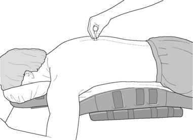
To prepare the superficial tissues of the lamina groove of the lumbar region for treatment of the tissues that lie deep to them, lubricated gliding strokes may be applied repeatedly with one or both thumbs in the lamina groove from L1 to the sacrum. The thumbnail is not involved in the stroke nor allowed to encounter the skin, as the thumb pads are used as the treatment tool (see p. 197 Box 9.7 for hand positioning and cautions in gliding). Each gliding stroke is applied several times from L1 to (but not onto) the coccyx while progressively increasing the pressure (if appropriate) with each new stroke (this stroke may be applied from C7 to the coccyx as described in Volume 1, Chapter 14). These gliding strokes are applied alternately to each side until each has been treated 4–5 times with several (8–10) repetitions each time. The power of these repetitious strokes should not be underestimated as changes within the tissues are often substantial and easy spinal realignment an added bonus. Excessive pressure on the bony protuberances of the pelvis and the spinous processes throughout the spinal column should be avoided. Progressive applications usually encounter less tenderness and significant softening of the myofascial tissues. Unless contraindicated by redness, edema, high levels of tenderness or other signs of inflammation, a hot pack may be placed alternately on each side while the other side is being treated so as to increase blood flow, warm the tissues and further soften the fascial elements between applications of strokes. Tissues that are treated with hot applications should subsequently be adequately drained either manually or by application of cold, if engorgement and congestion are to be avoided.

One should appreciate how thick the lumbar multifidus is, particularly in the lower lumbar region, where it fills the majority of the lamina. A portion of multifidus can be accessed by working lateral to and deep to the erector spinae, particularly at the level of L2-L4. It is suggested, however, to exercise caution to avoid compressing tissues against the lateral aspect of the transverse processes, which lie deep to the desired fibers of multifidi.

The finger tip (with the nail well trimmed) or the tips of the beveled pressure bar (see below) may be used to friction or assess individual areas of isolated tenderness and to probe for taut bands, which often house trigger points. Trigger points lying close to the lamina of the spinal column often refer pain across the back, wrapping around the rib cage, anteriorly into the chest or abdomen and frequently refer ‘itching’ patterns. The trigger points may be treated with static pressure or may respond to rapidly alternating applications of contrasting hot and cold (repeated 8–10 times for 10–15 seconds each), always concluding with cold (see hydrotherapy notes in Volume 1, Chapter 10).

The beveled pressure bar may also be used to assess the fibers attaching in the lamina (as described and illustrated in Volume 1, Box 14.7 and Fig. 14.14). The tip of the bar is placed parallel to the midline and at a 45° angle to the lateral aspect of the spinous process of L1. In this way it is ‘wedged’ into the lamina groove where cranial-caudal-cranial friction is repetitiously applied at tip-width intervals. The assessment begins at L1 and continues to (but not onto) the coccyx (see also a more thorough examination of sacrum on p. 343). Each time the pressure bar is moved, it is lifted and placed at the next point, which is a tip width further down the column. The short frictional stroke may also be applied unidirectionally (in either direction), which sometimes more clearly defines the fiber direction of the involved tissue. The location of each involved segment may be marked with a skin-marking pencil so that it may be retreated several times during the session. The ‘collection’ of skin markings may provide clues as to patterns of involved tissues.

  Page 271 

Many muscular attachments will be assessed with the use of applied friction to the lamina groove of the lumbar region. These attachments may include latissimus dorsi (or its fascial network), serratus posterior inferior, multifidus and rotatores. Determining exactly which fibers are involved is sometimes a difficult task and success is based strongly on the practitioner’s skill level and knowledge of anatomy, including the order of the multiple layers overlying each other and their fiber directions. Fortunately, the tissue response is not always based on the practitioner’s ability to decipher these fiber arrangements (especially in the lamina) and the tender or referring myofascia may prove to be responsive, even when tissue identification is unclear.

Interspinales muscles

Attachments: Connects the spinous processes of contiguous vertebrae, one on each side of the interspinous ligament, present only in the cervical and lumbar regions

Innervation: Dorsal rami of spinal nerves

Muscle type: Postural (type 1), shortens when stressed

Function: Extension of the spine or possibly as proprioceptive mechanisms for surrounding tissues (Bogduk 2005 – see below)

Synergists: All posterior muscles and especially (when contracting bilaterally) multifidi, rotatores and intertransversarii

Antagonists: Flexors of the spine

Indications for treatment

Tenderness between the spinous processes

Loss of flexion

Special notes

The interspinales muscles are present only in the cervical and lumbar regions and sometimes the extreme ends of the thoracic segment. In the cervical region, they sometimes span two vertebrae (Gray’s anatomy 2005).

Though extension of the spine is usually noted as the primary function of these small muscles, Bogduk (2005) suggests that intertransversarii (and similarly these interspinales muscles) may act more as proprioceptive transducers as ‘their value lies not in the force they can exert, but in the muscle spindles they contain’. Consequently, they provide feedback that influences the behavior of the muscles which surround them. Bogduk (2005) notes, ‘Indeed all unisegmental muscles of the vertebral column have between two and six times the density of muscle spindles found in the longer polysegmental muscles, and there is a growing speculation that this underscores the proprioceptive function of all short, small muscles of the body.’

NMT for interspinales (Fig. 10.41)

image

Figure 10.41 The short intersegmental muscles. ITLV: intertransversarii laterales ventrales; ITLD: intertransversarii laterales dorsales; ITM: intertransversarii mediales; IS: interspinales; AP: accessory process; MP: mamillary process; MAL: mamillo-accessory ligament

(reproduced with permission from Bogduk (1997)).

The tip of an index finger (or the carefully applied beveled tip of the pressure bar) is placed directly between (and in a line which is perpendicular to) the spinous processes (Fig. 10.42). Mild pressure is applied or gentle transverse friction used to examine the tissues that connect the spinous processes of contiguous vertebrae, which primarily affects the interspinales muscle pairs and the interspinous ligament, which lie between the processes. The lumbar region may be passively flexed by placing a bolster under the abdomen in order to slightly separate the spinous processes and allow a little more room for palpation. The bodyCushion™ by Body Support Systems Inc. (See next page for contact details) is especially helpful and illustrated here. Depending upon the amount of pressure being used and the segment level, the tissues being examined include the supraspinous ligament, interspinous ligament and interspinales muscles.

image

Figure 10.42 The beveled pressure bar is used between spinous processes of contiguous vertebrae. The bodyCushion™ by Body Support Systems Inc, PO Box 337, Ashland, OR 97520. Website: www.bodysupport.com. US: 800-448?2400 or 541?488-1172 is especially useful in helping to separate the spinous processes slightly through supportive positioning of the thoracic and lumbar regions.

  Page 272 

Intertransversarii lateralis and mediales (Fig. 10.25d)

Attachments: Laterally and medially placed muscle pairs that join the transverse processes of contiguous vertebrae

Innervation: Ventral and dorsal rami of spinal nerves (L1-4) (respectively)

Muscle type: Not established

Function: Lateral flexion of the spine, although this function is debated by Bogduk (2005)

Synergists: Interspinales, rotatores, multifidi

Antagonists: Lateral flexors of the contralateral side

Indications for treatment

Restriction in lateral flexion

Special notes

These short, laterally placed muscles most likely act as postural muscles and stabilize the adjoining vertebrae during movement of the spinal column as a whole. The pattern of movement of intertransversarii is unknown, but thought to be lateral flexion, although Bogduk (2005) suggests they may act as proprioceptive transducers, monitoring movements to provide feedback, which will influence surrounding tissues.

These muscles are difficult to reach and attempts to palpate them may not be fruitful. Positional release and muscle energy techniques may prove useful in releasing these deeply placed tissues.

MET for multifidi and other small, deep muscles of the low back

The protocols for MET application to local spinal and paraspinal muscles, which are often impossible to identify, specifically require generalized description of methods which can be applied to any local area of tension, induration and/or fibrocity.

Tense or restricted soft tissues should be identified by palpation, by loss of range of motion or by association with a vertebra that is tender when its spinous process is lightly tapped.

Manual stretching of the taut fiber is usually applied across the fiber direction if this can be identified. This might either involve a ‘C’-shaped form, where the tissue is being ‘bent’, or an ‘S’ shape in which the fibers are being stretched in two directions simultaneously by the practitioner’s two thumbs (Figs. 10.43 & 10.44).

image

Figure 10.44 Creating a ‘C’-shaped bend in tissues to effect lengthening

(reproduced with permission from Chaitow (2001)).

image

Figure 10.43 Creating an ‘S’-shaped bend in tissues to effect myofascial release

(reproduced with permission from Chaitow (2001)).

Once it has been decided that some degree of local stretching release is appropriate, the fibers should be eased toward a position where the slack is removed from the elastic components.

Once the barrier has been engaged the patient is asked to introduce a local isometric contraction into the tissues being treated for 5–7 seconds, following which the tissues are stretched beyond the previous resistance barrier and held for up to 30 seconds to encourage lengthening. This may be accomplished by an active range of motion if fiber has been clearly identified or by a precisely placed ‘miniature’ myofascial release if direction of fiber has been determined.

This approach can be used paraspinally or on a very localized level to free any shortened soft tissue structures.

  Page 273 

PRT for small deep muscles of the low back (Induration technique)

Treatment of small localized muscular stresses in the paraspinal muscles, using PRT methodology, is elegantly accomplished by means of an SCS derivative, induration technique. This extremely gentle method is fully explained and described in Volume 1, Fig. 14.

Muscles of the abdominal wall (Fig. 10.45)

image

Figure 10.45 Muscles of the abdominal wall. External oblique has been removed (except for rib attachments) to reveal internal oblique. The anterior lamina of the rectus sheath has been removed to reveal the rectus abdominis muscle

(reproduced with permission from Gray’s anatomy (1995)).

Like the erector system of the posterior thorax, the abdominal muscles play a significant role in positioning the thorax and in rotating the entire upper body. They (particularly transversus abdominis) are also now known to play a key part in spinal stabilization and inter-segmental stability (Hodges 1999). The rectus abdominis, external and internal obliques and transversus abdominis are also involved in respiration due to their role in positioning the abdominal viscera to provide stabilizing resistance to the diaphragm as well as depression of the lower ribs, thereby assisting in forced expiration, especially coughing.

The muscles of the abdominal wall can be subdivided into medial muscles (rectus abdominis and pyramidalis) and the superficial lateral muscles (external oblique, internal oblique and transversus abdominis). These muscles and their ensheathing fascia form a complex supportive tensional network for the lower back (see Fig. 10.22). Spinal stability depends upon these tensional elements as well as contributions from the deep lateral abdominal muscles (quadratus lumborum and psoas) and the paraspinal muscles discussed previously.

Posteriorly, the fascia of the superficial lateral abdominal muscles contributes to the thoracolumbar fascia, as previously discussed on p. 243. Anteriorly, these muscles give rise to the rectus sheath and the linea alba. Regarding the fascial sheath of the superior portion of rectus abdominis, Kapandji (1974) notes that the external oblique contributes to the anterior aspect while transversus abdominis contributes to the posterior portion. The internal oblique, which is sandwiched between the other two muscles, splits at the lateral border of rectus abdominis to wrap around both anteriorly and posteriorly, thereby ‘ensheathing’ the rectus, as its fascia blends with that of the other two lateral muscles to form the rectus sheath. The bilateral sheaths merge at the anterior mid-line to form the linea alba. Below the umbilicus, at the arcuate line, the posterior aspect of the rectus sheath ends and the rectus abdominis perforates the transversus abdominis to lie on the posterior surface of its aponeurosis. In this way, from the arcuate line caudally the transversus abdominis contributes to the anterior rectus sheath and the rectus is covered posteriorly only by transversalis fascia.

Directly deep to the abdominal muscles lie much of the abdominal viscera. Trigger points located in the abdominal muscles have been shown to initiate a number of somatovisceral responses involving organs from the urinary, digestive and reproductive systems (Simons et al 1999) as well as pain into the mid-thorax, sacroiliac and lower back regions, chest, abdomen, groin and (crossing the mid-line) into the contralateral side of the chest and abdomen.

The abdominal musculature has a high propensity for strong referral patterns into the visceral organs (Simons et al 1999) and each visceral organ is capable of producing varying degrees of pain in somatic tissues (Rothstein et al 1991, Simons et al 1999). When pain is reported in the abdominal region, it is imperative that the practitioner considers possible pathological conditions of the viscera as well as trigger point referral patterns of the abdominal (and sometimes lower back) muscles. Attempts should be made to differentiate between viscerally produced patterns of referral and those produced by myofascial trigger points (see Box 10.8).

Box 10.8 Abdominal palpation: is the pain in the muscle or an organ?

Since there is no immediately accessible underlying osseous structure available to allow compression of the musculature on much of the soft tissues of the abdomen, there is a need for strategies that distinguish palpated pain occurring as a result of visceral dysfunction or disease, from that being produced in surface tissues. Two protocols are suggested.

When a local area of abdominal pain is noted while using NMT or any other palpation method, the tissues should be firmly compressed by the palpating digit, sufficient to produce local pain and referred pain (if a trigger is involved) but not enough to cause distress. The supine patient should then be asked to raise either the head or both heels several inches from the table. As this happens there will be a contraction of the abdominal muscles and compression of the fibers against the palpating digit. The pain may increase or remain as before, particularly if trigger points are involved. If pain decreases or vanishes upon raising the head/heels, the site of the pain is beneath the muscle (which has lifted the palpating digit off the viscera) and points toward a visceral source of the pain. Sharpstone & Colin-Jones (1994), describing this test as Carnett’s sign, note: ‘In our experience just enough movement of the head and shoulders to tense the muscles without flexing the trunk gives the optimum opportunity for keeping one’s finger on the tender spot and eliciting any change or persistence in abdominal wall tenderness. …A careful history is crucial, and examination should always include an assessment of tender spots, with Carnett’s sign, hyperaesthesia, and tenderness over the vertebral bodies’. They suggest that Carnett’s sign is both specific and sensitive and that it may prevent the need for a large number of unnecessary and often unpleasant investigations. They warn, however, ‘Carnett’s sign is not infallible; a positive test must always be put into the whole clinical picture. None the less, following up a positive Carnett’s sign with a successful injection of local anesthetic must be one of the most cost effective procedures in gastroenterology.’

If single-digit (finger or thumb) pressure is noted as painful on a part of the abdominal wall musculature (with or without referral) and there is an increased level of pain noted on sudden release of the pressure (known as rebound pain), this suggests intraabdominal dysfunction or pathology (Thomson & Francis 1977), such as appendicitis.

It is, of course, possible for there to be a problem in the viscera as well as in the abdominal wall. The tests therefore offer clues but not absolute findings and, when positive, suggest professional referral to be appropriate.

  Page 274 

Simons et al (1999) report:

Activation and perpetuation of trigger points in the abdominal wall musculature secondary to visceral disease represents a viscerosomatic response…[these] visceral diseases…include peptic ulcer, intestinal parasites, dysentery, ulcerative colitis, diverticulosis, diverticulitis, and cholelithiasis. Once activated, TrPs may then be perpetuated by emotional stress, occupational strain, paradoxical respiration, faulty posture, and overenthusiasm for misdirected ‘fitness’ exercise.

These somatically produced trigger points can remain long after the visceral condition has improved and should be considered when pain persists after organ function has been restored. (See Box 10.9 regarding abdominal reflexes and Box 10.10 for a list of known somatovisceral referral patterns.)

Box 10.10 Somatovisceral patterns of the abdominal muscles

The musculature of the abdominal wall is well known for its numerous referral patterns which mimic, exacerbate or form as a result of visceral disease. This list, while not exhaustive, should be considered with any abdominal pain or suspected organ dysfunction. Organ pathologies should also be ruled out by a qualified clinician since these triggers are often secondary to underlying organ pathology. See also Box 10.1 for details regarding viscerosomatic referrals as well as Volume 1, Fig. 6.5 for common pain-referred zones of various organs.

Simons et al (1999) discuss the following somatovisceral responses from abdominal musculature and note that injection of the target referral zone of an organ may offer symptomatic relief. They caution, however: ‘Relief of pain in this way does not guarantee that the pain site is the site of origin’. We have also seen many of these patterns in clinical practice and fully agree with the warning to be constantly aware of both the possibility of underlying primary visceral involvement and the potential that the diagnosis may be significantly confused by trigger points with powerful visceral-stimulating potential. Although conclusive research has not yet confirmed this, less intense referral patterns could conceivably produce less abrupt symptoms than those listed here, resulting perhaps only in sluggish digestion, constipation, irregular menses, endocrine imbalances or other bothersome but not highly noxious or definitive symptoms.

Projectile vomiting

Anorexia

Nausea

Intestinal colic

Urinary bladder and sphincter spasm

Dysmenorrhea

Pain symptoms mimicking those of appendicitis and cholelithiasis

Symptoms of burning, fullness, bloating, swelling or gas (Gutstein 1944)

Heartburn and other symptoms of hiatal hernia

Urinary frequency

Groin pain

Chronic diarrhea

Pain when coughing

Belching

Chest pain that is not cardiac in origin

Abdominal cramping

Colic in infants as well as adults

Box 10.9 Different views of abdominal reflex areas

A number of clinicians and workers have identified reflex areas associated with the abdominal region including:

Mackenzie (1909) demonstrated a clear relationship between the abdominal wall and the viscera. These reflex patterns vary in individual cases but it is clear that the majority of the organs are able to protect themselves by producing contraction, spasm and hyperesthesia of the overlying, reflexively related muscle wall (the myotome) which is often augmented by hyperesthesia of the overlying skin (the dermatome) (see Volume 1, Fig. 6.5 for depiction of viscerosomatic reflexes).

Gutstein (1944) noted trigger ‘areas’ in the sternal, parasternal and epigastric regions and the upper portions of the rectus, all relating to varying degrees of retroperistalsis. He also noted that colonic dysfunction related to triggers in the mid and lower rectus abdominis muscle. These were all predominantly left-sided. Other symptoms that improved or disappeared with the obliteration of these triggers include excessive appetite, poor appetite, flatulence, nervous vomiting, nervous diarrhea, etc. The triggers were always tender spots, easily found by the palpation and situated mainly in the upper, mid and lower portions of the rectus abdominis muscles, over the lower portion of the sternum and the epigastrium including the xyphoid process and the parasternal regions.

Fielder & Pyott (1955) describe a number of reflexes, which were claimed to relate to adhesion formation, occurring in the connective tissue supporting and surrounding the large bowel. These could be localized by deep palpation and treated by specific deep soft tissue release techniques (Chaitow 2010).

Chapman (see Owen 1963) identified what he termed neurolymphatic reflexes, many of which were located in the thoracic and abdominal regions. Travell & Simons (1983) identified trigger points in similar locations in the abdominal musculature and a range of acupuncture/acupressure/tsubo points have also been mapped in these tissues. To what extent Gutstein’s myodysneuric points are interchangeable with Chapman’s reflexes or Fielder’s reflexes or other systems of reflex study (e.g. acupuncture or Tsubo points (Serizawa 1976)) or with Travell’s triggers, and to what extent these involve Mackenzie’s findings, is a matter of conjecture.

Slocumb (1984), working at the Department of Obstetrics and Gynecology, University of New Mexico Medical School, described trigger points which were causing chronic pelvic pain, many of which were located in the abdominal wall. Slocumb found that deactivation of such triggers frequently removed symptoms which had been present for years and which had at times resulted in abortive surgical investigation. He noted an overlap of referral patterns from a variety of locations: The same pain sensation was reproduced by pressure over localized points in several tissues seemingly anatomically unrelated…for example, 1) pinching the skin over the lower abdominal wall; 2) single-finger pressure in one reproducible abdominal wall location [trigger point]; 3) single-finger pressure on tissue overlying the pubic bone; 4) lateral pressure with one finger over one or both levator muscles; 5) single-finger or cotton tip applicator pressure lateral to the cervix; 6) single-finger or cotton tip applicator pressure over vaginal cuff scar tissue, more than 3 months after hysterectomy; 7) single-finger pressure on tissue over the dorsal sacrum’. Slocumb also noted that: ‘there was often observed an association of these pain points within a single dermatome’. The results of treating a series of patients with symptoms of chronic pelvic pain by means of trigger point deactivation (Slocumb used anesthetic injections to achieve this) were ‘successful response in 89.3% of 131 patients’ with nearly 70% followed up for 6 months or more.

Baldry (2005) details a huge amount of research that validates the link (a somatovisceral reflex) between abdominal wall trigger points and symptoms as diverse as anorexia, flatulence, nausea, vomiting, diarrhea, colic, dysmenorrhea and dysuria. Pain of a deep aching nature, or sometimes of a sharp or burning type, are reported as being associated with this range of symptoms, which mimic organ disease or dysfunction (Melnick 1954, Ranger 1971, Theobald 1949). Baldry (2005) has further summarized the importance of this region as a source of considerable pain and distress involving pelvic, abdominal and gynecological symptoms. He says: ‘The type of abdominal and pelvic pain most likely to be helped by acupuncture is that which occurs as a result of activation of trigger points (TrPs) in the muscles, fascia, tendons and ligaments of the anterior and lateral abdominal wall, the lower back, the floor of the pelvis and the upper anterior part of the thigh (Baldry 2001). Such pain, however, is all too often erroneously assumed to be due to some intra-abdominal lesion and, as a consequence of being inappropriately treated, is often allowed to persist for much longer than is necessary’. Note: If we replace the word ‘acupuncture’ (or the injection methods of Slocumb described above) with the term ‘appropriate manual methods’, we can appreciate that a large amount of abdominal and pelvic distress is remediable via the methods outlined in this text.

Many of Jones’s (1981) tender points, as used in strain-counterstrain techniques, are found in the abdominal region specifically relating to those low back strains that occur in a flexed position.

The characteristics of nearly all these myriad ‘point’ systems are that the dysfunctional tissues are palpable, sensitive and discrete -sometimes ‘stringy’, sometimes edematous but always ‘different’ from surrounding normal tissues – and they are usually sensitive to pressure or pinching compression of the overlying skin. Apart from pain, often of a deep aching nature, the reflex influences seem to involve interference or modifications of the functional integrity of local areas as well as reflexively with normal physiological function on a neural, circulatory and lymphatic level, sometimes mimicking serious pathological conditions. Our clinical experience suggests that these areas of dysfunction will often yield to simple NMT soft tissue manipulative techniques (such as ischemic compression, lengthening and draining procedures).

Causes of abdominal triggers

Soft tissue changes and triggers in the abdominal musculature are affected by very much the same factors that produce ‘stress’ anywhere else in the musculoskeletal system:

postural faults (and breathing dysfunction)

overuse and strain – occupational, sporting, patterns of use, overload, repetition of movement

trauma (including herniations)

environmental stressors, such as cold and damp

nutritional deficiencies

surgery (another form of direct trauma)

viscerosomatic influences as a result of visceral disease

viscerosomatic influences resulting from digestive disturbance (microbial, food sensitivities/allergies, etc.)

emotional stress.

Scars from previous surgeries may be sites of formation of connective tissue trigger points (Simons et al 1999). After sufficient healing has taken place, an incision site can be examined by pinching, compressing and rolling the scar tissue between the thumb and finger to examine for evidence of trigger points. These tissues frequently respond well to repeated rolling and sustained compression, which can usually be repeated at home by the patient. Both authors have had substantial success in reducing scar tissue-referred pain in scars of recent origin as well as older scar tissue. Sufficient time (8–12 weeks) should be allowed for healing to take place prior to handling the scar tissue; however, lymphatic drainage can be applied immediately after surgery to the surgical site and surrounding tissues to assist in drainage.

Transverse abdominal scars, such as those usually created by caesarean section and hysterectomy, may impede lymphatic flow in the lower abdominal region, which, in this area, flows downward to the inguinal nodes. Properly applied lymph drainage techniques may reestablish lymph movement and, in some cases, can reroute the lymphatic flow around the scar tissues.

  Page 275 

Differential assessment is obviously important in a region housing so many vital organs and attention to the overall pattern of symptom presentation is critical. The information discussed in this chapter regarding organ pathologies is intended to offer ‘red flags’ to the practitioner to proceed with caution or, in some cases, not to proceed at all until a clear diagnosis has been given. This information is not intended to be diagnostic itself, especially when diagnosis lies outside the scope of the practitioner’s license and/or training. When any doubt exists as to the primary origin of the condition (not to be confused with secondary symptom-producing evidence), expert diagnostic investigation is urged. This is true for all body regions but can be of crucial consideration in the abdominal region where underlying conditions can be life threatening.

  Page 276 

External oblique (Fig. 10.22)

Attachments: Outer surface and inferior borders of 5th–12th ribs (interdigitating with serratus anterior and latissimus dorsi) to join the broad abdominal aponeurosis (forming the linea alba) and to the anterior half of the iliac crest

Innervation: 7th–12th intercostal nerves

Muscle type: Phasic (type 2) with tendency to inhibition, weakening

Function: When contracting unilaterally, it contralaterally rotates the thoracolumbar spine and/or flexes the trunk ipsilaterally. Bilateral contraction produces anterior flexion of the trunk, support and compression of the abdominal viscera, anterior support of the spinal column (see discussions regarding thoracolumbar fascia), and anterior stabilization of pelvic position (decreasing lordosis). Also assists in forced expiration by depressing lower ribs

Synergists: For rotation: ipsilateral deep paraspinal muscles and contralateral serratus posterior inferior and internal obliques

  Page 277 

For lateral flexion: ipsilateral internal oblique, quadratus lumborum, iliocostalis

For compression and support of abdominal viscera: internal oblique, transversus abdominis, rectus abdominis, pyramidalis, quadratus lumborum and diaphragm

For flexion of spinal column: rectus abdominis, internal oblique and, depending upon spinal position, psoas

For forced expiration: rectus abdominis, internal oblique, transversus abdominis, internal intercostals (except parasternal internal intercostals) and (with increased demand) the latissimus dorsi, serratus posterior inferior, quadratus lumborum and iliocostalis lumborum

Antagonists: To rotation: contralateral deep paraspinal muscles and ipsilateral serratus posterior inferior and internal obliques

To lateral flexion: contralateral quadratus lumborum, iliocostalis, external and internal obliques To compression and support of abdominal viscera: gravity To flexion of spinal column: paraspinal muscles

To forced expiration: diaphragm, scalene, parasternal internal intercostals, levator costorum, upper and lateral external intercostals and (with increased demand) the sternocleidomastoid, upper trapezius, serratus anterior, serratus posterior inferior, pectoralis major and minor, latissimus dorsi, erector spinae, subclavius and omohyoid

Internal oblique (Fig. 10.45)

Attachments: From the cartilages of the last 3–4 ribs, the linea alba and the arch of the pubis (conjoined with transversus abdominis) to converge laterally onto the lateral half to two-thirds of the inguinal ligament, the anterior two-thirds of the iliac crest and portions of the thoracolumbar fascia

Innervation: 7th–12th intercostal nerves and iliohypogastric and ilioinguinal nerves (L1)

Muscle type: Phasic (type 2), with tendency to inhibition, weakening

Function: Unilaterally, ipsilaterally rotates the thoracolumbar spine, ipsilaterally flexes the trunk laterally. Bilateral contraction produces anterior flexion of the spine, support and compression of the abdominal viscera. Also assists in forced expiration

Synergists: For rotation: contralateral deep paraspinal muscles and external oblique and ipsilateral serratus posterior inferior

For lateral flexion: ipsilateral external oblique, quadratus lumborum, iliocostalis

For compression and support of abdominal viscera: external oblique, transversus abdominis, rectus abdominis, pyramidalis, quadratus lumborum and diaphragm

For flexion of spinal column: rectus abdominis, external oblique and, depending upon spinal position, psoas

For forced expiration: rectus abdominis, external oblique, transversus abdominis, some of the internal intercostals and (with increased demand) the latissimus dorsi, serratus posterior inferior, quadratus lumborum and iliocostalis lumborum

Antagonists: To rotation: ipsilateral deep paraspinal muscles and external oblique and contralateral serratus posterior inferior

To lateral flexion: contralateral external and internal obliques, quadratus lumborum, iliocostalis

To compression and support of abdominal viscera: gravity To flexion of spinal column: paraspinal muscles

To forced expiration: diaphragm, scalene, parasternal internal intercostals, levator costorum, upper and lateral external intercostals and (with increased demand) the sternocleidomastoid, upper trapezius, serratus anterior, serratus posterior inferior, pectoralis major and minor, latissimus dorsi, erector spinae, subclavius and omohyoid

Transverse abdominis (Fig. 10.46)

image

Figure 10.46 The left transverse abdominis

(reproduced with permission from Gray’s anatomy (1995)).

Attachments: From the inner surface of ribs 7–12, the deep layer of the thoracolumbar fascia, inner lip of iliac crest, ASIS and inguinal ligament to merge into its aponeurosis and participate in the formation of the rectus sheath, which merges at the mid-line to form the linea alba

Innervation: 7th–12th intercostal nerves and iliohypogastric and ilioinguinal nerves (L1)

Muscle type: Phasic (type 2), with tendency to inhibition, weakening

Function: Constricts abdominal contents; assists in forced expiration

Synergists: For compression and support of abdominal viscera: external oblique, internal oblique, rectus abdominis, pyramidalis, quadratus lumborum and diaphragm

For forced expiration: rectus abdominis, external oblique, internal oblique, some of the internal intercostals and (with increased demand) the latissimus dorsi, serratus posterior inferior, quadratus lumborum and iliocostalis lumborum

Antagonists: To compression and support of abdominal viscera: gravity

To forced expiration: diaphragm, scalene, parasternal internal intercostals, levator costorum, upper and lateral external intercostals and (with increased demand) the sternocleidomastoid, upper trapezius, serratus anterior, serratus posterior inferior, pectoralis major and minor, latissimus dorsi, erector spinae, subclavius and omohyoid

Indications for treatment of all lateral abdominals

Postural distortion of pelvis (see also Chapter 11 for discussions of pelvic osseous dysfunctions)

Rotoscoliosis

Dysfunctional gait

Loss of abdominal tone

Post abdominal surgery

Pain in chest or abdomen

Pain into lower abdomen, inguinal area and/or crossing mid-line to radiate into chest or into upper or lower abdomen

Testicular pain

Assorted gastrointestinal symptoms (gas, bloating, belching, heartburn, etc.)

Vomiting, diarrhea and other symptoms of visceral pathology (see Box 10.10 for a more complete visceral referral pattern list)

  Page 278 

Special notes

The diagonally oriented oblique muscles are involved in trunk rotation, lateral flexion, stabilization of the pelvis (which supports the spinal column), and (along with transversus abdominis and rectus abdominis) compression of the abdominal viscera. Compression of the viscera affects positioning of the organs so as to oppose the diaphragm’s downward movement. When the diaphragm encounters the viscera and its central tendon is stabilized, the diaphragmatic attachments on the ribs pull the ribs into ‘bucket handle’ movement, thereby influencing lateral dimension of the thorax (which ultimately influences anterior/posterior dimension (‘pump handle’)) and significantly affecting respiratory mechanics (further discussion is found in Volume 1, Chapter 14).

When producing rotation of the trunk, the external oblique is synergistic with the contralateral internal oblique. However, when performing sideflexion, it is synergistic with the ipsilateral internal oblique (and antagonistic to the contralateral one). This unique situation well illustrates how a muscle can be both a synergist and an antagonist to another muscle.

The more horizontally oriented transversus abdominis constricts the abdominal contents, thereby contributing to respiration by positioning the viscera as well as preventing the subsequent anterior rotation of the pelvis (which abdominal distension would produce) with its numerous postural consequences. Its attachment into the thoracolumbar fascia gives it potential to provide support for the lumbar region, as explained on p. 259.

Karlson (1998) implicates external oblique and serratus anterior in rib stress fractures of elite rowers due to repetitive bending forces. Ziprin et al (1999) have reported entrapment of the iliohypogastric nerve due to defect in the external oblique aponeurosis, through which the nerve passes. Twenty of 23 athletes presenting with groin pain reported good to excellent results following surgical repair.

Trigger point patterns for lateral abdominal muscles are known to cross the mid-line into the contralateral side of the abdomen and to radiate up into the chest and to the contralateral abdomen and chest. They also refer into the groin and testicular region and into the viscera, as previously discussed, causing (among other conditions) chronic diarrhea (Simons et al 1999) (Figs 10.47–49).

image image image

Figures 10.47, 10.48, 10.49 Trigger point patterns of lateral abdominal muscles. These patterns may include referrals that affect viscera and provoke viscera-like symptoms, including heartburn, vomiting, belching, diarrhea and testicular pain

(adapted with permission from Simons et al (1999), Fig. 49.1 A-C).

Stretching and strengthening of the lateral abdominal muscles are indicated in many respiratory and postural dysfunctions as these muscles are often significantly involved. Lack of tone in these muscles may contribute to lower back problems, as has been discussed within this chapter. Rehabilitation and strengthening of these muscles are critical to spinal stability and details are offered in Chapter 7, as a self-applied abdominal muscle rehabilitation.

NMT (and MFR) for lateral abdominal muscles

The patient is in a sidelying position with his head supported in neutral position. A bolster is placed under the contralateral waist area so as to create elongation of the side being treated. The patient’s uppermost arm is abducted to lie across the side of his head and the uppermost leg is pulled posteriorly to lie behind the lower leg or to drape off the side of the table while ensuring that the patient does not roll posteriorly off the table. This positioning places tension on the fibers of the oblique abdominal muscles and ‘opens up’ the lateral abdominal area, which results in better palpation. Due to the moderate elongation of the tissues by this particular patient position, excessive pressure or techniques that might tear the pre-stretched tissues, such as aggressively applied transverse friction, are avoided.

Myofascial release (MFR) of the lateral abdomen can be used as preparation for further NMT techniques or as a (sometimes profoundly successful) treatment itself. MFR techniques are applied to the tissues before any lubrication is used as they are most effectively employed on dry skin. They should be applied to each side of the body as their effectiveness can be profound and may cause postural imbalance if used unilaterally.

To most easily apply a broad myofascial release to the lateral abdominal muscles, the practitioner stands posterior to and just above the waist of the sidelying patient and treats one side of the body at a time. The patient’s uppermost arm is draped upward to lie on the side of his head while his uppermost leg is allowed to hang posteriorly off the table, if comfortable and stable. The practitioner’s arms are crossed so that the practitioner’s caudal hand (fingers wrapping anteriorly) is placed on the patient’s lower ribs and the cephalad hand (fingers facing anteriorly) cups the uppermost edge of the iliac crest and anchors itself on this bony ridge. When applying pressure to engage the elastic components, only a small amount of pressure is oriented into the torso, just enough to keep the hands from sliding on the skin. The remaining pressure is applied in a horizontal direction to create tension on the tissues located between the two hands. As the hands move away from each other, taking up the slack in the tissues located between the two hands, an elastic barrier will be felt and held under mild tension (Fig. 10.50).

image

Figure 10.50 A broad application of myofascial release to the lateral abdominal muscles. Stretch of the tissues can be augmented by placing the patient’s arm and leg (draped off table posteriorly) as shown, which produces mild traction on the tissues.

This elastic barrier is held until the fascial tissues elongate in response to the applied tension. After 90–120 seconds (less time if skin rolling has been applied first), the first release of the tissues will be felt as, it is hypothesized, the gel changes to a more solute state. The practitioner can follow the release into a new tissue barrier and again apply the sustained tension. The tissues usually become softer and more pliable with each ‘release’ (Barnes 1997).

  Page 279 

The lateral abdomen of the sidelying patient can now be lightly lubricated for a more detailed examination of the tissues. Tissues affected by these steps will include the external oblique, internal oblique and transversus abdominis.

The first column of tissues to be addressed lies just lateral to the quadratus lumborum. The horizontally placed thumb of one hand is used to secure the tissues and to provide a tensional element while the fingers of the opposite hand provide a curling, penetrating drag on the tissues (Fig. 10.51).

image

Figure 10.51 The tissues are secured by the thumb of one hand while the fingers of the opposite hand provide a curling, penetrating drag on the lateral abdominal muscles.

  Page 280 

The tissues are examined in small segments that are just inferior to the placed thumb. The fingers (nails well trimmed) are placed just caudal to the thumb and provide a 3–4 inch gliding stroke that drags the fingers down the tissues while the fingers simultaneously flex and curl. This curling action causes penetration into the underlying tissues, which is very different from (and more effective than) that provided by only dragging the fingers.

The curling, dragging technique is applied to this ‘segment’ 4–5 times before the thumb and fingers are moved caudally and placed on the next segment. These steps are repeated in small segments until the iliac crest is reached. The practitioner’s hands then return to the lower rib area and are moved anteriorly onto the next column of oblique tissues and the steps are repeated until the lateral edge of rectus abdominis is encountered.

The curling, dragging techniques can also be applied from an anterior/posterior direction to all of the lateral abdomen or transversely across the fibers. The fibers of each layer lie in a different direction, so taut fibers are sometimes more palpable in one direction than the other. Application at various angles, particularly those transverse to the fiber directions usually allow a more distinct palpation of taut bands.

Friction can be applied to all rib attachments as well as the attachments onto the iliac crest. If attachments are too tender for friction techniques to be used, sustained ischemic compression may result in satisfactory release of the tissue. Trigger points often occur on or near the attachments and these portions of the muscle may be exquisitely tender.

Since there are no bony surfaces to compress the mid-fiber trigger points against, attempts can be made to pick up and compress or roll the oblique fibers between thumb and fingers (Fig. 10.52). However, this may be unsuccessful, particularly on the deeper fibers. Spray-and-stretch techniques have been found by one author (JD) to be successful for reduction of trigger point patterns, as has briefly applied contrast hydrotherapy (alternating hot and cold applications for 10–20 seconds, repeated 10–12 times) followed by stretching of the tissues. Spray-and-stretch techniques are fully covered for these muscles by Simons et al (1999, Chapter 49).

image

Figure 10.52 Attempts can be made to pick up and compress or roll the oblique fibers between thumb and fingers.

Rectus abdominis (Figs. 10.45, 10.46)

Attachments: Costal cartilages of the 5th–7th ribs, xyphoid process and the intervening ligaments to attach caudally to the pubic crest and symphysis pubis

  Page 281 

Innervation: Intercostal nerves (T5–12)

Muscle type: Phasic (type 2) with tendency to inhibition, weakening

Function: Flexes the thoracic and lumbar spine, supports and compresses the abdominal viscera, anterior support of the spinal column by stabilization of pelvic position (decreasing lordosis). Also assists in forced expiration, especially coughing

Synergists: For flexion of spinal column: external oblique, internal oblique and, depending upon spinal position, possibly psoas

For compression and support of abdominal viscera: external oblique, internal oblique, transversus abdominis, pyramidalis, quadratus lumborum and diaphragm

For forced expiration: external oblique, internal oblique, transversus abdominis, internal intercostals (except parasternal internal intercostals) and (with increased demand) the latissimus dorsi, serratus posterior inferior, quadratus lumborum and iliocostalis lumborum

Antagonists: To flexion of spinal column: paraspinal muscles

To compression and support of abdominal viscera: gravity

To forced expiration: diaphragm, scalene, parasternal internal intercostals, levator costorum, upper and lateral external intercostals and (with increased demand) the sternocleidomastoid, upper trapezius, serratus anterior, serratus posterior inferior, pectoralis major and minor, latissimus dorsi, erector spinae, subclavius and omohyoid

Indications for treatment

Pain in the mid-posterior thorax, sacroiliac and lower back regions

Pain in the lower quadrant of the abdomen where, on the right side, McBurney’s point can duplicate the pain of appendicitis

Infant colic

Pain into the chest (not cardiac in origin)

Pain into the abdomen, thereby creating many of the visceral patterns discussed in Box 10.10

Loss of abdominal tone

Painful menses

Pyramidalis

Attachments: From the anterior pubis and its symphysis to the linea alba, midway between the symphysis and the umbilicus

Innervation: 12th thoracic nerve

Muscle type: Phasic (type 2) with tendency to inhibition, weakening

Function: Compresses the abdomen to support the viscera by tensing the linea alba

Synergists: external oblique, internal oblique, transversus abdominis, rectus abdominis, quadratus lumborum and diaphragm

Antagonists: Gravity

Indications for treatment

Pain at lower abdomen close to mid-line in the region of the muscle.

Special notes

The vertically oriented fibers of rectus abdominis primarily contribute to flexion of the thoracolumbar spine (possibly some lateral flexion) and assist in stabilizing the pelvis to avoid anterior tilt with its resultant significant postural consequences. The upper fibers can pull the upper body anterior to the coronal line and help sustain a forward head position. The lower fibers often show loss of tone and allow the pelvis to move into anterior tilt, thereby increasing lumbar lordosis and significantly influencing the spinal positioning in general.

Rectus abdominis is divided by 3–4 (sometimes more or less) tendinous inscriptions, which receive nerve supply from different levels. This allows each section to act independently and to influence each other. Considering how this anatomy applies to trigger point formation theories (see Volume 1, Chapter 6) one can readily see the great propensity for this muscle to form trigger points as each section lying between tendinous inscriptions may produce both central and attachment trigger points.

At the mid-line, the rectus sheaths merge to form the linea alba. Separation of the rectus muscles (common after pregnancy) is noted as a palpable groove at the mid-line, especially detectable when the person is asked to contract the muscles by performing a partial sit-up. Herniations of the linea alba may be palpable only when the patient is standing.

With rectus abdominis imbalance, the umbilicus can be seen to deviate toward the stronger (hyperactive) side and away from the weak, inhibited muscle, especially during various movement activities (laughing, coughing, leg lifting, etc.) (Simons et al 1999) and is usually apparent when the patient is asked to do a quarter sit-up with his arms crossed on his chest (Hoppenfeld 1976). This procedure is used to test the integrity of the spinal segment that innervates the rectus abdominis and the corresponding paraspinal muscles (which should also be assessed for weakness) and is considered a positive ‘Beevor’s sign’ when the umbilicus deviates to one side.

Transverse rectus abdominis musculocutaneous (TRAM) flap has been used for several decades in pelvic, vaginal, (Smith et al 2000) and breast reconstruction (Robbins 2005), and to reconstruct abdominal wall defects (Mathes & Bostwick 1977, Mathes et al 2000). Contributions from rectus abdominis occur in a variety of reconstructive surgeries in the head and neck, including defects of the orbit, the tongue, the cheek, the posterior mandible, and the neck (Kroll & Baldwin 1994). Lyos et al (1999) evaluated speech, articulation, and deglutition after replacement of the tongue in advanced carcinoma of the oral cavity (using rectus abdominis) to find that fifty percent of patients were able to eat pureed foods or better and sixty-four percent had acceptable speech.

  Page 282 

Trigger point patterns for the anterior abdominal muscles are into posterior mid-thoracic, sacroiliac and lower back regions, into the lower quadrant of the abdomen (into McBurney’s point on right side, duplicating the pain of appendicitis), into the chest and into the abdomen, thereby creating many of the visceral patterns previously discussed, including dysmenorrhea, pseudocardiac pain and colic (Figs 10.53-55). Trigger points can be found in any abdominal muscle (Sharpstone & Colin-Jones 1994) and can mimic visceral disease or follow actual visceral conditions, producing pain that may last much longer than the initial event that set it off (Doggweiler-Wiygul 2004). The practitioner should not overlook this possibility; however, suspicion of visceral pathology takes precedence since delay can have serious consequences.

image

Figure 10.54 Trigger points in rectus abdominis can duplicate pain symptoms of appendicitis

(Adapted from Travell & Simons (1992)).

image

Figure 10.55 Painful or difficult menstruation (dysmenorrhea) may be due to rectus abdominis trigger points

(adapted from Simons et al (1999), Fig. 49.2 A-C).

image

Figure 10.53 Trigger points in rectus abdominis can refer posteriorly into the back

(adapted with permission from Travell & Simons (1992)).

Stretching and strengthening of the abdominal muscles are indicated in many respiratory and postural dysfunctions as these muscles are often significantly involved. Lack of tone in these muscles may contribute to lower back problems, as has been discussed within this chapter. Rehabilitation and strengthening of these muscles are important to spinal stability and details are offered in Chapter 7 of a self-applied abdominal muscle rehabilitation.

NMT for anterior abdominal wall muscles

The practitioner uses lightly lubricated gliding strokes or finger friction on the anterior and lateral aspects of the inferior borders and external surfaces of the 5th through 12th ribs to search for taut fiber attachments and tender areas where many of the abdominal muscles fibers attach. Caution is exercised regarding the often sharp tips of the last two ribs, which are usually more posteriorly placed but may (rarely) wrap around anteriorly.

  Page 283 

Palpation of the upper 2 or 3 inches of the rectus abdominis fibers that lie over the abdominal viscera may reveal tenderness associated with trigger points or with postural distortions, such as forward slumping postures, which overapproximate these fibers and shorten them. Extreme tenderness in these tissues or tenderness which returns rather quickly after treatment may also be associated with underlying visceral conditions and caution should be exercised until visceral involvement of liver, gallbladder, stomach, etc. has been ruled out. If not contraindicated by visceral involvement, the pads of several fingers or the heel of the hand can be used to broadly apply sustained ischemic compression for a general release of the upper rectus abdominis (Fig. 10.56). A more precise application of sustained pressure can be placed on any taut bands found after this more general technique has been used.

image

Figure 10.56 The pads of several fingers or the heel of the hand can be used to broadly apply sustained ischemic compression for a general release of the upper rectus abdominis.

The fibers of rectus abdominis may be further softened with short effleurage strokes in the same manner as described previously for the oblique muscles. The curling, dragging techniques, applied both vertically and transversely to the lightly lubricated rectus abdominis area, may reveal tender areas and specific fiber bands worthy of more attention. (See hand positioning and treatment steps with the lateral abdomen descriptions.)

A separated linea alba may be felt in the direct center of the upper rectus abdominis and will be palpated as a lack of resistance where the tissue has split apart. Having the person curl up slightly to tense the rectus abdominis may produce a more profound palpation of the split. This area of weakness should be treated with caution (especially with transverse applications) and the tissues involved should be stroked toward the mid-line rather than away from it. This is easily accomplished by reaching across the body to treat the contralateral side while stroking the fingers toward the mid-line.

As the distal end of the rectus abdominis is approached, the patient should be informed about the attachment site onto the pubic crest and why the practitioner is approaching this area. The male patient should be asked to displace the genitals toward the non-treated side and to ‘protect’ the area while the practitioner palpates the upper aspect of the pubis and frictions the rectus abdominis attachments (Fig. 10.57). Whether the patient is male or female, it is advisable to have a second person in the room as a chaperone since both the practitioner and the patient are vulnerable when treating near the pubic area.

image

Figure 10.57 The patient can be asked to ‘protect’ the area while the practitioner palpates the upper aspect of the pubis and frictions the rectus abdominis attachments.

The pyramidalis muscle will also be treated during the final segment of the lower rectus abdominis as it merges into the linea alba midway between the umbilicus and its attachment on the pubic crest. Specifically applied gliding strokes or transverse friction can be appropriately used.

The most lateral aspect of the rectus abdominis should be examined due to its high propensity for trigger point formation. Additionally, it lies directly over the psoas muscle and discomfort in these lateral fibers of rectus abdominis during palpation of the psoas can be misleading. The practitioner’s thumbs, oriented tip to tip with the pads placed on the lateral aspect of the most lateral fibers of rectus abdominis, can be used to press into, probe and treat the fibers with sustained compression or transverse snapping strokes (if not too tender) (Fig. 10.58). Asking the patient to curl into a quarter sit-up will assist in locating the muscle’s edge but the muscle should be relaxed when palpated.

image

Figure 10.58 Discomfort in the most lateral fibers of rectus abdominis can be misleading as they lie directly over the psoas muscle and can be provoked when palpating for psoas.

  Page 284 

MET for abdominal muscles

Local

The abdominal muscles are phasic (using Janda’s nomenclature – see Volume 1, Chapter 2) and therefore do not shorten as a whole when stressed, but rather display evidence of inhibition and lengthening. This contributes to instability in the spinal structures, as discussed earlier in this chapter. However, all abdominal muscles are capable of developing trigger points and areas of localized shortening, fibrosis, adhesions, etc., which may require normalization if contributing to pain or dysfunction.

If there is confusion as to why a ‘weakened’ or inhibited phasic muscle should require stretching, it may be useful to reread Volume 1, Chapter 2. The discussion in that chapter explains that all muscles have both postural and phasic fiber types and that it is the mix, the ratio, of type 1 and type 2 fibers, as well as the function of the muscle, that determines whether it is classified as phasic or postural, and so whether its tendency is toward weakness/lengthening or tightness/shortening when ‘stressed’. This means that a phasic muscle contains postural fibers, which are likely to shorten under adverse conditions (overuse, misuse, disuse, etc.), just as the phasic fibers in a postural muscle may weaken and lengthen under similar conditions.

  Page 285 
  Page 286 

When treating local dysfunctional changes, tactics might usefully include initial use of an isometric contraction followed by local stretching involving ‘C’ or ‘S’ bends, local myofascial release or other deactivating and lengthening techniques as presented in this text (such as those previously discussed for paraspinal use in the multifidi section).

Global abdominal MET

The use of SEIS involves the slow stretching of a muscle, or group of muscles, while they are contracting or are attempting to maintain a shortened, contracted state (see p. 266 for description of this isotonic eccentric method in treatment of the erector spinae).

This method effectively tones the inhibited antagonists to the short, tight, postural musculature. By slowly stretching the abdominal muscles while they are holding a flexed position, the abdominal muscles will be toned and the erector spinae released from excessive tone (Liebenson 2001). The mechanisms whereby strength and tone are restored to the abdominal muscles, following this type of procedure, involve a combination of active exercise (resisted isotonic contraction) and release of previously tight erector spinae musculature through reciprocal inhibition.

PRT for abdominal muscles

Tender points (which may or may not also be trigger points) located in the abdominal musculature often represent dysfunction of the lumbothoracic region resulting from strain or stress that occurred in flexion. For a greater understanding of the strain-counterstrain concepts that support this assertion, see Volume 1, Chapter 10 (or Chaitow 2007, Deig 2001 or Jones 1981).

Gross positioning to relieve lumbar flexion stresses and strains takes the patient painlessly into flexion (Fig. 10.61), with the final position of ease being held for at least 90 seconds.

image

Figure 10.61 Position of ease for flexion strain of T9 to lower lumbar regions involving flexion, sidebending and rotation until ease is achieved in a monitored tender point on the lower abdominal wall or the ASIS area

(adapted from Chaitow (1996b)).

The position of ease is determined by means of palpation of the tender point, with the patient reporting on the change in ‘score’ as the positioning is fine tuned.

A score of 3 or less is the objective, having commenced from a score of 10 before the repositioning starts.

  Page 287 

Jones (1981) reports that L1 has two tender points: one is at the tip of the anterior superior iliac spine and the other on the medial surface of the ilium just medial to ASIS.

The tender point for 2nd lumbar anterior strain is found lateral to the anterior inferior iliac spine.

The tender point for L3 lies an inch (2.5 cm) below a line connecting L1 and L2 points.

L4 tender point is found at the attachment of the inguinal ligament on the ilium.

L5 points are on the body of the pubis, just to the side of the symphysis.

In bilateral strains both sides should be treated. L3 and L4 usually require greater sidebending in fine tuning than the other lumbar points.

Deep abdominal muscles (Fig. 10.62)

image

Figure 10.62 Psoas major and minor as well as quadratus lumborum comprise the deep abdominal muscles. Portions of piriformis, coccygeus and levator ani are also shown here and are discussed with the pelvis in Chapter 11

(reproduced with permission from Gray’s anatomy (1995)).

The muscles of the deep abdomen include the psoas major, psoas minor and quadratus lumborum. These muscles are also referred to as the lateral trunk muscles (Hoppenfeld 1976) and as the deep muscles of the abdominal wall (Platzer 2004). While the psoas is discussed at length here, the quadratus lumborum has been presented with the posterior lower back muscles since it was treated in a prone position (see p. 257–259).

The companion muscle to the psoas, the iliacus, is considered in this text with the treatment of the pelvis in Chapter 11 (p. 349) due to its extensive pelvic attachments and the tremendous influence it has on pelvic positioning. Both iliacus and psoas are hip flexors and so are also discussed in Chapter 12 (p. 411) with the hip region.

Psoas major

Attachments: From the lateral borders of vertebral bodies, their intervertebral discs of T12-L5 and the transverse processes of the lumbar vertebrae to attach (with iliacus) to the lesser trochanter of the femur

Innervation: Lumbar plexus (L1-3)

Muscle type: Postural (type 1), prone to shortening under stress

Function: Flexion of the thigh at the hip, (minimal) lateral rotation of the thigh, (minimal) abduction of the thigh, extends the lumbar spine when standing with normal lordosis, (perhaps) flexes the spine when the person is bending forward, compression of the lumbar vertebral column, questionable as to whether it can rotate, sidebend or otherwise move the spine (Bogduk 2005)

Synergists: For hip flexion: iliacus, rectus femoris, pectineus, adductors brevis, longus and magnus, sartorius, gracilis, tensor fascia latae

For lateral rotation of the thigh: long head of biceps femoris, the deep six hip rotators, gluteus maximus, sartorius, posterior fibers of gluteus medius and minimus, and iliacus

For abduction of the thigh: gluteus medius, minimus and part of maximus, tensor fascia latae, sartorius, piriformis and iliacus

For extension of the spine: paraspinal muscles

For flexion of the spine: rectus abdominis, external oblique, internal oblique, transversus abdominis

Antagonists: To hip flexion: gluteus maximus, the hamstring group and adductor magnus

To lateral rotation of the thigh: semitendinosus, semimembranosus, tensor fascia latae, pectineus, the most anterior fibers of gluteus minimus and medius and (perhaps) adductor longus and magnus

To abduction of the thigh: adductors brevis, longus and magnus, pectineus and gracilis

To spinal extension: rectus abdominis, external oblique, internal oblique, transversus abdominis

To spinal flexion: paraspinal muscles

Psoas minor

Attachments: From T12 and the upper one or two lumbar vertebrae and the disc between them to the superior ramus of the pubis and iliopubic eminence via iliac fascia

Innervation: Lumbar plexus

Muscle type: Postural (type 1), prone to shortening under stress

  Page 288 

Function: Assists in flexion of lumbar spine

Synergists: Rectus abdominis, external oblique, internal oblique, transversus abdominis

Antagonists: Paraspinal muscles

Indications for treatment of psoas muscles

Low back pain

Pain in the front of the thigh

Difficulty rising from seated position

Inability to perform a sit-up

Loss of full extension of the hip

‘Pseudo-appendicitis’ when appendix is normal

Scoliosis

Abnormal gait

Difficulty climbing stairs (where hip flexion must be significant)

Special notes

The bilateral psoas major bellies, subdivided into superficial and deep portions, descend the anterior aspect of the lumbar spine to join with the iliacus muscle as they both (surrounded by iliac fascia) course through the lacuna musculorum (deep to the inguinal ligament) to attach to the lesser trochanter of the femur. Two bursae, the iliopectineal bursa and the iliac subtendinous bursa, lie between the muscle (or its tendon) and the underlying bony surfaces.

The psoas major may also communicate with:

fibers of the diaphragm, psoas minor, iliacus, quadratus lumborum and pectineus

the posterior extremity of the plural sac

the medial arcuate ligament

extraperitoneal tissue and peritoneum

  Page 289 

kidney and its ureter

renal, testicular or ovarian vessels

the genitofemoral nerve, lumbar plexus and femoral nerve

the abdominal aorta, vena cava, external iliac artery and femoral artery and vein

the colon

the lumbar vertebrae and lumbar vessels

the sympathetic trunk

and aortic lymph nodes.

The sometimes present (50–60% according to Travell & Simons (1992) and Gray’s (2005)) psoas minor courses anterior to the major and ends at the pubic ridge with attachments also spanning to the iliac fascia. Since it does not cross the hip joint (and therefore cannot act upon it), it likely provides weak trunk flexion (Gray’s anatomy 2005), extension of the lordotic curve and elevation of the ipsilateral pelvis anteriorly (Travell & Simons 1992).

At the lumbar attachments of psoas major, the medial deep fascia of the muscle forms tendinous arches on the lateral side of the vertebral bodies and through these arches course the lumbar arteries, veins and filaments from the sympathetic trunk (Gray’s anatomy 2005). The lumbar plexus courses between the two layers of the psoas major and is vulnerable to neural entrapment; whether this is produced by taut bands of trigger points has yet to be established (Travell & Simons 1992).

Controversy exists as to the extent of various functions of the psoas major but all sources agree that it (along with iliacus) is a powerful flexor of the hip joint. EMG studies suggest that it laterally rotates the thigh, does not participate in medial rotation of the thigh, flexes the trunk forward against resistance (as in coming to a sitting position from a recumbent one) and that it is active in balancing the trunk while sitting (Gray’s anatomy 2005).

Psoas major is the most important of all postural muscles (Basmajian 1974). If it is hypertonic and the abdominals are weak, exercise is often prescribed to tone these weak abdominals, such as curl-ups with the dorsum of the foot stabilized. This can have a disastrously negative effect, far from toning the abdominals, as increased tone of the already hypertonic psoas may result, due to the sequence created by the dorsum of the foot being used as a point of support. When this occurs (dorsiflexion), the gait cycle is mimicked and there is a sequence of activation of tibialis anterior, rectus femoris and psoas. If, on the other hand, the feet could be plantarflexed during curl-up exercises, then the opposite chain is activated (triceps surae, hamstrings and gluteals), inhibiting psoas and allowing toning of the abdominals. Additionally, full sit-ups activate the psoas when T12 leaves the ground. Curl-ups or pelvic tilts are better designed for abdominal toning, with diagonal movements added to assist in toning the lateral abdominal wall, without placing undue stress on psoas.

The psoas major behaves in many ways as if it were an internal organ (Lewit 1985). Tension in the psoas may be secondary to kidney disease and may reproduce the pain of gall bladder disease (often after the organ has been removed). It has been noted that the psoas major communicates with fibers of the diaphragm as well as the posterior extremity of the plural sac above (Gray’s anatomy 2005) and Platzer (2004) notes that:

The fascia surrounds the psoas major as a tube, psoas fascia, stretching from the medial lumbocostal arch to the thigh. Thus, any inflammatory processes in the thoracic region can extend within the fascial tube to appear as wandering abscesses as far down as the thigh.

Psoas fibers merge with (become ‘consolidated’ with) the diaphragm and it therefore influences respiratory function directly. Quadratus lumborum has a similar influence with the diaphragm.

Regarding spinal influences, Fryette (1954) maintains that the distortions produced in inflammation and/or spasms in the psoas are characteristic and cannot be produced by other dysfunction. He notes that when psoas spasm exists unilaterally, the patient is drawn forward and sidebent to the involved side with the ilium on that side rotating backwards on the sacrum and the thigh being everted. With bilateral psoas spasm, the patient is drawn forward, with the lumbar curve locked in flexion, thereby producing a characteristic reversed lumbar spine. The latter, if chronic, creates either a reversed lumbar curve if the erector spinae of the low back are weak or an increased lordosis if they are hypertonic.

Lewit (1985) notes: ‘Psoas spasm causes abdominal pain, flexion of the hip and typical antalgesic (stooped) posture. Problems in psoas can profoundly influence thoracolumbar stability’. Travell & Simons (1992) note that trigger points in iliopsoas refer strongly to the lower back and may extend to include the sacrum and proximal medial buttocks (Fig. 10.63). Additionally, it may refer into the upper anterior thigh (not illustrated).

image

Figure 10.63 Referral pattern for iliopsoas may continue further than illustrated into the sacrum and proximal medial buttocks. Additionally, it may refer into the upper anterior thigh (not illustrated)

(adapted from Travell & Simons (1992)).

In unilateral psoas spasms, a primary mechanical involvement is usually at the lumbodorsal junction, though a rotary stress is noted at the level of the 5th lumbar. Attention to the muscular components should be a primary focus, as attempts to treat the resulting pain, which is frequently located in the region of the 5th lumbar and sacroiliac, by attention to the osseous element will be of little use (Chaitow 2006) until the muscular tension is reduced.

Bogduk et al (1992) and Bogduk (2005) provide evidence that psoas plays only a small role in the action of the spine and that it ‘uses the lumbar spine as a base from which to act on the hip’. Bogduk also notes:

Psoas potentially exerts massive compression loads on the lower lumbar disc…upon maximum contraction, in an activity such as sit-ups, the two psoas muscles can be expected to exert a compression load on the L5-S1 disc equal to about 100 kg of weight.

  Page 290 

Liebenson (Chaitow 2006) suggests that treatment aimed at relaxing a tight psoas and strengthening a weak gluteus maximus may be the ideal primary treatment for lumbo-sacral facet pain or paraspinal myofascial pain.

Some visual evidence exists in determining psoas involvement (Chaitow 2006).

Normal psoas function produces the abdomen ‘falling back’ rather than mounding when the standing patient flexes.

Similarly, if the supine patient flexes the knees and ‘drags’ the heels toward the buttocks (keeping them together), the abdomen should remain flat or ‘fall back’. If the abdomen bulges or the small of the back arches, thereby pulling the lumbar vertebrae into excessive lordosis, the psoas is suspect (Janda 1983).

If the supine patient raises both legs into the air and the belly mounds it shows that the recti and psoas are out of balance. Psoas should be able to raise the legs to at least 30° without any help from the abdominal muscles.

CAUTION: Kuchera (1997b) reports that: ‘there are organic causes for psoas spasm that must be ruled out by history, examination and tests, including:

femoral bursitis

arthritis of the hip

diverticulosis of the colon

ureteral calculi [stones]

prostatitis

cancer of the descending or sigmoid colon

salpingitis.’

When treating, it is sometimes useful to assess changes in psoas length by periodic comparison of apparent arm length. The supine patient’s arms are extended above the head, palms together, so that the relationship of the fingertips to each other can be compared. A shortness will commonly be observed in the arm on the side of the shortened psoas. This ‘functional arm length differential’ usually normalizes after successful treatment. This method provides an indication only of changes in psoas length (or as confirmation of other findings, such as in the test below) rather than a definitive diagnosis itself since there may be other reasons for apparent differences in arm length.

Assessment of shortness in iliopsoas (Fig. 10.64)

image

Figure 10.64 Test position for shortness of hip flexors. Note that the hip on the non-tested side must be fully flexed to produce full pelvic rotation. The position shown is normal

(adapted from Chaitow (2001)).

Patient lies supine with buttocks (coccyx) as close to the end of the table as possible and with the non-tested leg in full flexion at hip and knee, held there by the patient or by placing the sole of the non-tested foot against the lateral chest wall of the practitioner. Full flexion of the contralateral hip helps to maintain the pelvis in full posterior tilt with the lumbar spine flat, which is essential if the test is to be meaningful and stress on the spine avoided.

If the thigh of the tested leg fails to lie in a horizontal position in which it is (a) parallel to the floor/table and (b) capable of a movement into hip extension to approximately 10° without more than light pressure from the practitioner’s hand, then the indication is that iliopsoas is short.

If effort is required to achieve 10° of hip extension, this confirms iliopsoas shortening on that side.

If the thigh hangs down below a parallel (to the floor) position without additional effort by the practitioner, it indicates a degree of laxity in iliopsoas.

  Page 291 

Further causes of failure of the thigh to rest parallel to the floor can be shortness of tensor fascia latae (TFL) or of rectus femoris (RF). If TFL is short (a further test proves it: see Chapter 11) then there should be an obvious groove apparent at the iliotibial band on the lateral thigh and the patella, and sometimes the whole leg will deviate laterally at the hip. If rectus femoris is suspected as the cause of reduced range, the tested leg is held straight by the practitioner and the entire leg again lowered toward the floor for evaluation. If the thigh is now able to achieve 10° of hip extension, the responsible tissue is rectus femoris, whose tension on the hip joint was released when the knee (a joint it also crosses) was held in neutral.

A further indication of a short psoas is if the prone patient’s hip is observed to remain in flexion or the lumbar region is pulled into excessive lordosis while either prone or supine.

The prone patient is asked to extend the straight leg at the hip and if the movement commences with an anterior pelvic tilt, the psoas is assumed to have shortened (Fig. 10.65).

Mitchell’s psoas strength test

Before using MET methods to normalize a short psoas, its strength should be evaluated, according to the developers of osteopathic muscle energy technique, Mitchell, Moran and Pruzzo (1979).

They recommend that the supine patient should be placed at the end of the table, both legs hanging down and feet turned in so that they can rest on the practitioner’s lateral calf areas (practitioner stands facing the patient at the foot of the table).

The patient should press firmly against the practitioner’s calves with his feet as she rests her hands on his thighs.

The patient is asked to attempt to lift the practitioner from the floor. In this way the relative strength of one leg’s effort as against the other can be assessed.

The practitioner judges which psoas is weaker or stronger than the other. If a psoas has tested short (as in the test described above) and also tests strong in this test, then it is suitable for MET treatment.

If, however, it tests short and also as weak, then other factors such as associated trigger points or tight erector spinae muscles should be treated first, until psoas tests strong and short, at which time MET should be applied to start the lengthening process.

NMT for psoas major and minor

Method 1 (working ipsilaterally)

Patient is supine, knees flexed with feet resting flat on the treatment table. The practitioner stands on the side to be treated at the level of the abdomen.

The finger tips of the practitioner’s hands (nails well trimmed) are placed vertically at the lateral edge of rectus abdominis approximately 2 inches lateral to the umbilicus (Fig. 10.66).

image

Figure 10.66 A slowly rotating circular movement of the hands allows a steady, safe penetration deeply into the abdomen where psoas resides.

A steady, patient and painless pressure toward the spine is maintained with slight rotary movement of the fingers to insinuate the tips past any abdominal structures superficial to the anterior spine. If the aorta pulsation is strongly evident a slight deviation laterally should allow penetration of the finger tips until they sense contact with the psoas muscle (a fleshy or sometimes very hard, not intestinal, resistance).

  Page 292 

Once this contact has been made the patient is asked to slowly increase flexion of the hip. The elbow of the practitioner’s caudad arm is placed against the flexing thigh to offer resistance, which will cause the psoas to contract firmly to confirm that the finger position is accurately placed. If the fingers lose contact with the muscle fibers, the circular rotating approach is repeated to help assure direct contact without intestinal entrapment (Fig. 10.67).

image

Figure 10.67 Once psoas has been located, muscle testing is applied by having the person actively flex the hip, which presses the knee against resistance applied by the practitioner’s elbow. The contraction of psoas should be distinctly felt by the practitioner’s finger tips to ensure correct hand placement.

Once placement of the hands is confirmed to be directly on psoas, the practitioner uses her finger tips to apply a light direct compressive pressure onto the psoas. Fingers can be gently and slowly eased up or down the muscle (a couple of inches [2.5–5 cm] in each direction) as well as pulled laterally across the muscle, ever staying mindful of the organ structures previously noted. When tender areas or suspected trigger points are located, sustained pressure is applied for at least 8–12 seconds.

Modifications can be made to the leg position by rotating the thigh medially (for the lateral aspect) and laterally (for the medial aspect). Additionally, the patient’s foot on the side being treated can be actively slid (by the patient) down the table slowly (returning the thigh to neutral position) to drag the psoas fibers under the compressing fingers for an active myofascial release.

The iliopsoas tendon is accessible just inferior to the inguinal ligament when the fingers are immediately lateral to the femoral pulse. With the leg (knee bent) resting against the practitioner, the inguinal ligament is located as well as the femoral pulse. The practitioner’s first two fingers are placed between the femoral pulse and the sartorius muscle (Fig. 10.68). Static pressure is sustained or, if not too tender, gentle transverse friction is applied to the tendon of the psoas muscle, which may be exceptionally tender.

image

Figure 10.68 The iliopsoas tendon can be palpated between the femoral artery and the upper fibers of sartorius. Caution should be exercised regarding the femoral artery by locating its pulse and avoiding further palpation to the region of the artery. The tendon is the first myofascial tissue directly lateral to the femoral pulse.

Method 2 (working contralaterally)

An alternative approach is suggested for those whose knowledge of anatomy and pathophysiology is adequate to the recognition of the inherent risks involved in applying direct pressure, through the mid-line, toward the lumbar spinal attachments of psoas (Fig. 10.69).

image

Figure 10.69 Direct NMT treatment of psoas working through the linea alba

(adapted from Chaitow L (1988) Soft tissue manipulation. Healing Arts Press).

CAUTION: There is a very real risk attached to the application of pressure into the tissues of an aneurysm which may lie in the major blood vessels of this region and it is strongly suggested that this method only be used if there are no signs or symptoms of such a condition and if contact with all obviously pulsating structures is avoided.

The patient is positioned as in method 1.

The practitioner stands on the contralateral side and reaches across the body to treat the affected side.

The fingers of the practitioner’s cephalad hand are placed vertically on the mid-line (linea alba) approximately 1.5 inches (4 cm) below the umbilicus. The fingers use the same circular motion described above.

If the aorta pulsation is strongly evident a slight deviation laterally, one way or the other, should allow penetration of the finger tips until they sense a bony contact, the anterior surface of the lumbar spine.

Once this contact has been made, the fingers are slid away from the practitioner, around the curve of the lumbar vertebral body where a psoas contraction will be noted if the patient’s flexed knee is brought cephalad against resistance from the practitioner’s caudad hand (muscle test described in method 1).

  Page 293 

All other elements described in method 1 are used to treat the muscle, which combines elements of ischemic compression, muscle energy technique and facilitated myofascial release.

The entire procedure is repeated to the second side if both psoas muscles require this form of slow release.

MET treatment of psoas

Method 1

The patient is prone with a pillow under the abdomen to reduce the lumbar curve.

The practitioner stands on the contralateral side, with the caudad hand supporting the thigh.

The cephalad hand is placed so that the heel of that hand is on the sacrum and applies pressure toward the floor to maintain pelvic stability. The fingers of that hand are placed so that the middle, ring and small fingers are on one side of L2–3 segment and the index finger on the other side (while the heel of the hand remains on the sacrum). This hand position allows these fingers to sense a forward (anteriorly directed) ‘tug’ of the vertebrae, when psoas is moved past its barrier.

An alternative hand position is offered by Greenman (1996) who suggests that the stabilizing contact on the pelvis should apply pressure toward the table, on the ischial tuberosity, as thigh extension is introduced. The authors agree that this is a more comfortable contact than the sacrum. However, it does not allow access to palpation of the lumbar spine during the procedure (Fig. 10.70).

image

Figure 10.70 MET treatment of psoas in prone position with stabilizing contact on ischial tuberosity, as described by Greenman (1996)

(adapted from Chaitow (2001)).

The practitioner eases the thigh (knee flexed) off the table surface and senses for ease of movement into extension of the hip. If there is a strong sense of resistance there should be an almost simultaneous awareness of the palpated vertebral segment moving anteriorly when this resistance is due to psoas.

If psoas is normal, it should be possible to achieve approximately 10° of hip extension (without force) before that barrier is reached. Greenman (1996) suggests: ‘Normally the knee can be lifted 6 inches [15 cm] off the table. If less, tightness and shortness of psoas is present’.

Having identified the barrier, the patient is asked to bring the thigh toward the table against resistance, using 15–25% of his maximal voluntary contraction potential, for 7–10 seconds.

Following release of the effort (with appropriate breathing assistance, if warranted) the thigh is eased (if acute) to its new barrier or (if chronic) past that barrier and into patient-assisted stretch (‘Gently push your foot toward the ceiling’).

In chronic situations where the stretch is introduced, this is held for at least 20 seconds and ideally up to 30 seconds.

It is important that as stretch is introduced no hyper-extension of the lumbar spine occurs. Pressure from the heel of hand on the sacrum or ischial tuberosity can usually ensure that spinal stability is maintained.

The process is then repeated on the same side before the other side is evaluated and treated if necessary.

  Page 294 

Method 2 (Fig. 10.71)

image

Figure 10.71 MET treatment of psoas using Grieve’s method

(adapted from Chaitow (2001)).

This method involves using the supine test position (see Fig. 10.71), in which the patient lies with the buttocks at the very end of the table, non-treated leg fully flexed at hip and knee and either held in that state by the patient or by placement of the patient’s foot against the practitioner’s lateral chest wall.

The practitioner stands at the foot of the table facing the patient with both hands holding the thigh of the extended leg.

The leg on the affected side is placed so that the medioplantar aspect of the foot rests on the practitioner’s knee or shin.

The practitioner’s leg that supports the affected side foot should be flexed slightly at the knee and hip, so that the patient’s foot can rest as described. This places the hip flexors, including psoas, into a slightly mid-range position, not at their barrier.

The practitioner should request the patient to use a small degree of effort to externally rotate the leg and, at the same time, to flex the hip.

The practitioner resists both efforts and an isometric contraction of the psoas and associated muscles therefore takes place.

This combination of forces focuses the contraction effort into psoas very precisely.

After a 7–10 second isometric contraction and complete relaxation of effort, the thigh should, on an exhalation, either be taken (if acute) to the new restriction barrier without force or (if chronic) through that barrier, by applying slight painless pressure onto the anterior aspect of the thigh and toward the floor to stretch psoas. Either stretch position is held there for 30 seconds.

These steps are repeated until no further gain is achieved.

PRT for psoas (Fig.10.72)

image

Figure 10.72 Positional release of psoas using tender point monitor on the pubic bone

(adapted from Deig (2001)).

The tender point for psoas is usually located at the level of the inguinal ligament, where psoas crosses the pubic bone.

The practitioner stands on the affected side at the patient’s thigh level and with the cephalad hand palpates for the tender point, creating discomfort that the supine patient registers as ‘10’.

The practitioner slowly brings the ipsilateral leg into flexion at the knee and hip and externally (usually, but sometimes internally if this reduces sensitivity more) rotates the hip, until a reported score of ‘3’ or less is achieved.

This position is held for at least 90 seconds before a slow return to neutral is carried out.

  Page 295 

References

Adams M., Dolan P., Hutton W. Diurnal variations in the stresses on the lumbar spine. Spine. 1987;12:111-130.

AHCPR. Management guidelines for acute low back pain. Agency for Health Research. Rockville, Maryland: US Department of Health and Human Services, 1994.

Andriopoulos P., Tsironi M., Deftereos S., et al. Acute brucellosis: presentation, diagnosis, and treatment of 144 cases. Int J Infect Dis. 2007;11(1):52-57.

Arendt-Nielson Care Policy and L. The influence of low back pain on muscle activity and coordination during gait. Paint. 1984;64:231-240.

Aspden R. The spine as an arch. Spine. 1989;14:266-274.

Baldry P. Acupuncture, trigger points and musculoskeletal pain, ed 3. Edinburgh: Churchill Livingstone, 2005.

Baldry P. Myofascial pain and fibromyalgia syndrome. Edinburgh: Churchill Livingstone, 2001.

Barnes M. The basic science of myofascial release. Journal of Bodywork and Movement Therapies. 1997;1(4):231-238.

Bartelink D. The role of abdominal pressure in relieving the pressure on lumbar intervertebral discs. J Bone Joint Surg. 1957;39B:718-772.

Basmajian J. Muscles alive. Baltimore: Williams and Wilkins, 1974.

Bergmark A. Stability of the lumbar spine. Acta Orthop Scand. 1989;230(suppl):20-24.

Biederman H., Shanks G., Forrest W., et al. Power spectrum analysis of electromyographic activity. Spine. 1991;10:1179-1184.

Biering-Sorensen F. Physical measurements as risk indicators for low back trouble over a one-year period. Spine. 1984;9:106-119.

Bogduk N. Clinical anatomy of the lumbar spine and sacrum, ed 4. Edinburgh: Churchill Livingstone, 2005.

Bogduk N. Clinical anatomy of the lumbar spine, ed 3. Edinburgh: Churchill Livingstone, 1997.

Bogduk N., Johnson G., Spalding D. The morphology and biomechnics of latissimus dorsi. Clin Biomech (Bristol, Avon). 1998;13(6):377-385.

Bogduk N., Pearcy M., Hadfield G. Anatomy and biomechanics of psoas major. Clin Biomech (Bristol, Avon). 1992;7:109-119.

Bojadsen T., Silva E., Rodrigues A., et al. Comparative study of Mm. Multifidi in lumbar and thoracic spine. Journal of Electromyology and Kinesiology. 2000;10(3):143-149.

Braggins S. Back care: a clinical approach. Edinburgh: Churchill Livingstone, 2000.

Brostoff J. Complete guide to food allergy. London: Bloomsbury, 1992.

Bullock-Saxton J. Response from Joanne Bullock-Saxton. Bullock-Saxton J., Murphy D., Norris C., et al, editors, The muscle designation debate. Journal of Bodywork and Movement Therapies. 2000;4:225-241. issue 4

Butler D. Integrating pain awareness into physiotherapy. In: Gifford L., editor. Topical issues in pain. Physiotherapy Pain Association Yearbook. Adelaide: NOI Press; 1999:1998-1999.

Cailliet R. Low back pain syndrome. Philadelphia: F A Davis, 1995.

Chaitow L. Modern neuromuscular techniques, ed 3. Edinburgh: Churchill Livingstone, 2010.

Chaitow L. Positional release techniques, ed 3. Edinburgh: Churchill Livingstone, 2007.

Chaitow L. Muscle energy techniques. ed 2. 2001. Edinburgh

Chaitow L. Muscle energy techniques, ed 3. Edinburgh: Churchill Livingstone, 2006.

Chaitow L. Fibromyalgia syndrome, ed 3. Edinburgh: Churchill Livingstone, 2009.

Chaitow L., DeLany J. Clinical application of neuromuscular techniques: volume 1 – the upper body, ed 2. Edinburgh: Churchill Livingstone, 2008.

Cholewicki J., Panjabi M., Khachatryan A. Stabilizing function of the trunk flexor-extensor muscles around a neutral spine posture. Spine. 1997;19:2207-2212.

Clemente C. Gray’s anatomy, ed 30. Philadelphia: Lea and Febiger, 1985.

Cornwall J., Harris A.J., Mercer S. The lumbar multifidus muscle and patterns of pain. Man Ther. 2006;11:40-45.

Cresswell A. The influence of sudden perturbations on trunk muscle activity and intra-abdominal pressure while standing. Exp Brain Res. 1994;98:336-344.

Cyriax J. Textbook of orthopaedic medicine, volume 1: diagnosis of soft tissue lesions, ed 8. London: Baillière Tindall, 1982.

Danino A., Guberman D., Robe N. Use of bone anchors for flap fixation in burned patients. Burns. 2010;36(3):379-382.

Deig D. Positional release technique. Boston: Butterworth Heinemann, 2001.

Dittrich R. Somatic pain and autonomic concomitants. American Journal of Surgery. 1954;87:66-73.

Doggweiler-Wiygul R. Urologic myofascial pain syndromes. Curr Pain Headache Rep. 2004;8(6):445-451.

Dowling D. Evaluation of the thorax. In: DiGiovanna E., editor. An osteopathic approach to diagnosis and treatment. London: Lippincott, 1991.

Farfan H., Gracovetsky S. The abdominal mechanism. 1981. Paper presented at the International Society for the Study of the Lumbar Spine Meeting Paris

FCAT (Federal Committee on Anatomical Terminology). Terminologia anatomica: international anatomical terminology. New York: Thieme Stuttgart, 1998.

Fielder S., Pyott W. The science and art of manipulative surgery. Salt Lake City, Utah: American Institute of Manipulative Surgery Inc, 1955.

Fryer G., Johnson J. Dissection of the thoracic paraspinal region – implications for osteopathic palpatory diagnosis: a case study. International Journal of Osteopathic Medicine. 2005;8(2):69-74.

Fryette. Principles of osteopathic technic. Colorado Springs: Yearbook of the Academy of Applied Osteopathy, 1954.

Gardner E., Gray J.D., O’Rahilly D. Anatomia – estudo regional do corpo humano, ed 3. Rio de Janeiro: Guanabara Koogan, 1971.

Gardner-Morse M., Stokes I., Lauble J. Role of the muscles in lumbar spine stability in maximum extension efforts. J Orthop Res. 1995;13:802-808.

Garrett N., Mapp P., Cruwys S., et al. Role of substance P in inflammatory arthritis. Ann Rheum Dis. 1992;51:1014-1018.

Gibbons P., Tehan P. Manipulation of the spine, thorax and pelvis. Edinburgh: Churchill Livingstone, 2000.

Gilbert C. Hyperventilation and the body. Journal of Bodywork and Movement Therapies. 1998;2(3):184-191.

Gracovetsky S., Farfan H., Lamy C. A mathematical model of the lumbar spine. Orthop Clin North Am. 1977;8:135-153.

Gracovetsky S., Farfan H., Lamy C. The mechanism of the lumbar spine. Spine. 1981;6:249-262.

Gracovetsky S., Farfan H., Helleur C. The abdominal mechanism. Spine. 1985;10:317-324.

Gray’s anatomy. Standring S., editor, ed 39. Edinburgh: Elsevier Churchill Livingstone; 2005.

Gray’s anatomy. ed 38. Elsevier Churchill Livingstone; 1995.

Greenough C. Degenerative disc and vertebral disease. Clinical Surgery. 2009;27(7):301-305.

Grieve G. The masqueraders. In Boyling J., Palastanga N., editors: Grieve’s modern manual therapy of the vertebral column, ed 2, New York: Churchill Livingstone, 1994.

Greenman P. Principles of manual medicine, ed 2. Baltimore: Williams and Wilkins, 1996.

  Page 296 

Gutstein R. The role of abdominal fibrositis in functional indigestion. Miss Valley Med J. 1944;66:114-124.

Hides J., Stokes S., Saide M., et al. Evidence of lumbar multifidus wasting ipsilateral to symptoms in patients with acute/subacute low back pain. Spine. 1993;19:165-172.

Hodges P., Richardson C. Altered trunk muscle recruitment in people with low back pain with upper limb movement at different speeds. Archives of Physical Medicine and Rehabilitation. 1999;80:1005-1012.

Hodges P. Is there a role for transversus abdominis in lumbopelvic stability? Manual. 1999.

Hoffer J., Andreasson S. Regulation of soleus muscle stiffness in premammillary cats. J Neurophysiol. 1981;45:267-285.

Hoppenfeld S. Physical examination of the spine and extremities. Norwalk: Appleton and Lange, 1976.

Janda V. Muscles, central nervous motor regulation, and back problems. In: Korr I.M., editor. Neurobiologic mechanisms in manipulative therapy. New York: Plenum, 1978.

Janda V. Muscle function testing. London: Butterworths, 1983.

Janda V. Muscle weakness and inhibition (pseudoparesis) in back pain syndromes. In: Grieve G., editor. Modern manual therapy of the vertebral column. Edinburgh: Churchill Livingstone, 1986.

Janda V. Evaluation of muscular balance. In: Liebenson C., editor. Rehabilitation of the spine. Baltimore: Williams and Wilkins, 1996.

Jenkins D. Hollinshead’s functional anatomy of the limbs and back, ed 6. Philadelphia: W B Saunders, 1991.

Jones L. Strain and counterstrain. Colorado Springs: Academy of Applied Osteopathy, 1981.

Jull C. Active stabilization of the trunk. Edinburgh: Course notes, 1994.

Kader D., Wardlow D., Smith F. Correlation between the MRI changes in the lumbar multifidus muscles and leg pain. Clin Radiol. 2000;55(2):145-149.

Kapandji I. The physiology of the joints, vol. III: the trunk and the vertebral column, ed 2. Edinburgh: Churchill Livingstone, 1974.

Karlson K. Rib Stress fractures in elite rowers: a case series and proposed mechanism. Am J Sports Med. 1998;26(4):516-519.

Kellgren J. On the distribution of pain arising from deep somatic structures. Clin Sci. 1939;4:35.

Kim J., Lee J., Yoon J., et al. Reconstruction of the shoulder region using a pedicled latissimus dorsi flap after resection of soft tissue due to sarcoma. J Plast Reconstr Anesth Surg. 2009;62(9):1215-1218.

Knapp M. Exercises for lower motor neuron lesions. In Basmajian J., editor: Therapeutic exercise, ed 3, Baltimore: Williams and Wilkins, 1978.

Kroll S., Baldwin B. Head and neck reconstruction with the rectus abdominis free flap. Clin Plast Surg. 1994;21(1):97-105.

Kuchera M. Treatment of gravitational strain pathophysiology. In: Vleeming A., Mooney V., Dorman T., et al, editors. Movement, stability and low back pain. Edinburgh: Churchill Livingstone, 1997.

Kuchera W. Lumbar and abdominal region. In: Ward R., editor. Foundations of osteopathic medicine. Baltimore: Williams and Wilkins, 1997.

Lee J. The pelvic girdle, ed 3. Edinburgh: Churchill Livingstone, 2004.

Lee J., Hopkins V. What your doctor may not tell you about menopause: the breakthrough book on natural progesterone. New York: Warner Books, 1996.

Levine J., Fields H., Basbaum A. Peptides and the primary afferent nociceptor. J Neurosci. 1993;13:2273-2286.

Lewit K. Manipulative therapy in rehabilitation of the locomotor system. London: Butterworths, 1985.

Lewit K. Manipulative therapy in rehabilitation of the locomotor system, ed 2. London: Butterworths, 1992.

Lewit K. Chain reactions in the locomotor system. Journal of Orthopaedic Medicine. 1999;21:52-58.

Liebenson C., editor. Rehabilitation of the spine: a practitioner’s manual, ed 2, Philadelphia: Lippincott Williams & Wilkins, 2007.

Liebenson C. The quadratus lumborum and spinal stability. Journal of Bodywork and Movement Therapies. 2000;4(1):49-54.

Liebenson C. Role of transverse abdominis in promoting spinal stability. Journal of Bodywork and Movement Therapies. 2000;4(2):109-112.

Liebenson C. The pelvic floor muscles and the Silvertolpe phenomenon. Journal of Bodywork and Movement Therapies. 2000;4(3):195.

Liebenson C. The trunk extensors and spinal stability. Journal of Bodywork and Movement Therapies. 2000;4(4):246-249.

Liebenson C. Manual resistance techniques and rehabilitation. In Chaitow L., editor: Muscle energy techniques, ed 2, Edinburgh: Churchill Livingstone, 2001.

Luoto S. Static back endurance and the risk of low back pain. Clin Biomech. 1995;10:323-324.

Lyos A., Evans G., Perez D., et al. Tongue reconstruction: outcomes with the rectus abdominis flap. Plastic & Reconstructive Surgery. 1999;103(2):442-447.

Mathes S., Bostwick J. A rectus abdominis myocutaneous flap to reconstruct abdominal wall defects. Br J Plast Surg. 1977;30(4):282-283.

Mathes S., Steinwald P., Foster R., et al. Complex abdominal wall reconstruction: a comparison of flap and mesh closure. Ann Surg. 2000;232(4):586-596.

McGill S. Electromyographic activity of the abdominal and low back musculature during generation of isometric and dynamic axial trunk torque. J Orthop Res. 1991;9:91.

McGill S. Loads on spinal tissues during simultaneous lifting and ventilatory challenge. Ergonomics. 1995;38(9):1772-1792.

McGill S. Low back exercises prescription for the healthy back. Resources manual for guidelines for exercise testing and prescription. ed 3. Baltimore: American College of Sports Medicine, Williams and Wilkins; 1998.

McGill S., Norman R. Low back biomechanics in industry. In: Grabiner M., editor. Current issues in biomechanics. Human Kinetics. Illinois: Champaign, 1993.

McGill S., Juker D., Knopf P. Quantitative intramuscular myoelectric activity of quadratus lumborum during a wide variety of tasks. Clin Biomech. 1996;11:170-172.

Mackenzie J. Symptoms and their interpretations. 1909. London

Melnick J. Treatment of trigger mechanisms in gastrointestinal disease. N Y State J Med. 1954;54:1324-1330.

Mitchell F.Jr, Moran P., Pruzzo N. An evaluation of osteopathic muscle energy procedures. Valley Park: Pruzzo, 1979.

Moore K.L., Dalley A.F. Agur AMR Clinically oriented anatomy, ed 6, Philadelphia: Lippincott Williams & Wilkins; 2010:450-451.

Mulligan B. Manual therapy, ed 4. Wellington, New Zealand: Plane View Services, 1999.

Murphy D. Response from Donald R. Murphy. Bullock-Saxton J., Murphy D., Norris C., et al, editors, The muscle designation debate. Journal of Bodywork and Movement Therapies. 2000;4:225-241. Issue 4

Nachemson A. Valsalva maneuver biomechanics. Spine. 1986;11:476-479.

Norris C. Response from Chris Norris. Bullock-Saxton J., Murphy D., Norris C., et al, editors, The muscle designation debate. Journal of Bodywork and Movement Therapies. 2000;4:225-241. Issue4

Norris C. Back stability. Leeds: Human Kinetics, 2000.

Owen F. An endocrine interpretation of Chapman’s reflexes. Newark, Ohio: Academy of Applied Osteopathy, 1963.

Panjabi M. The stabilizing system of the spine. J Spinal Disord. 1992;5:383-389.

  Page 297 

Paris S. Differential diagnosis of lumbar, back and pelvic pain. In: Vleeming A., Mooney V., Dorman T., et al, editors. Movement, stability and low back pain. Edinburgh: Churchill Livingstone, 1997.

Petty N., Moore A. Neuromusculoskeletal examination and assessment. Edinburgh: Churchill Livingstone, 1998.

Petty N. Neuromusculoskeletal examination and assessment: a handbook for therapists, ed 3. Edinburgh: Churchill Livingstone, 2006.

Pizzorno J., Murray M. Encyclopaedia of natural medicine. London: Optima, 1990.

Platzer W. Color atlas of human anatomy: vol 1, locomotor system, ed 5. Stuttgart: Thieme, 2004.

Pongratz D., Vorgerd M., Schoser B. Scientific aspects and clinical signs of muscle pain. Journal of Musculoskeletal Pain. 2004;12(3–4):121-128.

RCR. Making the best use of the department of radiology: guidelines for doctors, ed 2. London: Royal College of Radiologists, 1993.

Ranger I. Abdominal wall pain due to nerve entrapment. Practitioner. 1971;206:791-792.

Rantanan J., Hyrme M., Falck B. The multifidus muscle five years after surgery for lumbar disc herniation. Spine. 1993;19:1963-1967.

Rasch P., Burke R. Kinesiology and applied anatomy. Philadelphia: Lea and Febiger, 1978.

Richardson C. Response from Carolyn Richardson. Bullock-Saxton J., Murphy D., Norris C., et al, editors, The muscle designation debate. Journal of Bodywork and Movement Therapies. 2000;4:225-241. Issue 4

Richardson C.A., Jull G.A. Muscle control – pain control. What exercises would you prescribe? Man Ther. 1995;1(1):2-10.

Richardson C., Jull G., Hodges P., et al. Therapeutic exercise for spinal segmental stabilization in low back pain. Edinburgh: Churchill Livingstone, 1999.

Robbins T. Post-mastectomy breast reconstruction using a rectus abdominis musculocutaneous island flap. Br J Plast Surg. 2005;34(3):286-290.

Roberts T., Arefi-Afshar Y. Not all who stand tall are proud: Gender differences in the proprioceptive effects of upright posture. Cognition and Emotion. 2007;21(4):714-727.

Rothstein J., Serge R., Wolf S. Rehabilitation specialist’s handbook. Philadelphia: F A Davis, 1991.

Schafer R. Clinical biomechanics, ed 2. Baltimore: Williams and Wilkins, 1987.

Selye H. Stress without distress. Philadelphia: Lippincott, 1974.

Serizawa K. Tsubo: vital points for oriental therapy. San Francisco: Japan Publications, 1976.

Sharpstone D., Colin-Jones D.G. Chronic, non-visceral abdominal pain. Gut. 1994;35:833-836.

Shealy C.N. Total life stress and symptomatology. Journal of Holistic Medicine. 1984;6(2):112-129.

Silvertolpe L. A pathological erector spinae reflex. Journal of Manual Medicine. 1989;4:28.

Simons D., Travell J., Simons L. Myofascial pain and dysfunction: the trigger point manual, vol 1, upper half of body, ed 2. Baltimore: Williams and Wilkins, 1999.

Slocumb J. Neurological factors in chronic pelvic pain: trigger points and the abdominal pelvic pain syndrome. Am J Obstet Gynecol. 1984;149:536.

Smith H., Genesen M., Runowicz C., et al. The rectus abdominis myocutaneous flap: modifications, complications, and sexual function. Cancer. 2000;83(3):510-520.

Snijders C., Vleeming A., Stoeckart R., et al. Biomechanics of the interface between spine and pelvis in different positions. In: Vleeming A., Mooney V., Dorman T., et al, editors. Movement, stability and low back pain. Edinburgh: Churchill Livingstone, 1997.

Solomonow M. Ligaments: A source of musculoskeletal disorders. Journal of Bodywork & Movement Therapies. 2009;13(2):136-154.

Storck K., Crispens M., Brader K. Squamous cell carcinoma of the cervix presenting as lymphangitic carcinomatosis. Gynecol Oncol. 2004;94(3):825-828.

Theobald G. Relief and prevention of referred pain. J Obstet Gynaecol Br Commonw. 1949;56:447-460.

Thompson B. Sacroiliac joint dysfunction: neuromuscular massage therapy perspective. Journal of Bodywork and Movement Therapies. 2001;5(4):229-234.

Thomson H., Francis D. Abdominal wall tenderness: a useful sign in the acute abdomen. Lancet. 1977;1:1053.

Travell J., Simons D. Myofascial pain and dysfunction – trigger point manual, vol. 1: upper half of the body. Baltimore: Williams and Wilkins, 1983.

Travell J., Simons D. Myofascial pain and dysfunction: the trigger point manual, vol. 2: the lower extremities. Baltimore: Williams and Wilkins, 1992.

Tunnell P. Response from Pamela W. Tunnell. Bullock-Saxton J., Murphy D., Norris C., et al, editors, The muscle designation debate. Journal of Bodywork and Movement Therapies. 2000;4:225-241. Issue 4

Vetto J., Luoh S., Nalk A. Breast cancer in premenopausal women. Curr Probl Surg. 2009;46(12):944-1004.

Vleeming A., Snijders C., Stoeckart R., et al. The role of the sacroiliac joints in coupling between spine, pelvis, legs and arms. In: Vleeming A., Mooney V., Dorman T., et al, editors. Movement, stability and low back pain. Edinburgh: Churchill Livingstone, 1997.

Vleeming A., Mooney V., Stoeckart R. Movement, stability & lumbopelvic pain: integration of research and therapy, ed 2. Edinburgh: Churchill Livingstone, 2007.

Vleeming A., Pool-Goudzwaard A., Stoeckart R., et al. The posterior layer of the thoracolumbar fascia: its function in load transfer from spine to legs. Spine. 1995;20(7):753-758.

Waddell G. The back pain revolution. Edinburgh: Churchill Livingstone, 1998.

Wallden M. The neutral spine principle. Journal of Bodywork and Movement Therapies. 2009;13(4):352-361.

Wallwork T., Stanton W., Freke M., et al. The effect of chronic low back pain on size and contraction of the lumbar multifidus muscle. Man Ther. 2009;14(5):496-500.

Ward R., editor. Foundations of osteopathic medicine. Baltimore: Williams and Wilkins, 1997.

Werbach M. Natural medicine for muscle strain. Journal of Bodywork and Movement Therapies. 1996;1(1):18-19.

Zhao G., Ren L., RenL-Q, et al. Segmental kinematic coupling of the human spinal column during locomotion. Journal of Bionic Engineering. 2008;5:328-334.

Ziprin P., Williams P., Foster M. External oblique aponeurosis nerve entrapment as a cause of groin pain in the athlete. Br J Surg. 1999;86(4):566-568.
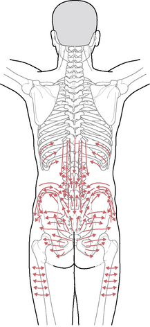
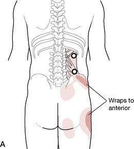
* From Bogduk (2005): ‘Some editors of journals and books have deferred to dictionaries that spell the word zygapophysial as zygapophyseal. It has been argued that this fashion is not consistent with the derivation of the word. The English word is derived from the singular zygapophysis. Consequently the adjective “zygapophysial” is also derived from the singular and is spelled with an “i”. This is the interpretation adopted by the International Anatomical Nomenclature Committee in the latest edition of Nomina Anatomica. [FCAT 1998]’
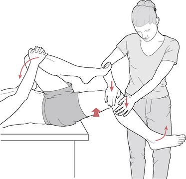
* The term ‘triage’ derives from battlefield settings where wounded soldiers were divided (by the senior physician) into three categories: those with serious injuries who were likely to recover with appropriate attention and who therefore received primary attention; those with minor wounds whose condition allowed for delay in their receiving treatment; and those whose injuries were so severe that recovery was unlikely and who therefore received only limited attention in the pressured environment of battle. As Waddell (1998) says: ‘The doctor does not attempt any more precise diagnosis or carry out any treatment, yet he makes the single most important decision in management. Everything follows from that first step. Triage decides who receives what treatment and, indeed, the final outcome’.

* Osteoporosis is more likely in a peri- or postmenopausal woman, who is slim/underweight, Caucasian and/or who has a history of anorexia, malabsorption or malnutrition. Significant other contributory factors for development of osteoporosis include metabolic acidosis (possibly associated with high-protein diet), smoking, excess coffee and alcohol consumption, corticosteroid medication usage, immobilization, endocrine imbalances (diabetes, thyrotoxicosis, hyperparathyroidism, Cushing’s syndrome) (Pizzorno & Murray 1990). See Chapter 6.

  Page 298