Rejection of Tissue Transplants

Transplant rejection is discussed here because it involves several of the immunological reactions that underlie immune-mediated inflammatory diseases. A major barrier to transplantation is the process of rejection, in which the recipient’s immune system recognizes the graft as being foreign and attacks it.

Mechanisms of Recognition and Rejection of Allografts

Rejection is a complex process in which both cell-mediated immunity and circulating antibodies play a role99; moreover, the contributions of these two mechanisms are often reflected in the histologic features of the rejected organs.

T Cell–Mediated Reactions

The critical role of T cells in transplant rejection has been documented both in humans and in experimental animals. T cell–mediated graft rejection is called cellular rejection, and it involves destruction of graft cells by CD8+ CTLs and delayed hypersensitivity reactions triggered by activated CD4+ helper cells. The major antigenic differences between a donor and recipient that result in rejection of transplants are differences in highly polymorphic HLA alleles. The recipient’s T cells recognize donor antigens from the graft (the allogeneic antigens, or alloantigens) by two pathways, called direct and indirect (Fig. 6-39).100

In the direct pathway, T cells of the transplant recipient recognize allogeneic (donor) MHC molecules on the surface of APCs in the graft. It is believed that dendritic cells carried in the donor organs are the most important APCs for initiating the antigraft response, because they not only express high levels of class I and II MHC molecules but also are endowed with costimulatory molecules (e.g., B7-1 and B7-2). The T cells of the host encounter the donor dendritic cells either within the grafted organ or after the dendritic cells migrate to the draining lymph nodes. CD8+ T cells recognize class I MHC mdecules and differentiate into active CTLs, which can kill the graft cells by mechanisms already discussed. CD4+ helper T cells recognize allogeneic class II molecules and proliferate and differentiate into TH1 (and possibly TH17) effector cells. Cytokines secreted by the activated CD4+ T cells trigger a delayed hypersensitivity reaction in the graft, resulting in increased vascular permeability and local accumulation of mononuclear cells (lymphocytes and macrophages), and graft injury caused by the activated macrophages. The direct recognition of allogeneic MHC molecules seems paradoxical to the rules of self-MHC restriction: If T cells normally are restricted to recognizing foreign peptides displayed by self-MHC molecules, why should these T cells recognize foreign MHC? The probable explanation is that allogeneic MHC molecules, with their bound peptides, resemble, or mimic, the self-MHC–foreign peptide complexes that are recognized by self-MHC–restricted T cells. Thus, recognition of allogeneic MHC molecules is a cross-reaction of T cells selected to recognize self-MHC plus foreign peptides.
In the indirect pathway of allorecognition, recipient T lymphocytes recognize MHC antigens of the graft donor after they are presented by the recipient’s own APCs. This process involves the uptake and processing of MHC and other foreign molecules from the grafted organ by host APCs. The peptides derived from the donor tissue are presented by the host’s own MHC molecules, like any other foreign peptide. Thus, the indirect pathway is similar to the physiologic processing and presentation of other foreign (e.g., microbial) antigens. The indirect pathway generates CD4+ T cells that enter the graft and recognize graft antigens being displayed by host APCs that have also entered the graft, and the result is a delayed hypersensitivity type of reaction. However, CD8+ CTLs that may be generated by the indirect pathway cannot directly recognize or kill graft cells, because these CTLs recognize graft antigens presented by the host’s APCs. Therefore, when T cells react to a graft by the indirect pathway, the principal mechanism of cellular rejection may be T-cell cytokine production and delayed hypersensitivity. It is postulated that the direct pathway is the major pathway in acute cellular rejection, whereas the indirect pathway is more important in chronic rejection. However, this separation is by no means absolute.
image

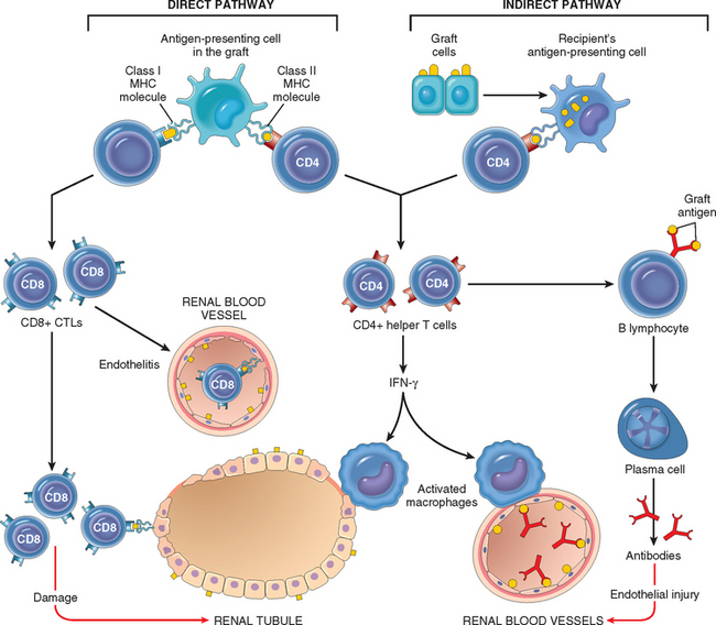
FIGURE 6-39 Recognition and rejection of organ allografts. In the direct pathway, donor class I and class II MHC antigens on antigen-presenting cells in the graft (along with costimulators, not shown) are recognized by host CD8+ cytotoxic T cells and CD4+ helper T cells, respectively. CD4+ cells proliferate and produce cytokines (e.g., IFN-γ), which induce tissue damage by a local delayed hypersensitivity reaction. CD8+ T cells responding to graft antigens differentiate into CTLs that kill graft cells. In the indirect pathway graft antigens are picked up, processed, and displayed by host APCs and activate CD4+ T cells, which damage the graft by a local delayed hypersensitivity reaction and stimulate B lymphocytes to produce antibodies.

Antibody-Mediated Reactions

Although T cells are pivotal in the rejection of organ transplants, antibodies produced against alloantigens in the graft are also important mediators of rejection.101 This process is called humoral rejection, and it can take two forms. Hyperacute rejection occurs when preformed antidonor antibodies are present in the circulation of the recipient. Such antibodies may be present in a recipient who has previously rejected a kidney transplant. Multiparous women who develop anti-HLA antibodies against paternal antigens shed from the fetus may have preformed antibodies to grafts taken from their husbands or children, or even from unrelated individuals who share HLA alleles with the husbands. Prior blood transfusions can also lead to presensitization, because platelets and white blood cells are rich in HLA antigens and donors and recipients are usually not HLA-identical. With the current practice of cross-matching, that is, testing recipient’s serum for antibodies against donor’s cells, hyperacute rejection is no longer a significant clinical problem.

In recipients not previously sensitized to transplantation antigens, exposure to the class I and class II HLA antigens of the donor graft may evoke antibodies. The antibodies formed by the recipient may cause injury by several mechanisms, including complement-dependent cytotoxicity, inflammation, and antibody-dependent cell-mediated cytotoxicity. The initial target of these antibodies in rejection seems to be the graft vasculature. Thus, antibody-dependent acute humoral rejection is usually manifested by a vasculitis, sometimes referred to as rejection vasculitis.

Rejection of Kidney Grafts

Because kidneys were the first solid organs to be transplanted and more kidneys have been transplanted than any other organ, much of our understanding of the clinical and pathologic aspects of solid-organ transplantation is based on studies of renal allografts.

Morphology. On the basis of the morphology and the underlying mechanism, rejection reactions are classified as hyperacute, acute, and chronic. The morphologic changes in these patterns are described below as they relate to renal transplants. Similar changes may occur in any other vascularized organ transplant and are discussed in relevant chapters.

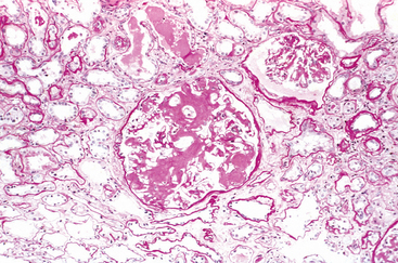
Hyperacute Rejection. This form of rejection occurs within minutes or hours after transplantation. A hyperacutely rejecting kidney rapidly becomes cyanotic, mottled, and flaccid, and may excrete a mere few drops of bloody urine. Immunoglobulin and complement are deposited in the vessel wall, causing endothelial injury and fibrin-platelet thrombi (Fig. 6-40A). Neutrophils rapidly accumulate within arterioles, glomeruli, and peritubular capillaries. As these changes become diffuse and intense, the glomeruli undergo thrombotic occlusion of the capillaries, and fibrinoid necrosis occurs in arterial walls. The kidney cortex then undergoes outright necrosis (infarction), and such nonfunctioning kidneys have to be removed.

image

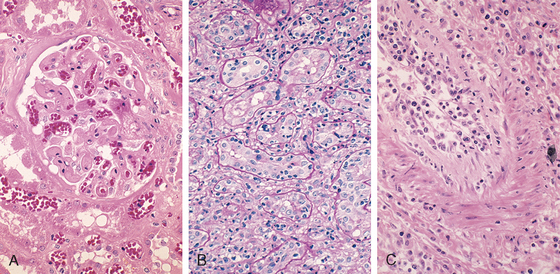
FIGURE 6-40 Morphology of hyperacute and acute graft rejection. A, Hyperacute rejection of a kidney allograft showing endothelial damage, platelet and thrombin thrombi, and early neutrophil infiltration in a glomerulus. B, Acute cellular rejection of a kidney allograft with inflammatory cells in the interstitium and between epithelial cells of the tubules. C, Acute humoral rejection of a kidney allograft (rejection vasculitis) with inflammatory cells and proliferating smooth muscle cells in the intima.

(Courtesy of Dr. Helmut Rennke, Department of Pathology, Brigham and Women’s Hospital and Harvard Medical School, Boston, MA.)

Acute Rejection. This may occur within days of transplantation in the untreated recipient or may appear suddenly months or even years later, after immunosuppression has been used and terminated. In any one patient, cellular or humoral immune mechanisms may predominate. Histologically, humoral rejection is associated with vasculitis, whereas cellular rejection is marked by an interstitial mononuclear cell infiltrate.

Acute cellular rejection is most commonly seen within the initial months after transplantation and is heralded by clinical and biochemical signs of renal failure (Chapter 20). Histologically, there may be extensive interstitial mononuclear cell infiltration and edema as well as mild interstitial hemorrhage (Fig. 6-40B). As might be expected, immunohistochemical staining reveals both CD4+ and CD8+ T lymphocytes, which express markers of activated T cells, such as the α chain of the IL-2 receptor. Glomerular and peritubular capillaries contain large numbers of mononuclear cells that may also invade the tubules, causing focal tubular necrosis. In addition to causing tubular damage, CD8+ T cells may injure vascular endothelial cells, causing a so-called endothelitis. The affected vessels have swollen endothelial cells, and at places the lymphocytes can be seen between the endothelium and the vessel wall. The recognition of cellular rejection is important because, in the absence of an accompanying humoral rejection, patients respond well to immunosuppressive therapy. Cyclosporine, a widely used immunosuppressive drug, is also nephrotoxic, and hence the histologic changes resulting from cyclosporine may be superimposed.

Acute humoral rejection (rejection vasculitis) is mediated by antidonor antibodies, and hence it is manifested mainly by damage to the blood vessels. This may take the form of necrotizing vasculitis with endothelial cell necrosis, neutrophilic infiltration, deposition of immunoglobulins, complement, and fibrin, and thrombosis. Such lesions are associated with extensive necrosis of the renal parenchyma. In many cases, the vasculitis is less acute and is characterized by marked thickening of the intima with proliferating fibroblasts, myocytes, and foamy macrophages (Fig. 6-40C). The resultant narrowing of the arterioles may cause infarction or renal cortical atrophy. The proliferative vascular lesions mimic arteriosclerotic thickening and are believed to be caused by cytokines that cause proliferation of vascular smooth muscle cells. Deposition of the complement breakdown product C4d in allografts is a strong indicator of humoral rejection, because C4d is produced during activation of the complement system by the antibody-dependent classical pathway.101,102 The importance of making this diagnosis is that it provides a rationale for treating affected patients with B cell–depleting agents.

Chronic Rejection. In recent years acute rejection has been significantly controlled by immunosuppressive therapy, and chronic rejection has emerged as an important cause of graft failure.103 Patients with chronic rejection present clinically with a progressive renal failure manifested by a rise in serum creatinine over a period of 4 to 6 months. Chronic rejection is dominated by vascular changes, interstitial fibrosis, and tubular atrophy with loss of renal parenchyma (Fig. 6-41). The vascular changes consist of dense, obliterative intimal fibrosis, principally in the cortical arteries. These vascular lesions result in renal ischemia, manifested by glomerular loss, interstitial fibrosis and tubular atrophy, and shrinkage of the renal parenchyma. The glomeruli may show scarring, with duplication of basement membranes; this appearance is sometimes called chronic transplant glomerulopathy. Chronically rejecting kidneys usually have interstitial mononuclear cell infiltrates of plasma cells and numerous eosinophils.

image

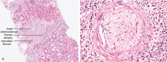
FIGURE 6-41 Chronic rejection of a kidney allograft. A, Changes in the kidney in chronic rejection. B, Graft arteriosclerosis. The vascular lumen is replaced by an accumulation of smooth muscle cells and connective tissue in the vessel intima.

(Courtesy of Dr. Helmut Rennke, Department of Pathology, Brigham and Women’s Hospital and Harvard Medical School, Boston, MA.)

Methods of Increasing Graft Survival

The value of HLA matching between donor and recipient varies in different solid-organ transplants. In kidney transplants, there is substantial benefit if all the polymorphic HLA alleles are matched (both inherited alleles of HLA-A, -B and DR). However, HLA matching is usually not even done for transplants of liver, heart, and lungs, because other considerations, such as anatomic compatibility, severity of the underlying illness, and the need to minimize the time of organ storage, override the potential benefits of HLA matching.

Except in the case of identical twins, who obviously express the same histocompatibility antigens, immunosuppressive therapy is a practical necessity in all other donor-recipient combinations.104 The mainstay of immunosuppression is the drug cyclosporine. Cyclosporine works by blocking activation of a transcription factor called nuclear factor of activated T cells (NFAT), which is required for transcription of cytokine genes, in particular, the gene that encodes IL-2. Additional drugs that are used to treat rejection include azathioprine (which inhibits leukocyte development from bone marrow precursors), steroids (which block inflammation), rapamycin and mycophenolate mofetil (both of which inhibit lymphocyte proliferation), and monoclonal anti–T-cell antibodies (e.g., monoclonal anti-CD3 and antibodies against the IL-2 receptor α chain [CD25], which opsonize and eliminate the cells and may also block T-cell activation). Another, more recent, strategy for reducing antigraft immune responses is to prevent host T cells from receiving costimulatory signals from dendritic cells during the initial phase of sensitization. This can be accomplished by interrupting the interaction between the B7 molecules on the dendritic cells of the graft donor with the CD28 receptors on host T cells, for example, by administration of proteins that bind to B7 costimulators.

Although immunosuppression prolongs graft survival, it carries its own risks. The price paid in the form of increased susceptibility to opportunistic infections is not small. These patients are also at increased risk for developing EBV-induced lymphomas, human papillomavirus–induced squamous cell carcinomas, and Kaposi sarcoma (Chapter 11), all probably the result of reactivation of latent viral infections because of diminished host defenses. To circumvent the untoward effects of immunosuppression, much effort is being devoted to induce donor-specific tolerance in graft recipients.105 For instance, giving donor cells to graft recipients may prevent reactions to the graft, perhaps because the donor inoculum contains cells, such as immature dendritic cells, that induce tolerance to the donor alloantigens. This approach may result in long-term mixed chimerism, in which the recipient lives with the injected donor cells. Other strategies being tested include injecting regulatory T cells at the time of transplantation, and promoting the death of alloreactive T cells in the recipient.

Transplantation of Other Solid Organs

In addition to the kidney, a variety of organs, such as the liver (Chapter 18), heart (Chapter 12), lungs, and pancreas, are also transplanted. The rejection reaction against liver transplants is not as vigorous as might be expected from the degree of HLA disparity. The molecular basis of this “privilege” is not understood.

Transplantation of Hematopoietic Cells

Use of hematopoietic stem cell transplants for hematologic malignancies, certain nonhematologic cancers, aplastic anemias, thalassemias, and certain immunodeficiency states is increasing. Transplantation of genetically engineered hematopoietic stem cells may also be useful for somatic cell gene therapy, and is being evaluated in some immunodeficiencies. Hematopoietic stem cells are usually obtained from the bone marrow but may also be harvested from peripheral blood after they are mobilized from the bone marrow by administration of hematopoietic growth factors. In most of the conditions in which bone marrow transplantation is indicated, the recipient is irradiated to destroy the immune system (and sometimes, cancer cells) and to create a graft bed. Several features distinguish bone marrow transplants from solid-organ transplants. Two problems that are unique to bone marrow transplantation are graft-versus-host (GVH) disease and immunodeficiency.

GVH disease occurs in any situation in which immunologically competent cells or their precursors are transplanted into immunologically crippled recipients, and the transferred cells recognize alloantigens in the host.106 It is seen most commonly in the setting of bone marrow transplantation but, rarely, may occur following transplantation of solid organs rich in lymphoid cells (e.g., the liver) or transfusion of unirradiated blood. When immune-compromised recipients receive normal bone marrow cells from allogeneic donors, the immunocompetent T cells present in the donor marrow recognize the recipient’s HLA antigens as foreign and react against them. To try to minimize GVH disease, bone marrow transplants are done between donor and recipient that are HLA-matched using sensitive DNA sequencing methods for molecular typing of HLA alleles.

Acute GVH disease occurs within days to weeks after allogeneic bone marrow transplantation. Although any organ may be affected, the major clinical manifestations result from involvement of the immune system and epithelia of the skin, liver, and intestines. Involvement of skin in GVH disease is manifested by a generalized rash that may lead to desquamation in severe cases. Destruction of small bile ducts gives rise to jaundice, and mucosal ulceration of the gut results in bloody diarrhea. Although tissue injury may be severe, the affected tissues are usually not heavily infiltrated by lymphocytes. It is believed that in addition to direct cytotoxicity by CD8+ T cells, considerable damage is inflicted by cytokines released by the sensitized donor T cells.

Chronic GVH disease may follow the acute syndrome or may occur insidiously. These patients have extensive cutaneous injury, with destruction of skin appendages and fibrosis of the dermis. The changes may resemble systemic sclerosis (discussed earlier). Chronic liver disease manifested by cholestatic jaundice is also frequent. Damage to the gastrointestinal tract may cause esophageal strictures. The immune system is devastated, with involution of the thymus and depletion of lymphocytes in the lymph nodes. Not surprisingly, the patients experience recurrent and life-threatening infections. Some patients develop manifestations of autoimmunity, postulated to result from the grafted CD4+ helper T cells reacting with host B cells and stimulating these cells, some of which may be capable of producing autoantibodies.

Because GVH disease is mediated by T lymphocytes contained in the donor bone marrow, depletion of donor T cells before transfusion virtually eliminates the disease. This protocol, however, has proved to be a mixed blessing: GVH disease is ameliorated, but the incidence of graft failures and EBV-related B-cell lymphoma and the recurrence of disease in leukemic patients increase. It seems that the multifaceted T cells not only mediate GVH disease but also are required for engraftment of the transplanted marrow stem cells, suppression of EBV-infected B-cell clones, and control of leukemic cells. The latter, called graft-versus-leukemia effect, can be quite dramatic. Deliberate induction of graft-versus-leukemia effect by infusion of allogeneic T cells is being used in the treatment of chronic myelogenous leukemia when patients relapse after bone marrow transplantation.

Immunodeficiency is a frequent complication of bone marrow transplantation. The immunodeficiency may be a result of prior treatment, myeloablative preparation for the graft, a delay in repopulation of the recipient’s immune system, and attack on the host’s immune cells by grafted lymphocytes. Affected individuals are profoundly immunosuppressed and are easy prey to infections. Although many different types of organisms may infect patients, infection with cytomegalovirus is particularly important. This usually results from activation of previously silent infection. Cytomegalovirus-induced pneumonitis can be a fatal complication.

Immunodeficiency Syndromes

Immunodeficiencies can be divided into the primary immunodeficiency disorders, which are almost always genetically determined, and secondary immunodeficiency states, which may arise as complications of cancers, infections, malnutrition, or side effects of immunosuppression, irradiation, or chemotherapy for cancer and other diseases. The primary immunodeficiency syndromes are accidents of nature that provide valuable insights into some of the critical molecules of the human immune system. Here we briefly discuss the more important primary immunodeficiencies, to be followed by a more detailed description of acquired immunodeficiency syndrome (AIDS), the most devastating example of secondary immunodeficiency.

PRIMARY IMMUNODEFICIENCIES

Most primary immunodeficiency diseases are genetically determined and affect the humoral and/or cellular arms of adaptive immunity (mediated by B and T lymphocytes, respectively) or the defense mechanisms of innate immunity (NK cells, phagocytes, or complement). Defects in adaptive immunity are often subclassified on the basis of the primary component involved (i.e., B cells or T cells or both). However, these distinctions are not clear-cut; for instance, T-cell defects almost always lead to impaired antibody synthesis, and hence isolated deficiencies of T cells are often indistinguishable clinically from combined deficiencies of T and B cells. Although these disorders were once thought to be quite rare, some form of mild genetic immune deficiency is, in fact, present in many individuals.107 Most primary immunodeficiencies manifest themselves in infancy, between 6 months and 2 years of life, and they are detected because the affected infants are susceptible to recurrent infections. The nature of infecting organisms depends to some extent on the nature of the underlying defect, as summarized in Table 6-11. Defects of phagocytes were discussed in Chapter 2. Here we present selected examples of other immunodeficiencies. We begin with isolated defects in B cells, followed by a discussion of combined immunodeficiencies and defects in complement proteins. Finally, Wiskott-Aldrich syndrome, a complex disorder affecting lymphocytes as well as platelets, is presented. With advances in genetic analyses, the mutations responsible for many of the common primary immunodeficiencies have now been identified (Fig. 6-42).108,109

TABLE 6-11 Examples of Infections in Immunodeficiencies

image
image

FIGURE 6-42 Simplified scheme of lymphocyte development and sites of block in some primary immunodeficiency diseases are shown. The affected genes are indicated in parentheses for some of the disorders. ADA, adenosine deaminase; AID, activation-induced deaminase; CD40L, CD40 ligand (also known as CD154); SCID, severe combined immune deficiency.

X-Linked Agammaglobulinemia (Bruton’s Agammaglobulinemia)

X-linked agammaglobulinemia is one of the more common forms of primary immunodeficiency.110 It is characterized by the failure of B-cell precursors (pro-B cells and pre-B cells) to develop into mature B cells. During normal B-cell maturation in the bone marrow, the Ig heavy-chain genes are rearranged first, in pre-B cells, and these are expressed on the cell surface in association with a “surrogate” light chain, where they deliver signals that induce rearrangement of the Ig light-chain genes and further maturation. This need for Ig-initiated signals is a quality control mechanism that ensures that maturation will proceed only if functional Ig proteins are expressed. X-linked agammaglobulinemia is caused by mutations in a cytoplasmic tyrosine kinase, called Bruton tyrosine kinase (Btk); the gene that encodes it is located on the long arm of the X chromosome at Xq21.22.95 Btk is a protein tyrosine kinase that is associated with the Ig receptor complex of pre-B and mature B cells and is needed to transduce signals from the receptor. When it is mutated, the pre-B cell receptor cannot deliver signals, and maturation stops at this stage. Because light chains are not produced, the complete antigen receptor molecule (which contains Ig heavy and light chains) cannot be assembled and transported to the cell membrane.

As an X-linked disease, this disorder is seen almost entirely in males, but sporadic cases have been described in females, possibly caused by mutations in some other gene that functions in the same pathway. The disease usually does not become apparent until about 6 months of age, as maternal immunoglobulins are depleted. In most cases, recurrent bacterial infections of the respiratory tract, such as acute and chronic pharyngitis, sinusitis, otitis media, bronchitis, and pneumonia, call attention to the underlying immune defect. Almost always the causative organisms are Haemophilus influenzae, Streptococcus pneumoniae, or Staphylococcus aureus. These organisms are normally opsonized by antibodies and cleared by phagocytosis. Because antibodies are important for neutralizing infectious viruses that are present in the bloodstream or mucosal secretions or being passed from cell to cell, individuals with this disease are also susceptible to certain viral infections, especially those caused by enteroviruses, such as echovirus, poliovirus, and coxsackievirus. These viruses infect the gastrointestinal tract, and from here they can disseminate to the nervous system via the blood. Thus, immunization with live poliovirus carries the risk of paralytic poliomyelitis, and echovirus can cause fatal encephalitis. For similar reasons, Giardia lamblia, an intestinal protozoan that is normally resisted by secreted IgA, causes persistent infections in persons with this disorder. In general, however, most intracellular viral, fungal, and protozoal infections are handled quite well by the intact T cell–mediated immunity.

The classic form of this disease has the following characteristics:

B cells are absent or markedly decreased in the circulation, and the serum levels of all classes of immunoglobulins are depressed. Pre-B cells, which express the B-lineage marker CD19 but not membrane Ig, are found in normal numbers in the bone marrow.
Germinal centers of lymph nodes, Peyer’s patches, the appendix, and tonsils are underdeveloped.
Plasma cells are absent throughout the body.
T cell–mediated reactions are normal.

Autoimmune diseases, such as arthritis and dermatomyositis, occur with increased frequency, in as many as 35% of individuals with this disease, which is paradoxical in the presence of an immune deficiency. It is likely that these autoimmune disorders are caused by a breakdown of self-tolerance resulting in autoimmunity, but chronic infections associated with the immune deficiency may play a role in inducing the inflammatory reactions. The treatment of X-linked agammaglobulinemia is replacement therapy with immunoglobulins. In the past, most patients succumbed to infection in infancy or early childhood. Prophylactic intravenous Ig therapy allows most individuals to reach adulthood.

Common Variable Immunodeficiency

This relatively common but poorly defined entity represents a heterogeneous group of disorders.111,112 The feature common to all patients is hypogammaglobulinemia, generally affecting all the antibody classes but sometimes only IgG. The diagnosis of common variable immunodeficiency is based on exclusion of other well-defined causes of decreased antibody production.

As might be expected in a heterogeneous group of disorders, both sporadic and inherited forms of the disease occur. In familial forms there is no single pattern of inheritance. Relatives of such patients have a high incidence of selective IgA deficiency (see later). These studies suggest that at least in some cases, selective IgA deficiency and common variable immunodeficiency may represent different expressions of a common genetic defect in antibody synthesis. In contrast to X-linked agammaglobulinemia, most individuals with common variable immunodeficiency have normal or near-normal numbers of B cells in the blood and lymphoid tissues. These B cells, however, are not able to differentiate into plasma cells.

Both intrinsic B-cell defects and abnormalities in T helper cell–mediated activation of B cells may account for the antibody deficiency in this disease. Families have been reported in which the underlying abnormality is in a receptor for a cytokine called BAFF that promotes the survival and differentiation of B cells, or in a molecule called ICOS (inducible costimulator) that is homologous to CD28 and is involved in T-cell activation and in interactions between T and B cells.111 However, the known mutations account for a minority of cases.

The clinical manifestations of common variable immunodeficiency are caused by antibody deficiency, and hence they resemble those of X-linked agammaglobulinemia. The patients typically present with recurrent sinopulmonary pyogenic infections. In addition, about 20% of patients present with recurrent herpesvirus infections. Serious enterovirus infections causing meningoencephalitis may also occur. Individuals with this disorder are also prone to the development of persistent diarrhea caused by G. lamblia. In contrast to X-linked agammaglobulinemia, common variable immunodeficiency affects both sexes equally, and the onset of symptoms is later—in childhood or adolescence. Histologically the B-cell areas of the lymphoid tissues (i.e., lymphoid follicles in nodes, spleen, and gut) are hyperplastic. The enlargement of B-cell areas probably reflects defective regulation, that is, B cells can proliferate in response to antigen but do not produce antibodies, and therefore the normal feedback inhibition by IgG is absent.

As in X-linked agammaglobulinemia, these patients have a high frequency of autoimmune diseases (approximately 20%), including rheumatoid arthritis. The risk of lymphoid malignancy is also increased, and an increase in gastric cancer has been reported.

Isolated IgA Deficiency

Isolated IgA deficiency is a common immunodeficiency. In the United States it occurs in about 1 in 600 individuals of European descent.113 It is far less common in blacks and Asians. Affected individuals have extremely low levels of both serum and secretory IgA. It may be familial, or acquired in association with toxoplasmosis, measles, or some other viral infection. The association of IgA deficiency with common variable immunodeficiency was mentioned earlier. Most individuals with this disease are asymptomatic. Because IgA is the major Ig in external secretions, mucosal defenses are weakened, and infections occur in the respiratory, gastrointestinal, and urogenital tracts. Symptomatic patients commonly present with recurrent sinopulmonary infections and diarrhea. Some individuals with IgA deficiency are also deficient in the IgG2 and IgG4 subclasses of IgG. This group of patients is particularly prone to developing infections. In addition, IgA-deficient patients have a high frequency of respiratory tract allergy and a variety of autoimmune diseases, particularly SLE and rheumatoid arthritis. The basis of the increased frequency of autoimmune and allergic diseases is not known. When transfused with blood containing normal IgA, some of these patients develop severe, even fatal, anaphylactic reactions, because the IgA behaves like a foreign antigen (since the patients do not produce it and are not tolerant to it).

The basic defect in IgA deficiency is impaired differentiation of naive B lymphocytes to IgA-producing cells. The molecular basis of this defect in most patients is still unknown. Defects in a receptor for the B cell–activating cytokine, BAFF, have been described in some patients.

Hyper-IgM Syndrome

In hyper-IgM syndrome the affected patients make IgM antibodies but are deficient in their ability to produce IgG, IgA, and IgE antibodies. It is now known that the defect in this disease affects the ability of helper T cells to deliver activating signals to B cells and macrophages. As discussed earlier in the chapter, many of the functions of CD4+ helper T cells require the engagement of CD40 on B cells, macrophages and dendritic cells by CD40L (also called CD154) expressed on antigen-activated T cells. This interaction triggers Ig class switching and affinity maturation in B cells, and stimulates the microbicidal functions of macrophages. Approximately 70% of individuals with hyper-IgM syndrome have the X-linked form of the disease, caused by mutations in the gene encoding CD40L located on Xq26.114 In the remaining persons the disease is inherited in an autosomal recessive pattern. Most of these patients have mutations in the gene encoding CD40 or the enzyme called activation-induced deaminase, a DNA-editing cytosine deaminase that is required for class switching and affinity maturation.

The serum of persons with this syndrome contains normal or elevated levels of IgM but no IgA or IgE and extremely low levels of IgG. The number of B and T cells is normal. Many of the IgM antibodies react with elements of blood, giving rise to autoimmune hemolytic anemia, thrombocytopenia, and neutropenia. In older patients there may be uncontrolled proliferation of IgM-producing plasma cells with infiltrations of the gastrointestinal tract. Although the proliferating B cells are polyclonal, extensive infiltration may lead to death.

Clinically, individuals with the hyper-IgM syndrome present with recurrent pyogenic infections, because the level of opsonizing IgG antibodies is low. In addition, those with CD40L mutations are also susceptible to pneumonia caused by the intracellular organism Pneumocystis jiroveci, because of the defect in cell-mediated immunity.

DiGeorge Syndrome (Thymic Hypoplasia)

DiGeorge syndrome is a T-cell deficiency that results from failure of development of the third and fourth pharyngeal pouches. The latter give rise to the thymus, the parathyroids, some of the clear cells of the thyroid, and the ultimobranchial body. Thus, individuals with this syndrome have a variable loss of T cell–mediated immunity (resulting from hypoplasia or lack of the thymus), tetany (resulting from lack of the parathyroids), and congenital defects of the heart and great vessels. In addition, the appearance of the mouth, ears, and facies may be abnormal. Absence of cell-mediated immunity is caused by low numbers of T lymphocytes in the blood and lymphoid tissues and poor defense against certain fungal and viral infections. The T-cell zones of lymphoid organs—paracortical areas of the lymph nodes and the periarteriolar sheaths of the spleen—are depleted. Ig levels may be normal or reduced, depending on the severity of the T-cell deficiency.

DiGeorge syndrome is not a familial disorder. It results from the deletion of a gene that maps to chromosome 22q11.115 This deletion is seen in 90% of patients, and DiGeorge syndrome is now considered a component of the 22q11 deletion syndrome, discussed in Chapter 5. One mutation that has been associated with the DiGeorge syndrome affects a member of the T-box family of transcription factors, which may be involved in development of the branchial arch and the great vessels.

Severe Combined Immunodeficiency

Severe combined immunodeficiency (SCID) represents a constellation of genetically distinct syndromes, all having in common defects in both humoral and cell-mediated immune responses.116 Affected infants present with prominent thrush (oral candidiasis), extensive diaper rash, and failure to thrive. Some patients develop a morbilliform rash shortly after birth because maternal T cells are transferred across the placenta and attack the fetus, causing GVH disease. Persons with SCID are extremely susceptible to recurrent, severe infections by a wide range of pathogens, including Candida albicans, P. jiroveci, Pseudomonas, cytomegalovirus, varicella, and a whole host of bacteria. Without bone marrow transplantation, death occurs within the first year of life. Despite the common clinical manifestations, the underlying defects are quite different in different forms of SCID, and in many cases the genetic lesion is not known. Often, the SCID defect resides in the T-cell compartment, with a secondary impairment of humoral immunity.

The most common form, accounting for 50% to 60% of cases, is X-linked, and hence SCID is more common in boys than in girls. The genetic defect in the X-linked form is a mutation in the common γ-chain (γc) subunit of cytokine receptors. This transmembrane protein is part of the signal-transducing components of the receptors for IL-2, IL-4, IL-7, IL-9, IL-11, IL-15, and IL-21. IL-7 is required for the survival and proliferation of lymphoid progenitors, particularly T-cell precursors. As a result of defective IL-7 receptor signaling, there is a profound defect in the earliest stages of lymphocyte development, especially T-cell development.117 T-cell numbers are greatly reduced, and although B cells are normal in number, antibody synthesis is severely impaired because of lack of T-cell help. IL-15 is important for the maturation and proliferation of NK cells, and because the common γ chain is a component of the receptor for IL-15, these individuals often have a deficiency of NK cells as well.

The remaining cases of SCID are inherited as autosomal recessive. The most common cause of autosomal recessive SCID is a deficiency of the enzyme adenosine deaminase (ADA). Although the mechanisms by which ADA deficiency causes SCID are not entirely clear, it has been proposed that deficiency of ADA leads to accumulation of deoxyadenosine and its derivatives (e.g., deoxy-ATP), which are toxic to rapidly dividing immature lymphocytes, especially those of the T-cell lineage.118 Hence there may be a greater reduction in the number of T lymphocytes than of B lymphocytes.

Several other less common causes of autosomal recessive SCID have been discovered:

Mutations in recombinase-activating genes prevent the somatic gene rearrangements essential for the assembly of T-cell receptor and Ig genes.119 This blocks the development of T and B cells.
An intracellular kinase called Jak3 is essential for signal transduction through the common cytokine receptor γ chain (which is mutated in X-linked SCID, as discussed above). Mutations of Jak3 therefore have the same effects as mutations in the γc chain.120
Several mutations have been described in signaling molecules, including kinases associated with the T-cell antigen receptor and components of calcium channels that are required for entry of calcium and activation of many signaling pathways.
Mutations that impair the expression of class II MHC molecules prevent the development of CD4+ T cells.121 CD4+ T cells are involved in cellular immunity and provide help to B cells, and hence class II MHC deficiency results in combined immunodeficiency. This disease, called the bare lymphocyte syndrome, is usually caused by mutations in transcription factors that are required for class II MHC gene expression.

The histologic findings in SCID depend on the underlying defect. In the two most common forms (ADA deficiency and γc mutation), the thymus is small and devoid of lymphoid cells. In SCID caused by ADA deficiency, remnants of Hassall’s corpuscles can be found, whereas in X-linked SCID the thymus contains lobules of undifferentiated epithelial cells resembling fetal thymus. In either case other lymphoid tissues are hypoplastic as well, with marked depletion of T-cell areas and in some cases both T-cell and B-cell zones.

Currently, bone marrow transplantation is the mainstay of treatment, but X-linked SCID is the first human disease in which gene therapy has been successful.122 For gene therapy a normal γc gene is expressed in bone marrow stem cells of patients using a retroviral vector, and the cells are transplanted back into the patients. The clinical experience is small, but some patients have shown reconstitution of their immune systems for over a year after therapy. Unfortunately, however, 20% of these patients have developed acute T-cell leukemias, which appear to have been triggered by the activation of oncogenes by the integrated retrovirus,123 highlighting the dangers of this particular approach to gene therapy. Patients with ADA deficiency have also been treated with bone marrow transplantation and, more recently, with gene therapy to introduce a normal ADA gene into T-cell precursors.

Immunodeficiency with Thrombocytopenia and Eczema (Wiskott-Aldrich Syndrome)

Wiskott-Aldrich syndrome is an X-linked recessive disease characterized by thrombocytopenia, eczema, and a marked vulnerability to recurrent infection, ending in early death.124 The thymus is morphologically normal, at least early in the course of the disease, but there is progressive secondary depletion of T lymphocytes in the peripheral blood and in the T-cell zones (paracortical areas) of the lymph nodes, with variable loss of cellular immunity. Patients do not make antibodies to polysaccharide antigens, and the response to protein antigens is poor. IgM levels in the serum are low, but levels of IgG are usually normal. Paradoxically the levels of IgA and IgE are often elevated. Patients are also prone to developing non-Hodgkin B-cell lymphomas. The Wiskott-Aldrich syndrome is caused by mutations in the gene encoding Wiskott-Aldrich syndrome protein (WASP), which is located at Xp11.23. This protein belongs to a family of proteins that are believed to link membrane receptors, such as antigen receptors, to cytoskeletal elements. The WASP protein may be involved in cytoskeleton-dependent responses, including cell migration and signal transduction, but the essential functions of this protein in lymphocytes and platelets are unclear. The only treatment is bone marrow transplantation.

Genetic Deficiencies of the Complement System

The complement system plays critical roles in host defense and inflammation. Hereditary deficiencies have been described for virtually all components of the complement system and several of the regulators.125 A deficiency of C2 is the most common of all. With a deficiency of C2 or the other early components of the classical pathway (i.e., C1 [C1q, r, or s] or C4), there is little or no increase in susceptibility to infections, but the dominant manifestation is an increased incidence of an SLE-like autoimmune disease, as discussed earlier. Presumably, the alternative complement pathway is adequate for the control of most infections. Deficiency of components of the alternative pathway (properdin and factor D) is rare. It is associated with recurrent pyogenic infections. The C3 component of complement is required for both the classical and alternative pathways, and hence a deficiency of this protein results in susceptibility to serious and recurrent pyogenic infections. There is also increased incidence of immune complex–mediated glomerulonephritis; in the absence of complement, immune complex–mediated inflammation is presumably caused by Fc receptor–dependent leukocyte activation. The terminal components of complement C5, 6, 7, 8, and 9 are required for the assembly of the membrane attack complex involved in the lysis of organisms. With a deficiency of these late-acting components, there is increased susceptibility to recurrent neisserial (gonococcal and meningococcal) infections; Neisseria bacteria have thin cell walls and are especially susceptible to the lytic actions of complement. Some patients inherit a form of mannose-binding lectin, the plasma protein that initiates the lectin pathway of complement, that does not polymerize normally and is functionally defective. These individuals also show increased susceptibility to infections.

A deficiency of C1 inhibitor gives rise to hereditary angioedema.126 This autosomal dominant disorder is more common than complement deficiency states. The C1 inhibitor is a protease inhibitor whose target enzymes are C1r and C1s of the complement cascade, factor XII of the coagulation pathway, and the kallikrein system. As discussed in Chapter 2, these pathways are closely linked, and their unregulated activation can give rise to vasoactive peptides such as bradykinin. Although the exact nature of the bioactive compound produced in hereditary angioedema is uncertain, these patients have episodes of edema affecting skin and mucosal surfaces such as the larynx and the gastrointestinal tract. This may result in life-threatening asphyxia or nausea, vomiting, and diarrhea after minor trauma or emotional stress. Acute attacks of hereditary angioedema can be treated with C1 inhibitor concentrates prepared from human plasma.

Deficiency of other complement-regulatory proteins is the cause of paroxysmal nocturnal hemoglobinuria. In this disease there are mutations in enzymes required for glycophosphatidyl inositol linkages, which are essential for the assembly of decay-accelerating factor and CD59, both of which regulate complement.127 Uncontrolled complement activation on the surface of red cells is believed to be the basis of hemolysis (Chapter 14). Mutations in the complementregulatory protein factor H underlie about 10% of cases of a renal disease called hemolytic uremic syndrome, which is characterized by microvascular thrombosis in the kidneys (Chapter 20).

SECONDARY IMMUNODEFICIENCIES

Secondary immune deficiencies may be encountered in individuals with cancer, diabetes and other metabolic diseases, malnutrition, chronic infection, and renal disease. They also occur in persons receiving chemotherapy or radiation therapy for cancer, or immunosuppressive drugs to prevent graft rejection or to treat autoimmune diseases. Some of these secondary immunodeficiency states can be caused by defective lymphocyte maturation (when the bone marrow is damaged by radiation or chemotherapy or involved by tumors, such as leukemias and metastatic cancers), loss of immunoglobulins (as in proteinuric renal diseases), inadequate Ig synthesis (as in malnutrition), or lymphocyte depletion (from drugs or severe infections). As a group, the secondary immune deficiencies are more common than the disorders of primary genetic origin. The most common secondary immunodeficiency is AIDS, and we will describe this in the next section.

ACQUIRED IMMUNODEFICIENCY SYNDROME (AIDS)

AIDS is a disease caused by the retrovirus human immunodeficiency virus (HIV) and characterized by profound immunosuppression that leads to opportunistic infections, secondary neoplasms, and neurologic manifestations. The magnitude of this modern plague is truly staggering. By the end of 2006, more than a million cases of AIDS had been reported in the United States, where AIDS is the second leading cause of death in men between ages 25 and 44, and the third leading cause of death in women in this age group. Though initially recognized in the United States, AIDS is a global problem. It has now been reported from more than 190 countries around the world, and the pool of HIV-infected persons in Africa and Asia is large and expanding. By the year 2006, HIV had infected 60 million people worldwide, and nearly 20 million adults and children have died of the disease. There are about 33 million people living with HIV, of whom 65% are in Africa and over 20% in Asia; the prevalence rate in adults in sub-Saharan Africa is over 8%. It is estimated that 2.5 million people were newly infected with HIV during 2006, and 2.1 million deaths were caused by AIDS in that year alone. In this dismal scenario, there may be some good news. Because of public health measures, the infection rate seems to be decreasing, and some authorities believe it may have peaked in the late 1990s. Furthermore, improved antiviral therapies have resulted in fewer people dying of the disease. This, however, raises its own tragic concern; because more people are living with HIV, the risk of spreading the infection will increase if vigilance is relaxed.

The enormous medical and social burden of the AIDS problem has led to an explosion of research aimed at understanding HIV and its remarkable ability to cripple host defenses. The literature on AIDS is vast and expanding. Here we summarize the currently available data on the epidemiology, pathogenesis, and clinical features of HIV infection.

Epidemiology

Epidemiologic studies in the United States have identified five groups of adults at risk for developing AIDS. The case distribution in these groups is as follows:

Homosexual or bisexual men constitute the largest group, accounting for over 50% of the reported cases. This includes about 5% who were intravenous drug abusers as well. Transmission of AIDS in this category appears to be on the decline: in 2005 about 48% of new cases were attributed to male homosexual contacts.
Intravenous drug abusers with no previous history of homosexuality are the next largest group, representing about 20% of infected individuals.
Hemophiliacs, especially those who received large amounts of factor VIII or factor IX concentrates before 1985, make up about 0.5% of all cases.
Recipients of blood and blood components who are not hemophiliacs but who received transfusions of HIV-infected whole blood or components (e.g., platelets, plasma) account for about 1% of patients. (Organs obtained from HIV-infected donors can also transmit AIDS.)
Heterosexual contacts of members of other high-risk groups (chiefly intravenous drug abusers) constitute about 10% of the patient population. About 30% of new cases in 2005 were attributable to heterosexual contact. This is the most rapidly growing group of infected individuals, particularly women; in sub-Saharan Africa, where the infection rate is estimated to be about 10,000 new cases every day, more than half the infected individuals are women.
In approximately 5% of cases the risk factors cannot be determined.

The epidemiology of AIDS is quite different in children under age 13. Close to 2% of all AIDS cases occur in this pediatric population, and worldwide over 500,000 new cases and almost 400,000 deaths were reported in children in the year 2006. In this group the vast majority acquired the infection by transmission of the virus from mother to child (discussed later).

It should be apparent from the preceding discussion that transmission of HIV occurs under conditions that facilitate exchange of blood or body fluids containing the virus or virus-infected cells. The three major routes of transmission are sexual contact, parenteral inoculation, and passage of the virus from infected mothers to their newborns.

Sexual transmission is clearly the predominant mode of infection worldwide, accounting for over 75% of all cases of HIV transmission. Because the majority of infected people in the United States are men who have sex with men, most sexual transmission has occurred among homosexual men. The virus is carried in the semen, and it enters the recipient’s body through abrasions in rectal or oral mucosa or by direct contact with mucosal lining cells. Viral transmission occurs in two ways: (1) direct inoculation into the blood vessels breached by trauma, and (2) infection of dendritic cells or CD4+ cells within the mucosa. Heterosexual transmission, though initially of less numerical importance in the United States, is globally the most common mode by which HIV is spread. In the past few years, even in the United States, the rate of increase of heterosexual transmission has outpaced transmission by other means. Such spread is occurring most rapidly in female sex partners of male intravenous drug abusers. As such, the number of women with AIDS is rising rapidly. In contrast to the U.S. experience, heterosexual transmission has always been the dominant mode of HIV infection in Asia and Africa.

In addition to male-to-male and male-to-female transmission, there is evidence supporting female-to-male transmission. HIV is present in vaginal secretions and cervical cells of infected women. In the United States this form of heterosexual spread is approximately 20-fold less common than male-to-female transmission. By contrast, in Africa and parts of Asia, the risk of female-to-male transmission is much higher. This observation is believed to be attributable to the presence of concurrent sexually transmitted disease. All forms of sexual transmission of HIV are enhanced by coexisting sexually transmitted diseases, especially those associated with genital ulceration. In this regard, syphilis, chancroid, and herpes are particularly important. Other sexually transmitted diseases, including gonorrhea and chlamydia, are also cofactors for HIV transmission, perhaps because in these genital inflammatory states there is greater concentration of the virus and virus-containing cells in genital fluids, as a result of increased numbers of inflammatory cells in the semen.

Parenteral transmission of HIV has occurred in three groups of individuals: intravenous drug abusers, hemophiliacs who received factor VIII and factor IX concentrates, and random recipients of blood transfusion. Of these three, intravenous drug users constitute by far the largest group. Transmission occurs by sharing of needles, syringes, and other paraphernalia contaminated with HIV-containing blood.

Transmission of HIV by transfusion of blood or blood products, such as lyophilized factor VIII and factor IX concentrates, has been virtually eliminated. This fortunate outcome resulted from increasing use of recombinant clotting factors and from three public health measures: screening of donated blood and plasma for antibody to HIV, stringent purity criteria for factor VIII and factor IX preparations, and screening of donors on the basis of history. However, an extremely small risk of acquiring AIDS through transfusion of seronegative blood persists, because a recently infected individual may be antibody-negative. Currently, this risk is estimated to be 1 in more than 2 million units of blood transfused. Because it is now possible to detect HIV-associated p24 antigens in the blood before the development of humoral antibodies, this small risk is likely to decrease even further.

As alluded to earlier, mother-to-infant transmission is the major cause of pediatric AIDS. Infected mothers can transmit the infection to their offspring by three routes: (1) in utero by transplacental spread, (2) during delivery through an infected birth canal, and (3) after birth by ingestion of breast milk. Of these, transmission during birth (intrapartum) and in the immediate period thereafter (peripartum) is considered to be the most common mode in the United States. The reported transmission rates vary from 7% to 49% in different parts of the world. Higher risk of transmission is associated with high maternal viral load and low CD4+ T-cell counts as well as chorioamnionitis. Currently, with antiretroviral therapy given to infected pregnant women in the United States, the mother-to-child transmission has been virtually eliminated.

Much concern has arisen in the lay public and among health care workers about spread of HIV infection outside the high-risk groups. Extensive studies indicate that HIV infection cannot be transmitted by casual personal contact in the household, workplace, or school. Spread by insect bites is virtually impossible. Regarding transmission of HIV infection to health care workers, an extremely small but definite risk seems to be present. Seroconversion has been documented after accidental needle-stick injury or exposure of nonintact skin to infected blood in laboratory accidents. After needle-stick accidents, the risk of seroconversion is believed to be about 0.3%, and antiretroviral therapy given within 24 to 48 hours of a needle stick can reduce the risk of infection eightfold. By comparison, approximately 30% of those accidentally exposed to hepatitis B–infected blood become seropositive.

Etiology: The Properties of HIV

AIDS is caused by HIV, a nontransforming human retrovirus belonging to the lentivirus family. Included in this group are feline immunodeficiency virus, simian immunodeficiency virus, visna virus of sheep, bovine immunodeficiency virus, and the equine infectious anemia virus.

Two genetically different but related forms of HIV, called HIV-1 and HIV-2, have been isolated from patients with AIDS. HIV-1 is the most common type associated with AIDS in the United States, Europe, and Central Africa, whereas HIV-2 causes a similar disease principally in West Africa and India. Specific tests for HIV-2 are available, and blood collected for transfusion is routinely screened for both HIV-1 and HIV-2 seropositivity. The ensuing discussion relates primarily to HIV-1 and diseases caused by it, but the information is generally applicable to HIV-2 as well.

Structure of HIV

Similar to most retroviruses, the HIV-1 virion is spherical and contains an electron-dense, cone-shaped core surrounded by a lipid envelope derived from the host cell membrane (Fig. 6-43). The virus core contains (1) the major capsid protein p24; (2) nucleocapsid protein p7/p9; (3) two copies of genomic RNA; and (4) the three viral enzymes (protease, reverse transcriptase, and integrase). p24 is the most readily detected viral antigen and is the target for the antibodies that are used for the diagnosis of HIV infection in the widely used enzyme-linked immunosorbent assay. The viral core is surrounded by a matrix protein called p17, which lies underneath the virion envelope. Studding the viral envelope are two viral glycoproteins, gp120 and gp41, which are critical for HIV infection of cells.

image

FIGURE 6-43 The structure of the human immune deficiency virus (HIV)–1 virion. The viral particle is covered by a lipid bilayer derived from the host cell and studded with viral glycoproteins gp41 and gp120.

The HIV-1 RNA genome contains the gag, pol, and env genes, which are typical of retroviruses (Fig. 6-44). The products of the gag and pol genes are translated initially into large precursor proteins that are cleaved by the viral protease to yield the mature proteins. The highly effective anti-HIV-1 protease inhibitor drugs prevent viral assembly by inhibiting the formation of mature viral proteins. In addition to these three standard retroviral genes, HIV contains several other accessory genes, including tat, rev, vif, nef, vpr, and vpu, that regulate the synthesis and assembly of infectious viral particles and the pathogenicity of the virus.128-130 For example, the product of the tat (transactivator) gene causes a 1000-fold increase in the transcription of viral genes and is therefore critical for virus replication. The functions of other accessory proteins are indicated in Figure 6-44.

image

FIGURE 6-44 The HIV genome. Several viral genes and the functions of the encoded proteins are illustrated. The genes outlined in red are unique to HIV; others are shared by all retroviruses.

Molecular analysis of different HIV-1 isolates has revealed considerable variability in certain parts of their genome. Most variations are clustered in particular regions of the envelope glycoproteins. Because the humoral immune response against HIV-1 is targeted against its envelope, such variability poses problems for the development of a single antigen vaccine. On the basis of genetic analysis, HIV-1 can be divided into three subgroups, designated M (major), O (outlier), and N (neither M nor O). Group M viruses are the most common form worldwide, and they are further divided into several subtypes, or clades, designated A through K. Various subtypes differ in their geographic distribution; for example, subtype B is the most common form in western Europe and the United States, whereas subtype E is the most common clade in Thailand. Currently, clade C is the fastest-spreading clade worldwide, being present in India, Ethiopia, and Southern Africa.

Pathogenesis of HIV Infection and AIDS

While HIV can infect many tissues, there are two major targets of HIV infection: the immune system and the central nervous system. The effects of HIV infection on each of these two systems are discussed separately.

Profound immune deficiency, primarily affecting cell-mediated immunity, is the hallmark of AIDS. This results chiefly from infection of and a severe loss of CD4+ T cells as well as impairment in the function of surviving helper T cells.131,132 As discussed later, macrophages and dendritic cells are also targets of HIV infection. HIV enters the body through mucosal tissues and blood and first infects T cells as well as dendritic cells and macrophages. The infection becomes established in lymphoid tissues, where the virus may remain latent for long periods. Active viral replication is associated with more infection of cells and progression to AIDS. We first describe the mechanisms involved in viral entry into T cells and macrophages and the replicative cycle of the virus within cells. This is followed by a more detailed review of the interaction between HIV and its cellular targets.

Life Cycle of HIV

The life cycle of HIV consists of infection of cells, integration of the provirus into the host cell genome, activation of viral replication, and production and release of infectious virus (Fig. 6-45).133 The molecules and mechanisms of each of these steps are understood in considerable detail.

image

FIGURE 6-45 The life cycle of HIV, showing the steps from viral entry to production of infectious virions.

(Adapted with permission from Wain-Hobson S: HIV. One on one meets two. Nature 384:117, 1996. Copyright 1996, Macmillan Magazines Limited.)

Infection of Cells by HIV

HIV infects cells by using the CD4 molecule as receptor and various chemokine receptors as coreceptors (Fig. 6-45). The requirement for CD4 binding explains the selective tropism of the virus for CD4+ T cells and other CD4+ cells, particularly monocytes/macrophages and dendritic cells. Binding to CD4 is not sufficient for infection, however. HIV gp120 must also bind to other cell surface molecules (coreceptors) for entry into the cell. Chemokine receptors, particularly CCR5 and CXCR4, serve this role.134 HIV isolates can be distinguished by their use of these receptors: R5 strains use CCR5, X4 strains use CXCR4, and some strains (R5×4) are dual-tropic. In approximately 90% of cases, the R5 (M-tropic) type of HIV is the dominant virus found in the blood of acutely infected individuals and early in the course of infection. Over the course of infection, however, T-tropic viruses gradually accumulate; these are especially virulent because T-tropic viruses are capable of infecting many T cells and even thymic T-cell precursors and cause greater T-cell depletion and impairment.

Molecular details of the deadly handshake between HIV glycoproteins and their cell surface receptors have been uncovered by elegant studies and are important to understand because they may provide the basis of anti-HIV therapy. The HIV envelope contains two glycoproteins, surface gp120 noncovalently attached to a transmembrane protein, gp41. The initial step in infection is the binding of the gp120 envelope glycoprotein to CD4 molecules. This binding leads to a conformational change that results in the formation of a new recognition site on gp120 for the coreceptors CCR5 or CXCR4. Binding to the coreceptors induces conformational changes in gp41 that result in the exposure of a hydrophobic region called the fusion peptide at the tip of gp41. This peptide inserts into the cell membrane of the target cells (e.g., T cells or macrophages), leading to fusion of the virus with the host cell.135 After fusion the virus core containing the HIV genome enters the cytoplasm of the cell. The requirement for HIV binding to coreceptors may have important implications for the pathogenesis of AIDS. Chemokines sterically hinder HIV infection of cells in culture by occupying their receptors, and therefore, the level of chemokines in the tissues may influence the efficiency of viral infection in vivo. Also, polymorphisms in the gene encoding CCR5 are associated with different susceptibility to HIV infection. About 1% of white Americans inherit two defective copies of the CCR5 gene and are resistant to infection and the development of AIDS associated with R5 HIV isolates.125 About 20% of individuals are heterozygous for this protective CCR5 allele; these persons are not protected from AIDS, but the onset of their disease after infection is somewhat delayed. Only rare homozygotes for the mutation have been found in African or East Asian populations.

Viral Replication

Once internalized, the RNA genome of the virus undergoes reverse transcription, leading to the synthesis of double-stranded complementary DNA (cDNA; proviral DNA) (see Fig. 6-45). In quiescent T cells, HIV cDNA may remain in the cytoplasm in a linear episomal form. In dividing T cells, the cDNA circularizes, enters the nucleus, and is then integrated into the host genome. After this integration, the provirus may be silent for months or years, a form of latent infection. Alternatively, proviral DNA may be transcribed, with the formation of complete viral particles that bud from the cell membrane. Such productive infection, when associated with extensive viral budding, leads to death of infected cells.

In vivo, HIV infects memory and activated T cells but is inefficient at productively infecting naive (unactivated) T cells. Naive T cells contain an active form of an enzyme that introduces mutations in the HIV genome. This enzyme has been given the rather cumbersome name APOBEC3G (for “apolipoprotein B mRNA-editing enzyme catalytic polypeptide-like editing complex 3”).136 It is a cytidine deaminase that introduces cytosine-to-uracil mutations in the viral DNA that is produced by reverse transcription. These mutations inhibit further DNA replication by mechanisms that are not fully defined. Activation of T cells converts cellular APOBEC3G into an inactive, high-molecular-mass complex, which explains why the virus can replicate in previously activated (e.g., memory) T cells and T-cell lines. HIV has also evolved to counteract this cellular defense mechanism; the viral protein Vif binds to APOBEC3G and promotes its degradation by cellular proteases.

Completion of the viral life cycle in latently infected cells occurs only after cell activation, and in the case of most CD4+ T cells virus activation results in cell lysis. Activation of T cells by antigens or cytokines upregulates several transcription factors, including NF-κB, which stimulate transcription of genes encoding cytokines such as IL-2 and its receptor. In resting T cells, NF-κB is sequestered in the cytoplasm in a complex with members of the IκB (inhibitor of κB) protein. Cellular activation by antigen or cytokines induces cytoplasmic kinases that phosphorylate IκB and target it for enzymatic degradation, thus releasing NF-κB and allowing it to translocate to the nucleus. In the nucleus, NF-κB binds to sequences within the promoter regions of several genes, including those of cytokines that are expressed in activated T cells. The long-terminal-repeat sequences that flank the HIV genome also contain NF-κB–binding sites that can be triggered by the same transcription factors.137 Imagine now a latently infected CD4+ cell that encounters an environmental antigen. Induction of NF-κB in such a cell (a physiologic response) activates the transcription of HIV proviral DNA (a pathologic outcome) and leads ultimately to the production of virions and to cell lysis. Furthermore, TNF and other cytokines produced by activated macrophages also stimulate NF-κB activity and thus lead to production of HIV RNA. Thus, it seems that HIV thrives when the host T cells and macrophages are physiologically activated, an act that can be best described as “subversion from within.” Such activation in vivo may result from antigenic stimulation by HIV itself or by other infecting microorganisms. HIV-infected people are at increased risk for recurrent exposure to other infections, which lead to increased lymphocyte activation and production of pro-inflammatory cytokines. These, in turn, stimulate more HIV production, loss of additional CD4+ T cells, and more infection. Thus, it is easy to visualize how in individuals with AIDS a vicious cycle may be set up that culminates in inexorable destruction of the immune system.

Mechanism of T-Cell Immunodeficiency in HIV Infection

Loss of CD4+ T cells is mainly because of infection of the cells and the direct cytopathic effects of the replicating virus (Fig. 6-46).138 Approximately 100 billion new viral particles are produced every day, and 1 to 2 billion CD4+ T cells die each day.139 Because the frequency of infected cells in the circulation is very low, for many years it was suspected that the immunodeficiency is out of proportion to the level of infection and cannot be attributed to death of infected cells. In fact, many infected cells may be in mucosal and other peripheral lymphoid organs, and death of these cells is a major cause of the relentless, and eventually profound, cell loss. Also, to a point the immune system can replace the dying T cells, and hence the rate of T cell loss may appear deceptively low, but as the disease progresses, renewal of CD4+ T cells cannot keep up with the loss of these cells. Possible mechanisms by which the virus directly kills infected cells include increased plasma membrane permeability associated with budding of virus particles from the infected cells, and virus replication interfering with protein synthesis.

image

FIGURE 6-46 Mechanisms of CD4+ T-cell loss in HIV infection, showing some of the known and postulated mechanisms of T-cell depletion after HIV infection. APC, antigen-presenting cell; CTL, cytotoxic T lymphocyte.

In addition to direct killing of cells by the virus, other mechanisms may contribute to the loss of T cells (see Fig. 6-46).140 These include:

HIV colonizes the lymphoid organs (spleen, lymph nodes, tonsils) and may cause progressive destruction of the architecture and cellular composition of lymphoid tissues.
Chronic activation of uninfected cells, responding to HIV itself or to infections that are common in individuals with AIDS, leads to apoptosis of these cells by the process of activation-induced cell death.140,141 Thus, the numbers of CD4+ T cells that die are far greater than the numbers of infected cells. The molecular mechanism of this type of cell death is not known.
Loss of immature precursors of CD4+ T cells can also occur, either by direct infection of thymic progenitor cells or by infection of accessory cells that secrete cytokines essential for CD4+ T-cell maturation.
Fusion of infected and uninfected cells with formation of syncytia (giant cells) can occur. In tissue culture the gp120 expressed on productively infected cells binds to CD4 molecules on uninfected T cells, followed by cell fusion. Fused cells develop ballooning and usually die within a few hours. This property of syncytia formation is generally confined to the T-tropic X4 type of HIV-1. For this reason, this type is often referred to as syncytia-inducing (SI) virus, in contrast to the R5 virus.
Apoptosis of uninfected CD4+ T cells by binding of soluble gp120 to the CD4 molecule, followed by activation through the T-cell receptor by antigens. It has been suggested that such cross-linking of CD4 molecules and T-cell activation lead to aberrant signaling and activation of death pathways. CD8+ CTLs may kill uninfected CD4+ T cells that are coated with gp120 released from infected cells.

Although marked reduction in CD4+ T cells, a hallmark of AIDS, can account for most of the immunodeficiency late in the course of HIV infection, there is compelling evidence for qualitative defects in T cells that can be detected even in asymptomatic HIV-infected persons. Reported defects include a reduction in antigen-induced T-cell proliferation, a decrease in TH1-type responses relative to the TH2 type, defects in intracellular signaling, and many more. The loss of TH1 responses results in profound deficiency in cell-mediated immunity, leading to increased susceptibility to infections by viruses and other intracellular microbes. There is also a selective loss of the memory subset of CD4+ helper T cells early in the course of disease, which explains poor recall responses to previously encountered antigens.

Low-level chronic or latent infection of T cells (and macrophages, discussed below) is an important feature of HIV infection. It is widely believed that integrated provirus, without virus expression (latent infection), can remain in the cells for months to years. Even with potent antiviral therapy, which practically sterilizes the peripheral blood, latent virus lurks within the CD4+ cells (both T cells and macrophages) in the lymph nodes. According to some estimates, 0.05% of resting CD4+ T cells in the lymph nodes are latently infected. Because these CD4+ T cells are memory T cells, they are long-lived, with a life span of months to years, and thus provide a persistent reservoir of virus.

CD4+ T cells play a pivotal role in regulating both cellular and humoral immune responses. Therefore, loss of this “master regulator” has ripple effects on virtually every other component of the immune system, as summarized in Table 6-12.

TABLE 6-12 Major Abnormalities of Immune Function in AIDS

LYMPHOPENIA
Predominantly caused by selective loss of the CD4+ helper T-cell subset
DECREASED T-CELL FUNCTION IN VIVO
Preferential loss of activated and memory T cells
Decreased delayed-type hypersensitivity
Susceptibility to opportunistic infections
Susceptibility to neoplasms
ALTERED T-CELL FUNCTION IN VITRO
Decreased proliferative response to mitogens, alloantigens, and soluble antigens
Decreased cytotoxicity
Decreased helper function for B-cell antibody production
Decreased IL-2 and IFN-γ production
POLYCLONAL B-CELL ACTIVATION
Hypergammaglobulinemia and circulating immune complexes
Inability to mount de novo antibody response to new antigens
Poor responses to normal B-cell activation signals in vitro
ALTERED MONOCYTE OR MACROPHAGE FUNCTIONS
Decreased chemotaxis and phagocytosis
Decreased class II HLA expression
Diminished capacity to present antigen to T cells

HLA, human leukocyte antigen; IFN-γ, interferon-γ; IL-1, etc., interleukin-1; TNF, tumor necrosis factor.

HIV Infection of Non-T Cells

In addition to infection and loss of CD4+ T cells, infection of macrophages142 and dendritic cells143 is also important in the pathogenesis of HIV infection. Similar to T cells, the majority of the macrophages that are infected by HIV are found in the tissues and the number of blood monocytes infected by the virus may be low. In certain tissues, such as the lungs and brain, as many as 10% to 50% of macrophages are infected. Several aspects of HIV infection of macrophages should be emphasized:

Although cell division is required for replication of most retroviruses, HIV-1 can infect and multiply in terminally differentiated nondividing macrophages. This property of HIV-1 is dependent on the HIV-1 vpr gene. The Vpr protein allows nuclear targeting of the HIV preintegration complex through the nuclear pore.
Infected macrophages bud relatively small amounts of virus from the cell surface, but these cells contain large numbers of virus particles often located in intracellular vacuoles. Even though macrophages allow viral replication, they are quite resistant to the cytopathic effects of HIV, in contrast to CD4+ T cells. Thus, macrophages may be reservoirs of infection, whose output remains largely protected from host defenses. In late stages of HIV infection, when CD4+ T-cell numbers decline greatly, macrophages may be an important site of continued viral replication.144
Macrophages, in all likelihood, act as gatekeepers of infection. Recall that in more than 90% of cases acute HIV infection is characterized by predominantly circulating M-tropic strains. This finding suggests that the initial infection of macrophages or dendritic cells may be important in the pathogenesis of HIV disease.

Even uninfected monocytes are reported to have unexplained functional defects that may have important consequences for host defense. These defects include impaired microbicidal activity, decreased chemotaxis, decreased secretion of IL-1, inappropriate secretion of TNF, and poor capacity to present antigens to T cells. Also, even the low number of infected blood monocytes may be vehicles for HIV to be transported to various parts of the body, including the nervous system.

Studies have documented that, in addition to macrophages, two types of dendritic cells are also important targets for the initiation and maintenance of HIV infection: mucosal and follicular dendritic cells. It is thought that mucosal dendritic cells are infected by the virus and transport it to regional lymph nodes, where the virus is transmitted to CD4+ T cells.143 Dendritic cells also express a lectin-like receptor that specifically binds HIV and displays it in an intact, infectious form to T cells, thus promoting infection of the T cells.145 Follicular dendritic cells in the germinal centers of lymph nodes, similar to macrophages, are potential reservoirs of HIV. Although some follicular dendritic cells may be susceptible to HIV infection, most virus particles are found on the surface of their dendritic processes. Follicular dendritic cells have receptors for the Fc portion of immunoglobulins, and hence they trap HIV virions coated with anti-HIV antibodies. The antibody-coated virions localized to follicular dendritic cells retain the ability to infect CD4+ T cells as they traverse the intricate meshwork formed by the dendritic processes of the follicular dendritic cells.

Although much attention has been focused on T cells, macrophages, and dendritic cells because they can be infected by HIV, individuals with AIDS also display profound abnormalities of B-cell function. Paradoxically, there is polyclonal activation of B cells, resulting in germinal center B-cell hyperplasia (particularly early in the disease course), bone marrow plasmacytosis, hypergammaglobulinemia, and formation of circulating immune complexes. This activation may result from multiple interacting factors: reactivation of or reinfection with cytomegalovirus and EBV, both of which are polyclonal B-cell activators, can occur; gp41 itself can promote B-cell growth and differentiation; and HIV-infected macrophages produce increased amounts of IL-6, which stimulates proliferation of B cells. Despite the presence of spontaneously activated B cells, patients with AIDS are unable to mount antibody responses to newly encountered antigens. This could be due, in part, to lack of T-cell help, but antibody responses against T-independent antigens are also suppressed, and hence there may be other intrinsic defects in B cells as well. Impaired humoral immunity renders these patients prey to disseminated infections caused by encapsulated bacteria, such as S. pneumoniae and H. influenzae, both of which require antibodies for effective opsonization and clearance.

Pathogenesis of Central Nervous System Involvement

The pathogenesis of neurologic manifestations deserves special mention because, in addition to the lymphoid system, the nervous system is a major target of HIV infection. Macrophages and microglia, cells in the central nervous system that belong to the macrophage lineage, are the predominant cell types in the brain that are infected with HIV.146 It is believed that HIV is carried into the brain by infected monocytes. In keeping with this, the HIV isolates from the brain are almost exclusively M-tropic. The mechanism of HIV-induced damage of the brain, however, remains obscure. Because neurons are not infected by HIV, and the extent of neuropathologic changes is often less than might be expected from the severity of neurologic symptoms, most workers believe that the neurologic deficit is caused indirectly by viral products and by soluble factors produced by infected microglia. Included among the soluble factors are the usual culprits, such as IL-1, TNF, and IL-6. In addition, nitric oxide induced in neuronal cells by gp41 has been implicated. Direct damage of neurons by soluble HIV gp120 has also been postulated.

Natural History of HIV Infection

HIV disease begins with acute infection, which is only partly controlled by the adaptive immune response, and advances to chronic progressive infection of peripheral lymphoid tissues (Fig. 6-47). Virus typically enters through mucosal epithelia. The subsequent pathogenetic events and clinical manifestations of the infection can be divided into several phases: (1) an acute retroviral syndrome; (2) a middle, chronic phase, in which most individuals are asymptomatic; and (3) clinical AIDS (Figs. 6-47 and 6-48).131,132

image

FIGURE 6-47 Pathogenesis of HIV-1 infection. The initial infection starts in mucosal tissues, involving mainly memory CD4+ T cells and dendritic cells, and spreads to lymph nodes. Viral replication leads to viremia and widespread seeding of lymphoid tissue. The viremia is controlled by the host immune response (not shown), and the patient then enters a phase of clinical latency. During this phase, viral replication in both T cells and macrophages continues unabated, but there is some immune containment of virus (not illustrated). There continues a gradual erosion of CD4+ cells and ultimately, CD4+ T-cell numbers decline, and the patient develops clinical symptoms of full-blown AIDS. CTL, cytotoxic T lymphocyte.

image

FIGURE 6-48 Clinical course of HIV infection. A, During the early period after primary infection there is dissemination of virus, development of an immune response to HIV, and often an acute viral syndrome. During the period of clinical latency, viral replication continues and the CD4+ T-cell count gradually decreases, until it reaches a critical level below which there is a substantial risk of AIDS-associated diseases. B, Immune response to HIV infection. A cytotoxic T lymphocyte (CTL) response to HIV is detectable by 2 to 3 weeks after the initial infection, and it peaks by 9 to 12 weeks. Marked expansion of virus-specific CD8+ T-cell clones occurs during this time, and up to 10% of a patient’s CTLs may be HIV specific at 12 weeks. The humoral immune response to HIV peaks at about 12 weeks.

(Redrawn from Fauci AS, Lane HC: Human immunodeficiency virus disease: AIDS and related conditions. In Fauci AS, et al (eds): Harrison’s Principles of Internal Medicine, 14th ed. New York, McGraw-Hill, 1997, p 1791.)

Primary Infection, Virus Dissemination, and the Acute Retroviral Syndrome

Acute (early) infection is characterized by infection of memory CD4+ T cells (which express CCR5) in mu-cosal lymphoid tissues, and death of many infected cells. Because the mucosal tissues are the largest reservoir of T cells in the body, and a major site of residence of memory T cells, this local loss results in considerable depletion of lymphocytes.147,148 Few infected cells are detectable in the blood and other tissues.

Mucosal infection is followed by dissemination of the virus and the development of host immune responses. Dendritic cells in epithelia at sites of virus entry capture the virus and then migrate into the lymph nodes. Once in lymphoid tissues, dendritic cells may pass HIV on to CD4+ T cells through direct cell-cell contact. Within days after the first exposure to HIV, viral replication can be detected in the lymph nodes. This replication leads to viremia, during which high numbers of HIV particles are present in the patient’s blood. The virus disseminates throughout the body and infects helper T cells, macrophages, and dendritic cells in peripheral lymphoid tissues.

As the HIV infection spreads, the individual mounts anti-viral humoral and cell-mediated immune responses.149 These responses are evidenced by seroconversion (usually within 3 to 7 weeks of presumed exposure) and by the development of virus-specific CD8+ cytotoxic T cells. HIV-specific CD8+ T cells are detected in the blood at about the time viral titers begin to fall and are most likely responsible for the initial containment of HIV infection. These immune responses partially control the infection and viral production, and such control is reflected by a drop in viremia to low but detectable levels by about 12 weeks after the primary exposure.

The acute retroviral syndrome is the clinical presentation of the initial spread of the virus and the host response.150 It is estimated that 40% to 90% of individuals who acquire a primary infection develop the viral syndrome. This typically occurs 3 to 6 weeks after infection, and resolves spontaneously in 2 to 4 weeks. Clinically, this phase is associated with a self-limited acute illness with nonspecific symptoms, including sore throat, myalgias, fever, weight loss, and fatigue, resembling a flulike syndrome. Other clinical features, such as rash, cervical adenopathy, diarrhea, and vomiting, may also occur.

The viral load at the end of the acute phase reflects the equilibrium reached between the virus and the host response, and in a given patient it may remain fairly stable for several years. This level of steady-state viremia, or the viral “set point,” is a predictor of the rate of decline of CD4+ T cells, and, therefore, progression of HIV disease. In one study, only 8% of patients with a viral load of less than 4350 copies of viral mRNA per microliter of blood progressed to clinical AIDS in 5 years, whereas 62% of those with a viral load of greater than 36,270 copies had developed AIDS in the same period.151 From a practical standpoint, therefore, the extent of viremia, measured as HIV-1 RNA levels, is a useful surrogate marker of HIV disease progression and is of clinical value in the management of people with HIV infection.

Because the loss of immune containment is associated with declining CD4+ T-cell counts, the Centers for Disease Control (CDC) classification of HIV infection stratifies patients into three categories on the basis of CD4+ cell counts: CD4+ cells greater than or equal to 500 cells/μL, 200 to 499 cells/μL, and fewer than 200 cells/μL (Table 6-13). For clinical management, blood CD4+ T-cell counts are perhaps the most reliable short-term indicator of disease progression. For this reason, CD4+ cell counts and not viral load are the primary clinical measurements used to determine when to start combination antiretroviral therapy.

TABLE 6-13 CDC Classification Categories of HIV Infection

image
Chronic Infection: Phase of Clinical Latency.

In the next, chronic phase of the disease, lymph nodes and the spleen are sites of continuous HIV replication and cell destruction (see Fig. 6-47). During this period of the disease, few or no clinical manifestations of the HIV infection are present. Therefore, this phase of HIV disease is called the clinical latency period. Although the majority of peripheral blood T cells do not harbor the virus, destruction of CD4+ T cells within lymphoid tissues continues during this phase, and the number of circulating blood CD4+ T cells steadily declines. More than 90% of the body’s approximately 1012 T cells are normally found in lymphoid tissues, and it is estimated that HIV destroys up to 1 × 10⁹ to 2 × 10⁹ CD4+ T cells every day. Early in the course of the disease, the body may continue to make new CD4+ T cells, and therefore CD4+ T cells can be replaced almost as quickly as they are destroyed. At this stage, up to 10% of CD4+ T cells in lymphoid organs may be infected, but the frequency of circulating CD4+ T cells that are infected at any one time may be less than 0.1% of the total CD4+ T cells. Eventually, over a period of years, the continuous cycle of virus infection, T-cell death, and new infection leads to a steady decline in the number of CD4+ T cells in the lymphoid tissues and the circulation.

Concomitant with this loss of CD4+ T cells, host defenses begin to wane, and the proportion of the surviving CD4+ cells infected with HIV increases, as does the viral burden per CD4+ cell. Not unexpectedly, HIV RNA levels may begin to increase as the host begins to lose the battle with the virus. How HIV escapes immune control is not entirely clear, but several mechanisms have been proposed.152,153 These include destruction of the CD4+ T cells that are critical for effective immunity, antigenic variation, and down-modulation of class I MHC molecules on infected cells so that viral antigens are not recognized by CD8+ CTLs. During this period the virus may evolve and switch from relying solely on CCR5 to enter its target cells to relying on either CXCR4 or both CCR5 and CXCR4. This coreceptor switch is associated with more rapid decline in CD4+ T-cell counts, presumably because of greater infection of T cells.

In this chronic phase of infection, patients are either asymptomatic or develop minor opportunistic infections, such as oral candidiasis (thrush), vaginal candidiasis, herpes zoster, and perhaps mycobacterial tuberculosis (the latter being particularly common in resource-poor regions such as sub-Saharan Africa). Autoimmune thrombocytopenia may also be noted (Chapter 14).

AIDS

The final phase is progression to AIDS, characterized by a breakdown of host defense, a dramatic increase in plasma virus, and severe, life-threatening clinical disease. Typically the patient presents with long-lasting fever (>1 month), fatigue, weight loss, and diarrhea. After a variable period, serious opportunistic infections, secondary neoplasms, or clinical neurologic disease (grouped under the rubric AIDS indicator diseases, discussed below) emerge, and the patient is said to have developed AIDS.

In the absence of treatment, most but not all patients with HIV infection progress to AIDS after a chronic phase lasting from 7 to 10 years. Exceptions to this typical course are exemplified by rapid progressors and long-term nonprogressors. In rapid progressors the middle, chronic phase is telescoped to 2 to 3 years after primary infection. About 5% to 15% of infected individuals are long-term nonprogressors, defined as untreated HIV-1–infected individuals who remain asymptomatic for 10 years or more, with stable CD4+ T-cell counts and low levels of plasma viremia (usually less than 500 viral RNA copies per milliliter).154 Remarkably, about 1% of infected individuals have undetectable plasma virus (50–75 RNA copies/mL); these have been called elite controllers. Individuals with such an uncommon clinical course have attracted great attention in the hope that studying them may shed light on host and viral factors that influence disease progression. Studies thus far indicate that this group is heterogeneous with respect to the variables that influence the course of the disease. In most cases, the viral isolates do not show qualitative abnormalities, suggesting that the course of the disease cannot be attributed to a “wimpy” virus. In all cases there is evidence of a vigorous anti-HIV immune response, but the immune correlates of protection are still unknown. Some of these individuals have high levels of HIV-specific CD4+ and CD8+ T-cell responses, and these levels are maintained over the course of infection. Further studies, it is hoped, will provide the answers to this and other questions critical to understanding disease progression.

Clinical Features of AIDS

The clinical manifestations of HIV infection can be readily surmised from the foregoing discussion. They range from a mild acute illness to severe disease. Because the salient clinical features of the acute early and chronic middle phases of HIV infection were described earlier, here we summarize the clinical manifestations of the terminal phase, AIDS. At the outset it should be pointed out that the clinical manifestations and opportunistic infections associated with HIV infection may differ in different parts of the world. Also, the course of the disease has been greatly modified by new antiretroviral therapies, and many complications that were once devastating are now infrequent.

In the United States, the typical adult patient with AIDS presents with fever, weight loss, diarrhea, generalized lymphadenopathy, multiple opportunistic infections, neurologic disease, and, in many cases, secondary neoplasms. The infections and neoplasms listed in Table 6-14 are included in the surveillance definition of AIDS.

TABLE 6-14 AIDS-Defining Opportunistic Infections and Neoplasms Found in Patients with HIV Infection

PROTOZOAL AND HELMINTHIC INFECTIONS
Cryptosporidiosis or isosporidiosis (enteritis)
Toxoplasmosis (pneumonia or CNS infection)
FUNGAL INFECTIONS
Pneumocystosis (pneumonia or disseminated infection)
Candidiasis (esophageal, tracheal, or pulmonary)
Cryptococcosis (CNS infection)
Coccidioidomycosis (disseminated)
Histoplasmosis (disseminated)
BACTERIAL INFECTIONS
Mycobacteriosis (“atypical,” e.g., Mycobacterium avium-intracellulare, disseminated or extrapulmonary; M. tuberculosis, pulmonary or extrapulmonary)
Nocardiosis (pneumonia, meningitis, disseminated)
Salmonella infections, disseminated
VIRAL INFECTIONS
Cytomegalovirus (pulmonary, intestinal, retinitis, or CNS infections)
Herpes simplex virus (localized or disseminated)
Varicella-zoster virus (localized or disseminated)
Progressive multifocal leukoencephalopathy

CNS, central nervous system.

Opportunistic Infections

Opportunistic infections account for the majority of deaths in untreated patients with AIDS. Many of these infections represent reactivation of latent infections, which are normally kept in check by a robust immune system but are not completely eradicated because the infectious agents have evolved to coexist with their hosts. The actual frequency of infections varies in different regions of the world, and has been markedly reduced by the advent of highly active antiretroviral therapy (HAART).155 A brief summary of selected opportunistic infections is provided here.

Approximately 15% to 30% of untreated HIV-infected people develop pneumonia at some time during the course of the disease, caused by the fungus Pneumocystis jiroveci (reactivation of a prior latent infection). Before the advent of HAART, this infection was the presenting feature in about 20% of cases, but the incidence is much less in patients who respond to HAART.

Many patients present with an opportunistic infection other than P. jiroveci pneumonia. Among the most common pathogens are Candida, cytomegalovirus, atypical and typical mycobacteria, Cryptococcus neoformans, Toxoplasma gondii, Cryptosporidium, herpes simplex virus, papovaviruses, and Histoplasma capsulatum.

Candidiasis is the most common fungal infection in patients with AIDS, and infection of the oral cavity, vagina, and esophagus are its most common clinical manifestations. In asymptomatic HIV-infected individuals oral candidiasis is a sign of immunological decompensation, and it often heralds the transition to AIDS. Invasive candidiasis is infrequent in patients with AIDS, and it usually occurs when there is drug-induced neutropenia or use of indwelling catheters.

Cytomegalovirus may cause disseminated disease, although, more commonly, it affects the eye and gastrointestinal tract. Chorioretinitis was seen in approximately 25% of patients before the advent of HAART, but this has decreased dramatically after the initiation of HAART. Cytomegalovirus retinitis occurs almost exclusively in patients with CD4+ T cell counts below 50 per microliter. Gastrointestinal disease, seen in 5% to 10% of cases, manifests as esophagitis and colitis, the latter associated with multiple mucosal ulcerations.

Disseminated bacterial infection with atypical mycobacteria (mainly M. avium-intracellulare) also occurs late, in the setting of severe immunosuppression. Coincident with the AIDS epidemic, the incidence of tuberculosis has risen dramatically. Worldwide, almost a third of all deaths in AIDS patients are attributable to tuberculosis, but this complication remains uncommon in the United States. Patients with AIDS have reactivation of latent pulmonary disease as well as outbreaks of primary infection. In contrast to infection with atypical mycobacteria, M. tuberculosis manifests itself early in the course of AIDS. As with tuberculosis in other settings, the infection may be confined to lungs or may involve multiple organs. The pattern of expression depends on the degree of immunosuppression; dissemination is more common in patients with very low CD4+ T-cell counts. Most worrisome are reports indicating that a growing number of isolates are resistant to multiple anti-mycobacterial drugs.

Cryptococcosis occurs in about 10% of AIDS patients. As in other settings with immunosuppression, meningitis is the major clinical manifestation of cryptococcosis. Toxoplasma gondii, another frequent invader of the central nervous system in AIDS, causes encephalitis and is responsible for 50% of all mass lesions in the central nervous system.

JC virus, a human papovavirus, is another important cause of central nervous system infections in HIV-infected patients. It causes progressive multifocal leukoencephalopathy (Chapter 28). Herpes simplex virus infection is manifested by mucocutaneous ulcerations involving the mouth, esophagus, external genitalia, and perianal region. Persistent diarrhea, which is common in untreated patients with advanced AIDS, is often caused by infections with protozoans such as Cryptosporidium, Isospora belli, or microsporidia. These patients have chronic, profuse, watery diarrhea with massive fluid loss. Diarrhea may also result from infection with enteric bacteria, such as Salmonella and Shigella, as well as M. avium-intracellulare.

Tumors

Patients with AIDS have a high incidence of certain tumors, especially Kaposi sarcoma (KS), non-Hodgkin B-cell lymphoma, cervical cancer in women, and anal cancer in men.156 It is estimated that 25% to 40% of untreated HIV-infected individuals will eventually develop a malignancy. A common feature of these tumors is that they are all believed to be caused by oncogenic DNA viruses, that is, kaposi sarcoma herpesvirus (Kaposi sarcoma), EBV (B-cell lymphoma), and human papillomavirus (cervical and anal carcinoma). Even in healthy people, any of these viruses may establish latent infections that are kept in check by a competent immune system. The increased risk of malignancy in AIDS patients exists mainly because of failure to contain the infections and reactivation of the viruses, as well as decreased immunity against the tumors.

Kaposi Sarcoma

Kaposi sarcoma, a vascular tumor that is otherwise rare in the United States, is the most common neoplasm in patients with AIDS. The morphology of KS and its occurrence in patients not infected with HIV are discussed in Chapter 11. At the onset of the AIDS epidemic, up to 30% of infected homosexual or bisexual men had KS, but in recent years, with use of HAART there has been a marked decline in its incidence, from 15 cases per 1000 person years to less than 5 cases.157

The lesions of KS are characterized by the proliferation of spindle-shaped cells that express markers of both endothelial cells (vascular or lymphatic) and smooth muscle cells (Chapter 11). There is also a profusion of slitlike vascular spaces, suggesting that the lesions may arise from primitive mesenchymal precursors of vascular channels. In addition, KS lesions display chronic inflammatory cell infiltrates. Many of the features of KS suggest that it is not a malignant tumor (despite its ominous name).158 For instance, spindle cells in many KS lesions are polyclonal or oligoclonal, although more advanced lesions occasionally show monoclonality. The spindle cells in many KS lesions are diploid and are dependent on growth factors for their proliferation. When these cells are implanted subcutaneously in immunodeficient mice they do not form tumors but transiently induce slitlike new blood vessels and inflammatory infiltrates in the surrounding tissue. These elements recall features of human KS but surprisingly are of murine origin, and when the human KS cells involute, these elements also regress. Based on these observations, the current model of KS pathogenesis is that the spindle cells produce pro-inflammatory and angiogenic factors, which recruit the inflammatory and neovascular components of the lesion, and the latter components supply signals that aid in spindle cell survival or growth (Fig. 6-49).

image

FIGURE 6-49 Postulated pathogenesis of Kaposi sarcoma (KS). Proposed roles of HIV, KS herpesvirus (KSHV; HHV8), and cytokines in the development of KS. Cytokines are produced by the mesenchymal cells infected by KSHV, or by HIV-infected CD4+ cells. B cells may also be infected by KSHV; they are the probable cells in body cavity lymphomas, also associated with KSHV infection, but their role in KS is unclear.

But what initiates this cycle of events? There is compelling evidence that HIV itself is not the culprit, and that KS is caused by the KS herpesvirus (KSHV), also called human herpesvirus 8 (HHV8).159 Epidemiologic and molecular studies have established the link between KSHV and KS development. KSHV DNA is found in virtually all KS lesions, including those that occur in HIV-negative populations, and in the lesions, KSHV is strikingly localized to the spindle cells, which display predominantly latent infection. However, KSHV infection, while necessary for KS development, is not sufficient, and additional cofactors are needed. In the AIDS-related form, that cofactor is clearly HIV. (The relevant cofactors for HIV-negative KS remain unknown.) Debate continues over exactly how HIV contributes to KS development. The simplest model is that HIV-mediated immune suppression allows widespread dissemination of KSHV in the host, allowing it to access more spindle cells and set them on the path to uncontrolled growth. Another idea is that HIV-infected T cells produce cytokines or other proteins that promote spindle cell proliferation and survival. Clearly, these possibilities are not mutually exclusive.

Exactly how KSHV infection leads to KS is also still unclear.158 Like other herpesviruses, KSHV establishes latent infection, during which several proteins are produced with potential roles in stimulating spindle cell proliferation and preventing apoptosis. These include a viral homologue of cyclin D and several inhibitors of p53. Such proteins could give latently infected cells a survival and growth advantage in vivo that would allow them to begin proliferating. But in addition to latent infection, a small subpopulation of cells in KS is undergoing lytic viral replication, with cell death and the release of viral progeny. The KSHV lytic cycle is remarkable for its production of numerous paracrine signaling molecules, including a viral homologue of the cytokine IL-6 and several chemokines. The latter probably play prominent roles in eliciting the inflammatory infiltrates that are an important feature of KS. The contribution of viral IL-6 is not yet clear. Another viral protein produced during lytic infection is a constitutively active G protein–coupled receptor. This protein has attracted attention because its expression activates the release of vascular endothelial growth factor (VEGF), which can promote angiogenesis in the surrounding tissue. Interestingly, expression of the viral G protein–coupled receptor in transgenic mice leads to the development of neovascular spaces vaguely reminiscent of those in KS. Thus, there is ample reason to believe that both latent and lytic KSHV infection contributes to KS pathogenesis.

KSHV infection is not restricted to endothelial cells. The virus is related phylogenetically to the lymphotropic sub-family of herpesviruses (γ-herpesvirus); in keeping with this, its genome is found in B cells of infected subjects. In fact, KSHV infection is also linked to rare B-cell lymphomas in AIDS patients (called body cavity-based primary effusion lymphoma) and to multicentric Castleman disease, a B-cell lymphoproliferative disorder.

Clinically, AIDS-associated KS is quite different from the sporadic form (Chapter 11). In HIV-infected individuals the tumor is usually widespread, affecting the skin, mucous membranes, gastrointestinal tract, lymph nodes, and lungs. These tumors also tend to be more aggressive than classic KS.

Lymphomas

AIDS-related lymphomas can be divided into three groups on the basis of their location: systemic, primary central nervous system, and body cavity–based lymphomas.160 Systemic lymphomas involve lymph nodes as well as extranodal, visceral sites; they constitute 80% of all AIDS-related lymphomas. The central nervous system is the most common extranodal site affected, followed by the gastrointestinal tract and, less commonly, virtually any other location, including the orbit, salivary glands, and lungs. The vast majority of these lymphomas are aggressive B-cell tumors that present in an advanced stage (Chapter 13). In addition to being commonly involved by systemic non-Hodgkin lymphomas, the central nervous system is also the primary site of lymphomatous involvement in 20% of HIV-infected patients who develop lymphomas. Primary central nervous system lymphoma is 1000 times more common in patients with AIDS than in the general population. Body cavity lymphomas are rare, but they attract attention because of their unusual presentation as pleural, peritoneal, or pericardial effusions.

The pathogenesis of AIDS-associated B-cell lymphomas probably involves sustained polyclonal B-cell activation, followed by the emergence of monoclonal or oligoclonal B-cell populations. It is believed that during the frenzy of proliferation, some clones undergo mutations or chromosomal translocations involving oncogenes or tumor suppressor genes, and subsequent neoplastic transformation (Chapter 7). There is morphologic evidence of B-cell activation in lymph nodes, and it is believed that such triggering of B cells is multifactorial. Patients with AIDS have high levels of several cytokines, some of which, including IL-6, are growth factors for B cells. In addition, there seems to be a role for EBV, known to be a polyclonal mitogen for B cells. Half of the systemic B-cell lymphomas and virtually all lymphomas primary in the central nervous system are latently infected with EBV. Other evidence of EBV infection includes oral hairy leukoplakia (white projections on the tongue), resulting from EBV-driven squamous cell proliferation of the oral mucosa (Chapter 16). In cases in which molecular footprints of EBV infection cannot be detected, other viruses and microbes may initiate polyclonal B-cell proliferation. There is no evidence that HIV by itself is capable of causing neoplastic transformation. The rare body cavity–based primary effusion lymphomas are uniformly latently infected with KSHV, discussed earlier.

With prolonged survival, the number of AIDS patients who develop non-Hodgkin lymphoma has increased steadily. It is currently believed that approximately 6% of all patients with AIDS develop lymphoma during their lifetime. Thus, the risk of developing non-Hodgkin lymphoma is approximately 120-fold greater than in the general population. In contrast to KS, immunodeficiency is firmly implicated as the central predisposing factor. It seems that patients with CD4+ T-cell counts below 50 per microliter incur an extremely high risk.

Other Tumors

In addition to KS and lymphomas, patients with AIDS also have an increased occurrence of carcinoma of the uterine cervix and of anal cancer. This is most likely due to reactivation of latent human papillomavirus (HPV) infection as a result of immunosuppression.161 This virus is believed to be intimately associated with squamous cell carcinoma of the cervix and its precursor lesions, cervical dysplasia and carcinoma in situ (Chapters 7 and 22. HPV-associated cervical dysplasia is 10 times more common in HIV-infected women as compared with uninfected women attending family planning clinics. Hence it is recommended that gynecologic examination be part of a routine work-up of HIV-infected women.

Central Nervous System Disease

Involvement of the central nervous system is a common and important manifestation of AIDS. Ninety percent of patients demonstrate some form of neurologic involvement at autopsy, and 40% to 60% have clinically manifest neurologic dysfunction. Importantly, in some patients, neurologic manifestations may be the sole or earliest presenting feature of HIV infection. In addition to opportunistic infections and neoplasms, several virally determined neuropathologic changes occur. These include a self-limited meningoencephalitis occurring at the time of seroconversion, aseptic meningitis, vacuolar myelopathy, peripheral neuropathies, and, most commonly, a progressive encephalopathy designated clinically as the AIDS-dementia complex (Chapter 28).

Effect of Antiretroviral Drug Therapy on the Clinical Course of HIV Infection

The advent of new antiretroviral drugs that target the viral reverse transcriptase, protease, and integrase has changed the clinical face of AIDS. These drugs are given in combination to reduce the emergence of mutants that develop resistance to any one; treatment regimens are commonly called highly active antiretroviral therapy (HAART) or combination antiretroviral therapy. Over 25 antiretroviral drugs from six distinct drug classes have been developed for the management of HIV infection. When a combination of at least three effective drugs is used in a motivated, adherent patient, HIV replication is invariably reduced to below the level of quantification (<50 copies RNA per milliliter) and remains there indefinitely (as long as the patient adheres to therapy). Even when a drug-resistant virus breaks through, there are several second- and third-line options to again suppress the virus. Once the virus is suppressed, the progressive loss of CD4+ T cells is halted. Over a period of several years the peripheral CD4+ T-cell count slowly increases and often returns to a normal level (although for unclear reasons, a significant proportion of patients with suppressed viremia fail to fully reconstitute a normal CD4+ T-cell count). With the use of these drugs, in the United States the annual death rate from AIDS has decreased from its peak of 16 to 18 per 100,000 people in 1995–1996 to about 4 per 100,000 in 2005. Many AIDS-associated disorders, such as opportunistic infections with P. jiroveci and KS, are very uncommon now. Because of the greatly reduced mortality, an increased number of people are living with HIV, but since they are not virus-free, there is increased risk of spreading the infection.

Despite these dramatic improvements, several complications associated with HIV infection and its treatment have emerged. Some patients with advanced disease who are given antiretroviral therapy develop a paradoxical clinical deterioration during the period of recovery of the immune system. This occurs despite increasing CD4+ T-cell counts and decreasing viral load. This disorder has been called the immune reconstitution inflammatory syndrome.162 Its basis is not understood but is postulated to be a poorly regulated host response to the high antigenic burden of persistent microbes. Perhaps a more important complication of long-term HAART pertains to an evolving series of long-term toxicities. These include but are not limited to lipoatrophy (loss of facial fat), lipoaccumulation (excess fat deposition centrally), elevated lipids, insulin resistance, peripheral neuropathy, premature cardiovascular disease, kidney disease, and hepatic dysfunction. The mechanisms underlying these toxicities remain undefined. Finally, it is now well recognized that non-AIDS morbidity is far more common than classic AIDS-related morbidity in long-term HAART-treated patients. Major causes of morbidity are cancer (including those not believed to be HIV related), accelerated cardiovascular disease, kidney disease, and liver disease. Many of these complications are occurring at a younger age in HIV-infected persons than in persons not infected with HIV. The mechanism for these non-AIDS related complications is not known, but persistent inflammation and/or T-cell dysfunction may be playing a role.

Morphology. The anatomic changes in the tissues (with the exception of lesions in the brain) are neither specific nor diagnostic. In general, the pathologic features of AIDS include those of widespread opportunistic infections, KS, and lymphoid tumors. Most of these lesions are discussed elsewhere, because they also occur in individuals who do not have HIV infection. Lesions in the central nervous system are described in Chapter 28.

Biopsy specimens from enlarged lymph nodes in the early stages of HIV infection reveal a marked follicular hyperplasia. The mantle zones that surround the follicles are attenuated, and hence the germinal centers seem to merge with the interfollicular area. These changes, affecting primarily the B-cell areas of the node, are the morphologic reflections of the polyclonal B-cell activation and hypergammaglobulinemia seen in patients with AIDS. Under the electron microscope and by in situ hybridization, HIV particles can be detected within the germinal centers. Here they seem to be concentrated on the processes of follicular dendritic cells, presumably trapped in the form of immune complexes. During the early phase of HIV infection, viral DNA can be found within the nuclei of CD4+ T cells located predominantly in the parafollicular regions. The B cell hyperplasia is also reflected in the bone marrow, which typically contains increased numbers of plasma cells, and in peripheral blood smears, which often demonstrate rouleaux, the abnormal stacking of red cells that results from hypergammaglobulinemia.

With disease progression, the frenzy of B-cell proliferation subsides and gives way to a pattern of severe follicular involution. The follicles are depleted of cells, and the organized network of follicular dendritic cells is disrupted. The germinal centers may even become hyalinized. During this advanced stage viral burden in the nodes is reduced, in part because of the disruption of the follicular dendritic cells. These “burnt-out” lymph nodes are atrophic and small and may harbor numerous opportunistic pathogens. Because of profound immunosuppression, the inflammatory response to infections both in the lymph nodes and at extranodal sites may be sparse or atypical. For example, mycobacteria may not evoke granuloma formation because CD4+ cells are deficient. In the empty-looking lymph nodes and in other organs, the presence of infectious agents may not be readily apparent without special stains. As might be expected, lymphoid depletion is not confined to the nodes; in later stages of AIDS, the spleen and thymus also appear to be “wastelands.”

Despite spectacular advances in our understanding of HIV infection, the prognosis of patients with AIDS remains dismal. Although with effective drug therapy the mortality rate has declined in the United States, the treated patients still carry viral DNA in their lymphoid tissues. In fact, there is compelling evidence that even treated patients who remain asymptomatic, with virtually undetectable plasma virus, for years, develop active infection if they stop the treatment. Can there be a cure with persistent virus? Although a considerable effort has been mounted to develop a vaccine, many hurdles remain to be crossed before vaccine-based prophylaxis becomes a reality.163,164 Molecular analyses have revealed an alarming degree of polymorphism in viral isolates from different patients; this renders the task of producing a vaccine extremely difficult. This task is further complicated by the fact that the correlates of immune protection are not yet fully understood. At present, therefore, prevention, effective public health measures, and antiretroviral drugs remain the mainstays in the fight against AIDS.

Amyloidosis

Immunological mechanisms are suspected of contributing to a large number of diseases in addition to those already described in this chapter. Some of the entities are discussed in the chapters dealing with individual organs and systems. Amyloidosis is described here because it is a systemic disease that may involve components of the immune system, although the pathogenesis of the disease is probably related to abnormal protein folding and immunological abnormalities are associated with only some forms of amyloidosis.

Amyloid is a pathologic proteinaceous substance, deposited in the extracellular space in various tissues and organs of the body in a wide variety of clinical settings. Because amyloid deposition appears insidiously and sometimes mysteriously, its clinical recognition ultimately depends on morphologic identification of this distinctive substance in appropriate biopsy specimens. With the light microscope and hematoxylin and eosin stains, amyloid appears as an amorphous, eosinophilic, hyaline, extracellular substance that, with progressive accumulation, encroaches on and produces pressure atrophy of adjacent cells. To differentiate amyloid from other hyaline deposits (e.g., collagen, fibrin), a variety of histochemical techniques, described later, are used. Perhaps most widely used is the Congo red stain, which under ordinary light imparts a pink or red color to tissue deposits, but far more striking and specific is the green birefringence of the stained amyloid when observed by polarizing microscopy (Fig. 6-50).

image

FIGURE 6-50 Amyloidosis. A, A section of the liver stained with Congo red reveals pink-red deposits of amyloid in the walls of blood vessels and along sinusoids. B, Note the yellow-green birefringence of the deposits when observed by polarizing microscope.

(Courtesy of Dr. Trace Worrell and Sandy Hinton, Department of Pathology, University of Texas Southwestern Medical School, Dallas TX.)

Even though all deposits have a uniform appearance and staining characteristics, amyloid is not a chemically distinct entity.165 There are three major and several minor biochemical forms. These are deposited by several different pathogenetic mechanisms, and therefore amyloidosis should not be considered a single disease; rather it is a group of diseases having in common the deposition of similar-appearing proteins. At the heart of the morphologic similarity is the remarkably uniform physical organization of amyloid protein, which we consider first.

Properties of Amyloid Proteins

Physical Nature of Amyloid

By electron microscopy amyloid is seen to be made up largely of continuous, nonbranching fibrils with a diameter of approximately 7.5 to 10 nm. This electron-microscopic structure is identical in all types of amyloidosis. X-ray crystallography and infrared spectroscopy demonstrate a characteristic cross-β-pleated sheet conformation (Fig. 6-51). This conformation is seen regardless of the clinical setting or chemical composition and is responsible for the distinctive Congo red staining and birefringence of amyloid.

image

FIGURE 6-51 Structure of amyloid. A, An amyloid fiber schematically showing four fibrils (there can be as many as six in each fiber) wound around one another with regularly spaced binding of the Congo red dye. B, Congo red staining shows apple-green birefringence under polarized light, a diagnostic feature of amyloid. C, Electron micrograph of 7.5- to 10-nm amyloid fibrils.

(Reproduced from Merlini G and Bellotti V. Molecular mechanisms of amyloidosis. N Engl J Med 349:583–596, 2003, with permission of the Massachusetts Medical Society.)

Chemical Nature of Amyloid

Approximately 95% of the amyloid material consists of fibril proteins, the remaining 5% being the P component and other glycoproteins. Of the more than 20 biochemically distinct forms of amyloid proteins that have been identified, three are most common: (1) AL (amyloid light chain) is derived from Ig light chains produced in plasma cells; (2) AA (amyloid-associated) is derived from a unique non-Ig protein synthesized by the liver; and (3) Aβ amyloid is produced from β amyloid precursor protein and is found in the cerebral lesions of Alzheimer disease.

The AL protein is made up of complete immunoglobulin light chains, the amino-terminal fragments of light chains, or both. Most of the AL proteins analyzed are composed of λ light chains or their fragments, but in some cases κ chains have been identified. The amyloid fibril protein of the AL type is produced from free Ig light chains secreted by a monoclonal population of plasma cells, and its deposition is associated with certain forms of plasma cell tumors (Chapter 13).
The second major class of amyloid fibril protein (AA) does not have structural homology to immunoglobulins. It has a molecular weight of 8500 and consists of 76 amino acid residues. AA fibrils are derived by proteolysis from a larger (12,000 daltons) precursor in the serum called SAA (serum amyloid–associated) protein that is synthesized in the liver and circulates in association with high density lipoproteins. The production of SAA protein is increased in inflammatory states as part of the “acute phase response”; therefore, this form of amyloidosis is associated with chronic inflammation, and is often called secondary amyloidosis.
β-amyloid protein (Aβ) is a 4000-dalton peptide that constitutes the core of cerebral plaques found in Alzheimer disease as well as the amyloid deposited in walls of cerebral blood vessels in individuals with this disease. The Aβ protein is derived by proteolysis from a much larger transmembrane glycoprotein, called amyloid precursor protein. This form of amyloid is discussed in Chapter 28.

Several other biochemically distinct proteins have been found in amyloid deposits in a variety of clinical settings. Some of the more common ones are the following:

Transthyretin (TTR) is a normal serum protein that binds and transports thyroxine and retinol. A mutant form of TTR (and its fragments) is deposited in a group of genetically determined disorders referred to as familial amyloid polyneuropathies.167 Several mutations have been identified in the TTR protein that contribute to its deposition in tissues in the form of amyloid. TTR is also deposited in the heart of aged individuals (senile systemic amyloidosis), but in such cases the amino acid sequence of the TTR molecule is normal.
β2-microglobulin, a component of MHC class I molecules and a normal serum protein, has been identified as the amyloid fibril subunit (Aβ2m) in amyloidosis that complicates the course of patients on long-term hemodialysis.
In a minority of cases of prion disease in the central nervous system, the misfolded prion proteins aggregate in the extracellular space and acquire the structural and staining characteristics of amyloid protein. Therefore, prion diseases are sometimes considered examples of local amyloidosis.

In addition, other minor components are always present in amyloid. These include serum amyloid P component, proteoglycans, and highly sulfated glycosaminoglycans. Serum amyloid P protein may contribute to amyloid deposition by stabilizing the fibrils and decreasing their clearance.

Pathogenesis of Amyloidosis

Amyloidosis results from abnormal folding of proteins, which are deposited as fibrils in extracellular tissues and disrupt normal function.165,166 Misfolded proteins are often unstable and self-associate, ultimately leading to the formation of oligomers and fibrils that are deposited in tissues. The reason diverse conditions are associated with amyloidosis may be that each results in excessive production of proteins that are prone to misfolding (Fig. 6-52). The proteins that form amyloid fall into two general categories: (1) normal proteins that have an inherent tendency to fold improperly, associate and form fibrils, and do so when they are produced in increased amounts; and (2) mutant proteins that are prone to misfolding and subsequent aggregation.

image

FIGURE 6-52 Pathogenesis of amyloidosis, showing the proposed mechanisms underlying deposition of the major forms of amyloid fibrils. See text for abbreviations.

Normally, misfolded proteins are degraded intracellularly in proteasomes, or extracellularly by macrophages. It seems that in amyloidosis these quality control mechanisms fail so that too much of a misfolded protein accumulates outside cells. This proposed mechanism may explain most forms of amyloidosis. For instance, SAA is synthesized by the liver cells under the influence of cytokines such as IL-6 and IL-1 that are produced during inflammation; thus, longstanding inflammation leads to elevated SAA levels. However, increased production of SAA by itself is not sufficient for the deposition of amyloid. There are two possible explanations for this. According to one view, SAA is normally degraded to soluble end products by the action of monocyte-derived enzymes. Conceivably, individuals who develop amyloidosis have an enzyme defect that results in incomplete breakdown of SAA, thus generating insoluble AA molecules. Alternatively, a genetically determined structural abnormality in the SAA molecule itself renders it resistant to degradation by macrophages.

In familial amyloidosis the deposition of TTRs as amyloid fibrils does not result from overproduction of TTRs. It has been proposed that genetically detemined alterations of structure render the TTRs prone to misfolding and aggregation, and resistant to proteolysis.

Classification of Amyloidosis

Because a given biochemical form of amyloid (e.g., AA) may be associated with amyloid deposition in diverse clinical settings, we follow a combined biochemical-clinical classification for our discussion (Table 6-15). Amyloid may be systemic (generalized), involving several organ systems, or it may be localized, when deposits are limited to a single organ, such as the heart.

TABLE 6-15 Classification of Amyloidosis

image

On clinical grounds, the systemic, or generalized, pattern is subclassified into primary amyloidosis, when associated with some immunocyte disorder, or secondary amyloidosis, when it occurs as a complication of an underlying chronic inflammatory or tissue-destructive process.167 Hereditary or familial amyloidosis constitutes a separate, albeit heterogeneous group, with several distinctive patterns of organ involvement.

Primary Amyloidosis: Immunocyte Dyscrasias with Amyloidosis

Amyloid in this category is usually systemic in distribution and is of the AL type. With approximately 1275 to 3200 new cases every year in the United States, this is the most common form of amyloidosis. In many of these cases, the patients have some form of plasma cell dyscrasia. Best defined is the occurrence of systemic amyloidosis in 5% to 15% of individuals with multiple myeloma, a plasma-cell tumor characterized by multiple osteolytic lesions throughout the skeletal system (Chapter 13). The malignant B cells characteristically synthesize abnormal amounts of a single specific Ig (monoclonal gammopathy), producing an M (myeloma) protein spike on serum electrophoresis. In addition to the synthesis of whole Ig molecules, only the light chains (referred to as Bence-Jones protein) of either the κ or the λ variety may be elaborated and found in the serum. By virtue of the small molecular size of the Bence-Jones protein, it is frequently excreted in the urine. The amyloid deposits contain the same light chain protein. Almost all the individuals with myeloma who develop amyloidosis have Bence-Jones proteins in the serum or urine, or both, but the great majority of myeloma patients who have free light chains do not develop amyloidosis. Clearly, therefore, the presence of Bence-Jones proteins, though necessary, is by itself not enough to produce amyloidosis. Other factors, such as the type of light chain produced (amyloidogenic potential) and the susceptibility to degradation, may have a bearing on whether Bence-Jones proteins are deposited as amyloid.

The great majority of persons with AL amyloid do not have classic multiple myeloma or any other overt B-cell neoplasm; such cases have been traditionally classified as primary amyloidosis, because their clinical features derive from the effects of amyloid deposition without any other associated disease. In virtually all such cases, however, monoclonal immunoglobulins or free light chains, or both, can be found in the serum or urine. Most of these patients also have a modest increase in the number of plasma cells in the bone marrow, which presumably secrete the precursors of AL protein. Clearly, these patients have an underlying plasma cell dyscrasia in which production of an abnormal protein, rather than production of tumor masses, is the predominant manifestation.

Reactive Systemic Amyloidosis

The amyloid deposits in this pattern are systemic in distribution and are composed of AA protein. This category was previously referred to as secondary amyloidosis because it is secondary to an associated inflammatory condition. At one time, tuberculosis, bronchiectasis, and chronic osteomyelitis were the most important underlying conditions, but with the advent of effective antimicrobial chemotherapy the importance of these conditions has diminished. More commonly now, reactive systemic amyloidosis complicates rheumatoid arthritis, other connective tissue disorders such as ankylosing spondylitis, and inflammatory bowel disease, particularly Crohn disease and ulcerative colitis. Among these the most frequent associated condition is rheumatoid arthritis. Amyloidosis is reported to occur in approximately 3% of patients with rheumatoid arthritis and is clinically significant in one half of those affected. Heroin abusers who inject the drug subcutaneously also have a high occurrence rate of generalized AA amyloidosis. The chronic skin infections associated with “skin-popping” of narcotics seem to be responsible for the amyloidosis. Reactive systemic amyloidosis may also occur in association with non-immunocyte–derived tumors, the two most common being renal cell carcinoma and Hodgkin lymphoma.

Hemodialysis-Associated Amyloidosis

Patients on long-term hemodialysis for renal failure develop amyloidosis as a result of deposition of β2-microglobulin. This protein is present in high concentrations in the serum of persons with renal disease and is retained in the circulation because it cannot be filtered through dialysis membranes. Patients often present with carpal tunnel syndrome because of β2-microglobulin deposition. In some series, over half the patients on long-term dialysis (>20 years) developed amyloid deposits in the synovium, joints, or tendon sheaths.

Heredofamilial Amyloidosis

A variety of familial forms of amyloidosis have been described. Most of them are rare and occur in limited geographic areas. The most common and best studied is an autosomal recessive condition called familial Mediterranean fever.168 This is an “autoinflammatory” syndrome associated with abnormally high production of the cytokine IL-1, and characterized clinically by attacks of fever accompanied by inflammation of serosal surfaces, including peritoneum, pleura, and synovial membrane. The gene for familial Mediterranean fever encodes a protein called pyrin (for its relation to fever), which is one of a complex of proteins that regulate inflammatory reactions via the production of pro-inflammatory cytokines (Chapter 2).169,170 This disorder is encountered largely in individuals of Armenian, Sephardic Jewish, and Arabic origins. It is sometimes associated with widespread amyloidosis. The amyloid fibril proteins are made up of AA proteins, suggesting that this form of amyloidosis is related to the recurrent bouts of inflammation.

In contrast to familial Mediterranean fever, a group of autosomal dominant familial disorders is characterized by deposition of amyloid predominantly in peripheral and autonomic nerves. These familial amyloidotic polyneuropathies have been described in different parts of the world. As mentioned before, in all of these genetic disorders, the fibrils are made up of mutant TTRs.

Localized Amyloidosis

Sometimes, amyloid deposits are limited to a single organ or tissue without involvement of any other site in the body. The deposits may produce grossly detectable nodular masses or be evident only on microscopic examination. Nodular deposits of amyloid are most often encountered in the lung, larynx, skin, urinary bladder, tongue, and the region about the eye. Frequently, there are infiltrates of lymphocytes and plasma cells in the periphery of these amyloid masses. At least in some cases, the amyloid consists of AL protein and may therefore represent a localized form of immunocyte-derived amyloid.

Endocrine Amyloid

Microscopic deposits of localized amyloid may be found in certain endocrine tumors, such as medullary carcinoma of the thyroid gland, islet tumors of the pancreas, pheochromocytomas, and undifferentiated carcinomas of the stomach, and in the islets of Langerhans in individuals with type II diabetes mellitus. In these settings the amyloidogenic proteins seem to be derived either from polypeptide hormones (e.g., medullary carcinoma) or from unique proteins (e.g., islet amyloid polypeptide).

Amyloid of Aging

Several well-documented forms of amyloid deposition occur with aging. Senile systemic amyloidosis refers to the systemic deposition of amyloid in elderly patients (usually in their 70s and 80s). Because of the dominant involvement and related dysfunction of the heart, this form was previously called senile cardiac amyloidosis. Those who are symptomatic present with a restrictive cardiomyopathy and arrhythmias (Chapter 12). The amyloid in this form is composed of the normal TTR molecule. In addition to the sporadic senile systemic amyloidosis, another form, affecting predominantly the heart, that results from the deposition of a mutant form of TTR has also been recognized. Approximately 4% of the black population in the United States is a carrier of the mutant allele, and cardiomyopathy has been identified in both homozygous and heterozygous patients. The precise prevalence of patients with this mutation who develop clinically manifest cardiac disease is not known.

Morphology. There are no consistent or distinctive patterns of organ or tissue distribution of amyloid deposits in any of the categories cited. Kidneys, liver, spleen, lymph nodes, adrenals, and thyroid as well as many other tissues are classically involved. Macroscopically the affected organs are often enlarged and firm and have a waxy appearance. If the deposits are sufficiently large, painting the cut surface with iodine imparts a yellow color that is transformed to blue violet after application of sulfuric acid.

As noted earlier, the histologic diagnosis of amyloid is based on its staining characteristics. The most commonly used staining technique uses the dye Congo red, which under ordinary light imparts a pink or red color to amyloid deposits. Under polarized light, the Congo red–stained amyloid shows a green birefringence (see Fig. 6-50B). This reaction is shared by all forms of amyloid and is due to the cross-β-pleated configuration of amyloid fibrils. Confirmation can be obtained by electron microscopy. AA, AL, and TTR amyloid can be distinguished in histologic sections by specific immunohistochemical staining. Because the pattern of organ involvement in different clinical forms of amyloidosis is variable, each of the major organ involvements is described separately.

Kidney. Amyloidosis of the kidney is the most common and potentially the most serious form of organ involvement. Grossly, the kidneys may be of normal size and color, or, in advanced cases, they may be shrunken because of ischemia caused by vascular narrowing induced by the deposition of amyloid within arterial and arteriolar walls.

Histologically, the amyloid is deposited primarily in the glomeruli, but the interstitial peritubular tissue, arteries, and arterioles are also affected. The glomerular deposits first appear as subtle thickenings of the mesangial matrix, accompanied usually by uneven widening of the basement membranes of the glomerular capillaries. In time the mesangial depositions and the deposits along the basement membranes cause capillary narrowing and distortion of the glomerular vascular tuft. With progression of the glomerular amyloidosis, the capillary lumens are obliterated, and the obsolescent glomerulus is flooded by confluent masses or interlacing broad ribbons of amyloid (Fig. 6-53).

image

FIGURE 6-53 Amyloidosis of the kidney. The glomerular architecture is almost totally obliterated by the massive accumulation of amyloid.

Spleen. Amyloidosis of the spleen may be inapparent grossly or may cause moderate to marked splenomegaly (up to 800 gm). For completely mysterious reasons, one of two patterns of deposition is seen. In one, the deposits are largely limited to the splenic follicles, producing tapioca-like granules on gross inspection, designated sago spleen. In the other pattern, the amyloid involves the walls of the splenic sinuses and connective tissue framework in the red pulp. Fusion of the early deposits gives rise to large, maplike areas of amyloidosis, creating what has been designated lardaceous spleen.

Liver. The deposits may be inapparent grossly or may cause moderate to marked hepatomegaly. Amyloid appears first in the space of Disse and then progressively encroaches on adjacent hepatic parenchymal cells and sinusoids (see Fig. 6-50). In time, deformity, pressure atrophy, and disappearance of hepatocytes occur, causing total replacement of large areas of liver parenchyma. Vascular involvement and deposits in Kupffer cells are frequent. Normal liver function is usually preserved despite sometimes quite severe involvement of the liver.

Heart. Amyloidosis of the heart (Chapter 12) may occur in any form of systemic amyloidosis. It is also the major organ involved in senile systemic amyloidosis. The heart may be enlarged and firm, but more often it shows no significant changes on gross inspection. Histologically the deposits begin as focal subendocardial accumulations and within the myocardium between the muscle fibers. Expansion of these myocardial deposits eventually causes pressure atrophy of myocardial fibers. When the amyloid deposits are subendocardial, the conduction system may be damaged, accounting for the electrocardiographic abnormalities noted in some patients.

Other Organs. Amyloidosis of other organs is generally encountered in systemic disease. The adrenals, thyroid, and pituitary are common sites of involvement. The gastrointestinal tract may be involved at any level, from the oral cavity (gingiva, tongue) to the anus. The early lesions mainly affect blood vessels but eventually extend to involve the adjacent areas of the submucosa, muscularis, and subserosa.

Nodular depositions in the tongue may cause macroglossia, giving rise to the designation tumor-forming amyloid of the tongue. The respiratory tract may be involved focally or diffusely from the larynx down to the smallest bronchioles. It involves so-called plaques as well as blood vessels (Chapter 28). Amyloidosis of peripheral and autonomic nerves is a feature of several familial amyloidotic neuropathies. Depositions of amyloid in patients on long-term hemodialysis are most prominent in the carpal ligament of the wrist, resulting in compression of the median nerve (carpal tunnel syndrome). These patients may also have extensive amyloid deposition in the joints.

Clinical Features.

Amyloidosis may be found as an unsuspected anatomic change, having produced no clinical manifestations, or it may cause death. The symptoms depend on the magnitude of the deposits and on the particular sites or organs affected. Clinical manifestations at first are often entirely nonspecific, such as weakness, weight loss, lightheadedness, or syncope. Somewhat more specific findings appear later and most often relate to renal, cardiac, and gastrointestinal involvement.

Renal involvement gives rise to proteinuria that may be severe enough to cause the nephrotic syndrome (Chapter 20). Progressive obliteration of glomeruli in advanced cases ultimately leads to renal failure and uremia. Renal failure is a common cause of death. Cardiac amyloidosis may present as an insidious congestive heart failure. The most serious aspects of cardiac amyloidosis are conduction disturbances and arrhythmias, which may prove fatal. Occasionally, cardiac amyloidosis produces a restrictive pattern of cardiomyopathy and masquerades as chronic constrictive pericarditis (Chapter 12). Gastrointestinal amyloidosis may be entirely asymptomatic, or it may present in a variety of ways. Amyloidosis of the tongue may cause sufficient enlargement and inelasticity to hamper speech and swallowing. Depositions in the stomach and intestine may lead to malabsorption, diarrhea, and disturbances in digestion.

The diagnosis of amyloidosis depends on the histologic demonstration of amyloid deposits in tissues. The most common sites biopsied are the kidney, when renal manifestations are present, or rectal or gingival tissues in patients suspected of having systemic amyloidosis. Examination of abdominal fat aspirates stained with Congo red can also be used for the diagnosis of systemic amyloidosis. The test is quite specific, but its sensitivity is low. In suspected cases of immunocyte-associated amyloidosis, serum and urine protein electrophoresis and immunoelectrophoresis should be performed. Bone marrow aspirates in such cases often show monoclonal plasmacytosis, even in the absence of overt multiple myeloma. Scintigraphy with radiolabeled serum amyloid P (SAP) component is a rapid and specific test, since SAP binds to the amyloid deposits and reveals their presence. It also gives a measure of the extent of amyloidosis and can be used to follow patients undergoing treatment.

The prognosis for individuals with generalized amyloidosis is poor. Those with immunocyte-derived amyloidosis (not including multiple myeloma) have a median survival of 2 years after diagnosis. Persons with myeloma-associated amyloidosis have a poorer prognosis. The outlook for individuals with reactive systemic amyloidosis is somewhat better and depends to some extent on the control of the underlying condition. Resorption of amyloid after treatment of the associated condition has been reported, but this is a rare occurrence. New therapeutic strategies aimed at correcting protein misfolding and inhibiting fibrillogenesis are being developed.

REFERENCES

1 Akira S, et al. Pathogen recognition and innate immunity. Cell. 2006;124:783.

2 Krogsgaard M, Davis MM. How T cells “see” antigen. Nat Immunol. 2005;6:239.

3 Jung D, Alt FW. Unraveling V(D)J recombination; insights into gene regulation. Cell. 2004;116:299.

4 Kuhns MS, et al. Deconstructing the form and function of the TCR/CD3 complex. Immunity. 2006;24:133.

5 Carding SR, Egan PJ. Gammadelta T cells: functional plasticity and heterogeneity. Nat Rev Immunol. 2002;2:336.

6 Bendelac A, et al. The biology of NKT cells. Annu Rev Immunol. 2007;25:297.

7 Davis SJ, et al. The nature of molecular recognition by T cells. Nat Immunol. 2003;4:217.

8 Steinman RM, Banchereau J. Taking dendritic cells into medicine. Nature. 2007;449:419.

9 Allen CD, et al. Germinal-center organization and cellular dynamics. Immunity. 2007;27:190.

10 Lanier LL. NK cell recognition. Annu Rev Immunol. 2005;23:225.

11 Cyster JG. Chemokines and cell migration in secondary lymphoid organs. Science. 1999;286:2098.

12 von Andrian UH, Mempel TR. Homing and cellular traffic in lymph nodes. Nat Rev Immunol. 2003;3:867.

13 Bajenoff M, et al. Highways, byways and breadcrumbs: directing lymphocyte traffic in the lymph node. Trends Immunol. 2007;28:346.

14 von Andrian UH, Mackay CR. T-cell function and migration. Two sides of the same coin. N Engl J Med. 2000;343:1020.

15 Klein J, Sato A. The HLA system. N Engl J Med. 2000;343:702. 782,

16 Hennecke J, Wiley DC. T cell receptor–MHC interactions up close. Cell. 2001;104:1.

17 Trombetta ES, Mellman I. Cell biology of antigen processing in vitro and in vivo. Annu Rev Immunol. 2005;23:975.

18 Thorsby E, Lie BA. HLA associated genetic predisposition to autoimmune diseases: genes involved and possible mechanisms. Transpl Immunol. 2005;14:175.

19 Germain RN, Jenkins MK. In vivo antigen presentation. Curr Opin Immunol. 2004;16:120.

20 Greenwald RJ, et al. The B7 family revisited. Annu Rev Immunol. 2005;23:515.

21 Reiner SL. Development in motion: helper T cells at work. Cell. 2007;129:33.

22 Bettelli E, et al. T(H)-17 cells in the circle of immunity and autoimmunity. Nat Immunol. 2007;8:345.

23 Steinman L. A brief history of T(H)17, the first major revision in the T(H)1/T(H)2 hypothesis of T cell–mediated tissue damage. Nat Med. 2007;13:139.

24 McHeyzer-Williams LJ, et al. Helper T cell–regulated B cell immunity. Curr Top Microbiol Immunol. 2006;311:59.

25 Sallusto F, et al. Central memory and effector memory T cell subsets: function, generation, and maintenance. Annu Rev Immunol. 2004;22:745.

26 Kay AB. Allergy and allergic diseases. N Engl J Med. 2001;344:30.

27 Galli SJ, et al. The development of allergic inflammation. Nature. 2008;454:445.

28 Romagnani S. Cytokines and chemoattractants in allergic inflammation. Mol Immunol. 2002;38:881.

29 Stetson DB, et al. Th2 cells: orchestrating barrier immunity. Adv Immunol. 2004;83:163.

30 Rothenberg ME, Hogan SP. The eosinophil. Annu Rev Immunol. 2006;24:147.

31 Wills-Karp M, Ewart SL. Time to draw breath: asthma-susceptibility genes are identified. Nat Rev Genet. 2004;5:376.

32 Golden DB. What is anaphylaxis? Curr Opin Allergy Clin Immunol. 2007;7:331.

33 Baumann U, Schmidt RE. The role of Fc receptors and complement in autoimmunity. Adv Exp Med Biol. 2001;495:219.

34 Jancar S, Sanchez Crespo M. Immune complex–mediated tissue injury: a multistep paradigm. Trends Immunol. 2005;26:48.

35 Nigrovic PA, Lee DM. Synovial mast cells: role in acute and chronic arthritis. Immunol Rev. 2007;217:19.

36 Gutcher I, Becher B. APC-derived cytokines and T cell polarization in autoimmune inflammation. J Clin Invest. 2007;117:1119.

37 Russell JH, Ley TJ. Lymphocyte-mediated cytotoxicity. Annu Rev Immunol. 2002;20:323.

38 Goodnow CC, et al. Cellular and genetic mechanisms of self tolerance and autoimmunity. Nature. 2005;435:590.

39 Singh NJ, Schwartz RH. Primer: mechanisms of immunologic tolerance. Nat Clin Pract Rheumatol. 2006;2:44.

40 Walker LS, Abbas AK. The enemy within: keeping self-reactive T cells at bay in the periphery. Nat Rev Immunol. 2002;2:11.

41 Mathis D, Benoist C. Back to central tolerance. Immunity. 2004;20:509.

42 Mathis D, Benoist C. A decade of AIRE. Nat Rev Immunol. 2007;7:645.

43 Nemazee D. Receptor editing in lymphocyte development and central tolerance. Nat Rev Immunol. 2006;6:728.

44 Schwartz RH. T cell anergy. Annu Rev Immunol. 2003;21:305.

45 Mueller DL. E3 ubiquitin ligases as T cell anergy factors. Nat Immunol. 2004;5:883.

46 Riley JL, June CH. The CD28 family: a T-cell rheostat for therapeutic control of T-cell activation. Blood. 2005;105:13.

47 Sakaguchi S, Powrie F. Emerging challenges in regulatory T cell function and biology. Science. 2007;317:627.

48 Zheng Y, Rudensky AY. Foxp3 in control of the regulatory T cell lineage. Nat Immunol. 2007;8:457.

49 Bidere N, et al. Genetic disorders of programmed cell death in the immune system. Annu Rev Immunol. 2006;24:321.

50 Rieux-Laucat F. Inherited and acquired death receptor defects in human autoimmune lymphoproliferative syndrome. Curr Dir Autoimmun. 2006;9:18.

51 Goodnow CC. Multistep pathogenesis of autoimmune disease. Cell. 2007;130:25.

52 Davidson A, Diamond B. Autoimmune diseases. N Engl J Med. 2001;345:340.

53 Gregersen PK, Behrens TW. Genetics of autoimmune diseases—disorders of immune homeostasis. Nat Rev Genet. 2006;7:917.

54 Rioux JD, Abbas AK. Paths to understanding the genetic basis of autoimmune disease. Nature. 2005;435:584.

55 Xavier RJ, Rioux JD. Genome-wide association studies: a new window into immune-mediated diseases. Nat Rev Immunol. 2008;8:631.

56 Gregersen PK, et al. PTPN22: setting thresholds for autoimmunity. Semin Immunol. 2006;18:214.

57 Cho JH, Abraham C. Inflammatory bowel disease genetics: Nod2. Annu Rev Med. 2007;58:401.

58 Bach JF. Infections and autoimmune diseases. J Autoimmun. 2005;25(Suppl):74.

59 Vanderlugt CL, Miller SD. Epitope spreading in immune-mediated diseases: implications for immunotherapy. Nat Rev Immunol. 2002;2:85.

60 D’Cruz DP, et al. Systemic lupus erythematosus. Lancet. 2007;369:587.

61 Riemekasten G, Hahn BH. Key autoantigens in SLE. Rheumatology (Oxford). 2005;44:975.

62 Migliorini P, et al. Anti-Sm and anti-RNP antibodies. Autoimmunity. 2005;38:47.

63 Hahn BH. Antibodies to DNA. N Engl J Med. 1998;338:1359.

64 Keren DF. Antinuclear antibody testing. Clin Lab Med. 2002;22:447.

65 Koike T, et al. Antiphospholipid antibodies: lessons from the bench. J Autoimmun. 2007;28:129.

66 Fischer MJ, et al. The antiphospholipid syndrome. Semin Nephrol. 2007;27:35.

67 Kyttaris VC, et al. Systems biology in systemic lupus erythematosus: integrating genes, biology and immune function. Autoimmunity. 2006;39:705.

68 Morel L. Genetics of human lupus nephritis. Semin Nephrol. 2007;27:2.

69 Harley JB, et al. Unraveling the genetics of systemic lupus erythematosus. Springer Semin Immunopathol. 2006;28:119.

70 Manderson AP, et al. The role of complement in the development of systemic lupus erythematosus. Annu Rev Immunol. 2004;22:431.

71 Fairhurst AM, et al. Systemic lupus erythematosus: multiple immunological phenotypes in a complex genetic disease. Adv Immunol. 2006;92:1.

72 Yurasov S, et al. B-cell tolerance checkpoints in healthy humans and patients with systemic lupus erythematosus. Ann N Y Acad Sci. 2005;1062:165.

73 Hoffman RW. T cells in the pathogenesis of systemic lupus erythematosus. Clin Immunol. 2004;113:4.

74 Rahman AH, Eisenberg RA. The role of toll-like receptors in systemic lupus erythematosus. Springer Semin Immunopathol. 2006;28:131.

75 Banchereau J, Pascual V. Type I interferon in systemic lupus erythematosus and other autoimmune diseases. Immunity. 2006;25:383.

76 Mackay F, et al. B cells and the BAFF/APRIL axis: fast-forward on autoimmunity and signaling. Curr Opin Immunol. 2007;19:327.

77 White S, Rosen A. Apoptosis in systemic lupus erythematosus. Curr Opin Rheumatol. 2003;15:557.

78 Borchers AT, et al. Drug-induced lupus. Ann N Y Acad Sci. 2007;1108:166.

79 Sigal LH. Basic science for the clinician 42: handling the corpses: apoptosis, necrosis, nucleosomes and (quite possibly) the immunopathogenesis of SLE. J Clin Rheumatol. 2007;13:44.

80 Calamia KT, Balabanova M. Vasculitis in systemic lupus erythematosis. Clin Dermatol. 2004;22:148.

81 Schwartz MM. The pathology of lupus nephritis. Semin Nephrol. 2007;27:22.

82 Stojanovich L, et al. Psychiatric manifestations in systemic lupus erythematosus. Autoimmun Rev. 2007;6:421.

83 Hanly JG. Neuropsychiatric lupus. Rheum Dis Clin North Am. 2005;31:273.

84 Tincani A, et al. Heart involvement in systemic lupus erythematosus, anti-phospholipid syndrome and neonatal lupus. Rheumatology (Oxford). 2006;45(Suppl 4):iv8.

85 Patel P, Werth V. Cutaneous lupus erythematosus: a review. Dermatol Clin. 2002;20:373.

86 Garcia-Carrasco M, et al. Pathophysiology of Sjögren’s syndrome. Arch Med Res. 2006;37:921.

87 Jonsson R, et al. Sjögren’s syndrome—a plethora of clinical and immunological phenotypes with a complex genetic background. Ann N Y Acad Sci. 2007;1108:433.

88 Witte T. Antifodrin antibodies in Sjögren’s syndrome: a review. Ann N Y Acad Sci. 2005;1051:235.

89 James JA, et al. Role of viruses in systemic lupus erythematosus and Sjögren syndrome. Curr Opin Rheumatol. 2001;13:370.

90 Varga J, Abraham D. Systemic sclerosis: a prototypic multisystem fibrotic disorder. J Clin Invest. 2007;117:557.

91 Boin F, Rosen A. Autoimmunity in systemic sclerosis: current concepts. Curr Rheumatol Rep. 2007;9:165.

92 Sakkas LI, et al. Mechanisms of disease: the role of immune cells in the pathogenesis of systemic sclerosis. Nat Clin Pract Rheumatol. 2006;2:679.

93 Cepeda EJ, Reveille JD. Autoantibodies in systemic sclerosis and fibrosing syndromes: clinical indications and relevance. Curr Opin Rheumatol. 2004;16:723.

94 Fonseca C, et al. A polymorphism in the CTGF promoter region associated with systemic sclerosis. N Engl J Med. 2007;357:1210.

95 Lemaire R, et al. Fibrillin in Marfan syndrome and tight skin mice provides new insights into transforming growth factor-beta regulation and systemic sclerosis. Curr Opin Rheumatol. 2006;18:582.

96 Venables PJ. Mixed connective tissue disease. Lupus. 2006;15:132.

97 Jennette JC, Falk RJ. Nosology of primary vasculitis. Curr Opin Rheumatol. 2007;19:10.

98 Guillevin L, Dorner T. Vasculitis: mechanisms involved and clinical manifestations. Arthritis Res Ther. 2007;9(Suppl 2):S9.

99 Rocha PN, et al. Effector mechanisms in transplant rejection. Immunol Rev. 2003;196:51.

100 Heeger PS. T-cell allorecognition and transplant rejection: a summary and update. Am J Transplant. 2003;3:525.

101 Colvin RB. Antibody-mediated renal allograft rejection: diagnosis and pathogenesis. J Am Soc Nephrol. 2007;18:1046.

102 Truong LD, et al. Acute antibody-mediated rejection of renal transplant: pathogenetic and diagnostic considerations. Arch Pathol Lab Med. 2007;131:1200.

103 Mitchell RN. Graft vascular disease: immune response meets the vessel wall. Annu Rev Pathol. 2009;4:19.

104 Tang IY, et al. Immunosuppressive strategies to improve outcomes of kidney transplantation. Semin Nephrol. 2007;27:377.

105 Girlanda R, Kirk AD. Frontiers in nephrology: immune tolerance to allografts in humans. J Am Soc Nephrol. 2007;18:2242.

106 Shlomchik WD. Graft-versus-host disease. Nat Rev Immunol. 2007;7:340.

107 Casanova JL, Abel L. Primary immunodeficiencies: a field in its infancy. Science. 2007;317:617.

108 Cunningham-Rundles C, Ponda PP. Molecular defects in T- and B-cell primary immunodeficiency diseases. Nat Rev Immunol. 2005;5:880.

109 Notarangelo L, et al. Primary immunodeficiency diseases: an update from the International Union of Immunological Societies Primary Immunodeficiency Diseases Classification Committee Meeting in Budapest, 2005. J Allergy Clin Immunol. 2006;117:883.

110 Conley ME, et al. Genetic analysis of patients with defects in early B-cell development. Immunol Rev. 2005;203:216.

111 Schaffer AA, et al. Deconstructing common variable immunodeficiency by genetic analysis. Curr Opin Genet Dev. 2007;17:201.

112 Castigli E, Geha RS. Molecular basis of common variable immunodeficiency. J Allergy Clin Immunol. 2006;117:740.

113 Latiff AH, Kerr MA. The clinical significance of immunoglobulin A deficiency. Ann Clin Biochem. 2007;44:131.

114 Durandy A, et al. Pathophysiology of B-cell intrinsic immunoglobulin class switch recombination deficiencies. Adv Immunol. 2007;94:275.

115 Sullivan KE. DiGeorge syndrome/velocardiofacial syndrome: the chromosome 22q11.2 deletion syndrome. Adv Exp Med Biol. 2007;601:37.

116 Buckley RH. Molecular defects in human severe combined immunodeficiency and approaches to immune reconstitution. Annu Rev Immunol. 2004;22:625.

117 Kovanen PE, Leonard WJ. Cytokines and immunodeficiency diseases: critical roles of the gamma(c)-dependent cytokines interleukins 2, 4, 7, 9, 15, and 21, and their signaling pathways. Immunol Rev. 2004;202:67.

118 Blackburn MR, Kellems RE. Adenosine deaminase deficiency: metabolic basis of immune deficiency and pulmonary inflammation. Adv Immunol. 2005;86:1.

119 Sobacchi C, et al. RAG-dependent primary immunodeficiencies. Hum Mutat. 2006;27:1174.

120 O’Shea JJ, et al. Jak3 and the pathogenesis of severe combined immunodeficiency. Mol Immunol. 2004;41:727.

121 Reith W, Mach B. The bare lymphocyte syndrome and the regulation of MHC expression. Annu Rev Immunol. 2001;19:331.

122 Cavazzana-Calvo M, et al. Gene therapy for severe combined immunodeficiency. Annu Rev Med. 2005;56:585.

123 Pike-Overzet K, et al. New insights and unresolved issues regarding insertional mutagenesis in X-linked SCID gene therapy. Mol Ther. 2007;15:1910.

124 Ochs HD, Thrasher AJ. The Wiskott-Aldrich syndrome. J Allergy Clin Immunol. 2006;117:725.

125 Sjoholm AG, et al. Complement deficiency and disease: an update. Mol Immunol. 2006;43:78.

126 Cicardi M, et al. C1 inhibitor: molecular and clinical aspects. Springer Semin Immunopathol. 2005;27:286.

127 Smith LJ. Paroxysmal nocturnal hemoglobinuria. Clin Lab Sci. 2004;17:172.

128 Frankel AD, Young JA. HIV-1: fifteen proteins and an RNA. Annu Rev Biochem. 1998;67:1.

129 Li L, et al. Roles of HIV-1 auxiliary proteins in viral pathogenesis and host-pathogen interactions. Cell Res. 2005;15:923.

130 Rohr O, et al. Regulation of HIV-1 gene transcription: from lymphocytes to microglial cells. J Leukoc Biol. 2003;74:736.

131 Stevenson M. HIV-1 pathogenesis. Nat Med. 2003;9:853.

132 Letvin NL, Walker BD. Immunopathogenesis and immunotherapy in AIDS virus infections. Nat Med. 2003;9:861.

133 Sierra S, et al. Basics of the virology of HIV-1 and its replication. J Clin Virol. 2005;34:233.

134 Lusso P. HIV and the chemokine system: 10 years later. EMBO J. 2006;25:447.

135 Arenzana-Seisdedos F, Parmentier M. Genetics of resistance to HIV infection: role of co-receptors and co-receptor ligands. Semin Immunol. 2006;18:387.

136 Harris RS, Liddament MT. Retroviral restriction by APOBEC proteins. Nat Rev Immunol. 2004;4:868.

137 Greene WC, Peterlin BM. Charting HIV’s remarkable voyage through the cell: basic science as a passport to future therapy. Nat Med. 2002;8:673.

138 Hazenberg MD, et al. T cell depletion in HIV-1 infection: how CD4+ T cells go out of stock. Nat Immunol. 2000;1:285.

139 Simon V, Ho DD. HIV-1 dynamics in vivo: implications for therapy. Nat Rev Microbiol. 2003;1:181.

140 Grossman Z, et al. CD4+ T-cell depletion in HIV infection: are we closer to understanding the cause? Nat Med. 2002;8:319.

141 McCune JM. The dynamics of CD4+ T-cell depletion in HIV disease. Nature. 2001;410:974.

142 Verani A, et al. Macrophages and HIV-1: dangerous liaisons. Mol Immunol. 2005;42:195.

143 Larsson M. HIV-1 and the hijacking of dendritic cells: a tug of war. Springer Semin Immunopathol. 2005;26:309.

144 Blankson JN, et al. The challenge of viral reservoirs in HIV-1 infection. Annu Rev Med. 2002;53:557.

145 Wu L, KewalRamani VN. Dendritic-cell interactions with HIV: infection and viral dissemination. Nat Rev Immunol. 2006;6:859.

146 Gonzalez-Scarano F, Martin-Garcia J. The neuropathogenesis of AIDS. Nat Rev Immunol. 2005;5:69.

147 Haase AT. Perils at mucosal front lines for HIV and SIV and their hosts. Nat Rev Immunol. 2005;5:783.

148 Brenchley JM, et al. HIV disease: fallout from a mucosal catastrophe? Nat Immunol. 2006;7:235.

149 Gandhi RT, Walker BD. Immunologic control of HIV-1. Annu Rev Med. 2002;53:149.

150 Picker LJ. Immunopathogenesis of acute AIDS virus infection. Curr Opin Immunol. 2006;18:399.

151 Mellors JW, et al. Prognosis in HIV-1 infection predicted by the quantity of virus in plasma. Science. 1996;272:1167.

152 Peterlin BM, Trono D. Hide, shield and strike back: how HIV-infected cells avoid immune eradication. Nat Rev Immunol. 2003;3:97.

153 Johnson WE, Desrosiers RC. Viral persistance: HIV’s strategies of immune system evasion. Annu Rev Med. 2002;53:499.

154 Deeks SG, Walker BD. Human immunodeficiency virus controllers: mechanisms of durable virus control in the absence of antiretroviral therapy. Immunity. 2007;27:406.

155 Kaplan JE, et al. Epidemiology of human immunodeficiency virus–associated opportunistic infections in the United States in the era of highly active antiretroviral therapy. Clin Infect Dis. 2000;30(Suppl 1):S5.

156 Scadden DT. AIDS-related malignancies. Annu Rev Med. 2003;54:285.

157 Yarchoan R, et al. Therapy insight: AIDS-related malignancies—the influence of antiviral therapy on pathogenesis and management. Nat Clin Pract Oncol. 2005;2:406.

158 Ganem D. KSHV infection and the pathogenesis of Kaposi’s sarcoma. Annu Rev Pathol Mech Dis. 2006;1:273.

159 Moore PS, Chang Y. Molecular virology of Kaposi’s sarcoma-associated herpesvirus. Philos Trans R Soc Lond B Biol Sci. 2001;356:499.

160 Carbone A, Gloghini A. AIDS-related lymphomas: from pathogenesis to pathology. Br J Haematol. 2005;130:662.

161 Einstein MH, Kadish AS. Anogenital neoplasia in AIDS. Curr Opin Oncol. 2004;16:455.

162 Murdoch DM, et al. Immune reconstitution inflammatory syndrome (IRIS): review of common infectious manifestations and treatment options. AIDS Res Ther. 2007;4:9.

163 McMichael AJ. HIV vaccines. Annu Rev Immunol. 2006;24:227.

164 Letvin NL. Correlates of immune protection and the development of a human immunodeficiency virus vaccine. Immunity. 2007;27:366.

165 Merlini G, Bellotti V. Molecular mechanisms of amyloidosis. N Engl J Med. 2003;349:583.

166 Pepys MB. Amyloidosis. Annu Rev Med. 2006;57:223.

167 Obici L, et al. Clinical aspects of systemic amyloid diseases. Biochim Biophys Acta. 2005;1753:11.

168 van der Hilst JC, et al. Hereditary periodic fever and reactive amyloidosis. Clin Exp Med. 2005;5:87.

169 Stojanov S, Kastner DL. Familial autoinflammatory diseases: genetics, pathogenesis and treatment. Curr Opin Rheumatol. 2005;17:586.

170 Ting JP, et al. CATERPILLERs, pyrin and hereditary immunological disorders. Nat Rev Immunol. 2006;6:183.