The term cardiomyopathy (literally, heart muscle disease) is used to describe heart disease resulting from an abnormality in the myocardium.89,90 Diseases of the myocardium usually produce abnormalities in cardiac wall thickness and chamber size, and mechanical and/or electrical dysfunction, and are associated with significant morbidity and mortality. Although chronic myocardial dysfunction secondary to ischemia, valvular abnormalities, or hypertension can cause ventricular dysfunction (see previous sections of this chapter), these conditions are not considered to be cardiomyopathies.
Clinical manifestations associated with the cardiomyopathies are either confined to the heart or can be a part of a generalized systemic disorder; in either situation, cardiac dysfunction is a key problem. Primary cardiomyopathies are diseases predominantly confined to the heart muscle, whereas secondary cardiomyopathies have myocardial involvement as a component of a systemic or multiorgan disorder. In most cases the mechanism by which the noncardiac problem affects the heart is well understood. In other disorders such as diabetes, the pathogenesis of the cardiac dysfunction is less obvious.91 A major advance in our understanding of cardiomyopathies is the increasing appreciation that many cases have underlying genetic causes,91,92 which we will discuss as we review the major categories of cardiomyopathy.
Cardiomyopathies of diverse etiology may have a similar morphologic appearance, and a clinician encountering a person with myocardial disease is usually unaware of the underlying cause. Hence the clinical approach is largely determined by which one of three clinical, functional, and pathologic patterns is present (Fig. 12-30 and Table 12-10): (1) dilated cardiomyopathy (DCM), (2) hypertrophic cardiomyopathy (HCM), or (3) restrictive cardiomyopathy. Another rare form of cardiomyopathy, left ventricular noncompaction, is characterized by a distinctive “spongy” appearance of the left ventricular myocardium. This congenital disorder is frequently associated with heart failure or arrhythmias and other clinical symptomatology; it may be diagnosed in children or adults as either an isolated finding or associated with other congenital heart anomalies, such as complex cyanotic congenital heart disease.93 Gene mutations affecting myocardial ion channel function have also been included in recent classifications of primary cardiomyopathy (this group of disorders has been discussed in the context of sudden cardiac death). For the purposes of this discussion, arrhythmogenic right ventricular cardiomyopathy (arrhythmogenic right ventricular dysplasia), discussed below, is considered a variant of DCM.
FIGURE 12-30 Representations of the three distinctive forms of myocardial disease. Ao, aorta; LA, left atrium; LV, left ventricle.
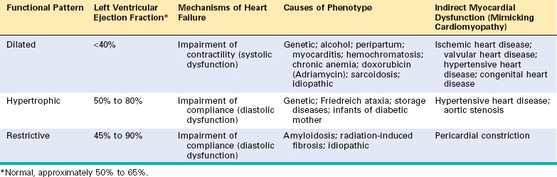
Within the hemodynamic patterns of myocardial dysfunction, there is a spectrum of clinical severity, and overlap of clinical features often occurs between groups. Moreover, each of these patterns can be either idiopathic or due to one of numerous specific identifiable causes (Table 12-11).
TABLE 12-11 Conditions Associated with Heart Muscle Diseases
CARDIAC INFECTIONS |
TOXINS |
METABOLIC |
NEUROMUSCULAR DISEASE |
STORAGE DISORDERS AND OTHER DEPOSITIONS |
INFILTRATIVE |
IMMUNOLOGICAL |
Endomyocardial biopsies are used in the diagnosis and management of individuals with myocardial disease and in cardiac transplant recipients.94,95 Endomyocardial biopsy involves inserting a device (called a bioptome) transvenously into the right side of the heart and using its jaws to snip a small piece of septal myocardium, which is then analyzed by a pathologist.
The term dilated cardiomyopathy (DCM) is applied to a form of cardiomyopathy characterized by progressive cardiac dilation and contractile (systolic) dysfunction, usually with concomitant hypertrophy. It is sometimes called congestive cardiomyopathy.
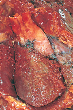
Morphology. In DCM the heart is usually enlarged, heavy (often weighing two to three times normal), and flabby, due to dilation of all chambers (Fig. 12-31). Mural thrombi are common and may be a source of thromboemboli. There are no primary valvular alterations, and mitral (or tricuspid) regurgitation, when present, results from left (or right) ventricular chamber dilation (functional regurgitation). Either the coronary arteries are free of significant narrowing or the obstructions present are insufficient to explain the degree of cardiac dysfunction.
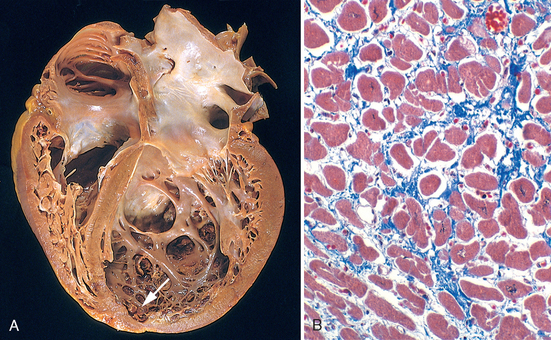
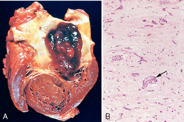
FIGURE 12-31 Dilated cardiomyo-pathy. A, Four-chamber dilatation and hypertrophy are evident. There is granular mural thrombus (arrow) at the apex of the left ventricle (on the right in this apical four-chamber view). The coronary arteries were patent. B, Histologic section demonstrating variable myocyte hypertrophy and interstitial fibrosis (collagen is highlighted as blue in this Masson trichrome stain).
The histologic abnormalities in DCM are nonspecific and usually do not point to a specific etiologic agent. Moreover, the severity of morphologic changes may not reflect either the degree of dysfunction or the patient’s prognosis. Most muscle cells are hypertrophied with enlarged nuclei, but some are attenuated, stretched, and irregular. Interstitial and endocardial fibrosis of variable degree is present, and small subendocardial scars may replace individual cells or groups of cells, probably reflecting healing of previous ischemic necrosis of myocytes caused by hypertrophy-induced imbalance between perfusion and demand.
Although many individuals with DCM have a familial (genetic) form, DCM can also result from various acquired myocardial insults or interactions of genetics and the environment that ultimately may yield a similar clinicopathologic pattern.96 These include (1) myocarditis (an inflammatory disorder that precedes the development of cardiomyopathy in at least some cases, and is sometimes caused by viral infections), (2) toxicities (including adverse effects of chemotherapeutic agents and chronic alcoholism, a history of which can be elicited in 10% to 20% of patients), and (3) childbirth. Each of these subgroups are summarized in Figure 12-32 and described below.
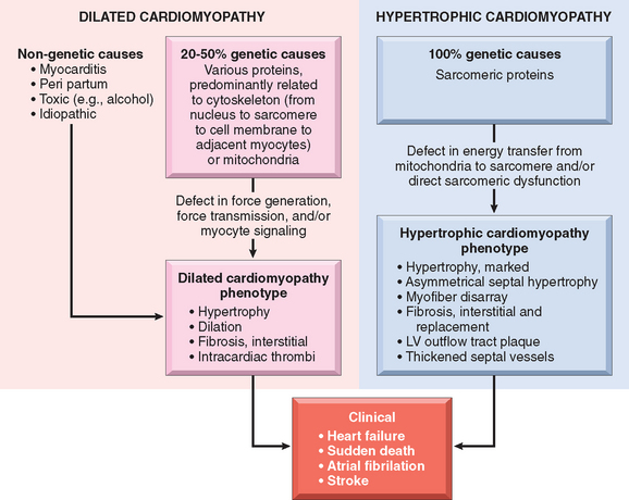
FIGURE 12-32 Causes and consequences of dilated and hypertrophic cardiomyopathy. Some dilated cardiomyopathies and virtually all hypertrophic cardiomyopathies are genetic in origin. The genetic causes of dilated cardiomyopathy involve mutations in any of a wide variety of proteins, predominantly of the cytoskeleton, but also the sarcomere, mitochondria, and nuclear envelope. In contrast, the mutated genes that cause hypertrophic cardiomyopathy encode proteins of the sarcomere. Although these two forms of cardiomyopathy differ greatly in subcellular basis and morphologic phenotypes, they share a common set of clinical complications. LV, left ventricle.
DCM may occur at any age, including in childhood, but it most commonly affects individuals between the ages of 20 and 50. It presents with slowly progressive signs and symptoms of CHF such as shortness of breath, easy fatigability, and poor exertional capacity. In the end stage, patients often have ejection fractions of less than 25% (normal, ∼50% to 65%). Fifty percent of patients die within 2 years, and only 25% survive longer than 5 years, but some severely affected patients may unexpectedly improve on therapy. Secondary mitral regurgitation and abnormal cardiac rhythms are common. Death is usually attributable to progressive cardiac failure or arrhythmia and can occur suddenly. Embolism from dislodgment of an intracardiac thrombus can occur. Cardiac transplantation is frequently done, and long-term ventricular assist may be beneficial in some patients. Interestingly, mechanical cardiac assist may induce lasting regression of cardiac dysfunction in some patients.104
Arrhythmogenic right ventricular cardiomyopathy (ARVC), or arrhythmogenic right ventricular dysplasia, is an inherited disease of the cardiac muscle that causes right ventricular failure and various rhythm disturbances, particularly ventricular tachycardia or fibrillation that can lead to sudden death, primarily in young people.105 Left-sided involvement with left-sided heart failure may also occur. Morphologically, the right ventricular wall is severely thinned because of loss of myocytes, with extensive fatty infiltration and fibrosis (Fig. 12-34). The condition appears to have autosomal-dominant inheritance and variable penetrance. The disease seems to be related to defective cell adhesion proteins in the desmosomes that link adjacent cardiac myocytes. Naxos syndrome is a disorder characterized by arrhythmogenic right ventricular cardiomyopathy and hyperkeratosis of plantar palmar skin surfaces that is associated with mutations in the gene encoding plakoglobin.106
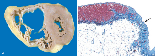
FIGURE 12-34 Arrhythmogenic right ventricular cardiomyopathy. A, Gross photograph, showing dilation of the right ventricle and near-transmural replacement of the right ventricular free-wall by fat and fibrosis. The left ventricle has a virtually normal configuration. B, Histologic section of the right ventricular free wall, demonstrating replacement of myocardium (red) by fibrosis (blue, arrow) and fat (Masson trichrome stain).
Hypertrophic cardiomyopathy (HCM) is characterized by myocardial hypertrophy, poorly compliant left ventricular myocardium leading to abnormal diastolic filling, and in about one third of cases, intermittent ventricular outflow obstruction. It is the leading cause of left ventricular hypertrophy unexplained by other clinical or pathologic causes.107 As discussed below, HCM is caused by mutations in genes encoding sarcomeric proteins. Since the incidence of unexplained cardiac hypertrophy is approximately 1 in 500, HCM may be the most common cardiovascular disorder caused by single gene mutations. The heart is thick-walled, heavy, and hypercontracting, in striking contrast to the flabby, hypocontracting heart of DCM. HCM causes primarily diastolic dysfunction; systolic function is usually preserved. The two most common diseases that must be distinguished clinically from HCM are deposition diseases of the heart (e.g., amyloidosis, Fabry’s disease) and hypertensive heart disease coupled with age-related subaortic septal hypertrophy (see “Hypertensive Heart Disease”). Occasionally, valvular or congenital subvalvular aortic stenosis can also mimic HCM.
Morphology. The essential feature of HCM is massive myocardial hypertrophy, usually without ventricular dilation (Fig. 12-35). The classic pattern is disproportionate thickening of the ventricular septum as compared with the free wall of the left ventricle (with a ratio greater than 1 : 3), frequently termed asymmetric septal hypertrophy. In about 10% of cases, however, the hypertrophy is symmetrical throughout the heart. On cross-section, the ventricular cavity loses its usual round-to-ovoid shape and may be compressed into a “banana-like” configuration by bulging of the ventricular septum into the lumen (Fig. 12-35A). Although marked hypertrophy can involve the entire septum, it is usually most prominent in the subaortic region. Often present are endocardial thickening or mural plaque formation in the left ventricular outflow tract and thickening of the anterior mitral leaflet. Both findings are a result of contact of the anterior mitral leaflet with the septum during ventricular systole, and they correlate with echocardiographically demonstrated functional left ventricular outflow tract obstruction during midsystole.
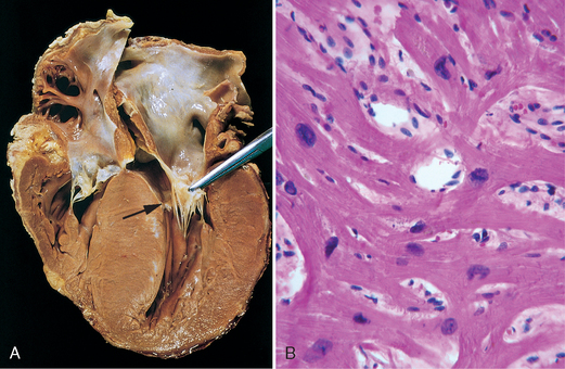
FIGURE 12-35 Hypertrophic cardiomyopathy with asymmetric septal hypertrophy. A, The septal muscle bulges into the left ventricular outflow tract, and the left atrium is enlarged. The anterior mitral leaflet has been moved away from the septum to reveal a fibrous endocardial plaque (arrow) (see text). B, Histologic appearance demonstrating disarray, extreme hypertrophy, and branching of myocytes as well as the characteristic interstitial fibrosis (collagen is blue in this Masson trichrome stain).
The most important histologic features of the myocardium in HCM are (1) extensive myocyte hypertrophy to a degree unusual in other conditions, with transverse myocyte diameters frequently greater than 40 μm (normal, ∼15 μm); (2) haphazard disarray of bundles of myocytes, individual myocytes, and contractile elements in sarcomeres within cells (termed myofiber disarray); and (3) interstitial and replacement fibrosis (Fig. 12-35B).
HCM is caused by mutations in any one of several genes that encode sarcomeric proteins.108 In most cases the pattern of transmission is autosomal dominant with variable penetrance. Remaining cases seem to be sporadic. More than 400 different mutations have been found in nine different genes in HCM, most being missense mutations. Mutations causing HCM are found most commonly in the gene encoding β-myosin heavy chain (β-MHC), with the genes for cardiac TnT, α-tropomyosin, and myosin-binding protein C (MYBP-C) being the next most frequently mutated. Mutations in β-MHC, MYBP-C, and TnT account for 70% to 80% of all cases of HCM. Different affected families may have distinct mutations involving the same protein. For example, approximately 50 different mutations of β-MHC are known to cause HCM. The prognosis of HCM varies widely and correlates strongly with specific mutations. Although it is clear that these genetic defects are critical to the etiology of HCM, the sequence of events leading from mutations to disease is still poorly understood. To make matters even more complicated, changes in certain genes, depicted in Figure 12-33, can give rise to both HCM and DCM.
The basic physiologic abnormality in HCM is reduced stroke volume due to impaired diastolic filling, which results from the reduced chamber size and compliance of the massively hypertrophied left ventricle. In addition, approximately 25% of patients with HCM have dynamic obstruction to the left ventricular outflow. The limitation of cardiac output and a secondary increase in pulmonary venous pressure cause exertional dyspnea. Auscultation discloses a harsh systolic ejection murmur, caused by ventricular outflow obstruction as the anterior mitral leaflet moves toward the ventricular septum during systole. Because of the massive hypertrophy, high left ventricular chamber pressure, and frequently abnormal intramural arteries, focal myocardial ischemia commonly results, even in the absence of concomitant coronary artery disease; thus, anginal pain is frequent. The major clinical problems in HCM are atrial fibrillation, mural thrombus formation leading to embolization and possible stroke, intractable cardiac failure, ventricular arrhythmias, and, not infrequently, sudden death, especially in some affected families. Indeed, HCM is one of the most common causes of sudden, otherwise unexplained death in young athletes.109
The natural history of HCM is highly variable. Most patients can be helped by treatment with drugs that decrease heart rate and contractility, such as β-adrenergic blockers. Some benefit may also be gained from reduction of the mass of the septum, which relieves the outflow tract obstruction. This can be achieved either by surgical excision of muscle or carefully controlled septal infarction, which is induced by infusion of alcohol through a catheter.
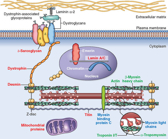
As discussed above, HCM is a disease caused by mutations in proteins of the sarcomere, and DCM is mostly associated with abnormalities of cytoskeletal proteins (Fig. 12-33). DCM seems to be a disease of abnormal force generation, force transmission, or myocyte signaling. In the past, HCM has been considered a disorder of sarcomeres that impairs cardiac function and induces a compensatory hypertrophic response. However, recent evidence suggests that HCM may stem from a defect in energy transfer from its source of generation (mitochondria) to its site of use (sarcomeres), leading to subcellular energy deficiency. Despite these etiologic differences, there are some common mechanistic and clinicopathologic threads between HCM and the genetic and acquired forms of DCM, as summarized in Figure 12-32.
Restrictive cardiomyopathy is a disorder characterized by a primary decrease in ventricular compliance, resulting in impaired ventricular filling during diastole. Because the contractile (systolic) function of the left ventricle is usually unaffected, the functional abnormality can be confused with that of constrictive pericarditis or HCM.110 Restrictive cardiomyopathy may be idiopathic or associated with distinct diseases or processes that affect the myocardium, principally radiation fibrosis, amyloidosis, sarcoidosis, metastatic tumors, or the deposition of metabolites that accumulate due to inborn errors of metabolism.
Morphology. The ventricles are of approximately normal size or slightly enlarged, the cavities are not dilated, and the myocardium is firm and noncompliant. Biatrial dilation is commonly observed. Microscopically, there may be only patchy or diffuse interstitial fibrosis, which can vary from minimal to extensive. However, endomyocardial biopsy will often reveal a specific etiology. An important specific subgroup is amyloidosis (described later).
Several other restrictive conditions require brief mention. Endomyocardial fibrosis is principally a disease of children and young adults in Africa and other tropical areas, characterized by fibrosis of the ventricular endocardium and subendocardium that extends from the apex upward, often involving the tricuspid and mitral valves. The fibrous tissue markedly diminishes the volume and compliance of affected chambers and so induces a restrictive functional defect. Ventricular mural thrombi sometimes develop, and indeed there is a suggestion that the fibrous tissue results from the organization of mural thrombi. The etiology is unknown.
Loeffler endomyocarditis also results in endomyocardial fibrosis, typically with large mural thrombi. It is similar in morphology to the tropical disease, but in addition to the cardiac changes, there is often a peripheral eosinophilia (i.e., elevated blood eosinophils) and eosinophilic infiltrates in other organs. The release of toxic products of eosinophils, especially major basic protein, is postulated to initiate endomyocardial necrosis, followed by scarring of the necrotic area, layering of the endocardium by thrombus, and finally organization of the thrombus. It is now recognized that some patients with Loeffler endomyocarditis have a myeloproliferative disorder associated with chromosomal rearrangements involving either the PDGFRα or PDGFRβ genes (Chapter 13). These rearrangements produce fusion genes that encode constitutively active PDGFR tyrosine kinases. Treatment of such patients with the tyrosine kinase inhibitor imatinib has resulted in hematologic remissions associated with reversal of the endomyocarditis, which otherwise is often rapidly fatal.
Endocardial fibroelastosis is an uncommon heart disease of obscure etiology characterized by focal or diffuse fibroelastic thickening usually involving the mural left ventricular endocardium. It is most common in the first 2 years of life, and is accompanied by aortic valve obstruction or other congenital cardiac anomalies in about one third of cases. Diffuse involvement may be responsible for rapid and progressive cardiac decompensation and death.
Under the designation myocarditis are a diverse group of pathologic entities in which infectious microorganisms and/or an inflammatory process cause myocardial injury.111 These disorders must be distinguished from conditions, such as ischemic heart disease, in which injuries caused by other mechanisms lead to inflammation secondarily.
In the United States, viral infections are the most common cause of myocarditis. Coxsackieviruses A and B and other enteroviruses probably account for most of the cases. Other less common etiologic agents include cytomegalovirus, HIV, and a host of other agents (listed in Table 12-12). The responsible virus can sometimes be identified by serologic studies or, more recently, identification of viral nucleic acids (DNA or RNA) in myocardial biopsies. Whether viruses cause the myocardial injury directly or initiate a destructive immune response is unclear.
TABLE 12-12 Major Causes of Myocarditis
INFECTIONS |
IMMUNE-MEDIATED REACTIONS |
UNKNOWN |
HIV, human immunodeficiency virus.
Nonviral agents are also important causes of infectious myocarditis, particularly the protozoa Trypanosoma cruzi, the agent of Chagas disease.112 Chagas disease is endemic in some regions of South America; myocardial involvement is present in most infected individuals. About 10% of patients die during an acute attack; others develop a chronic immune-mediated myocarditis that may progress to cardiac insufficiency 10 to 20 years later. Trichinosis is the most common helminthic disease associated with myocarditis. Parasitic diseases, including toxoplasmosis, and bacterial infections, including Lyme disease and diphtheria, can also cause myocarditis. In the case of diphtheritic myocarditis, toxins released by Corynebacterium diphtheriae seem to be responsible for the myocardial injury. Myocarditis occurs in approximately 5% of patients with Lyme disease, a systemic illness caused by the bacterial spirochete Borrelia burgdorferi (Chapter 8). Lyme myocarditis manifests primarily as a self-limited conduction system disorder that frequently requires a temporary pacemaker. Myocarditis occurs in many patients with AIDS. Two types have been identified: (1) inflammation and myocyte damage without a clear etiologic agent and (2) myocarditis caused by HIV directly or by an opportunistic pathogen.
There are also noninfectious causes of myocarditis. Myocarditis can be caused by hypersensitivity reactions (hypersensitivity myocarditis), often to drugs such as antibiotics, diuretics, or antihypertensive agents. Myocarditis can also be associated with systemic diseases of immune origin, such as rheumatic fever, systemic lupus erythematosus, and polymyositis. Cardiac sarcoidosis and rejection of a transplanted heart (see Figure 12-40) are also considered forms of myocarditis.
FIGURE 12-40 Complications of heart transplantation. A, Cardiac allograft rejection typified by lymphocytic infiltrate, with associated damage to cardiac myocytes. B, Graft coronary arteriosclerosis, demonstrating severe diffuse concentric intimal thickening producing critical stenosis. The internal elastic lamina (arrow) and media are intact (Movat pentachrome stain, elastin black).
(B, Reproduced by permission from Salomon RN et al.: Human coronary transplantation-associated arteriosclerosis. Evidence for chronic immune reaction to activated graft endothelial cells. Am J Pathol 138:791, 1991.)
Morphology. During the active phase of myocarditis the heart may appear normal or dilated; some hypertrophy may be present depending on disease duration. In advanced stages the ventricular myocardium is flabby and often mottled by either pale foci or minute hemorrhagic lesions. Mural thrombi may be present in any chamber.
During active disease, myocarditis is most frequently characterized by an interstitial inflammatory infiltrate associated with focal myocyte necrosis (Fig. 12-36). A diffuse, mononuclear, predominantly lymphocytic infiltrate is most common (Fig. 12-36A). Although endomyocardial biopsies are diagnostic in some cases, they can be spuriously negative because inflammatory involvement of the myocardium may be focal or patchy. If the patient survives the acute phase of myocarditis, the inflammatory lesions either resolve, leaving no residual changes, or heal by progressive fibrosis.
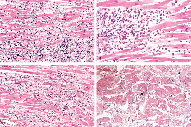
FIGURE 12-36 Myocarditis. A, Lymphocytic myocarditis, associated with myocyte injury. B, Hypersensitivity myocarditis, characterized by interstitial inflammatory infiltrate composed largely of eosinophils and mononuclear inflammatory cells, predominantly localized to perivascular and expanded interstitial spaces. C, Giant-cell myocarditis, with mononuclear inflammatory infiltrate containing lymphocytes and macrophages, extensive loss of muscle, and multinucleated giant cells. D, The myocarditis of Chagas disease. A myofiber distended with trypanosomes (arrow) is present along with inflammation and necrosis of individual myofibers.
Hypersensitivity myocarditis has interstitial infiltrates, principally perivascular, composed of lymphocytes, macrophages, and a high proportion of eosinophils (Fig. 12-36B). A morphologically distinctive form of myocarditis of uncertain cause, called giant-cell myocarditis, is characterized by a widespread inflammatory cellular infiltrate containing multinucleate giant cells interspersed with lymphocytes, eosinophils, plasma cells, and macrophages. Focal to frequently extensive necrosis is present (see Fig. 12-36C). This variant carries a poor prognosis.
The myocarditis of Chagas disease is rendered distinctive by parasitization of scattered myofibers by trypanosomes accompanied by an inflammatory infiltrate of neutrophils, lymphocytes, macrophages, and occasional eosinophils (see Fig. 12-36D).
The clinical spectrum of myocarditis is broad. At one end the disease is entirely asymptomatic, and such patients recover completely without sequelae; at the other extreme is the precipitous onset of heart failure or arrhythmias, occasionally with sudden death. Between these extremes are the many levels of involvement associated with symptoms such as fatigue, dyspnea, palpitations, precordial discomfort, and fever. The clinical features of myocarditis can mimic those of acute MI. Occasionally, patients develop dilated cardiomyopathy as a late complication of myocarditis.
Cardiac complications of cancer therapy are an important clinical problem.113 Cardiotoxicity can be associated with conventional chemotherapeutic agents, targeted drugs such as tyrosine kinase inhibitors, and certain forms of immunotherapy.114,115 The anthracyclines doxorubicin and daunorubicin are the chemotherapeutic agents that are most often associated with toxic myocardial injury, which often leads to a dilated cardiomyopathy and heart failure. Anthracycline toxicity is dose-dependent (cardiotoxicity becomes progressively more frequent above a total dose of 500 mg/m2) and is attributed primarily to peroxidation of lipids in myocyte membranes.
Many other pharmaceuticals and other agents, such as lithium, phenothiazines, chloroquine, and cocaine, have been implicated in myocardial injury and sometimes sudden death. Common findings in hearts injured by many of these chemicals and drugs (including diphtheria exotoxin and doxorubicin) are myofiber swelling, cytoplasmic vacuolization, and fatty change. With discontinuance of the toxic agent these changes may resolve completely, leaving no apparent sequelae. Sometimes, however, more extensive damage produces myocyte necrosis, which can evolve to a dilated cardiomyopathy.
Foci of myocardial necrosis with contraction bands, often associated with a sparse mononuclear inflammatory infiltrate consisting mostly of macrophages, can occur in individuals with a pheochromocytoma, a tumor that elaborates catecholamines (Chapter 24). This is considered to be a manifestation of the general problem of “catecholamine effect,” also seen in association with intense autonomic stimulation (secondary to intracranial lesions or stress), or the exogenous administration of large doses of vasopressor agents such as dopamine.116 Sudden, intense emotional or physical stress can also induce acute left ventricular dysfunction due to myocardial stunning, a phenomenon known as Takotsubo cardiomyopathy.117 Cocaine also causes similar catecholamine-mediated damage. The mechanism of catecholamine cardiotoxicity is uncertain, but it seems to relate either to direct toxicity of catecholamines to cardiac myocytes via calcium overload or to focal vasoconstriction in the coronary arterial macro- or microcirculation in the face of an increased heart rate. The mononuclear cell infiltrate is probably a secondary reaction to the foci of myocyte cell death. Similar changes may be encountered in individuals who have recovered from hypotensive episodes or have been resuscitated from a cardiac arrest; in such cases the damage is a result of ischemia-reperfusion (see earlier) with subsequent inflammation.
Amyloidosis is a prototypical myocardial disorder caused by deposition of an abnormal substance in the heart. Amyloidosis (Chapter 6) is caused by insoluble extracellular fibrillar deposits of protein fragments that are prone to forming β-pleated sheets.118 Cardiac amyloidosis may appear along with systemic amyloidosis or be restricted to the heart, particularly in the aged (senile cardiac amyloidosis).119 In senile cardiac amyloidosis, the amyloid deposits generally occur in the ventricles and atria. Senile cardiac amyloidosis occurs in older individuals and is caused by the deposition of transthyretin, a normal serum protein synthesized in the liver that is responsible for transporting thyroxine and retinol-binding protein. Senile cardiac amyloidosis120 has a far better prognosis than systemic amyloidosis. Mutant forms of transthyretin can accelerate cardiac (and associated systemic) amyloidosis. For example, the risk of isolated cardiac amyloidosis is four times greater in African Americans than in Caucasians after 60 years of age because 4% of African Americans have a transthyretin mutation leading to the substitution of isoleucine for valine at position 122. This substitution produces an amyloidogenic form of transthyretin (responsible for autosomal-dominant familial transthyretin amyloidosis). Isolated atrial amyloidosis can also occur secondary to deposition of atrial natriuretic peptide, but its clinical significance is uncertain.
Cardiac amyloidosis most frequently produces a restrictive cardiomyopathy, but it can also be asymptomatic or manifest as dilation, arrhythmias, or symptoms mimicking those of ischemic or valvular disease. These varied presentations depend on the predominant location of the deposits, which can be found in the interstitium, conduction system, vasculature, or valves.
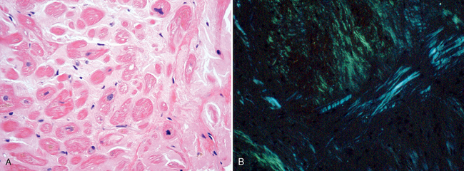
Morphology. The heart varies in consistency from normal to firm and rubbery. Usually the chambers are of normal size, but in some cases they are dilated and have thickened walls. Numerous small, semitranslucent nodules resembling drips of wax may be seen at the atrial endocardial surface, particularly on the left. Eosinophilic deposits of amyloid may be found in the interstitium, conduction tissue, valves, endocardium, pericardium, and small intramural coronary arteries; they can be distinguished from other hyaline deposits by special stains such as Congo red, which produces classic apple-green birefringence when viewed under polarized light (Fig. 12-37). Amyloid deposits often form rings around cardiac myocytes and capillaries. Intramural arteries and arterioles may have sufficient amyloid in their walls to compress and occlude their lumens, inducing myocardial ischemia (“small-vessel disease”).
Iron overload can result from either hereditary hemochromatosis (Chapter 18) or multiple blood transfusions (hemosiderosis). The heart in each is usually dilated. Iron deposition is more prominent in ventricles than atria and in the myocardium than in the conduction system. It is thought that iron causes systolic dysfunction by interfering with metal-dependent enzyme systems or by inducing oxygen free-radical injury.
Morphology. Grossly, the myocardium of the iron-overloaded heart is rust-brown in color but is usually otherwise indistinguishable from that of idiopathic dilated cardiomyopathy. Microscopically, there is marked accumulation of hemosiderin within cardiac myocytes, particularly in the perinuclear region, demonstrable with a Prussian blue stain. This is associated with varying degrees of cellular degeneration and fibrosis. Ultrastructurally, the cardiac myocytes contain abundant perinuclear siderosomes (ironcontaining lysosomes).
Cardiac manifestations are among the earliest, most consistent features of hyperthyroidism and hypothyroidism, and they reflect direct and indirect effects of thyroid hormones on the cells of the heart.121 In hyperthyroidism (Chapter 24), tachycardia, palpitations, and cardiomegaly are common; supraventricular arrhythmias occasionally appear. Cardiac failure is uncommon; when it occurs in this setting, it is usually superimposed on other cardiac diseases. In hypothyroidism (Chapter 24), cardiac output is decreased, due to reductions in stroke volume and heart rate. Increased peripheral vascular resistance and decreased blood volume result in narrowing of the pulse pressure, prolongation of circulation time, and decreased flow to peripheral tissues.
Morphology. In hyperthyroidism the gross and histologic features are those of nonspecific hypertrophy and can also include ischemic foci. In well-advanced hypothyroidism (myxedema) the heart is flabby, enlarged, and dilated. Histologic features of hypothyroidism include myofiber swelling with loss of striations and basophilic degeneration, accompanied by interstitial mucopolysaccharide-rich edema fluid. A similar fluid sometimes accumulates within the pericardial sac. The term myxedema heart has been applied to these changes.
The most important pericardial disorders cause fluid accumulation, inflammation, fibrous constriction, or some combination of these processes, usually in association with disease elsewhere in the heart or a systemic disease; isolated pericardial disease is unusual.122
Normally, there are about 30 to 50 mL of thin, clear, straw-colored fluid in the pericardial sac. Under various circumstances the parietal pericardium may be distended by serous fluid (pericardial effusion), blood (hemopericardium), or pus (purulent pericarditis). With long-standing pressure or volume overload, the pericardium dilates. This permits a slowly accumulating pericardial effusion to become quite large without interfering with cardiac function. Thus, with chronic effusions of less than 500 mL in volume, the only clinical significance is a characteristic globular enlargement of the heart shadow on chest radiographs. In contrast, rapidly developing fluid collections of as little as 200 to 300 mL—for example, due to hemopericardium caused by a ruptured MI or aortic dissection—may produce compression of the thin-walled atria and venae cavae, or the ventricles themselves. As a consequence, cardiac filling is restricted, producing potentially fatal cardiac tamponade.
Pericardial inflammation may occur secondary to a variety of cardiac diseases, thoracic or systemic disorders, metastases from neoplasms arising in remote sites, or a surgical procedure on the heart. Primary pericarditis is unusual and almost always of viral origin. The major causes of pericarditis are listed in Table 12-13. Most evoke an acute pericarditis, but a few, such as tuberculosis and fungi, produce chronic reactions.
TABLE 12-13 Causes of Pericarditis
INFECTIOUS AGENTS |
PRESUMABLY IMMUNOLOGICALLY MEDIATED |
MISCELLANEOUS |
This is characteristically produced by noninfectious inflammatory diseases, such as rheumatic fever, SLE, and scleroderma, tumors, and uremia. An infection in the tissues contiguous to the pericardium—for example, a bacterial pleuritis—may incite sufficient irritation of the parietal pericardial serosa to cause a sterile serous effusion that may progress to serofibrinous pericarditis and ultimately to a frank suppurative reaction. In some instances a well-defined viral infection elsewhere—upper respiratory tract infection, pneumonia, parotitis—antedates the pericarditis and serves as the primary focus of infection. Infrequently, usually in young adults, a viral pericarditis occurs as an apparent primary infection that may be accompanied by myocarditis (myopericarditis). Histologically, there is a mild inflammatory infiltrate in the epipericardial fat consisting predominantly of lymphocytes. Organization into fibrous adhesions rarely occurs.
These two anatomic forms are the most frequent type of pericarditis and are composed of serous fluid mixed with a fibrinous exudate. Common causes include acute MI (recall Fig. 12-19D), the postinfarction (Dressler) syndrome (probably an autoimmune condition appearing several weeks after an MI), uremia, chest radiation, rheumatic fever, SLE, and trauma. A fibrinous reaction also follows routine cardiac surgery.
Morphology. In fibrinous pericarditis the surface is dry, with a fine granular roughening. In serofibrinous pericarditis a more intense inflammatory process induces the accumulation of larger amounts of yellow to browen turbid fluid, which is made brown and cloudy by the presence of leukocytes and red cells (which may give the fluid a visibly bloody appearance), and often fibrin. As with all inflammatory exudates, fibrin may be lysed with resolution of the exudate, or it may become organized (see Chapter 3).
From the clinical standpoint the development of a loud pericardial friction rub is the most striking characteristic of fibrinous pericarditis, and pain, systemic febrile reactions, and signs suggestive of cardiac failure may be present. However, the collection of serous fluid may sometimes prevent rubbing by separating the two layers of the pericardium.
This form of pericarditis is caused by invasion of the pericardial space by microbes, which may reach the pericardial cavity by several routes: (1) direct extension from neighboring infections, such as an empyema of the pleural cavity, lobar pneumonia, mediastinal infections, or extension of a ring abscess through the myocardium or aortic root; (2) seeding from the blood; (3) lymphatic extension; or (4) direct introduction during cardiotomy. Immunosuppression predisposes to infection by all of these pathways. The exudate ranges from a thin cloudy fluid to frank pus up to 400 to 500 mL in volume. The serosal surfaces are reddened, granular, and coated with the exudate (Fig. 12-38). Microscopically there is an acute inflammatory reaction, which sometimes extends into surrounding structures to induce mediastinopericarditis. Complete resolution is infrequent, and organization by scarring is the usual outcome. The intense inflammatory response and the subsequent scarring frequently produce constrictive pericarditis, a serious consequence (see later).
FIGURE 12-38 Acute suppurative pericarditis arising from direct extension of a pneumonia. Extensive purulent exudate is evident.
The clinical findings in the active phase are essentially the same as those present in fibrinous pericarditis, but signs of systemic infection are usually marked: for example, spiking temperatures, chills, and fever.
In this type of pericarditis the exudate is composed of blood mixed with a fibrinous or suppurative effusion; it is most commonly caused by the spread of a malignant neoplasm to the pericardial space. In such cases, cytologic examination of fluid removed through a pericardial tap often reveals the presence of neoplastic cells. Hemorrhagic pericarditis can also be found in bacterial infections, in persons with an underlying bleeding diathesis, and in tuberculosis. Hemorrhagic pericarditis often follows cardiac surgery and is occasionally responsible for significant blood loss or even tamponade, requiring a “second-look” operation. The clinical significance is similar to that of fibrinous or suppurative pericarditis.
This rare type of pericarditis is, until proved otherwise, tuberculous in origin; infrequently, fungal infections evoke a similar reaction. Pericardial involvement occurs by direct spread from tuberculous foci within the tracheobronchial nodes. Caseous pericarditis is a frequent antecedent of disabling, fibrocalcific, chronic constrictive pericarditis.
In some cases organization merely produces plaquelike fibrous thickenings of the serosal membranes (“soldier’s plaque”) or thin, delicate adhesions of obscure origin that are observed fairly frequently at autopsy and rarely cause impairment of cardiac function. In other cases, fibrosis in the form of delicate, stringy adhesions completely obliterates the pericardial sac. In most instances, this adhesive pericarditis has no effect on cardiac function.
Adhesive mediastinopericarditis may follow infectious pericarditis, previous cardiac surgery, or irradiation to the mediastinum. The pericardial sac is obliterated, and adherence of the external aspect of the parietal layer to surrounding structures produces a great strain on cardiac function. With each systolic contraction, the heart pulls not only against the parietal pericardium but also against the attached surrounding structures. Systolic retraction of the rib cage and diaphragm, pulsus paradoxus, and a variety of other characteristic clinical findings may be observed. The increased workload causes cardiac hypertrophy and dilation, which may be substantial in severe cases.
In constrictive pericarditis the heart is encased in a dense, fibrous or fibrocalcific scar that limits diastolic expansion and cardiac output, features that mimic a restrictive cardiomyopathy. A prior history of pericarditis may or may not be present. The scar, often 0.5 to 1.0 cm thick, obliterates the pericardial space and sometimes calcifies; in extreme cases it can resemble a plaster mold (concretio cordis). Because of the dense enclosing scar, cardiac hypertrophy and dilation cannot occur. Cardiac output may be reduced at rest, but more importantly the heart has little if any capacity to increase its output in response to increased peripheral needs. Heart sounds are understandably distant or muffled. Treatment consists of surgical removal of the shell of constricting fibrous tissue (pericardiectomy).
Paradoxically, the prevalence and importance of cardiovascular manifestations of rheumatologic diseases (including rheumatoid arthritis, SLE, systemic sclerosis, ankylosing spondylitis, and psoriatic arthritis) has increased as a result of the longer life expectancy of persons with these disorders and enhanced detection of milder cases.123 Inflammatory mechanisms may cause vascular, myocardial, valvular, and pericardial manifestations. In addition, ischemic heart disease seems to be accelerated in individuals with inflammatory rheumatic conditions.
Rheumatoid arthritis is mainly a disorder of the joints, but it is also associated with many extra-articular features (e.g., subcutaneous rheumatoid nodules, acute vasculitis, and Felty syndrome; see Chapter 26). The heart is also involved in 20% to 40% of cases of severe prolonged rheumatoid arthritis. The most common finding is a fibrinous pericarditis that may progress to fibrous thickening of the visceral and parietal pericardium and dense adhesions. Granulomatous rheumatoid nodules resembling those that occur subcutaneously may also be identifiable in the myocardium. Much less frequently, rheumatoid nodules involve the endocardium, valves of the heart, and root of the aorta. Rheumatoid valvulitis can lead to marked fibrous thickening and secondary calcification of the aortic valve cusps, producing changes resembling those of chronic rheumatic valvular disease. The valvular lesions associated with SLE were discussed previously under “Valvular Heart Disease.”
Primary tumors of the heart are rare; in contrast, metastatic tumors to the heart occur in about 5% of persons dying of cancer. The most common primary cardiac tumors, in descending order of frequency (overall, including adults and children), are myxomas, fibromas, lipomas, papillary fibroelastomas, rhabdomyomas, angiosarcomas, and other sarcomas. The five most common tumors are all benign and collectively account for 80% to 90% of primary tumors of the heart.
Myxomas are the most common primary tumor of the heart in adults (Fig. 12-39). They are benign neoplasms often associated with clonal abnormalities of chromosomes 12 and 17 that are thought to arise from primitive multipotent mesenchymal cells. Although they may arise in any of the four chambers or, rarely, on the heart valves, about 90% are located in the atria (atrial myxomas), with a left-to-right ratio of approximately 4 : 1.
FIGURE 12-39 Left atrial myxoma. A, Gross photograph showing large pedunculated lesion arising from the region of the fossa ovalis and extending into the mitral valve orifice. B, Microscopic appearance, with abundant amorphous extracellular matrix in which there are scattered collections of myxoma cells in various groupings, including abnormal vessel-like formations (arrow).
Morphology. The tumors are usually single, but rarely several occur simultaneously. The region of the fossa ovalis in the atrial septum is the favored site of origin. Myxomas range in size from small (<1 cm) to large (≥10 cm). They are sessile or pedunculated lesions (Fig. 12-39A) that vary from globular hard masses mottled with hemorrhage to soft, translucent, papillary, or villous lesions having a gelatinous appearance. The pedunculated form is often sufficiently mobile to move into or even through the AV valves during systole, causing intermittent obstruction that may be position-dependent. Sometimes mobile tumors exert a “wrecking-ball” effect, causing damage to the valve leaflets.
Histologically, myxomas are composed of stellate or globular myxoma cells embedded within an abundant acid mucopolysaccharide ground substance (Fig. 12-39B). Peculiar vessel-like or gland-like structures are characteristic. Hemorrhage and mononuclear inflammation are usually present.
The major clinical manifestations are due to valvular “ball-valve” obstruction, embolization, or a syndrome of constitutional symptoms, such as fever and malaise. Sometimes fragmentation and systemic embolization calls attention to these lesions. Constitutional symptoms are probably due to the elaboration by some myxomas of the cytokine IL-6, a major mediator of the acute-phase response. Echocardiography provides the opportunity to identify these masses noninvasively. Surgical removal is usually curative; rarely, the neoplasm recurs months to years later.
Approximately 10% of individuals with myxoma have a familial syndrome (known as Carney complex) characterized by autosomal-dominant transmission, multiple cardiac and often extracardiac (e.g., skin) myxomas, pigmented skin lesions, and endocrine overactivity. A careful history and physical examination in persons with cardiac myxoma is important to identify the extra-cardiac signs of the Carney complex, since this diagnosis carries implications for family members of the patient. The gene PRKAR1 on chromosome 17 (encoding a regulatory subunit of cyclic adenosine monophosphate–dependent protein kinase A, possibly a tumor suppressor gene) is mutated in most Carney complex kindreds; other kindreds have abnormalities in a locus at chromosome 2p16.124
Lipomas are localized, well-circumscribed, benign tumors composed of mature fat cells that may occur in the subendocardium, subepicardium, or myocardium. They may be asymptomatic, or produce ball-valve obstructions or arrhythmias. Lipomas are most often located in the left ventricle, right atrium, or atrial septum and are not necessarily neoplastic. In the atrial septum, non-neoplastic depositions of fat sometimes occur that are called “lipomatous hypertrophy.”
Papillary fibroelastomas are curious, usually incidental, sea-anemone-like lesions, most often identified at autopsy. They may embolize and thereby become clinically important. Clonal cytogenetic abnormalities have been reported, suggesting that fibroelastomas are unusual benign neoplasms. They resemble the much smaller, usually trivial, Lambl excrescences that are frequently found on the aortic valves of older individuals.
Morphology. Papillary fibroelastomas are generally located on valves, particularly the ventricular surfaces of semilunar valves and the atrial surfaces of atrioventricular valves. They consist of a distinctive cluster of hairlike projections up to 1 cm in length, and several centimeters in diameter. Histologically, the projections are composed of a core of myxoid connective tissue containing abundant mucopolysaccharide matrix and elastic fibers that is covered by a surface endothelium.
Rhabdomyomas are the most frequent primary tumor of the heart in infants and children and are frequently discovered in the first years of life because of obstruction of a valvular orifice or cardiac chamber. Cardiac rhabdomyomas are often associated with tuberous sclerosis (see Chapter 28), which is caused by defects in the TSC1 or TSC2 tumor suppressor genes. The TSC1 and TSC2 proteins work together in a complex that inhibits the activity of the mammalian target of rapamycin (mTOR), a kinase that stimulates cell growth and regulates cell size. TSC1 or TSC2 expression is often completely lost in rhadmyosarcomas that occur in the setting of tuberous sclerosis, providing a mechanism for the myocyte overgrowth. Because they often regress spontaneously, rhabdomyomas are considered by some to be hamartomas rather than true neoplasms.
Morphology. Rhabdomyomas are generally small, gray-white myocardial masses up to several centimeters in diameter. They are usually multiple in number and involve the ventricles preferentially, often protruding into the chambers. Histologically they are composed of bizarre, markedly enlarged myocytes. In sections the abundant myocyte cytoplasm is often reduced to thin webs or strands that extend to cell membranes, an appearance referred to as spider cells.
Cardiac angiosarcomas and other sarcomas are not clinically or morphologically distinctive from their counterparts in other locations (see Fig. 11-35 and Chapter 26) and so require no further comment here.
With enhanced patient survival due to diagnostic and therapeutic advances, significant cardiovascular effects of noncardiac neoplasms and their therapy are now commonly encountered (Table 12-14). Pathology derives from infiltration by tumor, circulating mediators, and complications of therapy.
TABLE 12-14 Cardiovascular Effects of Noncardiac Neoplasms
DIRECT CONSEQUENCES OF TUMOR |
INDIRECT CONSEQUENCES OF TUMOR (COMPLICATIONS OF CIRCULATING MEDIATORS) |
EFFECTS OF TUMOR THERAPY |
Modified from Schoen FJ, et al: Cardiac effects of non-cardiac neoplasms. Cardiol Clin 2:657, 1984.
The most frequent metastatic tumors involving the heart are carcinomas of the lung and breast, melanomas, leukemias, and lymphomas. Metastases can reach the heart and pericardium by retrograde lymphatic extension (most carcinomas), by hematogenous seeding (many tumors), by direct contiguous extension (primary carcinoma of the lung, breast, or esophagus), or by venous extension (tumors of the kidney or liver). Clinical symptoms are most often associated with pericardial spread, which can cause symptomatic pericardial effusions or a mass-effect that is sufficient to restrict cardiac filling. Myocardial metastases are usually clinically silent or have nonspecific features, such as a generalized defect in ventricular contractility or compliance. Bronchogenic carcinoma or malignant lymphoma may infiltrate the mediastinum extensively, causing encasement, compression, or invasion of the superior vena cava with resultant obstruction to blood coming from the head and upper extremities (superior vena cava syndrome). Renal cell carcinoma often invades the renal vein, and may grow as a frond of tissue up the inferior vena cava and into the right atrium, blocking venous return to the heart.
Noncardiac tumors may also affect cardiac function indirectly, sometimes via circulating tumor-derived substances (e.g., nonbacterial thrombotic endocarditis, carcinoid heart disease, pheochromocytoma-associated myocardial damage, multiple myeloma–associated AL amyloidosis; all described earlier).
Complications of chemotherapy were discussed earlier in this chapter. Radiation used to treat breast, lung, or mediastinal neoplasms can cause pericarditis, pericardial effusion, myocardial fibrosis, and chronic pericardial disorders. Other cardiac effects of radiation therapy include accelerated coronary artery disease and mural and valvular endocardial fibrosis.
Transplantation of cardiac allografts is now frequently performed (∼3000 per year worldwide) for severe, intractable heart failure of diverse causes, the two most common being dilated cardiomyopathy and ischemic heart disease. Three major factors have contributed to the improved outcome of cardiac transplantation since the first human to human transplant in 1967: (1) more effective immunosuppressive therapy (including the use of cyclosporin A, glucocorticoids, and other agents), (2) careful selection of candidates, and (3) early histopathologic diagnosis of acute allograft rejection by endomyocardial biopsy.125
Of the major complications, allograft rejection is the primary problem requiring surveillance; scheduled endomyocardial biopsy is the only reliable means of diagnosing acute cardiac rejection before substantial myocardial damage has occurred and at a stage that is reversible in the majority of instances. Rejection is characterized by interstitial lymphocytic inflammation that, in its more advanced stages, damages adjacent myocytes; the histology resembles myocarditis (Fig. 12-40A). When myocardial injury is not extensive, the “rejection episode” is usually either self-limited or successfully reversed by increased immunosuppressive therapy. Advanced rejection may be irreversible and fatal if it is not promptly treated.
The major current limitation to the long-term success of cardiac transplantation is diffuse stenosing intimal proliferation of the coronary arteries, which may involve intramural vessels extensively (graft arteriopathy) (Fig. 12-40B). Because the transplanted heart is often denervated, patients with this disorder may not experience ischemic chest pain, and this vexing problem may lead to silent MI; in severe graft arteriopathy, CHF or sudden death is the usual outcome. The pathogenesis of these arterial lesions is uncertain. Low-level, chronic rejection reactions may induce inflammatory cells and vascular wall cells to secrete growth factors that promote the recruitment and proliferation of intimal smooth muscle cells and the synthesis of extracellular matrix, thereby expanding the intima (Chapter 6). Other postoperative problems include infection and malignancies, particularly Epstein-Barr virus-associated B cell lymphomas that arise in the setting of T-cell immunosuppression. Despite these problems, the overall outlook is good; the 1-year survival is 70% to 80% and 5-year survival is greater than 60%.
1 Gaziano TA. Cardiovascular disease in the developing world and its cost-effective management. Circulation. 2005;112:3547.
2 Beaglehole R, et al. Poverty and human development. The global implications of cardiovascular disease. Circulation. 2007;116:1871.
3 Heart Association Statistics Committee, Stroke Statistics Subcommittee. Heart Disease and Stroke Statistics—2007 Update: a report from the American Heart Association Statistics Committee and Stroke Statistics Subcommittee. Circulation. 2007;106:69.
4 Lee CY, Burnett JCJr. Natriuretic peptides and therapeutic applications. Heart Fail Rev. 2007;12:131.
5 Saffitz JE. Adhesion molecules: why they are important to the electrophysiologist. J Cardiovasc Electrophysiol. 2006;17:225.
6 Liu AC, et al. The emerging role of valve interstitial cell phenotypes in regulating heart valve pathobiology. Am J Pathol. 2007;171:1407.
7 Aikawa E, et al. Human semilunar cardiac valve remodeling by activated cells from fetus to adult: implications for postnatal adaptation, pathology, and tissue engineering. Circulation. 2006;113:1344.
8 Braunwald E, et al, editors. Braunwald’s Heart Disease: A Textbook of Cardiovascular Medicine. Philadelphia: WB Saunders, 2008.
9 Silver MD, et al, editors. Cardiovascular Pathology. 3rd ed. New York: Churchill Livingstone; 2001:808.
10 Chien KR, Karsenty G. Longevity and lineages: toward the integrative biology of degenerative diseases in heart, muscle, and bone. Cell. 2005;120:533.
11 Opie LH, et al. Controversies in ventricular remodeling. Lancet. 2006;367:356.
12 Chinnaiyan KM, et al. Integrated diagnosis and management of diastolic heart failure. Am Heart J. 2007;153:189.
13 Oka T, et al. Re-employment of developmental transcription factors in adult heart disease. Semin Cell Develop Biol. 2007;18:117.
14 Diwan A, Dorn GW. Decompensation of cardiac hypertrophy: cellular mechanisms and novel therapeutic targets. Physiology. 2007;22:56.
15 Neubauer S. The failing heart—an engine out of fuel. N Engl J Med. 2007;356:1140.
16 Ashrafian H, et al. Metabolic mechanisms in heart failure. Circulation. 2007;116:434.
17 Thum T, et al. MicroRNAs in the human heart. A clue to fetal gene reprogramming in heart failure. Circulation. 2007;116:258.
18 Van Rooij E. MicroRNAs: powerful new regulators of heart disease and provocative therapeutic targets. J Clin Invest. 2007;117:2369.
19 Rosanio S, et al. Sudden death prophylaxis in heart failure. Int J Cardiol. 2007;119:291.
20 Lip GY, Tse HF. Management of atrial fibrillation. Lancet. 2007;370:604.
21 deGoma EM, et al. Emerging therapies for the management of decompensated heart failure. J Am Coll Cardiol. 2006;48:2397.
22 Anversa P, et al. Cardiac regeneration. J Am Coll Cardiol. 2006;47:1769.
23 Angelini P. Coronary artery anomalies. An entity in search of an identity. Circulation. 2007;115:1296.
24 Hoffman JIE, Kaplan S. The incidence of congenital heart disease. J Am Coll Cardiol. 2002;39:1890.
25 Williams RG, et al. Report of the National Heart, Lung, and Blood Institute Working Group on Research in Adult Congenital Disease. J Am Coll Cardiol. 2006;47:701.
26 Warnes CA. The adult with congenital heart disease: born to be bad? J Am Coll Cardiol. 2005;46:1.
27 Schoen FJ, Edwards WD. Pathology of cardiovascular interventions, including endovascular therapies, revascularization, vascular replacement, cardiac assist/replacement, arrhythmia control and repaired congenital heart disease. In: Silva MD, et al, editors. Cardiovascular Pathology. 3rd ed. Philadelphia: WB Saunders; 2001:678.
28 Drenthen W, et al. Outcome of pregnancy in women with congenital heart disease: a literature review. J Am Coll Cardiol. 2007;49:2303.
29 Srivastava D. Making or breaking the heart: from lineage determination to morphogenesis. Cell. 2006;126:1037.
30 Bruneau BG. The developmental genetics of congenital heart disease. Nature. 2008;451:943.
31 Pierpont ME, et al. Genetic basis for congenital heart defects: current knowledge. A scientific statement from the American Heart Association Council on Cardiovascular Disease in the Young. Circulation. 2007;115:3015.
32 Marino B. Congenital heart disease in patients with Down’s syndrome: anatomic and genetic aspects. Biomed Pharmacother. 1993;47:197.
33 Jenkins KJ, et al. Noninherited risk factors and congenital cardiovascular defects: current knowledge. A scientific statement from the American Heart Association Council on Cardiovascular Disease in the Young. Circulation. 2007;115:2995.
34 Huhta JC, Hernandez-Robles JA. Homocysteine, folate, and congenital heart defects. Fetal Pediatr Pathol. 2005;24:71.
35 Webb G, Gatzoulis MA. Atrial septal defects in the adult. Recent progress and overview. Circulation. 2006;114:1645.
36 Hein R, et al. Atrial and ventricular septal defects can safely be closed by percutaneous intervention. J Interv Cardiol. 2005;18:515.
37 Hara H, et al. Patent foramen ovale: current pathology, pathophysiology, and clinical status. J Am Coll Cardiol. 2005;46:1768.
38 Homma S, Sacco RL. Patent foramen ovale and stroke. Circulation. 2005;112:1063.
39 Minette MS, Sahn DJ. Ventricular septal defects. Circulation. 2006;114:2190.
40 Schneider DJ, Moore JW. Patent ductus arteriosus. Circulation. 2006;114:1873.
41 Warnes CA. Transposition of the great arteries. Circulation. 2006;114:2699.
42 Aboulhosn J, Child JS. Left ventricular outflow obstruction. Subaortic stenosis, bicuspid aortic valve, supravalvar aortic stenosis, and coarctation of the aorta. Circulation. 2006;114:2412.
43 Lashkari A, et al. Williams-Beuren syndrome: an update and review for the primary physician. Clin Pediatr. 1999;38:189.
44 Topol EJ, et al. Genetic susceptibility to myocardial infarction and coronary artery disease. Hum Mol Gen. 2006;15:R117.
45 Regieli JJ, et al. Coronary collaterals—insights in molecular determinants and prognostic relevance. Int J Cardiol. 2007;116:139.
46 Fuster V, et al. Atherothrombosis and high-risk plaque: Part I: evolving concepts. J Am Coll Cardiol. 2005;46:937.
47 Falk E, et al. Coronary plaque disruption. Circulation. 1995;92:657.
48 Cohn PF. Silent myocardial ischemia: recent developments. Curr Atheroscler Rep. 2005;7:155.
49 Mendelsohn ME, Karas RH. Molecular and cellular basis of cardiovascular gender differences. Science. 2005;308:1583.
50 Wenger NK. Menopausal hormone therapy: is there evidence for cardiac protection? Int Urol Nephrol. 2004;36:617.
51 Boden WE, et al. Reperfusion strategies in acute ST-segment elevation myocardial infarction. J Am Coll Cardiol. 2007;50:917.
52 Yellon DM, Hausenloy DJ. Myocardial reperfusion injury. N Engl J Med. 2007;357:1121.
53 Kloner RA, Jennings RB. Consequences of brief ischemia: stunning, preconditioning, and their clinical implications: Part 1 and Part 2. Circulation. 2001;104:2981. 3158,
54 Heusch G, et al. Myocardial hibernation: a delicate balance. Am J Physiol Heart Circ Physiol. 2005;288:H984.
55 Eisen A, et al. Ischemic preconditioning: nearly two decades of research. A comprehensive review. Atherosclerosis. 2004;172:201.
56 Jaffe AS, et al. Biomarkers in acute cardiac disease: the present and the future. J Am Coll Cardiol. 2006;48:1.
57 Huikuri HV, et al. Sudden death due to cardiac arrhythmias. N Engl J Med. 2001;345:1473.
58 Farb A, et al. Sudden cardiac death. Frequency of active coronary lesions, inactive coronary lesions, and myocardial infarction. Circulation. 1995;92:1701.
59 Liberthson RR, et al. Prehospital ventricular defibrillation. Prognosis and followup course. N Engl J Med. 1974;291:317.
60 Roberts R. Genomics and cardiac arrhythmias. J Am Coll Cardiol. 2006;47:9.
61 Sarkozy A, Brugada P. Sudden cardiac death and inherited arrhythmia syndromes. J Cardiovasc Electrophysiol. 2005;16:S8.
62 Lehnart SE, et al. Inherited arrhythmias. A National Heart, Lung, and Blood Institute and Office of Rare Diseases Workshop consensus report about the diagnosis, phenotyping, molecular mechanisms, and therapeutic approaches for primary cardiomyopathies of gene mutations affecting ion channel function. Circulation. 2007;116:2325.
63 Passman R, Kadish A. Sudden death prevention with implantable devices. Circulation. 2007;16:561.
64 Voelkel NF, et al. Right ventricular function and failure. Report of a National Heart, Lung, and Blood Institute Working Group on Cellular and Molecular Mechanisms of Right Heart Failure. Circulation. 2006;114:1883.
65 Weitzenblum E. Chronic cor pulmonale. Heart. 2003;89:225.
66 Levine RA, Schwammenthal E. Ischemic mitral regurgitation on the threshold of a solution. From paradoxes to unifying concepts. Circulation. 2005;112:745.
67 Elkayam U, Bitar F. Valvular heart disease and pregnancy. Part I: Native valves. J Am Coll Cardiol. 2005;46:223.
68 Goldbarg SH, et al. Insights into degenerative aortic valve disease. J Am Coll Cardiol. 2007;50:1205.
69 Freeman RV, Otto CM. Spectrum of calcific aortic disease. Pathogenesis, disease progression, and treatment strategies. Circulation. 2005;111:3316.
70 Schoen FJ. Cardiac valves and valvular pathology. Update on function, disease, repair, and replacement. Cardiovasc Pathol. 2005;14:189.
71 Braverman AC, et al. The bicuspid aortic valve. Curr Probl Cardiol. 2005;30:470.
72 Roberts WC, Ko JM. Frequency by decades of unicuspid, bicuspid, and tricuspid aortic valves in adults having isolated aortic valve replacement for aortic stenosis, with or without associated aortic regurgitation. Circulation. 2005;111:920.
73 Cripe L, et al. Bicuspid aortic valve is heritable. J Am Coll Cardiol. 2004;44:138.
74 Hayek E, et al. Mitral valve prolapse. Lancet. 2005;365:507.
75 Robinson PN, et al. The molecular genetics of Marfan syndrome and related disorders. J Med Genet. 2006;43:769.
76 Ng CM, et al. TGF-β-dependent pathogenesis of mitral valve prolapse in a mouse model of Marfan syndrome. J Clin Invest. 2004;114:1586.
77 Roberts R. Another chromosomal locus for mitral valve prolapse. Close but no cigar. Circulation. 2005;112:1924.
78 Guilherme L, Kalil J. Rheumatic fever: from innate to acquired immune response. Ann NY Acad Sci. 2007;1107:426.
79 Carapetis JR, et al. Acute rheumatic fever. Lancet. 2005;366:155.
80 Ferrieri P. Proceedings of the Jones Criteria Workshop. Circulation. 2002;106:2521.
81 Beynon RP, et al. Infective endocarditis. BMJ. 2006;333:334.
82 Mylonakis E, Calderwood SB. Infective endocarditis in adults. N Engl J Med. 2001;345:1318.
83 Haldar SM, O’Gara PT. Infective endocarditis: diagnosis and management. Nat Clin Pract Cardiovasc Med. 2006;3:310.
84 Cervera R. Coronary and valvular syndromes and antiphospholipid antibodies. Thromb Res. 2004;114:501.
85 Roth BL. Drugs and valvular heart diseae. N Engl J Med. 2007;356:6.
86 Lifton RP. Lasker Award to heart valve pioneers. Cell. 2007;130:971.
87 Schoen FJ. Pathology of heart valve substitution with mechanical and tissue bioprostheses. In: Silver MD, et al, editors. Cardiovascular Pathology. 3rd ed. Philadelphia: WB Saunders; 2001:629.
88 Schoen FJ, Levy RJ. Calcification of tissue heart valve substitutes: progress toward understanding and prevention. Ann Thorac Surg. 2005;79:1072.
89 Richardson P, et al. Report of the 1995 World Health Organization/International Society and Federation of Cardiology Task Force on the Definition and Classification of Cardiomyopathies. Circulation. 1996;93:841.
90 Maron BJ, et al. Contemporary definitions and classification of the cardiomyopathies. Circulation. 2006;113:1807.
91 Boudina S, Abel ED. Diabetic cardiomyopathy revisited. Circulation. 2007;115:3213.
92 Ahmad F, et al. The genetic basis for cardiac remodeling. Annu Rev Genomics Hum Genet. 2005;6:185.
93 Burke A, et al. Left ventricular noncompaction: a pathological study of 14 cases. Hum Pathol. 2005;36:403.
94 Cooper LT, et al. The role of endomyocardial biopsy in the management of cardiovascular disease. Circulation. 2007;116:2216.
95 Stewart S, et al. Revision of the 1990 working formulation for the standardization of nomenclature in the diagnosis of heart rejection. J Heart Lung Transplant. 2005;24:1710.
96 Poller W, et al. Genome-environment interactions in the molecular pathogenesis of dilated cardiomyopathy. J Mol Med. 2005;83:579.
97 Burkett EL, Hershberger RE. Clinical and genetic issues in familial dilated cardiomyopathy. J Am Coll Cardiol. 2005;45:969.
98 Towbin JA, Bowles NE. Dilated cardiomyopathy: a tale of cytoskeletal proteins and beyond. J Cardiovasc Electrophysiol. 2006;17:919.
99 Gilbert-Arness E. Metabolic cardiomyopathy and conduction system defects in children. Ann Clin Lab Sci. 2004;34:15.
100 Benson DW. Genetics of atrioventricular conduction disease in humans. Anat Rec. 2004;280:934.
101 Kloner RA, Rezkalla SH. To drink or not to drink: that is the question. Circulation. 2007;116:1306.
102 Sliwa K, et al. Peripartum cardiomyopathy. Lancet. 2006;368:687.
103 Hilfiker-Kleiner D, et al. A cathepsin D-cleaved 16 kDa form of prolactin mediates postpartum cardiomyopathy. Cell. 2007;128:589.
104 Birks EJ, et al. Left ventricular assist device and drug therapy for reversal of heart failure. N Engl J Med. 2006;355:1873.
105 Frances RJ. Arrhythmogenic right ventricular dysplasia/cardiomyopathy. A review and update. Int J Cardiol. 2006;110:279.
106 Corrado D, Thiene G. Arrhythmogenic right ventricular cardiomyopathy/dysplasia. Clinical impact of molecular genetic studies. Circulation. 2006;113:1634.
107 Ho CY, Seidman CE. A contemporary approach to hypertrophic cardiomyopathy. Circulation. 2006;113:e858.
108 Seidman JG, Seidman C. The genetic basis for cardiomyopathy: from mutation identification to mechanistic paradigms. Cell. 2001;104:557.
109 Maron BJ. Sudden death in young athletes. N Engl J Med. 2003;349:1064.
110 Hancock EW. Differential diagnosis of restrictive cardiomyopathy and constrictive pericarditis. Heart. 2001;86:343.
111 Magnani JW, Dec GW. Myocarditis. Current trends in diagnosis and treatment. Circulation. 2006;113:876.
112 Marin-Neto JA, et al. Pathogenesis of chronic Chagas heart disease. Circulation. 2007;115:1109.
113 Yeh ETH, et al. Cardiovascular complications of cancer therapy. Diagnosis, pathogenesis and management. Circulation. 2004;109:3122.
114 Floyd JD, et al. Cardiotoxicity of cancer therapy. J Clin Oncol. 2005;23:7685.
115 Force T, et al. Molecular mechanisms of cardiotoxicity of tyrosine kinase inhibition. Nature. 2007;7:332.
116 Samuels MA. The brain-heart connection. Circulation. 2007;116:77.
117 Lyon AR, et al. Stress (Takotsubo) cardiomyopathy—a novel pathophysiological hypothesis to explain catecholamine-induced myocardial stunning. Nat Rev Clin Pract Cardiovasc Med. 2008;5:2.
118 Merlini G, Bellotti V. Molecular mechanisms of amyloidosis. N Engl J Med. 2003;349:583.
119 Hassan W, et al. Amyloid heart disease. New frontiers and insights in pathophysiology, diagnosis and management. Tex Heart Inst J. 2005;32:178.
120 Shah KB, et al. Amyloidosis and the heart. Arch Intern Med. 2006;166:1805.
121 Klein I, Danzi S. Thyroid disease and the heart. Circulation. 2007;116:1725.
122 Little WC, Freeman GL. Pericardial disease. Circulation. 2006;113:1622.
123 Roman MJ, Salmon JE. Cardiovascular manifestations of rheumatologic diseases. Circulation. 2007;116:2346.
124 Wilkes D, et al. Inherited disposition to cardiac myxoma development. Nat Rev Cancer. 2006;6:157.
125 Tan CD, et al. Update on cardiac transplantation pathology. Arch Pathol Lab Med. 2007;131:1169.