Respiratory tract infections are more frequent than infections of any other organ and account for the largest number of workdays lost in the general population. The vast majority are upper respiratory tract infections caused by viruses (common cold, pharyngitis), but bacterial, viral, mycoplasmal, and fungal infections of the lung (pneumonia) still account for an enormous amount of morbidity and are responsible for one sixth of all deaths in the United States.121 Pneumonia can be very broadly defined as any infection of the lung parenchyma.
Pulmonary defense mechanisms are described in Chapter 8. Pneumonia can result whenever these local defense mechanisms are impaired or the systemic resistance of the host is lowered. Factors that affect resistance in general include chronic diseases, immunological deficiency, treatment with immunosuppressive agents, and leukopenia. The local defense mechanisms of the lung can be interfered with by many factors, such as the following:
Defects in innate immunity (including neutrophil and complement defects) and humoral immunodeficiency typically lead to an increased incidence of infections with pyogenic bacteria. On the other hand, cell-mediated immune defects (congenital and acquired) lead to increased infections with intracellular microbes such as mycobacteria and herpesviruses as well as with microorganisms of very low virulence, such as Pneumocystis jiroveci.
Several other points should be emphasized. First, one type of pneumonia sometimes predisposes to another, especially in debilitated patients. For example, the most common cause of death in viral influenza epidemics is superimposed bacterial pneumonia. Second, although the portal of entry for most pneumonias is the respiratory tract, hematogenous spread from one organ to other organs can occur, and secondary seeding of the lungs may be difficult to distinguish from primary pneumonia. Finally, many patients with chronic diseases acquire terminal pneumonias while hospitalized (nosocomial infection). Bacteria common to the hospital environment may have acquired resistance to antibiotics; opportunities for spread are increased; invasive procedures, such as intubations and injections, are common; and bacteria may contaminate equipment used in respiratory care units.
Pneumonias are classified by the specific etiologic agent, which determines the treatment, or, if no pathogen can be isolated, by the clinical setting in which the infection occurs. The latter considerably narrows the list of suspected pathogens for administering empirical antimicrobial therapy. As Table 15-8 indicates, pneumonia can arise in seven distinct clinical settings (“pneumonia syndromes”), and the implicated pathogens are reasonably specific to each category.
TABLE 15-8 The Pneumonia Syndromes
COMMUNITY-ACQUIRED ACUTE PNEUMONIA |
COMMUNITY-ACQUIRED ATYPICAL PNEUMONIA |
HOSPITAL-ACQUIRED PNEUMONIA |
ASPIRATION PNEUMONIA |
Anaerobic oral flora (Bacteroides, Prevotella, Fusobacterium, Peptostreptococcus), admixed with aerobic bacteria (Streptococcus pneumoniae, Staphylococcus aureus, Haemophilus influenzae, and Pseudomonas aeruginosa) |
CHRONIC PNEUMONIA |
NECROTIZING PNEUMONIA AND LUNG ABSCESS |
PNEUMONIA IN THE IMMUNOCOMPROMISED HOST |
SARS, severe acute respiratory syndrome.
Community-acquired pneumonias may be bacterial or viral. Often, the bacterial infection follows an upper respiratory tract viral infection. Bacterial invasion of the lung parenchyma causes the alveoli to be filled with an inflammatory exudate, thus causing consolidation (“solidification”) of the pulmonary tissue. Many variables, such as the specific etiologic agent, the host reaction, and the extent of involvement, determine the precise form of pneumonia. Predisposing conditions include extremes of age, chronic diseases (congestive heart failure, COPD, and diabetes), congenital or acquired immune deficiencies, and decreased or absent splenic function (sickle cell disease or post-splenectomy, which puts the patient at risk for infection with encapsulated bacteria such as pneumococcus).
Streptococcus pneumoniae, or pneumococcus, is the most common cause of community-acquired acute pneumonia. Examination of Gram-stained sputum is an important step in the diagnosis of acute pneumonia. The presence of numerous neutrophils containing the typical gram-positive, lancet-shaped diplococci supports the diagnosis of pneumococcal pneumonia, but it must be remembered that S. pneumoniae is a part of the endogenous flora in 20% of adults, and therefore false-positive results may be obtained. Isolation of pneumococcifrom blood cultures is more specific but less sensitive (in the early phase of illness, only 20% to 30% of patients have positive blood cultures). Pneumococcal vaccines containing capsular polysaccharides from the common serotypes are used in patients at high risk.
Haemophilus influenzae is a pleomorphic, gram-negative organism that is a major cause of life-threatening acute lower respiratory tract infections and meningitis in young children. In adults it is a very common cause of community-acquired acute pneumonia.122 This bacterium is a ubiquitous colonizer of the pharynx, where it exists in two forms: encapsulated (5%) and unencapsulated (95%). Typically, the encapsulated form dominates the unencapsulated forms by secreting an antibiotic called haemocin that kills the unencapsulated H. influenzae.123 Although there are six serotypes of the encapsulated form (types a to f), type b, which has a polyribosephosphate capsule, used to be the most frequent cause of severe invasive disease. With routine use of H. influenzae conjugate vaccines, the incidence of disease caused by the b serotype has declined significantly. By contrast, infections with nonencapsulated forms are increasing. Also called nontypeable forms, they spread along the surface of the upper respiratory tract and produce otitis media (infection of the middle ear), sinusitis, and bronchopneumonia.
Pili on the surface of H. influenzae mediate adherence of the organisms to the respiratory epithelium.124 In addition, H. influenzae secretes a factor that disorganizes ciliary beating and a protease that degrades IgA, the major class of antibody secreted into the airways. Survival of H. influenzae in the bloodstream correlates with the presence of the capsule, which, like that of pneumococcus, prevents opsonization by complement and phagocytosis by host cells. Antibodies against the capsule protect the host from H. influenzae infection; hence the capsular polysaccharide b is incorporated in the vaccine against H. influenzae used for children.
H. influenzae pneumonia, which may follow a viral respiratory infection, is a pediatric emergency and has a high mortality rate. Descending laryngotracheobronchitis results in airway obstruction as the smaller bronchi are plugged by dense, fibrin-rich exudate of polymorphonuclear cells, similar to that seen in pneumococcal pneumonias. Pulmonary consolidation is usually lobular and patchy but may be confluent and involve the entire lung lobe. Before a vaccine became widely available, H. influenzae was a common cause of suppurative meningitis in children up to 5 years of age. H. influenzae also causes an acute, purulent conjunctivitis (pink eye) in children and, in predisposed older patients, may cause septicemia, endocarditis, pyelonephritis, cholecystitis, and suppurative arthritis. H. influenzae is the most common bacterial cause of acute exacerbation of COPD.
Moraxella catarrhalis is being increasingly recognized as a cause of bacterial pneumonia, especially in the elderly. It is the second most common bacterial cause of acute exacerbation of COPD. Along with S. pneumoniae and H. influenzae, M. catarrhalis constitutes one of the three most common causes of otitis media in children.
Staphylococcus aureus is an important cause of secondary bacterial pneumonia in children and healthy adults following viral respiratory illnesses (e.g., measles in children and influenza in both children and adults). Staphylococcal pneumonia is associated with a high incidence of complications, such as lung abscess and empyema. Intravenous drug abusers are at high risk of developing staphylococcal pneumonia in association with endocarditis. It is also an important cause of hospital-acquired pneumonia, as will be discussed later.
Klebsiella pneumoniae is the most frequent cause of gram-negative bacterial pneumonia. It commonly afflicts debilitated and malnourished people, particularly chronic alcoholics. Thick and gelatinous sputum is characteristic, because the organism produces an abundant viscid capsular polysaccharide, which the patient may have difficulty expectorating.
Although Pseudomonas aeruginosa most commonly causes hospital-acquired infections, it is mentioned here because of its occurrence in cystic fibrosis patients. It is common in patients who are neutropenic and it has a propensity to invade blood vessels with consequent extrapulmonary spread. Pseudomonas septicemia is a very fulminant disease.
Legionella pneumophila is the agent of Legionnaires’ disease, an eponym for the epidemic and sporadic forms of pneumonia caused by this organism. It also causes Pontiac fever, a related self-limited upper respiratory tract infection. This organism flourishes in artificial aquatic environments, such as water-cooling towers and within the tubing system of domestic (potable) water supplies. The mode of transmission is either inhalation of aerosolized organisms or aspiration of contaminated drinking water. Legionella pneumonia is common in individuals with some predisposing condition such as cardiac, renal, immunological, or hematologic disease. Organ transplant recipients are particularly susceptible. It can be quite severe, frequently requiring hospitalization, and immunosuppressed patients may have fatality rates of up to 50%. Rapid diagnosis is facilitated by demonstration of Legionella antigens in the urine or by a positive fluorescent antibody test on sputum samples; culture remains the gold standard of diagnosis.
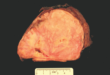
Morphology. Bacterial pneumonia has two patterns of anatomic distribution: lobular bronchopneumonia and lobar pneumonia (Fig. 15-31). Patchy consolidation of the lung is the dominant characteristic of bronchopneumonia (Fig. 15-32), while fibrinosuppurative consolidation of a large portion of a lobe or of an entire lobe defines lobar pneumonia (Fig. 15-33). These anatomic but still classic categorizations are often difficult to apply in individual cases because patterns overlap. The patchy involvement may become confluent, producing virtually total lobar consolidation; in contrast, effective antibiotic therapy for any form of pneumonia may limit involvement to a subtotal consolidation. Moreover, the same organisms may produce either pattern depending on patient susceptibility. Most important from the clinical standpoint are identification of the causative agent and determination of the extent of disease.
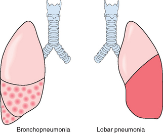
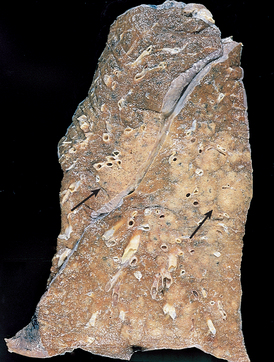
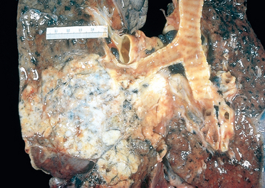
FIGURE 15-33 Lobar pneumonia—gray hepatization, gross photograph. The lower lobe is uniformly consolidated.
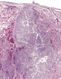
In lobar pneumonia, four stages of the inflammatory response have classically been described: congestion, red hepatization, gray hepatization, and resolution. Current effective antibiotic therapy frequently slows or halts the progression. In the first stage of congestion the lung is heavy, boggy, and red. It is characterized by vascular engorgement, intra-alveolar fluid with few neutrophils, and often the presence of numerous bacteria. The stage of red hepatization that follows is characterized by massive confluent exudation with neutrophils, red cells, and fibrin filling the alveolar spaces (Fig. 15-34A). On gross examination, the lobe now appears distinctly red, firm, and airless, with a liver-like consistency, hence the term hepatization. The stage of gray hepatization follows with progressive disintegration ofred cells and the persistence of a fibrinosuppurative exudate (Fig. 15-34B), giving the gross appearance of a grayish brown, dry surface. In the final stage of resolution the consolidated exudate within the alveolar spaces undergoes progressive enzymatic digestion to produce granular, semifluid debris that is resorbed, ingested by macrophages, expectorated, or organized by fibroblasts growing into it (Fig. 15-34C). Pleural fibrinous reaction to the underlying inflammation, often present in the early stages if the consolidation extends to the surface (pleuritis), may similarly resolve. More often it undergoes organization, leaving fibrous thickening or permanent adhesions.
Foci of bronchopneumonia are consolidated areas of acute suppurative inflammation. The consolidation may be patchy through one lobe but is more often multilobar and frequently bilateral and basal because of the tendency of secretions to gravitate into the lower lobes. Well-developed lesions are slightly elevated, dry, granular, gray-red to yellow, and poorly delimited at their margins (see Fig. 15-32). Histologically, the reaction usually elicits a suppurative, neutrophil-rich exudate that fills the bronchi, bronchioles, and adjacent alveolar spaces (Fig. 15-34A).
Complications of pneumonia include (1) tissue destruction and necrosis, causing abscess formation (particularly common with type 3 pneumococci or Klebsiella infections); (2) spread of infection to the pleural cavity, causing the intrapleural fibrinosuppurative reaction known as empyema; and (3) bacteremic dissemination to the heart valves, pericardium, brain, kidneys, spleen, or joints, causing metastatic abscesses, endocarditis, meningitis, or suppurative arthritis.
The major symptoms of community-acquired acute pneumonia are abrupt onset of high fever, shaking chills, and cough productive of mucopurulent sputum; occasional patients may have hemoptysis. When fibrinosuppurative pleuritis is present it is accompanied by pleuritic pain and pleural friction rub. The whole lobe is radiopaque in lobar pneumonia, whereas there are focal opacities in bronchopneumonia.
The clinical picture is markedly modified by the administration of antibiotics. Treated patients may be relatively afebrile with few clinical signs 48 to 72 hours after the initiation of antibiotics. The identification of the organism and the determination of its antibiotic sensitivity are the keystones to appropriate therapy. Fewer than 10% of patients with pneumonia severe enough to merit hospitalization now succumb, and in most such instances death may be attributed either to a complication, such as empyema, meningitis, endocarditis, or pericarditis, or to some predisposing influence, such as debility or chronic alcoholism.
The term primary atypical pneumonia was initially applied to an acute febrile respiratory disease characterized by patchy inflammatory changes in the lungs, largely confined to the alveolar septa and pulmonary interstitium. The term atypical denotes the moderate amount of sputum, no physical findings of consolidation, only moderate elevation of white cell count, and lack of alveolar exudate. The pneumonitis is caused by a variety of organisms, the most common being Mycoplasma pneumoniae. Mycoplasma infections are particularly common among children and young adults. They occur sporadically or as local epidemics in closed communities (schools, military camps, and prisons). Other etiologic agents are viruses, including influenza virus types A and B, the respiratory syncytial viruses, human metapneumovirus, adenovirus, rhinoviruses, rubeola, and varicella viruses; Chlamydia pneumoniae; and Coxiella burnetii (Q fever).124,125 In some cases the cause cannot be determined. Any one of these agents can cause merely an upper respiratory tract infection, recognized as the common cold, or a more severe lower respiratory tract infection. Factors that favor such extension of the infection include extremes of age, malnutrition, alcoholism, and underlying debilitating illnesses.
The common pathogenetic mechanism is attachment of the organisms to the upper respiratory tract epithelium followed by necrosis of the cells and an inflammatory response. When the process extends to the alveoli there is usually interstitial inflammation, but there may also be some outpouring of fluid into alveolar spaces, so that on chest x-ray the changes may mimic bacterial pneumonia. Damage to and denudation of the respiratory epithelium inhibit mucociliary clearance and predispose to secondary bacterial infections.
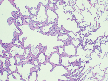
Morphology. All causal agents produce essentially similar morphologic patterns. The lung involvement may be quite patchy or may involve whole lobes bilaterally or unilaterally. The affected areas are red-blue and congested. The pleura is smooth, and pleuritis or pleural effusions are infrequent.
The histologic pattern depends on the severity of the disease. Predominant is the interstitial nature of the inflammatory reaction, virtually localized within the walls of the alveoli. The alveolar septa are widened and edematous and usually have a mononuclear inflammatory infiltrate of lymphocytes, macrophages, and occasionally plasma cells. In acute cases neutrophils may also be present. The alveoli may be free from exudate, but in many patients there is intra-alveolar proteinaceous material and a cellular exudate. When complicated by ARDS, characteristically pink hyaline membranes lining the alveolar walls are present (see Fig. 15-3). Eradication of the infection is followed by reconstitution of the normal architecture of the lung.
Superimposed bacterial infection modifies the histologic picture by causing ulcerative bronchitis, bronchiolitis, and bacterial pneumonia. Some viruses, such as herpes simplex, varicella, and adenovirus, may be associated with necrosis of bronchial and alveolar epithelium and acute inflammation. Characteristic viral cytopathic changes are described in Chapter 8.
The clinical course is extremely varied. Many cases masquerade as severe upper respiratory tract infections or as chest colds. Even individuals with welldeveloped atypical pneumonia have few localizing symptoms. Cough may be absent, and the major manifestations may consist only of fever, headache, muscle aches, and pains in the legs. The edema and exudation are both strategically located to cause mismatching of ventilation and blood flow and thus evoke symptoms out of proportion to the scanty physical findings.
The ordinary sporadic form of the disease is usually mild with a low mortality rate, below 1%. Interstitial pneumonia, however, may assume epidemic proportions with intensified severity and greater mortality, as documented in the devastating influenzal pandemics of 1915 and 1918 and the many smaller epidemics since then. Secondary bacterial infection by staphylococci or streptococci is common in such circumstances.
The genome of influenza virus is composed of eight helices of single-stranded RNA, each encoding a single gene and each bound by a nucleoprotein that determines the type of influenza virus (A, B, or C). The spherical surface of influenza virus is a lipid bilayer (envelope) containing the viral hemagglutinin and neuraminidase, which determine the subtype of the virus (H1 to H3; N1 or N2). Host antibodies to the hemagglutinin and neuraminidase prevent and ameliorate, respectively, future infection with the influenza virus. Two mechanisms account for the clearance of primary influenza virus infection: cytotoxic T cells kill virus-infected cells, and an intracellular anti-influenza protein (called Mx1) is induced in macrophages by the cytokines IFN-α and IFN-β.126
Influenza viruses of type A infect humans, pigs, horses, and birds and are the major cause of pandemic and epidemic influenza infections. A single subtype of influenza virus A predominates throughout the world at a given time.127 Epidemics of influenza occur through mutations of the hemagglutinin and neuraminidase that allow the virus to escape most host antibodies (antigenic drift). Pandemics, which are longer and more widespread than epidemics, may occur when both the hemagglutinin and the neuraminidase are replaced through recombination of RNA segments with those of animal viruses, making all individuals susceptible to the new influenza virus (antigenic shift). Polymerase chain reaction (PCR) analysis of influenza virus from the lungs of a soldier who died in the 1918 influenza pandemic that killed between 20 million and 40 million people worldwide identified a swine influenza virus belonging to the same family of influenza viruses causing illness today.128 Current antiviral drugs are effective against recombinant influenza viruses bearing the 1918 hemagglutinin, neuraminidase, and matrix genes.129 Influenza virus types B and C, which do not show antigenic drift or shift, infect mostly children, who develop antibodies that prevent reinfection. Rarely, influenza virus may cause interstitial myocarditis or, after aspirin therapy, Reye syndrome (Chapter 18).
Avian influenza refers to strains of influenza which primarily infect birds. One such strain with the antigenic type H5N1 is of great concern because infection is frequently lethal in humans (approximately 60%) and since 2003 the virus is spreading throughout the world in wild and domestic birds. As of the fall of 2008, a total of 387 H5N1 influenza virus infections in humans have been reported to the WHO. Nearly all cases of H5N1 influenza in humans have been acquired by close contact with domestic birds. The severity of the disease results from the ability of the virus to cause widespread infection in the human body, instead of infection being limited to the lung. The tissue tropism of H5N1 influenza is increased due to the unusual structure of its hemagglutinin protein. Cleavage of viral hemagglutinin by host proteases is required for the influenza virus to enter host cells. The hemagglutinin protein of H5N1 influenza virus, and other highly pathogenic influenza viruses, is unusual in that it can be cleaved by ubiquitous proteases in the human, while the hemagglutinin of less virulent influenza virus stains can only be cleaved by proteases found in limited organs, including the lung. Fortunately, the transmission of the current H5N1 virus is inefficient. Most patients with H5NI infection present with pneumonia. However, if antigenic recombination occurs between H5N1 influenza and a strain of influenza which is highly infectious for humans, sustained human-to-human transmission could give rise to a pandemic, similar to the Spanish pandemic of 1918. This concern has spurred efforts to develop a vaccine.130
Morphology. Viral upper respiratory infections are marked by mucosal hyperemia and swelling with a predominantly lymphomonocytic and plasmacytic infiltration of the submucosa accompanied by overproduction of mucus secretions. The swollen mucosa and viscous exudate may plug the nasal channels, sinuses or the Eustachian tubes, and lead to suppurative secondary bacterial infection. Virus-induced tonsillitis with enlargement of the lymphoid tissue within the Waldeyer ring is frequent in children, although lymphoid hyperplasia is not usually associated with suppuration or abscess formation, such as is encountered with streptococci or staphylococci.
In laryngotracheobronchitis and bronchiolitis there is vocal cord swelling and abundant mucus exudation. Impairment of bronchociliary function invites bacterial superinfection with more marked suppuration. Plugging of small airways may give rise to focal lung atelectasis. In the more severe bronchiolar involvement widespread plugging of secondary and terminal airways by cell debris, fibrin, and inflammatory exudate may, when prolonged, cause organization and fibrosis, resulting in obliterative bronchiolitis and permanent lung damage.
Human MPV, a paramyxovirus discovered in 2001, is found worldwide and is associated with upper and lower respiratory tract infections, most commonly in young children, elderly subjects, and immunocompromised patients. Human MPV can cause severe infections such as bronchiolitis and pneumonia and is responsible for 5% to 10% of hospitalizations and 12% to 20% of outpatient visits of children suffering from acute respiratory tract infections. Such infections are clinically indistinguishable from those caused by human respiratory syncytial virus. The first human MPV infection occurs during early childhood, but reinfections are common throughout life, especially in older subjects. Molecular methods such as reverse transcriptase–PCR are the preferred diagnostic modality because of fastidious growth in cell culture. No commercial treatments are yet available for human MPV, although ribavirin has shown activity both in vitro and in animal models. Live attenuated vaccines produced by genetically altered virus have also shown good efficacy in animals.131
SARS first appeared in November 2002 in the Guangdong Province of China and subsequently spread to Hong Kong, Taiwan, Singapore, Vietnam, and Toronto, where large outbreaks also occurred.132 The ease of travel between continents clearly contributed to this pandemic. Between Fall 2002 and Spring 2003, there were more than 8000 cases of SARS, including 774 deaths. The worldwide epidemic was halted, perhaps in part because of public health measures, and the last cases of SARS were laboratory-associated infections reported in April 2004.125
After an incubation period of 2 to 10 days, SARS begins with a dry cough, malaise, myalgias, fever, and chills. A third of patients improve and resolve the infection, but the rest progress to severe respiratory disease with shortness of breath, tachypnea, and pleurisy, and nearly 10% of patients die from the illness, for which there is no specific treatment.
The cause of SARS is a previously undiscovered coronavirus. Nearly a third of upper respiratory infections are caused by coronaviruses, but the SARS virus differs from previously known coronaviruses in that it infects the lower respiratory tract and spreads throughout the body. The SARS virus seems to have been first transmitted to humans through contact with wild masked palm civets that are eaten in China. Subsequent cases were spread person-to-person, mainly through infected respiratory secretions, although some cases may have been contracted from stool.
SARS can be diagnosed either by detection of the virus by PCR or by detection of antibodies to the virus. Levels of the virus are low initially and peak 10 days after onset of illness, so testing of different specimens (respiratory secretions, blood, and stool) collected on several days may be needed to detect the virus. Detection of antibodies specific for the SARS virus is a very sensitive and specific test; however, patients may not have a measurable antibody response for up to 28 days after infection.
The pathophysiology of SARS is not understood, nor is it known why the virus moved from animals to humans. Most human SARS coronaviruses have a 29-nucleotide deletion in the RNA when compared to the virus found in wild animals, which may enhance its transmission or pathogenicity. In patients who have died of SARS the lungs show diffuse alveolar damage and multinucleated giant cells. Coronaviruses can be seen within pneumocytes by electron microscopy.
Hospital-acquired pneumonias are defined as pulmonary infections acquired in the course of a hospital stay. They are common in patients with severe underlying disease, immunosuppression, prolonged antibiotic therapy, or invasive access devices such as intravascular catheters. Patients on mechanical ventilation are at particularly high risk. Superimposed on an underlying disease (that caused hospitalization), hospital-acquired infections are serious and often lifethreatening complications. Gram-negative rods (Enterobacteriaceae and Pseudomonas species) and S. aureus are the most common isolates; unlike community-acquired pneumonias, S. pneumoniae is not a major pathogen.
Aspiration pneumonia occurs in markedly debilitated patients or those who aspirate gastric contents either while unconscious (e.g., after a stroke) or during repeated vomiting. These patients have abnormal gag and swallowing reflexes that predispose to aspiration. The resultant pneumonia is partly chemical because of the extremely irritating effects of the gastric acid, and partly bacterial (from the oral flora). Typically, more than one organism is recovered on culture, aerobes being more common than anaerobes. This type of pneumonia is often necrotizing, pursues a fulminant clinical course, and is a frequent cause of death. In those who survive, lung abscess is a common complication.
The term “pulmonary abscess” describes a local suppurative process within the lung, characterized by necrosis of lung tissue. Oropharyngeal surgical procedures, sinobronchial infections, dental sepsis, and bronchiectasis play important roles in their development.
Although under appropriate circumstances any pathogen can produce an abscess, the commonly isolated organisms include aerobic and anaerobic streptococci, S. aureus, and a host of gram-negative organisms. Mixed infections often occur because of the important causal role played by inhalation of foreign material.133 Anaerobic organisms normally found in the oral cavity, including members of the Bacteroides, Fusobacterium, and Peptococcus species, are the exclusive isolates in about 60% of cases. The causative organisms are introduced by the following mechanisms:
When all these causes are excluded, there are still cases in which no reasonable basis for the abscess formation can be identified. These are referred to as primary cryptogenic lung abscesses.
Morphology. Abscesses vary in diameter from lesions of a few millimeters to large cavities of 5 to 6 cm. They may affect any part of the lung and may be single or multiple. Pulmonary abscesses due to aspiration are more common on the right (because of the more vertical right main bronchus) and are most often single. Abscesses that develop in the course of pneumonia or bronchiectasis are usually multiple, basal, and diffusely scattered. Septic emboli and pyemic abscesses are multiple and may affect any region of the lungs.
The abscess cavity might be filled with suppurative debris. If there is communication with an air passage, the contained exudate may be partially drained to create an air-containing cavity. Superimposed saprophytic infections are prone to flourishing within the already necrotic debris of the abscess cavity. Continued infection leads to large, fetid, green-black, multilocular cavities with poor demarcation of their margins, designated gangrene of the lung. The cardinal histologic change in all abscesses is suppurative destruction of the lung parenchyma within the central area of cavitation (Fig. 15-35). In chronic cases considerable fibroblastic proliferation produces a fibrous wall.
The manifestations of pulmonary abscesses are much like those of bronchiectasis and are characterized principally by cough, fever, and copious amounts of foul-smelling purulent or sanguineous sputum. Fever, chest pain, and weight loss are common. Clubbing of the fingers and toes may appear within a few weeks after the onset of an abscess. Diagnosis of this condition can be only suspectedfrom the clinical findings and must be confirmed radiologically. Whenever an abscess is discovered in older individuals, it is important to rule out an underlying carcinoma, because this is present in 10% to 15% of cases.
The course of abscesses is variable. With antimicrobial therapy, most resolve leaving behind a scar. Complications include extension of the infection into the pleural cavity, hemorrhage, the development of brain abscesses or meningitis from septic emboli, and (rarely) secondary amyloidosis (type AA).
Chronic pneumonia is most often a localized lesion in the immunocompetent patient, with or without regional lymph node involvement. Typically, the inflammatory reaction is granulomatous, and is caused by bacteria (e.g., M. tuberculosis) or fungi (e.g., Histoplasma capsulatum). Tuberculosis of the lung and other organs was described in Chapter 8. Here we will discuss chronic pneumonias caused by fungi.
Histoplasmosis, blastomycosis, and coccidioidomycosis are discussed together because (1) they are granulomatous diseases of the lungs that may resemble tuberculosis, (2) they are caused by fungi that are thermally dimorphic in that they grow as hyphae that produce spores at environmental temperatures but grow as yeasts (spherules or ellipses) at body temperature within the lungs, and (3) each fungus is geographic in that it causes disease primarily among immunocompetent individuals living along the Ohio and Mississippi rivers and in the Caribbean (Histoplasma), in the central and southeastern United States (Blastomyces), and in the Southwest and Far West of the United States and in Mexico (Coccidioides).
Histoplasma capsulatum infection is acquired by inhalation of dust particles from soil contaminated with bird or bat droppings that contain small spores (microconidia), the infectious form of the fungus. Like M. tuberculosis, H. capsulatum is an intracellular parasite of macrophages. The clinical presentations and morphologic lesions of histoplasmosis also strikingly resemble those of tuberculosis, including (1) a self-limited and often latent primary pulmonary involvement, which may result in coin lesions on chest radiography; (2) chronic, progressive, secondary lung disease, which is localized to the lung apices and causes cough, fever, and night sweats; (3) localized lesions in extrapulmonary sites, including mediastinum, adrenals, liver, or meninges; and (4) a widely disseminated disease in immunocompromised patients.
The pathogenesis of histoplasmosis is incompletely understood. It is known that macrophages are the major target of infection. H. capsulatum may be internalized into macrophages after opsonization with antibody. Histoplasma yeasts can multiply within the phagosome, and lyse the host cells. Histoplasma infections are controlled by helper T cells that recognize fungal cell wall antigens and heat-shock proteins and subsequently secrete IFN-γ, which activates macrophages to kill intracellular yeasts. In addition, Histoplasma induces macrophages to secrete TNF, which recruits and stimulates other macrophages to kill Histoplasma. Lacking cellular immunity, patients with acquired immunodeficiency syndrome are susceptible to disseminated infection with Histoplasma, which is an opportunistic pathogen in this disease.
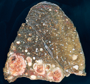
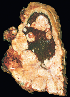
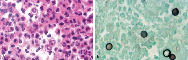
Morphology. In the lungs of otherwise healthy adults, Histoplasma infections produce epithelioid cell granulomas, which usually undergo caseation necrosis and coalesce to produce large areas of consolidation but may also liquefy to form cavities (seen in patients with COPD). With spontaneous or drug control of the infection, these lesions undergo fibrosis and concentric calcification (tree-bark appearance) (Fig. 15-36A). Histologic differentiation from tuberculosis, sarcoidosis, and coccidioidomycosis requires identification of the 3- to 5-μm thin-walled yeast forms that may persist in tissues for years.
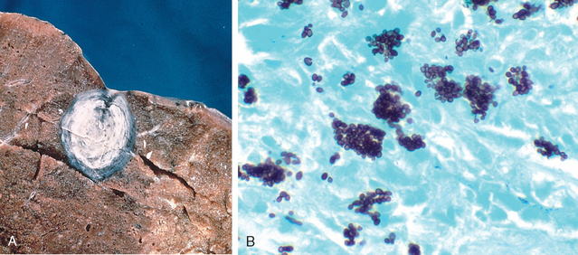
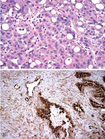
FIGURE 15-36 Histoplasmosis. A, Laminated Histoplasma granuloma of the lung. B, Histoplasma capsulatum yeast forms fill phagocytes in the lung of a patient with disseminated histoplasmosis (silver stain).
In fulminant disseminated histoplasmosis, which occurs in immunosuppressed individuals, epithelioid cell granulomas are not formed; instead, there are focal accumulations of mononuclear phagocytes filled with fungal yeasts throughout the tissues and organs of the body (Fig. 15-36B).
The diagnosis of histoplasmosis is established by culture or identification of the fungus in tissue lesions. In addition, serologic tests for antibodies and antigen are also available. Antigen detection in body fluids is most useful in the early stages, because antibodies are formed 2 to 6 weeks after infection.134
Blastomyces dermatitidis is a soil-inhabiting, dimorphic fungus that is remarkably difficult to isolate. It is a cause of disease in people living in or visiting the central and southeastern United States; infection also occurs in Canada, Mexico, the Middle East, Africa, and India. There are three clinical forms: pulmonary blastomycosis, disseminated blastomycosis, and a rare primary cutaneous form that results from direct inoculation of organisms into the skin. Pulmonary blastomycosis most often presents as an abrupt illness with productive cough, headache, chest pain, weight loss, fever, abdominal pain, night sweats, chills, and anorexia. Chest radiographs reveal lobar consolidation, multilobar infiltrates, perihilar infiltrates, multiple nodules, or miliary infiltrates. The upper lobes are most frequently involved. The process may resolve spontaneously, persist, or progress to a chronic lesion.
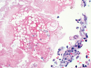
Morphology. In the normal host the lung lesions of blastomycosis are suppurative granulomas. Macrophages have a limited ability to ingest and kill B. dermatitidis, and the persistence of the yeast cells leads to continued recruitment of neutrophils. In tissue, B. dermatitidis is a round, 5- to 15-μm yeast cell that divides by broad-based budding. It has a thick, double -contoured cell wall and multiple nuclei (Fig. 15-37). Involvement of the skin and larynx is associated with marked epithelial hyperplasia, which may be mistaken for squamous cell carcinoma.
Almost everyone who inhales the spores of Coccidioides immitis becomes infected and develops a delayed-type hypersensitivity to the fungus, so more than 80% of people in endemic areas of the southwestern and western United States have a positive skin test reaction. One reason for the high rate of infectivity by C. immitis is that infective arthroconidia, when ingested by alveolar macrophages, block fusion of the phagosome and lysosome and so resist intracellular killing. As is the case with Histoplasma, most primary infections with C. immitis are asymptomatic, but 10% of people have lung lesions, fever, cough, and pleuritic pains, accompanied by erythema nodosum or erythema multiforme (the San Joaquin Valley fever complex). Less than 1% of people develop disseminated C. immitis infection, which frequently involves the skin and meninges.
Morphology. The primary and secondary lung lesions of C. immitis are similar to the granulomatous lesions of Histoplasma. Within macrophages or giant cells, C. immitis is present as thick-walled, nonbudding spherules 20 to 60 μm in diameter, often filled with small endospores. A pyogenic reaction is superimposed when the spherules rupture to release the endospores (Fig. 15-38). Rare progressive C. immitis disease involves the lungs, meninges, skin, bones, adrenals, lymph nodes, spleen, or liver. At all these sites, the inflammatory response may be purely granulomatous, pyogenic, or mixed. Purulent lesions dominate in patients with diminished resistance and with widespread dissemination.
The appearance of a pulmonary infiltrate, with or without signs of infection (e.g., fever), is one of the most common and serious complications in patients whose immune defenses are suppressed by disease, immunosuppressive therapy for organ transplants, chemotherapy for tumors, or irradiation.135 A wide variety of so-called opportunistic infectious agents, many of which rarely cause infection in normal hosts, can cause these pneumonias, and often more than one agent is involved. Mortality from these opportunistic infections is high. Table 15-9 lists some of the opportunistic agents according to their prevalence and whether they cause local or diffuse pulmonary infiltrates. The differential diagnosis of such infiltrates includes drug reactions and involvement of the lung by tumor. The specific infections are discussed in Chapter 8. Of these, the ones that commonly involve the lung can be classified according to the etiologic agent: (1) bacteria (P. aeruginosa, Mycobacterium species, L. pneumophila, and Listeria monocytogenes), (2) viruses (cytomegalovirus [CMV] and herpesvirus), and (3) fungi (P. jiroveci, Candida species, Aspergillus species, the Phycomycetes, and Cryptococcus neoformans).
TABLE 15-9 Causes of Pulmonary Infiltrates in Immunocompromised Hosts
Diffuse Infiltrate | Focal Infiltrate |
---|---|
COMMON | |
Cytomegalovirus | Gram-negative rods |
Pneumocystis jiroveci | Staphylococcus aureus |
Drug reaction | Aspergillus |
Candida | |
Malignancy | |
UNCOMMON | |
Bacteria | Cryptococcus |
Aspergillus | Mucor |
Cryptococcus | Pneumocystis jiroveci |
Malignancy | Legionella pneumophila |
Pulmonary disease continues to be the leading cause of morbidity and mortality in HIV-infected individuals. Although the use of potent antiretroviral agents and effective chemoprophylaxis has markedly altered the incidence and outcome of pulmonary disease in HIV-infected persons, the plethora of infectious agents and other pulmonary lesions make diagnosis and treatment a distinct challenge. Some of the individual microbial agents afflicting HIV-infected individuals have already been discussed; this section will focus only on the general principles of HIV-associated pulmonary disease.
Finally, it is useful to remember that pulmonary disease in HIV-infected persons may result from more than one cause, and even common pathogens may present with atypical manifestations. Therefore, the diagnostic work-up of these patients may be more extensive (and expensive) than would be necessary in an immunocompetent individual.
Indications for transplantation may include almost all non-neoplastic terminal lung diseases, provided that the patient does not have any other serious disease, which would preclude lifelong immunosuppressive therapy. The most common indications are end-stage emphysema, idiopathic pulmonary fibrosis, cystic fibrosis, and idiopathic/familial pulmonary arterial hypertension. While bilateral lung and heart-lung transplants are possible, in many cases a single-lung transplant is performed, offering sufficient improvement in pulmonary function for each of two recipients from a single (and all too scarce) donor. When bilateral chronic infection is present (e.g., cystic fibrosis, bronchiectasis), both lungs of the recipient must be replaced to remove the reservoir of infection.
Morphology. With improving surgical and organ preservation techniques, postoperative complications (e.g., anastomotic dehiscence, vascular thrombosis, primary graft dysfunction) are fortunately becoming rare. The transplanted lung is subject to two major complications: infection and rejection.
Pulmonary infections in lung transplant patients are essentially those of any immunocompromised host, discussed earlier. In the early post-transplant period (the first few weeks), bacterial infections are most common. With ganciclovir prophylaxis and matching of donor-recipient CMV status, CMV pneumonia occurs less frequently and is less severe, although some resistant strains are emerging. Most infections occur in the third to twelfth month after transplantation. Pneumocystis jiroveci pneumonia is rare, since almost all patients receive adequate prophylaxis, usually with Bactrim (trimethoprimsulfamethoxazole). Fungal infections are mostly due to Candida and Aspergillus species, and they involve the bronchial anastomotic site and/or the lung.
Acute rejection of the lung occurs to some degree in all patients despite routine immunosuppression. It often occurs during the early weeks to months after surgery but may occur years later whenever immunosuppression is decreased. Patients present with fever, dyspnea, cough, and radiologic infiltrates. Since these are similar to the picture of infections, diagnosis often relies on transbronchial biopsy. The morphologic features of acute rejection are primarily those of inflammatory infiltrates (lymphocytes, plasma cells, and few neutrophils and eosinophils), either around small vessels, in the submucosa of airways, or both.136
Chronic rejection is a significant problem in at least half of all lung transplant patients by 3 to 5 years. It is manifested by cough, dyspnea, and an irreversible decrease in lung function tests. The major morphologic correlate of chronic rejection is bronchiolitis obliterans, the partial or complete occlusion of small airways by fibrosis, with or without active inflammation (Fig. 15-39). Bronchiolitis obliterans is patchy and therefore difficult to diagnose via transbronchial biopsy. Bronchiectasis and pulmonary fibrosis may develop in long-standing cases.
Acute cellular airway rejection (the presumed forerunner of later, fibrous obliteration of these airways) is generally responsive to therapy, but the treatment of established bronchiolitis obliterans has been disappointing. Its progress may be slowed or even halted for some time, but it cannot be reversed. Infrequent complications of lung transplantation include accelerated pulmonary arteriosclerosis in the graft and lymphoproliferative disease. With continuing improvement in surgical, immunosuppressive, and antimicrobial therapies, the short-term outcome of lung transplantation has improved considerably, although it is still not as good as that for renal or cardiac transplantation. One-, five-, and ten-year survival rates are 78%, 50%, and 26%, respectively.137
A variety of benign and malignant tumors may arise in the lung, but 90% to 95% are carcinomas, about 5% are bronchial carcinoids, and 2% to 5% are mesenchymal and other miscellaneous neoplasms.60
Lung cancer is currently the most frequently diagnosed major cancer in the world and the most common cause of cancer mortality worldwide. This is largely due to the carcinogenic effects of cigarette smoke. Over the coming decades, changes in smoking habits will greatly influence lung cancer incidence and mortality as well as the prevalence of various histologic types of lung cancer.138
The number of new cases of lung cancer occurring in 2008 in the United States is estimated to be 215,020 (note that in 1950 it was 18,000), accounting for about 15% of cancer diagnoses and 29% of cancer-related deaths. The annual number of deaths from lung cancer in the United States is estimated to be 161,840 in 2008.139 Since the early 1990s lung cancer incidence and mortality rates have been decreasing in men, most likely from the decreased smoking rates over the past 30 years. However, decreases in smoking patterns among women lag behind those of men. Since 1987 more women have died each year of lung cancer than of breast cancer, which for over 40 years had been the major cause of cancer death in women. Cancer of the lung occurs most often between ages 40 and 70 years, with a peak incidence in the 50s or 60s. Only 2% of all cases appear before the age of 40. The outlook for individuals diagnosed with lung cancer is dismal. The 1-year survival rate has increased from 34% in 1975 to 41% in 2007, largely because of improvements in surgical techniques. However, the 5-year rate for all stages combined is only 16%.
Most carcinomas of the lung, similar to cancer at other sites, arise by a stepwise accumulation of genetic abnormalities that transform benign bronchial epithelium to neoplastic tissue. Unlike many other cancers, however, the major environmental insult that inflicts genetic damage is known. We begin our discussion with the well-known lung carcinogen—cigarette smoke.
The evidence provided by statistical and clinical observations establishing a positive relationship between tobacco smoking and lung cancer is overwhelming. Experimental data have also been pursued, but this approach is limited by species differences.
Statistical evidence is most compelling: 87% of lung carcinomas occur in active smokers or those who stopped recently. In numerous retrospective studies, there was an invariable statistical association between the frequency of lung cancer and (1) the amount of daily smoking, (2) the tendency to inhale, and (3) the duration of the smoking habit. Compared with nonsmokers, average smokers of cigarettes have a tenfold greater risk of developing lung cancer, and heavy smokers (more than 40 cigarettes per day for several years) have a 60-fold greater risk. Women have a higher susceptibility to tobacco carcinogens than men do. Cessation of smoking for 10 years reduces risk but never to control levels. It should be noted, however, that despite compelling evidence supporting the role of cigarette smoking, only 11% of heavy smokers develop lung cancer in their lifetime. Clearly, there are other (genetic) factors involved as will be discussed later. Epidemiologic studies also show an association between cigarette smoking and carcinoma of the mouth, pharynx, larynx, esophagus, pancreas, uterine cervix, kidney, and urinary bladder. Secondhand smoke, or environmental tobacco smoke, contains numerous human carcinogens for which there is no safe level of exposure. It is estimated that each year about 3000 nonsmoking adults die of lung cancer as a result of breathing secondhand smoke.140 Cigar and pipe smoking also increase risk, although much more modestly than smoking cigarettes. The use of smokeless tobacco is not a safe substitute for smoking cigarettes or cigars, as these products cause oral cancers and can lead to nicotine addiction.
Clinical evidence is obtained largely through observations of histologic changes in the lining epithelium of the respiratory tract in habitual smokers. These sequential changes have been best documented for squamous cell carcinoma, but they may also be present in other histologic subtypes. In essence, there is a linear correlation between the intensity of exposure to cigarette smoke and the appearance of ever more worrisome epithelial changes that begin with squamous metaplasia and progress to squamous dysplasia, carcinoma in situ, and invasive carcinoma. Lung tumors of smokers frequently contain a typical, though not specific, molecular fingerprint in the form of G : C > T : A mutations in the p53 gene that are probably caused by benzo[a]pyrene, one of the many carcinogens in tobacco smoke.138
Experimental work has consisted mainly of attempts to induce cancer in experimental animals with extracts of tobacco smoke.141 More than 1200 substances have been counted in cigarette smoke, many of which are potential carcinogens. They include both initiators (polycyclic aromatic hydrocarbons such as benzo[a]pyrene) and promoters, such as phenol derivatives. Radioactive elements may also be found (polonium-210, carbon-14, and potassium-40) as well as other contaminants, such as arsenic, nickel, molds, and additives. Protracted exposure of mice to these additives induces skin tumors. Efforts to produce lung cancer by exposing animals to tobacco smoke, however, have been unsuccessful. The few cancers that have developed have been bronchioloalveolar carcinomas, a type of tumor that is not strongly associated with smoking in humans.
Certain industrial exposures increase the risk of developing lung cancer. High-dose ionizing radiation is carcinogenic. There was an increased incidence of lung cancer among survivors of the Hiroshima and Nagasaki atomic bomb blasts. Uranium is weakly radioactive, but lung cancer rates among nonsmoking uranium miners are four times higher than those in the general population, and among smoking miners they are about 10 times higher.
The risk of lung cancer is increased with asbestos. Lung cancer is the most frequent malignancy in individuals exposed to asbestos, particularly when coupled with smoking.80 Asbestos workers who do not smoke have a five times greater risk of developing lung cancer than do nonsmoking control subjects, and those who smoke have a 50 to 90 times greater risk. The latent period before the development of lung cancer is 10 to 30 years.
Atmospheric pollutants may play some role in the increased incidence of lung carcinoma today. Attention has been drawn to the potential problem of indoor air pollution, especially by radon.142,143 Radon is a ubiquitous radioactive gas that has been linked epidemiologically to increased lung cancer in miners exposed to relatively high concentrations. The pathogenic mechanism is believed to be inhalation and bronchial deposition of radioactive decay products that become attached to environmental aerosols. These data have generated concern that low-level indoor exposure (e.g., in homes in areas of high radon in soil) could also lead to increased incidence of lung tumors; some attribute the bulk of lung cancers in nonsmokers to this insidious carcinogen (Chapter 9).144
Ultimately, the exposures cited previously are thought to act by causing genetic alterations in lung cells, which accumulate and eventually lead to the neoplastic phenotype. It has been estimated that 10 to 20 genetic mutations have occurred by the time the tumor is clinically apparent.145
As will be discussed below, for all practical purposes lung cancers can be divided into two clinical subgroups: small cell carcinoma and non-small cell carcinoma. Some molecular lesions are common to both types, whereas others are relatively specific. The dominant oncogenes that are frequently involved in lung cancer include c-MYC, KRAS, EGFR, c-MET, and c-KIT. The commonly deleted or inactivated tumor suppressor genes include p53, RB1, p16(INK4a), and multiple loci on chromosome 3p. At this locale there are numerous candidate tumor suppressor genes, such as FHIT, RASSF1A, and others that remain to be identified. Of the various cancer associated genes, C-KIT (40–70%), MYCN and MYCL (20–30%), p53 (90%), 3p (100%), RB (90%), and BCL2 (75–90%) are most commonly involved in small cell lung carcinoma. By comparison, EGFR (25%), KRAS (10–15%), p53 (50%), p16 INK4a (70%) are the ones most commonly affected in non-small cell lung carcinoma. In addition recent studies show that LKB1, PTEN, and TSC, all relating to the m-TOR pathway are also mutated in up to 30% of lung cancers (mostly non-small cell lung carcinoma).146 It should be noted that C-KIT is over expressed but only rarely mutated. Hence, drugs that target its tyrosine kinase domain (such as imatinib) are ineffective. Recall that in tumors with mutation of that kinase domain (e.g., gastrointestinal stromal tumor) this drug is useful for treatment. Telomerase activity is increased in over 80% of lung tumor tissues.
There are several signal transduction molecules that are activated in lung cancer, such as AKT, phosphatidylinositol-3-kinase, ERK1/2, STAT5, and focal adhesion proteins such as paxillin. Although certain genetic changes are known to be early (inactivation of chromosome 3p suppressor genes) or late (activation of KRAS), the temporal sequence is not yet well defined. More importantly, certain genetic changes such as loss of chromosome 3p material can be found in benign bronchial epithelium of individuals with lung cancer, as well as in the respiratory epithelium of smokers without lung cancers, suggesting that large areas of the respiratory mucosa are mutagenized after exposure to carcinogens (“field effect”).147 On this fertile soil, the cells that accumulate additional mutations ultimately develop into cancer.
Occasional familial clustering has suggested a genetic predisposition, as has the variable risk even among heavy smokers. Attempts at defining markers of genetic susceptibility are ongoing and have, for example, identified a role for polymorphisms in the cytochrome P-450 gene CYP1A1 (Chapter 7).148 People with certain alleles of CYP1A1 have an increased capacity to metabolize procarcinogens derived from cigarette smoke and, conceivably, incur the greatest risk of developing lung cancer. Similarly, individuals whose peripheral blood lymphocytes undergo chromosomal breakages following exposure to tobacco-related carcinogens (mutagen sensitivity genotype) have a greater than tenfold risk of developing lung cancer compared with controls. In addition, large scale linkage studies point to an autosomal susceptibility locus on 6q23-25. More recently, genome-wide association studies have revealed an intriguing link to polymorphisms in the nicotine acetylcholine receptor gene located on chromosome 15q25 and lung cancer in both smokers and nonsmokers.149
It should also be pointed out that 25% of lung cancers worldwide arise in nonsmokers and these are pathogenetically distinct. They occur more commonly in women, and most are adenocarcinomas. They tend to have EGFR mutations, almost never have KRAS mutations and p53 mutations, although common, occur less commonly. The nature of the p53 mutations are also distinct.150
Three types of precursor epithelial lesions are recognized: (1) squamous dysplasia and carcinoma in situ, (2) atypical adenomatous hyperplasia, and (3) diffuse idiopathic pulmonary neuroendocrine cell hyperplasia. It should be noted that the term precursor does not imply that progression to cancer will occur in all cases. Currently it is not possible to distinguish between precursor lesions that progress and those that remain localized or regress.
Tumor classification is important for consistency in patient treatment and because it provides a basis for epidemiologic and biologic studies. The most recent classification of the World Health Organization138 has gained wide acceptance (Table 15-10). Several histologic variants of each type of lung cancer are described; however, their clinical significance is still undetermined, except as mentioned below. The relative proportions of the major categories are151:
TABLE 15-10 Histologic Classification of Malignant Epithelial Lung Tumors
The incidence of adenocarcinoma has increased significantly in the last two decades; it is now the most common form of lung cancer in women and, in many studies, men as well.152 The basis for this change is unclear. A possible factor is the increase in women smokers, but this only highlights our lack of knowledge about why women tend to develop more adenocarcinomas. One interesting postulate is that changes in cigarette type (filter tips, lower tar and nicotine) have caused smokers to inhale more deeply and thereby expose more peripheral airways and cells (with a predilection to adenocarcinoma) to carcinogens.153 There may be mixtures of histologic patterns, even in the same cancer. Thus, combined types of squamous cell carcinoma and adenocarcinoma or of small-cell and squamous cell carcinoma occur in about 10% of patients. For common clinical use, however, the various histologic types of lung cancer can be clustered into two groups on the basis of likelihood of metastases and response to available therapies: small cell carcinomas (almost always metastatic, high initial response to chemotherapy) versus non-small cell carcinomas (less often metastatic, less responsive). The strongest relationship to smoking is with squamous cell and small cell carcinoma.
Morphology. Lung carcinomas arise most often in and about the hilus of the lung. About three fourths of the lesions take their origin from first-order, second-order, and third-order bronchi. An increasing number of primary carcinomas of the lung arise in the periphery of the lung from the alveolar septal cells or terminal bronchioles. These are predominantly adenocarcinomas, including those of the bronchioloalveolar type, to be discussed separately.
The preneoplastic lesions that antedate, and usually accompany, invasive squamous cell carcinoma are well characterized. Squamous cell carcinomas are often preceded for years by squamous metaplasia or dysplasia in the bronchial epithelium, which then transforms to carcinoma in situ, a phase that may last for several years (Fig. 15-40). By this time, atypical cells may be identified in cytologic smears of sputum or in bronchial lavage fluids or brushings, although the lesion is asymptomatic and undetectable on radiographs. Eventually, the growing neoplasm reaches a symptomatic stage, when a well-defined tumor mass begins to obstruct the lumen of a major bronchus, often producing distal atelectasis and infection. The tumor may then follow a variety of paths. It may continue to fungate into the bronchial lumen to produce an intraluminal mass. It can also rapidly penetrate the wall of the bronchus to infiltrate along the peribronchial tissue (Fig. 15-41) into the adjacent region of the carina or mediastinum. In other instances, the tumor grows along a broad front to produce a cauliflower-like intraparenchymal mass that appears to push lung substance ahead of it. In almost all patterns the neoplastic tissue is gray-white and firm to hard. Especially when the tumors are bulky, focal areas of hemorrhage or necrosis may appear to produce red or yellow-white mottling and softening. Sometimes these necrotic foci cavitate. Often these tumors erode the bronchial epithelium and can be diagnosed by cytologic examination of sputum, bronchoalveolar lavage fluid, or fine-needle aspiration (Fig. 15-42).
FIGURE 15-40 Precursor lesions of squamous cell carcinomas. Some of the earliest (and “mild”) changes in smoking-damaged respiratory epithelium include goblet cell hyperplasia (A), basal cell (or reserve cell) hyperplasia (B), and squamous metaplasia (C). More ominous changes include the appearance of squamous dysplasia (D), characterized by the presence of disordered squamous epithelium, with loss of nuclear polarity, nuclear hyperchromasia, pleomorphism, and mitotic figures. Squamous dysplasia may, in turn, progress through the stages of mild, moderate, and severe dysplasia. Carcinoma-in-situ (CIS) (E) is the stage that immediately precedes invasive squamous carcinoma (F), and apart from the lack of basement membrane disruption in CIS, the cytologic features are similar to those in frank carcinoma. Unless treated, CIS will eventually progress to invasive cancer.
(A–E, Courtesy of Dr. Adi Gazdar, Department of Pathology, University of Texas, Southwestern Medical School, Dallas. F, reproduced with permission from Travis WD, et al [eds]: World Health Organization Histological Typing of Lung and Pleural Tumors. Heidelberg, Springer, 1999.)
FIGURE 15-41 Lung carcinoma. The gray-white tumor tissue is seen infiltrating the lung substance. Histologically, this large tumor mass was identified as a squamous cell carcinoma.
FIGURE 15-42 Cytologic diagnosis of lung cancer. A sputum specimen shows an orange-staining, keratinized squamous carcinoma cell with a prominent hyperchromatic nucleus (arrow). Note the size of the tumor cells compared with normal polymorphonuclear leukocytes in the left lower corner.
Extension may occur to the pleural surface and then within the pleural cavity or into the pericardium. Spread to the tracheal, bronchial, and mediastinal nodes can be found in most cases. The frequency of nodal involvement varies slightly with the histologic pattern but averages greater than 50%.
Distant spread of lung carcinoma occurs through both lymphatic and hematogenous pathways. These tumors often spread early throughout the body except for squamous cell carcinoma, which metastasizes outside the thorax late. Metastasis may be the first manifestation of an underlying occult pulmonary lesion. No organ or tissue is spared in the spread of these lesions, but the adrenals, for obscure reasons, are involved in more than half the cases. The liver (30% to 50%), brain (20%), and bone (20%) are additional favored sites of metastases.
Adenocarcinoma. This is a malignant epithelial tumor with glandular differentiation or mucin production by the tumor cells. Adenocarcinomas grow in various patterns, including acinar, papillary, bronchioloalveolar, and solid with mucin formation. Of these, only pure bronchioloalveolar carcinoma has distinct gross, microscopic, and clinical features and will be discussed separately.
Adenocarcinoma is the most common type of lung cancer in women and nonsmokers. As compared with squamous cell cancers, the lesions are usually more peripherally located, and tend to be smaller. They vary histologically from well-differentiated tumors with obvious glandular elements (Fig. 15-43A) to papillary lesions resembling other papillary carcinomas to solid masses with only occasional mucin-producing glands and cells. The majority are positive for thyroid transcription factor-1 (TTF-1) and about 80% contain mucin. At the periphery of the tumor there is often a bronchioloalveolar pattern of spread (see below). Adenocarcinomas grow more slowly than squamous cell carcinomas but tend to metastasize widely and earlier. Peripheral adenocarcinomas with a small central invasive component associated with scarring and a predominantly peripheral bronchioloalveolar growth pattern may have a better outcome than invasive carcinomas of the same size. Adenocarcinomas, including bronchioloalveolar carcinomas, are less frequently associated with a history of smoking (still, greater than 75% are found in smokers) than are squamous or small cell carcinomas (>98% in smokers).
FIGURE 15-43 Histologic variants of lung carcinoma. A, Gland-forming adenocarcinoma, inset shows thyroid transcription factor 1 (TTF-1) positivity. B, Well-differentiated squamous cell carcinoma showing keratinization. C, Small cell carcinoma with islands of small deeply basophilic cells and areas of necrosis. D, Large cell carcinoma, featuring pleomorphic, anaplastic tumor cells with no squamous or glandular differentiation.
KRAS mutations occur primarily in adenocarcinoma, and are seen at a much lower frequency in nonsmokers (5%) than in smokers (30%). p53, RB1, and p16 mutations and inactivation have the same frequency in adenocarcinoma as in squamous cell carcinoma. Mutations and amplifications in the epidermal growth factor receptor gene (EGFR) occur in patients with adenocarcinoma (mostly women, nonsmokers, and those of Asian origin).154 A prospective trial has demonstrated that patients with EGFR mutations have improved survival with upfront EGFR inhibitor treatment. KRAS mutations highly correlate with worse outcome and resistance to EGFR inhibitors. 154 Also, c-MET can be amplified or mutated in lung cancer, for which targeted therapies are being developed.
As the name implies, bronchioloalveolar carcinoma occurs in the pulmonary parenchyma in the terminal bronchioloalveolar regions. It represents, in various series, 1% to 9% of all lung cancers. Macroscopically, the tumor almost always occurs in the peripheral portions of the lung either as a single nodule or, more often, as multiple diffuse nodules that sometimes coalesce to produce a pneumonia-like consolidation. The parenchymal nodules have a mucinous, gray translucence when secretion is present but otherwise appear as solid, gray-white areas that can be confused with pneumonia on gross inspection.
Histologically, the tumor is characterized by a pure bronchioloalveolar growth pattern with no evidence of stromal, vascular, or pleural invasion. The key feature of bronchioloalveolar carcinomas is their growth along preexisting structures without destruction of alveolar architecture. This growth pattern has been termed lepidic, an allusion to the neoplastic cells resembling butterflies sitting on a fence. It has two subtypes: nonmucinous and mucinous. The former has columnar, peg-shaped, or cuboidal cells, while the latter has distinctive, tall, columnar cells with cytoplasmic and intra-alveolar mucin, growing along the alveolar septa (Fig. 15-44). Ultrastructurally, bronchioloalveolar carcinomas are a heterogeneous group, consisting of mucin-secreting bronchiolar cells, Clara cells, or, rarely, type II pneumocytes.155
FIGURE 15-44 Bronchioloalveolar carcinoma, mucinous subtype, with characteristic growth along pre-existing alveolar septa, without invasion.
Nonmucinous bronchioloalveolar carcinomas often consist of a peripheral lung nodule with only rare aerogenous spread and therefore are amenable to surgical resection with an excellent 5-year survival. Mucinous bronchioloalveolar carcinomas, on the other hand, tend to spread aerogenously, forming satellite tumors. These may present as a solitary nodule or as multiple nodules, or an entire lobe may be consolidated by tumor, resembling lobar pneumonia and thus are less likely to be cured by surgery.
Analogous to the adenoma-carcinoma sequence in the colon, it is proposed that adenocarcinoma of the lung arises from atypical adenomatous hyperplasia progressing to bronchioloalveolar carcinoma, which then transforms into invasive adenocarcinoma. This is supported by the observation that lesions of atypical adenomatous hyperplasia are monoclonal and they share many molecular aberrations such as EGFR mutations with nonmucinous bronchioloalveolar carcinomas and with invasive adenocarcinomas.156 Microscopically, atypical adenomatous hyperplasia is recognized as a well-demarcated focus of epithelial proliferation composed of cuboidal to low columnar epithelium (Fig. 15-45). These cells demonstrate some cytologic atypia but not to the extent seen in frank adenocarcinoma. It should be pointed out, however, that not all adenocarcinomas arise in this manner, nor do all bronchioloalveolar carcinomas become invasive if left untreated.
FIGURE 15-45 Atypical adenomatous hyperplasia with cuboidal epithelium and mild interstitial fibrosis.
Squamous Cell Carcinoma. Squamous cell carcinoma is most commonly found in men and is closely correlated with a smoking history. Histologically, this tumor is characterized by the presence of keratinization and/or intercellular bridges. Keratinization may take the form of squamous pearls or individual cells with markedly eosinophilic dense cytoplasm (see Fig. 15-43B). These features are prominent in the well-differentiated tumors, are easily seen but not extensive in moderately differentiated tumors, and are focally seen in poorly differentiated tumors. Mitotic activity is higher in poorly differentiated tumors. In the past, most squamous cell carcinomas were seen to arise centrally from the segmental or subsegmental bronchi. However, the incidence of squamous cell carcinoma of the peripheral lung is increasing. Squamous metaplasia, epithelial dysplasia, and foci of frank carcinoma in situ may be seen in bronchial epithelium adjacent to the tumor mass (see Fig. 15-40).
Squamous cell carcinomas show the highest frequency of p53 mutations of all histologic types of lung carcinoma. p53 protein overexpression and, less commonly, mutations may precede invasion. Abnormal p53 accumulation is reported in 10% to 50% of dysplasias. There is increasing frequency and intensity of p53 immunostaining with higher grade dysplasia, and positivity can be seen in 60% to 90% of squamous cell carcinoma in situ. Loss of protein expression of the tumor suppressor gene RB1 is detected by immunohistochemistry in 15% of squamous cell carcinomas. The cyclin-dependent kinase inhibitor p16(INK4a) is inactivated, and its protein product is lost in 65% of tumors. Multiple allelic losses are observed in squamous cell carcinomas at locations bearing tumor suppressor genes. These losses, especially those involving 3p, 9p, and 17p, may precede invasion and be detected in histologically normal cells in smokers. Overexpression of EGFR has been detected in 80% of squamous cell carcinomas, but it is rarely mutated. HER-2/NEU is highly expressed in 30% of these cancers, but unlike in breast cancer, gene amplification is not the underlying mechanism.157
Small Cell Carcinoma. This highly malignant tumor has a distinctive cell type. The epithelial cells are relatively small, with scant cytoplasm, ill-defined cell borders, finely granular nuclear chromatin (salt and pepper pattern), and absent or inconspicuous nucleoli (see Fig. 15-43C). The cells are round, oval, or spindle-shaped, and nuclear molding is prominent. There is no absolute size for the tumor cells, but in general they are smaller than three small resting lymphocytes. The mitotic count is high. The cells grow in clusters that exhibit neither glandular nor squamous organization. Necrosis is common and often extensive. Basophilic staining of vascular walls due to encrustation by DNA from necrotic tumor cells (Azzopardi effect) is frequently present. All small cell carcinomas are high grade. A single variant of small cell carcinoma is recognized: combined small cell carcinoma, in which there is a mixture of small cell carcinoma and any other non-small cell component, including large cell neuroendocrine carcinoma and sarcoma.
Electron microscopy shows dense-core neurosecretory granules, about 100 nm in diameter, in two thirds of cases. The granules are similar to those found in the neuroendocrine cells present along the bronchial epithelium, particularly in the fetus and neonate. Though distinctive, electron microscopy is not needed for diagnosis. The occurrence of neurosecretory granules, the ability of some of these tumors to secrete polypeptide hormones, and the presence of neuroendocrine markers such as chromogranin, synaptophysin, and CD57 (in 75% of cases) and parathormone-like and other hormonally active products suggest derivation of this tumor from neuroendocrine progenitor cells of the lining bronchial epithelium. This lung cancer type is most commonly associated with ectopic hormone production (discussed later).
Small cell carcinomas have a strong relationship to cigarette smoking; only about 1% occur in nonsmokers. They may arise in major bronchi or in the periphery of the lung. There is no known preinvasive phase or carcinoma in situ. They are the most aggressive of lung tumors, metastasize widely, and are virtually incurable by surgical means.
p53 and RB1 tumor suppressor genes are frequently mutated (50% to 80% and 80% to 100% of small cell carcinomas, respectively). Immunohistochemistry demonstrates high levels of the anti-apoptotic protein BCL2 in 90% of tumors, in contrast with a low frequency of expression of the pro-apoptotic protein BAX.
Large Cell Carcinoma. This is an undifferentiated malignant epithelial tumor that lacks the cytologic features of small-cell carcinoma and glandular or squamous differentiation. The cells typically have large nuclei, prominent nucleoli, and a moderate amount of cytoplasm (see Fig. 15-43D). Large cell carcinomas probably represent squamous cell carcinomas and adenocarcinomas that are so undifferentiated that they can no longer be recognized by light microscopy. Ultrastructurally, however, minimal glandular or squamous differentiation is common. One histologic variant is large cell neuroendocrine carcinoma. This is recognized by such features as organoid nesting, trabecular, rosette-like, and palisading patterns. These features suggest neuroendocrine differentiation, which can be confirmed by immunohistochemistry or electron microscopy. This tumor has the same molecular changes as small cell carcinoma.
Combined Carcinoma. Approximately 10% of all lung carcinomas have a combined histology, including two or more of the above types.
Secondary Pathology.Lung carcinomas cause related anatomic changes in the lung substance distal to the point of bronchial involvement. Partial obstruction may cause marked focal emphysema; total obstruction may lead to atelectasis. The impaired drainage of the airways is a common cause for severe suppurative or ulcerative bronchitis or bronchiectasis. Pulmonary abscesses sometimes call attention to a silent carcinoma that has initiated the chronic suppuration. Compression or invasion of the superior vena cava can cause venous congestion and edema of the head and arm, and, ultimately, circulatory compromise—the superior vena cava syndrome. Extension to the pericardial or pleural sacs may cause pericarditis (Chapter 12) or pleuritis with significant effusions.
A uniform TNM system for staging cancer according to its anatomic extent at the time of diagnosis is extremely useful, chiefly for comparing treatment results from different centers (Table 15-11).
Lung cancer is one of the most insidious and aggressive neoplasms in the realm of oncology. In the usual case it is discovered in patients in their 50s whose symptoms are of several months’ duration. The major presenting complaints are cough (75%), weight loss (40%), chest pain (40%), and dyspnea (20%). Some of the more common local manifestations of lung cancer and their pathologic bases are listed in Table 15-12. Not infrequently the tumor is discovered by its secondary spread during the course of investigation of an apparent primary neoplasm elsewhere. Bronchioloalveolar carcinomas, by definition, are noninvasive tumors and do not metastasize; unless resected, they kill by suffocation.
TABLE 15-12 Local Effects of Lung Tumor Spread
Clinical Feature | Pathologic Basis |
---|---|
Pneumonia, abscess, lobar collapse | Tumor obstruction of airway |
Lipoid pneumonia | Tumor obstruction; accumulation of cellular lipid in foamy macrophages |
Pleural effusion | Tumor spread into pleura |
Hoarseness | Recurrent laryngeal nerve invasion |
Dysphagia | Esophageal invasion |
Diaphragm paralysis | Phrenic nerve invasion |
Rib destruction | Chest wall invasion |
SVC syndrome | SVC compression by tumor |
Horner syndrome | Sympathetic ganglia invasion |
Pericarditis, tamponade | Pericardial involvement |
SVC, superior vena cava.
The outlook is poor for most patients with lung carcinoma. Despite all efforts at early diagnosis by frequent radiologic examination of the chest, cytologic examination of sputum, and bronchial washings or brushings and the many improvements in thoracic surgery, radiation therapy, and chemotherapy, the overall 5-year survival rate is only 15%. In many large clinics, not more than 20% to 30% of lung cancer patients have lesions sufficiently localized to even permit resection. In general, the adenocarcinoma and squamous cell patterns tend to remain localized longer and have a slightly better prognosis than do the undifferentiated cancers, which are usually advanced by the time they are discovered. The survival rate is 48% for cases detected when the disease is still localized. Only 15% of lung cancers are diagnosed at this early stage, some of which can be cured by lobectomy or pneumonectomy. Late-stage disease is usually treated with palliative chemotherapy and/or radiation therapy. Treatment of patients with adenocarcinoma and activating mutations in EGFR with inhibitors of EGFR prolongs survival. Many tumors that recur carry new mutations that generate resistance to these inhibitors, proving that these drugs are “hitting” their target. In contrast, activating KRAS mutations appear to be associated with a worse prognosis, regardless of treatment, in an already grim disease. Untreated, the survival time for patients with small-cell carcinoma is 6 to 17 weeks. This cancer is particularly sensitive to radiation therapy and chemotherapy, and potential cure rates of 15% to 25% for limited disease have been reported in some centers. Most patients have distant metastases at diagnosis. Thus, even with treatment, the mean survival after diagnosis is only about 1 year.
Lung carcinoma can be associated with several paraneoplastic syndromes158 (Chapter 7), some of which may antedate the development of a detectablepulmonary lesion. The hormones or hormone-like factors elaborated include:
The incidence of clinically significant syndromes related to these factors ranges from 1% to 10% of all lung cancer patients, although a much higher proportion of patients show elevated serum levels of these (and other) peptide hormones. Any one of the histologic types of tumors may occasionally produce any one of the hormones, but tumors that produce ACTH and ADH are predominantly small cell carcinomas, whereas those that produce hypercalcemia are mostly squamous cell tumors. The carcinoid syndrome is more common with carcinoid tumors, described later, and is only rarely associated with small cell carcinoma. However, small cell carcinoma occurs much more commonly; therefore, one is much more likely to encounter carcinoid syndrome in these patients.
Other systemic manifestations of lung carcinoma include the Lambert-Eaton myasthenic syndrome (Chapter 27), in which muscle weakness is caused by auto-antibodies (possibly elicited by tumor ionic channels) directed to the neuronal calcium channel158; peripheral neuropathy, usually purely sensory; dermatologic abnormalities, including acanthosis nigricans (Chapter 25); hematologic abnormalities, such as leukemoid reactions; and finally, a peculiar abnormality of connective tissue called hypertrophic pulmonary osteoarthropathy, associated with clubbing of the fingers.
Apical lung cancers in the superior pulmonary sulcus tend to invade the neural structures around the trachea, including the cervical sympathetic plexus, and produce a group of clinical findings that includes severe pain in the distribution of the ulnar nerve and Horner syndrome (enophthalmos, ptosis, miosis, and anhidrosis) on the same side as the lesion. Such tumors are also referred to as Pancoast tumors.
The normal lung contains neuroendocrine cells within the epithelium as single cells or as clusters, the neuroepithelial bodies. While virtually all pulmonary neuroendocrine cell hyperplasias are secondary to airway fibrosis and/or inflammation, a rare disorder called diffuse idiopathic pulmonary neuroendocrine cell hyperplasia seems to be a precursor to the development of multiple tumorlets and typical or atypical carcinoids.
Neoplasms of neuroendocrine cells in the lung include benign tumorlets, small, inconsequential, hyperplastic nests of neuroendocrine cells seen in areas of scarring or chronic inflammation; carcinoids; and the (already discussed) highly aggressive small cell carcinoma and large cell neuroendocrine carcinoma of the lung. Neuroendocrine tumors are classified separately, since there are significant differences between them in incidence, clinical, epidemiologic, histologic, survival, and molecular characteristics. For example, in contrast to small cell and large cell neuroendocrine carcinomas, both typical and atypical carcinoids can occur in patients with multiple endocrine neoplasia type 1. Also note that neuroendocrine differentiation can be demonstrated by immunohistochemistry in 10% to 20% of lung carcinomas that do not show neuroendocrine morphology by light microscopy, the clinical significance of which is uncertain.
Carcinoid tumors represent 1% to 5% of all lung tumors. Most patients with these tumors are younger than 40 years of age, and the incidence is equal for both sexes. Approximately 20% to 40% of patients are nonsmokers. Carcinoid tumors are low-grade malignant epithelial neoplasms that are subclassified into typical and atypical carcinoids. Typical carcinoids have no p53 mutations or abnormalities of BCL2 and BAX expression, while atypical carcinoids show these changes in 20% to 40% and 10% to 20% of tumors, respectively. Some carcinoids also show loss of heterozygosity at 3p, 13q14 (RB1), 9p, and 5q22, which are found in all neuroendocrine tumors with increasing frequency from typical to atypical carcinoid to large cell neuroendocrine and small cell carcinoma.
Morphology. Carcinoids may arise centrally or may be peripheral. On gross examination, the central tumors grow as finger-like or spherical polypoid masses that commonly project into the lumen of the bronchus and are usually covered by an intact mucosa (Fig. 15-46A). They rarely exceed 3 to 4 cm in diameter. Most are confined to the main stem bronchi. Others, however, produce little intraluminal mass but instead penetrate the bronchial wall to fan out in the peribronchial tissue, producing the so-called collar-button lesion. Peripheral tumors are solid and nodular. Spread to local lymph nodes at the time of resection is more likely with atypical carcinoid.
FIGURE 15-46 Bronchial carcinoid. A, Carcinoid growing as a spherical, pale mass (arrow) protruding into the lumen of the bronchus. B, Histologic appearance, demonstrating small, rounded, uniform nuclei and moderate cytoplasm.
(Courtesy of Dr. Thomas Krausz, Department of Pathology, The University of Chicago, Pritzker School of Medicine, Chicago, IL.)
Histologically, the tumor is composed of organoid, trabecular, palisading, ribbon, or rosette-like arrangements of cells separated by a delicate fibrovascular stroma. In common with the lesions of the gastrointestinal tract, the individual cells are quite regular and have uniform round nuclei and a moderate amount of eosinophilic cytoplasm (see Fig. 15-46B). Typical carcinoids have fewer than two mitoses per ten high-power fields and lack necrosis, while atypical carcinoids have between two and ten mitoses per ten high-power fields and/or foci of necrosis.159 Atypical carcinoids also show increased pleomorphism, have more prominent nucleoli, and are more likely to grow in a disorganized fashion and invade lymphatics. On electron microscopy the cells exhibit the dense-core granules characteristic of other neuroendocrine tumors and, by immunohistochemistry, are found to contain serotonin, neuron-specific enolase, bombesin, calcitonin, or other peptides.
The clinical manifestations of bronchial carcinoids emanate from their intraluminal growth, their capacity to metastasize, and the ability of some of the lesions to elaborate vasoactive amines. Persistent cough, hemoptysis, impairment of drainage of respiratory passages with secondary infections, bronchiectasis, emphysema, and atelectasis are all by-products of the intraluminal growth of these lesions.
Most interesting, albeit rare, are functioning lesions capable of producing the classic carcinoid syndrome, that is, intermittent attacks of diarrhea, flushing, and cyanosis. Overall, most bronchial carcinoids do not have secretory activity and do not metastasize to distant sites but follow a relatively benign course for long periods and are therefore amenable to resection. The reported 5- to 10-year survival rates are 87% and 87% for typical carcinoids, 56% and 35% for atypical carcinoids, 27% and 9% for large cell neuroendocrine carcinoma, and 9% and 5% for small cell carcinoma, respectively.159
Lesions of the complex category of benign and malignant mesenchymal tumors, such as inflammatory myofibroblastic tumor, fibroma, fibrosarcoma, lymphangioleiomyomatosis, leiomyoma, leiomyosarcoma, lipoma, hemangioma, hemangiopericytoma, and chondroma, may occur but are rare. Benign and malignant hematopoietic tumors, similar to those described in other organs, may also affect the lung, either as isolated lesions or, more commonly, as part of a generalized disorder. These include Langerhans cell histiocytosis, non-Hodgkin and Hodgkin lymphomas, lymphomatoid granulomatosis, an unusual EBV-positive B cell lymphoma, and low-grade marginal zone B-cell lymphoma of the mucosa-associated lymphoid tissue (Chapter 13).
A lung hamartoma is a relatively common lesion that is usually discovered as an incidental, rounded focus of radio-opacity (coin lesion) on a routine chest film. The majority of these tumors are peripheral, solitary, less than 3 to 4 cm in diameter, and well circumscribed. Pulmonary hamartomaconsists of nodules of connective tissue intersected by epithelial clefts. Cartilage is the most common connective tissue, but there may also be cellular fibrous tissue and fat. The epithelial clefts are lined by ciliated columnar epithelium or nonciliated epithelium and probably represent entrapment of respiratory epithelium (Fig. 15-47). The traditional term hamartoma is retained for this lesion, but several features suggest that it is a neoplasm rather than a malformation, such as its rarity in childhood, its increasing incidence with age, and the finding of chromosomal aberrations involving either 6p21 or 12q14–q15, indicating a clonal origin.138
FIGURE 15-47 Pulmonary hamartoma. There are islands of cartilage and entrapped respiratory epithelium.
(Courtesy of Dr. Justine A. Barletta, Department of Pathology, Brigham and Women’s Hospital, Boston, MA.)
Inflammatory myofibroblastic tumor, though rare, is more common in children, with an equal male-to-female ratio. Presenting symptoms include fever, cough, chest pain, and hemoptysis. It may also be asymptomatic. Imaging studies show a single (rarely multiple) round, well-defined, usually peripheral mass with calcium deposits in about a quarter of cases. Grossly, the lesion is firm, 3 to 10 cm in diameter, and grayish white. Microscopically, there is proliferation of spindle-shaped fibroblasts and myofibroblasts, lymphocytes, plasma cells, and peripheral fibrosis. The anaplastic lymphoma kinase (ALK) gene, located on 2p23, has been implicated in the pathogenesis of this tumor.
Tumors in the mediastinum either may arise in mediastinal structures or may be metastatic from the lung or other organs. They may also invade or compress the lungs. Table 15-13 lists the most common tumors in the various compartments of the mediastinum. Specific tumor types are discussed in appropriate sections of this book.
TABLE 15-13 Mediastinal Tumors and Other Masses
SUPERIOR MEDIASTINUM |
ANTERIOR MEDIASTINUM |
POSTERIOR MEDIASTINUM |
MIDDLE MEDIASTINUM |
The lung is the most common site of metastatic neoplasms. Both carcinomas and sarcomas arising anywhere in the body may spread to the lungs via the blood or lymphatics or by direct continuity. Growth of contiguous tumors into the lungs occurs most often with esophageal carcinomas and mediastinal lymphomas.
Morphology. The pattern of metastatic growth within the lungs is quite variable. In the usual case, multiple discrete nodules (cannonball lesions) are scattered throughout all lobes, more being at the periphery (Fig. 15-48). Other patterns include solitary nodule, endobronchial, pleural, pneumonic consolidation, and mixtures of the above. Foci of lepidic growth similar to bronchioloalveolar carcinoma are seen occasionally with metastatic carcinomas and may be associated with any of the patterns listed above.
Pathologic involvement of the pleura is, most often, a secondary complication of some underlying disease. Secondary infections and pleural adhesions are particularly commonfindings at autopsy. Important primary disorders include (1) primary intrapleural bacterial infections that imply seeding of this space as an isolated focus in the course of a transient bacteremia and (2) a primary neoplasm of the pleura: mesothelioma (discussed later).
Pleural effusion is a common manifestation of both primary and secondary pleural diseases, which may be inflammatory or noninflammatory. Normally, no more than 15 mL of serous, relatively acellular, clear fluid lubricates the pleural surface. Accumulation of pleural fluid occurs in the following settings:
Serous, serofibrinous, and fibrinous pleuritis all are caused by essentially the same processes. Fibrinous exudations generally reflect a later, more severe exudative reaction that, in an earlier developmental phase, might have presented as a serous or serofibrinous exudate.
The common causes of pleuritis are inflammatory diseases within the lungs, such as tuberculosis, pneumonia, lung infarcts, lung abscess, and bronchiectasis. Rheumatoid arthritis, disseminated lupus erythematosus, uremia, diffuse systemic infections, other systemic disorders, and metastatic involvement of the pleura can also cause serous or serofibrinous pleuritis. Radiation used in therapy for tumors in the lung or mediastinum often causes a serofibrinous pleuritis. In most instances the serofibrinous reaction is only minimal, and the fluid exudate is resorbed with either resolution or organization of the fibrinous component. Accumulation of large amounts of fluid can sufficiently encroach on lung space to cause respiratory distress.
A purulent pleural exudate (empyema) usually results from bacterial or mycotic seeding of the pleural space. Most commonly, this seeding occurs by contiguous spread of organisms from intrapulmonary infection, but occasionally, it occurs through lymphatic or hematogenous dissemination from a more distant source. Rarely, infections below the diaphragm, such as the subdiaphragmatic or liver abscess, may extend by continuity through the diaphragm into the pleural spaces, more often on the right side.
Empyema is characterized by loculated, yellow-green, creamy pus composed of masses of neutrophils admixed with other leukocytes. Although empyema may accumulate in large volumes (up to 500 to 1000 mL), usually the volume is small, and the pus becomes localized. Empyema may resolve, but this outcome is less common than organization of the exudate, with the formation of dense, tough fibrous adhesions that frequently obliterate the pleural space or envelop the lungs; either can seriously restrict pulmonary expansion.
True hemorrhagic pleuritis manifested by sanguineous inflammatory exudates is infrequent and is found in hemorrhagic diatheses, rickettsial diseases, and neoplastic involvement of the pleural cavity. The sanguineous exudate must be differentiated from hemothorax (discussed later). When hemorrhagic pleuritis is encountered, careful search should be made for the presence of exfoliated tumor cells.
Noninflammatory collections of serous fluid within the pleural cavities are called hydrothorax. The fluid is clear and straw colored. Hydrothorax may be unilateral or bilateral, depending on the underlying cause. The most common cause of hydrothorax is cardiac failure, and for this reason it is usually accompanied by pulmonary congestion and edema. Transudates may collect in any other systemic disease associated with generalized edema and are therefore found in renal failure and cirrhosis of the liver.
The escape of blood into the pleural cavity is known as hemothorax. It is almost invariably a fatal complication of a ruptured aortic aneurysm or vascular trauma or it may occur post-operatively. Pure hemothorax is readily identifiable by the large clots that accompany the fluid component of the blood.
Chylothorax is an accumulation of milky fluid, usually of lymphatic origin, in the pleural cavity. Chyle is milky white because it contains finely emulsified fats. Chylothorax is most often caused by thoracic duct trauma or obstruction that secondarily causes rupture of major lymphatic ducts. This disorder is encountered in malignant conditions arising within the thoracic cavity that cause obstruction of the major lymphatic ducts. More distant cancers may metastasize via the lymphatics and grow within the right lymphatic or thoracic duct to produce obstruction.
Pneumothorax refers to air or gas in the pleural cavities and may be spontaneous, traumatic, or therapeutic. Spontaneous pneumothorax may complicate any form of pulmonary disease that causes rupture of an alveolus. An abscess cavity that communicates either directly with the pleural space or with the lung interstitial tissue may also lead to the escape of air. In the latter circumstance the air may dissect through the lung substance or back through the mediastinum (interstitial emphysema), eventually entering the pleural cavity. Pneumothorax is most commonly associated with emphysema, asthma, and tuberculosis. Traumatic pneumothorax is usually caused by some perforating injury to the chest wall, but sometimes the trauma pierces the lung and thus provides two avenues for the accumulation of air within the pleural spaces. Resorption of the pleural space air occurs slowly in spontaneous and traumatic pneumothorax, provided that the original communication seals itself.
Of the various forms of pneumothorax, the one that attracts greatest clinical attention is so-called spontaneous idiopathic pneumothorax. This entity is encountered in relatively young people, seems to be due to rupture of small, peripheral, usually apical subpleural blebs, and usually subsides spontaneously as the air is resorbed. Recurrent attacks are common and can be quite disabling.
Pneumothorax may have as much clinical significance as a fluid collection in the lungs because it also causes compression, collapse, and atelectasis of the lung and may be responsible for marked respiratory distress. Occasionally the lung collapse is marked. When the defect acts as a flap valve and permits the entrance of air during inspiration but fails to permit its escape during expiration, it effectively acts as a pump that creates the progressively increasing pressures of tension pneumothorax, which may be sufficient to compress the vital mediastinal structures and the contralateral lung.
The pleura may be involved by primary or secondary tumors. Secondary metastatic involvement is far more common than are primary tumors. The most frequent metastatic malignancies arise from primary neoplasms of the lung and breast. In addition to these cancers, malignancy from any organ of the body may spread to the pleural spaces. Ovarian carcinomas, for example, tend to cause widespread implants in both the abdominal and thoracic cavities. In most metastatic involvements, a serous or serosanguineous effusion follows that often contains neoplastic cells. For this reason, careful cytologic examination of the sediment is of considerable diagnostic value.
Previously called “benign mesothelioma” or “benign fibrous mesothelioma” in the pleura and “fibroma” in the lung, solitary fibrous tumor is now recognized as a soft-tissue tumor with a propensity to occur in the pleura and, less commonly, in the lung, as well as other sites. The tumor is often attached to the pleural surface by a pedicle.160 It may be small (1 to 2 cm in diameter) or may reach an enormous size, but it tends to remain confined to the surface of the lung (Fig. 15-49). Grossly, it consists of dense fibrous tissue with occasional cysts filled with viscid fluid; microscopically, the tumor shows whorls of reticulin and collagen fibers among which are interspersed spindle cells resembling fibroblasts. Rarely, this tumor may be malignant, with pleomorphism, mitotic activity, necrosis, and large size (>10 cm). The tumor cells are CD34+ and keratin-negative by immunostaining. This feature can be diagnostically useful in distinguishing these lesions from malignant mesotheliomas (which show the opposite phenotype). The solitary fibrous tumor has no relationship to asbestos exposure.
Malignant mesotheliomas in the thorax arise from either the visceral or the parietal pleura.161,162 Though uncommon, they have assumed great importance in the past few years because of their increased incidence among people with heavy exposure to asbestos (see “Pneumoconioses”). In coastal areas with shipping industries in the United States and Great Britain, and in Canadian, Australian, and South African mining areas, as many as 90% of reported mesotheliomas are asbestos-related. The lifetime risk of developing mesothelioma in heavily exposed individuals is as high as 7% to 10%. There is a long latent period of 25 to 45 years for the development of asbestos-related mesothelioma, and there seems to be no increased risk of mesothelioma in asbestos workers who smoke. This is in contrast to the risk of asbestos-related lung carcinoma, already high, which is markedly magnified by smoking. Thus, for asbestos workers (particularly those who are also smokers), the risk of dying of lung carcinoma far exceeds that of developing mesothelioma.
Asbestos bodies (see Fig. 15-20) are found in increased numbers in the lungs of patients with mesothelioma. Another marker of asbestos exposure, the asbestos plaque, has been previously discussed.
Cytogenetic studies have shown that approximately 60% to 80% of malignant mesotheliomas have deletions in chromosomes 1p, 3p, 6q, 9p, or 22q, and 31% have p16 mutations. There is a low frequency of p53 mutations, although p53 accumulation can be detected immunohistochemicallyin 70% of malignant mesotheliomas. Some but not all studies have demonstrated the presence of SV40 (simian virus 40) viral DNA sequences in 60% to 80% of pleural malignant mesotheliomas and in a smaller fraction of peritoneal mesotheliomas. The SV40 T-antigen is a potent carcinogen that binds to and inactivates several critical regulators of growth, such as p53 and RB. Whether SV40 is involved in the pathogenesis of mesothelioma remains controversial.163
Morphology. Malignant mesothelioma is a diffuse lesion that spreads widely in the pleural space and is usually associated with extensive pleural effusion and direct invasion of thoracic structures. The affected lung becomes ensheathed by a thick layer of soft, gelatinous, grayish pink tumor tissue (Fig. 15-50).
FIGURE 15-50 Malignant mesothelioma. Note the thick, firm, white pleural tumor tissue that ensheaths this bisected lung.
Microscopically, malignant mesotheliomas may be epithelioid (60%), sarcomatoid (20%), or mixed (20%). This is in keeping with the fact that mesothelial cells have the potential to develop as epithelium-like cells or mesenchymal stromal cells.
The epithelioid type of mesothelioma consists of cuboidal, columnar, or flattened cells forming tubular or papillary structures resembling adenocarcinoma (Fig. 15-51A). Epithelioid mesothelioma may at times be difficult to differentiate grossly and histologically from pulmonary adenocarcinoma. Features that favor mesothelioma include (1) positive staining for acid mucopolysaccharide, which is inhibited by previous digestion by hyaluronidase; (2) lack of staining for carcinoembryonic antigen and other epithelial glycoprotein antigens, markers that are generally expressed by adenocarcinoma; (3) strong staining for keratin proteins, with accentuation of perinuclear rather than peripheral staining; (4) positive staining for calretinin (Fig. 15-51B), Wilms tumor 1 (WT-1), cytokeratin 5/6, and D2–40; and (5) on electron microscopy, the presence of long microvilli and abundant tonofilaments but absent microvillous rootlets and lamellar bodies (Fig. 15-52). The panel of special stains is diagnostic in a majority of cases when interpreted in the context of morphology and clinical presentation. The mesenchymal type of mesothelioma appears as a spindle cell sarcoma, resembling fibrosarcoma (sarcomatoid type). The mixed type of mesothelioma contains both epithelioid and sarcomatoid patterns (see Fig. 15-51B).
FIGURE 15-51 Histologic variants of malignant mesothelioma. A, Epithelioid type. B, Mixed type, stained for calretinin (immunoperoxidase method). The epithelial component is strongly positive (dark brown), while the sarcomatoid component is less so.
(Courtesy of Dr. Thomas Krausz, Department of Pathology, The University of Chicago, Pritzker School of Medicine, Chicago, IL.)
FIGURE 15-52 Ultrastructural features of pulmonary adenocarcinoma (A), characterized by short, plump microvilli, contrasted with those of mesothelioma (B), in which microvilli are numerous, long, and slender.
(Courtesy of Dr. Noel Weidner, University of California, San Francisco, School of Medicine, San Francisco, CA.)
The presenting complaints are chest pain, dyspnea, and, as noted, recurrent pleural effusions. Concurrent pulmonary asbestosis (fibrosis) is present in only 20% of individuals with pleural mesothelioma. The lung is invaded directly, and there is often metastatic spread to the hilar lymph nodes and, eventually, to the liver and other distant organs. Fifty percent of patients die within 12 months of diagnosis, and few survive longer than 2 years. Aggressive therapy (extrapleural pneumonectomy, chemotherapy, radiation therapy) seems to improve this poor prognosis in some patients with epithelioid mesothelioma.
Mesotheliomas also arise in the peritoneum, pericardium, tunica vaginalis, and genital tract (benign adenomatoid tumor; see Chapter 21). Peritoneal mesotheliomas are particularly related to heavy asbestos exposure; 50% of such patients also have pulmonary fibrosis. Although in about 50% of cases the disease remains confined to the abdominal cavity, intestinal involvement frequently leads to death from intestinal obstruction or inanition.
1 Askin FB. Potter’s Pathology of the Fetus, Infant and Child, 2nd ed. St. Louis: Mosby/Elsevier, 2007.
2 Wheeler AP, Bernard GR. Acute lung injury and the acute respiratory distress syndrome: a clinical review. Lancet. 2007;369:1553.
3 Suratt BT, Parsons PE. Mechanisms of acute lung injury/acute respiratory distress syndrome. Clin Chest Med. 2006;27:579.
4 Ware LB. Pathophysiology of acute lung injury and the acute respiratory distress syndrome. Semin Respir Crit Care Med. 2006;27:337.
5 Rubenfeld GD, et al. Incidence and outcomes of acute lung injury. N Engl J Med. 2005;353:1685.
6 Ware LB, Matthay MA. The acute respiratory distress syndrome. N Engl J Med. 2000;342:1334.
7 Vouriekis JS, et al. Acute interstitial pneumonitis. Case series and review of the literature. Medicine. 2000;79:369.
8 Katzenstein AL, et al. Acute interstitial pneumonia. A clinicopathologic, ultrastructural, and cell kinetic study. Am J Surg Pathol. 1986;10:256.
9 Bouros D, et al. Acute interstitial pneumonia. Eur Respir J. 2000;15:412.
10 Katzenstein AL. Surgical Pathology of Non-Neoplastic Lung Disease, 4th ed. Philadelphia: Saunders/Elsevier, 2006.
11 Hogg JC, et al. The nature of small-airway obstruction in chronic obstructive pulmonary disease. N Eng J Med. 2004;350:2645.
12 Marsh SE, et al. Proportional classifications of COPD phenotypes. Thorax. 2008;63:761.
13 Shaw RJ, et al. The role of small airways in lung disease. Respir Med. 2002;96:67.
14 Barnes PJ. Novel approaches and targets for treatment of chronic obstructive pulmonary disease. Am J Respir Crit Care Med. 1999;160:S72.
15 Snider G. The definition of emphysema: report of the National Heart, Lung and Blood Institute, Division of Lung Diseases Workshop. Am Rev Respir Dis. 1985;132:182.
16 Morbidity and Mortality Chartbook on Cardiovascular Lung and Blood Disease. National Institutes of Health, 2007.
17 Rabe KF, et al. Global strategy for the diagnosis, management, and prevention of chronic obstructive pulmonary disease: GOLD executive summary. Am J Respir Crit Care Med. 2007;176:352.
18 Wright JL. Emphysema: concepts under change—a pathologist’s perspective. Mod Pathol. 1995;8:873.
19 Cazzola M, et al. One hundred years of chronic obstructive pulmonary disease (COPD). Respir Med. 2007;101:1049.
20 Pauwels RA, et al. Global strategy for the diagnosis, management, and prevention of chronic obstructive pulmonary disease. NHLBI/WHO Global Initiative for Chronic Obstructive Lung Disease (GOLD) Workshop summary. Am J Respir Crit Care Med. 2001;163:1256.
21 Kim V, et al. New concepts in the pathobiology of chronic obstructive pulmonary disease. Proc Am Thorac Soc. 2008;5:478.
22 Taraseviciene-Stewart L, Voekel NF. Molecular pathogenesis of emphysema. J Clin Invest. 2008;118:394.
23 Kohnlein T, Welte T. Alpha-1 antitrypsin deficiency: pathogenesis, clinical presentation, diagnosis, and treatment. Am J Med. 2008;121:3.
24 deMello DE, Reid L. Pathology of Pulmonary Disease. Philadelphia: JB Lippincott, 1994.
25 Hogg JC, Timens W. The pathology of chronic obstructive pulmonary disease. Ann Rev Path Mech Dis. 2009;4:435.
26 Thurlbeck WM. Chronic Obstructive Pulmonary Disease. Philadelphia: WB Saunders, 1991.
27 Bloemen K, et al. The allergic cascade: review of the most important molecules in the asthmatic lung. Immunol Lett. 2007;113:6.
28 Green RH, et al. The reclassification of asthma based on subphenotypes. Curr Opin Allergy Clin Immunol. 2007;7:43.
29 Corne JM, Holgate ST. Mechanisms of virus induced exacerbations of asthma. Thorax. 1997;52:380.
30 Galli SJ, et al. The development of allergic inflammation. Nature. 2008;24:445.
31 Shelhamer JH, et al. NIH conference. Airway inflammation. Ann Intern Med. 1995;123:288.
32 Lilly CM, et al. Expression of eotaxin by human lung epithelial cells: induction by cytokines and inhibition by glucocorticoids. J Clin Invest. 1997;99:1767.
33 Costa JJ, et al. The cells of the allergic response: mast cells, basophils, and eosinophils. JAMA. 1997;278:1815.
34 Barnes PJ. Severe Asthma: Pathogenesis and Clinical Management. New York: Marcel Dekker, 1996.
35 Schaub B, et al. The many faces of hygiene hypothesis. J Allergy Clin Immunol. 2006;117:969.
36 Moffat MF. Genes in asthma: new genes and new ways. Curr Opin Allergy Clin Immunol. 2008;8:411.
37 Martinez FD. CD14, endotoxin, and asthma risk: Actions and interactions. Proc Am Thorac Soc. 2007;4:221.
38 Kabesch M. A glitch the switch? Of endotxin, CD14, and allergy. Am J Respir Crit Care Med. 2006;174:365.
39 Shapiro SD, Owen CA. ADAM-33 surfaces as an asthma gene. N Engl J Med. 2002;347:936.
40 Barnes KC. Genetics of asthma. Available at <http://www.uptodateonline.com/online/content/topic>, 2008. Accessed March 2008.
41 Ober C, et al. Effect of variation in CHI3L1 on serum YKL-40 level, risk of asthma, and lung function. N Eng J Med. 2008;358:1682.
42 Ackerman SJ, et al. Charcot-Leyden crystal protein (galectin-10) is not a dual function galectin with lysophospholipase activity but binds a lysophospholipase inhibitor in a novel structural fashion. J Biol Chem. 2002;277:14859.
43 Solway J, Irvin CG. Airway smooth muscle as a target for asthma therapy. N Engl J Med. 2007;356:1367.
44 Luce L. Textbook of Respiratory Medicine. Philadelphia: WB Saunders, 1994.
45 Barker AF. Bronchiectasis. N Engl J Med. 2002;346:1383.
46 Notarangelo LD, et al. Genetic causes of bronchiectasis: primary immune deficiencies and the lung. Respiration. 2007;74:264.
47 Morrissey BM. Pathogenesis of bronchiectasis. Clin Chest Med. 2007;28:289.
48 Bush A, et al. Primary ciliary dyskinesia: current state of the art. Arch Dis Child. 2007;92:1136.
49 Al-Alawi A, et al. Aspergillus-related lung disease. Can Respir J. 2005;12:377.
50 Lazarus AA, et al. Allergic bronchopulmonary aspergillosis. Dis Mon. 2008;54:547.
51 Parameswaran GI, Murphy TF. Infections in chronic lung diseases. Infect Dis Clin North Am. 2007;21:673.
52 Collard HR, King TEJr. Demystifying idiopathic interstitial pneumonia. Arch Intern Med. 2003;163:17.
53 American Thoracic Society/European Respiratory Society International Multidisciplinary Consensus Classification of the Idiopathic Interstitial Pneumonias. Am J Respir Crit Care Med. 2002;165:277.
54 Noble PW, Homer RJ. Idiopathic pulmonary fibrosis: new insights into pathogenesis. Clin Chest Med. 2004;25:749.
55 Scotton CJ, Chambers RC. Molecular targets in pulmonary fibrosis: the myofibroblast in focus. Chest. 2007;132:1311.
56 Tsakiri KD, et al. Adult-onset pulmonary fibrosis caused by mutations in telomerase. Proc Natl Acad Sci U S A. 2007;104:7552.
57 Armanios MY, et al. Telomerase mutations in families with idiopathic pulmonary fibrosis. N Engl J Med. 2007;356:1317.
58 Li H, et al. Transforming growth factor beta suppresses human telomerase reverse transcriptase (hTERT) by Smad3 interactions with c-Myc and the hTERT gene. J Biol Chem. 2006;281:25588.
59 Wang XM, et al. Caveolin-1: a critical regulator of lung fibrosis in idiopathic pulmonary fibrosis. J Exp Med. 2006;203:2895.
60 Verma S, Slutsky AS. Idiopathic pulmonary fibrosis—new insights. N Engl J Med. 2007;356:1370.
61 Visscher DW, Myers JL. Histologic spectrum of idiopathic interstitial pneumonias. Proc Am Thorac Soc. 2006;3:322.
62 Noth I, Martinez FJ. Recent advances in idiopathic pulmonary fibrosis. Chest. 2007;132:637.
63 Katzenstein AL, Fiorelli RF. Nonspecific interstitial pneumonia/fibrosis. Histologic features and clinical significance. Am J Surg Pathol. 1994;18:136.
64 du Bois R, King TEJr. Challenges in pulmonary fibrosis ×5: the NSIP/UIP debate. Thorax. 2007;62:1008.
65 Travis WD, et al. Idiopathic nonspecific interstitial pneumonia: prognostic significance of cellular and fibrosing patterns: survival comparison with usual interstitial pneumonia and desquamative interstitial pneumonia. Am J Surg Pathol. 2000;24:19.
66 Cordier JF. Cryptogenic organising pneumonia. Eur Respir J. 2006;28:422.
67 Kim DS. Interstitial lung disease in rheumatoid arthritis: recent advances. Curr Opin Pulm Med. 2006;12:346.
68 Dockery DW, et al. An association between air pollution and mortality in six U.S. cities. N Engl J Med. 1993;329:1753.
69 Pope CA3rd, et al. Health effects of particulate air pollution: time for reassessment? Environ Health Perspect. 1995;103:472.
70 Borm PJ. Particle toxicology: from coal mining to nanotechnology. Inhal Toxicol. 2002;14:311.
71 Yucesoy B, Luster MI. Genetic susceptibility in pneumoconiosis. Toxicol Lett. 2007;168:249.
72 Izmerov NF, et al. Genetic-biochemical criteria for individual sensitivity in development of occupational bronchopulmonary diseases. Cent Eur J Public Health. 2002;10:35.
73 Green F, Vallyathan V. Pathology of Occupational Lung Disease. Philadelphia: JB Lippincott, 1998.
74 Godleski JJ. Pathology of Pulmonary Disease. Philadelphia: JB Lippincott, 1994.
75 Vanhee D, et al. Cytokines and cytokine network in silicosis and coal workers’ pneumoconiosis. Eur Respir J. 1995;8:834.
76 Ding M, et al. Diseases caused by silica: mechanisms of injury and disease development. Int Immunopharmacol. 2002;2:173.
77 Kazan-Allen L. Asbestos and mesothelioma: worldwide trends. Lung Cancer. 2005;49:S3.
78 Becklake MR, et al. Asbestos-related diseases of the lungs and pleura: uses, trends and management over the last century. Int J Tuberc Lung Dis. 2007;11:356.
79 Kamp DW, Weitzman SA. The molecular basis of asbestos induced lung injury. Thorax. 1999;54:638.
80 Hammond EC, et al. Asbestos exposure, cigarette smoking and death rates. Ann N Y Acad Sci. 1979;330:473.
81 Van Cleemput J, et al. Surface of localized pleural plaques quantitated by computed tomography scanning: no relation with cumulative asbestos exposure and no effect on lung function. Am J Respir Crit Care Med. 2001;163:705.
82 Rossi SE, et al. Pulmonary drug toxicity: radiologic and pathologic manifestations. Radiographics. 2000;20:1245.
83 Movsas B, et al. Pulmonary radiation injury. Chest. 1997;111:1061.
84 Abratt RP, Morgan GW. Lung toxicity following chest irradiation in patients with lung cancer. Lung Cancer. 2002;35:103.
85 Rosen Y. Pathology of sarcoidosis. Semin Respir Crit Care Med. 2007;28:36.
86 Baughman RP, et al. Sarcoidosis. Lancet. 2003;361:1111.
87 Zissel G, et al. Sarcoidosis—immunopathogenetic concepts. Semin Respir Crit Care Med. 2007;28:3.
88 Ziegenhagen MW, Muller-Quernheim J. The cytokine network in sarcoidosis and its clinical relevance. J Intern Med. 2003;253:18.
89 du Bois RM, et al. Is there a role for microorganisms in the pathogenesis of sarcoidosis? J Intern Med. 2003;253:4.
90 Ma Y, et al. The pathology of pulmonary sarcoidosis: update. Semin Diagn Pathol. 2007;24:150.
91 Barnard J, Newman LS. Sarcoidosis: immunology, rheumatic involvement, and therapeutics. Curr Opin Rheumatol. 2001;13:84.
92 Sharma OP, Fujimura N. Hypersensitivity pneumonitis: a noninfectious granulomatosis. Semin Respir Infect. 1995;10:96.
93 Ismail T, et al. Extrinsic allergic alveolitis. Respirology. 2006;11:262.
94 Silva CI, et al. Hypersensitivity pneumonitis: spectrum of high-resolution CT and pathologic findings. AJR Am J Roentgenol. 2007;188:334.
95 Kita H, et al. Cytokine production at the site of disease in chronic eosinophilic pneumonitis. Am J Respir Crit Care Med. 1996;153:1437.
96 Jeong YJ, et al. Eosinophilic lung diseases: a clinical, radiologic, and pathologic overview. Radiographics. 2007;27:617.
97 Hidalgo A, et al. Smoking-related interstitial lung diseases: radiologic-pathologic correlation. Eur Radiol. 2006;16:2463.
98 Nicholson AG, et al. The prognostic significance of the histologic pattern of interstitial pneumonia in patients presenting with the clinical entity of cryptogenic fibrosing alveolitis. Am J Respir Crit Care Med. 2000;162:2213.
99 Fraig M, et al. Respiratory bronchiolitis: a clinicopathologic study in current smokers, ex-smokers, and never-smokers. Am J Surg Pathol. 2002;26:647.
100 Uchida K, et al. GM-CSF autoantibodies and neutrophil dysfunction in pulmonary alveolar proteinosis. N Engl J Med. 2007;356:567.
101 Garmany TH, et al. Surfactant composition and function in patients with ABCA3 mutations. Pediatr Res. 2006;59:801.
102 Hamvas A, et al. Genetic disorders of surfactant proteins. Neonatology. 2007;91:311.
103 Ioachimescu OC, Kavuru MS. Pulmonary alveolar proteinosis. Chron Respir Dis. 2006;3:149.
104 Simonneau G, et al. Clinical classification of pulmonary hypertension. J Am Coll Cardiol. 2004;43:5S.
105 Runo JR, Loyd JE. Primary pulmonary hypertension. Lancet. 2003;361:1533.
106 Mark EJ, et al. Fatal pulmonary hypertension associated with short-term use of fenfluramine and phentermine. N Engl J Med. 1997;337:602.
107 Pietra GG, et al. Pathologic assessment of vasculopathies in pulmonary hypertension. J Am Coll Cardiol. 2004;43:25S.
108 Tuder RM, et al. Pathology of pulmonary hypertension. Clin Chest Med. 2007;28:23.
109 Fuster V, et al. Primary pulmonary hypertension: natural history and the importance of thrombosis. Circulation. 1984;70:580.
110 Alam S, Palevsky HI. Standard therapies for pulmonary arterial hypertension. Clin Chest Med. 2007;28:91.
111 Strauss WL, Edelman JD. Prostanoid therapy for pulmonary arterial hypertension. Clin Chest Med. 2007;28:127.
112 Langleben D. Endothelin receptor antagonists in the treatment of pulmonary arterial hypertension. Clin Chest Med. 2007;28:117.
113 Klinger JR. The nitric oxide/cGMP signaling pathway in pulmonary hypertension. Clin Chest Med. 2007;28:143.
114 O’Callaghan D, Gaine SP. Combination therapy and new types of agents for pulmonary arterial hypertension. Clin Chest Med. 2007;28:169.
115 Hudson BG, et al. Alport’s syndrome, Goodpasture’s syndrome, and type IV collagen. N Engl J Med. 2003;348:2543.
116 Travis WD, Fleming MV. Vasculitis of the lung. Pathology. 1996;4:23.
117 Gunnarsson A, et al. Molecular properties of the Goodpasture epitope. J Biol Chem. 2000;275:30844.
118 Lazor R, et al. Alveolar hemorrhage in anti-basement membrane antibody disease: a series of 28 cases. Medicine. 2007;86:181.
119 Collard HR, Schwarz MI. Diffuse alveolar hemorrhage. Clin Chest Med. 2004;25:583.
120 Nuesslein TG, et al. Pulmonary haemosiderosis in infants and children. Paediatr Respir Rev. 2006;7:45.
121 Pennington JE. Respiratory Infections: Diagnosis and Management, 3rd ed. New York: Raven Press, 1994.
122 Bartlett JG, et al. Community-acquired pneumonia in adults: guidelines for management. The Infectious Diseases Society of America. Clin Infect Dis. 1998;26:811.
123 Roche RJ, Moxon ER. Phenotypic variation of carbohydrate surface antigens and the pathogenesis of Haemophilus influenzae infections. Trends Microbiol. 1995;3:304.
124 Hasleton P. Spencer’s Pathology of the Lung. New York: McGraw-Hill, 1996.
125 Kahn JS. Newly identified respiratory viruses. Pediatr Infect Dis J. 2007;26:745.
126 Arnheiter H, et al. Transgenic mice with intracellular immunity to influenza virus. Cell. 1990;62:51.
127 Gorman OT, et al. Evolutionary processes in influenza viruses: divergence, rapid evolution, and stasis. Curr Top Microbiol Immunol. 1992;176:75.
128 Taubenberger JK, et al. Initial genetic characterization of the 1918 “Spanish” influenza virus. Science. 1997;275:1793.
129 Tumpey TM, et al. Existing antivirals are effective against influenza viruses with genes from the 1918 pandemic virus. Proc Natl Acad Sci U S A. 2002;99:13849.
130 Gambotto A, et al. Human infection with highly pathogenic H5N1 influenza virus. Lancet. 2008;371:1464.
131 Deffrasnes C, et al. Human metapneumovirus. Semin Respir Crit Care Med. 2007;28:213.
132 Peiris JS, et al. The severe acute respiratory syndrome. N Engl J Med. 2003;349:2431.
133 Lomotan JR, et al. Aspiration pneumonia. Strategies for early recognition and prevention. Postgrad Med. 1997;102:225.
134 Joseph Wheat L. Current diagnosis of histoplasmosis. Trends Microbiol. 2003;11:488.
135 Rosenow EC3rd. Diffuse pulmonary infiltrates in the immunocompromised host. Clin Chest Med. 1990;11:55.
136 Yousem SA, et al. Revision of the 1990 working formulation for the classification of pulmonary allograft rejection: Lung Rejection Study Group. J Heart Lung Transplant. 1996;15:1.
137 Trulock EP, et al. Registry of the International Society for Heart and Lung Transplantation: twenty-fourth official adult lung and heart-lung transplantation report-2007. J Heart Lung Transplant. 2007;26:782.
138 Travis WD. World Health Organization Classification of Tumours. Lyon: IARC Press, 2004.
139 Jemal A, et al. Cancer statistics, 2008. CA Cancer J Clin. 2008;58:71.
140 US Environmental Protection Agency. Respiratory Health Effects of Passive Smoking: Lung Cancer and Other Disorders. Washington, D.C.: Agency UEP, 1992.
141 Marchevsky A. Surgical Pathology of Lung Neoplasms. New York: Marcel Dekker, 1990.
142 Samet JM. Indoor radon and lung cancer. Estimating the risks. West J Med. 1992;156:25.
143 Pershagen G, et al. Residential radon exposure and lung cancer in Sweden. N Engl J Med. 1994;330:159.
144 Frumkin H, Samet JM. Radon. CA Cancer J Clin. 2001;51:337.
145 Mitsuo, et al. A translational view of molecular pathogenesis of lung cancer. J Thorac Oncol. 2007;2:327.
146 Makowski L, Hayes DN. Role of LKB1 in lung cancer development. Brit J Cancer. 2008;99:683.
147 Wistuba II, Gazdar AF. Lung cancer preneoplasia. Review in Advance. 2006;1:331.
148 Schwartz AG, et al. Molecular epidemiology of lung cancer. Carcinogenesis. 2007;28:507.
149 Hung RJ. A susceptibility locus for lung cancer maps to nicotinic acetylcholine receptor subunit genes on 15q25. Nature. 2008;452:633.
150 Sun S, et al. Lung cancer in never smokers—a different disease. Nat Rev Cancer. 2007;7:778.
151 Wahbah M, et al. Changing trends in the distribution of the histologic types of lung cancer: a review of 4,439 cases. Ann Diagn Pathol. 2007;11:89.
152 el-Torky M, et al. Significant changes in the distribution of histologic types of lung cancer. A review of 4928 cases. Cancer. 1990;65:2361.
153 Hoffmann D, et al. The biological significance of tobacco-specific N-nitrosamines: smoking and adenocarcinoma of the lung. Crit Rev Toxicol. 1996;26:199.
154 Herbst RS, et al. Molecular origins of cancer: Lung Cancer. N Eng J Med. 2008;359:1367.
155 Yousem SA, Beasley MB. Bronchioloalveolar carcinoma: a review of current concepts and evolving issues. Arch Pathol Lab Med. 2007;131:1027.
156 Sakuma Y, et al. Epidermal growth factor receptor gene mutations in atypical adenomatous hyperplasias of the lung. Mod Pathol. 2007;20:967.
157 Sekido Y, et al. Molecular genetics of lung cancer. Annu Rev Med. 2003;54:73.
158 Patel AM, et al. Paraneoplastic syndromes associated with lung cancer. Mayo Clin Proc. 1993;68:278.
159 Travis WD, et al. Survival analysis of 200 pulmonary neuroendocrine tumors with clarification of criteria for atypical carcinoid and its separation from typical carcinoid. Am J Surg Pathol. 1998;22:934.
160 Gold JS, et al. Clinicopathologic correlates of solitary fibrous tumors. Cancer. 2002;94:1057.
161 Corson JM. Pathology of diffuse malignant pleural mesothelioma. Semin Thorac Cardiovasc Surg. 1997;9:347.
162 Greillier L, Astoul P. Mesothelioma and asbestos-related pleural diseases. Respiration. 2008;76:1.
163 Rivera Z, et al. The relationship between simian virus 40 and mesothelioma. Curr Opin Pulmon Med. 2008;14:316.