Urinary Tract Infections (UTIs)

UTIs include but are not limited to pyelonephritis, ureteritis, cystitis, urethritis, and prostatitis. Infection occasionally is restricted to urine (bacteriuria).104 Identification of the site of a UTI may be difficult, as might be discrimination of the infection as primary or secondary. However, because infection in any region of the urinary tract can be accompanied by or result in infection throughout the tract, terminology inclusive of all sites (i.e., UTI) often is preferred to terms limited to a single site of infection (e.g., upper or lower UTI). However, the site of infection has implications because of potential differences in ease of treatment, including drug distribution. The incidence of UTIs in dogs is higher (estimated at 14%) than that in cats (1% to 3%).104 However, it is not clear whether this takes into account occult infections (see later discussion).

Microbial Targets

Because the urinary tract is a site for which bacterial contamination is common, the size of the inoculum should be considered when identifying target pathogens and the need for treatment. This need increases in the face of emerging antimicrobial resistance. Generally, the urinary tract is sterile above the urethra. Bacteriuria simply refers to the presence of bacteria in the urine. Infection of the urinary tract begins with bacterial adherence to uroepithelial cells of the urinary mucosal surface. The number of CFUs indicative of infection is higher than that for tissues for which contamination is unlikely and is influenced by both the method of sample collection and the gender. In his retrospective evaluating risk factors associated with UTI in dogs (n = 8354) from 1969 to 1995, Ling et al.105 defined clinically important bacteriuria when samples were collected by catheterization as ≥100,000 CFUs/mL in female dogs and ≥10,000 CFUs/mL in males, whereas ≥100,000 CFUs were considered significant in either gender if collected midstream. In an uncontaminated cystocentesis, any growth was considered significant. Other investigators (e.g., Seguin106) have considered growth significant in cystocentesis samples only if CFUs are greater than 1000. This seems to be a reasonable basis for criteria when considering treatment of UTI, as long as the criteria are considered in the context of the circumstances surrounding the infection, including the presence of clinical signs. The presence of three or more different organisms was considered by Ling et al.105 as indicative of contamination regardless of the method of collection and indicated the need to resample and reculture.

Historically, E. coli has been recognized as the predominant cause of UTIs in both dogs and cats (Table 8-9).107,108 Staphylococcus spp. and other gram-positive organisms historically have accounted for 25% of UTIs in dogs. Other cited causative agents in the dog include Proteus, Klebsiella, Enterobacter, and Pseudomonas spp.110 Proteus and Staphylococcus spp. cause urinary alkalinization and as such often are associated with struvite formation in dogs. In the cat, organisms other than E. coli that cause UTIs have included Proteus, Klebsiella, Pasteurella, Enterobacter, Pseudomonas, and Corynebacterium spp.108 Mycoplasma spp. also should be considered as a less common cause of UTIs.111

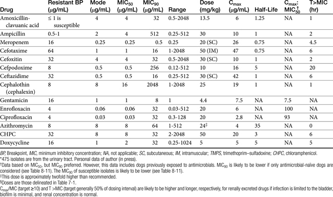
Table 8-9 Pharmacokinetic and Pharmacodynamic Indices for Treatment of Canine and Feline Escherichia coli Associated with Urinary Tract Infections

image

KEY POINT 8-22

The need to treat a urinary tract infection should be carefully critiqued. Resolution of the cause of the infection is critical to therapeutic success, including avoidance of resistance.

Enterococcus spp. increasingly is competing with E. coli as the predominant organism associated with UTI. Ling and coworkers105 found that most UTIs were caused by a single agent, with E. coli the organism most commonly isolated (45%; see Table 8-1). The number of animals exhibiting signs associated with UTI was not indicated in the study nor was identification of the UTI as a first occurrence or a recurrence. Although UTIs occurred more frequently in female dogs, the frequency of specific organisms causing UTIs was generally similar between genders, with the exception of Proteus and Enterococcus spp., which were cultured more freqently from females (10.5% and 9.6%, respectively) compared with males (5% and 5% to 6%, respectively). Multiple organism infections were more likely to be found in females than in males. Age did not appear to be an important predictive factor of the most likely pathogen. The majority of infections were associated with ≥100,000 CFUs/mL, whereas approximately 20% of infections, regardless of the number of infecting organisms or the gender of the dog, were characterized by ≤1000 CFUs/mL.

A retrospective study of UTI in dogs admitted to a veterinary teaching hospital and subsequently diagnosed with UTI (n = 240)112 found that E. coli was the causative organism in 50% of the cases. Although the majority of the remaining organisms were gram-negative coliforms (e.g., Proteus, Klebsiella spp.) selected gram-positive organisms (Staphylococcus and Enterococcus spp. being the majority) also were cultured.

The causative agents associated with recurrent UTI (defined later) appears to be similar in that E. coli is most common. However, the causative agents in the remainder may differ. Seguin106 retrospectively examined recurrent UTIs in dogs (n = 441 isolates from 373 positive cultures). E. coli (47%) was the most commonly isolated, followed by Enterococcus spp. (21%). Other organisms included Proteus (7.7%), Klebsiella (5.9%), Staphyloccoccus (5.2%), and Pseudomonas spp. (4.1%). Mixed infections occurred in 17% of the cultures. In a prospective study of dogs receiving glucocorticoids (n = 127), the most common organism isolated was E. coli followed by Enterococcus spp. A retrospective study in cats (n = 141) with diabetes mellitus113 also identified E. coli as the most common organism. However, a study of 123 specimens collected from asymptomatic cats found Enterococcus faecalis to be the most commonly isolated organism (43%), with E. coli the second most common (32%).114 There does not appear to be any information regarding the causative agent associated with infection in different regions of the urinary tract.

Limited information is available regarding the susceptibility of organisms causing UTI. Attention to this issue in clinical studies generally addresses patterns of susceptibility but not the level; however, the latter becomes important to the detection of emerging resistance in those isolates considered susceptible based on CLSI criteria. Boothe and coworkers115 (see Table 7-3) have described the proportion of E. coli isolates susceptible to traditional first-choice antimicrobials, with regional differences apparent. Decreasing susceptibility is likely to limit empirical predictability of those drugs that remain effective. These data underscore the importance of susceptibility methods that allow discernment of different levels of susceptibility. The data also underscore the importance of designing dosing regimens that maximize doses or (for time-dependent drugs) intervals such that resistance might be minimized (see Chapter 6).

In a retrospective study of UTI in dogs receiving glucocorticoids,116 organism susceptibility was recorded (n = 32): trimethorpim–sulfonamide (97%), amoxicillin–clavulanic acid (76%), tetracycline (70%), ampicillin (61%), and cephalexin (52%). Interestingly, only 7% were susceptible to enrofloxacin, but only 6 dogs were receiving enrofloxacin.

Pathophysiology

The clinical signs of UTI vary with the site of infection. As with other body systems, the inflammatory response largely is responsible for the clinical signs of UTI. Bacteriuria can be asymptomatic, detected on urinalysis but causing no clinical signs.109 Acute cystitis can cause dysuria but rarely causes signs of systemic inflammation. Acute pyelonephritis, however, is often associated with signs of systemic inflammation, including fever. As in any body system, evidence of inflammation is not necessarily evidence of infection and the need for antimicrobial therapy. Likewise, absence of inflammation does not rule out bacterial infection.

Possible sources of UTI include ascension from the urethra and hematogenous and lymphatic factors. Ascending infection is by far the most common route of infection, although the kidney is predisposed to develop infection associated with blood-borne organisms. In both humans and dogs the origin of bacteria infecting the urinary tract is generally fecal, with the frequency of infection by a particular strain depending on the virulence of the organism. Pathogens generally travel along the urethra to the bladder. Anatomic deformity and turbulent urine flow may facilitate antegrade movement of organisms toward the bladder. Female patients are more predisposed to ascending infection because of the shorter length of the tract and increased risk of contamination. Once in the bladder, infection can continue to ascend the ureter to the kidney, particularly if vesicoureteral reflux is present.

Because E. coli is consistently the most common organism associated with UTI, a focus on its pathophysiology is warranted. E. coli can be identified based on the presence of selected antigens that serve as a basis for serotyping: O, or somatic (more than 140 serotypes); K, or capsular (which may be associated with more virulence factors); and H, or flagella (necessary for ascending infection and renal invasion). E. coli is also represented by a number of pathogenic strains based on the presence of specific virulence factors that facilitate infection. Strains are broadly categorized as intestinal or extraintestinal; the latter group generally is recognized to originate from intestinal strains that subsequently acquire virulence factors that facilitate extra-intestinal survival. Among the extraintestinal strains are uropathogenic E. coli (UPEC) represented by many different serotypes.109 Initial infection by UPEC depends on adherence to uroepithelial cells (e.g., pili or fimbriae). Type 1 pili are among the most important virulence factors facilitating invasion, adherence, and persistence of E. coli infections; similar factors are associated with infection by Proteus spp. and Klebsiella spp.117b Toxins are released; examples include hemolysin, causing uroepithelial cell death (thus providing nutrients for UPEC) and cytotoxic necrotizing factor, which stimulates apotosis and inflammatory cell chemotaxis. Other virulence factors act to scavenge environmental iron necessary for extra-intestinal survival of the bacteria (e.g., enterobactin, which itself is inactivated by lipocalin 2, a bacteriostatic factor secreted by host leukocytes); others impair the hosts’ defensive ability to scavenge iron. Factors also facilitate uroepithelial cell penetration by E. coli; rapid intra-epithelial proliferation of microbes is accompanied by biofilm formation. Exfoliation of surface uroepithelial cells that occurs with urination is an important host protective mechanism but may facilitate recurrence as infected uroepithelial cells are exposed from the deeper layers. Inflammatory cells respond to cell destruction.

Much of the understanding associated with UPEC pathogenesis comes from contrasting it to a UPEC strain that is associated with much less pathogenicity, causing asymptomatic bacturia (ASB; strain 83792). This strain has been used prophylactically to infect the bladder of humans afflicted with UTI associated with more pathogenic UPEC; this unique approach to treatment is currently undergoing clinical trials.117c

Understanding the role of virulence factors in UTI is a prelude to identifying therapies and alternative antimicrobials. Among the factors targeted are adherence factors, in part because of their critically important role in initial infection. E. coli contains adhesins that bind to the glycolipid receptors. Mannose-containing receptors are present on most Enterobacteriaceae. On entry into the lower urinary tract E. coli organisms associated with canine UTI appear to adhere primarily through these mannose-sensitive adhesins, initiating colonization. In contrast, mannose-resistant fimbriae and other adhesins appear to be critical for colonization of the renal structures. Binding between receptor and adhesin changes the receptor-bearing cell. The severity of a UTI may be correlated with the degree of adherence to uroepithelial cells. Organisms causing acute pyelonephritis in human patients are characterized by higher adherence compared with organisms causing asymptomatic bacteria.117 The interaction between microbe and receptors may offer a target for treatment. Treatment with mannose or similar molecules has been proposed to block the receptors, thus reducing adherence.117 However, while this may decrease infection in the bladder, some of the most pathogenic E. coli do not recognize mannose (e.g, UPEC associated with renal infection). Further, interaction between the pathogen and the mannose receptor may be important in the initiation of host defense. Cranberry juice extract contains proanthocyanidins that appear to block uroepithelial cell adherance (Type pili) receptors, presumably precluding bacterial adherence. Several antimicrobials interfere with bacterial expression of fimbrial adhesins and thus may prevent bacterial attachment and colonization. Examples include penicillin, ampicillin, amoxicillin, and streptomycin. Once-daily administration of antimicrobials at reduced (one half to as little as one eighth) doses also may be able to prevent UTIs because of interference with fimbrial expression or formation.119 The role of fosfomycin for this indication requires further development. Among the adherence factors, biofilm has a profound influence on UTI, particularly persistent or recurrent UTI caused by E. coli.117a,b Although bioifilm clearly contributes to infections associated with urinary catheters, its role in chronic UTI is less appreciated. Biofilm physically and functionally contributes to antimicrobial resistance. A crystalline-based biofilm produced in association with urease production (and urinary alkalinization) appears to contribute to, and indeed, be necessary for the formation of struvite crystals. Use of compounds such as iodoacetamide (IDA) and N -ethyl maleimide (NEM) that inhibit enzymes necessary for the formation of the biofilm matrix of these crystials is an area of investigation.

Predisposing Factors

A number of microbial factors increase the risk of UTI.104 Siqueira and coworkers118 have described virulence factors associated with E. coli isolated from dogs with UTI (n = 51) and pyometra (n = 52) and feces of healthy dogs (n = 55). These include but are not limited to other microbial antigens, production of toxins such as hemolysin or urease, and the mucoid polysaccharide capsules (e.g., Pseudomona spp.). Urease producers (Staphylococcus, Proteus, or Mycoplasma spp.) increase the risk of struvite urolithiasis, predominantly in dogs.

Other host factors increase the risk of UTI. Anatomic predispositions to infection include perineal urethrostomy (particularly in cats). Bacterial infection is rarely the initial cause of disease of the lower urinary tract in cats, but development of infection is a common sequela.108 The role of viruses in feline lower urinary tract disease has been reviewed.120 Viruses that have been isolated from the urinary tract of cats with spontaneous disease include feline calicivirus, bovine herpesvirus 4, and feline syncytium-forming virus.121 Other potential uropathogens include mycoplasmas and ureaplasmas.111,121 Risk factors have been described in cats with diabetes mellitus, with infection present in 18 of 141 cats.113 Hyperthyroidism also is a risk factor. Immunosuppressive therapy is a risk factor, as has been demonstrated prospectively in dogs, (n = 127) receiving glucocorticoids (prednisolone or methylprednisolone): 18% were positive (compared with a matched control set of dogs not receiving glucocorticoids) on at least one culture.116

Recurrent UTIs reflect either persistence or relapse of the infection or reinfections.106,109 Persistent or relapsing infections reflect therapeutic failure, whereas reinfection reflects a new bacterial species or strain following a period of urine sterility (i.e., negative urine culture). Superinfection refers to infection with a different organism that emerges during treatment of the original organism. Discriminating among persistence, relapse, or reinfection is difficult. Drazenovich and coworkers122 used pulsed-field gel electrophoresis (PGFE) to pulsotype E. coli isolates associated with persistent UTI in dogs in an attempt to discriminate between recurrence and reinfection. Interestingly, of the E. coli in their study (n = 12 dogs, 47 isolates), only two dogs had the same PFGE genetic pattern (pulsotype); further, few virulence factors were identified among infecting isolates. Frietag and coworkers123 studied whether antimicrobial susceptibility profiles could predict a recurrent infection as persistent or relapsing, versus reinfection, with pulsotypes as the determining factor. Susceptibility was effective only 58% of the time in predicting the pattern of recurrence. However, the study group included only five cats (17 isolates); the interval between diagnosis ranged from 6 weeks to 2.5 years. Of the 17 isolates, 9 unique isolates were identified, with no more than 2 from each cat, but susceptibility patterns differed within and between clones. Because PGFE is based largely on chromosomal DNA (i.e., does not include plasmid DNA) and detects the presence of the gene but not its expression, pulsotypes may not be the most appropriate standard on which to base recurrence patterns.

Causes of recurrent UTI (defined as 2 or more in a 6-month period) were retrospectively studied in dogs (n = 100).106 The median age was 7.7 years. Persistence (defined as same organism and same susceptibility pattern; 42%) and reinfection (different isolate; 50%) were equally responsible for recurrence. Superinfections were identified in 2% of dogs; it is not clear how many animals were cultured while on antimicrobial therapy; the proportion may be higher if superinfections are prospectively sought. Dogs younger than 3 years of age were at greater risk, whereas dogs older than 10 years were associated with a decreased risk of recurrence. This may reflect, in younger dogs, anatomic or other underlying causes being more important to recurrence. Females were at greater risk than males, and sporting dogs, nonsporting dogs, and hounds were at risk compared with other breeds. E. coli (56%) and Enterococcus spp. (21%) were the most common organisms. Multiple isolates were present in 18% of infections. An underlying cause (or causes) was found in 71 of the dogs; correction occurred in approximately 25 of these. Disorders include abnormal micturition, anatomic defects, altered urothelium (i.e., tumors or uroliths), altered urine composition (hypoadrenocorticism or diabetes mellitus), and impaired immunity (e.g., chemotherapy, hyperadrenocorticism). Dogs treated without removing the underlying cause of disease were more likely to be considered poorly controlled (74.5%), with duration of a disease-free period being less than 8 weeks. Correction of underlying disease or therapy intended to prevent reinfection (i.e., low-dose, long-term antimicrobials) was associated with better control. Over 29% of the infections were associated with multidrug resistant microbes, with abnormal micturition more commonly associated with resistant organisms. Interestingly, 87% of the dogs in which relapse occurred were initially presented for a problem other than UTI, and 50% of the dogs studied were asymptomatic for UTI. Sediments in 15% of the dogs were not indicative of infection, and these isolates in particular tended to be resistant. The authors concluded that culture and susceptibility testing may be indicated in the presence of a predisposing disorder.

The need for preemptively culturing urine in animals predisposed to infection was supported by the prospective findings of Torres and others116 and Bailiff and others113 Torres and others116 studied UTI in dogs (n = 127) receiving glucocorticoids; 18% were positive (compared with a matched control set of dogs not receiving glucocorticoids) on at least one culture, although no dog had clinical signs of UTI. However, pyuria or bacturia predicted positive culture 90% and 95% of the time, respectively. The most common organism isolated was E. coli, followed by Enterococcus spp. Superinfection was present in 6 of 52 dogs whose urine was cultured while receiving glucocorticoids; each dog was receiving cephalexin (22 mg/kg bid by mouth), and the cultured isolate was resistant to cephlexin. Bailiff et al.113 retrospectively studied 141 cats with diabetes mellitus. Of these, 13% also had UTI, with E. coli the most common organism. Risk factors were female and low body weight; no treatment-related risk factors were identified, including level of diabetic control, or glucosuria.

KEY POINT 8-23

Culture and susceptibility testing should be strongly considered as the basis of therapy in any urinary tract infection that is not occurring for the first time.

Occult UTIs are not limited only to animals with underlying disease; it is possible that “asymptomatic bacteria” is a more appropriate term. Occult UTI appears to occur more commonly than previously thought in cats.114 In a study of 123 specimens collected from asymptomatic cats, 38% were positive. Positive cultures were more prevalent in older female cats. In contrast to most studies, E. faecalis was the most commonly isolated organism (43%), with E. coli the second most common (32%).

Other factors may contribute to recurrence or reinfection. Urinary tract catheterization contributes to an increased risk of UTI in an experimental model of male feline cystitis.124 Catheterization is a common cause of ascension of bacteria from the urethra to the bladder. In human patients one catheterization results in infection in 1% of patients, and infection develops in most, if not all, patients within 3 to 4 days of placement of an indwelling, open-drainage catheter system.109 A study of infection in cats after perineal urethrostomy found that while the surgical procedure does not predispose the cat to recurrent bacterial UTI, surgical alteration of the urethral surface coupled with underlying uropathy may increase the risk and thus prevalence of ascending infection.125 Catheters should be used only in cats for which obstruction is likely if catheterization is not performed.126 Urinary calculi will contribute to canine and feline lower UTIs. One study reports growth of bacteria in urine or calculi of 41% of cats with urinary calculi.127 Staphylococcus spp. were responsible for most (45%) of these infections. Pyometra may serve as a source of reinfecting organisms,128 as might tumors (e.g., transmissible venereal tumors).129

Resistant Urinary Tract Infection

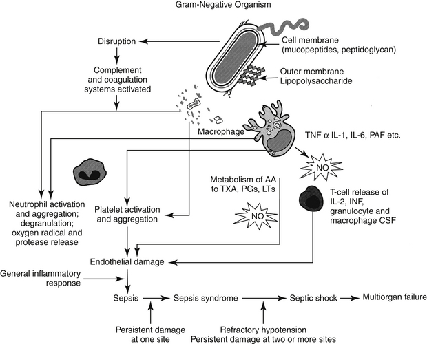
Resistant microbes, and particularly E. coli, are increasingly common causes of UTI (Figure 8-5; see also Table 7-3). In a prospective study of E. coli isolates, the majority of which were associated with UTI in dogs (n = 240) at a teaching hospital, Boothe and coworkers115 found the rate of resistance of E. coli to common first-choice antimicrobials limited empirical selection of an appropriate antimicrobial at that hospital (Figure 8-5). The percentage of organisms resistant to first-choice drugs exceeded 50% for ampicillin (e.g., amoxicillin) and was 40% or more for drugs considered relatively invulnerable resistant to beta-lactam resistance (i.e., amoxicillin–clavulanic acid and cephalothin). Moreover, 40% of the E. coli organisms also were resistant to trimethoprim–sulfamethoxazole. Disconcertingly, 40% of organisms were resistant to fluoroquinolones, the first choice for complicated infections, and 50% were resistant to extended-spectrum penicillins (carbenicillin, piperacillin, and ticarcillin). Indeed, the only drugs to which E. coli was predictably susceptible were nitrofurantoin and the aminoglycosides, particularly amikacin. Third-generation cephalosporins ceftiofur and particularly ceftazidime also exhibited susceptibility, although extended-spectrum beta-lactamases (see Chapter 7) were not tested. Several factors were associated with UTI resistance in these organisms, with antimicrobial therapy within the past 5 days and duration of hospital stay being the most important. The incidence of resistance in this retrospective study is in contrast to earlier reports of UTI resistance. For example, in a 2001 study, E. coli isolates (the majority being UTI) cultured from dogs were susceptible to norfloxacin (90%), enrofloxacin (87.5%), gentamicin (90.7%), and amikacin (85.9%).130 The proportion of E. coli classified as resistant to amoxicillin or amoxicillin–clavulanic acid is likely to increase in the next few years because CLSI has lowered the breakpoints for these two drugs (along with cephalexin). Other investigators have reported E. coli resistance, particularly to fluoroquinolone. In a study by the author of E. coli organisms (n = 50)—most of which were isolated from the urine—the MIC90 for the fluroquinolones was greater than 64 μg/mL, suggesting high-level multistep resistance.131 Multidrug-resistant E. coli has emerged as a cause of nosocomial infections in dogs132 and UTI in canine critical care patients.106,133 Data generated by the author suggest that resistance to fluoroquinolones is multidrug in nature, reflecting not only mutations in topoisomerases but also induction of efflux pumps.134 E. coli is not the only organism associated with UTI to which resistance has emerged in clinical cases. Enterococcus resistance in particular has emerged, probably reflecting its presence as the major gram-positive aerobe in the gastrointestinal tract, although its role as a uropathogen is not clear.

image

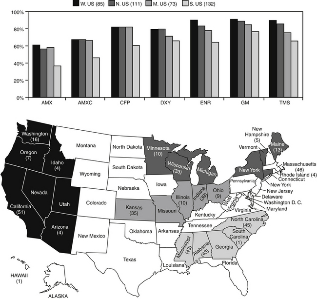
Figure 8-5 Percentage of canine and feline pathogenic Escherichia coli (n = 401) collected during May through September 2005 susceptible to selected antimicrobials. Resistance differed by regions (regions delineated in map), with the south characterized by the lowest level of susceptibility for each drug. AMX, Amoxicillin; AMXC, amoxicillin–clavulanic acid; CFP, cefpodoxime; DXY, doxycycline; ENR, enrofloxacin; GM, gentamicin; TMS, trimethoprim sulfonamide.

Although in vitro resistance may not necessarily predict in vivo failure, evidence in human medicine suggests otherwise. As in dogs, E. coli, is the most prevalent uropathogen in humans. However, antimicrobial resistance to first-choice drugs, trimethoprim–sulfonamethoxazole (TMP-SMX) and ampicillin, often exceeds 30% in humans. In women with UTI caused by E. coli characterized by in vitro resistance to TMP-SMX and subsequently treated with the combination drug, approximately 50% were bacteriologic failures and another 40% clinical failures.135 Previous antimicrobial therapy profoundly affected the likelihood of resistance, with the risk greater if the antimicrobial of interest has been used. Again, in women the most important independent risk factors for TMP-SMX resistance in nonhospitalized cases was use of the antimicrobial within the past 3 months. Those who had taken any antimicrobial were more than twice as likely to be infected with a resistant isolate; use of TMP-SMX within the past 2 weeks was associated with a sixteenfold greater risk of infection with a resistant isolate.135 Interestingly, trimethoprim by itself results in cure rates that are similar to combination with sulfamethoxazole; because it is associated with fewer side effects, it is the preferred drug for treatment of uncomplicated UTI by some.135

Urinary catheterization is not only a recognized risk factor for UTI but also for antimicrobial resistance. Catheterization has resulted in bacteriuria in previous bacteria-free urine and has been associated with changes in urine microflora, as well as increased resistance.136 Although aseptic techniques will reduce the risk of infection, infection is not prevented. The risk of persistent UTI in cats with experimentally induced cystitis was increased with catheter placement, despite the use of a closed system of urine drainage.110 The risk of infection can be correlated with duration of catheter placement, with the risk being reduced in patients catheterized for less than 3 days.137 Previous antimicrobial therapy is likely to contribute to the risk: resistance in dogs catheterized more than 5 days was strongly associated with the advent of resistant microrganisms.115 Catheter type influences the risk of infection, probably because of its impact on biofilm formation (see Chapter 6) and bacterial swarming.138 However, isolation of organisms within microcosms associated with biofilm and subsequently isolated from urine collected from the catheter—or from the catheter tip itself—does not necessarily indicate infection137; The incidence of resistance in E. coli collected from catheters tips is greater than that collected by cystocentesis112 If infection is present, the causative organism should be identified based on cystocentesis, or urine collected from the passage of a fresh, sterile catheter. However, catheterization should be kept to a minimum. Intermittent catheterization in spinal patients offers unique challenges. In humans, risk of resistance was related to frequency of catheterization (three times was associated with a greater risk than six times per day) and bladder overfilling (overfilling increased risk); previous indwelling catheterization also increased the risk. Trauma to the urethera during catheterization in itself did not increase the risk of infection, but the development of “false passages” or strictures resulting from repeat trauma did. Hydrophilic catheters appeared to reduce the risk of infection. Antimicrobials should be used judiciously in spinal patients. Whereas symptomatic infections are treated, asymptomatic bacteriuria is not necessarily treated; not surprisingly, long-term prophylactic antimicrobial therapy is associated with an increased risk of infection. As such, antimicrobials therapy tends to be limited to treatment of symptomatic infections or prophylaxis during initial (short-term) catheterization.139 The efficacy of antimicrobial infusion at the end of catheterization is not clear, with studies generating conflicting results. However, in contrast to multiple systemic dosing, single local infusion of high concentrations of drug is less likely to lead to resistance and potentially might do less harm than systemic therapy. Local intravesicular infusion of an antimicrobial might be considered of inducing minimal risk to resistance.

Difficulties encountered in the successful treatment of UTI, and particularly multidrug-resistant UTI, mandate that approaches be taken to prevent resistance, which includes preventing infection. Because previous use of antimicrobials consistently is a major predictor of emerging resistance, the question of the need to treat should be the first consideration for all infections. If reasonable alternatives exist to antimicrobial drug therapy, they might be considered first or in addition to antimicrobial therapy.

Prevention of Urinary Tract Infection

A number of host factors prevent or limit bacterial infection in the bladder.109 Among the host factors important in preventing infection are normal micturition, normal anatomic barriers, systemic immunocompetence, mucosal defense barriers, and the inherent antibacterial properties of urine. Recurring infections are most likely to reflect failed host defenses, whether originating spontaneously or iatrogenically (e.g., immunosuppressive drugs).106 Support of factors can be targeted with adjuvant therapies instead of antimicrobial treatment, when possible, or to facilitate antimicrobial therapy, particularly in the patient at risk for recurrent infections.

The decision to treat or not treat an infection might take into account the size of the inoculum (i.e., CFU/mL of urine) but should also take into account host factors (e.g., clinical signs, history, ability to control underlying cause, contributing factors, previous response to therapy). The location of infection may play a major role in determining the need for therapy. Whereas asymptomatic bacteriuria will often resolve or become self-limiting if left untreated, bacterial pyelonephritis is likely to progress.104,117 Virulence testing or serotyping eventually may be helpful in the decision to treat or not treat. The normal flora of the vulva and prepuce may be an important host defense mechanism against infecting microorganisms of the urinary tract. Normal flora may prevent colonization by pathogenic organisms or disrupt metabolism of pathogens. Secretory antibodies may coat infecting organisms, preventing adherence, and reduced antibody production may promote infection. Mucus may have other antibacterial effects. In the bladder, mucosal secretion of surface mucopolysaccharides is important to host defense by preventing attachment of bacteria. Destruction of this layer facilitates infection. Treatment with sulfonated glycosaminoglycans intraluminally may coat the uroepithelium and thus provide a barrier to bacterial adherence. Administration of carbenoxolone (a licorice derivative) stimulates secretion of mucosal polysaccharide and (in rabbits and humans) increases the clearance of E. coli infection. In the bladder, the composition of urine can affect bacterial growth. Urine concentration (unless extreme) is not likely to affect bacteria (bacteria are generally hypertonic compared with their environment), but high concentrations of urea or other compounds or low pH may impair bacterial growth. The addition of prostatic fluid inhibits bacterial growth. Exceptions include selected Staphylococcus and Proteus spp., which are relatively resistant to the antibacterial effects of urea.109 Tamm–Horsfall protein secreted by the cells of the ascending loop of Henle binds to E. coli by way of mannose-containing side chains and, as such, probably acts as a urinary bacterial defense mechanism. Other factors that help prevent or reduce bacterial infection include frequent urination, a small residual urine volume in the bladder, and rapid urination.

KEY POINT 8-24

The use of adjuvant therapies, particularly in the prevention of urinary tract infections, should be considered carefully, particularly with regard to clinical evidence of efficacy.

Therapy

The Need for Drug Therapy

The presence of bacteriuria is not necessarily an indication of the need for antimicrobial therapy (Figure 8-6). Antimicrobial therapy should be used only when reasonable evidence of infection exists. Bacterial UTI occurs much less frequently in cats than in dogs, and even the presence of clinical signs indicative of cystitis should not be interpreted as a need for antimicrobial drug therapy.

image

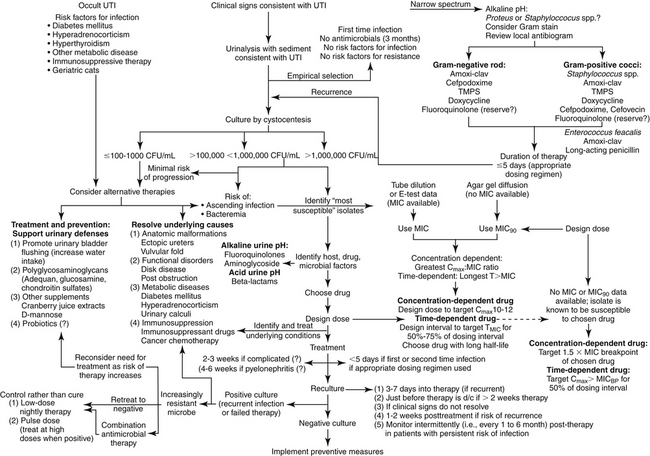
Figure 8-6 Suggested algorithm for treatment of urinary tract infections in dogs or cats. Treatment should be begun with assessing the need to treat with antimicrobial drugs (upper left) versus alternative therapies. Empirical treatment (upper right) should be pursued only for uncomplicated infections, including animals not recently exposed to antimicrobials (this may include any household member or pet). Culture and susceptibility data (lower right) should be used to both select drug and design the dose; in its absence, population pharmacodynamic data (MIC90) can be used if the identity of the infecting microbe is known. Treament of recurring infections (lower left) requires a series of reassessments, including recultures. With continued recurrence, the need for continued therapy should be balanced with the risk of therapy to the patient; prophylactic therapy might be considered to prevent recurrence once the urine has been cleared of infection (far lower left).

In humans, to prevent resistance, treatment generally is not indicated in asymptomatic bacteriuria except under certain conditions in which the patient is at risk, such as during pregnancy or invasive surgical procedures.140 Likewise, for veterinary patients the risk of emerging resistance must be weighed against the risk of failing to treat. If the patient is one for which aminoglycoside therapy is inadvisable, the need for treatment should be even more closely examined. With Enterococcus bacteriuria, treatment should not be implemented unless the need is clear. Clearly, if the decision is made to treat a UTI, therapy must be aggressive, designed to kill invading pathogens as well as emerging mutants as rapidly as possible. Non-antimicrobial alternatives should be considered in lieu of or in addition to antimicrobial therapy.

The Sequelae of Drug Therapy

The traditional goal of drug therapy for UTI is to eliminate bacteriuria in animals exhibiting clinical signs and urinalyses consistent with UTI. However, four sequelae of antimicrobial therapy may occur.104,109 Cure can be defined as negative urine cultures during and after (usually 1 to 2 weeks) antimicrobial therapy. Quantitative bacterial counts should decrease within 48 hours after initiation of an appropriate antimicrobial. Cure does not rule out the possibility of reinfection. UTIs may reflect first-time or recurrent infections. Chronic UTI is often used to refer to persistence of infection. The previously defined terms recurrence, persistence, and relapse often are used interchangeably when referring to UTIs. Persistence, or recurrence, can refer to presence of significant or low numbers of bacteria after 48 hours of therapy. If the numbers are significant, antimicrobial resistance or insufficient drug concentrations (e.g., improper dose, poor oral absorption, poor renal elimination) should be suspected. If numbers are very low, a continuous source of bacteria in the urinary tract (e.g., urinary calculi, prostate, kidney) or contamination from the lower urinary tract might be suspected.109 In such cases, cultures can identify persistent organisms after therapy has been discontinued. An appropriate approach would be classification of recurrent or persistent infections into three categories. Relapse occurs when the same organism causes infection 1 to 2 weeks after therapy has been discontinued. Relapse generally occurs within 1 to 2 weeks of cessation of therapy and may reflect either a very deep-seated infection or an abnormality of the urinary tract (e.g., structural, renal, or prostatic infection). The presence of a different organism is considered reinfection (i.e., a new infection). A new infection also can occur by the same organism located outside of the urinary tract. Generally, reinfection occurs more than 1 to 2 weeks after cessation of therapy. Superinfection may also occur and reflects infection with an additional organism during the course of antimicrobial treatment.104 Evidence of persistent or relapsing infection or superinfection should lead to more aggressive therapy and to the use of bactericidal rather than static drugs. Among the common causes of complications associated with UTI, antimicrobial resistance, unidentified underlying disease, and inappropriate dosing regimen should be considered.

Identification of the Target

In all but simple UTI, culture is indicated. Culture is recommended in patients whose history includes exposure to antimicrobial therapy within the last 3 months; exposure may include any household member, including other pets. The more complicated the infection or the greater the patient is at risk for resistance to emerge, the more important it is to base therapy on susceptibility data. In human patients diagnosis of UTI in asymptomatic patients is. based on at least two clean-catch midstream urine collections. The same organism should be present in significant (see previous discussion) amounts in both cultures. A single culture is sufficient in the presence of symptoms. Urinary cultures should be the basis of antimicrobial selection in complicated infections (e.g., reinfection or relapse, history of antimicrobial use in the past 4 to 6 weeks)104 or if the infection represents a risk to the patient’s health. Infection after recent urinary catheterization also should lead to culture collection. Increasingly, culturing at the outset, even in simple, first-time infections, may become prudent.

Quantitative urine culture (i.e., colony counts) should be implemented to facilitate discrimination of harmless bacterial contaminations (e.g., from the urethra) from pathogenic organisms (see previous discussion). Bacterial counts of more than 105 CFU are clearly indicative of infection regardless of the method of collection, whereas counts between 103 and 105 organisms are considered suspect if not collected by cystocentesis or if collected from female dogs. Counts of less than 1000 CFU should lead to a second culture and, in the absence of clinical signs or mitigating circumstances, consideration of alternative therapies. Methods have been described for culture procedures performed in practice.104 In addition to the method of collection, consideration must be given to sample handling. Samples should be kept refrigerated if the time from collection to processing by the lab is anticipated to exceed 12 hours, unless an appropriate amount of preservative is added. A viable alternative for submission of urine samples that will be in transit longer than 12 hours is collection and subsequent submission using a “paddle” apparatus (e.g., UriCult®).

Antimicrobial Selection

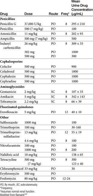
Presumably, a drug that is renally excreted should be selected for treatment of UTIs. Urinary concentrations of such drugs often surpass serum concentrations (up to 300-fold), which is particularly helpful for concentration-dependent drugs (Table 8-10). Susceptibility data do not take these higher concentrations into account, but this may be appropriate. Several caveats must be recognized when basing antimicrobial selection on renal elimination and anticipation of high urine drug concentrations:

Table 8-10 Mean Urine Concentrations for Antimicrobials Used to Treat Urinary Tract Infections in Dogs

image

KEY POINT 8-25

The design of the dosing regimen for treatment of a urinary tract infection should be based on plasma, not urine, drug concentrations.

1. Drug is not likely to be concentrated in some situations. Examples include but are not limited to fluid therapy, diuretic use, renal disease, and other diseases (e.g., hyperadrenocorticism).
2. Drugs characterized by a short elimination half-life may be limited in efficacy if they are time dependent in action. However, plasma elimination half-life may not accurately reflect contact time of drug in the urine if the drug is excreted renally. Contact can be enhanced by drug administration immediately after micturition or before an anticipated micturition-free period (e.g., at night).
3. Intraepithelial growth and biofilm may preclude drug efficacy even if concentrations are high in urine.
4. If the UTI is associated with infection in the blood (or in the presence of bacteremia), kidney, or prostate, then antimicrobial selection should be based on anticipated plasma (or tissue) drug concentrations and serum breakpoint MICs. A similar approach should be taken for recurrent infections or complicated infections.

A prudent approach for treatment of UTI is choice of a renally excreted drug but dosing based on drug concentrations achieved in plasma. Data from a few prospective studies, including animal model studies and human patient–based clinical trials, generally demonstrate that breakpoints based on plasma drug concentrations rather than urine appear to moderately predict outcomes associated with treatment of UTI.45 For example, maximum efficacy of aminopenicillins in treating UTI in human clinical trials is achieved when plasma drug concentrations are maintained above the MIC for 30 hours or more.271 Accordingly, CLSI interpretive criteria for infections in other tissues are relevant to UTI. Drugs that are not eliminated in urine might be characterized by lower concentrations in urine compared with tissues but still can be beneficial for UTI if dosing regimens are appropriate. Some drugs used to treat UTIs (e.g., indanyl–carbenicillin, nitrofurantoin) are recognized not to achieve effective concentrations in other tissues, and therefore use in areas other than the urinary bladder should be done cautiously.

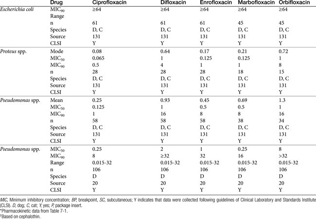
As resistance to E. coli and other uropathogens increases, empirical therapy for UTI increasingly will be limited (Table 8-9). Caution is recommended because of the recent recognition of the growing incidence of antimicrobial resistance. Among the organisms frequently causing UTIs in dogs and cats, E. coli, Proteus spp., P. aeruginosa, and Enterobacter spp. are among the organisms that vary widely in their susceptibility pattern and for which empirical therapy is more risky (Table 8-11; see also Table 7-3Table 7-3). Empirical therapy, if it is to be pursued, is indicated only for uncomplicated infection—that is, those patients in which no underlying structural, neurologic, or functional abnormality can be identified.104 The absence of previous antimicrobial therapy should also be interpreted as uncomplicated. Once relapse occurs (see later discussion), an infection should no longer be considered uncomplicated.

Table 8-11 Susceptibility Data (μg/mL) for Drugs for Gram-Negative Pathogens Collected from Antimicrobial-Free Dogs and Cats

image

Attention should be paid to the pH of the urine compared with the pKa of the chosen drug. In the presence of an alkaline pH, weakly basic antimicrobials might be considered (aminoglycosides, and fluoroquinolones, with the latter being amphoteric but more effective in an alkaline urine). Because urease producers may alkalinize the urine, drugs targeting such organisms (e.g., Proteus, Staphyloccocus, and some Klebsiella spp.) should be selected. In the presence of an acidic urinary pH (perhaps caused by E. coli), weakly acid drugs (e.g., penicillins, cephalosporins, potentiated sulfonamides) might be better empirical selections. However, if the drug is highly concentrated in the urine, even a predominantly ionized drug (e.g., a weak base in an acidic environment), may be sufficiently un-ionized to ensure effective concentrations.

Infections uncomplicated by previous antimicrobial therapy or in regions for which resistance has minimally emerged may respond to empiric therapy. E. coli or Enterobacter spp. traditionally have been considered responsive to trimethoprim–sulfadiazine, third-generation cephalosporins (cefpodoxime, cefovecin), or amoxicillin–clavulanic acid combinations. Among these, penicillins may be preferred because of their effects of fimbriae. Approximately 60% of E. coli are resistant to cephalexin. Klebsiella, Proteus, and Staphylococcus spp. are likely to respond to a first- or third-generation cephalosporin or to amoxicillin–clavulanic acid. Amoxicillin or ampicillin (preferably combined with a beta-lactamase protector) should be effective if a decision is made to treat Enterococcus spp. (see Tables 7-9, 8-3, and 8-6). However, for Enterobacteriaceae in particular, integration of pharmacokinetics and pharmacodynamics raise concerns regarding efficacy for drugs with short elimination half-lives, particularly if infection involves tissue other than the urinary bladder. In the absence of an MIC, the more stringent MIC90 (see Tables 8-9 and 8-11) and Cmax from package inserts or the literature (see Table 7-1) can be used to design dosing regimens. The highest end of the dosing regimen is indicated, particulary for at-risk patients. For selected concentration-dependent drugs, Cmax:MIC should exceed 10 to 12, and for time-dependent drugs, T>MIC should be at least 50% if not more of the dosing interval (generally, initial target concentrations should exceed the MIC by at least two to four fold (see Table 8-9). For time-dependent drugs, those with longer half-lives might be preferred (e.g., cefovecin and cefpodoxime). Although the package insert for cefovecin indicates that MIC90 concentrations for E. coli (1 μg/mL) are not achieved after a recommended dose of 8 mg/kg subcutaneously, predicted unbound plasma drug concentrations will be maintained at or above this concentration for 3 days. Cefovecin is approved for treatment of canine E. coli UTI in Canada at 8 mg/kg. Note that neither cefovecin nor cefpodoxime are effective against Enterococcus spp., indicating that the Gram-staining characteristics of the infecting organism might facilitate proper antimicrobial choice for empirical treatment of a UTI in dogs or cats. Because resistance in E. coli toward fluoroquinolones with rare exception is associated with MDR, their use for treatment of UTI should be considered second tier and ideally, be based on culture and susceptibility testing. Care should be taken using the fluoroquinolones for Enterococcus spp., for which efficacy can be variable. Boothe et al demonsrated that for E. coli, when using the MIC90 for fluoroquinolones, the Cmax:MIC90 failed to reach ≥10—as is suggested for concentration-dependent drugs—even with the highest dose for each drug. (see Tables 7-12 and 8-9 for additional MIC data).131 However, for isolates that are known to be susceptible to ciprofloxacin, enrofloxacin, and marbofloxacin reached the target Cmax:MIC90 at the high (but not low) dose. Even with susceptibility data that provide MIC, because the tested concentrations are close to the breakpoint MICs, isolates considered susceptible to fluoroquinolones may have already developed low-level resistance, increasing the risk of emergent resistance in at-risk patients.142 Certainly an “I” to any one fluoroquinolone should be interpreted as emerging resistance to others. Isolates with MIC approaching the breakpoint should be treated with alternative drugs, combination drugs, or “added doses” (e.g., high dose, twice daily). Marbofloxacin is preferred for cats at risk for retinal adversities, particularly if dosing twice daily. Although ciprofloxacin is more potent against gram-negative isolates, dosing should be increased to compensate for differences in oral bioavailability and oral dosing should not be used in cats. Fluoroquinolones are also effective against Mycoplasma and Ureaplasma spp.

A summary of treatment of UTI in humans might offer some guidance for empirical therapy in dogs or cats. In women with risk factors for infection with resistant bacteria, or in the setting of a high prevalence of TMP/SMX resistance, a fluoroquinolone or nitrofurantoin is recommended for empirical treatment. The goal of treatment is eradication of infection using shorter courses of therapy (i.e., 3 days) with once-daily dosing of a selected drug or a single dose of a particularly efficacious antimicrobial.141 The role of nitrofurantoin is increasing for treatment of UTI in women, particularly in the presence of increasing antimicrobial resistance to other urinary antimicrobials. Resistance among uropathogens to nitrofurantoin generally is consistently at a low level despite its use for 5 decades. An advantage to nitrofurantoin is its minimal effect on the normal gut flora. Consequently, selection pressure for antimicrobial resistance is reduced compared with other antimicrobials.135 Further, nitrofurantoin does not share cross-resistance with more commonly prescribed antimicrobials, and its use is justified from a public health perspective as a fluoroquinolone-sparing agent. For example, single-dose ciprofloxacin prophylaxis increased the prevalence of ciprofloxacin-resistant fecal E. coli from 3% to 12%.135 After treatment with ciprofloxacin for prostatitis, 50% resulted in post-treatment fecal colonization with quinolone-resistant E. coli genetically distinct from the prostatic infection. Indeed, in humans, although flouroquinolones are effective as short-course therapy for acute cystitis, widespread empirical use is discouraged because of potential promotion of resistance.140 An exception is made for acute (nonobstructive) pyelonephritis but only if culture results direct continuing therapy.

Beta-lactams and fosfomycin are also considered second-line (to TMP-SMX) agents for empirical treatment of cystitis in humans. Fosfomycin may be a reasonable first choice for treatment of UTI in dogs if data support its use for treatment of canine UTI. Advantages to fosfomycin tromethamine justify consideration of its use in dogs. Despite many years of use in humans, it is characterized by an extremely low incidence of resistance. Indeed, canine and feline isolates expressing MDR maintain susceptibility to fosfomycin.135a Further, pharmacokinetics in dogs support its use for treatment of UTI. Its role for treatment of UTI in dogs is emerging; a prudent approach might be as a second- or third-tier drug because of its importance in human medicine. In general, drugs metabolized by the liver (chloramphenicol) or excreted in the bile (macrolides, clindamycin) do not achieve high concentrations in the urine and should generally be avoided for treatment of UTI. Doxycycline might be appropriate if susceptibility data is available, although concentrations achieved in urine might be bacteriostatic.

Duration of Therapy

The “test for cure”104 (or perhaps, more appropriately, “response”) can be based on a second culture 3 to 5 days into therapy. Cure should be anticipated only if the organism count is less than 100 per mL of urine. Urine culture a second time just before discontinuation of therapy has been recommended,104 particularly if antimicrobial prophylaxis is to be implemented. The duration for successful treatment of uncomplicated lower UTIs might be as short as 3 to 5 days, particularly if doses are designed to target all infecting isolates, including those with the highest MICs.104 Such an approach is more likely to be successful if high doses and appropriate intervals are chosen. Treatment may need to be extended, however, if infection occurs anywhere other than the uroepithelium. Traditionally, a 10- to 14-day therapeutic regimen has been recommended for the first episode of therapy; the evidence for this recommendation is limited and clinical trials demonstrating appropriate duration of therapy are needed. Shorter-term antimicrobial therapy (a single high dose) has proved effective for female human patients with a lower UTI. Drugs that have been used successfully in humans for short-term dosing include trimethoprim–sulfonamide combinations, aminoglycosides, selected cephalosporins, and fluoroquinolones. Both single-dose and 3-day antimicrobial treatment regimens have been studied with dogs receiving amikacin and a trimethoprim–sulfonamide combination. Therapy was not uniformly successful, suggesting that caution should be used with this treatment regimen.143 However, these studies were implemented before understanding the importance of reaching targeted PDI (i.e., Cmax:MIC > 10-12 and T>MIC for more than 50% of the dosing interval). Factors that should preclude single-dose antimicrobial therapy for a lower UTI include recurrence, historical poor response to single-dose therapy, underlying predisposing factors to a UTI (including structural abnormalities or metabolic disorders, such as diabetes mellitus, and hyperadrenocorticism), and either pyelonephritis or symptoms of a UTI that have occurred for more than 7 days.

For infections that reflect a relapse, the duration of therapy should be at least 2 weeks; however, for human patients suffering from a relapse, a higher cure rate occurred with a 6-week course of therapy. For animals a duration of 4 to 6 weeks is recommended.104 The duration also might be based on the cause of the relapse and whether the underlying cause is curable and treated. Because relapse is likely to occur shortly after antimicrobial therapy is discontinued, cultures should be collected again 7 to 10 days after cessation of therapy to detect the recurrence. In general, regardless of the sequence of UTI, doses should always maximize efficacy and avoidance of resistance. A new antimicrobial should be selected if infection occurs more than 10 days after cessation of therapy;104 as more time elapses between cessation of therapy and the presence of bacteriuria, the more likely it is that reinfection is the cause of recurrence.

In the event of relapse after 6 weeks of therapy, 6 months of therapy or more may be necessary. A 3- to 6-month duration of therapy may be indicated for animals. However, clinical trials are lacking to document these recommendations as well. Greater care must be taken, in the selection of antimicrobials for longer-term therapy, with special consideration to toxicity. Drugs that are used for long-term therapy in human patients include amoxicillin, cephalexin, trimethoprim–sulfonamide combination, or a fluorinated quinolone. Cultures should be repeated monthly, and as long as significant bacteria are not present, the drug need not be changed. Should relapse occur after a drug is discontinued, the same drug or a new drug should be administered for a longer course of therapy. Long-term therapy may be particularly important for animals in which renal parenchymal damage is a risk. As with first-time infection, and perhaps more so, the need for treatment must be balanced with the disadvantages of treatment (or the risks of not treating should be balanced with the benefits of treating).

Clinical Trials

Despite the frequency of UTI in dogs and cats, the number of clinical trials examining therapy are limited. Pradofloxacin (n = 27 dogs; 5 mg/kg) has been compared with doxycycline (n = 23; 5 mg/kg load, followed by 2.5 mg/kg bid for 2 doses, then 2.5 mg qd) and amoxicillin–clavulanic acid (n = 28; 62.5 mg/kg bid for 10 days) in cats with clinical signs of UTI.144 Cats were not randomly assigned; assignment to treatment group was based on susceptibility; if more than one drug was designated susceptible, animals were assigned to keep groups balanced. All cats receiving pradofloxacin responded, with 13% and 10%, respectively, of the doxycycline and amoxicillin–clavulanic acid groups remaining infected after treatment. However, the proportion of responders was not different among treatment groups, probably reflecting small sample size.

Efficacy of cefovecin (8 mg/kg, administered subcutaneously) was compared with that of cephalexin for treatment of canine (n = 129) UTI.145 Efficacy was based on elimination of the pretreatment uropathogen. Exclusion criteria included local or systemic antimicrobials within the previous 14 days, or short- (7 days) or long-acting (30 days) glucocorticoids. The study was implemented as a multicenter, blinded, randomized study, with cephalexin (15 mg/kg, administered orally bid) serving as the positive control. Animals were treated for 14 days. The most common uropathogen was E. coli (60%), followed by P. mirabilis (12%) and S. intermedius (12%). As would be expected on the basis of the cephalexin MIC90 of E. coli, the overall cure rate for animals infected with E. coli was 79% in the cefovecin group compared with 36% for cephalexin. Bacteria were eliminated in 59% of cefovecin-treated dogs compared with 35% of cephalexin-treated dogs. Of the infecting isolates, 90.5% of E. coli infections were eradicated compared with 53% of E. coli infections for cephalexin. Of infections caused by S. intermedius, 6 of 6 were eliminated by cefovecin, compared with 7 of 10 for cephalexin; for Proteus mirabilius, 9 of 9 were eliminated by cefovecin, compared with 5 of 7 for cephalexin. Interestingly, although 319 dogs with clinical signs of UTI were evaluated for inclusion, only 137 (43%) actually were bacteriuric.

Prophylaxis

Long-term prophylaxis can be implemented for patients at risk for recurrence. Prophylaxis (by definition) can occur only after the infection has been eradicated. The use of low doses of antimicrobials in the presence of bacteriuria is likely to lead to the generation of resistant organisms and is contraindicated. Thus prophylactic antimicrobial therapy of UTIs is indicated for reinfection but not relapse (the latter suggests that the organism was never completely eradicated). The antimicrobial chosen for long-term prophylaxis should be both safe and inexpensive. Trimethoprim–sulfonamide combinations (monitor for immune-mediated reactions), nitrofurantoin, and fluoroquinolones are examples. However, neurologic side effects with nitrofurantoin may preclude its use, particularly long term. Fosfomycin might also fill this role. The dose for prophylaxis is generally reduced to 30% to 50% of the full dose.104 Subtherapeutic concentrations of drugs often are sufficiently inhibitory to prevent infection of the uroepithelium. The drug should be administered at night to maximize contact of the drug with the urinary tract. Intermittent urine cultures (monthly) are indicated to detect breakthrough infections in animals receiving long-term antimicrobial prophylaxis. Negative cultures for 6 to 9 months or more may indicate that prophylaxis is no longer necessary.

Adjuvant Therapy

Diuresis has been advocated in the treatment of UTIs in humans. Advantages include rapid dilution of bacteria, removal of infected urine, and subsequent rapid reduction of bacterial counts. In patients with pyelonephritis, an added advantage may be enhanced host defenses: medullary hypertonicity inhibits leukocyte migration, and high ammonia concentration inactivates complement. In the presence of vesicoureteral reflux, however, diuresis may increase the risk of acute urinary retention.

Use of drugs to modify urinary pH may facilitate the antibacterial effects of urine. The presence of ionizable organic acids (hippuric and gamma-hydroxybutyric acid) in an acidic pH may enhance the antibacterial activity of the urine. Antibacterial activity may be increased by ingestion of cranberry juice (if urinary pH is acidic), which contains precursors of hippuric acid. Cranberry juice extract also should be considered because of its potential ability to block adherence receptors. Methenamine, available as a hippuric acid or mandelate salt, releases formaldehyde at a urinary pH of 5.5 or less, which also can increase antibacterial activity of urine.

Local urinary analgesics, such as phenazopyridine, rarely are indicated for the management of UTIs. Dysuria is most likely to respond to appropriate antimicrobial therapy. These drugs cause methemoglobinemia and are contraindicated in cats.

Drugs or nutraceutical products that enhance polysulfated glycosaminoglycan synthesis (e.g., Adequan, pentosan polysulfate, glucosamine, chondroitin sulfate) might be considered for patients with complicated UTI. Such products may cover or help repair the uroepithelium, thus decreasing bacterial adherence. Gunn-Moore and Shenoy146 prospectively studied the effects of 60 days oral n-acetyl glucosamine in cats (n = 40) with feline idiopathic cystitis using a randomized, double-blinded, placebo controlled study. Response was based on owner assessment. Both groups improved significantly, with 26 of 40 cats suffering recurrences. Although the power of the study was large, the size of the placebo response limited the ability to detect a significant difference between treatment groups. Improvement in both groups was potentially attributed to owners, most of whom changed the cats to moist rather than dry diets. The quality of the glucosamine was not addressed. Wallius and Tidholm147 studied the effects of polysulfated glycosaminoglycans (PGAGs) in cats (n = 19) with clinical signs indicative of cystitis but culture negative. Cats were randomly assigned to receive either saline placebo or 3 mg/kg PGAGs on days 1, 2, 5, and 10. Assessment was based on owner perceptions of improvement. A treatment effect could not be detected because clinical signs resolved in essentially all cats (save one from each group). As with the previous study, the authors of this study could not conclude that PGAGs were not beneficial for treatment of inflammation, including that associated with UTI. Treatment with mannose or similar molecules may block mannose receptors, thus reducing adherence.119

The use of probiotics in the treatment of UTIs in human patients was reviewed by Lenoir-Wijnkoop.148 In general, Lactobacillus spp. are most commonly recommended for treatment of UTI. However, selected species are likely to emerge as more effective than others and microbiota may need to originate from the target species. The use of probiotics in the treatment of renal oxalate stones in human patients was also reviewed by Lenoir-Wijnkoop and coworkers.148 The absence of Oxalobacter formigenes from fecal microbiota increases the risk of kidney stones. Although no study has demonstrated an association between probiotic therapy and decreased renal stones, both animal and human studies have documented that O. formigenes can become established in the gastrointestinal tract, and establishment reduces urinary oxalate concentrations.

Pyelonephritis

Treatment of pyelonephritis may require hospitalization with intravenous fluid administration. Oral antimicrobial therapy is acceptable for mild to moderate cases as long as oral therapy is tolerated well. Because renal dysfunction can be life threatening, antimicrobial selection should ultimately be based on culture and susceptibility data. Therapy can be initiated empirically; however, resistance among E. coli organisms should lead to selections with known susceptibility. Combination therapy should be considered. Urine is not likely to be concentrated, thus added attention must be paid to ensure that effective concentrations reach the target tissue. Pyelonephritis can be associated with bacteremia, particularly gram-negative bacteremia. Clinical signs indicative of severe, life-threatening infection should lead to parenteral antimicrobial therapy with predictably effective drugs (e.g., aminoglycosides, fluoroquinolones, extended-spectrum beta-lactams, and third-generation cephalosporins known to not be extended-spectrum beta-lactam producers). Combination therapy also should be strongly considered. The high concentration of antimicrobial that facilitates treatment of the lower urinary tract (bladder and lower) may not occur in pyelonephritis; thus close attention must be paid to using sufficiently high doses and frequent dosing. Drugs whose efficacy is dependent on a hypotonic environment (compared with the target organism), such as beta-lactams, fosfomycin, or vancomycin, may be less effective in the face of medullary hypertonicity, although this may be less than normal in the face of pyelonephritis. Nonetheless, if drug combinations are used, at least one of the two drugs should not target cell walls. As with infection lower in the tract, bacterial numbers should decrease dramatically within the first 48 hours of treatment. For uncomplicated pyelonephritis, 14 days of therapy may be sufficient of treatment. Cultures should be repeated as previously indicated during and within 1 to 2 weeks of discontinuation of therapy. Complications such as abscessation may require surgical intervention and longer-term therapy.

KEY POINT 8-26

E. coli resistance to fluoroquinolones, when it does emerge, is generally characterized by multidrug resistance.

KEY POINT 8-27

Treatment of pyelonephritis should be approached as a potentially life-threatening disease requiring aggressive but appropriate antimicrobial therapy.

Prostatitis

Pathophysiology

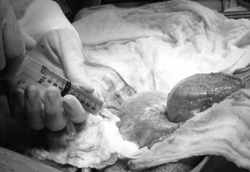
Bacterial prostatitis can present as either an acute or chronic infection. One does not necessarily precede or follow the other. Among the causes of prostatic infection, ascending infection and urine reflux appear to be most likely. Acute prostatitis often is accompanied by fever, pain, and symptoms typical of a UTI. Palpation of the prostate reveals tenderness, swelling, and (potentially) a fluctuant surface (Figure 8-7). Care should be taken when palpating the prostate so that the risk of bacteremia is minimized. Chronic bacterial prostatitis most commonly is caused by gram-negative coliforms, with E. coli being most common, followed by Klebsiella spp., Enterobacter spp., P. mirabilis, and S. aureus.

image

Figure 8-7 Aspiration of a prostatic abscess. Most prostatic abscesses require surgical removal. Abscessation carries the risk of peritonitis and bacteremia. Fluid should be submitted for culture and susceptibility testing.

(Photo courtesy Harry W. Boothe, DVM, MS, DACVS, Auburn, AL.)

Antimicrobial Selection and Use

Antimicrobial penetration into the noninflamed canine prostate is limited. Drugs that are basic and lipid soluble appear to diffuse through tissues best, including macrolides (e.g., erythromycin and presumably azithromycin and clarithromycin), fluoroquinolones, and trimethoprim–sulfonamide combinations. The intense inflammatory response that accompanies acute prostatitis facilitates antimicrobial movement into the prostate,109 although high doses should be used to ensure adequate concentrations at the site of infection. Parenteral antimicrobials based on culture (prostatic fluid or urine) should be used in the presence of life-threatening infection; otherwise, oral therapy is acceptable. Duration of therapy should be at least 4 weeks to minimize the risk of progression to chronic prostatitis. Prostatic abscessation generally requires surgical intervention. Chronic prostatitis in human patients is difficult to cure unless infected tissue is surgically excised. Chronic prostatitis generally results in relapse. Therapy, when successful, generally requires 1 to 2 months of therapy. Antimicrobials that penetrate the noninflamed prostate (e.g., fluoroquinolones, trimethoprim–sulfonamide combinations, or macrolides) should be selected.109

Adjuvant therapy for the treatment of prostatitis includes stool softeners and, if indicated, analgesics. Neutering should be considered.

Infections of the Uterus

Endometritis

Endometritis is associated with sanguineous or purulent vaginal discharge in either the pregnant bitch (if the cervix is open) or nonpregnant bitch.149 As with many organ systems, organisms causing uterine infections tend to be members of the normal flora of the reproductive tract. Organisms most commonly associated with uterine infections include Streptococcus spp., E. coli, Salmonella, Campylobacter, Mycoplasma, and Chlamydia spp.149 Bacterial infections of the uterus resulting in endometritis can be responsible for infertility, abortion, stillbirths, and fetal death. Treatment of endometritis differs in the presence of pregnancy (live fetuses) in part because of potential injury to developing fetuses. Regardless of the antimicrobial, any type of placentation is sufficiently intimate to allow movement of drug administered to the dam or queen to cross the placenta and enter the fetus. In general, however, the fetus can excrete water-soluble drugs more easily than lipid-soluble drugs because water-soluble drugs can be eliminated in the allantoic fluid. In contrast, drug-metabolizing enzymes of the fetal liver are immature and essentially nonfunctional. Although metabolizing activity increases as the term of pregnancy ends, activity remains sufficiently weak that drug elimination is probably ineffectual. Thus lipid-soluble drugs tend to remain in the fetus. When possible, drugs that are water soluble should be selected for treatment of infections in the pregnant bitch.

The beta-lactams are preferred with amoxicillin combined with clavulanic acid or a cephalosporin as first choice, assuming the target organism is included in the spectrum. Care should be taken to avoid cephalexin if an E. coli is suspected. Culture and susceptibility data should be the basis for antimicrobial selection for treatment of endometritis, even if antimicrobials are begun before results are received. Should susceptibility data indicate beta-lactams as resistant, aminoglycosides can be used. Pediatric (and presumably fetal) canine kidneys are protected from aminoglycoside damage because the cortical regions do not fully develop until several weeks postpartum. Fluoroquinolones also appear to be safe for the developing fetus (e.g, enrofloxacin at 15 mg/kg), although the risk of cartilage damage even in the developing fetus might lead to an alternative class of drugs. Response to antimicrobial therapy is indicated by resolution of vaginal discharge, which should occur within several days of beginning therapy. Therapy should continue for 2 to 3 weeks.149 In the case of fetal death, prostaglandin therapy may be indicated to evacuate the uterus. Evidence of septicemia indicates more dramatic and aggressive therapy (see the section on bacteremia).

Pyometra

Although bacterial infection is secondary to the cystic changes associated with pyometra, the infection is the primary cause of illness and death. Organisms most commonly associated with pyometra include E. coli (the majority of infections), hemolytic Streptococcus, Staphylococcus, Klebsiella, Pasteurella, Pseudomonas, Proteus, and Moraxella spp.150 Wadas and coworkers128 demonstrated that E. coli associated with pyometra in dogs was derived from fecal flora and was also identical to those simultaneously cultured from the urine. Hagman and Kühn151 confirmed that E. coli was from the normal flora and was the same as that cultured from the urinary bladder. Host and microbial factors (see Chapter 6) play a large role in the pathogenesis of pyometra and response to drug therapy. In addition to the obvious impact of inflammatory and other debris on antimicrobial efficacy, the local immune response of the uterus is impaired.150 Complications such as bacterial peritonitis and endotoxin contribute to mortality. Endotoxin concentrations generally are increased before implementation of drug therapy, further complicating antimicrobial selection.150

Surgical removal of the infected uterus is the treatment of choice; however, antimicrobial therapy should be implemented regardless of whether ovariohysterectomy is performed. Prostaglandin therapy may be implemented in selected cases (see Chapter 23).

Culture and susceptibility data should be collected before empirical therapy with a bactericidal drug that includes E. coli in its spectrum is begun. Although a beta-lactam antibiotic is generally an excellent first choice, concern with rapid cell death and release of lethal quantities of endotoxin should lead to (1) selection of a carbapenem (meropenem or imipenem), the beta-lactam associated with the least endotoxin release; (2) combination therapy with a non–beta-lactam antimicrobial (e.g., an aminoglycoside or fluorinated quinolone) that is administered before (1 to 2 hours) the beta-lactam; or (3) slow administration of the beta-lactam, perhaps over the course of 1 hour. Therapy for pyometra should include supportive measures such as fluid rehydration and, if indicated, therapy for endotoxic shock. Glucocorticoid therapy is discouraged unless the patient is characterized as adrenocortico-deficient (see Chapter 30).

Respiratory Tract Infections

Principles of Antimicrobial Treatment

A major barrier to passive drug movement from the blood to the site of infection in the respiratory tract is the bronchial–alveolar–blood barrier.152,153 Whereas drugs of a molecular weight up to 1000 can move easily through the open junctions of the capillaries, drugs must passively diffuse through the tight junctions of the alveolar epithelial cells.152 Movement of drugs into bronchial secretions occurs primarily by passive diffusion and is more likely to occur for drugs with favorable physiochemical characteristics such as high lipophilicity and low molecular weight (<450). Few drugs achieve concentrations in respiratory tissues equal to concentrations in the plasma.154 Thus achieving simply the MIC in the plasma against an organism infecting the respiratory tract is likely to result in therapeutic failure.154-156 Rather, the targeted plasma drug concentration must be sufficiently great to ensure that the MIC will be reached at the site of infection. The relationship between plasma and bronchial drug concentrations can be described by the partition ratio,152,153 which is the area under the plasma drug concentration versus time curve in plasma divided by the same in bronchial secretions. Such a relationship would compare not only peak concentrations but also the time that drug stays in tissues, which generally is longer than in plasma. Collection of sequential bronchial secretion samples necessary for kinetic analysis is, however, difficult, and such information currently is not available for many drugs. A more practical relationship is the ratio of bronchial drug concentration to plasma drug concentration. This ratio has been established for a number of antimicrobial drugs and is often available in package inserts and textbooks. The ratio can serve as a basis for antimicrobial selection and dose manipulation.152,153 Ratios can be further manipulated to take into account phagocytic accumulation of antimicrobials.154

Among the antimicrobial classes, the penicillin antibiotics are characterized by one of the lowest plasma to bronchial tissue drug concentrations (mean of 9%), although variation exists among the individual drugs (see Table 7-5). For example, amoxicillin reaches four to five times higher concentrations in bronchial secretions than ampicillin when given at the same dose, although this is probably due to higher plasma concentrations.152,153 As little as 1% of some beta-lactams and no more than 23% of plasma amoxicillin, however, reaches bronchial secretions. An exception can be made for meropenem, which appears to reach approximately 50% of plasma concentrations (see Table 7-5). The cephalosporins may be distributed slightly, albeit probably not clinically, better than the penicillins (mean of 15%), again with variation among the individual members. For example, cephalexin achieves only 15% of plasma concentrations, whereas cefoxitin and cefotaxime reach 25% of plasma concentrations. Selected third-generation cephalosporins may reach even higher concentrations.152,153

KEY POINT 8-28

Among the antimicrobial classes, the penicillin antibiotics are characterized by one of the lowest (often less than 10%) plasma to bronchial tissue drug concentrations.

Of the aminoglycosides, amikacin distributes into bronchial secretions somewhat better than most beta-lactams, generally reaching 20% to 30% of plasma concentrations. Clindamycin achieves 61% of plasma concentrations. Tetracyclines, and particularly doxycycline (38%) and minocycline, can reach 30% to 60% of plasma concentrations after a single dose. Macrolides such as erythromycin generally distribute well into bronchial secretions (41% to 43%), although newer drugs such as azithromycin (which can accumulate up to 200-fold in pulmonary tissues) distribute much better than erythromycin.152,153 The fluoroquinolones reach 70% or more of plasma concentrations; they also accumulate over fiftyfold to a 100-fold in alveolar macrophages. The potentiated sulfonamides have variable distribution. Whereas trimethoprim reaches 100% of serum concentrations in bronchial fluid, the sulfonamide component may achieve much lower concentrations. For example, sulfamethoxazole achieves only 18% of serum concentrations. Metronidazole, useful for anaerobic infections, achieves 100% of plasma concentrations in respiratory secretions.152,153 For drugs with a long elimination half-life (e.g., doxycycline, the azalides), the ratios of bronchial concentrations to plasma concentration may increase with repetitive dosing as drugs accumulate.152,153

Inflammation generally increases concentrations of selected antibacterials (e.g., beta-lactams and aminoglycosides) in bronchial secretions because of local vasodilation and vascular permeability. Excessive inflammation can, however, preclude antimicrobial distribution (Figure 8-8).152,153 In such instances, a drug that accumulates in white blood cells should be considered. Mucus produced in response to a bacterial infection in the respiratory tract can also interfere with antimicrobial therapy.152,153 Aminoglycoside efficacy may be decreased by chelation with magnesium and calcium in the mucus. Antimicrobials may bind to glycoproteins, and mucus may present a barrier to passive diffusion. In addition, some antimicrobials may alter function of the mucociliary apparatus, either by increasing mucous viscosity or decreasing ciliary activity (e.g., tetracyclines). Because of these negative effects, drugs that decrease mucous viscosity (e.g., expectorants or bromhexine, a drug available outside of the United States) or are mucolytic (e.g., acetylcysteine) may facilitate antimicrobial therapy.152,153 Cysteine-containing drugs may facilitate penetration of antimicrobials.157 N–acetylcysteine may be given by any route and reach effective concentrations in the lung. In addition to its mucolytic effects, the drug imparts some antiinflammatory effects (oxygen radical scavenging) and appears to help bacterial penetration of the mucopolysaccharide capsule of gram-negative bacteria. The use of N-acetylcysteine is addressed in Chapter 20.

image

Figure 8-8 The accumulation of inflammatory debris in the peribronchiolar region (A), the alveolus (B), lung lobe (C and D), or trachea (E) is likely to impair drug distribution to the site of infection and may hinder the activity of the drug that is able to penetrate the debris.

(Courtesy Bayer Animal Health.)

Increasingly, the nonantimicrobial benefits of selected antimicrobial drugs are being realized. The effects of long-term azithromycin use on lung function and progression of cystic fibrosis is discussed later in this chapter. The azolides have demonstrated synergistic effects against P. aeruginosa with a number of antimicrobials (see the discussion of bacterial rhinitis and sinusitis).

Bacterial Rhinitis and Sinusitis in Cats

Pathophysiology

The pathophysiology of bacterial rhinitis and sinusitis has not been confirmed in cats, but the role of viral disease in chronic infection increasingly is evident (see the section on microbial targets). Acute manifestations of rhinitis are likely to reflect viral infections; causative agents include FHV-1 (generally identified as the causative agent in the majority of cases), FCV, and Chlamydia spp.

Viruses can cause profound local changes in the respiratory epithelium of upper airways. In humans rhinoviral adherence to the epithelium upregulates production of biogenic amines, tumor necrosis factor (TNF), and other cytokines, as well as leukotrienes.158 At the same time, viruses suppress local immune response. The negative impact of viral infections on local (and potentially systemic) immune mechanisms, particularly if coupled with lysis of nasal turbinates, as can occur in cats, likely predisposes the nares and sinuses to secondary bacterial infection. As the syndrome progresses, the role of viral organisms is likely to become less important. The loss or reduction of mucociliary tract function probably has a profound impact on the pathophysiology of respiratory tract infections in dogs and cats (see Chapter 20). Impaired removal of inflammatory and bacterial debris or bacterial biofilm allows accumulation and retention of material that not only is responsible for clinical signs (e.g., sneezing) but also contributes to infection by promoting an environment conducive to microbial growth. Successful therapy is confounded by (presumed) limited drug distribution to the nares and sinuses, particularly in the face of chronic inflammatory debris. Bacterial adherence to epithelial cells might also profoundly affect therapeutic success. Initial clinical response may be followed by recrudescence of clinical signs and failed response to repeated antimicrobial therapy.

KEY POINT 8-29

Although the initial role of viruses in the pathophysiology of upper respiratory disease is clear, the role of bacteria in perpetuating the syndrome is not clear despite multiple studies that have demonstrated a microbial presence.

Complicating the treatment of chronic sinusitis is the inability to discriminate commensal organisms from pathogens (see Chapter 6). Culture of nasal flushes may be a better means to assess infection than rhinoscopy-guided biopsy.159 Cytology may be important in distinguishing a bacterial infection from sinusitis that is primarily chronic allergic inflammatory in origin and more likely to respond to immunomodulatory therapy (see Chapter 20). Properly identifying an infectious cause helps prevent the indiscriminate use of antimicrobials, which may contribute to the emergence of resistant bacterial infection.

Microbial Targets

Several studies have addressed the microbes associated with upper respiratory disease in cats, with varied results. Variability reflects, in part, variation in study methods and often failure to discriminate commensals from infection. A common void is lack of data in control animals. Johnson and coworkers160 have studied the organisms associated with rhinosinusitis in affected (10) cats, comparing the flora to normal cats (7). Aerobic bacteria were present in 5 of 7 normal and 9 of 10 affected cats; Mycoplasma spp. was present in only 2 of 10 diseased cats. Potential pathogens were cultured more frequently from diseased cats, but it was not clear if their growth reflected infection as opposed to colonization of previously diseased tissue. FHV-1 was detected using polymerase chain reaction (PCR) in 4 of 7 normal cats and 3 of 10 diseased cats. Bordetella bronchiseptica was cultured in 1 cat with disease that had been vaccinated with the same strain. In the review Johnson and coworkers160 also indicated that a previous study (by the same investigators) found that swab culture of 15 of 21 cats with rhinitis yielded Pseudomonas spp. (53%) and E. coli (40%). Both Pasteurella (4 of 11) and Pseudomonas spp. (7 of 11) also have been cultured from the frontal sinuses of affected cats.

Bannasch and Foley161 evaluated pathogens in animal shelter cats that were either normal (n = 259) or afflicted with clinically mild to severe upper respiratory infection (n = 314). The proportion of all isolates identified for all cats were FCV (54.7%), FHV-1 (28%), Mycoplasma spp. (14.4%), B. bronchiseptica (9.5%), and C. felis (2.8%). Unfortunately, the incidence of pathogens in controlled versus affected cats was not described, limiting interpretation of the role of pathogens in disease. More recently, Spindel and coworkers162 cultured cats with feline rhinitis before a clinical trial. Organisms most frequently cultured or detected based on PCR from nasal swabs included FHV-1 (75%), Mycoplasma spp. (75%), and Bordetella spp. (47.5%). Additionally, Staphyoloccoccus and Streptococcus were recovered in 12.5% and 10% of cases, respectively. Hartmann and colleagues163 prospectively studied cats (n = 39) presented to a veterinary teaching hospital with clinical signs of upper respiratory disease or conjunctivitis. Cats were tested for FCV, FHV-1, C. felis, and Mycoplasma spp.; 37 of 39 cats had at least one organism present. Both C. felis and Mycoplasma spp. were each present in approximately 50% of cats, with both present in 28% of cats and neither in approximately 5%. The role of other microorganisms was not addressed. Finally, Veir and coworkers164 described bacterial and viral pathogens isolated from nasal and pharyngeal swabs in affected (acute rhinitis) cats (n = 52). However, the impact of the findings on the relevance to clinical signs was limited by the lack of studies in control cats. Cultures in affected cats included an anterior nares swab (using a urethral swab after mucus was removed) and pharyngeal swabs from the oropharyngeal area. Infectious organisms were also detected using PCR for FHV-1, FCV, Mycoplasma, and Chlamydia spp. Eleven different organisms were cultured from nasal swabs, with the most common being Mycoplasma (48%) and Pasteurella spp. (32%). Thirteen different organisms were cultured from the pharyngeal swab, with the most common being Pasteurella (73%), Mycoplasma (52%), and Moraxella spp. (21%); Flavobacterium spp. was cultured from 17% of animals. Pharyngeal and nasal swabs matched in only 12% of the animals. Bordetella sp. was present only in nasal swabs (n = 3; 5%). FHV-1 was detected in 69% of the nasal swabs despite being PCR positive in 82% of cases. The authors considered B. bronchiseptica, C. felis, FCV, and FHV-1 as the primary pathogens, although detection of FHV-1 did not always indicate infection. Nine afflicted cats were negative for primary organisms in nasal culture but positive in pharyngeal culture. Agreement between nasal and pharyngeal data was considered moderate for FHV-1 and Mycoplasma PCR; substantial for FHV-1 isolation, Mycoplasma culture, and FCV; and perfect for C. felis. Collection of nasal swab data was described as difficult, time consuming, and uncomfortable for the subjects. The authors concluded that information yielded from aerobic culture of either nasal discharge or pharynx of cats is not sufficiently useful to justify collection; accordingly, empirical therapy was suggested by the authors. Previous studies had revealed that 80% of aerobic (non-Mycoplasma spp.) isolates were susceptibile to amoxicillin–clavulanic acid, cephalosporins, chloramphenicol, enrofloxacin, and tetracylines.

Based on these studies, although consensus has not been reached, the most likely initiating pathogens associated with rhinitis are FHV-1 and FCV. If bacteria are involved in the initiation, Mycoplasma spp., B. bronchiseptica, and C. felis are the primary bacterial causes.162 Previous antimicrobial history might be important for discerning the role of gram-negative organisms, particularly in sinusitis.

Antimicrobial Drugs and Treatment

Care must be taken to ensure proper antimicrobial selection and dosing regimen in to minimize the advent of resistance in upper respiratory tract infections. Indiscriminate therapy must be avoided. As infection becomes chronic, persistent antimicrobial therapy is likely to encourage development of resistant organisms through antimicrobial selection pressure. Although the normal lung may be well perfused, marked inflammatory debris will affect the ability of the drug to reach the site in effective concentrations (see Figure 8-8). Drug distribution to normal bronchial or sinus secretions is limited; inflammatory debris may pose an even greater risk of poor delivery. Even drug concentrations that achieve the MIC of the infecting organisms may contribute to antimicrobial failure. As weaker organisms die (e.g., P. multocida), persistent environmental changes coupled with the continued presence of antimicrobials will select the more hardy organisms (e.g., B. bronchiseptica). Eventually, organisms largely resistant to most clinically relevant bacteria (e.g., P. aeruginosa) emerge. In part because of the concern of emerging resistance and the desire to avoid the indiscriminate use of antimicrobials, fever in humans with acute rhinitis is not to be interpreted as secondary bacterial infection, nor is a change in the color of the discharge. Secondary bacterial infection is considered in cases for which acute clinical signs persist for 10 days or more or worsens after 5 to 7 days. Treatment is implemented for 10 to 14 days.158

Curing chronic rhinitis or sinusitis in cats may be an unreasonable expectation, particularly if damage to nasal turbinates precludes adequate mucociliary tract function or other local immune protections. Control of clinical signs, particularly during flare-ups, may be a more reasonable expectation. Little information exists regarding the role of bacteria in persistence of infection, but human patients with cystic fibrosis are frequently infected with P. aeruginosa, so much so that its appearance is generally associated with a downward spiral in clinical response. Consequently, much research has focused on infection by this organism, some of which may apply to cats with chonic sinusitis or rhinitis. The propensity of infection of cystic fibrosis patients with P. aeruginosa may reflect, in part, adherence of the organism to epithelial cells as well as generation of biofilm.165 Isolates causing infection appear to be different from isolates infecting other tissues. Biofilm may be induced by the presence of hypoxia and appears to be associated with resistant organisms; low oxygen tension also may cause emergence of the more resistant, nonmucoid pseudomonad organisms. Hypermutability of the organism has been associated with antimicrobial-induced selection pressure.165 “Late” infection with Burkholderia sp. and aspergillosis has been recognized.

Although chronic sinusitis or rhinitis is generally not life threatening in cats, a therapeutic approach similar to that for cystic fibrosis might be reasonable. Thus focusing on removal of organized bacterial debris is an important component of therapy.166 Two rules of antimicrobial therapy recommended in patients with cystic fibrosis may apply to chronic sinusitis or rhinitis: (1) Antimicrobial selection should be based on periodic isolation and identification of pathogens and their susceptibility, and (2) indiscriminate use should be avoided. Rather, a rational, clinical endpoint should be considered in the context of duration of therapy.165 Even with appropriate culture techniques, identifying the infecting organism may be difficult. For example, nonmucoid Pseudomonas spp., which tends to cause early infections in humans with cystic fibrosis, is slow growing and harder to culture, making MIC determination more difficult.

KEY POINT 8-30

Effective therapy of URI in cats may depend more on adjuvant therapy that alters the environment rather than repetitive antimicrobial therapy. Antimicrobial therapy might be approached with the goal of control, rather than cure, with dosing regimens designed to ensure eradication of infection with each therapeutic interlude.

When the decision to use antimicrobials is made, their use should be aggressive, designed to achieve effective concentrations—sufficient to kill even the potential mutants. Lipid-soluble drugs and drugs that concentrate in phagocytic cells should be considered; as infection progresses, combination (double to triple) therapy should be implemented. Pulse dosing at intervals sufficiently long to allow the microflora to normalize as much as possible is a reasonable approach. Early antipseudomonadal therapy designed to eradicate infection is recommended in humans with cystic fibrosis to prevent infection by resistant pseudomonads. Long-term azithromycin appears to improve lung function and slow the progression of the disease in humans with cystic fibrosis and increasingly is included in the therapeutic regimen.167,168 The effects of azithromycin may be immunomodulatory insofar as they occur at concentrations below the MIC of the infecting organisms. Proposed mechanisms have included, among others, a reduction in inteleukins (IL)-1β, IL-8, and neutrophils in bronchoalveolar lavage fluid; reduction in immune complexes directed toward the biofilm produced by mucoid P. aeruginosa; and impaired adherence of P. aeruginosa to the epithelium.169 Azithromycin and clarithromycin also have exhibited in vitro synergistic effects against both mucoid and nonmucoid P. aeruginosa. The most active combinations demonstrating synergy in patients with cystic fibrosis were azithromycin–sulfadiazine–trimethoprim and azithromycin–doxycycline. Azithromycin occasionally demonstrated synergism when combined with extended-spectrum beta-lactam antibiotics (meropenem, imipenem, and others), ciprofloxacin, chloramphenicol, and tobramycin.170 Azithromycin has a long half-life in lung tissues of cats (see Chapter 7) and is a reasonable choice for combination, but not sole, therapy of cats with sinusitis infected with Pseudomonas spp. Care should be taken in using antistaphylococcal drugs (unless specifically indicated on the basis of culture and susceptibility data) because of the potential advent of MRS.165 When used, doses should be modified to ensure concentrations that well exceed the MIC of the infecting organisms for most of the dosing interval.

Several clinical trials have examined the efficacy of selected antimicrobials. The efficacy of amoxicillin (22 mg/kg by mouth bid, tablet form) was compared with that of azithromycin (15 mg/kg by mouth; n = 10) in shelter cats (n = 31) suspected to have bacterial rhinitis (on the basis of nasal discharge). Treatments were randomized, but blinding was not addressed; negative controls were not included.171 Animals had been cultured before initiation of antimicrobial therapy.164 Response was based on clinical signs assessed before and every 3 days until infection resolved. Failures in one group were subsequently crossed over to the alternative drug for an additional 9 days. For azithromycin, 3 of 10 cats (30%) initially responded, whereas 6 were switched to amoxicillin. For amoxicillin 8 of 21 (38%) responded, whereas 13 were switched to azithromycin. In the second treatment period, 5 of 10 (50%) responded to amoxicillin, and 3 of 6 (50%) responded to azithromycin. No differences were detected in clinical outcome or scores at cross-over; the limitation may be sample size. At the time of the report, the cost of amoxicillin treatment (drug alone) was approximately 35% of that for azithromycin, but the inconvenience of twice-daily dosing was not included in the cost assessment. However, some limitations of this study complicate interpretation. No loading dose was given for azithromycin; therefore azithromycin was not quite at steady state at the 9-day crossover time, and treatment did not occur while drug concentrations were at steady state. For the same reason, because a washout was not implemented to accommodate for the long half-life of azithromycin, those cats switched from azithromycin to amoxicillin were likely to have significant azithromycin concentrations during at least 6 days of the second treatment period.

Pradofloxacin (5 mg/kg [n = 13] or 10 mg/kg [n = 12] qd) also has been compared with amoxicillin (22 mg/kg every 12 hr; n = 15) for treatment of feline rhinitis in shelter cats (n = 40) using a randomized design (blinding not addressed; no control included); cats that failed the first therapy (7 days’ duration) were treated with the alternative therapy.162 Organisms most frequently cultured or detected based on PCR from nasal swabs included FHV-1 (75%), Mycoplasma (75%), and Bordetella spp. (47.5%). Additionally, Staphyloccoccus and Streptococcus were recovered in 12.5% and 10% of cases, respectively. Response rates were 67% for amoxicillin and 85% and 92%, respectively, for 5 and 10 mg/kg pradofloxacin. Differences were not statistically significant, but this may reflect the small number of animals: All except one cat that failed initial therapy responded to the second therapy. Because cats that were infected with Mycoplasma spp. responded after treatment with amoxicillin (n = 3), Mycoplasma spp. may not have been associated with infection.

Although the contribution of Chlamydia spp. to upper respiratory infection (URI) is less clear, several studies have examined reponse to therapy. Experimentally induced C. felis was not eradicated, but clinical signs resolved in cats receiving 10 to 15 mg/kg azithromycin for 3 days, followed by twice-weekly for 3 weeks. In contrast to azithromycin, infection was eradicated at 5 to 10 mg/kg doxycycline for 3 to 4 weeks.16 Differences in the pathogenicity among strains may complicate application of the experimental infections to spontaneous disease; however, studies suggest that azithromycin may not offer sufficient advantages to doxycycline for treatment of chlamydiosis in cats.16 Hartmann and coworkers163 prospectively studied cats (n = 39) presented to a veterinary teaching hospital with clinical signs of upper respiratory disease or conjunctivitis. Cats were tested for FCV, FHV-1, C. felis and Mycoplasma; 37 of 39 cats had at least one organism present with the breakdown as follows: FCV (72%), C. felis (59%), Mycoplasma spp. (46 to 64%), FHV (28%) and the combination of C. felis and Mycoplasma spp. (28%) or FHV-FCV (13%); 10% had all four organisms present. The study was placebo controlled and double blinded; all cats were randomly assigned to receive either doxycycline (5 mg/kg bid by mouth) or pradofloxacin (5 mg/kg qd) for 42 days. Cats in both groups responded within 1 week of therapy, including those cats with viral, but not microbial, infection. Mycoplasma spp. was eliminated in all cats; C. felis was eliminated in all doxycycline-treated cats (n = 22) but remained in 4 of the 17 pradofloxacin-treated cats.

Inhalant therapy coupled with systemic therapy prolongs duration of eradication (mean of 8 months extended to 12 months) of Pseudomonas spp. infections in human patients with cystic fibrosis. However, the depth of penetration of sinuses in cats with inhaled therapy is questionable. Nevertheless, inhalant therapy should be helpful if coupled with appropriate systemic therapy. The ability to achieve high aminoglycoside concentrations in the presence of mucoid material is important because of the negative effects such debris has on aminoglycoside therapy; inhalation of aminoglycosides (28 days on, 28 days off) has been successful as sole therapy in humans with cystic fibrosis. The use of topical (e.g., ophthalmic solution) aminoglycoside therapy often implemented by clinicians dealing with chronic sinusitis in cats might provide therapeutic benefits similar to inhalant therapy; however, inhalant therapy also should be considered. Aerosolization of polypeptides such as polymyxin–colistin whose systemic use is precluded by systemic toxicity also has been used in human patients to treat Pseudomonas infections. Aerosolized doses of 500,000 to 1 million IU of colistin twice daily (potency of 30,000 IU/mL) have been used for several months.165 Aerosolization of beta-lactams might also be considered.

Facilitating removal of bacterial and host debris, although important, may be difficult. Use of drugs intended to “dry” secretions (i.e., decongestants, antihistamines) generally are not prudent unless serous secretions (e.g., those associated with acute viral infection) are prolific. Methods of assisted mechanical clearance used in human patients are not likely to be effective in the upper airway disease of cats. Recombinant human DNase digests polymeric extracellular DNA and reduces the viscosity of pulmonary secretions; as such, it has become the cornerstone of mucolytic therapy in patients with cystic fibrosis. The drug is delivered by inhalation and is generally used as maintenance rather than limited to acute exacerbations. Cost may preclude its use in cats. Inhaled hypertonic saline has also increased fluidity of secretions; pretreatment with adrenergic agonists may be prudent to minimize the risk of irritant bronchoconstriction. The use of N-acetylcysteine as a mucolytic is described in Chapter 20.

The efficacy of once daily L-lysine (250 mg if less than 5 months or 500 mg if greater than 5 months of age) was studied for its impact on emergent URI in animal shelter cats (n = 144 treated and 147 nontreated cats). No difference was found in the subsequent incidence of URI that emerged between the two treatment groups.172

Bacterial Tracheobronchitis

Pathophysiology

Infectious tracheobronchitis is a contagious disease in dogs.Although it is generally mild and self-limiting, it can become serious, particularly if multiple bacteria become involved. Likelihood of infection reflects, in part, variability among strains. Host ciliated epithelial cell receptors are recognized by fimbrial and nonfimbrial filamentous (hemagglutinin and pertactin) adhesions. Upon colonization, exotoxins (adenylate cyclase-hemolysin, dermonecrotic toxin, and tracheal cytotoxin) and endotoxin impair the host response (e.g., phagocytosis, humoral and cell-mediated response) and cause local damage. Recent evidence suggests the organism may not be limited to an extracellular location but may survive intracellularly under some conditions.173 The mucosal defense generally is able to clear most organisms within 3 days of infection. Bordetella spp. will, however, persist for up to 14 weeks after infection.174,175 Active attachment to cilia and ciliostasis induced by Bordetella spp. appear to be important reasons for bacterial persistence. Whereas the disease generally is self-limiting to 7 to 10 days’ duration, systemic signs indicative of pneumonia dictate the need for antimicrobial therapy.

KEY POINT 8-31

Canine tracheobronchitis generally is a self-limiting disease, with antimicrobial therapy generally not indicated unless infection persists beyond 7 to 10 days.

Microbial Targets

B. bronchiseptica remains the primary bacterial pathogen associated with infectious tracheobronchitis in dogs and appears to be the only agent that has induced classic kennel cough either experimentally or spontaneously. However, other viral organisms (e.g., canine parainfluenza virus and canine adenovirus type-2) may initiate or complicate the syndrome and Mycoplasma spp. may worsen it.173 Chalker and colleagues176 reviewed the role of pathogens and specifically mycoplasmas in canine respiratory disease. Dogs were from two groups: a rehoming kennel with a history of endemic canine infectious respiratory disease (n = 210; samples collected from nonvaccinated dogs with variable clinical signs during 1999 to 2002) and a training center (n = 153; dogs vaccinated regularly, with only sporadic outbreaks). At least 12 different Mycoplasma spp. were cultured from both groups of animals, with isolates obtained from tonsillar, tracheal, and bronchial lavage samples. Isolates were speciated on the basis of PCR. Differences in the two groups were limited to the lower respiratory tract (Mycoplasma cynos); its presence was associated with an increased severity of disease. Previous studies by the same group in the same rehoming facility based on bronchial alveolar lavage (n = 209) indicated that canine respiratory coronavirus predominated in dogs with mild disease, B. bronchiseptica in dogs with moderate disease, and Streptococcus equi subsp. zooepidemicus in dogs with severe disease, with the bacterial load being greater in dogs with more severe disease.177 Pasteurella spp. may also be involved (as reviewed by Chalker et al.177)

Therapy should be based on culture and susceptibility data because organisms other than Bordetella spp. frequently complicate infection. It is likely that Mycoplasma spp. also play a role in infectious tracheobronchitis in dogs, and drug selection should include this organism. Despite the presence of other organisms, once the need for antimicrobial therapy is identified, therapy should include a drug with known efficacy against B. bronchiseptica (see later discussion). Culture of B. bronchiseptica does not necessarily indicate infection; the significance of growth depends on the number of colonies and sample location. Whereas isolation from the trachea is significant, isolation from the nares or pharynx is likely to be significant only if growth is moderate to heavy.178 First-choice antimicrobials generally include the tetracyclines, chloramphenicol, and the macrolides (including erythromycin, clarithromycin, and azithromycin). Fluoroquinolones should be based on culture and susceptibility support. Glucocorticoids should not be used in the presence of bacteriostatic drugs; alternative mechanisms to control cough (e.g., narcotic antitussives) should be considered. Prophylaxis might be considered in animals exposed to B. bronchispetica; treatment, however, is not always indicated because the disease tends to be self-limiting.

Because of the location of the organism and the difficulty of drug penetration into bronchial secretions, aerosolization of selected antimicrobials (aminoglycosides, polymyxin B) should be considered as an adjunct to systemic antimicrobial therapy for kennel cough. Systemic therapy should include a drug that penetrates bronchial secretions well. Drugs with known in vitro efficacy against both B. bronchiseptica and Mycoplasma spp. include the fluoroquinolones, doxycycline or minocycline, chloramphenicol, and the macrolides. Among these, only the fluoroquinolones typically are associated with bactericidal concentrations. Accumulation of the macrolides in lung tissue may, however, result in bactericidal concentrations of these otherwise bacteriostatic drugs. Vaccination should be used to immunize dogs against the primary pathogens of infectious tracheobronchitis.

Bacterial Pneumonia

Bacterial pneumonia is much more common in dogs than in cats.

Microbial Targets

Although B. bronchiseptica and S. zooepidemicus are among the bacterial organisms commonly associated with pneumonia, many other organisms can cause infection, including E. coli, Pasteurella, Klebsiella, Staphylococcus, and Pseudomonas spp.175 The potential for Mycoplasma spp. and anaerobic organisms (particularly in the presence of abscessation) as a cause of infection should not be ignored. Generally, the respiratory tract is sterile below the larynx. Mycoplasma spp. were associated with pneumonia in cats on the basis of isolation in large numbers and pure growth. Clinical signs resolved on treatment with doxycycline or ciprofloxacin, potentially supporting their role.179 Foster and coworkers180 also retrospectively reviewed the microbiology associated with lower respiratory tract infection in cats (n = 21; Australia); isolates were collected by bronchoalveolar lavage between 1995 and 2000. Organisms were identified as pathogens on the basis of purity of growth, absence of oral contaminants, and a review of supportive clinical signs and diagnostic indicators (e.g., radiographs). Mycoplasma spp. was the sole pathogen in 11 of 19. Other infecting organisms included a combination of Mycoplasma spp. with P. multocida, or B. bronchiseptica. Pasteurella spp. was a cause of infection with other microbes in two other animals. Other infectious organisms include toxoplasmosis, cryptococcosis, mycobacterium, and miscellaneous bacteria. Other studies reporting on the bacteria associated with airways of healthy cats have identified Pasteurella spp., Pseudomonas spp., Staphylococcus spp., Streptococcus spp., E. coli, and Micrococcus spp. Those organisms most commonly reported as the causes of pneumonia in cats included P. multocida, E. coli, K. pneumoniae, B. bronchiseptica, Streptococcus canis, Mycobacterium spp., and Eugonic Fermenter-4.

Antimicrobial and Adjuvant Therapy

Cytology and culture collected by tracheal wash, bronchoscopy, bronchoalveolar lavage, or lung aspiration should serve as the basis for treatment. Cultures of the pharyngeal area should not be the basis of antimicrobial selection for infections of the respiratory tract. The more severe the infection, the more important lipid solubility becomes to drug selection and the greater the need to choose a drug that accumulates in inflammatory cells. Doses should be sufficiently high to establish bactericidal concentrations of drug at the site of infection. Among the antimicrobials, fluoroquinolones such as enrofloxacin should be considered because of their spectrum and lipid solubility. In addition, accumulation by alveolar macrophages might enhance drug distribution at the site of inflammation.63,65-67 Accumulation has been documented for enrofloxacin, marbofloxacin, and pradofloxacin. Combination therapy should be considered to not only broaden the spectrum of antimicrobials but also to enhance efficacy. Aerosolization should be considered in addition to (never instead of) systemic antimicrobial therapy, particularly in severe cases. Aerosolization may be particularly important for infections associated with B. bronchiseptica.

Mobilization of respiratory secretions may be important to resolution of infection. In addition to physical techniques such as coupage, mucolytic and mucokinetic agents should be administered. Bronchodilators may also facilitate movement of respiratory secretions as well as facilitate airway movement. Their use is controversial, with some authors suggesting that their antiinflammatory effects may be detrimental. As such, control of inflammation in a nonimmunosuppressive manner may be of benefit. Therefore theophylline should be considered because of its antiinflammatory effects. Theophyllilne can cause ventilation perfusion mismatching, and oxygen therapy should be available to patients when this drug is administered in moderate to severe cases. Alternatively, β2-selective bronchodilators such as terbutaline may be indicated; they may also facilitate ciliary activity by decreasing viscosity of airway secretions. Combination therapies also should be considered (e.g., theophylline, terbutaline) to facilitate bronchodilation. N-acetylcysteine (200 to 500 mg orally or intravenously bid) should be considered for adjuvant therapy in infections associated with marked inflammatory debris. Intravenous administration (e.g., Mucomyst®) is preferred in patients whose clinical signs are indicative of serious or life-threatening inflammation. The drug will facilitate antimicrobial penetration through mucoid debris present at the site of infection but also through the lipopolysaccharide membrane of gram-negative organisms.Treatment might continue until resolution of radiographic signs indicative of pneumonia, which may require up to 6 weeks. The use of non–immune-suppressing antiinflammatories should be considered in the presence of fulminating inflammation. Among the bronchodilators that might be used, theophylline is the most effective antiinflammatory. Pentoxyfylline also might be considered (see Chapter 20). Drugs that decrease bronchial secretions, including diuretics, should be avoided. Further, dehydrated patients should be rehydrated to facilitate the functions of the mucociliary tract.

Pyothorax

Pathophysiology

Pyothorax, otherwise known as empyema, refers to the accumulation of white blood cells in the pleural space.181 Organisms reach the pleural space by direct introduction (most commonly by way of a foreign body), hematogenous or lymphatic spread, or extension from an adjacent structure.182,183 In cats, parapneumonic spread currently appears to be the most common route of infection. The physiologic forces that keep the pleural space essentially free of fluid are overcome by inflammation, resulting in an increase in regional blood flow, capillary hypertension, increased capillary permeability, and increased oncotic draw (by inflammatory proteins) of fluid into the pleural space (Figure 8-9). Lymphatic drainage becomes progressively more important as oncotic draw into capillaries is lost, but accumulation of inflammatory debris and fibrosis ultimately preclude lymphatic drainage of the pleural space. Gram stain of fluids collected by thoracentesis can be a rapid method of diagnosing the bacterial cause of infection. The incidence of anaerobic organisms as either sole agents (particularly in cats) or in combination with other bacteria is high. Thus samples submitted for culture should include both aerobic and appropriately collected anaerobic specimens. The presence of a foul smell (reflecting the production of organic matter by-products) is supportive of infection by anaerobic organisms.

image

Figure 8-9 Intraoperative view of pyothorax in a dog. The inflammatory response can be profound, requiring analgesic therapy. Intrathoracic lavage with fluids containing heparin may increase the chance of long-term survival.

(Photo courtesy of Harry W. Boothe, DVM, MS, DACVS, Auburn, AL.)

Microbial Targets

In cats anaerobes predominate as the causative organisms of pyothorax. Organisms include Bacteroideaceae (Bacteroides, Porphyromonas, and Prevotella spp.), Fusobacterium, Actinomyces, Nocardia, Peptostreptococcus, Clostridium, Propionibacterium, Pasteurella, and Mycoplasma spp. Barrs and Betty182 recently reviewed feline pyothorax and observed that infecting flora is similar to oropharyngeal flora. Aspiration of oral flora was suggested as the most likely mechanism of infection in 15 of 18 cats in one study.183 Causative organisms in the dog include but are not necessarily limited to Fusobacterium, Actinomyces, Nocardia, Corynebacterium, Streptococcus, Bacteroides, Pasteurella, E. coli, Klebsiella, and Peptostreptococcus.181 The most common aerobic organisms isolated in dogs in a retrospective study were P. multocida (37%), followed by E. coli (26%); anaerobic organisms included Fusobacterium (13%) and Bacteroides spp. (10%). Actinomyces organisms were collected in 26% of the patients.184 In a more recent retrospective study of pyothorax in dogs Boothe and coworkers185 found E. coli to be the most common infecting organism.

Both cytology and culture and susceptibility data should be an important component of antimicrobial selection for treatment of pyothorax. A retrospective study185 revealed that empirical therapy resulted in inappropriate antimicrobial selection in 35% of the patients. Further, some dogs with cytologic evidence of infection yielded no growth on culture, whereas other dogs had no cytologic evidence of bacteria despite growth on culture. Multiple organisms should be suspected in all cases of pyothorax even if only a single culture is identified; infections were mixed in 46% of patients, with 65% of the organisms being gram negative, 17% gram positive, and 17% to 43% anaerobic.185 Because such a high percentage of infections may involve anaerobic organisms, antimicrobial selection might most appropriately include drugs effective against anaerobes.

KEY POINT 8-32

Both cytology and culture and susceptibility data should be an an important component of antimicrobial selection for treatment of pyothorax.

Fungal organisms also should be considered as a cause of pyothorax.

Antimicrobial and Adjuvant Therapy

For both dogs and cats, nonsurvivors usually die or are euthanized within the first several days of hospitalization.182,183,185 Removal of inflammatory debris is a critical component of effective antimicrobial therapy for the patient with pyothorax. Drainage by thoracentesis alone is not likely to be effective; indeed, up to an 80% mortality rate can be expected with this approach. Progression to a chronic stage can be expected in many patients in whom drainage has been inadequate, increasing both mortality and cost compared with patients in whom drainage was adequate. Thus therapy should include chest tube drainage (potentially by continuous water seal suction at 20 cm).

Adequate hydration will be important to offset the risk of hypotension induced by continuous removal of pleural effusion. Effective removal of debris not only will remove mediators associated with morbidity but also will facilitate antimicrobial distribution. Response to antimicrobial therapy should be based on repeated cytology and Gram stains. Clinical signs and cytologic findings should improve once chest tube drainage is in place. Bacteria generally are undetectable 2 to 3 days after therapy is begun; however, serial cultures should be used to confirm the absence of growth. Antimicrobial selection should be based on culture and susceptibility data. Unfortunately, growth often does not occur despite cytologic evidence of bacteria. An anaerobic environment should be assumed even if aerobes are cultured because many of these organisms are able to survive and grow in an anaerobic environment.

Bilateral chest tube placement may be necessary in some patients, whereas surgical intervention may be critical to many. In a retrospective study of pyothorax in dogs, medical management was successful (based on absence of disease for 1 year) in only 25% of dogs, as opposed to 78% that were surgically managed.184 Boothe and coworkers185 retrospectively compared drainage, chest tube–based drainage and lavage, and surgical intervention followed with chest tube placement in dogs (n = 46) with pyothorax. There was no difference in either short-term or long-term survival. In cats antimicrobial therapy coupled with needle thoracentesis is associated with a 50% to 80% mortality rate, indicating that chest tube placement with intermittent thoracic lavage is preferred.183 Lavage not only facilitates exudate drainage but also dilutes inflammatory debris and the inoculum, which should facilitate antimicrobial penetration. Lavage may also cause débridement of pleura and breakdown of adhesions. Recommended lavage fluids include warmed 0.9% NaCl or lactated Ringers solution at a rate of 10 to 30 mL/kg with each lavage. Up to 25% of lavage fluid may not be recovered. Hypokalemia is a potential complication; it may be necessary to add potassium to lavage fluid. Use of intrapleural fibrinolytics in an attempt to reduce adhesions is generally discouraged. However, Boothe and colleagues185 demonstrated that lavage with physiologic saline containing heparin (10 IU/mL) was positively associated with long-term survival in dogs with pyothorax. The addition of antimicrobial therapy to lavage fluid probably offers no added benefit and may be a detriment if it irritates the pleura.

Antimicrobials initially should be administered intravenously to ensure that the highest concentration reaches the site of infection. Once response is achieved, oral therapy can be implemented. Combination antimicrobial therapy should be considered for the treatment of pyothorax, in part because of the frequent mixed nature of infection. In particular, antimicrobials that are not effective in an anaerobic environment (e.g., aminoglycosides) should not be used as sole agents in the treatment of pyothorax. A penicillin should be considered with initial therapy because the spectrum includes anaerobic organisms and synergistic interactions that occur with a number of antimicrobials. However, limited susceptibility to aminopenicillins (ampicillin, amoxicillin) may indicate the need for extended-spectrum beta-lactams.115 Because many of the organisms (including anaerobes) associated with pyothorax produce beta-lactamases, protected drugs or drugs inherently resistant to beta-lactamases should be selected, although increasingly these drugs are also limited in susceptiblity. The use of cephalosporins for the treatment of pyothorax is generally not recommended because their efficacy against anaerobes is less than that of penicillins unless the anaerobic spectrum of the drug is confirmed (e.g., cefoxitin) or combination therapy is implemented. The aminoglycosides (particularly amikacin) are among the drugs to which Nocardia spp. is very susceptible and should be considered in combination with a beta-lactam for treatment of this organism. The advantages of fluoroquinolones include lipid solubility, general potency, and accumulation in phagocytic white blood cells, which will not only facilitate intracellular killing but also increase movement of drug to sites of inflammation. The newer-generation fluoroquinolones, including the cyanofluoroquninolone pradofloxacin, have an enhanced spectrum against anaerobic organisms compared with their earlier counterparts.

For initial therapy, particularly in serious cases, the author prefers a combination of parenteral (intravenous) meropenem, particularly in serious cases, and amikacin. If ampicillin is used, it should be protected with sulbactam and should be given at a high dose at least every 4 hours. The most appropriate dose and interval should be based on the MIC of the infecting organism when possible. Amikacin should be given once daily. Therapy should be continued for 3 to 5 days or until improvement is evident (e.g., 7 to 10 days). At that time, assuming improvement is evident, oral therapy can be implemented. The author prefers high doses of amoxicillin–clavulanic acid (25 to 30 mg/kg every 8 hours) and a sulfadiazine/trimethoprim combination (30 to 45 mg/kg twice daily) based on both drugs. Alternatively, metronidazole or clindamycin and a second or third-generation fluoroquinolone as sole therapy may be reasonable choices. Synergistic actions against Nocardia spp. have been documented (in vitro) with a number of antimicrobial combinations, including amikacin and sulfadiazine–trimethoprim.186 Other drugs that have shown efficacy against Nocardia or Actinomyces spp. and are characterized by adequate distribution to the pleural space include clindamycin, minocycline, and doxycycline.186 Precaution is advised, however, when using these bacteriostatic drugs in combination with a bactericidal drug.

Involvement of anaerobic organisms and presence of devitalized tissues is likely to mandate an extended period of antimicrobial therapy, with 4 to 6 weeks being a reasonable target.

Infections of the Gastrointestinal Tract

Oral Cavity

Pathophysiology

Dental diseases are associated with both the accumulation of dental plaque and the emergence of pathogenic organisms. The type of microbe appears to be more important in the initiation of disease, where the microbial load is more important for persistence (i.e., chronicity) of disease187 The impact of periodontal disease on general health warrants a proactive approach to management. Effective prevention can be realized with effective control of the microbes located in subgingival or supragingival plaque. Mechanical débridement is important to treatment, with antimicrobial therapy indicated with probing depths that exceed 5 mm.187 Therapy should target those microorganisms capable of destroying periodontal connective tissues. Specifically, therapy should target the eradication of organisms such as Porphyromonas gingivalis and Prevotella intermedia: their absence is a predictor of resolution of disease.

Microbial Targets

The aerobic and anaerobic flora from gingival pockets of 49 dogs with severe gingivitis and periodontitis were cultured. The susceptibility of each isolate to four antimicrobial agents currently approved for veterinary use in the United States (amoxicillin–clavulanic acid; clindamycin; cefadroxil; and enrofloxacin) was determined. In a study of organisms cultured from canine gingivitis and periodontitis, the combination of amoxicillin–clavulanic acid had the highest susceptibility against aerobes (94%) and anaerobes (100%) compared with clindamycin, cefadroxil, and enrofloxacin. Enrofloxacin had the highest in vitro susceptibility activity against aerobic gram negatives.188 Similar findings were reported in cats (n = 40): susceptibility against all isolates was greatest with amoxicillin–clavulanic acid (92%). Anaerobes were equally (99%) susceptible to amoxicillin–clavulanic acid and clindamycin, whereas susceptibility to aerobes was greatest to enrofloxacin (90%).189

Antimicrobial and Alternative Treatments

Use of culture and susceptibility data as a basis for antimicrobial selection in the mouth is complicated by normal polymicrobial anaerobic growth. Culture of a relatively pure growth may indicate the organism as the cause of infection. Representative cultures should be obtained from infections in deeper, isolated tissues (e.g., abscessation or osteomyelitis). Care should be taken in collection of the anaerobes; these organisms can be exquisitely sensitive to oxygen. Because infections are likely to involve anaerobic organisms, drugs that target anaerobes and distribute well to the mucosa and (if indicated) bone should be selected. Examples include clindamycin; the aminopenicillins, including amoxicillin–clavulanic acid combinations; and metronidazole. Among these drugs, clindamycin has proved the most effective in penetrating the glycocalyx and other material serving as a barrier to antimicrobial penetration in the presence of plaque. Metronidazole–fluoroquinolone combinations (e.g., ciprofloxacin [humans] or enrofloxacin) have been shown to act synergistically in the treatment of periodontitis. Pradofloxacin was demonstrated to be effective against subgingival bacteria associated with periodontal disease in dogs.187 Anaerobic organisms, including Bacteroides species, produce beta–lactamases, and therefore, clavulanic acid combinations should be considered for more complicated or serious infections. The role of gram-negative organisms in causing infections should not be ignored even in the presence of abscessation.

Prophylactic antimicrobial use has been commonplace for dental procedures but not warranted for healthy animals. Decreasing the microbial load is probably a more appropriate goal than is proplyaxis. The duration of antimicrobial therapy before the procedure depends on the intent of prophylaxis. Protection of the patient predisposed to endocarditis during the procedure is particularly important. A single dose of antimicrobials timed such that peak plasma concentrations occur as the dental procedure is begun may be sufficient for most animals. For an orally administered drug, the time of administration should approximate the time to peak tissue concentrations, which is generally 1 to 2 hours after administration. For intravenously administered drugs, drug concentrations will not be highest at the tissue site until distribution has occurred, generally 30 to 60 minutes after administration. If the intent of prophylactic therapy is to decrease the bacterial load of the oral cavity before the procedure, then the drug should be administered several days before the procedure. Because the oral cavity contains a large population of normal organisms, prophylaxis for surgical procedures involving the oral cavity often has been extended to 1 week or longer before or after the procedure. A novel drug delivery system has recently been designed for treatment of periodontal disease in dogs. Doxycycline, characterized by a broad antimicrobial spectrum, is prepared in a vehicle that is injected into the periodontal pocket after dentistry. The moist environment causes the vehicle to gel, resulting in a slow-release drug delivery system. In clinical trials associated with Food and Drug Administration approval, animals receiving the drug after dental work improved more rapidly than animals treated with a placebo. The cleaning procedure itself, however, appeared to be the more important means of improving the health of the local environment.

Antiseptic agents can prove beneficial in home care dentistry.190 Chlorhexidine is considered one of the most efficacious products and is indicated for patients with periodontal disease. It is available in several 12% preparations: an oral acetate rinse (Nolvadent, Fort Dodge); a more palatable gluconate solution (CHX Oral Cleansing Solution, VRx Products), or a gluconate gel. In addition to having an immediate bactericidal effect, chlorhexidine adsorbs to the tooth pellicle, which serves as a reservoir, allowing continuous release of chlorhexidine. Chlorhexidine has caused toxicity when applied as a disinfectant in catteries after grooming by the cats. Although there are no reports of chlorhexidine toxicity when used for periodontal disease in cats, research focused primarily on Beagles suggests that caution should be exercised when this product is used for cats. Fluoride-containing products also should be used cautiously because dogs appear to be more susceptible than other species to acute toxicosis.

Esophagus and Stomach

Primary infections of the upper gastrointestinal tract are unusual. Thus treatment of infections generally includes resolution of the underlying cause.191

Megaesophagus

Regardless of the cause of megaesophagus, aspiration pneumonia is a serious, potentially life-threatening complication. The continuous use of antimicrobials for prevention of pneumonia in cases of unresolvable megaesophagus is controversial and probably not indicated unless continuous bacterial infection has been documented. The inflammatory response of aspiration pneumonia is likely to be induced by chemicals (including hydrochloric acid) or foreign bodies. Accordingly, drugs that control the inflammatory response with minimal immune suppression should be used in a timely fashion (see Chapter 20). The treatment of pneumonia was discussed earlier.

KEY POINT 8-33

The continuous use of antimicrobials for prevention of pneumonia in cases of unresolvable megaesophagus is controversial and probably not indicated unless continuous bacterial infection has been documented.

Stomach

Vomiting originates from many central and peripheral causes, including acute gastritis. Acute gastritis, in turn, has many causes and is most likely to respond when the underlying cause is resolved, which generally occurs in 1 to 5 days. Bacteria are an unlikely cause of vomiting, and antimicrobial therapy rarely is indicated. Therapy is supportive, including fluids and appropriate additives (e.g., potassium if vomiting is profuse) and antiemetics.

The normal flora of the stomach of dogs consists of large spiral bacteria, including Helicobacter species.192 Spiral bacterial and a nonpathogenic chlamydial organism occur in the feline stomach. The role of spiral organisms (most notably Helicobacter spp.) in gastrointestinal diseases in dogs and cats is currently being investigated, but it is likely that these organisms will become the target of drug therapy in the medical management of several diseases. Numbers of bacteria in the gastrointestinal tract gradually increase distally to the ileocecal valve.193 Numbers of bacteria abnormally increase when normal bowel defenses are impaired. Several mechanisms exist to protect the normal gastrointestinal tract from infection. Among the most important are gastric and bile acids, which limit the concentration of bacteria. In humans more than 99.9% of ingested coliform bacteria are killed at a pH of less than 4 within 30 minutes. In contrast, no reduction of bacteria occurs in the stomach in the presence of achlorhydria.194 Among the reasons that Helicobacter pylori has been recognized for its pathogenicity in the cause of human gastrointestinal disorders is its ability to alter the gastrointestinal pH and thus increase host susceptibility to other infections.194 Treatment for Helicobacter is discussed in Chapter 18.

Drugs that contribute to bacterial overgrowth include antisecretory drugs and antacids. Mucus and mucosal tissue integrity provide physical barriers and help clear bacteria from the upper and small intestine.194

Small and Large Intestines

Normal Control Mechanisms

Intestinal motility

Intestinal motility has several roles in the prevention of gastrointestinal bacterial infection. Gastrointestinal motility and diarrhea help remove offending pathogens from the gastrointestinal tract, not unlike the cough reflex does in the respiratory tract. Bowel stasis increases the risk of bacterial overgrowth in the small intestine and contributes to the risk of inflammatory bowel disease. Antimotility drugs (e.g., opioids) increase the risk of overgrowth;194 in contrast, the use of prokinetic drugs may be beneficial if decreased motility is a factor in the development of diarrhea. Obstruction or stasis of intestinal or bile flow and decreased mucosal blood flow contribute to bacterial infections in the intestinal tract.193

Normal microflora

Microflora is established at 2 to 3 weeks of life. The population remains stable under normal conditions but does vary among species. The high-meat diet of carnivores supports a predominant population of streptococci and Clostridium perfringens and suppresses Lactobacillus.193 Anaerobes comprise the majority of intestinal microflora, outnumbering aerobic organisms by tenfold to 1000-fold;193 in humans 99.9% of enteric microbes are anaerobic. Aerobic gram-negative coliforms in humans include E. coli, Klebsiella, Proteus, and enterococci.194 The normal microflora is important to the control of infecting microorganisms in the gastrointestinal tract. Normal floral bacteria compete for available nutrients, maintain redox potentials, and produce antibacterial compounds that prevent colonization by infecting organisms. Normal flora also have a number of physiologic functions. Microflora produce volatile fatty acids and vitamins as well as metabolize bile acids and some drugs. Indigenous (anaerobic) microbes attach to the intestinal epithelial surface and act synergistically with host immune mechanisms to interfere with experimental Salmonella infections.194 Gram-negative aerobes such as Proteus spp., Enterobacter spp., and E. coli act in concert with host immune mechanisms to prevent infection with Vibrio species. Gram stains of fecal smears might be used to help identify a loss of balance in the normal microflora of dogs or cats. In meat eaters, normally 75% of the flora is represented by gram-negative organisms and 25% gram-positive. A loss of gram-positive or an increase (that might include Clostridium) may be supportive of an imbalance.

In the presence of diarrhea, anaerobic numbers decline because the organisms require stasis and a low oxygen potential. In the presence of antimicrobials, the loss of normal microflora in humans can shift the balance of bacteria to gram-negative aerobes and replacement of anaerobes with organisms such as Pseudomonas spp., Klebsiella spp., anaerobes such as Clostridium spp., and yeast (Candida).194 Diarrhea associated with antimicrobial use has long been associated with disruption in the balance of normal microflora, in part because of the loss of toxic products produced by normal microflora. The number of organisms (e.g., Salmonella) needed to cause infection is markedly reduced after administration of a single dose of selected antimicrobials (e.g., for Salmonella spp., streptomycin). Resistance to infection can be restored with a return of the normal enteric flora (in humans, especially Bacteroides spp.). Outbreaks of gastrointestinal infections in humans, particularly with Salmonella spp., can be associated with antimicrobial exposure.194

Host immunity

Host intestinal immunity plays an important role in the prevention of gastrointestinal infection. The intestine is normally in a state of physiologic inflammation because of the presence of neutrophils, macrophages, plasma cells, and lymphocytes. The loss of neutrophils and their phagocytic capability results in an increased susceptibility to gram-negative (rod) infections originating in the gastrointestinal tract.194 Secretory IgA is resistant to intraluminal degradation and as such provides an important source of local immune protection. The intestinal antibodies are directed toward a variety of bacterial antigens, including endotoxin, capsular material, and exotoxins. In addition, they may have bactericidal, opsonic, or neutralizing effects on bacteria.194 In the nursing animal, several compounds produced by the mother are important. In humans breast milk contains lactoferrin, lysosome, phagocytic activity, oligosaccharide fractions, and other materials that afford protection to the newborn.

Factors facilitating infection

A number of microbial factors facilitate gastrointestinal infection. Factors that determine bacterial virulence and pathogenicity are well represented by E. coli and include production of enterotoxins, the capacity to invade, induction of hemorrhagic colitis, and expression of adherence and enteroaggregation.194 Production of enterotoxins (defined as having a direct effect on intestinal mucosa such that fluid secretion increases) by organisms such as Staphylococcus spp., C. perfringens, Bacteroides spp., and E. coli) can result in disruption of fluid fluxes across the intestinal mucosa. The organisms producing enterotoxins generally are part of the normal microflora but, in the presence of predisposing factors (e.g., garbage enteritis, bacterial stasis, bacterial overgrowth), proliferate in the small intestine. Noninvasive organisms can induce diarrhea by actions of exotoxins that stimulate adenyl cyclase and subsequently sodium, chloride, and water secretion into the intestinal lumen. Cytotoxins produced by several pathogens cause mucosal destruction and a subsequent inflammatory response.193,194 Some organisms (Salmonella, particularly in the ileum, and Shigella spp.) are capable of causing mucosal invasion, resulting in hemorrhagic feces. Enterohemorrhagic E. coli produces cytotoxins, and the enterotoxin produced by C. perfringens also causes cytotoxicity. The pathogenesis of Campylobacter jejuni and H. pylori infections also has been attributed to cytotoxins.194

Adherence factors contribute to the ability of organisms to colonize the intestinal epithelium and cause infection. Adherence antigens generally are fimbrial in nature but are distinct from those responsible for urinary adherence. A number of distinctly different adhesions have been described for enteric microorganisms. Invasiveness, as represented by Shigella spp. and selected strains of E. coli, results in the destruction of epithelial cells and superficial inflammation. The degree of invasiveness depends on the protein to which the organism is bound and may also depend on the production of cytotoxic exotoxins.194 Newer antimicrobial agents may target the adherence of toxins, acting to prevent their synthesis or antagonizing their effects and thus blocking the ability of the microbe to infect. Drugs also may minimize the actions of enterotoxins. Other virulence factors that facilitate infection in the gastrointestinal tract include motility, chemotaxis, and production of mucinase.194 The ability of H. pylori to alter gastric acidity has already been mentioned.

Treatment of bacterial diarrhea

The role of bacteria in causing diarrhea and the treatment of these causative organisms are also discussed in Chapter 19. The role of translocation of bacteria is addressed in Chapter 26. Several diarrheal syndromes have been described in small animals.193 Neonatal colibacillosis occurs in dogs and cats. Puppies are generally only 1 week old, but diarrhea can persist in older puppies. Factors contributing to infection with E. coli include immunologic incompetency (including failure of passive transfer), immaturity of intestinal epithelial cells (nonselectively permeable) for the first 2 to 3 days of life, and exposure to E. coli in colostrum. The syndrome of bacterial overgrowth is a cause of chronic or recurrent diarrhea; German Shepherd Dogs appear to be predisposed. Increased numbers (more than 105/mL) of E. coli and enterococci (considered normal flora) and selected anaerobes (e.g., Clostridium spp.) are found in duodenal secretions of affected dogs. Underlying factors are not well described but include motility disorders, hypochlorhydria, and deficiency of secretory antibody (IgA). Animals generally respond to antimicrobial therapy.

Adjuvant therapy

The importance of fluid therapy for patients with diarrhea induced by bacterial infections should not be overlooked. The composition of electrolyte losses in severe diarrhea is similar to that of serum, and both intravenous and oral rehydration therapy should reflect this composition. Absorption of oral solutions depends on the intact intestinal mucosa. Sodium and glucose should be present in equimolar concentrations; amino acids (e.g., glycine) might be added in veterinary preparations to facilitate electrolyte absorption.193 In humans an oral recipe is recommended to contain 3.5 g NaCl, 2.5 g NaHCO3, 1.5 g KCl, and 20 g glucose (dextrose) per liter of boiled water.194

Infections Associated with Bacteremia

Bacteremia is defined as the presence of live bacteria in the bloodstream. Bacteremia does not necessarily lead to sepsis (the systemic inflammatory response to infection) unless microbial growth overwhelms host defenses.

Infective Bacterial Endocarditis

Causative organisms of infectious endocarditis (IE) are not limited to bacteria but include Chlamydia spp., Mycoplasma spp., fungi, and viruses. Because more is known about bacteremia caused by bacteria, it serves as the basis of discussion here. Regardless of the type of infecting organisms, the events that allow development of IE are probably the same. IE includes infection and colonization of the endocardial and adjacent surfaces of the cardiac valves, their supportive structures, and the wall of the heart. The incidence in dogs and cats appears to be low (0.06% to 6.6% based on necropsy findings).195

The incubation period of IE, that is, the time that elapses between the bacteremic event and the onset of clinical symptoms, may be up to 2 weeks. Historically, IE has been classified in humans on the basis of the progression of untreated disease. Acute disease is characterized by a fulminant course of events, characterized by high fever, evidence of systemic involvement, leukocytosis, and death generally within a couple of days (taking as long as several weeks). Subacute (death in 6 to 12 weeks) and chronic (duration longer than 12 weeks) IE are characterized by a slower course with low-grade fever and vague clinical signs.195

Pathophysiology

Microbial factors

Several independent events lead to the development of IE.196 The endothelial surface of the valve first must be altered (e.g., by blood turbulence induced by valvular insufficiency) such that bacteria can adhere to and colonize it. Initially, the damaged surface induces the deposition of platelets and fibrin, forming a nonbacterial thrombotic endocarditis. Organisms with the ability to adhere to platelets or fibrin have the advantage of inducing disease with smaller inoculums. Bacterial adhesion to the thrombus forming on the damaged valvular surface is critical to the initial stages of IE. The thrombus provides a protective environment for bacterial growth in that phagocytic and other host defenses are impaired below the surface.196 This thrombus provides a surface for bacterial adherence and colonization. Organisms particularly capable of adherence include S. aureus, S. epidermidis, and P. aeruginosa. Colonization is followed rapidly by the formation of a protective sheath of fibrin and platelets, which facilitates bacterial multiplication and vegetative growth. Transient bacteremia such as might occur during dental, gastrointestinal, or urogenital procedures can lead to colonization of a thrombus that has formed on a previously damaged valve. Infecting organisms tend to be nonpathogenic organisms associated with the mucosal surface (e.g., P. acnes, Actinomyces spp., S. epidermidis) or organisms that are resistant to complement-mediated bactericidal activity (e.g., E. coli, P. aeruginosa, Serratia marcescens). Within genera of bacteria, differences in the ability of strains to cause infection also might be related to their lack of encapsulation (thus allowing adherence). Dextran, a complex polysaccharide produced by some organisms (e.g., Streptococcus) spp., is an example of an extracellular molecule that may facilitate bacterial adhesion to the valvular surface. Some organisms are able to directly bind to endothelial surfaces; indeed, endothelial cells may ingest some organisms (e.g., S. auerus) as the initial event. Some organisms are potent stimulators of platelet aggregation (e.g., Staphylococcus and Streptococcus species), thus facilitating growth of the vegetative lesion as well as formation of thrombi in systemic circulation.196

Host factors

Host defenses can both impair and facilitate the formation of the vegetative lesion. Although humoral antibodies should decrease the number of circulating bacteria, they also may facilitate bacterial invasion by stimulating agglutination. Constant antigenic challenge results in the formation of circulating antibodies. Rheumatoid factors (anti-IgG IgM antibodies) develop in 50% of human patients within 6 weeks of developing IE. Antinuclear antibodies also are formed. Circulating immune complexes are more likely with long illnesses. Although antibodies may provide some protection, they also may contribute to the development of glomerulonephritis, musculoskeletal abnormalities, and low-grade fever. Interestingly, platelets provide the host with some defenses during the course of IE. Low-molecular-weight cationic proteins (thrombodefensins, or platelet microbicidal protein [PMP]) are released after exposure of the platelet to thrombin. These appear to damage the bacterial cell membrane or wall and may act synergistically with select antimicrobials to cause bactericidal effects. Organisms resistant to PMP may be more likely to contribute to the pathophysiology of IE.196

Complications of infectious endocarditis

Complications of IE that may require medical management develop in several organs. In humans myocardial abscesses are found in 20% of cases autopsied, generally as a result of acute staphylococcal endocarditis. Embolism is common, most commonly occurring in the splenic, renal, coronary, or cerebral circulation. The kidney is often afflicted in patients with IE because of septic embolization (with or without abscessation), infarction, or glomerulonephritis. Cerebral emboli occur in 33% of human cases of IE, leading to arteritis, abscessation, and infarction. Splenic abscesses, although potentially common, may not be clinically evident. Petechiae may indicate arteritis of the vascular supply of the skin or immune complex deposition.196

Antimicrobial therapy

Despite the low incidence of therapeutic success, venous blood culture is probably the most important diagnostic tool for IE. Multiple blood cultures (at least three are recommended in the first 24 hours for human patients) are more likely to yield a positive result. Bacterial counts in blood are generally low, and growth can be slow. Cultures should be held 3 weeks. Newer technologies may include methods that detect bacterial cell constituents.

Empirical therapy should begin after cultures have been collected. In human patients Staphylococcus and Streptococcus spp. are among the most common causes of IE. In dogs, S. aureus, E. coli, and beta–hemolytic streptococci are the most commonly isolated.195 Antimicrobial selection should be broadly based, however, targeting gram-negative as well as gram-positive organisms. Anaerobic organisms also should be included in the spectrum. The same approach is indicated for culture-negative IE. Drug distribution is less of a concern for IE, thus minimizing the need for lipid-soluble drugs. An exception must be made if bacterial embolization of organs (e.g., spleen, brain, or kidneys) has occurred or if the original source of infection remains a potential source of continuing infection. Antimicrobials should be based on efficacy in the presence of potential immune suppression. Bactericidal concentrations should be achieved in blood or tissues, indicating intravenous therapy. Consideration should be given to release of endotoxin. Combination therapy should be considered not only to enhance the spectrum of a single antimicrobial but also to enhance efficacy. Beta-lactams should be considered because of their broad spectrum as well as their ability to increase antimicrobial delivery into bacteria. Imipenem or meropenem stand out among the beta-lactams for their minimal release of endotoxin. Likewise, the fluoroquinolones and the aminoglycosides cause minimal release of endotoxin.

KEY POINT 8-34

Empirical antimicrobial selection for infective endocarditis should be broadly based, but properly collected cultures should be attempted first.

Peritonitis and Other Intraabdominal Infections

Pathophysiology

Infection of the abdomen includes infections of the peritoneal space, retroperitoneal space, and the viscera, including the liver, pancreas, spleen, and kidney.197 In veterinary medicine peritonitis is most commonly secondary to an intraabdominal infection. Bacteremia is a common finding in infections associated with aerobic organisms but less common if infection involves anaerobes. Bacteria can gain access to the peritoneal cavity directly by way of transmural migration, through the (damaged) intestinal wall, or through other intraabdominal abscesses. In patients with liver disease, organisms that might otherwise be removed from portal circulation can gain access to the peritoneum through lymph or blood. Fever, abdominal pain, nausea, vomiting, and diarrhea are common clinical signs associated with peritonitis. Analysis of peritoneal fluid collected by paracentesis should provide the basis of diagnosis and the need for surgical intervention. Surgical correction is indicated for both diagnostic and therapeutic intervention. Peritoneal fluid can also provide a basis for initial antimicrobial therapy as well as culture and susceptibility data.

The causative organisms of peritonitis are likely to vary with the source; each organ is characterized by its own natural flora. Gastric flora is variable, depending on the state of hydrochloric acid secretion, but can include flora from the oral cavity. The flora of the small intestine is also variable, but in the presence of disease (including achlorhydria), the number of organisms also increases. Large bowel organisms can be present in small bowel obstructions, stasis, and so forth. E. coli, enterococci, and anaerobes (e.g., Bacteroides, Fusobacterium, Peptostreptococcus spp.) are among the likely causative organisms. The primary flora of the large bowel are anaerobic.

Penetration of organisms into the peritoneal cavity generally is insufficient for the development of peritonitis. Chemical damage such as that associated with bile peritonitis may cause necrosis that increases the severity of peritonitis. The presence of free hemoglobin in the peritoneal cavity contributes to peritoneal infection, perhaps by providing iron required for bacterial metabolism. Intraperitoneal fluid and fibrin increase the inflammatory response to microbial organisms. Fibrin may serve to trap organisms, yet allow abscess formation. In addition, it causes abdominal organisms to adhere to one another. Bacteria produce a number of substances that contribute to the pathophysiology of peritonitis. Endotoxin concentrations increase rapidly in the presence of peritonitis; aerobic organisms possess more endotoxin with greater biologic activity than anaerobic organisms. Anaerobic organisms, however, produce collagenases and proteolytic and other enzymes. In addition, anaerobes may be more resistant to granulocyte killing mechanisms.197

The mixture of organisms may contribute to the pathophysiology of infection and may increase (synergistically) the pathogenicity of infection. On the other hand, facultative organisms may facilitate the growth of anaerobic organisms by reducing the oxygen tension of the environment. Each bacterial component may contribute differently to the peritoneal infection. Early peritonitis may be characterized predominantly by infection with gram-negative aerobes; later peritonitis reflects abscessation by obligate anaerobes. Either stage can be lethal.

Antimicrobial and Adjuvant Therapy

Antimicrobials should be used to control or prevent bacteremia, to limit infection locally, and to prevent an inflammatory response to infection. The presence of inflammation indicates a need for surgical intervention, including drainage of the abdomen. Therapy should begin immediately after cultures are collected, but antimicrobials should be modified on the basis of data received after therapy has begun.

Infections are generally polymicrobial. Data generated from human patients are likely to be applicable to small animals.197 Antimicrobial therapy should target a mixed infection, with organisms most likely derived from the gastrointestinal tract. E. coli, Klebsiella, Enterobacter, and Proteus, spp. are among the more common aerobic (but facultative anaerobic) organisms. In human patients infection that develops during hospitalization most commonly involves highly resistant strains of aerobic gram-negative organisms: Acinetobacter spp., Serratia spp., and P. aeruginosa.197 Bacteroides, Clostridium, Fusobacterium, Peptococcus, and Peptostreptococcus spp. are among the most common obligate anaerobic organisms. Antimicrobial therapy should include anaerobic organisms because they often are involved even when not present on culture. Several factors impair culture of anaerobic organisms. Cultures require longer time for growth and susceptibility testing. In addition, susceptibility data often have not been standardized for anaerobic organisms.

Data from human patients indicate that survival of subjects with peritonitis is decreased if initial therapy is inappropriate, even if “adequate” therapy is ultimately implemented.197 Thus initial antimicrobial therapy is very important. Combinations of two or more antimicrobials are generally selected for treatment of peritonitis; however, care must be taken to avoid antagonistic combinations (see Chapter 6). Attention should be given to the potential release of endotoxin. Antimicrobials need not be effective against all organisms. Eradication of the most virulent organisms may remove the synergistic effect of multiple organisms, thus allowing host defenses to destroy organisms not affected by antibiotics. Clindamycin is particularly appealing because of its efficacy against approximately 95% of anaerobic organisms and has been effective as the sole agent in infections caused by mixed infections with anaerobic Enterobacteriaceae. In addition, it is effective against Staphylococcus spp. Metronidazole is also a good choice because of its efficacy against anaerobes. In addition, it may have efficacy against E. coli in mixed aerobic–anaerobic infections. Beta-lactams, and particularly penicillins, remain excellent choices for treatment of abdominal infections. The penicillins are preferred because of their efficacy against anaerobes compared with cephalosporins. Beta-lactamase protectors provide enhanced efficacy against both aerobes and selected anaerobes. The carbapenems have among the broadest efficacy of the antimicrobials currently available; that of ticarcillin is also broad, especially when combined with clavulanic acid. Aminoglycosides are indicated for resistant aerobic gram-negative and selected positive organisms and provide synergistic activity when combined with beta-lactams. They are not, however, efficacious against anaerobic organisms and have limited efficacy against facultative anaerobes when in an anaerobic environment. Likewise, care must be taken in the selection of a first-generation cephalosporin. Cefoxitin, a second-generation drug, includes many gram-negative organisms and Bacteroides fragilis in its spectrum. The synergism expressed between fluoroquinolones and metronidazole in treatment of peridontitis may reasonably be expressed in treatment of peritonitis as well, which suggests that this combination is appealing.

KEY POINT 8-35

Survival of peritonitis is decreased by inappropriate initial antimicrobial use, even in patients in which appropriate therapy ultimately is implemented.

Intravenous administration is recommended to maximize drug delivery to the gastrointestinal tract, which may be poorly perfused. Once gastrointestinal function is normal, oral administration can be reinstituted. Irrigation of the peritoneal area and the peritoneum is recommended. Although bactericidal activities of the host (specifically opsonins) may be diluted by irrigation, dilution of microbes and fibrin is of greater advantage. Addition of heparin will further reduce fibrin deposition and the risk of adhesions or pockets of microbial growth. Although povidone–iodine can decrease the incidence of intraabdominal infection when used as an irrigant, it also may impair host defenses. More important, cytotoxicity and proinflammatory effects can worsen inflammation associated with peritonitis, hence it is to be avoided. Intraperitoneal administration of antimicrobials does not appear to offer distinct advantages over intravenous administration. An exception is made with peritoneal dialysis as adjuvant therapy; antimicrobials should be added to the peritoneal lavage to maintain antibiotic concentrations in the peritoneal fluid.

Prophylactic therapy includes use of preoperative cleansing with diet, as well as cathartics and enemas to reduce the total fecal and bacterial mass. Oral antimicrobials should be used to cleanse the gastrointestinal tract. Gastrointestinal flora are susceptible to oral neomycin (aerobic gram-negative organisms) and metronidazole (anaerobes). Intravenous antimicrobials should also be used preoperatively for patients for which the surgical procedure is accompanied by a high risk of contamination.

Peritoneal dialysis as a method of treating peritonitis should be accompanied by antimicrobial use. Contamination from organisms inhabiting the skin (e.g., Staphylococcus spp.) is the most common source of infection in patients receiving peritoneal dialysis as a means of controlling renal failure.

Sepsis and Septic Shock Syndrome

Definitions

Sepsis is defined by clinical evidence of a systemic response to an infection (e.g., tachycardia, fever, or hypothermia), with severe sepsis associated with hypotension or organ failure.198,199 Sepsis syndrome is characterized by altered perfusion of organs (e.g., respiratory or renal dysfunction), whereas septic shock is sepsis syndrome accompanied by hypotension that is not responsive to fluid therapy but is responsive to pharmacologic intervention. Refractory shock, on the other hand, is septic shock that (in humans) lasts longer than 1 hour and does not respond to conventional pharmacologic therapy. The systemic inflammatory response syndrome can result from sepsis but may also indicate a response to any number of systemic mediators of inflammation. Multiorgan response and potential failure is involved. Note that none of these definitions is based on the presence of bacterial (or other microbial) infection; rather, each is based on clinical signs. Presumably, at some time during the course of infection, bacteremia (positive blood cultures) and endotoxemia (presence of endotoxin in the blood) have been evident if a diagnosis is made. Regardless, because the syndrome can be a progressive, fatal clinical situation, management is intensive and meticulous (see Box 8-2).

Shortfalls of human studies regarding treatment of sepsis include inappropriate models and limited outcome measures, particularly mortality, which precludes identification of benefits of therapies. Although information can be drawn from the human literature, the relevance of findings to dogs or cats is often not clear. In a review of the state of sepsis in veterinary patients, Otto200 calls for the refinement of consensus guidelines for staging and treatment of sepsis in veterinary patients, as has been promulgated in humans199 (Box 8-2). In her editorial, she reports that respondents to a survey (approximately 100 small animal practices) reported an incidence of sepsis in 1% to 5% of feline patients, with a 10% to 25% survival rate, and 6% to 10% of dogs, with a 25% to 50% survival rate. A 50% survival rate has been cited for dogs (as reviewed by Hopper and Bateman201). Survival differs with the cause of sepsis, being 97% for pyometra compared with 40% for peritonitis. Although this section will attempt to address therapy of sepsis, such a review should not be substituted for a comprehensive review of the current state of sepsis management in veterinary patients, which is likely to be a dynamic approach evolving over the next decade.

Box 8-2 Management of Severe Sepsis199

1. Early (first 6 hours) goal-directed resuscitation for septic-induced shock is based on fluid therapy: goals for successful therapy are based on central venous pressure, mean arterial pressure, urine output, and central venous or mixed venous–oxygen saturation. Insufficient response indicates the need for treatment with packed cells or dobutamine infusion (or both).
2. Diagnosis should be based on cultures collected before initation of antimicrobial therapy if collection does not significantly delay treatment.
a. Cultures include two blood cultures (one percutaneously and the other through any catheter in place 48 hours or longer) and a quantitative culture from other obviously or potentially infected sites. If cultures in the vascular device and blood yield the same organism, the catheter might be considered the source of infection.
b. Imaging diagnostic tools should be used in an attempt to identify the source of infection.
3. Intravenous antimicrobial therapy with a sufficiently broad-spectrum drug should be empirically initiated within 1 hour of diagnosis of sepsis.
a. Decisions to cure infections should take precedence over decisions that minimize superinfection.
b. Empirical selection should include one or more drugs that target all likely pathogens and adequately penetrate the presumed infected tissue.
c. Recently used antimicrobials should be avoided.
d. Antimicrobial therapy should take into account local resistance patterns (e.g., the possibility of methicillin-resistant Staphylococcus infections).
e. Restriction of antimicrobials as a strategy to reduce the development of antimicrobial resistance or to reduce cost is not an appropriate initial strategy in this patient population.
f. Monitoring can be helpful if information is provided in a timely fashion.
g. Experienced clinicians (pharmacists and pharmacologists) should be consulted to ensure that doses maximize efficacy and minimize toxicity.
h. The antimicrobial regimen should be assessed daily to maximize efficacy, prevent resistance, reduce toxicity, and minimize cost.
i. Combination therapy is recommended for certain infections (e.g., Pseudomonas spp.) or neutropenic patients with severe sepsis.
j. De-escalation of antimicrobial therapy should be implemented as soon as possible, based on susceptibility data, including limitation of combination therapy to 3 to 5 days.
k. Anatomic infections requiring control at the source must be identified early and treated appropriately, using the least invasive but most effective intervention.
4. Fluid therapy: natural/artificial colloids and/or crystalloids:
a. Evidence does not support one over the other.
b. Fluid challenges should be implemented to assess patient response to fluids, which should be based on CVP and should be continued as long as hemodynamic improvement continues.
c. Fluid rates should be substantially decreased if indicators of cardiac filling increase without evidence of hemodynamic improvement.
5. Vasporessors (e.g., norepinephrine):
a. Perfusion should be maintained in the face of life-threatening hypotension even if hypotension has not been resolved.
b. Response is based on mean arterial pressure as measured by an arterial catheter.
c. Norepinephrine or dopamine are preferred initial choices with epinephrine indicated in norepinephrine non-responders.
6. Inotropic support:
a. Dobutamine should be administered in the face of myocardial dysfunction, as based on increased cardiac filling pressures and low cardiac output.
b. Combined vasopressor/inotrope (norepinephrine, dopamine) is indicated if cardiac output is not measured.
7. Glucocorticoids:
a. Intravenous hydrocortisone (targeting relative adrenal insufficiency) is given if blood pressure responds poorly to fluid resuscitation of vasopressor therapy.
b. Dexamethasone should be avoided because of the potential for suppression of the hypothalamic–pituitary–adrenal axis.
c. An ACTH stimulation test is not used to identify those patients for which glucocorticoid therapy is indicated.
d. Oral fludrocortisone is administered if the steroid used has no mineralocorticoid activity.
e. Steroid therapy is discontinued when vasopressor therapy is no longer needed.
8. (Recombinant Human Activated Protein C) is indicated in patients assessed at high risk for death, if no contraindication exists. This therapy is not currently recommended in animals because of the risk of allergic response).
9. Blood products:
a. Red blood cells are recommended after hypoperfusion has resolved to maintain hemoglobins above 6 g/dL.
b. Platelets are recommended when counts are below 5000 mm3 and can be considered when counts are between 5000 and 30,000 mm3.
c. Erythropoeitin is not indicated unless for reasons other than sepsis.
d. Fresh frozen plasma should not be used to correct clotting abnormalities, and antithrombin should not be administered for treatment of severe sepsis or shock.

Although ultimately, novel therapies may target specific mediators of disease (e.g., biomarkers), currently treatment is limited to supportive care.200 Application of human treatments to dogs or cats will require validation through studies, particularly for polypeptide therapies, which may be antigenic in animals.200

Pathophysiology

Systemic disease caused by gram-negative bacteria is the most common cause of the sepsis syndrome. Bacterial translocation of indigenous bacteria from the gastrointestinal tract to extraintestinal sites is the most likely source of the bacteria. The lipid moiety of the lipopolysaccharide (LPS) covering of gram-negative organisms is the most common virulence factor. It is LPS that triggers the host response to bacterial invasion, including both humoral and cellular aspects. Although the host response may be successful in killing the microbes, the negative sequelae increase host mortality. Cytokines and, in particular, TNF and interleukin-1 (IL-1; the classic endogenous pyrogen) are produced by macrophages and monocytes within minutes of contact with LPS (Figure 8-10). Each is capable of inducing fever and inflammation. TNF in particular has been implicated as the most potent mediator of the pathophysiology of sepsis. TNF alone, however, probably is not sufficient to be lethal; yet, when present with other released mediators and, in particular, interferon-γ, the effects of TNF become more lethal. The majority of lethal effects caused by TNF can be attributed to its effects on tissue metabolism, cardiac function, and vascular tone. Nitric oxide (endothelial-derived relaxing factor) is being delineated as an important mediator of the lethal effects of TNF. The inducible nitric oxide synthase is the likely cause of hypotension associated with septic shock.198

image

Figure 8-10 The sequelae of endotoxemia. Selected antimicrobials are more likely to cause endotoxin release, most notably the beta-lactams (with imipenem being an exception). The aminoglycosides cause little endotoxin release. AA, Amino acids; CSF, colony stimulating factor; IL, interleukin; INF, interferon; LTs, leukotrienes; PAFs, platelet-activating factor; PGs, peptidoglycans; TNF-α, tumor necrosis factor-α; TXA, thromboxane synthase A.

Humoral mediators contribute to the systemic response to LPS. The coagulation pathway can be directly initiated by either LPS or TNF and other cytokines (most commonly through the extrinsic pathway). The fibrinolytic pathway also can be activated. Disseminated intravascular coagulopathy is not an uncommon sequela of septic shock. Complement activation and platelet-activating factor contribute to the response. The relationship between the mediators of septic shock is intricate and yet to be defined. These relationships, however, lead to the progressive nature of the syndrome, as well as the difficulty in pharmacologically manipulating response.

Splanchnic ischemia and specifically intestinal mucosal ischemia may play a major role in bacterial translocation. Selected drugs may increase the risk owing to detrimental effects on the mucosa, including glucocorticoids, nonsteroidal antiinflammatories, and vasopressors. Drugs that target anaerobic bacteria may facilitate the growth of gram-negative organisms; their growth might be supported by antisecretory drugs that alter gastric pH and enteral nutrition. However, adequate nutrition is important to maintenance of the mucosal barrier. The most important means to prevent bacterial translocation is to protect the gastrointestinal barrier through maintenance of gastrointestinal perfusion, avoidance of increased gastric pH, and selective bacterial decontamination.

Studies that document the microbes associated with sepsis in animals are limited. Greiner and coworkers202 (in Germany) retrospectively described the microbes associated with sepsis in cats (n = 292) during the period of 1994 to 2005. Only 23% of the cats had positive cultures. Of the microbes cultured, the distribution of gram-positive and -negative organisms was equal. For gram-negative (43%), 31% of the total isolates were Enterobacteriaceae (16% E. coli, 8% Enterobacter spp., and 4% Salmonella spp.). The breakdown for gram-positive isolates (45% of total) was as follows: Staphylocccous coagulase negative (11%), Streptococcus spp. (11%), and miscellaneous gram-positive (14%). Obligate anaerobes represented another 12%. Overall susceptibility for all aerobic or facultative anaerobic organisms was highest for enrofloxacin (76%), followed by chloramphenicol (69%), gentamicin (67%), and cephalexin or amoxicillin–clavulanic acid (64%). For the remaining drugs, less than 50% of isolates were susceptible (doxycycline, trimethoprim sulfonamide, ampicillin).

Antibacterial Therapy

Successful antimicrobial therapy of sepsis will be enhanced by anticipation (i.e., looking for clinical signs), early diagnosis, aggressive yet appropriate antimicrobial therapy, and intensive supportive therapy. Identifying and correcting the cause of sepsis (e.g., loss of gastrointestinal mucosal integrity, granulocytopenia, foreign bodies) is an underlying focus of management. Although antimicrobial therapy is the cornerstone of therapy, it can also contribute to the pathophysiology.

In human critical care environments, an apparent increase in the incidence of sepsis is attributed to an increase in the severity of illnesses, the complicated nature of care, including invasive procedures and immunosuppressive drugs, as well as the longer survivial of patients with chronic illnesses. The high morality rate of sepsis (35% to 50%) reflects, in part, unacceptable delays in appropriate treatment. This partially reflects the difficulty in identifying sepsis. One of the most important predictors of outome in human patients with severe sepsis is prompt and appropriate antimicrobial therapy. Appropriate therapy includes use of drugs that target the infecting microbe within 1 hour of the recognition of sepsis. A major risk factor for mortality is failure to initiate antimicrobial therapy with a drug to which the causative agent is susceptible within 24 hours of receipt of susceptibility results. Disconcertingly, even subsequent correction of previously inappropriate therapy may not reduce the risk of death. Septic patients are predisposed to infection with resistant microbes (Box 8-3). Initial antimicrobial selection must cover a wide range of organisms, including resistant isolates, which makes adequate coverage difficult to achieve. Yet initial adequate antimicrobial therapy is the most important determinant of outcome. A meta-analysis203 in humans focused on mortality as an outcome indicator of appropriate versus inappropriate therapy in patients with severe sepsis (septic patients). Appropriate was defined as treatment with at least one antimicrobial to which all causative microorganisms (based on blood cultures) were susceptible within 24 hours of identification of the organism; inappropriate was defined as administration of an antimicrobial agent to which at least one of the infecting microorganisms was resistant or the lack of an antimicrobial to which the organisms were considered susceptible within 24 hours of microbial isolation. Of the 904 patients studied, 23% received inappropriate therapy. After adjusting for confounding factors, the overall 28-day mortality was greater in the inappropriate group (28%) compared with the appropriate group (39%).

Box 8-3 Risk Factors for Colonization with Resistant Microbes

Hospital stay longer than 5 days
Previous hospital stay longer than 2 days within past 90 days
Antimicrobials, especially broad-spectrum, within the past 90 days
Antimicrobial resistance in the environment
Poor underlying condition
Immunocompromised (including use of immunosuppressive drugs) or neutropenic patient
Invasive procedures/hardware

In addition to the most appropriate drugs, adequate coverage also includes proper modification of dosing regimens for the critical septic patient. Increased volume of distribution, decreased protein, and altered blood flow to organs of clearance complicate design of dosing regimens.158 The highest doses of antimicrobials are recommended in human critical care patients; intervals of time-dependent drugs should be designed to avoid resistance.204 Decreased doses or longer intervals should not necessarily be implemented in critical patients with altered renal function unless the risk of toxicity is imminent. Monitoring should be considered.

The approach to antimicrobial therapy in the critical care patient should be to “hit early, hit hard, and get out fast.”204 Accordingly, therapy should be initiated early with a broad-spectrum antimicrobial that targets all potential pathogens, including resistant organisms, at doses designed to kill. Therapy should then be de-escalated to a narrow spectrum as soon as possible, generally on the basis of culture and susceptibility testing. Because human critical care patients often succumb to illnesses that do not necessarily occur in veterinary patients (i.e., pneumonia, particularly that associated with ventilatory support caused by Streptococcus pneumoniae), initial treatment should not necessarily reflect empirical therapy in humans. However, resistance patterns are potentially similar. A number of risk factors have been identified for colonization with resistant organisms (see Box 8-3). Patients that fall into these categories warrant consideration as candidates for second- or third-tier choices. The initial therapy in these patients should be based on resistance data promulgated for dogs or cats in the treatment facility. Accordingly, surveillance programs that monitor patterns of resistance must be promoted in the interest of increased patient survivial and judicious antimicrobial use. Concerns in humans are the same for veterinary patients. These concerns include MRS and, increasingly, resistant gram-negative infections. Up to 40% of E. coli infections in humans are resistant to ampicillin; a similar pattern has been reported in veterinary medicine. The advent of extended-spectrum beta-lactamases is decreasing efficacy of third-generation cephalosporins. Increasing resistance to fluoroquinolones (10% in human medicine, more in veterinary medicine) is limiting their use as well. Accordingly, carbapenems are considered more often.

KEY POINT 8-36

The approach to antimicrobial therapy in the critical care patient should be to “hit early, hit hard, and get out fast.” Initial therapy should target any potential infecting organism (including nosocomials) with de-escalation to less potent drugs as therapy progresses.

Allthough evidence is lacking that confirms combination antimicrobial therapy improves the chances of a favorable outcome in human medicine, the consensus is that combination therapy is reasonable, and is indicated for treatment of selected organisms (e.g., Pseudomonas spp.). Combination therapy may be the reason that patient response to antimicrobials has improved (in human medicine) during the last several decades. Combination therapy should be considered not only to increase efficacy (particularly if the antimicrobials act synergistically) but also to reduce the advent of resistance. For bacteremia, antimicrobial combinations should be chosen that provide a broad spectrum, enhance (synergistic) efficacy against gram-negative organisms, and minimize resistance. In the presence of sepsis, selected drugs should also minimize endotoxin release. Organisms particularly adept at developing resistance include Pseudomonas, Serratia, and Enterobacter spp. Combination therapy is particularly important in neutropenic patients. Monotherapy, generally with a carbapenem, a fluoroquinolone (e.g., enrofloxacin), or a third-generation cephalosporin (e.g., ceftazidime), may be effective if the cause is a highly susceptible gram-negative organism. However, the prudent clinician will use a combination of drugs likely to be effective against the suspected organism. Combination therapy (discussed in Chapter 6) may be important to decrease the risk of resistance as well as enhance efficacy against the infecting microbe. For example, aminoglycosides should never be used alone because of their poor penetrability. The combination of an animoglycoside with a beta-lactam with either cefazolin or ampicillin–sulbactam might be indicated for treatment of bacterial translocation associated with parvovirus; because puppies are likely to be drug naïve, E. coli resistance is likely to be minimal for these drugs. In contrast, the combination of aminoglycosides with carbapenems may be indicated in the patient with a previous history of antimicrobial therapy. Treatment regimens should be changed if indicated by culture and susceptibility data. Design of dosing regimens is discussed in Chapter 6; monitoring as a tool to ensure adequate drug concentrations is particularly important for the septic patient.

Antimicrobial de-escalation, which is important to minimize the advent of resistance, is a two-pronged approach. Narrowing the spectrum as soon as possible is important in minimizing selection pressure.204 Further, decreasing the duration of therapy increasingly is being associated with an improved outcome in human medicine.204 Using hospital-acquired pneumonia as an example, studies in humans have demonstrated a reduced proportion of relapses in shorter-duration (5 to 8 days) treatment groups, with response to therapy as an indicator of the need for therapy. In contrast, infections associated with S. aureus bacteremia may require longer durations of therapy because of the potential to cause septic emboli. Catheter-related bacteremia in immunocompromised animals is an example of a condition for which duration of therapy might be 14 days. Therapy for neutropenic patients might continue as long as neutropenia persists. Therapy for infections of areas with poor blood supply may need to be longer; examples include valvular disease (4 to 6 weeks) or necrotic tissue (e.g., osteomyelitis; up to 8 weeks).

Adjuvant Therapy

Drugs that target inflammatory mediators associated with sepsis are also discussed in Chapters 31 and 32. Cardiovascular support can be provided in the form of volume replacement and positive inotropes/pressors. Fluid therapy should be intensive, with the goal of reestablishing normal perfusion. A number of colloids and some crystalloids are available. Drugs intended to maintain or increase blood pressure should be used cautiously. Certainly, adrenergic agents such as dopamine, dobutamine, norepinephrine, and isoproterenol should be administered only in the presence of adequate volume replacement and in conjunction with intensive monitoring of central venous pressure and, ideally, pulmonary wedge pressure. Fluid therapy likely will need to be more intensive in the presence of pressor agents, dobutamine in particular. Vasoconstrictive drugs should be avoided. Thus, although dopamine at low doses (5 μg/kg per minute) may be preferred in the presence of impaired renal perfusion, dobutamine may be preferred otherwise. Of the two, dobutamine (5 to 10 μg/kg) is more likely to increase oxygen delivery and consumption in tissues.205

Hopper and Bateman201 reviewed the hemostatic dysfunctions associated with sepsis, with a focus on the integration of hemostatic and inflammatory signals. The inflammatory mediators associated with multiple organ dysfunction syndrome systemically activate coagulation, causing intravascular fibrin formation typical of disseminated intravascular coagulation. The primary means by which inflammation associated with sepsis mediates coagulation is through mediator-induced expression of the procoagulant tissue factor (Factor III). Example mediators include endotoxin, TNF-α lipoprotein, and growth factor. Increased coagulation, in turn, contributes to inflammation, which can increase patient morbidity or mortality associated with sepsis. Endogenous tissue factor pathway inhibitor (TFPI) blockade may decrease mortality. Heparin, including low-molecular-weight heparin, increases the release of TFPIs from endothelial cells, platelets, and that stored with lipoproteins. Despite its role in the pathophysiology, clinical trials in humans receiving recombinant TFPIs have failed to demonstrate a difference in 28-day mortality rates. Antithrombin (AT, previously antithrombin III) binds to and inhibits thrombin and Factor Xa, as well as a number of other factors. High- (but not low-) molecular-weight (LMW) heparin and other glycosaminoglycans of sufficient length bind to and sterically hinder AT, potentiating its anticoagulant activity. Thus, while both unfractionated and low-molecular-weight heparin inactivate Factor Xa, only low-molecular-weight heparin (LMH) targets AT. The latter’s longer half-life allows its intermittent subcutaneous use in place of constant-rate infusions of unfractionated heparins. However, in addition to its anticoagulant effects, AT has substantial antiinflammatory effects; these effects require AT binding to endothelial cells, an action that is prevented by binding to heparin. Its antiinflammatory effect has led to studies examining potential efficacy of AT therapy for patients with sepsis. Despite its potential antiinflammatory effects, evidence demonstrating the beneficial effects of AT with clinical trials of humans with sepsis is lacking. The Surviving Sepsis Campaign Guidelines currently do not support treatment with AT. Protein C (PC) is a vitamin K–dependent protein that directly inactivates or enhances the inactivation of a number of coagulation factors, including Factors Va and VIII. It also has direct antiinflammatory actions. Activation of PC is accomplished by thrombomodulin (TM) located in endothelial cells; activation is enhanced 1000-fold if thrombomodulin is bound to thrombin. Therefore thrombin, which is one of the most procoagulant substances in the body, contributes to the primary anticoagulant pathway. Receptors on the surface of the cell bind to PC such that it concentrates at sites of TM–thrombin complexes. Clinical trials have demonstrated that human septic patients are deficient in PC (possibly because of increased destruction, increased consumption, decreased formation possibly related to vitamin K deficiency), and this deficiency affects survival. Although the same has been demonstrated in dogs, an impact on outcome was not demonstrated. Administration of recombinant PC has been associated with improved positive outcomes in human patients. However, species specificity necessitates a fifteenfold to twentyfold higher dose of the human recombinant product when administered to dogs. Further, it is rapidly eliminated. However, antigenicity precludes readministration.

Correction or prevention of impaired tissue perfusion (i.e., with fluid replacement or pressor agents) helps prevent decreased microcirculation. Additional preventive measures might be implemented in the presence of normal platelet counts and coagulation times. Synthetic colloids (which themselves can prolong bleeding times), or the combination of crystalloids and low-dose heparin, are indicated to maintain microcirculatory flow. Evidence of disseminated intravascular coagulation (prolonged coagulation times and low platelet counts) indicates the need for replacement of coagulation factors. Heparin therapy should be implemented with caution in the presence of disseminated intravascular coagulation to minimize the risk of bleeding. Certainly, heparin therapy is indicated in the presence of pulmonary thromboembolism.

The advent of oliguria or anuria indicates the need for diuretic therapy. Opiate antagonists have been shown to reverse the course of septic shock in selected studies. Because patients with prolonged hypotension were particularly responsive, it is likely that the drugs (e.g., naloxone) provide a transient vasopressor effect. Naloxone, however, appears to have no clinically relevant effect in patients in septic shock.

The intravenous administration of polyclonal immunoglobulin increasingly is emerging as a therapy associated with an increase in survival. The use of IgM, in particular is associated with decreased morality.206,207

The role of drugs intended to ameliorate the signs of sepsis syndrome is controversial. Glucocorticoids, nonsteroidal antiinflammatories, and lazeroids (an investigational category of drugs; see Chapters 29 and 30) are variably effective to noneffective, depending on the study. The most important variabilities that appear to affect response to these drugs are dose and timing of administration. For glucocorticoids, most studies in human patients afflicted with sepsis have failed to show a significant benefit to survival; some patients dosed with glucocorticoids were more likely to develop suprainfections. Yet experimental studies have shown that, when provided sufficiently early (within 4 hours of sepsis), “shock” doses of glucocorticoids may ameliorate release of the more important mediators of septic shock. Similarly, nonsteroidals (and, in particular, flunixin meglumine) may decrease the release of or response to a number of mediators of septic shock.208 Again, however, timing of administration is critical: Benefits are most likely to be realized when administration occurs within several hours of the onset of sepsis. Unfortunately for veterinary patients, clinical signs of sepsis are usually not identified until the critical period of antiinflammatory administration has passed.

Among the indications for glucocorticoids in septic patients is replacement therapy in the face of relative adrenocorticotrophic deficiency (see Chapter 30). A recent clinical trial in humans explored the impact of hydrocortisone therapy on 28-day survival rates and found no difference between groups. Although responders responded more rapidly if treated with hydrocortisone, the incidence of superinfection and subsequent recurrence of septic shock increased.209 Nonetheless, the current criteria for use of glucocorticoids, as delineated in the Surviving Sepsis Campaign Guidelines, are based on hypotension responsiveness to fluid and vasopressor therapy.

The advent of recombinant technologies has led to the development of granulocyte colony-stimulating factors. These drugs are likely to prove useful for selected patients (most notably granulocytopenic patients). Treatment protocols have not, however, been well established. Therapy with antiserum has enjoyed a resurgence of interest in human medicine. Administration of serum from patients that have recovered from shock induced by Pseudomonas spp. or patients “immunized” with mutant strains of E. coli has increased the survival of patients suffering from profound shock. Monoclonal antibodies that bind endotoxin, TNF, and other mediators of shock have been or are being studied. Pentoxifylline is a methylxanthine derivative that is accompanied by properties not present in other methylxanthines. Among those potentially beneficial in sepsis is the ability to scavenge oxygen radicals and alter the rheologic properties of blood, thus facilitating microcirculation.270

A number of adjuvant therapies have been studied for their ability to ameliorate clinical signs associated with sepsis. The use of low doses of an antimicrobial that binds endotoxin (e.g., polymyxin B; PMB) yet is otherwise nephrotoxic has not been well studied in either dogs and cats. A very early study reported that lipopolysacharides (LPS) modified with PMB was less lethal in dogs compared to PMB alone.272 A placebo-controlled clinical trial in naturally occurring parvovirus-associated endotoxic dogs (n = 30) compared PMB and ampicillin to ampicillin alone.273 Treatment with PMB (12,500 IU/kg, IM, every 12 hr for 5 days) administered with ampicillin (10 mg/kg IM, every 12 hr) was associated with greater hemodynamic improvement and lower serum TNFα concentrations compared with dogs receiving ampicillin alone (other supportive therapy in both groups included fluid therapy, colloidal solutions, metoclopramide, ranitidine and sucralfate). Serum urea nitrogen and creatinine actually were higher numerically in the control group. In cats, the effect of PMB was studied both ex vivo and in vivo, the latter using a randomized, blinded, placebo-controlled experimental model of low dose endotoxin infusion in cats (n = 12).274 Ex vivo, TNFα release was less in whole blood cultures. In vitro, response to endotoxin was significantly less in cats receiving PMB (1 mg/kg over 30 minutes, 30 minutes after LPS administration) compared with cats receiving only LPS. The authors interpreted this response as supportive for clinical trials addressing the clinical use of PMB in cats with sepsis.

A polyvalent equine origin antiserum against LPS endotoxin has been used in small animals (SEPTI-serum, Immvac, Inc., Columbia MO 75201); 4.4 mL/kg diluted 1:1 with intravenous crystalloid fluids was administered slowly over 30 to 60 minutes.275 The product should be administered before treatment with antimicrobials that might cause the release of endotoxin. Retreatment, if necessary, is recommended 5 to 7 days after the first treatment. The equine origin may result in anaphylaxis; patients should be monitored closely.

Use of sucralfate rather than antisecretory drugs is indicated if the patient is at risk for gastrointestinal ulceration in the face of possible bacterial translocation. Prevention of reperfusion injury is likely to be important, although pharmacologic interventions have not yet been well defined. Selective (e.g., gram-negative [polymyxin B, neomycin] and fungal organisms [amphotericin B]) decontamination of the gastrointestinal flora coupled with endotoxin binders (e.g., kaolin pectate, activated charcoal) might be considered. Use of dilute chlorhexidine or betadine enemas has been anectodally helpful in puppies with parvovirus.

Prophylaxis remains an important tool for the prevention of sepsis. Among the more important tools in the critical care environment is meticulous adherence to cleanliness policies in the environment to minimize nosocomial infections.210 Prophylactic antimicrobials are discussed later.