Stroke and cerebrovascular disease

Stroke is the third most common cause of death (11% of all deaths in the UK) and the leading cause of adult disability worldwide. Although data are difficult to obtain, approximately two-thirds of the global burden of strokes is in middle- and low-income countries. Stroke rates are higher in Asian and black African populations than in Caucasians. Stroke risk increases with age but one-quarter of all strokes occur before the age of 65. The death rate following stroke is 20–25%.

Definitions

image 
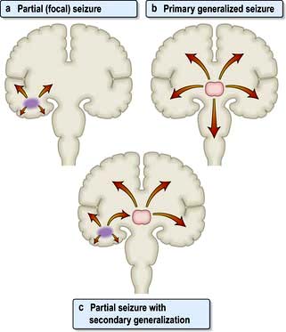
Stroke. To the public, stroke means weakness, usually permanent on one side, often with loss of speech. Stroke is defined as a syndrome of rapid onset of cerebral deficit (usually focal) lasting >24 h or leading to death, with no cause apparent other than a vascular one. Hemiplegia following middle cerebral arterial thromboembolism is the typical example.

image Transient ischaemic attack (TIA) means a brief episode of neurological dysfunction due to temporary focal cerebral or retinal ischaemia without infarction, e.g. a weak limb, aphasia or loss of vision, usually lasting seconds or minutes with complete recovery. TIAs may herald a stroke. The arbitrary time of <24 hours is no longer used.

Avoid the vague term ‘cerebrovascular accident’.

Pathophysiology

The underlying pathology responsible for stroke is either infarction or haemorrhage. Stroke mechanism and pathophysiology depends on the population studied but is broadly as follows (Fig. 22.27):

image Ischaemic stroke/infarction (80%)

Thrombotic
Large artery stenosis
Small vessel disease
Cardio-embolic
Hypoperfusion

image Haemorrhagic stroke (17%) (see p. 1104)

Intracerebral haemorrhage (12%) (see p. 1104)
Subarachnoid haemorrhage (5%) (see p. 1105)

image Other (3%), e.g. arterial dissection, venous sinus thrombosis, vasculitis (see p. 1108).

image

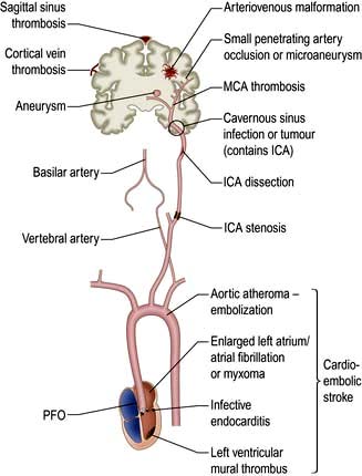
Figure 22.27 Pathophysiology of ischaemic stroke. ICA, internal carotid artery; MCA, middle cerebral artery; PFO, patent foramen ovale.

Ischaemic stroke

Arterial disease and atherosclerosis is the main pathological process causing stroke. Arterial branch points such as the origin of the great vessels arising from the aorta, the proximal internal carotid artery and its distal intracranial branches are particularly affected (Fig. 22.28). Non-Caucasian populations tend to have more intracranial narrowing and white populations more extracranial disease (which is strongly correlated with co-morbid coronary artery and peripheral vascular disease).

image

Figure 22.28 Principal sites of stenoses in extracerebral arteries (shown in bold, 1 to 4).

Thrombosis at the site of ruptured mural plaque leads to artery to artery embolism or vessel occlusion.

Large artery stenosis usually causes stroke by acting as an embolic source rather than by occlusion of the vessel (which may not in itself cause stroke if it occurs gradually and collateral circulation is adequate).

Small vessel disease. Small penetrating arterial branches supply the deep brain parenchyma and are affected by a different pathological process, an occlusive vasculopathy – lipohyalinosis – that is a consequence of hypertension. This leads to small infarcts called ‘lacunes’ and/or gradual accumulation of diffuse ischaemic change in deep white matter.

Cardio-embolic stroke. The heart is a common source of embolic material. Atrial fibrillation (and other arrhythmias) causing thrombosis in a dilated left atrium is the commonest cause. Cardiac valve disease, including congenital valve disorders, infective vegetations, rheumatic and degenerative calcific changes may cause embolization. Mural thrombosis may occur in a damaged or akinetic segment of the ventricle. A patent foramen ovale (PFO), which is a common variant, may occasionally allow passage of fragments of thrombus (e.g. from a lower limb DVT) from the right atrium to the left when Valsalva causes shunting of blood across the PFO. Pulmonary arteriovenous fistulas may also act as a conduit for paradoxical embolization. Rarer causes include fat emboli after long bone fracture, atrial myxomas and iatrogenic causes such as cardiac bypass and air embolism.

Simultaneous infarcts in different vascular territories are very suggestive of a proximal source of emboli in the heart or aorta.

Hypoperfusion. Severe hypotension, e.g. in cardiac arrest, may lead to borderzone infarction in the watershed areas between vascular territories, particularly if there is severe stenosis of proximal carotid vessels. The parieto-occipital area between the middle and posterior cerebral artery territories is particularly vulnerable.

Carotid and vertebral artery dissection

Dissection accounts for around 1 in 5 strokes below age 40 and is sometimes a sequel of trivial neck trauma or hyperextension, e.g. after whiplash, osteopathic manipulation, hairwashing in a salon, or exercise. It is now thought that subtle collagen disorders, e.g. partial forms of Marfan’s syndrome, may be a predisposing factor.

Most dissections are in large extracranial neck vessels. Blood penetrates the subintimal vessel wall forming a false lumen, but it is thrombosis within the true lumen due to tissue thromboplastin release that leads to embolization from the site of dissection and stroke, sometimes days after the initial event.

Pain in the neck or face is often the clue leading to diagnosis. Horner’s syndrome or lower cranial nerve palsies may occur with carotid dissection, as these structures are intimately associated with the carotid artery in the neck.

Venous stroke

Only 1% of strokes are venous. Thrombosis within intracranial venous sinuses, such as the superior sagittal sinus, or in cortical veins may occur in pregnancy, hypercoagulable states and thrombotic disorders or with dehydration or malignancy. Cortical infarction, seizures and raised intracranial pressure result.

For haemorrhagic stroke, see page 1104.

FURTHER READING

Furie KL. Guidelines for the prevention of stroke in patients with stroke or transient ischemic attack: a guideline for healthcare professionals from the American heart association/American stroke association. Stroke 2010; 42(1):227–276.

Transient ischaemic attacks

TIAs are usually the result of microemboli, but different mechanisms produce similar clinical events. For example, TIAs may be caused by a fall in cerebral perfusion (e.g. a cardiac dysrhythmia, postural hypotension or decreased flow through atheromatous arteries). Infarction is usually averted by autoregulation. Rarely, tumours and subdural haematomas cause episodes indistinguishable from thromboembolic TIAs. Principal sources of emboli to the brain are cardiac thrombus and atheromatous plaques/thrombus within the aortic arch, carotid and vertebral systems. Cardiac thrombus often results from atrial fibrillation or myocardial infarction. Cardiac valve disease may be a source of emboli, e.g. calcific material or vegetations in infective endocarditis. Polycythaemia is also a cause.

Risk factors for stroke

The principal risk factors and effects of altering these are shown in Table 22.12.

Table 22.12 Factors reducing stroke risk

image

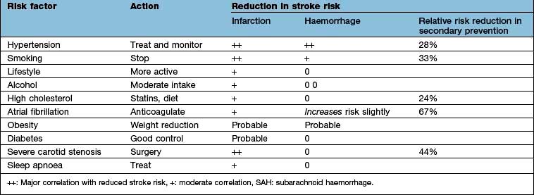
Hypertension is overall the most modifiable stroke risk factor; stroke is decreasing in the 40–60 age group partly because hypertension is now more effectively identified and treated. There is a linear relationship between blood pressure and stroke risk. Other risk factors are similar to vascular risk factors for cardiovascular disease, including smoking, dyslipidaemia, diabetes, obesity, inactivity and genetic/ethnic factors.

Other risk factors and rarer causes of stroke

image Thrombocythaemia, polycythaemia and hyperviscosity states. Thrombophilia (e.g. protein C deficiency, factor V Leiden) is weakly associated with arterial stroke but predisposes to cerebral venous thrombosis.

image Anti-cardiolipin and lupus anticoagulant antibodies (antiphospholipid syndrome, p. 538) predispose to arterial thrombotic strokes in young patients.

image Low-dose oestrogen-containing oral contraceptives do not increase stroke risk significantly in healthy women but probably do so with other risk factors, e.g. uncontrolled hypertension or smoking.

image Migraine is a rare cause of cerebral infarction.

image Vasculitis (systemic lupus erythematosus (SLE), polyarteritis, giant cell arteritis, granulomatous CNS angiitis) is a rare cause of stroke.

image Amyloidosis can present as recurrent cerebral haemorrhage (p. 1042).

image Hyperhomocysteinaemia predisposes to thrombotic strokes. Folic acid therapy does not reduce the incidence.

image Neurosyphilis, mitochondrial disease, Fabry’s disease (p. 1042).

image Sympathomimetic drugs such as cocaine, and possibly over-the-counter cold remedies containing vasoconstrictors, neuroleptics in older patients.

image CADASIL (cerebral dominant arteriopathy with subcortical infarcts and leucoencephalopathy) is a rare inherited cause of stroke/vascular dementia.
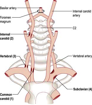
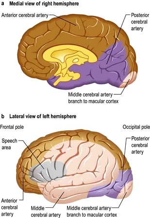
Vascular anatomy

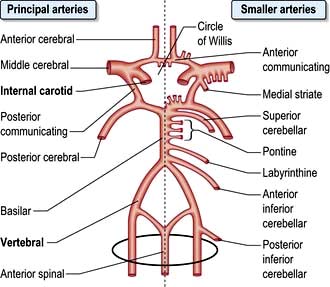
Knowledge of normal arterial anatomy and likely sites of atheromatous plaques and stenoses helps understanding of the main stroke syndromes.

The circle of Willis is supplied by the two internal carotid arteries (the anterior cerebral circulation) and by the vertebro-basilar posterior cerebral circulation. The distribution of the anterior, middle and posterior cerebral arteries that supply the cerebrum is shown in Figures 22.28-22.30.

image

Figure 22.29 Arteries supplying brain and cord.

image

Figure 22.30 The three major cerebral arteries.

Clinical syndromes

Transient ischaemic attacks (TIAs)

Features

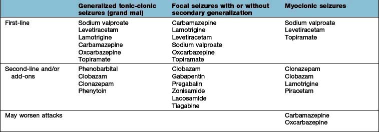
TIAs cause sudden loss of function, and usually last minutes or hours (by classical definition <24 h, but this is not used now). The site is often suggested by the type of attack. Clinical features of the principal forms of TIA are given in Box 22.12. Hemiparesis and aphasia are the commonest.

image Box 22.12

Features of transient ischaemic attacks

Anterior circulation

Carotid system

image Amaurosis fugax

image Aphasia

image Hemiparesis

image Hemisensory loss

image Hemianopic visual loss

Posterior circulation

Vertebrobasilar system

image Diplopia, vertigo, vomiting

image Choking and dysarthria

image Ataxia

image Hemisensory loss

image Hemianopic visual loss

image Bilateral visual loss

image Tetraparesis

image Loss of consciousness (rare)

image Transient global amnesia (possibly)

Amaurosis fugax

This is a sudden transient loss of vision in one eye. When due to the passage of emboli through the retinal arteries, arterial obstruction is sometimes visible through an ophthalmoscope during an attack. A TIA causing an episode of amaurosis fugax is often the first clinical evidence of internal carotid artery stenosis – and forerunner of a hemiparesis.

Clinical findings in TIA

Diagnosis of TIA is often based solely upon its description. It is unusual to witness an attack as they are so brief. Consciousness is usually preserved in TIA. There may be clinical evidence of a source of embolus, e.g.:

image Carotid arterial bruit (stenosis)

image Atrial fibrillation or other dysrhythmia

image Valvular heart disease/endocarditis

image Recent myocardial infarction.

An underlying condition may be evident:

image Atheroma

image Hypertension

image Postural hypotension

image Bradycardia or low cardiac output

image Diabetes mellitus

image Rarely, arteritis, polycythaemia, neurosyphilis, HIV

image Antiphospholipid syndrome (p. 538).

Differential diagnosis

TIAs can be distinguished, usually on clinical grounds, from other transient episodes (p. 1116). Occasionally, events identical to TIAs are produced by mass lesions. Focal epilepsy is usually recognized by its positive features (e.g. limb jerking and loss of consciousness) and progression over minutes. In a TIA, involuntary limb movements do occur occasionally; deficit is usually instantaneous. A focal prodrome in migraine sometimes causes diagnostic difficulty. Headache, common but not invariable in migraine, is rare in TIA. Typical migrainous visual disturbances are not seen in TIA.

Prognosis

Prospective studies show that 5 years after a single thromboembolic TIA:

image 30% have had a stroke, a third of these in the first year

image 15% have suffered a myocardial infarct.

TIAs in the anterior cerebral circulation carry a more serious prognosis than those in the posterior circulation (see Table 22.12).

The ABCD2 score can help to stratify stroke risk in the first 2 days:

image Age >60 years

image BP >140 mmHg systolic and/or diastolic >90 mmHg

1 point


1 point

image Clinical features

unilateral weakness
isolated speech disturbance
other

2

1

0

image Duration of symptoms (minutes)

>60
10–59
<10

2

1

0

image Diabetes

present
absent

1

0

A score of <4 is associated with a minimal risk whereas >6 is high risk for a stroke within 7 days of a TIA.

If patients are considered to have had a high risk TIA, i.e. ABCD2 score >4, or have had two recent TIAs, especially within the same vascular territory, then the patient should ideally be admitted for investigation and commencement of secondary prevention. ALL patients should be referred to a TIA Clinic and ideally seen within 24 h. Investigation and treatment should be regarded as urgent and completed within 2 weeks.

Investigations should include Doppler of internal carotid arteries, cardiac echo, ECG and 24 h tape CT/MR brain including angiography. Treat with medical therapy (p. 1095) and surgical treatment if appropriate (p. 1104).

Cerebral infarction

Major thromboembolic cerebral infarction usually causes an obvious stroke. The clinical picture is thus very variable, depending on the infarct site and extent. While the general site can be deduced from physical patterns (e.g. cortex, internal capsule, brainstem), clinical estimations of precise vascular territories are often inaccurate, when compared to imaging.

Following vessel occlusion brain ischaemia occurs, followed by infarction. The infarcted region is surrounded by a swollen area which does not function but is structurally intact. This is the ischaemic penumbra, which is detected on MRI and can regain function with neurological recovery.

Within the ischaemic area, hypoxia leads to neuronal damage. There is a fall in ATP with release of glutamate, which opens calcium channels with release of free radicals. These alterations lead to inflammatory damage, necrosis and apoptotic cell death.

Clinical features

Stroke most typically seen is caused by infarction in the internal capsule following thromboembolism in a middle cerebral artery branch (Fig. 22.31). A similar picture is caused by internal carotid occlusion (see Fig. 22.28). Limb weakness on the opposite side to the infarct develops over seconds, minutes or hours (occasionally longer). There is a contralateral hemiplegia or hemiparesis with facial weakness. Aphasia is usual when the dominant hemisphere is affected. Weak limbs are at first flaccid and areflexic. Headache is unusual. Consciousness is usually preserved. Exceptionally, an epileptic seizure occurs at the onset. After a variable interval, usually several days, reflexes return, becoming exaggerated. An extensor plantar response appears. Weakness is maximal at first; recovery occurs gradually over days, weeks or many months.

image

Figure 22.31 CT: Massive middle cerebral artery infarct.

Brainstem infarction

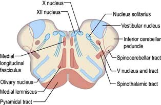
This causes complex signs depending on the relationship of the infarct to cranial nerve nuclei, long tracts and brainstem connections (Table 22.13).

image 
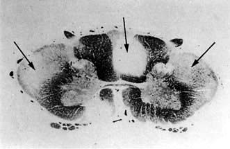
The lateral medullary syndrome (posterior inferior cerebellar artery (PICA) thrombosis and Wallenberg’s syndrome) is a common example of brainstem infarction presenting as acute vertigo with cerebellar and other signs (Table 22.14 and Fig. 22.32). It follows thromboembolism in the PICA or its branches, vertebral artery thromboembolism or dissection. Features depend on the precise structures damaged.

image Coma follows damage to the brainstem reticular activating system.

image The locked-in syndrome is caused by upper brainstem infarction (p. 1095).

image Pseudobulbar palsy (p. 1080) can follow lower brainstem infarction.

Table 22.13 Features of brainstem infarction

Clinical feature Structure involved

Hemiparesis or tetraparesis

Corticospinal tracts

Sensory loss

Medial lemniscus and spinothalamic tracts

Diplopia

Oculomotor system

Facial numbness

Vth nerve nuclei

Facial weakness

VIIth nerve nucleus

Nystagmus, vertigo

Vestibular connections

Dysphagia, dysarthria

IXth and Xth nerve nuclei

Dysarthria, ataxia, hiccups, vomiting

Brainstem and cerebellar connections

Horner’s syndrome

Sympathetic fibres

Coma, altered consciousness

Reticular formation

Table 22.14 Clinical deficits associated with problems in vascular supply

Vascular supply Neurological deficits

Left middle cerebral artery

Right-sided weakness involving face and arm > leg with dysphasia

Right middle cerebral artery

Left-sided weakness involving face and arm > leg, visual and/or sensory neglect, denial of disability

Lateral medulla (posterior inferior cerebral artery and/or parent vertebral artery)

Ipsilateral Horner’s syndrome, Xth nerve palsy, facial sensory loss, limb ataxia with contralateral spinothalamic sensory loss. Vertiginous and unable to eat due to failing laryngeal closure and ineffective coughing. Cervical radiculopathies if involvement of radicular branches of the vertebral artery

Posterior cerebral artery

Homonymous hemianopia with varied deficits due to parietal and/or temporal lobe

Internal capsule

Motor, sensory or sensorimotor loss, face = arm = leg. Possible profound dysarthria from involvement of corticobulbar fibres but not dysphasia or other cortical deficits

Bilateral paramedian thalamus

Coma or disturbed vigilance, ophthalmoplegia (internal and/or external), ataxia and memory impairment. Some require ventilation

Carotid artery dissection

Ipsilateral Horner’s syndrome from compression of sympathetic plexus around the carotid artery, can also affect lower cranial nerves (Xth and XIIth most clinically obvious). If ipsilateral cerebral infarction follows, clinical picture can mimic brain stem event

image

Figure 22.32 Medulla (cross-section): structures at risk after brainstem infarction.

Other patterns of infarction

Lacunar infarction

Lacunes are small (<1.5 cm3) infarcts seen on MRI or at autopsy. Hypertension is commonly present. Minor strokes (e.g. pure motor stroke, pure sensory stroke, sudden unilateral ataxia and sudden dysarthria with a clumsy hand) are syndromes caused typically by single lacunar infarcts. Lacunar infarction is often symptomless.

Hypertensive encephalopathy (p. 778)

This is due to cerebral oedema, causing severe headaches, nausea and vomiting. Agitation, confusion, fits and coma occur if the hypertension is not treated. Papilloedema develops, either due to ischaemic optic neuropathy or following the brain swelling due to multiple acute infarcts. MRI shows oedematous white matter in the parieto-occipital regions.

Multi-infarct dementia (vascular dementia)

Multiple lacunes or larger infarcts cause generalized intellectual loss seen with advanced cerebrovascular disease. In the late stages, there is dementia, pseudobulbar palsy and a shuffling gait – the marche à petits pas (small steps), sometimes called atherosclerotic Parkinsonism. Binswanger’s disease is a term for widespread low attenuation in cerebral white matter, usually with dementia, TIAs and stroke episodes in hypertensive patients (the changes being seen on imaging/autopsy).

Visual cortex infarction

Posterior cerebral artery infarction or infarction of the middle cerebral artery macular branch causes combinations of hemianopic visual loss and cortical blindness (Anton’s syndrome, Fig. 22.30 and p. 1073).

Weber’s syndrome

Ipsilateral IIIrd nerve palsy with contralateral hemiplegia is due to a unilateral infarct in the midbrain. Paralysis of upward gaze is usually present.

Watershed (borderzone) infarction

Cortical infarcts, often multiple, follow prolonged periods of low perfusion (e.g. hypotension after massive myocardial infarction or cardiac bypass surgery). Infarcts occur in the borderzones, between areas supplied by the anterior, middle and posterior cerebral arteries. Cortical visual loss, memory loss and intellectual impairment are typical. In some cases, a vegetative state or minimal conscious state follows (p. 1096).

Acute stroke: immediate care and thrombolysis

(See also Box 22.14)

image Box 22.14 Stroke

immediate management

1. Admit to multidisciplinary stroke unit

2. General medical measures

image Airway: confirm patency and monitor
image Continue care of the unconscious or stuporose patient
image Oxygen by mask
image Monitor BP
image Look for source of emboli
image Assess swallowing

3. Is thrombolysis appropriate?

image If so (Practical Box 22.4) immediate brain imaging is essential.

4. Brain imaging

image CT should always be available. This will indicate haemorrhage, other pathology and sometimes infarction
image MR is better overall, if immediately available

5. Cerebral infarction

image If CT excludes haemorrhage, give immediate thrombolytic therapy. Aspirin, 300 mg/day, is given if thrombolytic therapy is contraindicated

6. Cerebral haemorrhage

image If CT shows haemorrhage, give no drugs that could interfere with clotting. Neurosurgery may occasionally be needed

Paramedics and members of the public are encouraged to make the diagnosis of stroke on a simple history and examination – FAST:

image Face – sudden weakness of the face

image Arm – sudden weakness of one or both arms

image Speech – difficulty speaking, slurred speech

image Time – the sooner treatment can be started, the better.

Dedicated units with multidisciplinary, organized teams deliver higher standards of care than a general hospital ward, reducing stroke mortality and long-term disability. Evidence-based guidelines have contributed to clear protocols. Admission to hospital should proceed without delay, for imaging, care and investigation.

Following a stroke, immediate, continued and meticulous attention to the airway and to swallowing is essential. Management of unconscious or stuporose patients is outlined on page 1096.

In cerebral infarction, the issue is thrombolysis. Practical Box 22.4 outlines the current proposals for stroke thrombolysis. Alteplase is commonly used although tenecteplase is also effective. The benefit of thrombolysis is shown on CT perfusion scans (Fig. 22.33) and decreases with time, even within the time window of 4.5 h. Every minute counts. If thrombolysis is not given, aspirin 300 mg daily should be given as soon as a diagnosis of ischaemic stroke or thromboembolic TIA is confirmed, reducing to 75 mg after several days. Following thrombolysis aspirin should not be started until 24–48 h later.

image Practical Box 22.4

Thrombolysis in acute ischaemic stroke

Eligibility

image Age ≥18 years

image Clinical diagnosis of acute ischaemic stroke

image Assessed by experienced team

image Measurable neurological deficit

image Blood tests: results available

image CT or MRI consistent with acute ischaemic stroke

image Timing of onset well established

image Thrombolysis should commence as soon as possible and up to 4.5 hours after acute stroke

Exclusion criteria

Historical

image Stroke or head trauma within the prior 3 months

image Any prior history of intracranial haemorrhage

image Major surgery within 14 days

image Gastrointestinal or genitourinary bleeding within the previous 21 days

image Myocardial infarction in the prior 3 months

image Arterial puncture at a non-compressible site within 7 days

image Lumbar puncture within 7 days

Clinical

image Rapidly improving stroke syndrome

image Minor and isolated neurological signs

image Seizure at the onset of stroke if the residual impairments are due to postictal phenomena

image Symptoms suggestive of subarachnoid haemorrhage, even if the CT is normal

image Acute MI or post-MI pericarditis

image Persistent systolic BP >185, diastolic BP >110 mmHg, or requiring aggressive therapy to control BP

image Pregnancy or lactation

image Active bleeding or acute trauma (fracture)

Laboratory

image Platelets <100 000/mm3

image Serum glucose <2.8 mmol/L or >21.2 mmol/L

image INR >1.7 if on warfarin

image Elevated partial thromboplastin time if on heparin

Dose of i.v. alteplase (tissue plasminogen activator)

image Total dose 0.9 mg/kg (max. 90 mg)

image 10% of total dose by initial i.v. bolus over 1 minute

image Remainder infused i.v. over 60 minutes

Adapted from Adams HP Jr, del Zoppo G, Alberts MJ et al. Stroke 2007; 38(5):1655–1711.

image

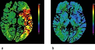
Figure 22.33 CT perfusion scans: (a) pre-thrombolysis; (b) post-thrombolysis showing reperfusion of the ischaemic site.

(Courtesy of Professor Adrian Dixon, Cambridge Radiology Department, UK.)

Investigations

The purpose of investigations in stroke is:

image to confirm the clinical diagnosis and distinguish between haemorrhage and thromboembolic infarction;

image to look for underlying causes and to direct therapy;

image to exclude other causes, e.g. tumour.

Sources of embolus should be sought (e.g. carotid bruit, atrial fibrillation, valve lesion, evidence of endocarditis, previous emboli or TIA) and hypertension/postural hypotension assessed. Brachial BP should be measured on each side; >20 mmHg difference is suggestive of subclavian artery stenosis.

Routine investigations in thromboembolic stroke and TIA are listed in Box 22.13.

image Box 22.13 Stroke

further investigation and management

Further investigations

image Routine bloods (for ESR, polycythaemia, infection, vasculitis, thrombophilia, syphilitic serology, clotting studies, autoantibodies, lipids)

image Chest X-ray

image ECG

image Carotid Doppler studies

image MR angiography, if appropriate

Further management

image Drugs for hypertension, heart disease, diabetes, other medical conditions

image Antiplatelet agents, e.g. aspirin and elopidogrel

image Question of endarterectomy

image Question of anticoagulation – Table 22.15

image Speech therapy, dysphagia care, physiotherapy, occupational therapy

image Specific issues, e.g. epilepsy, pain, incontinence

image Preparations for future care

Imaging in acute stroke

CT and MRI:

image 
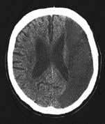
Non-contrast CT will demonstrate haemorrhage immediately but cerebral infarction is often not detected or only subtle changes are seen initially (Fig. 22.34a).

image MRI shows changes early in infarction (Fig. 22.35a) and a later MRI shows the full extent of the damaged area or penumbra (Fig. 22.35b).

image Diffusion-weighted MRI (DWI) can detect cerebral infarction immediately (see Fig. 22.34b) but is as accurate as CT for the detection of haemorrhage.

image

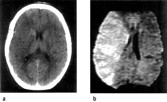
Figure 22.34 Middle cerebral artery infarction. (a) CT performed initially shows only very subtle low density changes in right MCA territory. (b) Diffusion-weighted MRI done at the same time shows full extent of the area of ischaemia.

(Courtesy of Dr Paul Jarman.)

image

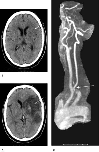
Figure 22.35 MRI of cerebral infarction and subsequent MRA performed in the same patient. (a) Early changes. (b) Changes after 5 days. (c) MR angiography showing internal carotid artery occlusion.

(Courtesy of Dr Paul Jarman.)

CT is still more widely available than MRI and should be performed if MRI is unavailable so that there is no delay in giving thrombolysis for cerebral infarction.

More detailed studies involving perfusion-weighted images and diffusion-weighted MRI will differentiate the infarct core and the penumbral area which is potentially recoverable.

FURTHER READING

Chalela JA, Kidwell CS, Nentwich LM et al. Magnetic resonance imaging and computed tomography in emergency assessment of patients with suspected acute stroke: a prospective comparison. Lancet 2007; 369:293–298.

Donnan GA, Fisher M, Macleod M et al. Stroke. Lancet 2008; 371:1612–1623.

Hacke W, Kaste M, Bluhmki E et al. Thrombolysis with alteplase 3 to 4.5 hours after acute ischemic stroke. N Engl J Med 2008; 359:1319–1329.

Khaja AM, Grotta JC. Established treatments for acute ischaemic stroke. Lancet 2007; 369:319–330.

Royal College of Physicians’ Intercollegiate Stroke Working Party. National Clinical Guidelines for Stroke, 2nd edn. London: Royal College of Physicians; 2008.

Strong K, Mather C, Bonita R. Preventing stroke; saving lives around the world. Lancet Neurol 2007; 6:182–187 + Series.

Treatment of acute stroke

This is shown in Box 22.14. Thrombolysis has been shown to improve outcome and should be used immediately if there are no contraindications. In a massive middle cerebral artery infarct, hemispheric swelling occurs with oedema (Fig. 22.36). Decompressive hemicraniectomy reduces the intracranial pressure and the mortality but extensive neurological deficits remain.

image

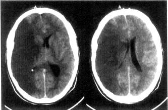
Figure 22.36 CT scan of massive infarction in the middle carotid artery territory. There is hemisphere swelling with brain shift.

(Courtesy of Dr Paul Jarman.)

Later investigations

MR angiography (MRA) or CT angiography is valuable in anterior circulation TIAs to confirm surgically accessible arterial stenoses, mainly internal carotid stenosis (Fig. 22.35c). If ultrasound suggests carotid stenosis, normotensive patients with TIA or stroke in the anterior circulation should have vascular imaging.

Carotid Doppler and duplex scanning. These screen for carotid (and vertebral) stenosis and occlusion: in skilled hands they demonstrate accurately the degree of internal carotid stenosis.

Long-term management

Medical therapy

Risk factors (Table 22.12) should be identified and addressed.

Antihypertensive therapy

Recognition and good control of high blood pressure is the major factor in primary and secondary stroke prevention. Transient hypertension, often seen following stroke, usually does not require treatment provided diastolic pressure does not rise >100 mmHg. Sustained severe hypertension needs treatment (p. 781); BP should be lowered slowly to avoid any sudden fall in perfusion.

Antiplatelet therapy (see also p. 425)

Long-term soluble aspirin (75 mg daily) reduces substantially the incidence of further infarction following thromboembolic TIA or stroke. Aspirin inhibits cyclo-oxygenase, which converts arachidonic acid to prostaglandins and thromboxanes; predominant therapeutic effects are reduction of platelet aggregation. Clopidogrel and dipyridamole are also used (p. 425). Combined aspirin 75 mg daily and clopidogrel 75 mg daily provide optimal prophylaxis against further thromboembolic stroke or TIA. Dipyridamole 200 mg twice daily is used if clopidogrel is contraindicated.

Anticoagulants

Heparin and warfarin should be given when there is atrial fibrillation, other paroxysmal dysrhythmias or when there are cardiac valve lesions (uninfected) or cardiomyopathies. Brain haemorrhage must be excluded by CT/MRI. Patients must be aware of the small risk of cerebral (and other) haemorrhage. Anticoagulants are potentially dangerous in the two weeks following infarction because of the risk of provoking cerebral haemorrhage; there are wide differences in clinical practice. Antithrombins are now being used. Table 22.15 outlines the issues in secondary stroke prevention.

Table 22.15 Anticoagulants and stroke prevention

Indication Comment

Valvular heart disease

Heparin/warfarin of benefit in chronic rheumatic heart disease, particularly mitral stenosis

Recent MI


Intracardiac thrombus

Heparin/warfarin if there is evidence of intracardiac thrombus

Atrial fibrillation

Anticoagulants long term reduce stroke incidence in atrial fibrillation

Acute internal carotid artery thrombus

Anticoagulants reserved for imaging-confirmed cases of arterial thrombosis or dissection. They have not been shown to be beneficial in stroke prevention after thromboembolism from carotid or vertebrobasilar sources

Acute basilar artery thrombus

Internal carotid artery dissection

Extracranial vertebral artery dissection

Prothrombic states, e.g. protein C deficiency

Anticoagulation, in consultation with haematologist

Recurrent TIAs or stroke on full antiplatelet therapy

If no remediable cause, a trial of anticoagulants may be justified

Cerebral venous thrombosis including sinus thrombosis

Benefits of anticoagulation outweigh risks of haemorrhage

Adapted from Brown M. Medicine 2000; 28(7):64.

Other measures

Polycythaemia and any clotting abnormalities should be treated (p. 402). Statin therapy should be given for all.

Surgical approaches
Internal carotid endarterectomy

Surgery is usually recommended in TIA or stroke patients with internal carotid artery stenosis >70% (see ABCD2, above). Successful surgery reduces the risk of further TIA/stroke by around 75%. Endarterectomy has a mortality around 3%, and a similar risk of stroke. Percutaneous transluminal angioplasty (stenting) is an alternative. The value of surgery for asymptomatic carotid stenosis is debatable.

Stroke in the elderly

While in the elderly the yield of investigation in stroke diminishes, age is no barrier to recovery; elderly patients benefit the most from good rehabilitation. Consider social isolation, pre-existing cognitive impairment, nutrition, skin and sphincter care, and reassess swallowing. Carotid endarterectomy over 75 years carries little more risk than in younger cases. Fibrinolysis is not contraindicated.

FURTHER READING

Rothwell PM, Algra A, Amarenco P. Medical treatment in acute and long-term secondary prevention after transient ischaemic attack and ischaemic stroke. Lancet 2011; 377:1681–1692.

Wechsler LR. Intravenous thrombolytic therapy for acute ischemic stroke. N Engl J Med 2011; 364:2138–2146.

Rehabilitation: multidisciplinary approach

Physiotherapy has particular value in the first few weeks after stroke to relieve spasticity, prevent contractures and teach patients to use walking aids. The benefits of physiotherapy for longer-term outcome are still inadequately researched. Baclofen and/or botulinum toxin are sometimes helpful in the management of severe spasticity.

Speech and language therapists have a vital understanding of aphasic patients’ problems and frustration. Return of speech is hastened by conversation generally. If swallowing is unsafe because of the risk of aspiration, either nasogastric feeding or percutaneous gastrostomy will be needed. Video-fluoroscopy while attempting to swallow is helpful.

Physiotherapy, occupational and speech therapy have a vital role in assessing and facilitating the future care pathway. Stroke is frequently devastating and, particularly during working life, alters radically the patient’s remaining years. Many become unemployable, lose independence and are financially embarrassed. Loss of self-esteem makes depression common.

At home, aids and alterations may be needed: stair and bath rails, portable lavatories, hoists, sliding boards, wheelchairs, tripods, stair lifts, electric blinds and modified sleeping arrangements, kitchen, steps, flooring and doorways. Liaison between hospital-based and community care teams, and primary care physician is essential.

FURTHER READING

Langhorne P, Bernhardt J, Kwakkel G. Stroke rehabilitation. Lancet 2011; 377:1693–1702.

Prognosis

About 25% of patients die within 2 years of a stroke, nearly 10% within the 1st month. This early mortality is higher following intracranial haemorrhage than thromboembolic infarction. Poor outcome is likely when there is coma, a defect in conjugate gaze and hemiplegia. Many complications, particularly in the elderly, are preventable, e.g. aspiration. Coordinated care reduces deaths.

Recurrent strokes are, however, common (10% in the 1st year) and many patients die subsequently from myocardial infarction. Of initial stroke survivors, some 30–40% remain alive at 3 years.

Gradual improvement usually follows stroke, although late residual deficits are typically substantial. One-third of survivors return to independent mobility and one-third have disability requiring institutional care.

The outlook for recovery of language varies: in general, if language is intelligible at all at 3 weeks, prognosis for fluent speech is good, but many are left with word-finding difficulties.

Intracranial haemorrhage

This comprises:

image intracerebral and cerebellar haemorrhage

image subarachnoid haemorrhage

image subdural and extradural haemorrhage/haematoma.

Intracerebral haemorrhage

Aetiology

Intracerebral haemorrhage causes approximately 15% of strokes. Rupture of microaneurysms (Charcot–Bouchard aneurysms, 0.8–1.0 mm diameter) and degeneration of small deep penetrating arteries are the principal pathology. Such haemorrhage is usually massive, often fatal, and occurs in chronic hypertension and at well-defined sites – basal ganglia, pons, cerebellum and subcortical white matter.

In normotensive patients, particularly over 60 years, lobar intracerebral haemorrhage occurs in the frontal, temporal, parietal or occipital cortex. Cerebral amyloid angiopathy (rare) is the cause in some of these haemorrhages, and the tendency to rebleed is associated with particular apolipoprotein E genotypes.

Recognition

At the bedside, there is no entirely reliable way of distinguishing between haemorrhage and thromboembolic infarction. Both produce stroke. Intracerebral haemorrhage tends to be dramatic with severe headache. It is more likely to lead to coma than thromboembolism.

Brain haemorrhage is seen on CT imaging immediately (cf. infarction, p. 1091) as intraparenchymal, intraventricular or subarachnoid blood. Routine MRI may not identify an acute small haemorrhage correctly in the first few hours but MRI diffusion-weighted (MRI-DW) is as good as CT.

Management: haemorrhagic stroke

The principles are those for cerebral infarction. The immediate prognosis is less good. Antiplatelet drugs and, of course, anticoagulants are contraindicated. Control of hypertension is vital. Urgent neurosurgical clot evacuation is occasionally necessary when there is deepening coma and coning (particularly in cerebellar haemorrhage).

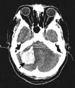
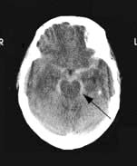
Cerebellar haemorrhage (Fig. 22.37)

There is headache, often followed by stupor/coma and signs of cerebellar/brainstem origin (e.g. nystagmus, ocular palsies). Gaze deviates towards the haemorrhage. Skew deviation (p. 1094) may develop. Cerebellar haemorrhage sometimes causes acute hydrocephalus, a potential surgical emergency.

image

Figure 22.37 CT: Cerebellar haemorrhage.

Subarachnoid haemorrhage (SAH)

SAH means spontaneous arterial bleeding into the subarachnoid space, and is usually clearly recognizable clinically from its dramatic onset. SAH accounts for some 5% of strokes and has an annual incidence of 6 per 100 000.

Causes

The causes of SAH are shown in Table 22.16; it is unusual to find any contributing disease.

Table 22.16 Underlying causes of subarachnoid haemorrhage

Cause of aneurysm (%)

Saccular (berry) aneurysms

70%

Arteriovenous malformation (AVM)

10%

No arterial lesion found

15%

Rare associations

(<5%)

 Bleeding disorders Mycotic aneurysms – endocarditis Acute bacterial meningitis Tumours, e.g. metastatic melanoma, oligodendroglioma Arteritis (e.g. SLE) Spinal AVM → spinal SAH Coarctation of the aorta Marfan’s, Ehlers–Danlos syndrome Polycystic kidneys


Saccular (berry) aneurysms (Fig. 22.38)

Saccular aneurysms develop within the circle of Willis and adjacent arteries. Common sites are at arterial junctions:

image Between posterior communicating and internal carotid artery – posterior communicating artery aneurysm

image Between anterior communicating and anterior cerebral artery – anterior communicating and anterior cerebral artery aneurysm

image At the trifurcation or a bifurcation of the middle cerebral artery – middle cerebral artery aneurysm.

image

Figure 22.38 Digital subtraction angiogram: posterior communicating artery aneurysm.

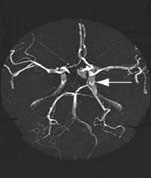
Other aneurysm sites are on the basilar, posterior inferior cerebellar, intracavernous internal carotid and ophthalmic arteries. Saccular aneurysms are an incidental finding in 1% of autopsies and can be multiple.

Aneurysms cause symptoms either by spontaneous rupture, when there is usually no preceding history, or by direct pressure on surrounding structures; for example, an enlarging unruptured posterior communicating artery aneurysm is the commonest cause of a painful IIIrd nerve palsy (p. 1081).

Arteriovenous malformation (AVM)

AVMs are vascular developmental malformations, often with a fistula between arterial and venous systems causing high flow through the AVM and high pressure arterialization of draining veins. An AVM may also cause epilepsy, often focal. The risk of a first haemorrhage (20% fatal and 30% resulting in permanent disability) is approximately 2–3% per year. Once an AVM has caused a haemorrhage, the risk of rebleeds is increased– to approximately 10% per year. AVMs may be ablated with endovascular treatment (catheter injection of glue into the nidus usually), surgery or stereotactic radiotherapy. A multidisciplinary team approach with neurologist, interventional neuro-radiologist and neurosurgeon is required in deciding on treatment options.

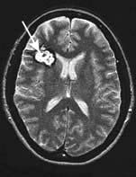
Cavernous haemangiomas (cavernomas) are common (0.1–0.5% prevalence) and consist of a tangle of low pressure dilated vessels without a major feeding artery; they are frequently symptomless (Fig. 22.39) and seen incidentally on imaging. Multiple cavernomas often have a genetic basis, with linkage to two loci on chromosome 7q. Cavernomas may cause seizures. Small haemorrhages may occur but are usually low pressure bleeds and rarely cause severe deficits. Surgical resection is rarely needed except where the cavernoma is gradually enlarging or causing significant neurological symptoms.

image

Figure 22.39 MR T2: Symptomless frontal cavernoma (arrow).

Clinical features of subarachnoid haemorrhage

There is a sudden very severe headache, often occipital (mean time to peak headache 3 min). Headache is usually followed by vomiting and often by coma and death. Survivors of SAH may remain comatose or drowsy for hours, days, or longer. SAH is a possible diagnosis in any sudden headache.

Following major SAH there is neck stiffness and a positive Kernig’s sign. Papilloedema is sometimes present, with retinal and/or subhyaloid haemorrhage (tracking beneath the retinal hyaloid membrane). Minor bleeds cause few signs, but almost invariably headache (approximately 17% of patients have small ‘sentinel bleeds’ in the weeks before presenting with SAH).

Investigations

CT imaging is the immediate investigation (Fig. 22.40). Subarachnoid and/or intraventricular blood is usually seen (sensitivity of CT to detect subarachnoid blood is 95% within 24 h of onset but much lower over subsequent days). Lumbar puncture is not necessary if SAH is confirmed by CT, but should be performed if doubt remains. CSF becomes yellow (xanthochromic) within 12 h of SAH and remains detectable for 2 weeks. Visual inspection of supernatant CSF is usually sufficiently reliable for diagnosis. Spectrophotometry to estimate bilirubin in the CSF released from lysed cells is used to define SAH with certainty. CT angiography or catheter angiography to identify the aneurysm or other source of bleeding is performed in patients potentially fit for surgery. In some, no aneurysm or source of bleeding is found, despite a definite SAH.

image

Figure 22.40 Subarachnoid haemorrhage CT showing blood around the brainstem (arrow).

Differential diagnosis

SAH must be differentiated from migraine. This is sometimes difficult – a short time to maximal headache intensity and the presence of neck stiffness usually indicate SAH. Thunderclap headache is used (confusingly) to describe either SAH or a sudden (benign) headache for which no cause is ever found. The syndrome of reversible cerebral vasoconstriction (Call–Fleming syndrome) presents with thunderclap headache. Acute bacterial meningitis occasionally causes a very abrupt headache, when a meningeal microabscess ruptures; SAH also occasionally occurs at the onset of acute bacterial meningitis. Cervical arterial dissection can present with a sudden headache.

Complications

Blood in the subarachnoid space can lead to obstructive hydrocephalus, seen on CT. Hydrocephalus can be asymptomatic but may cause deteriorating consciousness following SAH. Shunting may be necessary. Arterial spasm (visible on angiography and a cause of coma or hemiparesis) is a serious complication of SAH and a poor prognostic feature.

Management

Immediate treatment of SAH is bed rest and supportive measures. Hypertension should be controlled. Nimodipine, a calcium-channel blocker given for 3 weeks, reduces mortality.

All SAH cases should be discussed urgently with a neurosurgical centre. Nearly half of SAH cases are either dead or moribund before reaching hospital. Of the remainder, a further 10–20% rebleed and die within weeks. Failure to diagnose SAH, e.g. mistaking SAH for migraine, contributes to this mortality.

Where angiography demonstrates an aneurysm (the cause of the vast majority of SAH), endovascular treatment by placing platinum coils via a catheter in the aneurysm sac to promote thrombosis and ablation of the aneurysm, is now the first line treatment. Endovascular coiling has a lower complication rate than surgery but direct surgical clipping of the aneurysm neck is still required in some selected cases. For asymptomatic (unruptured) aneurysms over 8 mm in diameter the risk of treatment is less than the risk of haemorrhage if not treated. Patients who remain comatose or who have persistent severe deficits after SAH have a poor outlook.

FURTHER READING

White PM, Lewis SC, Gholkar A et al. Hydrogel-coated coils versus bare platinum coils for the endovascular treatment of intracranial aneurysms (HELPS): a randomised controlled trial. Lancet 2011; 377:1655–1662.

Subdural and extradural bleeding

These conditions can cause death following head injuries unless treated promptly.

Subdural haematoma (SDH)

SDH means accumulation of blood in the subdural space following rupture of a vein. This usually follows a head injury, sometimes trivial. The interval between injury and symptoms can be days, or extend to weeks or months. Chronic, apparently spontaneous SDH is common in the elderly, and also occurs with anticoagulants.

Headache, drowsiness and confusion are common; symptoms are indolent and can fluctuate. Focal deficits, e.g. hemiparesis or sensory loss, develop. Epilepsy occasionally occurs. Stupor, coma and coning may follow.

Extradural haemorrhage (EDH)

EDH typically follows a linear skull vault fracture tearing a branch of the middle meningeal artery. Extradural blood accumulates rapidly over minutes or hours. A characteristic picture is of a head injury with a brief duration of unconsciousness, followed by improvement (the lucid interval). The patient then becomes stuporose, with an ipsilateral dilated pupil and contralateral hemiparesis, with rapid transtentorial coning. Bilateral fixed dilated pupils, tetraplegia and respiratory arrest follow. An acute progressive SDH presents similarly.

Management

Possible extradural or subdural bleeding needs immediate imaging. CT (Fig. 22.41a) is the most widely used investigation because of its immediate availability. MRI is more sensitive for the detection of small haematomas. T1-weighted MRI (Fig. 22.41b) shows bright images due to the presence of methaemoglobin.

image

Figure 22.41 Bilateral subdural haematomas. (a) CT scan.

(Courtesy of Dr Paul Jarman). (b) MR T1.

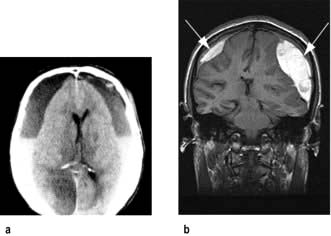
EDHs require urgent neurosurgery: if it is performed early, the outlook is excellent. When far from a neurosurgeon, e.g. in wartime or at sea, drainage through skull burr-holes has been lifesaving when an EDH has been diagnosed clinically.

Subdural bleeding usually needs less immediate attention but close neurosurgical liaison is necessary. Even large collections can resolve spontaneously without drainage. Serial imaging is needed to assess progress.

Cortical venous thrombosis and dural venous sinus thrombosis

Intracranial venous thromboses are usually (>50%) associated with a pro-thrombotic risk factor, e.g. oral contraceptives, pregnancy, genetic or acquired pro-thrombotic states and dehydration. Head injury is also a cause. Infection, e.g. from a paranasal sinus, may be present. Venous thromboses can also arise spontaneously.

Cortical venous thrombosis

The venous infarct leads to headache, focal signs (e.g. hemiparesis) and/or epilepsy, often with fever.

Dural venous sinus thromboses

Cavernous sinus thrombosis causes ocular pain, fever, proptosis and chemosis. External and internal ophthalmoplegia with papilloedema develops.

Sagittal and lateral sinus thrombosis cause raised intra-cranial pressure with headache, fever, papilloedema and often epilepsy.

Management

MRI, MRA and MR venography (MRV) show occluded sinuses and/or veins. Treatment is with heparin initially, though its value is questioned, followed by warfarin for 6 months. Anticonvulsants are given if necessary.

FURTHER READING

Stam J. Thrombosis of the cerebral veins and sinuses. N Engl J Med 2005; 352:1791–1798.

van Gijn J, Kerr RS, Rinkel GJ. Subarachnoid haemorrhage. Lancet 2007; 369:306–318.

Headache, migraine and facial pain

Headache is an almost universal experience and one of the most common symptoms in medical practice. It varies from an infrequent and trivial nuisance to a pointer to serious disease. Headache symptoms are unpleasant, disabling, common worldwide and have a substantial economic impact because of time lost from work.

Mechanisms

Pain receptors are located at the base of the brain in arteries and veins and throughout meninges, extracranial vessels, scalp, neck and facial muscles, paranasal sinuses, eyes and teeth. Curiously, brain substance is almost devoid of pain receptors. Head pain is mediated by the Vth and IXth cranial nerves and upper cervical sensory roots.

A general approach to assessing headache symptoms

In assessing patients with headache the aim should be to make a confident diagnosis based on the history. Examination is helpful in excluding underlying medical disorders as a cause of headache but will not distinguish between different types of primary headache.

There is an internationally agreed classification for headaches that defines all headache patterns. Headache is divided into primary headache disorders such as migraine, and secondary headaches due to underlying pathology such as raised intracranial pressure or meningitis (Box 22.15). It is also useful to distinguish between episodic (recurrent) headache, single first headache episodes and patients with chronic headache.

image Box 22.15

Some causes of secondary headache

image Raised intracranial pressure, e.g. idiopathic intracranial hypertension

image Infections, e.g. meningitis, sinusitis

image Giant cell arteritis

image Intracranial haemorrhage, esp. SAH or SDH

image Low CSF volume (low pressure) headache

image Post-traumatic headache

image Cervicogenic headache

image Acute glaucoma

In an outpatient clinic setting most headaches will be benign. Fewer than 1% of outpatients with non-acute headache have a serious underlying cause but in the Emergency Department there will be a much higher prevalence of serious underlying pathology presenting with headache. New onset severe headache in those without a previous headache history, especially in older patients (>50), requires exclusion of underlying pathology causing secondary headache.

There are some widely believed ‘headache myths’. Headaches are not caused by hypertension except rarely with malignant hypertension (p. 778). Eyestrain from refractive error does not cause headache and sinusitis is rarely the explanation for recurrent or chronic headache.

Taking a headache history

Ask about:

image Headache location (e.g. hemicranial), severity and character (e.g. throbbing vs non-throbbing)

image Associated symptoms, e.g. nausea, photophonia, phonophobia and motion sensitivity

image Presence of autonomic symptoms, e.g. tearing or ptosis

image Relieving or exacerbating features, e.g. effect of posture

image Headache pattern. Is headache episodic and part of a pattern of previous similar headaches? Age at onset and headache frequency

image Duration of headache episodes (helpful in distinguishing between different primary headache types)

image Triggers

image Pattern of analgesic use

image Family history of headache

image ‘Red flag’ symptoms – fever (meningitis, sinusitis)

image Sudden onset in less than 1 minute (SAH)

image Features of raised intracranial pressure

image Jaw claudication (giant cell arteritis).

Examination

Examination should include fundoscopy to look for papilloedema. In older patients, temporal arteries should be palpated for loss of pulsatility and tenderness that may be features of giant cell arteritis (GCA). Fever and neck stiffness suggest meningitis. Examination is generally normal in patients with primary headache disorders.

Investigations

Investigations, including brain imaging, do not contribute to the diagnosis of primary headache disorders. Neuroimaging is indicated only where history or examination suggests an underlying secondary cause. Older patients with new onset headache and those with ‘red flag’ symptoms should have brain CT. In patients over 50 with new headache ESR should be checked to exclude GCA.

Primary headache disorders

Migraine

Migraine is the commonest cause of episodic headache (15–20% of women and 5–10% of men); in 90%, onset is before 40 years of age. Episodes of headache are associated with sensory sensitivity such as to light, sound or movement, and sometimes with nausea and vomiting. There is a spectrum of severity between individuals and from one attack to another. Migraine is usually high impact, with inability to function normally during episodes. Headache frequency in migraine varies from an occasional inconvenience to frequent headaches severely impacting quality of life, and may transform into chronic daily headache.

Mechanisms

Genetic factors play a part in causing the neuronal hyper-excitability that is probably the biological basis of migraine. Migraine is polygenic but a rare form of familial migraine is associated with mutations in the α-1 subunit of the P/Q-type voltage-gated calcium channel or neuronal sodium channel (SCN1A), and a dominant loss of function mutation in a potassium channel gene (TRESK) has recently been identified in some patients with migraine with aura.

The pathophysiology of migraine is now thought to have a primarily neurogenic rather than vascular basis. Spreading cortical depression – a wave of neuronal depolarization followed by depressed activity spreading slowly anteriorly across the cerebral cortex from the occipital region – is thought to be the basis of the migraine aura. Activation of trigeminal pain neurones is the basis of the headache. The innervation of the large intracranial vessels and dura by the first division of the trigeminal nerve is known as the trigeminovascular system. Release of calcitonin gene-related peptide (CGRP), substance P and other vasoactive peptides including 5-HT by activated trigeminovascular neurones causes painful meningeal inflammation and vasodilation. Peripheral and central sensitization of trigeminal neurones and brainstem pain pathways makes otherwise innocuous sensory stimuli (such as CSF pulsation and head movement) painful and light and sound perceived as uncomfortable.

Clinical features

Migraine without aura

Migraine typically starts around puberty with increasing prevalence into the 4th decade. There is a spectrum of severity and associated features, but attacks have recognizable core features (Box 22.16). Most migraine attacks are usually of sufficient severity to prevent sufferers continuing with normal activities and sleep usually helps, with a washed out feeling following the attack. The scalp may be tender to touch during episodes (allodynia) and the preference is to be still in a dark, quiet environment.

image Box 22.16 Migraine

Simplified Diagnostic Criteria

Headache lasting 4 hours to 3 days (untreated)

At least two of:

image Unilateral pain (may become holocranial later in attack)

image Throbbing type pain

image Moderate to severe in intensity

image Motion sensitivity (headache made worse with head movement or physical activity)

At least one of:

image Nausea/vomiting

image Photophobia/phonophobia

image Normal examination and no other cause of headache

Some patients recognize changes in routine as trigger factors:

image Sleep (too little or too much)

image Stress (including let down after a period of stress)

image Hormonal factors for women – changes in oestrogen levels, e.g. menstrual migraine (usually just before menses) and worsening with the OC pill and menopause

image Eating – skipping meals and alcohol. Contrary to popular belief, individual foods are rarely a trigger

image Other – sensory stimuli such as bright lights or loud sounds. Physical exertion. Changes in weather patterns, e.g. stormy weather. Minor head injuries may trigger a worsening of migraine frequency and severity.

Migraine with aura

Approximately 25% of migraine sufferers experience focal neurological symptoms immediately preceding the headache phase in some or all attacks – termed migraine aura. Most never experience aura and presence of aura is therefore not required for a diagnosis of migraine. Aura usually evolves over 5–20 minutes with symptoms changing as the wave of spreading neuronal depression moves across the surface of the cortex. It rarely lasts longer than 60 minutes and is followed immediately by the headache phase.

Visual aura is the commonest type, with positive visual symptoms such as shimmering, teichopsia (zig-zag lines also called fortification spectra) and fragmentation of the image (like looking through a pane of broken glass) often accompanied by patches of loss of vision which may move across the visual field (scotomas) and even evolve into hemianopia or tunnel vision. Positive sensory symptoms (mainly tingling), dysphasia and rarely loss of motor function may also occur and may occur successively within the same epidose of aura following the visual symptoms.

Migraine aura usually presents no diagnostic difficulty, but problems with diagnosis may sometimes arise in men over the age of 50 who develop migraine aura for the first time without subsequent headache (sometimes referred to as acephalic migrainous aura). This is frequently misdiagnosed as a transient ischaemic attack (see p. 1098). Distinguishing the two conditions is often difficult and relies on the characteristic evolution of symptoms over minutes and presence of positive symptoms in aura in contrast to TIA where symptom onset is acute and negative symptoms (visual loss as opposed to visual distortion and teichopsia) the norm. There may also be a history of previous typical migraine aura in early adult life to help distinguish the conditions.

Migraine-related dizziness

Vertigo is now recognized as being a migrainous symptom in some individuals with attacks lasting for hours in the context of migraine attacks. There is an overlap with what is sometimes described as basilar migraine, a poorly defined migraine subtype associated with brainstem aura type symptoms before or during attacks, including perioral paraesthesiae, diplopia, unsteadiness and rarely reduced level of consciousness.

Hemiplegic migraine

This rare autosomal dominant disorder causes a hemiparesis and/or coma and headache, with recovery within 24 hours. Some patients have permanent cerebellar signs as it is allelic with episodic ataxia. It is distinct from commoner forms of migraine.

Management

General measures include:

image Explanation

image Avoidance of trigger factors and lifestyle modification where possible.

Acute treatment of attacks

Analgesics such as high-dose dispersible aspirin (900 mg), paracetamol 1g or a non-steroidal anti-inflammatory (e.g. naproxen 250–500 mg), are often effective, with an antiemetic such as metoclopramide if necessary. Acute treatment should be taken as soon as possible after onset of headache. Patients should be aware that repeated use of analgesics leads to further headaches (see medication overuse; headache p. 1110).

Triptans (5-HT1B/1D agonists) are specific for migraine and may be effective where simple analgesics are insufficient. Sumatriptan was the first marketed; almotriptan, eletriptan, frovatriptan, naratriptan, rizatriptan and zolmitriptan are now available, with various routes of administration. Subcutaneous sumatriptan injection may be effective where vomiting prevents absorption of oral medication (note: nasal triptan spays rely on gastrointestinal rather than nasal absorption). Triptans should be avoided when there is vascular disease, and like analgesics, not overused. CGRP antagonists, e.g. telcagepant, are proving to be very effective for acute treatment of migraine.

Migraine suppression medication

Where migraine episodes are frequent, e.g. >1–2 per month, and impacting on quality of life, migraine suppression medication should be offered. The key principles are that a period of 3–6 months treatment is usually sufficient to reduce headache frequency and severity by approximately 50%, with the effect of ‘resetting’ migraine frequency beyond the treatment period. However, these medications will not be effective where ongoing analgesic overuse is an issue. Treatment options include:

image Anticonvulsants. Valproate (800 mg) used off licence or topiramate (100–200 mg daily) are generally the most effective options

image Beta-blockers, e.g. propranolol slow release 80–160 mg daily

image Tricyclics, e.g. amitriptyline 10 mg increasing weekly in 10 mg steps to 50–60 mg

image Botulinum toxin was recently recommended as a treatment for chronic migraine (see p. 1110). The technique involves 31 injections over the scalp and neck repeated every 3 months

image Pizotifen is rarely used. Flunarizine (a calcium antagonist) and methysergide are used in refractory patients.

Tension-type headache (TTH)

The exact pathogenesis of this headache type remains unclear. There is overlap with migraine and many headaches traditionally subsumed under this category probably in fact represent mild migraine. Since there are no diagnostic tests to separate TTH from mild migraine it is difficult to know if the conditions are biologically distinct. In contrast to migraine, pain is usually mild to moderate severity, bilateral and relatively featureless, with tight band sensations, pressure behind the eyes, and bursting sensations being described.

Depression is also a frequent co-morbid feature. TTH is often attributed to cervical spondylosis, refractive errors or high blood pressure: evidence for such associations is poor.

Simple analgesics are often effective but overuse should be avoided. Physical treatments such as massage, ice packs, and relaxation are often recommended. Frequent or chronic TTH may respond to migraine suppression medications as above, with tricyclics often being used first-line.

Trigeminal autonomic cephalgias

The trigeminal autonomic cephalalgias are a group of primary headache disorders characterized by unilateral trigeminal distribution pain (usually in the ophthalmic division of the nerve) and prominent ipsilateral autonomic features.

Cluster headache

Cluster headache is distinct from migraine and much rarer (1 per 1000). It affects adults, mostly males aged between 20 and 40. Patients describe recurrent bouts (clusters) of excruciating unilateral retro-orbital pain with parasympathetic autonomic activation in the same eye causing redness or tearing of the eye, nasal congestion or even a transient Horner’s syndrome. The pain is reputed to be the worst known to man and patients often contemplate, and sometimes commit, suicide, such is the severity of the pain. Unlike migraine attacks patients prefer to move about or rock rather than remain still.

Attacks are shorter than migraine, usually 30–90 minutes, and may occur several times per day, especially during sleep. Clusters last one to two months with attacks most nights before stopping completely and typically recurring a year or more later, often at the same time of year. Although the cause is not known, hypothalamic activation is seen on functional imaging studies during an attack.

Management. Analgesics are unhelpful. Subcutaneous sumatriptan is the drug of choice for acute treatment as no other drug works quickly enough. High flow oxygen is also used. Most prophylactic migraine drugs are unhelpful. Verapamil, lithium and/or a short course of steroids help terminate a bout of cluster headaches.

Paroxysmal hemicrania and SUNCT

Paroxysmal hemicrania is a rare condition with similarities to cluster headache, differing mainly in that attacks are briefer (10–30 min) and more frequent (>5 per day, at any time of day) and do not occur in clusters. Women are more often affected than men. There is a rapid and complete response to indometacin.

SUNCT (short-lasting unilateral neuralgiform headache with conjunctival injection and tearing) is very rare. Attacks are short, 5 seconds to 2 minutes, and very frequently occurring in bouts. Distinguishing from trigeminal neuralgia can be difficult.

Other primary headache disorders

image Primary stabbing headache (‘ice pick headache’). Momentary jabs or stabs of localized pain occurring either in the same spot or moving about the head. Symptoms wax and wane and are more common in patients with other primary headache disorders, particularly migraine. Treatment is usually not needed but it responds well to indometacin.

image Primary cough headache is a sudden sharp head pain on coughing. No underlying cause is found but intracranial pathology should be excluded. The problem often resolves spontaneously. Indometacin is the treatment of choice; lumbar puncture with removal of CSF can help.

image Primary sex headache is characterized by explosive headache at or before orgasm. It often resolves spontaneously after several attacks. Investigation to exclude subarachnoid haemorrhage is required after the first episode.

image Other varieties of primary headache include hemicrania continua, primary exertional headache, hypnic headache (headache triggered by sleep), and primary thunderclap headache.

Chronic daily headache

Defined as headache on ≥15 days per month for at least 3 months. Up to 4% of the population are affected by daily or near daily headache. Although there are many possible causes, including secondary headache disorders, in practice primary headache disorders, particularly migraine, are responsible for the majority. Where migraine is the cause, the term chronic migraine is now preferred.

Overuse of analgesic medication or triptans (termed medication overuse headache) is often a major factor leading to and maintaining chronicity, particularly in those with migrainous biology. Use of ≥10 doses per month of any analgesic or triptan, particularly codeine or opiate-containing drugs such as co-codamol, or numerous over-the-counter analgesics, may eventually lead to transformation of episodic headache into chronic daily headache.

Explanation that medication overuse is part of the problem is essential to help patients withdraw from or substantially reduce analgesic intake. This is a difficult process for many patients, especially as there may be a period of transient rebound worsening of headache after withdrawal. Concurrent introduction of migraine suppression medication (see above) may help withdrawal but will not be effective if patients cannot withdraw from frequent analgesic use. Occasionally hospital admission for analgesic withdrawal with parenteral administration of dihydroergotamine is required.

Secondary headache disorders

Raised intracranial pressure headache

Any headache present on waking and made worse by coughing, straining or sneezing may be due to raised intracranial pressure (ICP) caused by a mass lesion. Vomiting often accompanies pressure headaches. Visual obscurations (momentary bilateral visual loss with bending or coughing) are characteristic and seen in the presence of papilloedema. Occasionally, where ICP rises quickly, papilloedema may not be present.

Neuroimaging is mandatory where raised ICP is suspected. Where no mass lesion, venous sinus thrombosis or hydrocephalus is detected on imaging in the presence of papilloedema, idiopathic intracranial hypertension (IIH) may be the cause and lumbar puncture is performed to measure CSF opening pressure.

Idiopathic intracranial hypertension (IIH)

IIH probably results from reduced CSF resorption. IIH typically develops in younger overweight female patients, many of whom have polycystic ovaries. Headaches and transient visual obscurations due to the florid papilloedema are the presenting features. A VIth nerve palsy may develop – a false localizing sign (p. 1076). CSF pressure is very elevated, with normal constituents. Brain imaging is normal although ventricles may be small and appear ‘slit-like’.

Various drugs, e.g. tetracyclines, and vitamin A supplements have been implicated. Other causes of papilloedema should be excluded. Sagittal sinus thrombosis can cause a similar picture and should always be looked for on MR venography.

It is usually self-limiting. However, optic nerve damage can result from longstanding severe papilloedema with progressive loss of peripheral visual fields. Regular monitoring of visual fields with perimetry is essential. Repeated lumbar puncture, acetazolamide, and thiazide diuretics are used to reduce CSF production. Weight reduction is helpful. Ventriculoperitoneal shunt insertion is sometimes necessary or optic nerve sheath fenestration to protect vision.

Low CSF volume (low pressure) headache

Although seen most often following lumbar puncture, CSF leaks may occur spontaneously leading to postural headache, worse on standing or sitting and relieved completely by lying flat. The patient may give a history of vigorous Valsalva, straining or coughing just prior to onset. Leptomeningeal enhancement may be seen on MRI but is not reliably present. Lumbar puncture is generally avoided for obvious reasons, but may reveal low opening pressure. The site of the leak is usually within the spine, thus treatment consists of injection of autologous blood into the spinal epidural space to seal the leak (a ‘blood patch’), or occasionally surgical repair of the dural tear. Intravenous caffeine infusion and bed rest are sometimes effective.

Post-traumatic headache

Pre-existing migraine may worsen following head injury. De novo headache sometimes follows a minor head injury but post-traumatic headache is an ill defined entity. Improvement over a few weeks is the norm but where litigation is ongoing symptoms can persist for long periods. Subdural haematoma and low pressure headache need to be considered as a possible cause.

Facial pain

The face has many pain-sensitive structures: teeth, gums, sinuses, temporomandibular joints, jaw and eyes. Dental causes are common and should always be considered. Facial pain is also caused by specific neurological conditions.

Trigeminal neuralgia

Trigeminal neuralgia typically starts in the 6th and 7th decades; hypertension is the main risk factor. Compression of the trigeminal nerve at or near the pons by an ectatic vascular loop is the usual cause. High resolution MRI studies may demonstrate the vascular loop in contact with the nerve in a high proportion of cases. Younger patients are more likely to have multiple sclerosis or cerebellopontine angle tumours (acoustic schwannomas, meningiomas, epidermoids) as the cause.

Clinical features

Paroxysms of knife-like or electric shock-like pain, lasting seconds, occur in the distribution of the Vth nerve. Pain tends to commence in the mandibular division (V3) but may spread over time to involve the maxillary (V2) and occasionally the ophthalmic divisions (V1). Bilateral TN is rare (3%) and usually due to intrinsic brainstem pathology such as demyelination. Episodes occur many times a day with a refractory period after each. They may be brought on by stimulation of one or more trigger zones in the face. Washing, shaving, a cold wind or chewing are examples of trivial stimuli that provoke pain. The face may be screwed up in agony. Spontaneous remissions last months or years before (almost invariable) recurrence. There are no signs of Vth nerve dysfunction on examination.

Treatment

Carbamazepine (600–1200 mg daily) reduces severity of attacks in the majority. Oxcarbazepine, lamotrigine and gabapentin are also used. If drugs fail or are not tolerated, a number of surgical options are available which in general are more effective than medical treatments. Percutaneous radiofrequency selective ablation of the trigeminal ganglion is performed as a day-case procedure; recurrence may occur after an average of two years. Microvascular decompression of the nerve in the posterior fossa has a high long-term success rate (approx. 90%).

Atypical facial pain

Facial pain differs from trigeminal neuralgia in quality and distribution and trigger points are absent. The condition is probably heterogeneous in aetiology but is believed by some (on little evidence) to be a somatic manifestation of depression. Tricyclic antidepressants and drugs used in neuropathic pain are sometimes helpful.

Other causes of facial pain

Facial pain occurs in the trigeminal autonomic cephalgias (see above), occasionally in migraine and in carotid dissection.

Giant cell arteritis (temporal arteritis)

A granulomatous large vessel arteritis seen almost exclusively in people over 50 (p. 1108).

Clinical features

image Headache is almost invariable in giant cell arteritis (GCA). Pain develops over inflamed superficial temporal and/or occipital arteries. Touching the skin over an inflamed vessel (e.g. combing hair) causes pain. Arterial pulsation is soon lost; the artery becomes hard, tortuous and thickened. The scalp over inflamed vessels may become red. Rarely, gangrenous patches appear.

image Facial pain. Pain in the face, jaw and mouth is caused by inflammation of facial, maxillary and lingual branches of the external carotid artery in GCA. Pain is characteristically worse on eating (jaw claudication). Mouth opening and protruding the tongue become difficult. A painful, ischaemic tongue occurs rarely.

image Visual problems. Visual loss from arterial inflammation and occlusion occurs in 25% of untreated cases. Posterior ciliary artery occlusion causes anterior ischaemic optic neuropathy in three-quarters of these. Other mechanisms are central retinal artery occlusion, cilioretinal artery occlusion and posterior ischaemic optic neuropathy. There is sudden monocular visual loss (partial or complete), usually painless. Amaurosis fugax (p. 1098) may precede permanent blindness.

When the posterior ciliary vessels are affected, ischaemic optic neuropathy causes the disc to become swollen and pale; retinal branch vessels usually remain normal. When the central retinal artery is occluded, there is sudden permanent unilateral blindness, disc pallor and visible retinal ischaemia. Bilateral blindness may develop, with the second eye being affected 1–2 weeks after the first.

Diagnosis and management

The condition should always be suspected in older patients with new facial pain or headache. The usual screening test is checking the ESR, which is greatly elevated (>50); liver enzymes are usually also elevated. The diagnosis should be established immediately by superficial temporal artery biopsy because of the risk of blindness. Immediate high doses of steroids (prednisolone, initially 1 mg/kg) should be started in a patient with typical features, even before biopsy. Since the risk of visual loss persists, long-term treatment is recommended, for a year at least.

FURTHER READING

Dodick D. Chronic daily headache. N Engl J Med 2006; 354:158–165.

Headache Classification Committee. The International Classification of Headache Disorders, 2nd edn. Cephalgia 2004; 24(Suppl 1):1–160.

Wessman M. Migraine: a complex genetic disorder. Lancet. Neurology 2007; 6:521–532.

SIGNIFICANT WEBSITE

British Association for the Study of Headache (BASH) Guidelines on headache diagnosis and management: www.bash.org.uk

Epilepsy and loss of consciousness

Epilepsy

An epileptic seizure can be defined as: a sudden synchronous discharge of cerebral neurones causing symptoms or signs that are apparent either to the patient or an observer. For example a limited discharge affecting only part of the cortex may cause a subjective aura apparent only to the patient, or a generalized seizure may cause a convulsion witnessed by an observer that the patient may be unaware of. This definition excludes disorders such as migrainous aura that are more gradual in onset and usually more prolonged, and EEG discharges that do not have a clinical correlate. Epilepsy is an ongoing liability to recurrent epileptic seizures.

Epidemiology

Epilepsy is common. Its population prevalence is 0.7–0.8% (higher in developing countries). Approximately 440 000 people in the UK have epilepsy. The incidence of epilepsy is age-dependent, being highest at the extremes of life, most cases starting before the age of 20 or after the age of 60. The cumulative incidence (lifetime risk) of epilepsy is over 3% and the lifetime risk of having a single seizure is 5%. The fact that the prevalence is much lower than the cumulative incidence in part reflects the fact that epilepsy often goes into remission.

Different types of epileptic seizure

Seizures are divided by clinical pattern into two main groups (Box 22.17, Fig. 22.42) – partial seizures and generalized seizures.

image A partial (focal) seizure is caused by electrical discharge restricted to a limited part of the cortex of one cerebral hemisphere. Partial seizures are further sub-divided according to whether or not there is loss of awareness:

simple partial seizures – without loss of awareness, e.g. one limb jerking (a Jacksonian seizure).
complex partial seizures – with loss of awareness, e.g. a temporal lobe seizure.

image In generalized seizures, there is simultaneous involvement of both hemispheres, always associated with loss of consciousness or awareness.

image Box 22.17

Classification of seizures

1. Generalized seizures

A. Absence seizures with 3 Hz spike-and-wave discharge (petit mal)
B. Generalized tonic-clonic seizures (grand mal)
C. Myoclonic seizures
D. Tonic and atonic seizures

2. Partial seizures

Focal seizures – localization-related epilepsy

A. Simple partial seizures (without impaired awareness, e.g. Jacksonian seizures)
B. Complex partial seizures (with impaired awareness, e.g. temporal lobe seizures)
C. Partial seizures with secondary generalization

3. Unclassifiable seizures

Seizures that do not fit a category above

Abridged from International League against Epilepsy.

image

Figure 22.42 Seizure types.

Partial seizures with electrical activity confined to one part of the brain may spread after a few seconds, due to failure of inhibitory mechanisms, to involve the whole of both hemispheres causing a secondary generalized seizure. The patient may remember the initial partial seizure before losing consciousness, in which case this is called an aura; but sometimes the spread of electrical activity is so rapid that the patient does not experience any warning before a secondary generalized seizure.

Generalized seizure types
Typical absence seizures (petit mal)

This generalized epilepsy almost invariably begins in childhood. Each attack is accompanied by 3 Hz spike-and-wave EEG activity (Fig. 22.16, p. 1090). There is loss of awareness and a vacant expression for <10 seconds before returning abruptly to normal and continuing as though nothing had happened. Apart from slight fluttering of the eyelids there are no motor manifestations. Patients often do not realize they have had an attack but may have many per day. Typical absence attacks are never due to acquired lesions such as tumours – they are a manifestation of primary generalized epilepsy. Children with absence seizures may go on to develop generalized convulsive seizures. Absence seizures are often confused with the complex partial seizures of temporal lobe epilepsy.

Generalized tonic–clonic seizures (GTCS, grand mal seizures)

Prodrome. There is often no warning or there may be an aura prior to a secondary generalized seizure.

Tonic-clonic phase. An initial tonic stiffening is followed by the clonic phase with synchronous jerking of the limbs, reducing in frequency over about 2 minutes until the convulsion stops. The patient may utter an initial cry and falls, sometimes suffering serious injury. The eyes remain open and the tongue is often bitten. There may be incontinence of urine or faeces.

Post-ictal phase. A period of flaccid unresponsiveness is followed by gradual return of awareness with confusion and drowsiness lasting 15 minutes to an hour or longer. Headache is common after a GTCS.

Myoclonic, tonic and atonic seizures

Myoclonic seizures or ‘jerks’ take the form of momentary brief contractions of a muscle or muscle groups, e.g. causing a sudden involuntary twitch of a finger or hand. They are common in primary generalized epilepsies. Tonic seizures consist of stiffening of the body, not followed by jerking. Atonic seizure. A sudden collapse with loss of muscle tone and consciousness.

Partial seizure types
Simple partial seizures

One example is a focal motor seizure (Jacksonian). These simple partial seizures originate within the motor cortex. Jerking typically begins on one side of the mouth or in one hand, sometimes spreading to involve the entire side. This visible spread of activity is called the march of a seizure. Local temporary paralysis of the limbs affected sometimes follows – Todd’s paralysis. With some frontal seizures, conjugate gaze (p. 1075) deviates away from the epileptic focus and the head turns; this is known as an adversive seizure.

Complex partial seizures (temporal lobe seizures)

These usually arise from the temporal lobe (60%) or the frontal lobe. The preceding aura (in effect a simple partial seizure) often includes a rising epigastric sensation and nausea with a wide variety of possible psychic phenomena (often hard for the patient to describe) or hallucinations including:

image Déjà vu or jamais vu

image Olfactory hallucinations

image Formed visual hallucinations or misperceptions, e.g. micropsia or macropsia (objects appear small or large, respectively)

image Fear (may be mistaken for panic attacks).

There follows a period of complete or partial loss of awareness of surroundings, lasting for 1–2 minutes on average (as opposed to 10 seconds in absence seizures) which the patient generally does not remember subsequently. This stage is accompanied by speech arrest and often by automatisms – semi-purposeful stereotyped motions such as lip smacking or dystonic limb posturing, or more complex motor behaviours such as walking in a circle or undressing. The attacks may be followed by a short period of post-ictal confusion or may develop into a secondary generalized convulsive seizure.

Epilepsy syndromes and causes of epilepsy (Box 22.18)

The range of causes of epilepsy is different at different ages and in different countries.

image Children and teenagers – genetic, perinatal and congenital disorders predominate

image Younger adults – trauma, drugs and alcohol are common

image Older ages (over 60 years) – cerebrovascular disease and mass lesions such as neoplasms

image Box 22.18

Causes of epilepsy

image Primary generalized epilepsy, e.g. JME

image Developmental, e.g. hamartomas, neuronal migration abnormalities

image Hippocampal sclerosis

image Brain trauma and surgery

image Intracranial mass lesions, e.g. tumour, neurocysticercosis

image Vascular, e.g. cerebral infarction, AVM

image Encephalitis and inflammatory conditions, e.g. herpes simplex, MS

image Metabolic abnormalities, e.g. hyponatraemia, hypocalcaemia

image Neurodegenerative disorders, e.g. Alzheimer’s

image Drugs, e.g. ciclosporin, lidocaine, quinolones, tricyclic antidepressants, antipsychotics, lithium, stimulant drugs, e.g. cocaine

image Alcohol withdrawal

Primary generalized epilepsies (PGE)

Presenting in childhood and early adult life, these account for up to 20% of all patients with epilepsy. The cause is thought to be polygenic with complex inheritance. The brain is structurally normal but abnormalities of ion channels influencing neuronal firing, abnormalities of neurotransmitter release and synaptic connections are probably the underlying molecular pathological substrates. They include:

image Childhood absence epilepsy: absence seizures. Spontaneous remission by age 18 is usual.

image Juvenile myoclonic epilepsy (JME, see Box 22.19): this accounts for 10% of all epilepsy patients. Typically myoclonic jerks start in teenage years (usually ignored by the patient – ask about jerks when taking an epilepsy history – see Box 22.19), followed by generalized tonic-clonic seizures that bring the patient to medical attention. One-third of patients also have absences. Seizures and jerks often occur in the morning after waking. Lack of sleep, alcohol and strobe or flickering lights are seizure triggers in JME. JME usually responds well to treatment, is usually associated with EEG abnormalities and requires life-long treatment.

image Monogenic disorders. Research has identified a number of single gene epilepsy disorders, e.g. autosomal dominant nocturnal frontal lobe epilepsy (caused by mutations in the nicotinic acetylcholine receptor gene). These are rarities.

image Box 22.19

Juvenile myoclonic epilepsy (JME)

image 10% of all epilepsy patients

image Starts in teenage years

image Clinical features:

Myoclonic jerks
Generalised Tonic Clonic Seizures (GTCS)
Absence in one-third
Triggers: sleep deprivation, alcohol, strobe lighting

image Abnormal EEG

image Good response to treatment

image Requires life-long treatment

Symptomatic and localization-related epilepsy (LRE)

Almost any process disrupting the cortical grey matter can cause epilepsy. This is usually localization-related epilepsy with partial seizures arising from the affected area of cortex, with or without secondary generalized seizures (these may obscure the focal onset). In general, the response to treatment is less good than with PGE.

Hippocampal sclerosis

This is a major cause of epilepsy. Hippocampal sclerosis (damage with scarring and atrophy of the hippocampus and surrounding cortex) is the main pathological substrate causing temporal lobe epilepsy and the leading cause of localization-related epilepsy. Childhood febrile convulsions are the main risk factor. Hippocampal sclerosis is usually visible on MRI. It is one of the commoner causes of refractory epilepsy, in which case it may be amenable to surgical resection of the damaged temporal lobe.

Genetic and developmental disorders

Over 200 genetic disorders include epilepsy among their features, e.g. tuberous sclerosis. These account for fewer than 2% of epilepsy cases. Neuronal migration defects during brain development, dysplastic areas of cerebral cortex and hamartomas contribute to seizures both in infancy and adult life.

Trauma, hypoxia and neurosurgery

Traumatic brain injury may cause epilepsy, sometimes years after the event. The risk is not increased after mild injury (loss of consciousness or post-traumatic amnesia <30 min). Depressed skull fracture, penetrating injury and intracranial hemorrhage increase risk significantly.

Perinatal brain injury and cerebral palsy. Periventricular leukomalacia and brain haemorrhage associated with prematurity and fetal hypoxia may cause early onset epilepsy. One- third of children with cerebral palsy have epilepsy.

Brain surgery is followed by seizures in up to 17% of cases. Prophylactic anticonvulsant use after surgery is not recommended.

Brain tumours and other mass lesions

Mass lesions involving the cortex cause epilepsy. Seizures are one of the commonest presenting features of brain tumours. 6% of cases of adult onset epilepsy are caused by brain tumours.

Vascular disorders

Stroke and small vessel cerebrovascular disease – is the commonest cause of epilepsy after the age of 60.

Cortical venous thrombosis or venous sinus thrombosis

Arteriovenous malformations commonly cause epilepsy

Cavernous haemangiomas (cavernomas) usually present with epilepsy (see Fig. 22.39).

Neurodegenerative disorders

Neurodegenerative disorders involving the cerebral cortex such as Alzheimer’s disease are associated with an increased risk of epilepsy.

Encephalitis and inflammatory conditions

Seizures are often the presenting feature of encephalitis, cerebral abscess, and tuberculomas. They also occur in chronic meningitis (e.g. TB) and may rarely be the first sign of acute bacterial meningitis. Neurocysticercosis is a major cause of seizures in countries where the pork tapeworm is endemic, e.g. India and South America.

Alcohol and drugs

Chronic alcohol use is a common cause of seizures. These occur either while drinking heavily or during periods of withdrawal. Alcohol-induced hypoglycaemia and head injury also cause seizures.

Several drugs including antipsychotic drugs, tricyclic antidepressants, SSRIs, lithium, class Ib anti-arrhythmics such as lidocaine, ciclosporin and mefloquine sometimes provoke fits, either in overdose or at therapeutic doses in individuals with a low seizure threshold. Stimulant drugs such as cocaine also cause seizures.

Withdrawal of antiepileptic drugs (especially barbiturates) and benzodiazepines may provoke seizures.

Metabolic abnormalities

Seizures can be caused by:

image Hypocalcaemia, hypoglycaemia, hyponatraemia

image Acute hypoxia

image Uraemia, hepatic encephalopathy

image Porphyria.

The first fit

Diagnosis

The diagnosis of a seizure is essentially a clinical one based on taking a history from the patient and any witnesses (Boxes 22.20, 22.21).

image Box 22.20 Diagnosis of a seizure

taking a history after an episode of loss of consciousness

Witness account is crucial

What happened:

image Before: aura vs presyncopal prodrome
image During: convulsion, automatisms vs brief syncopal blackout and pallor
image After: post-ictal confusion and headache vs rapid recovery in syncope

Circumstances

image Seizure triggers? Sleep deprivation, alcohol binge or drugs
image Syncope triggers? Pain, heat, prolonged standing, etc.

Epilepsy risk factors?

image Childhood febrile convulsions
image Significant head injury
image Meningitis or encephalitis
image Family history of epilepsy

Previous unrecognized seizures?

image Myoclonic jerks
image Absences
image Auras (simple partial seizures)

Alcohol excess?

Medication lowering seizure threshold?

Hold a driving licence?

image Box 22.21

Useful points when diagnosing epilepsy

Diagnosis is a clinical one. Tests such as EEG have little role in distinguishing between epilepsy and other attack types:

image Poor clinical discriminators between types of blackout

Urinary incontinence – may occur in both seizure and syncope
Presence of injury

image Good discriminators between types of blackout

Prolonged recovery period (seizure)
Bitten tongue – side usually (seizure)
Colour change – pallor (syncope), cyanosis (seizure)

image Stereotyped attacks are usually due to epilepsy

image If in doubt, do not diagnose epilepsy – best to wait and see rather than label and treat as epilepsy

image There is no role for a ‘trial of anticonvulsant treatment’ in uncertain cases

Investigations have a limited role in distinguishing between a seizure and other causes of a blackout or attack (p. 1116).

The majority of patients referred to a first fit clinic have not had a seizure. The commonest error is to misdiagnose a syncopal blackout for a seizure.

FURTHER READING

Shorvon S, Thomson T. Sudden unexpected death in epilepsy. Lancet 2001; 378:2028–2038.

Which investigations are needed?

Blood tests including serum calcium, and an ECG (rhythm, conduction abnormalities, QT interval) are necessary in most patients following an episode of loss of consciousness (Box 22.22).

image Box 22.22

Checklist after a first seizure

image Tests:

Bloods and ECG
EEG
MRI brain (most patients)

image Avoid or remove precipitants: drugs, alcohol, sleep deprivation

image Advice on safety, e.g. avoid swimming, baths, working at heights

image Stop driving and ask patient to inform DVLA (in the UK)

image Discuss recurrence risk and treatment

Electroencephalography

EEG is most useful to categorize epilepsy and understand its cause, rather than as a means of confirming a doubtful diagnosis of epilepsy. EEG has a high false negative rate in epilepsy (over 20% even with awake and sleep recordings) and a low false positive rate (1% of people without epilepsy have epileptiform changes on EEG).

image EEG abnormalities in epilepsy: focal cortical spikes (e.g. over a temporal lobe) or generalized spike-and-wave activity (in PGE). Epileptic activity is continuous in status epilepticus.

image Sleep recordings or 24 h ambulatory EEG increase sensitivity when routine EEG is normal.

image Inpatient EEG videotelemetry helpful for diagnosis in attacks of uncertain cause.

Brain imaging

MRI is indicated in most patients after a first seizure, particularly with partial onset seizures and in older patients where the chance of a focal brain lesion is greatest. In patients below the age of 30 with a definite electro-clinical diagnosis of primary generalized epilepsy brain imaging is not essential.

Recurrence risk after a first fit

Some 70–80% of people will have a second seizure, the risk being highest in the first 6 months after the initial seizure. The vast majority of those who have a second seizure will have further seizures if not started on treatment. The risk of seizure recurrence is significantly increased by features of PGE on EEG, partial seizures and the presence of structural brain lesions.

Treatment

Emergency measures

Most seizures last only minutes and end spontaneously. A prolonged seizure (>5 min) or repeated seizures may be terminated with rectal diazepam, i.v. lorazepam or buccal midazolam. Give oxygen and monitor airway in post-ictal phase.

Status epilepticus

This medical emergency (Practical Box 22.5) means continuous seizures for 30 minutes or longer (or two or more seizures without recovery of consciousness between them over a similar period). Status epilepticus has a mortality of 10–15%. The longer the duration of status, the greater the risk of permanent cerebral damage. Rhabdomyolysis may lead to acute kidney injury in convulsive status epilepticus.

image Practical Box 22.5

Status epilepticus – management

Early status (up to 30 min):

image Administer oxygen, monitor ECG, BP, routine bloods (include sugar, calcium, drug screen, anticonvulsant levels urgently)

image Give lorazepam i.v. 4 mg bolus. Repeat once if necessary. Buccal midazolam is an alternative

Established status (30–90 min):

image Phenytoin: give 15 mg/kg i.v. diluted to 10 mg/mL in saline into a large vein at 50 mg/min (ECG monitoring required)

or

image Fosphenytoin: this is a pro-drug of phenytoin and can be given faster than phenytoin. Doses are expressed in phenytoin equivalents (PE): fosphenytoin 1.5 mg = 1 mg phenytoin. Give 15 mg/kg (PE) fosphenytoin (15 mg × 1.5 = 21.5 mg) diluted to 10 mg/mL in saline at 50–100 mg (PE)/min

If ongoing seizures:

image Phenobarbital 10 mg/kg i.v. diluted 1 in 10 in water for injection at <100 mg/min.

image Valproate i.v. (25 mg/kg) is an alternative

Refractory status (over 90 min) – general anaesthesia

image Only in intensive care setting, intubation and ventilation usually required

image Propofol bolus 2 mg/kg, repeat, followed by continuous infusion of 5–10 mg/kg per hour

image Thiopentone and midazolam infusions may also be used

image Continuous EEG monitoring used to assess efficacy of treatment – aim for EEG burst suppression pattern

image Reinstate previous AED medication via NG tube

image Establish diagnosis: CT or MRI may reveal an underlying cause

image Remember: 25% of apparent status turns out to be pseudostatus

Over 50% of cases occur without a previous history of epilepsy. Some 25% with apparent refractory status have pseudostatus (non-epileptic attack disorder).

Not all status is convulsive. In absence status, for example, status is non-convulsive – the patient is in a continuous, distant, stuporose state. Focal status also occurs. Epilepsia partialis continua is continuous seizure activity in one part of the body, such as a finger or a limb, without loss of consciousness. This is often due to a cortical neoplasm or, in the elderly, a cortical infarct.

Antiepileptic drugs (AEDs) (Table 22.17)

AEDs are indicated when there is a firm clinical diagnosis of epilepsy and a substantial risk of recurrent seizures. Some general principles apply:

image Introduce AEDs at low dose and slowly titrate upwards until the seizures are controlled or side effects become unacceptable.

image Aim for monotherapy – 70% of patients will have good seizure control with a single AED.

image If seizures not controlled with first AED, gradually introduce second agent and then slowly withdraw the first AED. If still not seizure free then combination therapy is required.

image Epilepsy is one of the few disorders where non-generic (‘brand name’) prescribing is justified to ensure consistent drug levels.

image Routine monitoring of AED levels is not needed and should be reserved for assessing compliance and toxicity. Measuring sodium valproate levels is rarely useful as levels fluctuate widely.

image There are interactions between AEDs (and with other medications), e.g. between sodium valproate and lamotrigine. New generation AEDs have fewer interactions.

image Phenytoin is no longer considered a first-line AED; it is now principally used in emergency control of seizures (see status epilepticus). Levetiracetam is increasingly used in most types of epilepsy.

Table 22.17 Antiepileptic drugs and common seizure types

image

Unwanted effects of drugs. Intoxication with most AEDs causes unsteadiness, nystagmus and drowsiness. Side-effects are commoner with multiple AEDs. Skin rashes are seen particularly with lamotrigine, carbamazepine and phenytoin. A wide variety of idiosyncratic drug reactions may occur, e.g. blood dyscrasias with carbamazepine.

Epilepsy in women

Birth defects. The overall risk of birth defects in babies of mothers who take one AED is around 7%, as compared with 3% in women without epilepsy. Counselling before conception is essential. The risk to the fetus of uncontrolled seizures is greater than the risks of continuing AED treatment. If drugs cannot be safely stopped, monotherapy is preferable at the minimum effective dose. Sodium valproate is associated with a higher rate of serious malformations (e.g. neural tube defects) and should be stopped or substituted if possible. Folic acid (5 mg/day) supplements should be taken before conception and throughout the first trimester. Vitamin K 20 mg orally should also be taken during the month before delivery to prevent neonatal haemorrhage. Antenatal screening is necessary.

Contraception. AEDs inducing hepatic enzymes (e.g. carbamazepine, phenytoin and phenobarbital) reduce efficacy of oral contraceptives. A combined contraceptive pill containing a higher dose of oestrogen or the progesterone only pill provides greater contraceptive security. An IUCD or barrier methods of contraception are often used in preference to oral contraceptives.

Breast-feeding. Mothers taking AEDs need not in general be discouraged from breast-feeding, though manufacturers are often hesitant in assuring that there is no risk to the baby.

Epilepsy and driving

Patients should be asked to stop driving after a seizure and to inform the regulatory authorities if they hold a driving licence. After a seizure, a temporary driving ban until seizure free is usual but regulations vary from country to country. Many driving regulatory bodies also suggest refraining from driving while withdrawing from AEDs.

Lifestyle and safety

People with epilepsy (the term ‘epileptic’ is no longer used) should be encouraged to lead lives as unrestricted as reasonably possible, though with simple, safety measures such as avoiding swimming and dangerous sports such as rock-climbing. Advice at home includes leaving bathroom and lavatory doors unlocked and taking showers rather than baths. Epilepsy triggers such as sleep deprivation, excess alcohol and drugs should be avoided, and strobe lighting where there is EEG evidence of a photo-paroxysmal response.

FURTHER READING

Kwan P et al. Drug resistant epilepsy. N Engl J Med 2011; 365:919–926.

Drug withdrawal

Withdrawal of AEDs should be considered after a seizure-free period of at least 2–3 years. There is a 50% seizure recurrence rate after withdrawal so careful discussion and explanation are essential.

Refractory epilepsy

image Seizures may persist despite treatment, especially with temporal lobe partial epilepsy.

image Re-evaluate the diagnosis

image Consider concordance (compliance).

image Combine AEDs and use maximum tolerated dose.

image Refer to a specialist unit for consideration of epilepsy surgery.

image Other non-pharmacological treatments such as vagal nerve stimulation and the ketogenic low carbohydrate diet may sometimes be useful.

Epilepsy surgery

Temporal lobectomy will result in seizure freedom in 50–70% of selected patients with uncontrolled seizures caused by hippocampal sclerosis (defined by imaging and confirmed by EEG).

FURTHER READING

French JA, Pedley TA. Initial management of epilepsy. N Engl J Med 2008; 359:166–176.

Shorvon SD. Epilepsy and related disorders. In: Clarke C, Howard R, Rossor M, Shorvon S. (eds) Neurology: A Queen Square Textbook. Oxford: Blackwell; 2009.

UK Drivers Medical Group, DVLA. At a Glance Guide to the Current Medical Standards of Fitness to Drive. Swansea: DVLA; 2011.

SIGNIFICANT WEBSITE

National Society for Epilepsy, UK patient support group. Chesham Lane, Chalfont St Peter, Bucks SL9 0RJ: http://www.epilepsynse.org.uk

Other causes of blackouts (Box 22.23)

The distinction between a fit (seizure), a faint (syncope) or another type of attack is primarily a clinical one, dependent on the history and an eyewitness account. Mistaking a syncopal loss of consciousness for a seizure is the most frequent error made in differential diagnosis.

image Box 22.23

Causes of blackouts and ‘funny turns’

image Epilepsy

image Syncope

Neurocardiogenic syncope (vasovagal)
Cardiac syncope (Stokes–Adams attacks)
Micturition syncope
Cough syncope
Postural hypotension
Carotid sinus syncope

image Non-epileptic attacks (pseudoseizures)

image Panic attacks and hyperventilation

image Hypoglycaemia

image Drop attacks

image Hydrocephalic attacks

image Basilar migraine

image Severe vertigo

image Cataplexy, narcolepsy, sleep paralysis

Syncope or faints

The simple faint that over half the population experiences at some time (particularly in childhood, in youth or in pregnancy) is due to sudden reflex bradycardia with vasodilatation of both peripheral and splanchnic vasculature (neurocardiogenic or vasovagal syncope).

image Precipitants: a common response to prolonged standing, fear, venesection or pain. Syncope almost never occurs in the recumbent posture.

image Prodrome. Usually brief. Dizziness and light-headed feeling often with nausea, sweating, feeling of heat and visual grey-out.

image The blackout (p. 1068): Usually lie still but jerking and twitching movements can occur and are sometimes mistaken for a convulsion. Appearance is pale. Incontinence of urine or faeces can occur and is not a good discriminator between seizure and syncope.

image Recovery is rapid, usually seconds but may be followed by a feeling of general fatigue (as opposed to post-ictal drowsiness and confusion following a seizure).

Other types of syncope

Cardiac syncope (Stokes–Adams attacks) are potentially serious and often treatable. Typically, there is little or no warning. Cardiac arrhythmias, e.g. due to heart block, or left ventricular outflow tract obstruction may be the cause. Syncope during exercise is often cardiac in origin.

Micturition syncope occurs during micturition in men, particularly at night.

Cough syncope occurs when venous return to the heart is obstructed by bouts of severe coughing. Also occasionally seen with laughter.

Postural hypotension (p. 676) can cause syncope and occurs in the elderly, in autonomic neuropathy, with some drugs, e.g. antihypertensives.

Carotid sinus syncope (p. 676) due to a vagal response caused by pressure over the carotid sinus baroreceptors in the neck, e.g. due to a tight collar.

Convulsive syncope. collapsing in a propped up position following a syncope results in a delayed restoration of cerebral blood flow and may result in a secondary anoxic seizure following syncope.

Syncope: investigation

A 12-lead ECG should always be performed after a syncope to identify heart block, pre-excitation or long QT syndrome. Cardiac ECG holter monitoring and echocardiography are required where cardiac syncope is suspected. An implantable loop recorder is occasionally needed for infrequent events with a possible cardiac origin. Tilt table testing (p. 684) is sometimes diagnostic, but has low sensitivity.

Other conditions

Non-epileptic attack disorder (pseudoseizures) regularly cause difficulty in diagnosis. Attacks may look like grand mal fits. Usually there are bizarre thrashing, non-synchronous limb movements, but there can be extreme difficulty in separating these attacks from seizures. EEG videotelemetry is valuable. Apparent status epilepticus can occur. The serum prolactin level is of some value: this rises during a grand mal seizure but not during a pseudoseizure (or a partial seizure).

Panic attacks (see p. 1178) trigger sudden sympathetic activation and often hyperventilation leading to respiratory alkalosis. They cause some or all of the following symptoms: dizziness, chest pains or tightness, a feeling of choking or shortness of breath, tingling in face and extremities, palpitations, trembling and a feeling of dissociation or of impending doom. Consciousness is usually preserved and attacks easily recognized.

Hypoglycaemia (p. 1015) causes confusion followed by loss of consciousness, sometimes with a convulsion, dysphasia or hemiparesis. There is often warning, with hunger, malaise, shaking and sweating. Prompt recovery occurs with i.v. (or oral) glucose. Prolonged hypoglycaemia causes widespread cerebral damage. Hypoglycaemic attacks unrelated to diabetes are rare (p. 1030). Feeling faint after fasting does not indicate anything serious.

Vertigo. When acute, vertigo can be sufficiently severe as to cause prostration: a few seconds’ unresponsiveness sometimes follows.

Migraine. Severe basilar migraine and familial hemiplegic migraine may occasionally lead to loss of consciousness.

Drop attacks are instant, unexpected episodes of lower limb weakness with falling, largely in women over 60 years. Awareness is preserved. They are due to sudden change in lower limb tone, presumably of brainstem origin. Sudden attacks of leg weakness also occur in hydrocephalus.

Transient ischaemic attacks are almost never a cause of loss of consciousness.

Sleep disorders

Sleep architecture and insomnia are discussed on page 1167. Myoclonic jerks when falling asleep are a normal phenomenon (p. 1121). Seizures may occur predominantly or solely during sleep.

Narcolepsy and cataplexy

Narcolepsy is caused by abnormalities of the brain neurotransmitter hypocretin (orexin) which is a regulator of sleep. CSF levels are usually low, thought in most cases to be due to autoimmune damage to the hypothalamic cells secreting the neurotransmitter. Narcolepsy is strongly associated with HLA-DR2 and HLA-DQBl*0602 antigens. The prevalence is estimated at 30–50/100 000.

There are four main clinical features but not all patients have the full tetrad:

image Excessive daytime sleepiness (EDS). This is the usual presenting symptom and the main cause of disability. Patients have frequent irresistible sleep attacks during the day, often in inappropriate circumstances, e.g. during meals or conversations or while driving. EDS may be quantified with the Epworth Sleepiness Scale. Nighttime sleep may be disrupted and paradoxically insomnia may occur.

image Cataplexy is sudden loss of muscle tone leading to head droop or even falling with intact awareness. Attacks are often set off by sudden surprise or emotion, e.g. laughter.

image Hypnagogic/hypnopompic hallucinations. Dream-like hallucinations occurring while falling asleep or waking from sleep. Often frightening.

image Sleep paralysis. A brief paralysis on waking or while falling asleep due to intrusion of REM atonia into wakefulness. This occasionally occurs in people without narcolepsy.

Diagnosis and treatment

Multiple Sleep Latency Testing demonstrating rapid transition from wakefulness to sleep and short time to onset of REM sleep confirms the diagnosis. HLA testing may also be useful.

Good sleep hygiene advice is necessary. Modafinil dexamfetamine and methylphenidate are used to treat EDS, often with only partial response. Tricyclic antidepressants, particularly clomipramine, or SSRIs can improve cataplexy. Sodium oxybate is also used.

Parasomnias

Disruptive motor or verbal behaviours occurring during sleep. They are divided into REM and non-REM parasomnias depending on which stage of sleep they arise in. They include sleepwalking, night terrors, confusional arousals and REM sleep behaviour disorder (which may be an early feature of Parkinson’s disease).

Obstructive sleep apnoea (see p. 818)

FURTHER READING

Dauvilliers Y, Arnulf I, Mignot E. Narcolepsy with cataplexy. Lancet 2007; 369:499–511.

Zeman A, Reading P. The science of sleep. Clin Med 2005; 5:97–101.

Movement disorders

Disorders of movement divide broadly into two categories:

image Hypokinesias – characterized by slowed movements with increased tone (Parkinsonism)

image Hyperkinesias – excessive involuntary movements.

Both types may co-exist, for example in Parkinson’s disease where there are both slowed movements and tremor. Many of these disorders (not all) relate to dysfunction of the basal ganglia.

Parkinsonian disorders

Idiopathic Parkinson’s disease

In 1817, James Parkinson, a physician in Hoxton, London, published The Shaking Palsy, describing this common worldwide condition that has a prevalence of 150/100 000. Parkinson’s disease is clinically and pathologically distinct from other parkinsonian syndromes.

The causes of idiopathic Parkinson’s disease (PD) is still not fully understood. The relatively uniform worldwide prevalence suggests that a single environmental agent is not responsible. There may be multiple interacting risk factors including genetic susceptibility:

Age and gender. Prevalence increases sharply with age, particularly over 70 years with prevalence of 1 in 200 over age 80. Ageing changes are likely to be an important factor in causation. Prevalence is higher in men (1.5 : 1 M : F)

Environmental factors. Epidemiological studies consistently show a small increased risk with rural living and drinking well water. Pesticide exposure has been implicated and pesticide-induced rodent models of PD exist, which increases biological plausibility of a link. The chemical compound MPTP, a potent mitochondrial toxin, causes severe Parkinsonism, leading to suggestions that oxidative stress may be a factor leading to neuronal cell death in idiopathic PD. Studies consistently show that non-smokers have a higher risk of PD than smokers (even after controlling for shorter life expectancy in smokers), an observation that is difficult to explain.

Genetic factors. Idiopathic PD is not usually familial, but twin studies show there is a significant genetic component in early onset PD (onset before 40). Several genetic loci for Mendelian inherited monogenic forms of PD have now been identified (Table 22.18), designated PARK 1–11. Most of these are rare but together they account for a large proportion of early onset and familial PD, and a small proportion (perhaps 1–2%), of sporadic late onset cases. The main significance of the PARK genes is that they provide insights into the pathophysiological mechanisms underlying PD that may be relevant to sporadic cases. Research is ongoing to determine whether polymorphisms in these and other genes may, in combination, constitute a susceptibility to PD which can be triggered by environmental factors or the ageing process.

Table 22.18 Selected Parkinson’s disease genes

image

FURTHER READING

Lees AJ et al. Parkinson’s disease. Lancet 2009; 373:2055–2066.

SIGNIFICANT WEBSITE

NICE guidelines on Parkinson’s disease 2006: http://www.nice.org.uk/nicemedia/live/10984/30087/30087.pdf

Pathology

The pathological hallmarks of PD are the presence of neuronal inclusions called Lewy bodies and loss of the dopaminergic neurones from the pars compacta of the substantia nigra in the midbrain that project to the striatum of the basal ganglia (Fig. 22.10). Lewy bodies contain tangles of α-synuclein and ubiquitin and become gradually more widespread as the condition progresses, spreading from the lower brainstem, to the midbrain and then into the cortex. Degeneration also occurs in other basal ganglia nuclei. The extent of nigrostriatal dopaminergic cell loss correlates with the degree of akinesia.

Symptoms and signs

PD almost always presents with the typical motor symptoms of tremor and slowness of movement but it is likely that the pathological process starts many years before these symptoms develop. By the time of first presentation, on average 70% of dopaminergic nigrostriatal cells have already been lost.

Prodromal premotor symptoms

Patients develop a variety of nonspecific non-motor symptoms during the approximately seven years, sometimes longer, before the motor symptoms become manifest. These include:

image Anosmia (present in 90%) – the olfactory bulb is one of the first structures to be affected

image Depression/anxiety (50%)

image Aches and pains

image REM sleep behaviour disorder

image Autonomic features – urinary urgency, hypotension

image Constipation

image Restless legs syndrome.

Motor symptoms

These develop slowly and insidiously and are often initially attributed to ‘old age’ by patients. The core motor features of PD are:

image Akinesia

image Tremor

image Rigidity

image Postural and gait disturbance.

Slowness causes difficulty rising from a chair or getting into or out of bed. Writing becomes small (micrographia) and spidery, tending to tail off. Relatives often notice other features – slowness and an impassive face. Idiopathic PD is almost always initially more prominent on one side. The diagnosis is usually evident from the overall appearance.

Akinesia

Poverty/slowing of movement (also called bradykinesia) is the cardinal clinical feature of Parkinsonism and the main cause of disability. What distinguishes it from slowness of movement from other causes is a progressive fatiguing and decrement in amplitude of repetitive movements.

There is difficulty initiating movement. The upper limb is usually affected first and is almost always unilateral for the first years. Rapid dexterous movements are impaired causing difficulty writing (micrographia), and doing up buttons and zips for example. Facial immobility gives a mask-like semblance of depression. Frequency of spontaneous blinking diminishes, producing a serpentine stare.

Akinesia is tested for clinically by asking the patient to perform rapid alternating movements such as opening and closing the hand repetitively or pronating and supinating the arm, looking for progressive slowing and decrement in amplitude of movement.

Tremor

The presenting symptom in 70% of patients. Almost always starts in the fingers and hand and like akinesia, is unilateral initially, spreading later to the leg on the same side and then the opposite arm. The tremor is present at rest and reduces or stops completely when the hand is in motion. The frequency is 3–6 Hz and it is often described as pill-rolling because the patient appears to be rolling something between thumb and forefinger. As with most tremors it is made worse by emotion or stress.

Rigidity

A sign rather than a symptom usually. Stiffness on passive limb movement is described as ‘lead pipe’ as it is present throughout the range of movement and unlike spasticity, is not dependent on speed of movement. When stiffness occurs with tremor (not always visible), a ratchet-like jerkiness is felt, described as cogwheel rigidity.

Postural and gait changes

A stooped posture is characteristic. Gait gradually becomes shuffling with small stride length, slow turns, freezing and reduced arm swing. Postural stability eventually deteriorates, leading to falls, but this is a late stage feature which should arouse suspicion of an alternative diagnosis if present during the first 5 years.

Speech and swallowing

Speech becomes quiet, indistinct and flat. Drooling may be an embarrassing problem and swallowing difficulty is a late feature that may eventually lead to aspiration pneumonia as a terminal event.

Cognitive and psychiatric changes

Cognitive impairment is now recognized to be common in late stage PD (80%) and may develop into dementia. Visual hallucinations on treatment, and psychosis are not uncommon, and may herald evolving cognitive decline. Cholinesterase inhibitors (p. 1141) may be helpful.

Depression is common, probably due to involvement of serotonergic and adrenergic systems, and an important cause of reduced quality of life in PD. Anxiety is also co-morbid with PD.

The clinical evolution of PD

PD worsens slowly over the years as more neuronal cells become affected by the pathological process. Initial symptoms may be trivial, especially if affecting the non-dominant hand, but worsening akinesia and tremor eventually cause significant disability if untreated. Symptoms which are initially unilateral eventually spread to the opposite side, and axial symptoms such as walking difficulty and postural instability develop.

Most patients respond well to treatment and there is generally a period of several years in which symptoms are well controlled with relatively little disability. Response to dopaminergic drugs is never lost but treatment-related fluctuations may develop (see below) which can be limiting, especially for patients with early age at onset. Eventually, usually by mid-70s, late stage, treatment-unresponsive, features such as cognitive impairment, swallowing difficulty, loss of postural stability and falls start to emerge.

The rate of progression is very variable, with a benign form running over several decades. Usually the course is over 10–20 years, with death resulting from bronchopneumonia and immobility.

Diagnosis

There is no laboratory test; diagnosis is made by recognizing physical signs and distinguishing idiopathic PD from other Parkinsonian syndromes. Patients with suspected PD should be referred to a specialist without initiation of treatment.

MRI imaging is normal and not necessary in typical cases. Dopamine transporter (DaT) imaging makes use of a radiolabelled ligand binding to dopaminergic terminals to assess the extent of nigrostriatal cell loss. It may occasionally be needed to distinguish PD from other causes of tremor, or drug-induced Parkinsonism, but it cannot discriminate between PD and other akinetic-rigid syndromes.

Treatment

Education about the condition is necessary and physical activity is beneficial and should be encouraged. Dopamine replacement with levodopa or a dopamine agonist (DA) improves motor symptoms and is the basis of pharmacological therapy. Treatment of non-motor symptoms such as depression, constipation, pain and sleep disorders is also necessary and significantly improves quality of life.

Dopamine replacement may not always be needed in early stage PD and is only started when symptoms start to cause disability. The mechanism of action of drugs in PD is shown in Figure 22.43.

image

Figure 22.43 Drugs in Parkinson’s disease. Levodopa crosses the blood-brain barrier, enters the nigrostriatal neurone and is converted to dopamine. (a) Carbidopa and benserazide reduce peripheral conversion of levodopa to dopamine, thus reducing side-effects of circulating dopamine. (b) Dietary amino acids from high-protein meals can inhibit active transport across blood–brain barrier by competing with levodopa. (c) Levodopa is converted (AAAD) to dopamine. (d) Amantadine enhances dopamine release. (e) Dopamine agonists react with dopamine receptors. (f) Monoamine oxidase B inhibitors block dopamine breakdown. (g) COMT inhibitors prolong dopamine activity by blocking breakdown. AAAD, aromatic amino acid decarboxylase; COMT, catechol-O-methyl transferase.

Levodopa

Levodopa remains the most effective form of treatment and all patients with PD will eventually need it. It is combined with a dopa decarboxylase inhibitor – benserazide (co-beneldopa) or carbidopa (co-careldopa) – to reduce the peripheral adverse effects (e.g. nausea and hypotension); 50 mg of L-dopa (e.g. co-careldopa 62.5 mg) three times daily, increasing after 1 week to 100 mg three times daily is a typical starting dose. The response is often dramatic.

Dopamine agonists

Dopamine agonists (DA) may be used in combination with levodopa or as initial monotherapy in younger patients (below age 65–70) with mild to moderate impairment. Although less efficacious in symptom control than levodopa and generally less well tolerated, DAs are associated with fewer motor complications over a 5 year period. Non-ergot DAs (Pramipexole and ropinirole or rotigotine via transdermal patch) are used in preference to ergot-derived drugs, which may be associated with fibrotic reactions including cardiac valvular fibrosis. Domperidone is used as an antiemetic when initiating DA therapy (other antiemetics should not be used as they may worsen symptoms by blocking central dopamine receptors).

Other drugs used in PD

image Selegiline 5–10 mg once daily (a monoamine oxidase B inhibitor) reduces catabolism of dopamine in brain. Mild symptomatic effect. Rasagiline is another MAOB inhibitor.

image Amantadine has a modest anti-Parkinsonian effect but is mainly used to improve dyskinesias in advanced disease.

image Anticholinergics (e.g. trihexyphenidyl) may help tremor but are now rarely used in PD except in younger patients. High propensity to cause confusion in older patients.

image Apomorphine is a potent, short-acting, DA administered subcutaneously by an autoinjector pen as intermittent ‘rescue’ injection for off periods or by continuous infusion pump. Used in advanced PD.

Long-term response to treatment

As the disease progresses, medical therapy for PD becomes more difficult as higher doses of dopamine replacement therapy are required and response becomes more unpredictable with the development of motor fluctuations and dyskinesias, but response to dopaminergic drugs is never lost.

Approximately 10% of patients per year develop motor complications in the form of ‘wearing off’ (the duration of effect of individual doses of LD becomes progressively shorter), dyskinesias (involuntary choreiform movements) and eventually, on/off phenomenon (sudden, unpredictable transitions from mobile to immobile). Eventually, patients may alternate between the on state with dopamine-induced dyskinesias and periods of complete immobility (off).

Management of motor complications of treatment represents one of the greatest challenges in the management of PD. Management strategies include:

image Dose fractionation of levodopa – increasing dose frequency

image Addition of COMT (catecol-O-methyl transferase) inhibitor entacapone (200 mg with each levodopa dose) to prolong duration of action. Also available as a combined preparation with levodopa and carbidopa

image Slow release levodopa – mostly used for overnight symptoms as absorption is erratic and difficult to predict, so limiting effectiveness in control of daytime symptoms

image Avoiding protein-rich meals (which impair levodopa absorption) and taking doses at least 40 minutes prior to meals

image Apomorphine continuous subcutaneous infusion (see above)

image Deep brain stimulation and L-dopa intestinal gel (see below).

Deep brain stimulation (DBS)

Stereotactic insertion of electrodes into the brain has proved to be a major therapeutic advance in selected patients (usually under age 70) with disabling dyskinesias and motor fluctuations not adequately controlled with medical therapy. Targets include:

image Subthalamic nucleus – response similar to levodopa with reduction in dyskinesia

image Globus pallidus – improves dyskinesia but levodopa still required for motor symptoms

image Thalamus – for tremor only.

L-dopa intestinal gel infusion

Continuous infusion of this gel into the small intestine via a jejunostomy using a patient-operated pump is effective for selected patients with severe motor complications. At present, it is used only where apomorphine or DBS are contraindicated, partly because of high costs.

Tissue transplantation

Transplantation of embryonic mesencephalic dopaminergic cells directly into the putamen has produced mixed results but is potentially promising with research ongoing to refine the technique. Stem cells and gene therapy approaches are in development.

Physiotherapy, OT and physical aids

Physiotherapy, occupational therapy and speech therapy all have a role to play in managing PD and reducing disability, speech and swallowing problems and falls. Walking aids are often a hindrance early on, but later a frame or a tripod may help. A variety of external cueing techniques may help with freezing.

Other akinetic-rigid syndromes

Drug-induced Parkinsonism

Dopamine blocking or depleting drugs, particularly neuroleptics (with the exception of clozapine), induce Parkinsonism or worsen symptoms in affected patients, and may precipitate symptoms in elderly patients in the presymptomatic phase.

Atypical Parkinsonism

A number of neurodegenerative disorders affect the basal ganglia causing prominent Parkinsonism as part of the clinical picture and may be mistaken for idiopathic PD in the early stages. These include:

image Progressive supranuclear palsy (Steele–Richardson–Olszewski syndrome). Causes Parkinsonism, postural instability with early falls, vertical supranuclear gaze palsy, pseudobulbar palsy and dementia. Tau deposition seen pathologically.

image Multiple system atrophy. Autonomic symptoms and ataxia occur in addition to Parkinsonism. Pathologically α-synuclein positive glial cytoplasmic inclusions occur.

image Corticobasal degeneration. Alien limb phenomena, myoclonus and dementia.

These disorders are relentlessly progressive, although they sometimes respond to levodopa, and usually cause death within a decade. ‘Red flag’ symptoms suggesting one of these disorders include:

image Symmetrical presentation and absence of tremor

image Levodopa unresponsiveness (or poor response)

image Early falls (within first year)

image Additional neurological features.

Dementia with Lewy bodies

See page 1140.

Wilson’s disease

This rare and treatable disorder of copper metabolism is inherited as an autosomal recessive. Copper deposition occurs in the basal ganglia, the cornea and liver (p. 341), where it can cause cirrhosis. All young patients (below age 50) with an akinetic-rigid syndrome or any hyperkinetic movement disorder, or with liver cirrhosis should be screened for Wilson’s disease (check serum copper and caeruloplasmin). Intellectual impairment develops. Neurological damage is reversible with early treatment. Diagnosis, and treatment with the chelating agent penicillamine is discussed on page 341.

Hyperkinetic movement disorders

There are five hyperkinetic movement disorders. These can sometimes be difficult to separate from one another and may occur in combination.

image Tremor – rhythmic sinusoidal oscillation of a body part

image Chorea – excessive, irregular movements flitting from one body part to another (‘dance-like’)

image Myoclonus – brief electric shock-like jerks

image Tics – stereotyped movements or vocalizations (may be temporarily suppressed)

image Dystonia – sustained muscle spasms causing twisting movements and abnormal postures.

Essential tremor

This common condition, often inherited as an autosomal dominant trait, causes a bilateral, fast, low amplitude tremor, mainly in the upper limbs. The head and voice are occasionally involved. Tremor is postural, such as when holding a glass or cutlery. Essential tremor occurs at any age but usually starts in early life. Tremor is slowly progressive but rarely produces severe disability. There may be a cerebellar-type action tremor component. Anxiety exacerbates the tremor.

Treatment is often unnecessary, and unsatisfactory. Many patients are reassured to find they do not have PD, with which essential tremor is often confused. Small amounts of alcohol, beta-blockers (propranolol), primidone or gabapentin may help. Sympathomimetics (e.g. salbutamol) make all tremors worse. Stereotactic thalamotomy and thalamic DBS are used in severe cases.

Chorea

There are a wide variety of possible causes of chorea. These include:

image Systemic disease – thyrotoxicosis, SLE, antiphospholipid syndrome, primary polycythaemia

image Genetic disorders – Huntington’s disease and genetic phenocopies, neuroacanthocytosis, benign hereditary chorea

image Structural and vascular disorders affecting the basal ganglia

image Drugs (e.g. levodopa and OC pill)

image Post-infectious (Sydenham’s chorea), following months after streptococcal infection or as part of acute rheumatic fever

image Pregnancy.

Treatment is of the underlying cause, but dopamine blocking drugs such as phenothiazines (e.g. sulpiride) and dopamine depleting drugs (tetrabenazine) reduce chorea (as the prototypical excessive movement condition the treatment is the opposite of PD).

Huntington’s disease (HD)

A cause of chorea, usually presenting in middle life, initially with subtle ‘fidgetiness’ followed by development of progressive psychiatric and cognitive symptoms.

Prevalence worldwide is about 5/100 000. HD is due to a CAG trinucleotide repeat expansion which forms the basis of the diagnostic test (p. 42). This results in translation of an expanded polyglutamine repeat sequence in huntingtin, the protein gene product, the function of which is unclear. The expansion is thought to be a toxic ‘gain of function’ mutation. Most adult onset patients have 36–55 repeats and there is an inverse relationship between repeat length and age at onset, with juvenile onset patients having over 60 repeats. Expansion of the unstable CAG repeat during meiosis, particularly spermatogenesis, is the molecular basis for the phenomenon of anticipation (a tendency for successive generations to have earlier onset and more severe disease) particularly when inherited from the father.

HD is inherited in an autosomal dominant manner with complete penetrance (all gene carriers will develop the disease eventually). Previous family history is often not known. There is no disease modifying treatment at present, although chorea can improve with treatment, but progressive neurodegeneration leads to dementia and ultimately death after 10–20 years. Patients with small or intermediate range expansions may present in old age with isolated chorea.

Absence of treatment results in a low take-up rate for presymptomatic testing in at-risk individuals. Test centres have protocols for counselling families and addressing ethical issues.

FURTHER READING

Lorenz D, Deuschl G. Update on the pathogenesis and treatment of essential tremor. Curr Opin Neurol 2007; 20:447–452.

Hemiballismus

Hemiballismus (see Fig. 22.10) describes violent swinging movements of one side caused usually by infarction or haemorrhage in the contralateral subthalamic nucleus.

Acute chorea-hemiballismus also occurs after diabetic non-ketotic hyperglycaemia, with signal change seen in the basal ganglia on CT or MRI, thought to be due to osmotic shifts causing myelinolysis.

Myoclonus

Cortical myoclonus is usually distal (hands and fingers especially) and stimulus sensitive (spontaneous but also triggered by touch or loud noises) and caused by a wide variety of pathologies affecting the cerebral cortex; spinal and brainstem myoclonus are caused by localized lesions affecting these structures.

Primary myoclonus

Physiological myoclonus. Nocturnal myoclonus consisting of sudden jerks (often with a feeling of falling) on dropping off to sleep or waking – is common and not pathological. The startle response is also a form of brainstem myoclonus.

Myoclonic dystonia (DYT11). Myoclonic ‘lightning jerks’ often with dystonia, inherited as a rare autosomal dominant disorder due to mutations in the ε-sarcoglycan gene. The condition is thought to be allelic with benign essential myoclonus (caused by disruption of the same gene).

Myoclonus in epilepsy

Myoclonic jerks occurs in several forms of epilepsy (p. 1112). An antiepileptic drug, e.g. valproate, may be helpful.

Progressive myoclonic epilepsy-ataxia syndromes

These rare conditions include genetic and metabolic disorders where myoclonus accompanies progressive epilepsy, cognitive decline and/or ataxia. Lafora body disease, neuronal ceroid lipofuscinosis and Unverricht–Lundborg disease are examples.

Secondary myoclonus

Myoclonus may be seen in a wide variety of metabolic disorders including hepatic and renal failure (asterixis), as part of several dementias and neurodegenerative disorders (e.g. Alzheimer’s disease) and encephalitis.

Post-anoxic myoclonus sometimes follows severe cerebral anoxia.

Tics

Tics are common (15% lifetime prevalence), brief stereotyped movements usually affecting the face or neck but which may affect any body part including vocal tics. Unlike other movement disorders they may be transiently suppressed, leading to a build-up of anxiety and overflow after release.

Simple transient tics (e.g. blinking, sniffing or facial grimacing) are common in childhood, but may persist. Adult onset tics are rare and usually due to a secondary cause. The borderland between normal and pathological is vague.

Tourette’s syndrome

The commonest cause of tics, characterized by multiple motor tics and at least one vocal tic, starting in childhood and persisting longer than a year. Boys are affected more often than girls in a 3 : 1 ratio. Behavioural problems including attention deficit hyperactivity disorder (ADHD) and obsessive–compulsive disorder (OCD) are common and may sometimes be the major cause of disability. There is sometimes explosive barking and grunting of obscenities (coprolalia) and gestures (copropraxia) or echolalia (copying what other people say). Many affected individuals never come to medical attention. The cause is not known but it may be a complex problem with histaminergic neurotransmission.

Dystonias (Box 22.24)

Dystonia is most usefully classified by aetiology, into:

image Primary dystonias – where dystonia is the only, or main, clinical manifestation (usually genetic)

image Secondary dystonia – due to brain injury, cerebral palsy or drugs for example

image Heredo-degenerative dystonia – as part of a wider neurodegenerative disorder

image Paroxysmal dystonias – rare, mostly genetic, attacks of sudden involuntary movements with elements of dystonia and chorea.

image Box 22.24

A classification of dystonias

image Generalized dystonia

image Primary torsion dystonia (PTD)

image Dopamine-responsive dystonia (DRD)

image Drug-induced dystonia (e.g. metoclopramide)

image Symptomatic dystonia (e.g. after encephalitis lethargica or in Wilson’s disease)

image Paroxysmal dystonia (very rare, familial, with marked fluctuation)

image Focal dystonia

image Spasmodic torticollis

image Writer’s cramp

image Oromandibular dystonia

image Blepharospasm

image Hemiplegic dystonia, e.g. following stroke

image Multiple sclerosis – rare

Primary dystonias

Young onset. Mutations in the DYT1 gene locus, seen particularly in the Ashkenazi Jewish population, cause limb-onset dystonia (usually foot), before age 28. Most patients have a 3 base-pair GAG deletion in the torsinA endoplasmic reticulum ATPase protein encoded by the DYT1 gene. Penetrance is low (autosomal dominant) and phenotype very variable, but it often spreads over years to become generalized dystonia, and can result in severe disability. Cognitive function remains normal. The condition is rare and the definitive form of treatment for severe cases is now deep brain stimulation (electrodes inserted into globus pallidus).

Adult onset. Much the commonest type of primary dystonia. Onset is usually around 55 and dystonia is usually focal (restricted to one body part), particularly affecting the head and neck unlike DYT1 dystonia. Various patterns are recognized:

Torticollis

Dystonic spasms gradually develop in neck muscles causing the head to turn (torticollis) or to be drawn backwards (retrocollis). There may also be a jerky head tremor. A gentle touch with a finger tip at a specific site may relieve the spasm temporarily (sensory trick or ‘geste’).

Writer’s cramp and task-specific dystonias

A specific inability to perform a previously highly developed repetitive skilled movement, e.g. writing. The movement provokes dystonic posturing. Other functions of the hand remain normal. Overuse may lead to task-specific dystonias in certain occupations, e.g. musicians, typists and even golfers.

Blepharospasm and oromandibular dystonia

These consist of spasms of forced blinking or involuntary movement of the mouth and tongue (e.g. lip-smacking and protrusion of the tongue and jaw). Speech may be affected.

Dopa-responsive dystonia (DRD)

In this rare disorder dystonia is completely abolished by small doses of levodopa. Typically dystonic walking begins in childhood and may resemble a spastic paraparesis or even present as cerebral palsy. Dominantly inherited mutations in the GTP cyclohydrolase gene on chromosome 14q21.3 (necessary for synthesis of a co-factor – tetrahydrobiopterin – needed for dopamine synthesis) lead to brain dopamine deficiency. Patients with dystonic gaits are sometimes given test doses of levodopa.

Neuroleptics and movement disorders

Neuroleptics (antipsychotic drugs used to treat schizophrenia) and related drugs used as antiemetics (e.g. metoclopramide) can cause a variety of movement disorders.

image Akathisia. This is a restless, repetitive and irresistible need to move

image Parkinsonism. Due to D1 and D2 dopamine receptor blockade – see above

image Acute dystonic reactions. Spasmodic torticollis, trismus and oculogyric crises (episodes of sustained upward gaze) develop, dramatically and unpredictably, after single doses

image Tardive dyskinesia. These mouthing and lip-smacking grimaces occur after several years of neuroleptic use. They often become temporarily worse when the drug is stopped or the dose reduced. Even if treatment ceases, resolution seldom follows. Atypical neuroleptics are less prone to cause this complication.

FURTHER READING

Tarsy D, Simon DK. Dystonia. N Engl J Med 2006; 355:818–829.

Treatment

Targeted injection of botulinum toxin into affected muscles is now the principal form of treatment for all focal dystonias. Antimuscarinics (e.g. trihexyphenidyl) are sometimes helpful.

Multiple sclerosis (MS)

MS is a chronic autoimmune T-cell mediated inflammatory disorder of the CNS. Multiple plaques of demyelination occur throughout the brain and spinal cord, occurring sporadically over years (dissemination in space and time which is crucial for diagnosis).

MS is a major cause of disability in young adults but recent therapeutic advances mean that it is no longer an ‘untreatable’ disease.

Epidemiology

Prevalence: MS is a common neurological disorder in the UK with prevalence of 1.2/1000. Approximately 80 000 people in the UK have MS.

Gender: women outnumber men by 2 : 1. There is evidence that this ratio is widening with an increasing proportion of women being affected.

Age: presentation is typically between 20 and 40 years of age. Presentation after age 60 is rare although diagnosis may sometimes be much delayed, occurring years after initial symptoms.

Prevalence varies widely in different geographic regions and ethnic groups. This probably reflects both genetic and environmental influences in pathogenesis. It is much commoner in white populations and with increasing distance from the equator. Even within the UK there is a north–south divide with prevalence being higher in Scotland than southern England. Migration studies show that children moving from a low risk to a high risk area (e.g. the UK) develop a higher risk of MS, similar to the population of the country to which they migrate, indicating that environmental factors are a factor in pathogenesis.

Other autoimmune disorders occur with increased frequency in patients with MS and their relatives, indicating a genetic predisposition to autoimmunity.

Aetiology and pathogenesis

MS is a T-cell mediated autoimmune disease causing an inflammatory process mainly within the white matter of the brain and spinal cord. The aetiology of MS is complex and not yet fully understood.

Genetic susceptibility

Multiple genes interact to confer increased risk of MS, giving a complex polygenic inheritance pattern. Genetic differences between different populations probably account for part of the observed variation in MS incidence around the world.

Family studies show that there is a much increased risk of MS in 1st-degree relatives of affected patients (approx. 3–5% lifetime risk of developing MS). Twin studies confirm a major genetic component to susceptibility with 30% of monozygotic twins being concordant for MS versus 5% of dizygotic twins.

Genes. Variations in some 60 different genes have been identified so far as conferring an increased risk of MS; 80% of these are genes relating to immune system function and regulation, including HLA and MHC polymorphisms.

Environmental factors

Migration studies (see above) and twin studies indicate that environmental factors play a role in the development of MS but these factors are still largely unknown. Viral infections can precipitate MS relapses and exposure to infectious agents at critical times in development may trigger MS in genetically susceptible individuals. There is evidence that exposure to EBV may be linked to MS; EBV seropositivity is higher in patients with MS than the general population. Human herpesvirus 6 (HHV-6) has also been implicated. Exposure to infectious agents in childhood may reduce risk of developing MS and other autoimmune disorders (the ‘Hygiene Hypothesis’). There is also some evidence that low levels of vitamin D and lack of sunlight exposure may be a risk factor for MS.

Pathology (Fig. 22.44)

Plaques of demyelination, 2–10 mm in size, are the cardinal features. Plaques occur anywhere in CNS white matter (and sometimes grey matter) but have a predilection for distinct CNS sites: optic nerves, the periventricular region, the corpus callosum, the brainstem and its cerebellar connections and the cervical cord (corticospinal tracts and posterior columns). MRI studies show that most inflammatory plaques are asymptomatic. Peripheral myelinated nerves are not directly affected in MS.

image

Figure 22.44 Multiple sclerosis. Cross-section of spinal cord showing MS plaques in posterior column and lateral corticospinal tracts.

(Courtesy of the late Professor Ian Macdonald.)

Acute relapses are caused by focal inflammation causing myelin damage and conduction block. Recovery follows as inflammation subsides and remyelination occurs. When damage is severe, secondary permanent axonal destruction occurs. Progressive axonal damage is the pathological basis of the progressive disability seen in progressive forms of MS. The exact relationship between the inflammatory lesions seen in early relapsing-remitting forms of MS and the progressive axonal loss of chronic forms of MS is disputed.

Clinical features

No single group of signs or symptoms is diagnostic. A wide variety of possible symptoms may occur depending on the anatomical site of lesions; MS has been described as the modern ‘great imitator’. The clinical time course of attacks and tempo of evolution of symptoms are as good as the symptoms themselves in making the diagnosis of MS.

Types of MS

There are three main clinical patterns (Fig. 22.45):

image Relapsing-remitting MS (RRMS) (85–90%). The commonest pattern of MS. Symptoms occur in attacks (relapses) with a characteristic time course: onset over days and typically recovery, either partial or complete, over weeks. On average patients have one relapse per year but occasionally many years may separate relapses (benign MS – 10% of patients). Patients may accumulate disability over time if relapses do not recover fully.

image Secondary progressive MS – this late stage of MS consists of gradually worsening disability progressing slowly over years. 75% of patients with relapsing-remitting MS will eventually evolve into a secondary progressive phase by 35 years after onset. Relapses may sometimes occur in this progressive phase (relapsing–progressive MS).

image Primary progressive MS (10–15%). The least common form of MS, characterized by gradually worsening disability without relapses or remissions. Typically presents later and is associated with fewer inflammatory changes on MRI.

image

Figure 22.45 Clinical patterns of multiple sclerosis.

Clinical presentations

Three characteristic common presentations of MS are optic neuropathy (neuritis), brainstem demyelination and spinal cord lesions, described below.

Optic neuritis (ON)

See ‘Inflammatory optic neuropathy’, page 1073.

Brainstem demyelination

A relapse affecting the brainstem causes combinations of diplopia, vertigo, facial numbness/weakness, dysarthria or dysphagia. Pyramidal signs in the limbs occur when the corticospinal tracts are involved. A typical picture is sudden diplopia, and vertigo with nystagmus, but without tinnitus or deafness. Bilateral internuclear ophthalmoplegia (INO, in this chapter) is pathognomonic of MS.

Spinal cord lesions

Paraparesis developing over days or weeks (Box 22.27) is a typical result of a plaque in the cervical or thoracic cord, causing difficulty in walking and limb numbness with tingling, often asymmetric. Lhermitte’s sign may be present (p. 1087). Arms are sometimes also involved in high cervical cord lesions. A tight band sensation around the abdomen or chest is common with thoracic cord lesions.

Common symptoms in MS

Disability and neurological impairments accumulate gradually over the years. Several symptoms are common and many can be improved with symptomatic treatments.

image Visual changes (see p. 1073).

image Sensory symptoms – often unusual, e.g. sensation of water trickling down skin. The presenting feature in 40% of patients. Reduced vibration sensation and proprioception in the feet are among the commonest abnormalities on examination but examination may be normal despite significant sensory symptoms.

image Clumsy/useless hand or limb – due to loss of proprioception (often a dorsal column spinal plaque).

image Unsteadiness or ataxia.

image Urinary symptoms – bladder hyper-reflexia causing urinary urgency and frequency. Treat with antimuscarinics or intravesical botulinum toxin injections.

image Pain – neuropathic pain is common.

image Fatigue – a common and often debilitating symptom which can occur in patients with otherwise mild disease. This sometimes responds to amantadine or modafinil or a fatigue management programme.

image Spasticity – may require baclofen or other muscle relaxants. Occasionally botulinum toxin injections for focal spasticity.

image Depression.

image Sexual dysfunction.

image Temperature sensitivity – temporary worsening of pre-existing symptoms with increases in body temperature, e.g. after exercise or a hot bath, is known as Uhthoff’s phenomenon.

Unusual presentations

Epilepsy and trigeminal neuralgia (p. 1110) occur more commonly in MS patients than in the general population. Tonic spasms (frequent brief spasms of one limb) are rare but pathognomonic of MS.

Late stage multiple sclerosis

Late MS causes severe disability with spastic tetraparesis, ataxia, optic atrophy, nystagmus, brainstem signs (e.g. bilateral INO), pseudobulbar palsy and urinary incontinence. Cognitive impairment, often with frontal lobe features, may occur in late stage disease. In a proportion of patients, disability eventually becomes severe with median time to requiring walking aids of 15 years and time to wheelchair use 25 years from onset.

Diagnosis of MS

Few other neurological diseases have a similar relapsing and remitting course. The diagnosis of MS requires two or more attacks affecting different parts of the CNS, i.e. dissemination in time and space, and exclusion of other possible causes. History and support from investigations, particularly MR imaging, make the diagnosis. The McDonald criteria formalize the diagnostic criteria but are designed mainly for research purposes and rarely used in clinical practice.

When taking a history at the time of initial presentation it is essential to ask about previous episodes of neurological symptoms, often years previously, that may represent episodes of unrecognized demyelination. For example a severe episode of vertigo lasting weeks or loss of vision in one eye that gradually recovered.

FURTHER READING

Polman CH, Reingold SC, Banwell B et al. Diagnostic criteria for multiple sclerosis: 2010 Revisions to the McDonald criteria. Ann Neurol 2011; 69:292–302.

Investigations

The purpose of investigations is to provide supportive evidence of dissemination in time and space (i.e. to show scattered demyelinating lesions which evolve over time), to exclude other diseases and to provide evidence of immunological disturbance.

image MRI of brain and cord is the definitive investigation as it demonstrates areas of demyelination with high sensitivity.

Multiple scattered plaques are usually seen, demonstrating dissemination in space.

image

Multiple sclerosis: MR T2. Arrows indicate brain plaques.

Typical lesions are oval in shape, up to 2 cm in diameter, and often orientated perpendicular to the lateral ventricles. Occasionally, large ‘tumefactive’ (swelling) lesions are seen.

image

Tumefactive multiple sclerosis.

Acute lesions show gadolinium enhancement for 6–8 weeks.

Although a sensitive technique to demonstrate plaques (normal MRI in MS is possible but distinctly rare), it is limited by lower specificity. Over the age of 50, small ischaemic lesions may be difficult to distinguish from demyelination and in younger patients other neuro-inflammatory disorders such as sarcoidosis, Behçet’s syndrome and vasculitis may produce similar imaging appearances.

The presence of spinal cord lesions is quite specific for inflammatory disorders such as MS rather than ischaemic lesions so cord imaging is often useful where there is diagnostic difficulty.

Plaques are rarely visible on CT.

image CSF examination is often unnecessary with suggestive MR imaging and a compatible clinical picture. CSF analysis shows oligoclonal IgG bands in over 90% of cases but these are not specific for MS. The CSF cell count may be raised (5–60 mononuclear cells/mm3).

image Evoked responses, e.g. visual evoked responses in optic nerve lesions, may demonstrate clinically silent lesions. However since the advent of MRI they are less important in diagnosis.

image Blood tests are used to exclude other inflammatory disorders such as sarcoidosis or SLE or other causes of paraparesis, e.g. adrenoleucodystrophy, HIV, HTLV-1 and B12 deficiency.

The clinically isolated syndrome (CIS)

In patients presenting with a first ever episode of neurological symptoms suggestive of neuro-inflammation, termed a ‘clinically isolated syndrome’, a diagnosis of MS cannot be made by definition. In up to 70% of such patients MRI shows multiple clinically silent lesions. An abnormal brain MRI at presentation, with multiple inflammatory type lesions, confers an 85% chance of developing MS over subsequent years (50% if presenting with optic neuritis). Patients need to be made aware of this possibility.

A second clinical event indicative of a new lesion in a different anatomical location allows the diagnosis of MS to be confirmed. Alternatively, a repeat MRI brain scan at least 1 month later showing either a new lesion or a gadolinium enhancing lesion is sufficient to show dissemination in time and space and confirm the diagnosis even in the absence of new symptoms.

Treatment

There is no cure for MS but in recent years several immunomodulatory treatments for MS have been introduced that have dramatically altered the ability to modify the course of the inflammatory relapsing-remitting phase of MS. It is hoped that these will translate into reduced long-term disability but this has yet to be proven.

image General measures. Education, provision of appropriate written materials and support from a multidisciplinary team including an MS nurse specialist are essentials. Treatments are available for various symptoms, e.g. pain and urinary symptoms. Physiotherapy and occupational therapy are helpful where there is persisting impairment between relapses. Infections should be treated early as they may precipitate relapses or lead to worsening of existing symptoms. Immunizations are safe.

image Acute relapses. Short courses of steroids, such as i.v. methylprednisolone 1 g/day for 3 days or high-dose oral steroids, are used for severe relapses. They speed recovery but do not influence long-term outcome.

image Disease modifying drugs (DMDs) (Table 22.19). Immunomodulatory drugs such as β-interferon (both INF-β1b and 1a) and glatiramer acetate reduce relapse rate by one-third and serious relapses by up to half in RRMS. They also significantly reduce development of new MRI lesions and may reduce accumulation of disability over the short term. They are self-administered by s.c. or i.m. injection and are generally well tolerated apart from flu-like side-effects and injection site irritation. From a health economics point of view cost is an issue as these drugs are very expensive.

Table 22.19 Disease modifying drugs used in RRMS

image

Current recommendations in the UK are that DMDs are offered to ambulant patients with RRMS where there have been two or more significant relapses over a 2-year period or after one major disabling relapse. When used after CIS the conversion rate to definite MS is reduced from 50% to 30% over 3 years, but in the UK treatment with DMDs after CIS is rarely recommended. DMDs are not effective in primary progressive or secondary progressive MS.

image Treatment of aggressive RRMS. Immunomodulatory drugs and biological agents (monoclonal antibodies) such as natalizumab, alemtuzumab and oral fingolimod have shown high efficacy in preventing relapses and may reduce accumulation of disability significantly (Table 22.19). Their exact place in MS treatment is not yet established but they have the potential to cause serious adverse effects and are generally used only in very aggressive disease or where relapses are not reduced by β-interferon or glatiramer acetate. Recently, oral teriflunomide which reversibly inhibits dihydroorotate dehydrogenase (an enzyme involved in pyrimidine synthesis) has been shown in a phase III study to reduce relapses and MRI evidence of disease activity

Prognosis

The clinical course of MS is unpredictable: a high MR lesion load at initial presentation, high relapse rate, male gender and late presentation are poor prognostic features but not invariably so. There is wide variation in severity. Many patients continue to live independent, productive lives; a minority become severely disabled. Life expectancy is reduced by 7 years on average.

FURTHER READING

Compston A, Coles A. Multiple sclerosis. Lancet 2008; 371:1502–1517.

Kingwell K. Multiple sclerosis: surveying the genetic architecture of MS. Nat Rev Neurol 2011; 7(10):535.

SIGNIFICANT WEBSITES

Association of British Neurologists (ABN). Revised ABN Guidelines for Treatment of Multiple Sclerosis with β-Interferon and Glatiramer Acetate; 2009: www.abn.org.uk