A congenital cardiac malformation occurs in about 1% of live births. There is an overall male predominance, although some individual lesions (e.g. atrial septal defect and persistent ductus arteriosus) occur more commonly in females. As a result of improved medical and surgical management, more children with congenital cardiac disease are surviving into adolescence and adulthood. Thus, there is a need for an increased awareness among general physicians and cardiologists of the problems posed by these individuals.
In the developing fetus, oxygenated blood and nutrients are supplied to the fetus via the placenta and the umbilical vein. Half of that blood is directed to the fetal ductus venosus and carried to the inferior vena cava (IVC), the other half enters the liver.
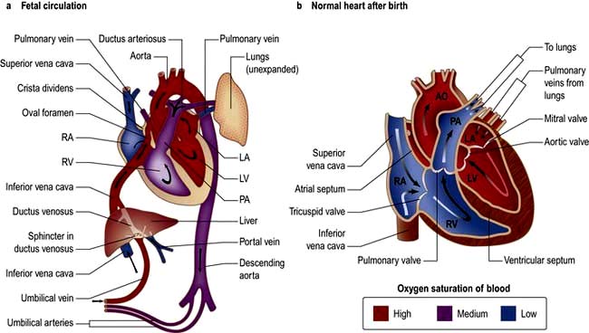
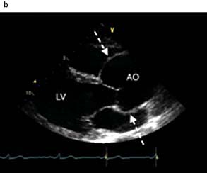
Figure 14.84 Anatomy showing circulation. AO, aorta; LA, left atrium; LV, left ventricle; PA, pulmonary artery; RA, right atrium; RV, right ventricle.
Blood moves from the IVC to the right atrium of the heart. In the fetus, there is an opening between the right and left atrium (the foramen ovale), and most of the blood (which is a mixture of oxygenated and de-oxygenated blood) flows from the right into the left atrium, bypassing pulmonary circulation. This blood goes into the left ventricle and is pumped through the aorta into the fetal body. Some of the blood flows from the aorta through the internal iliac arteries to the umbilical arteries and re-enters the placenta, where carbon dioxide and other waste products from the fetus are taken up and enter the woman’s circulation.
Some of the blood from the right atrium does not enter the left atrium, but enters the right ventricle and is pumped into the pulmonary artery. In the fetus, there is a connection between the pulmonary artery and the aorta, the ductus arteriosus, which directs most of this blood away from the lungs. With the first breath after delivery the vascular resistance in the pulmonary arteries falls and more blood moves from the right atrium to right ventricle and pulmonary arteries and oxygenated blood travels back to the left atrium through the pulmonary veins. The decrease in right atrial pressure and relative increase in left atrial pressure results in closure of the foramen ovale.
The ductus arteriosus usually closes off within one or two days of birth completely separating the left and right system. The umbilical vein and the ductus venosus closes off within 2–5 days after birth, leaving behind the ligamentum teres and the ligamentum venosus of the liver, respectively.
The aetiology of congenital cardiac disease is often unknown, but recognized associations include:
Maternal prenatal rubella infection (persistent ductus arteriosus, and pulmonary valvular and arterial stenosis)
Maternal alcohol misuse (septal defects)
Maternal drug treatment and radiation
Genetic abnormalities (e.g. The familial form of atrial septal defect and congenital heart block)
Chromosomal abnormalities (e.g. septal defects and mitral and tricuspid valve defects are associated with Down’s syndrome (trisomy 21) or coarctation of the aorta in Turner’s syndrome (45, XO).
See Table 14.41.
Table 14.41 Classification of congenital heart disease
Acyanotic | Cyanotic |
---|---|
With shunts |
With shunts |
Atrial septal defect |
Fallot’s tetralogy |
Ventricular septal defect |
Transposition of the great vessels |
Patent ductus arteriosus |
|
Partial anomalous venous drainage |
Severe Ebstein’s anomaly |
Without shunts |
Without shunts |
Coarctation of the aorta |
Severe pulmonary stenosis |
Congenital aortic stenosis |
Tricuspid atresia |
|
Pulmonary atresia |
|
Hypoplastic left heart |
Congenital heart disease should be recognized as early as possible, as the response is usually better the earlier the treatment is initiated. Some symptoms, signs and clinical problems are common in congenital heart disease:
Central cyanosis occurs because of right-to-left shunting of blood or because of complete mixing of systemic and pulmonary blood flow. In the latter case, e.g. Fallot’s tetralogy, the abnormality is described as cyanotic congenital heart disease.
Pulmonary hypertension results from large left-to-right shunts. The persistently raised pulmonary flow leads to the development of increased pulmonary artery vascular resistance and consequent pulmonary hypertension. This is known as the Eisenmenger’s reaction (or the Eisenmenger’s complex when due specifically to a ventricular septal defect). The development of pulmonary hypertension significantly worsens the prognosis.
Clubbing of the fingers occurs in congenital cardiac conditions associated with prolonged cyanosis.
Paradoxical embolism of thrombus from the systemic veins to the systemic arterial system may occur when a communication exists between the right and left heart. There is therefore an increased risk of cerebrovascular emboli and also abscesses (as with endocarditis).
Polycythaemia can develop secondary to chronic hypoxaemia, leading to a hyperviscosity syndrome and an increased thrombotic risk, e.g. strokes.
Growth retardation is common in children with cyanotic heart disease.
Syncope is common when severe right or left ventricular outflow tract obstruction is present. Exertional syncope, associated with deepening central cyanosis, may occur in Fallot’s tetralogy. Exercise increases resistance to pulmonary blood flow but reduces systemic vascular resistance. Thus, the right-to-left shunt increases and cerebral oxygenation falls.
Squatting is the posture adopted by children with Fallot’s tetralogy. It results in obstruction of venous return of desaturated blood and an increase in the peripheral systemic vascular resistance. This leads to a reduced right-to-left shunt and improved cerebral oxygenation.
Adolescents and adults with congenital heart disease present with specific common problems related to the long-standing structural nature of these conditions and any surgical treatment:
Endocarditis (particularly in association with otherwise innocuous lesions such as small VSDs or bicuspid aortic valve that can give up to 10% lifetime risk)
Progression of valvular lesions (calcification and stenosis of congenitally deformed valves, e.g. bicuspid aortic valve)
Atrial and ventricular arrhythmias (often quite resistant to treatment)
Right heart failure (especially when surgical palliation results in the right ventricle providing the systemic supply)
End-stage heart failure (rarely managed by heart or heart-lung transplantation).
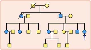
These conditions necessitate active follow-up of adult patients. Pregnancy is normally safe except if pulmonary hypertension or vascular disease is present, when the prognosis for both mother and fetus is poor.
Table 14.42 lists the most common congenital lesions and their occurrence in first-degree relatives.
Table 14.42 Common congenital lesions
Percentage of congenital lesions | Occurrence in first-degree relatives (%) | |
---|---|---|
Ventricular septal defect |
39 |
4 |
Atrial septal defect |
10 |
2 |
Persistent ductus |
10 |
4 |
Arteriosus |
|
|
Pulmonary stenosis |
7 |
|
Coarctation of the aorta |
7 |
2 |
Aortic stenosis |
6 |
4 |
Fallot’s tetralogy |
6 |
4 |
Others |
15 |
|
Genetic factors should be considered in all patients presenting with congenital heart disease. For example, parents with a child suffering from Fallot’s tetralogy stand a 4% chance of conceiving another child with the disease, and so fetal ultrasound screening of the mother during pregnancy is essential. Parents with congenital heart disease are also more likely to have affected offspring. Fathers have a 2% risk, while mothers have a higher risk (around 5%). Individual families can exhibit even higher risks of recurrence.
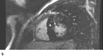
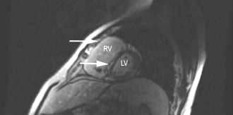
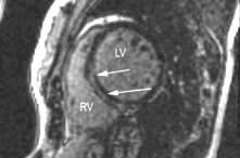
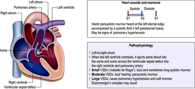
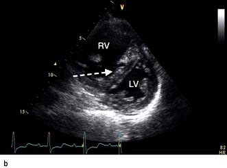
VSD is the most common congenital cardiac malformation (1 : 500 live births). The haemodynamic consequences of this are dependent on the shunt size. Left ventricular pressure is higher than right and blood therefore moves from left to right and pulmonary blood flow increases. In large defects the large volumes of blood flow through the pulmonary vasculature leads to pulmonary hypertension and eventual Eisenmenger’s complex when right ventricular pressure becomes higher than left and as a result blood starts to shunt from right to left leading to cyanosis.
Small restrictive VSDs (‘Maladie de Roger’) are often found incidentally as patients are asymptomatic. They are associated with a loud pan-systolic murmur. The majority close spontaneously by the age of 10 years.
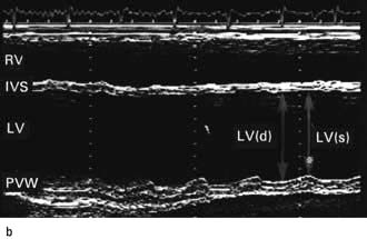
Large (non-restrictive) VSDs result in significant LA and LV dilatation (due to LV volume overload). Large defects usually present with heart failure symptoms in childhood and eventually lead to pulmonary hypertension and Eisenmenger’s complex. As pressures equalize the murmur becomes softer.
A small VSD produces no abnormal X-ray or ECG findings. In larger defects, the chest X-ray may demonstrate prominent pulmonary arteries owing to increased pulmonary blood flow but with pulmonary hypertension there may be ‘pruned’ pulmonary arteries. Cardiomegaly occurs when a moderate or a large VSD is present and the ECG may show both left and right ventricular hypertrophy. Echocardiography can assess the size and location of the VSD and its haemodynamic consequences. Interventional options are either surgical patch repair or device closure if it is an isolated muscular VSD. Indications for intervention include left atrial and ventricular enlargement with or without early LV dysfunction; reversible pulmonary hypertension (mild) where there is a residual left to right shunt and no significant desaturation with exercise; infective endocarditis. Patients with a restrictive VSD and patients after successful closure have an excellent long term outcome. Prophylaxis of endocarditis is discussed on page 87.
This condition is often first diagnosed in adulthood and represents one-third of congenital heart disease. It is two to three times more common in women than in men. There are three main types of ASD (Fig. 14.86):
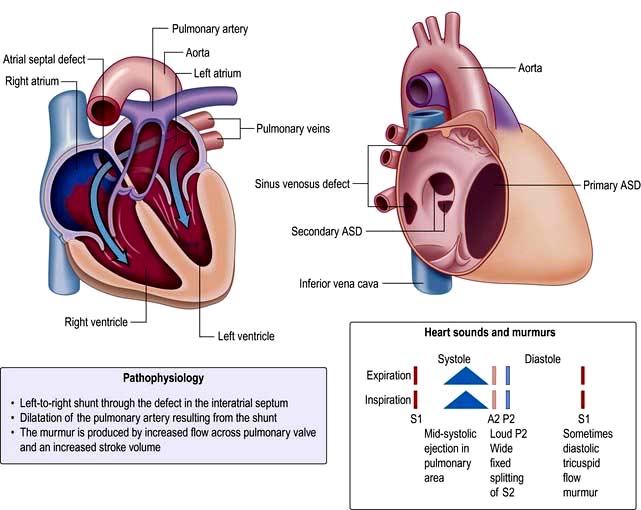
Sinus venosus defects – located in the superior part of the septum near the SVC (superior sinus venosus defect) or the inferior part of the septum near the IVC (inferior sinus venosus defect) entry point.
Ostium secundum defects (75%) – located in the mid-septum (fossa ovalis). This should not be confused with the patent foramen ovale (PFO), which is a normal variant and not a true septal defect. PFO is usually asymptomatic but is associated with paradoxical emboli and an increased incidence of embolic stroke.
Ostium primum (atrioventricular septal) defects (15%) – located in the lower part of atrial septum.
Adult patients with an unrepaired ASD with significant left to right shunt develop right heart overload and dilatation. These patients develop symptoms of dyspnoea and exercise intolerance and may develop atrial arrhythmias from right atrial dilatation. There is also increased pulmonary vascular flow, but significant pulmonary hypertension develops in <5% of patients and it is thought that these patients have additional factors including genetic predisposition to pulmonary hypertension. The physical signs of ASD reflect the volume overloading of the right ventricle (Fig. 14.86). A right ventricular heave can usually be felt.
The chest X-ray may demonstrate prominent pulmonary arteries with pulmonary plethora. Right bundle branch block and right axis deviation may be present on the ECG (because of dilatation of the right ventricle). Ostium primum defects may have left axis deviation on the ECG. Echocardiography may demonstrate hypertrophy and dilatation of the right heart and pulmonary arteries. Subcostal views with 2D and colour Doppler demonstrates the ASD (Fig. 14.87) and allow calculation of the left-right shunt (QP : QS ratio). CMR and CT are helpful to assess for anomalous pulmonary venous drainage which may accompany an ASD. Indications for intervention include: an ASD with significant left to right shunting resulting in right atrial/ventricular enlargement which should be closed irrespective of symptoms; thromboembolic events including certain patients with a patent foramen ovale. The options for intervention include device closure using a transcatheter clamshell device (Fig. 14.88) for most secundum ASDs (if suitable size) or surgical closure for all other ASD types.
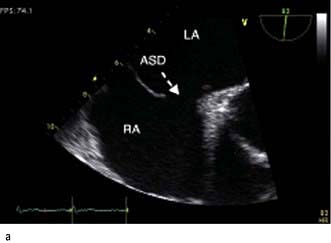
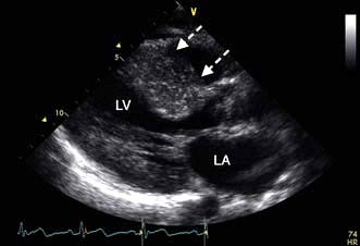
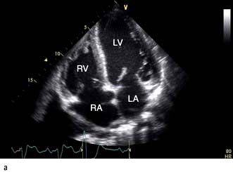
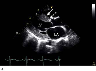
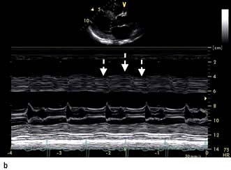
Figure 14.87 Ostium secundum atrial septal defect (arrows) in a young girl. (a) Shown by a 2-D echocardiogram subcostal four-chamber view. (b) Colour Doppler can demonstrate the left-to-right shunt. LA, left atrium; RA, right atrium.
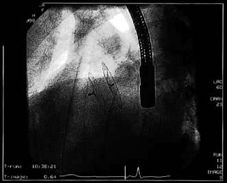
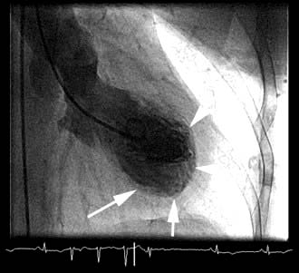
Figure 14.88 Angiographic appearance of a fully deployed ASD closure device. The device bridges the ASD and wedges against the surfaces of the right and left atrial septa, occluding flow. The metal object in frame is the distal end of a transoesophageal echocardiography probe.
(Courtesy of Dr D Ward, St George’s, University of London.)
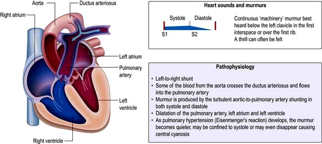
This is a persistent communication between the proximal left pulmonary artery and the descending aorta resulting in a continuous left to right shunt (Fig. 14.89). Normally the ductus arteriosus closes within a few hours of birth in response to decreased pulmonary resistance however in some cases (particularly premature babies and in cases with maternal rubella) the ductus persists. Indometacin (a prostaglandin inhibitor) is given to stimulate duct closure. If the shunt is moderate to large it will result in left heart volume overload overload and in some cases pulmonary hypertension and Eisenmenger’s syndrome. The characteristic clinical signs are a bounding pulse and continuous ‘machinery murmur’ however as pulmonary hypertension develops in a large PDA the murmur becomes softer.
With a large shunt, the aorta and pulmonary arterial system may be prominent on chest X-ray. The ECG may demonstrate left atrial abnormality and left ventricular hypertrophy. The development of Eisenmenger reaction will produce right ventricular hypertrophy. Echocardiography may show a dilated left atrium and left ventricle with right heart changes occurring late. Colour Doppler imaging of the proximal pulmonary arteries may demonstrate the shunt. Indications for intervention (usually with percutaneous devices) include left ventricular dilatation; mild–moderate pulmonary arterial hypertension (not Eisenmenger). Small defects may predispose to endarteritis and should be considered for device closure unless clinically silent.
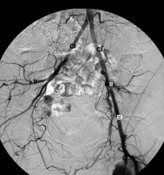
A coarctation of the aorta is a narrowing of the aorta at or just distal to the insertion of the ductus arteriosus (distal to the origin of the left subclavian artery (Fig. 14.90). Rarely it can occur proximal to the left subclavian. It occurs twice as commonly in men as in women. It is also associated with Turner’s syndrome (p. 978). In 80% of cases, the aortic valve is bicuspid (and potentially stenotic or endocarditic). Other associations include patent ductus arteriosus, ventricular septal defect, mitral stenosis or regurgitation and circle of Willis aneurysms. Severe narrowing of the aorta encourages the formation of a collateral arterial circulation involving the periscapular and intercostal arteries. Decreased renal perfusion can lead to the development of systemic hypertension that persists even after surgical correction.
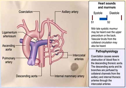
Coarctation of the aorta is often asymptomatic for many years. Headaches and nosebleeds (due to hypertension), and claudication and cold legs (due to poor blood flow in the lower limbs) may be present. Physical examination reveals hypertension in the upper limbs, and weak, delayed (radiofemoral delay) pulses in the legs. If coarctation is present in the aorta, proximal to the left subclavian artery, there will be asynchronous radial pulses in right and left arms. Poor peripheral pulses are seen in severe cases. For heart sounds and murmurs in coarctation of aorta, see Figure 14.90.
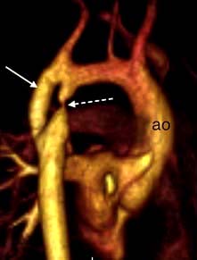
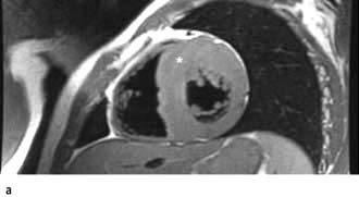
Chest X-ray may reveal a dilated aorta indented at the site of the coarctation. This is manifested by an aorta (seen in the upper right mediastinum) shaped like a ‘figure 3’. In adults, tortuous and dilated collateral intercostal arteries may erode the undersurfaces of the ribs (‘rib notching’). ECG demonstrates left ventricular hypertrophy. Echocardiography sometimes shows the coarctation and other associated anomalies. CT and CMR (Fig. 14.91) scanning can accurately demonstrate the coarctation and quantify flow.
Intervention is required if there is a peak–peak gradient across the coarctation of >20 mmHg and/or proximal hypertension. In neonates coarctation is treated with surgical repair. In older children and adults, balloon dilatation and stenting is an option although many centres still prefer surgery. Balloon dilatation is preferred for recoarctation.
Tetralogy of Fallot (Fig. 14.92) consists of:
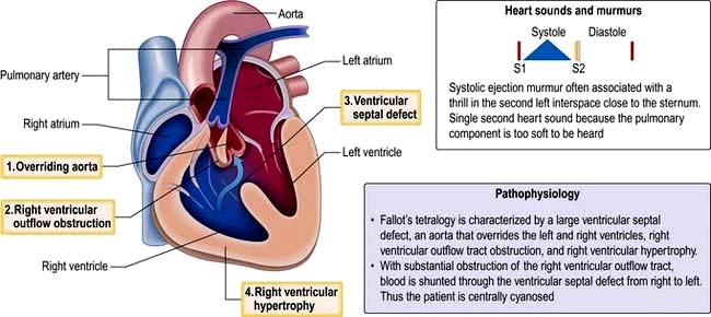
Symptoms depend on the degree of pulmonary stenosis. Often this is progressive in the first year of life and cyanosis develops due to increased right-sided pressures, resulting in a right to left shunt. Fallot’s spells are episodes of severe cyanosis noted in children due to spasm of the subpulmonary muscle – these can be relieved by increasing systemic resistance by postural manoeuvres, e.g. squatting. In babies with severe pulmonary stenosis systemic-to-pulmonary artery shunts (i.e. Blalock–Taussig – subclavian to pulmonary artery shunt) may be used initially to increase pulmonary blood flow in severe cases of pulmonary stenosis. The majority of adults with tetralogy of Fallot will have undergone complete repair which involves relief of the right ventricular outflow tract obstruction and closure of the VSD. The overall survival of those who have had operative repair is excellent. DiGeorge’s syndrome is found in 15% of those with tetralogy of Fallot.
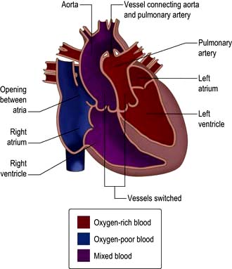
In complete transposition of the great arteries (TGA), the right atrium connects to the morphological right ventricle, which gives rise to the aorta and the left atrium connects to the morphological left ventricle, which gives rise to the pulmonary artery (Fig. 14.93). This is incompatible with life as blood circulates in two parallel circuits, i.e. deoxygenated blood from the systemic veins passes into the right heart and then via the aorta back to the systemic circulation. Oxygenated blood from the pulmonary veins passes through the left heart and back to the lungs. Babies with transposition are usually born cyanosed – if there is a significant ASD, VSD or PDA allowing a shunt (i.e. mixing of oxygenated and deoxygenated blood) the diagnosis might be delayed. In those without a shunt an atrial septostomy is performed. A Rashkind’s balloon is used to dilate the foramen ovale and is used to maintain saturations at 50–80% until a definitive procedure can be performed. The arterial switch procedure is now performed in the first 2 weeks of life – the aorta is reconnected to the left ventricle and the pulmonary artery is connected to the right ventricle. The coronary arteries are re-implanted.
Currently, the majority of adult patients with transposition of the great arteries will have had an ‘atrial switch’ operation. The right ventricle remains the systemic ventricle in this situation. Although most of these patients do well for many years, life expectancy is clearly limited by eventual failure of the systemic right ventricle.
In congenitally corrected transposition of the great arteries (ccTGA), systemic venous return to the right atrium enters a morphological left ventricle, which pumps into the pulmonary artery. Pulmonary venous blood returns to the left atrium and then via the morphological right ventricle to the aorta. The circulation is physiologically corrected but the systemic circulation is supported by a morphologic right ventricle. ccTGA is often associated with cardiac lesions; systemic (tricuspid) atrio-ventricular valve abnormalities with valve insufficiency; VSD; subpulmonary stenosis; complete heart block; Wolff–Parkinson–White syndrome; dextrocardia. Many patients with ccTGA live a normal life, although other patients require pacemaker insertion (the AV node is abnormal leading to heart block), surgery for a regurgitant tricuspid valve, or develop heart failure from the systemic (right ventricle).
Marfan’s syndrome (MFS) is one of the most common autosomal dominant inherited disorders of connective tissue, affecting the heart (aortic aneurysm and dissection, mitral valve prolapse), eye (dislocated lenses, retinal detachment) and skeleton (tall, thin body build with long arms, legs and fingers; scoliosis and pectus deformity) (Figs 14.94, 14.95; Table 14.43).
Figure 14.94 Marfan’s syndrome. (a) High arched palate and (b) eye lens dislocation.
(From: Forbes CD, Jackson WF. Color Atlas and Text of Clinical Medicine. St Louis: Mosby; 2003: 134, ©Elsevier.)
Figure 14.95 Marfan’s syndrome. (a) Photograph of a 63-year-old man. (b) 2-D Longitudinal echocardiogram of the aortic root. (c) Measurements of aortic size.
Clinically, two of three major systems must be affected, to avoid overdiagnosing the condition. Diagnosis may be confirmed by studying family linkage to the causative gene, or by demonstrating a mutation in the Marfan’s syndrome gene (MFS1) for fibrillin (FBN-Í) on chromosome 15q21.
MFS affects approximately 1 in 5000 of the population worldwide and 25% of patients are affected as a result of a new mutation. This group includes many of the more severely affected patients, with high cardiovascular risk. Other known associations with early death due to aortic aneurysm and dissection are: family history of early cardiac involvement; family history of dissection with an aortic root diameter of >5 cm; male sex; and extreme physical characteristics, including markedly excessive stature and widespread striae. Histological examination of aortas often shows widespread medial degeneration, described as ‘cystic medial necrosis’.
Chest X-ray is often normal but may show signs of aortic aneurysm and unfolding, or of widened mediastinum. Pneumothorax affects 11% and scoliosis is present in 70% of patients.
ECG may be misleadingly normal with an acute dissection. In conjunction with mitral valve prolapse, 40% of patients usually have arrhythmia, with premature ventricular and atrial arrhythmias.
Echocardiography shows mitral valve prolapse, and mitral regurgitation in the majority of patients. High-quality serial echocardiogram measurements of aortic root diameter in the sinuses of Valsalva, at 90° to the direction of flow are the basis for medical and surgical management (Fig. 14.95b).
CT or CMR can detect aortic dilatation and are useful in monitoring.
Beta-blocker therapy slows the rate of dilatation of the aortic root.
ACE receptor blockers. In Marfan’s there is upregulation of TNF-β, which is specifically inhibited by ACE blockers. A small trial has shown no increase in aortic root diameter on this therapy.
Lifestyle alterations are required because of ocular, cardiac or skeletal involvement. Sports that necessitate prolonged exertion at maximum cardiac output, such as cross-country running, are to be avoided. Sedentary occupations are usually best, as patients tend to suffer from easy fatiguability and hypermobile painful joints.
Monitoring with yearly echocardiograms up to aortic root diameter of 4.5 cm, 6-monthly from 4.5 to 5 cm, and then referred directly to a surgeon who is experienced in aortic root replacement in Marfan’s syndrome for elective surgery.
Pregnancy is generally well tolerated if no serious cardiac problems are present, but is preferably avoided if the aortic root diameter is over 4 cm, with aortic regurgitation. Echocardiography should be performed every 6–8 weeks throughout pregnancy and during the initial 6 months postpartum. Blood pressure should be regularly monitored and hypertension treated actively. Delivery should be by the least stressful method; ideally a vaginal delivery. Caesarean section should not be routinely performed. However, if the aortic root is over 4.5 cm, delivery at 39 weeks by induction or caesarean section should be considered. Beta-blocker therapy may be safely instituted or continued throughout pregnancy, to help prevent aortic dissection.
Medical and surgical management have increased the overall survival rate. On average, 13 years of life is added when surgical survival is compared to that reported in the natural history of MFS.
The condition is inherited in an autosomal dominant mode, with each child of one affected parent having a 50 : 50 chance of inheriting the condition. Males and females are equally often affected. In 25% of all cases, the condition arises as the result of a spontaneous mutation in gene 5 of one of the parents. Fibrillin-1 gene mutations can be identified in 80% of those affected, confirming diagnosis and aiding prognosis. The mutation can also be used to screen at-risk family members, including postnatal or prenatal offspring.
The normal mean pulmonary artery pressure (mPAP) at rest is 14 ± 3 mmHg with an upper limit of normal of 20 mmHg. The normal values for mean pulmonary artery pressure (mPAP), mean capillary wedge pressure (mPCWP) and cardiac output (CO) are 12 ± 2 mmHg, 6 ± 2 mmHg and 5 L/min, respectively. The fall in pressure across the lung circulation is known as the transpulmonary gradient and reflects the difference between mPAP and mPCWP. The normal transpulmonary gradient is 6 ± 2 mmHg.
The pulmonary vascular resistance (PVR) is calculated by the formula:
It is normally about 1.5 mmHg/L per min (1.5 Wood units). Approximately 60% of the body’s endothelial surface is in the lungs and the lungs normally offer a low resistance to blood flow. This is because the media of the precapillary pulmonary arterioles is thin as compared with their more muscular systemic counterparts that have to respond constantly to postural changes under the influence of gravity. The fact that the lung circulation normally offers a low resistance to flow explains the preferential passage of blood through the lungs in specific forms of congenital heart disease, which may eventually lead to remodelling of the lung circulation and pulmonary hypertension.
Pulmonary hypertension (PH) is defined as a mean pulmonary artery pressure (mPAP) of >25 mmHg at rest as measured on right heart catheterization. The clinical classification of PH is provided in Table 14.44. (PH can also complicate congenital heart disease as discussed in the ACHD section.)
Table 14.44 Updated clinical classification of pulmonary hypertension (Dana Point, 2008)
ALK1, activin receptor-like kinase type 1; BMPR2, bone morphogenetic protein receptor type 2; HIV, human immunodeficiency virus.
Reproduced with permission from Simmoneau G, Robbins IM, Beghetti M et al. Clinical classification of pulmonary hypertension. Journal of the American College of Cardiology 2009; 54:S43. Table used with permission of Elsevier Inc. All rights reserved.
The different groups are characterized by variable amounts of hypertrophy, proliferation and fibrotic changes in distal pulmonary arteries (pulmonary arterial hypertension PAH, pulmonary veno-occlusive disease PVOD, pulmonary hypertension PH due to left heart disease, PH due to lung disease and/or hypoxia). Pulmonary venous changes are seen in groups PVOD and PH due to left heart disease and the vascular bed may be destroyed in emphysematous or fibrotic areas seen in lung disease. In chronic thromboembolic pulmonary hypertension (CTEPH) organized thrombi are seen in the elastic pulmonary arteries. Patients with PH with unclear and/or multifactorial mechanisms have variable pathological findings.
Patients with progressive PH develop right ventricular hypertrophy, dilatation, failure and death.
Data from a recent French registry of 674 patients with pulmonary artery hypertension identified 39.2% with idiopathic pulmonary artery hypertension (IPAH), 3.9% familial (or heritable), 9.5% drugs and toxins (anorexigens), 15.3% connective tissue disorders (autoimmune rheumatic disease), 11.3% congenital heart disease, 10.4% portal hypertension and 6.2% HIV-associated. In familial or heritable PAH, mutations in the bone morphogenetic protein receptor 2 gene BMPR2 are detected in over 70% of cases; other mutations are seen in patients with hereditary haemorrhagic telangiectasia (Osler–Weber–Rendu syndrome). Drugs and toxins known to cause PAH include aminorex, fenfluramine, dexfenfluramine, toxic rapeseed oil and benfluorex.
Patients with PAH may present with symptoms of dyspnoea, fatigue, weakness, angina, syncope or abdominal distension. Clinical signs of PAH and right heart hypertrophy include a left parasternal heave, a loud P2 heart sound, a soft pan systolic murmur with tricuspid regurgitation or early diastolic murmur with pulmonary regurgitation. Right heart failure leads to jugular venous distension, ascites, peripheral oedema, and hepatomegaly. Clinical signs of associated diseases, e.g. systemic sclerosis, chronic liver disease, should be sought.
Chest X-ray shows enlargement of the pulmonary arteries and the major branches, with marked tapering (pruning) of peripheral arteries. The lung fields are usually lucent and there may be right atrial and right ventricular enlargement.
The ECG shows right ventricular hypertrophy and right atrial enlargement (P pulmonale).The chest X-ray may facilitate the diagnosis of PH due to left heart or chronic lung disease.
Echocardiography (Fig. 14.96) with tricuspid regurgitation can be used for determination of pulmonary artery pressure (PAP) using the simplified Bernoulli equation (PAP = 4 × (tricuspid regurgitation velocity)2 + estimated right atrial pressure). Right atrial pressure can be assumed at 5–10 mmHg unless there is significant dilatation of the inferior vena cava with reduced respiratory variation. Mean PAP = 0.61 × PA systolic pressure + 2 mmHg (although the Bernoulli equation may not be accurate in cases of severe tricuspid regurgitation).
Cardiac magnetic resonance imaging may be useful in ACHD and in assessing right ventricular function on serial assessment.
Routine blood tests include full blood count, renal and liver function tests, thyroid function tests, serological assays for underlying connective tissue diseases, HIV and hepatitis.
Abdominal liver ultrasound is useful to exclude liver cirrhosis and portal hypertension.
Right heart catheterization. As part of the clinical assessment right heart catheterization (RHC) may be indicated to confirm the diagnosis (elevated PAP), to determine the pulmonary wedge pressure (PWP), to calculate the cardiac output, and to assess for pulmonary vascular resistance and reactivity. In PAH vasodilator challenge (inhaled nitric oxide, intravenous adenosine or epoprostenol) should be performed to identify patients who may benefit from vasodilator therapies. A responder is defined as a reduction in mean PAP of ≥10 mmHg to reach an absolute mean PAP of ≤40 mmHg with increased or unchanged cardiac output. These vasodilator challenges are not recommended in patients with other types of PH (types 2–5).
Figure 14.96 Pulmonary hypertension. (a) Continuous wave Doppler echocardiography demonstrates markedly elevated tricuspid regurgitation velocity consistent with pulmonary hypertension. (b) Parasternal short-axis echocardiogram with a dilated right ventricle (RV) and septal flattening (arrows) in a patient with pulmonary hypertension.
Physical activity – patients should be encouraged to remain physically active but avoid exertion that precipitates severe dyspnoea, chest pain or pre-syncope.
Pregnancy – patients with PAH have a very high mortality rate during pregnancy (30–50%) and should be counselled against conception. Contraception may include barrier methods, progesterone-only pill, or the Mirena coil.
Travel – during plane travel supplementary oxygen at 2 L/minute may be appropriate for patients with reduced functional class and with resting hypoxia of <8 kPa.
Vaccination should be given for influenza and pneumococcal pneumonia.
Elective surgery – epidural anaesthesia may be preferable to a general anaesthetic.
Oral anticoagulation has evidence to support its use in patients with IPAH, heritable PAH and PAH due to anorexigens. The European target INR is 2.0–3.0.
Diuretics – these are used in patients with right heart failure and fluid overload.
Digoxin may be helpful in patients with tachyarrhythmias.
Calcium channel blockers can be effective in high doses in selected patients with IPAH who demonstrate a response to a vasodilator challenge. A right heart catheter should be repeated in 3–4 months to assess response to therapy.
Prostanoids – prostacyclin is a potent vasodilator that also inhibits platelet aggregation and cell proliferation. Synthetic prostacyclins are generally short-acting compounds requiring continuous intravenous or subcutaneous infusion or regular aerosol inhalation. They provide symptomatic relief and can improve exercise capacity and epoprostenol can improve survival in patients with IPAH and APAH.
Endothelin receptor antagonists – endothelin-1 is a potent vasoconstrictor and mitogen that binds to endothelin A and B receptors in the pulmonary vasculature. Both dual antagonists (bosentan) and selective A receptor antagonists (sitaxsentan, ambrisentan) can improve symptoms, exercise capacity and haemodynamics in patients with IPAH.
Phosphodiesterase type 5 inhibitors produce vasodilation in pulmonary vasculature and reduce cellular proliferation. Sildenafil and tadalafil have been demonstrated to provide symptomatic relief and improve exercise capacity in patients with IPAH.
Balloon atrial septostomy may be considered as palliative therapy in severe cases of PH.
Cardiac transplantation is used in patients with adverse prognosis although the 5-year survival following transplantation may be only 40–50%.
Left-sided heart disease (systolic and diastolic heart failure) and valvular heat disease is frequently associated with PH as is advanced chronic obstructive pulmonary disease (p. 814), pulmonary fibrosis and emphysema. Following acute pulmonary embolism 0.5–2.0 % of patients will develop CTEPH.
FURTHER READING
Galiè N, Hoeper MM, Humbert M et al. Guidelines for the diagnosis and treatment of pulmonary hypertension: the Task Force for the Diagnosis and Treatment of Pulmonary Hypertension of the European Society of Cardiology (ESC) and the European Respiratory Society (ERS), endorsed by the International Society of Heart and Lung Transplantation (ISHLT). Eur Heart J 2009; 30:2497.
Humbert M, Sitbon O, Chaouat A et al. Pulmonary arterial hypertension in France: results from a national registry. Am J Respir Crit Care Med 2006; 173:1023–1030.
Thrombus, usually formed in the systemic veins or rarely in the right heart (<10% of cases), may dislodge and embolize into the pulmonary arterial system. Post-mortem studies indicate that this is a very common condition (microemboli are found in up to 60% of autopsies) but it is not usually diagnosed this frequently in life. Of clinical pulmonary emboli, 10% are fatal.
Most clots which cause clinically relevant pulmonary emboli actually come from the pelvic and abdominal veins, but femoral deep venous thrombosis, and even occasionally axillary thrombosis, can be the origin of the clot. Clot forms as a result of a combination of sluggish blood flow, local injury or compression of the vein and a hypercoagulable state. Emboli can also occur from tumour, fat (long bone fractures), amniotic fluid and foreign material during i.v. drug use. Risk factors are shown in Table 8.26 and discussed on page 427.
After pulmonary embolism, lung tissue is ventilated but not perfused – producing an intrapulmonary dead space and resulting in impaired gas exchange. After some hours the non-perfused lung no longer produces surfactant. Alveolar collapse occurs and exacerbates hypoxaemia. The primary haemodynamic consequence of pulmonary embolism is a reduction in the cross-sectional area of the pulmonary arterial bed which results in an elevation of pulmonary arterial pressure and a reduction in cardiac output. The zone of lung that is no longer perfused by the pulmonary artery may infarct, but often does not do so because oxygen continues to be supplied by the bronchial circulation and the airways.
Sudden onset of unexplained dyspnoea is the most common, and often the only symptom of pulmonary embolism. Pleuritic chest pain and haemoptysis are present only when infarction has occurred. Many pulmonary emboli occur silently, but there are three typical clinical presentations. A clinical deep venous thrombosis is not commonly observed, although detailed investigation of the lower limb and pelvic veins will reveal thrombosis in more than half of the cases.
In this situation an embolus has impacted in a terminal pulmonary vessel. Symptoms are pleuritic chest pain and breathlessness. Haemoptysis occurs in 30%, often ≥3 days after the initial event. On examination, the patient may be tachypnoeic with a localized pleural rub and often coarse crackles over the area involved. An exudative pleural effusion (occasionally blood-stained) can develop. The patient may have a fever, and cardiovascular examination is normal.
This is a much rarer condition where sudden collapse occurs because of an acute obstruction of the right ventricular outflow tract. The patient has severe central chest pain (cardiac ischaemia due to lack of coronary blood flow) and becomes shocked, pale and sweaty. Syncope may result if the cardiac output is transiently but dramatically reduced, and death may occur. On examination, the patient is tachypnoeic, has a tachycardia with hypotension and peripheral shutdown. The jugular venous pressure (JVP) is raised with a prominent ‘a’ wave. There is a right ventricular heave, a gallop rhythm and a widely split second heart sound. There are usually no abnormal chest signs.
This leads to increased breathlessness, often over weeks or months. It is accompanied by weakness, syncope on exertion and occasionally angina. The physical signs are due to the pulmonary hypertension that has developed from multiple occlusions of the pulmonary vasculature. On examination, there are signs of right ventricular overload with a right ventricular heave and loud pulmonary second sound.
Chest X-ray is often normal, but linear atelectasis or blunting of a costophrenic angle (due to a small effusion) is not uncommon. These features develop only after some time. A raised hemidiaphragm is present in some patients. More rarely, a wedge-shaped pulmonary infarct, the abrupt cut-off of a pulmonary artery or a translucency of an underperfused distal zone is seen. Previous infarcts may be seen as opaque linear scars.
ECG is usually normal, except for sinus tachycardia, but sometimes atrial fibrillation or another tachyarrhythmia occurs. There may be evidence of right ventricular strain.
Blood tests. Pulmonary infarction results in a polymorphonuclear leucocytosis, an elevated ESR and increased lactate dehydrogenase levels in the serum. Immediately prior to commencing anticoagulants a thrombophilia screen should be checked.
Plasma D-dimer (see p. 416) – if this is undetectable, it excludes a diagnosis of pulmonary embolism.
Radionuclide ventilation/perfusion scanning (
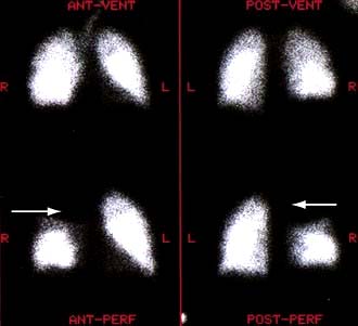
scan) is a good and widely available diagnostic investigation. Pulmonary 99mTc scintigraphy demonstrates underperfused areas (Fig. 14.97) which, if not accompanied by a ventilation defect on a ventilation scintigram performed after inhalation of radioactive xenon gas (see p. 802), is highly suggestive of a pulmonary embolus. There are limitations to the test, however. For example, a matched defect may arise with a pulmonary embolus which causes an infarct, or from emphysematous bullae. This test is therefore conventionally reported as a probability (low, medium or high) of pulmonary embolus and should be interpreted in the context of the history, examination and other investigations.
Ultrasound scanning can be performed for the detection of clots in pelvic or iliofemoral veins (see p. 789).
CT scans. Contrast-enhanced multidetector CT angiograms (CTA) (Fig. 14.98), have a sensitivity of 83% and specificity of 96%, with a positive predictive value of 92%. These values will increase with the use of 64-multislice scanners.
MR imaging gives similar results and is used if CT angiography is contraindicated.
Chest X-ray may show pulmonary oligaemia, sometimes with dilatation of the pulmonary artery in the hila. Often there are no changes.
ECG shows right atrial dilatation with tall peaked P waves in lead II. Right ventricular strain and dilatation give rise to right axis deviation, some degree of right bundle branch block, and T wave inversion in the right precordial leads (Fig. 14.99). The ‘classic’ ECG pattern with an S wave in lead I, and a Q wave and inverted T waves in lead III (S1, Q3, T3), is rare.
Blood gases show arterial hypoxaemia with a low arterial CO2 level, i.e. type I respiratory failure pattern.
Echocardiography shows a vigorously contracting left ventricle, and occasionally a dilated right ventricle and a clot in the right ventricular outflow tract.
Pulmonary angiography has now been replaced by CT and MR angiography.
Figure 14.99 Acute pulmonary embolism shown by a 12-lead ECG. There is an S wave in lead I, a Q wave in lead III and an inverted T wave in lead III (the S1, Q3, T3 pattern). There is sinus tachycardia (160 b.p.m.) and an incomplete right bundle branch block pattern (an R wave in AVR and V1 and an S wave in V6).
Chest X-ray may be normal. Enlarged pulmonary arterioles with oligaemic lung fields indicate advanced disease.
ECG can be normal or show signs of pulmonary hypertension (Fig. 14.99).
Leg imaging with ultrasound and venography may show thrombi.
Further tests looking for exercise-induced hypoxaemia and catheter studies to estimate pulmonary artery pressures are sometimes required.
The symptoms and signs of small and medium-sized pulmonary emboli are often subtle and nonspecific, so the diagnosis is often delayed or even completely missed. Pulmonary embolism should be considered if patients present with symptoms of unexplained cough, chest pain, haemoptysis, new-onset atrial fibrillation (or other tachycardia), or signs of pulmonary hypertension if no other cause can be found. Patients that are haemodynamically stable should have their clinical probability of a pulmonary embolus determined with the Revised Geneva Score (Table 14.45).
High clinical probability patients should proceed to multi-detector contrast-enhanced CT angiography (CTA) (see Fig. 14.98). A positive test confirms the diagnosis. A negative test but with an elevated D-dimer may require venous ultrasonography. (Patients with renal failure or contrast allergy can have ventilation/perfusion
scanning).
Low or intermediate clinical risk patients should have a D-dimer assay performed. A negative D-dimer rules out a pulmonary embolism. A positive D-dimer requires further investigation with CTA.
Table 14.45 Revised Geneva Score for the clinical prediction of a pulmonary embolism
Score | |
---|---|
Risk factors |
|
Age >65 years |
+1 |
Previous deep venous thrombosis or pulmonary embolism |
+3 |
Surgery or fracture within 1 month |
+2 |
Active malignancy |
+2 |
Symptoms |
|
Unilateral leg pain |
+3 |
Haemoptysis |
+2 |
Clinical signs |
|
Heart rate (b.p.m.): |
|
75–94 |
+3 |
≥95 |
+5 |
Pain on leg deep vein palpation and unilateral oedema |
+4 |
Clinical probability |
|
Low |
0–3 |
Intermediate |
4–10 |
High |
≥11 |
After Righini M, Le Gal G, Aujesky D et al. Lancet 2008; 371:1343–1352, with permission from Elsevier.
Patients who are haemodynamically unstable (shock, systolic blood pressure <90 mmHg, drop in pressure of ≥40 mmHg) may require urgent CTA or if critically ill with a high clinical probability, an echocardiogram should be performed – right ventricular dysfunction is highly suggestive of a pulmonary embolism – a normal right ventricle should suggest alternative diagnoses.
FURTHER READING
Agnelli G. Current concepts. Acute pulmonary embolism. N Engl J Med 2010; 363:266–284.
Bourjeily G. Pulmonary embolism in pregnancy. Lancet 2010; 375:500–512.
Torbicki A, Perrier A, Konstantinides S et al. Guidelines on the diagnosis and management of acute pulmonary embolism: the Task Force for the Diagnosis and Management of Acute Pulmonary Embolism of the European Society of Cardiology (ESC). Eur Heart J 2008; 29:2276–2315.
All patients should receive high-flow oxygen (60–100%) unless they have significant chronic lung disease. Patients with pulmonary infarcts require bed rest and analgesia.
Patients should be anticoagulated initially with subcutaneous low-molecular-weight heparin or fondaparinux or intravenous unfractionated heparin followed by warfarin therapy.
Massive pulmonary emboli. Intravenous fluids and even inotropic agents to improve the pumping of the right heart are sometimes required, and very ill patients will require care on the intensive therapy unit (see p. 885).
Fibrinolytic therapy such as streptokinase (250 000 units by i.v. infusion over 30 min, followed by streptokinase 100 000 units i.v. hourly for up to 12–72 hours, according to manufacturer’s instructions) has been shown in controlled trials to clear pulmonary emboli more rapidly and to confer a survival benefit in massive PE. It should be used in unstable patients and in some stable patients with adverse features, e.g. right ventricular dysfunction.
Surgical embolectomy is rarely necessary, but there may be no alternative when the haemodynamic circumstances are very severe.
Patients should be anticoagulated with vitamin K antagonists for a period of 3–6 months with a target INR of 2.0–3.0. Patients with cancer or pregnant women should be treated with long-term low-molecular-weight heparin. Occasionally, physical methods are required to prevent further emboli. This is usually because recurrent emboli occur despite adequate anticoagulation, but it is also indicated in high-risk patients in whom anticoagulation is absolutely contraindicated. The most common method by which pulmonary embolism is treated in this situation is by insertion of a filter in the inferior vena cava via the femoral vein to above the level of the renal veins.
This is the most common primary cardiac tumour. It occurs at all ages and shows no sex preference. Although most myxomas are sporadic, some are familial or are part of a multiple system syndrome. Histologically, they are benign. The majority of myxomas are solitary, usually develop in the left atrium and are polypoid, gelatinous structures attached by a pedicle to the atrial septum. The tumour may obstruct the mitral valve or may be a site of thrombi that then embolize. It is also associated with constitutional symptoms: the patient may present with dyspnoea, syncope or a mild fever. The physical signs are a loud first heart sound, a tumour ‘plop’ (a loud third heart sound produced as the pedunculated tumour comes to an abrupt halt), a mid-diastolic murmur and signs due to embolization. A raised ESR is usually present.
The diagnosis is easily made by echocardiography because the tumour is demonstrated as a dense space-occupying lesion (Fig. 14.100). Surgical removal usually results in a complete cure.
Figure 14.100 Atrial myxoma shown by a 2-D echocardiogram (long-axis view). The myxoma is an echo-dense mass obstructing the mitral valve orifice. It was removed surgically. LV, left ventricle; LA, left atrium.
Myxomas may also occur in the right atrium or in the ventricles. Other primary cardiac tumours include rhabdomyomas and sarcomas.
Myocardial disease that is not due to ischaemic, valvular or hypertensive heart disease, or a known infiltrative, metabolic/toxic or neuromuscular disorder may be caused by:
Acute inflammation of the myocardium has many causes (Table 14.46). Establishment of a definitive aetiology with isolation of viruses or bacteria is difficult in routine clinical practice.
Table 14.46 Causes of myocarditis
In western societies, the commonest causes of infective myocarditis are Coxsackie or adenoviral infection. Myocarditis in association with HIV infection is seen at post-mortem in up to 20% of cases but causes clinical problems in less than 10% of cases. Chagas’ disease, due to Trypanosoma cruzi, which is endemic in South America, is one of the commonest causes of myocarditis worldwide. Additionally, toxins (including prescribed drugs), physical agents, hypersensitivity reactions and autoimmune conditions may also cause myocardial inflammation.
In the acute phase, myocarditic hearts are flabby with focal haemorrhages; in chronic cases they are enlarged and hypertrophied. Histologically an inflammatory infiltrate is present – lymphocytes predominating in viral causes; polymorphonuclear cells in bacterial causes; eosinophils in allergic and hypersensitivity causes.
Myocarditis may be an acute or chronic process; its clinical presentations range from an asymptomatic state associated with limited and focal inflammation to fatigue, palpitations, chest pain, dyspnoea and fulminant congestive cardiac failure due to diffuse myocardial involvement. An episode of viral myocarditis, perhaps unrecognized and forgotten, may be the initial event that eventually culminates in an ‘idiopathic’ dilated cardiomyopathy. Physical examination includes soft heart sounds, a prominent third sound and often a tachycardia. A pericardial friction rub may be heard.
Chest X-ray may show some cardiac enlargement, depending on the stage and virulence of the disease.
ECG demonstrates ST- and T-wave abnormalities and arrhythmias. Heart block may be seen with diphtheritic myocarditis, Lyme disease and Chagas’ disease (see below).
Viral antibody titres may be increased. However, since enteroviral infection is common in the general population, the diagnosis depends on the demonstration of acutely rising titres.
Endomyocardial biopsy may show acute inflammation but false negatives are common by conventional criteria. Biopsy is of limited value outside specialized units.
Viral RNA. DNA can be measured from biopsy material using polymerase chain reaction (PCR). Specific diagnosis requires demonstration of active viral replication within myocardial tissue.
The underlying cause must be identified, treated, eliminated or avoided. Bed rest is recommended in the acute phase of the illness and athletic activities should be avoided for 6 months. Heart failure should be treated conventionally with the use of diuretics, ACE inhibitors/AII receptor antagonists, beta-blockers, spironolactone ± digoxin. Antibiotics should be administered immediately where appropriate. NSAIDs are contraindicated in the acute phase of the illness but may be used in the late phase. The use of corticosteroids is controversial and no studies have demonstrated an improvement in left ventricular ejection fraction or survival following their use. The administration of high-dose intravenous immunoglobulin on the other hand appears to be associated with a more rapid resolution of the left ventricular dysfunction and improved survival. Novel and effective antiviral, immunosuppressive (e.g. γ-interferon) and immunomodulating (e.g. IL-10) agents are available to treat viral myocarditis.
This is a severe form of myocarditis characterized by the presence of multinucleated giant cells within the myocardium. The cause is unknown but it may be associated with sarcoidosis, thymomas and autoimmune disease. It has a rapidly progressive course and a poor prognosis. Immunosuppression is recommended.
Chagas’ disease is caused by the protozoan Trypanosoma cruzi and is endemic in South America where upwards of 20 million people are infected. Acutely, features of myocarditis are present with fever and congestive heart failure. Chronically, there is progression to a dilated cardiomyopathy with a propensity towards heart block and ventricular arrhythmias. Treatment is discussed on page 148. Amiodarone is helpful for the control of ventricular arrhythmias; heart failure is treated in the usual way (p. 719).
Cardiomyopathies are a group of diseases of the myocardium that affect the mechanical or electrical function of the heart. They are frequently genetic and may produce inappropriate ventricular hypertrophy or dilatation and can be primarily a cardiac disorder or part of a multisystem disease (Table 14.47). Nevertheless, there is considerable heterogeneity and overlap between the conditions (Fig. 14.10). Abnormal myocardial function produces systolic or diastolic heart failure; abnormal electrical conduction results in cardiac arrhythmias and sudden cardiac death.
Primary cardiomyopathies |
||
Genetic |
Mixed (genetic/acquired) |
Acquired |
Hypertrophic cardiomyopathy |
Dilated cardiomyopathy |
Inflammatory (myocarditis) |
Arrhythmogenic right ventricular cardiomyopathy |
Restrictive (non-hypertrophic and non-dilated) |
Stress produced (Tako-tsubo) |
Left ventricular non-compaction |
|
Peripartum |
Conduction defects (see p. 707) |
|
|
Mitochondrial myopathies (see p. 40) |
|
|
Ion channel disordersa |
|
|
LQTS |
|
|
Brugada |
|
|
SQTS |
|
|
CPVT |
|
|
Asian SUNDS |
|
|
Secondary cardiomyopathies |
||
Infiltrativeb |
e.g. amyloidosis, Gaucher’s diseasec, Hurler’s diseasec, Hunter’s diseasec |
|
Storaged |
e.g. hereditary haemochromatosis, Fabry’s diseasec, glycogen storage disease (type II, Pompe)c, Niemann–Pick diseasec |
|
Toxicity |
e.g. drugs (e.g. cocaine), alcohol, heavy metals (e.g. cobalt), chemical agents |
|
Endomyocardial |
e.g. endomyocardial fibrosis, Loeffler’s endocarditis |
|
Inflammatory (granulomatous) |
e.g. sarcoidosis, post-infective (e.g. Chagas’ disease) |
|
Endocrine |
e.g. diabetes mellitusc, hyper- or hypothyroidism, hyperparathyroidism, phaeochromocytoma, acromegaly |
|
Cardiofacial |
||
Neuromuscular/neurological |
e.g. Friedreich’s ataxiac, Duchenne–Becker muscular dystrophyc, myotonic dystrophyc, neurofibromatosisc |
|
Nutritional deficiencies |
e.g. beriberi (thiamin), pellagra, scurvy, selenium, carnitine, kwashiorkor |
|
Autoimmune/rheumatic disorders |
e.g. systemic lupus erythematosus, dermatomyositis, systemic sclerosis |
|
Electrolyte imbalance |
|
|
Consequences of cancer therapy |
e.g. anthracyclines (e.g. doxorubicin), daunorubicin, cyclophosphamide, radiation |
LQTS, long QT syndrome; SQTS, short QT syndrome; CPVT, catecholaminergic polymorphic ventricular tachycardia; Asian SUNDS, Asian sudden unexpected nocturnal death syndrome.
a Excluded from the European Society of Cardiology classification.
b Accumulation of abnormal substances between myocytes (i.e. extracellular).
d Accumulation of abnormal substances within myocytes (i.e. intracellular).
HCM includes a group of inherited conditions that produce hypertrophy of the myocardium in the absence of an alternate cause (e.g. aortic stenosis or hypertension). It is the most common cause of sudden cardiac death in young people and affects 1 in 500 of the population. The majority of cases are familial autosomal dominant, due to mutations in the genes encoding sarcomeric proteins (Fig. 14.101). The most common causes of HCM are mutations of the β-myosin heavy chain MYH7 and myosin-binding protein C MYBPC3. Other mutations include troponin T and I, regulatory and essential myosin light chains, titin, α tropomyosin, α actin, α myosin heavy chain and muscle LIM protein (although over 400 mutations have been identified.) There are non-sarcomeric protein mutations in genes that control cardiac metabolism that result in glycogen storage diseases (Danon’s, Pompe’s and Fabry’s disease) that are indistinguishable from HCM.
Figure 14.101 Cardiomyopathy – clinical categories and genetic basis.Hypertrophic and dilated cardiomyopathies share the same genes, as do the less common restrictive cardiomyopathy and left ventricular non-compaction. Arrhythmogenic right ventricular cardiomyopathy (ARVC) is genetically different. AMPK, AMP-activated kinase; GLA, galactosidase A; LAMP2, lysosomal assisted membrane protein 2; TMEM43, transmembrane protein 43.
(Modified from Watkins H et al. Inherited cardiomyopathies. N Engl J Med 2011; 364:1643–1656, with permission.)
HCM is characterized by variable myocardial hypertrophy frequently involving the interventricular septum and disorganization (‘disarray’) of cardiac myocytes and myofibrils. Some 25% of patients have dynamic left ventricular outflow tract obstruction due to the combined effects of hypertrophy, systolic anterior motion (SAM) of the anterior mitral valve leaflet and rapid ventricular ejection. The salient clinical and morphological features of the disease vary according to the underlying genetic mutation. For example, marked hypertrophy is common with β myosin heavy chain mutations whereas mutations in troponin T may be associated with mild hypertrophy but a high risk of sudden death. The hypertrophy may not manifest before completion of the adolescent growth spurt, making the diagnosis in children difficult. HCM due to myosin-binding protein may not manifest until the sixth decade of life or later.
Many are asymptomatic and are detected through family screening of an affected individual or following a routine ECG examination.
Chest pain, dyspnoea, syncope or pre-syncope (typically with exertion), cardiac arrhythmias and sudden death are seen.
Sudden death occurs at any age but the highest rates (up to 6% per annum) occur in adolescents or young adults. Risk factors for sudden death are discussed below.
Dyspnoea occurs due to impaired relaxation of the heart muscle or the left ventricular outflow tract obstruction that occurs in some patients. The systolic cavity remains small until the late stages of disease when progressive dilatation may occur. If a patient develops atrial fibrillation there is often a rapid deterioration in clinical status due to the loss of atrial contraction and the tachycardia – resulting in elevated left atrial pressure and acute pulmonary oedema.
Double apical pulsation (forceful atrial contraction producing a fourth heart sound).
Jerky carotid pulse because of rapid ejection and sudden obstruction to left ventricular outflow during systole.
Ejection systolic murmur due to left ventricular outflow obstruction late in systole – it can be increased by manoeuvres that decrease afterload, e.g. standing or Valsalva, and decreased by manoeuvres that increase afterload and venous return, e.g. squatting.
Pansystolic murmur due to mitral regurgitation (secondary to SAM).
ECG abnormalities of HCM include left ventricular hypertrophy (see Fig. 14.76), ST and T wave changes, and abnormal Q waves especially in the inferolateral leads.
Echocardiography is usually diagnostic and in classical HCM there is asymmetric left ventricular hypertrophy (involving the septum more than the posterior wall), systolic anterior motion of the mitral valve, and a vigorously contracting ventricle (Fig. 14.102). However, any pattern of hypertrophy may be seen, including concentric and apical hypertrophy.
Cardiac MR can detect both the hypertrophy but also abnormal myocardial fibrosis (Fig. 14.103).
Genetic analysis, where available, may confirm the diagnosis and provide prognostic information for the patient and relatives.
Figure 14.102 Hypertrophic cardiomyopathy. A 2-D echocardiogram (short-axis view). The grossly thickened interventricular septum is shown, resulting in a small left ventricular cavity. This condition is associated with an abnormal anterior motion of the mitral valve during systole (arrows). LA, left atrium; LV, left ventricle.
The management of HCM includes treatment of symptoms and the prevention of sudden cardiac death in the patient and relatives.
Risk factors for sudden death:
Massive left ventricular hypertrophy (>30 mm on echocardiography)
Family history of sudden cardiac death (<50 years old)
Non-sustained ventricular tachycardia on 24-hour Holter monitoring
Abnormal blood pressure response on exercise (flat or hypotensive response).
The presence of these cardiac risk factors is associated with an increased risk of sudden death (Table 14.48), and patients with two or more should be assessed for implantable cardioverter-defibrillator (ICD). In patients in whom the risk is less, amiodarone is an appropriate alternative.
Table 14.48 Causes of sudden cardiac death
|
Chest pain and dyspnoea are treated with beta-blockers and verapamil either alone or in combination. An alternative agent is disopyramide if patients have left ventricular outflow tract obstruction. In some patients with significant left outflow obstruction and symptoms, dual-chamber pacing is necessary. Alcohol (non-surgical) ablation of the septum has been investigated and appears to give good results in reduction of outflow tract obstruction and subsequent improvement in exercise capacity. This procedure carries risks, including the development of complete heart block and myocardial infarction. Occasionally surgical resection of septal myocardium may be indicated. Vasodilators should be avoided because they may aggravate left ventricular outflow obstruction or cause refractory hypotension.
AVC is an uncommon (1 in 5000 population) inherited condition that predominantly affects the right ventricle with fatty or fibro-fatty replacement of myocytes, leading to segmental or global dilatation (Fig. 14.104). Left ventricular involvement has been reported in up to 75% of cases. The fibro-fatty replacement leads to ventricular arrhythmia and risk of sudden death in its early stages, and right ventricular or biventricular failure in its later stages.
Figure 14.104 Gross pathological specimen demonstrating thinning and fibro-fatty replacement of right ventricular free wall.
(From Basso C, Thiene G, Corrado D et al. Circulation 1996; 94:983–991, with permission.)
Autosomal dominant AVC has been mapped to eight chromosomal loci within mutations in four genes encoding for desmosomal proteins. These are the cardiac ryanodine receptor RyR2 (also responsible for familial catecholaminergic polymorphic ventricular tachycardia, CPVT), desmoplakin, plakophillin-2 and mutations altering the regulatory sequences of the transforming growth factor-β gene.
There are two recessive forms: Naxos disease (associated with palmoplantar keratoderma and woolly hair) that is due to a mutation in junctional plakoglobin, and Carvajal’s syndrome, due to a mutation in desmoplakin.
Most patients are asymptomatic. Symptomatic ventricular arrhythmias, syncope or sudden death occur. Occasionally presentation is with symptoms and signs of right heart failure, although this is more common in the later stages of the disease. Some patients may be detected through family screening, although frequently the morphological appearance of the right ventricle is normal, despite significant cardiac arrhythmias.
ECG is usually normal but may demonstrate T wave inversion in the precordial leads related to the right ventricle (V1–V3). Small-amplitude potentials occurring at the end of the QRS complex (epsilon waves) may be present (Fig. 14.105) and incomplete or complete RBBB is sometimes seen. Signal averaged ECG may indicate the presence of late potentials, the delayed depolarization of individual muscle cells; 24-hour Holter monitoring may demonstrate frequent extrasystoles of right ventricular origin or runs of non-sustained ventricular tachycardia.
Echocardiography is frequently normal but with more advanced cases may demonstrate right ventricular dilatation and aneurysm formation, and there may be left ventricular dilatation.
Cardiac MR can more accurately assess the right ventricle and in some cases can demonstrate fibro-fatty infiltration (Fig. 14.106).
Figure 14.105 Electrocardiogram from an adult with arrhythmogenic right ventricular cardiomyopathy (ARVC) demonstrating RBBB and precordial T wave insertion with epsilon waves visible at the terminal of the QRS complex (arrow).
Figure 14.106 Arrhythmogenic right ventricular cardiomyopathy (ARVC). Inversion recovery MR image post gadolinium demonstrates marked hyperenhancement (arrows) in the right ventricular free wall and infero-septum consistent with fibro-fatty replacement in right and left ventricles.
Clinical diagnosis is made using Task Force Criteria that include structural abnormalities of the right ventricle and RVOT (dilatation and abnormal wall motion on echocardiography or MRI), fibro-fatty replacement of myocytes on tissue biopsy, repolarization and conduction abnormalities on ECG or signal averaged ECG, ventricular tachycardia or frequent ventricular extrasystoles on Holter monitor, family history of ARVC/D in a first- or second-degree relative or premature sudden death (<35 years) due to ARVC/D.
Beta-blockers are first-line treatment for patients with non-life-threatening arrhythmias. Amiodarone or sotalol are used for symptomatic arrhythmias but for refractory or life-threatening arrhythmias an ICD is required. Occasionally cardiac transplantation is indicated either for intractable arrhythmia or cardiac failure.
DCM has a prevalence of 1 in 2500 and is characterized by dilatation of the ventricular chambers and systolic dysfunction with preserved wall thickness.
Familial DCM is predominantly autosomal dominant and can be associated with over 20 abnormal loci and genes (Fig. 14.107). Many of these are genes encoding cytoskeletal or associated myocyte proteins (dystrophin in X-linked cardiomyopathy; actin, desmin, troponin T, beta myosin heavy chain, sarcoglycans, vinculin and lamin a/c in autosomal dominant DCM) (Fig. 14.108). Many of these have prominent associated features such as skeletal myopathy or conduction system disease and therefore differ from the majority of cases of DCM.
Figure 14.107 Pedigree of a family with dilated cardiomyopathy. Blue symbols are affected family members. The arrow indicates the index case.
Figure 14.108 Schematic representation of myocyte proteins implicated in dilated cardiomyopathy (DCM).
Sporadic DCM can be caused by multiple conditions (Table 14.47):
Myocarditis – Coxsackie, adenoviruses, erythroviruses, HIV, bacteria, fungae, mycobacteria, parasitic (Chagas’s disease)
Toxins – alcohol, chemotherapy, metals (cobalt, lead, mercury, arsenic)
DCM can present with heart failure, cardiac arrhythmias, conduction defects, thromboembolism or sudden death. Increasingly, evaluation of relatives of DCM patients is allowing identification of early asymptomatic disease, prior to the onset of these complications. Clinical evaluation should include a family history and construction of a pedigree where appropriate.
Chest X-ray demonstrates generalized cardiac enlargement.
ECG may demonstrate diffuse nonspecific ST segment and T wave changes. Sinus tachycardia, conduction abnormalities and arrhythmias (i.e. atrial fibrillation, ventricular premature contractions or ventricular tachycardia) are also seen.
Echocardiogram reveals dilatation of the left and/or right ventricle with poor global contraction function (Fig. 14.109).
Cardiac MR may demonstrate other aetiologies of left ventricular dysfunction (e.g. previous myocardial infarction) or demonstrate abnormal myocardial fibrosis (Fig. 14.110).
Coronary angiography should be performed to exclude coronary artery disease in all individuals at risk (generally patients >40 years or younger if symptoms or risk factors are present).
Figure 14.109 Dilated cardiomyopathy. (a) 2-D (apical four-chamber view) and (b) M-mode echocardiograms. The heart has a ‘globular’ appearance with all four chambers dilated. The extremely impaired left ventricular function can be appreciated from the M-mode recording. Compare the systolic shortening fraction with that of Figure 14.23. LA, left atrium; RA, right atrium; LV, left ventricle.
Treatment consists of the conventional management of heart failure with the option of cardiac resynchronization therapy and ICDs in patients with NYHA III/IV grading. Cardiac transplantation is appropriate for certain patients.
LVNC is associated with a sponge-like appearance of the left ventricle. The condition predominantly affects the apical portion of the left ventricle and may be associated with congenital heart abnormalities. The condition is diagnosed by echocardiography, cardiac MR or left ventricular angiography. The natural history of the condition is unresolved but includes congestive cardiac failure, thromboembolism, cardiac arrhythmias and sudden death. Familial and spontaneous cases have been described.
Lenegre’s disease (p. 701) is a progressive disease of the cardiac conduction system (His-Purkinje system) that causes broad QRS duration, long pauses and bradycardia which presents with syncope. Sick-sinus syndrome is phenotypically similar.
This is a rare condition in which there is normal or decreased volume of both ventricles with bi-atrial enlargement, normal wall thickness, normal cardiac valves and impaired ventricular filling with restrictive physiology but near normal systolic function. The restrictive physiology produces symptoms and signs of heart failure. Conditions associated with this form of cardiomyopathy include amyloidosis (commonest), sarcoidosis, Loeffler’s endocarditis and endomyocardial fibrosis; in the latter two conditions there is myocardial and endocardial fibrosis associated with eosinophilia. The idiopathic form of restrictive cardiomyopathy may be familial.
Patients with restrictive cardiomyopathy may present with dyspnoea, fatigue and embolic symptoms. On clinical examination there will be elevated jugular venous pressure with diastolic collapse (Friedreich’s sign) and elevation of venous pressure with inspiration (Kussmaul’s sign), hepatic enlargement, ascites and dependent oedema. Third and fourth heart sounds may be present.
Chest X-ray may show pulmonary venous congestion. The cardiac silhouette can be normal or show cardiomegaly and/or atrial enlargement.
ECG may demonstrate low-voltage QRS and ST segment and T wave abnormalities.
Echocardiography shows symmetrical myocardial thickening and often a normal systolic ejection fraction but impaired ventricular filling. In amyloid patients the myocardium typically appears speckled with absent radial thickening as demonstrated by ‘tram-lines’ on M-mode echocardiography (Fig. 14.111).
Cardiac MR may demonstrate abnormal myocardial fibrosis in amyloidosis or sarcoidosis.
Cardiac catheterization and haemodynamic studies may help distinguish between restrictive cardiomyopathy and constrictive pericarditis, although volume loading may be required.
Endomyocardial biopsy in contrast with other cardiomyopathies is often useful in this condition and may permit a specific diagnosis, such as amyloidosis, to be made.
Figure 14.111 Amyloid. (a) Parasternal long-axis echocardiogram demonstrates left ventricular (LV) hypertrophy with reduced systolic function. The septum has a speckled appearance (arrows). (b) M-mode echocardiogram demonstrates reduced septal thickening with ‘tram-line’ appearance (arrows). These features are suggestive of cardiac amyloid – confirmed on cardiac biopsy. LA, left atrium.
There is no specific treatment. Cardiac failure and embolic manifestations should be treated. Cardiac transplantation is necessary in some severe cases, especially the idiopathic variety. In primary amyloidosis combination therapy with melphalan plus prednisolone with or without colchicine may improve survival. However, patients with cardiac amyloidosis have a worse prognosis than those with other forms of the disease, and the disease often recurs after transplantation. Liver transplantation may be effective in familial amyloidosis (due to production of mutant pre-albumin) and may lead to reversal of the cardiac abnormalities.
Patients with this condition present acutely with chest pain and breathlessness associated with ECG changes and elevated cardiac biomarkers consistent with acute myocardial infarction. Diagnostic coronary angiography typically demonstrates unobstructed coronary arteries with characteristic akinesia of the mid-apical segments of the left ventricle on ventriculography or echocardiography with preserved basal function (Fig. 14.112). The pathophysiology of the condition is uncertain but may be due to transient catecholamine excess, coronary vasospasm, abnormalities of the coronary microcirculation and hypertrophy of the basal septum. The syndrome is more common in middle-old aged women. Severe cases may have cardiogenic shock and pulmonary oedema. Patients with a significant left ventricular gradient may respond to cautious beta-blockade. Complete recovery of function is normal within 4–6 weeks.
This rare condition affects women in the last trimester of pregnancy or within 5 months of delivery. It presents as a dilated cardiomyopathy, is more common in obese, multiparous women over 30 years old and is associated with pre-eclampsia. Nearly half of patients will recover to normal function within 6 months but in some patients it can cause progressive heart failure and sudden death.
The pericardium acts as a protective covering for the heart. It consists of an outer fibrous pericardial sac and an inner serous pericardium made up of the inner visceral epicardium that lines the heart and great vessels and its reflection the outer parietal pericardium that lines the fibrous sac. The normal amount of pericardial fluid is 20–49 mL that lubricates the surface of the heart. Presentations of pericardial disease include:
This refers to inflammation of the pericardium. Classically, fibrinous material is deposited into the pericardial space and pericardial effusion often occurs. Acute pericarditis has numerous aetiologies (Table 14.49) although in most cases a cause is not identified (idiopathic).
Table 14.49 Aetiology of pericarditis
Viral pericarditis. The most common viral causes are Coxsackie B virus and echovirus. Viral pericarditis is usually painful but has a short time course and rarely long-term effects. Increasingly, HIV is implicated in the aetiology of pericarditis, both directly and via immunosuppression, which predisposes the subject to infective causes.
Post-myocardial infarction pericarditis occurs in about 20% of patients in the first few days following MI. It occurs more commonly with anterior MI and ST elevation MI with high serum cardiac enzymes, but its incidence is reduced to 5–6% with thrombolysis. It may be difficult to differentiate this pain from recurrent angina when it occurs early (day 1–2 post-infarct) but a good history of the pain and serial ECG monitoring is helpful. Pericarditis may also occur later on in the recovery phase after infarction. This usually occurs as a feature of Dressler’s syndrome, an autoimmune response to cardiac damage occurring 2–10 weeks’ post-infarct. Autoimmune reaction to myocardial damage is the main aetiology, and antimyocardial antibodies can often be found. Recurrences are common. Differential diagnosis includes a new myocardial infarction or unstable angina.
Uraemic pericarditis is due to irritation of the pericardium by accumulating toxins. It can occur in 6–10% of patients with advanced kidney disease if dialysis is delayed. It is an indication for urgent dialysis as it continues to be associated with significant morbidity and mortality.
Bacterial pericarditis may rarely occur with septicaemia or pneumonia, or it may stem from an early postoperative infection after thoracic surgery or trauma, or may complicate endocarditis. Staphylococcus aureus is a frequent cause of purulent pericarditis in HIV patients. This form of pericarditis, especially staphylococcal, is fulminant and often fatal.
Other endemic infectious pericarditis includes mycoplasmosis and Lyme pericarditis which are often effusive and require pericardial drainage. The diagnosis is based on serological tests of pericardial fluid and identification of organisms in pericardial or myocardial biopsies.
Tuberculous pericarditis usually presents with chronic low-grade fever, particularly in the evening, associated with features of acute pericarditis, dyspnoea, malaise, night sweats and weight loss. Pericardial aspiration is often required to make the diagnosis. Constrictive pericarditis is a frequent outcome. Treatment is as for pulmonary TB ( p. 842) but prednisolone 60 mg daily for 2–6 weeks.
Fungal pericarditis is a common complication of endemic fungal infections, such as histoplasmosis and coccidioidomycosis but may be also caused by Candida albicans, especially in immunocompromised patients, drug addicts or after cardiac surgery.
Malignant pericarditis. Carcinoma of the bronchus, carcinoma of the breast and Hodgkin’s lymphoma are the most common causes of malignant pericarditis. Leukaemia and malignant melanoma are also associated with pericarditis. A substantial pericardial effusion is very typical and is due to the obstruction of the lymphatic drainage from the heart. The effusion is often haemorrhagic. Radiation and therapy for thoracic tumours may cause radiation injury to the pericardium resulting in serous or haemorrhagic pericardial effusion and pericardial fibrosis. Absence of neoplastic cells in the pericardial fluid in these conditions often helps diagnosis.
Pericardial inflammation produces sharp central chest pain exacerbated by movement, respiration and lying down. It is typically relieved by sitting forward. It may be referred to the neck or shoulders. The main differential diagnoses are angina and pleurisy. The classical clinical sign is a pericardial friction rub occurring in three phases corresponding to atrial systole, ventricular systole and ventricular diastole. It may also be heard as a biphasic ‘to and fro’ rub. The rub is heard best with the diaphragm of the stethoscope at the lower left sternal edge at the end of expiration with the patient leaning forward. There is usually a fever, leucocytosis or lymphocytosis when pericarditis is due to viral or bacterial infection, rheumatic fever or myocardial infarction. Features of a pericardial effusion may also be present (p. 776). Large pericardial effusion can compress adjacent bronchi and lung tissue and may cause dyspnoea.
ECG is diagnostic. There is widespread concave-upwards (saddle-shaped) ST elevation (Fig. 14.113), reciprocal ST depression in leads aVR and V1, and PR segment depression. These changes evolve over time, with resolution of the ST elevation, T wave flattening/inversion and finally T wave normalization. The early ECG changes must be differentiated from the ST elevation found in myocardial infarction which is limited to the infarcted area, e.g. anterior or inferior. Sinus tachycardia may result from fever or haemodynamic embarrassment, and rhythm and conduction abnormalities may be present if myocardium is involved. Cardiac enzymes should be assayed as they may be elevated if there is associated myocarditis (see p. 767). Chest X-ray may demonstrate cardiomegaly (in cases with an effusion) which should be confirmed with echocardiography. CT and cardiac MR may be helpful for in cases with thickened (>4 mm) or inflamed (abnormal delayed enhancement) pericardium.
Figure 14.113 ECGs associated with pericarditis. (a) Acute pericarditis. Note the raised ST segment, concave upwards (arrow). (b) Chronic phase of pericarditis associated with a pericardial effusion. Note the T wave flattening and inversion, and the alternation of the QRS amplitude (QRS alternans). (c) The same patient after evacuation of the pericardial fluid. Note that the QRS voltage has increased and the T waves have returned to normal.
If a cause is found, this should be treated. Bed rest and oral NSAIDs (high-dose aspirin indometacin or ibuprofen) are effective in most patients. Aspirin is the drug of choice for patients with a recent myocardial infarction. Colchicine is also effective in combination with conventional therapy, as demonstrated in the COPE trial. Corticosteroids should be reserved for patients with a known immune cause as their use is associated with an increased rate of recurrence.
About 20% of cases of acute pericarditis go on to develop idiopathic relapsing pericarditis which may be incessant (recurs within 6 weeks during weaning of NSAIDs) or intermittent (recurs >6 weeks after the initial presentation). The first-line treatment is again oral NSAIDs. The colchicine as first-choice therapy for recurrent pericarditis trial demonstrated that prolonged colchicine (for 6 months) was more effective than aspirin alone in reducing recurrence. In resistant cases, oral corticosteroids may be effective and in some patients pericardiectomy may be appropriate.
A pericardial effusion is a collection of fluid within the potential space of the serous pericardial sac (Fig. 14.114), commonly accompanying an episode of acute pericarditis. When a large volume collects in this space, ventricular filling is compromised leading to embarrassment of the circulation. This is known as cardiac tamponade.
Symptoms of a pericardial effusion commonly reflect the underlying pericarditis. On examination:
Heart sounds are soft and distant
Apex beat is commonly obscured
A friction rub may be evident due to pericarditis in the early stages, but this becomes quieter as fluid accumulates and pushes the layers of the pericardium apart
Rarely, the effusion may compress the base of the left lung, producing an area of dullness to percussion below the angle of the left scapula (Ewart’s sign)
As the effusion worsens, signs of cardiac tamponade may become evident:
ECG reveals low-voltage QRS complexes (<0.5 mV in limb leads) with sinus tachycardia and there may be electric alternans (alteration of QRS amplitude or axis between beats).
Chest X-ray (Fig. 14.114) shows large globular or pear-shaped heart with sharp outlines. Typically, the pulmonary veins are not distended.
Echocardiography (Fig. 14.115) is the most useful technique for demonstrating the effusion and looking for evidence of tamponade – late diastolic collapse of the right atrium, early diastolic collapse of the right ventricle, ventricular septum displacement into the left ventricle during inspiration, diastolic flow reversal in the hepatic veins during expiration, dilated inferior vena cava with <50% reduction during inspiration.
Cardiac CT or MRI should be considered if loculated pericardial effusions are suspected (post-cardiac surgery).
Pericardiocentesis is the removal of pericardial fluid with aseptic technique under echocardiographic guidance. It is indicated when a tuberculous, malignant or purulent effusion is suspected.
Pericardial biopsy may be needed if tuberculosis is suspected and pericardiocentesis is not diagnostic.
Figure 14.115 2-D Echocardiogram (short-axis view) from a patient with a large pericardial effusion associated with pulmonary tuberculosis. The exudate is seen between the visceral and parietal layers of the pericardium and would give a false impression of cardiomegaly on a chest X-ray. Note the multiple fibrous strands within the effusion, showing that it is consolidating and will probably lead to constriction of cardiac function. LA, left atrium; LV, left ventricle; PE, pericardial effusion; RA, right atrium; RV, right ventricle.
Other tests include looking for underlying causes, e.g. blood cultures, autoantibody screen.
An underlying cause should be sought and treated if possible. Most pericardial effusions resolve spontaneously. However, when the effusion collects rapidly, tamponade may result. Pericardiocentesis is then indicated to relieve the pressure – a drain may be left in temporarily to allow sufficient release of fluid. Pericardial effusions may reaccumulate, most commonly due to malignancy (in the UK). This may require pericardial fenestration, i.e. creation of a window in the pericardium to allow the slow release of fluid into the surrounding tissues. This procedure may either be performed transcutaneously under local anaesthetic or using a conventional surgical approach.
Certain causes of pericarditis such as tuberculosis, haemopericardium, bacterial infection and rheumatic heart disease result in the pericardium becoming thick, fibrous and calcified. This may also develop late after open heart surgery, and fibrosis also occurs with the use of dopamine agonists, e.g. cabergoline, pergolide. In many cases these pericardial changes do not cause any symptoms. If, however, the pericardium becomes so inelastic as to interfere with diastolic filling of the heart, constrictive pericarditis is said to have developed. As these changes are chronic, allowing the body time to compensate, this condition is not as immediately life-threatening as cardiac tamponade, in which the circulation is more acutely embarrassed.
Constrictive pericarditis should be distinguished from restrictive cardiomyopathy (see p. 772). The two conditions are very similar in their presentation, but the former is fully treatable, whereas most cases of the latter are not. In the later stages of constrictive pericarditis, the subepicardial layers of myocardium may undergo fibrosis, atrophy and calcification.
The symptoms and signs of constrictive pericarditis occur due to:
reduced ventricular filling (similar to cardiac tamponade, i.e. Kussmaul’s sign, Friedreich’s sign, pulsus paradoxus)
systemic venous congestion (ascites, dependent oedema, hepatomegaly and raised JVP)
pulmonary venous congestion (dyspnoea, cough, orthopnoea, PND) less commonly
reduced cardiac output (fatigue, hypotension, reflex tachycardia)
rapid ventricular filling (‘pericardial knock’ heard in early diastole at the lower left sternal border)
Chest X-ray shows a relatively small heart in view of the symptoms of heart failure. Pericardial calcification may be present in up to 50%. A lateral chest film may be useful in detecting calcification that is missed on an AP film. However, a calcified pericardium is not necessarily a constricted one.
ECG reveals low-voltage QRS complexes with generalized T wave flattening or inversion.
Echocardiography shows thickened calcified pericardium, and small ventricular cavities with normal wall thickness. Doppler studies may be useful.
CT and CMR are used to assess pericardial anatomy and thickness (≥4 mm) (see Fig. 14.28).
Endomyocardial biopsy may be helpful in distinguishing constrictive pericarditis from restrictive cardiomyopathy in difficult cases.
Cardiac catheterization. End-diastolic pressures in the left and right ventricles measured during this procedure are usually equal, owing to pericardial constriction.
Restrictive cardiomyopathy is a close mimic of constrictive pericarditis and all the above tests may not help to distinguish the two conditions.
The treatment for chronic constrictive pericarditis is complete resection of the pericardium. This is a risky procedure with a high complication rate due to the presence of myocardial atrophy in many cases at the time of surgery. Thus early pericardiectomy is suggested in non-tuberculous cases, before severe constriction and myocardial atrophy have developed.
In cases of tuberculous constriction, the presence of pericardial calcification implies chronic disease. Current evidence tends to favour early pericardiectomy with antituberculous drug cover in these cases. If there is no calcification, a course of antituberculous therapy should be attempted first. If the patient’s haemodynamic state remains static or deteriorates after 4–6 weeks of therapy, pericardiectomy is recommended.
Elevated arterial blood pressure is a major cause of premature vascular disease leading to cerebrovascular events, ischaemic heart disease and peripheral vascular disease. Blood pressure is a characteristic of each individual, like height and weight, with marked interindividual variation, and has a continuous (bell-shaped) distribution. The levels of blood pressure observed depend on the characteristics of the population studied – in particular, the age and ethnic background. Blood pressure in industrialized countries rises with age, certainly up to the seventh decade. This rise is more marked for systolic pressure and is more pronounced in men. Hypertension is very common in the developed world. Depending on the diagnostic criteria, hypertension is present in 20–30% of the adult population. Hypertension rates are much higher in black Africans (40–45% of adults). The definition of an abnormal blood pressure is indicated in Table 14.50.
Table 14.50 Classification of blood pressure levels of the British Hypertension Society
Category | Systolic blood pressure (mmHg) | Diastolic blood pressure (mmHg) |
---|---|---|
Blood pressure |
|
|
Optimal |
<120 and |
<80 |
Normal |
120–129 and/or |
<80–84 |
High normala |
130–139 and/or |
85–89 |
Hypertension |
|
|
Grade 1 (mild) |
140–159 and/or |
90–99 |
Grade 2 (moderate) |
160–179 and/or |
100–109 |
Grade 3 (severe) |
≥180 |
≥110 |
Isolated systolic hypertension |
|
|
Grade 1 |
140–149 |
<90 |
Grade 2 |
>160 |
<90 |
The European Societies of Hypertension and Cardiology Guidelines 2007 are based on clinical blood pressure and not values for ambulatory blood pressure measurement. Threshold blood pressure levels for the diagnosis of hypertension using self/home monitoring are >135/85 mmHg. For ambulatory monitoring, 24-hour values are >125/80 mmHg. If systolic blood pressure and diastolic blood pressure fall into different categories, the higher value should be taken for classification.
a Equivalent to prehypertension.
The risk of mortality or morbidity rises progressively with increasing systolic and diastolic pressures, with each measure having an independent prognostic value; e.g. isolated systolic hypertension is associated with a two- to threefold increase in cardiac mortality.
A prehypertension category has been added to reflect the continuum between normal and abnormal blood pressure.
All adults should have blood pressure measured routinely at least every 5 years until the age of 80 years. Seated blood pressure when measured after 5 minutes’ resting with appropriate cuff size and arm supported is usually sufficient, but standing blood pressure should be measured in diabetic and elderly subjects to exclude orthostatic hypotension. The cuff should be deflated at 2 mm/s and the blood pressure measured to the nearest 2 mmHg. Two consistent blood pressure measurements are needed to estimate blood pressure, and more are recommended if there is variation in the pressure. When assessing the cardiovascular risk, the average blood pressure at separate visits is more accurate than measurements taken at a single visit.
The majority (80–90%) of patients with hypertension have primary elevation of blood pressure, i.e. essential hypertension of unknown cause.
Blood pressure tends to run in families and children of hypertensive parents tend to have higher blood pressure than age-matched children of parents with normal blood pressure. This familial concordance of blood pressure may be explained, at least in part, by shared environmental influences. However, there still remains a large, still largely unidentified genetic component.
Low birth weight is associated with subsequent high blood pressure. This relationship may be due to fetal adaptation to intrauterine undernutrition with long-term changes in blood vessel structure or in the function of crucial hormonal systems.
Among the several environmental factors that have been proposed, the following seem to be the most significant:
Obesity. Fat people have higher blood pressures than thin people. There is a risk, however, of overestimation if the blood pressure is measured with a small cuff. Adjust the bladder size to the arm circumference. Sleep disordered breathing (see p. 818) often seen with obesity may be an additional risk factor.
Alcohol intake. Most studies have shown a close relationship between the consumption of alcohol and blood pressure level. However, subjects who consume small amounts of alcohol seem to have lower blood pressure level than those who consume no alcohol.
Sodium intake. A high sodium intake has been suggested to be a major determinant of blood pressure differences between and within populations around the world. Populations with higher sodium intakes have higher average blood pressures than those with lower sodium intake. Migration from a rural to an urban environment is associated with an increase in blood pressure that is in part related to the amount of salt in the diet. Studies of the restriction of salt intake have shown a beneficial effect on blood pressure in hypertensives. There is some evidence that a high potassium diet can protect against the effects of a high sodium intake.
Stress. While acute pain or stress can raise blood pressure, the relationship between chronic stress and blood pressure is uncertain.
The autonomic nervous system, as well as the renin-angiotensin, natriuretic peptide and kallikrein-kinin system, plays a role in the physiological regulation of short-term changes in blood pressure and has been implicated in the pathogenesis of essential hypertension. A low renin, salt-sensitive, essential hypertension in which patients have renal sodium and water retention has been described. However, there is no convincing evidence that the above systems are directly involved in the maintenance of hypertension.
An association between diabetes and hypertension has long been recognized and a syndrome has been described of hyperinsulinaemia, glucose intolerance, reduced levels of HDL cholesterol, hypertriglyceridaemia and central obesity (all of which are related to insulin resistance) in association with hypertension. This association (also called the ‘metabolic syndrome’, p. 218) is a major risk factor for cardiovascular disease.
Secondary hypertension is where blood pressure elevation is the result of a specific and potentially treatable cause (Table 14.51).
Table 14.51 Secondary causes of hypertension
The pathogenesis of essential hypertension remains unclear. In some young hypertensive patients, there is an early increase in cardiac output, in association with increased pulse rate and circulating catecholamines. This could result in changes in baroreceptor sensitivity, which would then operate at a higher blood pressure level.
In established hypertension there are:
Renal changes: eventually, changes in the renal vasculature lead to a reduced renal perfusion, reduced glomerular filtration rate and, finally, a reduction in sodium and water excretion. The decreased renal perfusion may lead to activation of the renin-angiotensin system (renin converts angiotensinogen to angiotensin I, which is in turn converted to angiotensin II by angiotensin-converting enzyme) with increased secretion of aldosterone and further sodium and water retention.
Cerebral changes in small vessel cause lacunae (small infarcts) and reversible neurological deficits which do not show abnormalities on imaging. This may lead to dementia and stroke.
Cerebrovascular disease and coronary artery disease are the most common causes of death, although hypertensive patients are also prone to renal failure and peripheral vascular disease (Fig. 14.116).
Hypertensives have a six-fold increase in stroke (both haemorrhagic and atherothrombotic). There is a three-fold increase in cardiac death (due either to coronary events or to cardiac failure). Furthermore, peripheral arterial disease is twice as common.
Malignant or accelerated hypertension occurs when blood pressure rises rapidly and is considered with severe hypertension (diastolic blood pressure >120 mmHg) (see p. 784). The characteristic histological change is fibrinoid necrosis of the vessel wall and, unless treated, it may lead to death from progressive renal failure, heart failure, aortic dissection or stroke. The changes in the renal circulation result in rapidly progressive renal failure, proteinuria and haematuria. There is also a high risk of cerebral oedema and haemorrhage with resultant hypertensive encephalopathy. In the retina there may be flame-shaped haemorrhages, cotton wool spots, hard exudates and papilloedema (Fig. 14.117). Without effective treatment there is a 1-year survival of <20%.
Management should be considered in three stages: assessment, non-pharmacological treatment and drug treatment. During the assessment period, secondary causes of hypertension should be excluded, target-organ damage from the blood pressure should be evaluated and any concomitant conditions (e.g. dyslipidaemia or diabetes) that may add to the cardiovascular burden should be identified.
The patient with mild hypertension is usually asymptomatic. Attacks of sweating, headaches and palpitations point towards the diagnosis of phaeochromocytoma. Higher levels of blood pressure may be associated with headaches, epistaxis or nocturia. Breathlessness may be present owing to left ventricular hypertrophy or cardiac failure, while angina or symptoms of peripheral arterial vascular disease suggest the diagnosis of atheromatous renal artery stenosis. This is usually a local manifestation of more generalized atherosclerosis, and patients are often elderly with co-existent vascular disease (Fig. 14.118). Fibromuscular disease of the renal arteries encompasses a group of conditions in which fibrous or muscular proliferation results in morphologically simple or complex stenoses and tends to occur in younger patients (see Ch. 12). Malignant hypertension may present with severe headaches, visual disturbances, fits, transient loss of consciousness or symptoms of heart failure.
Elevated blood pressure is usually the only abnormal sign. Signs of an underlying cause should be sought, such as renal artery bruits in renovascular hypertension, or radiofemoral delay in coarctation of the aorta. The cardiac examination may also reveal features of left ventricular hypertrophy and a loud aortic second sound. If cardiac failure develops, there may be a sinus tachycardia and a third heart sound.
Fundoscopy is an essential part of the examination of any hypertensive patient (Fig. 14.117). The abnormalities are graded according to the Keith-Wagener classification:
Grade 1 – tortuosity of the retinal arteries with increased reflectiveness (silver wiring)
Grade 2 – grade 1 plus the appearance of arteriovenous nipping produced when thickened retinal arteries pass over the retinal veins
Grade 3 – grade 2 plus flame-shaped haemorrhages and soft (‘cotton wool’) exudates actually due to small infarcts
Grade 4 – grade 3 plus papilloedema (blurring of the margins of the optic disc)
Indirect automatic blood pressure measurements can be made over a 24-hour period using a measuring device worn by the patient. The clinical role of such devices remains uncertain, although they are used to confirm the diagnosis in those patients with ‘white-coat’ hypertension, i.e. blood pressure is completely normal at all stages except during a clinical consultation (Fig. 14.119a). These patients do not have any evidence of target-organ damage, and unnecessary treatment can be avoided. These devices may also be used to monitor the response of patients to drug treatment and, in particular, can be used to determine the adequacy of 24 hours control with once-daily medication (Fig. 14.119b,c).
Figure 14.119 24-Hour ambulatory blood pressure monitoring, showing: (a) white-coat hypertension; (b) pretreatment; (c) after 3 months’ treatment.
Ambulatory blood pressure recordings seem to be better predictors of cardiovascular risk than clinic measurements. Analysis of the diurnal variation in blood pressure suggests that those hypertensives with loss of the usual nocturnal fall in blood pressure (‘non-dippers’) have a worse prognosis than those who retain this pattern.
Routine investigation of the hypertensive patient should include:
Urine stix test for protein and blood
Fasting blood for lipids (total and HDL cholesterol) and glucose
If the urea or creatinine is elevated, more specific renal investigations are indicated – creatinine clearance, renal ultrasound (in case of polycystic kidney disease, or parenchymal renal artery disease) and a renal isotope scan or renal angiography if renovascular disease (either atheromatous or fibromuscular dysplasia) is suspected. A low serum potassium may indicate an endocrine disorder (either primary hyperaldosteronism or glucocorticoid excess), and aldosterone, cortisol and renin measurements must then be made, preferably prior to initiating pharmacological therapy. Clinical suspicion of phaeochromocytoma should be investigated further with measurement of urinary metanephrines and plasma or urinary catecholamines.
If the ECG shows evidence of coronary artery disease the coronary vascular status should be assessed. If left ventricular hypertrophy or aortic coarctation is suspected echocardiography (or MRI) should be undertaken.
Unless the patient has severe or malignant hypertension, there should be a period of assessment with repeated blood pressure measurements, combined with advice and nonpharmacological measures prior to the initiation of drug therapy (Table 14.52).
Table 14.52 Lifestyle modification in borderline and hypertensive patients
Measure | Recommendations |
---|---|
Body weight |
Maintain normal body weight (BMI 20–25 kg/m2) |
Aerobic exercise |
Perform ≥30 min brisk walk most days of the week |
Diet |
Reduce intake of fat and saturated fat |
Reduce salt intake <100 mmol/day (<6 g NaCl or <2.4 g Na/day) |
|
Limit alcohol to ≤3 units/day men and ≤2 units/day women |
|
Consume ≥5 portions of fresh fruit and vegetables/day |
|
Cardiovascular risk reduction |
Avoid cigarette smoking and increase oily fish intake |
The British Hypertension Society provides guidance on when treatment should be commenced (Fig. 14.120).
Figure 14.120 When to initiate treatment.
(From Williams B, Poulter MR, Brown MJ et al. British Hypertension Society guidelines for hypertension management 2004 (BHS-IV): summary. British Medical Journal 2004; 328:634–640, with permission from the BMJ Publishing Group.)
For most patients, a target of ~140 mmHg systolic blood pressure and ≈85 mmHg diastolic blood pressure is recommended. For patients with diabetes, renal impairment or established cardiovascular disease a lower target of ≈130/80 mmHg is recommended.
When using ambulatory blood pressure readings, mean daytime pressures are preferred and this value would be expected to be approximately 10/5 mmHg lower than the clinic blood pressure equivalent for both thresholds and targets. Similar adjustments are recommended for averages of home blood pressure readings.
The main determinant of outcome following treatment is the level of blood pressure reduction that is achieved rather than the specific drug used to lower blood pressure.
Most hypertensive patients will require a combination of antihypertensive drugs to achieve the recommended targets.
In most hypertensive patients therapy with statins and aspirin is added to reduce the overall cardiovascular risk burden. Glycaemic control should be optimized in diabetics (HbA1c <7%).
The decision to commence specific drug therapy should usually be made only after a careful period of assessment with lifestyle changes, of up to 6 months, with repeated measurements of blood pressure (Fig. 14.120). The aim of drug treatment to reduce the risk of complications of hypertension should be carefully explained to the patient and a plan for the patient’s treatment (drug dose titration, change of drug and combination of drugs) should be agreed with the patient. All of the drugs used to treat hypertension have side-effects and, since the benefits of drug treatment are not immediate, compliance may be a major problem.
Several classes of drugs are available to treat hypertension (Table 14.53). The norm are: (a) ACE inhibitors or angiotensin receptor antagonists; (b) beta-blockers; (c) calcium-channel blockers or (d) diuretics. It is recommended that drugs are chosen according to the scheme laid out in Figure 14.121.
Figure 14.121 Choosing drugs for patients newly diagnosed with hypertension.
(From: National Institute for Health and Clinical Excellence. ‘Choosing drugs for patients newly diagnosed with hypertension’ in CG 34 Hypertension: management of hypertension in adults in primary care (Quick Reference Guide). London: NICE; 2006. Available from: http://www.nice.org.uk/nicemedia/pdf/cg034quickrefguide.pdf. Reproduced with permission.)
The rationale for Step 1 in this scheme is that young Caucasians are more likely to have high renin hypertension, and older patients and black patients usually have low renin hypertension. If a drug within each pair is not tolerated, the alternative drug type can be used (e.g. if an ACE inhibitor is not tolerated, an angiotensin receptor antagonist). If a drug is not effective, a drug from the other group should be selected. Thus, if a calcium-channel blocker is not helpful, an ACE inhibitor/angiotensin receptor antagonist should be tried. Almost all patients will need more than one drug to effectively lower blood pressure.
Step 2 involves combining one drug from each group.
In Step 3 an ACE inhibitor (or angiotensin receptor antagonist) is combined with a calcium-channel blocker and diuretic. If triple therapy is not sufficient to achieve target blood pressure readings, an alpha-blocker, beta-blocker or spironolactone, or another agent may be used. It is not advised to combine a diuretic with a beta-blocker since both aggravate diabetes.
Thiazide diuretics in low dosage are well-established agents which have been shown to reduce the risk of stroke in patients with hypertension. Chlortalidone is the drug of choice. However, they are ineffective in patients who have glomerular filtration rates below 30 mL/min. The majority of side-effects occur with higher doses but include increased serum cholesterol, impaired glucose tolerance, hyperuricaemia (which may precipitate gout) and hypokalaemia. Loop diuretics do have a hypotensive effect, but are not routinely used in the treatment of essential hypertension. Potassium-sparing diuretics are not effective agents when used alone, with the exception of spironolactone in the treatment of hypertension and hypokalaemia associated with primary hyperaldosteronism.
FURTHER READING
Ernst ME, Moser M. Use of diuretics in patients with hypertension. N Engl J Med 2009; 361:2153–2164.
Mancia G et al. 2007 guidelines for the management of arterial hypertension. ESC and ESH guidelines. Eur Heart J 2007; 28(12):1412–1436.
Yusuf S et al.; ONTARGET Investigators. Telmisartan, ramipril, or both in patients at high risk for vascular events. N Engl J Med 2008; 358:1547–1559.
SIGNIFICANT WEBSITE
National Institute of Clinical Excellence. Hypertension: management of hypertension in adults in primary care. Clinical Guideline 34, 2006: www.nice.org.uk
Beta-blockers are no longer a preferred initial therapy for hypertension but they may be useful in younger people, particularly those with an intolerance or contraindication to ACE inhibitors and angiotensin-II receptor antagonists; women of child-bearing potential; or patients with evidence of increased sympathetic drive. If a second drug is required, add a calcium-channel blocker rather than a thiazide-type diuretic to reduce the patient’s risk of developing diabetes. Beta-blockers exert their effects by attenuating the effects of the sympathetic nervous and the renin-angiotensin systems. Atenolol has been shown to reduce brachial arterial pressure but not aortic pressure, which is more significant in causing strokes and heart attacks. The major side-effects of this class of agents are bradycardia, bronchospasm, cold extremities, fatigue, bad dreams and hallucinations. These agents are especially useful in the treatment of patients with both hypertension and angina.
These drugs block the conversion of angiotensin I to angiotensin II, which is a potent vasoconstrictor. They also block the degradation of bradykinin, a potent vasodilator. There is evidence that black African patients respond less well to ACE inhibitors unless combined with diuretics. They are particularly useful in diabetics with nephropathy, where they have been shown to slow disease progression, and in those patients with symptomatic or asymptomatic left ventricular dysfunction, where they have been shown to improve survival.
Profound hypotension following the first dose is occasionally seen in sodium-depleted patients or in those on treatment with large doses of diuretics. Renal function should be monitored during therapy as deterioration may occur in patients with severe bilateral renovascular disease (in whom the production of angiotensin II is playing a major role in maintaining renal perfusion by causing efferent arteriolar constriction at the glomerulus). The most common side effect is a mild dry cough due to their effect on bradykinin.
This group of agents selectively block the receptors for angiotensin II. They share many of the actions of ACE inhibitors but, since they do not have any effect on bradykinin, do not cause a cough. They are used for patients who cannot tolerate ACE inhibitors because of persistent cough. Angioneurotic oedema and renal dysfunction are encountered less with these drugs than with ACE inhibitors.
These agents effectively reduce blood pressure by causing arteriolar dilatation, and some also reduce the force of cardiac contraction. Like the beta-blockers, they are especially useful in patients with concomitant ischaemic heart disease. The major side-effects are particularly seen with the short-acting agents and include headache, sweating, swelling of the ankles, palpitations and flushing.
These agents cause postsynaptic α1-receptor blockade with resulting vasodilatation and blood pressure reduction. Earlier short-acting agents caused serious first-dose hypotension, but the newer longer-acting agents are far better tolerated.
Aliskerin is the first orally active renin inhibitor which directly inhibits plasma renin activity: it reduces the negative feedback by which angiotensin II inhibits renin release. It has been used in combination with ACE inhibitors and angiotensin receptor blockers with a significant reduction in blood pressure. However, a recent FDA warning suggests avoiding these drugs with allskerin. Side-effects are few but hypokalaemia occurs.
Patients with severe hypertension (diastolic pressure >140 mmHg), malignant hypertension (grades 3 or 4 retinopathy), hypertensive encephalopathy or with severe hypertensive complications, such as cardiac failure, should be admitted to hospital for immediate initiation of treatment. However, it is unwise to reduce the blood pressure too rapidly, since this may lead to cerebral, renal, retinal or myocardial infarction, and the blood pressure response to therapy must be carefully monitored, preferably in a high-dependency unit. In most cases, the aim is to reduce the diastolic blood pressure to 100–110 mmHg over 24–48 hours. This is usually achieved with oral medication, e.g. amlodipine. The blood pressure can then be normalized over the next 2–3 days.
When rapid control of blood pressure is required (e.g. in an aortic dissection), the agent of choice is intravenous sodium nitroprusside. Alternatively, an infusion of labetalol can be used. The infusion dosage must be titrated against the blood pressure response.
There are three types of hypertension seen in pregnant women:
During the normal pregnancy there is a reduction in blood pressure due to a fall in systemic vascular resistance which is maximal by weeks 22–24. Patients with pre-existing hypertension should discontinue ACE inhibitors and ARBs which are associated with abnormalities of the developing fetal renal system. The first-line therapy during pregnancy is methyldopa which has no adverse effects on the fetus although sedating side-effects may limit up-titration. Second-line agents include nifedipine and labetalol. The target blood pressure should be <150/100 mmHg.
Gestational hypertension is a blood pressure of >140/90 mmHg in the 2nd trimester in a previously normotensive woman. These patients should have twice-weekly BP measurements and urine tested for protein as there is an increased risk of developing pre-eclampsia. Patients with moderate hypertension (159–150/109–100 mmHg) should commence oral labetalol and have blood tests performed (electrolytes, blood count, liver function tests) and those with severe hypertension (≥160/110 mmHg) should be admitted to hospital.
Pre-eclampsia is a multi-system disorder that occurs after 20 weeks’ gestation consisting of:
These patients should be admitted and treated for hypertension with regular BP measurements (4 times daily) and blood tests 2–3/week. Patients require close fetal monitoring due to the risks of placental insufficiency and intrauterine growth retardation. Patients who progress to eclampsia (convulsions) and/or HELLP (haemolysis, elevated liver enzymes, low platelet count) syndrome should be admitted to a critical care unit and may require intravenous hydralazine, labetalol, and magnesium sulphate (for convulsions) and prompt delivery.
SIGNIFICANT WEBSITE
NICE 2010. NICE clinical guideline 107. Hypertension in pregnancy: http://www.nice.org.uk/nicemedia/live/13098/50418/50418.pdf
The prognosis from hypertension depends on a number of features:
Presence of target-organ changes (retinal, renal, cardiac or vascular)
Co-existing risk factors for cardiovascular disease, such as hyperlipidaemia, diabetes, smoking, obesity, male sex
Several studies have confirmed that the treatment of hypertension, even mild hypertension, will reduce the risk not only of stroke but of coronary artery disease as well.
Peripheral vascular disease (PVD) is commonly caused by atherosclerosis and usually affects the aorto-iliac or infrainguinal arteries. It is present in 7% of middle-aged men and 4.5% of middle-aged women, but these patients are more likely to die of myocardial infarction or stroke than lose their leg.
Limb ischaemia may be classified as chronic or acute.
SIGNIFICANT WEBSITE
Scottish Intercollegiate Guidelines Network. SIGN 89. Diagnosis and management of peripheral arterial disease: http://www.sign.ac.uk/pdf/sign89.pdf
Peripheral arterial disease can be described using the Fontaine classification:
Patients with intermittent claudication complain of exertional discomfort most commonly in the calf which is relieved by rest. Patients with aorto-iliac disease may experience pain in the buttock, hip or thigh and may notice erectile dysfunction. The ‘claudication-distance’ may be reproducible.
Patients with rest pain experience severe unremitting pain in the foot, which stops a patient from sleeping. It is partially relieved by dangling the foot over the edge of the bed or standing on a cold floor.
Patients with severe PVD or critical lower limb ischaemia may have ulceration or necrosis of the tissue (gangrene).
Symptoms may be confused with those of:
Spinal canal claudication (but all pulses are present)
Osteoarthritis hip/knee (knee pain often at rest)
Peripheral neuropathy (associated with numbness and tingling)
Popliteal artery entrapment (young patients who may have normal pulses)
Venous claudication (bursting pain on walking with a previous history of a DVT)
An estimation of the anatomical level of disease may be possible with the examination of pulses. The severity of disease is indicated by ankle/brachial pressure index (ABPI). This is a measurement of the cuff pressure at which blood flow is detectable by Doppler in the posterior tibial or anterior tibial arteries compared to the brachial artery (ankle/brachial pressure). Intermittent claudication is associated with an ABPI of 0.5–0.9. Values of <0.5 are associated with critical limb ischaemia. The sensitivity of the test may be improved by a fall in ABPI after exercise. If the arteries are heavily calcified and incompressible, i.e. in renal or diabetic disease, the ABPI will be falsely elevated. In these patients toe pressure values are more sensitive. Diagnostic imaging includes:
Digital subtraction angiography (DSA) – provides an arterial map (Fig. 14.122) but requires peripheral arterial cannulation and exposes the patient to iodinated contrast and should be reserved for patients immediately prior to intervention.
Duplex ultrasound using B-mode ultrasound and colour Doppler can provide an accurate anatomical map of the lower limbs with sensitivity of 87% and specificity of 94% compared to angiography although it is operator dependent.
3D-contrast enhanced magnetic resonance angiography provides excellent imaging of both legs with a single contrast injection without exposure to ionizing radiation. Sensitivity of 97% and specificity of 96% are reported.
Computed tomography angiography is an effective alternative to MRA although extensive calcification may obscure stenoses. CTA requires ionizing radiation and iodinated contrast media.
All patients with peripheral vascular disease need aggressive risk factor management. Patients are encouraged to stop smoking and need smoking cessation advice. Patients with diabetes mellitus need regular chiropody care and diabetic management. Hypercholesterolaemia should be treated as this reduces disease progression. It has been shown by the Heart Protection Study that even the reduction of a normal cholesterol level reduces mortality from cardiovascular disease. Patients with peripheral arterial disease and a total cholesterol above 3.5 mmol/L should be treated with statin therapy. Low-dose aspirin reduces the risk of myocardial infarction and stroke in patients with peripheral vascular disease. Patients should be encouraged to exercise and to avoid obesity.
Cilostazol is a phosphodiesterase III inhibitor that increases levels of cyclic AMP and produces vasodilation and reversibly inhibits platelet aggregation. At a dose of 100 mg twice daily it can increase walking distance in patients with short-distance claudication.
Naftidrofuryl is a vasodilator agent than inhibits vascular and platelet 5-HT2 receptors can reduce lactic acid levels. At a dose of 1–200 mg three times a day it may increase walking distance and improve quality of life.
Oxpentifylline, inositol nicotinate, and cinnarizine are not currently recommended for patients with claudication.
Vascular intervention for stable claudication is not generally recommended except in patients with severe or disabling symptoms. Percutaneous transluminal angioplasty is the first option and is carried out via a catheter inserted into the femoral artery. The long-term patency rates decrease as the angioplasty becomes more distal. The long-term results of angioplasty appear to be similar to those of a continued exercise programme. Arterial stents may be deployed in recurrent iliac disease, and drug-eluting stents allowing long-term patency are being used, e.g. paclitaxel. Bypass procedures may be performed using Dacron, polytetrafluroethylene (PTFE) or autologous veins. Bypasses to distal vessels have poorer long-term patencies. Prosthetic grafts have equal patencies in above-knee bypasses but are inferior to veins below the knee. In severe ischaemia with unreconstructable arterial disease an amputation may be necessary. An amputation may lead to loss of independence, with only 70% of below-knee and 30% of above-knee amputees achieving full mobility.
Patients complain of the five Ps. They complain of: pain, that the leg looks white (pallor), paraesthesia, paralysis and that it feels perishingly cold. The pain is unbearable and normally requires opioids for relief.
The limb is cold with mottling or marbling of the skin. Pulses are diminished or absent. The sensation and movement of the leg are reduced in severe ischaemia. Patients may develop a compartment syndrome with pain in the calf on compression.
Acute limb ischaemia (ALI) may occur because of embolic or thrombotic disease. Embolic disease is commonly due to cardiac thrombus and cardiac arrhythmias. Rheumatic fever is now an uncommon cause and the frequency of cardiac embolic ALI is also on the decline. Emboli may also occur secondary to aneurysm thrombus or thrombus on atherosclerotic plaques. Emboli from atrial myxomas are rare.
Acute limb ischaemia is now often due to thrombotic disease. Acute thrombus usually forms on a chronic atherosclerotic stenosis in a patient who has previously reported symptoms of claudication. Thrombus may also form in normal vessels in patients who are hypercoagulable because of malignancy or thrombophilia defects. Prosthetic or venous grafts may also thrombose either de novo or secondary to a developing stenosis either in the graft or in the native vessels. Popliteal aneurysms may thrombose or embolize distally. Acute upper limb ischaemia may be caused by similar processes or occur secondary to external compression with a cervical rib/band.
Investigations are similar to those described for chronic lower limb disease.
Management is dependent on the degree of ischaemia. Patients showing improvement may be treatable with heparin and appropriate treatment of the underlying cause. Patients with emboli following myocardial infarction or atrial fibrillation need long-term warfarin.
Patients with mild to moderate ischaemic symptoms who have occluded a graft may need graft thrombolysis. Intra-arterial thrombolysis may reveal an underlying stenosis within a graft or native vessel that could be treated with angioplasty. Patients with an embolus may benefit from its surgical removal (embolectomy). A bypass graft may be required after occlusion of a popliteal aneurysm or acute-on-chronic lower limb arterial disease. When an ischaemic limb is revascularized, the sudden improvement in blood flow can cause reperfusion injury with release of toxic metabolites into the circulation. In muscle compartments the consequent oedema may lead to a ‘compartment syndrome’, which requires fasciotomies (release of the fascia to prevent muscle damage). An amputation may be warranted in unreconstructable or severe ischaemia. In patients dying from other causes, acute limb ischaemia may occur and intervention may then be inappropriate.
Aneurysms are classified as true and false. An aneurysm is defined if there is a permanent dilatation of the artery to twice the normal diameter. In true aneurysms the arterial wall forms the wall of the aneurysm. The arteries most frequently involved are the abdominal aorta, iliac, popliteal, femoral artery and thoracic aorta (in decreasing frequency). In false aneurysms (pseudoaneurysms) the surrounding tissues form the wall of the aneurysm. False aneurysms can occur following femoral artery puncture. A haematoma is formed because of inadequate compression of the entry site and continued bleeding into the surrounding compressed soft tissue forms the wall of this aneurysm.
Abdominal aortic aneurysms (AAA) occur most commonly below the renal arteries (infrarenal). The incidence increases with age, being present in 5% of the population >60 years. They occur five times more frequently in men and in one in four male children of an affected individual. Aneurysms may occur secondary to atherosclerosis, infection (syphilis, Escherichia coli, Salmonella) and trauma, or may be genetic (Marfan’s syndrome, Ehlers–Danlos syndrome).
Most aneurysms are asymptomatic and are found on routine abdominal examination, plain X-ray or during urological investigations. Rapid expansion or rupture of an AAA may cause severe pain (epigastric pain radiating to the back). A ruptured AAA causes hypotension, tachycardia, profound anaemia and sudden death. The symptoms of rupture may mimic renal colic, diverticulitis and severe lower abdominal or testicular pain. Gradual erosion of the vertebral bodies may cause nonspecific back pain. The aneurysm may embolize distally. Inflammatory aneurysms can obstruct adjacent structures, e.g. ureter, duodenum and vena cava. Rarely patients with aneurysms can present with severe haematemesis secondary to an aortoduodenal fistula.
The aorta is retroperitoneal and in overweight patients there may be no overt signs. An aneurysm is suspected if a pulsatile, expansile abdominal mass is felt. The presence of an AAA should alert a clinician to the possibility of popliteal aneurysms. Patients may present with ‘trash feet’, dusky discoloration of the digits secondary to emboli from the aortic thrombus.
The UK NHS has introduced a screening programme for AAA using ultrasound for all men in their 65th year (http://aaa.screening.nhs.uk/public), although scanning at men and women at an earlier age may be appropriate in first-degree relatives of an affected individual. Patients diagnosed with an AAA may require further assessment with MRI or CT to assess the anatomical relationship to the renal and visceral vessels and for patients referred for intervention.
Like any operation, the management of an asymptomatic aneurysm depends on the balance of operative risk and conservative management. The UK Small Aneurysm Trial showed that patients with infrarenal AAA did best with an operation if the aneurysm was:
Patients with aneurysmal disease need careful control of hypertension, to stop smoking and to have lipid-lowering medication. Patients with AAA <5.5 cm are followed up by regular ultrasound surveillance.
Standard therapy is open surgical repair with insertion of a Dacron or Gore-Tex graft.
Endovascular stent insertion (via the femoral or iliac arteries) is a non-surgical approach to AAA repair. The endovascular Aneurysm Repair studies EVAR (stent vs open surgical repair) and EVAR 2 (stent vs medical therapy in patients unsuitable for open repair) investigated the role of endovascular stents in patients with AAA ≥5.5 cm on CT. In EVAR the 30-day mortality rate was 1.7% with stenting versus 4.7% with surgery (p=0.009) but the long-term mortality rate was similar in both groups at 4 years. In EVAR 2 the 30-day mortality rate with stenting was 9%. Long-term mortality rate was similar in both stent and medical therapy groups. A meta-analysis of three randomized control trials demonstrated a 30-day mortality rate of 2% for stent-graft repair versus 5% for open surgical repair; with reductions in ITU and in-hospital stay with stent-graft repair.
An alternative to open-surgical repair or endovascular stenting is laparoscopic repair that is performed with hand-assisted laparoscopic surgery (HALS, requiring a midline mini-laparotomy) or by total laparoscopic surgery (TLS). In non-randomized controlled trials both methods were associated with reduced length of stay, although the operating times were longer.
After repair, patients with an AAA should return to normal activity within a few months.
FURTHER READING
Greenhalgh RM, Brown LC, Kwong GP et al.; EVAR trial participants. Comparison of endovascular aneurysm repair with open repair in patients with abdominal aortic aneurysm (EVAR trial 1), 30-day operative mortality results: randomised controlled trial. Lancet 2004; 364:843–848.
Sakalihasan N, Limet R, Defawe OD. Abdominal aortic aneurysm. Lancet 2005; 365:1577–1589.
SIGNIFICANT WEBSITES
National Institute of Clinical Excellence. Stent-graft placement in abdominal aortic aneurysm. Interventional procedure guidance 163: www.nice.org.uk/IPG163
National Institute of Clinical Excellence. Laparoscopic repair of abdominal aortic aneurysm. Interventional procedure guidance 229: www.nice.org.uk/IPG229t
Most aneurysms are asymptomatic and are found on routine chest X-ray or cardiological investigation. Rapid expansion may cause severe pain (chest pain radiating to the upper back) and rupture is associated with hypotension, tachycardia and death. Chest symptoms from expansion may include stridor (compressed bronchial tree), haemoptysis (aortobronchial fistula) and hoarseness (compression of the recurrent laryngeal nerve). Aorto-oesophageal fistula uncommonly causes haematemesis.
If the aneurysm is >6 cm then operative repair or stenting may be appropriate, but these can be technically difficult and carry a high risk of mortality and paraplegia. EVAR is at present the procedure of choice for isolated descending thoracic aneurysms.
Acute aortic syndromes include aortic dissection, intramural haematoma (IMH) and penetrating aortic ulcers. Aortic dissection usually begins with a tear in the intima. Blood penetrates the diseased medial layer and then cleaves the intimal laminal plain leading to dissection. IMH is considered a precursor of dissection in which there is rupture of the vasa vasorum in the aortic media with aortic wall infarction. IMH is typically in the descending thoracic aorta. Deep penetrating aortic plaques may lead to IMH, dissection or ulceration/perforation. Aortic dissection is predisposed in patients with autoimmune rheumatic disorders and Marfan’s and Ehlers–Danlos syndromes.
Aortic dissection can be classified according to the timing of diagnosis from the origin of symptoms: acute <2 weeks, subacute 2–8 weeks, chronic >8 weeks with mortality and extension decreasing with time. They can also be classified anatomically:
Type A: involving the aortic arch and aortic valve proximal to the left subclavian artery origin. Includes De Bakey type I (extends to the abdominal aorta) and De Bakey type II (localized to ascending aorta)
Type B: involving the descending thoracic aorta distal to the left subclavian artery origin. De Bakey type III (Fig. 14.123).
Most patients present with a sudden onset of severe and central chest pain often that radiates to the back and down the arms, mimicking myocardial infarction. The pain is often described as tearing in nature and may be migratory.
Patients may be shocked and may have neurological symptoms secondary to loss of blood supply to the spinal cord. They may develop aortic regurgitation, coronary ischaemia, and cardiac tamponade. Distal extension may produce acute kidney failure, acute lower limb ischaemia or visceral ischaemia. Peripheral pulses may be absent.
The mediastinum may be widened on chest X-ray, but urgent CT scan or transoesophageal echocardiography or MRI will confirm the diagnosis (Fig. 14.95).
At least 50% of patients are hypertensive and they may require urgent antihypertensive medication to reduce blood-pressure to under 120 mmHg with intravenous beta-blockers (labetalol, metoprolol) and vasodilators (GTN). Type A dissections should undergo surgery (arch replacement) if fit enough, as medical management carries a high mortality (50% within 2 weeks). Type B dissections carry a better prognosis with survival of 89% at 1 month and should be initially be managed medically unless they develop complications. Endovascular intervention with stents may be indicated in patients with rapidly expanding dissections (>1 cm/year), critical diameter (>5.5 cm), refractory pain or malperfusion syndrome, blunt chest trauma, penetrating aortic ulcers or IMH. Patients will require long-term follow-up with CT or MRI.
Raynaud’s phenomenon consists of spasm of the digital arteries, usually precipitated by cold and relieved by heat. If there is no underlying cause, it is known as Raynaud’s disease. This affects 5% of the population, mostly women. The disorder is usually bilateral with fingers affected more commonly than toes.
Vasoconstriction causes skin pallor followed by cyanosis due to sluggish blood flow, then redness secondary to hyperaemia. The duration of the attacks is variable but they can sometimes last for hours. Numbness, a burning sensation and severe pain occur as the fingers warm up. In chronic, severe disease tissue infarction and digital loss can occur.
Primary Raynaud’s disease needs to be differentiated from secondary treatable causes leading to Raynaud’s phenomenon. These are the rheumatic autoimmune disorders such as systemic sclerosis. It can be associated with atherosclerosis or occupations that involve the use of vibrating tools. Ergot-containing drugs and beta-blockers, and smoking can aggravate symptoms.
This is rare, except in Japan. It is known as the pulseless disease or aortic arch syndrome. It is of unknown aetiology and occurs in females. There is a vasculitis involving the aortic arch as well as other major arteries. There is also a systemic illness, with pain and tenderness over the affected arteries. Absent peripheral pulses and hypertension are common. Corticosteroids help the constitutional symptoms. Eventually heart failure and strokes may occur but most patients survive for at least 5 years. Treatment may require a surgical bypass to improve perfusion of the affected areas.
This disease, involving the small vessels of the lower limbs, occurs in young men who smoke. It is thought by some workers to be indistinguishable from atheromatous disease. However, pathologically there is inflammation of the arteries and sometimes veins that may indicate a separate disease entity. Clinically, it presents with severe claudication and rest pain leading to gangrene. A thrombophlebitis is sometimes present. Treatment is as for all peripheral vascular disease, but patients must stop smoking.
Varicose veins are a common problem, sometimes giving rise to pain. They are treated by injection or surgery.
Thrombosis can occur in any vein, but the veins of the leg and the pelvis are the most common sites.
This commonly involves the saphenous veins and is often associated with varicosities. Occasionally the axillary vein is involved, usually as a result of trauma. There is local superficial inflammation of the vein wall, with secondary thrombosis.
The clinical picture is of a painful, tender, cord-like structure with associated redness and swelling.
The condition usually responds to symptomatic treatment with rest, elevation of the limb and analgesics (e.g. non-steroidal anti-inflammatory drugs). The Comparison of Arixtra in Lower Limb Superficial Vein Thrombosis with Placebo (CALISTO) trial demonstrated that a 45-day course of subcutaneous fondaparinux 2.5 mg o.d. compared with placebo significantly reduced the rate of thromboembolic events (pulmonary embolism and deep vein thrombosis) from 1.3% to 0.2% and limited the extension of superficial vein thrombosis to the saphenofemoral junction from 3.4% to 0.3% with no increased risk of bleeding.
A thrombus forms in the vein, and any inflammation of the vein wall is secondary to this.
Thrombosis commonly occurs after periods of immobilization, but it can occur in normal individuals for no obvious reasons. The precipitating factors are discussed on page 728.
A deep vein thrombosis in the legs occurs in 50% of patients after prostatectomy (without prophylactic heparin) or following a cerebral vascular event. In addition, 10% of patients with a myocardial infarct have a clinically detected deep vein thrombosis.
Thrombosis can occur in any vein of the leg or pelvis, but is particularly found in veins of the calf. It is often undetected; autopsy figures give an incidence of over 60% in hospitalized patients. Axillary vein thrombosis occasionally occurs, sometimes related to trauma, but usually for no obvious reason.
The individual may be asymptomatic, presenting with clinical features of pulmonary embolism (see p. 764).
A major presenting feature is pain in the calf, often with swelling, redness and engorged superficial veins. The affected calf is often warmer and there may be ankle oedema. Homan’s sign (pain in the calf on dorsiflexion of the foot) is often present, but is not diagnostic and occurs with all lesions of the calf.
Thrombosis in the iliofemoral region can present with severe pain, but there are often few physical signs apart from occasional swelling of the thigh and/or ankle oedema.
Complete occlusion, particularly of a large vein, can lead to a cyanotic discoloration of the limb and severe oedema, which can very rarely lead to venous gangrene.
Pulmonary embolism can occur with any deep vein thrombosis but is more frequent from an iliofemoral thrombosis and is rare with thrombosis confined to veins below the knee. In 20–30% of patients, spread of thrombosis can occur proximally without clinical evidence, so careful monitoring of the leg, usually by ultrasound, is required.
Clinical diagnosis is unreliable but combined with D-dimer level it has a sensitivity of 80%. Confirmation of an iliofemoral thrombosis can usually be made with B mode venous compression, ultrasonography or Doppler ultrasound with a sensitivity and specificity over 90%.
Below-knee thromboses can be detected reliably only by venography with non-invasive techniques, ultrasound, fibrinogen scanning and impedance plethysmography, having a sensitivity of only 70%. A venogram is performed by injecting a vein in the foot with contrast, which will detect virtually all thrombi that are present.
The main aim of therapy is to prevent pulmonary embolism, and all patients with thrombi above the knee must be anticoagulated. Anticoagulation of below-knee thrombi is now recommended for 6 weeks, as 30% of patients will have an extension of the clot proximally. Bed rest is advised until the patient is fully anticoagulated. The patient should then be mobilized, with an elastic stocking giving graduated pressure over the leg.
Low-molecular-weight heparins (LMWH) (see p. 427) have replaced unfractionated heparin as they are more effective, they do not require monitoring and there is less risk of bleeding. DVTs are being treated at home with low-molecular-weight heparin. Warfarin is started immediately and the heparin stopped when the INR is in the target range. The duration of warfarin treatment is debatable – 3 months is the period usually recommended, but 4 weeks is long enough if a definite risk factor (e.g. bed rest) has been present. Recurrent DVTs need permanent anticoagulants. The target INR should be 2.5. Anticoagulants do not lyse the thrombus that is already present. Unfractionated heparin should only be used if LMWH is unavailable.
Thrombolytic therapy (see p. 426) is occasionally used for patients with a large iliofemoral thrombosis.
FURTHER READING
House of Commons Health Committee (2005) The Prevention of Venous Thromboembolism in Hospitalised Patients. London: The Stationery Office.
Kyrle PA, Eichinger S. Deep venous thrombosis. Lancet 2005; 365:1163–1174.
NICE. Clinical guideline 92. Venous thromboembolism: Reducing the risk of venous thromboembolism (deep vein thrombosis and pulmonary embolism) in patients admitted to hospital. London: NICE.
Destruction of the deep vein valves produces a clinically painful, swollen limb that is made worse by standing and is accompanied by oedema and sometimes venous eczema. It occurs in approximately half of the patients with clinically symptomatic deep vein thrombosis, and it means that elastic support stockings are then required for life.
An estimated 25 000 people in the UK die every year from a preventable hospital-acquired venous thromboembolism (VTE). In January 2010 the National Institute for Health and Clinical Excellence provided guidelines on the assessment and prevention of VTE in patients admitted to hospital:
All patients should be assessed on admission to hospital (and again within 24 hours or when a change occurs in the patient’s clinical condition).
Medical patients are at risk if they have reduced mobility for ≥3 days or if their mobility is reduced and they have ≥1 risk factor for VTE (Table 14.54).
Table 14.54 Risk factors for venous thromboembolism
http://guidance.nice.org.uk/CG92/QuickRefGuide/pdf/English.
Patients at risk should be considered for pharmacological prophylaxis (fondaparinux or low-molecular-weight heparin or unfractionated heparin if renal impairment) unless they have a risk factor for bleeding (Table 14.55) that outweighs the benefits of VTE prophylaxis. Patients should also be encouraged to mobilize where possible and mechanical VTE (anti-embolism stockings (thigh or knee length), foot impulse devices, intermittent pneumatic compression devices (thigh or knee length) may be appropriate in certain patients. On discharge patients should be provided with advice on the signs and symptoms of VTE and if prescribed pharmacological or mechanical prophylaxis advice on their usage. (Antithrombin is now being used in orthopaedic surgery, see Chapter 8.)
Table 14.55 Risk factors for bleeding
http://www.dh.gov.uk/en/Publicationsandstatistics/Publications/PublicationsPolicyAndGuidance/DH_4094275
UK National Service Framework for Coronary Heart Disease (2000)
European Resuscitation Council