Nervous system infection and inflammation

Meningitis

Meningitis usually implies serious infection of the meninges (Box 22.25). Bacterial meningitis is fatal unless treated. Microorganisms reach the meninges either by direct extension from the ears, nasopharynx, cranial injury or congenital meningeal defect, or by bloodstream spread. Immunocompromised patients are at risk of infection by unusual organisms. Non-infective causes of meningeal inflammation include malignant meningitis, intrathecal drugs and blood following subarachnoid haemorrhage.

image Box 22.25

Infective causes of meningitis in the UK

Bacteria

image Neisseria meningitidis

image Streptococcus pneumoniaea

image Staphylococcus aureus

image Streptococcus Group B

image Listeria monocytogenes

image Gram-negative bacilli, e.g. E. coli

image Mycobacterium tuberculosis

image Treponema pallidum

Viruses

image Enteroviruses:

ECHO
Coxsackie

image (Poliomyelitis – mainly eradicated worldwide)

image Mumps

image Herpes simplex

image HIV

image Epstein–Barr virus

Fungi

image Cryptococcus neoformans

image Candida albicans

image Coccidioides immitis, Histoplasma capsulatum, Blastomyces dermatitidis (USA)

a These organisms account for 70% of acute bacterial meningitis outside the neonatal period. A wide variety of infective agents are responsible for the remaining 30% of cases. Haemophilus influenzae b (Hib) has been eliminated as a cause in many countries by immunization. Malaria often presents with cerebral symptoms and a fever.

Pathology

In acute bacterial meningitis, the pia-arachnoid is congested with polymorphs. A layer of pus forms. This may organize to form adhesions, causing cranial nerve palsies and hydrocephalus.

In chronic infection (e.g. TB), the brain is covered in a viscous grey-green exudate with numerous meningeal tubercles. Adhesions are invariable. Cerebral oedema occurs in any bacterial meningitis.

In viral meningitis there is a predominantly lymphocytic inflammatory CSF reaction without pus formation, polymorphs or adhesions; there is little or no cerebral oedema unless encephalitis develops.

Clinical features

The meningitic syndrome

This is a simple triad: headache, neck stiffness and fever. Photophobia and vomiting are often present. In acute bacterial infection there is usually intense malaise, fever, rigors, severe headache, photophobia and vomiting, developing within hours or minutes. The patient is irritable and often prefers to lie still. Neck stiffness and positive Kernig’s sign usually appear within hours.

In less severe cases (e.g. many viral meningitides), there are less prominent meningitic signs. However, bacterial infection may also be indolent, with a deceptively mild onset.

In uncomplicated meningitis, consciousness remains intact, although anyone with high fever may be delirious. Progressive drowsiness, lateralizing signs and cranial nerve lesions indicate complications such as venous sinus thrombosis (p. 1107), severe cerebral oedema, hydrocephalus, or an alternative diagnosis such as cerebral abscess (p. 1130) or encephalitis (p. 1128).

Specific varieties of meningitis

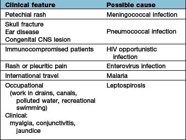
Clinical clues point to the diagnosis (Table 22.20). If there is access to the subarachnoid space via skull fracture (recent or old) or occult spina bifida, bacterial meningitis can be recurrent, and the infecting organism is usually pneumococcus.

Table 22.20 Clinical clues in meningitis

image
Acute bacterial meningitis

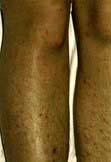
Onset is typically sudden, with rigors and high fever. Meningococcal meningitis is often heralded by a petechial or other rash, sometimes sparse (see Emergency Box 22.1). The meningitis may be part of a generalized meningococcal septicaemia (p. 127). Acute septicaemic shock may develop in any bacterial meningitis.

image Emergency Box 22.1 Meningococcal meningitis and meningococcaemia

emergency treatment

Suspicion of meningococcal infection is a medical emergency requiring treatment immediately.

Clinical features:

image Petechial or nonspecific blotchy red rash

image Fever, headache, neck stiffness.

All these features may not be present – and meningococcal infection may sometimes begin like any apparently non-serious infection.

Immediate treatment for suspected meningococcal meningitis at first contact before transfer to hospital or investigation:

image Benzylpenicillin 1200 mg (adult dose) slow i.v. injection or intramuscularly

image Alternative if penicillin allergy – cefotaxime 1 g i.v.

In meningitis, minutes count: delay is unacceptable.

On arrival in hospital:

image Routine tests including blood cultures immediately

image Watch out for septicaemic shock.

For further management and prophylaxis, see text.

Viral meningitis

This is almost always a benign, self-limiting condition lasting 4–10 days. Headache may follow for some months. There are no serious sequelae, unless an encephalitis is present (p. 1128).

Chronic meningitis (see below)

For further discussion on chronic meningitis, see below.

image

Rash of meningococcal septicaemia with meningitis.

Differential diagnosis

It may be difficult to distinguish between the sudden headache of subarachnoid haemorrhage, migraine and acute meningitis. Meningitis should be considered seriously in anyone with headache and fever and in any sudden headache. Neck stiffness should be looked for – it may not be obvious. Chronic meningitis sometimes resembles an intracranial mass lesion, with headache, epilepsy and focal signs. Cerebral malaria can mimic bacterial meningitis.

Management (Emergency Box 22.1)

Recognition and immediate treatment of acute bacterial meningitis is vital. Minutes save lives. Bacterial meningitis is lethal. Even with optimal care, mortality is around 15%. The following applies to adult patients; management is similar in children.

When meningococcal meningitis is diagnosed clinically by the petechial rash, immediate i.v. antibiotics should be given and blood cultures taken; lumbar puncture is unnecessary. In other causes of meningitis, a lumbar puncture is performed if there is no clinical suspicion of a mass lesion (p. 1091). If the latter is suspected an immediate CT scan must be performed because coning of the cerebellar tonsils may follow LP. Typical CSF changes are shown in Table 22.21. CSF pressure is characteristically elevated. If a presumptive diagnosis of the organism can be made (e.g. pneumococcus is likely with skull fracture or sinus infection), targeted treatment should be started immediately. Immediate antibiotic treatment in acute bacterial meningitis is shown in Table 22.22.

Table 22.21 Typical CSF changes in viral, pyogenic and TB meningitis

image

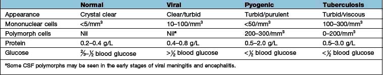
Table 22.22 Antibiotics in acute bacterial meningitis

Organism Antibiotic Alternative (e.g. allergy)

Unknown pyogenic

Cefotaxime

Benzylpenicillin and chloramphenicol

Meningococcus

Benzylpenicillin

Cefotaxime

Pneumococcus

Cefotaxime

Penicillin

Haemophilus

Cefotaxime

Chloramphenicol

Blood should be taken for cultures, glucose and routine tests. Chest and skull films should be obtained if appropriate.

CSF stains demonstrate organisms (e.g. Gram-positive intracellular diplococci – pneumococcus; Gram-negative cocci-meningococcus). Ziehl–Neelsen stain demonstrates acid-fast bacilli (TB), though TB organisms are rarely numerous. Indian ink stains fungi.

Meticulous attention should focus on microbiological studies in suspected CNS infection with close liaison between clinician and microbiologist. Specific techniques (e.g. polymerase chain reaction for meningococci and other bacteria) are invaluable. Syphilitic serology should always be carried out.

The clinical picture and CSF examination should thus yield a presumptive cause for acute meningitis within hours. Antibiotics, however, must be started before the actual organism is identified.

If bacterial meningitis is diagnosed, further discussion with the microbiologist should include antibiotics, drug resistance, recent infections in the locality, barrier nursing and prophylaxis.

In adults with pneumococcal meningitis, dexamethasone should be given first with the initial antibiotics.

Intrathecal antibiotics are no longer used.

Local infection (e.g. paranasal sinus) should be treated surgically if necessary. Repair of a depressed skull fracture or meningeal tear may be required.

Prophylaxis

Meningococcal infection should be notified to public health authorities, and advice sought about immunization and prophylaxis of contacts, e.g. with rifampicin or ciprofloxacin. MenC, a meningococcal C conjugate vaccine, is part of many countries’ immunization programme and often given to case contacts. A combined A and C meningococcal vaccine is sometimes used prior to travel from the UK to endemic regions, e.g. Africa, Asia; and a quadrivalent ACWY vaccine for specific events, e.g. the Hajj and Umrah in Mecca. There is no vaccine for Group B.

A polyvalent pneumococcal vaccine is used after recurrent meningitis, e.g. after a CSF leak following skull fracture.

Hib (Haemophilus influenzae) vaccine is given routinely in childhood in the UK and other countries, e.g. Gambia, virtually eliminating a common cause of fatal meningitis.

Chronic meningitis

Tuberculous meningitis (TBM) and cryptococcal meningitis commence typically with vague headache, lassitude, anorexia and vomiting. Acute meningitis can occur but is unusual. Meningitic signs often take some weeks to develop. Drowsiness, focal signs (e.g. diplopia, papilloedema, hemiparesis) and seizures are common. Syphilis, sarcoidosis and Behçet’s also cause chronic meningitis. In some cases of chronic meningitis, an organism is never identified.

Management of tuberculous meningitis

TBM is a common and serious disease worldwide. Brain imaging, usually with MRI, may show meningeal enhancement, hydrocephalus and tuberculomas (in this chapter), although it may remain normal (see Table 22.21 for CSF changes). In many cases the sparse TB organisms cannot be seen on staining and PCR testing should be performed, although results may be negative. Repeated CSF examination is often necessary and it will be some weeks before cultures are confirmatory. Treatment with antituberculosis drugs (p. 842) – rifampicin, isoniazid and pyrazinamide – must commence on a presumptive basis and continue for at least 9 months. Ethambutol should be avoided because of its eye complications. Adjuvant corticosteroids, e.g. prednisolone 60 mg for 3 weeks, are now recommended (often tapered off). Relapses and complications (e.g. seizures, hydrocephalus) are common in TBM. The mortality remains over 60% even with early treatment.

Malignant meningitis

Malignant cells can cause a subacute or chronic non-infective meningitic process. A meningitic syndrome, cranial nerve lesions, paraparesis and root lesions are seen, often in confusing and fluctuating patterns. The CSF cell count is raised, with high protein and low glucose. Treatment with intrathecal cytotoxic agents is rarely helpful.

Cells in a sterile CSF (pleocytosis)

A raised CSF cell count is present without an evident infecting organism. CSF pleocytosis, i.e. a mixture of lymphocytes and polymorphs, is the usual situation (Box 22.26).

image Box 22.26

Causes of sterile CSF pleocytosis

image Partially treated bacterial meningitis

image Viral meningitis

image TB or fungal meningitis

image Intracranial abscess

image Neoplastic meningitis

image Parameningeal foci, e.g. paranasal sinus

image Syphilis

image Cerebral venous thrombosis

image Cerebral malaria

image Cerebral infarction

image Following subarachnoid haemorrhage

image Encephalitis, including HIV

image Rarities, e.g. cerebral malaria, sarcoidosis, Behçet’s syndrome, Lyme disease, endocarditis, cerebral vasculitis

FURTHER READING

Hall AJ, Wild CP. Management of bacterial meningitis in adults. BMJ 2003; 326:996–997.

Tunbridge A, Read RC. Management of meningitis. Clin Med 2004; 4:499–505.

van de Beek D, de Gans J, Tunkel AR et al. Community-acquired bacterial meningitis in adults. N Engl J Med 2006; 354:44–53.

Encephalitis

Encephalitis means acute inflammation of brain parenchyma, usually viral. In viral encephalitis fever (90%) and meningism are usual, but in contrast to meningitis the clinical picture is dominated by brain parenchyma inflammation. Personality and behavioural change is a common early manifestation which progresses to a reduced level of consciousness and even coma. Seizures (focal and generalized) are very common and focal neurological deficits, e.g. speech disturbance, often occur (especially in herpes simplex encephalitis).

Viral encephalitis

The viruses isolated from adult UK cases are usually herpes simplex, VZV and other herpes group viruses, HHV-6, 7, enteroviruses and adenovirus. HSV encephalitis (HSVE) typically affects the temporal lobes initially, often asymmetric. Often, the virus is never identified. Outside the UK in endemic regions different pathogens cause encephalitis including Flaviviruses (Japanese encephalitis, West Nile virus, tick- borne encephalitis) and rabies.

Local epidemics can occur. For example, in New York in the 1990s, West Nile virus caused an epidemic and Venezuelan equine virus was isolated from encephalitis cases in South America.

Investigations

image 
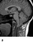
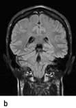
MR imaging shows areas of inflammation and swelling, generally in the temporal lobes in HSV encephalitis. Raised intracranial pressure and midline shift may occur leading to coning.

image EEG shows periodic sharp and slow wave complexes.

image CSF shows an elevated lymphocyte count (95%).

image Viral detection by CSF PCR is highly sensitive for several viruses such as HSV and VZV. However, a false negative result may occur within the first 48 h after symptom onset. Serology (blood and CSF) is also helpful.

image Brain biopsy is rarely required since the advent of MRI and PCR.

Treatment

Suspected HSV and VZV encephalitis is treated immediately with i.v. aciclovir (10 mg/kg 3 times a day for 14–21 days), even before investigation results are available. Early treatment significantly reduces both mortality and long-term neurological damage in survivors. Seizures are treated with anticonvulsants. Occasionally decompressive craniectomy is required to prevent coning but coma confers a poor prognosis.

Long-term complications are common including memory impairment, personality change and epilepsy.

Post-infectious encephalomyelitis

Acute disseminated encephalomyelitis (ADEM) follows many infections (e.g. measles, mycoplasma, mumps and rubella) and rarely follows immunization after 1–2 weeks. There is a monophasic illness with multifocal brain, brainstem and often spinal cord inflammatory lesions in white matter, with demyelination. ADEM is caused by an immune mediated host response to infection and occurs principally in children and young adults. Mild cases recover completely. Survivors often have permanent brain damage. Treatment is supportive, with steroids and anticonvulsants.

Autoimmune encephalitis

This group of disorders have been described in recent years and are increasingly recognized. Autoantibodies directed against neuronal epitopes cause a subacute encephalitic illness – limbic encephalitis or panencephalitis. Limbic encephalitis presents over weeks or months with memory impairment, confusion, psychiatric disturbance, and seizures – usually temporal lobe seizures reflecting involvement of the hippocampus and mesial temporal lobes.

Paraneoplastic limbic encephalitis (PLE). Seen particularly with small cell lung cancer and testicular tumours and associated with a variety of antibodies including anti-Hu and anti-Ma2. Antibodies can be detected in 60% of cases. MRI usually shows a hippocampal high signal. PLE precedes the diagnosis of cancer in most cases and should prompt investigation to identify the tumour.

Voltage gated potassium channel (VGKC) limbic encephalitis. VGKC antibodies (which can be tested for) produce a variety of disorders including limbic encephalitis, characteristic faciobrachial dystonic seizures, neuromyotonia and peripheral nerve hyperexcitability syndromes. This usually occurs in patients older than 50 but is rarely associated with cancer (thymoma).

Anti NMDA receptor antibody panencephalitis. Presents as limbic encephalitis followed by coma and often status epilepticus. Orofacial dyskinesias are characteristic. Usually younger patients, some have ovarian teratomas.

Patients may respond to immunotherapy – i.v. immunoglobulin or plasma exchange initially followed by steroids, rituximab or cyclophosphamide. PLE responds less well to treatment.

Herpes zoster (shingles)

This is caused by reactivation of varicella zoster virus (VZV), usually within dorsal root ganglia. Primary infection with VZV causes chickenpox following which the virus remains latent in sensory ganglia. Development of shingles may indicate a decline in cell mediated immunity, e.g. due to age or malignancy.

Clinical patterns and complications

Dermatomal shingles. Thoracic dermatomes are most commonly affected. Tingling or painful dysaesthesias precede the vesicular rash by a few days (p. 81). Motor radiculopathy can occur (usually lumbar or cervical, as thoracic involvement is often clinically silent). It rarely occurs without the rash – zoster sin herpete. Antiviral drugs such as aciclovir, famciclovir or valaciclovir reduce the incidence of postherpetic neuralgia.

Postherpetic neuralgia. Defined as pain lasting more than 4 months after developing shingles; occurs in 10% of patients (often elderly). Burning, intractable pain responds poorly to analgesics. Response to treatment is unsatisfactory but there is a trend towards gradual recovery over 2 years. Amitriptyline or gabapentin are commonly used and topical lidocaine patches may help.

Cranial nerve involvement. Only cranial nerves with sensory fibres are affected, particularly the trigeminal and facial nerves. Ophthalmic herpes is due to involvement of V1. This can lead to corneal scarring and secondary panophthalmitis. Involvement of the geniculate ganglion of the facial nerve is also called Ramsay Hunt syndrome (p. 1098).

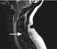
Myelitis may occur in the context of shingles when the inflammatory process spreads from the dorsal root ganglion to the adjacent spinal cord.

Immunization. The Centers for Disease Control and Prevention (CDC) in America have suggested that all adults over 60 years old should be vaccinated against herpes zoster (even those who have had shingles previously), as it reduces the incidence of shingles by about 50%.

Neurosyphilis

Many neurological symptoms occur, sometimes mixed (see also syphilis, p. 166).

Asymptomatic neurosyphilis

This means positive CSF serology without signs.

Meningovascular syphilis

This causes:

image Subacute meningitis with cranial nerve palsies and papilloedema

image A gumma – a chronic expanding intracranial mass

image Paraparesis – a spinal meningovasculitis.

Tabes dorsalis

Demyelination in dorsal roots causes a complex deafferentation syndrome. The elements of tabes:

image Lightning pains (p. 1086)

image Ataxia, stamping gait, reflex/sensory loss, wasting

image Neuropathic (Charcot) joints

image Argyll Robertson pupils (p. 1074)

image Ptosis and optic atrophy.

General paralysis of the insane (GPI)

The grandiose title describes dementia and weakness. GPI dementia is typically similar to Alzheimer’s (p. 1138). Progressive cognitive decline, seizures, brisk reflexes, extensor plantar reflexes and tremor develop. Death follows within 3 years. Argyll Robertson pupils are usual. GPI and tabes are rarities in the UK.

Other forms of neurosyphilis

In congenital neurosyphilis (acquired in utero), features of combined tabes and GPI develop in childhood – taboparesis.

Secondary syphilis can be symptomless or cause a self-limiting subacute meningitis.

Treatment

Benzylpenicillin 1 g daily i.m. for 10 days in primary infection eliminates any risk of neurosyphilis. Allergic (Jarisch–Herxheimer) reactions can occur; steroid cover is usually given with penicillin (p. 1129). Established neurological disease is arrested but not reversed by penicillin.
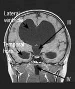
Neurocysticercosis

The pork tapeworm, Taenia solium, is endemic in Latin America, Africa, India and much of South-east Asia (p. 159). Epilepsy is the commonest clinical manifestation of neurocysticercosis and one of the commonest causes of epilepsy in endemic countries. Most infected people remain asymptomatic.

Brain CT and MRI show ring-enhancing lesions with surrounding oedema when the cyst dies and later calcification. Multiple cysts are often seen in both brain and skeletal muscle. Serological tests indicate infection but not activity. Biopsy of a lesion is rarely necessary. Management is primarily the control of seizures and the anthelminthic agent albendazole is often also given (usually with steroid cover).

HIV and neurology

HIV-infected individuals frequently present with or develop neurological conditions. The HIV virus itself is directly neuroinvasive and neurovirulent. Immunosuppression leads to indolent, atypical clinical patterns (p. 176). HIV patients also have a high incidence of stroke. The pattern of disease is changing where antiretroviral (ARV) therapy is available.

CNS and peripheral nerve disease in HIV

HIV seroconversion can cause meningitis, encephalitis, Guillain–Barré syndrome and Bell’s palsy (the commonest cause of Bell’s palsy in South Africa).

Chronic meningitis occurs with fungi (e.g. Cryptococcus neoformans or Aspergillus), TB, listeria, coliforms or other organisms. Raised CSF pressure is common in cryptococcal meningitis.

AIDS-dementia complex (ADC). A progressive, HIV-related dementia, sometimes with cerebellar signs, is still seen where antiretroviral therapy is unavailable.

Encephalitis and brain abscess. Toxoplasma, cytomegalovirus, herpes simplex and other organisms cause severe encephalitis. Multiple brain abscesses develop in HIV infection, usually due to toxoplasmosis.

CNS lymphoma. this is typically fatal (Chapter 9).

Progressive multi-focal leucoencephalopathy (PML) is due to JC virus and occurs with very low CD4 counts (p. 191).

Spinal vacuolar myelopathy occurs in advanced disease.

Peripheral nerve disease. HIV related peripheral neuropathy is common (70%) and can be difficult to distinguish from the effects of ARV treatment which is also toxic to peripheral nerves.

Other infections

Many other infections involve the CNS and are discussed in Chapter 4, e.g. rabies, tetanus, botulism, Lyme disease and leprosy.

Other inflammatory conditions

Subacute sclerosing panencephalitis (SSPE)

Persistence of measles antigen in the CNS is associated with this rare late sequel of measles. Progressive mental deterioration, fits, myoclonus and pyramidal signs develop, typically in a child. Diagnosis is made by high measles antibody titre in blood and CSF. Measles immunization protects against SSPE, which has now been almost eliminated in the UK.

Progressive rubella encephalitis

Some 10 years after primary rubella infection, this causes progressive cognitive impairment, fits, optic atrophy, cerebellar and pyramidal signs. Antibody to rubella viral antigen is produced locally within the CNS. It is even rarer than SSPE.

Mollaret’s meningitis

This is recurrent self-limiting episodes of aseptic meningitis (i.e. no bacterial cause is found) over many years. Viral (possibly herpes simplex) infection is postulated.

Whipple’s disease

CNS Whipple’s disease, due to Tropheryma whipplei infection is characterized by myoclonus, dementia and supranuclear ophthalmoplegia (p. 268). Diagnosis of CNS involvement is made by CSF PCR (only 50% sensitivity) or brain biopsy.

Neurosarcoidosis

Neurosarcoid with or without systemic sarcoid causes chronic meningoencephalitis, cord lesions, cranial nerve palsies, particularly bilateral VIIth nerve lesions, polyneuropathy and myopathy (p. 845).

Behçet’s syndrome (see also p. 544)

Behçet’s principal features are recurrent oral and/or genital ulceration, inflammatory ocular disease (uveitis, p. 1062) and neurological syndromes. Brainstem and cord lesions, aseptic meningitis, encephalitis and cerebral venous thrombosis occur.

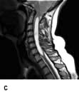
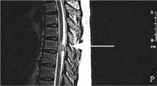
Brain and spinal abscesses

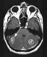
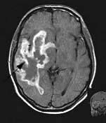
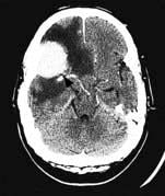
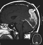
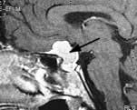
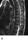
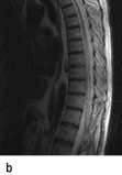
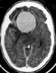
Brain abscess (Fig. 22.46)

Focal bacterial infection behaves as any expanding mass. Typical bacteria found are Streptococcus anginosus and Bacteroides species (paranasal sinuses and teeth) and staphylococci (penetrating trauma). Mixed infections are common. Multiple abscesses develop, particularly in HIV infection. Fungi also cause brain abscesses. A parameningeal infective focus (e.g. ear, nose, paranasal sinus, skull fracture) or a distant source of infection (e.g. lung, heart, abdomen) may be present. Frequently no underlying cause is found. An abscess is more than 10 times rarer than a brain tumour in the UK.

image

Figure 22.46 Pyogenic cerebral abscess: CT. There is a ring enhancing lesion with adjacent oedema.

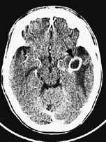
Clinical features and management

Headache, focal signs (e.g. hemiparesis, aphasia, hemianopia), epilepsy and raised intracranial pressure develop. Fever, leucocytosis and raised ESR are usual although not invariable.

Urgent imaging is essential. MRI shows a ring-enhancing mass, usually with considerable surrounding oedema. The search for a focus of infection should include a detailed examination of the skull, ears, paranasal sinuses and teeth, and distant sites such as heart and abdomen. Lumbar puncture is dangerous and should not be performed. Neurosurgical aspiration with stereotactic guidance allows the infective organism to be identified. Treatment is with high-dose antibiotics and sometimes surgical resection/decompression. Despite treatment, mortality remains high at approximately 25%. Epilepsy is common in survivors.

Brain tuberculoma

TB causes chronic caseating intracranial granulomatous masses – tuberculomas. These are the commonest intracranial masses in countries where TB is common, e.g. India. Brain tuberculomas either present as mass lesions de novo or develop during tuberculous meningitis; they are also found as symptomless intracranial calcification on imaging. Spinal cord tuberculomas also occur. Treatment is described on page 842.

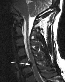
Subdural empyema and intracranial epidural abscess

Intracranial subdural empyema is a collection of subdural pus, usually secondary to local skull or middle ear infection. Features are similar to those of a cerebral abscess. Imaging is diagnostic.

In intracranial epidural abscess a layer of pus, 1–3 mm thick, tracks along the epidural space causing sequential cranial nerve palsies. There is usually local infection, e.g. in the middle ear. MRI shows the collection; CT is typically normal. Drainage is required, and antibiotics.

Spinal epidural abscess

Staphylococcus aureus is the usual organism, reaching the spine via the bloodstream, e.g. from a boil. Fever and back pain are followed by paraparesis and/or root lesions. Emergency imaging and antibiotics are essential and surgical decompression is often necessary.

Brain tumours

Primary intracranial tumours account for some 10% of neoplasms. The most common tumours are outlined in Table 22.23. Metastases are the commonest intracranial tumours (Fig. 22.47). Symptomless meningiomas (benign) are found quite commonly on imaging or at autopsy.

Table 22.23 Common brain tumours

Tumour Approximate frequency

Metastases

50%

 Bronchus Breast Stomach Prostate Thyroid Kidney


Primary malignant tumours of neuroepithelial tissues

35%

 Astrocytoma Oligodendroglioma Mixed (oligoastrocytomas) gliomas Ependymoma Primary cerebral lymphoma Medulloblastoma


Benign

15%

 Meningioma Neurofibroma


image

Figure 22.47 Bilateral cerebellar metastases: MR T1.

Gliomas (Fig. 22.48)

These malignant tumours of neuroepithelial origin are usually seen within the hemispheres, but occasionally in the cerebellum, brainstem or cord. Their cause is unknown. Gliomas are occasionally associated with neurofibromatosis. They tend to spread by direct extension, virtually never metastasizing outside the CNS.

image Astrocytomas are gliomas that arise from astrocytes. They are classified histologically into grades I–IV. Grade I astrocytomas grow slowly over many years, while grade IV tumours (glioblastoma multiforme) cause death within several months. Cystic astrocytomas of childhood are relatively benign, and usually cerebellar.

image Oligodendrogliomas arise from oligodendrocytes. They grow slowly, usually over several decades. Calcification is common.

image

Figure 22.48 Cerebral glioblastoma multiforme: MR T1.

Meningiomas

These benign tumours (Figs 22.49-22.51) arise from the arachnoid and may grow to a large size, usually over years. Those close to the skull erode bone or cause local hyperostosis. They often occur along the intracranial venous sinuses, which they may invade. They are unusual below the tentorium. Common sites are the parasagittal region, sphenoidal ridge, subfrontal region, pituitary fossa and skull base.

image

Figure 22.49 Frontal meningioma: CT.

image

Figure 22.50 Falx meningioma (occipital): MR T1.

image

Figure 22.51 Meningioma within sella and suprasellar extension: MR T1.

Neurofibromas (Schwannomas)

These solid benign tumours arise from Schwann cells and occur principally in the cerebellopontine angle, where they arise from the VIIIth nerve sheath (acoustic neuroma, p. 1070). They may be bilateral in neurofibromatosis type 2 (p. 1143).

Other neoplasms

Other less common neoplasms include cerebellar haemangioblastoma, ependymomas of the IVth ventricle, colloid cysts of the IIIrd ventricle, pinealomas, chordomas of the skull base, glomus tumours of the jugular bulb, medulloblastomas (a cerebellar childhood tumour), craniopharyngiomas (p. 937) and primary CNS lymphomas (p. 468). For pituitary tumours, see page 946.

FURTHER READING

Bredel M, Scholtens DM, Yadav AK et al. NFKBIA deletion in glioblastomas. N Engl J Med 2011; 364:627–637.

Clinical features

Mass lesions within the brain produce symptoms and signs by three mechanisms:

image By direct effect – brain is infiltrated and local function impaired

image By secondary effects of raised intracranial pressure and shift of intracranial contents (e.g. papilloedema, vomiting, headache)

image By provoking generalized and/or partial seizures.

Although neoplasms, either secondary or primary, are the commonest mass lesions in the UK, cerebral abscess, tuberculoma, neurocysticercosis, subdural and intracranial haematomas can also produce features that are clinically similar.

Direct effects of mass lesions

The hallmark of a direct effect of a mass is local progressive deterioration of function. Tumours can occur anywhere within the brain. Three examples are given:

image A left frontal meningioma caused a frontal lobe syndrome over several years with vague disturbance of personality, apathy and impaired intellect. Expressive aphasia developed, followed by progressive right hemiparesis as the corticospinal pathways became involved. As the mass enlarged further, pressure headaches and papilloedema developed.

image A right parietal lobe glioma caused a left homonymous field defect (optic radiation). Cortical sensory loss in the left limbs and left hemiparesis followed over 3 months. Partial seizures (episodes of tingling of the left limbs) developed.

image A left VIIIth nerve sheath neurofibroma (an acoustic neuroma, Schwannoma) in the cerebellopontine angle caused, over 3 years, progressive deafness (VIII), left facial numbness (V) and weakness (VII), followed by cerebellar ataxia on the same side.

With a hemisphere tumour, epilepsy and the direct effects commonly draw attention to the problem. The rate of progression varies greatly, from a few days or weeks in a highly malignant glioma, to several years with a slowly enlarging mass such as a meningioma. Cerebral oedema surrounds mass lesions: its effect is difficult to distinguish from that of the tumour itself.

Raised intracranial pressure

Raised intracranial pressure causing headache, vomiting and papilloedema is a relatively unusual presentation of a mass lesion in the brain. These symptoms often imply hydrocephalus – obstruction to CSF pathways. Typically this is produced early by posterior fossa masses that obstruct the aqueduct and IVth ventricle but only later with lesions above the tentorium. Shift of the intracranial contents produces features that co-exist with the direct effects of any expanding mass:

image Distortion of the upper brainstem, as midline structures are displaced either caudally or laterally by a hemisphere mass (Fig. 22.48). This causes impairment of consciousness progressing to coning and death as the medulla and cerebellar tonsils are forced into the foramen magnum.

image False localizing signs – false only because they do not point directly to the site of the mass.

Three examples of false localizing signs are:

image A VIth nerve lesion, first on the side of a mass and later bilaterally as the VIth nerve is compressed or stretched during its long intracranial course.

image A IIIrd nerve lesion develops as the temporal lobe uncus herniates caudally, compressing III against the petroclinoid ligament. The first sign is ipsilateral pupil dilatation as parasympathetic fibres are compressed.

image Hemiparesis on the same side as a hemisphere tumour, i.e. the side you might not expect, from compression of the contralateral cerebral peduncle on the edge of the tentorium.

Seizures

Seizures are a common presenting feature of malignant brain tumours. Partial seizures, simple or complex, that may evolve into generalized tonic-clonic seizures, are characteristic of many hemisphere masses, whether malignant or benign. The pattern of partial seizure is of localizing value (p. 1112).

Investigations

Imaging

Both CT and MRI are useful in detecting brain tumours; MRI is superior for posterior fossa lesions. Benign and malignant tumours, brain abscess, TB, neurocysticercosis and infarction have characteristic, but not entirely reliable, appearances and refined imaging techniques and biopsy are often necessary. MR angiography is used occasionally to define blood supply and MR spectroscopy to identify patterns typical of certain gliomas. PET is sometimes helpful to locate an occult primary tumour with brain metastases.

Routine tests

Since metastases are common, routine tests, e.g. chest X-ray, should be performed.

Lumbar puncture

This is contraindicated when there is any possibility of a mass lesion as withdrawing CSF may provoke immediate coning. CSF examination is rarely helpful and has been superseded by imaging.

Biopsy and tumour removal

Stereotactic biopsy via a skull burr-hole is carried out to ascertain the histology of most suspected malignancies. With a benign tumour, e.g. a symptomatic, accessible meningioma, craniotomy and removal is usual.

Management

Cerebral oedema surrounding a tumour responds rapidly to steroids: i.v. or oral dexamethasone. Epilepsy is treated with anticonvulsants.

While complete surgical removal of a tumour is an objective, it is often not possible, nor is surgery always necessary. Follow-up with serial imaging is sometimes preferable in low- grade gliomas. At exploration, some benign tumours can be entirely removed (e.g. acoustic neuromas, some parasagittal meningiomas). With a malignant tumour it is not possible to entirely remove an infiltrating mass. Biopsy and debulking are performed.

Within the posterior fossa, tumour removal is often necessary because of raised pressure and danger of coning. Overall mortality for posterior fossa exploration remains around 10%.

For gliomas and metastases, radiotherapy is usually given and improves survival, if only slightly. Solitary metastases can often be excised successfully. Chemotherapy has little real value in the majority of primary or secondary brain tumours. Vincristine, procarbazine and temozolomide (an oral alkylating agent) can be used. Most malignant gliomas have a poor prognosis despite advances in imaging, surgery, chemotherapy and radiotherapy – <50% survival for grade IV gliomas at 2 years. Surgical debulking and radiotherapy improve survival by 4–5 months.

Stereotactic radiotherapy (gamma knife)

Only available in selected centres, a collimated radiotherapy beam can deliver high doses of radiation to small targets up to 3 cm in diameter with precision. It may be used to target small metastases, and inaccessible skull base tumours such as meningiomas or schwannomas. It may also be used to treat intracerebral AVMs.

FURTHER READING

Wen PY, Kesari S. Malignant gliomas in adults. N Engl J Med 2008; 359:492–507.

Hydrocephalus

Hydrocephalus is an excessive accumulation of CSF within the head caused by a disturbance of formation, flow or absorption. High pressure and ventricular dilatation result (Fig. 22.52).

image

Figure 22.52 Hydrocephalus with gross IIIrd, IVth and lateral ventricle enlargement: MR T1.

Infantile hydrocephalus

Head enlargement in infancy occurs in 1 in 2000 live births. There are several causes:

image Arnold–Chiari malformations. Cerebellar tonsils descend into the cervical canal. Associated spina bifida is common. Syringomyelia may develop (p. 1137).

image image image

Arnold–Chiari malformation with spinal cord syrinx. a, b, and c show different views.

image Stenosis of the aqueduct of Sylvius. Aqueduct stenosis is either congenital (genetic) or acquired following neonatal meningitis/haemorrhage.

image Dandy–Walker syndrome. There is cerebellar hypoplasia and obstruction to IVth ventricle outflow foramina.

Hydrocephalus in adults

Hydrocephalus is sometimes an unsuspected finding on imaging. Stable childhood hydrocephalus can become apparent in adult life (’arrested hydrocephalus’) but can suddenly decompensate. Combinations of headache, cognitive impairment, features of raised intracranial pressure, and ataxia develop depending on how high the CSF pressure rises and rapidity of onset. Elderly patients with more compliant brains may present with gradual onset gait apraxia and subtle cognitive slowing.

Hydrocephalus may be caused by:

image Posterior fossa and brainstem tumours obstructing the aqueduct or IVth ventricular outflow

image Subarachnoid haemorrhage, head injury or meningitis (particularly tuberculous), causing obstruction of CSF flow and reabsorption

image A IIIrd ventricle colloid cyst causing intermittent hydrocephalus – recurrent prostrating headaches with episodes of lower limb weakness

image Choroid plexus papilloma (rare) secretes CSF.

Frequently, the underlying cause for hydrocephalus remains obscure.

Treatment

Ventriculoperitoneal shunting is necessary when progressive hydrocephalus causes symptoms. Removal of tumours is carried out when appropriate. Endoscopic third ventriculostomy may be performed.

Normal pressure hydrocephalus (NPH)

NPH describes a syndrome of enlarged lateral ventricles in elderly patients with the clinical triad of:

image A gait disorder – gait apraxia

image Dementia

image Urinary incontinence.

The term is a misnomer, as it is a low-grade hydrocephalus with intermittently raised ICP. Ventriculoperitoneal shunting may be required. A trial of prolonged drainage of lumbar CSF over several days predicts response to shunt insertion.

Traumatic brain injury

In most western countries head injury accounts for about 250 hospital admissions per 100 000 population annually. Traumatic brain injury (TBI) describes injuries with potentially permanent consequences. For each 100 000 people, 10 die annually following TBI; 10–15 are transferred to a neurosurgical unit – the majority of these require rehabilitation for a prolonged period of 1–9 months. The prevalence of survivors with a major persisting handicap is around 100/100 000. Road traffic accidents and excessive alcohol use are the principal aetiological factors in this major cause of morbidity and mortality, in many countries.

Skull fractures

Linear skull fracture of the vault or base is one indication of the severity of a blow, but is itself not necessarily associated with any brain injury. Healing of linear fractures takes place spontaneously. Depressed skull fracture is followed by a high incidence of post-traumatic epilepsy. Surgical elevation and debridement are usually necessary.

Principal local complications of skull fracture are:

image Meningeal artery rupture – causing extradural haematoma

image Dural vein tears – causing subdural haematoma (p. 1106)

image CSF rhinorrhoea/otorrhoea and consequent meningitis.

Mechanisms of brain damage

Older classifications attempted to separate concussion, transient coma for hours followed by apparent complete clinical recovery, from brain contusion, i.e. bruising, with prolonged coma, focal signs and lasting damage. Pathological support for this division is poor. Mechanisms of TBI are complex and interrelated:

image Diffuse axonal injury – shearing and rotational stresses on decelerating brain, sometimes at the site opposite impact (the contrecoup effect)

image Neuronal and axonal damage from direct trauma

image Brain oedema and raised intracranial pressure

image Brain hypoxia

image Brain ischaemia.

Clinical course

In a mild TBI, a patient is stunned or dazed for a few seconds or minutes. Following this the patient remains alert without post-traumatic amnesia. Headache can follow; complete recovery is usual. In more serious injuries duration of unconsciousness and particularly of post-traumatic amnesia (PTA) helps grade severity. PTA of >24 hours defines severe TBI. The Glasgow Coma Scale (GCS, p. 1092) is used to record the degree of coma; this has prognostic value. A GCS below 5/15 at 24 hours implies a serious injury; 50% of such patients die or remain in a vegetative or minimal conscious state (p. 1096). However, prolonged coma of up to some weeks is occasionally followed by good recovery.

Recovery after severe TBI takes many weeks or months. During the first few weeks, patients are often intermittently restless or lethargic and have focal deficits such as hemiparesis or aphasia. Gradually they become more aware, though they may remain in post-traumatic amnesia, being unable to lay down any continuous memory despite being awake. This amnesia may last some weeks or more, and may not be obvious clinically. PTA is one predictor of outcome. PTA over a week implies that persistent organic cognitive deficit is almost inevitable, although return to unsupported paid work may be possible.

Late sequelae

Sequelae of TBI are major causes of morbidity and can have serious social and medicolegal consequences. They include:

image Incomplete recovery, e.g. cognitive impairment, hemiparesis

image Post-traumatic epilepsy (p. 1113)

image The post-traumatic (post-concussional) syndrome. This describes the vague complaints of headache, dizziness and malaise that follow even minor head injuries. Litigation is frequently an issue. Depression is prominent. Symptoms may be prolonged

image Benign paroxysmal positional vertigo (BPPV, Chapter 21)

image Chronic subdural haematoma (p. 1071)

image Hydrocephalus (p. 1133)

image Chronic traumatic encephalopathy. This follows repeated and often minor injuries. It is known as the ‘punch-drunk syndrome’ and consists of cognitive impairment, extrapyramidal and pyramidal signs, seen typically in professional boxers.

Immediate management

Attention to the airway is vital. If there is coma, depressed fracture or suspicion of intracranial haematoma, CT imaging and discussion with a neurosurgical unit are essential. Indications for CT imaging vary from imaging all minor head injuries in some US centres to more stringent criteria elsewhere.

In many severe TBI cases, assisted ventilation will be needed. Intracranial pressure monitoring is valuable. Hypothermia lowers intracranial pressure when used early after a TBI; an effect on outcome has only been seen in specialized neurotrauma centres. Care of the unconscious patient is described on page 1096. Prophylactic antiepileptic drugs have been shown to be of no value in prevention of late post-traumatic epilepsy.

Rehabilitation

TBI cases require skilled, prolonged and energetic support. Survivors with severe physical and cognitive deficits require rehabilitation in specialized units. Rehabilitation includes care from a multidisciplinary team with physiotherapeutical, psychological and practical skills. Many survivors are left with cognitive problems (amnesia, neglect, disordered attention and motivation) and behavioural/emotional problems (temper dyscontrol, depression and grief reactions). Long-term support for both patients and families is necessary.

FURTHER READING

Giacino JT et al. Placebo-controlled trial of amantadine for severe traumatic brain injury. N Engl J Med 2012; 366:814–826.

Polderman KH. Induced hypothermia and fever control for prevention and treatment of neurological injuries. Lancet 2008; 371:1955–1969.

Ropper AH, Gorson KC. Concussion. N Engl J Med 2007; 356:166–172.

Spinal cord disease

The cord extends from C1, the junction with the medulla, to the lower vertebral body of L1, where it becomes the conus medullaris. Blood supply is from the anterior spinal artery and a plexus on the posterior cord. This network is supplied by the vertebral arteries, and several branches from lumbar and intercostal vessels including the artery of Adamkiewicz.

Spinal cord compression

Principal features of chronic and subacute cord compression are spastic paraparesis or tetraparesis, radicular pain at the level of compression, and sensory loss below the compression (Box 22.27).

image Box 22.27

Causes of spinal cord compression

image Spinal cord tumours

image Extramedullary, e.g. meningioma or neurofibroma

image Intramedullary, e.g. ependymoma or glioma

image Vertebral body destruction by bone metastases, e.g. prostate primary

image Disc and vertebral lesions:

Chronic degenerative and acute central disc prolapse
Trauma

image Inflammatory:

Epidural abscess
Tuberculosis
Granulomatous

image Epidural haemorrhage/haematoma

For example, in compression at T4 (Fig. 22.15) a band of pain radiates around the thorax, characteristically worse on coughing or straining. Spastic paraparesis develops over months, days or hours, depending upon underlying pathology. Numbness commencing in the feet rises to the level of compression. This is called the sensory level and is usually 2–3 dermatome levels below the level of anatomical compression. Retention of urine and constipation develop.

Causes

Disc and vertebral lesions. Central cervical disc and thoracic disc protrusion cause cord compression (p. 1148). Chronic compression due to cervical spondylotic myelopathy is frequently seen in clinical practice and is the commonest cause of a spastic paraparesis in an elderly person.

Trauma. Stabilize the neck and back and move patient with extreme caution in trauma. Any trauma to the back is potentially serious and the patient should be immobilized until the extent of the injury can be determined.

Spinal cord tumours. Extramedullary tumours, e.g. meningiomas and neurofibromas, cause cord compression (Fig. 22.53 and Box 22.28) gradually over weeks to months, often with root pain and a sensory level (p. 1087). Vertebral body destruction by bony metastases, e.g. in prostate or breast cancer, is a common cause of spinal cord compression.

image

Figure 22.53 Thoracic meningioma compressing cord: MR T2.

image Box 22.28

Principal spinal cord neoplasms

Extradural

image Metastases:

Bronchus
Breast
Prostate
Lymphoma
Thyroid
Melanoma

Extramedullary

image Meningioma

image Neurofibroma

image Ependymoma

Intramedullary

image Glioma

image Ependymoma

image Haemangioblastoma

image Lipoma

image Arteriovenous malformation

image Teratoma

Intramedullary tumours (e.g. ependymomas) are less common and typically progress slowly, sometimes over many years. Sensory disturbances similar to syringomyelia may develop (p. 1137).

Tuberculosis. Spinal TB is the commonest cause of cord compression in countries where TB is common. There is destruction of vertebral bodies and disc spaces, with local spread of infection. Cord compression and paraparesis follow, culminating in paralysis – Pott’s paraplegia.

Spinal epidural abscess. This is described on page 1131.

Epidural haemorrhage and haematoma. These are rare sequelae of anticoagulant therapy, bleeding disorders or trauma and can follow lumbar puncture when clotting is abnormal. A rapidly progressive cord or cauda equina lesion develops.

Management

Acute spinal cord compression is a medical emergency. Early diagnosis and treatment is vital. MRI is the imaging technique of choice.

Routine tests (e.g. chest X-ray) may indicate a primary neoplasm or infection. Surgical exploration is frequently necessary; if decompression is not performed promptly, irreversible cord damage results. Results are excellent if benign tumours and haematomas are removed early. Radiotherapy is used to treat cord malignancies, or compression due to inoperable malignant vertebral body disease causing cord compression.

Other spinal cord disorders

Inflammatory cord lesions (transverse myelitis)

This is one of the commoner causes of a non-compressive spinal cord syndrome. MS is the commonest cause of a spastic paraparesis in a young adult. Typically 1 or 2 spinal segments are affected with part or all of the cord area at that level involved. Clinically a myelopathy evolves over days and recovery (often partial) follows over weeks or months. MR is sensitive and shows cord swelling and oedema with gadolinium enhancement at the affected level(s). CSF may be inflammatory with an excess of lymphocytes. Causes include:

image Parainfectious autoimmune inflammatory response is the commonest cause, e.g. after viral infection or immunization or in the context of MS (sometimes the presenting feature of MS)

image Systemic inflammatory disorders, e.g. SLE, Sjögren’s, sarcoidosis

image Infection: viruses including herpesviruses such as VZV or EBV, HIV, HTLV-1&2 (tropical spastic paraparesis), mycobacteria-TB, syphilis, Lyme or schistosomiasis

image Neuromyelitis optica (Devic’s disease) causes long cord lesions (≥3 segments) and optic nerve demyelination. Diagnosis is made by testing for aquaporin-4 antibodies.

Treatment is usually with high-dose steroids or other immunosuppression or antimicrobial therapy in the case of specific infections.

Anterior spinal artery (ASA) occlusion

There is acute paraplegia and loss of spinothalamic (pain and temperature) sensation, with infarction of the anterior two-thirds of the spinal cord. It may result from aortic atherosclerosis, dissection, trauma or cross-clamping in surgery. Vasculitis, emboli, haematological disorders causing thrombosis and severe hypotension are also causes. Occlusion of the artery of Adamkiewicz, which supplies the thoracic ASA, causes watershed infarction of the cord typically at the T8 level where perfusion is relatively poor.

Arteriovenous malformations (AVMs) of the cord

Although rare, spinal AVMs may be difficult to diagnose but are potentially curable. The two main types seen are dural AV fistulas (acquired) and true intramedullary AVMs (probably congenital but gradually enlarge). Dural AV fistulas occur mainly in middle aged men due to formation of a direct connection between an artery and vein in a dural nerve root sleeve. This causes arterialization of veins with venous hypertension and thus oedema and congestion of the spinal cord at and below the affected level. Presentation is with a gradually progressive myelopathy over months or a few years, often with thoracic back pain. MRI usually shows cord swelling and may show the enlarged arterialized veins over the surface of the cord. Spinal angiography demonstrates the fistula and allows endovascular ablation with glue, often with complete resolution of symptoms if permanent neuronal damage has not already occurred.

image image

Dural arteriovenous fistulas.

Genetic disorders – hereditary spastic paraparesis (HSP)

Several genetic disorders may present with a gradually evolving upper motor neurone syndrome resembling a myelopathy. Typically spasticity and stiffness dominate the clinical picture rather than weakness especially in HSP. Muscle relaxants such as baclofen improve gait. There are 28 known genes associated with HSP, some causing ‘pure’ spasticity and others with associated neurological features, e.g. thinning of the corpus callosum.

Other genetic disorders such as adrenoleucodystrophy may cause a slowly progressive spastic paraparesis (including in manifesting female carriers) as can the spinocerebellar ataxias (p. 1143) or presenilin-1 mutations (p. 1140).

Vitamin B12 deficiency

Subacute combined degeneration of the cord resulting from vitamin B12 deficiency (p. 382) is the most common example of metabolic disease causing spinal cord damage. Abuse of nitric oxide may precipitate functional B12 deficiency with normal serum B12 levels.

Other causes of a spastic paraparesis

Motor neurone disease may present initially with a spastic paraparesis before lower motor neurone features develop (p. 1141). Paraneoplastic disorders, radiotherapy, copper deficiency, liver failure and rare toxins (e.g. lathyrism) may case spinal cord damage. Not all causes of paraparesis relate to spinal cord pathology – beware a parasagittal cerebral meningioma presenting with a paraparesis due to bilateral compression of the leg area of the motor cortex.

Care of the patient with paraplegia

Where patients are left with a severe paraplegia there are several issues in long-term care and specialist nursing is vital.

Bladder management. The bladder does not empty and urinary retention results. Patients self-catheterize, or develop reflex bladder emptying, helped by abdominal pressure. Early treatment of urine infections is essential. Chronic kidney disease is a common cause of death.

Bowel function. Constipation and impaction must be avoided. Following acute paraplegia, manual evacuation is necessary; reflex emptying develops later.

Skin care. Risks of pressure sores and their sequelae are serious. Meticulous attention must be paid to cleanliness and regular turning. The sacrum, iliac crests, greater trochanters, heels and malleoli should be inspected frequently (p. 1227). Pressure relieving mattresses are useful initially until patients can turn themselves. If pressure sores develop, plastic surgical repair may be required. Pressure palsies, e.g. of ulnar nerves, can occur.

Lower limbs. Passive physiotherapy helps to prevent contractures. Severe spasticity, with flexor or extensor spasms, may be helped by muscle relaxants such as baclofen or by botulinum toxin injections.

Rehabilitation. Many patients with traumatic paraplegia or tetraplegia return to self-sufficiency (especially if the level is at C7 or below). A specialist spinal rehabilitation unit is necessary. Lightweight, specially adapted wheelchairs provide independence. Tendon transfer operations may allow functional grip if hands are weak. Autonomic dysreflexia may be a problem. Patients with paraplegia have substantial practical, psychological and sexual needs.

Syringomyelia and syringobulbia

A syrinx is a fluid-filled cavity within the spinal cord. Syringobulbia means a cavity in the brainstem. Syringomyelia is frequently associated with the Arnold–Chiari malformation (p. 1133). The abnormality at the foramen magnum probably allows normal pulsatile CSF pressure waves to be transmitted to fragile tissues of the cervical cord and brainstem, causing secondary cavity formation. The syrinx is in continuity with the central canal of the cord. Syrinx formation may also follow spinal cord trauma and lead to secondary damage years later and can also be caused by intrinsic cord tumours.

Pathological anatomy

The expanding cavity in the cord gradually destroys spinothalamic neurones, anterior horn cells and lateral corticospinal tracts. In the medulla (syringobulbia), lower cranial nerve nuclei are affected.

Clinical features

Cases associated with the Arnold–Chiari malformation usually develop symptoms around the age of 20–30. Upper limb pain exacerbated by exertion or coughing is typical. Spinothalamic sensory loss – pain and temperature – leads to painless upper limb burns and trophic changes. Paraparesis develops. The following are typical signs of a substantial cervical syrinx (Fig. 22.54):

image Suspendedarea of dissociated sensory loss, i.e. spinothalamic loss in the arms and hands without loss of light touch.

image Loss of upper limb reflexes.

image Muscle wasting in the hand and forearm.

image Spastic paraparesis – initially mild.

image Brainstem signs – as the syrinx extends into the brainstem (syringobulbia) there may be tongue atrophy and fasciculation, bulbar palsy, Horner’s syndrome and impairment of facial sensation.

image

Figure 22.54 Cervical syrinx – cavity within cord: MR T2.

Investigation and management

MRI demonstrates the cavity and herniation of cerebellar tonsils. Syringomyelia is gradually progressive over several decades. Sudden deterioration sometimes follows minor trauma, or occurs spontaneously. Surgical decompression of the foramen magnum often causes the syrinx to collapse.

FURTHER READING

Frohman EM, Wingerchuk DM. Clinical practice. Transverse myelitis. N Engl J Med 2010; 363(6):564–572.

Ginsberg L. Disorders of the spinal cord and roots. Pract Neurol 2011; 11(4):259–267.

Neurodegenerative diseases

An umbrella term for disorders characterized by progressive neuronal cell loss with distinct patterns in different disorders. These disorders are increasing in an ageing population. Understanding of the molecular pathogenesis of these disorders has advanced rapidly and common themes are emerging, including genetic causes, protein misfolding disorders and disorders of protein degradation by the proteosome system.

Dementia

Dementia is a clinical syndrome with multiple causes, defined by:

image An acquired loss of higher mental function, affecting two or more cognitive domains including:

Episodic memory – usually (but not always) involved
Language function
Frontal executive function
Visuospatial function
Apraxia or agnosia

image Of sufficient severity to significantly cause social or occupational impairment

image Occurring in clear consciousness (to distinguish it from delirium).

Although dementia is usually progressive it is not invariably so and may even be reversible in some cases. Dementia robs patients of their independence, is a serious burden on carers and a major socioeconomic challenge for society as a whole.

Epidemiology

Dementia is common and becoming commoner as a result of an ageing population and better case ascertainment. Age is the main risk factor followed by family history. Over age 65 there is a 6% prevalence and over age 85 the prevalence increases to 20%.

Clinical assessment

There are two main considerations:

image Does the patient have dementia?

image Is the pattern of cognitive deficits, tempo of progression, or associated features suggestive of a distinct cause?

Taking a history from a spouse or relative is essential. Patients may tend to down play or deny symptoms (anosognosia) or constantly look to the relative for answers (the ‘head turning sign’). See Box 22.29 for key elements in taking a history.

image Box 22.29

Taking a dementia history

image Memory:

Is (s)he repetitive, e.g. with questions?
Is there a temporal gradient of amnesia – preservation of more distant memories with amnesia for recent events?
Is there difficulty learning to use new devices, e.g. computer, mobile phone?

image Functional ability

Has work performance declined or ability to cook and do domestic tasks?
Has responsibility for finances and admin. shifted to the spouse?
Does (s)he get easily muddled?

image Personality and frontal lobe function

Has personality altered?
More aggressive/apathetic/lacking initiative?
Disinhibition?
Change in food preference or religiosity?

image Language

Difficulty with word finding or remembering names?

image Visuospatial ability

Does (s)he get lost in familiar places?
Difficulty dressing, e.g. putting jacket on the wrong way round?

image Psychiatric features

Features of depression?

image Tempo of progression?

image Family history of dementia?

image Alcohol and drug use?

image Medication?

image Any other neurological problems, e.g. Parkinsonism, gait disorder, strokes?

Examination

Conversation with the patient during history taking may be as revealing as formal cognitive assessment but many patients hide deficits well behind an intact social façade.

Bedside cognitive assessment

The mini-mental state examination (MMSE) (see p. 1155) is commonly used to assess cognitive function but has its limitations, such as relative insensitivity to milder cognitive impairment and to frontal lobe dysfunction.

The Addenbrooke’s Cognitive Examination (ACE) is a tool developed to address the deficiencies of the MMSE but is short enough to use in clinical practice.

It is useful to ask patients to give an account of recent news events to assess episodic memory.

Individual cognitive domains can be tested separately in detail, e.g. clock drawing for parietal lobe function, naming and reading tasks for language function, verbal fluency, cognitive estimates and stop-go tasks for frontal lobe function (frontal assessment battery – FAB).

Check for primitive reflexes (frontal release signs) such as grasp, palmo-mental and pout reflexes and perseveration or utilization behaviour with frontal lobe involvement

Test praxis – copying hand gestures and miming tasks – e.g. ‘show me how you brush your teeth’.

Complete neurological examination to look for evidence of, e.g. papilloedema, Parkinsonism, myoclonus, gait disorders is also necessary as is general examination and assessment of mental state.

Investigations (Box 22.30)

Investigations are aimed at identifying treatable causes and helping support a clinical diagnosis of dementia type. For most patients this should include:

image Box 22.30

Tests in dementia

Blood tests

image Full blood count, ESR, vitamin B12

image Urea and electrolytes

image Glucose

image Liver biochemistry

image Serum calcium

image TSH, T3, T4

image HIV serology

Imaging

image CT or MR brain scan

Other – selected patients only

image CSF – including tau and Aβ42 measurement

image Genetic studies, e.g. for AD genes, HD, prion mutations

image EEG

image Brain biopsy

Blood tests. FBC, B12, thyroid function, urea and electrolytes, liver function, glucose and ESR.

image

CT scan showing large frontal meningioma presenting with apathy and frontal dementia.

Brain imaging. CT is adequate to exclude structural lesions, e.g. tumours or hydrocephalus. The superior anatomical resolution of MRI may help identify patterns of regional brain atrophy and so distinguish between different types of degenerative dementia (e.g. hippocampal atrophy in AD vs temporal lobe and frontal atrophy in frontotemporal dementia). Imaging also allows assessment of brain ‘vascular load’.

Detailed neuropsychometric assessment. In some patients, this allows quantification of the relative involvement of different cognitive domains and may be helpful if performed serially over time to assess progression.

Younger patients (<65 years). More intensive investigation is usually necessary. In addition to the tests listed above, EEG, genetic tests (e.g. for Huntington’s, familial AD genes), HIV serology, metabolic tests, and occasionally brain biopsy, may be appropriate.

CSF examination is not routine. However, recently measurement of CSF protein biomarkers has been shown to be useful in distinguishing between different types of dementia, e.g. in AD, CSF and tau is raised and Aβ42 reduced. In CJD and other rapidly progressive dementias protein 14–3–3 is increased.

New imaging modalities. Radionuclide scans using radioactively labelled ligands such as 18F-PIB that bind directly to amyloid allowing amyloid deposition in the brain to be directly imaged have great potential for earlier and more accurate diagnosis of AD. PIB scans are likely to be a part of routine clinical practice in the near future.

Mild cognitive impairment (MCI)

MCI is an intermediate state between normal cognition and dementia. Often mild memory impairment, greater than expected for age but not sufficient to classify as dementia, is the only symptom (‘amnestic MCI’). MCI may be a pre-dementia state with 10–15% of patients per year developing overt Alzheimer’s dementia.

Causes of dementia (Box 22.31)

There are many causes of dementia, by far the commonest being Alzheimer’s disease. Cause varies according to age (Fig. 22.55).

image Box 22.31

Causes of dementia

image Degenerative

Alzheimer’s disease
Dementia with Lewy bodies
Frontotemporal dementia
Huntington’s disease
Parkinson’s disease
Prion diseases, e.g. Creutzfeldt–Jakob

image Vascular

Vascular dementia
Cerebral vasculitis (rare)

image Metabolic

Uraemia
Liver failure

image Toxic

Alcohol
Solvent abuse
Heavy metals

image Vitamin deficiency

B12 and thiamine

image Traumatic

Severe or repeated brain injury

image Intracranial lesions

Subdural haematoma
Tumours
Hydrocephalus

image Infections

HIV
Neurosyphilis
Whipple’s disease
Tuberculosis

image Endocrine

Hypothyroidism
Hypoparathyroidism

image Psychiatric

image Pseudodementia

image image

Figure 22.55 Causes of dementia at different ages. (a) <65 years. (b) ≥65 years.

(Modified from Kester MI, Scheltens P. Dementia: the bare essentials. Practical Neurology 2009; 9(4):241–251.)

Alzheimer’s disease (AD)

Although technically a definitive diagnosis can only be made by histopathology, in practice the clinical features are sufficiently characteristic that a diagnosis can usually be made with considerable accuracy in life, often supported by diagnostic investigations. The key clinical features are:

image Memory impairment. Episodic (autobiographical) memory is affected. There is progressive loss of ability to learn, retain and process new information. There is a characteristic temporal gradient with relative preservation of distant memory and amnesia for more recent events. This is not ‘short-term memory loss’ which technically refers to loss of working memory, e.g. digit span, which is preserved in AD.

image Language – usually becomes impaired as the disease advances. Difficulty with word finding is characteristic.

image Apraxia – impaired ability to carry out skilled motor activities (p. 1068).

image Agnosia – failure to recognize objects, e.g. clothing, places or people.

image Frontal executive function – impairment of organizing, planning and sequencing.

image Parietal presentation with visuospatial difficulties and difficulty with orientation in space and navigation may occur. Parietal lobe involvement is also seen as a later feature in more typical presentations.

image Posterior cortical atrophy. The least common presentation of AD with visual disorientation due to initial involvement of the occipital lobes and occipito-parietal regions. Patients have complex visual symptoms that may be difficult to describe; they often say that it is easier to see distant than close up objects. Memory is initially well preserved.

image Personality. In contrast to other dementias such as FTD, basic personality and social behaviour remain intact until late AD.

image Anosognosia. Lack of insight by the patient into their difficulties is common and they may be reluctant to seek medical attention but be brought by a family member.

image Tempo. Onset is insidious and often not noticed by family members initially. Progression is gradual but inexorable over a decade or longer with eventual severe deficits in multiple cognitive domains.

image Late non-cognitive features. Myoclonus may develop, sometimes followed by seizures (the cortex is the main site of pathology). Sleep–wake cycle reversal and incontinence may place a great strain on carers. Motor function is usually striking preserved so patients are capable of wandering and getting lost. Swallowing may become impaired leading to aspiration pneumonia – often a terminal event.

Molecular pathology and aetiology

Although the cause of AD is still not known, a great deal is now understood about the molecular pathology of AD. The pathological hallmarks are the deposition of β-amyloid (Aβ) in amyloid plaques in the cortex and formation of tau-containing intracellular neurofibrillary tangles. These protein aggregates damage synapses and ultimately lead to neuronal death. Amyloid may also be laid down in cerebral blood vessels leading to amyloid angiopathy.

The amyloid precursor protein (APP) is processed by secretase enzymes to form pathogenic Aβ1–42 monomers which polymerize into amyloid plaques (Fig. 22.56).

image

Figure 22.56 Molecular pathogenesis of AD. Processing of the APP molecule by secretase enzymes releases Aβ1–42 monomers which are the building block of amyloid plaques. Aβ oligomers may damage synaptic membranes. Mutations in APP or γ-secretase (presenilin-1) cause genetic forms of AD. Disruption of the balance between formation and degradation of Aβ1–42 is thought to be important.

A basal forebrain cholinergic deficit occurs and may explain the therapeutic response to cholinesterase inhibitor drugs.

Genetics of AD

A 1st-degree relative with AD confers a doubled lifetime risk of AD. There are rare autosomal dominant monogenic early onset forms of familial AD with high penetrance caused by mutations in specific genes – taken together these only account for 1% of cases of AD.

Amyloid precursor protein (APP). Point mutations in the APP gene can cause AD and the presence of 3 copies of the APP gene on chromosome 21 in Down’s syndrome patients is responsible for the high incidence of AD in that condition.

Presenilin-1 and 2. Mutations in these genes affect the γ-secretase enzyme function (Fig. 22.56). PS1 mutations account for 50% of monogenic forms of AD. The PS1/2 and APP genes may be sequenced for mutations in selected early onset cases with a family history.

Other genes. The E4 allele of the apolipoprotein E gene confers an increased risk of AD (×2–3 lifetime risk), especially if two copies of the E4 allele are inherited (×6–8 risk). Recently, several other candidate genes have been identified as risk factors for AD in large genome-wide association studies.

Environmental risk factors

Age is the main risk factor for AD as incidence increases exponentially with age. Head trauma and vascular risk factors also increase AD risk. Epidemiological studies show that taking anti-inflammatory drugs over a long period may confer some protection.

Dementia with Lewy bodies (DLB) and Parkinson’s disease dementia (PDD)

DLB is characterized by visual hallucinations, fluctuating cognition with variation in attention and alertness, sleep disorders (especially REM sleep behaviour disorder), dysautonomia and Parkinsonism. The visual hallucinations often take the form of people or animals or the sense of a presence (‘extracampine hallucinations’). Memory loss may not occur in the early stages. Delusions and transient loss of consciousness occur. Lewy bodies, inclusions containing aggregates of the protein α-synuclein first described in Parkinson’s disease are found in the cortex.

In DLB the cognitive features dominate; Parkinsonism may evolve later and is typically mild. In PDD cognitive problems are a late feature, occurring at least 1 year after onset and usually after age 75. Both conditions may respond to cholinesterase inhibitors. Patients with DLB may be very sensitive to neuroleptic drugs with dramatic worsening.

Vascular dementia (multi-infarct dementia)

This common cause of dementia is distinguished from AD by its clinical features and imaging but both may co-exist (mixed dementia). Dementia can be progressive and similar to AD. There is sometimes a history of TIAs or the dementia follows a succession of cerebrovascular events or has a stepwise course. Apraxic gait disorder, pyramidal signs and urinary incontinence are common additional features. Widespread small vessel disease seen on MRI is the typical finding and may produce a variety of cognitive deficits reflecting the site of ischaemic damage.

Frontotemporal dementia (FTD)

A group of neurodegenerative disorders characterized by frontal lobe and temporal lobe atrophy on MRI and at postmortem. Onset is usually below the age of 65 and there is often a family history. There are three distinct presentations depending on which anatomical region is affected first.

Frontal presentation: personality change, emotional blunting, apathy, disinhibition, carelessness and behavioural change with striking preservation of memory.

Temporal presentations: characterized by progressive impairment of language function. Involvement of the left temporal lobe produces ‘semantic dementia’, with progressive loss of word-finding ability but fluent speech relatively lacking in meaningful content and difficulty with comprehension of language. The second temporal lobe presentation is progressive non-fluent aphasia due to peri-sylvian atrophy, with loss of verbal fluency and increasingly telegrammatic speech.

About 30% of cases are familial, associated with mutations in the tau or progranulin genes; 10% of patients have associated motor neurone disease. There is no cure or specific treatment at present.

Prion diseases including Creutzfeldt–Jakob disease (CJD) (p. 113)

Prion diseases are transmissible neurodegenerative disorders with a long incubation period caused by accumulation of misfolded native prion protein (PrPc). Misfolding and conformational change in PrPc is caused either by exposure to the abnormal misfolded isoform of the protein (PrPsc) or mutations in the PrP gene (PRNP), leading to toxic accumulation of PrPsc as amyloid in beta-pleated sheets. Neuronal cell damage and ‘spongiform’ change in the brain result (p. 113), the clinical correlate being a rapidly progressive dementia in most cases.

Creutzfeldt–Jakob disease (CJD) is the commonest prion disease in man, the animal equivalents being bovine spongiform encephalopathy (BSE) in cattle and scrapie in sheep. It may be sporadic, iatrogenic or familial.

Sporadic CJD is the commonest form, occurring over the age of 50, with an incidence of approximately 1 per million. It is thought to be due to spontaneous somatic mutations in the PRNP gene or stochastic conformational change in PrPc to PrPsc with a subsequent ‘domino effect’ inducing misfolding in other PrP molecules. A rapidly progressive dementia leads to death within 6 months from onset. Rapidly progressive cognitive decline should always lead to suspicion of CJD. The presence of myoclonus is also a clinical clue (present in 90%). New forms of monoclonal antibody treatment are being studied.

Iatrogenic CJD is transmitted from neurosurgical instruments (prions are resistant to sterilization), transplant material (e.g. corneal grafts) and cadaveric pituitary derived growth hormone taken from patients with CJD or presymptomatic CJD. Iatrogenic CJD has a long incubation period of several years.

Familial CJD (rare) is associated with PRNP gene mutations. Other clinical phenotypes such as familial fatal insomnia also occur.

Variant CJD (vCJD) was first seen in the UK in 1995. vCJD patients are younger than sporadic cases with a mean age of 29. Early symptoms are neuropsychiatric, followed by ataxia and dementia with myoclonus or chorea. The diagnosis can be confirmed by tonsillar biopsy but very recently a sensitive blood test has been developed. vCJD has a longer course than sporadic CJD – up to several years. vCJD and BSE are caused by the same prion strain, giving rise to speculation that transmission from animal to human food chain occurred, i.e. infection from BSE-infected cattle to humans (p. 113). Transmission via blood transfusion may also occur. Most patients with vCJD and sporadic CJD have a specific polymorphism at codon 129 of the PRNP gene that leads to susceptibility.

Other dementias

Other neurodegenerative disorders may include dementia as one of their clinical manifestations, e.g. corticobasal degeneration (CBD), progressive supranuclear palsy (PSP), and dementia may be a feature of a number of genetic and metabolic disorders, e.g. Huntington’s disease (p. 1121) and the leucodystrophies.

Treatment

It is rare that a treatable cause for dementia is found, for example hypothyroidism. Management is supportive, to preserve dignity and to provide care for as long as possible in the familiar home environment. The burden of illness falls frequently on relatives. Dementia clinical nurse specialists form a central part of the multidisciplinary team.

General measures. Some evidence suggests that participation in cognitively demanding activities in later life may protect against or delay the onset of dementia. High-dose B vitamins may possibly slow conversion from MCI to AD.

Cognitive enhancers. They have a modest symptomatic benefit in AD, equivalent to an increase in 1–2 points on the MMSE. They are not disease modifying, so do not slow or prevent progression. Whilst there is dispute about the place for these drugs – all are costly – they contribute in some cases to patients being able to prolong independence and remain at home for longer than might otherwise be the case.

Cholinesterase inhibitors (donepezil, rivastigmine and galantamine) increase brain acetylcholine levels by inhibiting CNS acetylcholinesterase. Cholinesterase inhibitors are also effective in DLB and Parkinson’s dementia but not in FTD or vascular dementia.

Memantine is an NMDA receptor antagonist. It is used in moderate or severe AD or where cholinesterase inhibitors are not tolerated. There is some evidence that combination of Memantine and cholinesterase inhibitors is better than either used alone.

Psychiatric and behavioural problems. Depression is common in dementia and may be difficult to distinguish from dementia symptoms such as apathy and worsening cognitive function. A trial of an antidepressant is appropriate where depression is suspected. Distinguishing depressive pseudo-dementia from organic dementia can be difficult but is crucial. Behavioural disturbance (e.g. due to agitation or delusions) and hallucinations may occur in late stage disease. Use of antipsychotic medications is associated with significantly increased stroke risk in patients with dementia and should only be used as a last resort.

Drugs in development. The greatest need is for disease modifying therapies that halt or slow progression in early stage disease. Potential treatments in development include anti-amyloid therapies such as monoclonal antibodies directed against Aβ and inhibitors of secretase enzymes that process APP into Aβ fragments.

Financial and legal issues. Patients may want to set up a Lasting Power of Attorney (if they retain mental capacity to do so in legal terms) to allow a spouse or relative to deal with their financial affairs on their behalf when they lose the capacity to do so. Patients and carers may be entitled to state financial benefits.

Driving. After a diagnosis of dementia patients in the UK have a duty to inform the DVLA licensing authority who may request a driving safety assessment.

FURTHER READING

Burns A, Iliffe S. Dementia. BMJ 2009; 338:b75.

Howard R et al. Donepezil and memantine for moderate-to-severe Alzheimer’s disease. N Engl J Med 2012; 366:893–903.

Petersen RC. Mild cognitive impairment. N Engl J Med 2011; 364:2227–2234.

Querfurth HW, LaFerla FM. Alzheimer’s disease. N Engl J Med 2010; 362:329–344.

SIGNIFICANT WEBSITE

NICE Dementia guideline and technology appraisal; 2011: www.nice.org.uk

Motor neurone disease (MND)

Motor neurone disease is a devastating condition causing progressive weakness and eventually death, usually as a result of respiratory failure or aspiration. It is relatively uncommon with an annual incidence of 2/100 000. Presentation is usually between ages 50 and 75. Below age 70 men are affected more often than women. ALS (amyotrophic lateral sclerosis ) is the term more commonly used for MND in some countries.

Pathogenesis

MND predominantly affects upper and lower motor neurones in the spinal cord, cranial nerve motor nuclei and cortex. However, other neuronal systems may also be affected – 5% of patients also develop frontotemporal dementia (p. 1087) and up to 40% have some measurable frontal lobe cognitive impairment. MND is usually sporadic and of unknown cause with no known environmental risk factors. Ubiquinated cytoplasmic inclusions containing the RNA processing proteins TDP-43 and FUS are the pathological hallmarks found in axons, indicating that protein aggregation may be involved in pathogenesis as with other neurodegenerative disorders. Oxidative neuronal damage and glutamate mediated excitotoxicity have also been implicated in pathogenesis.

5–10% of cases of MND are familial and mutations in the free radical scavenging enzyme superoxide dismutase (SOD-1) and in a number of other genes including TDP-43 and FUS have been identified. A hexanucleotide GGGGCC repeat expansion in the C9ORF72 gene on chromosome 9 accounts for a significant proportion of familial cases of MND-FTD overlap.

FURTHER READING

Renton AE, Majounie E, Waite A et al. A hexanucleotide repeat expansion in C9ORF72 is the cause of chromosome 9p21-linked ALS-FTD. Neuron 2011; 72:257–268.

Clinical features

Four main clinical patterns are seen. These different presentations usually merge as MND progresses. The sensory system is not involved so sensory symptoms such as numbness, tingling and pain do not occur.

image Amyotrophic lateral sclerosis (ALS). The classic presentation with simultaneous involvement of upper and lower motor neurones, usually in one limb, spreading gradually to other limbs and trunk muscles. The typical picture is of progressive focal muscle weakness and wasting (e.g. in one hand) with muscle fasciculations due to spontaneous firing of abnormally large motor units formed by surviving axons branching to innervate muscle fibres that have lost their nerve supply. Cramps are a common but nonspecific symptom. Examination often reveals upper motor neurone signs such as brisk reflexes (a brisk reflex in a wasted muscle is a classic sign), extensor plantar responses and spasticity. Sometimes an asymmetric spastic paraparesis is the presenting feature with lower motor neurone features developing months later. Relentless progression of signs and symptoms over months allows a diagnosis that may initially be suspected to be confirmed.

image Progressive muscular atrophy. A pure lower motor neurone presentation with weakness, muscle wasting and fasciculations, usually starting in one limb and gradually spreading to involve other adjacent spinal segments.

image Progressive bulbar and pseudobulbar palsy (20%). The lower cranial nerve nuclei and their supranuclear connections are initially involved. Dysarthria, dysphagia, nasal regurgitation of fluids and choking are the presenting symptoms. A fasciculating tongue with slow, stiff tongue movements is the classic finding in a mixed bulbar palsy. Emotional incontinence with pathological laughter and crying may occur in pseudobulbar palsy.

image Primary lateral sclerosis (rare 1–2%). The least common form of MND confined to upper motor neurones, causing a slowly progressive tetraparesis and pseudobulbar palsy.

Diagnosis

Diagnosis is largely clinical. There are no diagnostic tests but investigations allow exclusion of other disorders and may confirm subclinical involvement of muscle groups, e.g. paraspinal muscles. Denervation of muscles due to degeneration of lower motor neurones is confirmed by EMG.

Cervical spondylosis causing radiculopathy with myelopathy (upper and lower motor neurone signs), can cause diagnostic difficulty. Motor neuropathies such as multifocal motor neuropathy can also appear like motor neurone disease (see p. 1146).

Prognosis and treatment

Survival for more than 3 years is unusual, although there are rare MND cases who survive for a decade or longer.

No treatment has been shown to influence outcome substantially. Riluzole, a sodium-channel blocker that inhibits glutamate release, slows progression slightly, increasing life expectancy by 3–4 months on average. Non-invasive ventilatory support and feeding via a gastrostomy help prolong survival. Patients should be supported by a specialist multidisciplinary team with access to palliative care and a clinical nurse specialist.

Congenital disorders

Cerebral palsy

Cerebral palsy (CP) is an umbrella term encompassing disparate disorders apparent at birth or in childhood characterized by non-progressive motor deficits. It is the commonest form of physical disability in childhood and most affected children survive into adulthood. A variety of intrauterine and neonatal cerebral insults may cause CP including prematurity and its complications, hypoxia, intrauterine infections and kernicterus. In many cases, no specific cause can be identified.

Clinical features

Failure to achieve normal milestones is usually the earliest feature. Specific motor syndromes become apparent later in childhood or, rarely, in adult life.

image Spastic diplegia – lower limb spasticity, with scissoring of gait

image Athetoid cerebral palsy (p. 1121)

image Infantile hemiparesis. Hemiparesis may be noted at birth or later. One hemisphere is hypotrophic and the contralateral, hemiparetic limbs small (hemiatrophy)

image Ataxic and dystonic CP

image Co-morbidity – particularly epilepsy and learning difficulty are common and at least as disabling as the motor deficit.

Dysraphism

Failure of normal fusion of the fetal neural tube leads to a group of congenital anomalies. Folate deficiency during pregnancy is contributory: supplements should always be given (p. 211). Antiepileptic drugs, e.g. valproate, are also implicated (p. 1116). If there is access from the skin, e.g. a sinus connecting to the subarachnoid space, bacterial meningitis may follow.

image Meningoencephalocele. Brain and meninges extrude through a midline skull defect – protrusion can be minor or massive.

image Spina bifida is failure of lumbosacral neural tube fusion. Several varieties occur.

image Spina bifida occulta is isolated failure of vertebral arch fusion (usually lumbar), often seen incidentally on X-rays (3% of the population). A dimple or a tuft of hair may overlie the anomaly; clinical abnormalities are unusual.

image Meningomyelocele with spina bifida.

Meningomyelocele consists of elements of spinal cord and lumbosacral roots within a meningeal sac. This herniates through a vertebral defect. In severe cases both lower limbs and sphincters are paralysed. Meningocele is a meningeal defect alone. The defect should be closed in the first 24 h after birth.

FURTHER READING

Kiernan MC et al. Amyotrophic lateral sclerosis. Lancet 2011; 377:942–955.

Neurogenetic disorders

Of the 20–30 000 human genes approximately 80% are expressed in the brain. It is therefore not surprising that of the 5000 or so Mendelian disorders of man, a high proportion are neurological disorders. Several hundred neurological disease genes have been identified and molecular genetic testing in now part of the neurological diagnostic process. Many of these disorders are largely covered in individual disease sections of this chapter.

Neurocutaneous syndromes

Neurofibromatosis type 1 (von Recklinghausen’s disease, NF-1)

One of the commonest neurogenetic disorders with a prevalence 1 in 3000. Inheritance is autosomal dominant but 50% of cases are due to new mutations with no family history. The protein is called neurofibromin1. NF-1 is characterized by multiple skin neurofibromas and pigmentation (café-au-lait patches p. 1219, axillary freckling and iris Lisch nodules). The neurofibromas arise from the neurilemmal sheath.

Skin neurofibromas present as soft subcutaneous, sometimes pedunculated, lumps (p. 1219). They increase in number throughout life. Plexiform neurofibromas may develop on major nerves and proximal nerve roots, sometimes involving the spinal cord. Treatment is surgical removal if pressure symptoms develop. Associated features include: learning difficulties, malignant transformation of neurofibromas, bone abnormalities including scoliosis and fibrous dysplasia.

image

Plexiform neurofibroma.

Neurofibromatosis type 2 (NF-2)

NF-2 is much less common than NF-1. It is also autosomal dominant; the gene product Merlin or Schwannomin is a cytoskeletal protein. Many neural tumours occur:

image Acoustic neuromas (usually bilateral) in 90%

image Meningiomas

image Gliomas (including optic nerve glioma)

image Cutaneous neurofibromas (30%).

Tuberous sclerosis (epiloia)

Features of this rare multi-system autosomal dominant condition include adenoma sebaceum, renal tumours and glial overgrowth in the brain (cortical tubers and sub-ependymal nodules). Epilepsy (70%) and learning difficulties (50%) are common complications.

Von Hippel–Lindau disease

This rare condition is dominantly inherited. Cerebellar, spinal and retinal haemangioblastomas develop and can be surgically removed. Tumours – renal cell carcinoma and phaeochromocytomas – may also occur. Polycythaemia sometimes develops.

Spinocerebellar ataxias (SCAs)

A wide variety of genetic disorders cause cerebellar ataxia as the sole or predominant clinical feature. Many are due to trinucleotide repeat insertions.

Early onset ataxia (<20 years of age)

Most early childhood onset inherited ataxias are autosomal recessive. Friedreich’s ataxia (FRDA) is by far the commonest, caused by a GAA trinucleotide repeat expansion in the frataxin gene (involved in mitochondrial iron metabolism). Onset is in early teens with progressive difficulty in walking due to cerebellar ataxia and sensory neuropathy. Associated features include: scoliosis, cardiomyopathy, optic atrophy, areflexia and diabetes.

Ataxia telangiectasia and ataxia with vitamin E deficiency are other rarer forms of autosomal recessive inherited ataxia.

Late onset ataxia (>20 years of age)

Adult onset inherited ataxias are usually dominantly inherited and there are some 30 different genetic forms, many caused by CAG trinucleotide repeats. There are three main categories of autosomal dominant cerebellar ataxia (ADCA):

ADCA-1: progressive ataxia with variable additional features including peripheral neuropathy, pyramidal and extrapyramidal signs, and cognitive impairment. Caused by mutations in loci SCA1–3.

ADCA-2: progressive ataxia with macular dystrophy. Rare. SCA7 gene.

ADCA-3: late adult life onset ‘pure’ ataxia. SCA6 gene in 50%. Non-genetic phenocopies must be excluded.

Paraneoplastic syndromes

Neurological disease may accompany malignancy in the absence of metastases. These paraneoplastic syndromes are associated with anti-neuronal antibodies, believed to be involved in generation of signs and symptoms. Numerous anti-neuronal antibodies have been described.

Clinical pictures include:

image Sensorimotor neuropathy (p. 1146)

image Lambert–Eaton myasthenic-myopathic syndrome (LEMS, p. 1152) and myasthenia gravis with thymoma

image Motor neurone disease variants (p. 1152)

image Spastic paraparesis (p. 1136)

image Cerebellar syndrome (p. 1084)

image Limbic encephalitis (p. 1129)

image Paraneoplastic stiff person syndrome (see p. 1153).

The neurological syndrome usually precedes evidence of the neoplasm – often a small-cell bronchial carcinoma, breast or ovarian cancer. Diagnosis is based on the clinical pattern and antibody profile. Neuroimaging is typically normal. Treatment is often unsatisfactory.

FURTHER READING

Darnell RB, Posner JB. Paraneoplastic syndromes involving the nervous system. N Engl J Med 2003; 349:1543–1554.

Peripheral nerve disease

Mechanisms of damage to peripheral nerves

Peripheral nerves consist of two principal cellular structures – the nerve nucleus with its axon and the myelin sheath, which is produced by Schwann cells between each node of Ranvier (Fig. 22.1). Blood supply is via vasa nervorum. Several mechanisms, some co-existing, cause nerve damage.

Demyelination

Schwann cell damage leads to myelin sheath disruption. This causes marked slowing of conduction, seen for example in Guillain–Barré syndrome and many genetic neuropathies.

Axonal degeneration

Axon damage leads to the nerve fibre dying back from the periphery. Conduction velocity initially remains normal (cf. demyelination) because axonal continuity is maintained in surviving fibres. Axonal degeneration occurs typically in toxic neuropathies. A wide range of toxic and metabolic disorders damage peripheral nerves as their long axons (requiring cellular transport of proteins from cell body to nerve terminals) make them uniquely vulnerable. This explains the concept of length dependent neuropathy with the longest, most vulnerable axons (to the toes) being affected first.

Compression

Focal demyelination at the point of compression causes disruption of conduction. This occurs typically in entrapment neuropathies, e.g. carpal tunnel syndrome (p. 1144).

Infarction

Microinfarction of vasa nervorum occurs in diabetes and arteritis, e.g. polyarteritis nodosa, Churg–Strauss syndrome (p. 847). Wallerian degeneration occurs distal to the infarct.

Infiltration

Infiltration of peripheral nerves by inflammatory cells occurs in leprosy and granulomas, e.g. sarcoid, and by neoplastic cells.

Nerve regeneration

Regeneration occurs either by remyelination – Schwann cells produce new myelin sheaths around an axon – or by axonal growth down the nerve sheath with sprouting from the axonal stump. Axonal growth takes place at up to 1 mm/day.

Types of peripheral nerve disease (Fig. 22.57)

image Neuropathy simply means a pathological process affecting a peripheral nerve or nerves.

image Mononeuropathy means a process affecting a single nerve.

image Mononeuritis multiplex, several individual nerves are affected.

image Polyneuropathy describes diffuse, symmetrical disease, usually commencing peripherally. The course may be acute, chronic, static, progressive, relapsing or towards recovery. Polyneuropathies are motor, sensory, sensorimotor and autonomic. They are classified broadly into demyelinating and axonal types, depending upon which principal pathological process predominates. It is often impossible to separate these clinically. Many systemic diseases cause neuropathies. Widespread loss of tendon reflexes is typical, with distal weakness and distal sensory loss.

image Radiculopathy means disease affecting nerve roots and plexopathy, the brachial or lumbosacral plexus.

image

Figure 22.57 Peripheral neuropathies. The type of neuropathy (axonal or demyelinating) can be assessed by electrical nerve studies (p. 1090).

Diagnosis is made by clinical pattern, nerve conduction/EMG, nerve biopsy, usually sural or radial, and identification of systemic or genetic disease.

Mononeuropathies

Peripheral nerve compression and entrapment (Table 22.24)

Nerves are vulnerable to mechanical compression at a few key sites, e.g. the common peroneal nerve at the head of the fibula, or the ulnar nerve at the elbow. Entrapment develops in relatively tight anatomical passages, e.g. the carpal tunnel. Focal demyelination predominates at the compression site, and some distal axonal degeneration occurs.

Table 22.24 Nerve compression and entrapment

Nerve Entrapment/compression site

Median

Carpal tunnel (wrist)

Ulnar

Cubital tunnel (elbow)

Radial

Spiral groove (of humerus)

Posterior interosseous

Supinator muscle (forearm)

Lateral cutaneous of thigh

Inguinal ligament

Common peroneal

Neck of fibula

Posterior tibial

Tarsal tunnel (flexor retinaculum – foot)

These neuropathies are recognized largely by clinical features. Diagnosis is confirmed by nerve conduction studies.

The commonest are mentioned here. All are seen more frequently in people with diabetes.

Carpal tunnel syndrome (CTS)

This common mononeuropathy, median nerve entrapment at the wrist, is usually known as carpal tunnel syndrome (CTS) (p. 502). CTS is typically not associated with any underlying disease. CTS is, however, seen in:

image Hypothyroidism

image Pregnancy (3rd trimester)

image Rheumatoid disease

image Acromegaly

image Amyloid including in dialysis patients.

There is nocturnal painful tingling in the hand and/or forearm – usually poorly localized and not confined to the anatomical sensory territory of the nerve. Weakness and wasting of thenar muscles is a late feature. Tinel’s sign is often present and Phalen’s test positive. Tinel’s is elicited by tapping the flexor aspect of the wrist: this causes tingling and pain. In Phalen’s, symptoms are reproduced on passive maximal wrist flexion.

A wrist splint at night or a local steroid injection (p. 502) in the wrist gives relief in mild cases. In pregnancy CTS is often self-limiting as fluid retention subsides postpartum. Surgical decompression of the carpal tunnel is the definitive treatment.

Ulnar nerve compression

The nerve is compressed in the cubital tunnel at the elbow. This follows prolonged or recurrent pressure and elbow fracture (‘tardy ulnar palsy’ as onset is very delayed).

There is clawing of the hand, wasting of interossei and hypothenar muscles, and weakness of interossei and medial two lumbricals – with sensory loss in the little finger and splitting the ring finger. Decompression and transposition of the nerve at the elbow is sometimes helpful but often disappointing.

The deep, solely motor branch of the ulnar nerve can be damaged in the palm by repeated trauma, e.g. from a crutch, screwdriver handle, or cycle handlebars.

Radial nerve compression

The radial nerve is compressed acutely against the humerus, e.g. when the arm is draped over a hard chair for several hours, known as Saturday night palsy. Wrist drop and weakness of brachioradialis and finger extension follow. Recovery is usual, though not invariable, within 1–3 months. Posterior interosseous nerve compression in the forearm also leads to wrist drop, without weakness of brachioradialis.

Lateral cutaneous nerve of the thigh compression

This is also known as meralgia paraesthetica and is described on page 506.

FURTHER READING

England JD, Ashbury AK. Peripheral neuropathy. Lancet 2004; 363:2151–2161.

Common peroneal nerve palsy

The common peroneal nerve is compressed against the head of the fibula following prolonged squatting, yoga, pressure from a cast, prolonged bed rest or coma, or for no apparent reason. There is foot drop and weakness of ankle eversion. The ankle jerk (S1) is preserved. A patch of numbness develops on the anterolateral border of the dorsum of the foot and/or lateral calf. Confusion with an L5 motor radiculopathy may occur. Recovery is usual, though not invariable, within several months.

Hereditary neuropathy with pressure palsies (HNPP)

The genetic converse of CMT 1A (p. 1148), this dominantly inherited disorder is due to deletion of the PMP-22 gene. Patients are susceptible to pressure palsies after minor compression episodes; even the brachial plexus may be involved. There is also a mild background neuropathy that gradually develops. Genetic testing can be performed.

Mononeuritis multiplex

This occurs in:

image Diabetes mellitus

image Leprosy

image Vasculitis including Churg–Strauss

image Amyloidosis

image Malignancy

image Neurofibromatosis

image HIV and hepatitis C infection

image Multifocal motor neuropathy with conduction block.

Several nerves become affected sequentially or simultaneously, e.g. ulnar, median, radial and lateral popliteal nerves. When multifocal neuropathy is symmetrical, there is difficulty distinguishing it from polyneuropathy.

Polyneuropathies (peripheral neuropathy)

Many diseases cause polyneuropathy. The diagnosis should not stop with identification of the polyneuropathy, but involve a full diagnostic work-up to identify the underlying cause (Box 22.32). However, despite thorough investigation the aetiology remains unknown in 50% of cases.

image Box 22.32

Varieties of polyneuropathy

image Guillain–Barré syndrome

image Chronic inflammatory demyelinating polyradiculoneuropathy

image Idiopathic sensorimotor neuropathy

image Metabolic, toxic and vitamin deficiency neuropathies (see Box 22.33)

Clinical features. Duration, distribution and pattern of the different types of polyneuropathy vary considerably

Neurophysiological features. Nerve conduction studies allow separation into axonal and demyelinating forms.

Diagnostic investigations (in addition to neurophysiology). A stepped approach can be taken – Table 22.25.

Table 22.25 Investigations in peripheral neuropathy

Initial investigations


 Blood tests

FBC, ESR, B12

Renal, liver, Thyroid function

Glucose

Protein electrophoresis, immunoglobulins/immunofixation

ANA

 Chest X-ray


 Urine

Bence Jones proteins, casts

Selected patients


 Nerve biopsy


 Blood tests

Anti-ganglioside antibodiesAnti-MAG antibodiesHIV, Lyme, Hep C,Cryoglobulins, vasculitis screenAnti-Ro and LaPorphyrinsGenetic tests (e.g. Friedreich’s ataxia)ACESerum free light chainsAnti-neuronal antibodies

 Search for malignancy


 CSF analysis for protein


 Labial salivary gland biopsy for Sjögren’s


 Slit skin smear for leprosy


Nerve conduction studies are performed to differentiate axonal from demyelinating forms

Immune-mediated neuropathies

Guillain–Barré syndrome (GBS)

Clinical features

Also called acute inflammatory demyelinating polyradiculoneuropathy (AIDP). GBS is the most common acute polyneuropathy (3/100 000 per year); it is usually demyelinating or occasionally axonal and has an immune-mediated, often post-infectious, basis. GBS is monophasic – it does not recur. The clinical spectrum of GBS extends to an acute motor axonal neuropathy (AMAN) and the Miller–Fisher syndrome – a rare proximal form causing ocular muscle palsies and ataxia.

Paralysis follows 1–3 weeks after an infection that is often trivial and seldom identified. Campylobacter jejuni and cytomegalovirus infections are well-recognized causes of severe GBS. Infecting organisms induce antibody responses against peripheral nerves. Molecular mimicry, i.e. sharing of homologous epitopes between microorganism liposaccharides and nerve gangliosides (e.g. GM1), is the possible mechanism.

The patient complains of weakness of distal limb muscles and/or distal numbness. Low back pain is a frequent early feature. The weakness and sensory loss progresses proximally, over several days to 6 weeks. Predominant proximal muscle involvement may occur and rarely pure sensory forms. Loss of tendon reflexes is almost invariable. In mild cases, there is mild disability before spontaneous recovery begins, but in some 20% respiratory and facial muscles become weak, sometimes progressing to complete paralysis. Autonomic features sometimes develop.

Diagnosis

This is established on clinical grounds and confirmed by nerve conduction studies; these show slowing of conduction in the common demyelinating form, prolonged distal motor latency and/or conduction block. CSF protein is often raised to 1–3 g/L; cell count and glucose level remain normal.

In the Miller–Fisher syndrome antibodies against GQ1b (ganglioside) have a sensitivity of 90%.

Differential diagnosis includes other acute paralytic illnesses, e.g. botulism, cord compression, muscle disease and myasthenia.

Course and management

Paralysis may progress rapidly (hours/days) to require ventilatory support. It is essential that ventilation (vital capacity) is monitored repeatedly to recognize emerging respiratory muscle weakness. LMW heparin (p. 429) and compression stockings should be used to reduce the risk of venous thrombosis.

Immunoglobulin given intravenously within the first 2 weeks reduces duration and severity of paralysis. Patients should be screened for IgA deficiency before immunoglobulin is given – severe allergic reactions due to IgG antibodies may occur when congenital IgA deficiency is present. Plasma exchange is an alternative. Prolonged ventilation may be necessary. Improvement towards independent mobility is gradual over many months or even years but may be incomplete. 5–8% either die and 30% are left disabled.

Chronic inflammatory demyelinating polyradiculoneuropathy (CIDP)

CIDP develops over months, causing progressive or relapsing proximal and distal limb weakness with sensory loss. Variants such as the sensory ataxic form and multifocal motor neuropathy occur. In some cases cranial nerves may be involved.

There is no single diagnostic test but CSF protein is raised and patchy demyelination is usually seen on nerve conduction studies. Some cases are associated with a serum paraprotein. Nerve biopsy is sometimes required. CIDP responds to long-term immunosuppression with steroids or to i.v. immunoglobulin in acute stages.

Multifocal motor neuropathy with conduction block (MMNCB)

A distal immune-mediated focal demyelinating motor neuropathy (often asymmetrical and predominantly in the hands) develops gradually over months with profuse fasciculation, hence confusion with motor neurone disease (in this chapter). Conduction block and denervation are seen electrically. Antibodies to the ganglioside GM1 are found in over 50% of cases; this is nonspecific – antibodies are sometimes seen in other neuropathies, e.g. Guillain–Barré syndrome.

Treatment is usually with regular intravenous immunoglobulin infusions that produce immediate improvement. Steroids may cause worsening and should be avoided.

Paraproteinaemic neuropathies

Up to 70% of patients with a serum paraprotein have a neuropathy and some 10% of patients with no other identifiable cause for their neuropathy have a paraprotein. Most are associated with MGUS (p. 471) but they are also seen in myeloma (see neuropathy in cancer, p. 1148). The antibody may be pathogenic for the neuropathy (e.g. antiMAG) or coincidental in some cases.

IgM paraproteins: usually a demyelinating neuropathy. Often directed against myelin associated glycoprotein (anti-MAG). The anti-MAG phenotype is a slowly progressive distal neuropathy with ataxia and prominent tremor.

POEMS syndrome: Polyneuropathy (demyelinating), Organomegaly (hepatomegaly 50%), Endocrinopathy (reduced testosterone usually), an M (para)protein band, and Skin changes. Probably caused by vascular endothelial growth factor (VEGF) release from a plasmacytoma. Treatment is of the plasmacytoma/plasma cell dyscrasia.

Chronic sensorimotor neuropathy: no cause found

This situation is common – progressive symmetrical numbness and tingling occurs in hands and feet, spreading proximally in a glove and stocking distribution. Distal weakness also ascends. Tendon reflexes are lost. Symptoms may progress, remain static or occasionally remit. Autonomic features are sometimes seen.

Nerve conduction studies usually show axonal degeneration. Nerve biopsy helps to classify some cases, for example diagnosing CIDP, or unsuspected vasculitis.

Metabolic, toxic and vitamin deficiency neuropathies

Causes of the most common neuropathies are shown in Box 22.33.

image Box 22.33

Metabolic, toxic and vitamin deficiency neuropathies

Metabolic

image Diabetes mellitus

image Uraemia

image Hepatic disease

image Thyroid disease

image Porphyria

image Amyloid disease

image Malignancy

image Refsum’s disease

image Critical illness

Toxic

image Drugs (Table 22.26)

image Alcohol

image Industrial toxins, e.g. lead, organophosphates

Vitamin deficiency

image B1 (thiamin)

image B6 (pyridoxine)

image Nicotinic acid

image B12

image Drug-related neuropathies (Table 22.26)

image Hereditary sensorimotor neuropathies, e.g. Charcot–Marie–Tooth

image Other polyneuropathies:

Neuropathy in cancer
Neuropathies in systemic diseases
Autonomic neuropathy
HIV-associated neuropathy
Critical illness neuropathy
Metabolic neuropathies

Diabetes mellitus. The commonest cause of neuropathy in developed countries; 50% of patients with diabetes have neuropathy after 25 years – good glycaemic control is protective from this microvascular complication of diabetes. Several varieties of neuropathy occur (p. 1026):

image Distal symmetrical sensory neuropathy: usually mild and asymptomatic. Related to diabetes duration and glycaemic control

image Acute painful sensory neuropathy (reversible with improved glycaemic control)

image Mononeuropathy and multiple mononeuropathy (mononeuritis multiplex):

cranial nerve lesions
individual mononeuropathies (e.g. carpal tunnel syndrome) or mononeuritis multiplex

image Diabetic amyotrophy: a reversible vasculitic plexopathy or femoral neuropathy

image Autonomic neuropathy.

Uraemia. Progressive sensorimotor neuropathy develops in chronic uraemia. Response to dialysis is variable; the neuropathy usually improves after transplantation.

Thyroid disease. A mild chronic sensorimotor neuropathy is sometimes seen in both hyperthyroidism and hypothyroidism. Myopathy also occurs in hyperthyroidism (p. 964).

Porphyria. In acute intermittent porphyria (p. 1043) there are episodes of a severe, mainly proximal neuropathy in the limbs, sometimes with abdominal pain, confusion and coma. Alcohol, barbiturates and intercurrent infection can precipitate attacks.

Amyloidosis. Polyneuropathy or multifocal neuropathy develops (p. 1042).

Toxic neuropathies
Alcohol

Polyneuropathy, mainly in the lower limbs, occurs with chronic alcohol use. It is a common cause of neuropathy. A myopathy may accompany it. For other neurological consequences of alcohol, see Box 22.34.

image Box 22.34

Neurological effects of ethyl alcohol

image Acute intoxication:

Disturbance of balance, gait and speech
Coma
Head injury and sequelae

image Alcohol withdrawal:

Morning shakes
Tremor
Delirium tremens

image Thiamin deficiency:

Polyneuropathy
Wernicke–Korsakoff

image Epilepsy

image Acute intoxication

image Alcohol withdrawal

image Hypoglycaemia

image Cerebellar degeneration

image Cerebral infarction

image Cerebral atrophy, dementia

image Central pontine myelinolysis

Marchiafava–Bignami syndrome (corpus callosum degeneration, rare)
Drugs and industrial toxins

Many drugs (Table 22.26) and a wide variety of industrial toxins cause polyneuropathy. Toxins include:

image Lead – motor neuropathy

image Acrylamide (plastics industry), trichlorethylene, hexane, fat-soluble hydrocarbons, e.g. glue-sniffing, page 1184

image Arsenic, thallium and heavy metals.

Table 22.26 Drug-related neuropathies

Drug Neuropathy Mode/site of action

Phenytoin

M

A

Chloramphenicol

S, M

A

Metronidazole

S, S/M

A

Isoniazid

S, S/M

A

Dapsone

M

A

Antiretroviral drugs

S > M

A

Nitrofurantoin

S/M

A

Vincristine

S > M

A

Paclitaxel

S > M

A

Disulfiram

S, M

A

Cisplatin

S

A

Amiodarone

S, M

D, A

Chloroquine

S, M

A, D

Suramin

M > S

D, A

A, axonal; D, demyelinating; M, motor; S, sensory.

Vitamin deficiencies

Vitamin deficiencies cause nervous system damage that is potentially reversible if treated early, and progressive if not. Deficiencies, often of multiple vitamins, develop in malnutrition.

Thiamin (vitamin B1)

Dietary deficiency causes beriberi (p. 209). Its principal features are polyneuropathy and cardiac failure. Thiamine deficiency also leads to Wernicke’s encephalopathy and Korsakoff psychosis. Alcohol is the commonest cause in western countries and, rarely, anorexia nervosa or vomiting of pregnancy.

Wernicke–Korsakoff syndrome. This thiamin-responsive encephalopathy is due to damage in the brainstem and its connections. It consists of:

image Eye signs – nystagmus, bilateral lateral rectus palsies, conjugate gaze palsies

image Ataxia – broad-based gait, cerebellar signs and vestibular paralysis

image Cognitive change – acutely stupor and coma, later an amnestic syndrome with confabulation.

Wernicke–Korsakoff syndrome is underdiagnosed. Thiamine should be given parenterally if the diagnosis is a possibility. Untreated Wernicke–Korsakoff syndrome commonly leads to an irreversible amnestic state. Erythrocyte transketolase activity is reduced but the test is rarely available.

Pyridoxine (vitamin B6)

Deficiency causes a mainly sensory neuropathy. In practical terms this is seen as limb numbness developing during anti-TB therapy in slow isoniazid acetylators (p. 913). Prophylactic pyridoxine 10 mg daily is given with isoniazid.

Vitamin B12 (cobalamin)

Deficiency causes damage to the spinal cord, peripheral nerves and brain.

Subacute combined degeneration of the cord (SACD). Combined cord and peripheral nerve damage is a sequel of Addisonian pernicious anaemia and rarely other causes of vitamin B12 deficiency (p. 382). Initially there is numbness and tingling of fingers and toes, distal sensory loss, particularly posterior column, absent ankle jerks and, with cord involvement, exaggerated knee jerks and extensor plantars. Optic atrophy and retinal haemorrhage may occur. In later stages sphincter disturbance, severe generalized weakness and dementia develop. Exceptionally, dementia develops in the early stages.

Macrocytosis with megaloblastic marrow is usual though not invariable in SACD. Parenteral B12 reverses nerve damage but has little effect on the cord and brain. Without treatment, SACD is fatal within 5 years. Copper deficiency is a very rare cause of a similar picture.

Genetic neuropathies

Inherited neuropathy may occur as ‘pure’ neuropathy disorders (e.g. CMT) or as part of a neurological multi-system disorder, e.g. SCAs (p. 1143).

Charcot–Marie–Tooth (CMT) disease

CMT disease is a complex group of heterogeneous motor and sensory neuropathies with multiple causative genes. Distal limb wasting and weakness typically progress slowly over many years, mostly in the legs, with variable loss of sensation and reflexes. In advanced disease severe foot drop results but patients usually remain ambulant. Mild cases have pes cavus and toe clawing that can pass unnoticed.

image HSMN Ia (CMT 1A) – the commonest (70% of CMT; 1 : 2500 births), autosomal dominant demyelinating neuropathy caused by duplication (or point mutation) of a 1.5 megabase portion p11.2 of chromosome 17 encompassing the peripheral myelin protein 22 gene (PMP-22, 17p11.2).

image HSMN Ib (CMT 1B) – the second commonest, an autosomal dominant demyelinating neuropathy due to mutations in the myelin protein zero gene (MPZ) on chromosome 1 (1q22).

image HSMN II (CMT 2) – rare axonal polyneuropathies also caused by MFN2 or KIFIB on chromosome 1p36 and other mutations. There is prominent sensory involvement with pain and paraesthesias.

image Distal spinal muscular atrophy – a rare cause of CMT phenotype.

image CMT with optic atrophy, deafness, retinitis pigmentosa and spastic paraparesis.

image CMTX is an X-linked dominant HSMN on chromosome Xq13.1. The gene product is a gap junction B1 protein (GJB1) or connexin 32 (p. 24).

HSMN III

HSMN III is a rare childhood demyelinating sensory neuropathy (Déjérine-Sottas disease) leading to severe incapacity during adolescence. Nerve roots become hypertrophied. CSF protein is greatly elevated to ≥10 g/L. Point mutations either of PMP-22 gene or of P0 can generate this phenotype.

FURTHER READING

Triggs WJ, Brown RH, Menkes DL. Case records of the Massachusetts General Hospital. Case 18-2006. A 57-year-old woman with numbness and weakness of the feet and legs. N Engl J Med 2006; 354:2584–2592.

Other polyneuropathies

Neuropathy in cancer

Polyneuropathy is seen as a paraneoplastic syndrome (non-metastatic manifestation of malignancy). Polyneuropathy occurs in myeloma and other plasma cell dyscrasias via several mechanisms including: direct effects of paraproteins, amyloidosis and nerve infiltration, POEMS and effects of chemotherapy. Individual nerves may be infiltrated with malignant cells, e.g. lymphoma.

Neuropathies in systemic diseases

Vasculitic neuropathy occurs in SLE (p. 535), polyarteritis nodosa (p. 521), Churg–Strauss syndrome (p. 847), and rheumatoid disease (p. 543). Both multifocal neuropathy and symmetrical sensorimotor polyneuropathy occur.

Autonomic neuropathy

Autonomic neuropathy causes postural hypotension, urinary retention, erectile dysfunction, nocturnal diarrhoea, diminished sweating, impaired pupillary responses and cardiac arrhythmias. This can develop in diabetes and amyloidosis and may complicate Guillain–Barré syndrome and Parkinson’s disease. Many varieties of neuropathy cause autonomic problems in a mild form. Occasionally, e.g. with amyloidosis, a severe autonomic neuropathy may occur.

Neuromuscular weakness complicating critical illness (p. 896)

Some 50% of critically ill ITU patients with multiple organ failure and/or sepsis develop an axonal polyneuropathy. Typically distal weakness and absent reflexes are seen during recovery from critical illness. Resolution is usual.

Plexus and nerve root lesions

The common conditions that cause these are summarized in Box 22.35.

image Box 22.35

Common root and plexus problems

Nerve root

image Cervical and lumbar spondylosis

image Trauma

image Herpes zoster

image Tumours, e.g. neurofibroma, metastases

image Meningeal inflammation, e.g. syphilis, arachnoiditis

Plexus

image Trauma

image Malignant infiltration

image Neuralgic amyotrophy

image Thoracic outlet syndrome (cervical rib)

Cervical and lumbar degeneration

Spondylosis (Tables 11.3, 11.6) describes vertebral and ligamentous degenerative changes occurring during ageing or following trauma. Several factors produce neurological signs and symptoms:

image Osteophytes – local overgrowth of bony spurs or bars

image Thickening of spinal ligaments

image Congenital narrowing of the spinal canal

image Disc degeneration and protrusion (posterior and lateral protrusion: cord and root compression)

image Vertebral collapse (osteoporosis, infection)

image Rheumatoid synovitis (p. 521)

image Ischaemic changes within cord and nerve roots.

Narrowing of disc spaces, osteophytes, narrowing of exit foramina, and narrowing of the spinal canal are also seen on X-rays and MRI in the symptomless population, commonly in the mid and lower cervical and lower lumbar region, and imaging must not be over interpreted.

Lateral cervical disc protrusion (Fig. 22.58)

The patient complains of pain in the arm. A C7 protrusion is the most common problem. There is root pain that radiates to the C7 myotome (triceps, deep to scapula and extensor aspect of forearm), with a sensory disturbance, tingling and numbness in the C7 dermatome.

image

Figure 22.58 Central and lateral disc protrusions. (a) Central disc protrusion compressing cord. (b) Lateral disc protrusion compressing nerve roots.

In an established C7 root lesion there is:

image Weakness/wasting – triceps, wrist and finger extensors

image Loss of the triceps jerk (C7 reflex arc)

image C7 dermatome sensory loss.

Although the initial pain can be very severe, most cases recover with rest and analgesics. It is usual to immobilize the neck. Disc protrusion with root compression is seen on MRI. Root decompression is sometimes helpful.

Lateral lumbar disc protrusion

The L5 and S1 roots are commonly compressed by lateral prolapse of L4–L5 and L5–S1 discs – the root number below a disc interspace is compressed. There is low back pain and sciatica, i.e. pain radiating from the back to buttock and leg. Onset is typically acute. This can follow lifting, bending or minor injury. When pain follows such an event, it is tempting to ascribe the disc protrusion to it. However, lateral lumbar disc protrusion is commonly apparently spontaneous – lifting or injury is usually only bringing forward an inevitable disc prolapse.

Straight-leg raising is limited. There is reflex loss, e.g. ankle jerk in an S1 root lesion, weakness of plantar flexion (S1) or great toe extension (L5). Sensory loss is found in the affected dermatome.

Most sciatica resolves with initial rest and analgesia followed by early mobilization. MRI is sometimes appropriate: surgery is indicated when a substantial persistent symptomatic disc lesion is shown.

Acute low back pain

Acute low back pain is extremely common. Often pain is of disc or facet joint origin. Significant nerve root compression is unusual. Maintaining activity and a trial of gentle manipulation is recommended (see also p. 504).

Cervical spondylotic myelopathy

This is a relatively common disorder of older adults. Posterior disc protrusion (Fig. 22.59), common at C4–5, C5–6 and C6–7 levels, causes spinal cord compression. Congenital spinal canal narrowing, osteophytic bars, ligamentous thickening and ischaemia are contributory. Usually there are no or few neck symptoms. The patient complains of slowly progressive difficulty walking as a spastic paraparesis develops. A reflex level in the upper limbs and evidence of cervical radiculopathy may co-exist. MRI demonstrates the level and extent of cord compression and T2 signal change is usually evident in the cervical cord at the point of maximal compression. Neck manipulation should be avoided.

image

Figure 22.59 C5/6 disc compressing cord: MR T2.

Decompression by anterior cervical discectomy may be necessary when cord compression is severe or progressive. Complete recovery of the pyramidal signs may occur; progression is generally halted.

Central thoracic disc protrusion

Central protrusion of a thoracic disc is an unusual cause of paraparesis as the thoracic spine is relatively non-mobile and disc degeneration and protrusion, other than due to trauma, is rare.

The cauda equina syndrome

A central lumbosacral disc protrusion causes a cauda equina syndrome, i.e. bilateral flaccid (cf. spasticity in higher lesions) lower limb weakness, sacral numbness, retention of urine, erectile dysfunction and areflexia – usually with back pain. Multiple lumbosacral nerve roots are involved. Onset is either acute – an acute flaccid paraparesis – or chronic, sometimes with intermittent claudication. A central lumbosacral protrusion should be suspected if a patient with back pain develops retention of urine or sacral numbness. Urgent imaging and surgical decompression is indicated for this emergency. Neoplasms in the lumbosacral region can present with similar features.

Spinal stenosis

A narrow spinal canal is developmental and frequently symptomless but a congenital narrowing of the cervical canal predisposes the cervical cord to damage from minor disc protrusion later.

In the lumbosacral region, further narrowing of the canal by disc protrusion causes root pain, and/or buttock and lower limb claudication. As the patient walks, nerve roots become hyperaemic and swell, producing buttock and lower limb pain with numbness. Surgical decompression is required.

Neuralgic amyotrophy

Severe pain in muscles around one shoulder is followed by wasting, usually of infraspinatus, supraspinatus, deltoid and serratus anterior. A demyelinating brachial plexopathy develops over several days. The cause is unknown; an allergic or viral basis is postulated. Rarely, a similar condition develops in distal upper limb muscles or in a lower limb. Recovery of wasted muscles is usual, but not invariable, over several months.

Thoracic outlet syndrome

A fibrous band or cervical rib extending from the tip of the C7 transverse process towards the first rib compresses the lower brachial plexus roots, C8 and T1. There is forearm pain (ulnar border), T1 sensory loss and thenar muscle wasting, principally abductor pollicis brevis. Horner’s syndrome may develop. The rib or band can be excised. Frequency of this diagnosis varies widely – thoracic outlet problems are sometimes invoked to explain ill-defined arm symptoms, typically on poor evidence.

A rib or band can also produce subclavian artery or venous occlusion. Neurological and vascular problems rarely occur together.

Malignant infiltration and radiation plexopathy

Metastatic disease of nerve roots, the brachial or lumbosacral plexus causes a painful radiculopathy and/or plexopathy. An example is apical bronchial carcinoma (Pancoast’s tumour) causing a T1 and sympathetic outflow lesion – wasting of small hand muscles, pain and T1 sensory loss with ipsilateral Horner’s syndrome. This also occurs in apical TB. In the upper limb, radiotherapy following breast cancer can produce a plexopathy.

Muscle diseases

Definitions

image Myopathy means a disease of voluntary muscle.

image Myositis indicates inflammation.

image Muscular dystrophies are inherited disorders of muscle cells.

image Myasthenia means fatiguable (worse on exercise) weakness – seen in neuromuscular junction diseases.

image Myotonia is sustained contraction/slow relaxation.

image Channelopathies are ion channel disorders of muscle cells.

Weakness is the predominant feature of muscle disease. A selection of these conditions is mentioned in Box 22.36.

image Box 22.36 Muscle disease

classification

Acquired

image Inflammatory
image Polymyositis
image Dermatomyositis
image Inclusion body myositis
image Viral, bacterial and parasitic infection
image Sarcoidosis

Endocrine and toxic

image Corticosteroids/Cushing’s
image Thyroid disease
image Calcium disorders
image Alcohol
image Drugs, e.g. statins

Myasthenic

image Myasthenia gravis
image Lambert–Eaton myasthenic-myopathic syndrome (LEMS)

Genetic dystrophies

image Duchenne
image Facioscapulohumeral
image Limb girdle, and others

Myotonic

image Myotonic dystrophy
image Myotonia congenita

Channelopathies

image Hypokalaemic periodic paralysis
image Hyperkalaemic periodic paralysis

Metabolic

image Myophosphorylase deficiency (McArdle’s syndrome)
image Other defects of glycogen and fatty acid metabolism

Mitochondrial disease

Pathophysiology

Muscle fibres are affected by:

image Acute inflammation and fibre necrosis (e.g. polymyositis, infection)

image Genetically determined metabolic failure (e.g. Duchenne muscular dystrophy)

image Infiltration by inflammatory tissue (e.g. sarcoidosis)

image Fibre hypertrophy and regeneration

image Mitochondrial diseases

image Immunological damage, e.g. myasthenia gravis and Lambert–Eaton myasthenic syndrome

image Ion channel disorders, e.g. chloride channel mutations in hereditary myotonias.

Diagnosis

Clinical features including the distribution of weakness, wasting or hypertrophy, and the tempo of progression and presence of family history help make a clinical diagnosis. Several investigations help make a definitive diagnosis:

Serum muscle enzymes

Serum creatine kinase (CK) is a marker of muscle fibre damage and is greatly elevated in many dystrophies, e.g. Duchenne, and in inflammatory muscle disorders, e.g. polymyositis.

Neurogenetic tests

These are essential in muscular dystrophies and mitochondrial disease.

Electromyography

Characteristic EMG patterns:

image Myopathy. Short-duration spiky polyphasic muscle action potentials are seen. Spontaneous fibrillation is occasionally recorded.

image Myotonic discharges. A characteristic high-frequency whine is heard.

image Decrement and increment. In myasthenia gravis, a characteristic decrement in evoked muscle action potential follows repetitive motor nerve stimulation. The reverse occurs, i.e. increment, following repetitive stimulation in LEMS, page 1152.

image In denervation, profuse fibrillation potentials are seen.

Muscle biopsy

Unlike neural tissue skeletal muscle can be easily biopsied to provide a definitive diagnosis using powerful molecular immunohistochemical techniques. Histology and muscle histochemistry of fibre types demonstrate denervation, inflammation and dystrophic changes. Electron microscopy is often valuable. In dystrophies immunohistochemistry in specialist labs allows identification of the abnormal muscle protein and a precise molecular diagnosis.

Imaging

MRI shows signal changes within muscles in some cases of myositis and fatty replacement of muscle in chronically damaged muscles.

Inflammatory myopathies

Inflammatory myopathies including polymyositis, dermatomyositis and inclusion body myositis are described on page 541. Granulomatous muscle infiltration and inflammation may occur in sarcoidosis and other disorders such as rheumatoid arthritis, causing a mild myopathy. Viral myositis may also occur and muscles may be involved in other infections such as neurocysticercosis (p. 1130).

Metabolic and endocrine myopathies

Corticosteroids and Cushing’s syndrome

Proximal weakness occurs with prolonged high-dose steroid therapy and in Cushing’s syndrome (p. 943). Selective type-2 fibre atrophy is seen on biopsy.

Thyroid disease

Several myopathies occur (see also p. 965). Thyrotoxicosis can be accompanied by severe proximal myopathy. There is also an association between thyrotoxicosis and myasthenia gravis, and between thyrotoxicosis and hypokalaemic periodic paralysis (p. 1153). Both associations are seen more frequently in South-east Asia. In ophthalmic Graves’ disease, there is swelling and lymphocytic infiltration of extraocular muscles (p. 967).

Hypothyroidism is sometimes associated with muscle pain and stiffness, resembling myotonia. A proximal myopathy also occurs.

Disorders of calcium and vitamin D metabolism

Proximal myopathy develops in hypocalcaemia, rickets and osteomalacia (p. 559).)

Hypokalaemia

Acute hypokalaemia (e.g. with diuretics) causes flaccid paralysis reversed by potassium, given slowly (p. 654). Chronic hypokalaemia leads to mild, mainly proximal, weakness. (See also periodic paralysis (p. 1080)).

Alcohol and drugs

Severe myopathy with muscle pain, necrosis and myoglobinuria occurs in acute excess. A subacute proximal myopathy occurs with chronic alcohol use. A similar syndrome occurs in diamorphine and amphetamine addicts.

Drugs

Drug-induced muscle disorders include proximal myopathy (steroids), muscle weakness (lithium), painful muscles (fibrates), rhabdomyolysis (fibrate combined with a statin or interaction between statins and other drugs such as certain antibiotics) and malignant hyperpyrexia. Most respond to drug withdrawal.

Myophosphorylase deficiency (McArdle’s syndrome)

See page 1039.

Malignant hyperpyrexia

Widespread skeletal muscle rigidity with hyperpyrexia as a sequel of general anaesthesia or neuroleptic drugs, e.g. haloperidol, is due to a genetic defect in the sarcoplasmic reticulum calcium-release channel of the muscle ryanodine receptor, RyR1. Death during or following anaesthesia can occur in this rarity, sometimes inherited as an autosomal dominant. Dantrolene is of some help for rigidity.

Neuromuscular junction disorders

Myasthenia gravis (MG)

MG is an acquired and probably heterogeneous condition. It is characterized by weakness and fatiguability of proximal limb, bulbar and ocular muscles, the latter sometimes in isolation. The heart is not affected. The prevalence is about 4 in 100 000. MG is twice as common in women as in men, with a peak age incidence around 30. The underlying cause is unknown.

Antibodies to acetylcholine receptor protein (anti-AChR antibodies) are commonly found. Immune complexes of anti-AChR IgG and complement are deposited at the postsynaptic membranes, causing interference with and later destruction of AChRs.

A second group of antibodies against muscle-specific receptor tyrosine kinase (anti-MuSK antibodies) have been identified in anti-AChR antibody negative cases. Ocular muscle MG is another subgroup.

Thymic hyperplasia is found in 70% of MG patients below the age of 40. In some 10%, a thymic tumour is found, the incidence increasing with age; antibodies to striated muscle can be demonstrated in some of these patients. Young patients without a thymoma have an increased association with HLA-B8 and DR3.

There is an association between MG and thyroid disease, rheumatoid disease, pernicious anaemia and SLE. Transient MG is sometimes caused by D-penicillamine treatment.

Clinical features

Fatiguability is typical. Limb muscles (proximal), extraocular, speech, facial expression and mastication muscles are commonly affected. Respiratory difficulties can be prominent. The clinical picture of fluctuating, fatiguable weakness is usually diagnostic. Muscle pain is typically absent. Early complaints of fatigue are frequently dismissed.

Complex extraocular palsies, ptosis and fluctuating proximal weakness are found. The reflexes are initially preserved but may be fatiguable, i.e. disappear following repetitive activity. Wasting is sometimes seen after many years.

Investigations

image Serum anti-AChR and anti-MuSK antibodies. Anti-AChR antibodies are present in some 80–90% of cases of generalized MG. These antibodies are not found in healthy controls but are seen rarely in other muscle disorders. In pure ocular MG, anti-AChR antibodies are detectable in less than 30% of cases.

Anti-MuSK antibodies define a subgroup of MG patients characterized by weakness predominantly in bulbar, facial and neck muscles.

image Repetitive nerve stimulation. A characteristic decrement occurs in the evoked muscle action potential during repetitive stimulation. EMG is otherwise normal.

image Tensilon (edrophonium) test. This is seldom required and the drug is not available worldwide. Edrophonium 10 mg is given intravenously following a 1–2 mg test dose from the 10 mg vial. When the test is positive, there is substantial improvement in weakness within seconds and this lasts for up to 5 minutes. Perform a control test using saline and have an observer. The sensitivity of the test is 80% but there are false negative and positive tests. Occasionally, edrophonium (an anticholinesterase) causes bronchospasm and syncope. Resuscitation facilities must be available.

image Imaging and other tests. Mediastinal MR provides optimal structural imaging for thymoma. Routine blood studies are normal: the ESR is not raised, and CPK is normal. Antibodies to striated muscle suggest a thymoma; intrinsic factor and thyroid antibodies may be found. Rheumatoid factor and anti-nuclear antibody tests can be positive. Muscle biopsy is usually not performed, though ultrastructural neuromuscular junction abnormalities are well described.

Course and management

Myasthenia gravis fluctuates in severity; most cases have a protracted, lifelong course. Respiratory impairment, nasal regurgitation and dysphagia occur; emergency assisted ventilation may be required. Simple monitoring tests, such as the duration for which an arm can be held outstretched, and the vital capacity are useful.

Exacerbations are usually unpredictable and unprovoked but may be brought on by infections and by aminoglycosides. Magnesium sulphate enemas can provoke severe weakness.

Treatment

Oral anticholinesterases

Pyridostigmine (60 mg tablet) is widely used. The duration of action is 3–4 hours, the dose (usually 4–16 tablets daily) determined by response. Pyridostigmine prolongs acetylcholine action by inhibiting cholinesterase. Overdose of anticholinesterases causes severe weakness (cholinergic crisis). Muscarinic side-effects, e.g. colic and diarrhoea, are common; oral atropine (antimuscarinic) 0.5 mg helps to reduce this. Anticholinesterases help weakness but do not alter the natural history of myasthenia.

Immunosuppressant drugs

These drugs are used in patients who do not respond to pyridostigmine or who relapse on treatment. Steroids are often used. There is improvement in 70%, although this may be preceded by an initial relapse. Azathioprine, mycophenolate and other immunosuppressants are also used.

Thymectomy

Thymectomy improves prognosis, more so in women than men below 50 years with positive AchR antibodies, even in patients without a thymoma. Cases positive for anti-MuSK antibodies tend not to improve following thymectomy. When a thymoma is present, the potential for malignancy also makes surgery necessary.

Plasmapheresis and intravenous immunoglobulin

During exacerbations these interventions are of value.

Other rare myasthenic syndromes exist, e.g. congenital myasthenia.

Lambert–Eaton myasthenic-myopathic syndrome (LEMS)

This paraneoplastic manifestation of small-cell bronchial carcinoma is due to defective acetylcholine release at the neuromuscular junction. Proximal limb muscle weakness, sometimes with ocular/bulbar muscles develops, with some absent tendon reflexes, a cardinal sign. Weakness tends to improve after a few minutes of muscular contraction, and absent reflexes return (cf. myasthenia). Diagnosis is confirmed by EMG and repetitive stimulation (increment, see above). Antibodies to P/Q-type voltage-gated calcium channels are found in most cases (90%). 3,4-Diaminopyridine (DAP) is a reasonably safe and sometimes effective treatment.

Muscular dystrophies

These progressive genetically determined disorders of skeletal and sometimes cardiac muscle have a complex clinical and neurogenetic classification.

Duchenne muscular dystrophy (DMD) and Becker’s muscular dystrophy

These are inherited as X-linked recessive disorders, though one-third of cases are spontaneous mutations. DMD occurs in 1 in 3000 male infants. There is absence of the gene product dystrophin, a rod-shaped cytoskeletal muscle protein in DMD. In Becker’s dystrophy dystrophin levels are present but low. DMD is usually obvious by the 4th year, and often causes death by the age of 20.

Dystrophin is essential for cell membrane stability. Deficiency leads to reduction in three glycoproteins (α-, β- and γ-sarcoglycans) in the dystrophin-associated protein complex (DAP-complex) that link dystrophin to laminin within cell membranes.

Becker’s muscular dystrophy is less severe than Duchenne and weakness only becomes apparent in young adults.

Clinical features

A boy with DMD is noticed to have difficulty running and rising to his feet – he uses his hands to climb up his legs (Gowers’ sign). There is initially a proximal limb weakness with calf pseudohypertrophy. The myocardium is affected. Severe disability is typical by the age of 10.

Investigations

The diagnosis is often suspected clinically. CK is grossly elevated (100–200 × normal). Biopsy shows variation in muscle fibre size, necrosis, regeneration and replacement by fat, and on immunochemical staining, absence of dystrophin.

Management

There is no curative treatment. Passive physiotherapy helps prevent contractures in the later stages. Portable respiratory support improves life expectancy.

Carrier detection. Females with an affected brother have a 50% chance of carrying the DMD gene. In carriers, 70% have a raised CK, usually EMG abnormalities and/or changes on biopsy. Carrier and prenatal diagnosis are available with genetic counselling.

Limb-girdle and facioscapulohumeral dystrophy

These less severe but disabling dystrophies are summarized in Table 22.27. There are many other varieties of dystrophy. Many are associated with a sarcoglycan deficiency. Genes for numerous limb-girdle muscular dystrophies (LGMD) have been identified.

Table 22.27 Limb-girdle and facioscapulohumeral dystrophies

  Limb-girdle Facioscapulohumeral

Inheritance

Autosomal, various

Dominant, usually

Onset

10–20 years

10–40 years

Muscles affected

Shoulders, pelvic girdle

Face, shoulders, pelvic girdle

Progress

Severe disability <25 years

Life expectancy normal, slow progress

Hypertrophy

Rare

Very rare

Myotonias

Myotonias are characterized by continued, involuntary muscle contraction after cessation of voluntary effort, i.e. failure of muscle relaxation. EMG is characteristic (p. 1090). The two most common myotonias are described below. Patients with myotonia tolerate general anaesthetics poorly.

Myotonic dystrophy (MD)

This autosomal dominant condition is a genetic disorder with two different triple repeat mutations, most commonly an expanded CTG repeat in a protein-kinase (DMPK) gene (DM1). The less common variety (DM2) is caused by expanded CCTG repeat in a zinc finger protein gene. There is a correlation between disease severity, age at onset and approximate size of triple repeat mutations. There is progressive distal muscle weakness, with ptosis, weakness and thinning of the face and sternomastoids. Myotonia is typically present. Muscle disease is part of a syndrome comprising:

image Cataracts

image Frontal baldness

image Cognitive impairment (mild)

image Oesophageal dysfunction (and aspiration)

image Cardiomyopathy and conduction defects (sudden death can occur in type 1)

image Small pituitary fossa and hypogonadism

image Glucose intolerance

image Low serum IgG.

This gradually progressive condition usually becomes evident between 20 and 50 years. Phenytoin or procainamide sometimes helps the myotonia.

FURTHER READING

Cooper TA. A reversal of misfortune for myotonic dystrophy. N Engl J Med 2006; 355:1825–1827.

Myotonia congenita

Autosomal dominant myotonia, usually mild, becomes evident in childhood. The gene, CLC1, codes for a muscle chloride channel. The myotonia, which persists, is accentuated by rest and by cold. Diffuse muscle hypertrophy occurs – the patient has bulky muscles.

Channelopathies

The common feature in these genetic disorders is malfunction of muscle membrane ion channels.

Hypokalaemic periodic paralysis

This disorder is characterized by generalized weakness, including bulbar muscles, that often starts after a heavy carbohydrate meal or following exertion. Attacks last for several hours. It often first comes to light in the teenage years and tends to remit after the age of 35. Serum potassium is usually below 3.0 mmol/L in an attack. The weakness responds to (slow) i.v. potassium chloride. It is usually an autosomal dominant trait caused by mutation in a muscle voltage-gated calcium channel gene (CACLN1A3). Other mutations in the sodium channels (SCNA4) and potassium channels (KCNE3) also occur. Acetazolamide sometimes helps prevent attacks. Weakness can be caused by diuretics. A similar condition can also occur with thyrotoxicosis.

Hyperkalaemic periodic paralysis

This condition, also autosomal dominant, is characterized by attacks of weakness, sometimes with exercise. Attacks start in childhood and tend to remit after the age of 20; they last about 30–120 min. Myotonia may occur. Serum potassium is elevated. An attack can be terminated by i.v. calcium gluconate or chloride. There are point mutations in a muscle voltage-gated sodium channel gene (SCN4A). Acetazolamide or a thiazide diuretic can be helpful.

A very rare normokalaemic, sodium-responsive periodic paralysis also occurs.

Stiff person syndrome

Stiff person syndrome (SPS) is a rare autoimmune disease, commoner in females, causing axial muscle stiffness with abnormal posture, spasms and falls. Attacks of stiffness are sometimes provoked by noise or emotion, but sometimes occur spontaneously. Between attacks, which last from hours to days or even weeks, the patient may appear normal.

Widespread muscle stiffness is typical during an attack; there are no other neurological signs. SPS has been mistaken for Parkinson’s, dystonia and non-organic conditions. Anti-glutamic acid decarboxylase antibodies (anti-GAD) are found in very high titre in >50% of cases and are believed to be involved in the generation of muscle stiffness. Continuous motor activity in paraspinal muscles is seen on EMG.

Treatment with diazepam, other muscle relaxants and i.v. immunoglobulin can be helpful during attacks.

A form of SPS is also seen occasionally as a paraneoplastic condition associated with antibodies to the synaptic protein amphiphysin (p. 437).

Mitochondrial diseases

These comprise a complex group of rare disorders involving muscle, peripheral nerves and CNS, characterized by morphological and biochemical abnormalities in mitochondria. Mitochondrial DNA is inherited maternally (p. 37). The spectrum is wide, ranging from optic atrophy (see Leber’s, p. 35) to myopathies, neuropathies and encephalopathy.

image MELAS (mitochondrial encephalomyopathy, lactic acidosis, stroke-like episodes) is one well-recognized form.

image Chronic progressive ophthalmoplegia (CPEO) is another.

image MERRF describes myoclonic epilepsy with abnormal muscle histology, the muscle appearance being described as ragged red fibres.

FURTHER READING

Ryan A, Matthews E, Hanna MG. Skeletal muscle channelopathies. Curr Opin Neurol 2007; 20:558–563.

Shapira AHV. Mitochondrial disease. Lancet 2006; 368:70–82.

Bibliography

Clarke C, Howard R, Rossor M, et al. Neurology: A Queen Square Textbook. Oxford: Blackwell, 2009.

DoH. Knowing about Meningitis and Septicaemia (a leaflet for parents). Department of Health, PO Box 410, Wetherby LS23 7LN, UK.

Significant Websites

http://www.theabn.org

Association of British Neurologists information service

http://jnnp.bmjjournals.com

Journal of Neurology, Neurosurgery, and Psychiatry

http://www.epilepsynse.org.uk

UK National Society for Epilepsy

http://www.patient.co.uk/selfhelp.asp

UK Patient Support Group

http://www.narcolepsy.org.uk

Narcolepsy Association UK, 121 Kingsway, London WC2B 6PA

UK National Charities

http://www.mssociety.org.uk

The MS Society

http://www.meningitis-trust.org.uk

Meningitis Trust