Myeloproliferative disorders

In these disorders, there is uncontrolled clonal proliferation of one or more of the cell lines in the bone marrow, namely erythroid, myeloid and megakaryocyte lines. Myeloproliferative disorders include polycythaemia vera (PV), essential thrombocythaemia (ET), myelofibrosis (all of which have a JAK-2 molecular lesion) and chronic myeloid leukaemia (CML) (a genetic BCR-ABL lesion). These disorders are grouped together as there can be transition from one disease to another; e.g. PV can lead to myelofibrosis. They may also transform to acute myeloblastic leukaemia. The non-leukaemic myeloproliferative disorders (PV, ET and myelofibrosis) will be discussed in this section. Chronic myeloid leukaemia is described on page 455.

Polycythaemia

Polycythaemia (or erythrocytosis) is defined as an increase in haemoglobin, PCV and red cell count. PCV is a more reliable indicator of polycythaemia than is Hb, which may be disproportionately low in iron deficiency. Polycythaemia can be divided into absolute erythrocytosis where there is a true increase in red cell volume, or relative erythrocytosis where the red cell volume is normal but there is a decrease in the plasma volume (Fig. 8.6).

Absolute erythrocytosis is due to primary polycythaemia (PV) or secondary polycythaemia. Secondary polycythaemia is due to either an appropriate increase in red cells in response to anoxia, or an inappropriate increase associated with tumours, such as a renal carcinoma. The causes of polycythaemia are given in Table 8.15.

Table 8.15 Causes of polycythaemia

Primary

Polycythaemia vera

Mutations in erythropoietin receptor

High-oxygen-affinity haemoglobins

Secondary

Due to an appropriate (hypoxic) increase in erythropoietin

Due to an inappropriate increase in erythropoietin

High altitude

Renal disease–renal cell carcinoma, Wilms’ tumour

Chronic lung disease

Hepatocellular carcinoma

Cardiovascular disease (right-to-left shunt)

Adrenal tumours

Sleep apnoea

Cerebellar haemangioblastoma

Morbid obesity

Massive uterine leiomyoma

Heavy smoking

Overadministration of erythropoietin

Increased affinity of haemoglobin, e.g. familial polycythaemia

Chuvash polycythaemia mutation in von Hippel–Lindau gene

Relative

Stress or spurious polycythaemia

Dehydration

Burns

Primary polycythaemia: polycythaemia vera (PV)

PV is a clonal stem cell disorder in which there is an alteration in the pluripotent progenitor cell leading to excessive proliferation of erythroid, myeloid and megakaryocytic progenitor cells. Over 95% of patients with PV have acquired mutations of the gene Janus Kinase 2 (JAK2). There is a V617F mutation which causes the substitution of phenylalanine for valine at position 617. JAK2 is a cytoplasmic tyrosine kinase that transduces signals, especially those triggered by haematopoietic growth factors such as erythropoietin, in normal and neoplastic cells. The significance of the discovery is two-fold: first of immediate significance is the clinical utility of the detection of JAK2 mutations for the diagnosis of PV and second is the prospect of the development of new treatments for the myeloproliferative disorders based on targeting JAK2 activity.

Clinical features

The onset is insidious. It usually presents in patients aged over 60 years with tiredness, depression, vertigo, tinnitus and visual disturbance. It should be noted that these symptoms are also common in the normal population over the age of 60 and consequently, PV is easily missed. These features, together with hypertension, angina, intermittent claudication and a tendency to bleed, are suggestive of PV.

Severe itching after a hot bath or when the patient is warm is common. Gout due to increased cell turnover may be a feature, and peptic ulceration occurs in a minority of patients. Thrombosis and haemorrhage are the major complications of PV.

The patient is usually plethoric and has a deep dusky cyanosis. Injection of the conjunctivae is commonly seen. The spleen is palpable in 70% and is useful in distinguishing PV from secondary polycythaemia. The liver is enlarged in 50% of patients.

FURTHER READING

Tefferi A. JAK2 mutations in polycythemia vera – molecular mechanisms and clinical applications. N Engl J Med 2007; 356:444–445.

Diagnosis

Box 8.2 shows the revised WHO criteria for diagnosis in adults. The measurement of red cell and plasma volume is not necessary. There may be a raised serum uric acid, leucocyte alkaline phosphatase and a raised serum vitamin B12 and vitamin B12 binding protein (transcobalamin 1).

image Box 8.2

Polycythaemia vera (PV) – modified from revised WHO criteria for

Major criteria

image Haemoglobin >185 g/L in men, 165 g/L in women or other evidence of increased red cell volume

image Presence of JAK2 tyrosine kinase V617F or other functionally similar mutation such as JAK2 exon 12 mutation

Minor criteria

image Bone marrow biopsy, showing hypercellularity for age with trilineage growth (panmyelosis) with prominent erythroid, granulocytic and megakaryocytic proliferation

image Serum erythropoietin level below the reference range for normal

image Endogenous erythroid colony (EEC) formation in vitroa

Diagnosis requires the presence of both major criteria and one minor criterion or the presence of the first major criterion together with two minor criteria.

a EEC. This is not routinely available but colony formation in the absence of exogenous erythropoietin in vitro is 100% specific and sensitive in patients without previous treatment. (This research was originally published in: Tefferi A, Thiele J, Orazi A et al. Proposals and rationale for revision of the World Health Organization diagnostic criteria for polycythemia vera, essential thrombocythemia, and primary myelofibrosis: recommendations from an ad hoc international expert panel. Blood 2007; 110:1092–1097. ©American Society of Hematology.)

Course and management

Treatment is designed to maintain a normal blood count and to prevent the complications of the disease, particularly thromboses and haemorrhage. Treatment is aimed at keeping the PCV below 0.45 L/L and the platelet count below 400 × 109/L. There are three types of specific treatment:

image Venesection. The removal of 400–500 mL weekly will successfully relieve many of the symptoms of PV. Iron deficiency limits erythropoiesis. Venesection is often used as the sole treatment and other therapy is reserved to control the thrombocytosis. The aim is to maintain a packed cell volume (PVC) of <0.45 L/L.

image Chemotherapy. Continuous or intermittent treatment with hydroxycarbamide (hydroxyurea) is used frequently because of the ease of controlling thrombocytosis and general safety in comparison to the alkylating agents such as busulfan, which carry an increased risk of acute leukaemia. Low-dose intermittent busulfan may be more convenient for elderly people, and this must be weighed against the potential risk of long-term complications.

image Low-dose aspirin 100 mg daily with the above treatments is used for patients with recurrent thrombotic episodes.

image Anagrelide inhibits megakaryocyte differentiation and is useful for thrombolysis.

General treatment

Radioactive 32P is only given to patients over 70 years because of the increased risk of transformation to acute leukaemia. Allopurinol is given to block uric acid production. The pruritus is lessened by avoiding very hot baths. H1-receptor antagonists have largely proved unsuccessful in relieving distressing pruritus, but H2-receptor antagonists such as cimetidine are occasionally effective.

Surgery. Polycythaemia should be controlled before surgery. Patients with uncontrolled PV have a high operative risk; 75% of patients have severe haemorrhage following surgery and 30% of these patients die. In an emergency, reduction of the haematocrit by venesection and appropriate fluid replacement must be carried out.

Prognosis

PV develops into myelofibrosis in 30% of cases and into acute myeloblastic leukaemia in 5% as part of the natural history of the disease.

Secondary polycythaemias

Many high-oxygen affinity haemoglobin mutants (HOAHM) have been described which lead to increased oxygen affinity but decreased oxygen delivery to the tissues, resulting in compensatory polycythaemia. A congenital autosomal recessive disorder (Chuvasch polycythaemia) is due to a defect in the oxygen-sensing erythropoietin production pathway caused by a mutation of the von Hippel–Lindau (VHL) gene, resulting in an increased production of erythropoietin.

The causes of secondary polycythaemias are shown in Table 8.15.

Serum erythropoietin (EPO) levels are normal or raised in secondary polycythaemia. Rarely the discovery of a high EPO level may be the clue to the presence of an EPO secreting tumour.

The treatment is that of the precipitating factor; e.g. renal or posterior fossa tumours need to be resected. The commonest cause is heavy smoking, which can produce as much as 10% carboxyhaemoglobin and this can produce polycythaemia because of a reduction in the oxygen-carrying capacity of the blood. Heavy smokers also often have respiratory disease.

Complications of secondary polycythaemia are similar to those seen in PV, including thrombosis, haemorrhage and cardiac failure, but the complications due to myeloproliferative disease such as progression to myelofibrosis or acute leukaemia do not develop. Venesection may be symptomatically helpful in the hypoxic patient, particularly if the PCV is above 0.55 L/L.

‘Relative’ or ‘apparent’ polycythaemia (Gaisböck’s syndrome)

This condition was originally thought to be stress-induced. The red cell volume is normal, but as the result of a decreased plasma volume, there is a relative polycythaemia. ‘Relative’ polycythaemia is more common than PV and occurs in middle-aged men, particularly in smokers who are obese and hypertensive. The condition may present with cardiovascular problems such as myocardial or cerebral ischaemia. For this reason, it may be justifiable to venesect the patient. Smoking should be stopped.

Essential thrombocythaemia

Essential thrombocythaemia (ET) is a myeloproliferative disorder closely related to PV. Patients have normal Hb levels and WBC but elevated platelet counts. At diagnosis the platelet count will usually be >600 × 109/L, and may be as high as 2000 × 109/L or rarely even higher. ET presents either symptomatically with thromboembolic or less commonly bleeding problems or incidentally (e.g. at a routine medical check).

The diagnosis of ET is not straightforward as there is no global gold standard test. The JAK2 mutation tests (see PV) are useful in that the gene is mutated in about half of all cases of ET, confirming a myeloproliferative disorder. For the remaining 50% of patient with a normal JAK2 gene, clinical assessment and observation over a period of time are required. As a generalization a person with a very high platelet count (>1000 × 109/L) who is clinically normal with good health will most likely prove to have ET. In a patient with a lower platelet count, e.g. 600 × 109/L, and in poor health the diagnosis can be more difficult. Other disorders which may give rise to reactive high platelet counts include autoimmune rheumatic disorders and malignancy. Individuals who have been splenectomized (for any reason, including trauma) sometimes have high platelet counts.

Treatment

Treatment is with hydroxycarbamide (hydroxyurea), anagrelide or busulfan to control the platelet count to less than 400 × 109/L.

α-Interferon is also effective; it is administered by subcutaneous injection. ET may eventually transform into PV, myelofibrosis or acute leukaemia, but the disease may not progress for many years.

Myelofibrosis

Myelofibrosis is a very debilitating chronic myeloproliferative neoplasm. It may be primary or develop late in the course of essential thrombocythaemia or polycythaemia vera. There is clonal proliferation of stem cells and myeloid metaplasia in the liver, spleen and other organs. Increased fibrosis in the bone marrow is caused by hyperplasia of abnormal megakaryocytes which release fibroblast-stimulating factors such as platelet-derived growth factor.

Clinical features

The disease presents insidiously with lethargy, weakness and weight loss. Patients often complain of a ‘fullness’ in the upper abdomen due to splenomegaly. Severe pain related to respiration may indicate perisplenitis secondary to splenic infarction, and bone pain and attacks of gout can complicate the illness. Bruising and bleeding occur because of thrombocytopenia or abnormal platelet function. Other physical signs include anaemia, fever and massive splenomegaly (for other causes, see p. 406).

Investigations

image Anaemia with leucoerythroblastic features is present (see p. 413). Poikilocytes and red cells with characteristic tear-drop forms are seen. The WBC count may be over 100 × 109/L, and the differential WBC count may be very similar to that seen in chronic myeloid leukaemia (CML); later leucopenia may develop.

image The platelet count may be very high, but in later stages, thrombocytopenia occurs.

image Bone marrow aspiration is often unsuccessful and this gives a clue to the presence of the condition. A bone marrow trephine is necessary to show the markedly increased fibrosis. Increased numbers of megakaryocytes may be seen.

image The Philadelphia chromosome is absent; this helps to distinguish myelofibrosis from most cases of CML.

image JAK2 mutation is present in approximately half of the cases.

Treatment

This consists of general supportive measures such as blood transfusion, folic acid, analgesics and allopurinol. If the spleen becomes very large and painful, and transfusion requirements are high, it may be advisable to perform splenectomy. Splenectomy may also result in relief of severe thrombocytopenia. Treatment for myelofibrosis is often difficult but an estimation of prognosis from a prognostic scoring system is a good basis to start planning a treatment strategy for the individual patient. This may range from observation alone in those with the best prognosis to drug treatment to allogeneic stem cell transplantation. A new and very promising development in the treatment of myelofibrosis is the targeted therapy with JAK inhibitors. Ruxolitinib is being used in trials and has shown benefit.

FURTHER READING

Vannucchi AM. From palliation to targeted therapy in myelofibrosis. New England Journal of Medicine 2010; 363:1180–1182.

Prognosis

Patients may survive for ≥10 years; median survival is 3 years. Death may occur in 10–20% of cases from transformation to acute myeloblastic leukaemia. The most common causes of death are cardiovascular disease, infection and gastrointestinal bleeding.

Myelodysplasia

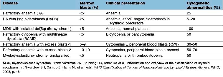
Myelodysplasia (MDS) describes a group of acquired bone marrow disorders that are due to a defect in stem cells. They are characterized by increasing bone marrow failure with quantitative and qualitative abnormalities of all three myeloid cell lines (red cells, granulocyte/monocytes and platelets). The natural history of MDS is variable, but there is a high morbidity and mortality owing to bone marrow failure, and transformation into acute myeloblastic leukaemia occurs in about 30% of cases. WHO classification of the myelodysplastic syndrome is shown in Table 8.16.

Table 8.16 World Health Organization MDS classification system

image

Somatic point mutations are commonly seen. A poor survival is seen in those carrying mutations in TP53, E2H2, ETV6, RUNX1 and ASXL1.

FURTHER READING

Tefferi A, Vardiman JW. Myelodysplastic syndromes. N Engl J Med 2009; 361:1872–1885.

Tafferi A. Challenges facing JAK inhibitory therapy for myeloproliferative neoplasms. N Engl J Med 2012; 366:844−846.

Clinical and laboratory features

MDS occurs mainly in the elderly, and presents with symptoms of anaemia, infection or bleeding due to pancytopenia. Serial blood counts show evidence of increasing bone marrow failure with anaemia, neutropenia, monocytosis and thrombocytopenia, either alone or in combination. By contrast, in chronic myelomonocytic leukaemias (CMML), monocytes are >1 × 109/L and the WBC count may be >100 × 109/L.

The bone marrow usually shows increased cellularity despite the pancytopenia. Dyserythropoiesis is present, and granulocyte precursors and megakaryocytes also have abnormal morphology. Ring sideroblasts are present in some types. Table 8.16 shows the classification and the clinical presentation.

Management

Patients with <5% blasts in the bone marrow are usually managed conservatively with red cell and platelet transfusions and antibiotics for infections, as they are needed. Haemopoietic growth factors (e.g. erythropoietin, G-CSF) may be useful in some patients.

Patients with >5% blasts have a less favourable prognosis, and a number of treatment options are available:

image Supportive care only is suitable for elderly patients with other medical problems.

image ‘Gentle’ chemotherapy (low-dose or single-agent, e.g. azacytidine) may be useful in patients with high WBC counts.

image Intensive chemotherapy schedules used for acute myeloblastic leukaemia (see p. 454) may be tried in patients under the age of 60, but the remission rate is less, and prolonged pancytopenia may occur owing to poor haemopoietic regeneration because of the defect in stem cells.

image Lenalidomide (a thalidomide analogue) has been proven to be remarkably successful in the treatment of early stage myelodysplasia with a chromosome 5q deletion (the 5q– syndrome). Avoid use in women of child-bearing age.

image Bone marrow transplantation offers the hope of cure in the small proportion of MDS patients who are under the age of 50 and who have an HLA-identical sibling or an unrelated HLA-matched donor.
FURTHER READING

Bejar R, Stevenson K, Abdel-Wahab O et al. Clinical effect of point mutations in myelodysplastic syndromes. N Engl J Med 2011; 364:2496–2506.

The spleen

The spleen is the largest lymphoid organ in the body and is situated in the left hypochondrium. There are two anatomical components:

image The red pulp, consisting of sinuses lined by endothelial macrophages and cords (spaces)

image The white pulp, which has a structure similar to lymphoid follicles.

Blood enters via the splenic artery and is delivered to the red and white pulp. During the flow the blood is ‘skimmed’, with leucocytes and plasma preferentially passing to white pulp. Some red cells pass rapidly through into the venous system while others are held up in the red pulp.

Functions

Sequestration and phagocytosis. Normal red cells, which are flexible, pass through the red pulp into the venous system without difficulty. Old or abnormal cells are damaged by the hypoxia, low glucose and low pH found in the sinuses of the red pulp and are therefore removed by phagocytosis along with other circulating foreign matter. Howell–Jolly and Heinz bodies and sideroblastic granules have their particles removed by ‘pitting’ and are then returned to the circulation. IgG-coated red cells are removed through their Fc receptors by macrophages.

Extramedullary haemopoiesis. Pluripotential stem cells are present in the spleen and proliferate during severe haematological stress, such as in haemolytic anaemia or thalassaemia major.

Immunological function. About 25% of the body’s T lymphocytes and 15% of B lymphocytes are present in the spleen. The spleen shares the function of production of antibodies with other lymphoid tissues.

Blood pooling. Up to one-third of the platelets are sequestrated in the spleen and can be rapidly mobilized. Enlarged spleens pool a significant percentage (up to 40%) of the red cell mass.

Splenomegaly

Causes

A clinically palpable spleen can have many causes.

image Infection:

acute, e.g. septic shock, infective endocarditis, typhoid, infectious mononucleosis
chronic, e.g. tuberculosis and brucellosis
parasitic, e.g. malaria, kala-azar and schistosomiasis.

image Inflammation: rheumatoid arthritis, sarcoidosis, SLE.

image Haematological: haemolytic anaemia, haemoglobinopathies and the leukaemias, lymphomas and myeloproliferative disorders.

image Portal hypertension: liver disease.

image Miscellaneous: storage diseases, amyloid, primary and secondary neoplasias, tropical splenomegaly.

Massive splenomegaly. This is seen in myelofibrosis, chronic myeloid leukaemia, chronic malaria, kala-azar or, rarely, Gaucher’s disease.

Hypersplenism

This can result from splenomegaly due to any cause. It is commonly seen with splenomegaly due to haematological disorders, portal hypertension, rheumatoid arthritis (Felty’s syndrome) and lymphoma. Hypersplenism produces:

image pancytopenia

image haemolysis due to sequestration and destruction of red cells in the spleen

image increased plasma volume.

Treatment. This is often dependent on the underlying cause, but splenectomy is sometimes required for severe anaemia or thrombocytopenia.

Splenectomy

Splenectomy is performed mainly for:

image trauma

image immune thrombocytopenic purpura (see p. 419)

image haemolytic anaemias (see p. 386)

image hypersplenism.

Problems after splenectomy

An immediate problem is an increased platelet count (usually 600–1000 × 109/L) for 2–3 weeks. Thromboembolic phenomena may occur. In the longer term, there is an increased risk of overwhelming infections, particularly pneumococcal infections.

FURTHER READING

Di Sabatino A, Carsetti R, Corazza GR. Post-splenectomy and hyposplenic states. Lancet 2011; 378:86–97.

Prophylaxis against infection after splenectomy or splenic dysfunction (Box 8.3)

All patients should be educated about the risk of infection and the importance of its early recognition and treatment. They should be given an information leaflet and should carry a card or bracelet to alert health professionals to their risk of overwhelming infection.

image Box 8.3

Prophylaxis against infection after splenectomy or splenic dysfunction

Vaccinate 2–3 weeks before elective splenectomy.

image A 23-valent unconjugated pneumococcal polysaccharide vaccine repeated every 5 years

image Meningococcal group C conjugate vaccine

image Annual influenza vaccine

image Haemophilus influenzae type b (Hib) vaccine

image Long-term penicillin V 500 mg 12-hourly (if sensitive, use erythromycin)

image Meningococcal polysaccharide vaccine (ACWY) for travellers to Africa/Saudi Arabia, e.g. during Hajj and Umrah pilgrimages

Post-splenectomy haematological features

image Thrombocytosis persists in about 30% of cases.

image The WBC count is usually normal but there may be a mild lymphocytosis and monocytosis.

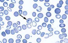
image Abnormalities in red cell morphology are the most prominent changes and include Howell–Jolly bodies (contain basophilic nuclear remnants), Pappenheimer bodies (contain sideroblastic granules), target cells and irregular contracted red cells (Fig. 8.29). Pitted red cells can be counted.

image

Figure 8.29 Post-splenectomy film with Howell–Jolly bodies (arrowed), target cells and irregularly contracted cells.

Splenic atrophy

This is seen in sickle cell disease due to infarction. It is also seen in coeliac disease, in dermatitis herpetiformis, and occasionally in ulcerative colitis and essential thrombocythaemia. Post-splenectomy haematological features are seen.

FURTHER READING

Davies JM, Barnes R, Milligan D. Update of guidelines for the prevention and treatment of infection in patients with an absent or dysfunctional spleen. Clin Med 2003; 2:440–443.

Blood transfusion

The cells and proteins in the blood express antigens which are controlled by polymorphic genes; that is, a specific antigen may be present in some individuals but not in others. A blood transfusion may immunize the recipient against donor antigens that the recipient lacks (alloimmunization), and repeated transfusions increase the risk. Similarly, the transplacental passage of fetal blood cells during pregnancy may alloimmunize the mother against fetal antigens inherited from the father. Antibodies stimulated by blood transfusion or pregnancy, such as Rh antibodies, are termed immune antibodies and are usually IgG, in contrast to naturally occurring antibodies, such as ABO antibodies, which are made in response to environmental antigens present in food and bacteria and which are usually IgM.

Blood groups

The blood groups are determined by antigens on the surface of red cells; more than 280 blood groups are recognized. The ABO and Rh systems are the two major blood groups, but incompatibilities involving many other blood groups (e.g. Kell, Duffy, Kidd) may cause haemolytic transfusion reactions and/or haemolytic disease of the newborn (HDN).

ABO system

This blood group system involves naturally occurring IgM anti-A and anti-B antibodies which are capable of producing rapid and severe intravascular haemolysis of incompatible red cells.

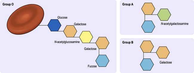
The ABO system is under the control of a pair of allelic genes, H and h, and also three allelic genes, A, B and O, producing the genotypes and phenotypes shown in Table 8.17. The A, B and H antigens are very similar in structure; differences in the terminal sugars determine their specificity. The H gene codes for enzyme H, which attaches fucose to the basic glycoprotein backbone to form H substance, which is the precursor for A and B antigens (Fig. 8.30).

Table 8.17 The ABO system: antigens and antibodies

image
image

Figure 8.30 Sugar chains in the ABO blood group system.

The A and B genes control specific enzymes responsible for the addition to H substance of N-acetylgalactosamine for Group A and d-galactose for Group B. The O gene is amorphic and does not transform H substance and therefore O is not antigenic. The A, B and H antigens are present on most body cells. These antigens are also found in soluble form in tissue fluids such as saliva and gastric juice in the 80% of the population who possess secretor genes.

Rh system

There is a high frequency of development of IgG RhD antibodies in RhD-negative individuals after exposure to RhD positive red cells. The antibodies formed cause HDN and haemolytic transfusion reactions.

This system is coded by allelic genes, C and c, E and e, D and no D, which is signified as d; they are inherited as triplets on each chromosome 1, one from each pair of genes (i.e. CDE/cde). RhD-negative individuals have no D protein in the red cell membrane, which explains why it is so immunogenic when RhD-negative individuals are exposed to RhD antigen through transfusion or pregnancy. In Caucasians, the RhD-negative phenotype almost always results from a complete deletion of the RhD gene; in black Africans, it can also result from an inactive gene containing stop codons in the reading frame.

Procedure for blood transfusion

The safety of blood transfusion depends on meticulous attention to detail at each stage leading to and during the transfusion. Avoidance of simple errors involving patient and blood sample identification at the time of collection of the sample for compatibility testing and at the time of transfusion would avoid most serious haemolytic transfusion reactions, almost all of which involve the ABO system.

Pre-transfusion compatibility testing

Blood grouping

The ABO and RhD groups of the patient are determined.

Antibody screening

The patient’s serum or plasma is screened for atypical antibodies that may cause a significant reduction in the survival of the transfused red cells. The patient’s serum or plasma is tested against red cells from at least two group O donors, expressing a wide range of red cell antigens, for detection of IgM red cell alloantibodies (using a direct agglutination test of cells suspended in saline) and IgG antibodies (using an indirect antiglobulin test, see p. 398). About 10% of patients have a positive antibody screening result; in which case, further testing is carried out using a comprehensive panel of typed red cells to determine the blood group specificity of the antibody (clinically significant red cell antibodies are detected in about 20% of patients with positive antibody screens).

Selection of donor blood and crossmatching

Donor blood of the same ABO and RhD group as the patient is selected. Matching for additional blood groups is carried out for patients with clinically significant red cell antibodies (see below), for patients who are likely to be multitransfused and at high risk of developing antibodies, e.g. sickle cell disease, and many hospitals routinely provide Kell-negative blood for women of child-bearing age to minimize the risk of alloimmunization and subsequent HDN.

Crossmatching procedures

Patients without atypical red cell antibodies. The full crossmatch involves testing the patient’s serum or plasma against the donor red cells suspended in saline in a direct agglutination test, and also using an indirect antiglobulin test. In many hospitals this serological crossmatch has been omitted as the negative antibody screen makes it highly unlikely that there will be any incompatibility with the donor units. A greater risk is that of a transfusion error involving the collection of the patient sample or a mix-up of samples in the laboratory. Laboratories can use their information system to check the records of the patient and authorize the release of the donor units if a number of criteria are met (computer or electronic crossmatching), including:

image The system is automated for ABO and RhD grouping and antibody screening including positive sample identification and electronic transfer of results.

image The antibody screening procedure conforms to national recommendations.

image The patient’s serum or plasma does not contain clinically significant red cell antibodies.

image The release of ABO incompatible blood must be prevented by conformation of laboratory computer software to the following requirements:

the issue of blood is not allowed if the patient has only been grouped once
the issue of blood is not allowed if the current group does not match the historical record
the system must not allow the reservation and release of units which are ABO incompatible with the patient.

image The laboratory must assure the validity of the ABO and RhD group of the donor blood either by written verification from the Blood Service supplying the donor units or confirmatory testing in the laboratory; the UK Blood Transfusion Services guarantee that the blood group information is correct.

Electronic issue can be extended to issue of previously unallocated blood at blood fridges remote from the main laboratory (’remote blood issue’). This is only possible using blood fridges electronically linked to the blood transfusion laboratory information system. The printing of compatibility labels for the blood and its collection are under electronic control using the same rules as electronic issue from the main laboratory. Issue of blood using this process reduces the time it takes to provide blood for patients needing it urgently, particularly at hospitals without a blood transfusion laboratory because transport of blood from the central laboratory is not required. There are good examples of centralized transfusion services in cities such as Pittsburgh and Seattle in the USA, and this model is likely to be increasingly used in other developed countries including the UK.

Patients with atypical red cell antibodies. Donor blood should be selected that lacks the relevant red cell antigen(s), as well as being the same ABO and RhD group as the patient. A full crossmatch should always be carried out.

Several other systems for blood grouping, antibody screening and crossmatching are available to hospital transfusion laboratories. They do not depend on agglutination of red cells in suspension, but rather on the differential passage of agglutinated and unagglutinated red cells through a column of dextran gel matrix (e.g. DiaMed and Ortho Biovue systems), or on the capture of antibodies by red cells immobilized on the surface of a microplate well (e.g. Capture-R solid phase system).

Blood ordering

Elective surgery

Many hospitals have guidelines for the ordering of blood for elective surgery (maximum surgical blood ordering schedules). These are aimed at reducing unnecessary crossmatching and the amount of blood that eventually becomes outdated. Many operations in which blood is required only occasionally for unexpectedly high blood loss can be classified as ‘group and save’; this means that, where the antibody screen is negative, blood is not reserved in advance but can be made available quickly if necessary, i.e. in a few minutes, using the electronic crossmatch procedure. If a patient has atypical antibodies, compatible blood should always be reserved in advance; this may take several days if the patient has multiple or unusual antibodies.

Emergencies

There may be insufficient time for full pretransfusion testing. The options include:

image Blood required immediately – use of 2 units of O RhD-negative blood (‘emergency stock’), to allow additional time for the laboratory to group the patient.

image Blood required in 10–15 min – use of blood of the same ABO and RhD groups as the patient (‘group compatible blood’).

image Blood required in 45 min – most laboratories will be able to provide fully crossmatched blood within this time.

Complications of blood transfusion (Table 8.18)

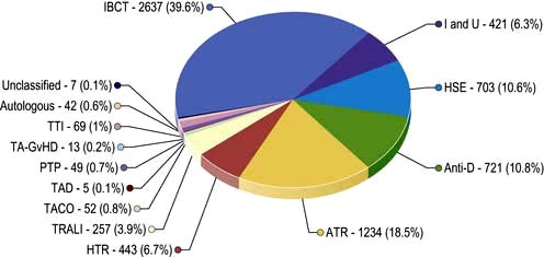
In the USA, it has been mandatory to report transfusion-associated deaths to the Food and Drug Administration since 1975; such reports have provided useful data which have contributed to efforts to improve the safety of blood transfusion. Similar reporting schemes under the term ‘haemovigilance’ have been set up in other countries, including the Serious Hazards of Transfusion (SHOT) scheme, which produced its first report in the UK in 1997. Figure 8.31 shows the reports to SHOT up to 2009, indicating that ‘incorrect blood component transfused’ is the most frequent type of serious incident, and the second most frequent cause of mortality and serious morbidity after transfusion-related acute lung injury (TRALI). Errors at the time of collection of blood from the fridge and/or the administration of blood (53%) were the commonest source of error in 2005, followed by laboratory errors (43%) and mistakes in the collection of blood samples for compatibility testing (5%). Death or serious morbidity can also be attributed to other complications of blood transfusion including transfusion-associated circulatory overload (TACO), transfusion-associated graft-versus-host disease (TA-GvHD) and bacterial infection of blood components.

Table 8.18 Complications of blood transfusion

Immunological Non-immunological

Alloimmunization and incompatibility

Transmission of infection

Red cells

Viruses

Immediate haemolytic transfusion reactions

HAV, HBV, HCV

HIV, HHV8

Delayed haemolytic transfusion reactions

CMV, EBV, HTLV-1, WNV

Parasites

Leucocytes and platelets

Malaria, trypanosomiasis, toxoplasmosis

Non-haemolytic (febrile) transfusion reactions

Bacteria

Post-transfusion purpura

Prion – CJD

Poor survival of transfused platelets and granulocytes

Circulatory failure due to volume overload

Iron overload due to multiple transfusions (see p. 391)

Graft-versus-host disease

Lung injury (TRALI)

Plasma proteins

Massive transfusion of stored blood may cause bleeding reactions and electrolyte changes

Urticarial and anaphylactic reactions


Physical damage due to freezing or heating


Thrombophlebitis


Air embolism

image

Figure 8.31 Overview of 6653 cases reported to the Serious Hazards of Transfusion (SHOT) scheme between 1996 and 2009. ATR, acute transfusion reaction; HSE, handling and storage errors; HTR, haemolytic transfusion reaction; I and U, inappropriate and unnecessary; IBCT, incorrect blood component transfused; PTP, post-transfusion purpura; TACO, transfusion-associated circulatory overload; TAD, transfusion-associated dyspnoea; TA-GvHD, transfusion-associated graft-vs-host disease; TRALI, transfusion-related acute lung injury; TTI, transfusion-transmitted infection.

(From the Serious Hazards of Transfusion Steering Group, SHOT Annual Report 2010 Summary, with permission.)

Prevention of wrong blood transfusions

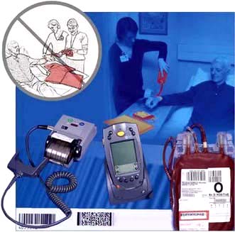
The serious consequences of such failures emphasize the need for meticulous checks at all stages in the procedure of blood transfusion. Written procedures and good training of staff are essential. Regular audit to ensure compliance with these procedures is also required. New approaches are also being used including the use of barcode patient identification and new technology at the bedside. Handheld devices can be used to prompt staff through the key steps and check that the barcode on the patient’s wristband matches the barcode on the unit of blood (Fig. 8.32).

image

Figure 8.32 Bedside checks. The traditional method of pre-transfusion bedside checking requires two nurses and checks of multiple items of written documentation. With barcode technology, a handheld computer reads a barcode on the patient wristband containing full patient details. The handheld computer checks that the patient details on the wristband barcode match those on the barcode (in the red box) on the compatibility label attached to the unit after pre-transfusion testing. This barcode also contains the unique number of the unit, and is matched with the barcode number of the unit (top left of the bag) to ensure that the blood bank has attached the right compatibility label.

(This figure was originally published as the front cover of the September 2003 issue of Transfusion 43. Reproduced with permission from Wiley–Blackwell.)

Immunological complications

Alloimmunization

Blood transfusion carries a risk of alloimmunization to the many ‘foreign’ antigens present on red cells, leucocytes, platelets and plasma proteins. Alloimmunization also occurs during pregnancy – to fetal antigens inherited from the father and not shared by the mother (see p. 400).

Alloimmunization does not usually cause clinical problems with the first transfusion but these may occur with subsequent transfusions. There may also be delayed consequences of alloimmunization, such as HDN and rejection of tissue or organ transplants.

Incompatibility

This may result in poor survival of transfused cells, such as red cells and platelets, and also in harmful effects of the antigen–antibody reaction.

1. Red cells

Haemolytic transfusion reactions

Immediate reaction. This is the most serious complication of blood transfusion and is usually due to ABO incompatibility. There is complement activation by the antigen–antibody reaction, usually caused by IgM antibodies, leading to rigors, lumbar pain, dyspnoea, hypotension, haemoglobinuria and renal failure. The initial symptoms may occur a few minutes after starting the transfusion. Activation of coagulation also occurs and bleeding due to disseminated intravascular coagulation (DIC) is a bad prognostic sign. Emergency treatment for shock (p. 885) is needed to maintain the blood pressure and renal function.

Diagnosis

This is confirmed by finding evidence of haemolysis (e.g. haemoglobinuria), and incompatibility between donor and recipient. All documentation should be checked to detect errors such as:

image Failure to check the identity of the patient when taking the sample for compatibility testing (i.e. sample from the wrong patient)

image Mislabelling the blood sample with the wrong patient’s name

image Simple labelling or handling errors in the laboratory

image Errors in the collection of blood, leading to delivery of the wrong blood to the ward/theatre

image Failure to perform proper identity checks before the blood is transfused (i.e. blood transfused to the wrong patient).

Investigations

To confirm where the error occurred, blood grouping should be carried out on:

image the patient’s original sample (used for the compatibility testing)

image a new sample taken from the patient after the reaction

image the donor units.

At the first suspicion of any serious transfusion reaction, the transfusion should always be stopped and the donor units returned to the blood transfusion laboratory with a new blood sample from the patient to exclude a haemolytic transfusion reaction.

Delayed reaction. This occurs in patients alloimmunized by previous transfusions or pregnancies. The antibody level is too low to be detected by pretransfusion compatibility testing, but a secondary immune response occurs after transfusion, resulting in destruction of the transfused cells, usually by IgG antibodies.

Haemolysis is usually extravascular as the antibodies are IgG, and the patient may develop anaemia and jaundice about a week after the transfusion, although most of these episodes are clinically silent. The blood film shows spherocytosis and reticulocytosis. The direct antiglobulin test is positive and detection of the antibody is usually straightforward.

2. Leucocytes and platelets

Non-haemolytic (febrile) transfusion reactions

Febrile reactions are a common complication of blood transfusion in patients who have previously been transfused or pregnant. The usual causes are the presence of leucocyte antibodies in an alloimmunized recipient acting against donor leucocytes in red cell concentrates leading to release of pyrogens, or the release of cytokines from donor leucocytes in platelet concentrates. Typical signs are flushing and tachycardia, fever (>38°C), chills and rigors. Aspirin may be used to reduce the fever, although it should not be used in patients with thrombocytopenia. The routine introduction of leucocyte-depleted blood in the UK, to minimize the risk of transmission of variant Creutzfeldt–Jakob disease (vCJD) by blood transfusion (see below), has reduced the incidence of febrile reactions. Universal leucocyte-depletion of all blood components is common in European countries, but the proportion of blood components which are leucocyte-depleted is variable across the USA.

Potent leucocyte antibodies in the plasma of donors, who are usually multiparous women, may cause TRALI characterized by dyspnoea, fever, cough, and shadowing in the perihilar and lower lung fields on the chest X-ray. Prompt respiratory support is essential; mechanical ventilation is frequently necessary. It usually resolves within 48–96 hours, but the mortality is 16% in the 257 cases of TRALI reported to SHOT, up to 2009. The avoidance of female plasma in the preparation of FFP and testing female platelet donors for leucocyte antibodies has been implemented in many developed countries to reduce the risk of TRALI.

Platelets

Post-transfusion purpura. See page 420.

3. Plasma proteins

Urticaria and anaphylaxis

Urticarial reactions are often attributed to plasma protein incompatibility, but in most cases, they are unexplained. They are common but rarely severe; stopping or slowing the transfusion and administration of chlorphenamine 10 mg i.v. are usually sufficient treatment.

Anaphylactic reactions (see p. 69) occasionally occur; severe reactions are seen in patients lacking IgA who produce anti-IgA that reacts with IgA in the transfused blood. The transfusion should be stopped and epinephrine (adrenaline) 0.5 mg i.m. and chlorphenamine 10 mg i.v. should be given immediately; endotracheal intubation may be required. Patients who have had severe urticarial or anaphylactic reactions should receive washed red cells, autologous blood or blood from IgA-deficient donors for patients with IgA deficiency.

Immunosuppression

Transfusions are known to have a favourable effect on the survival of subsequent renal allografts, due to transfusion-induced immunomodulation. The precise mechanism is unclear, but may be associated with the transfusion of allogeneic leucocytes. Other possible clinical effects caused by transfusion-induced immunosuppression, such as an increase in postoperative infection and tumour recurrence, have been proposed, but remain unproven.

Non-immunological complications

Transmission of infection

Viral transmission: donor blood in the UK is currently tested for HBV, HCV, HIV-1 and HTLV-1. CMV-seronegative tested blood is given to immunosuppressed patients who are susceptible to acquiring CMV. Blood services continue a vigilant search for new infectious agents (‘emerging’ infections) which may be transmitted by blood transfusion, and for methods to prevent their transmission including donor screening, testing and pathogen inactivation. Donor questionnaires record recent travel to exclude possible risks of West Nile virus (WNV) and severe acute respiratory syndrome (SARS). WNV is the causal agent of meningoencephalitis and has been transmitted by transfusion and transplantation in the USA.

The risk of transmission of viral infections by blood transfusion varies from country to country depending on factors such as the underlying prevalence of transfusion-transmitted infections in the population and the measures taken to minimize the risk of transmission. Viral transmission via blood transfusion is still a major issue in the developing world. In the UK, the incidence of transmission of HIV by blood transfusion is extremely low – <1 in 5 million units transfused. Prevention is based on self-exclusion of donors in ‘high-risk’ groups and testing each donation for anti-HIV. The incidence of transmission of HBV is about 1 in 400 000 units transfused. The incidence of HCV is <1 in 30 million units transfused since the introduction of testing donor plasma for viral nucleic acid.

Measures for inactivating viruses in plasma such as coagulation factor concentrates or intravenous immunoglobulin include treatment with solvents and detergents.

Bacterial contamination of blood components is rare but it is one of the most frequent causes of death associated with transfusion. Some organisms such as Yersinia enterocolitica can proliferate in red cell concentrates stored at 4°C, but platelet concentrates stored at 22°C are a more frequent cause of this problem. Measures to avoid bacterial contamination include strict donor arm cleansing, diversion of the initial blood collection to samples for testing rather than into the collection bag, and bacterial detection systems for platelet concentrates, which have been implemented in most developed countries including the USA. It was implemented in England in 2011.

Transfusion-transmitted syphilis is very rare. Spirochaetes do not survive for >72 hours in blood stored at 4°C, and each donation is tested using the Treponema pallidum haemagglutination assay (TPHA).

In the UK, there continues to be concern about the risk of transmitting the prion protein causing vCJD (see p. 113) by transfusion: four transmissions of vCJD have occurred following a blood transfusion in the UK. A number of measures have been taken in the UK, including universal leucocyte-depletion of blood components (in 1999) because the prion protein was thought to be primarily associated with lymphocytes. Blood donors are excluded if they have had a blood transfusion since 1980. UK donor plasma is not used for the manufacture of blood products; imported plasma from the USA is used instead. For children under the age of 16 years, fresh frozen plasma (FFP) is sourced from plasma (from unremunerated donors) imported from the USA, on the basis that exposure to bovine spongiform encephalitis (BSE) from food was eliminated by 1 January 1996. FFP for this group is treated with methylthioninium chloride (methylene blue) to inactivate viruses.

While stringent measures are being taken to minimize the risk of transfusion-transmitted infection, it may never be possible to guarantee that donor blood is absolutely ‘safe’. The current approach to the safety of blood components and plasma in the UK and other developed countries is cautious, but it is not an absolute guarantee of safety. Clinicians should always carefully consider the patient’s requirement for transfusion, and only transfuse if clinically appropriate (see below).

Circulatory failure due to volume overload see management of acute heart failure, page 723.

Iron overload – see page 391.

Strategies for the avoidance of unnecessary transfusion

These include:

image Strict criteria for the use of blood components and blood products

image Stopping drug therapy (anticoagulants and antiplatelet drugs) that may potentiate bleeding in surgical patients

image The identification and treatment of anaemia prior to surgery

image The use of anti-fibrinolytic drugs, e.g. aprotinin and tranexamic acid in major surgery

image Recombinant factor VIIa is licensed to treat patients with haemophilia with inhibitors, and is being used ‘off-license’ to treat patients with severe bleeding, e.g. postoperative, trauma, intracerebral haemorrhage. However, there is little evidence of its safety and effectiveness for this latter indication.

Artificial haemoglobin solutions and other blood substitutes suitable for clinical use have not yet been developed. They generally have a short intravascular half-life, and a recent meta-analysis found a significant risk of mortality and myocardial infarction.

Autologous transfusion

An alternative to using blood from volunteer donors is to use the patient’s own blood. There are three types of autologous transfusion:

image Predeposit. The patient donates 2–5 units of blood at approximately weekly intervals before elective surgery.

image Preoperative haemodilution. 1 or 2 units of blood are removed from the patient immediately before surgery and retransfused to replace operative losses.

image Blood salvage. Blood lost during or after surgery may be collected and retransfused. Several techniques of varying levels of sophistication are available. The operative site must be free of bacteria, bowel contents and tumour cells.

The use of predeposit autologous transfusion was largely driven by concerns about transfusion-transmitted infection particularly in the USA, but its use has decreased in the USA and most countries. It has been abandoned in the UK except for those rare patients where it is not possible to identify compatible blood because of multiple antibodies. There is little evidence that this approach reduces blood requirements, and blood is perceived as being ‘safe’. Blood salvage is increasingly being used as a way of avoiding the use of donor blood. In developing countries, autologous blood and blood from relatives are commonly used because of a lack of donor blood.

Blood, blood components and blood products

Most blood collected from donors is processed as follows:

image Blood components, such as red cell and platelet concentrates, fresh frozen plasma (FFP) and cryoprecipitate are prepared from a single donation of blood by simple separation methods such as centrifugation and are transfused without further processing. Platelet concentrates are also prepared by plateletpheresis (see below).

image Blood products, such as coagulation factor concentrates, albumin and immunoglobulin solutions, are prepared by complex processes using the plasma from many donors as the starting material (UK donor plasma is not used, see above).

In most circumstances it is preferable to transfuse only the blood component or product required by the patient (‘component therapy’) rather than use whole blood. This is the most effective way of using donor blood, which is a scarce resource, and reduces the risk of complications from transfusion of unnecessary components of the blood.

Whole blood

A unit of whole blood consists of 450 mL ± 10% of blood from a suitable donor plus 63 mL of anticoagulant, which is then leucocyte depleted. Blood stored at 4°C is given a ‘shelf-life’ of 5 weeks in the UK (6 weeks in some other countries), when at least 70% of the transfused red cells should survive normally. Whole blood is now rarely used for transfusion; donated blood is processed into red cell concentrates and other blood components.

Red cell concentrates

Virtually all the plasma is removed and is replaced by about 100 mL of an optimal additive solution, such as SAG-M, which contains sodium chloride, adenine, glucose and mannitol. The mean volume is about 330 mL. The PCV is about 0.57 L/L, but the viscosity is low as there are no plasma proteins in the additive solution, and this allows fast administration if necessary.

Washed red cell concentrates

These are preparations of red cells suspended in saline, produced by cell separators to remove all but traces of plasma proteins. They are used in patients who have had severe recurrent urticarial or anaphylactic reactions.

Platelet concentrates

These are prepared either from whole blood by centrifugation or by plateletpheresis of single donors using cell separators. They may be stored for up to 5 days at 22°C with agitation. They are used to treat bleeding in patients with severe thrombocytopenia, and prophylactically to prevent bleeding in patients with bone marrow failure.

Granulocyte concentrates

These are prepared from whole blood as ‘buffy coats’ or from single donors using cell separators and are used for patients with severe neutropenia with definite evidence of bacterial infection. The numbers of granulocytes collected may be increased by treating donors with G-CSF and steroids.

Fresh frozen plasma

FFP is prepared by freezing the plasma from 1 unit of blood to −30°C to maintain the concentration of coagulation factors. The volume is approximately 200 mL. FFP contains all the coagulation factors present in fresh plasma and is used mostly for replacement of coagulation factors in acquired coagulation factor deficiencies. It may be further treated by a pathogen-inactivation process, e.g. methylene blue or solvent detergent, to minimize the risk of disease transmission. For children, see page 411.

Cryoprecipitate

This is obtained by allowing the frozen plasma from a single donation to thaw at 4–8°C and removing the supernatant. The volume is about 20 mL and it is stored at −30°C. It contains factor VIII: C, von Willebrand factor (VWF) and fibrinogen, and may be useful in DIC and other conditions where the fibrinogen level is very low. It is no longer used for the treatment of haemophilia A and von Willebrand’s disease because of the greater risk of virus transmission compared with virus-inactivated coagulation factor concentrates. Fibrinogen concentrates are now available, but not yet approved for the treatment of patients with acquired disorders of haemostasis such as massive transfusion.

Factor VIII and IX concentrates

These are freeze-dried preparations of specific coagulation factors prepared from large pools of plasma. They are used for treating patients with haemophilia and von Willebrand’s disease, where recombinant coagulation factor concentrates are unavailable. Recombinant coagulation factor concentrates, where they are available, are the treatment of choice for patients with inherited coagulation factor deficiencies (see p. 421).

Albumin

There are two preparations:

image Human albumin solution 4.5% contains 45 g/L albumin and 160 mmol/L sodium. It is available in 50, 100, 250 and 500 mL bottles.

image Human albumin solution 20% contains approximately 200 g/L albumin and 130 mmol/L sodium and is available in 50 and 100 mL bottles.

Human albumin solutions are generally considered to be inappropriate fluids for acute volume replacement or for the treatment of shock because they are no more effective in these situations than synthetic colloid solutions such as polygelatins (Gelofusine) or hydroxyethyl starch (Haemaccel). However, albumin solutions are indicated for treatment of acute severe hypoalbuminaemia and as the replacement fluid for plasma exchange. The 20% albumin solution is particularly useful for patients with nephrotic syndrome or liver disease who are fluid overloaded and resistant to diuretics. Albumin solutions should not be used to treat patients with malnutrition or chronic renal or liver disease with low serum albumin.

Normal immunoglobulin

This is prepared from normal plasma. It is used in patients with hypogammaglobulinaemia, to prevent infections, and in patients with, for example, immune thrombocytopenic purpura (see p. 419).

Specific immunoglobulins

These are obtained from donors with high titres of antibodies. Many preparations are available, such as anti-D, antihepatitis B and anti-varicella zoster.

FURTHER READING

Alter HJ, Stramer SL, Dodd RY. Emerging infectious diseases that threaten the blood supply. Semin Hematol 2007; 44:32–41.

Birchall J, Stanworth SJ, Duffy MR et al. Evidence for the use of recombinant factor VIIa in the prevention and treatment of bleeding in patients without hemophilia. Transfus Med Rev 2008; 22:177–187.

Murphy MF, Pamphilon D (eds). Practical Transfusion Medicine, 3rd edn. Oxford: Wiley-Blackwell; 2009.

Natanson C, Kern SJ, Lurie P et al. Cell-free hemoglobin-based blood substitutes and risk of myocardial infarction and death: a meta-analysis. JAMA 2008; 299:2304–2312.

The white cell

The five types of leucocytes found in peripheral blood are: neutrophils, eosinophils and basophils (which are all called granulocytes) and lymphocytes and monocytes (see also Ch. 3). The development of these cells is shown in Figure 8.1.

Neutrophils

The earliest morphologically identifiable precursors of neutrophils in the bone marrow are myeloblasts, which are large cells constituting up to 3.5% of the nucleated cells in the marrow. The nucleus is large and contains 2–5 nucleoli. The cytoplasm is scanty and contains no granules. Promyelocytes are similar to myeloblasts but have some primary cytoplasmic granules, containing enzymes such as myeloperoxidase. Myelocytes are smaller cells without nucleoli but with more abundant cytoplasm and both primary and secondary granules. Indentation of the nucleus marks the change from myelocyte to metamyelocyte. The mature neutrophil is a smaller cell with a nucleus with 2–5 lobes, with predominantly secondary granules in the cytoplasm, which contain lysozyme, collagenase and lactoferrin.

Peripheral blood neutrophils are equally distributed into a circulating pool and a marginating pool lying along the endothelium of blood vessels. In contrast to the prolonged maturation time of about 10 days for neutrophils in the bone marrow, their half-life in the peripheral blood is extremely short, only 6–8 hours. In response to stimuli (e.g. infection, corticosteroid therapy), neutrophils are released into the circulating pool from both the marginating pool and the marrow. Immature white cells are released from the marrow when a rapid response (within hours) occurs in acute infection (described as a ‘shift to the left’ on a blood film).

Function

The prime function of neutrophils is to ingest and kill bacteria, fungi and damaged cells. Neutrophils are attracted to sites of infection or inflammation by chemotaxins. Recognition of foreign or dead material is aided by coating of particles with immunoglobulin and complement (opsonization) as neutrophils have Fc and C3b receptors (see p. 52). The material is ingested into vacuoles where it is subjected to enzymic destruction, which is either oxygen-dependent with the generation of hydrogen peroxide (myeloperoxidase) or oxygen-independent (lysosomal enzymes and lactoferrin). Leucocyte alkaline phosphatase (LAP) is an enzyme found in leucocytes. It is raised when there is a neutrophilia due to an acute illness. It is also raised in polycythaemia and myelofibrosis and reduced in CML.

Neutrophil leucocytosis

A rise in the number of circulating neutrophils to >10 × 109/L occurs in bacterial infections or as a result of tissue damage. This may also be seen in pregnancy, during exercise and after corticosteroid administration (Table 8.19). With any tissue necrosis there is a release of various soluble factors, causing a leucocytosis. Interleukin 1 is also released in tissue necrosis and causes a pyrexia. The pyrexia and leucocytosis accompanying a myocardial infarction are a good example of this and may be wrongly attributed to infection.

Table 8.19 Neutrophil leucocytosis

Bacterial infections

Tissue necrosis, e.g. myocardial infarction, trauma

Inflammation, e.g. gout, rheumatoid arthritis

Drugs, e.g. corticosteroids, lithium

Haematological:

Myeloproliferative disease
Leukaemoid reaction
Leucoerythroblastic anaemia

Physiological, e.g. pregnancy, exercise

Malignant disease, e.g. bronchial, breast, gastric

Metabolic, e.g. renal failure, acidosis

Congenital, e.g. leucocyte adhesion deficiency, hereditary neutrophilia

A leukaemoid reaction (an overproduction of white cells, with many immature cells) may occur in severe infections, tuberculosis, malignant infiltration of the bone marrow and occasionally after haemorrhage or haemolysis.

In leucoerythroblastic anaemia, nucleated red cells and white cell precursors are found in the peripheral blood. Causes include marrow infiltration with metastatic carcinoma, myelofibrosis, osteopetrosis, myeloma, lymphoma, and occasionally severe haemolytic or megaloblastic anaemia.

Neutropenia and agranulocytosis

Neutropenia is defined as a circulatory neutrophil count below 1.5 × 109/L. A virtual absence of neutrophils is called agranulocytosis. The causes are given in Table 8.20. It should be noted that black patients may have somewhat lower neutrophil counts. Neutropenia caused by viruses is probably the most common type. Chemotherapy and radiotherapy predictably produce neutropenia; many other drugs have been known to produce an idiosyncratic cytopenia and a drug cause should always be considered.

Table 8.20 Causes of neutropenia

Acquired

Viral infection
Severe bacterial infection, e.g. typhoid
Felty’s syndrome
Immune neutropenia – autoimmune, autoimmune neonatal neutropenia
Pancytopenia from any cause, including drug-induced marrow aplasia (see p. 25)
Pure white cell aplasia

Inherited

Ethnic (neutropenia is common in black races)
Kostmann’s syndrome (severe infantile agranulocytosis) due to mutation in elastane 2 (ELA2) gene
Cyclical (genetic mutation in ELA2 gene with neutropenia every 2–3 weeks)
Others, e.g. Schwachman–Diamond syndrome, dyskeratosis congenita, Chédiak–Higashi syndrome
Clinical features

Infections may be frequent, often serious, and are more likely as the neutrophil count falls. An absolute neutrophil count of <0.5 × 109/L is regarded as ‘severe’ neutropenia and may be associated with life-threatening infections such as pneumonia and septicaemia. A characteristic glazed mucositis occurs in the mouth, and ulceration is common.

Investigations

The blood film shows marked neutropenia. The appearance of the bone marrow will indicate whether the neutropenia is due to depressed production or increased destruction of neutrophils. Neutrophil antibody studies are performed if an immune mechanism is suspected.

Treatment

Antibiotics should be given as necessary to patients with acute severe neutropenia (see p. 449).

If the neutropenia seems likely to have been caused by a drug, all current drug therapy should be stopped. Recovery of the neutrophil count usually occurs after about 10 days. G-CSF (see p. 373) is used to decrease the period of neutropenia after chemotherapy and haemopoietic transplantation. It is also used successfully in the treatment of chronic neutropenia.

Steroids and high-dose intravenous immunoglobulin are used to treat patients with severe autoimmune neutropenia and recurrent infections, and G-CSF has produced responses in some cases.

Eosinophils

Eosinophils are slightly larger than neutrophils and are characterized by a nucleus with usually two lobes and large cytoplasmic granules that stain deeply red. The eosinophil plays a part in allergic responses (see p. 52) and in the defence against infections with helminths and protozoa.

Eosinophilia is >0.4 × 109/L eosinophils in the peripheral blood. The causes of eosinophilia are listed in Table 8.21.

Table 8.21 Causes of eosinophilia

Parasitic infestations, such as:

Ascaris
Hookworm
Strongyloides

Allergic disorders, such as:

Hayfever (allergic rhinitis)
Other hypersensitivity reactions, including drug reactions

Skin disorders, such as:

Urticaria
Pemphigus
Eczema

Pulmonary disorders, such as:

Bronchial asthma
Tropical pulmonary eosinophilia
Allergic bronchopulmonary aspergillosis
Churg–Strauss syndrome

Malignant disorders, such as:

Hodgkin’s lymphoma
Carcinoma
Eosinophilic leukaemia

Miscellaneous, such as:

Hypereosinophilic syndrome
Sarcoidosis
Hypoadrenalism
Eosinophilic gastroenteritis

Basophils

The nucleus of basophils is similar to that of neutrophils but the cytoplasm is filled with large black granules. The granules contain histamine, heparin and enzymes such as myeloperoxidase. The physiological role of the basophil is not known. Binding of IgE causes the cells to degranulate and release histamine and other contents involved in acute hypersensitivity reactions (see p. 68).

Basophils are usually few in number (<1 × 109/L) but are significantly increased in myeloproliferative disorders.

Monocytes

Monocytes are slightly larger than neutrophils. The nucleus has a variable shape and may be round, indented or lobulated. The cytoplasm contains fewer granules than neutrophils. Monocytes are precursors of tissue macrophages and dendritic cells and spend only a few hours in the blood but can continue to proliferate in the tissues for many years.

A monocytosis (>0.8 × 109/L) may be seen in chronic bacterial infections such as tuberculosis or infective endocarditis, chronic neutropenia and patients with myelodysplasia, particularly chronic myelomonocytic leukaemia.

Lymphocytes

Lymphocytes form nearly half the circulating white cells. They descend from pluripotential stem cells (Fig. 8.1). Circulating lymphocytes are small cells, a little larger than red cells, with a dark-staining central nucleus. There are two main types: T and B lymphocytes (see p. 50).

Lymphocytosis (lymphocyte count >5 × 109/L) occurs in response to viral infections, particularly EBV, CMV and HIV, and chronic infections such as tuberculosis and toxoplasmosis. It also occurs in chronic lymphocytic leukaemia and in some lymphomas.

Haemostasis and thrombosis

The integrity of the circulation is maintained by blood flowing through intact vessels lined by endothelial cells. Haemostasis is the host defence mechanism that protects this integrity after injury to the vessel wall and tissue injury.

Haemostasis

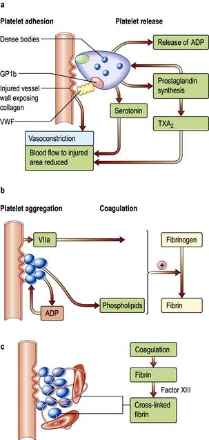
Haemostasis is a complex process depending on interactions between the vessel wall, leucocytes, platelets, coagulation and fibrinolytic mechanisms. Haemostatic systems are normally quiescent but following tissue injury become rapidly activated. The formation of the haemostatic plug is shown in Figure 8.33.

image

Figure 8.33 Formation of the haemostatic plug: sequential interactions between the vessel wall, platelets and coagulation factors. (a) Contact of platelets with collagen via the platelet receptor GP1b and factor VWF in plasma activates platelet prostaglandin synthesis which stimulates release of ADP from the dense bodies. Vasoconstriction of the vessel occurs as a reflex and by release of serotonin and thromboxane A2 (TXA2) from platelets. (b) Release of ADP from platelets induces platelet aggregation and formation of the platelet plug. The coagulation pathway is stimulated leading to formation of fibrin. (c) Fibrin strands are cross-linked by factor XIII and stabilize the haemostatic plug by binding platelets and red cells.

Vessel wall

The vessel wall is lined by endothelium which, in normal conditions, prevents platelet adhesion and thrombus formation. This property is partly due to its negative charge but also to:

image Thrombomodulin and heparan sulphate expression

image Synthesis of prostacyclin (PGI2) and nitric oxide (NO), which cause vasodilatation and inhibit platelet aggregation

image Production of plasminogen activator.

Injury to vessels causes reflex vasoconstriction, while endothelial damage results in loss of antithrombotic properties, activation of platelets and coagulation and inhibition of fibrinolysis (Fig. 8.33).

Platelets

Platelet adhesion. When the vessel wall is damaged, the platelets escaping come into contact with and adhere to collagen and subendothelial bound von Willebrand factor. This adherence is mediated through glycoprotein Ib (GPIb). Glycoprotein IIb–IIIa is then exposed, forming a second binding site for VWF. Within seconds of adhesion to the vessel wall platelets begin to undergo a shape change, from a disc to a sphere, spread along the subendothelium and release the contents of their cytoplasmic granules. These are the dense bodies (containing ADP and serotonin) and the α-granules (containing platelet-derived growth factor, platelet factor 4, β-thromboglobulin, fibrinogen, VWF, fibronectin, thrombospondin and other factors).

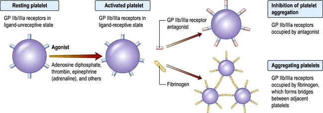
Platelet release. The release of ADP leads to a conformational change in the fibrinogen receptor, the glycoprotein IIb–IIIa complex (GPIIb–IIIa), on the surfaces of adherent platelets allowing it to bind to fibrinogen (see also Fig. 8.41).

Platelet aggregation (Fig. 8.33b). As fibrinogen is a dimer it can form a direct bridge between platelets and so binds platelets into activated aggregates (platelet aggregation) and further platelet release of ADP occurs. A self-perpetuating cycle of events is set up leading to formation of a platelet plug at the site of the injury.

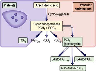
Coagulation. After platelet aggregation and release of ADP, the exposed platelet membrane phospholipids are available for the assembly of coagulation factor enzyme complexes (tenase and prothrombinase); this platelet phospholipid activity has been called platelet factor 3 (PF-3). The presence of thrombin encourages fusion of platelets, and fibrin formation reinforces the stability of the platelet plug. Central to normal platelet function is platelet prostaglandin synthesis, which is induced by platelet activation and leads to the formation of TXA2 in platelets (Fig. 8.34). Thromboxane (TXA2) is a powerful vasoconstrictor and also lowers cyclic AMP levels and initiates the platelet release reaction. Prostacyclin (PGI2) is synthesized in vascular endothelial cells and opposes the actions of TXA2. It produces vasodilatation and increases the level of cyclic AMP, preventing platelet aggregation on the normal vessel wall as well as limiting the extent of the initial platelet plug after injury.

image

Figure 8.34 Prostaglandin synthesis.

Coagulation and fibrinolysis

Coagulation involves a series of enzymatic reactions leading to the conversion of soluble plasma fibrinogen to fibrin-based clot (Fig. 8.35). Roman numerals are used for most of the factors, but I and II are referred to as fibrinogen and prothrombin respectively; III, IV and VI are redundant. The active forms are denoted by ‘a’. The coagulation factors are primarily synthesized in the liver and are either serine protease enzyme precursors (factors XI, X, IX and thrombin) or cofactors (V and VIII), except for fibrinogen, which is polymerized to form fibrin.

image

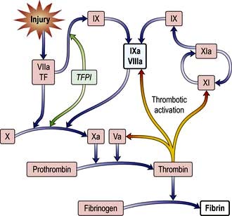
Figure 8.35 Coagulation mechanism. The in vivo pathway begins with tissue factor–factor VIIa complex activating factor X and also factor IX. Factor XI is activated by thrombin. TF, tissue factor; TFPI, tissue factor pathway inhibitor.

Coagulation pathway

This enzymatic amplification system was traditionally divided into ‘extrinsic’ and ‘intrinsic’ pathways. This concept is useful for the interpretation of clinical laboratory tests such as the prothrombin time (PT) and activated partial thromboplastin time (APTT) (see p. 417) but unrepresentative and an oversimplification of in vivo coagulation. Coagulation is initiated by tissue damage (Fig. 8.35):

image Tissue damage exposes tissue factor (TF) which binds to factor VII.

image The TF–factor VII complex directly converts factor X to active factor Xa and some factor IX to factor IXa.

image In the presence of factor Xa, tissue factor pathway inhibitor (TFPI) inhibits further generation of factor Xa and factor IXa.

image Following inhibition by TFPI the amount of factor Xa produced is insufficient to maintain coagulation. Further factor Xa, to allow haemostasis to progress to completion, can only be generated by the alternative factor IX/factor VIII pathway. However, enough thrombin exists at this point to activate factor VIII, which dramatically increases the activity of factor IXa (generated by TF-factor VIIa) so further activation of factor X can proceed. Without the amplification and consolidating action of factor VIII/factor IX, bleeding will ensue as generation of factor Xa is insufficient to sustain haemostasis.

image Similarity thrombin activates factor V dramatically enhancing the conversion of prothrombin to thrombin by factor Xa.

image Thrombin hydrolyses the peptide bonds of fibrinogen, releasing fibrinopeptides A and B, and allowing polymerization between fibrinogen molecules to form fibrin. At the same time, thrombin, in the presence of calcium ions, activates factor XIII, which stabilizes the fibrin clot by cross-linking adjacent fibrin molecules.

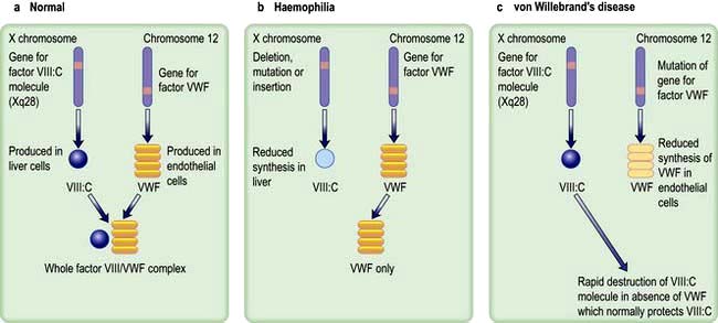
Factor VIII consists of a molecule with coagulant activity (VIII: C) associated with von Willebrand factor. Factor VIII increases the activity of factor IXa by ~200 000 fold. VWF functions to prevent premature factor VIII: C breakdown and locate it to areas of vascular injury. VIII: C has a molecular weight of about 350 000.

Von Willebrand factor (VWF) is a glycoprotein with a molecular weight of about 200 000 which readily forms multimers in the circulation with molecular weights of up to 20 × 106. It is synthesized by endothelial cells and megakaryocytes and stored in platelet granules as well as the endothelial cells. The high-molecular-weight multimeric forms of VWF are the most biologically active (see p. 422 and Fig. 8.39).

Physiological limitation of coagulation

Without a physiological system to limit blood coagulation dangerous thrombosis could ensue. The natural anticoagulant mechanism regulates and localizes thrombosis to the site of injury.

Antithrombin. Antithrombin (AT), a member of the serine protease inhibitor (serpin) superfamily, is a potent inhibitor of coagulation. It inactivates the serine proteases by forming stable complexes with them, and its action is greatly potentiated by heparin.

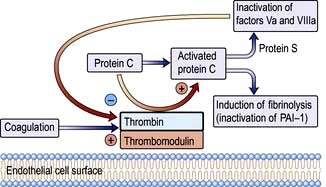
Activated protein C. This is generated from its vitamin K-dependent precursor, protein C, by thrombin; thrombin activation of protein C is greatly enhanced when thrombin is bound to thrombomodulin on endothelial cells (Fig. 8.36). Activated protein C inactivates factor V and factor VIII, reducing further thrombin generation.

image

Figure 8.36 Activation of protein C. PAI-1, plasminogen activator inhibitor 1.

Protein S. This is a cofactor for protein C, which acts by enhancing binding of activated protein C to the phospholipid surface. It circulates bound to C4b binding protein but some 30–40% remains unbound and active (free protein S).

Other inhibitors. Other natural inhibitors of coagulation include α2-macroglobulin, α1-antitrypsin and α2-antiplasmin.

Fibrinolysis

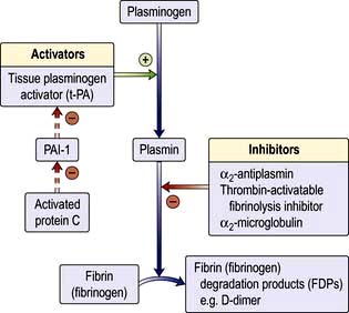
Fibrinolysis is a normal haemostatic response that helps to restore vessel patency after vascular damage. The principal component is the enzyme plasmin, which is generated from its inactive precursor plasminogen (Fig. 8.37). This is achieved principally via tissue plasminogen activator (t-PA) released from endothelial cells. Some plasminogen activation may also be promoted by urokinase, produced in the kidneys. Other plasminogen activators (factor XII and prekallikrein) are of minor physiological importance.

image

Figure 8.37 Fibrinolytic system. PA-1, plasminogen activator inhibitor-1.

Plasmin is a serine protease, which breaks down fibrinogen and fibrin into fragments X, Y, D and E, collectively known as fibrin (and fibrinogen) degradation products (FDPs). D-dimer is produced when cross-linked fibrin is degraded. Its presence in the plasma indicates that the coagulation mechanism has been activated.

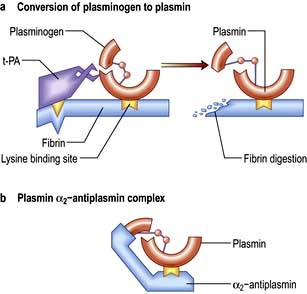
The fibrinolytic system is activated by the presence of fibrin. Plasminogen is specifically adsorbed to fibrin and fibrinogen by lysine-binding sites. However, little plasminogen activation occurs in the absence of polymerized fibrin, as fibrin also has a specific binding site for plasminogen activators, whereas fibrinogen does not (Fig. 8.38).

image

Figure 8.38 Fibrinolysis. (a) The conversion of plasminogen to plasmin by plasminogen activator (t-PA) occurs most efficiently on the surface of fibrin, which has binding sites for both plasminogen and t-PA. (b) Free plasmin in the blood is rapidly inactivated by α2-antiplasmin. Plasmin generated on the fibrin surface is partially protected from inactivation. The lysine-binding sites on plasminogen are necessary for the interaction between plasmin(ogen) and fibrin and between plasmin and α2-antiplasmin.

t-PA is inactivated by plasminogen activator inhibitor-1 (PAI-1). Activated protein C inactivates PAI-1 and therefore induces fibrinolysis (Fig. 8.36). Inactivators of plasmin, such as α2-antiplasmin (Fig. 8.38) and thrombin-activatable fibrinolysis inhibitor (TAFI), also contribute to the regulation of fibrinolysis.

Investigation of bleeding disorders

Although the precise diagnosis of a bleeding disorder will depend on laboratory tests, much information is obtained from the history and physical examination:

image Is there a generalized haemostatic defect? Supportive evidence for this includes bleeding from multiple sites, spontaneous bleeding, and excessive bleeding after injury.

image Is the defect inherited or acquired? A family history of a bleeding disorder should be sought. Severe inherited defects usually become apparent in infancy, while mild inherited defects may only come to attention later in life, for example with excessive bleeding after surgery, childbirth, dental extractions or trauma. Some defects are revealed by routine coagulation screens which are performed before surgical procedures.

image Is the bleeding suggestive of a vascular/platelet defect or a coagulation defect?

Vascular/platelet bleeding is characterized by easy bruising and spontaneous bleeding from small vessels. There is often bleeding into the skin. Purpura includes both petechiae, which are small skin haemorrhages varying from pinpoint size to a few millimetres in diameter and which do not blanch on pressure, and ecchymoses, which are larger areas of bleeding into the skin. Bleeding also occurs from mucous membranes especially the nose and mouth.

Coagulation disorders are typically associated with bleeding after injury or surgery, and in more severe forms, haemarthroses and muscle haematomas. There is often a short delay between the precipitating event and overt haemorrhage or haematoma formation.

Laboratory investigations

image Blood count and film show the number and morphology of platelets and any blood disorder such as leukaemia or lymphoma. The normal range for the platelet count is 150–400 × 109/L.

image Coagulation tests are performed using blood collected into citrate, which neutralizes calcium ions and prevents clotting.

The prothrombin time (PT) (also see p. 428) is measured by adding tissue factor (thromboplastin) and calcium to the patient’s plasma. The normal PT is 12–16 s and when used to measure oral anticoagulants is expressed as the international normalized ratio, INR (see p. 369). The PT measures VII, X, V, prothrombin and fibrinogen (classic ‘extrinsic’ pathway) and is prolonged with abnormalities of these factors. It may also be abnormal in liver disease, or if the patient is on warfarin.

The activated partial thromboplastin time (APTT) is also sometimes known as the PTT with kaolin (PTTK). It is performed by adding a surface activator (such as kaolin, micronized silica or ellagic acid), phospholipid (to mimic platelet membrane) and calcium to the patient’s plasma. The normal APTT is 26–37 s and depends on the exact methodology. The APTT measures XII, XI, IX, VIII, X, V, prothrombin and fibrinogen (classic ‘intrinsic’ pathway) and is prolonged with deficiencies of one or more of these factors. It is not dependent on factor VII.

The thrombin time (TT) is performed by adding thrombin to the patient’s plasma. The normal TT is 12–14 s, and it is prolonged with fibrinogen deficiency, qualitative defects of fibrinogen (dysfibrinogenaemia) or inhibitors such as heparin or FDPs.

Correction tests can be used to differentiate prolonged times in the PT, APTT and TT due to various coagulation factor deficiencies and inhibitors of coagulation. Prolonged PT, APTT or TT due to coagulation factor deficiencies can be corrected by addition of normal plasma to the patient’s plasma. Failure to correct after addition of normal plasma is suggestive of the presence of an inhibitor of coagulation.

Factor assays are used to confirm coagulation defects, especially where a single inherited disorder is suspected.

Special tests of coagulation will often be required to confirm the precise haemostatic defect. Such tests include estimation of fibrinogen and FDPs, platelet function tests such as platelet aggregation and platelet granule contents.

Bleeding time measures platelet plug formation in vivo. A sphygmomanometer cuff is inflated to 40 mmHg and 1 mm deep, 1 cm long incisions in the forearm are made with a template. Wounds are blotted every 30 s and the time taken for bleeding to stop recorded (normally 3–10 min). Prolonged bleeding times are found in patients with platelet function defects, and there is a progressive prolongation with platelet counts less than 100 × 109/L – hence the bleeding time should not be performed at low platelet counts. Nowadays it is rarely done as it can scar and is painful.

Vascular disorders

The vascular disorders (Table 8.22) are characterized by easy bruising and bleeding into the skin. Bleeding from mucous membranes sometimes occurs but the bleeding is rarely severe. Laboratory investigations including the bleeding time are normal. The vascular disorders include the following.

Table 8.22 Vascular disorders

Congenital

Hereditary haemorrhagic telangiectasia (Osler–Weber–Rendu disease)

Connective tissue disorders (Ehlers–Danlos syndrome, osteogenesis imperfecta, pseudoxanthoma elasticum, Marfan&rsquo;s syndrome)

Acquired

Severe infections:
Septicaemia
Meningococcal infections
Measles
Typhoid

Allergic

Henoch–Schönlein purpura
Autoimmune rheumatic disorders (SLE, rheumatoid arthritis)

Drugs

Steroids
Sulphonamides

Others

Senile purpura
Easy bruising syndrome
Scurvy
Factitious purpura

Hereditary haemorrhagic telangiectasia is a rare disorder with autosomal dominant inheritance. Mutations occur in most cases in one of three genes, ENG, ALK1 or SMAD4, that encode components of the TGF-β signalling pathway that is involved in blood vessel development. Dilatation of capillaries and small arterioles produces characteristic small red spots that blanch on pressure in the skin and mucous membranes, particularly the nose and gastrointestinal tract. Recurrent epistaxis and chronic gastrointestinal bleeding are the major problems which causes chronic iron deficiency anaemia. Vascular malformations also occur in pulmonary, hepatic cerebral and spinal vasculature.

Easy bruising syndrome is a common benign disorder occurring in otherwise healthy women. It is characterized by bruises on the arms, legs and trunk with minor trauma, possibly because of skin vessel fragility. It may give rise to the suspicion of a serious bleeding disorder.

Senile purpura and purpura due to steroids are both due to atrophy of the vascular supporting tissue.

Purpura due to infections is mainly caused by damage to the vascular endothelium. The rash of meningococcal septicaemia is particularly characteristic (see p. 127).

Henoch–Schönlein purpura (see p. 582) occurs mainly in children. It is a type III hypersensitivity (immune complex) reaction that is often preceded by an acute upper respiratory tract infection. Purpura is mainly seen on the legs and buttocks. Abdominal pain, arthritis, haematuria and glomerulonephritis also occur. Recovery is usually spontaneous, but some patients develop renal failure.

Episodes of inexplicable bleeding or bruising may represent abuse, either self-inflicted or caused by others. These various forms of artificial or factitious purpura may be expressions of emotional or psychiatric disturbances.

Platelet disorders

Bleeding due to thrombocytopenia or abnormal platelet function is characterized by purpura and bleeding from mucous membranes. Bleeding is uncommon with platelet counts above 50 × 109/L, and severe spontaneous bleeding is unusual with platelet counts above 20 × 109/L (Table 8.23).

Table 8.23 Clinical effects caused by different levels of platelet count

Platelet count (×109/L) Clinical defect

>500

Haemorrhage or thrombosis

500–100

No clinical effect

100–50

Moderate haemorrhage after injury

50–20

Purpura may occur

Haemorrhage after injury

<20

Purpura common

Spontaneous haemorrhage from mucous membranes

Intracranial haemorrhage (rare)

From Colvin BT. Medicine 2004; 32(5):27–33, with permission from Elsevier.

Thrombocytopenia

This is caused by reduced platelet production in the bone marrow, excessive peripheral destruction of platelets or sequestration in an enlarged spleen (Table 8.24). The underlying cause may be revealed by history and examination but a bone marrow examination will show whether the numbers of megakaryocytes are reduced, normal or increased, and will provide essential information on morphology. Specific laboratory tests may be useful to confirm the presence of such conditions as paroxysmal nocturnal haemoglobinuria (PNH) or systemic lupus erythematosus (SLE).

Table 8.24 Causes of thrombocytopenia

Impaired production

Selective megakaryocyte depression:
Rare congenital defects
Drugs, chemicals and viruses
As part of a general bone marrow failure:
Cytotoxic drugs and chemicals
Radiation
Megaloblastic anaemia
Leukaemia
Myelodysplastic syndromes
Myeloma
Myelofibrosis
Solid tumour infiltration
Aplastic anaemia
HIV infection

Excessive destruction or increased consumption

Immune
Autoimmune – ITP
Drug induced, e.g. GP IIb/IIIa inhibitors, pencillins, thiazides
Secondary immune (SLE, CLL, viruses, drugs, e.g. heparin, bivalirudin)
Alloimmune neonatal thrombocytopenia
Post-transfusion purpura
Disseminated intravascular coagulation
Thrombotic thrombocytopenic purpura

Sequestration

Splenomegaly
Hypersplenism

Dilutional

Massive transfusion

In patients with thrombocytopenia due to failure of production, no specific treatment may be necessary but the underlying condition should be treated if possible. Where the platelet count is very low or the risk of bleeding is very high, then platelet transfusion is indicated.

Immune thrombocytopenic purpura (ITP)

Thrombocytopenia is due to immune destruction of platelets. The antibody-coated platelets are removed following binding to Fc receptors on macrophages.

ITP in children

This occurs most commonly in age group 2–6 years. ITP has an acute onset with mucocutaneous bleeding and there may be a history of a recent viral infection, including varicella zoster or measles. Although bleeding may be severe, life-threatening hemorrhage is rare (~ 1%). Bone marrow examination is not usually performed unless treatment becomes necessary on clinical grounds.

ITP in adults

The presentation is usually less acute than in children. ITP is characteristically seen in women and may be associated with other autoimmune disorders such as SLE, thyroid disease and autoimmune haemolytic anaemia (Evans’ syndrome). It is also seen in patients with chronic lymphocytic leukaemia and solid tumours, and after infections with viruses such as HIV. Platelet autoantibodies are detected in about 60–70% of patients, and are presumed to be present, although not detectable, in the remaining patients; the antibodies often have specificity for platelet membrane glycoproteins IIb/IIIa and/or Ib.

Clinical features

Major haemorrhage is rare and is seen only in patients with severe thrombocytopenia. Easy bruising, purpura, epistaxis and menorrhagia are common. Physical examination is normal except for evidence of bleeding. Splenomegaly is rare.

Investigation

The only blood count abnormality is thrombocytopenia. Normal or increased numbers of megakaryocytes are found in the bone marrow (if examination is performed), which is otherwise normal. The detection of platelet autoantibodies is not essential for confirmation of the diagnosis, which often depends on exclusion of other causes of excessive destruction of platelets.

Treatment
Children

Children do not usually require treatment. Where this is necessary on clinical grounds, corticosteroids, intravenous immunoglobulin (i.v. IgG) and anti-D are effective; i.v. IgG is effective in >80% of children and raises the count more rapidly that steroids. Treatment should be reserved for very serious bleeding or urgent surgery. Chronic ITP is rare and requires specialist management.

Adults

Patients with platelet counts >30 × 109/L generally require no treatment unless they are about to undergo a surgical procedure. Patients with even lower platelet counts may not require treatment unless they have spontaneous bruising or bleeding.

First-line therapy consists of oral corticosteroids 1 mg/kg body weight. Approximately 66% will respond to prednisolone but relapse is common when the dose is reduced. Only 33% of patients can expect a long-term response and long-term remission is seen in only 10–20% of patients following stopping prednisolone. Patients who fail to respond to corticosteroids or require high doses to maintain a safe platelet count should be considered for splenectomy.

Intravenous immunoglobulin (i.v. IgG) is effective. It raises platelet count in 75% and in 50% the platelet count will normalize. Responses are only transient (3–4 weeks) with little evidence of any lasting effect. However, it is very useful where a rapid rise in platelet count is desired, especially before surgery. There are also advocates for high-dose corticosteroids for additional therapy.

Second-line therapy involves splenectomy, to which the majority of patients respond – two-thirds will achieve a normal platelet count. Patients who do not have a complete response can still expect some improvement.

Third-line therapy. For those that fail splenectomy, a wide range of other therapies are available. These include highdose corticosteroids, intravenous immunoglobulin, Rh0(D) immune globulin (anti-D), vinca alkaloids, danazol, immunosuppressive agents such as azathioprine, ciclosporin and dapsone, combination chemotherapy, mycophenolate mofetil. Major difficulties with many third-line therapies are modest response rates and slow onset of action. Consequently there is also interest in the use of specific immunomodulatory monoclonal antibodies such as rituximab, which yields a 60% response rate. Due to a relative lack of thrombopoietin in ITP, clinical trials of recombinant thrombopoietin have been undertaken. However, these were stopped because of thrombocytopenia arising due to neutralization of endogenous thrombopoietin by cross-reacting antibodies. Alternative drug development is now based on thrombopoietin receptor agonists that do not have any homology with native thrombopoietin. Two such drugs, eltrombopag and romiplostim, have been shown to significantly increase platelet count in ITP on a long-term basis and are approved drugs for refractory ITP. Platelet transfusions are reserved for intracranial or other extreme haemorrhage, where emergency splenectomy may be justified.

FURTHER READING

Imbach P, Crowther M. Thrombopoietin-receptor agonists for primary immune thrombocytopenia. N Engl J Med 2011; 365:734–741.

Other immune thrombocytopenias

Drugs cause immune thrombocytopenia by the same mechanisms as described for drug-induced immune haemolytic anaemia (see p. 400). The same drugs can be responsible for immune haemolytic anaemia, thrombocytopenia or neutropenia in different patients.

Heparin-induced thrombocytopenia. See page 427.

Neonatal alloimmune thrombocytopenia is due to fetomaternal incompatibility for platelet-specific antigens, usually for HPA-1a (human platelet alloantigen), and is the platelet equivalent of haemolytic disease of the newborn (HDN). The mother is HPA-1a-negative and produces antibodies, which destroy the HPA-1a-positive fetal platelets. Thrombocytopenia is self-limiting after delivery, but platelet transfusions may be required initially to prevent or treat bleeding associated with severe thrombocytopenia; platelets are prepared from HPA-1a-negative volunteers or the mother herself. Severe bleeding such as intracranial haemorrhage may also occur in utero.

Antenatal treatment of the mother – usually with high-dose IgG and/or steroids – has been effective in preventing haemorrhage in severely affected cases.

Post-transfusion purpura (PTP) is rare, occurring 7–10 days after a transfusion of platelet containing blood components, usually red cells. PTP is associated with a platelet-specific alloantibody, usually anti-HPA-1a in an HPA-1a-negative individual. PTP always occurs in patients previously immunized either by blood transfusion or by pregnancy – hence it is more common in women. The cause of the destruction of the patient’s own platelets is not well understood, but they may be destroyed as ‘bystanders’ during the acute immune response to HPA-1a. PTP is self-limiting, but intravenous IgG or plasma exchange may be required in severe bleeding

Thrombotic thrombocytopenic purpura (TTP) (see p. 590)

TTP is a rare, but very serious condition, in which platelet consumption leads to profound thrombocytopenia. There is a characteristic symptom complex of florid purpura, fever, fluctuating cerebral dysfunction and microangiopathic haemolytic anaemia with red cell fragmentation, often accompanied by acute kidney injury. The coagulation screen is usually normal but lactic dehydrogenase (LDH) levels are markedly raised as a result of haemolysis. TTP arises due to endothelial damage and microvascular thrombosis. This occurs due to a reduction in ADAMTS-13 (A Disintegrin-like and Metalloproteinase domain with Thrombospondin-type motifs), a protease which is normally responsible for regulating the size of VWF. ADAMTS-13 is needed to break down ultra large von Willebrand factor multimers (UL VWFMs) into smaller haemostatically active fragments that interact with platelets. Reduction in ADAMTS-13 results in the adhesion and aggregation of platelets to UL VWFMs and multiorgan microthrombi. In most sporadic cases there is a true deficiency of the ADAMTS-13, associated with antibodies to ADAMTS-13. In some congenital cases the deficiency is due to mutations in the ADAMTS-13 gene. Secondary causes of acute TTP include pregnancy, oral contraceptives, SLE, infection and drug treatment, including the use of ticlopidine and clopidogrel. Such cases may have a variable ADAMTS-13 activity at presentation and may or may not have associated antibodies to ADAMTS-13.

Treatment

Treatment consists of plasma exchange as the mainstay of treatment. It provides a source of ADAMTS-13 and removes associated autoantibody in acute TTP. Cryoprecipitate and solvent-detergent FFP (fresh frozen plasma) both contain ADAMTS-13. Pulsed intravenous methylprednisolone is given acutely, as is increasingly rituximab as a primary treatment of choice. Disease activity is monitored by measuring the platelet count and serum LDH. Platelet concentrates are contraindicated. The untreated condition has a mortality of up to 90% but modern management has reduced this figure to about 10%. Recurrent and relapsing TTP occurs, often associated with a persistent lack of ADAMTS-13. In secondary TTP cases, identifiable precipitating drugs should be stopped.

Platelet function disorders (Box 8.4)

These are usually associated with excessive bruising and bleeding and, in some of the acquired forms, with thrombosis. The platelet count is normal or increased and the bleeding time is prolonged. The rare inherited defects of platelet function require more detailed investigations such as platelet aggregation studies and factor VIII: C and VWF assays, if von Willebrand’s disease is suspected.

image Box 8.4

Inherited and acquired types of platelet dysfunction

Inherited

image Glanzmann’s thrombasthenia – lack of platelet membrane glycoprotein IIb–IIIa complex resulting in defective fibrinogen binding and failure of platelet aggregation.

image Bernard–Soulier syndrome – lack of platelet membrane glycoprotein Ib–IX–V complex (the binding site for VWF), causing failure of platelet adhesion and moderate thrombocytopenia.

image Storage pool disease – lack of the storage pool of platelet dense bodies, causing poor platelet function.

Acquired

image Myeloproliferative disorders

image Renal and liver disease

image Paraproteinaemias

image Drug-induced, such as NSAIDs (aspirin) or other platelet inhibitory drugs.

If there is serious bleeding or if the patient is about to undergo surgery, drugs with antiplatelet activity should be withdrawn and any underlying condition should be corrected if possible.

Bleeding in renal disease is multifactorial, although platelet dysfunction is a major component. The degree of the defect of haemostasis is broadly proportional to the plasma urea concentration – platelet function is impaired by urea, guanidinosuccinic acid and other phenolic metabolites that accumulate in chronic kidney disease. Dialysis partially corrects platelet function. The haematocrit should be increased to >0.30 and the use of desmopressin may be helpful. Platelet transfusions may be required if these measures are unsuccessful or if the risk of bleeding is high.

Thrombocytosis

The platelet count may rise above 400 × 109/L as a result of:

image Splenectomy

image Malignant disease

image Inflammatory disorders such as rheumatoid arthritis and inflammatory bowel disease

image Major surgery and post haemorrhage

image Myeloproliferative disorders

image Iron deficiency.

Thus, thrombocytosis is part of the acute-phase reaction, although following splenectomy platelet numbers are also elevated because of the loss of a major site of platelet destruction.

Essential thrombocythaemia, a myeloproliferative disorder which is described on page 404, and other myeloproliferative conditions such as polycythaemia vera (PV), myelofibrosis and chronic myeloid leukaemia (CML) may also be associated with a high platelet count.

A persistently elevated platelet count can lead to arterial or venous thrombosis. It is usual to treat the underlying cause of the thrombocytosis but a small dose of aspirin (75 mg) is also sometimes given. In myeloproliferative diseases the primary risk is thrombosis and specific action to reduce the platelet count, usually with hydroxycarbamide (hydroxyurea), is often taken. Paradoxically there is also a risk of abnormal bleeding if the platelet count is very high.

FURTHER READING

British Committee for Standards in Haematology. Guideline for the investigation and management of adults and children presenting with a thrombocytosis. Br J Haematol 2010; 149:352–375.

George JN. Management of immune thrombocytopenia – something old, something new. N Engl J Med 2010; 363:1959–1961.

Murphy MF, Bussel JB. Advances in the management of alloimmune thrombocytopenia. Br J Haematol 2007; 136:366–378.

Provan D, Stasi R, Newland AC, et al. International consensus report on the investigation and management of primary immune thrombocytopenia. Blood 2010; 115:168–186.

Inherited coagulation disorders

Inherited coagulation disorders are uncommon and usually involve deficiency of one factor only. Acquired coagulation disorders occur more frequently and almost always involve several coagulation factors (see p. 423).

In inherited coagulation disorders, deficiencies of all factors have been described. Those leading to abnormal bleeding are rare, apart from haemophilia A (factor VIII deficiency), haemophilia B (factor IX deficiency) and von Willebrand’s disease.

Haemophilia A

This is due to a lack of factor VIII. VWF is normal in haemophilia (Fig. 8.39). The prevalence of haemophilia A is about 1 in 5000 of the male population. It is inherited as an X-linked disorder. If a female carrier has a son, he has a 50% chance of having haemophilia, and a daughter has a 50% chance of being a carrier. All daughters of men with haemophilia are carriers and the sons are normal.

image

Figure 8.39 (a) Normal factor VIII synthesis. (b) Haemophilia A showing defective synthesis of factor VIIIc. (c) von Willebrand’s disease showing reduced synthesis of VWF.

Although a large number of different genetic defects have been found in the factor VIII gene, including deletions, duplications, frameshift mutations and insertions, in approximately 50% of families with severe disease, a common gene inversion in intron 22 is causative There is a high mutation rate, with one-third of cases being apparently sporadic with no family history of haemophilia.

Clinical and laboratory features

The clinical features depend on the level of factor VIII. The normal level of factor VIII is 50–150 IU/dL.

image Levels of less than 1 IU/dL (severe haemophilia) are associated with frequent spontaneous bleeding from early life, typically into joints and muscles. Such recurrent bleeding into joints leads to joint deformity and crippling if adequate treatment is not given.

image Levels of 1–5 IU/dL (moderate haemophilia) are associated with severe bleeding following injury and occasional spontaneous bleeds.

image Levels above 5 IU/dL (mild haemophilia) are associated usually with bleeding only after injury or surgery. Diagnosis in this group is often delayed until quite late in life.

With treatment, the most common causes of death in people with haemophilia are cancer and heart disease, as for the general population, although cerebral haemorrhage is much more frequent than in the general population. In recent years, HIV infection and liver disease (due to hepatitis C) have become a more common cause of death. These infections were acquired from blood transfusion by many patients that were treated with factor concentrates prior to 1986. Since 1986 such plasma-derived products are all virally inactivated with heat or chemicals.

The main laboratory features of haemophilia A are shown in Table 8.25. The abnormal findings are a prolonged APTT and a reduced level of factor VIII. The PT, bleeding time and VWF level are normal.

Table 8.25 Blood changes in haemophilia A, von Willebrand’s disease and vitamin K deficiency

image
Treatment

Bleeding is treated by administration of factor VIII concentrate by intravenous infusion to achieve normalization of levels. For surgery levels should be kept to normal levels until healing has occurred.

Factor VIII has a half-life of 12 h and therefore must be administered at least twice daily to maintain the required therapeutic level. Continuous infusion is sometimes used to cover surgery. Factor VIII concentrate is freeze-dried and available as a small volume infusion so facilitating treatment at home and allowing it to be administered by the patient immediately after bleeding has started, reducing the likelihood of chronic damage to joints and the need for inpatient care.

Factor VIII concentrate is available as plasma-derived and recombinant products. Recombinant products are the treatment of choice for people with haemophilia, but economic constraints often limit availability, particularly in developing countries.

To prevent recurrent bleeding into joints and subsequent joint damage, patients with severe haemophilia are given factor VIII infusions regularly three times per week. Such ‘prophylaxis’ treatment is usually started in early childhood (around 2 years of age).

Synthetic vasopressin (desmopressin – an analogue of vasopressin) – intravenous, subcutaneous or intranasal – produces a 3–5-fold rise in factor VIII and is very useful in patients with a baseline level of factor VIII >10 IU/dL. It avoids the complications associated with blood products and is useful for treating bleeding episodes in mild haemophilia and as prophylaxis before minor surgery. It is ineffective in severe haemophilia.

People with haemophilia should be registered at comprehensive care centres (CCC), which take responsibility for their full medical care, including social and psychological support.

Complications

Up to 30% of people with severe haemophilia will, during their lifetime, develop antibodies to factor VIII that inhibit its action. Such inhibitors usually develop after the first few treatment doses of factor VIII. The prevalence of inhibitors is, however, only 5–10% because inhibitory antibodies develop only rarely in moderate and mild haemophilia and often disappear spontaneously or with continued treatment.

Management of inhibitor patients is very difficult, as infused factor VIII is rapidly inactivated. Recombinant factor VIIa at very high ‘pharmacological’ levels can bypass factor IX/VIII activity and is an effective treatment in more than 80% of bleeding episodes in patients with high levels of inhibitor antibodies. Some prothrombin complex concentrates are also deliberately activated to produce factors, which also may ‘bypass’ the inhibitor and stop the bleeding.

The long-term aim of management is to eradicate the inhibitory antibody, particularly in those that have recently developed inhibitors. This is done using immune tolerance induction strategies, sometimes using additional immunosuppression and immunoabsorption.

Although a historical legacy of plasma-derived concentrates, the risk of viral transmission has been virtually eliminated (see p. 411)

Hepatitis A and B vaccination is offered routinely to all patients with haemophilia and von Willebrand’s disease. The clinical consequences of haemophilia patients infected with HIV are similar to other HIV-infected patients (see p. 175), except that Kaposi’s sarcoma does not occur. Similarly some hepatitis C infected patients progress to develop chronic liver disease and cirrhosis (see p. 323).

Carrier detection and antenatal diagnosis

Determination of carrier status in females begins with a family history and coagulation factor assays. Female carriers may have a low factor VIII level but the exact value is very variable, because of lyonization. Owing to this process early in embryonic life (that is, random inactivation of one chromosome; see p. 36), some carriers have very low levels of factor VIII while others will have normal levels. Carrier detection is carried out using molecular genetic testing/mutation analysis. Antenatal diagnosis may be carried out by molecular analysis of chorionic villus biopsy at 11–12 weeks’ gestation if selective termination is being considered or by third trimester amniocentesis if not.

Haemophilia B (Christmas disease)

Haemophilia B is caused by a deficiency of factor IX. The inheritance and clinical features are identical to haemophilia A, but the incidence is only about 1 in 30 000 males. It has been identified as the type of haemophilia affecting the Russian Royal Family. The half-life of factor IX is longer at 18 hours. Haemophilia B is treated with factor IX concentrates, recombinant factor IX being generally available, and prophylactic doses are given twice a week. Desmopressin is ineffective. Gene therapy may be effective in managing seven haemophilia B.

Von Willebrand’s disease (VWD)

In VWD, there is defective platelet function as well as factor VIII deficiency, and both are due to a deficiency or abnormality of VWF (Fig. 8.39). VWF plays a role in platelet adhesion to damaged subendothelium as well as stabilizing factor VIII in plasma (see p. 414).

The VWF gene is located on chromosome 12 and numerous mutations of the gene have been identified. VWD has been classified into three types:

image Type 1 is partial quantitative deficiency of VWF and significant type 1 VWD is usually inherited as an autosomal dominant.

image Type 2 is due to a qualitative abnormality of VWF, and it too is usually inherited as an autosomal dominant.

image Type 3 is recessively inherited and patients have virtually complete deficiency of VWF. Their parents are often phenotypically normal.

Many subtypes of VWD are described, particularly type 2 variants, which reflect the specific qualitative changes in the VWF protein.

FURTHER READING

Berntorp E, Shapiro AD. Modern haemophilia care. Lancet 2012; 379:1447−1456.

Nathwani AC, Tüddenham EG et al. Adenovirus-associated virus vector-mediated gene transfer in hemophilia B. N Engl J Med 2011; 365:2357−2365.

Clinical features

These are very variable. Type 1 and type 2 patients usually have relatively mild clinical features. Bleeding follows minor trauma or surgery, and epistaxis and menorrhagia often occur. Haemarthroses are rare. Type 3 patients have more severe bleeding but rarely experience the joint and muscle bleeds seen in haemophilia A.

Characteristic laboratory findings are shown in Table 8.25. These also include defective platelet aggregation with ristocetin.

Treatment

Treatment depends on the severity of the condition and may be similar to that of mild haemophilia, including the use of desmopressin where possible. Some plasma-derived factor VIII concentrates contain intact von Willebrand factor. These specific products are used to treat bleeding or to cover surgery in patients who require replacement therapy, such as those with type 3 (severe) VWD and those who do not respond adequately to desmopressin. Cryoprecipitate is used as a source of VWF, but should be avoided if possible since it is not virus inactivated.

Acquired coagulation disorders

Vitamin K deficiency (see also p. 207)

Vitamin K is necessary for the γ-carboxylation of glutamic acid residues on coagulation factors II, VII, IX and X and on proteins C and S. Without it, these factors cannot bind calcium.

Deficiency of vitamin K may be due to:

image inadequate stores, as in haemorrhagic disease of the newborn and severe malnutrition (especially when combined with antibiotic treatment) (see p. 208)

image malabsorption of vitamin K, a fat-soluble vitamin, which occurs in cholestatic jaundice owing to the lack of intraluminal bile salts

image oral anticoagulant drugs, which are vitamin K antagonists.

The PT and APTT are prolonged (Table 8.25) and there may be bruising, haematuria and gastrointestinal or cerebral bleeding. Minor bleeding is treated with phytomenadione (vitamin K1) 10 mg intravenously. Some correction of the PT is usual within 6 h but it may not return to normal for 2 days.

Newborn babies have low levels of vitamin K, and this may cause minor bleeding in the first week of life (classical haemorrhagic disease of the newborn). Vitamin K deficiency also causes late haemorrhagic disease of the newborn, which occurs 2–26 weeks after birth and results in severe bleeding such as intracranial haemorrhage. Most infants with these syndromes have been exclusively breast-fed, and both conditions are prevented by administering 1 mg i.m. vitamin K to all neonates (see p. 208). Concerns about the safety of this are unfounded.

FURTHER READING

Tripodi A, Mannucci. The coagulopathy of chronic liver disease. N Engl J Med 2011; 365:147–156.

Liver disease

Liver disease may result in a number of defects in haemostasis:

image Vitamin K deficiency. This occurs owing to intrahepatic or extrahepatic cholestasis.

image Reduced synthesis. Reduced synthesis of coagulation factors may be the result of severe hepatocellular damage. The use of vitamin K does not improve the results of abnormal coagulation tests, but it is generally given to ensure that a treatable cause of failure of haemostasis has not been missed.

image Thrombocytopenia. This results from hypersplenism due to splenomegaly associated with portal hypertension or from folic acid deficiency.

image Functional abnormalities. Functional abnormalities of platelets and fibrinogen are found in many patients with liver failure.

image Disseminated intravascular coagulation. DIC (see below) occurs in acute liver failure.

Disseminated intravascular coagulation (DIC)

Causes of DIC are listed in Box 8.5. There is widespread generation of fibrin within blood vessels, owing to activation of coagulation by release of procoagulant material, and by diffuse endothelial damage or generalized platelet aggregation. Activation of leucocytes, particularly monocytes causing expression of tissue factor and the release of cytokines, may play a role in the development of DIC. There is consumption of platelets and coagulation factors and secondary activation of fibrinolysis leading to production of fibrin degradation products (FDPs), which contributes to the coagulation defect by inhibiting fibrin polymerization (Fig. 8.40). The consequences of these changes are a mixture of initial thrombosis followed by a bleeding tendency due to consumption of coagulation factors and fibrinolytic activation.

image Box 8.5

Causes of DIC

image Malignant disease

image Septicaemia (e.g. Gram-negative including meningococcal)

image Haemolytic transfusion reactions

image Obstetric causes (e.g. abruptio placentae, amniotic fluid embolism, pre-eclampsia)

image Trauma, burns, surgery

image Other infections (e.g. falciparum malaria)

image Liver disease

image Snake bite

image

Figure 8.40 Disseminated intravascular coagulation. FDPs, fibrin degradation products.

Clinical features

The underlying disorder is usually obvious. The patient is often acutely ill and shocked. The clinical presentation of DIC varies from no bleeding at all to profound haemostatic failure with widespread haemorrhage. Bleeding may occur from the mouth, nose and venepuncture sites and there may be widespread ecchymoses.

Thrombotic events occur as a result of vessel occlusion by fibrin and platelets. Any organ may be involved, but the skin, brain and kidneys are most often affected.

Investigations

The diagnosis is often suggested by the underlying condition of the patient.

Severe cases with haemorrhage

image The PT, APTT and TT are usually very prolonged and the fibrinogen level markedly reduced.

image High levels of FDPs, including D-dimer, are found owing to the intense fibrinolytic activity stimulated by the presence of fibrin in the circulation.

image There is severe thrombocytopenia.

image The blood film may show fragmented red blood cells.

Mild cases without bleeding

image Increased synthesis of coagulation factors and platelets

image Normal PT, APTT, TT and platelet counts

image FDPs are raised.

Treatment

The underlying condition is treated and this is often all that is necessary in patients who are not bleeding. Maintenance of blood volume and tissue perfusion is essential. Transfusions of platelet concentrates, FFP, cryoprecipitate and red cell concentrates is indicated in patients who are bleeding. Inhibitors of fibrinolysis such as tranexamic acid should not be used in DIC as dangerous fibrin deposition may result. Activated protein C concentrates have been used in selected cases. In those cases with a dominant thrombotic component the use of heparin seems logical but there is little evidence to suggest any benefit.

Excessive fibrinolysis

Excessive fibrinolysis occurs during surgery involving tumours of the prostate, breast, pancreas and uterus owing to release of tissue plasminogen activators.

Primary hyperfibrinolysis is very rare but activation of fibrinolysis occurs in DIC as a secondary event in response to intravascular deposition of fibrin.

The clinical picture is similar to DIC with widespread bleeding. Laboratory investigations are also similar with a prolonged PT, APTT and TT, a low fibrinogen level, and increased FDPs, although fragmented red cells and thrombocytopenia are not seen, since disseminated coagulation is not present.

If the diagnosis is certain, fibrinolytic inhibitors such as epsilon-aminocaproic acid (EACA) or tranexamic acid can be given but evidence for efficacy is lacking.

Massive transfusion

Few platelets and reduced levels of clotting factors are found in stored blood, although there are adequate amounts of the other coagulation factors. During massive transfusion (defined as transfusion of a volume of blood equal to the patient’s own blood volume within 24 hours, e.g. >10 units in an adult), the platelet count and PT and APTT should be checked at intervals.

Transfusion of platelet concentrates and FFP should be given if thrombocytopenia or defective coagulation are thought to be contributing to continued blood loss. Other problems of massive transfusion are described in Chapter 16.

Inhibitors of coagulation

Factor VIII autoantibodies arise occasionally in patients without haemophilia but with autoimmune disorders such as SLE, in elderly patients, with malignant disease and sometimes after childbirth. There can be severe bleeding. Immediate bleeding problems are managed as with bypassing factor concentrates (see p. 422). Longer-term therapy is to eliminate the autoantibody using immunosuppression, such as steroids, cyclophosphamide and, in severe cases, rituximab.

Lupus anticoagulant antibodies (see p. 538) are autoantibodies directed against phospholipids (antiphospholipid antibodies) and lead to prolongation of phospholipid dependent coagulation tests, particularly the APTT, but do not inhibit coagulation factor activity.

FURTHER READING

George JN. Thrombocytopenic purpura. New England Journal of Medicine 2006; 354:1927–1935.

Schafer AI. Thrombocytosis. New England Journal of Medicine 2004; 350:1211–1219.

Wilde JT. Von Willebrand disease. Clinical Medicine 2007; 7:629–631.

Thrombosis

A thrombus is defined as a solid mass formed in the circulation from the constituents of the blood during life. Fragments of thrombi (emboli) may break off and block vessels downstream. Thromboembolic disease is much more common than abnormal bleeding; nearly half of adult deaths in England and Wales are due to coronary artery thrombosis, cerebral artery thrombosis or pulmonary embolism.

A thrombus results from a complex series of events involving coagulation factors, platelets, red blood cells and the vessel wall.

Arterial thrombosis

This usually occurs in association with atheroma, which tends to form at areas of turbulent blood flow such as the bifurcation of arteries. Platelets adhere to the damaged vascular endothelium and aggregate in response to ADP and TXA2 to form a ‘white thrombus’. The growth of the platelet thrombus is limited at its margins by PGI2 and NO. Plaque rupture leads to the exposure of blood containing factor VIIa to tissue factor within the plaque, which may trigger blood coagulation and lead to thrombus formation. This results in complete occlusion of the vessel or embolization that produces distal obstruction. The risk factors for arterial thrombosis are related to the development of atherosclerosis (see p. 725).

Arterial thrombi may also form in the heart, as mural thrombi in the left ventricle after myocardial infarction, in the left atrium in mitral valve disease, or on the surfaces of prosthetic valves.

Venous thrombosis

Unlike arterial thrombosis, venous thrombosis often occurs in normal vessels. Major causes are stasis and hypercoagulability. The majority of venous thrombi occur in the deep veins of the leg, originating around the valves as ‘red thrombi’ consisting mainly of red cells and fibrin. The propagating thrombus is formed of fibrin and platelets and is particularly liable to embolize. Chronic venous obstruction following thrombosis in the deep veins of the leg frequently results in a permanently swollen limb and may lead to ulceration (post-phlebitic syndrome).

Risk factors for venous thrombosis are shown in Table 8.26. Venous thrombosis may occur with changes in blood cells such as polycythaemia and thrombocythaemia, and with coagulation abnormalities (thrombophilia; see below).

Table 8.26 Risk factors for venous thromboembolism

Patient factors

Age
BMI >30 kg/m2
Varicose veins
Continuous travel >3 h in preceding 4 weeks
Immobility (bed rest ≥3 days)
Pregnancy and puerperium
Previous deep vein thrombosis or pulmonary embolism
Thrombophilia
Antithrombin deficiency
Protein C or S deficiency
Factor V Leiden
Resistance to activated protein C (caused by factor V Leiden variant)
Prothrombin gene variant
Hyperhomocysteinaemia
Antiphospholipid antibody/lupus anticoagulant
Oestrogen therapy including HRT
Dysfibrinogenaemia
Plasminogen deficiency

Disease or surgical procedure

Trauma or surgery, especially of pelvis, hip or lower limb
Malignancy
Cardiac or respiratory failure
Recent myocardial infarction or stroke
Acute medical illness/severe infection
Inflammatory bowel disease
Behçet’s disease
Nephrotic syndrome
Myeloproliferative disorders
Paroxysmal nocturnal haemoglobinuria
Paraproteinaemia
Sickle cell anaemia
Central venous catheter in situ

The clinical features and diagnosis of venous thrombosis are discussed on page 789.

Thrombophilia

Thrombophilia is a term describing inherited or acquired defects of haemostasis leading to a predisposition to venous or arterial thrombosis. It occurs in people with:

image Recurrent venous thrombosis

image Venous thrombosis for the first time under age 40 years

image An unusual venous thrombosis such as mesenteric or cerebral vein thrombosis

image Unexplained neonatal thrombosis

image Recurrent miscarriages

image Arterial thrombosis in the absence of arterial disease.

Coagulation abnormalities
Factor V Leiden

Factor V Leiden differs from normal factor V by a single nucleotide substitution (Arg506Gln). This variation makes factor V less likely to be cleaved by activated protein C. As factor V is a cofactor for thrombin generation (Fig. 8.35 impaired inactivation by activated protein C (Fig. 8.36 results in a tendency to thrombosis. Factor V Leiden is found in 3–5% of healthy individuals in the western world and in about 20–30% of patients with venous thrombosis.

Factor V Leiden acts synergistically with other acquired thrombosis risk factors, for example in those taking oral contraceptive pills or when pregnant. Thrombosis risk rises 35-fold in those on a combined oral contraceptive pill although the absolute risk for thrombosis remains at significantly <0.5% per year for any single individual.

Prothrombin variant

A mutation in the 3′ untranslated region of the prothrombin gene has been described (G20210A). This variant is associated with elevated levels of prothrombin and a 2–3-fold increase in the risk of venous thrombosis. There is an interaction with factor V Leiden and contraceptive pill use or pregnancy. The prevalence is 2% in Caucasian populations, 6% in unselected patients with thrombosis.

Antithrombin (AT) deficiency

This deficiency can be inherited as an autosomal dominant. Many variations have been described that lead to a conformational change in the protein. It can also be acquired following trauma, with major surgery and with the contraceptive pill. Low levels are also seen in severe proteinuria (e.g. the nephrotic syndrome). Recurrent thrombotic episodes occur starting at a young age in the inherited variety. Patients may be relatively resistant to heparin as antithrombin is required for its action. Antithrombin concentrates are available.

Protein C and S deficiency

These autosomal dominant conditions result in an increased risk of venous thrombosis, often before the age of 40 years. Homozygous protein C or S deficiency causes neonatal purpura fulminans, which is fatal without immediate replacement therapy. Protein C concentrate and a recombinant activated protein C are available.

Antiphospholipid antibody

See page 538.

Homocysteine

When elevated, this amino acid is associated with both arterial thrombosis and venous thromboembolism. The mechanism of vascular damage is unclear. Folate, B12 and B6 supplementation are often helpful in reducing levels.

Investigations

Haemostatic screening test

image Full blood count including platelet count

image Coagulation screen including a fibrinogen level. These tests will detect erythrocytosis, thrombocytosis, and dysfibrinogenaemia and the possible presence of a lupus anticoagulant.

Testing for specific causes of thrombophilia

image Assays for naturally occurring anticoagulants such as AT, protein C and protein S

image Assay for activated protein C resistance and molecular testing for factor V Leiden and the prothrombin variant

image Screen for a coagulation factor inhibitor including a lupus anticoagulant (and anticardiolipin antibodies) (see p. 538).

Prevention and treatment of arterial thrombosis

Attempts to prevent or reduce arterial thrombosis are directed mainly at minimizing factors predisposing to atherosclerosis. Treatment of established arterial thrombosis includes the use of antiplatelet drugs and thrombolytic therapy.

Antiplatelet drugs

Platelet activation at the site of vascular damage is crucial to the development of arterial thrombosis, and this can be altered by the following drugs (Table 8.27):

image Aspirin irreversibly inhibits the enzyme cyclo-oxygenase (COX), resulting in reduced platelet production of TXA2 (Fig. 8.34). At the low doses used in cardiovascular disease prevention or treatment, there is selective inhibition of the isoform COX-1 found within platelets. This inhibition cannot be repaired and is effective for the life of the circulating platelet, which is about 1 week. In recent years, it has been suggested there may be significant individual variability in the response to aspirin, although there is no clear reason for this. The term ‘aspirin resistance’ has been loosely applied when the clinical effects of aspirin are less than expected. No large body of clinical trial data is specifically available to correlate clinical events and laboratory findings with respect to aspirin response and so it is difficult to determine if the breakthrough events experienced by patients treated with aspirin represent aspirin resistance or are related to more mundane issues such as aspirin dose, drug interactions or drug non-compliance.

image Dipyridamole – which inhibits platelet phosphodiesterase, causing an increase in cyclic AMP with potentiation of the action of PGI2 – has been used widely as an antithrombotic agent, but there is little evidence that it is effective.

image Clopidogrel – irreversibly blockades the ADP (P2Y12) receptor on platelet cell membranes, so affecting the ADP-dependent activation of the glycoprotein IIb/IIIa complex. It is similar to ticlopidine but has fewer side-effects. Trials support its use in acute coronary syndromes (see p. 735).

image Prasugrel, a novel thienopyridine, is like clopidogrel and is licensed for use in acute coronary syndromes (see p. 735).

image Glycoprotein IIb/IIIa receptor antagonists block the receptor on the platelet for fibrinogen and von Willebrand factor (Fig. 8.41). Three classes have been described:

murine–human chimeric antibodies (e.g. abciximab)
synthetic peptides (e.g. eptifibatide)
synthetic non-peptides (e.g. tirofiban).

Table 8.27 Drugs used in the treatment of thrombotic disorders

Antiplatelet

Aspirin
Thromboxane synthase, e.g. dipyridamole
GP IIb/IIIa inhibitors, e.g. abciximab, eptifibatide, tirofiban
ADP receptor antagonists/P2Y12 inhibitors, e.g. clopidogrel, prasugrel, ticagrelor
Epoprostenol
Thromboxane prostaglandin receptor antagonists, e.g. terutroban

Thrombolytic

Streptokinase
Tissue-type plasminogen activator (t-PA or alteplase)
Reteplase (r-PA)
Tenecteplase (TNK-tPA)

Anticoagulant

Heparin:
Unfractionated (or standard)
Low molecular weight
Hirudin-like, e.g. lepirudin, bivalirudin
Fondaparinux
Warfarin
Bivalirudin
Xa inhibitors, e.g. apixaban, rivaroxaban, otamixaban, edoxaban, betrixaban
Direct thrombin inhibitors, e.g. dabigatran
image

Figure 8.41 The role of glycoprotein IIb/IIIa in platelet aggregation and the inhibition of platelet aggregation by inhibitors of glycoprotein IIb/IIIa receptors.

They have been used as an adjunct in invasive coronary artery intervention and as primary medical therapy in coronary heart disease. Excessive bleeding has been a problem.

image Epoprostenol is a prostacyclin, which is used to inhibit platelet aggregation during renal dialysis (with or without heparin) and is also used in primary pulmonary hypertension.

image Terutoban is a thromboxane prostaglandin receptor antagonist which is being trialled in secondary prevention of cerebrovascular and cardiovascular disease as an alternative to aspirin.

The indications for and results of antiplatelet therapy are discussed in the appropriate sections (p. 735).

Thrombolytic therapy

Streptokinase

Streptokinase is a purified fraction of the filtrate obtained from cultures of haemolytic streptococci. It forms a complex with plasminogen, resulting in a conformational change, which activates other plasminogen molecules to form plasmin. Streptokinase is antigenic and the development of streptococcal antibodies precludes repeated use. Activation of plasminogen is indiscriminate so that both fibrin in clots and free fibrinogen are lysed, leading to low fibrinogen levels and the risk of haemorrhage.

Plasminogen activators

Tissue-type plasminogen activators (alteplase (t-PA), tenecteplase (TNK-tPA)) are produced by recombinant technology. Reteplase (r-PA) is also a recombinant plasminogen activator. They are not antigenic and do not give allergic reactions. They are relatively fibrin-specific, have relatively little systemic activity, and short half-lives (~5 min). The bleeding complications observed are similar in severity and frequency to those observed with streptokinase, suggesting that fibrin specificity does not confer protection against hemorrhage.

Indications

The use of thrombolytic therapy in myocardial infarction is discussed on page 739. The combination of aspirin with thrombolytic therapy produces better results than thrombolytic therapy alone. The extent of the benefit depends on how quickly treatment is given. They are also used in cerebral infarction (see p. 1102) and in massive pulmonary embolism where there is haemodynamic instability. The main risk of thrombolytic therapy is bleeding. Treatment should not be given to patients who have had recent bleeding, uncontrolled hypertension or a haemorrhagic stroke, or surgery or other invasive procedures within the previous 10 days.

Prevention and treatment of venous thromboembolism

Venous thromboembolism is a common problem after surgery, particularly in high-risk patients such as the elderly, those with malignant disease and those with a history of previous thrombosis (Table 8.28). The incidence is also high in patients confined to bed following trauma, myocardial infarction or other illnesses. The prevention and treatment of venous thrombosis includes the use of anticoagulants.

Table 8.28 Risk assessment for deep vein thrombosis and pulmonary embolism for hospital patients (NICE CG92)

Patients who are at risk of VTE

Medical patients

Surgical patients and patients with trauma

If mobility significantly reduced for ≥3 days or
If expected to have ongoing reduced mobility due to normal state plus any VTE risk factor

If total anaesthetic + surgical time >90 min or

If surgery involves pelvis or lower limb and total anaesthetic + surgical time >60 min or

If acute surgical admission with inflammatory or intra-abdominal condition or

If expected to have significant reduction in mobility or

If any VTE risk factor present

VTE risk factorsa

Active cancer or cancer treatment

Age >60 years

Critical care admission

Dehydration

Known thrombophilias

Obesity (BMI >30 kg/m2)

One or more significant medical co-morbidities (e.g. heart disease, metabolic endocrine or respiratory pathologies, acute infectious diseases, inflammatory conditions)

Personal history or 1st-degree relative with a history of VTE

Use or HRT

Use of oestrogen-containing contraceptive therapy

Varicose veins with phlebitis

Patients who are at risk of bleeding

All patients who have any of the following:

Active bleeding

Acquired bleeding disorders (such as acute liver failure)

Concurrent use of anticoagulants known to increase the risk of bleeding (such as warfarin with INR >2)

Lumbar puncture/epidural/spinal anaesthesia within the previous 4 h or expected within the next 12 h

Acute stroke

Thrombocytopenia (platelets <75 × 109/L

Uncontrolled systolic hypertension (≥230/120 mmHg)

Untreated inherited bleeding disorders (such as haemophilia or von Willebrand’s disease)

a For women who are pregnant or have given birth within the previous 6 weeks, see recommendations 1.6.4–1.6.6 in NICE guideline CG92.

Anticoagulants

Heparin (standard or unfractionated)

Heparin is not a single substance but a mixture of polysaccharides. Commercially available unfractionated heparin consists of components with molecular weights varying from 5000 to 35 000, with an average of about 13 000. It was initially extracted from liver (hence its name) but it is now prepared from porcine gastric mucosa. Heparin acts immediately, binding to antithrombin. This induces a conformational change which increases the inhibitory activity of antithrombin (at least 5000-fold) towards activated serine protease coagulation factors (thrombin, XIIa, XIa, Xa, IXa and VIIa).

Low-molecular-weight heparins (LMW heparins)

These are produced by enzymatic or chemical degradation of standard heparin, producing fractions with molecular weights in the range of 2000–8000. Potentiation of thrombin inhibition (anti-IIa activity) requires a minimum length of the heparin molecule with an approximate molecular weight of 5400, whereas the inhibition of factor Xa requires only a smaller heparin molecule with a molecular weight of about 1700. LMW heparins have the following properties:

image Bioavailability is better than that of unfractionated heparin.

image They have greater activity against factor Xa than against factor IIa, suggesting that they may produce an equivalent anticoagulant effect to standard heparin but have a lower risk of bleeding, although this has not generally been confirmed. In addition, LMW heparins cause less inhibition of platelet function.

image They have a longer half-life than standard heparin and so can be given as a once-daily subcutaneous injection instead of every 8–12 h.

image They produce little effect on tests of overall coagulation, such as the APTT at doses recommended for prophylaxis. They are not fully neutralized by protamine.

LMW heparins are excreted renally and therefore dose reductions are required in those with renal impairment.

LMW heparins are widely used for antithrombotic prophylaxis, e.g. high-risk surgical patients and for the treatment of established thrombosis (see p. 747).

The main complication of all heparin treatment is bleeding. This is managed by stopping heparin. Very occasionally it is necessary to neutralize unfractionated heparin with protamine. Other complications include osteoporosis with prolonged therapy and thrombocytopenia.

Heparin-induced thrombocytopenia (HIT). HIT is an uncommon complication of heparin therapy and usually occurs 5–14 days after first heparin exposure. It is due to an immune response directed against heparin/platelet factor 4 complexes. All forms of heparin have been implicated but the problem occurs less often with LMW heparins.

HIT is paradoxically associated with severe thrombosis and when diagnosed all forms of heparin must be discontinued, including heparin flush. Unfortunately the diagnosis can be difficult to make because patients on heparin are often very sick and may be thrombocytopenic for many other reasons. Laboratory tests based on bioassay or immunoassay are available but are neither sensitive nor specific and management decisions often have to be made before results are available.

It is necessary to continue some form of anticoagulation in patients with HIT and the choice lies between the heparinoid danaparoid and the direct thrombin inhibitor hirudin. The introduction of warfarin should be covered by one of these agents, as warfarin alone may exacerbate thrombosis as protein C levels fall.

Fondaparinux

This is a synthetic pentasaccharide, which inhibits activated factor X, similar to the LMW heparins. It is used in acute coronary syndrome (see p. 736). A long-acting version, idraparinux, which only needs to be given weekly is also available. Neither bind to platelet factor 4 and so have no capacity to cause HIT.

Direct thrombin inhibitors

A recombinant form of hirudin, lepirudin, is available. Hirudins bind directly to thrombin and are effectively irreversible inhibitors. They can be monitored by the use of the APTT and are excreted by the kidney, so must be used with caution in chronic kidney disease. Lepirudin is used for anticoagulation in patients with HIT.

Bivalirudin is a 20 amino acid synthetic analogue of hirudin. Compared with hirudin, it appears to cause less bleeding, is a reversible thrombin inhibitor (as it is broken down by thrombin) and has a shorter half-life. It is used in percutaneous coronary interventions.

Oral anticoagulants

These act by interfering with vitamin K metabolism. There are two types of oral anticoagulants, the coumarins and indanediones. The coumarin warfarin is most commonly used because it has a low incidence of side-effects other than bleeding.

The dosage is controlled by prothrombin tests (PT). Thromboplastin reagents for PT testing are derived from a variety of sources and give different PT results for the same plasma.

It is standard practice to compare each thromboplastin with an international reference preparation so that it can be assigned an international sensitivity index (ISI). The international normalized ratio (INR) is the ratio of the patient’s PT to a normal control when using the international reference preparation. Therapeutic ranges using the INR for warfarin in various conditions are shown in Box 8.6.

image Box 8.6

Indications for warfarin and target INR

Target INR  

2.5

Pulmonary embolism, proximal and calf deep vein thrombosis, recurrence of venous thromboembolism when no longer on warfarin therapy, symptomatic inherited thrombophilia, atrial fibrillation, cardioversion, mural thrombus, cardiomyopathy

3.5

Recurrence of venous thromboembolism while on warfarin therapy, antiphospholipid syndrome, mechanical prosthetic heart valve, coronary artery graft thrombosis

British Society for Haematology (2000).

Each laboratory can use a chart adapted to the ISI of their thromboplastin to convert the patient’s PT to the INR. Suitably selected control plasmas can also be used to achieve the same objective. The use of this system means that PT tests on a given plasma sample using different thromboplastins result in the same INR and that anticoagulant control is comparable in different hospitals across the world.

Contraindications to the use of oral anticoagulants are seldom absolute and include:

image Severe uncontrolled hypertension

image Non-thromboembolic strokes

image Peptic ulceration (unless cured by Helicobacter pylori eradication)

image Severe liver and renal disease

image Pre-existing haemostatic defects

image Non-compliance.

Warfarin should be avoided in pregnancy because they are teratogenic in the first trimester and may be associated with fetal haemorrhage later in pregnancy. When anticoagulation is considered essential in pregnancy, self-administered subcutaneous heparin should be used as an alternative, although this may not be as effective for women with prosthetic cardiac valves. Specialist advice should be sought.

Many drugs interact with warfarin (see Ch. 17). More frequent PT testing should accompany changes in medication, which should occur with the full knowledge of the anticoagulant clinic.

An increased anticoagulant effect due to warfarin is usually produced by one of the following mechanisms:

A decreased anticoagulant effect due to warfarin This is usually produced by drugs that increase the clearance of warfarin by induction of hepatic enzymes that metabolize warfarin, such as rifampicin and barbiturates.

image Drugs causing a reduction in the metabolism of warfarin, including tricyclic antidepressants, cimetidine, sulphonamides, phenothiazines and amiodarone

image Drugs such as clofibrate and quinidine which increase the sensitivity of hepatic receptors to warfarin

image Drugs interfering with vitamin K absorption (such as broad-spectrum antibiotics and cholestyramine) which also potentiate the action of warfarin

image Displacement of warfarin from its binding site on serum albumin by drugs such as sulphonamides (this is not usually responsible for clinically relevant interactions)

image Drugs that inhibit platelet function (such as aspirin) which increase the risk of bleeding

image Alcohol excess, cardiac failure, liver or renal disease, hyperthyroidism and febrile illnesses which result in potentiation of the effect of warfarin.

Anticoagulant related bleeding. Bleeding is the most serious side-effect of warfarin. Bleeding occurs in up to 4% of patients on oral anticoagulants per year, requires hospital admission in 2% and has a 0.25% morbidity associated with it. The benefit of anticoagulants must therefore be notably more than the risk of bleeding. Management of warfarin related bleeding (Emergency Box 8.1) is given dependent upon the INR and the degree of bleeding. Minor bleeding may be treated with cessation of warfarin alone, while serious bleeding will require additional use of vitamin K and factor concentrates.

image Emergency Box 8.1

Management of warfarin-related bleeding and excessive oral anticoagulation

INR >3.0 <6.0 (target INR 2.5)

(1) Reduce warfarin dose or stop

INR >4.0 <6.0 (target INR 3.5)

(2) Restart warfarin when INR <5.0

INR >6.0 <8.0 no bleeding or minor bleeding

(1) Stop warfarin

(2) Restart when INR <5.0

INR >8.0, no bleeding or minor bleeding

(1) Stop warfarin

(2) Restart warfarin when INR <5.0

(3) If other risk factors for bleeding give 0.5–2.5 mg of vitamin K (oral)

Major bleeding

(1) Stop warfarin

(2) Give prothrombin complex concentrate 50 U/kg (FFP 15 mL/kg if concentrate not available)

(3) Give 5 mg of vitamin K (oral or i.v.)

If unexpected bleeding occurs investigate the possibility of a local anatomical cause.

Modified from British Society for Haematology (1998).

FURTHER READING

Henegan C et al. Self-monitoring of oral anticoagulation: systematic review and meta-analysis of individual patient data. Lancet 2012; 379:322−324.

New orally active anticoagulants

A large number of orally active direct thrombin (e.g. dabigatran) and Xa inhibitor drugs (e.g. rivaroxaban, apixaban) have been introduced for the treatment and prevention of venous and arterial thrombosis. Such drugs have a much broader therapeutic window than warfarin and offer the prospect of fixed drug dosing without the need to monitor coagulation. They do not however have specific antidotes. Dagibatran, apixiban and rivaroxaban are licensed for prevention of thrombosis in hip and knee replacement surgery as well as for the prevention of stroke in atrial fibrillation. Rivaroxaban is also licensed for the treatment of VTE. Large-scale studies in various aspects of thrombosis treatment and prevention are being undertaken using such drugs. These drugs, if clinical trial data is encouraging, will replace warfarin in a significant number of patients as they will be simpler to administer (no blood monitoring required) and as safe as, or safer than, warfarin with regard to bleeding.

Prophylaxis to prevent venous thromboembolism (VTE)

Risk factors for VTE are well defined. Most hospitalized patients have one or more of these risk factors and VTE is common in hospitalized patients. The risk of developing deep vein thrombosis (DVT) after hip replacement surgery has been estimated to be as high as 50% when thromboprophylaxis is not used. Approximately 10% of hospital deaths may be due to pulmonary embolism (PE) and more people die from hospital-acquired venous thrombosis than the combined deaths from road traffic accidents, AIDS and breast cancer. PE is the most common preventable cause of hospital death.

Appropriate thromboprophylaxis is highly effective and cost effective. Such prophylactic measures include early mobilization, elevation of the legs, compression stockings, intermittent compression devices and use of anticoagulant drugs, such as LMW heparins and thrombin inhibitors. All patients, medical and surgical, admitted to hospital should be risk assessed for thrombotic risk and given appropriate thromboprophylaxis. National guidelines are available to guide appropriate management (NICE CG92). (See Table 8.28.)

Low-risk patients (Table 8.28) require no specific measures other than early mobilization.

High-risk patients based on risk assessment are most effectively managed using graduated compression stockings and LMW heparin subcutaneously daily.

The antithrombin agents dabigatran and rivaroxaban are routinely used after lower limb joint replacement surgery. They are as effective as LMW heparins and, as they are given orally, can be used for extended periods out of hospital.

Treatment of established venous thromboembolism

image The aim of anticoagulant treatment is to prevent further thrombosis and pulmonary embolization while resolution of venous thrombi occurs by natural fibrinolytic activity. Anticoagulation is started with heparin as it produces an immediate anticoagulant effect. Heparin should be administered for approximately 5 days, the time taken for simultaneously administered warfarin to produce an anticoagulant effect (INR 2.5).

image LMW heparin (e.g. tinzaparin 175 U/kg daily, dalteparin 200 U/kg daily, enoxaparin 1.5 mg/kg daily) is equally effective and as safe as unfractionated heparin in the immediate treatment of deep vein thrombosis and pulmonary embolism. This creates the opportunity for treatment of venous thromboembolism without admission to hospital, in compliant patients without co-existing risk factors for haemorrhage.

image Length of anticoagulation. This is recommended for at least 6 weeks after precipitated isolated calf vein thrombosis and at least 3 months after precipitated proximal DVT or PE in patients who have temporary risk factors. For patients with idiopathic VTE or permanent risk factors at least 3 months’ anticoagulation is recommended and consideration should be given to indefinite anticoagulation.

image Use of longer-term anticoagulation in patients with previous thrombosis. It has been suggested that a lower INR might be safer and equally effective but the current view is that the target INR should be 2.0–3.0 where oral anticoagulation is used. Indefinite anticoagulation is considered appropriate for those with two or more episodes of VTE.

image Outpatient anticoagulation is best supervised in anticoagulant clinics. Patients are issued with national booklets for recording INR results and anticoagulant doses. Home monitoring is possible in well-motivated patients.

Inferior vena caval filters are an important tool to prevent PE in patients that have a contraindication to anticoagulation. Many are now retrievable allowing removal once a temporary contraindication to anticoagulation has passed. Long-term use of an IVC filter is associated with a risk of thrombosis at and below the site of the filter.

The role of thrombolytic therapy in the treatment of venous thrombosis is not established. It is used in patients with massive pulmonary embolism who are haemodynamically unstable and in patients with extensive deep venous thrombi.

Thrombolytic therapy should be followed by anticoagulation with heparin for a few days and then by oral anticoagulants to prevent rethrombosis.

Bibliography

Baglin T, Barrowcliffe A, Greaves M. The British Committee for Standards in Haematology. Guidelines on the use and monitoring of heparin 2006. Br J Haematol. 2006;133:19–34.

Baglin TP, Cousins D, Keeling DM, et al. Recommendations from the British Committee for Standards in Haematology and National Patient Safety Agency. Br J Haematol. 2006;136:26–29.

Baglin TP, Gray E, Greaves M, et al. Guidelines for testing for heritable thrombophilia. Br J Haematol. 2010;149:209–220.

British Society for Haematology. Guidelines for the diagnosis and management of disseminated intravascular coagulation. Br J Haematol. 2009;145:24–33.

Kearon C, et al. Antithrombotic therapy for VTE disease: Antithrombotic Therapy and Prevention of Thrombosis, 9th ed: American College of Chest Physicians Evidence-based Clinical Practice Guidelines. Chest. 2012;141(2 Suppl):e419S–494S.

Keeling D, Baglin T, Tait C, et al. British Committee for Standards in Haematology. Guidelines on oral anticoagulation with warfarin − fourth edition. Br J Haematol. 2011;154(3):311–324.

Keeling D, Davidson S, Watson H. The Haemostasis and Thrombosis Task Force of the British Committee for Standards in Haematology. Management of heparin-induced thrombocytopenia. Br J Haematol. 2006;133:259–269.

Provan AB, Stasi R, Newland AC, et al. International consensus report on the investigation and management of primary immune thrombocytopenia. Blood. 2010;115:168–186.

Significant websites

http://www.bcshguidelines.com

British Society for Haematology guidelines

http://www.blood.co.uk

UK National Blood Service

http://www.bloodline.net

General website on haematology

http://www.betterblood.org.uk

UK CMO’s Better Blood Transfusion Conference

http://www.hemophilia.org

US National Hemophilia Foundation

http://www.isth.org/default/index.cfm

International Society on Thrombosis and Haemostasis (ISTH)

http://www.shotuk.org

Serious Hazards of Transfusion (SHOT) scheme, covering UK and Ireland NHS and private hospitals, affiliated to the Royal College of Pathologists (based at the Manchester Blood Transfusion Centre)

http://www.transfusion.org

Journal of the American Association of Blood Banks

http://www.transfusionguidelines.org.uk

UK Blood Transfusion and Tissue Transplantation Services Professional Guidelines

http://www.wfh.org

World Federation of Hemophilia