Other Extracellular Accumulations

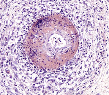
Fibrinoid Change: Fibrinoid change, also known as fibrinoid necrosis and fibrinoid degeneration, is a term applied to a pattern of lesions most often observed in the vascular system. The terms fibrinoid degeneration and fibrinoid necrosis are inappropriate because the process is not a true regressive alteration of cells. Rather, fibrinoid change is the result of the deposition of immunoglobulin, complement, and/or plasma proteins, including fibrin in the wall of a vessel. This lesion is caused by injury to the intima and media such as occurs in the immune-mediated vasculitides.

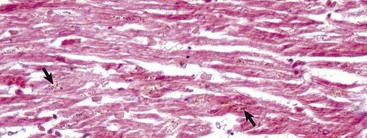
Grossly, fibrinoid change cannot be observed; however, it is often accompanied by thrombosis and hemorrhage, and when these two lesions are present in a vascular pattern of distribution, fibrinoid change of the vasculature should be considered. Microscopically, direct injury to endothelial cells, basement membrane, or myocytes, such as caused by viruses or toxins, or indirect injury such as caused by activation of complement proteins, can lead to activation of the acute inflammatory cascade and the deposition of plasma proteins in the vessel walls. These proteins, especially fibrin, stain intensely red (eosinophilic) with H&E stains and involve the vessel wall circumferentially to varying depths of the tunica intima and tunica media (Fig. 1-52). This lesion is also often accompanied by cellular and nuclear debris from injured vascular cells and inflammatory cells. These proteins contribute to the vascular “eosinophilia,” which has been described somewhat differently by different pathologists. There is general agreement that the material is eosinophilic, which is sometimes described as “smudgy” or “deeply eosinophilic.” Some pathologists add “homogeneous” and others “amorphous” to the descriptive terminology of fibrinoid change.

image

Fig. 1-52 Fibrinoid change, artery.
Note the deeply eosinophilic circumferential band in the tunica media of this artery. It is accompanied by acute inflammation and necrosis. H&E stain. (Courtesy Dr. J.F. Zachary, College of Veterinary Medicine, University of Illinois.)

Gout: Gout is the deposition of sodium urate crystals or urates in tissue. It occurs in humans, birds, and reptiles but has not been reported in domestic mammals.

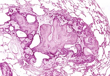
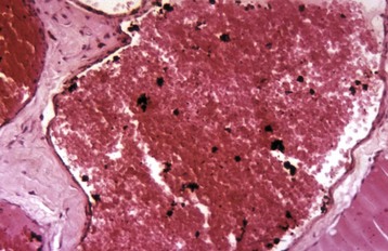
In the most common form in humans, urate crystals are deposited in the articular and periarticular tissues and elicit an acute inflammatory response characterized by the presence of neutrophils and macrophages and large aggregations of urate crystals called tophi. These tophi may be visible grossly and are pathognomonic of gout. Later in the course of the disease, the inflammation becomes chronic and a foreign body reaction to the tophi develops. Microscopically, urate crystals are acicular and birefringent and they, or the spaces left after they have been dissolved during the preparation of paraffin-embedded histologic sections, are visibly surrounded by numerous neutrophils, macrophages, and giant cells.

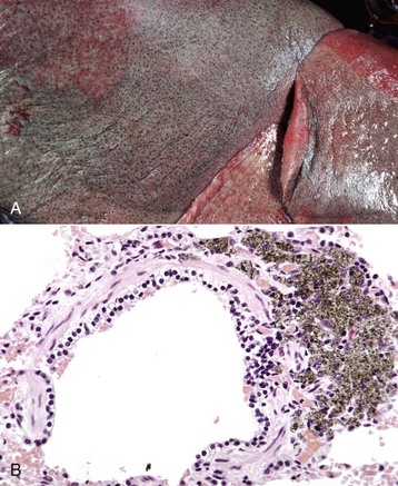
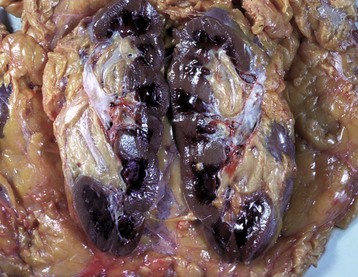
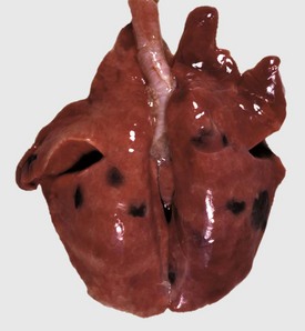
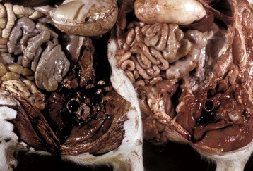
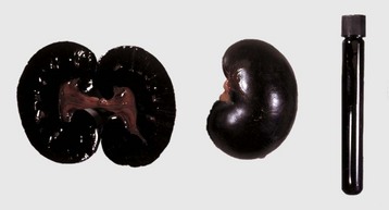
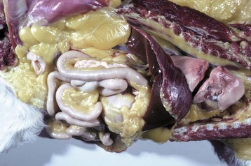
In birds and reptiles, there are two forms, namely (1) the articular type, which is rare, and (2) a visceral type. The latter characteristically affects the visceral serosae, particularly the parietal pericardium, and the kidneys. The serosa is covered with a thin layer of gray granules, and the gross appearance is diagnostic. In the renal form, urate deposits are visible in renal tubules and ureters. Uric acid and urates are the end-products of purine metabolism, and in birds and reptiles, these products are eliminated as semisolid urates. Visceral gout is usually diagnosed only at necropsy and is seen sporadically as the result of vitamin A deficiency, high-protein diets, and renal injury.

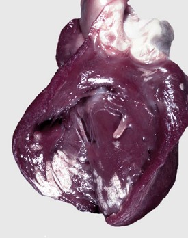
Pseudogout: Pseudogout is characterized by deposits of calcium pyrophosphate crystals. It is well recognized in humans but has been reported in the dog, in which it is rare. The pathogenesis of the canine disease is unknown, but in humans, one form is inherited as an autosomal dominant trait. Grossly, there are chalky white deposits in the joints, which histologically show a chronic reaction with aggregates of crystalline material, macrophages, and fibrosis. The disease may be differentiated from gout by the chemical analysis of the crystalline deposits.

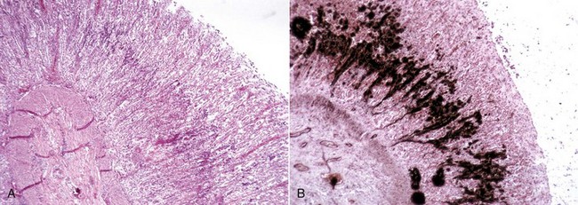
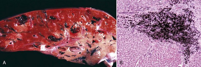
Cholesterol: Cholesterol crystals are the by-products of hemorrhage and necrosis. They are dissolved out of the tissue specimen during the preparation of paraffin-embedded sections, leaving characteristic clefts that, in tissue sections, resemble shards of glass. Actually, the crystals are thin rhomboidal plates with one corner notched out, their outline resembling that of the state of Utah. Cholesterol crystals in tissue have no significance except that they indicate the site of an old hemorrhage or tissue necrosis, and they may be present in atheromas (i.e., mass of degenerated, thickened arterial intima occurring in atherosclerosis). However, in the choroid plexus of the lateral ventricles of old horses, cholesterol crystals can induce a granulomatous response, and the resultant cholesterol granuloma or cholesteatoma can become so large as to obstruct the outflow of cerebrospinal fluid through the interventricular foramen (foramen of Munro), resulting in obstructive hydrocephalus. It is thought that these granulomas are secondary to cholesterol crystals from hemorrhages into the choroid plexus. Grossly, the cholesterol appears as firm, crumbly gray nodules in the cholesteatomas.

Pathologic Calcification*

Calcium salts, usually in the form of phosphates or carbonates, may be deposited in dead, dying, or normal tissue. This process is known as pathologic calcification and occurs in two forms: dystrophic and metastatic. When the deposition occurs locally in dying tissue, it is known as dystrophic calcification; it occurs despite normal serum concentrations of calcium and in the absence of derangements in calcium metabolism. In contrast, the deposition of calcium salts in otherwise normal tissue is known as metastatic calcification, and it almost always results from hypercalcemia secondary to some disturbance in calcium metabolism. Less common forms of calcification are idiopathic (occurs in the absence of tissue injury or abnormalities in calcium or phosphorus metabolism) and iatrogenic (e.g., via ingestation of calcium salts from calcium chloride de-icing solutions).

Dystrophic Calcification

Dystrophic calcification occurs in areas of necrosis, no matter the type of necrosis-coagulative, caseous, liquefactive, or fat necrosis, but is minimal in liquefactive necrosis. Dead and dying cells can no longer regulate the influx of calcium into their cytosol, and calcium accumulates in the mitochondria.

Common sites include necrotic myocardium (Fig. 1-53), necrotic skeletal muscle, granulomas such as tuberculoid granulomas in cattle, and dead parasites, such as hydatid cysts in cattle and trichinae in pigs. Calcium deposits are relatively permanent but harmless unless they interfere mechanically (e.g., the movement of a calcified heart valve). Their significance is that they are an indicator of previous injury to a tissue.

image

Fig. 1-53 Calcification, vitamin E/selenium deficiency, myodegeneration, heart, lamb.
The multiple white lesions are areas of necrosis of cardiac myocytes that have been calcified. (Courtesy Dr. M.D. McGavin, College of Veterinary Medicine, University of Tennessee.)

Calcification in or under the skin has been designated calcinosis. The two main forms are (1) calcinosis cutis and (2) calcinosis circumscripta (see Chapter 17). Calcinosis cutis occurs in dogs with hyperadrenocorticism from either endogenous or exogenous glucocorticoids and has been regarded as idiopathic calcification by some pathologists and dystrophic calcification by others. There is mineralization of the dermal collagen and epidermal and follicular basement membranes. Calcinosis circumscripta is considered to be dystrophic. It has a preference for German shepherds and Great Danes, in which it is familial. Also, it has been associated with repetitive trauma and at the site of buried sutures of polydioxanone.

Grossly, the affected areas of tissue are white and when incised have a gritty feel to them (see Fig. 1-53). Microscopically, calcium salts stain blue with hematoxylin and appear as fine amorphous granules or clumps, which can be either intracellular or extracellular. However, the full extent of the calcification may not be evident in H&E-stained sections (Fig. 1-54, A) but is revealed more dramatically by special stains, such as von Kossa and Alizarin red S (Fig. 1-54, B). The von Kossa method is not specific for calcium but stains phosphates and carbonates. These substances are almost always complexed with calcium.

image

Fig. 1-54 Uremia, stomach, dog.
A band of calcium has been laid down the middle of the gastric mucosa. A, The calcium is stained blue with hematoxylin. H&E stain. B, The calcium is stained black. von Kossa stain. (A and B courtesy Dr. M.D. McGavin, College of Veterinary Medicine, University of Tennessee.)

Metastatic Calcification

Metastatic calcification occurs in normal tissue and is secondary to hypercalcemia. The basic abnormality is the entry of large amounts of calcium ions into cells. These ions precipitate on organelles, particularly mitochondria.

The four causes of metastatic calcification in order of their importance in veterinary medicine are as follows:

1. Renal failure. Renal failure results in retention of phosphates, which induce a secondary renal hyperparathyroidism and hypercalcemia. Calcium is deposited in the gastric mucosa, kidney, and alveolar septa.

2. Vitamin D toxicosis. The ingestion of calcinogenic plants, such as Cestrum diurnum by herbivores, results in severe soft tissue mineralization, chiefly involving the aorta, heart, and lungs. In the heart, the endocardium of the right and left atria and left ventricle is often strikingly mineralized. Acute vitamin D toxicosis in dogs and cats is commonly caused by ingestion of rodenticides containing cholecalciferol. Intestinal mucosa, vessel walls, lung, and kidneys are mineralized.

3. Parathormone (PTH) and PTH-related protein. Primary hyperparathyroidism is rare. Hypercalcemia and elevated concentrations of PTH-related protein can be associated with canine malignant lymphomas and canine adenocarcinoma of the apocrine glands of the anal sac. Intestinal mucosa, vessel walls, lung, and kidneys are mineralized.

4. Destruction of bone from primary or metastatic neoplasms.

Heterotopic Bone (Ectopic Bone)

Some lesions of dystrophic and metastatic calcification may be confused on gross examination with ectopic ossification, the name given to the process of production of bone at an abnormal site. Ectopic bone is of two types: heterotopic or osseous metaplasia. “Heterotopia” refers to foci of cells or tissues, which are microscopically normal but present at an abnormal location. They are considered to arise from embryonic cell rests. The other type of bone is formed by osseous metaplasia, usually from another type of connective tissue. Fibroblasts differentiate into osteoblasts that form osteoid, which is calcified as in normal bone (Fig. 1-55). This is the more common type.

image

Fig. 1-55 Ectopic bone, lung, dog.
A nodule of mature bone in the connective tissue of the lung. H&E stain. (Courtesy Dr. M.D. McGavin, College of Veterinary Medicine, University of Tennessee.)

Heterotopic bone is found at many sites, commonly as spicules or nodules of bone in connective tissue of lungs of dogs and cattle, and in the canine dura mater (dural ossification or ossifying pachymeningitis) and at sites of dystrophic and metastatic calcification.

Pigments

It is customary in pathology textbooks to group substances that impart an unusual color to the body (systemic) or its tissues (localized), under the category of pigments. Many of these pigments are unrelated in their origin, but their importance lies in the fact that the clinician and the pathologist need to be able to recognize them grossly, and the pathologist also needs to be able to identify them histologically. Recognition may provide valuable clues in understanding the disease process at hand and its underlying pathogenesis. Because of their diversity, pigments are usually classified broadly into two groups: exogenous (formed outside the body) and endogenous (formed inside the body).

Exogenous Pigments

These pigments include carbon, tattoos, dusts, carotenoids, and tetracycline.

Carbon: Carbon is the most common exogenous pigment. The usual route of entry into the body is via inhalation, and its accumulation in the lung results in a condition called anthracosis (also known as black lung).

Carbon is ubiquitous in the air and all animals are exposed, but those most likely to show gross lesions live in an environment with substantial air pollution, such as adjacent to busy highways (e.g., animals in a zoo near a highway or animals living in a house with a smoker). In the alveoli, the carbon is phagocytosed by macrophages, which transport it via the lymphatics to the regional tracheobronchial lymph nodes. Because elemental carbon is inert and not metabolized by the body, it remains in the tissue for the life of the animal.

Grossly, the lungs are usually speckled with fine 1- to 2-mm-diameter subpleural black foci, which are most visible if the lungs have been exsanguinated (Fig. 1-56, A). In severely affected cases, the medulla of the tracheobronchial lymph nodes may be black. The heavy deposits are in this location because of the concentration of sinus histiocytes (macrophages) in the medulla.

image

Fig. 1-56 Anthracosis, lung, aged dog.
A, The fine black foci are peribronchiolar deposits of carbon. The animal was exsanguinated at euthanasia to remove the blood from the lung to render the carbon deposits more visible. B, Carbon (black) inhaled into the alveoli has been phagocytosed by macrophages and transported to the peribronchial region. H&E stain. (A and B courtesy Dr. M.D. McGavin, College of Veterinary Medicine, University of Tennessee.)

Microscopically, carbon presents as fine black granules and may be extracellular or intracellular (within macrophages). Carbon pigment may be within the alveolar walls or be frequently present as black peribronchiolar or peribronchial foci (Fig. 1-56, B). Because of the nonreactiveness of carbon, there are no histochemical tests for it. Unlike many other pigments, it is resistant to solvents and bleaching agents.

Tattoos: Animals are frequently tattooed as a method of identification. These pigments, which include carbon, are introduced into the dermis. Some of the pigments are phagocytosed by macrophages, whereas the remainder remains free in the dermis where it can remain indefinitely and does not evoke any inflammatory reaction.

Dusts: Pneumoconiosis is the general term used for any dust inhaled into and retained in the lung. Anthracosis, from the inhalation of carbon, is a subtype of pneumoconiosis. Inhalation of silicon (e.g., from quarries) is called silicosis. These minute particles enter the lungs by escaping the mucociliary defense mechanisms of the nasal and upper respiratory systems (see Chapter 9) and are deposited in pulmonary alveoli where they may be phagocytosed and carried to the peribronchial regions. Some types of silica evoke a fibrous reaction, which may ultimately form nodules. Microscopically the mineral is visible as birefringent crystals under polarized light.

Carotenoid Pigments: Carotenoid pigments are also called lipochrome pigments, although this term is sometimes confused with lipofuscin (see later discussion). They are fat-soluble pigments of plant origin and include the precursor of vitamin A, namely β-carotene.

Grossly, these pigments normally occur in a wide variety of tissue, such as adrenal cortical cells, corpus luteum-lutein cells, Kupffer cells, and testicular cells, and in the plasma/serum and fat of horses and Jersey and Guernsey cattle and sometimes dogs (Fig. 1-57). Carotenoids discolor fat yellow to orange-yellow. The concentration of carotenoids retained in tissue depends on the species of animal. Some animals store little or no carotenoids and have white fat and clear serum. These animals include Holstein cattle, sheep, goats, and cats. As fat stores are depleted (e.g., in starvation or cachexia), carotenoids become concentrated in the adipocytes, giving them a dark yellowish-brown color.

image

Fig. 1-57 Carotenosis, kidney and the perirenal fat, Jersey ox.
Accumulation of carotenoids in the adipocytes has colored the fat yellow to dark yellow. (Courtesy Dr. M.D. McGavin, College of Veterinary Medicine, University of Tennessee.)

Microscopically, carotenoids are not seen in routine formalin-fixed paraffin-embedded sections because the alcohols and clearing agents remove the fat-soluble pigments.

The significance of carotenoids is that they may obscure or confuse the detection of icterus. In those animals whose fat and serum are devoid of carotenoids, a yellow discoloration is easily detected and is most likely to be caused by bilirubin (i.e., icterus).

Tetracycline: Tetracycline-based antibiotics administered during the development of teeth are deposited in mineralizing dentin, enamel, and cementum, staining the teeth or portions of them yellow or brown (Fig. 1-58). Thus tetracycline administered to a pregnant animal stains the deciduous teeth of the offspring. Tetracycline also stains bone that is being laid down and has been used experimentally as a marker for that bone.

image

Fig. 1-58 Tetracycline staining, teeth, young dog.
The teeth of this dog have been stained yellow by the tetracycline ingested during their development. (Courtesy Dr. M.D. McGavin, College of Veterinary Medicine, University of Tennessee.)

Endogenous Pigments

Melanin: Melanin is the pigment normally present in the epidermis and is responsible for the color of the skin and hair. It is also normally present in the retina, iris, and in small amounts in the pia-arachnoid of black animals (e.g., Suffolk sheep [Fig. 1-59]) and in the oral mucous membrane of some breeds (e.g., Jersey cows and Chow dogs).

image

Fig. 1-59 Congenital melanosis, leptomeninges, Suffolk sheep.
The leptomeninges have scattered black areas of melanin. This is normal in black-faced sheep. (Courtesy Dr. M.D. McGavin, College of Veterinary Medicine, University of Tennessee.)

Cells called melanocytes secrete melanin. In the skin of animals, these cells are in the basal layer and transfer their pigment by means of dendritic processes to adjacent keratinocytes, where the melanin is often arranged as a cap over the nucleus to provide some protection from ultraviolet radiation. Melanin is formed by the oxidation of tyrosine, which requires the copper-containing enzyme tyrosinase. Thus, in copper deficiency, particularly in cattle and sheep, there is a fading of the coat color, and this is most obvious in black wool. A general lack of melanin can be the result of a metabolic defect: a lack of tyrosinase. This condition is called albinism, and the affected animal is called an albino. Histologically, the melanocytes appear normal.

Pathologically, melanin is present in hyperpigmentation of the skin associated with many types of chronic injury and endocrinopathies, such as hyperadrenalism, and in primary neoplasms of melanocytes (melanocytomas and malignant melanomas), although highly malignant tumors may have little or no pigment.

Microscopically, melanin is stored in melanosomes in the cytoplasm of melanocytes. However, if there is irreversible injury to the cells containing melanin (e.g., necrosis of melanocytes and basal cells of the skin), melanin is released from the dead cells and is phagocytosed by macrophages, which are termed melanophages.

Extensive deposits of congenital melanin in tissues are termed congenital melanosis. It occurs in the lungs and aorta (intima) of cattle, sheep, and pigs as brown-to-black spots up to a couple of centimeters in diameter (Fig. 1-60). Melanosis of the lung is visible both subpleurally and in cross-sections of the parenchyma. These deposits of melanin have no adverse effects, but organs with extensive melanosis may be aesthetically unacceptable as food and thus will be condemned at the packing plant.

image

Fig. 1-60 Congenital melanosis, lung, pig.
Melanin deposits are subpleural and extend into the substance of a lung. The lesion has no pathological significance. (Courtesy Dr. M.D. McGavin, College of Veterinary Medicine, University of Tennessee.)

Lipofuscin-Ceroid: Lipofuscin, known as “wear-and-tear” pigment, has in the past been described as accumulating with age and in certain pathologic conditions. However, in recent years, lipofuscin, now referred to as “age” pigment, has been differentiated from a pathologically accumulating similar pigment called ceroid, which is described later.

Lipofuscin accumulates in a time-dependent manner in postmitotic cells (neurons, cardiac myocytes [Fig. 1-61], and skeletal muscle myocytes) and in slowly dividing cells, such as hepatocytes and glial cells, and this process is present at a few months of age. Lipofuscin is also found in other cells, but as these replicate, the lipofuscin is divided between the daughter cells and thus does not accumulate to the same extent as it does in postmitotic cells. Lipofuscin is the end result of autophagocytosis of cell constituents, such as organelles, and is the final undegradable remnant of that process. As the pigment cannot be removed by further lysosomal degradation or exocytosis, it accumulates in lysosomes, a form of biologic garbage.

image

Fig. 1-61 Lipofuscinosis, heart, dog.
Note the brown lipofuscin granules (arrows) at the poles of the myocyte nuclei. H&E stain. (Courtesy Dr. J.F. Zachary, College of Veterinary Medicine, University of Illinois.)

Ceroid has many of the same histochemical features as lipofuscin (see later discussion) but is found in response to severe malnutrition, including vitamin E deficiency, cachexia from cancer, irradiation, and in the inherited disease neuronal ceroid-lipofuscinosis. It accumulates in Kupffer cells and to a lesser extent in hepatocytes, skeletal and smooth muscle myocytes, and in inherited neuronal ceroid-lipofuscinosis, where it accumulates in neurons. It can be either intracellular or extracellular. Unlike lipofuscin, it is considered to have a deleterious effect on the cell.

Both lipofuscin and ceroid have many common histologic and histochemical features, such as autofluorescence (golden yellow) and staining with stains for fat such as Sudan black (sudanophilia), although oil-red-O is more sensitive, PAS positive, and acid-fast (long Ziehl-Neelsen technique). All of these characteristics increase in intensity with age for lipofuscin but not for ceroid. Lipofuscin consists chiefly of proteins and lipids with very little carbohydrate, but lectin-binding histochemistry in humans and rats has revealed differences in the saccharides of lipofuscin and ceroid.

Grossly, large amounts of lipofuscin in the heart and skeletal muscles impart a brown tinge. It is commonly seen in aged dairy cows sent to slaughter. Ceroid is grossly evident in the small intestine of dogs with so-called intestinal lipofuscinosis (Fig. 1-62) (also see Chapter 7) and in nutritional panniculitis in cats, mink, foals, and pigs. Both of these conditions are associated with a vitamin E deficiency and the ingestion of unsaturated fatty acids. In the dog, the tunica muscularis, usually of the caudal small intestine, is discolored brown because of accumulations of ceroid in myocytes. In the cat with nutritional panniculitis, the subcutaneous fat is discolored lemon yellow to orange. This disease is considered to be the result of the ingestion of fish products with a high concentration of unsaturated fatty acids and a vitamin E deficiency, frequently brought about by the fats becoming rancid and destroying the vitamin E.

image

Fig. 1-62 Ceroid, intestine, serosal surface, dog.
Note the brown discoloration of the muscular layer. The condition has been called intestinal lipofuscinosis but is not age related. (Courtesy Dr. M.D. McGavin, College of Veterinary Medicine, University of Tennessee.)

Microscopically, in routine H&E-stained sections or in unstained sections, lipofuscin varies from a light golden brown to dark brown with advancing age. Because it is intralysosomal, it is perinuclear in neurons and in cardiac, skeletal, and smooth muscle myocytes. In feline nutritional panniculitis, globules of ceroid are extracellular in the interstitial tissue or have been ingested by macrophages and giant cells.

The significance of these two pigments is that lipofuscin is a clear indicator of the age of the cell and ceroid is a pathologic pigment, often associated with vitamin E deficiency. Lectin-binding histochemistry, which has shown differences between lipofuscin and ceroid from rats and humans, may be applicable to differentiating these pigments in domestic animals, but it is a very laborious research tool and only provides semiquantitative data. Isolation and physicochemical analysis is more precise but even more laborious. Thus, until some other specific test becomes available, differentiation between the two pigments for diagnostic purposes will be based on the features listed in Table 1-1.

TABLE 1-1

Differences between Lipofuscin and Most Ceroid Pigments In Vivo

  Lipofuscin Ceroid
Universality (invariably present in humans and all domestic animals) Yes No
Intrinsically (intracellularly in lysosomes of postmitotic and stable cells) Yes No
Time dependence Yes No
Initial occurrence Infancy Anytime
Deleteriousness Never demonstrated Frequent
Accumulation rate Very slow Usually rapid
Tissue distribution Only intracellular Intracellular and extracellular
Mode of formation Mainly autophagy Mainly heterophagy
Origin of precursors Mainly intracellular Mainly extracellular

From Porta EA: Ann N Y Acad Sci 959:57-65, 2002.

Hematogenous Pigments

The hematogenous pigment category includes hemoglobin, oxyhemoglobin, unoxygenated hemoglobin, methemoglobin, carboxyhemoglobin, hemosiderin, bilirubin, and hematin. Some are produced normally but can accumulate excessively (unoxygenated hemoglobin, hemosiderin, and bilirubin). Other pigments, such as methemoglobin, carboxyhemoglobin, and hematin, are pathologic.

Hemoglobin: The normal pigment of erythrocytes, hemoglobin, can be responsible for gross changes in the color of the body. Oxygenated hemoglobin is red and imparts the pink appearance to unpigmented skin and tissues. Normally, arterial blood (oxygenated hemoglobin) is red, and venous blood with more unoxygenated blood is bluish. However, if the blood is not sufficiently oxygenated (unoxygenated hemoglobin), the tissues appear blue, exhibiting so-called cyanosis (Fig. 1-63).

image

Fig. 1-63 Cyanosis, feet, cat.
The footpads of the paw on the left are bluish due to unoxygenated hemoglobin, the result of a partial obstruction of the iliac artery at the aortic bifurcation by a saddle thrombus. Normal control paw is on the right. (Courtesy Dr. M.D. McGavin, College of Veterinary Medicine, University of Tennessee.)

In acute cyanide poisoning, cyanide binds to cytochrome oxidase, the enzyme in the cell responsible for oxidative phosphorylation, and this results in paralysis of cellular respiration. Tissues cannot use the oxygen delivered by the blood. Consequently, in acute cyanide toxicity the oxygen content and color of venous blood may be similar to those of arterial blood, and the venous blood will be bright red.

In carbon monoxide (CO) poisoning, as from exhaust gases from automobiles, the blood is a bright cherry red from the formation of carboxyhemoglobin (Fig. 1-64). Methemoglobin is an oxide of hemoglobin, in which the ferrous ion of hemoglobin is converted to the ferric ion, resulting in a reddish-brown (chocolate brown) color to the blood and tissue (Fig. 1-65). Methemoglobin is seen most often in poisoning by nitrites, especially after ingestion of nitrate-accumulating plants, but has been reported as a result of ingestion of acetaminophen, naphthalene, and chlorates and of treatment with local anesthetic agents (e.g., lidocaine, benzocaine, and tetracaine).

image

Fig. 1-64 Carbon monoxide (CO) poisoning, brain, human.
The blood in the brain is cherry red from the carboxyhemoglobin formed by the inhalation of CO in exhaust gases. (Courtesy Dr. J.C. Parker, School of Medicine, University of Louisville.)

image

Fig. 1-65 Methemoglobinemia, experimental nitrite poisoning, hindleg, pig.
Left, The methemoglobin in the blood has discolored the blood and muscle chocolate brown. Right, Normal control. (Courtesy Dr. L. Nelson, College of Veterinary Medicine, Michigan State University.)

In intravascular hemolysis, hemoglobin is released from the lysed erythrocytes and stains the plasma pink. This hemoglobin may be excreted by the kidney, staining it dark red to reddish-black and the urine red (Fig. 1-66). Similar changes can result from myoglobinuria after the destruction of large numbers of myofibers (see Chapter 15).

image

Fig. 1-66 Acute hemolysis from chronic copper poisoning, kidney and urine, sheep.
The dark bluish color of the kidney and the dark red of the urine are caused by hemoglobin excreted via the kidney. (Courtesy Dr. M.D. McGavin, College of Veterinary Medicine, University of Tennessee.)

Hematins: The hematin category of pigments includes “formalin pigment” and the excreta of parasites such as Fascioloides magna (liver fluke) and Pneumonyssus simicola (lung mite).

Formalin Pigment: Formalin pigment, also called acid formalin hematin, is an annoying microscopic artefact that occurs when tissue rich in blood comes in contact with acid solutions of formalin, particularly if there has been a delay between death and fixation, allowing time for the erythrocytes to lyse and release their hemoglobin.

Grossly, formalin pigment is not visible because this change occurs only after fixation. Microscopically the pigment is brown to almost black, fine, and granular (Fig. 1-67), and can have birefringent spicules. It occurs mainly in blood vessels but also in other tissues in which there are large accumulations of red blood cells. Pigment can lie between or on top of red blood cells and is negative for iron when stained by the Prussian blue reaction.

image

Fig. 1-67 Formalin pigment, blood.
Note the black spicules of hematin that lie between and on the erythrocytes, the result of fixation in unbuffered (acid) 10% formalin. H&E stain. (Courtesy Dr. M.D. McGavin, College of Veterinary Medicine, University of Tennessee.)

Because formalin pigment is formed only during fixation, it has no pathologic significance. Its significance is that it can interfere with the interpretation of histologic sections. Fortunately, it is easy to prevent its formation. Formalin pigment does not form if the pH of the fixative is above 6. Aqueous solutions of unbuffered formalin are highly acid. A common fixative is 10% buffered neutral formalin (really buffered neutral 10% formalin), which is buffered with a Sorensen phosphate buffer, and despite the name “neutral” has a pH of 6.8. It does not cause the formation of formalin pigment. Another commonly used and commercially available formalin fixative is Carson’s fixative (also called modified Millonig’s formalin fixative), with a pH of 7.3 and can be used as a dual-purpose fixative for both routine histopathologic and electron microscopic examinations. If formalin pigment is present in a tissue section, it can be removed by a variety of techniques including soaking the dewaxed tissue section before H&E staining in a saturated alcoholic solution of picric acid.

Parasite Hematin: The two most common causes of parasite hematin in veterinary medicine are Fascioloides magna (liver fluke) in ruminants and Pneumonyssus simicola in the lungs of macaques.

Parasite hematin from Fascioloides magna causes black tracts throughout the liver and is colloquially known as fluke exhaust (Fig. 1-68, A). This lesion can be so severe that it affects the whole liver. Microscopically, the black pigment accumulates adjacent to the migration tracts of the parasite and is phagocytosed by macrophages (Fig. 1-68, B). Pneumonyssus simicola produces a similar brown-to-black anisotropic pulmonary pigment presumed to be from the metabolism of hemoglobin by the parasite.

image

Fig. 1-68 Hematin pigment from Fascioloides magna, liver, ox.
A, Large areas of the liver are black from the pigment excreted by the fluke as it migrated through the liver. B, Hematin (black) pigment deposited in a fluke migration tract in the liver. H&E stain. (A courtesy Dr. J. Wright, College of Veterinary Medicine, North Carolina State University; and Noah’s Arkive, College of Veterinary Medicine, The University of Georgia. B courtesy Dr. M.D. McGavin, College of Veterinary Medicine, University of Tennessee.)

Hemosiderin: Iron is stored in the body in two forms, ferritin and hemosiderin, both of which are protein-iron complexes. Ferritin is in all tissues, but the heaviest concentrations are found in the liver, spleen, bone marrow, and skeletal muscle. Hemosiderin is formed from intracellular aggregates of ferritin (Fig. 1-69). It appears as golden-yellow to golden-brown globules and is the most visible form of storage iron. Normally, most storage iron is found in the spleen.

image

Fig. 1-69 Hemosiderosis, spleen, dog.
A, Hemosiderin is present as fine golden brown granules in macrophages. H&E stain. B, Granules of hemosiderin are stained dark blue by the Prussian blue reaction, which is specific for iron. Prussian blue reaction. (A and B courtesy Dr. J.F. Zachary, College of Veterinary Medicine, University of Illinois.)

Excess iron from the breakdown of senescent erythrocytes, or the result of a hemolytic crisis (e.g., because of autoimmune diseases or hemotropic parasites) or reduced erythropoiesis (e.g., from malnutrition), is stored mainly in the spleen. Rarely in veterinary medicine, excess iron can be present in the body because of excessive absorption from the gut, multiple injections of iron, or from multiple blood transfusions.

Besides splenic storage, there may be local iron storage at sites of erythrocyte breakdown, such as in hemorrhages, and in areas of poor blood flow, as in chronic passive congestion of the lungs. In the latter case, because of the poor blood flow through the lungs, erythrocytes may come to the end of their natural life and be lysed or enter the alveoli by diapedesis, where they are phagocytosed by alveolar macrophages. These cells are termed heart failure cells (Fig. 1-70). Localized deposits of iron may also be present from the intramuscular injection of iron dextran, and this iron may drain to the regional lymph node.

image

Fig. 1-70 Chronic passive congestion, lung, dog.
A, Alveolar macrophages containing hemosiderin (blue) are present in the alveoli. Prussian blue reaction. B, The lungs have chronic passive congestion attributed to chronic left-sided heart failure. They are moderately firm and yellow-brown caused by alveolar macrophages containing hemosiderin. Inflammatory mediators produced by these macrophages have induced fibroplasia, thus in the long term, there has been extensive formation of interstitial collagen. This collagen is the reason the lungs have failed to collapse after incision of the diaphragm, which releases the negative pressure in the pleural cavity (note the rib impressions in the lung). (A courtesy Dr. M.D. McGavin, College of Veterinary Medicine, University of Tennessee. B courtesy College of Veterinary Medicine, University of Illinois.)

Grossly, no change is seen in an organ or tissue if there are only small amounts of hemosiderin, but very large amounts cause a yellow-to-brown discoloration (Fig. 1-71). This color change can also be seen at sites of old bruises and other hemorrhages or hematomas. The spleen and the liver in hemolytic disease and the lungs in chronic passive congestion also appear brown. Microscopically, hemosiderin deposits are golden-yellow to golden-brown globules, which may be intracellular or extracellular (see Fig. 1-69, A). It can be confirmed by the Prussian blue reaction (see Fig. 1-69, B), which is sometimes incorrectly called a stain but is a chemical reaction, of which the end-product is Prussian blue. In the acid solution that liberates ferric iron from the hemosiderin, the ferric iron is reacted with potassium ferrocyanide (colorless) to form ferric ferrocyanide, which is Prussian blue.

image

Fig. 1-71 Subcutis, old bruise, leg, horse.
The display of colors—red, yellow, and brown—is due to hemoglobin, bilirubin, and hemosiderin, respectively, from the breakdown of the erythrocytes. (Courtesy Dr. M.D. McGavin, College of Veterinary Medicine, University of Tennessee.)

The significance of hemosiderin deposits depends on their location and the amount. Normally, the spleen contains some hemosiderin, but excess hemosiderin is seen in the spleen and liver (Kupffer cells and hepatocytes) from hemolytic diseases, such as in autoimmune hemolytic anemia, and hemotropic diseases, such as babesiosis, anaplasmosis, or equine infectious anemia. Local tissue aggregations of hemosiderin are usually the result of the breakdown of erythrocytes in an old hemorrhage.

Excess hemosiderin is called hemosiderosis and must be differentiated from hemochromatosis, in which there are extreme accumulations of hemosiderin.

Hematoidin: Grossly, hematoidin is yellow-brown to orange-red pigment derived from hemoglobin but free of iron. Hematoidin closely resembles bilirubin (see next section) but is formed by cells of the macrophage-monocyte system when they phagocytose and digest red blood cells and hemoglobin in areas of hemorrhage. Microscopically, hematoidin is crystalline and polarizes light.

Bilirubin: Low concentrations of bilirubin are normally present in the plasma from the breakdown of senescent erythrocytes (see Chapter 13). Briefly, when erythrocytes have come to the end of their natural lifespan (average 70 days for a cat; average 150 days for cattle and horses), they are phagocytosed by the macrophage-monocyte system, chiefly by macrophages of the spleen and to a lesser extent by those of the bone marrow and liver (Kupffer cells). Within these cells, the iron is removed and stored, and the remainder of the porphyrin ring is broken down to bilirubin, which is released into the blood where it attaches to albumin. This bilirubin-albumin complex is too large to be excreted by the kidney. It is carried to the liver, where it enters the space of Disse, is taken up by the microvilli of the hepatocytes, is conjugated to form bilirubin glucuronide or diglucuronide, and is then excreted into the bile canaliculus.

Icterus (jaundice), the yellow staining of the tissue by bilirubin, is the result of an imbalance between production and clearance of bilirubin because there is either excess production or reduced clearance of bilirubin such that it accumulates in the plasma. The mutant Corriedale sheep model is an animal model for Dubin-Johnson syndrome in humans (Fig. 1-72).

image

Fig. 1-72 Defective bilirubin excretion, mutant Corriedale sheep, animal model for Dubin-Johnson syndrome.
Note the faint yellow discoloration of the lung from bilirubin. The other tissues are discolored dark green from phylloerythrin, which also has a similar defect in excretion in the liver. (Courtesy Dr. M.D. McGavin, College of Veterinary Medicine, University of Tennessee.)

Mechanisms leading to icterus can involve one or more of the following:

1. Excess production of bilirubin—as in hemolytic diseases such as babesiosis, anaplasmosis, and equine infectious anemia—or the breakdown of erythrocytes in a large hemorrhage such as a hematoma.

2. Reduced uptake of bilirubin from the plasma by hepatocytes.

3. Impaired or absent conjugation in hepatocytes, often a congenital or inherited abnormality, as in the Gunn rat.

4. Hepatic necrosis. Because the cell membranes of several adjacent hepatocytes form the bile canaliculus, any necrosis of these cells will disrupt the wall of the canaliculus and allow leakage of bilirubin into the circulation. Extensive hepatic necrosis can cause icterus.

5. Decreased excretion of conjugated bilirubin by the hepatocytes into the bile canaliculus.

6. Reduced flow of bile from the liver to the intestine caused by either intrahepatic or extrahepatic blockage of the biliary system.

Icterus is classified several different ways. A convenient approach uses the classification of prehepatic, hepatic, and posthepatic. The most common cause of prehepatic icterus is a hemolytic crisis, which produces high plasma concentrations of unconjugated bilirubin that exceed the uptake capacity of the hepatocytes. Hepatic icterus is caused by hepatocellular damage, which results in release of bilirubin, both conjugated and unconjugated into the blood and can be the result of one or more of factors 2 to 4. Posthepatic icterus is secondary to obstruction of the biliary system, either intrahepatic or extrahepatic (hepatic bile ducts and the common bile duct), with reflux of the conjugated bilirubin into the blood. In contrast to unconjugated bilirubin, which is carried in the blood attached to albumin and cannot be excreted by the kidney, conjugated bilirubin is not bound to a plasma protein such as albumin and can be excreted.

Grossly, icteric tissues are discolored yellow, and the color change is distributed systemically. Clinically, icterus is most easily recognized in lightly pigmented animals. In living animals, icterus is detected in mucous membranes of the oral cavity, urogenital systems, and alimentary system and in normally white areas such as the sclera of the eyes. At necropsy, in addition to the sites listed previously, icterus can be identified in the omentum, mesentery, and adipose tissue (Fig. 1-73), except in Jersey and Guernsey cattle, horses, and nonhuman primates, whose sera and fat are normally discolored yellow by carotenoids. The intima of the large vessels is also a good site to detect icterus, and unless the plasma concentration is extremely high, the brain is usually unaffected.

image

Fig. 1-73 Icterus, hemolytic anemia, abdominal and thoracic viscera, dog.
The yellow discoloration from the bilirubin is particularly evident in fat and mesentery. (Courtesy Dr. M.D. McGavin, College of Veterinary Medicine, University of Tennessee.)

Microscopically, icterus is not detected, but excessive quantities of bilirubin can be seen in the bile ducts and bile canaliculi in obstructive jaundice (Fig. 1-74).

image

Fig. 1-74 Icterus.
A, Icterus, liver, cat. Note the enlarged liver with rounded edges and yellow-orange color caused by retained bilirubin. B, Bile casts in bile canaliculi. Acute hemolytic anemia, babesiosis, liver, cow. The bile casts are the result of a high rate of bilirubin excretion by the liver secondary to intravascular hemolysis. H&E stain. (A courtesy College of Veterinary Medicine, University of Illinois. B courtesy Dr. M.D. McGavin, College of Veterinary Medicine, University of Tennessee.)

Icterus is a very important clinical sign and may be detected by examination of the sclera, and in cases of anemia, in which the mucous membranes are pale; it may be visible there. Laboratory tests to determine the exact plasma or serum concentrations of bilirubin, and preferably whether it is or is not conjugated, are essential. It is critical to realize that hyperbilirubinemia is not the same as icterus. Most domestic animals normally have very low serum concentrations of bilirubin, usually less than 1 mg/dL, except for the horse, in which it may range from 1 to 3 mg/dL. Icterus is not detected until the serum concentration exceeds 1.5 to 2.0 mg/100 mL. Thus hyperbilirubinemia can be present without causing icterus.

Porphyria: Congenital erythropoietic porphyria of calves, cats, and pigs is an inherited metabolic defect in heme synthesis caused by a deficiency of uroporphyrinogen III cosynthetase. The disease is sometimes incorrectly called osteohemachromatosis. It is also known colloquially as pink tooth because of the discoloration by the porphyrins accumulating in dentin and bone (Fig. 1-75, also see Chapter 7). The teeth and bones of young animals are reddish (pink tooth) and those of adults are dark brown. In these cases, both bones and teeth fluoresce red under ultraviolet radiation.

image

Fig. 1-75 Pink tooth, congenital porphyria teeth, adult ox.
The teeth are discolored brown from the accumulation of porphyrins in the dentin. (Courtesy Dr. M.D. McGavin, College of Veterinary Medicine, University of Tennessee.)

Cellular Aging*

The common age-related diseases in animals include renal failure, osteoarthritis, muscle atrophy, cerebral atrophy from loss of cortical neurons, cessation of the growth of the teeth of horses, and loss of elasticity of the skin. The incidence of different causes of mortality in all domestic animals is not available, but data from laboratory beagles maintained for their lifespan are known. One-quarter of these dogs died of neoplastic disease. Of the organ systems involved in the cause of death, the urinary system was responsible for 13%, the respiratory system for 6%, and the CNS for 7%. The leading cause of death from nonneoplastic diseases was renal failure. Death is often the end result of declining cell function, including cellular aging.

With age, there are physiologic and structural alterations in almost all organ systems. Aging in individuals is affected to a great extent by genetic factors, diet, social conditions, and occurrence of age-related diseases such as atherosclerosis, diabetes, and osteoarthritis (in humans) and renal failure and neoplasia in animals. There is good evidence that aging-induced alterations in cells are an important component of the aging of the organism. Cellular aging could be the result of progressive accumulation over the years of the effects of sublethal injury to cells. This injury may lead to cell death or at least to the diminished capacity of the cell to respond to injury.

Cellular aging is also the result of a progressive decline in the proliferative capacity and lifespan of cells and the effects of continuous exposure to exogenous influences that result in the progressive accumulation of cellular and molecular damage (Web Fig. 1-1).

image

Web Fig. 1-1 Mechanisms of cellular aging.
Genetic factors and environmental insults combine to produce the cellular abnormalities characteristic of aging. How calorie restrictions prolong lifespan is not established. IGF-1, Insulin-like growth factor-1. (From Kumar V, Abbas A, Fausto N, et al: Robbins & Cotran pathologic basis of disease, ed 8, Philadelphia, 2009, Saunders.)

Structural and Biochemical Changes with Cellular Aging

A number of cell functions decline progressively with age. Oxidative phosphorylation by mitochondria and the synthesis of nucleic acids and structural and enzymatic proteins, cell receptors, and transcription factors are all reduced. Senescent cells have a decreased capacity for uptake of nutrients and for repair of chromosomal damage. The morphologic alterations in aging cells include irregular and abnormally lobed nuclei, pleomorphic vacuolated mitochondria, decreased ER, and distorted Golgi apparatus. Concomitantly, there is a steady accumulation of lipofuscin, which represents a product of lipid peroxidation and evidence of oxidative damage.

Replicative Senescence

The concept that many cells have a limited capacity for replication was developed from a simple experimental model for aging. Normal human fibroblasts, when placed in tissue culture, have limited division potential. Cells from children undergo more rounds of replication than cells from older people. After a fixed number of divisions, all cells become arrested in a terminally nondividing state, known as cellular senescence. Many changes in gene expression occur during cellular aging, but a key question is which of these changes are causes and which are effects of cellular senescence.

How dividing cells can count their divisions is under intensive investigation. One likely mechanism is that with each cell division, there is incomplete replication of chromosome ends (telomere shortening), which ultimately results in cell cycle arrest (Web Fig. 1-2). Telomeres are short repeated sequences of DNA (TTAGGG) present at the linear ends of chromosomes and are important for ensuring the complete replication of chromosome ends and for protecting them from fusion and degradation.

image

Web Fig. 1-2 The role of telomeres and telomerase in replicative senescence of cells.
A, Telomerase directs RNA template-dependent DNA synthesis, in which nucleotides are added to one strand at the end of a chromosome. DNA polymerase fills in the lagging strand. B, Telomere-telomerase hypothesis and proliferative capacity of cells. Telomere length is plotted against the number of cell divisions. Germ cells and stem cells both contain active telomerase, but only the germ cells have sufficient levels of the enzyme to stabilize telomere length completely. In normal somatic cells, there is no telomerase activity, and telomeres progressively shorten with successive cell divisions until growth arrest, or senescence, occurs. Telomerase activation in cancer cells counteracts the telomere shortening that limits the proliferative capacity of normal somatic cells. (From Kumar V, Abbas A, Fausto N, et al: Robbins & Cotran pathologic basis of disease, ed 8, Philadelphia, 2009, Saunders.)

When somatic cells replicate, a small section of the telomere is not duplicated, and telomeres become progressively shortened. As the telomeres become shorter, the ends of chromosomes cannot be protected and are seen as broken DNA, which signals cell cycle arrest. The lengths of the telomeres are normally maintained by nucleotide addition, mediated by an enzyme called telomerase. Telomerase is a specialized RNA-protein complex that uses its own RNA as a template for adding nucleotides to the ends of chromosomes. The activity of telomerase is repressed by regulatory proteins, which restrict telomere elongation, thus providing a length-sensing mechanism. Telomerase activity is present in germ cells and is present at low levels in stem cells, and it is usually absent in most somatic tissue. Therefore, as cells age, their telomeres become shorter and ultimately the cell ceases to cycle in the cell cycle, resulting in an inability to generate new cells to replace damaged ones. Conversely, in immortal cancer cells, telomerase is reactivated and telomeres are not shortened, suggesting that telomere elongation might be an important—possibly essential—step in tumor formation. However, the relationship of telomerase activity and telomeric length to aging and cancer still needs to be fully established.

Genetic Basis of Aging

Studies in Drosophila, Caenorhabditis elegans, and mice are leading to the discovery of genes that influence the aging process. In a mouse model, changes resembling those of aging in humans are caused by disruption of a single gene. Analyses of humans with premature aging are also establishing the fundamental concept that aging is not a random process but is regulated by specific genes, receptors, and signals.

Accumulation of Metabolic and Genetic Damage

In addition to the importance of age and a genetic clock, cellular lifespan may also be determined by the balance between cellular damage resulting from metabolic activities occurring within the cell and the response of the counteracting molecular mechanisms responsible for repair. Smaller animals have generally shorter lifespans and faster metabolic rates, suggesting that the lifespan of a species is limited by fixed total metabolic consumption over a lifetime.

One group of products of normal metabolism is reactive oxygen species. These by-products of oxidative phosphorylation cause covalent modifications of proteins, lipids, and nucleic acids. The amount of oxidative damage, which increases as an organism ages, may be an important component of senescence, and the accumulation of lipofuscin in aging cells is seen as the indicator of this damage. The role of oxidative damage in aging is supported by the following observations: (1) variation in longevity among different species is inversely correlated with the rates of mitochondrial generation of a superoxide anion radical and (2) overexpression of the antioxidative enzymes SOD and catalase extends the lifespan in transgenic forms of Drosophila. Thus part of the mechanism that monitors the duration of aging may be the cumulative damage caused by toxic by-products of metabolism such as oxygen radicals. Increased oxidative damage could result from repeated environmental exposure to such influences as ionizing radiation, progressive reduction of antioxidant defense mechanisms (e.g., vitamin E and glutathione peroxidase), or both.

A number of protective responses counterbalance progressive damage in cells, and an important one is the cell’s recognition and repair of damaged DNA. Although most DNA damage is repaired by endogenous DNA repair enzymes, some persists and therefore damaged DNA progressively accumulates as the cell ages. Thus the balance between cumulative metabolic damage and the effectiveness of the response to that damage could determine the rate at which animals age. It is possible that aging may be delayed by decreasing the accumulation of the effects of that damage or by increasing the response to that damage.

Not only damaged DNA but also damaged cellular organelles accumulate as cells age. In part, this may be the result of declining function of the proteasome, the proteolytic machine that serves to eliminate abnormal and unwanted intracellular proteins.

Caloric Restriction

Animal studies have shown that the most effective way of extending a particular animal’s life is caloric restriction, without malnutrition; in other words the diet is adequate in such things as vitamins, minerals, and essential amino acids and protein. Their effect is thought to be mediated by a family of proteins called sirtuins, which promote the products of several genes. These include proteins that reduce apoptosis, stimulate protein folding, and inhibit the harmful effect of oxygen free radicals.

Senescence may be the result of the relationships among age, genetic programming, loss of telomeres, accumulated cell damage, and apoptotic cell death.

Genetic Basis of Disease*

This section provides an overview of (1) the structure and function of chromosomes and genes, (2) the mechanisms of genetic disorders, and (3) the outcomes of specific genetic diseases. The roles of genes have consequences for and are affected by cellular adaptations, cell injury, and cell death. Their roles in controlling immune responses and neoplastic transformation are discussed in Chapters 5 and 6, respectively. The function of microbial genes and their interplay with animal genes in determining host resistance to infectious diseases are discussed in Chapter 4.

Chromosome Structure and Function

Nuclear Chromosomes

Each animal species has a unique chromosomal complement called a karyotype (i.e., the number and morphology of the chromosomes that make up its genome). With the exception of cells that develop into ova and spermatozoa (i.e., germline cells), all cells in the body are called somatic cells (e.g., soma: body). The genome contained in the nucleus of somatic cells consists of chromosomes arranged in pairs. All except one pair are similar in both males and females and are called autosomes, and the remaining pair is the sex chromosomes: two X chromosomes in females and an X and a Y chromosome in males. Although each chromosome has different genes, members of a pair of chromosomes, also called homologous chromosomes or homologues, carry matching genetic information, that is, they have the same genes in the same sequence. Any specific locus (i.e., the specific location of a gene) on a chromosome may have either identical or slightly different forms of the same gene, called alleles. One member of each pair of chromosomes is inherited from the sire, the other from the dam.

Mitochondrial Chromosomes

Mitochondria are the site of aerobic energy production in all cells of the body. In highly active cells, such as type I myofibers of equine athletes, up to 10,000 mitochondria may be present in the cytoplasm of a myofiber. Each mitochondrion contains a single chromosome formed by circular double-stranded DNA, called mitochondrial DNA (mtDNA). The genome of a mitochondrial chromosome encodes for 37 genes, including those for messenger RNAs (mRNAs), ribosomal RNAs (rRNAs), transfer RNAs (tRNAs), and 13 protein subunits for enzymes, such as cytochrome b and cytochrome oxidase, which are involved in the production of energy by oxidative phosphorylation. The mitochondrial genome also has distinct transcription and protein-synthesis (i.e., translation) systems. A specialized RNA polymerase, encoded in the nuclear genome, is used to transcribe the mitochondrial genome and then the mitochondrial transcripts are processed to generate the various individual mitochondrial mRNAs, tRNAs, and rRNAs.

Gene Structure and Function

On average, there are approximately 20,000 genes in the nucleus of an individual cell of the different domestic animal species. Genes, the heredity units of the genetic code, determine the structural and functional biologic traits (i.e., expression of genes) necessary to create and maintain cells, tissues, and organs and to pass the genetic code on to offspring. Additionally, genes, especially those of the major histocompatibility complex (see Chapter 5), play important roles in establishing the resistance (or susceptibility) of an animal to infectious diseases. Most biologic traits expressed by genes have a Mendelian pattern of inheritance. They also have varied patterns of expression based on cell structure and function and the responses of cells to injury. Some genes are expressed continually and are called constitutively expressed genes and include genes for tRNAs, rRNAs, cell membranes, and enzymes. A different group of genes may be expressed only in a particular tissue such as the gene for galactocerebrosidase in myelinating cells of the nervous system (see the section on Globoid Cell Leukodystrophy in Chapter 14). These tissue-specific genes may be either differentially (i.e., in response to or as a result of cellular perturbations such as neoplastic transformation [see Chapter 6]) or constitutively expressed.

Certain genes may only be active (i.e., turned on and then off) in utero during specific times of fetal development as specific proteins are required for development, differentiation, and growth. Other genes are active continuously or transiently postpartum and are involved in growth, physiologic homeostasis, or reproduction. Thus an important consideration in the occurrence of a genetic disease is the period during which a normal gene is active. If the normal gene is mutated and the mutated gene is expressed during the active period for the normal gene, the outcome may have detrimental effects on structural and functional processes. Mutated genes of those that are normally important for early embryogenesis or organogenesis have earlier and generally more severe consequences than genes not expressed until sexual maturation or after. When the normal functions of genes are lost, disrupted, or altered, the outcome, many of which are heritable, can be harmful and result in congenital development anomalies such as palatoschisis/cheiloschisis (see Chapter 7). Metabolic dysfunction of cells can also occur and result in cell death in diseases such as multisystem neuronal degeneration (see Chapter 14). Examples of known or suspected genetic disorders in domestic animals are listed in Web Box 1-1 and are discussed in the chapters covering pathology of organ systems.

WEB BOX 1-1   A Partial List of Known or Suspected Genetic Disorders in Domestic Animals

Alimentary System and the Peritoneum, Omentum, Mesentery, and Peritoneal Cavity

• Abomasal emptying defect, ovine

• Amyloidosis

• Bovine bloat

• Boxer colitis

• Cheiloschisis

• Choanal atresia

• Chronic giant hypertrophic gastropathy

• Cricopharyngeal achalasia

• Gastric dilation and volvulus

• Lymphangiectasia

• Malocclusions

• Megaesophagus

• Overo lethal white foal syndrome (aganglionosis)

• Palatoschisis

• Pyloric stenosis

• Umbilical hernia

• Ventral ankyloglossia

• Wheat sensitivity of Irish setters

Hepatobiliary System and Exocrine Pancreas

• Chronic hepatitis

• Copper-associated hepatitis

• Copper toxicosis in Bedlington terriers

• Exocrine pancreatic insufficiency

• Hyperlipoproteinemia

• Pancreatitis

• Respiratory system, mediastinum, and pleurae

• Brachycephalic syndrome

• Hypoplastic trachea

• Laryngeal paralysis

• Tracheal collapse

Cardiovascular System and Lymphatic Vessels

• Cardiomyopathy, hypertrophic and dilated

• Valvular endocardiosis (myxomatous valvular degeneration)

• Aortic stenosis (subvalvular)

• Tetralogy of Fallot (conotruncal defects)

• Patent ductus arteriosus

• Pulmonic stenosis

• Tricuspid dysplasia

Bone Marrow, Blood Cells, and Lymphatic System

• Chédiak-Higashi syndrome

• Cyclic hematopoiesis (cyclic neutropenia

• Immunodeficiencies

• Leukocyte adhesion deficiencies

Nervous System

• Canine degenerative myelopathy

• Cerebellar abiotrophy

• Dysmyelination

• Hypomyelinogenesis

• Lysosomal storage diseases

• Mitochondrial encephalopathies

• Multisystem neuronal diseases

Skeletal Muscle

• Congenital myasthenia gravis

• Glycogenoses

• Hyperkalemic periodic paralysis

• Malignant hyperthermia

• Mitochondrial myopathies

• Muscular dystrophies

• Polysaccharide storage myopathy

Bone, Joints, Tendons, and Ligaments

• Osteogenesis imperfecta

• Cervical vertebral instability

• Craniomandibular osteopathy

• Osteochondrosis

• Hemivertebra

• Hip dysplasia

• Intervertebral disk disease

• Legg-Calvé-Perthes disease

• Osteochondrodysplasia (skeletal dwarfism)

• Panosteitis

• Patellar luxation

Integumentary System

• Autoimmune skin diseases

• Collagen dysplasia

• Congenital hypotrichosis with anodontia

• Dermatomyositis

• Epitheliogenesis imperfecta

• Follicular dysplasias

• Hairless animals

• Hereditary zinc deficiency

• Ichthyosis

• Junctional epidermolysis bullosa

• Lupus erythematosus

• Mucinosis

• Porcine juvenile pustular psoriasiform dermatitis (pityriasis rosea)

• Primary seborrhea

• Schnauzer comedo syndrome

• Sebaceous adenitis

• Type II photosensitization

• Zinc responsive dermatosis

Female Reproductive System and Mammary Gland

• Disorders of sexual development

• Early embryonic mortality

• Ovarian hypoplasia

• Segmental aplasia

Male Reproductive System

• Cryptorchidism

• Freemartinism

• Hermaphroditism

• Pseudohermaphroditism

• Segmental aplasia of epididymis

• Testicular hypoplasia

Ear and Eye

• Photoreceptor dysplasias

• Progressive retinal atrophy

In its simplest form, a gene is a segment of a DNA molecule in a chromosome, which contains the nucleotide code for the amino acid sequence of a protein (Fig. 1-76). Genes are arranged in a linear order along each chromosome, each gene having a precise position (e.g., locus). The composition of genes in an animal’s genome is determined by the DNA of the chromosomes in the nucleus. In nuclear DNA, each chromosome consists of a single, continuous DNA double helix; in other words, each chromosome is a long, linear double-stranded DNA molecule, and the genome consists therefore of DNA molecules totaling billions of nucleotides (pyrimidines: cytosine [C], thymine [T], uracil [U, found in RNA in place of T in DNA]; purines: adenine [A] and guanine [G]. Chromosomes are not naked DNA double helices within the nucleus. The genome is packaged as chromatin, in which DNA is complexed with one or more of five types of chromosomal proteins called histones. Histones have large quantities of arginine and lysine, which are amino acids that carry a positive charge. This charge allows histones to bind tightly to negatively charged DNA, forming complexes called nucleosomes that are further condensed into chromosomes. This type of arrangement allows long strands of DNA to be condensed into physically smaller (i.e., less volume) molecules that easily fit into the nucleus (Fig. 1-77). However, for a gene to be active, the condensed DNA must revert to long strands of DNA to allow it to be transcribed.

image

Fig. 1-76 Organization of DNA.
DNA is organized in an antiparallel configuration: one strand is 5′ to 3′ in one direction and the other strand is 5′ to 3′ in the opposite direction. A purine is bound to a pyrimidine by hydrogen bonds: A:T and G:C. The helix occurs naturally because of the bonds in the phosphate backbone. (From Adkison L, Brown MS: Elsevier’s integrated genetics, St. Louis, 2007, Mosby.)

image

Fig. 1-77 Chromatin structure.
DNA is organized around histones into nucleosomes. The nucleosomes are wound into a helix to form chromatin. In chromosomes, this is then wound again into a supercoiled structure. (From Stevens A, Lowe JS: Human histology, ed 3, St. Louis, 2005, Mosby.)

Genes contain nucleotide sequences that determine what the gene does, and other nucleotide sequences that determine when the gene is to be expressed. When a gene is active, RNA is synthesized from the DNA template through a process known as transcription (Fig. 1-78). The RNA carrying the coded information is in a form called mRNA, which is then transported from the nucleus to the cytoplasm where the RNA sequence is decoded, or translated, to determine the sequence of amino acids of the protein to be synthesized (e.g., triplet codon in sequence codes for a specific amino acid in sequence). The key to translation is a code along the mRNA that relates specific amino acids to a combination of three adjacent nucleotide bases. Each set of three bases forms a codon that is the “code” for a particular amino acid that is inserted into the protein during translation of the mRNA. In theory, an almost infinite variation in the arrangement of the bases along a polynucleotide chain is possible.

image

Fig. 1-78 Protein synthesis: transcription and translation.
Schematic representation of the way in which genetic information is transcribed in the nucleus (upper portion) and translated into protein in the cytoplasm (lower portion). (From Turnpenny P, Ellard S: Emery’s elements of medical genetics, ed 13, Philadelphia, 2007, Churchill Livingstone.)

DNA is a macromolecule composed of nucleotides arranged in long chains. For each nucleotide in the chain that forms the chromosome, there are four possible base choices (A, T, C, or G). Thus, with a codon of three nucleotides, there are 64 possible triplet combinations and these 64 codon combinations constitute the genetic code. The process of translation occurs on ribosomes (free in cytoplasm and in RER), which are cytoplasmic organelles with binding sites for all of the interacting molecules, including the mRNA. Ribosomes themselves are made up of many different structural proteins in association with specialized types of RNA known as rRNA. Translation also involves a third type of RNA, tRNA, which during synthesis of a protein transfers the appropriate amino acid to the code contained in the base sequence of each mRNA.

Proteins resulting from the expression of genes are responsible for the development (e.g., formation of structural proteins) and function (e.g., formation of enzymes) of cells, tissues, and organs. Many genes belong to gene families, which share closely related DNA sequences and encode polypeptides with closely related amino acid sequences. However, if there is a simple one-to-one correspondence between genes and proteins, then the approximately 20,000 genes in an animal should give rise to about 20,000 different proteins. This number seems insufficient to account for all of the structural needs and functions requiring proteins that occur in animal cells. The answer to this conundrum is found in two features of gene structure and function. Genes are capable of generating multiple different proteins, not just one, and translated proteins can be modified through a process called posttranslational modification. Many proteins, before they attain their normal structure or functional activity, undergo posttranslational modification, which can include chemical modification of amino acid side-chains (e.g., by hydroxylation, methylation), the addition of carbohydrate (e.g., glycosylation), or proteolytic cleavage of polypeptides (e.g., the conversion of proinsulin to insulin). The polypeptide chain that is the primary translation product is folded and bonded into a specific three-dimensional structure determined by the amino acid sequence itself. Two or more polypeptide chains, products of the same gene or of different genes, may combine to form a single mature protein complex. Thus it has been estimated that 20,000 genes can encode as many as a million different proteins. This collection of proteins is called the proteome and represents all of the proteins expressed by a cell (cellular proteome), tissue, or animal (complete proteome). Individual proteins in a proteome do not function by themselves. They form elaborate networks, involving many different proteins and respond in a coordinated fashion to many different genetic, developmental, or environmental signals. Combinations of such gene networks result in an even greater diversity of cellular functions.

Mechanisms of Genetic Disorders

The expression of the estimated 20,000 genes encoded in animal genomes involves a set of complex interrelationships among the factors controlling proper gene dosage (controlled by mechanisms of chromosome replication and segregation), gene structure, and finally, transcription, RNA splicing, mRNA stability, translation, protein processing, and protein degradation. For the normal function of some genes, fluctuations in the level of a functional gene product, resulting from either inherited variation in the structure of a particular gene or changes induced by nongenetic factors such as diet or the environment, are of relatively little importance in genetic disorders. However, for other genes, changes in the level of expression can have dire clinical consequences, reflecting the importance of those gene products in specific biologic pathways. The nature of inherited variation in the structure and function of chromosomes and genes and the influence of this variation on the expression of specific biological traits underlie the mechanisms of genetic disorders.

Genetic disorders can involve germline (i.e., cells that give rise to gametes) or somatic (i.e., cells forming the structure of an animal) cells and can be broadly classified into the following three categories:

1. Single-gene disorders caused by mutations in DNA of a single gene such as point, frameshift, and trinucleotide-repeat mutations

2. Chromosomal disorders caused by alterations in the number and/or structure of chromosomes (i.e., its karyotype)

3. Complex multigenic disorders

Single-Gene Disorders

Most germline cells are meiotic cells, and disorders involving them can be inherited. Somatic cells are mitotic cells, and disorders involving mitotic cells are not heritable but are important in the genesis of cancers and some congenital malformations. Single-gene disorders can affect either germline cells or somatic cells and usually result from mutations in DNA from (1) environmental causes such as exposure to excessive ultraviolet light, excessive radiation, or certain chemicals (i.e., mutagens) or (2) errors in cell division when somatic or germline cells copy their DNA in preparation for mitosis or meiosis, respectively. The actual occurrence of mutations is very low because cells have DNA repair proteins that correct mistakes in the DNA caused by mutagens (Fig. 1-79). These repair proteins determine which nucleotide bases are paired incorrectly, and then replace the incorrect base with the correct one. Mutations of the genes for these repair proteins often have serious outcomes, especially in neoplastic transformation of somatic cells. Single-gene disorders must be differentiated from single nucleotide polymorphisms (SNPs). SNPs represent differences in a single DNA nucleotide between animals of the same species (i.e., different breeds) and are the most common type of genetic variation among animals. They are usually found in regions between genes whose functions are known, thus they have no effect on health or development. However, if they occur within a gene or in a regulatory region near a gene, they may play a more direct role in disease. SNPs most commonly serve as biologic markers to locate genes that are associated with disease or hereditable traits such as muscling, weight gain, and milk production.

image

Fig. 1-79 Repair of damaged DNA.
The steps of DNA repair. In step 1, the damaged section is removed; in steps 2 and 3, the original DNA sequence is restored. (From Copstead L, Banasik J: Pathophysiology, ed 4, St Louis, 2010, Mosby.)

Because single-gene disorders arise from a mutation in a single gene, they result in a permanent change of the cell’s nuclear DNA (Figs. 1-80 and 1-81). Such mutations can affect the synthesis of proteins by disrupting one or more steps in the normal transcriptional and translational processes and lead to the following:

image

Fig. 1-80 Types of base pair substitutions in gene mutations.
A, Normal process of transcription and translation. B, If a base-pair substitution does not result in a change in the amino acid, the mutation is termed a silent mutation. C, Missense mutation produces a change in a single amino acid. D, Nonsense mutation produces a stop codon in the mRNA, which terminates translation of the polypeptide. (From McCance KL, Huether SE, Brashers VL, et al: Pathophysiology: the biologic basis for disease in adults and children, ed 6, St. Louis, 2010, Mosby.)

image

Fig. 1-81 Types of base pair substitutions in gene mutations, frameshift mutations.
Frameshift mutations result from the addition or deletion of a number of bases that is not a multiple of three. This alters all of the codons downstream from the site of insertion or deletion. Thus a frameshift mutation can greatly alter the resulting amino acid sequence. (From Jorde LB, Carey JC, Bamshad MJ: Medical genetics, ed 4, St. Louis, 2010, Mosby.)

1. The formation of an abnormal protein

2. A reduction in the synthesis of a protein

3. The formation of abnormal proteins without impairing any step in protein synthesis

4. Modification in the rate of synthesis, posttranslational mechanisms, or transporting of proteins out of the cell

Virtually any type of protein may be affected in single-gene disorders, and the mechanisms involved can be classified into the following four categories:

1. Enzyme defects and their consequences

2. Defects in membrane receptors and transport systems

3. Alterations in the structure, function, or quantity of nonenzyme proteins

4. Mutations resulting in unusual reactions to drugs

In enzyme defects, mutations may result in the synthesis of a defective enzyme with reduced activity or in a reduced amount of a normal enzyme. An enzyme defect may lead to three major consequences as follows:

1. Accumulation of a substrate that, depending on the site of the blockage, may be accompanied by accumulation of one or more intermediate substrates. Under these conditions, tissue injury may result if the precursor, the intermediate substrates, or the products of alternative pathways are toxic in high concentrations. For example, excessive accumulation of complex substrates within lysosomes as a result of defect in the degradative enzymes is responsible for a group of diseases generally referred to as storage diseases (see Chapter 14).

2. A metabolic block resulting in a decreased amount of end-product that may be necessary for normal function.

3. Failure to inactivate tissue-damaging substrates.

Mutations resulting in the accumulation of substrates or the blockage of normal metabolic pathways are best illustrated by a group of diseases called storage diseases, in which defective processing of a metabolic substrate leads to the accumulation of the substrate in cell cytoplasm or within lysosomes in the cell. Such diseases are discussed in the organ system chapters of this book. Storage diseases are commonly grouped as lysosomal storage diseases and glycogen storage diseases. Lysosomal storage diseases are characterized by a deficiency of lysosomal acid hydrolases, in which breakdown of their substrates remain incomplete, leading to the accumulation of the partially degraded insoluble metabolite within the lysosomes (Fig. 1-82). Lysosomal acid hydrolases break down a variety of complex macromolecules derived from the metabolic turnover of intracellular organelles or acquired from outside the cells by phagocytosis. Stuffed with incompletely digested macromolecules, these organelles become large and numerous enough to interfere with normal cell functions. Lysosomal storage diseases are exemplified by globoid cell leukodystrophy in which a deficiency in the function of a lysosomal enzyme called lysosomal galactocerebroside β-galactosidase (galactosylceramidase) results in the accumulation of a substrate, galactocerebroside, in lysosomes of macrophages recruited as monocytes from the vascular system (see Fig. 14-64).

image

Fig. 1-82 Pathogenesis of lysosomal storage diseases.
In the example shown, a complex substrate is normally degraded by a series of lysosomal enzymes (A to C) into soluble end products. If there is a deficiency or malfunction of one of the enzymes (e.g., B), catabolism is incomplete and insoluble intermediates accumulate in the lysosomes. (From Kumar V, Abbas A, Fausto N, et al: Robbins & Cotran pathologic basis of disease, ed 8, Philadelphia, 2009, Saunders.)

Glycogen storage diseases (glycogenoses) are caused by a deficiency of one of the enzymes involved in the synthesis or sequential degradation of glycogen (Figs. 1-83 and 14-63). Glycogen storage diseases are exemplified by glycogenosis type III (Cori’s disease), in which a deficiency in the function of an amylo-1, 6-glucosidase (debranching enzyme) results in the accumulation of a structurally abnormal glycogen within hepatocytes.

image

Fig. 1-83 Pathways of glycogen metabolism.
Asterisks mark the enzyme deficiencies associated with glycogen storage diseases. Roman numerals indicate the type of glycogen storage disease associated with the given enzyme deficiency. Types V and VI result from deficiencies of muscle and liver phosphorylases, respectively. (From Kumar V, Abbas A, Fausto N, et al: Robbins & Cotran pathologic basis of disease, ed 8, Philadelphia, 2009, Saunders.)

A failure to inactivate tissue-damaging substrates is illustrated by α1-antitrypsin deficiency. Animals that have an inherited deficiency of serum α1-antitrypsin are unable to inactivate neutrophil elastase in their lungs. Unchecked activity of this protease leads to destruction of elastin in the walls of lung alveoli, eventually leading to pulmonary emphysema. The consequences of defects in membrane receptors and transport systems; alterations in the structure, function, or quantity of nonenzyme proteins such as collagen, spectrin, and dystrophin in osteogenesis imperfecta; or mutations resulting in unusual reactions to drugs have not been adequately documented in animals.

Single-Gene Disorders of Germ Cells: Mutations that affect germ cells are transmitted to the progeny and can give rise to inherited diseases that usually follow the classic Mendelian pattern of inheritance. Inherited mutations involving single genes, typically follow one of three patterns: autosomal dominant, autosomal recessive, and X-linked.

Autosomal Dominant Disorders: In autosomal dominant disorders, only one allele of a mutated gene at a given locus is necessary for an animal to be affected by the disorder. This gene may come from the sire or from the dam, thus there is a 50% chance that each offspring will inherit the mutated gene. Examples of autosomal dominant disorders in animals include polycystic kidney disease of the urinary system (see Fig. 11-32, F) and osteogenesis imperfecta (see Chapter 16) and chondrodysplasia (see Fig. 16-42) of the skeletal system. In autosomal dominant disorders, most mutations lead to reduced production of a protein or give rise to an inactive protein. The clinical effect of these loss-of-function mutations depends on the biologic activity of the protein affected. If such mutations involve an enzyme in heterozygotic animals, only one allele of the gene is affected. Therefore they are usually clinically normal because up to 50% loss of an enzyme’s activity can be compensated for by the normal gene allele for that enzyme. In contrast, nonenzyme structural proteins, such as collagen and cytoskeletal elements of the red cell membrane (e.g., spectrin), are significantly affected in autosomal dominant disorders. As an example, a 50% reduction in the amount of such proteins results in abnormal structure and assembly of collagen and cytoskeletal elements in red blood cells, resulting in a hereditary spectrin deficiency in golden retriever dogs.

Less common than loss-of-function mutations are gain-of-function mutations. In this type of mutation, the protein product acquires new biologic activities not usually associated with the normal-type protein.

Autosomal Recessive Disorders: In autosomal recessive disorders, both alleles at a given gene locus must be mutated for an animal to be affected by the disorder. One mutated allele is provided by the sire and the other by the dam. Thus there is a 25% chance that each offspring from heterozygous parents will inherit both mutated gene alleles. Both parents are clinically normal and serve as carrier animals. Animals with autosomal recessive mutations usually have clinical disease, and the onset is usually early in life. Many of the mutated genes encode enzymes. Examples of autosomal recessive disorders in animals include lysosomal storage diseases (see Figs. 1-82 and 14-63 and 14-64), glycogen storage diseases (see Fig. 14-63) and mucopolysaccharidoses, and aminoacidopathies that affect organs such as the brain, spinal cord, skeletal muscle, liver, and kidney.

X-Linked Disorders: All sex-linked disorders are X-linked, and almost all are recessive and caused by mutations in genes on the X chromosome. Examples of autosomal recessive X-linked disorders in animals include Duchenne muscular dystrophy and agammaglobulinemia of the immune system.

Single-Gene Disorders of Somatic Cells: Mutations that arise in somatic cells do not cause hereditary diseases but are important in the genesis of cancers (Fig. 1-84) (see also Chapter 6) and some congenital malformations. Tumor-specific acquired single-gene mutations are expressed in some types of tumors. During the clinical management of cancer, such mutations can serve as a means to detect the growth of a tumor and monitor its response to therapy.

image

Fig. 1-84 Sequence of events in tumorigenesis.
Replication of somatic cells, which do not express telomerase, leads to shortened telomeres. In the presence of competent checkpoints, cells undergo arrest and enter nonreplicative senescence. In the absence of checkpoints, DNA-repair pathways are inappropriately activated, leading to the formation of dicentric chromosomes. At mitosis the dicentric chromosomes are pulled apart, generating random double-stranded breaks, which then activate DNA-repair pathways, leading to the random association of double-stranded ends and the formation, again, of dicentric chromosomes. Cells undergo numerous rounds of this bridge-fusion-breakage cycle, which generates massive chromosomal instability and numerous mutations. If cells fail to re-express telomerase, they eventually undergo mitotic catastrophe and death. Re-expression of telomerase allows the cells to escape the bridge-fusion-breakage cycle, thus promoting their survival and tumorigenesis. (From Kumar V, Abbas A, Fausto N, et al: Robbins & Cotran pathologic basis of disease, ed 8, Philadelphia, 2009, Saunders.)

Single-Gene Disorders of Mitochondria: Some single-gene disorders have a non-Mendelian pattern of inheritance and include disorders arising from mutations in mtDNA and those in which the transmission is influenced by trinucleotide-repeat mutations, genomic imprinting, or gonadal mosaicism. In such cases, mitochondrial mutations occur in mtDNA rather than in the nuclear genome and cause mitochondrial encephalopathies in the nervous system.

Diseases resulting from mitochondrial inheritance are rare and many affect the neuromuscular system. There are only 37 mitochondrial genes, and a feature unique to mtDNA is maternal inheritance. Dams and only dams transmit mtDNA to their offspring, both male and female. Sires make no contribution of mtDNA to offspring. This peculiarity exists because ova contain numerous mitochondria within their cytoplasm, whereas spermatozoa contain few, if any. Thus the mtDNA complement of the fertilized ovum is derived entirely from the ovum.

Each type of tissue requires a certain amount of ATP for normal function. Although some variation in ATP levels may be tolerated, there is typically a threshold level below which cells begin to degenerate and die. Organ systems with large ATP requirements and high thresholds tend to be the ones most seriously affected by mitochondrial diseases. Because mtDNA encodes enzymes involved in oxidative phosphorylation, mutations affecting these genes exert their deleterious effects primarily on the organs most dependent on oxidative phosphorylation such as the CNS, skeletal muscle (type 2 myofibers), cardiac muscle, liver, and kidneys. For example, the CNS produces about 20% of the body’s ATP production and therefore is often affected by mtDNA mutations. The mutation rate of mtDNA is about 10 times higher than that of nuclear DNA. This difference is caused by a relative lack of DNA repair mechanisms in the mtDNA and also by damage from free oxygen radicals released during the oxidative phosphorylation process.

Chromosomal Disorders

Errors in Cell Division: Abnormalities of chromosome number and/or structure can arise in autosomes (somatic cells) or sex chromosomes (germline cells), by errors in cell division. Cell division transfers genetic disorders within somatic and/or germline cells. Under normal conditions there are two kinds of cell division, mitosis and meiosis (Web Fig. 1-3). Mitosis is somatic cell division by which the body grows and differentiates and tissues regenerate. Mitotic division results in two daughter cells, each with chromosomes and genes identical to those of the parent cell. There may be dozens or even hundreds of successive mitoses of a somatic cell during its lifetime. In contrast, meiosis occurs only in cells of the germline and results in the formation of ova or spermatozoa, with, under normal conditions, each cell type having half of the normal karyotype (one of each kind of autosome and either an X or a Y chromosome).

image

Web Fig. 1-3 Meiosis and mitosis.
A comparison of meiosis and normal mitotic cell division showing only one homologous chromosome pair. In meiosis, the homologous chromosomes form a pair and exchange sections of DNA in a process called crossing over. Two nuclear divisions are required in meiosis to form the haploid germ cells. (From Copstead L, Banasik J: Pathophysiology, ed 4, St Louis, 2010, Mosby.)

Most chromosomal disorders are caused by errors in cell division. An animal begins life as a fertilized ovum (e.g., zygote) after fusion of an ovum and spermatogonium, which originate through cell division during meiosis. This single cell zygote gives rise to all cells of the body (estimated at about 100 trillion [1 × 10¹⁴ cells]) that are derived from dozens or even hundreds of mitoses crucial for growth and differentiation. The biologic significance of meiosis and mitosis lies in ensuring the constancy of chromosome number and thus the integrity of the genome from one cell to its progeny and from one generation to the next. The medical significance of these processes involves errors of cell division, which lead to the formation of an individual cell or of a cell lineage with an abnormal number of chromosomes and thus an inappropriate amount of genomic material. Such errors are called nondisjunctions and represent a failure of chromosome pairs to disjoin (separate) during cell division, and the result is that both chromosomes go to one cell and none to the other. Meiotic nondisjunction, particularly in oogenesis, is a common mutational mechanism, responsible for chromosomally abnormal fetuses. In those fetuses that survive to term, chromosome abnormalities cause developmental defects, failure to thrive, and reduced mental function. Mitotic nondisjunctions can also be inherited. Nondisjunction soon after fertilization, either in the developing embryo or in extraembryonic tissues like the placenta, leads to chromosomal mosaicism that can be the basis for some genetic disorders. Additionally, abnormal chromosome segregation in rapidly dividing tissues can be a step in the development of tumors.

Numeric Alterations: Cells with normal chromosome numbers have euploid karyotypes (i.e., normal number of chromosomes for a species). If an error occurs in meiosis or mitosis during cell division and a cell acquires a lesser or greater number of chromosomes and thus does not have its normal karyotype, it is referred to as aneuploidy. A cause of aneuploidy is nondisjunction during meiosis to form ova or spermatogonia. This outcome result is either extra chromosomes (i.e., trisomy, tetrasomy, and so on) or one less chromosome (i.e., monosomy) (Fig. 1-85). Fertilization of such ova by normal spermatogonia results in two types of zygotes, trisomic (or tetrasomic) or monosomic. Trisomic or tetrasomic offspring are extremely rare in domestic animals, but an autosomal trisomy has been reported in a malformed Italian Friesian calf that had malformed limbs, congenital opisthotonus, brachygnathia, blindness, and absence of external genitalia. Monosomic offspring are more common in domestic animals, in which an X chromosome monosomy (Turner-like syndrome) affecting primarily fillies (mares) has been reported. Affected mares have smaller than normal vulvas, uteri, and ovaries, and most fail to cycle and show estrous behavior.

image

Fig. 1-85 Nondisjunction.
A, Nondisjunction occurs in meiosis I when homologous chromosome pairs segregate to the same daughter cell. B, Nondisjunction occurs in meiosis II when sister chromatids segregate to the same daughter cell. When nondisjunction occurs in meiosis I, all gametes are abnormal, whereas when it occurs in meiosis II, there is a 50% chance that a normal gamete will be fertilized. (From Adkison L, Brown MS: Elsevier’s integrated genetics, St. Louis, 2007, Mosby.)

Occasionally, mitotic errors in early development give rise to two or more populations of cells with different chromosomal karyotypes in the same animal, a condition referred to as mosaicism. Mosaicism can result from mitotic errors during the division of the fertilized ovum or in somatic cells. Mosaicism affecting the sex chromosomes is relatively common. In the division of the fertilized ovum, an error may lead to one of the daughter cells receiving three sex chromosomes, whereas the other receives only one, yielding, for example, an n-1, X/n+1, XXX mosaic. All cells derived from each of these cells will have the same abnormal karyotype. An example of X (sex) chromosome mosaicism occurs in Tortoiseshell and Calico cats. In all female mammalian cells, the function of one X chromosome is inactivated through a random process called X chromosome inactivation. In the cells of these cats, approximately 50% of the cells have paternal X chromosomes that have been inactivated and a different 50% of the cells have maternal X chromosomes that have been inactivated. Thus normal female cats have roughly equal populations of two genetically different cell types and are therefore a type of mosaic that is expressed in the patterns of hair coloration (orange, black, and white).

Autosomal mosaicism seems to be much less common than that involving the sex chromosomes. An error in an early mitotic division affecting the autosomes usually leads to a nonviable mosaic fetus.

Structural Alterations: Changes in the structure of chromosomes are caused by deletion, inversion, isochromosome formation, or translocation of a portion of a sex or autosomal chromosome and occur during cell division (Fig. 1-86). During embryogenesis, structural alterations of sex chromosomes are more common than those of autosomes and can result in some cells having XX sex chromosomes and others having XY sex chromosomes. During embryogenesis, these cells coexist and both male and female reproductive structures develop to varying degrees dependent on the expression of the sex chromosomes. As a result, these diseases are characterized by sexual ambiguity of the fetus and include hermaphroditism and pseudohermaphroditism (see Chapters 18 and 19). Structural alterations also likely involve autosomes in animals, but their occurrence and significance have not been adequately characterized.

image

Fig. 1-86 Types of chromosomal rearrangements.
Chromosome rearrangements are structural changes in chromosomes such as deletions, duplications, inversions, and translocations. They are caused by breakage of DNA double helices from errors in DNA replication and from damage caused by mutagens. (From Copstead L, Banasik J: Pathophysiology, ed 4, St Louis, 2010, Mosby.)

Complex Multigenic Disorders

Complex multigenic disorders are caused by interactions between variant forms of genes (e.g., polymorphisms) and environmental factors. Each variant gene confers a small increase in disease risk, but no single gene is necessary or able alone to produce disease. It is only when several polymorphisms are present in an animal that disease occurs, hence the terms multigenic or polygenic. Thus, unlike the single-mutant gene disorders (discussed previously) that commonly cause disease and give rise to Mendelian-inherited disorders, each individual polymorphism has a small effect and rarely causes disease by itself. Because environmental interactions are important in the pathogenesis of these diseases and the complex traits do not follow a Mendelian pattern of inheritance, the genes and polymorphisms that contribute to these diseases have been very difficult to determine. Assigning a disease to this mode of inheritance must be done with caution. Diagnosis of diseases that are considered complex multigenic disorders such as type I diabetes (a known human complex multigenic disorder) depends on many factors and the exclusion of Mendelian (single-gene) and chromosomal modes of transmission. The occurrence of complex multigenic disorders in animals has not been demonstrated except in laboratory animal models of human disease, but their existence in domestic animal species is highly likely.

Suggested Readings

Adkison, L, Brown, MS. Elsevier’s integrated genetics. St. Louis: Mosby; 2007.

Dong, Z, Saikumar, P, Weinberg, JM, et al. Calcium in cell injury and death. Annu Rev Pathol Mech Dis. 2006;1:405–434.

Elmore, S. Apoptosis: a review of programmed cell death. Toxicol Pathol. 2007;35:495–516.

Jorde, LB, Carey, JC, Bamshad, MJ. Medical genetics, ed 4. St Louis: Mosby; 2010.

Kroemer, G, Galluzzi, L, Vandenabeele, P, et al. Classification of cell death: recommendations of the Nomenclature Committee on Cell Death 2009. Cell Death Differ. 2009;16:3–11.

Kumar, V, Abbas, AK, Fausto, N, et al. Robbins & Cotran pathologic basis of disease, ed 8. St Louis: Elsevier Saunders; 2010.

Levin, S, Bucci, TJ, Cohen, SM, et al. The nomenclature of cell death: recommendations of an ad hoc committee of the Society of Toxicologic Pathologists. Toxicol Pathol. 1999;27:484–490.

Lockshin, RA, Zakeri, Z. Review: apoptosis, autophagy, and more. J Biochem Cell Biol. 2004;36:2405–2419.

Majno, G, Joris, I. Cells, tissues, and disease: principles of general pathology, ed 2. Oxford: Oxford University Press; 2004.

Maxie MG, ed. Jubb, Kennedy & Palmer’s pathology of domestic animals, ed 5, St Louis: Saunders, 2007.

Merlini, G, Bellotti, V. Molecular mechanisms of amyloidosis. N Engl J Med. 2003;349:583–596.

Mohr U, Carlton WW, Dungworth DL, et al, eds. Pathobiology of the aging dog. Ames, IA: Iowa State University Press, 2001.

Nezelof, C, Seemayer, TA. The history of pathology: an overview. In Damjanov I, Linder J, eds.: Anderson’s pathology, ed 10, St Louis: Mosby, 1996.

Nussbaum, RL, McInnes, RR, Willard, HF. Thompson & Thompson’s genetics in medicine, ed 7. Philadelphia: Saunders; 2007.

Pearse, AGE. Histochemistry: theoretical and applied. Boston: Little, Brown, and Co; 1961.

Porta, EA. Pigments in aging: an overview. Ann N Y Acad Sci. 2002;959:57–65.

Riedl, SJ, Shi, Y. Molecular mechanisms of caspase regulation during apoptosis. Nat Rev Mol Cell Biol. 2004;5(11):897–907.

Trump, BF, Berensky, IK. The reaction of cells to lethal injury: oncosis and necrosis—the role of calcium. In: Lockshin RA, Zakeri Z, Tilly J, eds. When cells die. New York: Wiley-Liss, 1998.

Turnpenny, P, Ellard, S. Emery’s elements of medical genetics, ed 13. Edinburgh: Churchill Livingstone; 2008.


*Portions of this section are from Kumar V, Abbas A, Fausto N, et al: Robbins & Cotran pathologic basis of disease, ed 8, Philadelphia, 2009, Saunders.

*Portions of this section are from Kumar V, Abbas A, Fausto N, et al: Robbins & Cotran pathologic basis of disease, ed 8, Philadelphia, 2009, Saunders.

*Portions of this section are from Kumar V, Abbas A, Fausto N, et al: Robbins & Cotran pathologic basis of disease, ed 8, Philadelphia, 2009, Saunders.

*Derived and modified slightly from Kumar V, Abbas A, Fausto N, et al: Robbins & Cotran pathologic basis of disease, ed 8, Philadelphia, 2009, Saunders.

*Portions adapted and modified from Nussbaum RL, McInnes RR, Willard HF: Thompson & Thompson’s genetics in medicine, ed 7, Philadelphia, 2007, Saunders; Jorde LB, Carey JC, Bamshad MJ: Medical genetics, ed 4, St Louis, 2010, Mosby; Turnpenny P, Ellard S: Emery’s elements of medical genetics, ed 13, Edinburgh, 2008, Churchill Livingstone; Adkison L, Brown MS: Elsevier’s integrated genetics, St. Louis, 2007, Mosby; Kumar V, Abbas AK, Fausto N, et al: Robbins & Cotran pathologic basis of disease, ed 8, St Louis, 2010, Elsevier Saunders.