PHENETHYLISOQUINOLINE ALKALOIDS
This group of alkaloids was first recognized in 1966 during investigations on the biosynthetic origin of colchicine, the principal alkaloid of the autumn crocus. They represent analogues of the benzyltetrahydroisoquinoline alkaloids and are found in a number of genera of the Liliaceae.
Colchicine-type alkaloids are present in many species of Colchicum (e.g. C. luteum and C. speciosum). Also, the genera Androcymbium, Bulbocodium, Camptorrhiza, Dipidax, Gloriosa, Iphigenia, Littonia, Merendera, Ornithoglossum and Sandersonia possess similar constituents.
Colchicum seed and corm are derived from the autumn crocus or meadow saffron, Colchicum autumnale (Liliaceae). The plant, whose life cycle is described below, is found in Britain and in many other parts of Europe. Commercial supplies come from Poland, Czechoslovakia, former Yugoslavia and The Netherlands. Colchicum luteum is used in Indian medicine.
Drugs believed to have been derived from species of Colchicum have long been known under the names of ‘colchicum’, ‘hermodactyl’, ‘surinjan’ and ‘ephemeron’, and some have been identified as C. autumnale. Dioskurides was aware of the poisonous nature of a Colchicum which may or may not have been the species now used in medicine. The genus derives its name from Colchis on the Black Sea, one of the places where this plant is found. The drug was recommended in Arabian writings for use in gout, but it was little employed in either classical or mediaeval times, owing to the wholesome fear inspired by its poisonous properties. Colchicum corm appeared in the London Pharmacopoeias of 1618, 1627, 1632 and 1639. It was then deleted but reappeared in the edition of 1788. The uncertain action of the corm led Dr W. H. Williams, of Ipswich, to introduce the use of the seeds about 1820, and these were admitted to the Pharmacopoeia of 1824. Colchicine was isolated by Pelletier and Caventou in 1820.
The corm consists of an enlarged underground stem bearing foliage leaves, sheathing leaves and fibrous roots. If the plants are examined in the latter part of the summer, it will be found that a new corm is developing in the axil of a scale leaf near the base of the old corm, the new plant occupying an infolding in the side of the parent corm. In September the parent corm bears the remains of recently withered leaves and is very much larger than the daughter corm. For medicinal purposes the corm would have been collected shortly after the withering of the leaves (‘early summer’) and before the enlargement of its axial bud. The corms are surrounded by a dark, membranous coat. The young corm develops fibrous roots at its base, and in August or September two to six flowers emerge from it, but its foliage leaves do not appear above ground until the following spring. The flowers are 10–12 cm long. Each has six stamens and a perianth consisting of six lilac or pale-purple segments which fuse into an exceptionally long perianth tube, at the base of which lies the superior ovary. More than half the length of the flower is below ground, and the fruit lies protected throughout the winter by the surrounding corm and earth. The fruit is a three-lobed, three-celled, septicidal capsule, which is carried above ground in the spring by the expanding leaves. The fully grown leaves are radical, linear-lanceolate and about 12 cm long. During these changes the daughter corm grows at the expense of the parent, which now gradually perishes. Before doing so, however, it may produce in its second spring one or more small corms by means of which the number of plants may be increased.
The seeds are collected when ripe, usually in July or August, and dried. They are ovoid or globular in shape and 2–3 mm in diameter. They are extremely hard and have a reddish-brown, minutely pitted testa. During drying the seeds darken in colour and become covered with a sugary exudation. The seed, as in most Liliaceae, develops from an amphitropous ovule. From a slight projection at the hilum there extends for about one-quarter of the circumference a well-marked strophiole. The small embryo lies embedded in horny endosperm.
Microscopical examination shows that the testa consists of somewhat thick-walled reddish-brown parenchyma; that the endosperm cells have pitted walls and contain fixed oil and aleurone grains up to 5 mm in diameter; and that the strophiole contains starch.
Colchicum seeds contain 0.6–1.2% of colchicine, a number of other colchicine-type alkaloids, a resin, fixed oil and reducing sugars.
The corms are collected about July, cut into transverse slices and dried at a temperature not exceeding 65°C. The outer membranes are rejected. The whole corms are 2–3 cm diameter, but the dried drug consists of somewhat reniform, transverse slices and occasional more ovate longitudinal slices, about 2–5 mm thick. The epidermal surface is cinnamon-brown and slightly wrinkled. The interior is white and starchy and, if carefully smoothed, shows scattered fibrovascular bundles. The drug breaks with a short mealy fracture. The odour is much less marked than in the fresh drug. Taste, bitter.
Microscopical examination shows numerous starch grains contained in parenchyma, some simple but the majority consisting of two to seven components. Individual grains are from 6 to 30 μm diameter, and show a triangular or star-shaped hilum. Their shape varies from spherical or ovoid to polygonal. Vessels with a spiral or annular thickening and portions of brownish epidermis with very occasional circular stomata may also be seen.
On treating the drug with 60–70% sulphuric acid or with concentrated hydrochloric acid, a yellow colour, due to colchicine, is produced. The corms contain up to about 0.6% colchicine, other related alkaloids and starch.
This is an amorphous, yellowish-white alkaloid, which darkens on exposure to light and gives a yellow coloration with strong mineral acids. Colchicine readily dissolves in water, alcohol or chloroform but is only slightly soluble in ether or petroleum spirit. It is a weak base and may be extracted from either acid or alkaline solution by means of chloroform. Colchicine BP/EP is assayed by non-aqueous titration.
The rather unusual chemical structure of colchicine meant that its probable biogenetic origin from simpler molecules could not be easily predicted. Examples of the occurrence of the tropolone ring (ring C) are rare in higher plants, although it features in mould metabolism; also, the position of the nitrogen atom is unusual. Owing mainly to the work of Battersby, Leete and their coworkers involving tracer studies on C. autumnale and C. byzantium, the principal pathway for the biogenesis of colchicine has now been established.
Ring A and carbons 5, 6 and 7 are derived from phenylalanine; the tropolone moiety arises from tyrosine by ring cleavage followed by closure to give a seven-membered ring. In contrast to mould metabolism, acetate does not contribute directly to the tropolone ring but is merely effective in supplying the N-acetyl group. An intermediate formed early in the pathway as the result of union of the two amino acids is a 1-phenylethylisoquinoline derivative. This is a member of a class of alkaloids first reported in 1966, the first two representatives being androcymbine and the dimer melanthioidine, alkaloids of Androcymbium melanthioides, a close relative of colchicum. Demecolcine, also a constituent of Colchicum spp., is a more immediate precursor of colchicine. The sequential formation of these compounds is indicated in Fig. 26.24. (For a study on the early stages of colchicine biosynthesis leading to the formation of phenethylisoquinoline intermediates see R. B. Herbert et al., Tetrahedron, 1990, 46, 7119 and for more recent refinements to the biosynthetic sequence see A. Nasreen et al., Phytochemistry, 1997, 46, 107).
The richest sources of colchicine are the corms and seeds, but the difficulty of obtaining adequate supplies of these has led Šantavý and coworkers (Planta Med., 1979, 36, 119; 1981, 43, 153) to investigate the possibility of using leaves and flowers for extraction purposes. In colchicine content the flowers compare with the seeds. The leaves contain only one-fifteenth the alkaloid content of the seeds but, compared with the corms, they contain half the amount of 2-demethyl-demecolcine. The latter alkaloid can be chemically converted to demecolcine. On slow drying of the leaves, the proportion of 2- and 3-demethylated derivatives of colchicine increases; these are not glycosidic breakdown products but arise from unknown compounds as a result of enzymatic liberation. Suspension and callus cultures of C. autumnale have been shown to produce colchicine.
Colchicum preparations are used to relieve gout, but must be employed with caution. Colchicine is frequently prescribed in tablet form and transdermal preparations containing colchicine are the subject of a Japanese patent (1991). The alkaloid is also used in biological experiments to produce polyploidy or multiplication of the chromosomes in a cell nucleus (see Chapter 14).
TRYPTOPHAN-DERIVED ALKALOIDS
With a few minor exceptions, tryptophan and its decarboxylation product, tryptamine, give rise to the large class of indole alkaloids. These bases usually contain two nitrogen atoms; one is the indolic nitrogen and the second is generally two carbons removed from the β-position of the indole ring. Of the several alkaloid groups within the indole class, two may be produced, depending on the type of condensation occurring between tryptamine and an aldehyde or ketoacid. A Mannich reaction involving the α-carbon atom of the indole nucleus affords a β-carboline derivative; reaction involving the β-position gives rise to an indolenine (Fig. 26.25).
A number of simple tryptamine derivatives and β-carbolines have psychomimetic properties; for a review of their phytochemistry, chemotaxonomy and pharmacology, see Allen and Holmstedt (Phytochemistry, 1980, 19, 1573).
Some examples of alkaloids of pharmaceutical interest, derived from tryptamine, are given in Fig. 26.26. It will be noted that the more complex indole alkaloids contain a non-tryptophan-derived portion of the molecule and this is supplied by mevalonic acid, which in the case of the ergot alkaloids is a C5-isopentenyl unit and with the alkaloids of the Apocynaceae, Loganiaceae, Rubiaceae etc., a C10-geraniol (monoterpenoid) contribution. Some 2000 monoterpenoid alkaloids are known and Fig. 26.27 illustrates how a number of alkaloid types within this group can arise.
A key intermediate in the biogenesis of the monoterpene indole alkaloids is 3α(S)-strictosidine; it was first isolated in 1968 by G. N. Smith from Rhazya stricta and until 1997 its structure was based on compounds of known stereochemistry, then, direct instrumental measurements furnished its first detailed stereochemical analysis (Á. Patthy Lukats, J. Nat. Prod., 1997, 60, 69). It is formed by the enzymatic condensation of tryptamine and secologanin (Fig. 26.28). The enzyme responsible for this important reaction, strictosidine synthase, has been isolated and characterized fromcell cultures of a number of species including Rauwolfia serpentina, Cinchona robusta and Catharanthus roseus and a number of isoforms have been described. The R. serpentina gene relating to this enzyme has been cloned and heterologously expressed in microorganisms including Escherichia coli and Saccharomyces cerevisiae (baker’s yeast). This example represented the first cloning of cDNA for an enzyme of alkaloid biosynthesis. The gene is a single polypeptide Mr about 34 000, possessing a 5.3% carbohydrate content. An investigation of 10 spp. of Rauwolfia using a polymerase chain reaction comparison showed the gene to be highly conserved, which was unexpected considering the geographical range of the species and the fact that it would be conventionally considered as an unimportant gene of secondary metabolism.
Strictosidine glucosidase was reported in 1996 from a suspension cell culture of Tabernaemontana divaricata (T. J. C. Luijendijk et al., Phytochemistry, 1996, 41, 1451).
Prior to 1977, 3β(R)-vincoside (Fig. 26.28), the epimer of 3α(S)-strictosidine was accepted as the naturally occurring precursor of the monoterpenoid indole alkaloids and elaborate biomimetric models had to be conceived to accommodate the necessary inversion of configuration at C-3 to give the natural alkaloids. Quinine is also derived from tryptophan but this is not immediately obvious by inspection of its formula; its biogenesis is outlined under ‘Cinchona’. The antileukaemic alkaloids of Catharanthus roseus, vinblastine and vincristine are dimeric alkaloids of this group (see Chapter 27) and their biogenesis, production in artificial culture and enzymic aspects remain a most active area of research.
Ergot (Ergot of Rye) is the dried sclerotium of a fungus, Claviceps purpurea Tulasne (Clavicipitaceae), arising in the ovary of the rye, Secale cereale. Controlled field cultivation on rye is the main source of the crude drug. The most important producers are Czechoslovakia, Hungary, Switzerland and former Yugoslavia. With modern farming the supply of ‘natural’ ergot is decreasing and fields of rye are devoted to its cultivation. Different selected strains of C. purpurea are used for the production of the alkaloids ergotamine, ergocristine, or ergocornine and ergokryptine. Commercially, ergot of rye is becoming less important and by 1994 UK dealers were trading mainly in ergot of wheat.
There is considerable doubt as to whether ergot and ergotism were known to the ancients, and it is impossible to say whether the ‘ignis sacer’ of the Romans referred to ergotism. The outbreaks of ‘ignis St Antonii’, or St Antony’s fire, which occurred during the Middle Ages, do, however, appear to have been of ergot origin. Outbreaks of ergotism occurred in Germany in 1581, 1587 and 1596 and at intervals in Europe until recent years. Ergotism was never common in England, probably owing to the fact that rye is little grown, and the only serious outbreak recorded, which took place in 1762, was caused by wheat.
World-wide, sporadic reports of ergot poisoning still appear in the literature and in 1992 an analysis of rye flour sold in Canada showed that low-level contamination by the fungus still exists; of 128 samples tested 118 proved positive for ergot alkaloids at concentrations of 70–414 ng g−1 whereas with wheat flour the incidence and levels were much lower.
The obstetric use of ergot was known in the sixteenth century, but the drug was not widely employed until the nineteenth century. It was first introduced into the London Pharmacopoeia of 1836. The fungoid origin of ergot was recognized by Münchausen in 1764, while the life history of the fungus was worked out and the name Claviceps purpurea given to it by Tulasne in 1853.
The fungus C. purpurea and other species such as C. microcephala Wallr., C. nigricans Tul. and C. paspali produce ergots on many members of the Gramineae (including the genera Triticum, Avena, Festuca, Poa, Lolium, Molinia and Nardus) and Cyperaceae (including the genera Scirpus and Ampelodesma). Many of these ergots appear to be extremely toxic and to produce typical ergotism.
For the life-cycle and illustrations of the fungus, see earlier editions.
The drug consists almost entirely of sclerotia, the amount of other organic matter being generally limited to not more than 1%. Each sclerotium is about 1.0–4 cm long and 2–7 mm broad; fusiform in shape and usually slightly curved. The outer surface, which is of a dark, violet-black colour, is often longitudinally furrowed and may bear small transverse cracks. Ergot breaks with a short fracture and shows within the thin, dark outer layer a whitish or pinkish-white central zone of pseudoparenchyma in which darker lines radiating from the centre may be visible. Ergot has a characteristic odour and an unpleasant taste.
Powdered ergot when treated with sodium hydroxide solution develops a strong odour of trimethylamine. In filtered ultraviolet light it has a strong reddish colour by means of which its presence in flour may be detected.
Ergot shows an outer zone of purplish-brown, rectangular cells, which are often more or less obliterated. The pseudoparenchyma consists of oval or rounded cells containing fixed oil and protein, and possessing highly refractive walls which give a reaction for chitin. Cellulose and lignin are absent.
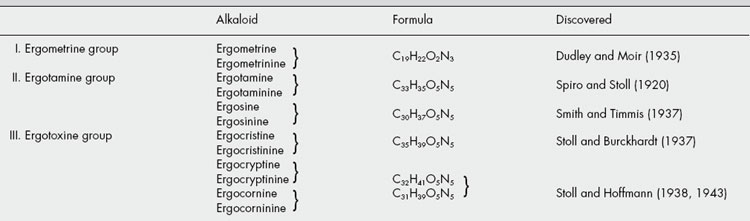
The ergot alkaloids (ergolines) can be divided into two classes: (1) the clavine-type alkaloids, which are derivatives of 6,8-dimethylergoline and have been extensively studied in cultures of the mycelium of the ergot fungus; and (2) the lysergic acid derivatives, which are peptide alkaloids. It is the latter class that contains the pharmacologically active alkaloids that characterize the ergot sclerotium (ergot). Each active alkaloid occurs with an inactive isomer involving isolysergic acid; the inactive isomers are not formed initially in the sclerotium but tend to accumulate as a result of unsuitable processing and poor or long storage. These alkaloids have been studied over many years and were not easy to characterize. Thus ‘ergotoxine’, which since its isolation in 1906 (by Barger and Carr and independently by Kraft) had been accepted as a pure substance, and in the form of ergotoxine ethanosulphonate was formerly used as a standard, was shown to be a mixture of the three alkaloids ergocristine, ergocornine and ergocryptine.
Six pairs of alkaloids predominate in the sclerotium and fall into either the water-soluble ergometrine (or ergonovine) group or the water-insoluble ergotamine and ‘ergotoxine’ groups. Table 26.6 gives the morephysiologically active member of each pair first. Alkaloids of groups II and III are polypeptides in which lysergic acid or isolysergic acid is linked to other amino acids. In the ergometrine alkaloids lysergic acid or its isomer is linked to an amino alcohol. Ergometrine was synthesized by Stoll and Hofmann in 1943. Other, new, peptide alkaloids have been isolated from submerged cultures of C. purpurea and from the field-growing fungus (L. Cvak et al., Phytochemistry, 1996, 42, 231; 1997, 44, 365).
Among the less important constituents of ergot may be mentioned histamine, tyramine and other amines and amino acids; acetylcholine; colouring matters; sterols (ergosterol and fungisterol); and about 30% fat. The cell walls are chitinous.
Variation in alkaloid constituents. Not only are chemical races very evident in C. purpurea with respect to alkaloid production but also the host plant is not without influence. Thus a new commercial strain of ergot adapted from a wild grass (Anthraxon lancifolius) to rye gave sclerotia containing 0.5% total alkaloids involving ergometrine (33%), ergotamine (17.6%), ergocornine (18.7%) and ergocryptine (22.7%). However, sclerotia produced on the grass as a result of natural infection did not contain ergometrine (K. K. Janardhanan et al., Planta Med., 1982, 44, 166). The application of specific amino acids to maturing sclerotia can also be used to influence the type of alkaloids produced (a technique also used with saprophytic cultures).
For recent studies and references on the investigation of the alkaloid gene cluster in C. purpurea see T. Haarmann et al., Phytochemistry, 2005, 66, 1312.
The artificial culture of the ergot fungus has received considerable attention, and, obviously, large-scale submerged fermentation with selected strains to give alkaloids of choice has commercial possibilities. Abe’s initial work in Japan showed that submerged cultures did not produce the typical alkaloids associated with the sclerotium but, rather, a series of new non-peptide bases (clavines) which unfortunately possessed no significant pharmacological action. Attempts were made by many workers to influence alkaloid production by modification of the culture medium and the fungus strain. As a result of successful experiments in 1960, the commercial manufacture of simple lysergic acid derivatives by fermentative growth of a strain of Claviceps paspali became feasible. The alkaloids produced are converted to lysergic acid which is used for the part-synthesis of ergometrine and related alkaloids. Other strains are now available which produce the peptide alkaloids in culture; not only can different chemical races of the fungus be used to produce specific groups of alkaloids but synthesis can also be directed by the addition of certain amino acids or their analogues to the fermentation liquid. In this way new unnatural alkaloids can be produced.
Flieger et al. (J. Nat. Prod., 1989, 52, 1003) found that with submerged cultures in the postproduction stage both the alkaloid concentration and the composition of the alkaloid mixtures underwent dramatic changes including the production of two new alkaloids, 8-hydroxyergine and 8-hydroxyerginine. (For papers pertaining to the isolation of new and unnatural alkaloids from submerged cultures of C. purpurea see N. C. Perellino et al., J. Nat. Prod., 1992, 55, 424; 1993, 56, 489.)
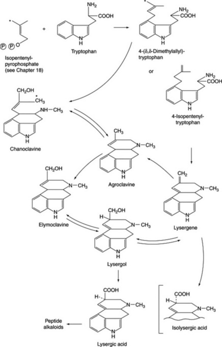
The majority of biosynthetic studies were at first directed to the clavine alkaloids, which could be easily produced in cultures but, until recently, their biological relationship to the lysergic acid derivatives remained obscure. The ergoline nucleus is derived from tryptophan and mevalonate, and current work has involved elucidating the biosynthetic relationship between the various clavine alkaloids, determining which of these intermediates is the true natural precursor of lysergic acid, and studying the initial hydrogen elimination from the C-4 of mevalonate to yield the stereo-configuration of chanoclavine-I. Possible biosynthetic routes for lysergic acid involving two isomeric intermediates are given in Fig. 26.29. A major problem has been not so much in discovering which reactions the fungus can effect when supplied with a given substrate as which route is actually involved in its normal metabolism. As a result of work with cell-free systems, Abe assigned a primary role to 4-isopentenyltryptophan and lysergene rather than to the dimethylallyl compound, agroclavine or chanoclavine. Later work by Gröger et al. (Planta Med., 1980, 40, 109) appeared to favour a scheme involving the latter compounds and this has now been generally substantiated with the intermediates involved with the ring closure between dimethylallyltryptophan and chanoclavine having been investigated (A. P. Kozikowski et al., J. Amer. Chem. Soc., 1993, 115, 2482).
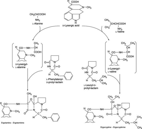
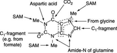
Work on the origin of the nitrogen of the peptide portions of the ergot alkaloids indicates that appropriate amino acids are specifically incorporated. Abe’s scheme is shown in Fig. 26.30 and he obtained intact incorporation of the units shown, in certain strains, but workers in Germany were unable to confirm these results. As has been mentioned earlier, unnatural amino acids can also be incorporated into the alkaloids.
Commercial ergot varies considerably in activity from batch to batch, and the differences cannot be fully explained by differences in storage; further, it is often found that inferior-looking ergot is highly active. Such variations are apparently due to the fact that there are a number of different chemical races of C. purpurea and in cultivating ergot by modern methods it is obviously important to prepare the spore-cultures used for infecting the rye from a race of the fungus known to develop ergots having a high content of the requiredalkaloids. Cultivated ergot may contain up to 0.5% of total alkaloids, and 0.15% is a minimum commercial value. In addition, there may be a minimal requirement for water-soluble alkaloids. The alkaloids can be determined by colorimetry, as they give a blue colour with a solution of p-dimethylaminobenzaldehyde.
Ergot of wheat is now imported into Britain and has been used medicinally in France. The sclerotia are shorter and thicker than those of rye. Instances of ergot on barley and rye in Britain, and on wheat and rye in the USA and Canada have been reported.
Ergot of oats has been used medicinally in Algiers. The sclerotia are black in colour, 10–12 mm long and 3–4 mm diameter.
Ergot of diss, which is produced on the Algerian reed Ampelodesma tenax, has appeared in commerce and is said to be highly active. The sclerotia may attain as much as 9 cm in length and are spirally twisted.
Ergot is particularly liable to attack by insects, moulds and bacteria. After collection it should be thoroughly dried, kept entire, and stored in a cool, dry place. If powdered and not immediately defatted, the activity decreases, but if defatted and carefully stored in an air-tight container, it will remain active for a long period. However, as indicated above, under certain conditions, loss of activity arises by the conversion of the pharmacologically important alkaloids to inactive isomers. Any sample of ergot which shows worm holes or a considerable amount of insect debris will almost certainly deteriorate further on storage.
Although whole ergot preparations were traditionally used in labour to assist delivery and to reduce post-partum haemorrhage, ergot itself has been largely replaced in the pharmacopoeias by the isolated alkaloids. Only ergometrine produces an oxytocic (literally ‘quick delivery’) effect, ergotoxine and ergotamine having quite a different action. Ergometrine is soluble in water or in dilute alcohol. It is often known, particularly in the USA, as ergonovine. Ergotamine and the semisynthetic dihydroergotamine salts are employed as specific analgesics for the treatment of migraine. Lysergic acid diethylamide (LSD-25), prepared by partial synthesis from lysergic acid, is a potent specific psychotomimetic.
Calabar beans (Ordeal beans) are the dried ripe seeds of Physostigma venenosum (Leguminosae), a perennial woody climber found on the banks of streams in West Africa. The plant bears typical papilionaceous flowers, and legumes about 15 cm long, each containing two or three seeds.
The seeds were formerly used by the west African tribes as an ‘ordeal poison’. They were first known in England in 1840. The myotic effect of the drug was noted in 1862 by Fraser, and physostigmine was isolated in 1864 by Jobst and Hesse.
Calabar beans have a somewhat flattened, reniform shape. They are 15–30 mm long, 10–15 mm wide and up to 15 mm thick. The seeds are extremely hard. The dark brown testa is smooth, except in the neighbourhood of the grooved hilum, which runs the whole length of the convex side and round one end, where it is somewhat wrinkled. On either side of the groove is a well-marked ridge and in the groove itself are the greyish, papery remains of the funiculus. A transverse section shows a large central cavity and two, very hard, concavo-convex cotyledons.
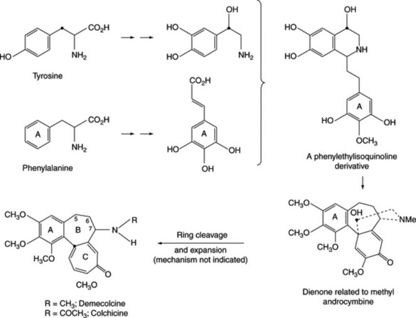
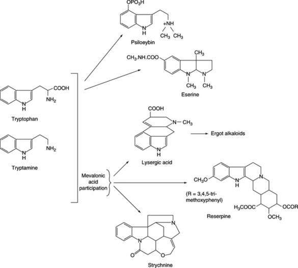
The seeds contain the alkaloids physostigmine or eserine, eseramine, isophysostigmine, physovenine, geneserine, N-8- norphysostigmine, calabatine and calabacine. The structure of geneserine, long regarded as an N-oxide, has been revised to include the oxygen in a ring system. The chief alkaloid, physostigmine, is present to the extent of about 0.15%. It is derived from tryptophan (see Fig. 26.26). On exposure to air it oxidizes into a red compound, rubreserine, and should therefore be protected from air and light. Both physostigmine salicylate and sulphate are included in the BP/EP. The former of the two is more stable and non-deliquescent. For both salts there is a colorimetic test for the elimination of eseridine and a non-aqueous titration assay.
Physostigmine salicylate is used for contracting the pupil of the eye, often to combat the effect of mydriatics. It has also been investigated as an intravenous injection for reversing the effects of a number of sedatives. With Alzheimer’s disease it has shown some evidence of inducing a slight improvement in intellectual and cognitive performance (Pharm. J., 1992, 249, 376) but galanthamine (q.v.) may prove superior. Physovenine has the same order of activity but that of eseramine is much lower.
Nux vomica consists of the dried, ripe seeds of Strychnos nux-vomica (Loganiaceae), a tree 10–13 m high with a distribution including Ceylon, India, East Bengal, Burma, Thailand, Laos, Cambodia and S. Vietnam. The drug is mainly collected in India and exported from Mumbai (Bombay), Madras, Cochin, Cocanada and Calcutta.
Nux vomica was known in Europe in the sixteenth century and was sold in England in the time of Parkinson (1640), mainly for poisoning animals. Strychnine was isolated in 1817 and brucine in 1819.
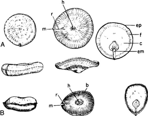
The fruit is a berry about the size of a small orange. When ripe it has a rather hard orange-yellow epicarp and a white, pulpy, interior in which 1–5 seeds are embedded. The seeds are washed free from pulp and dried. They are exported in small sacks, known as ‘pockets’, holding about 18–25 kg.
Nux vomica seeds are extremely hard and should be boiled in water for at least an hour in order to soften them sufficiently for dissection. The seeds are greenish-grey, disc-shaped, 10–30 mm diameter and 4–6 mm thick. Most of the seeds are nearly flat and regular in shape, but a few are irregularly bent and somewhat oval in outline. The edge is rounded or acute. The testa is covered with silky, closely appressed, radiating hairs. In the centre of one of the flattened sides is a distinct hilum, and a small prominence on the circumference marks the position of the micropyle, which is joined to the hilum by a radial ridge. To examine further, a boiled seed should be cut transversely and another one opened like an oyster by inserting the blade of a small knife or scalpel at a point on the circumference opposite the micropyle. The small embryo with two cordate cotyledons and a cylindrical radicle, the latter directed towards the micropyle, will be seen embedded in a grey, horny endosperm (Fig. 26.31). In the centre of the seed is a slit-like cavity. The seeds are odourless when dry; but if soaked in water and left for a day or two, they develop a very unpleasant odour. They have a very bitter taste.
Fig. 26.31 A, Strychnos nux-vomica seed; B, S. nux-blanda seed. Surface and lateral views of entire seed and inner surface of horizontally split seeds. All ×0.8. m, Micropyle; r, ridge; h, hilum; b, lateral ridge; ep, epidermis; f, area of fusion of two endosperm halves; c, area of central cavity; em, embryo.
(Drawn by Dr. T. D. Turner. For further details, see J. Pharm. Pharmacol., 1963, 15, 594.)
A radial section shows a very thin testa consisting of collapsed parenchyma and an epidermal layer of very characteristic lignified hairs (Fig. 41.7M). The latter have a very large, thick-walled base with slit-like pits. Surface irregularities in the bases of the hairs cause them to interlock with one another. The upper portions of the hairs are set at almost a right angle to the bases and all radiate out towards the margin of the seed, giving the testa its characteristic silky appearance. On the ridge connecting hilum and micropyle, however, the hairs are irregularly arranged. The upper part of the wallof the hair is composed of about 10 longitudinal ridge-like thickenings united by a thin wall so that the lignified ribs readily separate from one another on powdering. The lumen is circular in the upper part, but in the base has branches corresponding with the oblique pits in the wall. Fragments of testa, removed from a soaked seed, may be disintegrated by treatment with 50% nitric acid and a little potassium chlorate; the hairs can then be separated.
The endosperm consists of large, thick-walled cells, which are nonlignified and yield galactose and mannose on hydrolysis. When mounted in solution of iodine, they show well-marked protoplasmic threads (plasmodesma) passing through the walls (see Fig. 42.1G) and an oily plasma containing a few aleurone grains and the alkaloids strychnine and brucine. Strychnine is most abundant in the inner part of the endosperm and brucine in the outer layers. The presence of strychnine is shown by mounting a section in a solution of ammonium vanadate in sulphuric acid, when a violet colour is produced; of brucine, by mounting in nitric acid, when a crimson colour is observed.
The length of lignified ribs of the hairs per milligram of nux vomica seed has been used for the determination of the content of seeds in veterinary medicines, see ‘Quantitative Microscopy’.
Nux vomica usually contains about 1.8–5.3% of the indole alkaloids strychnine and brucine. Strychnine (formula Fig. 26.26) is physiologically much more active than brucine and the seeds are therefore assayed for strychnine and not for total alkaloids. They usually contain about 1.23% of strychnine and about 1.55% of brucine. Minor related alkaloids include α-colubrine, β-colubrine, icajine, 3-methoxyicajine, protostrychnine, vomicine, novacine, N-oxystrychnine, pseudo-strychnine and isostrychnine.
For a review (188 refs) of recent studies concerning the synthesis of strychnine, see M. Shibasaki and T. Ohshima, The Alkaloids, 2006, 64, 103.
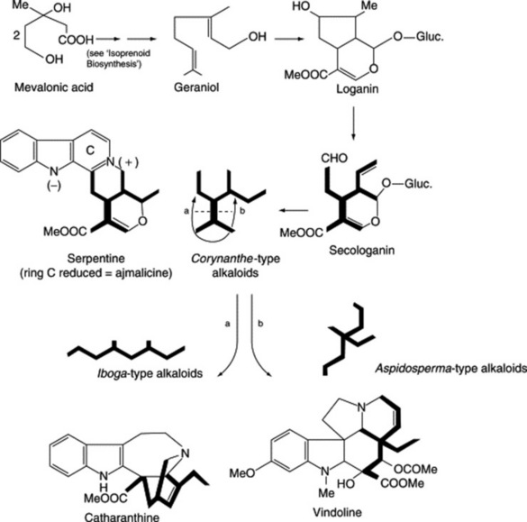
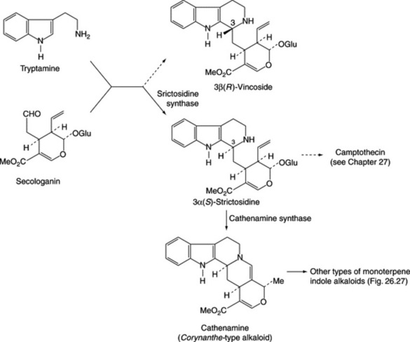
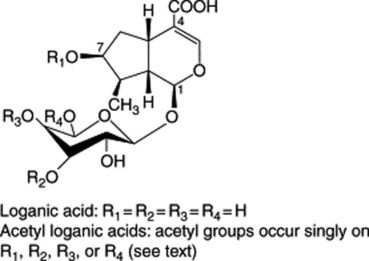
Iridoids of the seeds include the glycoside loganin (Fig. 26.27), loganic acid and 7-O-acetyl loganic acid together with three new iridoids, 6′-O-acetyl loganic acid, 4′-O-acetyl loganic acid and 3′-O- acetyl loganic acid (X. Zhang et al., Phytochemistry, 2003, 64, 1341).
The seeds also contain chlorogenic acid (see Fig. 19.5) and about 3% of fixed oil.
Loganin, although present only in small amounts in the seed, occurs to the extent of about 5% in the fruit pulp together with secologanin; these compounds are intermediates in the biogenesis of the strychnine-type alkaloids (Fig. 26.27).
Seasonal variations in alkaloid content of S. nux-vomica have been studied (K. H. C. Baser and N. G. Bisset, Phytochemistry, 1982, 21, 1423).
The action of the whole drug closely resembles that of strychnine. The alkaloid was formerly used as a circulatory stimulant in such cases as surgical shock, but its use is now more limited to that of a respiratory stimulant in certain cases of poisoning. Like other bitters, strychnine improves the appetite and digestion, but it has been considerably misused as a ‘general tonic’. Nux vomica is used in Chinese medicine for much the same purposes as in Western medicine and the seeds are usually processed to reduce their toxicity. Heat-treatment of the seeds reduces the normal levels of the principal alkaloids and the amounts of isostrychnine, isobrucine, strychnine N-oxide and brucine N-oxide are increased (B.-C. Cai et al., Chem. Pharm. Bull., 1990, 38, 1295).
The genus Strychnos continues to attract considerable attention. (For extensive reviews on the taxonomy, chemistry and ethnobotany of the American, African and Asian species see N. G. Bisset et al., Lloydia, 33, 201; 34, 1; 35, 95, 193; 36, 179; 37, 62; 39, 263; also Bisset’s review on alkaloids of the Loganiaceae in Indole and Biogenetically Related Alkaloids, 1980 (eds J. D. Phillipson and M. K. Zenk) p. 27, London: Academic Press; and J. Quetin-Leclercq et al., J. Ethnopharm., 1990, 28, 1, review with c. 150 refs.)
Ignatius beans are the seeds of Strychnos ignatii, a plant occurring in the Philippines, Vietnam and elsewhere. The fruits are larger than those of nux vomica and may contain as many as 30 seeds. These are about 25 mm long, dark grey in colour and irregularly ovoid in shape. The structure closely resembles that of nux vomica, but the testa, which bears irregularly arranged greyish hairs, is easily rubbed off and is almost entirely absent in the commercial drug. The seeds contain about 2.5–3.0% of total alkaloids, of which about 46–62% is strychnine. They are mainly used for the preparation of strychnine and brucine. The seeds of S. ignatii from Java (S. tieute) contain 1.5% strychnine and no brucine and from Hainen (S. hainanensis) mainly brucine with little strychnine.
In addition to the seeds, other parts of the plants of Strychnos spp. may contain alkaloids including strychnine (B. De Datta and N. G. Bisset, Planta Med., 1990, 56, 133; G. Massiot et al., Phytochemistry, 1992, 31, 2873).
S. potatorum, from India, and S. nux-blanda, from Burma, have been substituted for nux vomica; although they contain no strychnine or brucine, seeds of the former have been reported to contain the alkaloid diaboline and its acetyl derivative, triterpenes and sterols. They are best distinguished by means of the ammonium vanadate reagent. The seeds of S. potatorum are used in India for clearing water, whence the specific name. They will also flocculate heavy metal contaminants in water and are capable of mopping up radioactive isotopes from nuclear waste. The protein responsible for this property has now been isolated (Pharm. J., 1994, 252, 238). The tannins present in the seeds are suggested as the possible active constituents associated with the folklore treatment of chronic diarrhoea (S. Biswas et al., Fitoterapia, 2002, 73, 43).
Gelsemium consists of the dried rhizomes and roots of the American yellow jasmine, Gelsemium sempervirens (G. nitidum) family Loganiaceae, indigenous to southern USA. It is a climbing plant and produces scented yellow flowers; it should not be confused with the yellow-flowering jasmine (Jasminum nudiflorum, family Oleaceae) cultivated as an ornamental in Europe.
The drug occurs in cylindrical pieces 3–20 cm long and 3–30 mm diameter. The outer cork cells of the rhizome are reddish-brown and the inner ones yellowish. As growth takes place, the outer cork cells crack and the inner cork shows itself as a yellowish-brown reticulation. The roots are somewhat smaller than the rhizome and have a uniform yellowish-brown cork. Gelsemium breaks with an irregular splintery fracture. It has a slightly aromatic odour and a bitter taste. A transverse section of the rhizome shows a thick cork, a cortex containing groups of sclerenchyma, a dense wood, internal as well as external phloem and a small pith. The roots, on the other hand, have no sclerenchyma in the cortex and no pith.
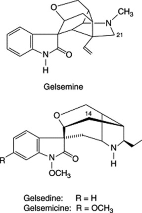
Gelsemium contains extremely toxic alkaloids of unique skeletal type. Gelsemine is the principal alkaloid and is the one most studied although it is not as toxic as gelsemicine. Other oxindole bases characterized are sempervirine, 1-methoxy- and 21-oxo-gelsemine, 14- hydroxygelsemicine, gelsedine and 14-hydroxy-gelsedine. Three new alkaloids of the gelsidine type, together with an iridoid, have been reported by M. Kitajima et al., Chem. Pharm. Bull., 2003, 51, 1211.
In a review (129 refs) H. Takayama and S. Sakai list 45 alkaloids derived from G. elegans, G. sempervirens and G. rankinii; they are classified into five groups according to structure (The Alkaloids, 1997, 49, 1).
Scopoletin is responsible for the blue fluorescence of the broken drug in ultraviolet light. Iridoids and glucoiridoids have been isolated from the aerial parts.
Gelsemium is used (BHP, 1983) in the treatment of trigeminal neuralgia and migraine, but its use requires great care, as dangerous side-effects may develop. It has been studied for its anticancer properties.
G. elegans is used in Oriental folk medicine for much the same purposes as G. nitidum. For information on new and known alkaloids of the leaves and stems of this species see Y.-K. Xu et al., J. Nat. Prod., 2006, 69, 1347.
Rauwolfia consists of the dried rhizome and roots of Rauwolfia serpentina (Rauvolfia serpentina), Apocynaceae, a small shrub found in India, Pakistan, Burma, Thailand and Java. The geographical source appears to influence the alkaloidal content, and manufacturers tend to prefer drug obtained from India or Pakistan. Reserpine, the most important constituent, is contained in many other species of Rauwolfia (see ‘African Rauwolfia’ below); it is included in the BP/EP.
Although used in India from time immemorial, it was not until 1942 that favourable reports were published of the use of the drug in powdered form. Since then research workers have studied the pharmacognosy, chemistry, pharmacology and clinical uses of many species of Rauwolfia and of the alkaloids obtained from them.
The drug is collected mainly from wild plants, but cultivation of the drug will probably increase as wild plants become more scarce; in parts of India collectors are required to leave some root from each plant in the ground for future growth. Nevertheless, and coupled with the low seed viability, the plant is regarded as an endangered species in India. Consequently, the potential for the regeneration of plants from cell cultures and the possible utilization of nodal culture has received some attention (see C. M. Ruyter et al., Planta Med., 1991, 57, 328; N. Sharma and K. P. S. Chandel, Plant Cell Rep., 1992, 11, 200).
As other species of Rauwolfia are found in India, care is needed to identify the correct plant. When first imported, many commercial samples were found to be adulterated; this was due in many cases to lack of knowledge, and substitution of, or adulteration with, other species has become much rarer in recent years. After collection the drug is cut transversely into convenient-sized pieces and dried.
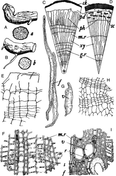
The first detailed description of the drug was made by Wallis and Rohatgi in 1949. It usually occurs in cylindrical or slightly tapering, tortuous pieces about 2–10 cm long and 5–22 mm in diameter (Fig. 26.32A). The roots are rarely branched and rootlets, 0.5–1 mm in diameter, are rare. Pieces of rhizome closely resemble the root but may be identified by a small central pith; they occasionally have attached to them small pieces of aerial stem.
Fig. 26.32 Rauwolfia serpentina and R. vomitoria roots. A, Root of R. serpentina (×1); a, transverse section (TS) of same (×1); B, root of R. vomitoria, ×1; b, TS of same (×1); C, diagrammatic TS of R. serpentina root (×15); D, diagrammatic TS of R. vomitoria root (×15); E, TS of cork of R. serpentina; F, TS of the secondary wood of R. serpentina; G, fibres and vessel of R. serpentina, isolated by maceration; H, TS of cork of R. vomitoria; I, TS of the secondary wood of R. vomitoria; E, F, G, H and I, (all ×150). ck, cork; f, fibre; g.r, growth ring; m.r, medullary ray; pd, phelloderm; ph, phloem; r, resinous material; s, starch; sc, group of sclereids; v, wood vessel; xy, xylem (J. D. Kulkarni, partly after T. E. Wallis and S. Rohatgi (R. serpentina) and W. C. Evans (R. vomitoria))
The outer surface is greyish-yellow, light brown or brown with slight wrinkles (young pieces) or longitudinal ridges (older pieces); occasional circular scars of rootlets. In this species the bark exfoliates readily, particularly in the older pieces, and may leave patches of exposed wood. The drug breaks readily with a short fracture. The smoothed transverse surface shows a narrow, yellowish-brown bark and a dense pale yellow wood, which occupies about three-quarters of the diameter. Both bark and wood contain abundant starch. Some commercial samples show mould. The recently dried drug has a slight odour which seems to decrease with age. Taste, bitter.
The cork is stratified into about two to eight zones (Fig. 26.32E), which consist of smaller and radially narrower suberized but unlignified cells alternating with larger radially broader cells which are lignified. In many pieces much of the cork is exfoliated, and for section cutting it may be best to select pieces with little exfoliation, separate these from the wood and cut sections of the bark and wood separately. Most of the cells of the secondary cortex are parenchymatous and contain starch; isolated latex cells may occur in this region, particularly in the Dehra Dun variety. The phloem is narrow and consists mainly of parenchyma with scattered sieve tissue. Sclerenchyma is absent (distinction from many other species such as R. tetraphylla (R. canescens), R. micrantha, R. densiflora, R. perakensis and R. vomitoria; see Fig. 26.32 C, D). Most of the parenchymatous cells of the bark contain starch grains, and others prisms or conglomerate crystals of calcium oxalate.
The xylem is entirely lignified and usually shows three to six annual rings. The medullary rays, which are one to five cells wide, contain starch and alternate with the rays of the secondary xylem, which consist of vessels, fibres and xylem parenchyma. Compared with many other species of Rauwolfia, the vessels of R. serpentina are small (up to 57 μm) and are less numerous than in most of the likely adulterants. The starch grains are larger in the wood than in the bark and measure from 5–8 to 12–20 μm.
Rauwolfia contains at least 40 alkaloids, which total some 0.7–2.4%. Other substances present include phytosterols, fatty acids, unsaturated alcohols and sugars.
In 1931 Siddiqui and Siddiqui isolated ajmaline (rauwolfine), ajmalinine, ajmalicine, serpentine and serpentinine. The chief therapeutically important alkaloids are reserpine (isolated in 1952; formula Fig. 26.26) and rescinnamine (isolated in 1954). These are esters derived from methyl reserpate and trimethoxybenzoic acid in the case of reserpine and trimethoxycinnamic acid in the case of rescinnamine. New alkaloids continue to be isolated; recently, five anhydronium bases (e.g. 3,4,5,6-tetradehydroyohimbine) for the first time (O. Wachsmuth and R. Matusch, Phytochemistry, 2002, 61, 705) and five new indole alkaloids together with a new iridoid glycoside, 7-epiloganin, a new sucrose derivative and 20 known compounds (A. Itoh et al., J. Nat. Prod., 2005, 68, 848).
R. serpentina cell suspension cultures have proved an important tool in the elucidation of monoterpenoid indole alkaloid biogenesis and in this connection the significance, and isolation, of strictosidine synthase has been considered. By the use of cell cultures Stöckigt in 1988 was able to clarify a 10-step biosynthetic pathway from strictosidine to the typical rauwolfia alkaloid, ajmaline. His group has also shown that the principal alkaloid of cell suspension cultures of R. serpentina is raucaffricine occurring in amounts of up to 1–6 g l−1 in the nutrient medium, representing 2.3% of the dried cells, a value some 67 times higher than found for the roots. Ajmalicine, an antiarrhythmic drug, is also produced to the extent of 0.6% (cell dry wt.) together with over 30 different monoterpenoid alkaloids in trace amounts including five glucoalkaloids. Addition of high levels of ajmaline to the cell culture medium promoted the formation of a new group of alkaloids, the raumaclines, not found in the roots (for further details see S. Endress et al., Phytochemistry, 1993, 32, 725 and references cited therein).
Hairy root cultures of R. serpentina, produced by Agrobacterium rhizogenes transformation, synthesized ajmaline (0.045% dry wt.) and serpentine (0.007% dry wt.) (B. D. Benjamin et al., Phytochemistry, 1994, 35, 381); minor alkaloids have also been recorded (H. Falkenhagen et al., Can. J. Chem., 1993, 71, 2201). R. verticillata hairy roots have been shown to produce reserpine and aimaline. Working on hairy root cultures, Stockigt’s group has reported on the isolation of three new monoterpenoid indole alkaloids of the sarpagine group along with 16 known compounds together with the first natural occurrence (cf. cell cultures above), of the rare raumacline type alkaloids (Y. Sheludko et al., J. Nat. Prod., 2002, 65, 1006; Planta Medica 2002, 68, 435).
Two monoterpenoid indole alkaloids and four β-carbolines have been isolated from cultured hybrid cells of Rauwolfia serpentina and Rhazya stricta, not all of the compounds being found in the parent plants (N. Aimi et al., Chem. Pharm. Bull., 1996, 44, 1637).
An assay for total alkaloids is not a true measure of therapeutic activity, since only some of the alkaloids have the desired pharmacological action. The BPC 1988 and USP/NF 1995 determine the reserpine-like alkaloids by utilizing the colour reaction between an acid solution of reserpine (and rescinnamine) and sodium nitrite solution.
The 110 Rauwolfia species classified by Pichon in 1947 were reduced by Woodson in 1957 to 86. Some of these occur in more than one geographic area but their approximate geographic areas are as follows: Central and South America 34, Africa 20, Far East 24, India and Burma 7, Hawaii, New Guinea and New Caledonia 6. A large number of these species have been examined for reserpine and related alkaloids.
In the identification of the roots of species of Rauwolfia useful characters to be seen in transverse sections are: cork (whether stratified or lignified); cortex and phloem (presence or absence of sclereids or fibres); wood (relative number, distribution and size of vessels). As the species vary from herbs to large trees, the roots vary considerably in size. Some samples of drug contain aerial stems, which usually contain less reserpine than the roots and have unlignified pericyclic fibres.
R. tetraphylla (R. canescens, R. hirsuta) is a species of wide distribution—tropical South America, the Caribbean, India, Australia (Queensland). The root was at one time occasionally substituted for R. serpentina and could be recognized by its non-stratified cork, and sclereid groups in the phloem. It has served as a commercial source of reserpine and the alkaloid deserpidine, possibly particularly important as R. serpentina is now classed as an endangered species. Micropropagation protocols have been described for in vitro mass multiplication of the plant (D. Sarma et al., Planta Medica, 1999, 65, 277).
R. nitida is a West Indian species from the root-bark of which 33 indole alkaloids have been isolated.
Rauwolfia preparations and reserpine are used in the management of essential hypertension and in certain neuropsychiatric disorders. Ajmaline, which has pharmacological properties similar to those of quinidine, is marketed in Japan for the treatment of cardiac arrhythmias.
An estimated 3500 kg of ajmalicine is isolated annually from either Rauwolfia or Catharanthus spp. by pharmaceutical industries for the treatment of circulatory diseases.
Conflicting reports on the possible involvement of the rauwolfia alkaloids in breast cancer engendered a natural hesitation in their use. A report in the Lancet (1976) suggested that the alkaloids do not initiate the carcinogenic process but that they promote breast cancer from previously initiated cells.
African rauwolfia consists of the dried roots of Rauwolfia vomitoria Afz. The plant is a bush or tree widely distributed in tropical Africa from the west coast to Mozambique. It is the most important African rauwolfia for the commercial preparation of reserpine.
As the tree may attain a height of 10 m, the roots are larger than those of R. serpentina. Before drying they are cut transversely, but are rarely sliced longitudinally. Roots up to 5 cm or more in diameter are sometimes found but the commercial drug usually consists of much smaller pieces. Occasional shipments have been made consisting of the bark only.
The first detailed description of the drug was published by Evans in 1956. It occurs in cylindrical or flattened pieces, usually 0.15–1.5 cm diameter and up to 30 cm long. The roots taper slightly and are occasionally branched. The outer surface is greyish-brown, longitudinally furrowed or rubbed smooth, since the outer cork easily flakes off. Pieces do not break easily, but the fracture is short in the bark and splintery in the wood. The smoothed transverse surface shows a narrow brown bark and a buff or yellowish, finely radiate wood. Odourless; taste, bitter. Pieces of rootstock with attached stem-bases are sometimes found in the drug.
The drug is easily distinguished from R. serpentina by the groups of sclereids in the bark arranged in up to five discontinuous bands and by the large vessels of the wood which are up to 180 mu;m in diameter (Fig. 26.32 D and I).
African rauwolfia contains reserpine and rescin-namine and alkaloids of the same type such as reserpoxidine and seredine. Many other alkaloids such as ajmaline, alstonine and yohimbine are also present. Court’s group (see Planta Med., 1982, 45, 105) isolated 42 indole alkaloids from the stem-bark and identified 39. The major alkaloids were heteroyohimbines (especially reserpiline) and Na-demethyldihydroindoles. The interrelationship of these alkaloids with those in the root of the plant is discussed in the same paper. New indole alkaloids from R. vomitoria extracts have continued to be reported.
Other African rauwolfias containing reserpine are R. caffra (R. natalensis), R. mombasiana, R. oreogiton, R. obscura, R. cumminsii, R. volkensii and R. rosea. Court and coworkers have made a systematic study of the microscopy and chemistry of African species (for a report see W. E. Court, Planta Med., 1983, 48, 228). The roots of R. caffra closely resemble those of R. vomitoria but the cork has not the same tendency to flake off. In sections the main difference is that in R. vomitoria there are alternating lignified and unlignified cork cells, while in R. caffra all the cork cells are lignified. R. caffra contains the alkaloid raucaffricine—one of relatively few examples of monoterpenoid indole glucoalkaloids within the group. R. mombasiana differs from R. vomitoria in the structure of the wood of the root; R. rosea, R. volkensii and R. obscura lack sclereid development.
Several Alstonia species (Apocynaceae) have been used in the past as antimalarials, and the barks of the Indian Alstonia scholaris and the Australian A. constricta were included in the 1914 edition of the British Pharmacopoeia. At least 11 species are known to contain alkaloids, such as alstonine, alstoniline, cillastonine and echitamine. Interest in them was again awakened by the isolation in 1955 of reserpine in moderate yield from the root-bark of A. constricta; reserpine has since been isolated from A. venenata. For a report on the isolation of a new indole alkaloid and a new glycosidic indole alkaloid from the trunk bark of Indonesian Alstonia scholaris, see A. A. Salim et al., J. Nat. Prod., 2004, 67, 1591. Descriptions of Alstonia barks will be found in the older editions of reference books (e.g. the 22nd edition of the USD, p. 1227).
Yohimbe bark is derived from Pausinystalia yohimbe (Rubiaceae), a tree growing in the Cameroon Republic. It occurs in flat or slightly quilled pieces up to 75 cm long and 2 cm thick. The grey-brown cork has furrows and cracks and patches of lichen. The inner surface is reddish-brown and striated. Taste, bitter. It contains the indole alkaloid yohimbine, which is structurally related to reserpine.
The bark is well-recognized for its aphrodisiac property and yohimbine is effective in the symptomatic treatment of erectile dysfunction, producing fewer side-effects than invasive treatments (M. H. Pittler, Fortschritte der Medizin, 1998, 116, 32).
The large genus Aspidosperma (Apocynaceae) contains many alkaloid-containing South American trees. The alkaloids are of various indole types formed by a number of different biogenetic pathways. Among the many investigated are yohimbine, which is structurally related to reserpine, and aspidospermine, which has the same general structure as vindoline (see Fig. 26.27). The Aspidosperma barks are therefore potential sources of alkaloids, because the trees are large and the barks would be commercially available cheaply and in almost unlimited quantities.
The genus Mitragyna (Rubiaceae) occurs in West and East Africa, India and S.E. Asia. More than 30 different alkaloids have been characterized, and the majority of these are indole or oxindole structures with an open or closed ring E; they exist in various isomeric forms. One alkaloid, mitragynine, isolated from Mitragyna speciosa has analgesic and antitussive properties similar to those of codeine. Shellard and coworkers published extensively on the genus during the 1960s and 1970s (for more recent work on M. speciosa from the same Department see P. J. Houghton et al., Phytochemistry, 1991, 30, 347). The mis-use of mitragyna as an hallucinogen is considered in Chapter 39.
In addition to Uncaria gambir, the source of catechu (q.v.), the genus is notable for its alkaloids, which resemble those of Mitragyna. Uncaria hooks, the dried climbing hooks and stems of U. sinensis, have sedative and antispasmodic properties. They are used in Chinese medicine for the relief of headaches and dizziness caused by hypertension and for the treatment of convulsions in children. The drug contains indole alkaloids, e.g. rhyncophylline and indole alkaloid glycosides which exhibit a long-lasting hypotensive effect (K. Endo et al., Planta Med., 1983, 49, 188; S. Kawazoe et al., ibid., 1991, 57, 47).
Uncaria rhynchophylla, a species also used in Chinese medicine, is reported to contain various alkaloids including rhynchophylline, corynoxeine, corynantheine and among others, hirsutine which exhibit antihypertensive, neuroprotective and vasodilator effects; (+)-catechin and (−)-epicatechin have been isolated for the first time from this species (W.-C. Hou et al., J. Ethnopharmacology 2005, 100, 216). Other research suggests this species to be an effective anxiolytic agent acting via the serotonergic nervous system (K. W. Jung et al., J. Ethnopharmacology, 2006, 108, 193).
U. tomentosa, one of two species found in S. America, features in the traditional medicine of Peru. It produces similar hooks to U. sinensis being known locally by the Spanish as ‘una degato’ (tomcat’s claw).
In a number of reports on this species, M. Kitajima et al., have recorded a new glucoindole akaloid, 3,4-dehydro-5-carboxystrictosodine, various triterpenes including nor-triterpene glycosides and cincholic acid glycosides (see Chem. Pharm. Bull., 2004, 52, 1258 and references cited therein). The plant has a potential immunostimulant action and has been examined for its pharmacological and toxicological properties (K. Keplinger et al., J. Ethnopharmacology, 1999, 64, 23; I. Lemaire et al., J. Ethnopharmacology, 64, 109). It is being used traditionally to treat a large number of conditions, including cancer (R. Pilarski et al., J. Ethnopharmacology, 2006, 104, 18; L. De Martino et al., J. Ethnopharmacology, 2006, 107, 91; G. Gonçalves et al., Phytochemistry, 2005, 66, 89).
Vinca major and V. minor
The greater periwinkle (Vinca major) together with the lesser periwinkle (V. minor) are the only members of the essentially tropical and subtropical family Apocynaceae found wild in the British Isles. The former is listed in the BHP (1983) for the treatment of menorrhagia and topically as an application for haemorrhoids but is now seldom used. The following indole alkaloids have been characterized: reserpine, majdine, akuammicine, strictosodine (Fig. 26.34), pseudoakuammigine, akuammine and possibly 10-hydroxycathofoline. New alkaloids continue to be reported e.g. Atta-ur-Rahman et al., Phytochemistry, 1995, 38, 1057. More than 50 alkaloids have been isolated from the leaves of V. minor, a few of them quaternary (for a recent report see D. Uhrin et al., J. Nat. Prod., 1989, 52, 637). Vincamine, first isolated from V. minor in 1953, is available as a vasodilatory drug.
Cinchona bark consists of various species, races and hybrids of Cinchona (Rubiaceae), large trees indigenous to Colombia, Ecuador, Peru and Bolivia. The BP/EP recognizes the whole or cut, dried bark of Cinchona pubescens Vahl (C. succirubra Pavon), C. calisaya (Weddell), of C. ledgeriana (Moens ex Trimen) or its varieties or hybrids, containing not less than 6.5% of total alkaloids, 30–60% of which consists of quinine-type alkaloids. The former importance of cinchona bark and its alkaloids in the treatment of malaria has been lessened by the introduction of synthetic drugs, but it remains of great economic importance, and salts of quinine and quinidine are included in most pharmacopoeias.
Collection from wild trees was soon replaced by cultivation, and most research was undertaken by the Dutch in Java and the British in India to obtain hybrids which are rich in alkaloids. While Indonesia and India remain important producers of cinchona, a high percentage of the total crop is now grown on plantations in Tanzania, Kenya, Guatemala and Bolivia.
The natives of South America do not appear to have been acquainted with the medicinal properties of cinchona bark, the bitter taste of which inspired them with fear. Although Peru was discovered in 1513, the bark was first used for the cure of fevers about 1630. The name ‘Cinchona’ is said to be derived from a Countess of Chinchon, wife of a viceroy of Peru who it was long believed was cured in 1638 from a fever by the use of the bark. According to recent study of the Count’s diary, it appears that the Countess never suffered from malaria or other fever during her stay in Peru, and although the Count himself did so, there is no record of his having been treated with cinchona bark. The remedy, which became known as ‘Pulvo de la Condesa’, acquired a considerable reputation and was known in Spain in 1639. The further distribution of the bark was largely due to the Jesuit priests, and the drug became known as Jesuit’s Powder or Peruvian Powder. It first appeared in the London Pharmacopoeia in 1677 under the name of ‘Cortex Peruanus’.
The bark was originally obtained by felling the wild trees, which were exterminated in many districts. Ruiz (1792) and Royle (1839) suggested the cultivation of cinchonas in other parts of the world. Weddell germinated seeds in Paris in 1848, and the plants were introduced into Algiers in the following year but without much success. A further attempt by the Dutch was made in 1854, seeds and plants being obtained from Peru by Hasskarl and introduced into Java. An English expedition under Markham in 1860 led to the introduction of C. succirubra (the most hardy species), C. calisaya, and C. micrantha into India. Seeds of C. ledgeriana were obtained in Bolivia by Charles Ledger in 1865 and were bought by the Dutch for their Javanese plantations. A fascinating book covering Ledger’s exploits is The Life of Charles Ledger (1818–1905) by G. Grammicia, Macmillan Press, London, 1988. World War II and subsequent fighting in Malaya and Vietnam increased the demand for cinchona and stimulated cultivation in Africa and Central and South America.
The production of cinchona bark is a highly specialized section of tropical agriculture. An acid soil, rainfall and altitude are all important factors in cinchona production. Selection of high-yielding strains is of paramount importance, and grafting techniques with C. succirubra as stock may be employed. Seedlings need careful treatment and propagation to avoid disease attack, etc. Since the mid-1970s a disease of the cinchona tree, known as stripe canker, has posed a threat to the plantations of Central Africa. The disease, also known in Central America, is caused by the phytopathogenic fungus Phytophthora cinnamomi, which causes sunken necrotic stripes in the bark and kills thousands of trees a year.
In view of the number of hybrids which are cultivated, the distinction of the various commercial cinchona barks is a matter of some difficulty. In Table 26.7 the notes on four important species have been made as concise as possible to facilitate comparison.
Cinchona barks have the general microscopical structure shown in Fig. 26.33. The cork is composed of several layers of thin-walled cork cells, arranged in regular radial rows and appearing polygonal in surface view. Their cell contents are dark reddish in colour. Within the cork cambium is a phelloderm of several layers of regular cells with dark walls. The cortex is composed of tangentially elongated, thin-walled cells containing amorphous reddish-brown matter or small starch grains 6–10 µm diameter. Scattered in the cortex are idioblasts containing microcrystals of calcium oxalate and secretion cells. The phloem consists of narrow sieve-tubes showing transverse sieve plates, phloem parenchyma resembling that of the cortex and large characteristic spindle-shaped phloem fibres with thick conspicuously striated walls traversed by funnel-shaped pits. The phloem fibres occur isolated or in irregular radial rows. The distribution and size of the phloem fibres differ in the various species(those of C. succirubra are 350–600–700–1400 mu;m long and 30–40–70–100 mu;m in diameter; for other species see 10th edition). The medullary rays are two or three cells wide, the cells being thin-walled and somewhat radially elongated.
Fig. 26.33 Cinchona bark. A, specimen of Cinchona succirubra (× 0.5); B, transverse section of bark (×25); C, isolated phloem fibres (×50); D, portion of phloem fibre with surrounding parenchyma; E, cork cells in surface view; F, idioblast with calcium oxalate; G, starch (all × 200). ck, Cork; ct, cortex; f, fibres protruding from fracture; id, idioblast; I, lichen patches; l.f, longitudinal fissure; m.r, medullary ray; pd, phelloderm; pg, phellogen; p.f, phloem fibres; s.c, secretory cell; t.f, transverse fissure.
Cinchona bark contains quinoline alkaloids (see Fig. 26.34). The principal alkaloids are the stereoisomers quinine and quinidine and their respective 6′-demethoxy derivatives, cinchonidine and cinchonine. The quinine series has the configuration 8S, 9R and the quinidine 8R, 9S (Fig. 26.34); other alkaloids of lesser importance have been isolated. Some of these (e.g. quinicine and cinchonicine) are amorphous. The amount of alkaloids present and the ratios between them vary considerably in the different species and hybrids, also according to the environment of the tree and the age and method of collection of the bark.
Fig. 26.34 Outline of possible biogenetic pathway for Cinchona alkaloids. Marked atoms illustrate the structural changes.
The alkaloids appear to be present in the parenchymatous tissues of the bark in combination with quinic acid and cinchotannic acid. Quinicacid (see Fig. 19.5) is present to the extent of 5–8%. Cinchotannic acid is a phlobatannin and a considerable amount of its decomposition product, ‘cinchona red’, is also found in the bark. Other constituents are quinovin (up to 2%), which is a glycoside yielding on hydrolysis quinovaic acid and quinovose (isorhodeose).
Anthraquinones, which as a group of compounds are associated with the family Rubiaceae (see Table 21.3), are not normally found in quantity in the bark of cinchona as indicated by the isolation of norsolorinic acid, a tetrahydroxyanthraquinone, in 0.0008% yield from the bark of C. ledgeriana. However, they are produced in cell cultures of the plant and by infection of the bark with Phytophthora cinnamomi. The latter case may be associated with a phytoalexin defence mechanism; in infected material, alkaloid production is lowered. In connection with the production of anthraquinones in cell cultures, enzymes associated with the later stages of glycoside formation have been isolated. This work involved glucosidases in C. succirubra cell cultures and theisolation of five distinct glucosyltransferases (EC 2,4,1,-) which catalyse the transfer of the glucosyl moiety from UDP-glucose to the hydroxyl groups of those anthraquinones found in cinchona cultures (e.g. emodin, anthrapurpurin, quinizarin, etc.).
Grafting experiments and organ culture suggest that the alkaloids are formed principally in the aerial parts of the plant. Although the quinoline alkaloids have structures which by inspection might suggest anthranilic acid as a biological precursor, they are, in fact, as originally suggested by Janot et al. in 1950, derived from indolic precursors. This has been demonstrated by the specific incorporation of tryptophan (indole moiety), loganin and geraniol (terpenoid moiety) into the quinine of Cinchona spp. The pathway, largely established by Battersby’s and Leete’s groups, involves alkaloids of the serpentine type as illustrated in Fig. 26.34.
The proposed biosynthetic route has been supported and elaborated by enzyme studies. The important role of strictosidine synthase in the initial stages of the biogenesis of some tryptophan-derived alkaloids and its isolation from C. robusta was mentioned at the beginning of this section. The enzyme tryptophan decarboxylase (EC 4.1.1.28), which provides tryptamine, is also involved in these early reactions. An enzyme (cinchoninone: NADPH oxidoreductase) associated with the pathway has been isolated from cells of a suspension culture of Cinchona ledgeriana; it catalyses the reduction of cinchoninone to an unequal mixture of cinchonine and cinchonidine. The enzyme can be resolved (by ion exchange) into two isoenzymic forms both of which have an absolute requirement for NADPH and catalysed reversible reactions. Isoenzyme I acts specifically on cinchoninone in the forward direction of the pathway and on cinchonidine and cinchonine in the reverse direction. Isoenzyme II has a broad specificity acting on all the quinoline alkaloids of cinchona tested.
The barks of certain species of Remijia (Rubiaceae) contain alkaloids. That of R. pedunculata is quoted (USP) as a source of quinidine. It also contains cupreine, an alkaloid which responds to the thalleioquin test and by methylation forms quinine. False cuprea bark (R. purdiena) contains no quinine but an alkaloid cusconidine and small proportions of cinchonine and cinchonamine.
Alkaloids of the indole type (e.g. cinchophylline) are generally considered to typify the leaves, so that it is of interest that Phillipson et al. (J. Pharm. Pharmacol., 1981, 33, 15P) isolated quinine from leaves of C. succirubra grown in Thailand. Thirteen alkaloids have been separated by HPLC.
Galenicals of cinchona have long been used as bitter tonics and stomachics. On account of the astringent action, a decoction and acid infusion are sometimes used as gargles. Before World War II, quinine was the drug of choice for the treatment of malaria but became largely superseded by the advent of synthetic antimalarials developed during that period. It has, however, remained of importance in Third World countries and has re-emerged as suitable for the treatment of Plasmodium falciparum infections (falciparum malaria) in the many areas where the organism is now resistant to chloroquine and other antimalarials.
Quinidine is employed for the prophylaxis of cardiac arrhythmias and for the treatment of atrial fibrillation; it also has antimalarial properties and like quinine is effective against chloroquine-resistant organisms.
There are a number of relatively small groups of alkaloids, some of whose biosynthetic relationships to particular amino acids have not been firmly established or whose formation does not involve direct amino acid participation.
Only a small number of indolizidine alkaloids are currently known but they have recently become of pharmaceutical interest through the discovery of the tetrahydroxy alkaloids castanospermine and 6-epi-castanospermine, which are possible lead compounds in the search for-anti-AIDS drugs (see Chapter 30). Also, like the above, swainsonine, the toxic constituent of locoweeds and Australian Swainsona spp., is a powerful glycosidase inhibitor; this alkaloid is a trihydroxy-indolizidine. Both alkaloids are biosynthesized from lysine via pipecolic acid.
(For a review of the simple indolizidine alkaloids (154 refs) see J. Takahata and T. Momose, Alkaloids, 1993, 44, 189.)
The most important pharmaceutical examples of this group are the Pilocarpus alkaloids, pilocarpine finding use as an ophthalmic cholinergic drug. Possible biosynthetic routes to pilocarpine (see formula under ‘Jaborandi Leaf and Pilocarpine’, below) could involve either of the amino acids histidine or threonine.
The name ‘jaborandi’ is now applied to the leaflets of various species of Pilocarpus (Rutaceae), a genus of trees and shrubs well represented in South America and found to a lesser extent in the West Indies and Central America. The principal jaborandi now imported, Maranham jaborandi, is that derived from the Brazilian plant Pilocarpus microphyllus.
The state of Maranhão accounts for about 90% of the Brazilian leaf. Traditionally the crop is collected from wild plants but over the years production has fallen due to non-sustainability of the supply and attempts to cultivate the plant commercially have been undertaken. As with a number of medicinal plants, transforming a wild species into a cultivable crop is not necessarily easy and a balanced collection between wild and cultivated plants may be desirable.
A comprehensive account relating to jaborandi production is given by C. U. B. Pinheiro (Economic Botany, 1997, 51, 49).
Jaborandi was formerly official but is now used mainly as a source of the medicinally important alkaloid pilocarpine.
A compound, 1-phenyl-5-vinyl-5,9-dimethyl decane has been obtained from the foliar epicuticular wax of P. jaborandi and by TLC it can be used to distinguish this species from others of Pilocarpus (G. Negri et al., Phytochemistry, 1998, 49, 127).
Maranham leaves contain about 0.7–0.8% of the alkaloids, pilocarpine, isopilocarpine, pilosine and isopilosine and about 0.5% of volatile oil. An examination of the volatile oil composition of a number of species indicated a total of 22 components occurring throughout the samples. These included monoterpenes (e.g. limonene, sabiene, α-pinene) sesquiterpenes (e.g. caryophyllene) but not in P. jaborandi, and 2-undecanone or 2-tridecanone.
Pilocarpine, the lactone of pilocarpic acid, contains a glyoxaline nucleus and with heat or alkalis is converted into its isomer isopilocarpine. Isopilocarpine occurs in small quantity in the leaf but more is formed during the extraction process. The dried leaves soon lose their activity on storage.
Salts of pilocarpine (e.g. Pilocarpine Hydrochloride BP/EP and Nitrate BP/EP) are used in ophthalmic practice, as they cause contraction of the pupil of the eye, their action being antagonistic to that of atropine. In early glaucoma treatment they serve to increase the irrigation of the eye and relieve pressure. A study in the USA involving 207 patients suffering from dry mouth resulting from radiation treatment for head or neck cancer indicated that oral pilocarpine can possibly offer relief (Pharm. J., 1993, 251, 215). In 1994 its use was approved for this purpose by the US Food and Drug Administration.
The purine nucleotides, together with the pyrimidine nucleotides, constitute vital structural units of the nucleic acids; they also function as coenzymes (Chapter 18) and as portions of complex substrate molecules. Adenine and guanine are the purines most commonly involved in these roles, but xanthine and hypoxanthine feature in their biosynthesis.
‘Purine alkaloids’ constitute secondary metabolites and are derivatives of xanthine; three well-known examples are caffeine (1,3,7-trimethylxanthine), theophylline (1,3-dimethylaxanthine) and theobromine (3,7-dimethylxanthine).
Beverages such as tea and coffee owe their stimulant properties to these substances. Caffeine stimulates the central nervous system and has a weak diuretic action, whereas theobromine acts in the reverse way. Theophylline has generally similar properties to the above, with a shorter, though more powerful diuretic action than caffeine; it relaxes involuntary muscles more effectively than either caffeine or theobromine. The three alkaloids are official in the EP and BP.
The ring formation of the purine alkaloids appears to follow the classical scheme for the biosynthesis of purine nucleotides with C1-moieties arising from such compounds as formates and formaldehyde. Methylamine is also effectively incorporated into the ring system; studies by Suzuki et al. indicated that methylamine is oxidized to formaldehyde and then metabolized as a C1 compound. For caffeine, the purine bases such as hypoxanthine, adenine and guanine, and the nucleosides can also be incorporated by the plant into the molecule.
In both tea and coffee plants and in suspension cultures of Coffea arabica it has been clearly demonstrated that theobromine is methylated to caffeine. In 1979, as a result of work involving N-methyltransferases, Roberts and Waller suggested the pathway 7-methylxanthosine → 7-methylxanthine (heteroxanthin) → 3,7-dimethylxanthine (theobromine) → 1,3,7-trimethylxanthine (caffeine) which has been substantiated by later work. S-Adenosylmethionine is utilized as a donor of the methyl groups. Attempts to isolate the individual N-methyltransferase enzymes do not yet appear to have been successful due partly to their extreme lability. However progress has been made on their biochemical characterization and their time course during leaf development of Coffea arabica (S. S. Mösli Waldhauser et al., Phytochemistry, 1997, 44, 853). The origin of the caffeine molecule is shown in Fig. 26.36.
(For a review on this aspect see T. Suzuki et al., Phytochemistry, 1992, 31, 2575.)
Commercial cola (Kola seeds, bissy or gooroo nuts) consists of the dried cotyledons of the seeds of various species of Cola (Sterculiaceae), trees found in West Africa, the West Indies, Brazil and Java. The colour of the fresh seeds varies, those of C. acuminata being white or crimson, C. astrophora red, C. alba white and C. vera (C. nitida) (which is possibly a hybrid of the two latter species) either red or white. The BP/EP specifies C. nitida and its varieties as well as C. acuminata, containing not less than 1.5% caffeine. The dried cotyledons are usually of a dull, reddish-brown colour and more or less broken. They are usually graded as ‘halves’ and ‘quarters’. The whole seeds are 2–5 cm long, and in the seeds usually imported there are two cotyledons. A microscopical examination of the powder shows portions of thick-walled, reddish polygonal cells of the cotyledons containing concentrially striated starch granules, reniform to ovoid in shape and 5–25 μm in size. Odourless; taste, slightly astringent.
Kola seeds contain caffeine (1–2.5%) and a little theobromine, which appear to be partly in the free state and partly combined. Cola also contains about 5–10% of tannoids (the ‘kolatin’ of earlier workers), particularly catechol and epicatechol. During preparation, oxidation and polymerization of these produces the insoluble phlobaphene ‘kola-red’. It has been suggested (C. Maillard et al., Planta Med., 1985, 515) that the differences in the stimulatory action between fresh and dried seeds may be due to the formation of a caffeine–catechin complex in the latter. The pharmacopoeia uses a TLC test for identity using caffeine and theobromine as reference compounds. Liquid chromatography is used for the official assay with absorption measurements at 272 nm.
Cocoa seeds (Cocoa Beans) are obtained from Theobroma cacao (Sterculiaceae), a tree usually 4–6 m high. Cocoa is produced in South America (Ecuador, Colombia, Brazil, Venezuela and Guiana), Central America, the West Indies, West Africa (Ivory Coast, Nigeria and Ghana), Ceylon and Java.
Cocoa has long been used in Mexico and was known to Columbus and Cortez. Cocoa butter was prepared as early as 1695.
Cocoa fruits are 15–25 cm long and are borne on the trunk as well as on the branches. Cocoa plantations are very vulnerable to pest attack and recently modern pheromone technology has been used to control the cocoa pod borer, also known as the cocoa moth (Conopomorpha cramerella), the most serious pest of the crop in S.E. Asia. Collection continues throughout the year, but the largest quantities are obtained in the spring and autumn. The fruits have a thick, coriaceous rind and whitish pulp in which 40–50 seeds are embedded. In different countries the seeds are prepared in different ways, but the following may be taken as typical: the fruits are opened and the seeds, embedded in the whole pulp or roughly separated from it, are allowed to ferment. Fermentation occurs in tubs, boxes or cavities in the earth; the process lasts 3–9 days, and the temperature is not allowed to rise above 60°C. In Jamaica fermentation is allowed to proceed for 3 days at a temperature of 30–43°C. During this process a liquid drains from the seeds, which change in colour from white or red to purple, and also acquire a different odour and taste. After fermentation the seeds may or may not be washed. They are then roasted at 100–140°C, when they lose water and acetic acid and acquire their characteristic odour and taste. Roasting facilitates removal of the testa. The seeds are cooled as rapidly as possible and the testa removed by a ‘nibbling’ machine. The nibs or kernels are separated from the husk by winnowing. Sometimes the seeds are simply dried in the sun but these are not as highly regarded owing to their astringent and bitter taste.
Plain or bitter chocolate is a mixture of ground cocoa nibs with sucrose, cocoa butter and flavouring. Milk chocolate contains in addition milk powder.
Cocoa seeds are flattened ovoid in shape, 2–3 cm long and 1.5 cm wide. The thin testa is easily removed from prepared cocoa beans, but is difficult to remove from those that have not been fermented and roasted. The embryo is surrounded by a thin membrane of endosperm. The cotyledons form the greater part of the kernel and are planoconvex and irregularly folded. Each shows on its plane face three large furrows, which account for the readiness with which the kernel breaks into angular fragments. Both testa and kernel are of a reddish-brown colour, which varies, however, in different commercial varieties and depends on the formation of ‘cacao-red’ during processing.
Cocoa kernels contain 0.9–3.0% of theobromine and the husks contain 0.19–2.98% of this alkaloid. The seeds also contain 0.05–0.36% caffeine, cocoa fat or butter (nibs 45–53%, husk 4–8%). During the fermentation and roasting, much of the theobromine originally present in the kernel passes into the husk. The constituents other than fat and theobromine are extremely complex and have been intensely studied in recent years. The fresh seeds contain about 5–10% of water-soluble polyphenols (epicatechol, leucoanthocyanins and anthocyanins) which are largely decomposed during processing, forming the coloured complex formerly known as ‘cocoa-red’. Condensed tannins are also present, and some 84 different volatile compounds, including glucosinolates, are responsible for the aroma (see M. S. Gill et al., Phytochemistry, 1984, 23, 1937).
Theobromine is produced on the commercial scale from cocoa husks. The process consists of decocting the husks with water, filtering, precipitating ‘tannin’ with lead acetate, filtering, removing excess of lead and evaporating to dryness. Theobromine is extracted from the residue by means of alcohol and purified by recrystallization from water.
Theobromine is 3,7-dimethylxanthine (see p. 409), the lower homologue of caffeine (trimethylxanthine). It is isomeric with theophylline (1,3-dimethylxanthine), which occurs in small quantities in tea. Theobromine crystallizes in white rhombic needles. It gives the murexide reaction (see p. 356), and may be distinguished from caffeine by the fact that it is precipitated from a dilute nitric acid solution by silver nitrate. Theobromine sublimes at 220°C, caffeine at 178–180°C.
Callus and suspension cultures of cocoa both produce caffeine, theobromine and theophylline and are considered useful for studying secondary metabolism in vitro.
Cocoa has nutritive, stimulant and diuretic properties. Theobromine is used as a diuretic. It has less action on the central nervous system than caffeine but is more diuretic. With its isomer, theophylline, the diuretic effect is even more marked. Oil of theobroma (q.v.) is used in pharmacy chiefly as a suppository base.
Guarana (Pasta Guarana or Brazilian cocoa) is a dried paste prepared mainly from the seeds of Paullinia cupana (Sapindaceae). The seeds are collected from wild or cultivated plants in the upper Amazon basin by members of the Guaranis tribe. The kernels are roughly separated from the shell, broken and made into a paste with water, starch and other substances being frequently added. The paste is then made into suitable shapes and dried in the sun or over fires.
The drug usually occurs in cylindrical rolls 10–30 cm long and 2.5–4 cm diameter. Portions of broken seed project from the dark chocolate-brown outer surface. When broken, similar fragments project from the fractured surface. The drug has no marked odour but an astringent bitter taste.
Guarana contains 2.5–7.0% of caffeine, other xanthine derivatives, tannins about 12% (‘guarana red’) and other constituents resembling, as far as is known, those of cola and cocoa. Guarana resembles tea and coffee in its action and the powder grated from the masses is used in South America with water to make a drink. In the West it is now a popular remedy for combating fatigue, for slimming, and for the treatment of diarrhoea. The fat content of the drug is stated to effect a slow but steady release of the alkaloids (for a short article on guarana see P. Houghton, Pharm. J., 1995, 254, 435).
Coffee consists of the seeds of Coffea arabica and other species of Coffea (Rubiaceae). It contains caffeine (1–2%), tannin and chlorogenic (caffeotannic) acid (see Fig. 19.5), fat, sugars and pentosans.
Prepared coffee is the kernel of the dried ripe seeds of various species, including C. arabica (Arabica coffee), C. liberica and C. canephora (Robusta coffee) (Rubiaceae), deprived of most of the seed coat and roasted. The kernels are dark brown, hard and brittle, elliptical or planoconvex and about 1.0 cm long. Coffee has a characteristic odour and taste. A decoction is used as a flavouring agent in Caffeine Iodide Elixir BPC (1979). Prepared coffee contains about 1–2% of caffeine, probably combined with chlorogenic acid and potassium. Other constituents include nicotinic acid, fixed oil and carbohydrates caramelized during roasting.
C. arabica, both as whole plants and as cell suspension cultures, has been considerably employed to study purine alkaloid variations and biosynthesis (q.v.).
Tea consists of the prepared leaves of Camellia sinensis (Thea sinensis) (Theaceae), a shrub cultivated in India, Sri Lanka, East Africa, Mauritius, China and Japan. The leaves contain thease, an enzymic mixture containing an oxidase, which partly converts the phlobatannin into phlobaphene. This oxidase may be destroyed by steaming for 30 s. Tea contains 1–5% of caffeine and 10–24% of tannin; also small quantities of theobromine, theophylline and volatile oil. The alkaloid content of the leaves is very much dependent on age and season.
C. sinensis is known locally as the Chinese tea plant, the flower buds and seeds of which contain acylated oleane-type triterpenes (theasaponins) with antiallergic activities (M. Yashikawa et al., Chem. Pharm. Bull., 2007, 55, 57; 55, 598). The flower buds of C. japonica yield noroleane and oleane-type triterpenoids having gastroprotective and platelet aggregation activities (M. Yashikawa et al., Chem. Pharm. Bull., 2007, 55, 606).
Callus and root suspension cultures of C. sinensis have been shown to accumulate caffeine and theobromine (A. Shervington et al., Phytochemistry, 1998, 47, 1535).
The possible beneficial effects of drinking black or green tea have received considerable coverage in the medical and national press. An infusion of tea contains in addition to caffeine a mixture of polyphenols including epigallocatechin-3-gallate possessing strong antioxidant and free-radical scavenging properties. Possible beneficial effects are: inhibition of angiogenesis, a process involving the growth of blood vessels necessary for tumour growth and metastasis; the treatment of genetic haemochromatosis by the inhibition of absorption of iron by tannates and other ligands; treatment of blindness caused by diabetes (an angiogenic related condition); and a lowering of the risk of ischemic heart disease in older men (a finding not substantiated with tea with milk added) see M. G. L. Hertog et al., Amer. J. Clin. Nutr., 1997, 65, 1489; J. P. Kaltwasser, E. Werner, K. Schalk et al., Gut, 1998, 43, 649; Y. Cao and R. Cao, Nature, 1999, 398, 381.
Maté (Yerba maté; Paraguay tea) consists of the dried and cured leaves of Ilex paraguensis (Aquifoliaceae) and other species of Ilex, small trees or shrubs indigenous to the region where Argentina, Paraguay and Brazil meet. The drug is obtained partly from wild plants (e.g. in Brazil) and partly from cultivated ones (in Argentina).
The branches are cut when the fruits are ripe and ‘toasted’ for a moment over a fire until they show blisters. The leaves are then separated and spread on a platform over a small wood fire for about 24–36 h. They are then reduced to a coarse powder and put into sacks (formerly into hide serons), in which the leaf should be allowed to mature for at least a year. Rapid drying in ovens gives an inferior product.
The whole leaves, seldom seen in commerce, are shortly petiolate, ovate or oblong-lanceolate, 5–15 cm long, and dark green to yellowish-green. They have a crenate-serrate margin and a coriaceous texture. The commercial drug consists of fragments of leaf with a variableamount of ‘stalk’. It has a characteristic odour and a somewhat bitterish taste.
Maté contains about 0.2–2% of caffeine, about 10–16% of chlorogenic acid (caffeotannic acid) and a little volatile oil. It is said to be very rich in vitamins. Maté tea is very widely used in South America with some consumption in Europe and America. (For studies on maté drinking in S. America, see A. Vázquez and P. Moyna, J. Ethnopharm., 1986, 18, 267.)
The roots and aerial parts of Symphytum officinale (comfrey), family Boraginaceae, have long been important drugs in herbal medicine for the treatment of pulmonary and gastric conditions and various rheumatic complaints. In addition to mucilage and tannin these contain allantoin (0.6–0.8% in the roots) which can be regarded as a breakdown product of uric acid. Allantoin stimulates tissue regeneration and therefore the drug has been used for external injuries and gastric ulcers. A new saponin involving oleanic acid glycosylated at C-3 with arabinose-glucose-glucose has been reported (V. U. Ahmad et al., J. Nat. Prod., 1993, 56, 329). (For a report on the isolation of other bidesmosidic triterpenoidal saponins see F. V. Mohammed et al., Planta Med., 1995, 61, 94.) The relatively recent discovery of a range of pyrrolizidine alkaloids in comfrey and in Russian comfrey (S. × uplandicum) has cast doubt on the desirability of using the drug for internal medication (see p. 370). N.-C. Kim et al. (J. Nat. Prod., 2001, 64, 251) have described the separation of three such alkaloids by counter-current chromatography, each having 1,2-unsaturation of the pyrrolizidine nucleus and ester functions on two side-chains (variously angelic, tiglic, viridifloric or echimidinic acids), e.g. symphytine q.v. Only the root is included in the BHP 1996 and is listed as a vulnerary.
In addition to the lysine-derived alkaloids (see Fig. 26.11), there are a number of other alkaloids having a reduced pyridine moiety. They include coniine (from hemlock), arecoline (see ‘Areca Nut’) and ricinine, the alkaloid of the castor seed (q.v.). Ricinine has been shown to be derived from nicotinic acid or other participants of the pyridine nucleotide cycle; hence, glycerol and succinic acid proved to be good precursors but quinolinic acid was not detected as an intermediate, as originally expected in the pathway shown in Fig. 26.37.
(Conii Fructus). The drug consists of the dried unripe fruits of Conium maculatum (Umbelliferae), the spotted hemlock, a poisonous biennial plant indigenous to Europe.
Hemlock was the plant used by the Greeks for preparing a draught by means of which criminals were put to death. It was employed inAnglo-Saxon medicine and was in considerable use until about 80 years ago. Although now rarely employed, it merits attention as one of the commonest of our indigenous poisonous plants and on account of the fact that coniine was the first alkaloid to be synthesized (Ladenburg, 1886).
The fruit is a broadly ovate, somewhat laterally compressed cremocarp about 3 mm long. It bears a small stylopod and the remains of the stigmas. Each mericarp has five prominent, primary ridges, the width of which is constantly altering so as to give them a beaded appearance. The transverse section differs from that of most umbelliferous fruits in not showing conspicuous vittae, although numerous very small ones are actually present. The endosperm is deeply grooved and is surrounded by well-marked, alkaloid-containing layers.
When hemlock is treated with solution of potassium hydroxide, it develops a strong, mouse-like odour owing to liberation of the alkaloid coniine. The latter is volatile and may be steam-distilled. It is present to the extent of 1–2.5% together with N-methyl coniine, conhydrine, pseudoconhydrine, conhydrinone and γ-coniceine. Roberts reported (Phytochemistry, 1981, 20, 447) South African Conium to contain a high volatile oil composition, the main component being myrcene. The alkaloids were similar to those of European plants but consisted, in addition, of N-methyl pseudoconhydrine.
Daily fluctuations in the proportions of these alkaloids in the living plant have been reported. Unlike a large number of alkaloids, coniine does not appear to be biosynthesized in the plant directly from an amino acid, but from four molecules of acetic acid with the participation of ammonia or some other nitrogen source. Leete’s experiments (J. Am. Chem. Soc., 1972, 94, 5472) involving the isolation of [1′-14C]-coniine and γ-[1′-14C]-coniceine after feeding hemlock plants with 5-oxo[6-14C]octanol and 5-oxo[6-14C] octanoic acid are consistent with Fig. 26.38 for the biogenesis of coniine and related alkaloids from acetate.
The origin of the nitrogen may be indicated by Roberts’ work, in which an enzyme, mol. wt 56 200, catalysing a transamination between 5-ketooctanal and L-alanine to give γ-coniceine and pyruvic acid, has been isolated.
Areca nuts (betel nut) are the seeds of Areca catechu (Palmae), a feather-palm 15–17 m high, which is cultivated in tropical India, Sri Lanka, Malaysia, south China, the East Indies, the Philippine Islands and part of East Africa (including Zanzibar and Tanzania). Large quantities are exported from Madras, Singapore, Penang and Sri Lanka.
Areca was known in China under the name pinlang (probably a corruption of the Malay name for the tree pinang) from at least 100 bc. Immense quantities have been consumed in the East from very early times in the form of a masticatory known as betel, which consists of a mixture of areca nuts, the leaves of Piper betle, and lime. The value of areca as a taenicide was also known in the East.
The fruits, of which about 100 are annually borne on each tree, are detached by means of bamboo poles and the seeds extracted. The latter, before exportation, are usually boiled in water containing lime, and dried.
The areca nut is about 2.5 cm long and rounded conical in shape. Patches of a silvery coat, the inner layer of the pericarp, occasionally adhere to the testa. The deep brown testa is marked with a network of depressed, fawn-coloured lines. The seed is very hard, has a faint odour when broken and an astringent, somewhat acrid taste. Sections of the seed show dark-brown, wavy lines (folds of testa and perisperm) extending into the lighter-coloured interior (ruminate endosperm). At the flattened end of the seed is a small embryo.
Areca contains alkaloids which are reduced pyridine derivatives. Of these, arecoline (methyl ester of arecaine) (Fig. 26.38), arecaine (N-methylguvacine) and guvacine (tetrahydronicotinic acid) may be mentioned. Only arecoline, which is present to the extent of 0.1–0.5%, is medicinally important. Ether extraction yields about 14% of fat, consisting mainly of the glycerides of lauric, myristic and oleic acids; subsequent extraction with alcohol yields about 15% of amorphous red tannin matter (areca red) of phlobaphene nature.
Included in the terpenoid alkaloids are monoterpenes (e.g. skytanthine), sesquiterpenes (e.g. patchoulipyridine) and diterpenes (e.g. the alkaloids of Aconitum, Delphinium and Taxus spp.). Various Taxus spp. are considered elsewhere and aconite, which has some medicinal interest, is described below. Valerian root, which contains monoterpene alkaloids of the skytanthine type, is grouped with the iridoids in Chapter 24. (For reviews of the literature covering diterpenoid alkaloids for the period 1985–92 (308 refs in all) see N. S. Yunusov, Nat. Prod. Rep., 1991, 8, 499; 1993, 10, 471; also F. P. Wang and X. T. Liang, Alkaloids, 1992, 42, 151 (196 refs) and Atta-ur-Rahman and M. I. Choudhary, Nat. Prod. Rep., 1995, 12, 361).
Aconite (Wolfsbane Root) consists of the dried roots of Aconitum napellus (Ranunculaceae), collected from wild or cultivated plants. A. napellus is a polymorphic aggregate extending from Western Europe to the Himalayas. Cultivated forms have deeper coloured flowers, and darker green and less narrowly divided leaves than the wild plants; the former are in considerable demand in Europe as cut flowers and to meet this demand a rapid micropropagation method using floating membrane rafts and shoot tips has been developed (A. A. Watad et al., Plant Cell Rep., 1995, 14, 345). The greater part of the commercial drug is derived from wild plants grown in central and southern Europe, particularly Spain.
Aconite differs in appearance according to the season of collection. The aconite formerly cultivated in England was harvested in the autumn and consisted of both parent and daughter roots. Both are obconical in shape, dark-brown in colour, 4–10 cm long and 1–3 cm diameter at the crown. Most Continental aconite is collected from plants at the flowering stage and therefore consists mainly of parent roots. The parent roots bear the remains of aerial stems and are more shrivelled than the daughter roots, which bear large, apical buds. Rootlets may be present but these are usually broken off. The odour is usually slight but samples vary in this respect. Taste at first slightly sweet, followed by tingling and numbness (taste with care; long chewing may be painful).
Transverse sections cut about one-third of the length from the crown show a stellate cambium with five to eight angles. The amount of lignified tissues is small, the greater part of the root consisting of starch-containing parenchyma of the pith and secondary phloem.
Aconite contains terpene ester alkaloids, of which the most important is aconitine.
Aconite also contains other alkaloids such as mesaconitine, hypaconitine, neopelline, napelline and neoline. Hikino et al. (J. Nat. Prod., 1984, 47, 190) isolated eight alkaloids from roots of Swiss origin, five being new to the species.
From ssp. vulgare Arlandini et al., (J. Nat. Prod., 1987, 50, 937) isolated N-deethylaconitine, and Chen et al., (J. Nat. Prod., 1999, 62, 701) obtained twelve diterpenoid alkaloids, characterized by NMR and MS, from the herb and flowers.
The percentage of total alkaloid in the drug is about 0.3–1.2%. About 30% of the total is ether-soluble aconitine. In view of the different groups of alkaloids reported by workers over the years, and the large variation in aconitine contents of roots, it seems that in all probability there is considerable chemical variation between varieties of A. napellus. Aconite also contains aconitic acid (see p. 177) and abundant starch.
Other Aconitum species may contain aconitine or similar alkaloids of very varied toxicity and the hydrolysis products of those given in Table 26.8 may be compared with aconitine. According to Zhu et al. (Phytochemistry, 1993, 32, 767) more than 96 spp. of Aconitum have been studied chemically, resulting in reports regarding over 250 C19-diterpene alkaloids and a number of C20-diterpene alkaloids. For a recent report on the alkaloids of the aerial parts of A. variegatum from the Carpathians and Pyrenees, see J. G. Diaz et al., Phytochemistry, 2005, 66, 837.
Table 26.8 Hydrolysis products of Aconitum alkaloids.
Alkaloid | Base | Acids |
---|---|---|
Hypaconitine | Hypaconine | Acetic and benzoic acids |
Jesaconitine | Aconine | Acetic acid and anisic acid (p-methoxybenzoic acid) |
Pseudaconitine | Pseudaconine | Acetic acid and veratric acid (Fig. 19.5) |
Lycaconitine | Lycoctonine | Lycoctoninic acid (N-succinyl anthranilic acid) |
The employment of Aconitum spp. as arrow poisons in China, India and other parts of Asia has been reviewed by Bisset in a series of publications (J. Ethnopharmacology, 1981, 4, 247; 1984, 12, 1; 1989, 25, 1; 1991, 32, 71).
Japanese aconite was formerly an article of European commerce. The roots are shorter and plumper than the European drug, and dark grey or brownish in colour. Aconitum japonicum possesses cardiotonic properties and the principal alkaloid associated with this activity is higenamine [(±)- demethylcoclaurine], formula Fig. 26.19, which is activeat about the same dosage levels as the Digitalis glycosides. The only other cardioactive alkaloid obtained is coryneine chloride (dopamine methochloride) from A. carmichaelii. The reported yield of both alkaloids was small. These species are important in Oriental medicine and have clinical usage.
A. carmichaelii, A. kusnezofii and A. brachypodum are three species employed in Chinese medicine. Traditionally, as with other very poisonous drugs, such as nux vomica, the toxicity is reduced by processing—in this case by soaking or boiling in water which causes some hydrolysis of the alkaloids. However this treatment may not always be properly controlled and as reported in the Lancet (1992), in Hong Kong 17 Chinese were poisoned, two fatally, as a result of consuming a herbal preparation involving the above species.
For the characterization of trans-2,2′,4,4′-tetramethyl-6,6′- dinitroazobenzene from the traditional Chinese medicinal plant, A. sungpanense see X. Wang et al., Fitoterapia, 2004, 75, 789.
The Indian Pharmacopoeia includes the dried root of A. chasmanthum. This it describes as being 2.5–4.5 cm long. It contains indaconitine. Several other aconites have been imported from India and Pakistan, including roots from A. deinorrhizum and A. balfourii, with smaller quantities of A. spicatum and A. laciniatum. In 1970 Mehra and Purie considered that some six species were being collectively exported under the commercial name of A. ferox. Samples often consist of daughter roots about 15 cm long and 4 cm diameter at the crown. The surface is dark brown and coarsely wrinkled. The drug is very hard and horny, the starch being usually gelantinized by excessive heating. (For the isolation of alkaloids from A. ferox see J. B. Hanuman and A. Katz, J. Nat. Prod., 1993, 56, 801.)
Steroidal alkaloids arise by the inclusion of a basic nitrogen at some point in the steroid molecule. Those of the C27 group include the Solanum alkaloids mentioned in Chapter 23 in relation to their potential as steroid precursors, and the Veratrum alkaloids, considered in more detail below, which have a similar structure. A second, C21 group, of which many examples are found in the Apocynaceae (Holarrhena and Funtumia) and in the Buxaceae, probably arise from pregnenolone by amination at either C-3 or C-20 (see formulae of examples overleaf). Conessine is a common alkamine of the group and represents a desirable starting material for the synthesis of some hormones (e.g. aldosterone). Whereas holaphylline has little toxicity, the quaternary diamine malouetine, which is found in the same family, is a potent curare-type poison.
For reviews of steroidal alkaloids see R. Shakirov et al., Nat. Prod. Rep., 1990, 7, 557; Atta-ur-Rahman and M. I. Choudhary, ibid., 1995, 12, 361. References pertaining to the investigation and traditional uses of neotropical S. American steroidal akaloids may be found in J. Nino et al., Pharm. Biol., 2006, 44, 14.
American veratrum (Green Hellebore), Veratrum viride (Liliaceae), and European veratrum (White Hellebore), V. album, are very similar perennial herbs, whose rhizomes and roots are almost indistinguishable either macroscopically or microscopically. Some alkaloidal constituents are common to both species. The American drug is collected in the eastern parts of Canada and the USA and white hellebore in central and southern Europe.
The North American Indians were aware of the therapeutic activity of American hellebore and it was employed by the early European settlers. Its use spread to England about 1862. In Europe the closely allied drug obtained from V. album had long been used. Until about 1950 veratrums, except as insecticides, were being little used. Since then they have been the subject of much research and are now employed in the treatment of hypertension.
The rhizome is dug up in the autumn, often sliced longitudinally into halves or quarters to facilitate drying, and sometimes deprived of many of the roots.
The rhizome, if entire, is more or less conical and 3–8 cm long and 2–3.5 cm wide; externally brownish-grey. The roots, if present, are numerous and almost completely cover the rhizome. Entire roots are up to 8 cm long and 4 mm diameter, light brown to light orange, and usually much wrinkled (for transverse section, see Fig. 41.8H). Commercial American veratrum is more frequently sliced than is the drug from V. album, and more of the roots remain attached to the rhizome. Odourless, but sternutatory; taste, bitter and acrid.
The various species of Veratrum resemble one another very closely in microscopical structure. The rhizomes of V. viride and V. album are virtually identical microscopically but minor differences occur in the roots. Microscopical distinction of the powders is nevertheless difficult.
Youngken (1952) reported on the following substitutes for V. viride, which have been offered commercially: V. album, V. eschscholtzii, V. woodii, V. californicum and what is believed to be a variety of V. viride from Montana. In addition to these, V. fimbriatum has been the subject of chemical investigation.
Numerous steroidal alkaloids are present in both V. album and V. viride; over 100 have been recorded in the former and new alkaloids of both groups (see below) continue to be isolated (Atta-ur-Rahman et al., J. Nat. Prod., 1992, 55, 565; K. A. El Sayed et al., Int. J. Pharmacognosy, 1996, 34, 111). V. nigrum L. var. ussuriense is used for the preparation of the Chinese drug ‘Li-lu’, together with other species (W. Zhao et al., Chem. Pharm. Bull., 1991, 39, 549). Both drugs have long been used as insecticides, but their more recent importance results from those alkaloids that have hypotensive properties. Alkaloids present in some other species, e.g. V. californicum, can cause serious damage to livestock grazing in locations where the plant occurs as they have teratogenic properties (see ‘Teratogens of Higher Plants’, Chapter 39).
There are two distinct chemical groups of veratrum steroidal alkaloids and these are now referred to as the jerveratrum and ceveratrum groups.
Jerveratrum alkaloids contain only 1–3 oxygen atoms and occur in the plant as free alkamines and also combined, as glucosides, with one molecule of D-glucose. Examples are pseudojervine derived from jervine and veratrosine derived from veratramine.
Ceveratrum alkaloids are highly hyroxylated compounds with 7–9 oxygen atoms. They usually occur in the plant esterified with two or more various acids (acetic, α-methylbutyric, α-methyl-α-hydroxybutyric, α-methyl-α,β-dihydroxybutyric), but are also found unconjugated. It is these ester alkaloids that are responsible for the hypotensive activity of veratrum; examples are the esters of germine, protoverine and veracevine.
Cevadilla seeds, which contain alkaloids similar to those of veratrum, are considered under ‘Pesticides’, Chapter 40.
The stem-bark of Holarrhena pubescens (H. antidysenterica) (Apocynaceae), has long been valued for its antidysenteric properties. The plant is a small tree found in many parts of India and up to about 1250 m in the Himalayas; Than reports that the Burmese material is also satisfactory. The drug should be obtained from trees about 8–12 years old, which yield a stem bark about 6–12 mm in thickness. The pieces are recurved. The outer surface shows deep cracks and is buff to brownish in colour. Fracture, brittle and splintery. Odour, none; taste, bitter.
Kurchi contains numerous steroidal-type alkaloids (1.8–4.5%) including conessine, norconessine, isoconessine and kurchine. Bhutani et al. (Phytochemistry, 1988, 27, 925; 1990, 29, 969) isolated six new steroidal alkaloids named regholarrhenines A–F, and P. J. Houghton and M. L. Dias Diogo (Int. J. Pharmacognosy, 1996, 34, 305) have reported on two bark samples from Malawi showing levels of conessine comparable with those of Nepalese material.
The bark, official in India, is required to contain not less than 2% of alkaloids and Kurchin Bismuth Iodide, a preparation much used for amoebic dysentery, 23–27% of total alkaloids. Conessine hydrobromide is manufactured from the seeds of H. antidysenterica and the polymorphous W. African species H. floribunda has been cultivated for the same purpose. For the isolation of a new steroidal alkaloid from the seeds of holarrhena and for other references associated with the drug see A. Kumar and M. Ali, Fitoterapia, 2000, 71, 101.
The root-bark also contains conessine and other steroidal alkaloids.
New isolated flavonoids of the leaves are recorded by P. Tuntiwachwuttikul et al., Fitoterapia, 2007, 78, 271.
Callus and cell suspension cultures of H. antidysenterica produce principally conessine and the addition of cholesterol to the nutrient medium has been shown to enhance alkaloid production.