Male hypogonadism may be a presenting complaint or an incidental finding, such as during investigation for subfertility. The testes may be small and soft and there may be gynaecomastia. Except with subfertility, the symptoms are usually of androgen deficiency, primarily poor libido, erectile dysfunction and loss of secondary sexual hair (Table 19.20) rather than deficiency of semen production. Sperm makes up only a very small proportion of seminal fluid volume; most is prostatic fluid.
Table 19.20 Effects of androgens and consequences of androgen deficiency in the male
Physiological effect | Consequences of deficiency |
---|---|
General |
|
Maintenance of libido |
Loss of libido |
Deepening of voice |
High-pitched voice (if prepubertal) |
Frontotemporal balding |
No temporal recession |
Facial, axillary and limb hair |
Decreased hair |
Maintenance of erectile and ejaculatory function |
Loss of erections/ejaculation |
Pubic hair |
|
Maintenance of male pattern |
Thinning and loss of pubic hair |
Testes and scrotum |
|
Maintenance of testicular size/consistency (needs gonadotrophins as well) |
Small soft testes |
Rugosity of scrotum |
Poorly developed penis/scrotum |
Stimulation of spermatogenesis |
Subfertility |
Musculoskeletal |
|
Epiphyseal fusion |
Eunuchoidism (if prepubertal) |
Maintenance of muscle bulk and power |
Decreased muscle bulk |
Maintenance of bone mass |
Osteoporosis |
Causes of male hypogonadism are shown in Table 19.21.
Table 19.21 Causes of male hypogonadism
Reduced gonadotrophins (hypothalamic-pituitary disease) |
Hyperprolactinaemia |
Primary gonadal disease (congenital) |
Primary gonadal disease (acquired) |
Androgen receptor deficiency/abnormality |
Testicular disease may be immediately apparent but basal levels of testosterone, LH and FSH should be measured. These will allow the distinction between primary gonadal (testicular) failure and hypothalamic-pituitary disease to be made. Depending on the causes, semen analysis, chromosomal analysis (e.g. to exclude Klinefelter’s syndrome) and bone age estimation are required.
In clear-cut gonadotrophin deficiency, pituitary MRI scan, prolactin levels and other pituitary function tests are needed. However, equivocal lowering of serum testosterone (7–10 nmol/L) without elevation of gonadotrophins is a relatively common biochemical finding, and is a frequent cause of referral in men with poor libido or erectile dysfunction. Such tests are compatible with mild gonadotrophin deficiency, but may also be seen in acute illness of any cause and often simply represent the lower end of the normal range or the normal circadian rhythm of testosterone when bloods are checked in afternoon or evening surgeries. ‘Anabolic’ steroid (i.e. androgen) abuse causes similar biochemical findings, and is likely if the patient appears well virilized. People with obesity and diabetes mellitus commonly have low circulating SHBG levels associated with insulin resistance and therefore low total testosterone levels.
A therapeutic trial of testosterone replacement is often justified and forms part of the investigation in some patients; full pituitary evaluation may be required in such cases to exclude other pituitary disease.
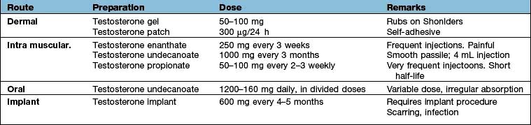
The cause of hypogonadism can rarely be reversed and testosterone replacement therapy should be commenced to control current symptoms and prevent osteoporosis in the long term. Replacement is usually given by transdermal gel or by intramuscular injection (Table 19.22). In gonadotrophin deficiency LH and FSH (purified or synthetic) or pulsatile GnRH may be used when fertility is required
Cryptorchidism (or undescended testes) is usually treated by surgical exploration and orchidopexy in early childhood. After that age, the germinal epithelium is increasingly at risk, and lack of descent by puberty is associated with subfertility. A short trial of HCG occasionally induces descent: an HCG test with a testosterone response 72 h later excludes anorchia. Intra-abdominal testes have an increased risk of developing malignancy; if presentation is after puberty, orchidectomy is advised. Patients may present in adulthood with primary testicular failure (due to the testicular damage before or during surgery) or gonadotrophin deficiency (presumably the cause of maldescent initially).
Klinefelter’s syndrome is a common congenital abnormality, affecting 1 in 1000 males. It is a chromosomal disorder (47XXY and variants, e.g. 46XY/47XXY mosaicism), i.e. a male with an extra X chromosome. There is both a loss of Leydig cells and seminiferous tubular dysgenesis. Patients usually present in adolescence with poor sexual development, small or undescended testes, gynaecomastia or infertility. In 47XXY there is long leg length with tall stature – the androgen deficiency leads to lack of epiphyseal closure in puberty. Patients occasionally have behavioural problems and learning difficulties. There is also a predisposition to diabetes mellitus, breast cancer, emphysema and bronchiectasis; these are all unrelated to the testosterone deficiency.
Clinical examination shows a wide spectrum of features with small pea-sized but firm testes, usually gynaecomastia and other signs of androgen deficiency. Some patients have a normal puberty and may present later with infertility. Confirmation is by chromosomal analysis. Treatment is androgen replacement therapy unless testosterone levels are normal. No treatment is possible for the abnormal seminiferous tubules and infertility.
This is isolated GnRH deficiency. It is associated with decreased or absent sense of smell (anosmia), and sometimes with other bony (cleft palate), renal and cerebral abnormalities (e.g. colour blindness). It is often familial and is usually X-linked, resulting from a mutation in the KAL1 gene which encodes anosmin-1 (producing loss of smell); one sex-linked form is due to an abnormality of a cell adhesion molecule. Management is that of secondary hypogonadism (see p. 976). Fertility is possible.
This refers to isolated GnRH deficiency in the absence of anosmia. Known mutations account for less than 15% of normosmic idiopathic hypogonadotropic hypogonadism (nIHH). Mutations include the KISS1 gene which codes for kisspeptin, the protein which acts on GPR54 receptor, and the FGFR1 gene.
These may be secondary to gonadotrophin deficiency and can be corrected by gonadotrophin therapy. More often they result from primary testicular diseases, in which case they are rarely treatable.
Azoospermia with normal testicular size and low FSH levels suggests a vas deferens block, which is sometimes reversible by surgical intervention.
Lack of libido is a loss of sexual desire; erectile dysfunction (ED) is inability to achieve or maintain erection; they may occur together or separately, and each can precipitate the other. Both are common symptoms in hypogonadism, but most people with either symptom have normal hormones and many have no definable organic cause. ED may be psychological, neurogenic, vascular, endocrine or related to drugs, and often includes contributions from several causes. Vascular disease is a common aetiology, especially in smokers, and is often associated with vascular problems elsewhere. Autonomic neuropathy, most commonly from diabetes mellitus, is a common contributory cause (see p. 1025) and many drugs produce ED. The endocrine causes are those of hypogonadism (see above) and can be excluded by normal testosterone, gonadotrophin and prolactin levels. The presence of nocturnal emissions and frequent satisfactory morning erections make endocrine disease unlikely.
Psychogenic erectile dysfunction is frequently a diagnosis of exclusion, though complex tests of penile vasculature and function are available in some centres.
Treatment. Offending drugs should be stopped. Phosphodiesterase type-5 inhibitors (sildenafil, tadalafil, vardenafil) which increase penile blood flow (see p. 1027) are first choice for therapy. Other treatments include apomorphine, intracavernosal injections of alprostadil, papaverine or phentolamine, vacuum expanders and penile implants.
If no organic disease is found, or if there is clear evidence of psychological problems, the couple should receive psychosexual counselling.
Gynaecomastia is development of breast tissue in the male. Causes are shown in Table 19.23. It is due to an imbalance between free oestrogen and free androgen effects on breast tissue.
Table 19.23 Causes of gynaecomastia
Physiological |
Drugs |
Oestrogenic: |
Pubertal gynaecomastia occurs in perhaps 50% of normal boys, often asymmetrically. It usually resolves spontaneously within 6–18 months, but after this duration may require surgical removal, as fibrous tissue will have been laid down. The cause is thought to be relative oestrogen excess, and the oestrogen antagonist tamoxifen is occasionally helpful.
Gynaecomastia in the older male requires a full assessment to exclude potentially serious underlying disease, such as bronchial carcinoma and testicular tumours (e.g. Leydig cell tumour). However, aromatase activity (p. 972) increases with age and may be the cause of gynaecomastia in this group. Aromatase is an enzyme of the cytochrome P450 family and converts androgens to produce oestrogens. Drug effects are common (especially digoxin and spironolactone), and once these and significant liver disease are excluded most cases have no definable cause. Surgery is occasionally necessary.
Impaired ovarian function, whether primary or secondary, will lead both to oestrogen deficiency and abnormalities of the menstrual cycle. The latter is very sensitive to disruption, cycles becoming anovulatory and irregular before disappearing altogether. Symptoms will depend on the age at which the failure develops. Thus, before puberty, primary amenorrhoea will occur, possibly with delayed puberty; if after puberty, secondary amenorrhoea and hypogonadism will result.
The physiological effects of oestrogens and symptoms/signs of deficiency are shown in Table 19.24.
Table 19.24 Effects of oestrogens and consequences of oestrogen deficiency
Physiological effect | Consequence of deficiency |
---|---|
Breast |
|
Development of connective and duct tissue |
Small, atrophic breast |
Nipple enlargement and areolar pigmentation |
|
Pubic hair |
|
Maintenance of female pattern |
Thinning and loss of pubic hair |
Vulva and vagina |
|
Vulval growth |
Atrophic vulva |
Vaginal glandular and epithelial proliferation |
|
Vaginal lubrication |
|
Uterus and tubes |
|
Myometrial and tubal hypertrophy |
Small, atrophic uterus and tubes |
Endometrial proliferation |
Amenorrhoea |
Skeletal |
|
Epiphyseal fusion |
Eunuchoidism (if prepubertal) |
Maintenance of bone mass |
Osteoporosis |
Absence of periods or markedly irregular infrequent periods (oligomenorrhoea) is the commonest presentation of female gonadal disease. The clinical assessment of such patients is shown in Box 19.8, and common causes are listed in Table 19.25.
Box 19.8
Clinical assessment of amenorrhoea
Polycystic ovary syndrome is the most common cause of oligomenorrhoea and amenorrhoea (see below).
A minimum body weight is necessary for regular menstruation. While anorexia nervosa is the extreme form of weight loss (see p. 1222), amenorrhoea is common and may be seen at weights within the ‘normal’ range. The biochemistry is indistinguishable from gonadotrophin deficiency and some patients have additional mild endocrine disease (e.g. polycystic ovarian disease). It is possible that alterations in leptin levels are responsible for the hypothalamic dysfunction seen in this situation. Restoration of body weight to above the 50th centile for height is usually effective in restoring menstruation, but in the many cases where this cannot be achieved then oestrogen replacement is necessary. Similar problems occur with intensive physical training in athletes and dancers.
Amenorrhoea with low oestrogen and gonadotrophins in the absence of organic pituitary disease, weight loss or excessive exercise is described as hypothalamic amenorrhoea. This may be related to ‘stress’, to previous weight loss or stopping the contraceptive pill, but some patients appear to have defective cycling mechanisms without apparent explanation.
Oligomenorrhoea and amenorrhoea are frequent findings in severe hypothyroidism in young women.
Other causes include pregnancy. Genital tract abnormalities, such as an imperforate hymen, cause primary amenorrhoea.
Severe illness, even in the absence of weight loss, can lead to amenorrhoea.
Turner’s syndrome (p. 983) is a cause of primary amenorrhoea. The phenotype is female with female external genitalia. There is gonadal dysgenesis with streak ovaries. Features include short stature, webbing of the neck (up to 40%), a wide carrying angle of the elbows, high arched palate and low-set ears. These patients also have an increased incidence of autoimmune disease (2%), bicuspid aortic valves, aortic coarctation and dissection and coronary artery disease, hypertension, type 2 diabetes, horseshoe kidneys, lymphoedema, reduced bone density, hearing problems and inflammatory bowel disease (0.3%).
Basal levels of FSH, LH, oestrogen and prolactin allow initial distinction between primary gonadal and hypothalamic–pituitary causes (Table 19.25). Ovarian biopsy may occasionally be necessary to confirm the diagnosis of primary ovarian failure, although elevation of LH and FSH to menopausal levels is usually adequate. Subsequent investigations are shown in Table 19.25.
Treatment is that of the cause wherever possible (e.g. hypothyroidism, low weight, stress, excessive exercise).
Primary ovarian disease is rarely treatable except in the rare condition of ‘resistant’ ovary, where high-dose gonadotrophin therapy can occasionally lead to folliculogenesis. Hyperprolactinaemia should be corrected (see below). Polycystic ovary syndrome is discussed in detail below. In all other cases oestrogen replacement is usually indicated to prevent the long-term consequences of deficiency.
The extent of normal hair growth varies between individuals, families and races, being more extensive in the Mediterranean and some Asian subcontinent populations. These normal variations in body hair, and the more extensive hair growth seen in patients complaining of hirsutism, represent a continuum from no visible hair to extensive cover with thick dark hair. It is therefore impossible to draw an absolute dividing line between ‘normal’ and ‘abnormal’ degrees of facial and body hair in the female. Soft vellus hair is normally present all over the body, and this type of hair on the face and elsewhere is ‘normal’ and is not sex hormone dependent. Hair in the beard, moustache, breast, chest, axilla, abdominal midline, pubic and thigh areas is sex hormone dependent. Any excess in the latter regions is thus a marker of increased ovarian or adrenal androgen production, most commonly polycystic ovary syndrome (PCOS) but occasionally other rarer causes.
Polycystic ovary syndrome. PCOS is the most common cause of hirsutism in clinical practice, affecting about 1 in 5 women worldwide. It is characterized by multiple small cysts within the ovary (Fig. 19.23) (which represent arrested follicular development) and by excess androgen production from the ovaries (and to a lesser extent from the adrenals). It was originally described in its severe form as the Stein–Leventhal syndrome.
Figure 19.23 Polycystic ovary syndrome. (a) Ultrasound of ovary, revealing multiple cysts with central ovarian stroma showing increased echo texture. (b) MR image (coronal) of polycystic ovaries, also showing pelvic anatomy. C, cyst; B, bowel; LO, left ovary; RO, right ovary; U, uterine cavity; V, vagina.
(Reproduced by kind permission of Barbara Hochstein and Geoffrey Cox, Auckland Radiology Group.)
Measured levels of androgens in blood vary widely from patient to patient and may remain within the normal range but SHBG levels are often low (due to high insulin levels), leading to high free androgen levels. In PCOS there is thought to be increased frequency of the GnRH pulse generator, leading to an increase in LH pulses and androgen secretion. The response of the hair follicle to circulating androgens also seems to vary between individuals with otherwise identical clinical and biochemical features, and the reason for this variation in end-organ response remains poorly understood.
PCOS is frequently associated with:
hyperinsulinaemia and insulin resistance, the prevalence of type 2 diabetes being 10 times higher than in normal women
hypertension, hyperlipidaemia and increased cardiovascular risk (the metabolic syndrome, p. 201), which is 2–3 times higher in PCOS, although it is currently unclear whether PCOS per se confers an absolute increase in cardiovascular mortality.
Obesity with PCOS is an additional risk factor for insulin resistance. The precise mechanisms which link the aetiology of polycystic ovaries, hyperandrogenism, anovulation and insulin resistance remain to be elucidated and whether the basic defect is in the ovary, adrenal, pituitary or a more generalized metabolic defect remains unknown.
In routine clinical practice, the majority of people with objective signs of androgen-dependent hirsutism will have PCOS, and investigation is mainly required to exclude rarer and more serious causes of virilization.
Idiopathic hirsutism. People with hirsutism, no elevation of serum androgen levels and no other clinical features are sometimes labelled as having ‘idiopathic hirsutism’. However, studies suggest that most people with ‘idiopathic hirsutism’ have some radiological or biochemical evidence of PCOS on more detailed investigation, and several studies have demonstrated evidence of mild PCOS in up to 20% of the normal female population.
Familial or idiopathic hirsutism does occur, but usually involves a distribution of hair growth which is not typically androgenic.
Other causes. Rarer and more serious endocrine causes of hirsutism and virilization include congenital adrenal hyperplasia (CAH, see p. 987), Cushing’s syndrome (p. 957) and virilizing tumours of the ovary and adrenal.
Ovarian hyperthecosis is a non-malignant ovarian disorder characterized by luteinized thecal cells in the ovarian stroma which secrete testosterone. The clinical features are similar to PCOS but tend to present in perimenopausal women, and serum testosterone levels are higher than typically seen in PCOS.
Iatrogenic hirsutism also occurs after treatment with androgens, or more weakly androgenic drugs such as progestogens or danazol.
Non-androgen-dependent hair growth (hypertrichosis) occurs with drugs such as phenytoin, diazoxide, minoxidil and ciclosporin.
PCOS presents with amenorrhoea/oligomenorrhoea, hirsutism and acne (alone or in combination), shortly after menarche. Clinical, biochemical and radiological features of PCOS merge imperceptibly into those of the normal populations. The development of hirsutism commonly provokes severe distress in young women and may lead to avoidance of normal social activities.
Hirsutism should be recorded objectively, ideally using a scoring system, to document the problem and to monitor treatment. The method and frequency of physical removal (e.g. shaving, plucking) should also be recorded. Most patients who complain of hirsutism will have an objective excess of hair on examination, but occasionally very little will be found (and appropriate counselling is then indicated).
Age and speed of onset. Hirsutism related to PCOS usually begins around the time of the menarche and increases slowly and steadily in the teens and twenties. Rapid progression and prepubertal or late onset suggest a more serious cause.
Accompanying virilization. Hirsutism due to PCOS may be severe and affect all androgen-dependent areas on the face and body. However, more severe virilization (clitoromegaly, recent-onset frontal balding, male phenotype) implies substantial androgen excess, and usually indicates a rarer cause rather than PCOS. Thinning of head hair in a male pattern – androgenic alopecia – occurs in a proportion of women with uncomplicated PCOS, typically with a familial tendency for premature androgen-related hair loss in both sexes.
Menstruation. Most people with hirsutism will have some disturbance of menstruation, typically oligo-/amenorrhoea, although more frequent erratic bleeding can also occur. However, PCOS can present as hirsutism with regular periods or as irregular periods, with no evidence of hirsutism or acne.
Weight. Many people with hirsutism are also overweight or obese. This worsens the underlying androgen excess and insulin resistance and inhibits the response to treatment, and is an indication for appropriate advice on diet and exercise. In severe cases the insulin resistance may have a visible manifestation as acanthosis nigricans on the neck and in the axillae (see Fig. 24.24).
A variety of investigations aid the diagnosis of people with hirsutism:
Serum total testosterone is often elevated in PCOS and is invariably substantially raised in virilizing tumours (usually >5 nmol/L). People with hirsutism and normal testosterone levels frequently have low levels of sex hormone-binding globulin (SHBG), leading to high free androgen levels. The free androgen index ([testosterone/SHBG] *100) is often used and is high; free testosterone is difficult to measure directly.
Other androgens. Androstenedione and dehydroepiandrosterone sulphate are frequently elevated in PCOS, and even more elevated in congenital adrenal hyperplasia (CAH) and virilizing tumours.
17α-Hydroxyprogesterone is elevated in classical CAH, but may be apparent in late-onset CAH only after stimulation tests.
Gonadotrophin levels. LH hypersecretion is a frequent feature of PCOS, but the pulsatile nature of secretion of this hormone means that a ‘classic’ increased LH/FSH ratio is not always observed on a random sample.
Oestrogen levels. Oestradiol is usually normal in PCOS, but oestrone levels (which are rarely measured) are elevated because of peripheral conversion. Levels are variable in other causes.
Ovarian ultrasound is a useful investigation (Fig. 19.23). Typical features are those of a thickened capsule, multiple 3–5 mm cysts and a hyperechogenic stroma. Prolonged hyperandrogenization from any cause may lead to polycystic changes in the ovary. Ultrasound may also reveal virilizing ovarian tumours, although these are often small.
Serum prolactin. Mild hyperprolactinaemia is common in PCOS but rarely exceeds 1500 mU/L.
If a virilizing tumour is suspected clinically or after investigation, then more complex tests include dexamethasone suppression tests, CT or MRI of adrenals, and selective venous sampling.
Most patients presenting with a combination of hirsutism and menstrual disturbance will be shown to have polycystic ovary syndrome, but the rarer alternative diagnoses should be excluded, e.g. late-onset congenital adrenal hyperplasia (early-onset, raised serum 17α-OH-progesterone), Cushing’s syndrome (look for other clinical features) and virilizing tumours of the ovary or adrenals (severe virilization, markedly elevated serum testosterone).
The consensus (Rotterdam) criteria 2003 for diagnosis of PCOS are at least two of:
The underlying cause should be removed in the rare instances where this is possible (e.g. drugs, adrenal or ovarian tumours). Treatment of CAH and Cushing’s is discussed on page 987 and page 957, respectively. Other therapy depends upon whether the aim is to reduce hirsutism, regularize periods or produce fertility.
Regular plucking, bleaching, depilatory cream, waxing or shaving is used. Such removal neither worsens nor improves the underlying severity of hirsutism. More ‘permanent’ solutions include electrolysis and a variety of ‘laser’ hair removal systems – all appear effective but have not been evaluated in long-term studies, are expensive, and still often require repeated long-term treatment. Eflornithine cream (an antiprotozoal) inhibits hair growth by inhibiting ornithine decarboxylase but is effective in only a minority of cases and should be discontinued if there is no improvement after 4 months.
This always requires a year or more of treatment for maximal benefit, and long-term treatment is frequently required as the problem tends to recur when treatment is stopped. The patient must therefore always be an active participant in the decision to use systemic therapy and must understand the rare risks as well as the benefits.
Oestrogens (e.g. oral contraceptives) suppress ovarian androgen production and reduce free androgens by increasing SHBG levels. Combined hormone pills, which contain ethinylestradiol and a non-androgenic progestogen, e.g. desogestrel drospirenone, or cyproterone acetate plus ethinylestradiol (co-cyprindiol), will result in a slow improvement in hirsutism in a majority of cases and should normally be used first unless there is a contraindication, e.g. history of thrombosis. The risk of venous thrombosis appears to be 2–4-fold higher than on other low-dose oral contraceptive pills. After the menopause, HRT preparations which contain medroxyprogesterone (rather than more androgenic progestogens) may be helpful.
Cyproterone acetate (50–100 mg daily) is an antiandrogen but is also a progestogen, teratogenic and a weak glucocorticoid. Given continuously it produces amenorrhoea, and so is normally given for days 1–14 of each cycle. In women of childbearing age, contraception is essential.
Spironolactone (200 mg daily) also has antiandrogen activity and can cause useful improvements in hirsutism.
Finasteride (5 mg daily), a 5α-reductase inhibitor which prevents the formation of dihydrotestosterone in the skin, has also been shown to be effective but long-term experience is limited.
Flutamide, another antiandrogen, is less commonly used owing to the high incidence of hepatic side-effects.
Cyclical oestrogen/progestogen administration will regulate the menstrual cycle and remove the symptom of oligo- or amenorrhoea. This is most frequently an additional benefit of the treatment of hirsutism, but may also be used when menstrual disturbance is the only symptom.
Drugs to improve the hyperinsulinaemia associated with PCOS and obesity are increasingly used (and requested by patients). Metformin (500 mg three times daily) improves menstrual cyclicity and ovulation in short-term studies, and some patients also report improvement in hirsutism and ease of weight loss, but gastrointestinal upset may limit use.
Thiazolidinediones are also effective, but concerns about the long-term cardiovascular effects of these agents now preclude their use.
Metformin alone may improve ovulation and achieve conception.
Clomifene 50–100 mg can be given daily on days 2–6 of the cycle and is more effective than metformin alone in achieving ovulation. This can occasionally cause the ovarian hyperstimulation syndrome, an iatrogenic complication of ovulation induction therapy, consisting of ovarian enlargement, oedema, hypovolaemia, acute kidney injury, and possibly shock; specialist supervision is essential. It is recommended that clomifene should not normally be used for longer than six cycles (owing to a possible increased risk of ovarian cancer in patients treated for longer than recommended).
Reverse circadian rhythm. Prednisolone (2.5 mg in the morning, 5 mg at night) suppresses pituitary production of ACTH, upon which adrenal androgens partly depend. Regular ovulatory cycles often ensue. A steroid instruction leaflet and a card must be supplied.
More intensive techniques to stimulate ovulation may also be indicated in specialist hands, including low-dose gonadotrophin therapy, and ovarian hyperstimulation techniques associated with in vitro fertilization.
Wedge resection of the ovary was a traditional therapy but is now rarely required, although laparoscopic ovarian electrodiathermy may be helpful.
The combined oestrogen–progestogen pill is widely used for contraception and has a low failure rate (<1 per 100 woman-years). ‘Pills’ contain 20–40 µg of oestrogen, usually ethinylestradiol, together with a variable amount of one of several progestogens. The mechanism of action is two-fold:
Suppression by oestrogen of gonadotrophins, thus preventing follicular development, ovulation and luteinization
Progestogen effects on cervical mucus, making it hostile to sperm, and on tubal motility and the endometrium.
Side-effects of these preparations are shown in Box 19.9. Most of the serious ones are rare and are less common with typical modern 20–30 µg oestrogen pills, although evidence suggests that thromboembolism may be slightly more common with ‘third-generation pills’ containing desogestrel and gestodene (approximately 30/100 000 woman-years compared with 15/100 000 on older pills and 5/100 000 on no treatment). While some problems require immediate cessation of the pill, other milder side-effects must be judged against the hazards of pregnancy occurring with inadequate contraception, especially if other effective methods are not practicable or acceptable.
Box 19.9
Adverse effects and drug interactions of oral contraceptives (mixed oestrogen–progesterone combinations)
Hazards of the combined pill are increased in smokers, in obesity and in those with other risk factors for cardiovascular disease (e.g. hypertension, hyperlipidaemia, diabetes) especially in women aged over 35 years (avoid if over 50 years). The ‘mini-pill’ (progestogen only, usually norethisterone) is less effective but is often suitable where oestrogens are contraindicated (Box 19.9). A progesterone antagonist, mifepristone, in combination with a prostaglandin analogue (vaginal gemeprost), induces abortion of pregnancy at up to 9 weeks’ gestation. It prevents progesterone-induced inhibition of uterine contraction.
Subfertility, or ‘infertility’, is defined as the inability of a couple to conceive after 1 year of unprotected intercourse. Investigation requires the combined skills of gynaecologist, endocrinologist and, ideally, andrologist. Both partners must be involved and every aspect of the physiology critically examined.
A significant proportion of couples have both male and female contributing factors.
Figure 19.24 Major factors involved in subfertility and their investigation. LH, luteinizing hormone; FSH, follicle-stimulating hormone; PCOS, polycystic ovarian syndrome; PID, pelvic inflammatory disease; GC, gonorrhoea; STI, sexually transmitted infection.
Inadequate intercourse, hostile cervical mucus and vaginal factors are uncommon (5%). 15% of cases appear to be idiopathic, and natural fertility decreases with increasing age. Conception over 40 years of age for both males and females is reduced to below 30%.
About 30–40% of couples have a major identifiable male factor. There is some evidence that male sperm counts are declining in many populations. Untreated male hypogonadism of any cause (see Table 19.21) is likely to be associated with subfertility.
Female tubal problems due to pathologies such as pelvic inflammatory disease and endometriosis account for perhaps 20%; a similar proportion have ovulatory disorders. Any cause of oligomenorrhoea or amenorrhoea (see Table 19.25) is likely to be associated with suboptimal ovulation or anovulation.
Both partners should be seen and the following factors checked:
The man. Look for previous testicular damage (orchitis, trauma), undescended testes, urethral symptoms and evidence of sexually transmitted infection, local surgery, and use of alcohol and drugs. A semen analysis early in the investigations is essential.
The woman. Look for previous pelvic infection, regularity of periods, previous surgery, alcohol intake and smoking and body weight (see p. 978).
Together. Check the frequency and adequacy of intercourse, and the use of lubricants.
Counselling of both partners is essential. Any defect(s) found should be treated if possible. Ovulation can usually be induced by exogenous hormones if simpler measures fail, while in vitro fertilization (IVF) and similar techniques are widely used, especially where there is tubal blockage, oligo-spermia or ‘idiopathic subfertility’. Intracytoplasmic sperm injection (ICSI) appears particularly effective for severe oligo-spermia and poor sperm function.
Disorders of sexual differentiation are rare but may affect chromosomal, gonadal, endocrine and phenotypic development (Table 19.26). Such cases always require extensive, multidisciplinary clinical management. An individual’s sex can be defined in several ways:
Chromosomal sex. The normal female is 46XX, the normal male 46XY. The Y chromosome confers male sex; if it is not present, development follows female lines.
Gonadal sex. This is determined predominantly by chromosomal sex, but requires normal embryological development.
Phenotypic sex. This describes the normal physical appearance and characteristics of male and female body shape. This in turn is a manifestation of gonadal sex and subsequent sex hormone production.
Social sex (gender). This is heavily dependent on phenotypic sex and normally assigned on appearance of the external genitalia at birth.
The human adrenals weigh 8–10 g together and comprise an outer cortex and inner medulla. The cortex has three zones: the zona glomerulosa, which secretes aldosterone under the control of the renin-angiotensin system, and the zona reticularis and zona fasciculata, which produce cortisol and androgens under feedback control of the hypothalamic-pituitary-adrenal (HPA) axis. The inner medulla synthesizes, stores and secretes catecholamines (see below and Fig. 19.25).
The steroids produced by the adrenal cortex are grouped into three classes based on their predominant physiological effects: glucocorticoids, mineralocorticoids and androgens.
These are so named after their effects on carbohydrate metabolism. Major actions are listed in Table 19.27. They act on intracellular corticosteroid receptors and combine with coactivating proteins to bind the ‘glucocorticoid response element’ (GRE) in specific regions of DNA to cause gene transcription. Glucocorticoid action is modified locally by the action of 11β-hydroxysteroid dehydrogenase (11βHSD). 11βHSD type 1 converts inactive cortisone in cortisol, hence amplifying the hormone signal, whilst 11βHSD type 2 does the opposite.
Table 19.27 The major actions of glucocorticoids
Increased or stimulated | Decreased or inhibited |
---|---|
Gluconeogenesis |
Protein synthesis |
Glycogen deposition |
Host response to infection |
Protein catabolism |
Lymphocyte transformation |
Fat deposition |
Delayed hypersensitivity |
Sodium retention |
Circulating lymphocytes |
Potassium loss |
Circulating eosinophils |
Free water clearance |
|
Uric acid production |
|
Circulating neutrophils |
|
The relative potency of common steroids is shown in Table 19.28.
Table 19.28 The relative glucocorticoid and mineralocorticoid potency of equal amounts of common natural and synthetic steroids
Steroid | Glucocorticoid effect | Mineralocorticoid effect |
---|---|---|
Cortisol (hydrocortisone)a |
1 |
1 |
Prednisolone |
4 |
0.7 |
Dexamethasone |
40 |
2 |
Aldosterone |
0.1 |
400 |
Fludrocortisone |
10 |
400 |
The predominant effect of mineralocorticoids is on the extracellular balance of sodium and potassium in the distal tubule of the kidney. Aldosterone, produced solely in the zona glomerulosa, is the predominant mineralocorticoid in humans (about 50%); corticosterone makes a small contribution to overall mineralocorticoid activity. Mineralocorticoids act on type 1 corticosteroid receptors, whilst glucocorticoids act on type 2 receptors, both having a very similar structure. The mineralocorticoid activity of cortisol is weak but cortisol is present in considerable excess. The mineralocorticoid receptor in the kidney is largely protected from this excess by the intrarenal conversion (‘shuttle’) of cortisol to cortisone by 11β-hydroxysteroid dehydrogenase type 2.
Although secreted in considerable quantities, most androgens have only relatively weak intrinsic androgenic activity until metabolized peripherally to testosterone or dihydrotestosterone. Dihydrotestosterone is metabolized from testosterone by 5α-reductase and is a potent androgen receptor agonist. The androgen receptor has been well characterized and mutations within this gene may cause androgen insensitivity syndromes.
All steroids have the same basic skeleton (Fig. 19.26b) and the chemical differences between them are slight. The major biosynthetic pathways are shown in Fig. 19.22a.
Figure 19.26 (a) The major steroid biosynthetic pathways. Steroid hormones are synthesized in adrenal and gonads from the initial substrate via a series of interconnected enzymatic steps. The reactions catalysed are shown by shaded boxes with italic labels. The molecular identity of the enzymes is shown in red – some catalyse more than one reaction. p450 enzymes are in mitochondria, 3βHSD (hydroxysteroid dehydrogenase) in cytoplasm. 17βHSD and p450aro (aromatase) are found mainly in gonads. (b) The steroid molecule.
Glucocorticoid production by the adrenal is under hypothalamic-pituitary control (Fig. 19.27).
Figure 19.27 Control of the hypothalamic-pituitary-adrenal axis. Pituitary ACTH is secreted in response to hypothalamic CRH (corticotrophin-releasing hormone) triggered by circadian rhythm, stress and other factors, and stimulates secretion of cortisol from the adrenal. Cortisol has multiple actions in peripheral tissues and exerts negative feedback on pituitary and hypothalamus.
Corticotrophin-releasing hormone (CRH) is secreted in the hypothalamus in response to circadian rhythm, stress and other stimuli. CRH travels down the portal system to stimulate adrenocorticotrophin (ACTH) release from the anterior pituitary. Hypothalamic vasopressin (ADH) also stimulates ACTH secretion and acts synergistically. ACTH is derived from the prohormone pro-opiomelanocortin (POMC), which undergoes complex processing within the pituitary to produce ACTH and a number of other peptides including beta-lipotrophin and beta-endorphin. Many of these peptides, including ACTH, contain melanocyte-stimulating hormone (MSH)-like sequences which cause pigmentation when levels of ACTH are markedly raised.
Circulating ACTH stimulates cortisol production in the adrenal. The secreted cortisol (or any other synthetic corticosteroid administered to the patient) causes negative feedback on the hypothalamus and pituitary to inhibit further CRH/ACTH release. The set-point of this system clearly varies through the day according to the circadian rhythm, and is usually overridden by severe stress. Unlike cortisol, mineralocorticoids and sex steroids do not cause negative feedback on the CRH/ACTH axis.
Following adrenalectomy or other adrenal damage (e.g. Addison’s disease), cortisol secretion will be absent or reduced; ACTH levels will therefore rise.
Mineralocorticoid secretion is mainly controlled by the renin-angiotensin system (see p. 566).
ACTH and cortisol are released episodically and in response to stress. When taking a blood sample, remember:
Sampling time should be recorded. Basal levels should be taken between 08:00 hours and 09:00 hours near the peak of the circadian variation.
Appropriate reference ranges (for time and assay method) should be used.
Suppression and stimulation tests are used in suspected excess and deficient cortisol production, respectively.
Administration of a synthetic glucocorticoid (dexamethasone) to a normal subject produces prompt feedback suppression of CRH and ACTH levels and thus of endogenous cortisol secretion (dexamethasone is not measured by most cortisol assays). Three forms of the test, used in the diagnosis and differential diagnosis of Cushing’s syndrome, are available (see Table 19.11).
Synthetic ACTH (tetracosactide, which consists of the first 24 amino acids of human ACTH) is given to stimulate adrenal cortisol production. Details are given in Box 19.1 and Figure 19.5 (p. 944).
In this condition, there is destruction of the entire adrenal cortex. Glucocorticoid, mineralocorticoid and sex steroid production are therefore all reduced. (This differs from hypothalamic-pituitary disease, in which mineralocorticoid secretion remains largely intact, being predominantly stimulated by angiotensin II. Adrenal sex steroid production is also largely independent of pituitary action.) In Addison’s disease reduced cortisol levels lead, through feedback, to increased CRH and ACTH production, the latter being directly responsible for the hyperpigmentation.
Incidence. Addison’s disease is rare, with an incidence of 3–4/million per year and prevalence of 40–60/million. Primary hypoadrenalism shows a marked female preponderance and is most often caused by autoimmune disease (>90% in UK) but in countries with a high prevalence of HIV/AIDS, tuberculosis is an increasing cause. Autoimmune adrenalitis results from the destruction of the adrenal cortex by organ-specific autoantibodies, with 21-hydroxylase as the common antigen. There are associations with other autoimmune conditions in the polyglandular autoimmune syndromes types I and II (e.g. type 1 diabetes mellitus, pernicious anaemia, thyroiditis, hypoparathyroidism, premature ovarian failure) (p. 997).
All other causes are rare (Table 19.29).
Table 19.29 Causes of primary hypoadrenalism
These are shown in Figure 19.28. The symptomatology of Addison’s disease is often vague and nonspecific. These symptoms may be the prelude to an Addisonian crisis with severe hypotension and dehydration precipitated by intercurrent illness, accident or operation.
Figure 19.28 Primary hypoadrenalism (Addison’s disease): symptoms and signs. Bold type indicates signs of greater discriminant value.
Pigmentation (dull, slaty, grey-brown) is the predominant sign in over 90% of cases.
Postural systolic hypotension, due to hypovolaemia and sodium loss, is present in 80–90% of cases, even if supine blood pressure is normal. Mineralocorticoid deficiency is the cause of the hypotension.
Once Addison’s disease is suspected, investigation is urgent. If the patient is seriously ill or hypotensive, hydrocortisone 100 mg should be given intravenously or intramuscularly together with intravenous 0.9% saline. Ideally this should be done immediately after a blood sample is taken for later measurement of plasma cortisol. Alternatively, an ACTH stimulation test can be performed immediately. Full investigation should be delayed until emergency treatment (see below) has improved the patient’s condition. Otherwise, tests are as follows:
Single cortisol measurements are of little value, although a random cortisol below 100 nmol/L during the day is highly suggestive, and a random cortisol >550 nmol/L makes the diagnosis unlikely.
The short ACTH stimulation test should be performed (see Box 19.11 and Fig. 19.5). Note that an absent or impaired cortisol response confirms the presence of hypoadrenalism but does not differentiate Addison’s disease from ACTH deficiency or iatrogenic suppression by steroid medication.
The long ACTH test is no longer used because of the availability of accurate ACTH assays.
A 09:00 hours plasma ACTH level – a high level (>80 ng/L) with low or low-normal cortisol confirms primary hypoadrenalism.
Electrolytes and urea classically show hyponatraemia, hyperkalaemia and a high urea, but they can be normal.
Blood glucose may be low, with hypoglycaemia.
Adrenal antibodies are present in many cases of autoimmune adrenalitis.
Chest and abdominal X-rays may show evidence of tuberculosis and/or calcified adrenals.
Plasma renin activity is high due to low serum aldosterone.
Hypercalcaemia and anaemia (after rehydration) are sometimes seen.
Acute hypoadrenalism needs urgent treatment (Emergency Box 19.1).
Emergency Box 19.1
Management of acute hypoadrenalism
Hypotension, hyponatraemia, hyperkalaemia, hypoglycaemia, dehydration, pigmentation often with precipitating infection, infarction, trauma or operation. The major deficiencies are of salt, steroid and glucose.
Assuming normal cardiovascular function, the following are required:
1 litre of 0.9% saline should be given over 30–60 min with 100 mg of intravenous bolus hydrocortisone.
Subsequent requirements are several litres of saline within 24 hours (assessing with central venous pressure line if necessary) plus hydrocortisone, 100 mg i.m., 6-hourly, until the patient is clinically stable.
Glucose should be infused if there is hypoglycaemia.
Oral replacement medication is then started, unless unable to take oral medication, initially hydrocortisone 20 mg, 8-hourly, reducing to 20–30 mg in divided doses over a few days (Table 19.30).
Fludrocortisone is unnecessary acutely as the high cortisol doses provide sufficient mineralocorticoid activity – it should be introduced later.
Long-term treatment is with replacement glucocorticoid and mineralocorticoid; tuberculosis must be treated if present or suspected. Replacement dosage details are shown in Table 19.30. Dehydroepiandrosterone (DHEA) replacement has also been advocated, and some studies suggest that this may cause symptomatic improvements, although others show no clear benefit.
Table 19.30 Average replacement steroid dosages for adults with primary hypoadrenalism
Drug | Dose |
---|---|
Glucocorticoid |
|
Hydrocortisone |
20–30 mg daily (e.g. 10 mg on waking, 5 mg at 12:00 hours, 5 mg at 18:00 hours) |
or |
|
Prednisolone |
7.5 mg daily (5 mg on waking, 2.5 mg at 18:00 hours) |
rarely |
|
Dexamethasone |
0.75 mg daily (0.5 mg on waking, 0.25 mg at 18:00 hours) |
Mineralocorticoid |
|
Fludrocortisone |
50–300 µg daily |
Adequacy of glucocorticoid dose is judged by:
Clinical wellbeing and restoration of normal, but not excessive, weight
Normal cortisol levels during the day while on replacement hydrocortisone (cortisol levels cannot be used for synthetic steroids).
Adequacy of fludrocortisone replacement is assessed by:
Restoration of serum electrolytes to normal
Blood pressure response to posture (it should not fall >10 mmHg systolic after 2 minutes’ standing)
All patients requiring replacement steroids should:
know how to increase steroid replacement dose for intercurrent illness
wear a Medic-Alert bracelet (or similar), which gives details of their condition so that emergency replacement therapy can be given if found unconscious
keep an (up-to-date) ampoule of hydrocortisone at home in case oral therapy is impossible, for administration by self, family or doctor.
hypothalamic-pituitary disease (inadequate ACTH production) or
long-term steroid therapy leading to hypothalamic-pituitary-adrenal suppression.
Most people with hypothalamic-pituitary disease have panhypopituitarism (see p. 950) and need T4 replacement as well as cortisol; in this case hydrocortisone must be started before T4.
Long-term corticosteroid medication for non-endocrine disease is the most common cause of secondary hypoadrenalism. The hypothalamic-pituitary axis and the adrenal may both be suppressed and the patient may have vague symptoms of feeling unwell. ACTH levels are low in secondary hypoadrenalism. Weaning off steroids is often a long and difficult process.
This condition results from an autosomal recessive deficiency of an enzyme in the cortisol synthetic pathways. There are six major types, but most common is 21-hydroxylase deficiency (CYP21A2), which occurs in about 1 in 15 000 births and which has been shown to be due to defects on chromosome 6 near the HLA region affecting one of the cytochrome p450 enzymes (p450C21).
As a result, cortisol secretion is reduced and feedback leads to increased ACTH secretion to maintain adequate cortisol – leading to adrenal hyperplasia. Diversion of the steroid precursors into the androgenic steroid pathways occurs (see Fig. 19.26a). Thus, 17-hydroxyprogesterone, androstenedione and testosterone levels are increased, leading to virilization. Aldosterone synthesis may be impaired with resultant salt wasting.
The other forms affect 11β-hydroxylase, 17α-hydroxylase, 3β-hydroxysteroid dehydrogenase and a cholesterol side-chain cleavage enzyme (p450scc) (see Fig. 19.26a).
If severe, CAH presents at birth with sexual ambiguity or adrenal failure (collapse, hypotension, hypoglycaemia), sometimes with a salt-losing state (hypotension, hyponatraemia). In the female, clitoral hypertrophy, urogenital abnormalities and labioscrotal fusion are common, but the syndrome may be unrecognized in the male.
Precocious puberty with hirsutism is a later presentation, whereas rare, milder cases only present in adult life, usually accompanied by primary amenorrhoea. Hirsutism developing before puberty is suggestive of CAH.
Expert advice is essential in the confirmation and differential diagnosis of 21-hydroxylase deficiency, and with ambiguous genitalia such advice must be sought urgently before any assignment of gender is made.
A profile of adrenocortical hormones is measured before and one hour after ACTH administration.
Glucocorticoid activity must be replaced, as must mineralocorticoid activity if deficient. In CAH the larger dose of glucocorticoid is often given at night to suppress the morning ACTH peak with a smaller dose in the morning (cf. Addison’s disease, p. 987; Table 19.30). Correct dosage is often difficult to establish in the child but should ensure normal 17-hydroxyprogesterone levels while allowing normal growth; excessive replacement leads to stunting of growth. In adults, clinical features and biochemistry (plasma renin, androstenedione and 17-OH-progesterone) are used to modify treatment. Genetic counselling (p. 43) and antenatal diagnosis is essential, particularly in 21-hydroxylase deficiency. The mother of an affected fetus can take dexamethasone daily to prevent virilization.
Apart from their use as therapeutic replacement for endocrine deficiency states, synthetic glucocorticoids are widely used for many non-endocrine conditions (Box 19.10). Short-term use (e.g. for acute asthma) carries only small risks of significant side-effects except for the simultaneous suppression of immune responses. The danger lies in their continuance, often through medical oversight or patient default. In general, therapy for 3 weeks or less, or a dose of prednisolone less than 5 mg per day, will not result in significant long-term suppression of the normal adrenal axis.
Box 19.10
Common therapeutic uses of glucocorticoids
Long-term therapy with synthetic or natural steroids will, in most respects, mimic endogenous Cushing’s syndrome. Exceptions are the relative absence of hirsutism, acne, hypertension and severe sodium retention, as the common synthetic steroids have low androgenic and mineralocorticoid activity.
Excessive doses of steroids may also be absorbed from skin when strong dermatological preparations are used, but inhaled steroids rarely cause Cushing’s syndrome, although they commonly cause adrenal suppression.
The major hazards are detailed in Box 19.11. In the long term, many are of such severity that the clinical need for high-dose steroids should be continually and critically assessed. Steroid-sparing agents (e.g. azathioprine) should always be considered and screening and prophylactic therapy for osteoporosis introduced (see p. 556). New targeted biological therapies for inflammatory conditions may reduce the incidence of steroid-induced adrenal suppression.
All patients receiving steroids should carry a ‘Steroid Card’. They should be made aware of the following points:
Long-term steroid therapy must never be stopped suddenly.
Doses should be reduced very gradually, with most being given in the morning at the time of withdrawal – this minimizes adrenal suppression. Many authorities believe that ‘alternate-day therapy’ produces less suppression.
Doses need to be increased in times of serious intercurrent illness (defined as presence of a fever), accident and stress. Double doses should be taken during these times.
Other physicians, anaesthetists and dentists must be told about steroid therapy.
Patients should also be informed of potential side-effects, and all this information should be documented in the clinical record. If prophylactic use of bisphosphonate therapy is required to prevent the development of osteoporosis (NICE guidance), they should be informed of the rationale.
Any patient receiving steroids or who has recently received them (within the last 12 months) and may still have adrenal suppression requires special control of steroid medication around the time of surgery. Details are shown in Table 19.31.
With the advent of abdominal CT, MRI and high-resolution ultrasound scanning, unsuspected adrenal masses have been discovered in 3–10% of scans (increasing with age). The two issues of concern with an incidental adrenal mass are:
Most incidentalomas are asymptomatic and benign, but direct questioning may reveal symptoms of endocrine hypersecretion such as cushingoid features, catecholamine excess, virilization in women, or evidence of endocrine hypertension (p. 943). Even in the absence of symptoms, functional tests to exclude secretory activity should be performed as adrenal adenomas often secrete cortisol at a low level, ‘sub-clinical Cushing’s syndrome’, which may confer increased cardiovascular risk. If no endocrine activity is found then most authorities recommend removal only of large adrenal tumours (>4–5 cm) because of the risk of malignancy. Smaller hormonally inactive lesions are usually observed as long as there are no worrying radiological features.
Phaeochromocytoma must be excluded before surgery due to the risk of perioperative hypertensive or hypotensive crises (see p. 991).
Increased mineralocorticoid secretion from the adrenal cortex, termed primary hyperaldosteronism, is thought to account for 5–10% of all hypertension. Other endocrine causes of hypertension should also be considered if there is clinical suspicion (Table 19.32). It is impracticable and unnecessary to screen all hypertensive patients for secondary endocrine causes. The highest chances of detecting such causes are in patients:
under 35 years, especially those without a family history of hypertension
with accelerated (malignant) hypertension
with hypokalaemia before diuretic therapy
resistant to conventional antihypertensive therapy (e.g. more than three drugs) or
Table 19.32 Endocrine causes of hypertension
Excessive renin, and thus angiotensin II, production |
Excessive production of catecholamines |
Excessive GH production |
Excessive aldosterone production |
Excessive production of other mineralocorticoids |
Exogenous ‘mineralocorticoids’ or enzyme inhibitors |
Primary hyperaldosteronism is a disorder of the adrenal cortex characterized by excess aldosterone production leading to sodium retention, potassium loss and the combination of hypokalaemia and hypertension. This must be distinguished from secondary hyperaldosteronism, which arises when there is excess renin (and hence angiotensin II) stimulation of the zona glomerulosa. Common causes of secondary hyperaldosteronism are accelerated hypertension and renal artery stenosis, when the patient will also be hypertensive. Causes associated with normotension include congestive cardiac failure and cirrhosis, where excess aldosterone production contributes to sodium retention.
Adrenal adenomas (Conn’s syndrome) originally accounted for 60% of cases of in series of primary hyperaldosteronism but represented a rare cause of hypertension. The use of the aldosterone:renin ratio in the routine investigation of hypertension now suggests that hyperaldosteronism due to bilateral adrenal hyperplasia (idiopathic hyperaldosteronism) is much more common than the classical Conn’s adenoma. Some claim that idiopathic hyperalosteronism s the cause of up to 10% of cases of ‘essential’ hypertension.
The usual presentation is simply hypertension; hypokalaemia (<3.5 mmol/L) is not frequently present. The few symptoms are nonspecific; rarely muscle weakness, nocturia and tetany are seen. The hypertension may be severe and associated with renal, cardiac and retinal damage.
Adenomas, often very small, are more common in young females, while bilateral hyperplasia rarely occurs before age 40 years and is more common in males.
Beta-blockers and other drugs may interfere with renin activity, and spironolactone, ACE inhibitors and angiotensin II receptor antagonists will all affect results and all should be discontinued if possible. The characteristic features are as follows:
Plasma aldosterone:renin ratio (ARR) is now most frequently used as a screening test for the condition, but raised ARR alone does not confirm the diagnosis (if the renin is low enough ARR will always be high). Note that normal ranges are highly assay-dependent.
Elevated plasma aldosterone levels that are not suppressed with 0.9% saline infusion (2 L over 4 hours) or fludrocortisone administration. Between 30% and 50% of people with raised ARR on screening will suppress normally, excluding the diagnosis.
Suppressed plasma renin activity or immunoreactivity.
Hypokalaemia is often present but a normal serum potassium does not exclude the diagnosis.
Urinary potassium loss. Levels >30 mmol daily during hypokalaemia are inappropriate.
Once a diagnosis of hyperaldosteronism is established, differentiation of adenoma from hyperplasia involves adrenal CT or MRI, but small adenomas may be missed and non-functioning incidentalomas also occur. Further information is obtained from diurnal/postural changes in plasma aldosterone levels (which tend to rise with adenomas between 09:00 hours supine and 13:00 hours erect samples; in contrast, they fall with hyperplasia), measurement of 18-OH cortisol levels (raised in adenoma) and venous catheterization for aldosterone levels. All of these tests have their pitfalls and exceptions.
An adenoma can be removed surgically – usually laparoscopically; blood pressure falls in 70% of patients. Those with hyperplasia should be treated with the aldosterone antagonist spironolactone (100–400 mg daily); frequent side-effects include nausea, rashes and gynaecomastia, and the pure aldosterone receptor antagonist eplerenone can be a useful alternative if side-effects preclude the use of spironolactone (p. 640) Spironolactone metabolites have been linked with tumour development in animals but this has not been described in humans. Amiloride and calcium-channel blockers are moderately effective in controlling the hypertension but do not correct the hyperaldosteronism.
This rare condition is caused by a chimeric gene on chromosome 8. A fusion gene resulting from an unusual cross-over at meiosis between the genes encoding aldosterone synthase and adrenal 11β-hydroxylase produces aldosterone which is under ACTH control. Treatment with glucocorticoid resolves the problem.
This causes the clinical syndrome of primary hyperaldosteronism but with low renin and aldosterone levels. Reduced activity of the enzyme 11β-hydroxysteroid dehydrogenase type 2 (11β-HSD2) prevents the normal conversion in the kidney of cortisol (which is active at the mineralocorticoid receptor) to cortisone (which is not) and therefore ‘exposes’ the mineralocorticoid receptor in the kidney to the usual molar excess of cortisol over aldosterone in the blood. While the inherited syndrome is rare, the same clinical syndrome can occur with excessive ingestion of liquorice, which inhibits the 11β-HSD2 enzyme.
The adrenal medulla is the innermost part of the adrenal gland, consisting of cells that secrete the major catecholamines, noradrenaline (norepinephrine) and adrenaline (epinephrine), which produce the sympathetic nervous response. The catecholamines are interconverted in the adrenal medulla and an increase in levels of their metabolites in the urine is a marker of abnormal hypersecretion (Fig. 19.29).
Figure 19.29 The synthesis and metabolism of catecholamines. COMT, catechol-O-methyl transferase; MAO, monoamine oxidase.
These are very rare tumours of the sympathetic nervous system (less than 1 in 1000 cases of hypertension) that secrete catecholamines, noradrenaline (norepinephrine) adrenaline (epinephrine) and their metabolites (Fig. 19.29):
90% arise in the adrenal medulla (phaeochromocytomas)
10% occur elsewhere in the sympathetic chain (paragangliomas).
Some are associated with MEN 2 syndromes (see below) and the von Hippel–Lindau (VHL) syndrome (p. 1143). Most tumours release both noradrenaline (norepinephrine) and adrenaline (epinephrine) but large tumours and extra-adrenal tumours produce almost entirely noradrenaline.
Paragangliomas typically occur in the head and neck but are also found in the thorax, pelvis and bladder. They are more closely associated with other genetic associations than is phaeochromocytoma. The association of paraganglioma, bilateral adrenal phaeochromocytomas, positive family history or young age at presentation is seen in multiple endocrine neoplasms (p. 997). Mutations in the succinate dehydrogenase (SDHD) gene have been shown to be strongly associated with the development of paraganglioma.
Oval groups of cells occur in clusters and stain for chromogranin A. Some 25% are multiple and 10% malignant, the latter being more frequent in the extra-adrenal tumours. Malignancy cannot be determined on simple histological examination alone.
The clinical features are those of catecholamine excess and are frequently, but not necessarily, intermittent (Table 19.33). All people with suspected phaeochromocytomas must be investigated because phaeochromocytomas may cause acute cardiovascular compromise during routine medical procedures, and can also present with sudden death if the diagnosis is missed.
Table 19.33 Symptoms and signs of phaeochromocytoma
Symptoms |
Signs |
Anxiety or panic attacks |
Hypertension |
Tachycardia/arrhythmias |
|
Bradycardia |
|
Orthostatic hypotension |
|
Pallor or flushing |
|
Glycosuria |
|
Fever |
|
(Signs of hypertensive damage) |
|
|
|
|
|
|
|
|
Measurement of urinary catecholamines and metabolites (metanephrines are most sensitive and specific – Fig. 19.29) is a useful screening test; normal levels on three 24-hour collections of metanephrines virtually exclude the diagnosis. Many drugs and dietary vanilla interfere with these tests.
Resting plasma catecholamines are raised.
Plasma chromogranin A (a storage vesicle protein) is raised.
Clonidine suppression test may be appropriate, but should only be performed in specialist centres.
CT scans, initially of the abdomen, are helpful to localize the tumours, which are often large.
Scanning with [131I]meta-iodobenzylguanidine (MIBG) produces specific uptake in sites of sympathetic activity with about 90% success. It is particularly useful with extra-adrenal tumours. 18F-deoxyglucose PET is also used by some centres in the localization of phaeochromocytomas.
Genetic testing for MEN2, VHL and SDHD mutations should be performed in all people with confirmed phaeochromocytoma or paraganglioma.
Tumours should be removed if this is possible; 5-year survival is about 95% for non-malignant tumours. Medical preoperative and perioperative treatment is vital and includes complete alpha- and beta-blockade with phenoxybenzamine (20–80 mg daily initially in divided doses), then propranolol (120–240 mg daily), plus transfusion of whole blood to re-expand the contracted plasma volume. The alpha-blockade must precede the beta-blockade, as worsened hypertension may otherwise result. Labetalol is not recommended. Surgery in the unprepared patient is fraught with dangers of both hypertension and hypotension; expert anaesthesia and an experienced surgeon are both vital, and sodium nitroprusside and phentolamine (a rapid acting alpha blocker) should be available in case sudden severe hypertension develops.
When operation is not possible, combined alpha- and beta-blockade can be used long term. Radionucleotide treatment with MIBG has been used but with limited success in malignant phaeochromocytoma.
Patients should be kept under clinical and biochemical review after tumour resection as over 10% recur or develop a further tumour. Catecholamine excretion measurements should be performed at least annually.
Thirst and water regulation are largely controlled by vasopressin, also known as antidiuretic hormone (ADH), which is synthesized in the hypothalamus and then migrates in neurosecretory granules along axonal pathways to the posterior pituitary. Pituitary disease alone without hypothalamic involvement therefore does not lead to ADH deficiency as the hormone can still ‘leak’ from the damaged end of the intact axon.
At normal concentrations the kidney is the predominant site of action of vasopressin. Vasopressin stimulation of the V2 receptors allows the collecting ducts to become permeable to water via the migration of aquaporin-2 water channels, thus permitting reabsorption of hypotonic luminal fluid (p. 640). Vasopressin therefore reduces diuresis and results in overall retention of water. At high concentrations vasopressin also causes vasoconstriction via the V1 receptors in vascular tissue.
Changes in plasma osmolality are sensed by osmoreceptors in the anterior hypothalamus. Vasopressin secretion is suppressed at levels below 280 mOsm/kg, thus allowing maximal water diuresis. Above this level, plasma vasopressin increases in direct proportion to plasma osmolality. At the upper limit of normal (295 mOsm/kg) maximum antidiuresis is achieved and thirst is experienced at about 298 mOsm/kg (Fig. 19.30).
Figure 19.30 Plasma vasopressin response to increasing osmolality in normal subjects and in a patient with diabetes insipidus.
Other factors affecting vasopressin release are shown in Table 19.34.
Table 19.34 Factors affecting vasopressin (ADH) release
Increased by: | Decreased by: |
---|---|
Increased osmolality |
Decreased osmolality |
Hypovolaemia |
Hypervolaemia |
Hypotension |
Hypertension |
Nausea |
Ethanol |
Hypothyroidism |
α-Adrenergic stimulation |
Angiotensin II |
|
Adrenaline (epinephrine) |
|
Cortisol |
|
Nicotine |
|
Antidepressants |
|
Disorders of vasopressin secretion or activity include:
deficiency as a result of hypothalamic disease (‘cranial’ diabetes insipidus)
inappropriate excess of the hormone
‘nephrogenic’ diabetes insipidus – a rare condition in which the renal tubules are insensitive to vasopressin; an example of a receptor abnormality.
While all these are uncommon, they need to be distinguished from the occasional patient with ‘primary polydipsia’ and those whose renal tubular function has been impaired by electrolyte abnormalities, such as hypokalaemia or hypercalcaemia.
Deficiency of vasopressin (ADH) or insensitivity to its action leads to polyuria, nocturia and compensatory polydipsia. Daily urine output may reach as much as 10–15 L, leading to dehydration that may be very severe if the thirst mechanisms or consciousness are impaired or the patient is denied fluid.
Causes of DI are listed in Table 19.35. The most common is hypothalamic-pituitary surgery, following which transient DI is common, frequently remitting after a few days or weeks.
Table 19.35 Causes of diabetes insipidus
Cranial diabetes insipidus |
|
Familial (e.g. DIDMOAD) |
|
Idiopathic (often autoimmune) |
|
Tumours: |
Craniopharyngioma |
Hypothalamic tumour, e.g. glioma, germinoma |
|
Metastases, especially breast |
|
Lymphoma/leukaemia |
|
Pituitary with suprasellar extension (rare) |
|
Infections: |
Tuberculosis |
Meningitis |
|
Cerebral abscess |
|
Infiltrations: |
Sarcoidosis |
Langerhans’ cell histiocytosis |
|
Inflammatory: |
Hypophysitis |
Post-surgical: |
Transfrontal |
Trans-sphenoidal |
|
Post-radiotherapy (cranial) |
|
Vascular: |
Haemorrhage/thrombosis |
Sheehan’s syndrome |
|
Aneurysm |
|
Trauma (e.g. head injury) |
|
Nephrogenic diabetes insipidus |
|
Familial (e.g. vasopressin receptor gene, aquaporin-2 gene defect) |
|
Idiopathic |
|
Renal disease (e.g. renal tubular acidosis) |
|
Hypokalaemia |
|
Hypercalcaemia |
|
Drugs (e.g. lithium, demeclocycline, glibenclamide) |
|
Sickle cell disease |
|
Mild temporary nephrogenic DI can occur after prolonged polyuria due to any cause, including cranial DI and primary polydipsia. |
DI may be masked by simultaneous cortisol deficiency – cortisol replacement allows a water diuresis and DI then becomes apparent.
Familial isolated vasopressin deficiency causes DI from early childhood and is dominantly inherited, caused by a mutation in the AVP-NPII gene. DIDMOAD (Wolfram) syndrome is a rare autosomal recessive disorder comprising diabetes insipidus, diabetes mellitus, optic atrophy and deafness due to mutations in the WFS1 gene on chromosome 4. MR scanning may show an absent or poorly developed posterior pituitary.
High or high-normal plasma osmolality with low urine osmolality (in primary polydipsia plasma osmolality tends to be low).
Resultant high or high-normal plasma sodium (hypernatraemia).
High 24-h urine volumes (less than 2 L excludes the need for further investigation).
Failure of urinary concentration with fluid deprivation.
Restoration of urinary concentration with vasopressin or an analogue.
The latter two points are studied with a formal water deprivation test (Box 19.12). In normal subjects, plasma osmolality remains normal while urine osmolality rises above 600 mOsm/kg. In DI, plasma osmolality rises while the urine remains dilute, only concentrating after exogenous vasopressin is given (in ‘cranial’ DI) or not concentrating after vasopressin if nephrogenic DI is present. An alternative is measurement of plasma vasopressin during hypertonic saline infusion, but these measurements are not widely available.
Box 19.12
Water deprivation test
Fasting and no fluids from 07:30 hours (or overnight if only mild DI is expected and polyuria is only modest).
Monitor serum and urine osmolality, urine volume and weight hourly for up to 8 hours.
Abandon fluid deprivation if weight loss >3% occurs.
If serum osmolality >300 mOsm/kg and/or urine osmolality <600 mOsm/kg give desmopressin 2 µg i.m. at end of test. Allow free fluid but measure urine osmolality for 2–4 hours.
Normal response. Serum osmolality remains within normal range (275–295 mOsm/kg). Urine osmolality rises to >600 mOsm/kg.
Diabetes insipidus (DI). Serum osmolality rises above normal without adequate concentration of urine osmolality (i.e. serum osmolality >300 mOsm/kg; urine osmolality <600 mOsm/kg).
Nephrogenic DI – if desmopressin does not concentrate urine.
Cranial DI – if urine osmolality rises by >50% after desmopressin.
The synthetic vasopressin (ADH) analogue desmopressin is the treatment of choice. It has a longer duration of action than vasopressin and has no vasconstrictive effects. It is most reliably given intranasally as a spray 10–40 µg once or twice daily, but can also be given orally as 100–200 µg three times daily, or intramuscularly 2–4 µg daily. Response is variable and must be monitored carefully with enquiry about fluid input/output and plasma osmolality measurements. The main problem is avoiding water overload and consequent hyponatraemia (p. 650). Where there is a reversible underlying cause (e.g. a hypothalamic tumour) this should be investigated and treated.
Alternative agents in mild DI, probably working by sensitizing the renal tubules to endogenous vasopressin, include thiazide diuretics, carbamazepine (200–400 mg daily) or chlorpropamide (200–350 mg daily) but these are rarely used.
In this condition, renal tubules are resistant to normal or high levels of plasma vasopressin (ADH). It may be inherited as a rare sex-linked recessive, with an abnormality in the vasopressin-2 receptor, or as an autosomal post-receptor defect in an ADH-sensitive water channel, aquaporin-2. More commonly it can be acquired as a result of renal disease, sickle cell disease, drug ingestion (e.g. lithium), hypercalcaemia or hypokalaemia. Wherever possible the cause should be reversed. Polyuria is helped by thiazide diuretics.
Diabetes mellitus, hypokalaemia and hypercalcaemia should be excluded. In the case of diabetes mellitus the cause is an osmotic diuresis secondary to glycosuria which leads to dehydration and an increased perception of thirst owing to hypertonicity of the extracellular fluid.
This is a relatively common cause of thirst and polyuria. It is a psychiatric disturbance characterized by the excessive intake of water. Plasma sodium and osmolality fall as a result and the urine produced is appropriately dilute. Vasopressin levels become virtually undetectable. Prolonged primary polydipsia may lead to the phenomenon of ‘renal medullary washout’, with a fall in the concentrating ability of the kidney.
Characteristically the diagnosis is made by a water deprivation test. A low plasma osmolality is usual at the start of the test, and since vasopressin secretion and action can be stimulated, the patient’s urine becomes concentrated (albeit ‘maximum’ concentrating ability may be impaired); the initially low urine osmolality gradually increases with the duration of the water deprivation.
Inappropriate secretion of ADH (also called vasopressin) leads to retention of water and hyponatraemia. The presentation is usually vague, with confusion, nausea, irritability and, later, fits and coma. There is no oedema. Mild symptoms usually occur with plasma sodium levels below 125 mmol/L and serious manifestations are likely below 115 mmol/L. The elderly may show symptoms with milder abnormalities.
The syndrome must be distinguished from dilutional hyponatraemia due to excess infusion of glucose/water solutions or diuretic administration (thiazides or amiloride, see p. 651).
Dilutional hyponatraemia due to excessive water retention
Euvolaemia (in contrast to hypovolaemia of sodium and water depletion states)
Low plasma osmolality with ‘inappropriate’ urine osmolality >100 mOsm/kg (and typically higher than plasma osmolality)
Continued urinary sodium excretion >30 mmol/L (lower levels suggest sodium depletion and should respond to 0.9% saline infusion)
The causes are listed in Table 19.36.
Table 19.36 Common causes of the syndrome of inappropriate ADH secretion (SIADH)
Tumours |
Metabolic causes |
Small-cell carcinoma of lung |
Alcohol withdrawal |
Prostate |
Porphyria |
Drugs |
|
Chlorpropamide |
|
Carbamazepine |
|
Pulmonary lesions |
Cyclophosphamide |
Pneumonia |
|
Tuberculosis |
|
Lung abscess |
|
CNS causes |
|
Meningitis |
|
Tumours |
|
Head injury |
|
Subdural haematoma |
|
Cerebral abscess |
|
SLE |
|
Hyponatraemia is very common during illness in frail elderly patients and it may sometimes be clinically difficult to distinguish SIADH from salt and water depletion, particularly when mixed clinical features are present. Under these circumstances, a trial infusion of 1–2 L 0.9% saline is given. SIADH will not respond (but will excrete the sodium and water load effectively) – sodium depletion will respond. ACTH deficiency can give a very similar biochemical picture to SIADH, therefore it is necessary to ensure the hypothalamic-pituitary-adrenal axis is intact, particularly in neurosurgical patients, in whom ACTH deficiency may be relatively common.
The underlying cause should be corrected where possible. Symptomatic relief can be obtained by the following measures:
Fluid intake should be restricted to 500–1000 mL daily. If tolerated, and complied with, this will correct the biochemical abnormalities in almost every case.
Plasma osmolality, serum sodium and body weight should be measured frequently.
If water restriction is poorly tolerated or ineffective, demeclocycline (600–1200 mg daily) is given; this inhibits the action of vasopressin on the kidney, causing a reversible form of nephrogenic diabetes insipidus. It often, however, causes photosensitive rashes.
When the syndrome is very severe (i.e. acute and symptomatic), hypertonic saline may be indicated but this is potentially dangerous and should only be used with extreme caution (p. 650).
Vasopressin V2 antagonists, e.g. tolvaptan 15 mg daily, are being used with good results.
Serum calcium levels are mainly controlled by parathyroid hormone (PTH) and vitamin D. Hypercalcaemia is much more common than hypocalcaemia and is frequently detected incidentally with multichannel biochemical analysers. Mild asymptomatic hypercalcaemia occurs in about 1 in 1000 of the population, with an incidence of 25–30 per 100 000 population. It occurs mainly in elderly females, and is usually due to primary hyperparathyroidism (primary HPT).
There are normally four parathyroid glands which are situated posterior to the thyroid, but occasionally additional glands exist or they may be found elsewhere in the neck or mediastinum. PTH, an 84-amino-acid hormone derived from a 115-residue preprohormone, is secreted from the chief cells of the parathyroid glands. PTH levels rise as serum ionized calcium falls. The latter is detected by specific G protein coupled calcium-sensing receptors on the plasma membrane of the parathyroid cells. PTH has several major actions, all serving to increase plasma calcium by:
increasing osteoclastic resorption of bone (occurring rapidly)
increasing intestinal absorption of calcium (a slow response)
increasing synthesis of 1,25-(OH)2D3
PTH effects are mediated at specific membrane receptors on the target cells, resulting in an increase of adenyl cyclase messenger activity.
Vitamin D metabolism is discussed on page 550.
PTH measurements use two-site immunometric assays that measure only the intact PTH molecule; interpretation requires a simultaneous calcium measurement in order to differentiate most causes of hyper- and hypocalcaemia.
The major causes of hypercalcaemia are listed in Table 19.37; primary hyperparathyroidism and malignancies are by far the most common (>90% of cases). Hyperparathyroidism itself may be primary, secondary or tertiary. Primary hyperparathyroidism is caused by single (>80%) parathyroid adenomas or by diffuse hyperplasia of all the glands (15–20%); multiple parathyroid adenomas are rare. Involvement of multiple parathyroid glands may be part of a familial syndrome (e.g. multiple endocrine neoplasia (MEN) syndrome type 1 or 2a). Parathyroid carcinoma is rare (<1%), though it usually produces severe and intractable hypercalcaemia. Hyperparathyroidism-jaw tumour syndrome is a rare familial cause of hyperparathyroidism which may be associated with parathyroid carcinoma and maxillary or mandibular tumours.
Table 19.37 Causes of hypercalcaemia
Excessive parathormone (PTH) secretion |
Malignant disease – low PTH levels (second commonest cause) |
Excess action of vitamin D |
Excessive calcium intake |
Other endocrine disease (mild hypercalcaemia only) |
Thyrotoxicosis |
Addison’s disease |
Drugs |
Miscellaneous |
Primary hyperparathyroidism is of unknown cause, though it appears that adenomas are monoclonal. Hyperplasia may also be monoclonal. Chromosomal rearrangements in the 5′ regulatory region of the parathyroid hormone gene have been identified, and inactivation of some tumour suppressor genes at a variety of sites may also be involved.
Secondary hyperparathyroidism (see p. 618) is physiological compensatory hypertrophy of all parathyroids because of hypocalcaemia, such as occurs in chronic kidney disease or vitamin D deficiency. PTH levels are raised but calcium levels are low or normal, and PTH falls to normal after correction of the cause of hypocalcaemia where this is possible.
Tertiary hyperparathyroidism is the development of apparently autonomous parathyroid hyperplasia after longstanding secondary hyperparathyroidism, most often in renal failure. Plasma calcium and phosphate are both raised, the latter often grossly so. Parathyroidectomy is necessary at this stage.
Mild hypercalcaemia (e.g. adjusted calcium <3 mmol/L) is frequently asymptomatic, but more severe hypercalcaemia can produce a number of symptoms:
General. There may be tiredness, malaise, dehydration and depression.
Renal. Renal colic from stones, polyuria or nocturia, haematuria and hypertension occurs. The polyuria results from the effect of hypercalcaemia on renal tubules, reducing their concentrating ability – a form of mild nephrogenic diabetes insipidus. Primary hyperparathyroidism is present in about 5% of patients who present with renal calculi.
Bones. There may be bone pain. Hyperparathyroidism mainly affects cortical bone, and bone cysts and locally destructive ‘brown tumours’ occur but only in advanced disease. Only 5–10% of all cases have definite bony lesions even when sought. Bone disease may be more apparent when there is co-existing vitamin D deficiency.
Abdomen. There may be abdominal pain.
Chondrocalcinosis and ectopic calcification. These are occasional features.
Corneal calcification. This is a marker of longstanding hypercalcaemia but causes no symptoms.
There may also be symptoms from the underlying cause. Malignant disease is usually advanced by the time hypercalcaemia occurs, typically with bony metastases. The common primary tumours are bronchus, breast, myeloma, oesophagus, thyroid, prostate, lymphoma and renal cell carcinoma. True ‘ectopic PTH secretion’ by the tumour is very rare, and most cases are associated with raised levels of PTH-related protein. This is a 144-amino-acid polypeptide, the initial sequence of which shows an approximate homology with the biologically active part of PTH, which is necessary in fetal development but does not have a clearly defined role in the adult. Local bone-resorbing cytokines and prostaglandins may be involved locally where there are metastatic skeletal lesions, leading to local mobilization of calcium by osteolysis with subsequent hypercalcaemia.
Severe hypercalcaemia (>3 mmol/L) is usually associated with malignant disease, hyperparathyroidism, chronic kidney disease or vitamin D therapy.
Several fasting serum calcium and phosphate samples should be performed.
Serum PTH. The hallmark of primary hyperparathyroidism is hypercalcaemia and hypophosphataemia with detectable or elevated intact PTH levels during hypercalcaemia. When this combination is present in an asymptomatic patient then further investigation is usually unnecessary. However, an undetectable PTH level in the context of hypercalcaemia always requires further investigation to exclude malignancy or other pathology (Table 19.37).
Hyperchloraemic acidosis – often mild.
Renal function is usually normal but should be measured as a baseline.
24-hour urinary calcium or single calcium creatinine ratio should be measured in a young patient with modest elevation in calcium and PTH to exclude familial hypocalciuric hypercalcaemia (see p. 996).
Elevated serum alkaline phosphatase is found in severe parathyroid bone disease, but otherwise it suggests an alternative cause for hypercalcaemia.
Where PTH is undetectable or equivocal, a number of other tests may lead to the diagnosis:
Protein electrophoresis/immunofixation: to exclude myeloma
Serum TSH: to exclude hyperthyroidism
09:00 hours cortisol and/or ACTH test: to exclude Addison’s disease
Serum ACE: helpful in the diagnosis of sarcoidosis
Hydrocortisone suppression test: hydrocortisone 40 mg three times daily for 10 days leads to suppression of plasma calcium in sarcoidosis, vitamin D-mediated hypercalcaemia and some malignancies.
Abdominal X-rays may show renal calculi or nephrocalcinosis. High-definition hand X-rays can show subperiosteal erosions in the middle or terminal phalanges. DXA bone density scan is useful to detect bone effects in asymptomatic people with hyperparathyroidism (HPT) in whom conservative management is planned.
The success of parathyroid imaging is highly operator dependent and choice therefore depends on local skills and experience. Imaging is frequently far less accurate than parathyroid exploration by an expert surgeon where the success rate is at least 90%. Methods include:
Details of emergency treatment for severe hypercalcaemia are given in Emergency Box 19.2. This should be followed by oral therapy unless the underlying disease can be treated.
Emergency Box 19.2
Treatment of acute severe hypercalcaemia
Acute hypercalcaemia often presents with dehydration, nausea and vomiting, nocturia and polyuria, drowsiness and altered consciousness. The serum Ca2+ is over 3 mmol/L and sometimes as high as 5 mmol/L. While investigation of the cause is under way, immediate treatment is mandatory if the patient is seriously ill or if the Ca2+ is above 3.5 mmol/L.
Rehydrate at least 4–6 L of 0.9% saline on day 1, and 3–4 L for several days thereafter. Central venous pressure (CVP) may need to be monitored to control the hydration rate.
Intravenous bisphosphonates are the treatment of choice for hypercalcaemia of malignancy or of undiagnosed cause. Pamidronate is preferred (60–90 mg as an intravenous infusion in 0.9% saline or glucose over 2–4 hours or, if less urgent, over 2–4 days). Levels fall after 24–72 hours, lasting for approximately two weeks. Zoledronate is an alternative.
Prednisolone (30–60 mg daily) is effective in some instances (e.g. in myeloma, sarcoidosis and vitamin D excess) but in most cases is ineffective.
Calcitonin (200 units i.v. 6-hourly) has a short-lived action and is little used.
Oral phosphate (sodium cellulose phosphate 5 g three times daily) produces diarrhoea.
There are no effective medical therapies at present for primary hyperparathyroidism, but a high fluid intake should be maintained, a high calcium or vitamin D intake avoided, and exercise encouraged. New therapeutic agents that target the calcium-sensing receptors (e.g. cinacalcet) are of proven value in parathyroid carcinoma and in dialysis patients (p. 631), and are used in primary hyperparathyroidism where surgical intervention is contraindicated.
There is agreement that surgery is indicated in primary hyperparathyroidism for:
people with renal stones or impaired renal function
bone involvement or marked reduction in cortical bone density
unequivocal marked hypercalcaemia (in UK typically >3.0 mmol/L; USA guidelines state >1 mg/dL above reference range)
The situation where plasma calcium is mildly raised (2.65–3.00 mmol/L) is more controversial. Most authorities feel that young patients should be operated on, as should those who have reduced cortical bone density or significant hypercalciuria, as this is associated with stone formation.
In older patients without these problems, or in those unfit for or unwilling to have surgery, conservative management is indicated. Regular measurement of serum calcium and of renal function is necessary. Bone density of cortical bone should be monitored if conservative management is used. Hyperparathyroidism can cause nonspecific symptoms of weakness, fatigue and depression, and it can be difficult to determine whether these symptoms are related to hypercalcaemia or coincidental.
Parathyroid surgery should be performed only by experienced surgeons, as the minute glands may be very difficult to define, and it is difficult to distinguish between an adenoma and normal parathyroid. In expert centres over 90% of operations are successful, involving removal of the adenoma, or removal of all four hyperplastic parathyroids. Minimal access surgery is used, and some centres measure PTH levels intraoperatively to ensure the adenoma has been removed.
Other than postoperative hypocalcaemia (see below), the other rare complications are those of thyroid surgery – bleeding and recurrent laryngeal nerve palsies (<1%). Vocal cord function should be checked preoperatively.
If initial exploration is unsuccessful, a full work-up including venous catheterization and scanning is essential, remembering that parathyroid tissue can be ectopic.
The major danger after operation is hypocalcaemia, which is more common in patients who have significant bone disease and/or vitamin D deficiency– the ‘hungry bone’ syndrome. Some authorities pre-treat such patients, with alfacalcidol 2 µg daily from 2 days preoperatively for 10–14 days, and routine vitamin D replacement (preferably without calcium) is always indicated if deficiency is diagnosed. Chvostek’s and Trousseau’s signs (see p. 997) are monitored as well as biochemistry. Plasma calcium measurements are performed at least daily until stable – with or without replacement – a mild transient hypoparathyroidism often continues for 1–2 weeks. Depending on its severity, oral or intravenous calcium should be given temporarily, as only a few patients (<1%) will develop longstanding surgical hypoparathyroidism.
This uncommon autosomal dominant, and usually asymptomatic, condition demonstrates increased renal reabsorption of calcium despite hypercalcaemia. PTH levels are normal or slightly raised and urinary calcium is low. It is caused by loss of function mutations in the gene on the long arm of chromosome 3 encoding for the calcium-ion-sensing G-protein coupled receptor in the kidney and parathyroid gland. Family members are often affected, detected by genetic analysis. Parathyroid surgery is not indicated as the course appears benign. This diagnosis can be differentiated from hyperparathyroidism in an isolated case by the calcium creatinine ratio in blood and urine.
Hypocalcaemia may be due to deficiencies of calcium homeostatic mechanisms, secondary to high phosphate levels or other causes of hypocalcaemia (Table 19.38). All forms of hypoparathyroidism, except transient surgical effects, are uncommon.
Table 19.38 Causes of hypocalcaemia
Increased phosphate levels |
Hypoparathyroidism |
Vitamin D deficiency |
Resistance to PTH |
Drugs |
Other |
Chronic kidney disease is the most common cause of hypocalcaemia.
Severe vitamin D deficiency may cause mild, and occasionally severe, hypocalcaemia.
Hypocalcaemia after thyroid or parathyroid surgery is common but usually transient – fewer than 1% of thyroidectomies leave permanent damage (see above).
Idiopathic hypoparathyroidism is one of the rarer autoimmune disorders, often accompanied by vitiligo, cutaneous candidiasis and other autoimmune disease.
DiGeorge’s syndrome (p. 66) is a familial condition in which the hypoparathyroidism is associated with intellectual impairment, cataracts and calcified basal ganglia, and occasionally with specific autoimmune disease.
Pseudohypoparathyroidism is a syndrome of end-organ resistance to PTH owing to a mutation in the GSα-protein (GNAS1) which is coupled to the PTH receptor. It is associated with short stature, short metacarpals, subcutaneous calcification and sometimes intellectual impairment. Variable degrees of resistance involving other G protein-linked hormone receptors may also be seen (TSH, LH, FSH).
Pseudo-pseudohypoparathyroidism describes the phenotypic defects but without any abnormalities of calcium metabolism. These individuals may share the same gene defect as individuals with pseudohypoparathyroidism and be members of the same families.
Hypoparathyroidism presents as neuromuscular irritability and neuropsychiatric manifestations. Paraesthesiae, circumoral numbness, cramps, anxiety and tetany (Box 19.13) are followed by convulsions, laryngeal stridor, dystonia and psychosis. Two signs of hypocalcaemia are Chvostek’s sign (gentle tapping over the facial nerve causes twitching of the ipsilateral facial muscles) and Trousseau’s sign, where inflation of the sphygmomanometer cuff above systolic pressure for 3 min induces tetanic spasm of the fingers and wrist. Severe hypocalcaemia may cause papilloedema and frequently a prolonged QT interval on the ECG.
The clinical history and picture is usually diagnostic and is confirmed by a low serum calcium (after correction for any albumin abnormality). Additional tests include:
Serum and urine creatinine for renal disease
PTH levels in the serum: absent or inappropriately low in hypoparathyroidism, high in other causes of hypocalcaemia
Parathyroid antibodies (present in idiopathic hypoparathyroidism)
25-hydroxy vitamin D serum level (low in vitamin D deficiency)
Magnesium level: severe hypomagnesaemia results in functional hypoparathyroidism which is reversed by magnesium replacement
X-rays of metacarpals, showing short fourth metacarpals which occur in pseudohypoparathyroidism.
In vitamin D deficiency, cholecalciferol is the most appropriate treatment (see p. 559). In other cases, alpha-hydroxylated derivatives of vitamin D are preferred for their shorter half-life, and especially in renal disease as the others require renal hydroxylation. Usual daily maintenance doses are 0.25–2 µg for alfacalcidol (1α-OH-D3). During treatment, plasma calcium must be monitored frequently to detect hypercalcaemia. Oral calcium supplements may be used in early stages of treatment and severe hypocalcaemia presenting as an emergency may occasionally require replacement with i.v. calcium gluconate.
These are caused by autoimmune disease as detailed in Table 19.2 on page 939. Most common are the associations of primary hypothyroidism and type 1 diabetes, and either of these with Addison’s disease or pernicious anaemia.
Autoimmune polyendocrinopathy type 1 (APS-1) is an autosomal recessive disorder and is caused by AIRE gene mutations. The AIRE gene is present in the epithelium of the thymus and is involved in the presentation of self-antigens to thymocytes. Mutations will allow persistence of thymic lymphocytes, which react against self-antigens and cause development of autoimmune disorders. Mucocutaneous candidiasis often develops before the onset of endocrine deficiencies, such as Addison’s disease, type 1 diabetes, hypoparathyroidism, nail dystrophy, vitiligo and dental enamel hypoplasia.
APS-2 is not associated with candidiasis and is also known as Schmidt’s syndrome, typically when hypothyroidism, Addison’s disease and type 1 diabetes are present in combination; coeliac disease is also an association.
This is the name given to the simultaneous or metachronous occurrence of tumours involving a number of endocrine glands (Table 19.39). The condition is inherited in an autosomal dominant manner and arises from the expression of recessive oncogenic mutations, most of which have been isolated. Affected persons may pass on the mutation to their offspring in the germ cell, but for the disease to become evident a somatic mutation must also occur, such as deletion or loss of a normal homologous chromosome.
Table 19.39 Multiple endocrine neoplasia (MEN) syndromes
Organ | Frequency | Tumours/manifestations |
---|---|---|
Type 1 |
||
Parathyroid |
95% |
Adenomas/hyperplasia |
Pituitary |
70% |
Adenomas – prolactinoma, ACTH or growth hormone secreting (acromegaly) |
Pancreas |
50% |
Islet cell tumours (secreting insulin, glucagon, somatostatin, VIP, pancreatic polypeptide, growth hormone-releasing factor) |
Adrenal |
40% |
Non-functional adenoma |
Thyroid |
20% |
Adenomas – multiple or single |
Type 2a |
||
Adrenal |
Most |
Phaeochromocytoma (70% bilateral) |
Thyroid |
Most |
Medullary carcinoma (calcitonin producing) |
Parathyroid |
60% |
Hyperplasia |
Type 2b |
||
Type 2a with marfanoid phenotype and intestinal and visceral ganglioneuromas but not hyperparathyroidism. |
The defect in MEN 1 is in a novel gene (menin) on the long arm of chromosome 11 which encodes for a 610-amino-acid protein. Menin represses a transcription factor (JunD) and lack of JunD suppression leads to decreased apoptosis and oncogenesis. People with the MEN1 gene carry one mutant gene and a wild type gene (i.e. are heterozygous). When the wild type gene undergoes a random somatic mutation during life, this leads to loss of heterozygosity and explains the late onset of tumours at any stage (the ‘two hit’ hypothesis). MEN 1 is classically associated with pancreatic, parathyroid and pituitary tumours, although other glands may be affected (Table 19.39).
MEN 2a and 2b are caused by mutations of the RET proto-oncogene on chromosome 10 (see medullary thyroid cancer, p. 971). This gene encodes for a transmembrane glycoprotein receptor. For MEN 2a the mutation is in the extracellular domain; for 2b it is in the intracellular domain. MEN 2 is classically associated with parathyroid tumours, phaeochromocytoma and medullary thyroid carcinoma (Table 19.39). Unlike MEN 2a, MEN 2b is associated with a marfanoid phenotype and intestinal and visceral ganglioneuromas, as well as neuromas around the lips and tongue.
Treatment of established tumours in MEN is largely the same as treatment for similar tumours occurring sporadically. In MEN 1 four-gland parathyroidectomy is usually recommended when surgery is needed since all glands are typically involved. However, the essence of management in MEN is annual screening to detect tumours at an early, treatable stage.
A careful family history is essential. If the precise gene mutation has been identified in a particular family, then family members at risk can be offered genetic screening for the presence of the mutation, ideally in childhood. In affected individuals, biochemical screening and periodic imaging is then required.
Hyperparathyroidism is usually the first manifestation, and serum calcium is the simplest screening test in families with no identified mutation. In an established case (or gene-positive family member) other screening bloods include prolactin, GH/IGF-1 and ‘gut hormones’ (p. 247). Periodic imaging of pancreas, adrenals and pituitary is usually performed. People with MEN 1 can develop metastases to the liver from non-functional pancreatic tumours which are clinically silent; this emphasizes the need for regular screening imaging.
This condition is associated with autonomous hypersecretion of a number of endocrine glands at a young age. Gonadotrophin-independent puberty with Leydig cell hyperplasia in males and ovarian oestrogen production in girls occurs. Pituitary hypersecretion may lead to hyperprolactinaemia, acromegaly or gigantism. Cushing’s syndrome due to nodular hyperplasia of the adrenal cortex is observed, as well as autonomous functioning thyroid nodules. Non-endocrine manifestations include café-au-lait patches and increased bone deformity and fractures due to polyostotic fibrous dysplasia. The pathological basis is a point mutation of the GNAS1 gene that inhibits GTPase activity, leading to persistent activation of cAMP-mediated endocrine secretion.
This term refers to hormone synthesis, and normally secretion, from a neoplastic non-endocrine cell, most usually seen in tumours that have some degree of embryological resemblance to specialist endocrine cells. The clinical effects are those of the hormone produced, with or without manifestations of systemic malignancy. The most common situations seen are the following:
Hypercalcaemia of malignant disease, often from squamous cell tumours of lung and breast, often with bone metastases. Where metastases are not present, most cases are mediated by secretion of PTH-related protein (PTHrP), which has considerable sequence homology to PTH; a variety of other factors may sometimes be involved, but very rarely PTH itself (see p. 995). Treatment is also discussed on page 995.
SIADH (see p. 993). Again, this is most common from a primary lung tumour.
Ectopic ACTH syndrome (see p. 953). Small-cell carcinoma of the lung, carcinoid tumours and medullary thyroid carcinomas are the most common causes, though many other tumours rarely cause it.
Production of insulin-like activity may result in hypoglycaemia (see p. 1029).
http://www.thyroidmanager.org/
Online Thyroid disease textbook
http://www.endocrine.niddk.nih.gov/
US National Institutes of Health, National Institute of Diabetes and Digestive and Kidney Diseases
The Addison’s Self Help Group (UK): information and guidelines on Addison’s and steroid replacement
The Pituitary Foundation (UK charity): comprehensive information for patients and GPs
UK Turner Syndrome Support Society
Emergency identification system for people with hidden medical conditions