Help the Family Adjust to the Disorder

Once parents learn of the heart defect, they are initially in a period of shock, followed by high anxiety, especially fear of the child’s death. This reaction may occur soon after the child’s birth or at a later period. Whatever its timing, the family needs a period of grief before assimilating the meaning of the defect. Unfortunately, the demands for medical treatment may not allow this, necessitating that the parents be informed of the condition to give informed consent for diagnostic and therapeutic procedures. The nurse can be instrumental in supporting parents in their loss, assessing their level of understanding, supplying information as needed, and helping other members of the health team understand the parents’ reactions.

Severely ill newborns usually remain in the hospital. The nurse can promote parent-infant attachment by encouraging parents to hold, touch, and look at their child and by providing time and privacy to the parents to spend with their newborn. (See Chapter 10 for suggestions for promoting attachment between parents and their hospitalized newborn.)

The effect of a child with a serious heart defect on the family is complex. No members, regardless of how well they adjust, are unaffected. Mothers frequently feel inadequate in their mothering ability because of the more complex care such an infant requires. They may be constantly exhausted from the pressures of caring for this child and the other family members. Likewise, fathers and siblings may feel neglected and resentful, a reaction similar to that in families with children with other chronic conditions. (See Chapter 22.) Often parents do not feel confident leaving the child in the care of anyone else, which affords parents no relief from the constant daily caregiving. This problem can be minimized by gradually teaching someone else (a reliable relative or neighbor) how to care for the child.

The need to maintain discipline and set consistent limits can be difficult for parents. A study by Uzark and Jones (2003) found higher levels of stress in parents of children with heart disease, particularly with regard to limit setting and discipline. Behavior modification techniques using either concrete rewards (e.g., a favorite food) or social reinforcement (e.g., approval) can be effective. However, these techniques are most beneficial if used before the child learns to control the family. Therefore guiding parents toward the need for discipline while the child is in infancy is necessary to prevent later problems.

Another problem that may develop within family relationships is overdependency on the part of the child. This is often a result of parental fear that the child may die and overcompensation through what has been termed benevolent overreaction. (See Chapter 22.) Research has shown no correlation between the severity of the child’s heart defect and maternal anxiety or parental stress (Morelius, Lundh, and Nelson, 2002). The best approach to dealing with this dilemma is prevention. Parents need guidance to recognize the eventual hazards of continuing dependency and protectiveness as the child grows older, and the nurse can assist parents in learning ways to foster optimum development. Unless parents have help to see what activities the child can do, they may focus on physical limitations and encourage dependency. The child needs opportunities for normal social interaction with other children to foster normal social development.

Frequently the unremitting stresses of care—physical exhaustion, financial costs, emotional upset, fear of death, and concern for the child’s future—are not fully appreciated by those caring for the family. Even when the child’s condition is stabilized or corrected, the family may need to make new adjustments in their lifestyle. Introducing them to other families with similarly affected children can help them adjust to the daily stresses* (see Family-Centered Care box).

image FAMILY-CENTERED CARE

The Diagnosis of Heart Disease

Remember, we don’t have your experience. We don’t see children every day who have heart disease. We would have been upset finding out our child had to have his tonsils out. How could we ever be prepared for this? Please remember, we only know people who have trivial heart murmurs. How could we ever expect this to happen? And to us, this is the worst problem we’ve ever heard of.

We still fear most what we don’t know and understand. Be honest with us. If you don’t know either, tell us. But at least don’t leave us wondering about what you know and we don’t. Not knowing anything really can be worse than knowing something bad. Be honest, but don’t strip us of hope. …

Please, remember we are trying to learn complex information in a moment of time. And trying to learn it in a context of great pain and emotional investment. This is our lives you’re talking about. Please be thorough, but keep it simple. Tell us again, maybe even again and again, when we can hear better.

From Schrey C, Schrey M: A parent’s perspective: our needs and our message, Crit Care Nurs Clin North Am 6(1):113-119, 1994.

Educate the Family About the Disorder

Once parents are ready to hear about their child’s heart condition, it is essential that they receive a clear explanation based on their level of understanding. A review of normal cardiac anatomy is helpful before explaining the anatomic defect. A simple diagram, pictures, or a model of the heart can be most helpful in visualizing the heart and the congenital defect. Parents appreciate receiving written information about the specific condition.* Health care professionals should take advantage of subsequent encounters with the family to assess parental understanding of the condition and clarify information as needed.

Different health personnel may convey the same information using different diagrams and medical terms. To prevent this from becoming a problem, the same type of diagram should be used by all, and the parents should write down any unclear terms or ask for clarification. Sometimes it is helpful to provide the family with a glossary of frequently used words for reference.

Parents often use multiple resources to obtain information about their child’s heart defect. Increasingly, families are using the Internet as a source of information. Locating information can be easy with helpful information located at national organizations and large parent support groups. Parents also find support through contacts with other parents and parent groups. It is important for parents to realize that not all websites offer medically accurate information and that information from other parents may not be applicable to their own situation. Some children with rare, complex heart defects require individualized treatment plans, and general information on the Internet or in books may not apply to them. Parents should talk to their health care team, in particular their cardiologist, about information they have received from other sources.

The nurse must give information to the child in a manner that is appropriate to the child’s developmental age. As the child matures, the level of information is revised to match the child’s new cognitive level. Preschoolers need basic information about what they will experience more than what is actually occurring physiologically. School-age children benefit from a concrete explanation of the defect. Preadolescents and adolescents often appreciate a more detailed description of how the defect affects their heart. Children of all ages need to be able to express their feelings concerning the diagnosis.

Help the Family Manage the Illness at Home

Parents are the child’s principal caregivers and need to develop a positive, supportive working relationship with the health care team. Because most children spend the majority of their time at home with episodic trips to the hospital, parents manage their child’s illness on a daily basis. They monitor for signs of illness, give medications and treatments, bring their child to appointments, work with a variety of caregivers, and alert the team to problems. Successful relationships are a partnership between parents and caregivers that is built on mutual trust and respect. Good communication between the family, the cardiology specialists, and the primary care practitioner is essential. As children reach adolescence, they begin to take a larger role in managing their illness and making decisions about their care.

Parents should be aware of the symptoms of their child’s cardiac condition and signs of worsening clinical status. Parents should know how to contact their child’s cardiologist at all times and know what to do in an emergency. Parents of children who may develop HF should be familiar with the symptoms (see p. 1352) and know when to contact the practitioner. Parents of children with cyanosis should be informed about fluid management and hypercyanotic spells (see p. 1362). Parents should have an information sheet with their child’s diagnosis, significant treatments such as surgical procedures, allergies, other health care problems, current medications, and health care providers’ contact numbers available in case of emergencies and to share with other caregivers such as teachers, baby-sitters, or daycare providers.

The family also needs to be knowledgeable regarding the therapeutic management of the disorder and the role that surgery, other procedures, medications, and a healthy lifestyle play in maintaining good health. Medications play a critical role in the management of some cardiac conditions such as arrhythmias and severe HF, in anticoagulation after implantation of artificial valves, and in antirejection treatment after heart transplantation. Some patients must take multiple medications daily for life. Many medications can be dangerous if taken incorrectly and require close monitoring. Teach parents the correct procedure for giving medications and caution them to keep them in a safe area to prevent accidental ingestion (see Family-Centered Care box, p. 1358).

Another area of parental concern is the child’s level of physical activity. Most children do not need to restrict activity, and the best approach is to treat the child normally and allow self-limited activity. Exceptions primarily involve strenuous recreational and competitive sports in children with specific cardiac problems. Discuss activities and exercise restrictions with the child’s cardiologist. Avoid deliberately attempting to prevent crying because it can establish a maladaptive parental pattern of relating to the infant.

Infants and children with CHD require good nutrition. Breast-feeding should be possible for many infants with CHD. Countering a common misconception that breast-feeding would not be possible for these infants because they would get tired or exhibit poor growth, Barbas and Kelleher (2004) found that breast-feeding could be successful with adequate support and education of the mother. Providing adequate nutrition to infants with HF or complex congenital defects is especially difficult due to their high caloric requirements and inability to suck effectively because of fatigue and tachypnea. Instructing parents in feeding methods that decrease the work of the infant and giving high-calorie formula are important interventions (see p. 1359 for a discussion on feeding the infant with HF).

Children with severe cardiac defects are often anorexic. Encouraging them to eat can be a tremendous challenge. Because of the parents’ concern over eating, children learn early to manipulate parents through eating, such as making unrealistic demands for foods that are not available. The nurse advises parents of this potential problem, since prevention yields greater success than intervention. For example, give the child a choice of available high-nutrient foods. Chapter 27 provides suggestions for encouraging sick children to eat.

Infants with heart disease should be immunized according to the current guidelines. Immunization schedules may need to be modified around times of acute illness or surgical procedures (Smith, 2001). Infants and children younger than 2 years of age with unrepaired heart defects, cyanotic lesions, pulmonary hypertension, or a history of prematurity should receive the vaccine for respiratory syncytial virus (RSV) monthly during RSV season (November to April in North America) to prevent RSV infection (American Academy of Pediatrics, 2009). Use of the RSV vaccine palivizumab has been shown to reduce hospitalization due to RSV infection in infants and young children with hemodynamically significant CHD (Feltes, Cabalka, Meissner, et al, 2003). (See Chapter 32.)

Infants and children who have serious heart disease are at risk for developmental delays and there is growing interest in characterizing these outcomes (see Research Focus box). Multiple factors can influence neurodevelopmental outcomes, including genetics (chromosomal abnormalities and microdeletions), family background (parental intelligence quotient [IQ] and socioeconomic status), preoperative factors (including prematurity, cyanosis, shock), intraoperative factors (use of cardiopulmonary bypass, deep hypothermic circulatory arrest), and postoperative factors (hemodynamic instability, hypoxia, acidosis, cardiac arrest, stroke, ischemic events).

image RESEARCH FOCUS

Congenital Heart Disease and Cognitive Development

Research in the past decade has begun to identify specific risk factors and common developmental concerns for congenital heart disease. Bellinger, Wypij, duPlessis, and colleagues (2003) found an association between longer periods of deep hypothermic circulatory arrest (a cardiopulmonary bypass technique commonly used in infants needing complex repairs) and the presence of postoperative seizures, delayed motor development, and a downward trend in full-scale intelligence quotient (IQ). At 8 years of age, more than a third of the study patients had received remedial services in school. Shillingford, Glanzman, Ittenbach, and colleagues (2008) also found that a significant proportion of children with complex congenital heart disease were at risk for inattention and hyperactivity, and nearly half were using remedial school services. In another longitudinal study, Limperopoulos, Majnemer, Shevell, and colleagues (2002) found that preoperative and early postoperative neurologic status, microcephaly, type of cardiac lesion, length of deep hypothermic circulatory arrest, age at surgery, and length of intensive care unit stay were predictors of developmental disability.

Recent efforts to limit the time of deep hypothermic circulatory arrest and provide better neuroprotection during surgery on infants may improve outcomes in the future. Although most children with serious heart disease are within the normal range for IQ, there is a higher incidence of neurodevelopmental deficits—specifically deficits in speech and language, fine motor skills, and cognitive processes—in children who have undergone heart surgery than in the normal population (Majnemer and Limperopoulos, 1999). Severe neurologic problems such as cerebral palsy, epilepsy, and cognitive impairment are uncommon.

Prepare the Child and Family for Invasive Procedures

Chapter 27 provides an extensive discussion of the principles for preparing children for invasive procedures. In 2003 the American Heart Association published a scientific statement, “Recommendations for Preparing Children and Adolescents for Invasive Cardiac Procedures” (LeRoy, Elixson, O’Brien, et al, 2003), that addresses issues specific to the child with heart disease. The reader is referred to these resources for a complete review of the topic. The following discussion highlights some important aspects of preparation for cardiac catheterization and cardiac surgery.

The expected outcomes for preprocedure preparation include reducing anxiety, improving patient cooperation with procedures, enhancing recovery, developing trust with caregivers, and improving long-term emotional and behavioral adjustment following procedures (LeRoy, Elixson, O’Brien, et al, 2003). Important factors to consider in planning preparation strategies are the child’s cognitive developmental level, the child’s previous hospital experiences, the child’s temperament and coping style, the timing of the preparation, and the involvement of the parents. The most beneficial preparation strategies usually combine information giving and training in coping skills such as conscious breathing exercises, distraction techniques, guided imagery, and other behavioral interventions.

Handling preoperative and precatheterization workups on an outpatient basis is common for most elective procedures. Children are then admitted on the morning of the procedure. Preprocedure teaching is often done in the clinic setting or at home, and a tour of the ICU and the inpatient facilities may be added. Children of different ages and developmental levels require different amounts of information and different approaches. Young children should be prepared close in time to the event; older children and adolescents may benefit from teaching several weeks in advance. Include parents in the preparation session to support their child and learn about upcoming events.

The preoperative or precatheterization preparation should include information on the environment, equipment, and procedures that the child will encounter during and following the procedure. The nurse can use many educational techniques, such as verbal and written information, hospital tours, preoperative classes, and picture books or videos. Information about what the child will see, hear, and feel should be included, especially for older children and adolescents. Some of the sensory experiences of being in an ICU or catheterization laboratory include sights (monitors, many people, lots of equipment), sounds (beeping noises, alarms, voices), and sensations (lines and dressings, tape, feelings of discomfort, thirst). Familiar aspects of the environment, like BP cuffs, stethoscopes, or oximeter probes, are reviewed, and new equipment such as monitors, IV lines, and oxygen masks are described. Comforting aspects of the environment are emphasized, such as play areas, chairs for parents, and televisions. Many patients who will be sedated during catheterization or receive narcotic pain relievers after surgery will have minimal recall of that period and will not need detailed information about the equipment or procedures used. Information should be specific to the planned procedure for each patient.

Discuss ways the child can cope with the experience and be helped to recover. For young children, bringing a familiar stuffed animal or comfort object with them will help relieve anxiety, whereas for older children bringing a music player with headphones and favorite recordings to the catheterization laboratory will help distract them during the procedure. Topics to discuss regarding recovery after catheterization include the need to lie still to prevent bleeding at the catheter site, progression of the diet, pain control measures, and monitoring methods. Review the importance of ambulation, coughing and deep breathing, and drinking and eating after surgery, and describe pain management and monitoring routines. Review simple coping strategies for use during painful procedures, including distraction techniques such as counting, blowing, singing, or telling stories.

Children and their families should have a choice about an ICU tour. Exposure to the ICU environment can actually increase anxiety in some children, particularly young children, those with previous hospital experiences, and those who are highly anxious (LeRoy, Elixson, O’Brien, et al, 2003). If a visit to the recovery room and ICU is planned, it should take place when there is minimal activity in the area, when the parents can accompany the child, and when the child is well rested. Usually the day before the procedure is ample time to allow the child to ask questions and to prevent undue fantasizing about the experience. Protect the child from frightening sights in the unit. Equipment that will not be in view postoperatively, such as equipment located behind or below the bed, needs less attention. The child and parents are encouraged to ask questions and to explore further any equipment in the room, but they should not be pushed to assimilate more information than they are able.

Preoperative physical care differs little, if any, from that provided for any other surgery and is discussed in Chapter 27. Assure the child that the parents will be there when the child wakes up. Also allow the parents to accompany the child as far as possible to the operating suite. (See Evidence-Based Practice box, p. 1211.) After all of the equipment and procedures have been explained, it is important to talk about “getting well” and going home.

Provide Postoperative Care

Immediate postoperative care is usually provided by specially trained nurses in the ICU. Performing many of the procedures, such as arterial pressure and CVP monitoring and observations related to vital functions, requires advanced educational training (the reader should refer to critical care texts for further information). However, nurses caring for the child before surgery and during the convalescent period need to be familiar with the major principles of care.

Observe Vital Signs and Arterial and Venous Pressures: Record vital signs frequently, including BP, until the child’s condition is stable. The heart rate and respirations are counted for 1 full minute, compared with the values on the ECG monitor, and recorded with activity. The heart rate is normally increased after surgery. The nurse observes cardiac rhythm and notifies the practitioner of any changes in regularity. Dysrhythmias may occur postoperatively secondary to administration of anesthetics, acid-base and electrolyte imbalance, hypoxia, surgical intervention, or trauma to conduction pathways.

At least hourly, auscultate the lungs for breath sounds. Diminished or absent breath sounds may indicate an area of atelectasis, pleural effusion, or pneumothorax. All such cases require further assessment. Auscultation guides the nurse’s selective use of postural drainage and percussion to those pulmonary lobes most in need. It also allows a more objective evaluation of effective ventilation.

Temperature changes are typical during the early postoperative period. Hypothermia is expected immediately after surgery due to hypothermia procedures, effects of anesthesia, and loss of body heat to the cool environment. During this period the child is kept warm to prevent additional heat loss. Infants may be placed under radiant heat warmers. During the next 24 to 48 hours the body temperature may rise to 37.8° C (100° F) or slightly higher as part of the inflammatory response to tissue trauma. After this period an elevated temperature is most likely a sign of infection and warrants immediate investigation for probable cause.

Intraarterial monitoring of BP is almost always done following open-heart surgery. Residual vasoconstriction after cardiopulmonary bypass makes indirect BP readings less reliable, and intraarterial monitoring permits continuous rather than intermittent observation. A catheter is passed into the radial artery or the dorsalis pedis or posterior tibial artery, and the other end is attached to an electronic monitoring system, which provides a continuous recording of the BP. The intraarterial line is maintained with a low-rate, constant infusion of heparinized saline to prevent clotting. Continuous BP readings are compared with those taken indirectly using a sphygmomanometer or oscillometric device (Dinamap). A discrepancy between the two may indicate a change in peripheral vascular resistance, a malfunction in the electronic device, or human error in using the wrong-size BP cuff. The nurse also observes for potential complications of intraarterial monitoring, such as arterial thrombosis, infection, air emboli, or blood loss through the catheter. Prevention of each of these hazards is similar to care for any other type of infusion line.

The intraarterial line is maintained with a low-rate, constant infusion of heparinized saline to prevent clotting. The amount of irrigant is recorded as intake fluid. The dressing at the site is changed daily.

Several IV lines are inserted preoperatively: a peripheral IV to give fluids and medications and a CVP line that is usually inserted in a large vessel in the neck. Intracardiac monitoring lines are placed intraoperatively in the RA, LA, or pulmonary artery. Intracardiac lines allow assessment of pressures inside the cardiac chambers, which give vital information on blood volume, cardiac output, ventricular function, pulmonary artery pressures, and responses to drug therapy in the immediate postoperative period. The RA and CVP lines may also be used to infuse fluids and medications. LA lines and pulmonary artery lines are used with more complex repairs. Intracardiac lines are used only in the ICU, although CVP lines may remain for use as a central IV line outside the ICU. All lines must be cared for using strict aseptic technique to prevent infection. Patients must be carefully assessed for bleeding at the time of line removal. See critical care texts for a more complete discussion of intracardiac lines.

Maintain Respiratory Status: Infants usually require mechanical ventilation in the immediate postoperative period. Children may be extubated in the operating room or in the first few postoperative hours, especially if cardiopulmonary bypass was not required. When weaning and extubation are completed, oxygen is delivered by mask, hood, or nasal cannula and is humidified to prevent drying of mucosa. Encourage the child to turn and deep breathe at least hourly. Every means is employed to enhance ventilation and decrease pain, such as splinting of the operative site and use of analgesics.

Suctioning is performed only as needed and is done carefully to avoid vagal stimulation (which can trigger cardiac dysrhythmias) and laryngospasm, especially in infants. Suctioning is intermittent and is maintained for no more than 5 seconds to prevent depleting the oxygen supply. Supplemental oxygen is administered with a manual resuscitation bag before and after the procedure to prevent hypoxia. The heart rate is monitored after suctioning to detect changes in rhythm or rate, especially bradycardia. The child should always be positioned facing the nurse to permit assessment of the child’s color and tolerance of the procedure.

image NURSING ALERT

During suctioning, observe for signs and symptoms of respiratory distress, such as tachypnea, use of accessory muscles for breathing, and restlessness.

Chest tubes may be inserted into the pleural or mediastinal space during surgery or in the immediate postoperative period to remove secretions and air and allow reexpansion of the lung. The chest tube is attached to a disposable water-seal drainage system. The underwater drainage prevents air from traveling up the tube into the pleural space and causing pneumothorax. Nursing considerations include (1) do not interrupt water-seal drainage unless the chest tube is clamped, (2) check for tube patency (fluctuation in the water-seal chamber), and (3) maintain sterility.

Check drainage hourly for color and quantity. Immediately postoperatively the drainage may be bright red, but afterward it should be serous. The largest volume of drainage occurs in the first 12 to 24 hours, and drainage is greater after extensive heart surgery.

image NURSING ALERT

Chest tube drainage of more than 3 ml/kg/hr for more than 3 consecutive hours or 5 to 10 ml/kg in any 1 hour is excessive and may indicate postoperative hemorrhage. Notify the surgeon immediately, since cardiac tamponade can develop rapidly and is life threatening.

Chest radiographs are taken when the tubes are inserted to check their location and after they are removed to evaluate the inflation of the lungs. Chest tubes are usually removed on the first to third postoperative day when drainage has diminished.

Removal of chest tubes can be an uncomfortable, frightening experience (see Atraumatic Care box). Warn children that they will feel a sharp, momentary pain. After the suture is cut, the tubes are quickly pulled out at the end of full inspiration in the extubated patient to prevent intake of air into the pleural cavity. (In the intubated patient, the tubes are pulled out on inspiration, since the lungs are stented open with the positive pressure ventilation.) A purse-string suture (placed when the tubes were inserted) is pulled tight to close the opening. A petrolatum-covered gauze dressing is immediately applied over the wound and securely taped to the skin on all four sides so that an airtight seal is formed. The dressing is checked for signs of drainage. It is removed the next day. Breath sounds are auscultated, since pneumothorax is a possible complication of chest tube removal. A chest x-ray film is usually obtained after removal to assess for pneumothorax or pleural effusion.

ATRAUMATIC CARE

Chest Tube Removal

Intravenous analgesics such as morphine sulfate (0.1 mg/kg), often in combination with midazolam (Versed), may be given before the procedure. Oral analgesics and sedatives have also been used.

Provide Maximum Rest: After heart surgery maximum rest should be provided to decrease the workload of the heart and promote healing. Nursing care is planned according to the child’s usual activity and sleep patterns. The simplest way to ensure individualized, efficient, high-quality care is to plan at the beginning of the shift the nursing procedures to be done. Identify periods of rest. Share the schedule with parents to allow them to visit at the most advantageous times, such as after a rest period when no special treatments are anticipated.

Provide Comfort: Heart surgery is both painful and frightening for children, and providing comfort is a primary nursing concern. Several incisions are used for heart surgery. A median sternotomy following the sternum down the center of the chest is most common. A ministernotomy opens the lower sternum. A thoracotomy incision is most uncomfortable because it goes through muscle tissue. It allows access to the side of the chest through an incision that runs from under the arm around the back to the scapula.

Adequate pain control decreases postoperative complications such as atelectasis, pneumonia, and deep vein thrombosis by improving coughing and ambulation. Pain level is now considered the fifth vital sign. Many pain assessment tools are available for infants and children of different ages. (See Pain Assessment, Chapter 7.)

Continuous IV infusion of opioids, particularly morphine and fentanyl, is a safe and effective method of pain control. Patient-controlled analgesia may be used with children old enough to understand the concept (Macfadyen and Buckmaster, 1999). Epidural morphine is another option. Children receiving opioid infusions for a prolonged period are weaned slowly from the medication to prevent withdrawal symptoms. Nonsteroidal antiinflammatory drugs (NSAIDs) such as IV ketorolac (Toradol) or oral ibuprofen may be used to provide relief of moderate postoperative pain.

Most patients need IV analgesics for pain control during the 24- to 48-hour postoperative period. After lines and tubes have been removed and when patients are tolerating oral fluids, pain may be controlled with oral narcotics such as codeine or oxycodone, often combined with acetaminophen or an oral NSAID such as ibuprofen, or with acetaminophen alone. As noted earlier, thoracotomy incisions are usually more painful than sternotomies because the incision is through muscle. Higgins, Turley, Harr, and colleagues (1999) have found that round-the-clock use of acetaminophen or ibuprofen to augment narcotics is advantageous in providing pain relief after heart surgery. Acetaminophen or ibuprofen alone is usually adequate for pain control after discharge. (See Pain Management, Chapter 7.)

In addition to providing pharmacologic pain control, make every effort to minimize the discomfort of procedures by other means, such as by placing a firm pillow or favorite stuffed animal against the chest incision during coughing and performing treatments after pain medication is given, preferably at a time that coincides with the drug’s peak effect. Employ nonpharmacologic measures to lessen the perception of pain, and encourage parents to comfort their child as much as possible. (See Pain Management, Chapter 7.)

Monitor Fluids: Intake and output of all fluids must be accurately calculated. Intake is primarily IV fluids; however, the nurse also needs to keep a record of fluid used to flush the arterial and CVP lines or to dilute medications. Monitoring of output includes hourly recordings of urine (usually a Foley catheter is inserted and attached to a closed collecting device), drainage from chest and nasogastric tubes, and blood drawn for analysis. Urine is analyzed for specific gravity to evaluate the kidneys’ concentrating ability and to assess the body’s degree of hydration. Renal failure is a potential risk from a transient period of low cardiac output.

image NURSING ALERT

The signs of renal failure are decreased urinary output (<1 ml/kg/hr) and elevated levels of blood urea nitrogen and serum creatinine.

During open-heart surgery, the cardiopulmonary pump is primed with a large volume of fluid (usually electrolyte solution), which may greatly dilute the patient’s blood. The large amount of fluid also diffuses into the interstitial spaces, causing total-body edema and pulmonary edema. Patients return from the operating room with fluid overload. Fluids are restricted to less than maintenance level during the first postoperative day, and drugs are used to promote diuresis. A return to maintenance fluid levels then occurs over the next few days as patients resume normal nutrition. Electrolyte levels are closely monitored because electrolyte imbalances, especially hypokalemia, are a common result of diuresis and fluid shifts, and electrolytes may need replacement.

Fluid requirements are based on the child’s weight and body surface area. The child is weighed daily, preferably in the morning, using the same scale and in similar clothing. The child is usually given nothing by mouth for the first 24 hours. Oral fluids are usually withheld until the child is extubated. Patients begin taking clear liquids when bowel sounds are heard and advance slowly to a regular diet. Nausea and vomiting are common in the first few days after surgery, likely a side effect of anesthesia and analgesics. Providing adequate nutrition, ideally by oral intake, becomes important by the fourth or fifth postoperative day. Consider nasogastric tube feedings or parenteral nutrition for patients who are unable to tolerate oral feedings.

Plan for Progressive Activity: Fatigue and weakness are common after heart surgery. However, moderate activity is essential to prevent pulmonary and vascular complications. Initially, turning, coughing, and deep breathing are sufficient to promote respiratory expansion. Passive range-of-motion exercises, especially to the lower extremities, are instituted to prevent venous stasis.

A progressive schedule of ambulation and activity is planned, based on the child’s preoperative activity patterns and postoperative cardiovascular and pulmonary function. Provide the child with toys to encourage movement. It is important to plan the activity at times when the child is well rested, is comfortable (usually has had analgesic medication), and is not scheduled for any strenuous procedure or treatment immediately afterward.

Ambulation is initiated early, usually by the second postoperative day, after extubation and when many lines and tubes have been removed. Patients progress from sitting on the edge of the bed and dangling the legs to standing up and to sitting in a chair while being assisted and assessed by the nursing staff. Carefully monitor the heart rate and respirations to assess the degree of cardiac demand imposed by each activity. Tachycardia, dyspnea, cyanosis, desaturation, progressive fatigue, or dysrhythmias indicate the need to limit further energy expenditure. After ambulation a rest period is scheduled.

Observe for Complications of Heart Surgery: Several complications can occur after heart surgery, most of which are related to open-heart surgery and the use of cardiopulmonary bypass. Many of the procedures discussed in the preceding paragraphs are aimed at preventing these problems. Only those that have not already been discussed are included here. A serious complication, infective endocarditis (bacterial), is discussed on p. 1382.

Cardiac Changes: Preoperatively the workload of the heart is increased because of the abnormal hemodynamics caused by the congenital defect. In the initial postoperative period the heart is under increased stress because of the effects of surgery and the use of the heart-lung machine. In some cases cardiac function can actually be worse in the early postoperative period despite repair of the congenital defect. HF, hypoxia, low cardiac output, dysrhythmias, and tamponade are all potential postoperative problems.

HF may occur postoperatively because of excessive pulmonary blood flow or fluid overload (see p. 1354 for assessment and management of HF). Hypoxia may occur because of inadequate pulmonary blood flow or because of respiratory problems. Rapid assessment of the causes of hypoxia and appropriate interventions to improve ventilation and perfusion are vital, since hypoxia can rapidly lead to acidosis, which can impair ventricular function.

Low cardiac output syndrome and decreased peripheral perfusion can occur from hypothermia or inability of the LV to maintain systemic circulation. It affects up to 25% of infants and young children after cardiac surgery (Hoffman, Wernovsky, Atz, et al, 2003). The most important signs of adequate peripheral perfusion are rapid capillary refill, good skin color, warm extremities, and strong pulses. Indications of low cardiac output are similar to signs of shock (i.e., decreased BP, decreased pulse pressure, cool extremities, metabolic acidosis, and oliguria). Low cardiac output states are aggressively treated with IV inotropic medications such as dopamine, dobutamine, and milrinone. Milrinone, widely used in pediatrics, has also been shown to prevent low cardiac output syndrome (Hoffman, Wernovsky, Atz, et al, 2003). If maximum medical therapy is failing, cardiac assist methods such as extracorporeal membrane oxygenation or a ventricular assist device may be used in some centers under certain circumstances. Mortality is higher than 50% for patients who require mechanical support. Patients who have recovery of ventricular function within 2 to 3 days and have a short period of support have the best outcomes (Craig, Smith, and Fineman, 2001).

Dysrhythmias are common in the early postoperative period and can result from electrolyte imbalance, especially hypokalemia, and surgical intervention to the septum or myocardium. The heart rate and rhythm are carefully monitored by observing the ECG pattern and by counting the apical pulse for 1 full minute. In some children, a faster than normal rate may be required to maintain an adequate cardiac output in the postoperative period, and a slower than normal rhythm can impair cardiac output. Epicardial pacing wires may be inserted during surgery for managing cardiac dysrhythmias postoperatively.

image Cardiac tamponade is compression of the heart by blood and other effusion (clots) in the pericardial sac, which severely restricts the normal heart movement. Signs include rising and equalizing RA and LA filling pressures, narrowing pulse pressure, tachycardia, dyspnea, apprehension, and an abrupt stop to chest tube drainage from mediastinal tubes. The nurse immediately reports any evidence of this potentially fatal complication. An echocardiogram confirms the diagnosis. Treatment consists of prompt pericardiocentesis to remove the blood or fluid. If active hemorrhage and coagulopathy are present, steps are taken to enhance blood clotting.

imageAnimation—Cardiac Tamponade, Infant

Pulmonary Changes: Areas of atelectasis are common immediately after surgery as a result of deflation of the lung during cardiopulmonary bypass. Other pulmonary complications include pneumothorax, especially caused by faulty chest tubes; pulmonary edema from increased pulmonary blood flow or heart failure; and pleural effusion caused by persistent venous congestion. Signs of pneumothorax are persistent decreased breath sounds, sudden dyspnea, tachycardia, rapid shallow respirations, cyanosis, and sometimes sharp chest pain. Signs of pulmonary edema are tachypnea, rales, wheezing, moist dyspneic respirations, tachycardia, cyanosis, and restlessness. Signs and symptoms of pleural effusions include increased respiratory rate, vomiting, decreased breath sounds, fatigue, irritability, and decreased oxygen saturation. Chest radiography is important in the accurate diagnosis of pulmonary complications and is done frequently postoperatively.

Neurologic Changes: Neurologic complications such as seizures, strokes, cerebral edema, and hypoxic or ischemic brain injury are uncommon after open-heart surgery but can be devastating when they occur. Nurses are alert to the possibility of neurologic symptoms and perform ongoing neurologic assessments, including evaluation of the equality of strength and reflexes in both extremities for evidence of paralysis; pupil size, equality, reaction to light, and accommodation; and the child’s orientation to the environment. The nurse also observes for focal or generalized seizure activity. Any evidence of cerebral damage is reported immediately. Further neurologic evaluation and management are needed for all abnormalities.

Seizures are the most common neurologic condition, seen most often in infants. Longer periods of deep hypothermic cardiopulmonary bypass (>40 minutes), sometimes needed in complex repairs on neonates, have been associated with an increased risk of seizure activity and later developmental delays (Wypij, Newberger, Rappaport, et al, 2003). Significant improvements in cardiopulmonary bypass techniques, arterial filters, and equipment and a better understanding of neuroprotection during heart surgery have resulted in a reduced incidence of seizures, movement disorders, and coma (Menasche, duPlessis, Wessel, et al, 2002).

Infection: All patients are at risk for infections postoperatively; especially vulnerable are infants, those with poor cardiac function, and those who require multiple invasive lines and procedures for a prolonged period. Prophylactic antibiotics are given for the first 1 or 2 days. All dressings are applied and changed using aseptic technique. Good hand washing, careful use of aseptic technique when placing and accessing lines, and close attention to surgical wounds are all important to prevent infection. Monitor patients closely for fever and signs of infection. Monitor all IV sites for signs of infection or phlebitis. Appropriate treatment is instituted if an infection is identified.

Hematologic Changes: While passing through the heart-lung machine, blood is exposed to substantial trauma because of mechanical action and direct contact with oxygen, foreign substances, and massive doses of anticoagulants. The result of mechanical trauma is red blood cell hemolysis and potential renal tubular necrosis. Heparinization of the blood during extracorporeal circulation can result in clotting abnormalities from decreased thrombin and prothrombin levels, decreased levels of platelets, and altered platelet aggregation.

Hemolysis of red blood cells leads to blood loss and anemia, which may require packed red blood cell transfusion. The nurse monitors results of complete blood counts to identify the severity of the hemolysis. All urine is tested for blood. If transfusions are required, the child is closely observed for signs of reaction and fluid overload. (See Table 35-2.) The need to measure urinary output hourly has already been discussed.

Because blood-clotting mechanisms are affected, signs of hemorrhage, especially bleeding from the chest tubes and a fall in arterial and venous pressures, are important observations. Hemorrhage is more likely to occur in patients who undergo repair of cyanotic heart defects because of the associated physiologic thrombocytopenia.

Normally the filter and bubble trap on the heart-lung machine remove air emboli, tiny clots, fat debris, and organisms from the arterialized (oxygenated) blood before its return to the body. However, the entry of impure blood into the systemic circulation can cause fat embolism, thromboembolism, and infection anywhere in the body and, most important, in the brain.

Postpericardiotomy Syndrome: The postpericardiotomy syndrome of fever, leukocytosis, pericardial friction rub, or pericardial and pleural effusion can occur anytime the pericardium is opened, either in the immediate postoperative period or after surgery, typically around day 7 to 21. The cause is unknown, although etiologic theories include viral infection, autoimmune response to myocardial tissue, and a reaction to blood in the pericardium. The syndrome is self-limiting and is treated with rest, salicylates, NSAIDs, and sometimes steroids. Pericardiocentesis or pleurocentesis may be needed to treat large effusions.

Provide Emotional Support

Children may become depressed after surgery. This is thought to be caused by preoperative anxiety, postoperative psychologic and physiologic stress, and sensory overstimulation. Typically the child’s disposition improves on leaving the ICU. (See Chapter 26.)

Children may also be angry and uncooperative after surgery as a response to the physical pain and to the loss of control imposed by the surgery and treatments. They need an opportunity to express feelings, either verbally or through activity. Nurses can praise children for their efforts to cooperate and should refrain from expecting too much courage or bravery. Children often regress in their behavior during the stress of surgery and hospitalization. Children also may express feelings of anger or rejection toward parents. The nurse must reassure parents that this is normal and that with continued support the anger will subside.

The nurse can support the parents by being available to provide information and explaining all the procedures to them. The first few postoperative days are particularly difficult because parents see their child in pain and realize the potential risks from surgery. They often are overwhelmed by the physical environment of the ICU and feel useless because they can do so little for their child. The nurse can minimize such feelings by including parents in caregiving activities and comfort and play activities; by providing information about the child’s condition; and by being sensitive to their emotional and physical needs. The importance of their presence in making the child feel more secure is stressed, even if they do not provide physical care.

Plan for Discharge and Home Care

Assessment of discharge needs should begin at admission so parents and health care providers have ample time to plan for a safe discharge and arrange for necessary equipment and supports. The family needs verbal and written instructions on medication, nutrition, activity restrictions, wound care, pain management, and signs and symptoms of infection or complications. Other discussion topics may include return to school and work, special medication teaching for warfarin (Coumadin) or other drugs that require detailed home management, and infective endocarditis (subacute bacterial endocarditis [SBE]) prophylaxis. Referrals to community agencies may be necessary to assist parents in the transition from hospital to home and to reinforce the teaching (see Family-Centered Care box).

image FAMILY-CENTERED CARE

Topics to Include in Discharge Teaching After Cardiac Surgery

• Medication teaching (for digoxin, see p. 1358)

• Activity restrictions

• Diet and nutrition

• Wound care (include dressings if any, suture removal, bathing)

• Infective endocarditis (bacterial) prophylaxis (see p. 1383)

• Follow-up appointments (cardiologist, primary care provider)

• Contact information for community agencies as needed (visiting nurse service, early developmental intervention)

• Circumstances in which to call practitioner; signs and symptoms of postoperative problems

• Review of cardiac defect and surgical repair

The parents also need clear instructions on when to seek medical care for complications and how to contact the health care provider. Follow-up with the cardiologist, usually within 2 weeks, and with the primary care provider is also arranged before discharge. Encourage parents to keep an updated summary of their child’s diagnosis, surgical procedures, allergies, medications, health care providers with contact information, and other health problems readily available for emergencies and to share this summary with school personnel, baby-sitters, and others. Appropriate medical identification, such as a MedicAlert bracelet, is indicated for children with a pacemaker or a heart transplant and for those receiving anticoagulation therapy or antidysrhythmic medication.

The nurse also discusses common behavior disturbances that may occur after discharge, such as nightmares, sleep disturbances, separation anxiety, and overdependence. A supportive, consistent response is essential to allow the child to overcome the surgical experience. The child may work out feelings and fears through therapeutic play, and this should be encouraged.

Although surgical correction of heart defects has improved dramatically, it is still not possible to totally repair many complex anomalies. Some repairs require several operations over a period of years. For many children, repeat procedures are required to replace conduits or valves or to manage complications such as restenosis. Consequently, the long-term prognosis is uncertain, and full recovery is not always possible. For these families, close medical follow-up and continued emotional support are essential.
Acquired Cardiovascular Disorders

Acquired cardiac disorders include disease processes or abnormalities that occur after birth and can be seen in the otherwise normal heart or in the presence of congenital heart defects. They occur for a variety of reasons, including infection, autoimmune response, environmental factors, and familial tendencies. Nursing care often plays a critical role in the identification and supportive management of these cardiovascular disorders.

Bacterial (Infective) Endocarditis

BE or SBE is now commonly referred to as infective endocarditis (IE). IE is an infection of the valves and inner lining of the heart, which can potentially damage or destroy the heart valves with high morbidity and mortality for affected patients. Although IE can occur without underlying heart disease, it most often is a sequela of bacteremia in children with acquired or congenital anomalies of the heart or great vessels. It especially affects children who have undergone surgery to repair or palliate complex cyanotic heart defects, valvular abnormalities, prosthetic valves, conduits, ventricular septal defects, patent ductus arteriosus, tetralogy of Fallot, or sequelae of rheumatic heart disease with valve involvement. Children with indwelling catheters are also at risk. Endocarditis can also occur without any known risk factors, commonly affecting the mitral or aortic valve. The incidence of IE appears to have increased in the pediatric and neonatal population, most likely due to improved survival among children at risk for BE (those with congenital heart defects and hospitalized infants) (Ferrieri, Gewitz, Gerber, et al, 2002).

The most common causative agents are Streptococcus viridans and Staphylococcus aureus. Gram-negative bacteria, enterococcus and fungi such as Candida albicans are also causes of IE (Wilson, Taubert, Gewitz, et al, 2007).

The presentation of IE varies among individuals. Signs and symptoms may include fever, malaise, a new murmur, and the findings of vegetations (verrucae) on echocardiography. Positive blood cultures are present in most patients; however, endocarditis can also be present despite negative blood cultures, especially if antibiotics have already been given.

Pathophysiology

The microorganisms in IE usually grow on a section of the endocardium that has been subjected to abnormal blood streaming and turbulence, such as occurs when blood flow is restricted by an anatomic narrowing or forced through an abnormal opening. Growth may also begin where the abnormal jet of blood strikes the opposing endocardium, causing thickening of the lining or damage to the valvular endothelium. Changes in the endocardium predispose it to the deposition of platelets and fibrin and ultimately make the area susceptible to the growth of invading organisms.

Organisms may enter the bloodstream from any site of localized infection. Transient bacteremia also occurs with trauma to mucosal surfaces encountered in normal daily activities (brushing, flossing gums, chewing, etc.). The microorganisms grow on the endocardium, forming vegetations. The lesion may grow to invade adjacent tissues, such as aortic and mitral valves and myocardium, and may break off and embolize elsewhere, especially in the spleen, kidney, central nervous system, lung, skin, and mucous membranes.

Clinical Manifestations

The onset of symptoms is usually insidious, with unexplained low-grade, intermittent fever. Other common nonspecific symptoms are malaise, myalgias, arthralgias, headache, diaphoresis, and weight loss (Day, Gauvreau, Shulman, et al, 2009). Sometimes, children can manifest IE more acutely with high fevers and rapidly declining health, requiring immediate hospitalization and treatment.

A new murmur or a change in a previously existing one is frequently found as a result of damage to valves or perforation of the myocardium. Another finding, especially in those with prolonged illness, is splenomegaly. Other signs that result from embolus formation elsewhere in the body include splinter hemorrhages (thin black lines) under the nails, Osler nodes (red, painful intradermal nodes with white centers found on the pads of the phalanges), Janeway spots (painless hemorrhagic areas on the palms and soles), and petechiae on the oral mucous membranes, although these signs are less common in children than in adults. Neonates may have feeding difficulties, respiratory distress, tachycardia, HF, or symptoms of septicemia (Ferrieri, Gewitz, Gerber, et al, 2002).

Diagnostic Evaluation

A combination of laboratory and other findings may support the diagnosis of IE, such as ECG changes (AV block), anemia, an elevated erythrocyte sedimentation rate, leukocytosis, microscopic hematuria, and radiographic evidence of cardiomegaly. Definitive diagnosis can be made after growth of the organism and identification of the causative agent in the blood. Several blood specimens (three are recommended) are drawn for culturing to rule out contamination during venipuncture and dilution. Strict sterile technique is practiced in obtaining cultures to avoid contamination. As soon as an organism is isolated, sensitivity studies are done to determine appropriate antibiotic therapy. Vegetation formation and myocardial abscess may be visualized on 2-D echocardiography. Transesophageal echocardiography is used in patients with unsatisfactory transthoracic echo windows. Echocardiographic findings include new or increasing valvular insufficiency, vegetations, and abscesses. A diagnosis of culture-negative endocarditis is made when the patient has echocardiographic or clinical evidence of IE but no organism can be cultured (Ferrieri, 2002).

The Duke criteria are guidelines for the diagnosis of IE in adults and are useful in the diagnosis of childhood endocarditis as well. These criteria divide signs and symptoms into major and minor criteria. The two major criteria are positive blood culture results and echocardiographic evidence of endocardial involvement (vegetations or new valvular regurgitation); minor criteria include fever, predisposing risk factors, and vascular and immunologic signs (Durack, Lukes, and Bright, 1994).

Therapeutic Management

Treatment for IE includes the administration of high-dose antibiotics given intravenously for 2 to 8 weeks to completely eradicate the infecting microorganism (Taubert and Gewitz, 2008). Infectious disease specialists should be consulted to assist in determining the appropriate regimen for each patient. Blood cultures are performed periodically to evaluate the response to antibiotic therapy. In cases in which antibiotic therapy is unsuccessful, HF develops, valvular obstruction is present, or recurrent systemic emboli occur, surgical intervention is warranted.

Early medical treatment for IE is successful in many patients. However, cases diagnosed late; those caused by antibiotic-resistant organisms or fungi; or those that involve HF, embolic events, or significant valvular dysfunction carry a higher mortality rate and may necessitate surgical intervention. Death is most often caused by HF, myocardial infarction from coronary emboli, or cardiac perforation. Nonfatal complications result from embolism to other structures, especially to the central nervous system (causing hemiplegia, aphasia, meningitis, convulsions), kidney (resulting in hematuria, proteinuria), spleen, and bowel.

Prevention of Endocarditis

The American Heart Association has established new guidelines for the prevention of IE (Wilson, Taubert, Gewitz, et al, 2007). The new guidelines no longer recommend antibiotic prophylaxis for all patients with CHD. The data reviewed in the recent AHA statement conclude that antibiotic prophylaxis prevents only a small percentage of the cases of IE and that the risk of widespread use of antibiotics exceeds the potential benefit. The majority of cases of IE occur randomly rather than in association with a particular procedure. Therefore the new guidelines now recommend antibiotic prophylaxis only in those patients with the highest risk of adverse outcomes if they get IE (Wilson, Taubert, Gewitz, et al, 2007). High-risk patients are defined as those patients with cardiac conditions listed in Box 34-8.

BOX 34-8

HIGH-RISK PATIENTS FOR ENDOCARDITIS

Artificial heart valves

Previous diagnosis of infective endocarditis

Congenital heart disease (CHD), including only*:

• Unrepaired cyanotic CHD, including palliative shunts and conduits

• Repaired CHD using prosthetic material or device during the first 6 months after the procedure (including surgical or catheterization placement of these materials)

• Residual defects after CHD repair at the site or adjacent to the site of a prosthetic patch or prosthetic device (inhibiting endothelialization)

Cardiac transplantation recipients with cardiac valvulopathy


*Other than these high-risk patients, antibiotic prophylaxis is no longer routinely recommended.

Adapted from Wilson W, Taubert KA, Gewitz M, et al: Prevention of bacterial endocarditis: guidelines from the American Heart Association, Circulation 116(15):1736-1754, 2007.

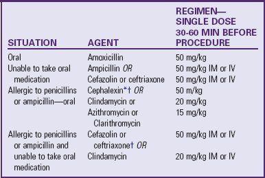
Prevention in these high-risk patients involves administration of prophylactic antibiotic therapy 1 hour before dental procedures (Table 34-5). If a patient is taking an antibiotic for another reason, it is recommended that they wait 10 days after antibiotics are completed to have a dental procedure so that normal flora can regenerate. If a patient requires chronic antibiotic therapy, a different antibiotic should be used for prophylaxis. Additional procedures that require prophylaxis (only in this high-risk group) include invasive procedures of the respiratory tract and procedures on infected skin or musculoskeletal tissue. Antibiotic prophylaxis is no longer recommended for gastrointestinal-genitourinary procedures. Maintenance of excellent oral hygiene is still of utmost importance, especially in children with CHD.

TABLE 34-5

DENTAL PROPHYLAXIS REGIMENS FOR PATIENTS AT HIGH RISK FOR INFECTIVE ENDOCARDITIS

image

IM, Intramuscular; IV, intravenous.

*Or other first- or second-generation oral cephalosporin in equivalent adult or pediatric dosage.

Cephalosporins should not be used in an individual with a history of anaphylaxis, angioedema, or urticaria with penicillins or ampicillin.

Modified from Wilson W, Taubert KA, Gewitz M, et al: Prevention of infective endocarditis: guidelines from the American Heart Association, Circulation 116(15):1736-1754, 2007.

Nursing Care Management

The objective of nursing care is to counsel parents of high-risk children concerning the need for prophylactic antibiotic therapy before procedures such as dental work. The family’s regular dentist should be advised of existing cardiac problems in the child as an added precaution to ensure preventive treatment in appropriate patients. These children should also maintain the highest level of oral health to reduce the chance of bacteremia from oral infections. (See also discussion on dental care in Chapter 14.)

QUALITY PATIENT OUTCOMES

Bacterial (Infective) Endocarditis

• Prevention in high-risk patients with antibiotic prophylaxis

• Early recognition and treatment

Parents should also have a high index of suspicion regarding potential infections. Without unduly alarming them, the nurse should educate patients and families to bring unexplained fever, weight loss, or change in behavior (lethargy, malaise, anorexia) to the practitioner’s attention. Such symptoms should not be self-diagnosed as a cold or flu, and children at risk (e.g., those with CHD) should have blood drawn for culture if they have a fever without an obvious source. Early diagnosis and treatment are important in preventing further cardiac damage, embolic complications, and growth of resistant organisms.

Treatment of endocarditis requires long-term parenteral antibiotics. In some cases IV antibiotics may be administered at home via a peripherally inserted central catheter, with nursing supervision for part of the treatment course. Nursing goals during this period include (1) preparation of the child for IV infusion, usually with an intermittent-infusion device, and performance of several venipunctures to draw blood for culture and laboratory values; (2) observation for side effects of antibiotics, especially inflammation along venipuncture sites along with assessments of renal function; (3) observation for complications, including embolism and HF; and (4) education regarding the importance of follow-up visits for cardiac evaluation, echocardiographic monitoring, and blood culturing.

Rheumatic Fever

Rheumatic fever (RF) is a poorly understood inflammatory disease that occurs after pharyngitis caused by group A β-hemolytic streptococci (GABHS). It is a self-limiting illness that involves the joints, skin, brain, serous surfaces, and heart. Cardiac valve damage (referred to as rheumatic heart disease) is the most significant complication of RF (World Health Organization, 2004; Bitar, Hayek, Obeid, et al, 2000). In developed countries RF and rheumatic heart disease have become uncommon, probably as a result of antibacterial control of streptococcal infection, successful treatment of rheumatic heart disease, and a change in the organism itself. However, RF remains a devastating problem in developing (third world) countries and is more likely to recur in this setting. It has also reappeared in some parts of the United States (Gentles, Colan, Wilson, et al, 2001).

Etiology

Strong evidence supports a relationship between upper respiratory tract infection with GABHS and subsequent development of RF (usually within 2 to 6 weeks). In almost all cases of RF a previous infection with GABHS can be documented by laboratory evidence of rising antibody titers. Diagnosis and treatment of GABHS infection prevents RF.

Pathophysiology and Clinical Manifestations

The principal manifestations of RF are observed in the heart, joints, skin, and central nervous system. Inflammatory hemorrhagic bullous lesions, called Aschoff bodies, are formed, which cause swelling, fragmentation, and alterations in the connective tissue. Aschoff bodies are found in virtually all patients with clinical rheumatic activity. These lesions occur in the heart, blood vessels, brain, and serous surfaces of the joints and pleura.

The major cardiac manifestation of RF is carditis involving the endocardium, pericardium, and myocardium. In the acute illness, clinical signs and symptoms reflect valvulitis, myocarditis, and pericarditis. Clinically, rheumatic carditis is most commonly associated with the mitral valve. The presence of an apical systolic murmur reflecting mitral regurgitation is a common clinical finding in acute rheumatic carditis. This murmur is a long, high-pitched, blowing murmur that begins with the first heart sound (S1) and continues throughout systole. Other murmurs in the acute phase may reflect aortic regurgitation. In addition, myocarditis produces tachycardia that is out of proportion to the degree of fever, especially during rest or sleep. Signs and symptoms of HF may result, and chest radiographic examination may demonstrate cardiomegaly. Signs and symptoms of pericarditis include muffled heart sounds because of pericardial effusion. In addition, the patient may demonstrate a pericardial friction rub and complain of chest pain. Pericardial effusions can be documented by echocardiography. Patients with mitral or aortic valve involvement may experience progressive valvular damage as time passes. Carditis is the only manifestation that can lead to permanent damage (Narula, Chandrasekhar, Rahimtoola, et al, 1999).

The second major manifestation is polyarthritis caused by edema, inflammation, and effusions in joint tissue. The arthritis is reversible and migratory, favoring large joints such as the knees, elbows, hips, shoulders, and wrists. The affected joint is swollen, hot, red, and exquisitely painful for 1 or 2 days, after which a different joint is affected. Joint manifestations usually accompany the acute febrile period, most often in the first 1 to 2 weeks; however, they can persist for 4 weeks in untreated patients.

The third major manifestation is erythema marginatum. This is a distinct erythematous macule with a clear center and wavy, well-demarcated border. This transitory, nonpruritic rash is most often found on the trunk and proximal portion of the extremities.

The fourth major manifestation is the development of subcutaneous nodules, which are small (0.5- to 1-cm), nontender swellings that persist indefinitely after the onset of the disease and gradually resolve with no resulting damage. They are rare but may be found in crops over bony prominences such as the feet, hands, elbows, scalp, scapulae, and vertebrae.

The last major manifestation, which reflects central nervous system involvement, is chorea, referred to as St. Vitus dance or Sydenham chorea. Chorea is characterized by sudden, aimless, irregular movements of the extremities, involuntary facial grimaces, speech disturbances, emotional lability, and muscle weakness that can be profound. It is usually exaggerated by anxiety and attempts at deliberate fine motor activity and is relieved by rest, especially sleep. Chorea is seen almost exclusively in children, with a higher incidence in females (World Health Organization, 2004). The time course of symptoms of chorea are variable, but 75% of patients recover in 6 months.

In addition to these major manifestations, minor manifestations that may support the diagnosis include arthralgia and fever, which may be low grade and which often spikes in the late afternoon. Laboratory findings reflect an inflammatory process. Other vague signs and symptoms include unexplained epistaxis, abdominal pain that may be severe enough to simulate appendicitis, weakness, fatigue, pallor, anorexia, and weight loss.

Diagnostic Evaluation

No single symptom or laboratory test can provide a definitive diagnosis of RF. Rather, the diagnosis is based on a set of guidelines by the American Heart Association (Dajani, Ayoub, Bierman, et al, 1993). These guidelines are designed to aid in the diagnosis only of the initial episode of RF (Ferrieri, 2002) (Box 34-9). Clinical and laboratory findings are divided into major and minor manifestations; evidence of recent streptococcal infection is present in the majority of instances.

BOX 34-9

GUIDELINES FOR DIAGNOSING INITIAL ATTACK OF RHEUMATIC FEVER (JONES CRITERIA, 1992 UPDATE)*

Major Manifestations

Carditis

Polyarthritis

Chorea

Erythema marginatum

Subcutaneous nodules

Minor Manifestations

Clinical findings

Arthralgia

Fever

Laboratory findings: elevated values for acute-phase reactants

• Erythrocyte sedimentation rate

• C-reactive protein level

Supporting Evidence of Antecedent Group A Streptococcal Infection

Positive results on throat specimen culture or rapid streptococcal antigen test

Elevated or rising streptococcal antibody titer


*If supported by evidence of preceding group A streptococcal infection, the presence of two major manifestations or of one major and two minor manifestations indicates a high probability of acute rheumatic fever.

From Guidelines for the diagnosis of rheumatic fever: Jones criteria, 1992 update, JAMA 268(15):2070, 1992.

Although the majority of patients with RF meet these criteria, in three circumstances exceptions are allowed. In patients who have chorea as the only symptom and in patients who are seen late with continued carditis, the late diagnosis may preclude the presence of supporting manifestations and laboratory findings. Finally, a recurrence of RF in a patient with a previous history of the disorder may not fulfill the standard Jones criteria. However, if a single major or several minor manifestations are seen in a patient who has a history of prior disease along with evidence of recent GABHS infection, the diagnosis may be made without strict adherence to the criteria.

Streptolysin O (O because it is oxygen labile) is a streptococcal extracellular product that produces lysis of the red blood cell. Antistreptolysin O (ASLO) titers measure the concentration of antibodies formed in the blood against this product. Normally the titers begin to rise about 7 days after onset of the infection and reach maximum levels in 4 to 6 weeks. Therefore a rising titer demonstrated by at least two ASLO tests is the most reliable evidence of recent streptococcal infection. Normal values are between 0 and 120 Todd units. Elevations over 333 Todd units indicate recent streptococcal infection in children.

Therapeutic Management

The goals of management include (1) eradication of GABHS (primary prevention), (2) prevention of permanent cardiac damage, (3) palliation of the other symptoms, and (4) prevention of recurrences (secondary prevention) (Gerber, Baltimore, Eaton, et al, 2009).

Penicillin (oral or intramuscular injections) remains the drug of choice, with macrolides or cephalosporins as a substitute in penicillin-sensitive children. Initial therapy includes a full 10-day course of penicillin or an alternative antibiotic.

Children who have had acute RF are susceptible to recurrent RF for the rest of their lives, and therefore prophylactic treatment against RF recurrence is started immediately after the initial course of antibiotics is complete. Secondary prevention involves monthly intramuscular injections of benzathine penicillin G, two daily oral doses of penicillin V, or one daily dose of sulfadiazine-sulfisoxazole or erythromycin. The duration of long-term prophylaxis varies and depends on whether the child has had cardiac involvement (Table 34-6).

TABLE 34-6

SUGGESTED DURATION OF SECONDARY PROPHYLAXIS FOR RHEUMATIC FEVER

CATEGORY OF PATIENT DURATION OF PROPHYLAXIS
No proven carditis For 5 yr after the last attack, or until 21 yr of age (whichever is longer)
Carditis (but no residual heart disease; no valvular disease) For 10 yr after the last attack, or until 21 yr of age (whichever is longer)
Carditis and residual heart disease (ongoing valvular disease) For 10 yr or until 40 yr old (whichever is longer); sometimes lifelong prophylaxis (see guidelines for indications)

Modified from Gerber MA, Baltimore RS, Taubert KA, et al: Prevention of rheumatic fever and diagnosis and treatment of acute streptococcal pharyngitis; a scientific statement from the AHA, Circulation 119:1541-1551, 2009.

The use of SBE prophylaxis should follow the recent American Heart Association guidelines that recommend prophylaxis only for very-high-risk patients. Therefore SBE prophylaxis is no longer recommended for patients with RF unless they have had a valve replacement. (Wilson, Taubert, Gewitz, et al, 2007).

Salicylates are used to reduce fever and discomfort and to control the inflammatory process, especially in the joints. Patients with arthritis from RF are generally responsive to salicylate therapy; however, salicylates should not be administered before diagnosis, since their use may mask the polyarthritis. Administration of prednisone may be indicated in some patients with heart failure. Neither salicylates nor prednisone has been shown to affect cardiac sequelae. Traditionally, bed rest or at least limited activity has been recommended during the acute illness.

Patients who have symptoms of heart failure due to significant valvular heart disease require medical therapy for their HF. They are also at risk for atrial fibrillation and embolic complications. Surgery may be indicated in this group of patients and may include valve repair or valve replacement.

Nursing Care Management

The objectives of nursing care for the child with RF are to (1) encourage compliance with drug regimens, (2) facilitate recovery from the illness, (3) provide emotional support, and (4) prevent recurrence of the disease. Because compliance is a major concern in long-term drug therapy, make every effort to encourage adherence to the therapeutic plan. (See Compliance, Chapter 27.) When compliance is poor, monthly injections may be substituted for daily oral administration of antibiotics, and children need preparation for this often dreaded procedure.

Interventions during home care are primarily concerned with providing rest and adequate nutrition. Usually, once the febrile stage is over, children can resume moderate activity and their appetite improves. If carditis is present, the family must be aware of any activity restrictions and may need help choosing less strenuous activities for the child.

QUALITY PATIENT OUTCOMES

Rheumatic Fever

• GABHS tonsillopharyngitis identified and treated

• Early recognition and treatment to prevent cardiac valve damage

• Recurrence prevented with prophylaxis compliance

One of the most disturbing and frustrating manifestations of the disease is chorea. The onset is gradual and may occur weeks to months after the illness, sometimes even in children who have not been diagnosed with RF. It may be mistaken for nervousness, clumsiness, behavioral changes, inattentiveness, and learning disability. It is usually a source of great frustration to the child because the movements, incoordination, and weakness severely limit physical skill. The child needs an opportunity to verbalize feelings. Of utmost importance is stressing to parents and schoolteachers that the movements are involuntary and sudden, that the chorea is transitory, and that all manifestations eventually disappear.

Nurses also have a role in prevention, primarily in screening school-age children for sore throats that may be caused by GABHS. This may involve actively participating in throat culture screening programs or referring children with a possible streptococcal infection for testing.

Kawasaki Disease (Mucocutaneous Lymph Node Syndrome)

Kawasaki disease (KD) is an acute systemic vasculitis of unknown cause. The illness is self-limiting and resolves in 6 to 8 weeks. Without treatment, however, approximately 20% to 25% of children develop cardiac sequelae. Damage to the blood vessels that supply the heart muscle (the coronary arteries) and damage to the heart muscle itself can occur. The most common sequela is ectasia (dilation) of the coronary arteries, or coronary artery aneurysm formation. Infants younger than 1 year of age are at the greatest risk for heart involvement. Children older than 5 years of age are also at increased risk of developing coronary sequelae, perhaps because KD is often not suspected in older children, which may lead to delayed diagnosis and treatment. KD has become a leading cause of acquired heart disease in children in the United States.

KD is seen in children of most racial and ethnic backgrounds. The incidence rate is estimated at 112 cases per 100,000 children under 5 years of age. KD occurs 1.5 to 1.7 times more frequently in males than in females, and 76% of affected children are younger than 5 years of age, with peak incidence in the toddler age-group (Holman, Curns, Belay, et al, 2003).

The etiology of KD remains unconfirmed. Although KD is not spread by person-to-person contact, several factors support an infectious cause. KD is often seen in geographic and seasonal outbreaks, with most cases reported in the late winter and early spring. KD is also a pediatric illness, which suggests the development of passive immunity. Because an etiologic agent has not been found, some experts believe that the illness may represent a final common pathway for more than one potential agent. Some recent studies have focused on genetics, trying to ascertain why some children are more likely to get KD than others (i.e., the possibility that the illness represents a final common pathway in a genetically susceptible host).

Pathophysiology

KD involves widespread inflammation of the small and medium-sized blood vessels (Connor and McCance, 2000), with the coronary arteries being the most susceptible to damage. During the acute stage of the illness there is progressive inflammation of the small vessels (capillaries, venules, arterioles) along with pancarditis. This inflammation is reflected in the clinical signs and symptoms and in laboratory test results. Inflammatory markers (C-reactive protein level and erythrocyte sedimentation rate) are elevated in the acute illness. The vasculitis progresses to the medium-sized muscular arteries, potentially damaging the walls of the vessels and leading to the formation of coronary artery aneurysms in some children. Initial evidence of enlargement of the coronary arteries by echocardiogram can be detected as early as day 7 of illness. Affected vessels continue to enlarge for several weeks and generally reach their largest diameter approximately 4 to 6 weeks from the onset of fever. Longer duration of fever (most likely reflecting the severity of inflammation) is strongly associated with the development of aneurysms. Aneurysms of the peripheral vessels (axillary, brachial, iliac, cervical, and renal arteries) can occur, although this is rare and usually is seen only in children who also have giant coronary aneurysms (>8 mm). In the acute phase, myocarditis (inflammation of the myocardium) is common. Decreased LV function may be evident on echocardiogram; however, the majority of children do not have clinical signs of heart failure. Ventricular function usually improves after the administration of IV immune globulin (IVIG). Occasionally, however, a child will be seen with severe ventricular dysfunction and/or cardiogenic shock. The systemic inflammation gradually subsides and eventually ceases with normalization of inflammatory markers 6 to 8 weeks from the onset of fever.

Over time, aneurysmal vessels try to heal through multiplication of cells in the vessels in an attempt to restore a “normal” lumen diameter. This process is called myointimal proliferation. The smaller the dilation, the more likely that the vessel will regress to a normal size. However, even if the lumen size is restored, the affected vessel may not be completely normal. The walls of these vessels are thicker and may be subject to scarring and calcification, especially at the distal ends of the aneurysm in patients with large aneurysms.

Almost all of the morbidity and morality resulting from KD are due to cardiac complications and mainly occur in patients who have giant aneurysms (≥8 mm). Coronary thrombosis may result from sluggish blood flow in a dilated or aneurysmal vessel. Over the years, stenosis and scarring may also lead to impeded blood flow, which can result in myocardial ischemia or infarction.

Clinical Manifestations

The course of KD can be divided into three phases: acute, subacute, and convalescent. The acute phase begins with an abrupt onset of high fever that is unresponsive to antibiotics and antipyretics. Over the next week or so, the diagnostic symptoms become evident. The bulbar conjunctivae of the eyes become reddened, with clearing around the iris (limbal sparing). The eyes are generally dry, without significant drainage. Inflammation of the pharynx and the oral mucosa develops, with red, cracked lips and the characteristic “strawberry tongue” (the normal coating of the tongue sloughs off, leaving the large papillae exposed, so that the tongue resembles a strawberry). The rash of KD differs from child to child but is never vesicular and is most often accentuated in the perineum. Often the area affected by the rash may desquamate. In addition, the child’s hands and feet become edematous, and the palms and soles become erythematous. The child may have cervical lymphadenopathy (a single node ≥1.5 cm). The node is not usually very tender or red. To meet “classic” criteria, children have prolonged fever (≥4 days) along with four out of five of the diagnostic criteria. During the acute stage, the child is typically very irritable and inconsolable. This behavior may continue for several weeks. Approximately one third of patients develop a temporary arthritis beginning in the small joints. Cardiac manifestations during this period include myocarditis with resultant potential ECG changes, decreased LV function, pericardial effusion, and mitral regurgitation. Generally, these findings are subclinical, but occasionally children with poor function are seen with symptoms of cardiogenic shock. On physical examination, the child may be tachycardic with a gallop rhythm. The coronary arteries may begin to show enlargement during this phase.

The subacute phase begins with resolution of the fever and lasts until all outward clinical signs of KD have disappeared. If changes in the coronary arteries occur, some enlargement or dilation is generally evident by echocardiography during the second week of illness. Damaged vessels can continue to enlarge and reach their maximum diameter approximately 4 to 6 weeks from the onset of illness. Thrombocytosis and hypercoagulability in a child with expanding aneurysms and disrupted blood flow place him or her at risk for coronary thrombosis. During the subacute period, the child may develop periungual desquamation (peeling that begins under the fingertips and toes) of the hands and feet. Arthritis may be evident during this phase and can affect the larger weight-bearing joints. Irritability persists during this period.

In the convalescent phase the clinical signs of KD have mostly resolved, but the laboratory values are still abnormal. The erythrocyte sedimentation rate and C-reactive protein level may remain elevated, which reflects lingering inflammation. Thrombocytosis may still be present. Arthritis may continue into this stage, and coronary complications may remain a concern as coronary dimensions peak 4 to 6 weeks from the onset of illness. This phase is complete when all blood values return to normal (6 to 8 weeks after onset). At the end of this stage, parents report that the child appears to have returned to normal in terms of temperament, energy, and appetite.

Cardiac Involvement: The most serious complication of KD is the development of coronary artery aneurysms and the potential for myocardial infarction in children with aneurysm formation. Myocardial ischemia can result from thrombotic occlusion or stenotic occlusion of a coronary aneurysm. The groups at highest risk for thrombus formation are children with “giant” aneurysms (>8 mm in diameter). Symptoms of acute myocardial infarction in young children can be subtle and may include abdominal pain, vomiting, restlessness, inconsolable crying, pallor, and shock. Complaints of actual chest pain or pressure are more typical in older children (Kato, Ichinose, and Kawasaki, 1986).

Diagnostic Evaluation

Currently no specific diagnostic test exists for KD. Therefore the diagnosis is established on the basis of clinical findings and associated laboratory results. The criteria in Box 34-10 should be used as guidelines. Many children with KD do not meet standard diagnostic criteria, and infants often have an incomplete presentation. It is therefore important to consider KD as a possible diagnosis in any infant or child with prolonged elevated temperature that is unresponsive to antibiotics and is not attributable to another cause. The American Heart Association recently published guidelines that include a diagnostic algorithm to guide the evaluation and treatment of patients with incomplete symptoms who have prolonged fever or other features of KD (Fig. 34-16).

BOX 34-10

EPIDEMIOLOGIC CASE DEFINITION OF KAWASAKI DISEASE (CLASSIC CLINICAL CRITERIA)

Fever persisting at least 5 days

Presence of at least four of the five following principal features:

1. Changes in extremities

Acute—Erythema of palms and soles, edema of hands and feet

Subacute—Periungual peeling of fingers and toes in second and third week

2. Polymorphous exanthem

3. Bilateral bulbar conjunctival injection without exudate

4. Changes in the lips and oral cavity—Erythema and cracking of lips, strawberry tongue, diffuse injection of oral and pharyngeal mucosae

5. Cervical lymphadenopathy (nodes >1.5 cm in diameter), usually unilateral

Patients with fever for at least 5 days and fewer than four principal features can be diagnosed as having Kawasaki disease when coronary artery abnormalities are detected by two-dimensional echocardiography or angiography. When four or more principal features are present, the diagnosis of Kawasaki disease can be made on day 4 of illness.

note: These criteria should be used as a guideline; however, not all of the symptoms need to be present at once. In addition, atypical or incomplete Kawasaki disease should be considered in patients with some features, prolonged fever, and no alternative diagnosis.

Modified from Newburger JW, Takahashi M, Gerber MA, et al: Diagnosis, treatment, and long-term management of Kawasaki disease: a statement for health professionals from the Committee on Rheumatic Fever, Endocarditis, and Kawasaki Disease, Council on Cardiovascular Disease in the Young, American Heart Association, Circulation 110(17):2747-2771, 2004.

image

Fig. 34-16 Algorithm for evaluation of suspected incomplete Kawasaki disease (KD). CRP, C-reactive protein; echo, echocardiography; ESR, erythrocyte sedimentation rate. (From Newburger JW, Takahashi M, Gerber MA, et al: Diagnosis, treatment, and long-term management of Kawasaki disease: a statement for health professionals from the Committee on Rheumatic Fever, Endocarditis, and Kawasaki Disease, Council on Cardiovascular Disease in the Young, American Heart Association, Circulation 110(17):2747-2771, 2004.)

Associated laboratory findings, when combined with clinical data, can be helpful in making the diagnosis. The typical child with KD is anemic and has a leukocytosis with a “shift to the left” (increased immature white blood cells) during the acute phase. Thrombocytosis with hypercoagulability becomes evident in the subacute phase and peaks approximately 3 weeks after the onset of fever. An elevated erythrocyte sedimentation rate and C-reactive protein level reflect ongoing inflammation and generally persist for 6 to 8 weeks. The erythrocyte sedimentation rate can be further elevated by the administration of IVIG; therefore it is better to measure C-reactive protein level as an indicator of inflammation. Microscopic urinalysis reveals a sterile pyuria with mononuclear cells that will not be evident with a regular dipstick test, since the white blood cells are not polymorphonuclear neutrophils. Transient elevation of liver enzymes may occur during the acute phase, reflecting inflammation of the liver. Examination of cerebrospinal fluid may show aseptic meningitis (presence of inflammatory cells). Albumin levels may be lower than normal.

Echocardiograms are used to monitor myocardial and coronary artery status. Obtain a baseline echocardiogram at the time of diagnosis for comparison with future studies and to evaluate ventricular and valvular function. Common findings on echocardiogram during the acute phase include pericardial effusions, mitral regurgitation, and decreased ventricular function. Follow-up echocardiograms should be performed at approximately 2 weeks after onset and again at 6 to 8 weeks from the onset of fever to determine the diameter of the coronary arteries and to assess LV contractility and valvular function. More frequent studies to assess coronary dimensions may be indicated in patients who have continued fever, those who require retreatment with IVIG, and those with coronary dilation on baseline or other studies.

Therapeutic Management

The current treatment of KD includes high-dose IVIG along with salicylate therapy. High-dose IVIG has been shown to reduce the duration of fever and the incidence of coronary artery abnormalities when given within the first 10 days of illness and optimally within the first 7 days. A single large infusion of 2 g/kg over 8 to 12 hours is recommended (Newburger, Takahashi, Gerber, et al, 2004).

Aspirin is given initially in an antiinflammatory dosage (80 to 100 mg/kg/day in divided doses every 6 hours) to control fever and symptoms of inflammation. The duration of therapy varies among institutions. Once fever has subsided and the child has been afebrile for 48 to 72 hours, the aspirin dosage is generally decreased to an antiplatelet dosage (3 to 5 mg/kg/day). Low-dose aspirin therapy is continued in patients without echocardiographic evidence of coronary abnormalities until the platelet count has returned to normal (6 to 8 weeks). If the child develops coronary abnormalities, low-dose (antiplatelet) salicylate therapy is continued indefinitely. Additional anticoagulation therapy, such as clopidogrel or warfarin administration, may be indicated in children with coronary enlargement. Children with giant aneurysms (>8 mm), who are at the greatest risk for morbidity and mortality, are usually maintained on warfarin and aspirin (Newburger, Takahashi, Gerber, et al, 2004). International normalized ratio (INR) levels are maintained at 2.0 to 3.0 in these patients.

Prognosis: Most children with KD recover fully after treatment. When cardiovascular complications occur, however, serious morbidity may result. Death occurs rarely (<0.1% to 0.2%) and is almost always a result of ischemia caused by coronary thrombosis or stenosis. Children with coronary abnormalities are followed closely. Long-term testing may include periodic ECGs, echocardiography, stress testing (stress echoes and/or myocardial perfusion scans) at rest and during exercise, and cardiac MRI, depending on individual risk and the availability of various testing modalities at the individual centers (Newburger, Takahashi, Gerber, et al, 2004; Baker and Newburger, 2008). Although echocardiography is sensitive in visualizing coronary dilation, it does not detect stenoses of the coronary arteries. Cardiac catheterization of the coronary arteries remains the gold standard and may be performed in children who still have significant abnormalities after 1 year and in situations in which myocardial ischemia is suspected from the results of noninvasive testing. A rare sequela of KD can be sensorineural hearing loss. If decreased hearing is suspected, the child should undergo audiologic testing.

Children without coronary artery aneurysms have now been followed for more than 30 years in Japan and the United States and do not show an increased incidence of premature heart disease. However, both coronary and peripheral arteries may be stiffer than normal, even in individuals who did not suffer obvious coronary artery dilation. For this reason it is especially important that children who have had KD have as few other risk factors for coronary disease as possible. Cholesterol levels and BP should be monitored, and these children should be encouraged to lead a heart-healthy lifestyle in terms of diet, exercise, and avoidance of smoking.

Nursing Care Management

The nursing care of children with KD is challenging. Inpatient care focuses on symptomatic relief, emotional support, diagnostic assistance, medication administration, and education of the child and family.

QUALITY PATIENT OUTCOMES

Kawasaki Disease

• Early diagnosis and treatment

• Prevention of cardiovascular complications

In the initial phase of illness, the nurse must monitor the child’s cardiac status carefully. Intake and output and daily weight measurements are recorded. Although the child may be reluctant to eat and therefore may be partially dehydrated, fluids need to be administered with care because of the usual finding of myocarditis. Assess the child frequently for signs of HF, including decreased urinary output, gallop rhythm, tachycardia, and respiratory distress. Cardiac monitoring is suggested in the following cases: before the initial ECG and echocardiogram are recorded and shown to be normal, during the infusion of IVIG (because of the large fluid load), in children younger than 1 year of age, and in any child with cardiac symptoms. Sedation is generally required before echocardiography in children younger than image to 3 years of age, since the child must remain still for up to 1 hour to obtain adequate visualization of the coronary arteries and cardiac structures and function.
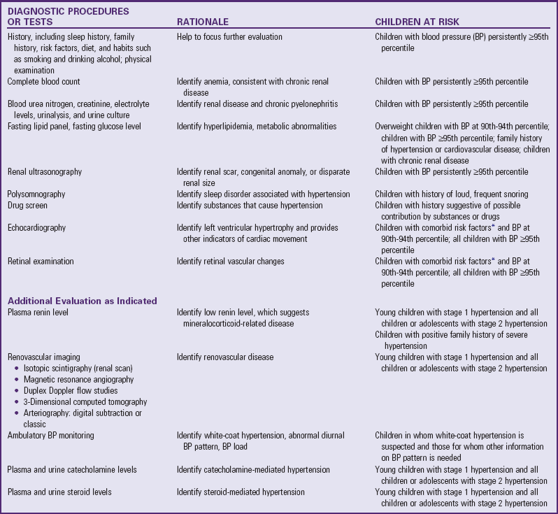
Nursing care focuses primarily on symptomatic relief. To minimize skin discomfort, application of cool cloths and unscented lotions and use of soft, loose clothing are helpful. During the acute phase, mouth care, including application of lubricating ointment to the lips, is important for the mucosal inflammation. Offer clear liquids and soft foods and monitor temperature carefully. It is important to document temperature just before aspirin administration, since fever reflects ongoing inflammation and may indicate the need for further treatment. If the temperature is very high, acetaminophen may be given in addition to high-dose aspirin. (See Controlling Elevated Temperatures, Chapter 27.) If arthritis develops, passive range-of-motion exercise may be indicated and can be done most easily during the child’s bath.

The administration of IVIG should follow the same guidelines as for administration of any blood product, with frequent monitoring of vital signs. Patients must be watched for allergic reactions. (See Table 35-2.) The nurse must monitor cardiac status because of the large fluid volume being administered to patients who may have subclinical myocarditis or diminished LV function. Check patency of the IV line because extravasation can result in tissue damage. Hypercoagulability and venous fragility often make it difficult to maintain IV access in children with KD (Connor and McCance, 2000).

Patient irritability is perhaps the most challenging problem. These children need to be placed in a quiet environment that promotes adequate rest. Their parents need to be supported in their efforts to comfort an often inconsolable child. They may need time away from their child, and nurses can often provide respite care for the family. Parents need to understand that irritability is a hallmark of KD and that they need not feel guilty or embarrassed about their child’s behavior.

Discharge Teaching: Parents need accurate information about the usual course of KD, including the importance of follow-up monitoring and the circumstances under which they should contact their practitioner. Irritability is likely to persist for up to 2 months after the onset of symptoms. Peeling of the hands and feet is painless and occurs primarily in the second and third weeks. Arthritis, especially of the larger weight-bearing joints, may persist for several weeks. Children are typically most stiff in the mornings, during cold weather, and after naps. Passive range-of-motion exercises in the bathtub are often helpful in increasing flexibility. Although the arthritis in KD is always temporary, it can be severe enough that some children require treatment with antiarthritic agents once they are no longer on high-dose aspirin. (note: Other high-dose NSAIDs should not be given with high-dose salicylates.)

Despite treatment with IVIG, approximately 10% to 15% of children develop recurrent fever and symptoms that necessitate retreatment with IVIG (2 g/kg). Educate parents about recrudescent illness after discharge. Persistent or recrudescent fever 48 hours after the initiation of IVIG infusion would prompt reevaluation and probably retreatment with IVIG. Instruct the parents to take the child’s temperature daily after discharge and to contact their physician or practitioner if there is any increase in temperature.

Also instruct parents about the administration of salicylates and, if the child is receiving high dosages, make them aware of the signs of aspirin toxicity: ringing in the ears (tinnitus), headache, dizziness, and confusion. The main side effect of low-dose aspirin is easy bruising. In addition, stop the aspirin and notify the practitioner if the child is exposed to chickenpox or influenza because of the drug’s possible association with Reye syndrome.

All parents should understand the unlikely but real possibility of myocardial infarction and the signs and symptoms of cardiac ischemia in a child. At the time of hospital discharge, the final cardiac sequelae are generally not fully known, since changes in the coronary arteries occur over the first 4 to 6 weeks after the onset of KD. In addition, parents of children with known severe coronary artery sequelae should know cardiopulmonary resuscitation. Finally, children with coronary abnormalities may require indefinite antiplatelet therapy with low-dose aspirin or other anticoagulants. In such cases children should avoid contact sports and have yearly influenza vaccines. The administration of measles-mumps-rubella vaccine should be delayed for 11 months after the administration of IVIG because the body might not produce the appropriate number of antibodies. In addition, the varicella vaccine should not be given for at least 11 months after IVIG therapy (American Academy of Pediatrics, 2009).

Systemic Hypertension

Hypertension is the most common cause of CVA and is a major risk of atherosclerotic disease in adults. Traditionally, primary hypertension has been considered a disease of older adults and is a major health problem in the United States. However, in recent years, there has been increasing evidence that primary hypertension does occur in children and adolescents, posing long-term health risks. End-organ effects can also be seen in young children as a result of chronic hypertension. These findings emphasize the importance of BP evaluation for all individuals beginning at the age of 3 years. A working group of the National High Blood Pressure Education Program (2004) published updated guidelines for the diagnosis, evaluation, and treatment of high BP in children and adolescents. In addition, this publication provides updated normative data for BP (including the 50th, 90th, 95th, and 99th percentiles based on age, sex, and height) (see inside back cover). Note that the normative BP data for children are based on auscultatory readings.

The diagnostic workup for hypertension depends on the degree of BP elevation. Prehypertension in young people is defined as a BP that falls above the 90th percentile for age and height, but does not meet criteria for true hypertension. Stage 1 hypertension is classified as a BP that falls persistently between the 95th and 99th percentile value plus 5 mm Hg for age, sex, and height. Stage 2 hypertension is defined as a BP that is persistently at or above the 99th percentile value plus 5 mm Hg for age, sex, and height. White-coat hypertension is diagnosed when the patient’s BP is higher that the 90th percentile in the clinician’s office but falls within the normal range outside of this setting.

Etiology

Hypertension in young children, compared to older adolescents and adults, more commonly is secondary to a structural abnormality or an underlying pathologic process, although the results of screening programs of relatively healthy children have challenged this view. The most common cause of secondary hypertension in young children is renal disease, followed by cardiovascular, endocrine, and some neurologic disorders. As a rule, the younger the child and the more severe the hypertension, the more likely it is to be secondary. The conditions associated with secondary hypertension in children and adolescents are listed in Box 34-11.

BOX 34-11   CONDITIONS ASSOCIATED WITH SECONDARY HYPERTENSION IN CHILDREN

Renal Disorders

Congenital defects

• Polycystic kidney, ectopic kidney, horseshoe kidney

• Obstructive anomalies

• Hydronephrosis

Renal tumor

• Wilms tumor

• Renovascular tumor

Abnormalities of renal arteries

Renal vein thrombosis

Acquired disorders

• Glomerulonephritis (acute or chronic)

• Pyelonephritis

• Nephritis associated with collagen disease

Cardiovascular Disease

Coarctation of aorta

Arteriovenous fistula

Patent ductus arteriosus

Aortic or mitral insufficiency

Metabolic and Endocrine Diseases

Adrenal tumors

• Adenoma

• Pheochromocytoma

• Neuroblastoma

Cushing syndrome

Adrenogenital syndrome

Hyperthyroidism

Aldosteronism

Hypercalcemia

Diabetes mellitus

Neurologic Disorders

Space-occupying lesions of the cranium (increased intracranial pressure)

• Tumors, cysts, hematoma

• Cerebral edema

• Encephalitis (including Guillain-Barré and Reye syndromes)

Miscellaneous Causes

Drugs (corticosteroids, oral contraceptives, pressor agents, amphetamines)

Burns

Genitourinary surgery

Trauma (e.g., stretching of femoral nerve with leg traction)

Insect bites (e.g., scorpion)

Intravascular overload (blood, fluid)

Hypernatremia

Toxemia of pregnancy

Heavy metal poisoning

The causes of primary hypertension are undetermined. There is evidence that both genetic and environmental factors play a role. The incidence of hypertension is greater in children with a family history of hypertension. African-Americans have a higher incidence of hypertension than Caucasians. In the African-American population hypertension develops earlier and is frequently more severe. Environmental factors that contribute to the risk of developing hypertension include obesity; salt ingestion; smoking; lead exposure; medications, including certain stimulant drugs; and stress.

Clinical Manifestations

Although the clinical manifestations associated with hypertension depend largely on the underlying cause, some observations can provide clues to the practitioner that an elevated BP may be a factor. Adolescents and older children with hypertension may complain of frequent headaches, dizziness, or changes in vision. In infants or young children who cannot communicate symptoms, observation of behavior provides clues, although gross behavioral changes may not be apparent until complications are present. Parents of infants and small children who have been treated for hypertension report that their child had previously been irritable and often indulged in an abnormal degree of head banging or rubbing.

Diagnostic Evaluation

It is clear from the increasing numbers of cases of hypertension and prehypertension being identified in children and adolescents that a BP determination should be a routine part of annual assessment in all children older than 3 years of age. Measure BP in children of any age if they are diagnosed as having or are suspected of having coarctation of the aorta, unexplained heart failure, unexplained heart murmurs, prematurity, unexplained seizures or other neurologic signs, an abdominal mass or masses, edema, ascites, evidence of renal failure, hypernatremia, failure to thrive, possible obstructive sleep apnea, respiratory distress, hyperlipidemia, or unexplained headaches. Because hypertension is strongly associated with obesity, a BMI should be calculated for each child at the routine physical examination.

Before a diagnosis of hypertension is made, measure BP in the sitting position on at least three separate occasions. To obtain an accurate reading, take care to quiet the child or relax the adolescent while the measurement is recorded to avoid false readings caused by excitement. The chief cause of falsely elevated BP readings is the use of improperly fitting, narrow cuffs. Therefore attention to correct measurement technique is essential. (See Blood Pressure, Chapter 6.) Note that a child who is large for his or her age may normally have a higher BP than a child who is of average size. Document upper and lower extremity BP when hypertension is suspected, along with the presence of femoral pulses. Twenty-four-hour BP (ambulatory BP [AMBP]) monitoring devices detect changes in pressure throughout the day and night, and thus may give a more realistic picture. These devices are especially helpful in diagnosing white-coat hypertension and are most easily used with older children or adolescents, who are able to tolerate being attached to an ambulatory monitor. The child or parent should keep a log during AMBP monitoring to document level of activity or, at a minimum, sleep and waking times. AMBP monitors should document readings at least hourly and ideally more often. Evaluation of the child with high BP is aimed at assessment of lifestyle and additional risk factors, detection of potential secondary causes, and documentation of the presence or absence of end-organ effects. Table 34-7 outlines the recommended workup for children with BP over the 90th percentile (National High Blood Pressure Education Program Working Group, 2004).

TABLE 34-7

CHILDHOOD HYPERTENSION EVALUATION

image

*Comorbid risk factors include diabetes mellitus and kidney disease.

Modified from National High Blood Pressure Education Program Working Group on High Blood Pressure in Children and Adolescents: The fourth report on the diagnosis, evaluation, and treatment of high blood pressure in children and adolescents, Pediatrics 114(2):555-576, 2004.

Take a careful medical history of the child or adolescent, including additional medical problems and current medications, drug use, or smoking. In addition, obtain a thorough family history to screen for other relatives with hypertension or other cardiovascular risk factors. In children with documented hypertension or prehypertension, initial laboratory data are also obtained.

The extent of additional testing depends on the degree of BP elevation. Diagnostic testing may include urinalysis, urine culture, renal function studies such as a creatinine and blood urea nitrogen levels, a lipid profile, fasting glucose level, complete blood count, and electrolyte levels. Additional laboratory data may include urine and blood catecholamine, renin, and aldosterone levels. In children who have significant hypertension, secondary causes should be investigated thoroughly. A renal ultrasonographic scan provides a first-line screen for renovascular hypertension or other renal disease (e.g., polycystic kidneys). Additional renovascular imaging may include a 99mTc dimercaptosuccinic acid (DMSA) scan (to rule out scarring) and/or a renal CT angiogram. Echocardiography is indicated in patients with BPs over the 90th percentile to assess LV hypertrophy. A finding that the child’s LV mass is higher than normal would support the presence of chronically elevated BP, since heart muscle thickens in response to chronic hypertension. A retinal examination may provide additional information about end-organ effects.

Therapeutic Management

Therapy for secondary hypertension involves diagnosis and treatment of the underlying cause. In those cases amenable to surgical repair, the nature of the condition, the type of surgery, and the child’s age are all important considerations. Children or adolescents with consistently elevated BP readings from no known cause (primary hypertension) or those with secondary hypertension not amenable to surgical correction may be treated with a combination of nonpharmacologic and pharmacologic interventions.

Dietary practices and lifestyle changes are important in the control of hypertension both for children and for adults and should be instituted first, except in severe cases.

Because obesity and hypertension are closely related, a weight reduction program is recommended for overweight youngsters. In salt-sensitive children, high salt intake increases the risk of hypertension for those genetically predisposed and aggravates existing hypertension unless salt intake is limited. Regular aerobic exercise augments weight reduction and alone has been shown to normalize BP. The exercise regimen is individualized to the child’s interest. It is helpful to quantify how much time is spent doing sedentary activities (e.g., watching television, using the computer, playing video games) compared with how much time daily is spent in aerobic activities. Stress reduction strategies may be beneficial and include biofeedback and relaxation. Smoking should be avoided.

Drug therapy is initiated with caution in children. Because the long-term effects of antihypertensive agents on children are not known, drug treatment of asymptomatic children with mild or borderline hypertension is not recommended. Antihypertensive drug therapy is indicated for treating those patients who have significant elevations of BP despite nonpharmacologic intervention. This includes children and adolescents with symptomatic hypertension; those with secondary hypertension; those with end-organ evidence of hypertension (e.g., increased LV mass); and those who have significant additional risk factors, such as diabetes (National High Blood Pressure Education Program Working Group, 2004).

The National High Blood Pressure Education Program Working Group on High Blood Pressure in Children and Adolescents (2004) recommends beginning pharmacologic therapy with one drug and adding other agents only if control is not obtained. The goal of therapy is to reduce BP levels below the 95th percentile. If additional risk factors (e.g., diabetes or renal disease) are present, however, BP values should ideally be reduced to less than the 90th percentile.

The oral antihypertensive drugs used most often in children are the ACE inhibitors (lisinopril, captopril, and enalapril), beta blockers (propranolol, atenolol [Tenormin]), calcium channel blockers (amlodipine [Norvasc]), angiotensin receptor blockers (losartan [Cozaar]), and diuretics (hydrochlorothiazide [HydroDIURIL]), to mention a few.

image DRUG ALERT

Beta Blockers and ACE Inhibitors

Beta blockers can cause lipid abnormalities or mood disturbances such as depression in some children. In addition, ACE inhibitors and angiotensin receptor blockers are teratogenic and therefore should not be used by teenage girls who are at risk of becoming pregnant.

Pharmacologic intervention is tailored to meet the needs of the individual child and is determined by the hypotensive effect produced and the appearance of any side effects. For example, ACE inhibitors and angiotensin receptor blockers have been effective in children with diabetes or certain renal diagnoses, whereas beta blockers and calcium channel blockers are often used by children with a history of migraine headaches (National High Blood Pressure Education Program Working Group, 2004). The goal is to achieve a normotensive state throughout the day without accompanying side effects. For many antihypertensive drugs, minimal data are available regarding side effects in children. Therefore consider any behavioral or physical changes that occur after institution of therapy a possible effect, and revise therapy as needed.

Nursing Care Management

The nurse is a valuable link in the delivery of health care for hypertension in the pediatric age-group. Active in detection, diagnosis, and therapy in any setting—hospital, school, clinic, private office, public health service, and private practice—nurses are frequently the primary contact in well-child care and follow-up clinics. They are often the liaison between the family and the health care services.

QUALITY PATIENT OUTCOMES

Hypertension

• Underlying cause of hypertension identified

• Blood pressure control maintained

• Dietary practices and lifestyle changes effectively used to control hypertension

• Compliance with medication regimen, if prescribed

A BP measurement should be part of the routine assessment of children over the age of 3 years and in younger children who have risk factors for hypertension. In carrying out the procedure, it is important for the nurse to use the correct cuff size. Any questionable reading is repeated. Ideally, BP should be assessed with the child in the sitting position with both feet on the floor. The right arm should be used for consistency in measurements. In addition, initial comparisons should be made between the upper and lower extremities.

Nursing counseling and guidance of affected children is a challenge. Education aimed at understanding hypertension and its implications over the life span is essential in promoting patient and family compliance with both nonpharmacologic and pharmacologic therapies. (See Compliance, Chapter 27.)

Home BP measurement can facilitate surveillance in youngsters with chronic hypertension and can document the effectiveness of therapy. A family member can be instructed in how to take and record accurate BP measurements, which can decrease the number of trips to a health care facility. This individual needs to understand when to contact the practitioner regarding elevated values. When this option is not feasible, the school nurse can often be a valuable resource in monitoring BP.

The nurse plays an important role in assessing individual families and providing targeted information regarding nonpharmacologic modes of intervention, such as diet, weight loss, smoking cessation, and exercise programs. If extensive dietary counseling is required, the child should be referred to a registered dietitian with expertise in working with children and adolescents. Exercise regimens should be individualized. Schoolchildren and young adolescents generally prefer team sports rather than individual training, which they may view as a burden rather than an enjoyable activity. If peers and family members can participate in any of the management strategies, the child is more likely to comply with the plan.

If drug therapy is prescribed, the nurse needs to provide information to the family regarding the reasons for drug therapy, how the drug works, frequency of BP monitoring, and possible side effects of the medication (Box 34-12). Explain that the drug needs to be taken consistently to achieve any prolonged control of BP. Stress the need for follow-up, especially because antihypertensive therapy can sometimes be safely discontinued if BP remains under control over time.

BOX 34-12   ANTIHYPERTENSIVE DRUGS COMMONLY USED IN THE TREATMENT OF PEDIATRIC HYPERTENSION, WITH NURSING INTERVENTIONS*

Angiotensin-Converting Enzyme Inhibitors

Action—Act primarily by interfering with the production of angiotensin II, a potent vasoconstrictor

Captopril (Capoten)

Monitor blood pressure and pulse.

Instruct to take 1 hour before meals to increase absorption.

Instruct to report any evidence of infection.

Advise to avoid rapid position changes (can initially cause dizziness).

Enalapril (Vasotec)

Monitor blood pressure and pulse (may cause hypotension).

Instruct to report any swelling of face or lips and difficulty breathing (may rarely cause laryngeal edema).

Instruct to report any evidence of infection.

Advise not to use potassium supplements (can increase serum levels).

Lisinopril (Zestril, Prinivil)

Longer half-life; can be administered in a single daily dose.

May cause hypotension, dizziness.

Monitor levels of electrolytes, blood urea nitrogen, creatinine (can increase serum levels).

Beta Blockers

Actions—Block response to beta stimulation; depress renin output

Propranolol (Inderal)

Monitor pulse and blood pressures (can cause bradycardia and hypotension).

Instruct to take with meals.

Advise that drug may cause fatigue, a decrease in exercise tolerance, weakness, and cold extremities.

Warn males of possible impotence.

Atenolol (Tenormin)

Monitor pulse and blood pressures (can cause bradycardia and hypotension).

Advise that drug can be given once a day.

Instruct not to discontinue abruptly (needs to be withdrawn over a 2-week period).

Calcium Channel Blockers

Actions—Decrease the force of contraction of the myocardium.

Amlodipine (Norvasc)

May cause tachycardia, peripheral edema, flushing, dizziness, or headache.

Do no discontinue suddenly.

Vasodilators

Actions—Act on vascular smooth muscle; thought to produce their effects by direct action on blood vessels to cause arterial vasodilation

Hydralazine (Apresoline)

Instruct to take with meals.

Advise that drug may cause drowsiness and that caution should be used in operating machinery or doing other hazardous activity.

Instruct to report if sore throat, fever, muscle and joint aches, or skin rash develops.

Angiotensin Receptor Blocker

Losartan (Cozaar]

Contraindicated in pregnancy.

Check serum potassium and creatinine levels.

Not used in children with decreased creatinine clearance.

Diuretics

See Table 34-3.


*For all drugs, instruct child or adolescent (and family) that (1) child should rise slowly from a horizontal position and avoid sudden position changes, (2) drug should be taken as prescribed, and (3) practitioner should be notified if unpleasant side effects occur but drug should not be discontinued.

Learning needs vary greatly depending on developmental levels and individual differences. Some children and families require a great deal of support, education, and guidance, whereas others need only education and periodic follow-up. A positive approach is essential; negative feedback will only alienate the family. Exploring the reasons for difficulty in compliance can often provide realistic alternatives. Continued education, support, and reinforcement for positive behavior are major nursing responsibilities.

Hyperlipidemia (Hypercholesterolemia)

Hyperlipidemia is a general term for excessive lipids (fat and fatlike substances); hypercholesterolemia refers to excessive cholesterol in the blood (Cook, 2009). High lipid or cholesterol levels play an important role in producing atherosclerosis (buildup of fatty plaques in the arteries), which eventually can lead to coronary artery disease (CAD), the leading cause of morbidity and mortality in the adult population in the United States (Box 34-13). The risk of premature CAD has been shown to increase with elevated plasma concentrations of total cholesterol and low-density lipoprotein (LDL) cholesterol and with low levels of high-density lipoprotein (HDL) cholesterol. Interventions that decrease LDL levels and increase HDL levels have been shown to lower the risk for CAD. In addition to abnormal cholesterol levels, risk factors for CAD include:

BOX 34-13   WHAT IS CHOLESTEROL?

Cholesterol, a fatlike steroid alcohol, is part of the lipoprotein complex in plasma that is essential for cellular metabolism. Triglycerides, natural fats synthesized from carbohydrates, are used for energy. Both are major lipids transported on lipoproteins, a combination of lipids and proteins, which include the following:

Chylomicrons—Produced in the intestine in response to the intake of dietary fat. These are the principal transporters of dietary fat (triglycerides) from the intestine to the blood and ultimately to the fatty tissue. Chylomicrons are usually not present in the blood after a 12- to 14-hour fast.

Very-low-density lipoproteins (VLDLs)—Contain high concentrations of triglycerides, moderate concentrations of cholesterol, and little protein.

Low-density lipoproteins (LDLs)—Contain low concentrations of triglycerides, high levels of cholesterol, and moderate levels of protein. The end product of VLDL synthesis, LDLs are the major carriers of cholesterol to the cells. Cells use cholesterol for synthesis of membranes and steroid production. Elevated levels of circulating LDL are a strong risk factor for cardiovascular disease.

High-density lipoproteins (HDLs)—Contain very low concentrations of triglycerides, relatively little cholesterol, and high levels of protein. HDLs transport free cholesterol to the liver for secretion in the bile. High levels of HDL are thought to protect against cardiovascular disease, whereas low levels of HDLs are considered an independent risk factor.

The cholesterol profile includes the following:

Total cholesterol = LDL + HDL + VLDL

Levels of total cholesterol, triglycerides, and HDL cholesterol are measured directly via a blood test. In the fasting state, LDL concentration is calculated using the following formula:

LDL = Total cholesterol − [HDL + (Triglycerides/5)]

A calculated LDL is considered accurate as long as the fasting triglyceride level is below 350 to 400 mg/dl. If triglycerides are higher than this, LDL cholesterol can be measured directly using a more specialized test.

• Positive family history of elevated cholesterol or early heart disease

• Cigarette smoking

• Diabetes (type 1 or 2)

• Obesity

• Hypertension

Research over the past four decades indicates that a presymptomatic phase of atherosclerosis begins in childhood with the development of fatty streaks evident on autopsies of children who died of noncardiac causes (Kavey, Allada, Daniels, et al, 2007). The extent of atherosclerosis is positively associated with the number of adult risk factors such as obesity, cholesterol abnormalities, and hypertension. These data continue to support early screening and management of lipid levels to identify children with risk factors for cardiovascular disease and provide early intervention (lifestyle modifications and/or medications).

In addition to the risk factors noted above, children are considered to be at high risk for atherosclerosis because of coexisting health problems such as:

• Chronic inflammatory diseases

• Cancer

• Transplantation

• CHD

• A history of KD

Lifestyle habits, including diet, exercise patterns, and smoking—all known to be potential risk factors for cardiovascular disease—are normally established at a young age. Risk factor modification and lipid-lowering measures in adults are known to decrease the incidence of CAD. There is increasing evidence that modification of risk factors earlier in life will have a positive effect on the likelihood of atherosclerosis in adult life.

Diagnostic Evaluation and Screening

Diagnosis of hyperlipidemia is based on analysis of blood. A blood specimen for determination of a full lipid profile should be drawn after a 12-hour fast. Total cholesterol and HDL cholesterol values obtained at any time in the nonfasting state are also accurate. It is important to note that lipid values can be affected by febrile illnesses and therefore lipids should not be drawn within 3 weeks of a febrile illness. Average lipid values vary by age and gender, with HDL values decreasing in boys as they go through puberty. Children are considered to have elevated total cholesterol if their total cholesterol value is more than 200 mg/dl and/or their LDL cholesterol value is more than 130 mg/dl. Some providers recommend using age- and gender-specific cutpoints.

Screening of children for hypercholesterolemia remains controversial. Consistent with previous recommendations, current guidelines continue to recommend a two-pronged strategy, providing complementary approaches: (1) a population approach that aims to lower the average levels of blood cholesterol among all American children through population-wide changes in nutrient intake and eating patterns, and (2) an individualized approach based on selective screening (see Evidence-Based Practice box). First presented by the National Cholesterol Education Panel in 1992, selective screening of individuals thought to be at high risk was recommended by the American Academy of Pediatrics in their 2008 statement (Daniels, Greer, and the Committee on Nutrition, 2008). Children more than 2 years old should be screened if they have a first- or second-degree relative with lipid abnormalities or with early cardiovascular disease (<55 years in a man or <65 years in a woman). In addition, perform screening in children with individual risk factors (see Box 34-2). The major change in the recent guidelines is that a full fasting profile is now recommended as the initial screen in children with familial or individual risk factors, including obesity. In addition, the American Academy of Pediatrics endorses screening for children with an unknown family history and those who have disease-state risk factors, such as diabetes (Daniels, Greer, and the Committee on Nutrition, 2008). Individualized treatment for these patients includes lifestyle modification and possibly lipid-lowering medication in the most severely affected.

EVIDENCE-BASED PRACTICE

Cholesterol Screening for Children

Ask the Question

Should cholesterol screening be performed in children?

Search for the Evidence

Search strategies

Selection criteria included English-language publications, research-based articles (level 3 or lower), infant and child populations.

Databases used

PubMed, Cochrane Collaboration, MD Consult, Joanna Briggs Institute, National Guidelines Clearinghouse (AHRQ), TRIP Database Plus, PedsCCM, BestBETs

Critically Analyze the Evidence

GRADE criteria: Evidence quality moderate; recommendation strong (Guyatt, Oxman, Vist, et al, 2008)

The rationale for lipid screening and management in children is evolving now that lipid levels have been followed from childhood into adulthood. Children who have cholesterol levels in the upper percentiles seem to have an increased risk of remaining in the upper percentiles into adulthood (Nicklas, von Duvillard, and Berenson, 2002). The more severely affected children are generally the ones targeted for dietary and possibly pharmacologic intervention. On the other hand, children in the lower percentiles are unlikely to have high cholesterol levels as adults.

Cholesterol levels in childhood appear to be a major population predictor for adult cholesterol levels. Cholesterol screening can begin in children over the age of 2 when risk factors like family history are present. The precursors of atherosclerosis are present in young people. Findings from autopsy studies of young people who have died of accidents and injuries have shown that the atherosclerotic process begins early in life (Enos, Holmes, and Beyer, 1953; Strong, Malcom, McMahan, et al, 1999). Furthermore, the extent of atherosclerosis is related to the presence and degree of cardiovascular risk factors in adults (Berenson, Srinivasan, Bao, et al, 1998).

Many experts favor selective screening because high blood cholesterol levels aggregate in families as a result of shared genetic and environmental factors (American Academy of Pediatrics, 2008). In addition, the most severely affected children generally come from families in which there is a high incidence of early heart disease.

Apply the Evidence: Nursing Implications

Current recommendations include selective screening of children over the age of 2 years who have a sibling, parent, or grandparent with an elevated cholesterol level of 240 mg/dl or higher. In addition, children should be screened if they have a first- or second-degree relative with early atherosclerotic disease (stroke, myocardial infarction, sudden cardiac death, angina, or peripheral vascular disease). Screening should also be done if the child has any individual risk factors such as diabetes, hypertension, obesity, a history of Kawasaki disease, or nephrotic syndrome. Screening should include a fasting lipid profile in these patients. Lastly, cholesterol screening should be performed if the child’s genetic family history is unknown.

References

American Academy of Pediatrics. Lipid screening and cardiovascular health in childhood. Pediatrics. 2008;122:198–208.

Berenson, GS, Srinivasan, SR, Bao, W, et al. Association between multiple cardiovascular risk factors and atherosclerosis in children and young adults. N Engl J Med. 1998;338(23):1650–1656.

Enos, WF, Holmes, RH, Beyer, J. Coronary disease among United States soldiers killed in action in Korea. JAMA. 1953;152(12):1090–1093.

Guyatt, GH, Oxman, AD, Vist, GE, et al. GRADE: an emerging consensus on rating quality of evidence and strength of recommendations. BMJ. 2008;336:924–926.

Nicklas, TA, von Duvillard, SP, Berenson, GS. Tracking of serum lipids and lipoproteins from childhood to dyslipidemia in adults: the Bogalusa Heart Study. Int J Sports Med. 2002;23(Suppl 1):S39–S43.

Strong, JP, Malcom, GT, McMahan, CA, et al. Prevalence and extent of atherosclerosis in adolescents and young adults: implications for prevention from the Pathobiological Determinants of Atherosclerosis in Youth Study. JAMA. 1999;281(8):727–735.

Therapeutic Management

Treatment of high cholesterol levels in children begins with lifestyle modification. The American Heart Association and the American Academy of Pediatrics recommend a heart-healthy diet for all American children, with dietary counseling recommended for those children with known elevated cholesterol values (Daniels, Greer, and the Committee on Nutrition, 2008). A heart-healthy diet focuses on a balanced intake for children over 2 years old, favoring low-fat dairy products, avoiding trans fats, and reducing sweetened beverages. The recommended diet is rich in fruits and vegetables and whole grains. In addition, children with abnormal LDL cholesterol levels should reduce saturated fat intake to 7% of total calories and dietary cholesterol intake to less than 200 mg/day.

Research supports the benefit of diets low in saturated fats and trans fats and higher in monounsaturated fats (such as those found in olive and canola oil). It is important to provide accurate dietary information to families. Food labeling is confusing. Without counseling, patients tend to replace foods high in fat with foods low in fat but high in simple sugars; this can raise triglyceride values, decreasing the effectiveness of nutritional interventions and providing wasted calories.

Current thinking favors a Mediterranean-type diet, based on whole grains, fruits, and vegetables. In addition, this diet allows the use of monounsaturated fats, such as olive oil and canola oil, which have beneficial effects on HDL cholesterol values. The use of these fats also makes the diet more realistic and helps keep children satiated. Dietary recommendations can be confusing, and individualized guidelines should be provided by a certified nutritionist or dietitian with expertise in lipid management.

If the child’s BMI is elevated, weight management should be addressed. In addition to food choice, portion size may also be an issue for these children. Increased physical exercise should be encouraged (ideally 45 minutes to 1 hour, 5 days a week) and is critical to the success of a weight-loss program. Population recommendations meant to decrease cardiovascular risk have been in place for almost two decades, yet unfortunately the incidence of childhood obesity in this country has tripled during the last 25 years (de Ferranti and Ludwig, 2008). Education and programs aimed at decreasing this trend and increasing heart-healthy living (e.g., diet, exercise, avoidance of smoking) are extremely important to decrease cardiovascular risk for the next generation. Assessment of BMI, education, referral to weight-loss programs if indicated, and follow-up should be an integral part of every well-child visit.

Dietary changes alone can decrease LDL values 10% to 15%; however, it is likely that children with severe genetic hypercholesterolemia will require lipid-lowering medication in addition to lifestyle modification. Lipid-lowering drugs used in children and teenagers include bile acid–resin binders such as cholestyramine (Questran) and colestipol (Colestid), hydroxymethylglutaryl–coenzyme A (HMG-CoA) reductase inhibitors (statins such as atorvastatin, simvastatin, pravastatin). Nicotinic acid can help raise HDL cholesterol but is rarely used in pediatrics because of symptoms of flushing and documentation of elevated liver transaminases.

The American Academy of Pediatrics guidelines (Daniels, Greer, and the Committee on Nutrition, 2008; McCrindle, Urbina, Dennison, et al, 2007) lowered the recommended age for initiation of lipid-lowering medication, including statins, to children as young as 8 years old in patients with:

• LDL cholesterol greater than 190 mg/dl in patients without a positive history of early heart disease

• LDL cholesterol greater than 160 mg/dl with a positive family history or two risk factors

• LDL cholesterol greater than 130 mg/dl in patients with diabetes

image DRUG ALERT

Statins

In clinical practice, statins are used for children severely affected with particularly adverse family histories. The tendency is to allow a progression through puberty, particularly for young girls. The target level for LDL cholesterol is less than 130 mg/dl, and optimally less than 100 mg/dl. Certain individual risk factors, such as diabetes, hypertension, or a history of coronary artery aneurysms from KD, all lower the threshold for pharmacologic intervention.

Bile acid–resin binders act by binding bile acids in the intestinal lumen. Because they are not absorbed by the intestine, they are not thought to produce systemic toxicity and are generally safe for children. Cholestyramine and colestipol are both powders that are mixed with water or juice just before ingestion. Many patients cannot tolerate resin binders because they have a gritty texture and do not dissolve completely in water. Side effects may include constipation, abdominal pain, gastrointestinal bloating, flatulence, and nausea. The average dosage for a child is 4 g three times daily or 6 g twice daily. Colesevelam (Welchol) comes in a pill form (625 mg/tablets); the effective dosage is two or three tablets taken twice a day.

Patients who take resin binders should take one multivitamin supplement daily, since bile acid–binding agents may interfere with the absorption of fat-soluble vitamins. Since they can interfere with absorption of other medications, any other medications should be given at least 1 hour before or 6 hours after the bile acid–binding agent is ingested. The results of a complete blood count; chloride and folate levels; and serum concentrations of vitamins A, D, and E should be evaluated yearly.

HMG-CoA reductase inhibitors (statins) continue to be the most effective medication for lipid lowering and are the first-line treatment for adults. Statins are being used more commonly in children and adolescents, especially those with severe familial dyslipidemia. Statin medications are most effective if taken in the evening. Because statins are metabolized by the liver, baseline liver function tests (alanine aminotransferase, aspartate aminotransferase) results, as well as creatine kinase levels, are obtained before the initiation of therapy. These tests, along with a fasting lipid profile, are repeated at approximately 4 weeks and 8 weeks after the initiation of therapy and with any dosage changes. Once the patient is taking a stable dosage, laboratory tests are repeated at 6-month intervals. In addition to elevation of liver transaminase levels, other potentially serious side effects include rhabdomyolysis, which can cause renal failure. Rhabdomyolysis has been reported rarely in adult patients. Instruct patients to discontinue the medication and contact their clinician if they experience the new onset of muscle aches or dark brown urine. In addition, female patients need to be educated that statins are considered teratogenic and cannot be taken during pregnancy. Oral contraceptives may be given in conjunction with lipid-lowering medication; however, they may increase lipid values, potentially necessitating modification of the statin dosage.

A relatively new drug, ezetimibe, works by inhibiting cholesterol absorption. It lowers LDL levels by preventing intestinal uptake of dietary and biliary cholesterol. Recommended use is in combination therapy with a statin, further lowering LDL values. This medication is currently approved for children older than 10 years of age with extremely severe hyperlipidemia. However, clinical trials have shown inconclusive evidence regarding this drug’s clinical benefit. Several large clinical trials are in process, and more information should be available in the next few years.

Nursing Care Management

Nurses play an important role in the screening, education, and support of children with hyperlipidemia and their families. When a child is referred to a lipid clinic, it is essential that the family be adequately prepared for the first visit. Generally, the parents are asked to keep a dietary history for the child before this visit. Sometimes they need to complete a questionnaire regarding the child’s normal dietary habits over the preceding year. Families are instructed to keep their child fasting for at least 12 hours before screening. Therefore it is important to schedule the blood test early in the morning and to arrange for nourishment immediately thereafter. At the visit, a complete individual and family health history is taken. The family history should include both biologic parents and all first-degree relatives. Questions are asked about early heart disease, hypertension, CVAs, sudden death, hyperlipidemia, diabetes, metabolic syndrome, and endocrine abnormalities. Nurses may also uncover risk factors when obtaining a health history for other purposes. It is therefore important that nurses be familiar with current screening practices and with available resources for children with positive family histories.

Parents and extended families should be informed about cholesterol and hyperlipidemia. This education should include a brief introduction to the different lipoprotein categories, including cholesterol, HDL, LDL, and triglycerides. Also, review behavioral risk factors for heart disease, such as smoking and lack of exercise. For management to be effective, parents and older children need to understand the rationale for dietary and pharmacologic intervention in the prevention of future cardiovascular disease.

Nutritional education is part of the treatment of any child or teenager with high cholesterol and ideally should be provided by a nutritionist with expertise in lipid disorders. Dietary compliance may become an issue of control and a source of great stress for many families, particularly for teenagers. Children with high cholesterol levels should not be viewed as having a disease. Instead, emphasize the positive aspects of healthy eating, regular exercise, and avoidance of smoking. Encourage basic dietary changes for the whole family, so that the affected child is not singled out. The focus is positive, with emphasis on making healthy dietary choices, such as substituting chicken and fish for hot dogs and hamburgers and substituting frozen yogurt for ice cream (Box 34-14). Cultural differences must be considered and recommendations individualized. For example, it is more realistic to suggest frying food in a monounsaturated oil such as canola oil than to forbid frying food altogether in families in which this is common practice. Emphasize substitution rather than elimination. Visual aids (e.g., test tubes depicting the amount of fat in a hot dog) are often helpful, especially for children. Diets should be flexible and individually tailored by a nutritionist experienced in combining recommendations that meet both the nutritional demands of the growing child and lipid modifications. Encourage parents to participate in dietary and educational sessions, ask questions, and share ideas and experiences.

BOX 34-14

AMERICAN HEART ASSOCIATION DIETARY RECOMMENDATIONS FOR INFANTS, CHILDREN, AND ADOLESCENTS TO PROMOTE CARDIOVASCULAR HEALTH

Infancy

Breast-feeding is ideal nutrition and sufficient to support optimal growth and development for approximately the first 4 to 6 months after birth. Try to maintain breast-feeding for 12 months. Transition to other sources of nutrients should begin at approximately 4 to 6 months of age to ensure sufficient micronutrients in the diet.

Delay the introduction of 100% juice until at least 6 months of age and limit to no more than 4 to 6 oz/day. Juice should be fed only from a cup, not a bottle.

Do not overfeed infants and young children. Children should not be forced to finish meals if not hungry because they often vary caloric intake from meal to meal.

Introduce healthy foods and continue offering if initially refused. Do not introduce foods without overall nutritional value simply to provide calories.

Eating Pattern for Families*

Energy (calories) should be adequate to support growth and development and to reach or maintain desirable body weight.

Eat foods low in saturated fat, trans fat, cholesterol, salt (sodium), and added sugars.

Keep total fat intake between 30% and 35% of calories for children 2 to 3 years of age and between 25% and 35% of calories for children and adolescents 4 to 18 years of age, with most fats coming from sources of polyunsaturated and monounsaturated fatty acids, such as fish, nuts, and vegetable oils.

Choose a variety of foods to get enough carbohydrates, protein, and other nutrients.

Eat only enough calories to maintain a healthy weight for height and build. Be physically active for at least 60 minutes a day.

Serve whole grain breads and cereals rather than refined grain products. Look for “whole grain” as the first ingredient on the food label and make at least half of grain servings whole grain. Recommended grain intake ranges from 2 oz/day for a 1-year-old to 7 oz/day for a 14- to 18-year-old boy.

Serve a variety of fruits and vegetables daily, while limiting juice intake. Each meal should contain at least one fruit or vegetable. Children’s recommended fruit intake ranges from 1 cup/day between ages 1 and 3 years to 2 cups/day for a 14- to 18-year-old boy. Recommended vegetable intake ranges from image cup/day at age 1 year to 3 cups/day for a 14- to 18-year-old boy.
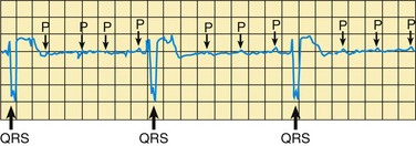
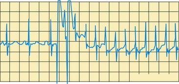
Introduce and regularly serve fish as an entrée. Avoid commercially fried fish.

Serve nonfat and low-fat dairy foods. From ages 1 to 8 years, children need 2 cups of milk or its equivalent each day. Children ages 9 to 18 years need 3 cups.

Don’t overfeed. Estimated calories needed by children range from 900/day for a 1-year-old to 1800/day for a 14- to 18-year-old girl and 2200/day for a 14- to 18-year-old boy.


*This eating pattern supports a child’s normal growth and development. It provides enough total energy and meets or exceeds the recommended dietary allowances for all nutrients for children and adolescents, including iron and calcium.

From American Heart Association: Dietary guidelines for healthy children, 2005, available at www.americanheart.org/presenter.jhtml?identifier=4575 (accessed March 22, 2006). Reprinted with permission.

Parents often feel guilty about the hereditary component of hyperlipidemia. Many of these same parents believe they have failed if diet alone is not making a significant difference in their child’s lipid profile. They are reassured that a dietary approach alone is often not sufficient, especially for children with values higher than the 95th percentile.

Parents of children who require pharmacologic therapy must understand the purpose, dosage, and possible side effects of the various drugs. Medication schedules should remain flexible and should not interfere with the child’s daily activities. As an example, children of elementary school age may have better compliance if they take a resin-binding agent (e.g., cholestyramine, colestipol) twice a day (i.e., before school and at night) rather than the standard three times a day. Follow-up phone calls by the nurse between visits allow parents to discuss their concerns and ask any questions that have arisen.

image NURSING ALERT

The recommendations for fat intake for the general population are intended for children over 2 years of age. Children under the age of 2 require a higher percentage of calories from fat. However, recent studies have supported the safety of low-fat dairy products in younger children. Therefore low-fat dairy products are appropriate for children less than 1 year old who are obese or who have additional cardiovascular risk factors, as long as adequate sources of monounsaturated and polyunsaturated fats are maintained (Daniels, Greer, and the Committee on Nutrition, 2008).

Cardiac Dysrhythmias

Dysrhythmias, or abnormal heart rhythms, can occur in children with structurally normal hearts, as features of some congenital heart defects, and in patients following surgical repair of congenital heart defects. They occur in patients with cardiomyopathy and cardiac tumors. They can occur secondary to metabolic and electrolyte imbalances. Childhood dysrhythmias can have a genetic or familial etiology. Dysrhythmias can be classified in several ways, such as by heart rate characteristics (bradycardia and tachycardia) or by the origin of the dysrhythmia in the atria or ventricles. Most are due to abnormalities in impulse generation in the RA or to abnormalities in the conduction pathways.

Some dysrhythmias are well tolerated and self-limiting. Others may cause decreased cardiac output with associated symptoms. Some dysrhythmias can cause sudden death. Treatment depends on the cause of the dysrhythmia and its severity. Underlying causes are treated if possible (as with electrolyte imbalances). Some dysrhythmias (such as bradycardia caused by congenital heart block) are well tolerated and may not require treatment for many years. Others may require medications, radiofrequency ablation, or pacemaker placement. Some can be difficult to treat and require multiple therapies.

Many advances have been made in the diagnosis and treatment of pediatric dysrhythmias. Improvements in technology have allowed better diagnosis, the development of ablation techniques, and the expansion of pacemaker capabilities. New antidysrhythmic medications have proven safe and effective in children. Radiofrequency ablation has offered a cure for some dysrhythmias. Pediatric electrophysiology has become a highly specialized field, and the student is referred to more detailed sources for an in-depth discussion. The following sections describe diagnostic studies and provide a general discussion of the most common tachycardia (supraventricular tachycardia) and the most common bradycardia (complete heart block) that require treatment in the pediatric population.

Diagnostic Evaluation

Before diagnosing an infant or child with an abnormal heart rate, nurses must be familiar with the standards for normal heart rate in the particular age-group. (See inside back cover.) Heart rate patterns considered normal for a particular child can vary tremendously. An initial nursing responsibility is recognition of an abnormal heartbeat, either in rate or in rhythm. When a dysrhythmia is suspected, the apical rate is counted for 1 full minute and compared with the radial rate, which may be lower because not all of the apical beats are felt. Consistently high or low heart rates should be regarded as suspicious. Accurate nursing assessment is essential. The patient should be placed on a cardiac monitor with recording capabilities. A 12-lead ECG yields more information than the monitor recording and should be taken as soon as possible. Recent advances in bedside telemetry allow storage of ECG tracings for later analysis.

Several advances in the diagnosis of cardiac dysrhythmias have greatly improved the understanding and treatment of these conditions in children. The basic diagnostic procedure is the ECG, including 24-hour Holter monitoring. However, more definitive procedures include both noninvasive and invasive techniques.

Electrophysiologic cardiac catheterization allows identification of conduction disturbances and immediate investigation of drugs that may control the dysrhythmia. Electrode catheters are introduced intravenously and directed toward the right side of the heart. The heart is then selectively stimulated to induce dysrhythmias. Once a dysrhythmia occurs, different antidysrhythmic drugs are administered intravenously to monitor which pharmacologic agent is most successful in terminating the dysrhythmia.

Another procedure that may be employed is transesophageal recording. An electrode catheter is passed to the lower esophagus and, when in position at a point proximal to the heart, is used to stimulate and record dysrhythmias.

The onset and diagnosis of a cardiac dysrhythmia are frightening experiences for parents and older children. Sometimes the dysrhythmia rapidly leads to heart failure and a medical crisis. In this situation parents need much support to express their feelings and to understand the diagnosis and its treatment. Often parents and children have an unspoken fear of potential death even if the dysrhythmia is benign, and repeated explanations are needed to relieve anxiety.

Bradydysrhythmias: Sinus bradycardia in children can be due to the influence of the autonomic nervous system, as with hypervagal tone, or can be in response to hypoxia and hypotension. Once the infant receives adequate oxygenation and any acidosis is eliminated, the heart rate often returns to baseline. Sinus bradycardias are also known to develop after atrial repairs involving atrial suture lines such as in the Fontan procedure.

Complete AV block is also referred to as complete heart block (Fig. 34-17). This can be either congenital (occurring in children with structurally normal hearts) or acquired after surgery to repair cardiac defects. AV blocks are most often related to edema around the conduction system and resolve without treatment. Temporary epicardial wires are placed in most patients at surgery; if a rhythm disturbance occurs, temporary pacing can be employed. Just before discharge the health practitioner removes the wires by pulling slowly and deliberately down on them from the site of insertion.

image

Fig. 34-17 Complete heart block. Note slow rhythm and several P waves not followed by a QRS complex.

A permanent pacemaker may be needed in some children, such as those with postsurgical AV block or, less frequently, congenital AV block. The pacemaker takes over or assists in the heart’s conduction function. The surgical implantation of a pacemaker is usually a low-risk procedure. Once the wire has been introduced, a small incision is made and a pocket is formed under the muscle to house and protect the generator. The generator is placed under the abdominal muscle in infants and young children and in the upper chest below the clavicle in older children and adolescents. Depending on patient size and cardiac anatomy, some pacemakers can be placed transvenously in the catheterization laboratory, rather than in the operating room. Continuous ECG monitoring is necessary during the recovery phase to assess pacemaker function. The nurse should be aware of the programmed rate and expected individual generator variations. A baseline ECG and chest x-ray film are obtained for future comparison. The pacemaker pocket site is monitored for signs of infection. Analgesics are given for pain.

Pacemaker functions have become dramatically more sophisticated; pacemakers can control heart rate according to activity, cardiac output, and respirations. In addition, some models can be programmed for overdrive pacing or cardioversion when the generator detects accelerated rates beyond established normal values.

When a pacemaker is implanted, the education of the parents and child includes an explanation of the device, a description of the component parts, an explanation of the surgical procedure, and discharge teaching. The pacemaker is made up of two basic parts: the pulse generator and the lead. The pulse generator is composed of the battery and the electronic circuitry. The function is to produce the electrical impulse sent to the heart and to receive and respond to signals produced by the heart. The lead is an insulated, flexible wire that conducts the electrical impulse from the pulse generator to the heart. Two types of leads are available: transvenous and epicardial. The child’s size and the heart’s structure determine which lead is more appropriate. Transvenous leads are inserted into a large vein, often the subclavian, and advanced into the right side of the heart. Placement is secured by engaging a small corkscrew or fishhook attachment at the end of the lead into the endocardium. Epicardial leads are attached directly to the epicardial layer of the heart. Parents should be aware of which type of lead is in place in their child.

Discharge teaching includes information about the signs and symptoms of infection, general wound care, and activity restrictions. Parents, and patients if they are old enough, should learn to take the pulse and should know the settings of the pacemaker. If the patient’s low rate is set at 80 beats/min and the heart rate is only 68 beats/min, there is a possible problem with the pacemaker that needs to be investigated. Instructions for telephone transmission of ECG readings are also given. Telephone connections can be used to transmit ECG data and also to monitor battery life and pacemaker function. The pacemaker generator has to be replaced periodically because of battery depletion. Children with pacemakers should wear a medical alert device, and their parents should have a paper identification card with specific pacer data in case of an emergency. Cardiopulmonary resuscitation instruction is suggested for parents.

Tachydysrhythmias: Sinus tachycardia (abnormally fast heart rate) secondary to fever, anxiety, pain, anemia, dehydration, or any other etiologic factor requiring increased cardiac output should be ruled out first before diagnosing it as pathologic. Supraventricular tachycardia (SVT), the most common tachydysrhythmia found in children, refers to a rapid regular heart rate of 200 to 300 beats/min (Fig. 34-18). As many as 1 in 250 children experiences SVT (Schlente, Boramanand, and Funk, 2008). The rapid rhythm originates in the atria. The onset and termination of SVT are abrupt. The QRS complex is usually narrow (in contrast with ventricular tachycardia, in which the QRS complexes are typically wide), and the P waves are often absent. Infants and young children with SVT may be unable to compensate for the rapid heart rate, and the clinical course can progress to HF. Important signs in the infant and young child are poor feeding, extreme irritability, and pallor. Children may experience palpitations, dizziness, chest pain, and diaphoresis.

image

Fig. 34-18 Supraventricular tachycardia (SVT). Note normal sinus rhythm (three PQRST complexes) on the left and abrupt onset of a very fast rhythm (SVT) on the right.

Ventricular tachycardias are rare in children and are not discussed here.

The treatment of SVT depends on the degree of compromise imposed by the dysrhythmia. In some instances, vagal maneuvers, such as applying ice to the face, massaging the carotid artery (on one side of the neck only), or having an older child perform a Valsalva maneuver (e.g., exhaling against a closed glottis, blowing on the thumb as if it were a trumpet for 30 to 60 seconds), can reverse the SVT. When vagal maneuvers fail, adenosine may be used to end the episode of SVT by impairing AV node conduction. IV adenosine is the first-line pharmacologic measure for termination of SVT in infants and children in the emergency setting (Dixon, Foster, Wyllie, et al, 2005). Adenosine must be given by rapid IV push with a saline bolus immediately following the drug. Incrementally increasing doses given about 2 minutes apart may be needed. The desired effect usually occurs in 10 to 20 seconds.

Traditional first-line medical management of chronic SVT includes digoxin. If the infant or child is minimally symptomatic, digitalization can be initiated, with careful monitoring of vital signs and patient response to the intervention. More aggressive pharmacologic treatment with medications such as propranolol or amiodarone may be needed for those with more severe symptoms or recurrence of SVT while digoxin is being taken.

If cardiac output is significantly compromised or signs of HF exist, esophageal overdrive pacing or synchronized cardioversion can be employed in the intensive care setting. Transesophageal atrial overdrive pacing is accomplished through placement of a protected lead into the esophagus, behind the LA of the heart. The lead is then attached to a stimulator capable of pacing at very rapid rates to interrupt the tachydysrhythmia. Synchronized cardioversion is the timed delivery of a preset amount of energy through the chest wall in an attempt to reestablish an organized rhythm. Sedation is needed for both procedures. Cardioversion should never be performed on a conscious patient.

Radiofrequency ablation has become first-line therapy for some types of SVT. The procedure is done in the cardiac catheterization laboratory and begins with mapping of the conduction system to identify the dysrhythmia focus. A catheter delivering radiofrequency current is directed at the site, and the identified area is heated to destroy the tissue in the area. Success rates vary between 60% and 90% depending on the type of SVT (LeRoy, 2001). A successful ablation is curative, and antidysrhythmic medications can be discontinued.

A newer procedure, cryoablation, is also used in treatment of SVT. Liquid nitrous oxide is used to cool a catheter to subfreezing temperatures, which then destroys the tissue of target by freezing. This procedure takes pace in the cardiac electrophysiology catheterization laboratory. This method allows reversible cooling so that the electrophysiologist can test an area first before freezing it to a point where a permanent lesion is formed (Chun and Van Hare, 2004).

Preparation is similar to that for cardiac catheterization and other electrophysiologic studies. The risks and benefits of ablation need to be reviewed. These are lengthy procedures, often 6 to 8 hours, and sedation or general anesthesia is required. Postprocedure care is similar to that for cardiac catheterization (see p. 1349) with the addition of careful dysrhythmia monitoring. Patients and their families often have great hope for a cure and are disappointed if the ablation is unsuccessful.

A primary focus of nursing care is education of the family regarding the symptoms of SVT and the treatment. SVT may occur again despite therapy. After the first episode of SVT, parents should learn to take a radial pulse for 1 full minute. If medication is prescribed, instructions regarding accurate dosage and the importance of administering the correct dose at specified intervals are stressed (see Critical Thinking Exercise).

image CRITICAL THINKING EXERCISE

The Infant with a Tachydysrhythmia

You are working in the emergency department when a father comes through the doors, carrying his 1-month-old crying infant. The infant is awake and very irritable. Father reports that the infant has not been feeding well for the past 6 hours, and dad has noticed sweating (diaphoresis) with attempted feeds. No history of fever noted. Further assessment reveals a diaphoretic infant, crying, with a respiratory rate of 60, BP 60/40 mm Hg, and a heart rate that is too fast to count by auscultation. When the infant is attached to the cardiorespiratory monitor, heart rate is 220 beats/min, nonvariable, with an oxygen saturation of 97%. Capillary refill time is slightly prolonged at 3 seconds, and femoral pulses are palpable, but weak.

1. Evidence—Is there sufficient evidence to draw conclusions about this infant?

2. Assumptions—Describe an underlying assumption about each of the following:

a. Symptoms associated with heart failure

b. An infant less than 3 months with poor feeding

c. Tachydysrhythmias in infants

3. What priorities for nursing care should be established?

4. Does the evidence support your nursing interventions?

Pulmonary Artery Hypertension

Pulmonary artery hypertension (PAH) refers to a group of rare conditions that result in an elevation of pulmonary artery pressure. Generally, these abnormalities result in remodeling of the pulmonary circulation, characterized by occlusion of the lumen in medium and small pulmonary arteries due to cellular proliferation (Michelakis, Wilkins, and Rabinovitch, 2008). These disorders can occur in children and adults and are poorly understood. Until recently they had no treatment beyond supportive care. There is now evidence of a genetic basis for some cases of PAH; some mutations localized to chromosome 2 have been identified in about half of patients with familial PAH (Lane, Machado, Pauciulo, et al, 2000).

PAH is a progressive, eventually fatal disease for which there is no known cure. It can be difficult to diagnose in the early stages. Often when patients become symptomatic and a diagnosis is made, their disease progresses rapidly, treatment is unsuccessful, and death occurs within several years. Significant new information about the disease process, genetic links, diagnosis, and treatment has recently been learned. Recent therapeutic advances, including use of endothelial receptor antagonists and vasodilator therapy with nitric oxide and prostacyclin (epoprostenol), have improved the outlook for this group of patients. Improvements in quality of life, exercise capacity, hemodynamics, and long-term survival have been seen with treatment.

Pulmonary hypertensive diseases have been classified into five categories: (1) pulmonary arterial hypertension, including idiopathic PAH, familial PAH, and PAH related to risk factors or associated conditions, including collagen vascular disease, congenital systemic-to-pulmonary shunts, and human immunodeficiency virus infection; (2) pulmonary venous hypertension (often related to left-sided heart disease); (3) PAH associated with hypoxemia; (4) PAH related to chronic thrombotic or embolic disease; and (5) PAH due to miscellaneous causes (Simonneau, Galie, Rubin, et al, 2004).

Congenital heart defects with a large left-to-right shunt (such as in ventricular septal defect, patent ductus arteriosus, or complete AV canal), which cause increased pulmonary blood flow, may result in pulmonary hypertension. If these defects are not repaired early, the high pulmonary flow will cause changes in the pulmonary artery vessels and the vessels will lose their elasticity. This causes increased resistance in the pulmonary bed and results in eventual right-sided heart failure because the heart cannot pump against the greater resistance. The flow of blood becomes right to left, and cyanosis is seen. This is known as Eisenmenger syndrome. Because of surgical repair of these defects early in life, this occurs infrequently now.

The diagnostic workup is extensive, and diagnostic guidelines have been developed to guide clinicians (Barst, McGoon, Torbicki, et al, 2004; McGoon, Gutterman, Steen, et al, 2004). Detection of PAH can be difficult. It may be diagnosed when symptoms arise, through screening of patients at risk, or as an incidental finding. Initial evaluation involves physical examination, chest radiography, ECG, and echocardiography. An extensive workup is needed to better characterize the causes, associated factors, hemodynamics, and disease severity and includes evaluation of cardiac and pulmonary function, coagulation tests, collagen vascular evaluation, and other studies. Right-sided cardiac catheterization is essential to evaluate the degree of pulmonary hypertension and the response to vasodilator therapy. Oxygen, nitric oxide, and prostacyclin may all be used during the catheterization to assess the ability of various therapies to reduce pulmonary artery pressure. Exercise capacity, as assessed by the 6-minute walk test, is predictive of disease severity.

Clinical Manifestations

The clinical manifestations include dyspnea with exercise, chest pain, and syncope. Dyspnea is the most common symptom and is caused by impaired oxygen delivery. Chest pain is the result of coronary ischemia in the RV from severe hypertrophy. Syncope reflects a limited cardiac output leading to decreased cerebral blood flow. Right-sided heart dysfunction is steadily progressive as the pulmonary vessels become obstructed and the pulmonary artery pressure increases. The RV hypertrophies to attempt to maintain a normal cardiac output. With time and continued increases in pulmonary vascular resistance, the cardiac output decreases. When signs of right-sided heart failure with systemic venous congestion and edema are evident, the prognosis is poor.

Therapeutic Management

Although no cure is known, several therapies have shown promise in slowing the progression of the disease and improving quality of life. In general, situations that may exacerbate the disease and cause hypoxia are avoided. Exercise prescriptions are specific to each patient. Patients should avoid high altitudes because of the relative hypoxia, and some patients have moved to sea level to slow the progress of the disease. Supplemental oxygen is commonly used to relieve hypoxia, especially at night while sleeping. Patients with PAH are at risk for thromboembolic events. Anticoagulation therapy has been shown to increase survival in adults. Many patients are treated with warfarin to prevent pulmonary embolism, which can be fatal. Digoxin and diuretics are often used to treat right-sided heart failure.

A number of new drug treatments have been used in this patient population in the past decade and have shown promise in improving both quality of life and survival. Medications are often used in combination, and different drugs are used at different stages of illness. Current research efforts are expanding the understanding of the disease and offering new treatments. Several medications are in clinical trials. Evidence-based guidelines for medical therapy and reviews of therapies in clinical trials have been outlined (Badesch, Abman, Ahearn, et al, 2004; Galie, Seeger, Naeije, et al, 2004).

For patients who respond to vasodilator drug testing during cardiac catheterization, oral calcium channel blockers have been successful and are the treatment of choice. Some patients eventually become nonresponders and then need another therapy. For patients who are nonresponders in vasodilator testing, bosentan, an endothelin-receptor antagonist, is available; it reduces pulmonary artery pressure and resistance and is safe and well tolerated in children (Barst, Ivy, Dingemanse, et al, 2003). It has been used in combination with IV prostacyclin.

Continuous IV prostacyclin has been used with success in children who did not respond to a trial of vasodilation during catheterization. The drug imitates a natural prostacyclin that dilates smooth muscle. It also prevents thrombus formation. It is given by a continuous IV infusion through an indwelling catheter with a portable battery-operated pump. It has been shown to improve exercise capacity and survival.

Nitric oxide is an endothelium-derived relaxing factor. When inhaled, it can relax pulmonary vascular smooth muscle. It is short acting and is inactivated by contact with hemoglobin in the capillary bed. It is a selective pulmonary vasodilator with minimal hemodynamic side effects. Nitric oxide has been used most often in the ICU to manage acute pulmonary hypertensive crisis after congenital cardiac surgery or for diagnostic purposes in the cardiac catheterization laboratory.

Lung transplantation may be another treatment option for children, primarily those with severe disease. Patients with PAH and Eisenmenger syndrome have had a higher early mortality after lung transplantation than other lung transplant patients. Bilateral lung transplantation is the procedure of choice (Doyle, McCrory, Channick, et al, 2004).

As new information is learned and new drugs and new combination therapies are tested and evaluated, the management of patients with PAH will continue to evolve.

Nursing Care Management

The diagnosis of PAH is devastating for the child and family. There is no known cure, and the treatments require significant lifestyle changes and commitment on the part of patient and family to make them successful. Anxiety, depression, and fear of the future are common. Patients and families require extensive education about the disease and its management. They need emotional support to cope with a poor prognosis and make decisions about treatment options.

The medical treatment is complex and involves different medications and therapies. Families are often referred to a specialized center that has experience in the management of PAH. This may involve travel far from home with associated emotional and financial hardships. The patient and family must cope with the symptoms of the disease and the side effects of the treatment. Dealing with a continuous IV infusion or continuous oxygen administration requires a major adjustment in lifestyle to accommodate the therapy. The prostacyclin infusion cannot be interrupted at any time, since symptoms can worsen and cause acute pulmonary hypertensive crisis, which can be fatal. Backup systems must be in place at all times. The patient and family must make a commitment to adhere to a complex regimen of preparing the infusion, maintaining the equipment, and maintaining sterility of the central line. Treatments are expensive, so insurance coverage and financial issues are critical. Nurses have an important role in preparing families to perform these complex therapies. Discharge planning involves many team members and outside agencies. The nurse has a pivotal role in coordinating the child’s care in the hospital and the transition to home.

Cardiomyopathy

Cardiomyopathy refers to abnormalities of the myocardium in which the cardiac muscles’ ability to contract is impaired. Cardiomyopathies are relatively rare in children. Possible causes include familial or genetic factors, infection, deficiency states, metabolic abnormalities, and collagen vascular diseases. Most cardiomyopathies in children are considered primary or idiopathic disorders, in which the cause is unknown and the cardiac dysfunction is not associated with systemic disease. Abnormalities of the cardiac myocyte and essential cellular functions underlie the clinical manifestations of organ dysfunction. Some of the known causes of secondary cardiomyopathy are toxicity from anthracyclines (e.g., the antineoplastic agents doxorubicin [Adriamycin] and daunomycin), hemochromatosis (from excessive iron storage), Duchenne muscular dystrophy, KD, collagen diseases, and thyroid dysfunction.

Cardiomyopathies can be divided into three broad clinical categories according to the type of abnormal structure and dysfunction present: dilated cardiomyopathy, hypertrophic cardiomyopathy, and restrictive cardiomyopathy. Dilated cardiomyopathy is characterized by ventricular dilation and greatly decreased contractility, which result in symptoms of HF. This is the most common type of cardiomyopathy in children. Its cause is often unknown, although carnitine and selenium deficiency, metabolic diseases, drug toxicities, dysrhythmias, and infection causing myocarditis should be considered. A specific syndrome or genetic abnormality was diagnosed in 27% of children in the Pediatric Cardiomyopathy Registry (Lipshultz, Sleeper, Towbin, et al, 2003). The clinical findings are of HF with tachycardia, dyspnea, hepatosplenomegaly, fatigue, and poor growth. Dysrhythmias may be present and may be more difficult to control with worsening heart failure. Chest radiography demonstrates cardiomegaly and congested lung fields. The echocardiogram demonstrates poor ventricular contractility, dilated LV, and reduced shortening and ejection fraction. Cardiac catheterization with endomyocardial biopsy is usually performed for diagnosis and identification of a possible infectious cause. In one review, mortality of patients with dilated cardiomyopathy was 13% at 2 years (Lipshultz, Sleeper, Towbin, et al, 2003).

Hypertrophic cardiomyopathy is characterized by an increase in heart muscle mass without an increase in cavity size. It usually occurs in the LV and is associated with abnormal diastolic filling. Mutations of eight genes that encode proteins of the cardiac sarcomere have been identified. The expression of clinical disease varies greatly among patients. Infants of diabetic mothers may have a hypertrophic cardiomyopathy that resolves with time. Clinical symptoms usually appear in the school-age period or adolescence and may include anginal chest pain, dysrhythmias, and syncope. Sudden death is possible. One recent study confirmed that unexplained syncope in the childhood age-group (<18 years of age) with known hypertrophic cardiomyopathy had a 60% cumulative risk of sudden death within 5 years of the syncopal event (Spirito, Autore, Rapezzi, et al, 2009). Presentation in infancy includes signs of HF and carries a poor prognosis. Chest radiography shows a mildly enlarged heart. The ECG demonstrates LV hypertrophy, often with ST-T changes. The echocardiogram is most helpful and demonstrates asymmetric septal hypertrophy and an increase in LV wall thickness, with a small LV cavity.

Restrictive cardiomyopathy, rare in children, involves a restriction to ventricular filling caused by endocardial or myocardial disease or both. RA or LA enlargement or both, apparent on the ECG, are often seen. The chest radiograph shows an enlarged heart. The echocardiogram reveals atrial dilation. Systolic function, the ability of the heart to squeeze, is often normal or mildly impaired, whereas diastolic function, the ability of the heart to relax, is very abnormal. Patients are at risk for embolic events and the development of pulmonary hypertension. Symptoms are those of HF (see p. 1352).

Therapeutic Management

Treatment is directed at correcting the underlying cause whenever feasible. In most affected children, however, this is not possible, and treatment is aimed at managing HF (see p. 1354) and dysrhythmias. Administration of digoxin and diuretics and aggressive use of afterload-reduction agents have been found to be helpful in managing symptoms in those with dilated cardiomyopathy. The use of beta blockers, specifically carvedilol, is limited in pediatric patients, but one study reported improvement in symptoms in some patients (Bruns, Chrisant, Lamour, et al, 2001). Practice guidelines for the management of heart failure in children have been outlined and provide an in-depth review of available therapies (Rosenthal, Chrisant, Edens, et al, 2004). Digoxin and inotropic agents are usually not helpful in the other forms of cardiomyopathy because increasing the force of contraction may exacerbate the muscular obstruction and actually impair ventricular ejection. Beta blockers such as propranolol or calcium channel blockers such as verapamil (Calan) have been used to reduce LV outflow obstruction and improve diastolic filling in those with hypertrophic cardiomyopathy.

Careful monitoring and treatment of dysrhythmias are essential. The placement of an implantable defibrillator should be considered for patients at high risk of sudden death due to ventricular arrhythmias. Anticoagulants may be given to reduce the risk of thromboembolism, a complication of the sluggish circulation through the heart. For worsening heart failure and signs of poor perfusion, severely ill children may benefit from mechanical ventilation, oxygen administration, IV inotropic support, and IV administration of afterload-reduction agents such as milrinone. Mechanical support devices such as extracorporeal membrane oxygenation or LV assist devices may be used in patients with progressive decline in cardiac status. Extracorporeal membrane oxygenation is employed primarily for infants and younger patients. Its use is limited to several weeks or less because of complications such as bleeding and infection. Ventricular assist devices, currently available for older children and adolescents, can be used for longer periods. Risks include infection and embolic complications. Both devices can be used as a bridge to heart transplantation to allow more time to wait for a donor organ. Heart transplantation may be a treatment option for patients who have worsening symptoms despite maximum medical therapy (see below).

Nursing Care Management

Because of the poor prognosis for many children with cardiomyopathy, nursing care is consistent with that for any child with a life-threatening disorder. (See Chapter 23.) One of the most difficult adjustments for the child may be the realization of failing health and the need for restricted activity, especially if the child is a normally active youngster. Include the child in decisions regarding activity and allow him or her to discuss feelings, particularly if the disease follows a progressive and fatal course. Once symptoms of HF or dysrhythmias develop, implement the same nursing care as discussed on pp. 1354-1360. If cardiac transplantation is being considered, the child and family have great needs in terms of psychologic preparation and postoperative care. The nurse plays an important role in assessing the family’s understanding of the procedure and long-term consequences. Children of school age and older should be fully informed to give their assent to the procedure. (See Informed Consent, Chapter 27.)

Heart Transplantation

Heart transplantation has become a treatment option for infants and children with worsening heart failure and a limited life expectancy despite maximum medical and surgical management. Indications for cardiac transplantation in children are cardiomyopathy and end-stage CHD. It is also an option for patients with some forms of complex congenital cardiac defects such as hypoplastic left heart syndrome for whom conventional surgical approaches have a high mortality.

The heart transplant procedure may be orthotopic or heterotopic. Orthotopic heart transplantation refers to removal of the recipient’s own heart and implantation of a new heart from a donor who has experienced brain death but whose heart is healthy. The donor and recipient are matched by weight and blood type. In heterotopic heart transplantation, the recipient’s own heart is left in place and a new heart is implanted to act as an additional pump or “piggyback” heart; this type of transplantation is rarely done in children.

Before transplantation, potential recipients undergo a careful cardiac evaluation to determine whether any other medical or surgical options could improve the patient’s cardiac status. Other organ systems are assessed to identify problems that might preclude or increase the risk of transplantation. A psychosocial evaluation of the patient and family is done to assess family function, support systems, and ability to comply with the complex medical regimen after the transplant. Support services to help the family successfully care for their child are provided when possible. Parents and older adolescents need extensive education about the risks and benefits of transplantation so that they can make an informed decision.

Patients are listed on a national computer network organized by the United Network for Organ Sharing (2001) to match donors and recipients. (See Organ or Tissue Donation and Autopsy, Chapter 23.) Although the total number of pediatric candidates on the waiting list has steadily increased from 1739 in 1997 to 2124 candidates in 2006, the number of pediatric candidates has been steady with 106 active on the waiting list by the end of 2006, according to the Scientific Registry of Transplant Recipients (2007). The 1-year survival rate for pediatric heart recipients increased with increasing age from 81% for those less than 1 year old to 91% for 11- to 17-year-olds.

Waiting list mortality remains high, particularly in the smallest children. Recent progress in suitable ventricular assist devices for use in children as a bridge to transplantation has made outcomes to survival for cardiac transplantation more successful (Blume, Naftel, Bastardi, et al, 2006). A multicenter study using the U.S. Scientific Registry of Transplant Recipients was recently conducted (Almond, Thiagarajian, Piercy, et al, 2009). Among 3098 children listed for a heart transplant between 1999 and 2006, the median age was 2 years. Sixty percent of patients were listed as top status (30% ventilated and 18% on supportive measures), and of those children, 17% died, 63% received transplants, 8% recovered, and 12% remained listed. These numbers indicate that waiting time in the United States remains high in the current era, and high-risk groups in these categories could benefit from emerging cardiac assist devices, such as extracorporeal membrane oxygenation and ventricular assist devices.

The posttransplantation course is complex. Although heart function is greatly improved or normal after transplantation, the risk of rejection is serious. The leading cause of death in the first 3 years after heart transplantation is rejection, with the greatest risk in the first 6 months (Blume, 2003). Rejection of the heart is diagnosed primarily by endomyocardial biopsy in older children. Serial echocardiograms are often used in infants and young children to reduce the need for invasive biopsies. Immunosuppressants must be taken for life and have many systemic side effects. Triple drug therapy for immunosuppression with a calcineurin inhibitor (cyclosporine or tacrolimus), steroids, and azathioprine is most commonly used in pediatric patients, although mycophenolate mofetil is being used more frequently and replacing azathioprine. Steroid dosages are progressively lowered in the first year, and the drugs may be discontinued in some patients.

Infection is always a risk. Potential long-term problems that may limit survival include chronic rejection, which causes CAD; renal dysfunction and hypertension resulting from cyclosporine administration; lymphoma; and infection. CAD is the leading cause of death among late survivors of heart transplantation (Boucek, Edwards, Keck, et al, 2004). In the short term, after successful transplantation, children are able to return to full participation in age-appropriate activities and appear to adapt well to their new lifestyle. Transplantation is not a cure, because patients must live with the lifetime consequences of chronic immunosuppression.

Nursing Care Management

Nursing care following transplantation is complex, demanding careful attention to both the physical needs of the child and the emotional needs of the child and family. Successfully caring for a child after heart transplantation requires the expertise and dedication of many members of the health care team. Nurses play vital roles in assessment, coordination of care, psychosocial support, and patient and family education. The nurse must monitor the heart transplant recipient carefully for signs of rejection, infection, and the side effects of the immunosuppressant medications. Optimizing long-term health includes managing cholesterol levels, participating in routine exercise, refraining from smoking, aggressively controlling BP, and optimizing bone health (Blume, 2003). The nurse also needs to assess the patient and family’s psychosocial well-being to identify issues such as increased family stress, depression, substance abuse, and school problems. Noncompliance with an intense medication regimen, especially during adolescence, can lead to serious medical problems and can be fatal. Some patients and families need psychiatric support, and many patients need supportive services for learning problems. Chapter 30 discusses immunosuppressants and their nursing implications in relation to renal transplantation. Chapter 36 reviews care of the immunosuppressed child. Chapter 23 presents psychosocial concerns and appropriate interventions for the child with a life-threatening disorder.

The first 6 months to 1 year after transplantation are most intense, since the risk of complications is greatest and the patient and family are adjusting to a new lifestyle. Parents of heart transplant recipients also have a high incidence of posttraumatic stress symptoms, and the transplant team should routinely assess the parent or caretaker’s psychologic functioning (Farley, DeMaso, D’Angelo, et al, 2007). The health care team monitors patients closely, with frequent visits and laboratory tests. Care is usually shared between local health care providers and the transplant center. Many patients are able to return to school and other age-appropriate activities within 2 to 3 months after the transplant.

Key Points

• CHD is the most common form of cardiac disease in children and the most common congenital anomaly.

• The most common tests used in assessing cardiac function are radiography, ECG, echocardiography, and cardiac catheterization.

• Cardiac catheterization procedures can be divided into three groups: (1) diagnostic procedures, including angiography, that measure pressures and saturations to establish a cardiac diagnosis; (2) interventional procedures, in which catheters or balloon devices are used to correct cardiac defects; and (3) electrophysiologic procedures for diagnosis and treatment of dysrhythmias.

• Cardiac catheterization provides important information about oxygen saturation of blood within the chambers and great vessels, pressure changes, changes in cardiac output or stroke volume, and anatomic abnormalities.

• Several prenatal factors may increase the child’s risk for CHD: maternal rubella during pregnancy, maternal alcoholism, and maternal type 1 diabetes.

• Congenital heart defects can be divided into four main groups, as determined by hemodynamic patterns: (1) defects that result in increased pulmonary blood flow, (2) obstructive defects, (3) defects that result in decreased pulmonary blood flow, and (4) mixed defects.

• Cardiac output is determined by the interaction of several factors: preload, afterload, contractility, and heart rate.

• Clinical consequences of congenital heart defects include HF and hypoxemia. A child can have both hypoxemia and HF, although usually they occur independently.

• Clinical manifestations of HF are impaired myocardial function (tachycardia, cardiomegaly), pulmonary congestion (dyspnea, tachypnea, orthopnea, cyanosis), and systemic congestion (hepatosplenomegaly, edema, distended veins).

• Nursing measures in the care of a child with HF are to assist in improving cardiac function, decrease cardiac demands, reduce respiratory distress, maintain nutritional status, promote fluid loss, and provide family support.

• Clinical manifestations of hypoxemia are cyanosis, polycythemia, clubbing, and delayed growth and development. The child is at increased risk for hypercyanotic spells, CVAs, and BE.

• Caring for the child with CHD and the family requires helping them adjust to the disorder and cope with the effects of the defect and fostering growth-promoting family relationships.

• Preoperative care of the child with a congenital defect involves introducing the child and family to the hospital and preparing them for preoperative and postoperative procedures.

• Provision of postoperative care includes observing vital signs and arterial and venous pressures, maintaining respiratory status, allowing maximum rest, providing comfort, monitoring fluids, planning for progressive activities, giving emotional support, observing for complications of surgery, and planning for discharge and home care.

• Acquired cardiovascular disorders include IE, RF, KD, systemic hypertension, hyperlipidemia, cardiomyopathy, and cardiac dysrhythmias.

• Prevention of BE in certain children with CHD involves administration of prophylactic antibiotics when specific procedures are performed.

• Acute RF is a systemic inflammatory disease that can damage the cardiac valves and is associated with previous GABHS infection. Its incidence has increased in some areas of the United States.

• KD is an extensive inflammation of small vessels and capillaries that may progress to involve the coronary arteries, causing aneurysm formation. The administration of IVIG is an important aspect of treatment.

• Education of children with hypertension and their families focuses on drug therapy, diet control, and appropriate exercise.

• Cholesterol screening in children is controversial; currently children with known risk factors for hyperlipidemia are screened and treated as needed. The influence of childhood cholesterol levels on later development of CAD is under investigation.

• Cardiomyopathy, or abnormality of the myocardium, is a serious and often fatal disorder. Heart transplantation may offer more favorable options for some children than drug therapy or other treatment regimens.

• Common dysrhythmias in children include slow rhythms (bradycardias, heart block) and fast rhythms (sinus tachycardia, SVT).

• Heart transplantation may benefit infants and children with cardiomyopathy and complex congenital heart defects resulting in severe ventricular dysfunction.

Answers to Critical Thinking Exercises

Hypercyanotic Spell

1. Yes. The patient has a history of tetralogy of Fallot, which is associated with acute episodes of cyanosis and hypoxia. Hypercyanotic episodes occur suddenly and are common with crying.

2. a. Infants with tetralogy of Fallot may be acutely cyanotic at birth; others have mild cyanosis that progresses over the first year of life as pulmonic stenosis worsens.

b. Symptoms of diarrhea, low-grade fever, and poor oral intake can be indicative of an acute infection in a young child. However, the hypercyanotic spell requires immediate attention.

c. Acute cyanotic spells, called blue spells or tet spells, can occur suddenly when the infant’s oxygen requirements exceed oxygen availability. This may occur during crying or after feeding.

3. The priorities are to immediately calm the infant, place in the knee-chest position, administer blow-by oxygen, and call for assistance.

4. Yes. The infant is having a hypercyanotic spell, and the first actions should be to calm the infant, place in the knee-chest position, and give supplemental oxygen. A hypercyanotic spell will likely worsen without immediate intervention, so prompt action is needed. If the nurse fails to accept the conclusions, negative implications may result, since a severe hypercyanotic spell may require intravenous medications, hydration, and resuscitative measures to stabilize the infant.

The Infant with a Tachydysrhythmia

1. Yes. The infant has a history of poor feeding and irritability and has an abnormally fast heart rate that is nonvariable, consistent with supraventricular tachycardia (SVT).

a. Clinical manifestations of heart failure include irritability, tachypnea, poor feeding, and pallor.

b. Because the infant is less than 3 months old, an accurate temperature should be taken because of the infant’s increased risk for infection, which can also correlate with poor feeding and irritability. Newborns are at increased risk for meningitis and other community-acquired infections (both viral and bacterial) and has not been immunized against common organisms that could otherwise be tolerated in an older child.

c. SVT is the most common arrhythmia in the pediatric population and is characterized by a consistent heart rate over 200 beats/min. The QRS complex is narrow, and there is no variation in the rate.

3. The nurse should immediately ensure that respiratory status is closely observed and that the infant maintains stable oxygen saturations at more than 95%. Oxygen therapy should be administered if there is any compromise in perfusion (as in this case). Blood pressure should be monitored closely. A practitioner should immediately be notified, since infants can tolerate SVT for 6 hours, but then may rapidly deteriorate. If no intravenous (IV) access is readily accessible, a bag of ice may be placed on the infant’s face or on the diaper region (femoral area) for 15 to 20 seconds to stimulate the vagal-dive reflex. Continuous cardiorespiratory monitoring should be in place. The practitioner, after IV access is obtained, may order adenosine if the infant remains in SVT.

4. Yes. The infant is in SVT and, following basic life support protocol, airway and respiratory management are the priority. In the case of stable SVT, vagal maneuvers and adenosine are the first line in management. If those interventions are unsuccessful, electrical cardioversion may be performed, only in the presence of an experienced practitioner.

References

Almond, C, Thiagarajian, RR, Piercy, GE, et al. Waiting list mortality among children listed for heart transplantation in the United States. Circulation. 2009;119:717–727.

American Academy of Pediatrics. Pickering L, ed. 2009 Red book: report of the Committee on Infectious Diseases, ed 28, Elk Grove Village, Ill: The Academy, 2009.

Arnold, R, Ley-Zaporozhan, J, Ley, S, et al. Outcome after mechanical aortic valve replacement in children and young adults. Ann Thorac Surg. 2008;85(2):604–610.

Badesch, DB, Abman, SH, Ahearn, GS, et al. Medical therapy for pulmonary arterial hypertension. Chest. 2004;126(Suppl):35S–62S.

Baker, AL, Newburger, JW. Kawasaki disease. Circulation. 2008;118:e110–e112.

Barbas, KH, Kelleher, DK. Breastfeeding success among infants with congenital heart disease. Pediatr Nurs. 2004;30:285–289.

Barst, RJ, Ivy, D, Dingemanse, J, et al. Pharmacokinetics, safety, and efficacy of bosentan in pediatric patients with pulmonary artery hypertension. Clin Pharmacol Ther. 2003;73:372–382.

Barst, RJ, McGoon, M, Torbicki, A, et al. Diagnosis and differential assessment of pulmonary artery hypertension. JACC. 2004;43(12, Suppl S):40S–48S.

Beekman, RH. Coarctation of the aorta. In Allen HD, Driscoll DJ, Shaddy RE, et al, eds.: Moss and Adams’ heart disease in infants, children, and adolescents, ed 7, Philadelphia: Lippincott Williams & Wilkins, 2008.

Bellinger, DC, Wypij, D, duPlessis, AJ, et al. Neurodevelopmental status at eight years in children with dextro-transposition of the great arteries: the Boston Circulatory Arrest Trial. J Thorac Cardiovasc Surg. 2003;126:1385–1396.

Bitar, FF, Hayek, P, Obeid, M, et al. Rheumatic fever in children: a 15-year experience in a developing country. Pediatr Cardiol. 2000;21(2):119–122.

Blume, ED. Current status of heart transplantation in children: update 2003. Pediatr Clin North Am. 2003;50:1375–1391.

Blume, ED, Naftel, DC, Bastardi, HJ, et al. Outcomes of children bridged to heart transplantation with ventricular assist devices: A multi-institutional study. Circulation. 2006;113:2313–2319.

Boucek, MM, Edwards, LB, Keck, BM, et al. The Registry of the International Society for Heart and Lung Transplantation: seventh official pediatric report—2004. J Heart Lung Transplant. 2004;23:933–947.

Bruns, LA, Chrisant, MK, Lamour, JM, et al. Carvedilol as therapy in pediatric heart failure: an initial multicenter experience. J Pediatr. 2001;138:505–511.

Cecchin, F, Frangini, PA, Brown, DW, et al. Cardiac resynchronization therapy (and multisite pacing) in pediatrics and congenital heart disease: five years experience in a single institution. J Cardiovasc Electrophysiol. 2009;20(1):58–65.

Chun, TUH, Van Hare, GH. Advances in the approach to treatment of supraventricular tachycardia in the pediatric population. Curr Cardiol Rep. 2004;6:322–326.

Connor, J, McCance, K. Alterations of cardiovascular function. In Huether S, McCance K, eds.: Understanding pathophysiology, ed 2, St Louis: Mosby, 2000.

Cook, S. Hypercholesterolemia Among Children: When Is It High, and When Is It Really High? Circulation. 2009;119:1075–1077.

Craig, J, Smith, JB, Fineman, LD. Tissue perfusion. In Curley MAQ, Moloney-Harmon P, eds.: Critical care nursing of infants and children, ed 2, Philadelphia: Saunders, 2001.

Dajani, AS, Ayoub, E, Bierman, FZ, et al. Guidelines for the diagnosis of rheumatic fever: Jones criteria, updated 1992. Circulation. 1993;87:302–307.

Daniels, SR, Greer, FR. the Committee on Nutrition: Lipid Screening and Cardiovascular Health in Childhood. Pediatrics. 2008;122:198–208.

Day, MD, Gauvreau, K, Shulman, S, et al. Characteristics of children hospitalized with infective endocarditis. Circulation. 2009;119(6):865–870.

de Ferranti, S, Ludwig, DS. Storm over statins: the controversy surrounding pharmacologic treatment of children. N Engl J Med. 2008;359(13):1309–1312.

Dixon, J, Foster, K, Wyllie, J, et al. Guidelines and adenosine dosing in supraventricular tachycardia. Arch Child Dis. 2005;90:1190–1191.

Doyle, RL, McCrory, D, Channick, RN, et al. Surgical treatments/interventions for pulmonary arterial hypertension: ACCP evidence-based clinical practice guidelines. Chest. 2004;126(Suppl):63S–72S.

Dubin, AM, Janousek, J, Rhee, E, et al. Resynchronization therapy in pediatric and congenital heart disease patients. JACC. 2005;46(12):2277–2283.

Durack, DT, Lukes, AS, Bright, DK. New criteria for diagnosis of infective endocarditis: utilization of specific echocardiographic findings, Duke Endocarditis Service. Am J Med. 1994;96:200–209.

Farley, LM, DeMaso, DR, D’Angelo, E, et al. Parenting stress and parental post-traumatic stress disorder in families after pediatric heart transplantation. J Heart Lung Transplant. 2007;2:120–126.

Feltes, TF, Cabalka, AK, Meissner, HC, et al. Palivizumab prophylaxis reduces hospitalization due to respiratory syncytial virus in young children with hemodynamically significant congenital heart disease. J Pediatr. 2003;143:532–540.

Ferrieri, P. Jones Criteria Working Group: proceedings of the Jones Criteria Workshop. Circulation. 2002;106:2521–2523.

Ferrieri, P, Gewitz, MH, Gerber, MA, et al. Unique features of infective endocarditis in childhood. Circulation. 2002;105(17):2115–2126.

Galie, N, Seeger, W, Naeije, R, et al. Comparative analysis of clinical trials and evidence-based treatment algorithm in pulmonary arterial hypertension. JACC. 2004;43(Suppl):81S–88S.

Gentles, T, Colan, SD, Wilson, NJ, et al. Left ventricular mechanics during and after acute rheumatic fever: contractile dysfunction is closely related to valve regurgitation. JACC. 2001;37(1):201–207.

Gerber, MA, Baltimore, RS, Eaton, CB, et al. Prevention of rheumatic fever and diagnosis and treatment of acute streptococcal pharyngitis: a scientific statement from the American Heart Association. Circulation. 2009;119:1541–1551.

Geva, T, Van der Velde, ME. Imaging techniques: echocardiography, magnetic resonance imaging and computerized tomography. In Keane JF, Lock J, Fyler DF, eds.: Nadas’ pediatric cardiology, ed 2, Philadelphia: Saunders, 2006.

Goldmuntz, E, Lin, A. Genetics of Congenital Heart Defects. In Allen HD, Driscoll DJ, Shaddy RE, et al, eds.: Moss and Adams’ heart disease in infants, children, and adolescents, ed 7, Philadelphia: Lippincott Wiliams & Wilkins, 2008.

Higgins, SS, Turley, KM, Harr, J, et al. Prescription and administration of around the clock analgesics in postoperative pediatric cardiovascular surgery patients. Prog Cardiovasc Nurs. 1999;14:19–24.

Hoffman, JIE, Kaplan, S. The incidence of congenital heart disease. JACC. 2002;39:1890–1900.

Hoffman, TM, Wernovsky, G, Atz, AM, et al. Efficacy and safety of milrinone in preventing low cardiac output syndrome in infants and children after corrective surgery for congenital heart disease. Circulation. 2003;107:996–1002.

Holman, RC, Curns, AT, Belay, ED, et al. Kawasaki syndrome hospitalizations in the United States, 1997 and 2000. Pediatrics. 2003;112(3 pt 1):495–501.

Holzer, RJ, Chisolm, JL, Hill, SL, et al. Stenting complex aortic arch obstructions. Catheter Cardiovasc Interv. 2008;71(3):375–382.

Jacobs, JP, Mavroudis, C, Jacobs, ML, et al. Lessons learned from the data analysis of the second harvest (1998-2001) of the Society of Thoracis Surgeons (STS) Congenital Heart Surgery Database. Eur J Cardiothroac Surg. 2004;26(1):18–37.

Kato, H, Ichinose, E, Kawasaki, T. Myocardial infarction in Kawasaki disease: clinical analysis in 195 cases. J Pediatr. 1986;108(6):923–927.

Kavey, RW, Allada, V, Daniels, SR, et al. Cardiovascular risk reduction in high-risk pediatric patients: a scientific statement from the American Heart Association Expert Panel on Population and Prevention Science; the Councils on Cardiovascular Disease in the Young, Epidemiology and Prevention, Nutrition, Physical Activity and Metabolism, High Blood Pressure Research, Cardiovascular Nursing, and the Kidney in Heart Disease; and the Interdisciplinary Working Group on Quality of Care and Outcomes Research: endorsed by the American Academy of Pediatrics. Circulation. 2007;114:2710–2738.

Knauth, AL, Lock, JE, Perry, SB, et al. Transcatheter device closure of congenital and postoperative residual ventricular septal defects. Circulation. 2004;110(5):501–507.

Lane, KB, Machado, RD, Pauciulo, MW, et al. Heterozygous germline mutations in BMPR2 encoding a TGF-beta receptor, causing familial primary pulmonary hypertension: the International PPH Consortium. Nat Genet. 2000;26:81–84.

LeRoy, SS. Clinical dysrhythmias after surgical repair of congenital heart disease. AACN Clin Issues. 2001;12:87–99.

LeRoy, S, Elixson, EM, O’Brien, P, et al. Recommendations for preparing children and adolescents for invasive cardiac procedures: AHA scientific statement. Circulation. 2003;108:2550–2564.

Limperopoulos, C, Majnemer, A, Shevell, MI, et al. Predictors of developmental disabilities after open heart surgery in young children with congenital heart defects. J Pediatr. 2002;141:51–58.

Lipshultz, SE, Sleeper, LA, Towbin, JA, et al. The incidence of pediatric cardiomyopathy in two regions of the United States. N Engl J Med. 2003;348:1647–1655.

Lock, JE. Cardiac catheterization. In Keane JF, Lock J, Fyler DF, eds.: Nadas’ pediatric cardiology, ed 2, Philadelphia: Saunders, 2006.

Macfadyen, AJ, Buckmaster, MA. Pain management in the PICU. Crit Care Clin. 1999;15:185–200.

Majnemer, A, Limperopoulos, C. Developmental progress of children with congenital heart defects requiring open heart surgery. Semin Pediatr Neurol. 1999;6:12–19.

Margossian, R. Contemporary management of pediatric heart failure. Expert Rev Cardiovasc Ther. 2008;6(2):187–197.

McCrindle, VW, Urbina, EM, Dennison, BA, et al. Drug therapy of high-risk lipid abnormalities in children and adolescents: a scientific statement from the American Heart Association Atherosclerosis, Hypertension, and Obesity in Young Committee, Council of Cardiovascular Disease in the Young, with the Council of Cardiovascular Nursing. Circulation. 2007;115:1948.

McGoon, M, Gutterman, D, Steen, V, et al. Screening, early detection, and diagnosis of pulmonary arterial hypertension: ACCP evidence-based clinical practice guidelines. Chest. 2004;126(Suppl):14S–34S.

Menasche, CC, duPlessis, AJ, Wessel, DL, et al. Current incidence of acute neurological complications after open heart operations in children. Ann Thorac Surg. 2002;73:1752–1758.

Michelakis, ED, Wilkins, MR, Rabinovitch, M. Emerging concepts and translational priorities in pulmonary arterial hypertension. Circulation. 2008;118:1486–1495.

Moffett, BS, Chang, AC. Future pharmacologic agents for treatment of heart failure in children. Pediatr Cardiol. 2006;27:533–551.

Morelius, E, Lundh, U, Nelson, N. Parental stress in relation to the severity of congenital heart disease in the offspring. Pediatr Nurs. 2002;28:28–34.

Narula, J, Chandrasekhar, Y, Rahimtoola, S, et al. Diagnosis of acute rheumatic carditis. Circulation. 1999;100:1576–1581.

National High Blood Pressure Education Program Working Group on High Blood Pressure in Children and Adolescents. The fourth report on the diagnosis, evaluation, and treatment of high blood pressure in children and adolescents. Pediatrics. 2004;114(2):555–576.

Newburger, JW, Takahashi, M, Gerber, MA, et al. Diagnosis, treatment, and long-term management of Kawasaki disease: a statement for health professionals from the Committee on Rheumatic Fever, Endocarditis, and Kawasaki Disease, Council on Cardiovascular Disease in the Young, American Heart Association. Circulation. 2004;110(17):2747–2771.

Park, MK. Pediatric cardiology handbook, ed 5. Philadelphia: Mosby; 2008.

Phillips, BG, Somers, VK. Drug information handbook for cardiology 2000-2001. Cleveland: Lexi-Comp; 2000.

Porter, CJ, Edwards, WD. Atrial septal defects. In Allen HD, Driscoll DJ, Shaddy RE, eds.: Moss and Adams’ heart disease in infants, children, and adolescents, ed 7, Philadelphia: Lippincott Wiliams & Wilkins, 2008.

Prakash, A, Powell, AJ, Krishnamurthy, R, et al. Magnetic resonance imaging evaluation of myocardial perfusion and viability in congenital and acquired pediatric heart disease. Am J Cardiol. 2004;93:657.

Rome, JJ, Kreutzer, J. Pediatric interventional catheterization: reasonable expectations and outcomes. Pediatr Clin North Am. 2004;51:1589–1610.

Rosenthal, D, Chrisant, MR, Edens, E, et al. International Society for Heart and Lung Transplantation: practice guidelines for management of heart failure in children. J Heart Lung Transplant. 2004;23:1313–1333.

Schlente, EA, Boramanand, N, Funk, MF. Supraventricular tachycardia in the pediatric primary care setting: age-related presentation, diagnosis, and management. J Pediatr Healthcare. 2008;22(5):289–299.

Schneider, DJ, Moore, JW. Aortic stenosis. In Allen HD, Driscoll DJ, Shaddy RE, et al, eds.: Moss and Adams’ heart disease in infants, children, and adolescents, ed 7, Philadelphia: Lippincott Williams & Wilkins, 2008.

Scientific Registry of Transplant Recipients: 2007 Annual Report of the US Organ Procurement and Transplantation Network and the Scientific Registry of the Transplant Recipients: transplant data 1998-2006, US Department of Health and Human Services, Health Resources and Services Administration, Healthcare Systems Bureau, Division of Transplantation, 2007, Rockville, Md.

Shillingford, AJ, Glanzman, MM, Ittenbach, RF, et al. Inattention, hyperactivity, and school performance in a population of school-age children with complex congenital heart disease. Pediatrics. 2008;121:e759–e767.

Simonneau, G, Galie, N, Rubin, LJ, et al. Clinical classification of pulmonary hypertension. JACC. 2004;43(12 Suppl S):5S–12S.

Smallhorn, JF. Intraoperative transesophageal echocardiography in congenital heart disease. Echocardiography. 2002;119:709–723.

Smith, PA. Primary care in children with congenital heart disease. J Pediatr Nurs. 2001;16:308–319.

Spirito, P, Autore, C, Rapezzi, C, et al. Syncope and risk of sudden death in hypertrophic cardiomyopathy. Circulation. 2009;1109:1703–1710.

Taketomo, C, Hodding, J, Kraus, D. Pediatric dosage handbook, ed 16. Hudson, Ohio: Lexi-Comp; 2009.

Taubert, KA, Gewitz, M. Infective endocarditis. In Allen HD, Driscoll DJ, Shaddy RE, et al, eds.: Moss and Adams’ heart disease in infants, children, and adolescents, ed 7, Philadelphia: Lippincott Williams & Wilkins, 2008.

Tweddell, JS, Hoffman, GM, Ghanayem, NS, et al. Hypoplastic left heart syndrome. In Allen HD, Driscoll DJ, Shaddy RE, et al, eds.: Moss and Adams’ hearth disease in infants, children, and adolescents, ed 7, Philadelphia: Lippincott Williams & Wilkins, 2008.

United Network for Organ Sharing. UNOS scientific registry annual report. Richmond, Va: The Network; 2001.

Uzark, K, Jones, K. Parenting stress and children with heart disease. J Pediatr Health Care. 2003;17:163–168.

Wilson, W, Taubert, KA, Gewitz, M, et al. Prevention of infective endocarditis: guidelines from the American Heart Association. Circulation. 2007;116(15):1736–1754.

Wood, J:. Anatomical assessment of congenital heart disease. J Cardovasc Magnetic Resonance. 2006;8:595–606.

World Health Organization, Rheumatic fever and rheumatic heart disease: report of a WHO Expert Consultation. Tech Rep Series no 923. Geneva: The Organization; 2004.

Wypij, D, Newberger, JW, Rappaport, LA, et al. The effect of duration of deep hypothermic circulatory arrest in infant heart surgery on late neurodevelopment: the Boston Circulatory Arrest Trial. J Thorac Cardiovasc Surg. 2003;126:1397–1403.


*Some local affiliates of the American Heart Association have organized parent groups.

*American Heart Association, 7272 Greenville Ave., Dallas, TX 75231; 800-242-8721; www.americanheart.org; Kids with Heart National Association for Children’s Heart Disorders, PO Box 12504, Green Bay, WI 54307; 800-538-5390; http://kidswithheart.org; Little Hearts, Inc., PO Box 171, Cromwell, CT 06416; 860-635-0006, 866-435-4673; www.littlehearts.org.

Congenital Heart Information Network, http://tchin.org; Pediheart Organization, www.pediheart.org/parents; Heart Rhythm Society (information on arrhythmias), www.hrsonline.org; Adult Congenital Heart Association, www.achaheart.org; Congenital Heart Defects, www.congenitalheartdefects.com; Children’s Heart Foundation, www.childrensheartfoundation.com. Many major medical centers that perform pediatric heart surgery also have information on their websites.